EP1736484B1 - An gene engineering recombinant anti-cea, anti-cd3 and anti-cd28 single-chain tri-specific antibody - Google Patents
An gene engineering recombinant anti-cea, anti-cd3 and anti-cd28 single-chain tri-specific antibody Download PDFInfo
- Publication number
- EP1736484B1 EP1736484B1 EP05737919A EP05737919A EP1736484B1 EP 1736484 B1 EP1736484 B1 EP 1736484B1 EP 05737919 A EP05737919 A EP 05737919A EP 05737919 A EP05737919 A EP 05737919A EP 1736484 B1 EP1736484 B1 EP 1736484B1
- Authority
- EP
- European Patent Office
- Prior art keywords
- cea
- sctsab
- cells
- dna
- seq
- Prior art date
- Legal status (The legal status is an assumption and is not a legal conclusion. Google has not performed a legal analysis and makes no representation as to the accuracy of the status listed.)
- Expired - Lifetime
Links
Images






Classifications
-
- C—CHEMISTRY; METALLURGY
- C07—ORGANIC CHEMISTRY
- C07K—PEPTIDES
- C07K16/00—Immunoglobulins [IG], e.g. monoclonal or polyclonal antibodies
-
- A—HUMAN NECESSITIES
- A61—MEDICAL OR VETERINARY SCIENCE; HYGIENE
- A61P—SPECIFIC THERAPEUTIC ACTIVITY OF CHEMICAL COMPOUNDS OR MEDICINAL PREPARATIONS
- A61P35/00—Antineoplastic agents
-
- A—HUMAN NECESSITIES
- A61—MEDICAL OR VETERINARY SCIENCE; HYGIENE
- A61P—SPECIFIC THERAPEUTIC ACTIVITY OF CHEMICAL COMPOUNDS OR MEDICINAL PREPARATIONS
- A61P43/00—Drugs for specific purposes, not provided for in groups A61P1/00-A61P41/00
-
- C—CHEMISTRY; METALLURGY
- C07—ORGANIC CHEMISTRY
- C07K—PEPTIDES
- C07K16/00—Immunoglobulins [IG], e.g. monoclonal or polyclonal antibodies
- C07K16/18—Immunoglobulins [IG], e.g. monoclonal or polyclonal antibodies against material from animals or humans
- C07K16/28—Immunoglobulins [IG], e.g. monoclonal or polyclonal antibodies against material from animals or humans against receptors, cell surface antigens or cell surface determinants
- C07K16/2803—Immunoglobulins [IG], e.g. monoclonal or polyclonal antibodies against material from animals or humans against receptors, cell surface antigens or cell surface determinants against the immunoglobulin superfamily
- C07K16/2809—Immunoglobulins [IG], e.g. monoclonal or polyclonal antibodies against material from animals or humans against receptors, cell surface antigens or cell surface determinants against the immunoglobulin superfamily against the T-cell receptor (TcR)-CD3 complex
-
- C—CHEMISTRY; METALLURGY
- C07—ORGANIC CHEMISTRY
- C07K—PEPTIDES
- C07K16/00—Immunoglobulins [IG], e.g. monoclonal or polyclonal antibodies
- C07K16/18—Immunoglobulins [IG], e.g. monoclonal or polyclonal antibodies against material from animals or humans
- C07K16/28—Immunoglobulins [IG], e.g. monoclonal or polyclonal antibodies against material from animals or humans against receptors, cell surface antigens or cell surface determinants
- C07K16/2803—Immunoglobulins [IG], e.g. monoclonal or polyclonal antibodies against material from animals or humans against receptors, cell surface antigens or cell surface determinants against the immunoglobulin superfamily
- C07K16/2818—Immunoglobulins [IG], e.g. monoclonal or polyclonal antibodies against material from animals or humans against receptors, cell surface antigens or cell surface determinants against the immunoglobulin superfamily against CD28 or CD152
-
- C—CHEMISTRY; METALLURGY
- C07—ORGANIC CHEMISTRY
- C07K—PEPTIDES
- C07K16/00—Immunoglobulins [IG], e.g. monoclonal or polyclonal antibodies
- C07K16/18—Immunoglobulins [IG], e.g. monoclonal or polyclonal antibodies against material from animals or humans
- C07K16/28—Immunoglobulins [IG], e.g. monoclonal or polyclonal antibodies against material from animals or humans against receptors, cell surface antigens or cell surface determinants
- C07K16/30—Immunoglobulins [IG], e.g. monoclonal or polyclonal antibodies against material from animals or humans against receptors, cell surface antigens or cell surface determinants from tumour cells
- C07K16/3007—Carcino-embryonic Antigens
-
- A—HUMAN NECESSITIES
- A61—MEDICAL OR VETERINARY SCIENCE; HYGIENE
- A61K—PREPARATIONS FOR MEDICAL, DENTAL OR TOILETRY PURPOSES
- A61K39/00—Medicinal preparations containing antigens or antibodies
- A61K2039/505—Medicinal preparations containing antigens or antibodies comprising antibodies
-
- C—CHEMISTRY; METALLURGY
- C07—ORGANIC CHEMISTRY
- C07K—PEPTIDES
- C07K2317/00—Immunoglobulins specific features
- C07K2317/30—Immunoglobulins specific features characterized by aspects of specificity or valency
- C07K2317/34—Identification of a linear epitope shorter than 20 amino acid residues or of a conformational epitope defined by amino acid residues
-
- C—CHEMISTRY; METALLURGY
- C07—ORGANIC CHEMISTRY
- C07K—PEPTIDES
- C07K2317/00—Immunoglobulins specific features
- C07K2317/60—Immunoglobulins specific features characterized by non-natural combinations of immunoglobulin fragments
- C07K2317/62—Immunoglobulins specific features characterized by non-natural combinations of immunoglobulin fragments comprising only variable region components
- C07K2317/626—Diabody or triabody
-
- C—CHEMISTRY; METALLURGY
- C07—ORGANIC CHEMISTRY
- C07K—PEPTIDES
- C07K2317/00—Immunoglobulins specific features
- C07K2317/70—Immunoglobulins specific features characterized by effect upon binding to a cell or to an antigen
- C07K2317/73—Inducing cell death, e.g. apoptosis, necrosis or inhibition of cell proliferation
Definitions
- This invention refers to the field of a recombinant genetically engineered antibody, more concretely, a linear single chain recombinant anti-CEA/CD3/CD28 trispecific antibody (scTsAb).
- This invention also refers DNA sequence coding for the said scTsAbs, plasmids containing the said sequences, and host cells containing the said plasmids.
- T lymphocytes require two kinds of signals in vivo: 1) the interaction between the MHC/antigen peptide complex on APC (antigen presenting cells) and the TCR/CD3 complex on T lymphocytes provides the first signal; and 2) the interaction between the co-stimulatory receptor on APC and the co-stimulatory molecule on T lymphocytes provides the second signal, a co-stimulatory signal. It was generally accepted that T lymphocytes cannot be activated fully in the presence of the first signal alone (Baxter and Hodgkin 2002; Bernard, Lamy et al. 2002).
- T lymphocytes There are two kinds of T lymphocytes including cytotoxic T lymphocytes (CTL) and T helper cells (TH).
- CTL cytotoxic T lymphocytes
- TH T helper cells
- CTL is the major effector cell for a cellular immunological response
- TH participates in a cellular immunological response indirectly by secreting cytokines (such as interleukin-2 (IL-2)).
- IL-2 interleukin-2
- designing anti-tumor drugs to activate CTL specifically is of great importance in tumor immunotherapy (Foss 2002).
- BsAbs anti-TAA/CD3 bispecific antibodies
- T lymphocytes have been proven to activate T lymphocytes specifically and induce obvious tumor specific cytolysis. However, most of them could not activate T lymphocytes fully and sometimes may result in activation of induced cell death (AICD) of T lymphocytes (Daniel, Kroidl et al. 1998).
- AICD induced cell death
- TsAb there are three types of TsAb. These include 1) the chemically conjugated TsAb (Jung, Freimann et al. 1991; Tutt, Stevenson et al. 1991; French 1998; Wong, Vakis et al. 2000), 2) the recombinant polymeric TsAb (Atwell, Breheney et al. 1999; Dolezal, Pearce et al. 2000; Schoonjans, Willems et al. 2000; Schoonjans, Willems et al. 2000; Kortt, Dolezal et al. 2001; Schoonjans, Willems et al. 2001; Willems, Leoen et al.
- TscTsAb single chain recombinant TsAb
- CEA carcinoembryonic antigen
- scTsAb single chain recombinant TsAb
- the third type of TsAb is believed to be superior to the others for its simplification in construction, expression, and purification.
- carcinoembryonic antigen CEA
- CEA carcinoembryonic antigen
- the scTsAb containing an anti-CEA antibody may be used in preventing or curing diverse tumors in clinic.
- Document EP 1 378 520 discloses a cyclic strand trispecific TAA/CD3/CD28 antibody.
- anti-CEA antibody in an anti-CEA/CD3/CD28 scTsAb in this invention provides a convenience to distinguish tumor cells from normal cells in vivo, and avoid or decrease the non-specific killing by the activated T lymphocytes.
- CEA is widely expressed on many tumor cells, and it also provides a broad application for curing or preventing different tumors in the future.
- Yet another aspect of this invention is it provides a method for constructing the scTsAb.
- amino acid sequence SEQ ID NO: 1 of an murine anti-CEA single chain fragment of a variable region contained in the CEA-scTsAb is listed:
- amino acid sequence SEQ ID NO: 2 of the anti-CD3 single chain fragment of the variable region contained in CEA-scTsAb is listed below:
- the nucleic acid sequence (SEQ ID NO: 3) of CEA-scTsAb is listed as follows:
- CEA-scTsAb The amino acid sequence (SEQ ID NO: 4) of CEA-scTsAb is listed below:
- Another aspect of this invention is that it provides a vector for expressing the CEA-scTsAb: CEA-scTsAb/pTRI.
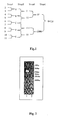
- Fig.1 The diagram process of constructing a multi-cloning DNA frame with an overlapping PCR.
- the numbers from 2 to 11 represent different synthetic fragments of polymeric nucleic acids.
- the signs of " A, B, C, D, E, I , II, III, IV, UP, DOWN" represent the semi-finished products of constructions.
- the sign of "WHOLE” represents the final product.
- Fig.2 Detection of the over-lapping PCR products by Agarose Gel electrophoresis. Lane 1: the product of the over-lapping PCR; Lane 2: DL2000 DNA marker (Dalian Takara Biotech.).
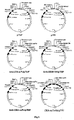
- Fig.5 The diagram maps of vectors for constructing and expressing CEA-scTsAb.
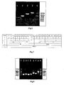
- Fig.6 Identification of the constructing process by Agarose Gel electrophoresis.
- Lane 1 PCR product amplified from the empty vector pTRI;
- Lane 2 PCR product amplified from vector CD28 V H /pTRI;
- Lane 3 PCR product amplified from the vector CD3scFv/CD28 V H /pTRI;
- Lane 4 PCR product amplified from the vector CEA-scTsAb/pTRI;
- Lane 5 DL2000 DNA marker (Dalian Takara Biotech.).
- Fig.7 The diagram process for constructing murine anti-CEA scFv with the overlapping PCR is shown.
- the numbers from 1 to 22 represent different synthetic fragments of polymeric nucleic acids.
- the signs of " A, B, C, D, E, F, G, H, I, J, K, a, b, c, d, e, f, g, I , II , III, IV, UP, DOWN" represent the semi-finished products of the construction.
- the sign of "WHOLE” represents the finished product.
- Fig.8 Detection of the over-lapping PCR product by Agarose Gel electrophoresis.
- Lane 1 and 9 DL2000 DNA markers (Dalian Takara Biotech.).
- Lane 2-5 semi-finished products: I, II , III, IV;
- Lane 6 and lane 7 semi-finished products UP and DOWN;
- Lane 8 the finished product WHOLE.
- Fig.9 SDS-PAGE of the soluble expression of the CEA-scTsAb.
- Lane 1 the ultrasonic deposition of the CEA-scTsAb/pTRI expression
- Lane 2 the ultrasonic supernatant of the CEA-scTsAb/pTRI expression
- Lane 3 the protein molecular weight standards (Shanghai Biochemistry Institute)
- Lane 4 the ultrasonic deposition of the empty vector pTRI expression
- Lane 5 the ultrasonic supernatant of the empty vector pTRI expression.
- the bands of the CEA-scTsAb are arrowed in corresponding lanes.
- Fig.10 Western blotting of the soluble expression of CEA-scTsAb.
- Lane 1 protein molecular weight standards (NEB);
- Lane 2 the ultrasonic deposition of the CEA-scTsAb/ pTRI expression;
- Lane 3 the ultrasonic deposition of empty vector pTRI expression;
- Lane 4 the ultrasonic supernatant of the CEA-scTsAb/pTRI expression;
- Lane 5 the ultrasonic supernatant of the empty vector pTRI expression.
- Fig.11 SDS-PAGE of CEA-scTsAb purified with DEAE anion exchange chromatography.
- Lane 1 the ultrasonic supernatant of the empty vector pTRI expression
- Lane 2 the ultrasonic supernatant of the CEA-scTsAb/ pTRI expression
- Lane 3 the flow-through of DEAE anion exchange chromatography
- Lane 4 the NaCl elution of DEAE anion exchange chromatography
- Lane 5 the NaOH elution of DEAE anion exchange chromatography
- Lane 6 the protein molecular weight standards (Shanghai Biochemistry Institute).
- the bands of CEA-scTsAb are arrowed in corresponding lanes.
- Fig.12 ELISA (enzyme linked immunosorbent assay) results of CEA-scTsAb. From top to bottom, the first curve: 10 ⁇ g/ml Jurkat membrane antigen; the second one: 1 ⁇ g/ml purified CEA (R&D); the third one: 1 ⁇ g/ml CD28-Fc chimera (R&D); the fourth one with no antigen coated.
- Fig.13 FCM of the binding of CEA-scTsAb to different tumor cells.
- the shadowed peak is the negative control with no CEA-scTsAb added.
- Fig.14 FCM of the binding of CEA-scTsAb to Jurkat cells and peripheral blood mononuclear cells (PBMC).
- the shadowed peak is the negative control with no CEA-scTsAb.
- Fig.16 Effect of the CEA-scTsAb concentration on tumor specific cytolysis in a MTT assay.
- efficiency of the tumor specific cytolysis displays a negative correlation with the concentration of CEA-scTsAb and reached a peak at 6 ⁇ g/ml.
- second phase from 750 ng/ml to 6 ⁇ g/ml, it displayed a direct correlation and reaches the bottom at 750 ng/ml.
- the third phase from 24 ng/ml to 750 ng/ml, it turns back into a negative correlation.
- the fourth phase from 24ng/ml to zero, the direct correlation appeared again.
- Fig.17 Effect of the CEA-scTsAb concentration on the proliferation of effector cells as determined by MTT assay.
- SI stimulating index
- the third phase from 50ng/ml to zero, it turns back into a direct correlation.
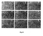
- Fig.18 The morphological changes of mixed cells in the process of the tumor specific cytolysis induced by CEA-scTsAb.
- A SW1116 tumor cells after 20 hours of culturing.
- B The adherent target cells begin to detach.
- C The effector cells aggregate on the surface of the target cells.
- E Partial membrane of target cells breaks up.
- F The whole membranes of target cells break up.
- G -(I) The target cells break into fragments.
- Fig.19 The mechanism diagram of the tumor specific cytolysis induced by CEA-scTsAb.
- the upper map the structure of CEA-scTsAb; the lower map: The mechanism diagram of the tumor specific cytolysis induced by CEA-scTsAb; CEA-scTsAb binds to both target cell and the effector cell simultaneously, thus resulting in activation of the effector cell which is providing dual signals, which causes specific target cell killing.
- Fig.20 Micrographic photos of killed target cells (SW1116). Lines A, B, and C represent three different states of tumor cell death respectively: necrosis, late apoptosis and early apoptosis.
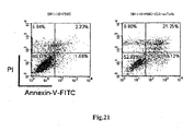
- FCM PI/Annexin V-FITC (Fluorescein Isothiocyanate) of tumor specific cytolysis.
- Four quadrants represent different states of tumor cells: live cells in the lower left quadrant (LL); early apoptosis cells in the lower right quadrant (LR); late apoptosis cells in the upper right quadrant (UR); necrosis cells in the upper left quadrant (UL).
- the sample added with 50ng/ml CEA-scTsAb LL (52.83%), LR (16.12%), UL(9.8%), UR (21.25%).
- the method for inducing the cytoplasmic soluble expression of CEA-scTsAb at low temperature mentioned in this invention requires that the host bacteria is induced with 0.4mM IPTG at 30°C. With this method, the ratio of inclusion bodies can be decreased remarkably and about 50% of expressed CEA-scTsAb is soluble.
- the soluble expression of CEA-scTsAb can be used in a further step of purification directly, without de-naturation or re-naturation, which can reduce the cost of production and improve the output.
- the method of the single-step purification collecting flow-through of DEAE anion exchange chromatography requires that the soluble expression products are loaded into the column with the DEAE anion exchange resin at pH 8.0. Subsequently, almost all proteins of non-interest can be absorbed while most of the CEA-scTsAb flows through with an approximate 75% purity.
- CEA-scTsAb With transformation into E.coli BL21(DE3) and induction with IPTG at low temperature (30°C), CEA-scTsAb is expressed solubly in cytoplasm. With the single step of DEAE anion exchange chromatography, it is primarily purified. The binding specificities to the three antigens (CEA, CD3, CD28) are detected by ELISA. The binding specificities to tumor cells are detected by FCM after conjugating CEA-scTsAb with FITC. The cytolysis of tumor cells and the proliferation of T lymphocytes induced by CEA-scTsAb are both analyzed by the MTT assay. The morphological changes of tumor cells are recorded by microphotography with an inverted microscope. The necrosis and apoptosis of tumor cells induced by CEA-scTsAb are visualized with a dual-color FCM, PI/annexin-V-FITC, and fluorescence-microscope.
- the DNA fragment containing multiple cloning sites and empty vector pTMF (Zhang, Li et al. 2002) are both cut with NcoI/BamHI and ligated together.
- the products of ligation are transformed into E.coli strain TOP10 (Invitrogen).
- the plasmid isolated from the transformed bacterial cells is named pTRI.
- the DNA fragment coding anti-CD28V H is amplified from CD28 V H /pTMF (Cheng, Wang et al. 2002) with P1 (P1: 5'-TCACATATGCA GGTACAGC TACAG-3') (SEQ ID NO: 19) as the up-stream primer and P2 (P2: 5'-TTCGCTAGCGGAAGATACGGTA CCA-3') (SEQ ID NO; 20) as the down-stream primer.
- P1 P1: 5'-TCACATATGCA GGTACAGC TACAG-3'
- P2 P2: 5'-TTCGCTAGCGGAAGATACGGTA CCA-3'
- the restriction sites NdeI/NheI are introduced at the 5'end and 3' end respectively during the process of PCR.
- PCR reaction mixture 1 ⁇ l primers(each); 2 ⁇ l dNTP (2mmol/ml each); 2 ⁇ l 10 ⁇ pfu buffer; 100ng CD28 V H /pTRI, plasmid; 0.3 ⁇ l Pfu (Promega Co.); add distilled water to the volume of 20 ⁇ l.
- PCR reaction condition to pre-denature at 94°C for 3 minutes; to denature at 94°C for 30 seconds; to anneal at 55°C for 30 seconds; to elongate at 72°C for 50 seconds; 25 cycles.
- the PCR products are purified by agarose electrophoresis (1%) and DNA Gel purification kit (Watson Biotech. Inc.).
- the above PCR product and pTRI plasmid are cut with NdeI/Nhel (Promega Co.) at the same time.
- the cutting of the PCR product (about 350bp) and of pTRI (about 5300bp) are ligated together and transformed into TOP10 E.coli strain.
- the plasmids isolated from the positive clones are named as CD28 V H /pTRI, which are identified by PCR with the product of about 750bp (As shown in Fig.6 ). All operating procedures needed here come from step (1).
- the DNA fragment coding anti-CD3 scFv is amplified from CD3 scFv/pTMF (Liu XF 1996)with P1 (P1: 5'-AAGAGTACTGAGGTGAAGCTGGTGG-3') (SEQ ID NO: 21) as the up-stream primer and P2 (P2: 5'-GAAGTCGACAGCGCGCTTCAGTTCCAG-3') (SEQ ID NO: 22) as the down-stream primer.
- P1 P1: 5'-AAGAGTACTGAGGTGAAGCTGGTGG-3'
- P2 P2: 5'-GAAGTCGACAGCGCGCTTCAGTTCCAG-3'
- the restriction sites, Scal and Scall are introduced at the 5'end and 3' end respectively during the process of PCR.
- PCR reaction mixture 1 ⁇ l primers(each); 2 ⁇ l dNTP (2mmol/ml each);2 ⁇ l 10 ⁇ pfu buffer; 100ng CD28 V H /pTRI, plasmid; 0.3 ⁇ l Pfu (Promega Co.); add distilled water to the volume of 20 ⁇ l.
- PCR reaction condition to pre-denature at 94°C for 3 minutes; to denature at 94"C for 30 seconds; to anneal at 55°C for 30 seconds; to elongate at 72°C for 50 seconds; 25 cycles.
- the PCR products are purified by agarose electrophoresis (1%) and DNA Gel purification kit (Watson Biotech. Inc.).
- the above PCR product and CD28 V H /pTRI plasmid are cut with Scal/Scall (Promega Co.) at the same time.
- the cutting product of PCR (about 750bp) and that of CD28 V H /pTRI (about 5700bp) are ligated together and transformed into TOP 10 E.coli strain.
- the plasmids isolated from the positive clones are named as CD3 scFv/CD28 V H /pTRI, which are identified by PCR with the product of about 1400bp (As shown in Fig.6 ). All operating procedures needed here come from step (1).
- Example 3 Soluble cytoplasmic expression of CEA-scTsAb induced at lower temperature.
- the competent BL21 (DE3) cells are prepared referring to the method in example 2.
- the plasmid CEA scTsAb/pTRI) is isolated with plasmid isolating kit (Watson Biotech.Inc.). The subsequent procedures of transformation and identification of positive clones are performed according to example 2.
- the single clone of BL21 (DE3) containing CEA-scTsAb/pTRI is picked up from the LB-K plate and inoculated in 5ml LB-K medium. After being cultured at 37°C with shaking overnight, the culture is transferred into 250 ml LB-K medium at a ratio of 1/100, shake at 37°C to reach A600 of 0.6. IPTG (Takara Biotech. (Dalian)) is added to the final concentration of about 0.4mmol/l to induce soluble expression at 30°C for 4 hours.
- the bacterial cells are harvested by centrifuging at 12,000 rpm for 10 minutes and then re-suspended in phosphate buffered saline (PBS: 8g NaCl, 0.2gKCl, 1.44g Na 2 HPO 4 , 0.24g KH 2 PO 4 , pH7.4, 1 liter) (1/5 volume of culture medium).
- PBS phosphate buffered saline
- cytoplasmic soluble CEA-scTsAb is released into the supernatant produced by centrifuging ultrasonic-lyzed cells.
- soluble expression and inclusion body expression of CEA-scTsAb are detected by reducing SDS-PAGE and Western-blotting according to "molecular cloning: a laboratory manual"(Sambrook and Russell 2001).
- Example 4 Purification of CEA-scTsAb by a single step of DEAE anion exchange chromatography.
- 250ml culture medium containing bacterial cells expressing CEA-scTsAb are centrifuged at 12,000rpm at 4°C for 10 minutes.
- the pellet is resuspended in 50ml equilibrium buffer (20mmol/l NaCl, 20mmol/l Tris-HCl, pH 8.0) of DEAE anion exchange chromatography for further sonication.
- the supernatant containing solubly expressed CEA-scTsAb is applied in the purifying step directly.
- the purified sample is then dialyzed against PBS at 4°C overnight.
- the protein concentration is quantified by Bradford method(Ausubel 1999). After supplementing sodium azide (0.05%(W/V)) and fucose (0.15mol/l), the dialyzed solution is divided into 1ml aliquots and stored at-80°C.
- Example 5 Detection of the binding specificity to three antigens (CEA, CD3, CD28) by ELISA.
- Jurkat cell membrane antigen 5 ⁇ 10 6 Jurkat cells (American type culture collection, ATCC, TIB-152) are harvested by centrifuging at 1000g for 10 minutes. The cell pellet is suspended in 0.5ml PBS and lysed by ultra-sonication. The supernatant of the ultrasonication produced by centrifuging at 12,000rpm for 10 minutes is supplemented with sodium azide (0.05%(W/V)) and fucose (0.15mol/l), divided into 100 ⁇ l aliquots and stored at -80°C.
- CEA-scTsAb binds to three antigen, CEA, CD3 rich Jurkat membrane antigen, and rhCD28-Fc chimera very specifically.
- CEA-scTsAb binds to SW1116 and SK-OV-3 the best; CEA-scTsAb binds to A549 modestly; CEA-scTsAb does not bind to MCF-7.
- Example 7 FACSanalysis of the binding specificity of CEA-scTsAb to PBMC and Jurkat cells
- a direct FCM method is used here to test the binding specificity of CEA-scTsAb to PBMC and Jurkat cells.
- CEA-scTsAb binds to PBMC and Jurkat cells specifically.
- CEA-scTsAb could bind to PBMC, Jurkat, and several tumor cells specifically, which is very important for inducing tumor specific cytolysis.
- Example 8 Detection of tumor specific cytolysis of colorectal carcinoma cell, SW1116, induced by CEA-scTsAb in the presence of lymphocytes.
- CEA expressing tumor cell line, SW1116, is used as the target cell (T), while lymphocytes from PBMC are used as the effector cells (E).
- T target cell
- E effector cells
- the efficiency of tumor specific cytolysis displays negative correlation with the concentration of CEA-scTsAb and reaches a peak at 6 ⁇ g/ml;
- the second phase from 750ng/ml to 6 ⁇ g/ml it displayed a direct correlation and reaches the bottom at 750 ng/ml;
- the third phase from 24ng/ml to 750ng/ml, it changes back into a negative correlation;
- the fourth phase from 24ng/ml to zero, the direct correlation appeared again. Anyhow, two peaks of tumor specific cytolysis exist: 85% at 6 ⁇ g/ml and 70% at 24ng/ml. It can be concluded from the above data that extremely efficient tumor specific cytolysis could be induced at an even lower E/T ratio or lower concentration of CEA-scTsAb.
- Example 9 Morphological observation of tumor cells during the process of tumorspecific cytolysis.
- PBMC effector cells
- SW1116 cells target cells
- L15 medium 10% FBS
- CEA-scTsAb purified CEA-scTsAb at a concentration of 1 ⁇ g/ml
- the mixture is incubated at 37°C for 20-40 h in a 5% CO 2 incubator.
- morphological changes of tumor cells and PBMC are observed under a 40 ⁇ objective lens with an OLYMPUS IMT-2 inverted microscope, and recorded by photomicrography.
- Fig. 18 (A)-(G) there are four steps of morphological changes.
- effector cells mainly T lymphocytes
- MTT assay The proliferation of effector cells (mainly T lymphocytes) detected with MTT assay can be used to evaluate the activation of T lymphocytes induced by co-incubated tumor cells and CEA-scTsAb.
- SI stimuli index
- the first phase from 750ng/ml to 12 ⁇ g/ml
- SI displays direct correlation with the concentration of CEA-scTsAb and reaches the bottom at 750ng/ml.
- the second phase from 50ng/ml to 6 ⁇ g/ml it displayed a negative correlation and reaches the peak at 50ng/ml.
- the third phase from 50ng/ml to zero, it changes back into a direct correlation. It can be concluded that CEA-scTsAb possesses stimulating ability of T lymphocytes even at extremely lower concentrations.
- CEA-scTsAb tumor specific cytolysis induced by CEA-scTsAb corresponded to the activating state of co-incubated T lymphocytes.
- the function CEA-scTsAb focuses on two aspects: (1) retargeting effector cells around tumor cells; and (2) stimulating effector cells to kill target cells specifically.
- retargeted cytotoxic T lymphocytes (CTL) are activated to kill target tumor cells directly; T helper cells secret cytokines, such as IL-2, IFN- ⁇ and TNF- ⁇ , to assist CTL or natural killing cells (NK cell) in killing target tumor cells indirectly.
- Example 11 The mechanism of tumor specific cytolysis induced by CEA-scTsAb
- activated CTLs secret perforins to make holes on the membrane surface of tumor cells, which are broken up and induce necrosis; grazymes secreted by activated CTL can enter tumor cells through the above holes and induce apoptosis; activated CTL would be induced to express Fas ligands on its surface, which interact with Fas molecules on tumor cells and induce apoptosis.
- PI/annexin-V-FITC dual-color FCM fluorescence cytometry
- subsequent fluorescence microphotography are used here to distinguish necrosis from apoptosis of tumor cells in an in vitro assay of tumor specific cytolysis.
Landscapes
- Health & Medical Sciences (AREA)
- Chemical & Material Sciences (AREA)
- Immunology (AREA)
- Organic Chemistry (AREA)
- Life Sciences & Earth Sciences (AREA)
- Medicinal Chemistry (AREA)
- General Health & Medical Sciences (AREA)
- Proteomics, Peptides & Aminoacids (AREA)
- Molecular Biology (AREA)
- Genetics & Genomics (AREA)
- Biophysics (AREA)
- Biochemistry (AREA)
- Oncology (AREA)
- Veterinary Medicine (AREA)
- Public Health (AREA)
- Cell Biology (AREA)
- Animal Behavior & Ethology (AREA)
- Pharmacology & Pharmacy (AREA)
- Nuclear Medicine, Radiotherapy & Molecular Imaging (AREA)
- General Chemical & Material Sciences (AREA)
- Chemical Kinetics & Catalysis (AREA)
- Engineering & Computer Science (AREA)
- Bioinformatics & Cheminformatics (AREA)
- Peptides Or Proteins (AREA)
- Micro-Organisms Or Cultivation Processes Thereof (AREA)
- Preparation Of Compounds By Using Micro-Organisms (AREA)
- Medicines Containing Antibodies Or Antigens For Use As Internal Diagnostic Agents (AREA)
- Medicines Containing Material From Animals Or Micro-Organisms (AREA)
- Medicines That Contain Protein Lipid Enzymes And Other Medicines (AREA)
Abstract
Description
- This invention refers to the field of a recombinant genetically engineered antibody, more concretely, a linear single chain recombinant anti-CEA/CD3/CD28 trispecific antibody (scTsAb). This invention also refers DNA sequence coding for the said scTsAbs, plasmids containing the said sequences, and host cells containing the said plasmids.
- The activation of T lymphocytes requires two kinds of signals in vivo: 1) the interaction between the MHC/antigen peptide complex on APC (antigen presenting cells) and the TCR/CD3 complex on T lymphocytes provides the first signal; and 2) the interaction between the co-stimulatory receptor on APC and the co-stimulatory molecule on T lymphocytes provides the second signal, a co-stimulatory signal. It was generally accepted that T lymphocytes cannot be activated fully in the presence of the first signal alone (Baxter and Hodgkin 2002; Bernard, Lamy et al. 2002).
- There are two kinds of T lymphocytes including cytotoxic T lymphocytes (CTL) and T helper cells (TH). CTL is the major effector cell for a cellular immunological response, while TH participates in a cellular immunological response indirectly by secreting cytokines (such as interleukin-2 (IL-2)). As tumor immunity majors in cellular immunity, designing anti-tumor drugs to activate CTL specifically is of great importance in tumor immunotherapy (Foss 2002).
- In practice, some recombinant anti-TAA/CD3 bispecific antibodies (BsAbs) have been designed to provide the first signal for CTL activation and retarget activated CTLs around tumor cells for killing them specifically. Among them, several ones have entered into clinic (Daniel, Kroidl et al. 1998; Holliger, Manzke et al. 1999; Loffler, Kufer et al. 2000; Manzke, Tesch et al. 2001; Manzke, Tesch et al. 2001; Dreier, Lorenczewski et al. 2002; Fang M 2002; Dreier, Baeuerle et al. 2003; Loffler, Gruen et al. 2003; Fang, Zhao et al. 2004). They have been proven to activate T lymphocytes specifically and induce obvious tumor specific cytolysis. However, most of them could not activate T lymphocytes fully and sometimes may result in activation of induced cell death (AICD) of T lymphocytes (Daniel, Kroidl et al. 1998).
- To overcome the above defects, another kind of BsAb, anti-TAA/CD28 BsAb, was designed. Accompanied with anti-TAA/CD3 BsAb, CTLs are provided with dual activating signals, which induces more efficient tumor specific cytolysis. However, there are several disadvantages in the combinatorial use of two BsAbs. These include the duplicate steps in expression and purification, the consequential increase of production cost, and the partnership of two BsAbs in clinical medication. A trispecific antibody (TsAb) with three binding specificities (TAA, CD3, and CD28) might replace the above two BsAbs in providing dual activating signals (Jung, Brandl et al. 2001; Kodama, Suzuki et al. 2002) and be superior to them in expression, purification, and clinical medication.
- Heretofore, there are three types of TsAb. These include 1) the chemically conjugated TsAb (Jung, Freimann et al. 1991; Tutt, Stevenson et al. 1991; French 1998; Wong, Vakis et al. 2000), 2) the recombinant polymeric TsAb (Atwell, Breheney et al. 1999; Dolezal, Pearce et al. 2000; Schoonjans, Willems et al. 2000; Schoonjans, Willems et al. 2000; Kortt, Dolezal et al. 2001; Schoonjans, Willems et al. 2001; Willems, Leoen et al. 2003), and 3) the single chain recombinant TsAb (scTsAb) (Li-ping, Ju-long et al. 2003). The third type of TsAb is believed to be superior to the others for its simplification in construction, expression, and purification. As carcinoembryonic antigen (CEA) is a broad-spectrum TAA (Ganjei, Nadji et al. 1988; Horie, Miura et al. 1996; Kuo, Tsai et al. 1996; Feil, Wechsel et al. 1999; Kammerer, Thanner et al. 2003; Shang-zhi, Chang-cheng et al. 2004), the scTsAb containing an anti-CEA antibody may be used in preventing or curing diverse tumors in clinic.
Document EP 1 378 520 discloses a cyclic strand trispecific TAA/CD3/CD28 antibody. - The introduction of anti-CEA antibody in an anti-CEA/CD3/CD28 scTsAb in this invention provides a convenience to distinguish tumor cells from normal cells in vivo, and avoid or decrease the non-specific killing by the activated T lymphocytes.
- Another aspect of this invention is CEA is widely expressed on many tumor cells, and it also provides a broad application for curing or preventing different tumors in the future.
- Even another aspect of this invention is it provides a method for constructing the scTsAb.
- The amino acid sequence (SEQ ID NO: 1) of an murine anti-CEA single chain fragment of a variable region contained in the CEA-scTsAb is listed:

- The amino acid sequence (SEQ ID NO: 2) of the anti-CD3 single chain fragment of the variable region contained in CEA-scTsAb is listed below:

- The nucleic acid sequence (SEQ ID NO: 3) of CEA-scTsAb is listed as follows:


- The amino acid sequence (SEQ ID NO: 4) of CEA-scTsAb is listed below:

- Another aspect of this invention is that it provides a vector for expressing the CEA-scTsAb: CEA-scTsAb/pTRI.
- However, in the context of this invention, other aspects and advantages of this invention are obvious to the ordinary persons engaged in the similar field, especially based on that disclosed in the " Example" part.
- It will be understood that the drawings are intended to provide assistance to the present invention, which do not constitute a departure from the spirit and scope of the invention.
-
Fig.1 . The diagram process of constructing a multi-cloning DNA frame with an overlapping PCR. The numbers from 2 to 11 represent different synthetic fragments of polymeric nucleic acids. The signs of " A, B, C, D, E, I , II, III, IV, UP, DOWN" represent the semi-finished products of constructions. The sign of "WHOLE" represents the final product. -
Fig.2 . Detection of the over-lapping PCR products by Agarose Gel electrophoresis. Lane 1: the product of the over-lapping PCR; Lane 2: DL2000 DNA marker (Dalian Takara Biotech.). -
Fig.3 . The sequence of the multi-cloning DNA frame -
Fig.4 . The process for constructing CAA-scTsAb -
Fig.5 . The diagram maps of vectors for constructing and expressing CEA-scTsAb. -
Fig.6 . Identification of the constructing process by Agarose Gel electrophoresis. Lane 1: PCR product amplified from the empty vector pTRI; Lane 2: PCR product amplified from vector CD28 VH/pTRI; Lane 3: PCR product amplified from the vector CD3scFv/CD28 VH/pTRI; Lane 4: PCR product amplified from the vector CEA-scTsAb/pTRI; Lane 5: DL2000 DNA marker (Dalian Takara Biotech.). -
Fig.7 . The diagram process for constructing murine anti-CEA scFv with the overlapping PCR is shown. - The numbers from 1 to 22 represent different synthetic fragments of polymeric nucleic acids. The signs of " A, B, C, D, E, F, G, H, I, J, K, a, b, c, d, e, f, g, I , II , III, IV, UP, DOWN" represent the semi-finished products of the construction. The sign of "WHOLE" represents the finished product.
-
Fig.8 . Detection of the over-lapping PCR product by Agarose Gel electrophoresis.Lane 1 and 9: DL2000 DNA markers (Dalian Takara Biotech.). Lane 2-5: semi-finished products: I, II , III, IV;Lane 6 and lane 7: semi-finished products UP and DOWN; Lane 8: the finished product WHOLE. -
Fig.9 . SDS-PAGE of the soluble expression of the CEA-scTsAb. Lane 1: the ultrasonic deposition of the CEA-scTsAb/pTRI expression; Lane 2: the ultrasonic supernatant of the CEA-scTsAb/pTRI expression; Lane 3: the protein molecular weight standards (Shanghai Biochemistry Institute); Lane 4: the ultrasonic deposition of the empty vector pTRI expression; Lane 5: the ultrasonic supernatant of the empty vector pTRI expression. The bands of the CEA-scTsAb are arrowed in corresponding lanes. -
Fig.10 . Western blotting of the soluble expression of CEA-scTsAb. Lane 1: protein molecular weight standards (NEB); Lane 2: the ultrasonic deposition of the CEA-scTsAb/ pTRI expression; Lane 3: the ultrasonic deposition of empty vector pTRI expression; Lane 4: the ultrasonic supernatant of the CEA-scTsAb/pTRI expression; Lane 5: the ultrasonic supernatant of the empty vector pTRI expression. -
Fig.11 . SDS-PAGE of CEA-scTsAb purified with DEAE anion exchange chromatography. Lane 1: the ultrasonic supernatant of the empty vector pTRI expression; Lane 2: the ultrasonic supernatant of the CEA-scTsAb/ pTRI expression; Lane 3: the flow-through of DEAE anion exchange chromatography; Lane 4: the NaCl elution of DEAE anion exchange chromatography; Lane 5: the NaOH elution of DEAE anion exchange chromatography; Lane 6: the protein molecular weight standards (Shanghai Biochemistry Institute). The bands of CEA-scTsAb are arrowed in corresponding lanes. -
Fig.12 . ELISA (enzyme linked immunosorbent assay) results of CEA-scTsAb. From top to bottom, the first curve: 10µg/ml Jurkat membrane antigen; the second one: 1µg/ml purified CEA (R&D); the third one: 1µg/ml CD28-Fc chimera (R&D); the fourth one with no antigen coated. -
Fig.13 . FCM of the binding of CEA-scTsAb to different tumor cells. The shadowed peak is the negative control with no CEA-scTsAb added. -
Fig.14 . FCM of the binding of CEA-scTsAb to Jurkat cells and peripheral blood mononuclear cells (PBMC). The shadowed peak is the negative control with no CEA-scTsAb. -
Fig.15 . Effect of E/T ratio (effector cells/target cells) on tumor specific cytolysis induced by CEA-scTsAb as determined by the MTT assay. From top to bottom, the first curve: E/T=10; the second one: E/T=5; the third one: E/T=1. Effector cells: PBMC. Target cells: SW1116 tumor cells. -
Fig.16 . Effect of the CEA-scTsAb concentration on tumor specific cytolysis in a MTT assay.
There are four stepwise-phases for tumor specific cytolysis. In the first phase, from 6µg/ml to 12µg/ml, efficiency of the tumor specific cytolysis displays a negative correlation with the concentration of CEA-scTsAb and reached a peak at 6 µg/ml. In the second phase, from 750 ng/ml to 6µg/ml, it displayed a direct correlation and reaches the bottom at 750 ng/ml. In the third phase from 24 ng/ml to 750 ng/ml, it turns back into a negative correlation. In the fourth phase, from 24ng/ml to zero, the direct correlation appeared again. -
Fig.17 . Effect of the CEA-scTsAb concentration on the proliferation of effector cells as determined by MTT assay. There are three stepwise phases for stimulating index (SI). In the first phase from 750ng/ml to 12µg/ml, SI displays direct correlation with the concentration of CEA-scTsAb and reaches the bottom at 750ng/ml. In the second phase from 50ng/ml to 750ng/ml, it displayed a negative correlation and reached a peak at 50ng/ml. In the third phase from 50ng/ml to zero, it turns back into a direct correlation. -
Fig.18 . The morphological changes of mixed cells in the process of the tumor specific cytolysis induced by CEA-scTsAb. (A) SW1116 tumor cells after 20 hours of culturing. (B)-(I) the mixture of SW1116 (target cells) and PBMC (effector cells) added with CEA-scTsAb. E/T=5. (B) The adherent target cells begin to detach. (C) The effector cells aggregate on the surface of the target cells. (D) TsAbs appeared on the surface of the target cells. (E) Partial membrane of target cells breaks up. (F) The whole membranes of target cells break up. (G)-(I) The target cells break into fragments. -
Fig.19 . The mechanism diagram of the tumor specific cytolysis induced by CEA-scTsAb. The upper map: the structure of CEA-scTsAb; the lower map: The mechanism diagram of the tumor specific cytolysis induced by CEA-scTsAb; CEA-scTsAb binds to both target cell and the effector cell simultaneously, thus resulting in activation of the effector cell which is providing dual signals, which causes specific target cell killing. -
Fig.20 . Micrographic photos of killed target cells (SW1116). Lines A, B, and C represent three different states of tumor cell death respectively: necrosis, late apoptosis and early apoptosis. -
Fig.21 . FCM (PI/Annexin V-FITC (Fluorescein Isothiocyanate) of tumor specific cytolysis. Four quadrants represent different states of tumor cells: live cells in the lower left quadrant (LL); early apoptosis cells in the lower right quadrant (LR); late apoptosis cells in the upper right quadrant (UR); necrosis cells in the upper left quadrant (UL). The sample with no CEA-scTsAb added: LL (90.17%), LR (1.66%), UL (5.94%), UR (2.23%); The sample added with 50ng/ml CEA-scTsAb: LL (52.83%), LR (16.12%), UL(9.8%), UR (21.25%). - In this invention, all terms are easy to understand for ordinary workers engaged in this field except which is explained explicitly. Here, some terms are described as below:
- Linear single chain recombinant trispecific antibody is the linear single molecule constructed with three different antigen-binding specificities by a genetic engineering method. Specifically, a linear single chain recombinant anti-CEA/CD3/CD28 trispecific antibody is the single linear molecule constructed by fusing three different antibody fragments (anti-CEA antibody, anti-CD3 antibody, anti-CD28 antibody), which are spaced with two linkers (Fc linker and HSA linker) (Min Fang 2003). As a choice, c-myc tag and (His)6-tag can be added at the C terminal of the molecule for activity detection or further purification (Hengen 1995; Fan, Villegas et al. 1998). The antibody fragments mentioned here could be a single chain fragment of the variable region (scFv), an Fab fragment of an antibody, or a single domain antibody (VH or VL). More concretely, CEA-scTsAb is constructed by fusing an anti-CEA scFv, an Fc linker, an anti-CD3 scFv, an HSA linker, and an anti-CD28 VH in tandem, a with c-myc tag and a His-6 tag at its C terminal. There are two advantages in this module:
- 1. Based on a dual-signal activating model for T cells, it is endowed with the ability of activating T cells fully.
- 2. As CEA is a broad-spectrum TAA, it is endowed with broad applications for curing or preventing many tumors in clinic.
- The method for inducing the cytoplasmic soluble expression of CEA-scTsAb at low temperature mentioned in this invention requires that the host bacteria is induced with 0.4mM IPTG at 30°C. With this method, the ratio of inclusion bodies can be decreased remarkably and about 50% of expressed CEA-scTsAb is soluble. The soluble expression of CEA-scTsAb can be used in a further step of purification directly, without de-naturation or re-naturation, which can reduce the cost of production and improve the output.
- The method of the single-step purification collecting flow-through of DEAE anion exchange chromatography requires that the soluble expression products are loaded into the column with the DEAE anion exchange resin at pH 8.0. Subsequently, almost all proteins of non-interest can be absorbed while most of the CEA-scTsAb flows through with an approximate 75% purity.
- The operating procedure of this invention is listed below:
- At first, the parent vector pTRI is constructed by introducing new special multi-cloning sites (MCS). Then the DNA fragment coding anti-CD28 VH is amplified with PCR from the vector CD28 VH/pTMF, at both ends of which a special pair of restriction sites, Nde I/Kpn I, is added. With the same method, the DNA fragment coding anti-CD3 scFv with restriction sites Scal/Sall, is prepared. The DNA fragment coding anti-CEA scFv with restriction sites Xhol/EcoRI, is cut from CEA scFv/pTMF. At last, all three fragments are introduced into pTRI in tandem to produce the final vector CEA scTsAb/pTRI. The DNA fragments coding anti-CEA scFv, anti-CD3 scFv and anti-CD28 VH are arrayed in tandem from N terminus to C terminus.
- With transformation into E.coli BL21(DE3) and induction with IPTG at low temperature (30°C), CEA-scTsAb is expressed solubly in cytoplasm. With the single step of DEAE anion exchange chromatography, it is primarily purified. The binding specificities to the three antigens (CEA, CD3, CD28) are detected by ELISA. The binding specificities to tumor cells are detected by FCM after conjugating CEA-scTsAb with FITC. The cytolysis of tumor cells and the proliferation of T lymphocytes induced by CEA-scTsAb are both analyzed by the MTT assay. The morphological changes of tumor cells are recorded by microphotography with an inverted microscope. The necrosis and apoptosis of tumor cells induced by CEA-scTsAb are visualized with a dual-color FCM, PI/annexin-V-FITC, and fluorescence-microscope.
- The schematic process is shown in
Fig.1 . All synthetic fragments used here are listed below: - 1. 5'-TAT ACC ATG GGT CTC GAG-3' (SEQ ID NO:5)
- 2. 5'-TAT ACC ATG GGT CTC GAG ATG TAC CCG CGC GGT AAC ACT AGT GAA TTC AAC AGC ACG TA-3' (SEQ ID NO:6)
- 3. 5'-AGC CAG TCC TGG TGC AGT ACG GTG AGG ACG CTT ACA ACC CGG TAC GTG CTG TTG AAT TC-3' (SEQ ID NO:7)
- 4. 5'-CTG CAC CAG GAC TGG CTG AAT GGC AAG GAA TAC AAA TGC AAG AGT ACT TCT AGA ATG TA-3' (SEQ ID NO:8)
- 5. 5'-CGA ACC AGC AGC GCA TTC TGG AAG TCG ACG TTA CCG CGC GGG TAC ATT CTA GAAGTACT-3' (SEQ ID NO:9)
- 6. 5'-AAT GCG CTG CTG GTT CGT TAC ACC AAG AAA GTA CCC CAA GTG TCA ACT CCA ACT CCT GT-3' (SEQ ID NO:10)
- 7. 5'-GCG GTA CCG TTA CCG CGC GGG TAC ATC ATA TGT GAG ACC TCT ACA GGA GTT GGA GTT GA-3' (SEQ ID NO:11)
- 8. 5'-CGC GGT AAC GGT ACC GCG CTG GAA GTT GAC GAA ACC TAC GTT CCG AAA GAA TTT AAC GC-3' (SEQ ID NO:12)
- 9. 5'-TCG CTA GCC CCA TCC GCG GGA TGT CAG CGT GGA AGG TGA AGG TTT CCG CGT TAAATT CTT TCG G-3' (SEQ ID NO:13)
- 10. 5'-ATC GAG CTC ATG TAC CCG CGC GGT AAC GCT AGC GAA CAA AAA CTC ATC TCA GAA GAG GA-3' (SEQ ID NO:14)
- 11. 5'-TA TTG CTC GTG ATG GTG ATG ATG ATG TGC GGC CCC ATT CAG ATC CTC TTC TGA GAT GAG-3' (SEQ ID NO:15)
- 12.5'-CTCGACGGATCCTTATTGCTCGTGATGGTG-3' (SEQ ID NO:16)
- The operating steps:
- Step 1: According to
Fig.1 , mix the fragments (from 2 to 11) in pairs and carry out the elongating reaction as below.
Reaction mixture: the synthetic fragments, 1 µl (each); 10×PCR bluffer,2µl; dNTPs (2 mmol/ml each) (made in Dalian TaKaRa Biotechnology Co. Ltd.), 2µl; Taq (1U) (made in Dalian TaKaRa Biotechnology Co. Ltd.) 0.5µl; distilled water, 14µl.
Reaction conditions: to pre-denature at 94°C for 1 minute; to denature at 94°C for 30 seconds; to anneal at 45°C for 30 seconds; to elongate at 72°C for 30 seconds; 10 cycles.
All products are collected without any purification and applied in the next step directly. - Step 2: according to
Fig.1 , to mix the products (A, B, C, D, E) ofstep 1 in pairs and carry out the elongating reaction as below.
Reaction mixture: the products ofstep 1, 10µl (each).
Reaction condition: to pre-denature at 94°C for 1 minute; to denature at 94°C for 30 seconds; to anneal at 45°C for 30 seconds; to elongate at 72°C for 30 seconds; 10 cycles.
All products are applied to agarose electrophoresis (1%) and purified by DNA Gel purifying Kit (Watson Biotech. Inc.). - Step 3: according to
Fig.1 , mix the products (I, II, III, IV) ofstep 2 in pairs and carry out the elongating reaction as below.
Reaction mixture: the products ofstep
Reaction condition: to pre-denature at 94°C for 1 minute; to denature at 94°C for 30 seconds; to anneal at 45°C for 30 seconds; to elongate at 72°C for 30 seconds; 10 cycles.
All products are applied to agarose electrophoresis (1%) and purified by DNA Gel Purifying Kit (Watson Biotech. Inc.). - Step 4: according to
Fig.1 , mix the products (UP, DOWN) ofstep 3 in pairs and carry out the amplifying reaction as below.
Reaction mixture: the products ofstep synthetic fragments 1 and 12), 1µl; 10×PCR buffer, 2µl; dNTPs (2 mmol/ml each) (Dalian TaKaRa Biotechnology Co. Ltd.), 2µl; Taq (1 U) (Dalian TaKaRa Biotechnology Co. Ltd.) 0.5µl; distilled water, 12µl.
Reaction condition: to pre-denature at 94°C for 1 minute; to denature at 94°C for 30 seconds; to anneal at 45°C for 30 seconds; to elongate at 72°C for 50 seconds; 25 cycles.
The ultimate products (439bp) are applied to agarose electrophoresis (1%) (Fig.2 .) and purified by the DNA Gel purifying Kit (Watson Biotech. Inc.). The sequence, restriction sites and its components are shown inFig.3 . - The process of construction is shown in
Fig.4 , and the schematic map of all vectors used in the process are listed inFig. 5 . - The DNA fragment containing multiple cloning sites and empty vector pTMF (Zhang, Li et al. 2002) are both cut with NcoI/BamHI and ligated together. The products of ligation are transformed into E.coli strain TOP10 (Invitrogen). The plasmid isolated from the transformed bacterial cells is named pTRI.
- Restriction enzyme digestion, ligation, preparation and transformation of TOP10 competent cells are carried out as below:
- Restriction enzyme digesting reaction: in a volume of 20 µl, 1µg of pTMF or the DNA fragment containing multiple cloning sites are digested according to the operating manual from Promega company. The products are applied to agarose electrophoresis (1%) and purified by DNA Gel Purifying Kit (Watson Biotech. Inc.).
- Ligating reaction: 50-100ng cut vector and 3-10 times(mol ratio) cut DNA fragments are mixed in a volume of 20 µl which contained 2µl 10×T4 DNA ligase buffer, 1U T4 DNA ligase (Dalian TaKaRa Biotechnology Co. Ltd.) and necessary distilled water. The ligating reaction is carried out at 16 °C overnight.
- Preparation of TOP10 competent cells: inoculate the TOP10 bacteria (Invitrogen Co.) in 2ml LB medium ((10g/l tryptone (GIBCO Co.), 5g/l yeast extract (GIBCO Co.), 5g/l NaCl, pH 7.5)), and incubate overnight at 37°C with shaking. Transfer to 20-40 ml LB medium at a ratio of 1:100, incubate at 37°C with shaking to reach A600 of 0.3-0.4 (about 2.5 hour). Chill on ice for 15 minutes and centrifuge at 4°C at 4000 rpm for 10 minutes. The pellet is resuspended in 10 ml of pre-chilled 0.1 mol/l CaCl2 (Sigma Co.) and chilled on ice for 20 minutes. After a second centrifugation at 4°C at 4000 rpm for 10 minutes, the pellet is gently resuspended in 1-2ml of pre-chilled 0.1 mol/l CaCl2 solution with 12% glycerol. Divide into aliquots of 200 µl per EP tube, store at -80°C.
- Transformation: the ligating mixture is added to 200 µl competent cells. After being mixed gently and chilled on ice for 30 minutes, it is put in a 42 degrees (Celsius) water bath for 100 seconds then chilled on ice for 2 minutes. After adding 0.8 ml LB medium into the mixture, shake it at 37°C (<150 rpm) for 45 minutes to recover the cells. The cells are centrifuged at 10,000 rpm for 1 minute, re-suspended in 50∼100µl LB medium, spread onto the LB-K plate (10g/l tryptone, 5g/l yeast extract, 5g/l NaCl, 15g/l agar (SIGMA Co.), 50 µg/ml kanamycin (SIGMA Co.), pH 7.5), and incubated at 37°C overnight.
- Selection of positive clones: pick the single clones on the LB-K plate and transfer them into 2ml LB-K medium (10g/l tryptone, 5g/l yeast extract, 5g/l NaCl, 15g/l agar (SIGMA Co.), 50 µg/ml kanamycin (SIGMA Co.), pH 7.5) separately. After shaking at 37°C overnight, the plasmids are isolated with a Plasmid Isolating Kit.(Watson Biotechnologies, Inc) according to the standard manual. The positive clones are identified by PCR with the isolated plasmids as the templates. PCR reaction mixture: 0.1∼1 µ I plasmid DNA (about 20-200ng); 10 pmol upstream primer (T7-up: 5'-TAATACGACTCACTATAGGGGA-3') (SEQ ID NO:17); 10 pmol down stream primer (T7-down: 5'-GCTAGTTATTGCTCAGCGG-3') (SEQ ID NO:18); 2
µl 10×Taq buffer; 2 µl 2mmol/ml dNTPs; 1 U Taq; 12 µl distilled-water. The PCR reaction condition: pre-denature at 94°C for 5 minutes; denature at 94°C for 40 seconds; anneal at 53 °C for 40 seconds; elongate at 72°C for 40 seconds; 25 cycles. At last 5 µl PCR product are applied to agarose electrophoresis (1%). As shown inFig.6 , the PCR product is about 500bp. - The DNA fragment coding anti-CD28VH is amplified from CD28 VH/pTMF (Cheng, Wang et al. 2002) with P1 (P1: 5'-TCACATATGCA GGTACAGC TACAG-3') (SEQ ID NO: 19) as the up-stream primer and P2 (P2: 5'-TTCGCTAGCGGAAGATACGGTA CCA-3') (SEQ ID NO; 20) as the down-stream primer. The restriction sites NdeI/NheI are introduced at the 5'end and 3' end respectively during the process of PCR.
- PCR reaction mixture: 1 µl primers(each); 2µl dNTP (2mmol/ml each); 2
µl 10×pfu buffer; 100ng CD28 VH/pTRI, plasmid; 0.3µl Pfu (Promega Co.); add distilled water to the volume of 20µl. PCR reaction condition: to pre-denature at 94°C for 3 minutes; to denature at 94°C for 30 seconds; to anneal at 55°C for 30 seconds; to elongate at 72°C for 50 seconds; 25 cycles. The PCR products are purified by agarose electrophoresis (1%) and DNA Gel purification kit (Watson Biotech. Inc.). - The above PCR product and pTRI plasmid are cut with NdeI/Nhel (Promega Co.) at the same time. The cutting of the PCR product (about 350bp) and of pTRI (about 5300bp) are ligated together and transformed into TOP10 E.coli strain. The plasmids isolated from the positive clones are named as CD28 VH/pTRI, which are identified by PCR with the product of about 750bp (As shown in
Fig.6 ). All operating procedures needed here come from step (1). - The DNA fragment coding anti-CD3 scFv is amplified from CD3 scFv/pTMF (Liu XF 1996)with P1 (P1: 5'-AAGAGTACTGAGGTGAAGCTGGTGG-3') (SEQ ID NO: 21) as the up-stream primer and P2 (P2: 5'-GAAGTCGACAGCGCGCTTCAGTTCCAG-3') (SEQ ID NO: 22) as the down-stream primer. The restriction sites, Scal and Scall, are introduced at the 5'end and 3' end respectively during the process of PCR.
- PCR reaction mixture: 1µl primers(each); 2µl dNTP (2mmol/ml each);2
µl 10×pfu buffer; 100ng CD28 VH/pTRI, plasmid; 0.3µl Pfu (Promega Co.); add distilled water to the volume of 20µl. PCR reaction condition: to pre-denature at 94°C for 3 minutes; to denature at 94"C for 30 seconds; to anneal at 55°C for 30 seconds; to elongate at 72°C for 50 seconds; 25 cycles. The PCR products are purified by agarose electrophoresis (1%) and DNA Gel purification kit (Watson Biotech. Inc.). - The above PCR product and CD28 VH /pTRI plasmid are cut with Scal/Scall (Promega Co.) at the same time. The cutting product of PCR (about 750bp) and that of CD28 VH /pTRI (about 5700bp) are ligated together and transformed into
TOP 10 E.coli strain. The plasmids isolated from the positive clones are named as CD3 scFv/CD28 VH/pTRI, which are identified by PCR with the product of about 1400bp (As shown inFig.6 ). All operating procedures needed here come from step (1). - Construction of anti-CEA scFv by overlapping PCR:
- Anti-CEA scFv is designed by Linking VH (the variable region of heavy chain) and VL (the variable region of light chain) of anti-CEA monoclonal antibody(Koga, Kanda et al. 1990) with a special polypeptide GGGGSGGGGSGGGGS) (SEQ ID NO: 23). The whole amino acid sequence of anti-CEA scFv is back translated into a DNA sequence according to the E.coli preferred codon table(Nakamura, Gojobori et al. 2000), which is split into 22 complemental oligo-nucleotides. The 22 oligo-nucleotides listed below are synthesized and assembled into the whole DNA fragment coding anti-CEA scFv by overlapping PCR.
- 1. 5'-TTCCTCGAGCAGGTTCAGCT-3' (SEQ ID NO:24)
- 2. 5'-TCGCGCCCGGTTTCATCAGTTCCGCACCGCTCTGCTGCAGCTGAACCTGCTCGAGGAA-3' (SEQ ID NO:25)
- 3. 5'-ACTGATGAAACCGGGCGCGAGCGTGAAAATCAGCTGCAAAGCGACCGGCTATACCTTC-3' (SEQ ID NO:26)
- 4. 5'-CACCCATTCGATCCAATAATCGCTGAAGGTATAGCCGGTCGCTT-3' (SEQ ID NO:27)
- 5. 5'-ATTATTGGATCGAATGGGTGAAACAGCGTCCGGGTCACGGCCTGGAATGGATCGGTGAA-3 (SEQ ID NO:28)
- 6. 5'-ACGTTCGTTGTAGTCGGTACGGCCGCTGCCCGGCAGGATTTCACCGATCCATTCCAGG-3' (SEQ ID NO:29)
- 7. 5'-CGTACCGACTACAACGAACGTTTCAAAGGCAAAGCGACCTTCACCGGCGACGTTTCTAGC -3' (SEQ ID NO:30)
- 8. 5'-TTCGCTGGTCAGGCTAGACAGTTTCATATACGCGGTGTTGCTAGAAACGTCGCCGGTGAA-3' (SEQ ID NO:31)
- 9. 5'-TGTCTAGCCTGACCAGCGAAGATAGCGCGGTGTATTACTGCGCGACCGGCACCACCCCG-3' (SEQ ID NO:32)
- 10. 5'-GCTCACGGTCACCAGGGTGCCCTGACCCCAGTAACCGAACGGGGTGGTGCCGGTCGC GCA-3' (SEQ ID NO:33)
- 11. 5'-GCACCCTGGTGACCGTGAGCGCGACTAGTACCCCGAGCCATAACAGCCATCAGGTGCC G-3' (SEQ ID NO:34)
- 12. 5'-GTCTCTAGAGCCGCTGTTCGCGGTCGGGCCGCCCGCGCTCGGCACCTGATGGCTGTTA T-3' (SEQ ID NO:35)
- 13. 5'-CGAACAGCGGCTCTAGAGACATCGTGCTGACCCAGAGCCCGGCGAGCCTGGCGGTGT C-3' (SEQ ID NO:36)
- 14. 5'-CTGGGAAGCACGGCAGGAGATGGTCGCACGCTGACCCAGAGACACCGCCAGGCTCGC CGG-3' (SEQ ID NO:37)
- 15. 5'-TCTCCTGCCGTGCTTCCCAGTCCGTTTCCACCTCCTCCTACACCTACATGCACTGGTAT-3 (SEQ ID NO:38)
- 16. 5'-GATCAGCAGTTTCGGCGGCTGACCCGGTTTCTGCTGATACCAGTGCATGTAGGTGT-3' (SEQ ID NO:39)
- 17. 5'-AGCCGCCGAAACTGCTGATCAAATATGCGAGCAACCTGGAATCTGGTGTGCCGGCGCG T-3' (SEQ ID NO:40)
- 18. 5'-GTTCAGGGTGAAGTCGGTGCCGCTGCCAGAACCGCTGAAACGCGCCGGCACACCAGA TT-3' (SEQ ID NO:41)
- 19. 5'-GCACCGACTTCACCCTGAACATCCACCCGGTGGAAGAAGAAGATACCGCGTATTACTAT-3' (SEQ ID NO:42)
- 20. 5'-GCCACCGAAGGTACGCGGGATTTCCCAAGAGTGCTGGCAATAGTAATACGCGGTATCTT-3' (SEQ ID NO:43)
- 21. 5'-TCCCGCGTACCTTCGGTGGCGGCACCAAACTGGAAATCAAAGAATTCGCC-3' (SEQ ID NO:44)
- 22. 5'-GGCGAATTCTTTGATTTCCAG-3' (SEQ ID NO:45)
- S1) 5'-GGCGAATTCTTTGATTTCCAG-3' (SEQ ID NO:46)
- S17) 5'-AGCCGCCGAAACTGCTGATC-3' (SEQ ID NO:47)
- S16) 5'-GATCAGCAGTTTCGGCGGCT-3' (SEQ ID NO:48)
- S13) 5'-CGAACAGCGGCTCTAGAGAC-3' (SEQ ID NO:49)
- S12) 5'-GTCTCTAGAGCCGCTGTTCG-3' (SEQ ID NO:50)
- S7) 5'-GTACCGACTACAACGAACGT-3' (SEQ ID NO:51)
- S6) 5'-ACGTTCGTTGTAGTCGGTAC-3' (SEQ ID NO:52)
- The operating steps:
- Step1: according to
Fig.7 , mix the fragments (from 1 to 22) in pairs and carry out the elongating reaction as below.
Reaction mixture: the synthetic fragments, 1 µl (each); 10×PCR buffer, 2µl; dNTPs (2 mmol/ml each) (Dalian TaKaRa Biotechnology Co. Ltd.), 2µl; Taq (1U) (Dalian TaKaRa Biotechnology Co. Ltd.) 0.5µl; distilled water, 14µl.
Reaction condition: to pre-denature at 94°C for 1 minute; to denature at 94°C for 30 seconds; to anneal at 45°C for 30 seconds; to elongate at 72°C for 30 seconds; 10 cycles.
All products are collected without any purification and applied in next step directly. - Step2: according to
Fig.7 , mix the products (A, B, D, E, G, H, J, K) ofstep 1 in pairs and carry out the elongating reaction as below.
Reaction mixture: the products ofstep
Reaction condition: to pre-denature at 94°C for 1 minute; to denature at 94°C for 30 seconds; to anneal at 45"C for 30 seconds; to elongate at 72°C for 30 seconds; 10 cycles.
All products are applied to agarose electrophoresis (1%) and purified by DNA Gel purifying Kit (Watson Biotech. Inc.). The fragments of "a" and "g" are about 120bp; the fragments of "c" and "e" are about 170bp; the fragments of "b", "d" and "f' are about 100bp. - Step3: according to
Fig.7 , mix the products (a, b, c, d, e, f, g) ofstep 2 in pairs and carry out the amplifying reaction as below. Primer S1 and S6 correspond to the pair of "a" and "b"; Primer S7 and S12 correspond to the pair of "c" and "d"; Primer S13 and S16 correspond to "e", Primer S17 and 22 correspond to the pair of "f" and "g".
Reaction mixture: the product ofstep
Reaction condition: to pre-denature at 94°C for 1 minute; to denature at 94°C for 30 seconds; to anneal at 45°C for 30 seconds; to elongate at 72°C for 30 seconds; 25 cycles.
All products are applied to agarose electrophoresis (1%) and purified by DNA Gel purifying Kit (Watson Biotech. Inc.). The fragment of I is about 200bp; the fragment of II is about 250bp; the fragment of III is about 140bp; the fragment of IV is about 230bp. - Step4: according to
Fig.7 , mix the products (I, II, III, IV) ofstep 3 in pairs and carry out the amplifying reaction as below. Primer S1 and S12 correspond to the pair of I and II; Primer S13 and 22 correspond to the pair of III and IV.
Reaction mixture: the product ofstep
Reaction condition: to pre-denature at 94°C for 1 minute; to denature at 94°C for 30 seconds; to anneal at 45°C for 30 seconds; to elongate at 72°C for 30 seconds; 25 cycles.
All products are applied to agarose electrophoresis (1%) and purified by DNA Gel purifying Kit (Watson Biotech. Inc.). The fragment of UP is about 430bp; the fragment of DOWN is about 340bp. - Step5: according to
Fig.7 , mix the products (UP and DOWN) ofstep 4 in pair and carry out the amplifying reaction as below. Primer S1 and 22 correspond to the pair of UP and DOWN.
Reaction mixture: the product ofstep
Reaction condition: to pre-denature at 94°C for 1 minute; to denature at 94°C for 30 seconds; to anneal at 45°C for 30 seconds; to elongate at 72°C for 60 seconds; 25 cycles.
All products are applied to agarose electrophoresis (1%) and purified by DNA Gel purifying Kit (Watson Biotech. Inc.). The fragment of WHOLE is about 750bp.
The schematic process of above operations is shown inFig. 7 and the results of identifying PCR is shown inFig.8 .
The above PCR product and pTMF plasmid are cut with XhoI/EcoRI (Promega Co.) at the same time. The cutting product of PCR (about 750bp) and that of pTMF (about 5200bp) are ligated together and transformed into TOP10 E.coli strain. The plasmids isolated from the positive clones are named as CEA scFv/pTMF, which are identified by PCR with the product of about 750bp. All operating procedures needed here come from step (1).
The CEA scFv/pTMF plasmid and the CD3 scFv/CD28 VH/pTRI plasmid are cut with XhoI/EcoRI (Promega Co.) at the same time. The cutting product of the former (about 750bp) and that of the latter (about 6000bp) are ligated together and transformed into TOP10 E.coli strain. The plasmids isolated from the positive clones are named CEA scTsAb/pTRI, which are identified by PCR with the product of about 2100bp (As shown inFig.6 ). All operating procedures needed here come from step (1). - The competent BL21 (DE3) cells are prepared referring to the method in example 2. The plasmid CEA scTsAb/pTRI) is isolated with plasmid isolating kit (Watson Biotech.Inc.). The subsequent procedures of transformation and identification of positive clones are performed according to example 2.
- The single clone of BL21 (DE3) containing CEA-scTsAb/pTRI is picked up from the LB-K plate and inoculated in 5ml LB-K medium. After being cultured at 37°C with shaking overnight, the culture is transferred into 250 ml LB-K medium at a ratio of 1/100, shake at 37°C to reach A600 of 0.6. IPTG (Takara Biotech. (Dalian)) is added to the final concentration of about 0.4mmol/l to induce soluble expression at 30°C for 4 hours. The bacterial cells are harvested by centrifuging at 12,000 rpm for 10 minutes and then re-suspended in phosphate buffered saline (PBS: 8g NaCl, 0.2gKCl, 1.44g Na2HPO4, 0.24g KH2PO4, pH7.4, 1 liter) (1/5 volume of culture medium). Thus, cytoplasmic soluble CEA-scTsAb is released into the supernatant produced by centrifuging ultrasonic-lyzed cells. Furthermore, soluble expression and inclusion body expression of CEA-scTsAb are detected by reducing SDS-PAGE and Western-blotting according to "molecular cloning: a laboratory manual"(Sambrook and Russell 2001). The results are photographed with Alpha-Image 2200 Documentation and analysis system (American Alpha Innotech Company). As shown in
Fig.10 , the soluble expression of CEA-scTsAb occupies about 70% of its total expression. As ultrasonic supernatant can be applied in further steps of purification and in vitro activity assays directly, with no need of denaturation or renaturation, the cost and time of production would be saved remarkably. - 250ml culture medium containing bacterial cells expressing CEA-scTsAb are centrifuged at 12,000rpm at 4°C for 10 minutes. The pellet is resuspended in 50ml equilibrium buffer (20mmol/l NaCl, 20mmol/l Tris-HCl, pH 8.0) of DEAE anion exchange chromatography for further sonication. After a second centrifugation at 12,000 rpm at 4°C for 10 minutes, the supernatant containing solubly expressed CEA-scTsAb is applied in the purifying step directly.
- 20ml of DEAE anion exchange resin (Amersham Bioscience) is suspended in 100ml equilibrium buffer and packed into a 16×20cm column (Shanghai Hua-mei). The column is equilibrated with 5 volumes of equilibrium buffer at a rate of 1 ml/minute. The above supernatant is then loaded at a rate of 0.25ml/minute. Purified CEA-scTsAb exists in the flow-through fraction. The column is washed or eluted with 2 volumes of eluting buffer (500mmol/l NaCl, 20mmol/l Tris-HCl, pH 8.0) at a rate of 0.25ml/minute and cleaned with 2 volumes of 500mmol/l NaOH at a rate of 0. 5ml/minute. During the regenerating step, the column is equilibrated with 2 volumes of equilibrium buffer at a rate of 1 ml/minute.
- Reducing SDS-PAGE of the above flow through fraction, the result of purification is shown in
Fig.11 . As a result, most of the bacterial protein in the supernatant can be removed by a single step of DEAE anion exchange chromatography. CEA-scTsAb occupied 70% of the flow-through fraction. - The purified sample is then dialyzed against PBS at 4°C overnight. The protein concentration is quantified by Bradford method(Ausubel 1999). After supplementing sodium azide (0.05%(W/V)) and fucose (0.15mol/l), the dialyzed solution is divided into 1ml aliquots and stored at-80°C.
- Preparation of Jurkat cell membrane antigen: 5×10⁶ Jurkat cells (American type culture collection, ATCC, TIB-152) are harvested by centrifuging at 1000g for 10 minutes. The cell pellet is suspended in 0.5ml PBS and lysed by ultra-sonication. The supernatant of the ultrasonication produced by centrifuging at 12,000rpm for 10 minutes is supplemented with sodium azide (0.05%(W/V)) and fucose (0.15mol/l), divided into 100µl aliquots and stored at -80°C.
-
- (1) Coating: purified CEA (R&D), rhCD28-Fc chimera(R&D) and the above purified Jurkat membrane antigen are diluted in coating buffer (1.36g Na2CO3, 7.35g NaHCO3, 1 liter, pH 9.2) in the concentration of 1µg/ml (CEA and rhCD28-Fc chimera) or 10µg/ml (Jurkat membrane antigen) and coated with 100µl/well in 96 well ELISA plate (Nunc). The plate is placed at 37°C for 2 hours or at 4"C overnight.
- (2) Blocking: the coating plate is washed with PBS 1-2 times and the blocking buffer (PBS-1% BSA (Bovine Serum Albumin, w/v) is added at 200µl/well. The plate is placed at 37°C for 2 hours.
- (3) Addition of Samples: the blocked plate is washed with
PBS 3 times and the diluted samples in PBS is added with 100µl/well. The samples of CEA-scTsAb are serially diluted from the primary concentration of 10µg/ml 6 times. The plate is placed at 37°C for 2 hours. - (4) Addition of the primary antibody: the sample buffer in the plate is washed with PBS-T (PBS-0.05%Twen-20 (w/v)) 3 times and 1/1000 diluted mouse anti-cmyc tag monoclonal antibody (Santa Crutz) is added at 100µl/well. The plate is placed at 37°C for 2 hours.
- (5) Addition of the secondary antibody: the primary antibody in the plate is washed with PBS-
T 3 times and 1/1000 diluted HRP (horse-radish peroxidase) conjugated goat anti-mouse IgG (Santa Crutz) is added at 100µl/well. The plate is placed at 37°C for 2 hours. - (6) Visualization: the secondary antibody in the plate is washed with PBS-
T 5 times and the visualizing solution containing 10ml substrate buffer (36.6 g Citric Acid, monohydrate, 113.5g Potassium dibasic phosphate, 1 liter, pH6.0) and 4mg OPD (orthofenylenediamin.diHCl, Sigma) is added at 100µl/well. The plate is placed at room temperature in the dark for 2 0 minutes. - (7) Stop of reaction: 1mol/l HCl is added with 100µl/well to stop the reaction.
- (8) Measurement: the absorbent result is read at 490nM.
- As shown in
Fig.12 , CEA-scTsAb binds to three antigen, CEA, CD3 rich Jurkat membrane antigen, and rhCD28-Fc chimera very specifically. - An indirect FCM method is used to detect the binding to various tumor cells. The sources of these tumor cells are listed below.
Designation Source ATCC Number A549 Lung, carcinoma CCL-185 MCF-7 Human, mammary gland, breast adenocarcinoma HTB-22 SK-OV-3 Ovary, adenocarcinoma HTB-77 SW 1116 Colorectal, adenocarcinoma CCL-233 - (1) Culture and collection of tumor cells: three type of tumor cells (A549, MCF-7, SK-OV-3) are cultured in 10% fetal calf serum (FCS)-containing RPMI 1640 medium (Gibco) plus antibiotics (100 U penicillin) in a humidified 5 % CO2 incubator at 37°C. SW1116 is cultured in 10% fetal calf serum (FCS)-containing L15 medium (Gibco) plus antibiotics (100 U penicillin) in a humidified 5 % CO2 incubator at 37°C. 5×10⁵ tumor cells in the exponential phase are collected by centrifuging at 1000g for 10 minutes and suspended in 100µl PBS.
- (2) Incubation of CEA-scTsAb with tumor cells: CEA-scTsAb is added into the above tumor cells containing-PBS solution at a final concentration of 10µg/ml. The isotype control is set for each tumor cell. The cell suspensions are incubated at 4°C for 30 minutes.
- (3) Incubation of the primary antibody, mouse anti-cmyc tag monoclonal antibody (Santa Crutz), with tumor cells: unbound CEA-scTsAbs are removed by discarding the supernatant after centrifuging at 1000g for 10 minutes. The cell pellet is suspended in 100µl PBS containing 1/1000 diluted primary antibody. The cell suspensions are incubated at 4°C for 30 minutes.
- (4) Incubation of secondary antibody, FITC conjugate of Goat anti-mouse IgG, with tumor cells: the unbound primary antibodies are removed by discarding the supernatant after centrifuging at 1000g for 10 minutes. The cell pellet is suspended in 100µl PBS containing 1/1000 diluted secondary antibody. The cell suspensions are incubated at 4°C for 30 minutes.
- (5) FCM analysis: the unbound secondary antibodies are removed by discarding the supernatant after centrifuging at 1000g for 10 minutes. The cell pellet is suspended in 400µl PBS and analyzed with FACS Calibur (BD). The extinction wavelength is 488nM. 10,000 cells are collected every time.
- As shown in
Fig.13 , CEA-scTsAb binds to SW1116 and SK-OV-3 the best; CEA-scTsAb binds to A549 modestly; CEA-scTsAb does not bind to MCF-7. - A direct FCM method is used here to test the binding specificity of CEA-scTsAb to PBMC and Jurkat cells.
Operating: - (1) Conjugation of FITC (Sigma) to CEA-scTsAb: FITC is conjugated to CEA-scTsAb with the "Clark method" (Qiu Fa-zhu et al., 2002)
- (2) Preparation of PBMC: PBMC are prepared by Ficoll gradient centrifugation (Qiu Fa-zhu et al., 2002) and cultured in 10% fetal calf serum (FCS)-containing RPMI1640 medium (Gibco) plus antibiotics (100 U penicillin) in a humidified 5 % CO2 incubator at 37°C. After being incubated for 4 hours, the suspended cells, mainly lymphocytes, are transferred to a new flask. Thus the adherent cells are removed.
- (3) Culture of Jurkat cells: Jurkat cells are cultured in 10% fetal calf serum (FCS)-containing RPMI1640 medium (Gibco) plus antibiotics (100 U penicillin) in a humidified 5 % CO2 incubator at 37°C.
- (4) FACS analysis: 5×10⁵ PBMC or Jurkat cells in the exponential phase are collected by centrifuging at 1000g for 10 minutes and suspended in 100µl PBS, which contains 10µg/ml FITC conjugated of CEA-scTsAb. The cell suspensions are incubated at 4°C for 30 minutes then centrifuged at 1000g for 10 minutes. The cell pellet is re-suspended in 400µl PBS and analyzed with FACS Calibur (BD). The extinction wavelength is 488nM. 10,000 cells are collected every time.
- As shown in
Fig.14 , CEA-scTsAb binds to PBMC and Jurkat cells specifically. - In summary of example 6 and example 7, CEA-scTsAb could bind to PBMC, Jurkat, and several tumor cells specifically, which is very important for inducing tumor specific cytolysis.
- In the system of an in vitro assay of tumor specific cytolysis, CEA expressing tumor cell line, SW1116, is used as the target cell (T), while lymphocytes from PBMC are used as the effector cells (E). After the target cells and effector cells are mixed together at a certain ratio of E/T, and CEA-scTsAb is added, tumor specific cytolysis is induced by incubating at 37°C for 48 hours. The result is then tested by MTT assay.
- (1) PBMC are prepared according to example 7.
- (2) SW1116 cells are cultured and collected according to example 6.
- (3) SW1116 cells (1 × 10⁵/ml) are first plated in 96-well plates (Nunc) with 100µl/well. Then the effector cells (PBMC) are added at different E/T ratios (1, 5, 10) with 100µl/well. Concentrated CEA-scTsAbs are supplemented with 50µl/well to reach a final concentration of 1µg/ml. The mixture is incubated at 37°
C 5%CO2 incubator for about 48 hours. Quadruplicate wells are set for each concentration. The setting of negative controls: no CEA-scTsAb wells for each E/T ratio; the wells containing effector cells only; the wells containing target cells only; the wells containing no cells. - (4) MTT assay: the medium supernatants are removed by aspirating, and the adherent cells are washed with PBS one time. Add 200µl MTT solution (MTT: (3-(4, 5-dimethylthiazolyl-2)-2, 5-diphenyltetrazolium bromide, 500µg/ml, Sigma) for each well and incubate at 37°C for 4 hours. Wash the plate one time with PBS and add 200µl DMSO (Sigma) for each well. Continue to incubate at 37°C for 30 minutes. Absorbance of each well is measured at a wavelength of 570nm with background subtraction at 620nm.
- (5) The percent of tumor specific cytolysis is calculated according to the formula:
- The percent of tumor specific cytolysis (%)=[A600 (ET)-A600 (ETA)]/[A600 (ET)-A600 (M)] × 100.
- A600 (ET): the absorbance of the negative wells without CEA-scTsAb.
- A600(ETA): the absorbance of the sample wells.
- A600(M): the absorbance of the negative wells containing no cells.
- The effect of E/T ratio on tumor specific cytolysis induced by CEA-scTsAb is shown in
Fig.15 . It can be concluded that E/T=5 is the optimal ratio, at which tumor specific cytolysis reaches 85%. It is suggested that there are other affecting factors for tumor specific cytolysis except E/T ratio. Fixing the E/T ratio at 5, the effect of increasing the concentration of CEA-scTsAb from 0.4 ng/ml to 12µg/ml on tumor specific cytolysis is shown inFig.16 . The curve displays four-stepwise phases for tumor specific cytolysis. In the first phase from 6µg/ml to 12µg/ml, the efficiency of tumor specific cytolysis displays negative correlation with the concentration of CEA-scTsAb and reaches a peak at 6 µg/ml; In the second phase from 750ng/ml to 6µg/ml it displayed a direct correlation and reaches the bottom at 750 ng/ml; In the third phase from 24ng/ml to 750ng/ml, it changes back into a negative correlation; In the fourth phase from 24ng/ml to zero, the direct correlation appeared again. Anyhow, two peaks of tumor specific cytolysis exist: 85% at 6 µg/ml and 70% at 24ng/ml. It can be concluded from the above data that extremely efficient tumor specific cytolysis could be induced at an even lower E/T ratio or lower concentration of CEA-scTsAb. - After mixing PBMC (effector cells) with SW1116 cells (target cells) in L15 medium (10% FBS) at an E/T ratio of 5, and adding purified CEA-scTsAb at a concentration of 1 µg/ml, the mixture is incubated at 37°C for 20-40 h in a 5% CO2 incubator. Then morphological changes of tumor cells and PBMC are observed under a 40× objective lens with an OLYMPUS IMT-2 inverted microscope, and recorded by photomicrography. As shown in
Fig. 18 (A)-(G) , there are four steps of morphological changes. - The proliferation of effector cells (mainly T lymphocytes) detected with MTT assay can be used to evaluate the activation of T lymphocytes induced by co-incubated tumor cells and CEA-scTsAb. Operations:
- (1) PBMC are prepared according to example 6.
- (2) SW1116 tumor cells are cultured and collected according to example 6.
- (3) Adjust the concentration of SW1116 cells to about 106/ml in mitomycin C containing (25µg/ml Sigma) L15 medium and incubate the cell mixture at 37°
C 5%CO2-incubator for 20 minutes. After washing the tumor cells with PBS three times, residual mitomycin C is removed from culture medium. - (4) Adjust the concentration of SW1116 cells to 105/ml and that of PBMC to
5X 10⁵/ml in L15 medium. Plate 100µl of the cells in a 96-well plate and incubate the cell mixture at 37°C in a 5% CO2 incubator for 4 days. Quadruplicate wells are set for each concentration of CEA-scTsAb. Negative controls: no CEA-scTsAb wells for each E/T ratio; the wells containing effector cells only; the wells containing target cells only; the wells containing no cells. - (5) MTT assay: the medium supernatants are removed by aspirating, and the adherent cells are washed with PBS one time. Add 200µl MTT solution (MTT: (3-(4, 5-dimethylthiazolyl-2)-2, 5-diphenyltetrazolium bromide, 500µg/ml, Sigma) for each well and incubate at 37°C for 4 hours. Wash the plate one time with PBS and add 200µl DMSO (Sigma) to each well. Continue to incubate at 37°C for 30 minutes. Absorbance of each well is measured at a wavelength of 570nm with background subtraction at 620nm.
- (6) The stimuli index (SI) is calculated according to the formula below:
- SI=[A600 (ETA)-A600(ET)]
- A600 (ET): the absorbance of the negative wells without CEA-scTsAb.
- A600 (ETA): the absorbance of the sample wells.
- As shown in
Fig.17 , There are three stepwise phases for stimuli index (SI). In the first phase from 750ng/ml to 12µg/ml, SI displays direct correlation with the concentration of CEA-scTsAb and reaches the bottom at 750ng/ml. In the second phase from 50ng/ml to 6µg/ml it displayed a negative correlation and reaches the peak at 50ng/ml. In the third phase from 50ng/ml to zero, it changes back into a direct correlation. It can be concluded that CEA-scTsAb possesses stimulating ability of T lymphocytes even at extremely lower concentrations. It is also found that tumor specific cytolysis induced by CEA-scTsAb corresponded to the activating state of co-incubated T lymphocytes. To sum up the results from examples 4-9, the function CEA-scTsAb focuses on two aspects: (1) retargeting effector cells around tumor cells; and (2) stimulating effector cells to kill target cells specifically. As described inFig.19 , retargeted cytotoxic T lymphocytes (CTL) are activated to kill target tumor cells directly; T helper cells secret cytokines, such as IL-2, IFN- γ and TNF- α, to assist CTL or natural killing cells (NK cell) in killing target tumor cells indirectly. - There are three pathways for activated CTLs to kill tumor cells in vivo(Kagi, Vignaux et al. 1994; Lowin, Hahne et al. 1994): activated CTLs secret perforins to make holes on the membrane surface of tumor cells, which are broken up and induce necrosis; grazymes secreted by activated CTL can enter tumor cells through the above holes and induce apoptosis; activated CTL would be induced to express Fas ligands on its surface, which interact with Fas molecules on tumor cells and induce apoptosis. PI/annexin-V-FITC dual-color FCM (fluorescence cytometry) and subsequent fluorescence microphotography are used here to distinguish necrosis from apoptosis of tumor cells in an in vitro assay of tumor specific cytolysis.
- (1) PBMC are prepared according to example 7.
- (2) SW1116 cells are cultured and collected according to example 6.
- (3) 104 SW1116 cells in L15 medium are first plated in 48-well plate (Nunc) for each well. Then effector celis (PBMC) are added at an E/T ratio of 5. Concentrated CEA-scTsAb in L15 medium is supplemented to reach a final concentration of 1µg/ml. The mixture is incubated at 37°
C 5%CO2 incubator for about 20 hours. Quadruplicate wells are set for each concentration. Negative controls: no CEA-scTsAb wells for each E/T ratio; the wells containing effector cells only; the wells containing target cells only. - (4) Suspended effector cells and some dead tumor cells are collected by aspirating the medium from every well. The adherent cells are trypsinized and collected too. Combine these two cells in 1.5 ml vials.
- (5) Being washed with PBS one time, the cell pellet of each well is prepared by centrifuging at 1000g for 10 minutes are then suspended in 100µl binding buffer (BD) and incubated with 5µl FITC conjugate of Annexin-V (BD) and 5µl PI solution (Sigma, 50µg/ml). The mixture is incubated at room temperature in the dark for 10 minutes.
- (6) After being supplemented with 300µl binding buffer and photographed under a fluorescence microscope (Leica DMRA2), the results are shown in
Fig. 20 . - (7) The diluted cell mixtures are analyzed by FCM (FACS Calibur, BD) directly. The extinction wavelength is 488nM. 20,000 cells are collected for each vial.
As shown inFig.20 , early apoptosis, late apoptosis, and necrosis are distinguished with two dyes: early apoptosis cells are dyed with green fluorescence (FITC conjugate of annexin V) only; necrosis cells are dyed with red fluorescence (PI) only; late apoptosis cells are dyed with both of them.
InFig.21 , four quadrants represent four states of tumor cells: the upper left quadrant (UL) is necrosis cells; the upper right quadrant (UR) is late apoptosis cells; the lower left quadrant is live cells; the lower right quadrant is early apoptosis cells. The representative results are shown inFig, 21 . Negative controls without CEA-scTsAb: UL is 5.94%; UR is 2.23%; LL is 90.17%; LR is 1.66. Sample wells: UL is 9.80%; UR is 21.25%; LL is 52.83%; LR is 16.12%. It can be concluded that both types of tumor specific cytolysis are induced: necrosis and apoptosis. Compared with negative control, both early apoptosis and late apoptosis are increased 9 fold, while necrosis is increased 2 fold. -
- Atwell, J. L., K. A. Breheney, et al. (1999). "scFv multimers of the anti-neuraminidase antibody NC10: length of the linker between VH and VL domains dictates precisely the transition between diabodies and triabodies." Protein Eng 12(7): 597-604.
- Ausubel, F. M. (1999). Short protocols in molecular biology : a compendium of methods from Current protocols in molecular biology. New York, Wiley.
- Baxter, A. G. and P. D. Hodgkin (2002). "Activation rules: the two-signal theories of immune activation." Nat Rev Immunol 2(6): 439-46.
- Bernard, A., Lamy, et al. (2002). "The two-signal model of T-cell activation after 30 years." Transplantation 73(1 Suppl): S31-5.
- Cheng, J., X. Wang, et al. (2002). "Construction and expression of a reshaped VH domain against human CD28 molecules." Prep Biochem Biotechnol 32(3): 239-51.
- Daniel, P. T., A. Kroidl, et al. (1998). "Immunotherapy of B-cell lymphoma with CD3x19 bispecific antibodies: costimulation via CD28 prevents "veto" apoptosis of antibody-targeted cytotoxic T cells." Blood 92(12): 4750-7.
- Dolezal, O., L. A. Pearce, et al. (2000). "ScFv multimers of the anti-neuraminidase antibody NC10: shortening of the linker in single-chain Fv fragment assembled in V(L) to V(H) orientation drives the formation of dimers, trimers, tetramers and higher molecular mass multimers." Protein Eng 13(8): 565-74.
- Dreier, T., P. A. Baeuerle, et al. (2003). "T cell costimulus-independent and very efficacious inhibition of tumor growth in mice bearing subcutaneous or leukemic human B cell lymphoma xenografts by a CD19-/CD3- bispecific single-chain antibody construct." J Immunol 170(8): 4397-402.
- Dreier, T., G. Lorenczewski, et al. (2002). "Extremely potent, rapid and costimulation-independent cytotoxic T-cell response against lymphoma cells catalyzed by a single-chain bispecific antibody." Int J Cancer 100(6): 690-7.
- Fan, H., C. Villegas, et al. (1998). "Myc-epitope tagged proteins detected with the 9E10 antibody in immunofluorescence and immunoprecipitation assays but not in western blot analysis." Biochem Cell Biol 76(1): 125-8.
- Fang, M., R. Zhao, et al. (2004). "Characterization of an anti-human ovarian carcinomaxanti-human CD3 bispecific single-chain antibody with an albumin-original interlinker." Gynecol Oncol 92(1): 135-46.
- Fang M, Z. R., Li H, Jiang X, Yin C, Lin Q, Huang H (2002). "Construction and expression of an anti-human ovarian carcinoma-anti-human CD3 bispecific single-chain antibody and its refolding studies." High Technol. Lett 12(11): 47-50.
- Feil, G., H. W. Wechsel, et al. (1999). "Immunohistological investigations of carcinoembryonic antigen (CEA) in urothelial carcinomas." Anticancer Res 19(4A): 2591-7.
- Foss, F. M. (2002). "Immunologic mechanisms of antitumor activity." Semin Oncol 29(3 Suppl 7): 5-11.
- French, R. R. (1998). "Production of bispecific and trispecific F(ab)2 and F(ab)3 antibody derivatives." Methods Mol Biol 80: 121-34.
- Ganjei, P., M. Nadji, et al. (1988). "Histologic markers in primary and metastatic tumors of the liver." Cancer 62(9): 1994-8.
- Hengen, P. (1995). "Purification of His-Tag fusion proteins from Escherichia coli." Trends Biochem Sci 20(7): 285-6.
- Holliger, P., O. Manzke, et al. (1999). "Carcinoembryonic antigen (CEA)-specific T-cell activation in colon carcinoma induced by anti-CD3 x anti-CEA bispecific diabodies and B7 x anti-CEA bispecific fusion proteins." Cancer Res 59(12): 2909-16.
- Horie, Y., K. Miura, et al. (1996). "Marked elevation of plasma carcinoembryonic antigen and stomach carcinoma." Cancer 77(10): 1991-7.
- Jung, G., M. Brandl, et al. (2001). "Local immunotherapy of glioma patients with a combination of 2 bispecific antibody fragments and resting autologous lymphocytes: evidence for in situ t-cell activation and therapeutic efficacy." Int J Cancer 91(2): 225-30.
- Jung, G., U. Freimann, et al. (1991). "Target cell-induced T cell activation with bi- and trispecific antibody fragments." Eur J Immunol 21(10): 2431-5.
- Kagi, D., F. Vignaux, et al. (1994). "Fas and perforin pathways as major mechanisms of T cell-mediated cytotoxicity." Science 265(5171): 528-30.
- Kammerer, U., F. Thanner, et al. (2003). "Expression of tumor markers on breast and ovarian cancer cell lines." Anticancer Res 23(2A): 1051-5.
- Kodama, H., M. Suzuki, et al. (2002). "Specific and effective targeting cancer immunotherapy with a combination of three bispecific antibodies." Immunol Lett 81(2): 99-106.
- Koga, H., H. Kanda, et al. (1990). "Mouse-human chimeric monoclonal antibody to carcinoembryonic antigen (CEA): in vitro and in vivo activities." Hybridoma 9(1): 43-56.
- Kortt, A. A., O. Dolezal, et al. (2001). "Dimeric and trimeric antibodies: high avidity scFvs for cancer targeting." Biomol Eng 18(3): 95-108.
- Kuo, W. R., S. M. Tsai, et al. (1996). "Significance of tumour markers in nasopharyngeal carcinoma." J Otolaryngol 25(1): 32-6.
- Li-ping, S., C. Ju-Iong, et al. (2003). "A new model of trispecific antibody resulting the cytotoxicity directed against tumor cells." Sheng Wu Hua Xue Yu Sheng Wu Wu Li Xue Bao (Shanghai) 35(6): 503-10.
- Liu XF, X. S., Gu Z, Huang HL (1996). "Expression of anti-CD3 scFv and VH." Science in China (series C) 26(5): 428-435.
- Loffler, A., M. Gruen, et al. (2003). "Efficient elimination of chronic lymphocytic leukaemia B cells by autologous T cells with a bispecific anti-CD19/anti-CD3 single-chain antibody construct." Leukemia 17(5): 900-9.
- Loffler, A., P. Kufer, et al. (2000). "A recombinant bispecific single-chain antibody, CD19 x CD3, induces rapid and high lymphoma-directed cytotoxicity by unstimulated T lymphocytes." Blood 95(6): 2098-103.
- Lowin, B., M. Hahne, et al. (1994). "Cytolytic T-cell cytotoxicity is mediated through perforin and Fas lytic pathways." Nature 370(6491): 650-2.
- Manzke, O., H. Tesch, et al. (2001). "Locoregional treatment of low-grade B-cell lymphoma with CD3xCD19 bispecific antibodies and CD28 costimulation. I. Clinical phase I evaluation." Int J Cancer 91(4): 508-15.
- Manzke, O., H. Tesch, et al. (2001). "Locoregional treatment of low-grade B-cell lymphoma with CD3xCD19 bispecific antibodies and CD28 costimulation. II. Assessment of cellular immune responses." Int J Cancer 91(4): 516-22.
- Min Fang, X. J., Zhi Yang, Cong-Xiao Yu, Cheng-Chang Yin, Hua Li,Rui Zhao, Zhang-Zhang, Qin-Lin, Hua-Liang Huang (2003). "Effect of inter-linker on the activaty of single chain bispecific antibody." Chines science bulletin, 48(98): 1912-1918.
- Nakamura, Y., T. Gojobori, et al. (2000). "Codon usage tabulated from international DNA sequence databases: status for the year 2000." Nucleic Acids Res 28(1): 292.
- Sambrook, J. and D. W. Russell (2001). Molecular cloning: a laboratory manual. Cold Spring Harbor, N.Y., Cold Spring Harbor Laboratory Press.
- Schoonjans, R., A. Willems, et al. (2000). "Efficient heterodimerization of recombinant bi- and trispecific antibodies." Bioseparation 9(3): 179-83.
- Schoonjans, R., A. Willems, et al. (2000). "Fab chains as an efficient heterodimerization scaffold for the production of recombinant bispecific and trispecific antibody derivatives." J Immunol 165(12): 7050-7.
- Schoonjans, R., A. Willems, et al. (2001). "A new model for intermediate molecular weight recombinant bispecific and trispecific antibodies by efficient heterodimerization of single chain variable domains through fusion to a Fab-chain." Biomol Eng 17(6): 193-202.
- Shang-zhi, W., Y. Chang-cheng, et al. (2004). "Approach for Construction of a Completely Synthetic Human Antibody Library from Well-expressed Framework." High Technology Letters 14(6): 32-38.
- Tuft, A., G. T. Stevenson, et al. (1991). "Trispecific F(ab')3 derivatives that use cooperative signaling via the TCR/CD3 complex and CD2 to activate and redirect resting cytotoxic T cells." J Immunol 147(1): 60-9.
- Willems, A., J. Leoen, et al. (2003). "Optimizing expression and purification from cell culture medium of trispecific recombinant antibody derivatives." J Chromatogr B Analyt Technol Biomed Life Sci 786(1-2): 161-76.
- Wong, W. M., S. A. Vakis, et al. (2000). "Rheumatoid arthritis T cells produce Th1 cytokines in response to stimulation with a novel trispecific antibody directed against CD2, CD3, and CD28." Scand J Rheumatol 29(5): 282-7.
- Zhang, Z., Z. H. Li, et al. (2002). "Overexpression of DsbC and DsbG markedly improves soluble and functional expression of single-chain Fv antibodies in Escherichia coli." Protein Expr Purif 26(2): 218-28.
-
- <110> BEIJIKG ABT GENETIC ENGINEERING TECHNOLOGY CO.. LTD
DONGGUAN HAOFA BIOTECHNOLOGY DEVELOPMENTAL Co., LTD. - <120> Linear single chain recombinant anti-CEA/CD3/CD28 trispecific antibody
- <130> 1040179
- <160> 52
- <170> Patent In version 3.1
- <210> 1
<211> 251
<212> PRT
<213> Artificial - <100> 1


- <210> 2
<211> 250
<212> PRT
<213> Artificial - <400> 2


- <210> 3
<211> 2103
<212> DNA
<213> Artificial - <400> 3


- <210> 4
<211> 701
<212> PRT
<213> Artificial - <400> 4


- <210> 5
<211> 18
<212> DNA
<213> Artificial - <400> 5
tataccatgg gtctcgag 18 - <210> 6
<211> 59
<212> DNA
<213> Artificial - <400> 6
tataccatgg gtctcgagat gtacccgcgc ggtaacacta gtgaattcaa cagcacgta 59 - <210> 7
<211> 59
<212> DNA
<213> Artificial - <400) 7
agccagtcct ggtgcagtac ggtgaggacg cttacaaccc ggtacgtgct gttgaattc 59 - <210> 8
<211> 59
<212> DNA
<213> Artificial - <400> 8
ctgeaccagg actggctgaa tggcaaggaa tacaaatgca agagtacttc tagaatgta 59 - <210> 9
C211> 59
<212> DNA
<213> Artificial - <400> 9
cgaaccagca gcgcattctg gaagtcgacg ttaccgcgcg ggtacattct agaagtact 59 - <210> 10
<211> 59
<212> DNA
<213> Artificial - <100> 10
aatgcgctgc tggttcgtta caccaagaaa gtacccccaag tgtcaactcc aatcctgt 59 - <210> 11
<211> 59
<212> DNA
<213> Artificial - <400> 11
gcggtaccgt taccgegcgg gtacatcata tgtgagacct ctacaggagt tggagttga 59 - <210> 12
<211> 59
<212> DNA
<213> Artificial - <400> 12
cgcggtaacg gtaccgcgct ggaagttgac gaaacctacg ttccgaaaga atttaacgc 59 - <210> 13
<211> 64
<212> DNA
<213> Artificial - <400> 13

- <210> 14
<211> 59
<212> DNA
<213> Artificial - <400> 14
atcgagctca tgtaccgcg cggtaacgct agcgaacaaa aactcatctc agaagagga 59 - <210> 15
<211> 59
<212> DNA
<213> Artificial - <100> 15
tattgctcgt gatggtgatg atgatgtgcg gccccattca gatcctcttc tgagatgag 59 - <210> 16
<211> 30
<212> DNA
<213> Artificial - <400> 16 30
ctcgacggat ccttattgct cgtgatggtg 30 - <210> 17
<211> 22
<212> DNA
<213> Artificial - <400> 17 22
taatacgact cactatagggga 22 - <210> 18
<211> 19
<212> DNA
<213> Artificial - <400> 18 19
gctagttatt gctcagcgg 19 - <210> 19
<211> 24
<212> DNA
<213> Artificial - <400> 19 24
tcacatat gc aggtacagct acag 24 - <210> 20
<211> 25
<212> DNA
<213> Artificial - <400> 20
ttcgctagcg gaagatacgg tacca 25 - <210> 21
<211> 25
<212> DNA
<213> Artificial - <400> 21
aagagtactg aggtgaagct ggtgg 25 - <210> 22
<211> 27
<212> DNA
<213> Artificial - <400> 22
gaagtcgaca gcgcgcttca gttccag 27 - <210> 23
<211> 15
<212> PRT
<213> Artificial - <400> 23

- <210> 24
<211> 20
<212> DNA
<213> Artificial - <400> 24
ttcctcgagc aggttcagct 20 - <210> 25
<211> 58
<212> DNA
<213> Artificial - <400> 25
tcgcgcccgg tttcatcagt tccgcaccgc tctgctgcag ctgaacctgc tcgatggaa 58 - <210> 26
<211> 58
<212> DNA
<213> Artificial - <400> 26
actgatgaaa ccgggcgcga gcgtgaaaat cagctgcaaa gcgaccggct ataccttc 58 - <210> 27
<211> 44
<212> DNA
<213> Artificial - <400> 27
cacccattcg atccaataat cgctgaaggt atagccggtc gctt 44 - <210> 28
<211> 59
<212> DNA
<213> Artificial - <400> 28
attattggat cgaatgggtg aaacagcgtc cgggtcacgg cctggaatgg atcggtgaa 59 - <210> 29
<211> 58
<212> DNA
<213> Artificial - <400> 29
acgttcgttg tagtcggtac ggccgctgcc cggcaggatt tcaccgatcc attccagg 58 - <210> 30
<211> 60
<212> DNA
<213> Artificial - <400> 30
cgtaccgact acaacgaacg tttcaaaggc aaagcgacct tcaccggcga cgtttctagc 60 - <210> 31
<211> 60
<212> D\A
<213> Artificial - <400> 31
ttcgctggte aggctagaca gtttcatata cgcggtgttg ctagaaacgt cgccggtgaa 60 - <210> 32
<211> 59
<212> DNA
<213> Artificial - <400> 32
tgtctagcct gaccagcgaa gatagcgcgg tgtattactg cgcgaccggc accaccccg 39 - <210> 33
<211> 60
<212> DNA
<213> Artificial - <400> 33
gctcacggtc accagggtgc cctgacccca gtaaccgaac ggggtggtgc cggtcgcgca 60 - <210> 34
<211> 59
<212> DNA
<213> Artificial - <400> 34
gcaccctggt gaccgtgagc gcgactagta ccccgagcca taacagccat caggtgccg 59 - <210> 35
<211> 59
<212> DNA
<213> Artificial - <400> 35
gtctctagag ccgctgttcg cggtcgggcc gcccgcgctc ggcacctgat ggctgttat 59 - <210> 36
<211> 58
<212> DNA
<213> Artificial - <400> 36
cgaacagcgg ctctagagac atcgtgctga cccagagccc ggcgagcctg gcggtgtc 58 - <210> 37
<211> 60
<212> DNA
<213> Artificial - <400> 37
ctgggagca cggcaggaga tggtcgcacg ctgacccaga gacaccgcca ggctcgccgg 60 - <210> 38
<211> 59
<212> D\A
<213> Artificial - <400> 38
tctcctgccg tgcttcccag tccgtttcca cctcctccta cacctaeutg cactggtat 59 - <210> 39
<211> 56
<212> DNA
<213> Artificial - <400> 39
gatcagcagt ttcggcggct gacccggttt ctgctgatac cagtgcatgt aggtgt 56 - <210> 40
<211> 59
<212> DNA
<213> Artificial - <400> 40
asccgccgaa actgctgatc aaatatgcga gcaacctgga atctggtgtg ccggcgcgt 59 - <210> 41
<211> 59
<212> DNA
<213> Artificial - <400> 41
gttcagggtg aagtcggtgc cgctgccaga accgctgaaa cgcgccggca caccagatt 59 - <210> 42
<211> 59
<212> DNA
<213> Artificial - <400> 42
gcaccgactt caccctgaac atccacccgg tggaagaaga agataccgcg tattactat 59 - <210> 43
<211> 59
<212> DNA
<215> Artificial - <400> 43
gccaccgaag gtacgcggga tttcccaaga gtgctggcaa tagtaatacg cggtatctt 59 - <210> 44
<211> 50
<212> DNA
<213> Artificial - <400> 44
tcccgcgtac cttcggtggc ggcaccaaac tggaaatcaa agaattcgcc 50 - <210> 45
<211> 21
<212> DNA
<213> Artificial - <400> 45
ggcgaattct ttgatttccag 21 - <210> 46
<211> 21
<212> DNA
<213> Artificial - <400> 46
ggcgaattct ttgatttccag 21 - <210> 47
<211> 20
<212> DNA
<213> Artificial - <400> 47 20
agccgccgaa actgctgatc 20 - <210> 48
<211> 20
<212> DNA
<213> Artificial - <400> 48
gatcagcagt ttcggcggct 20 - <210> 49
<211> 20
<212> DNA
<213> Artificial - <400> 49 20
cgaacagcgg ctctagagac 20 - <210> 50
<211> 20
<212> DNA
<213> Artificial - <400> 50 20
gtctctagag ccgctgttcg 20 - <210> 51
<211> 20
<212> DNA
<213> Artificial - <400> 51
gtaccgacta caacgaacgt 20 - <210> 52
<211> 20
<212> DNA
<213> Artificial - <400> 52
acgttcgttg tagtcggtac 20
Claims (10)
- A linear single chain recombinant trispecific antibody (scTsAb) constructed by fusing an anti-Carcinoma-Embryonic Antigen (CEA) single-chain antibody fragment of the variable region (scFv), an Fc linking peptide, an anti-CD3 single-chain antibody fragment of the variable region (scFv), a human serum albumin (HSA) linking peptide, and an anti-CD28 single-domain antibody (VH) in tandem.
- The scTsAb as claimed in claim 1, wherein a c-myc-tag and/or a histidine-tag are fused to its C terminus.
- The scTsAb as claimed in claim 1, wherein said anti-CEA scFv comprises the amino acid sequence of SEQ ID NO: 1.
- The scTsAb as claimed in claim 1, wherein said anti-CD3 scFv comprises the amino acid sequence of SEQ ID NO: 2.
- The scTsAb as claimed in claim 1, wherein said scTsAb comprises the amino acid sequence of SEQ ID NO: 4.
- A DNA sequence encoding the scTsAb of claim 5, comprising the nucleic acid sequence of SEQ ID NO: 3.
- An expression vector comprising the DNA sequence of claim 6.
- The expression vector as claimed in claim 7, wherein said expressing vector is constructed by cloning the DNA sequence of an anti CEA single chain trispecific antibody (CEA-scTsAb) into pTRI.
- A host cell comprising the expression vector of claim 7 or 8.
- The host cell as claimed in claim 9, wherein said host cell is E. coli.
Applications Claiming Priority (2)
| Application Number | Priority Date | Filing Date | Title |
|---|---|---|---|
| CNB2004100321583A CN100376599C (en) | 2004-04-01 | 2004-04-01 | Recombining single chained three specific antibodies of anti CCA, anti CD 3, anti CD 28 through genetic engineering |
| PCT/CN2005/000408 WO2005095456A1 (en) | 2004-04-01 | 2005-03-29 | An gene enginf; ering recombinant anti-cea, anti-cd3 and anti-cd28 single-chain tri-specific antibody |
Publications (3)
| Publication Number | Publication Date |
|---|---|
| EP1736484A1 EP1736484A1 (en) | 2006-12-27 |
| EP1736484A4 EP1736484A4 (en) | 2008-06-11 |
| EP1736484B1 true EP1736484B1 (en) | 2010-10-06 |
Family
ID=34481297
Family Applications (1)
| Application Number | Title | Priority Date | Filing Date |
|---|---|---|---|
| EP05737919A Expired - Lifetime EP1736484B1 (en) | 2004-04-01 | 2005-03-29 | An gene engineering recombinant anti-cea, anti-cd3 and anti-cd28 single-chain tri-specific antibody |
Country Status (10)
| Country | Link |
|---|---|
| US (1) | US20090117108A1 (en) |
| EP (1) | EP1736484B1 (en) |
| JP (1) | JP4648382B2 (en) |
| CN (1) | CN100376599C (en) |
| AT (1) | ATE483731T1 (en) |
| CA (1) | CA2561826C (en) |
| DE (1) | DE602005023968D1 (en) |
| ES (1) | ES2351510T3 (en) |
| RU (1) | RU2361878C2 (en) |
| WO (1) | WO2005095456A1 (en) |
Cited By (3)
| Publication number | Priority date | Publication date | Assignee | Title |
|---|---|---|---|---|
| US9173960B2 (en) | 2011-11-04 | 2015-11-03 | Novartis Ag | Methods of treating cancer with low density lipoprotein-related protein 6 (LRP6)—half life extender constructs |
| US9290573B2 (en) | 2010-05-06 | 2016-03-22 | Novartis Ag | Therapeutic low density lipoprotein-related protein 6 (LRP6) multivalent antibodies |
| US9428583B2 (en) | 2010-05-06 | 2016-08-30 | Novartis Ag | Compositions and methods of use for therapeutic low density lipoprotein-related protein 6 (LRP6) multivalent antibodies |
Families Citing this family (65)
| Publication number | Priority date | Publication date | Assignee | Title |
|---|---|---|---|---|
| WO2006138670A2 (en) * | 2005-06-16 | 2006-12-28 | Virxsys Corporation | Antibody complexes |
| WO2007006870A2 (en) | 2005-07-11 | 2007-01-18 | Suomen Punainen Risti, Veripalvelu | Novel carbohydrate profile compositions from human cells and methods for analysis and modification thereof |
| EP2014680A1 (en) * | 2007-07-10 | 2009-01-14 | Friedrich-Alexander-Universität Erlangen-Nürnberg | Recombinant, single-chain, trivalent tri-specific or bi-specific antibody derivatives |
| MX2011000501A (en) * | 2008-07-18 | 2011-05-23 | Squibb Bristol Myers Co | Compositions monovalent for cd28 binding and methods of use. |
| JP5897035B2 (en) * | 2010-12-14 | 2016-03-30 | ユニバーシティ オブ メリーランド,ボルチモア | General-purpose anti-tag chimeric antigen receptor-expressing T cells and methods for treating cancer |
| RU2522004C2 (en) * | 2012-04-10 | 2014-07-10 | Владимир Константинович Боженко | Single-chain antibody to carcinoembryonic antigen, chimeric monomolecular t-cell receptor, vector and host cell for provision of such receptor and method of diagnostics or treatment |
| EP3456734B1 (en) | 2012-10-04 | 2022-01-05 | Research Development Foundation | Serine protease molecules and therapies |
| ES2773306T3 (en) | 2013-09-16 | 2020-07-10 | Helmholtz Zentrum Muenchen Deutsches Forschungszentrum Gesundheit & Umwelt Gmbh | Bi or multispecific polypeptides binding to immune effector cell surface antigens and HBV antigens to treat HBV infections and associated states |
| AU2014337367B2 (en) | 2013-10-15 | 2020-04-30 | The Scripps Research Institute | Peptidic chimeric antigen receptor T cell switches and uses thereof |
| US10647768B2 (en) | 2014-05-29 | 2020-05-12 | Macrogenics, Inc. | Multi-chain polypeptide-containing tri-specific binding molecules |
| RU2733496C2 (en) * | 2015-03-16 | 2020-10-02 | Гельмгольц Центрум Мюнхен - Дойчес Форшунгсцентрум Фюр Гезундхайт Унд Умвельт (Гмбх) | Trispecific binding molecules for treating viral hepatitis b infection and bound states |
| US10800828B2 (en) | 2015-03-26 | 2020-10-13 | The Scripps Research Institute | Switchable non-scFv chimeric receptors, switches, and methods of use thereof to treat cancer |
| US11091546B2 (en) | 2015-04-15 | 2021-08-17 | The Scripps Research Institute | Optimized PNE-based chimeric receptor T cell switches and uses thereof |
| EP3297672B1 (en) * | 2015-05-21 | 2021-09-01 | Harpoon Therapeutics, Inc. | Trispecific binding proteins and methods of use |
| ES2894304T3 (en) | 2015-10-25 | 2022-02-14 | Sanofi Sa | Trispecific and/or trivalent binding proteins for the prevention or treatment of HIV infection |
| DK3390450T3 (en) * | 2015-12-15 | 2021-03-01 | Ose Immunotherapeutics | ANTI-CD28 HUMANIZED ANTIBODIES FORMULATED FOR ADMINISTRATION TO HUMANS |
| MY192090A (en) * | 2016-04-13 | 2022-07-26 | Sanofi Sa | Trispecific and/or trivalent binding proteins |
| EP3443006B1 (en) | 2016-04-13 | 2023-08-02 | Sanofi | Trispecific and/or trivalent binding proteins |
| KR102530742B1 (en) | 2016-05-20 | 2023-05-09 | 하푼 테라퓨틱스, 인크. | single domain serum albumin binding protein |
| US11623958B2 (en) | 2016-05-20 | 2023-04-11 | Harpoon Therapeutics, Inc. | Single chain variable fragment CD3 binding proteins |
| HRP20251020T1 (en) | 2016-05-20 | 2025-10-24 | Harpoon Therapeutics, Inc. | CD3-BINDING SINGLE-CHAIN VARIABLE FRAGMENT PROTEINS |
| EP3529268B1 (en) | 2016-10-19 | 2025-02-26 | The Scripps Research Institute | Chimeric antigen receptor effector cell switches with humanized targeting moieties and/or optimized chimeric antigen receptor interacting domains and uses thereof |
| JP7215997B2 (en) * | 2016-11-23 | 2023-01-31 | ハープーン セラピューティクス,インク. | Trispecific proteins targeting prostate specific membrane antigen (PSMA) and methods of use |
| KR102275008B1 (en) * | 2016-11-23 | 2021-07-13 | 하푼 테라퓨틱스, 인크. | prostate specific membrane antigen binding protein |
| CN111533805B (en) * | 2016-12-04 | 2022-02-08 | 深圳市国创纳米抗体技术有限公司 | High-affinity nano antibody for resisting carcinoembryonic antigen and application thereof |
| CA3045944A1 (en) | 2016-12-07 | 2018-06-14 | Progenity, Inc. | Gastrointestinal tract detection methods, devices and systems |
| CN108264559B (en) * | 2016-12-30 | 2021-08-10 | 惠和生物技术(上海)有限公司 | Tri-functional molecule combined with CD19, CD3 and T cell positive co-stimulatory molecule and application thereof |
| US11535666B2 (en) | 2016-12-30 | 2022-12-27 | Cytocares (Shanghai) Inc. | Trifunctional molecule and application thereof |
| CN106589129B (en) * | 2016-12-30 | 2021-03-05 | 上海近岸生物科技有限公司 | Tri-functional molecule combined with CD19, CD3 and CD28 and application thereof |
| CN108264562B (en) * | 2016-12-30 | 2021-08-10 | 惠和生物技术(上海)有限公司 | Bifunctional molecule combining CD3 and T cell positive co-stimulatory molecule and application thereof |
| CN108264558B (en) * | 2016-12-30 | 2021-01-15 | 上海近岸生物科技有限公司 | Trispecific molecule fusing anti-CD 19, anti-CD3 antibody structural domain and T cell positive co-stimulatory molecule ligand and application |
| WO2018160754A2 (en) | 2017-02-28 | 2018-09-07 | Harpoon Therapeutics, Inc. | Inducible monovalent antigen binding protein |
| CA3054632A1 (en) | 2017-03-30 | 2018-10-04 | Progenity, Inc. | Treatment of a disease of the gastrointestinal tract with an immune modulatory agent released using an ingestible device |
| MY200973A (en) | 2017-04-11 | 2024-01-26 | Inhibrx Inc | Multispecific Polypeptide Constructs Having Constrained Cd3 Binding And Methods Of Using The Same |
| SG10202107880XA (en) | 2017-05-12 | 2021-09-29 | Harpoon Therapeutics Inc | Mesothelin binding proteins |
| US10730954B2 (en) | 2017-05-12 | 2020-08-04 | Harpoon Therapeutics, Inc. | MSLN targeting trispecific proteins and methods of use |
| JP7009517B2 (en) | 2017-06-21 | 2022-01-25 | ギリアード サイエンシーズ, インコーポレイテッド | Multispecific antibody targeting HIV GP120 and CD3 |
| CN111788225B (en) | 2017-10-10 | 2025-02-18 | 赛诺菲 | Anti-CD38 antibodies and combinations with anti-CD3 and anti-CD28 antibodies |
| IL315737A (en) | 2017-10-13 | 2024-11-01 | Harpoon Therapeutics Inc | B cell maturation antigen binding proteins |
| CA3078969A1 (en) | 2017-10-13 | 2019-04-18 | Harpoon Therapeutics, Inc. | Trispecific proteins and methods of use |
| US10654944B2 (en) * | 2018-04-10 | 2020-05-19 | Y-Biologics Inc. | Cell engaging binding molecules |
| TW202010757A (en) | 2018-04-11 | 2020-03-16 | 美商英伊布里克斯公司 | Multispecific polypeptide constructs having constrained CD3 binding and related methods and uses |
| MX2020012217A (en) | 2018-05-14 | 2021-03-02 | Harpoon Therapeutics Inc | Binding moiety for conditional activation of immunoglobulin molecules. |
| CN108659131B (en) * | 2018-05-28 | 2021-09-14 | 长春力太生物技术有限公司 | anti-CEACAM-5 single-domain antibody and application thereof |
| US20230041197A1 (en) | 2018-06-20 | 2023-02-09 | Progenity, Inc. | Treatment of a disease of the gastrointestinal tract with an immunomodulator |
| US12227565B2 (en) | 2018-06-20 | 2025-02-18 | Biora Therapeutics, Inc. | Method of formulating a pharmaceutical composition comprising administering an immune modulator to the small intestine |
| WO2020023553A1 (en) | 2018-07-24 | 2020-01-30 | Inhibrx, Inc. | Multispecific polypeptide constructs containing a constrained cd3 binding domain and a receptor binding region and methods of using the same |
| US12195544B2 (en) | 2018-09-21 | 2025-01-14 | Harpoon Therapeutics, Inc. | EGFR binding proteins and methods of use |
| CN119306844A (en) | 2018-09-25 | 2025-01-14 | 哈普恩治疗公司 | DLL3 binding proteins and methods of use |
| TWI856979B (en) | 2018-10-09 | 2024-10-01 | 法商賽諾菲公司 | Trispecific anti-cd38, anti-cd28, and anti-cd3 binding proteins and methods of use for treating viral infection |
| CN113348011B (en) | 2018-11-19 | 2023-04-18 | 比奥拉治疗股份有限公司 | Method and apparatus for treating disease with biotherapeutic agents |
| KR20210099614A (en) * | 2018-12-04 | 2021-08-12 | 노파르티스 아게 | Binding molecules for CD3 and uses thereof |
| US11613576B2 (en) | 2019-04-09 | 2023-03-28 | Sanofi | Trispecific binding proteins, methods, and uses thereof |
| WO2020232303A1 (en) | 2019-05-14 | 2020-11-19 | Harpoon Therapeutics, Inc. | EpCAM BINDING PROTEINS AND METHODS OF USE |
| AU2020279224B2 (en) | 2019-05-21 | 2024-12-05 | Novartis Ag | Trispecific binding molecules against BCMA and uses thereof |
| UY38701A (en) | 2019-05-21 | 2020-12-31 | Novartis Ag | MOLECULES OF BINDING TO CD19, CONJUGATES, COMPOSITIONS THAT INCLUDE THEM AND USES OF THE SAME |
| CN121197633A (en) | 2019-12-13 | 2025-12-26 | 比特比德科有限责任公司 | Ingestible device for delivering therapeutic agents to the gastrointestinal tract |
| EP4097129A1 (en) * | 2020-01-29 | 2022-12-07 | Inhibrx, Inc. | Cd28 single domain antibodies and multivalent and multispecific constructs thereof |
| WO2021154839A1 (en) * | 2020-01-30 | 2021-08-05 | Umoja Biopharma, Inc. | Bispecific transduction enhancer |
| BR112022016550A2 (en) | 2020-02-21 | 2022-11-16 | Harpoon Therapeutics Inc | FLT3-BINDING PROTEINS AND METHODS OF USE |
| US11919958B2 (en) | 2020-08-19 | 2024-03-05 | Xencor, Inc. | Anti-CD28 compositions |
| CN114605540B (en) * | 2021-08-26 | 2024-06-21 | 北京大学深圳研究生院 | Anti-CD28 nanoantibody, encoding gene and application |
| IL314993A (en) | 2022-02-23 | 2024-10-01 | Xencor Inc | Anti-CD28 X anti-PSMA antibodies |
| CN114805592A (en) * | 2022-05-13 | 2022-07-29 | 南京吉盛澳玛生物医药有限公司 | Design, preparation and application of trispecific antibody |
| CN117467009A (en) * | 2022-07-28 | 2024-01-30 | 四川思柏沃生物技术有限公司 | Anti-CD28 humanized single domain antibody |
Citations (1)
| Publication number | Priority date | Publication date | Assignee | Title |
|---|---|---|---|---|
| EP1378520A1 (en) * | 2001-04-11 | 2004-01-07 | Institute of Genetics, Chinese Academy of Sciences | Cyclic single strand trispecific antibody |
Family Cites Families (3)
| Publication number | Priority date | Publication date | Assignee | Title |
|---|---|---|---|---|
| US5618920A (en) * | 1985-11-01 | 1997-04-08 | Xoma Corporation | Modular assembly of antibody genes, antibodies prepared thereby and use |
| EP0582595A1 (en) * | 1991-04-26 | 1994-02-16 | Surface Active Limited | Novel antibodies, and methods for their use |
| US5922845A (en) * | 1996-07-11 | 1999-07-13 | Medarex, Inc. | Therapeutic multispecific compounds comprised of anti-Fcα receptor antibodies |
-
2004
- 2004-04-01 CN CNB2004100321583A patent/CN100376599C/en not_active Expired - Fee Related
-
2005
- 2005-03-29 US US10/594,908 patent/US20090117108A1/en not_active Abandoned
- 2005-03-29 DE DE602005023968T patent/DE602005023968D1/en not_active Expired - Lifetime
- 2005-03-29 JP JP2007505359A patent/JP4648382B2/en not_active Expired - Fee Related
- 2005-03-29 AT AT05737919T patent/ATE483731T1/en not_active IP Right Cessation
- 2005-03-29 ES ES05737919T patent/ES2351510T3/en not_active Expired - Lifetime
- 2005-03-29 WO PCT/CN2005/000408 patent/WO2005095456A1/en not_active Ceased
- 2005-03-29 RU RU2006138490/13A patent/RU2361878C2/en not_active IP Right Cessation
- 2005-03-29 CA CA2561826A patent/CA2561826C/en not_active Expired - Fee Related
- 2005-03-29 EP EP05737919A patent/EP1736484B1/en not_active Expired - Lifetime
Patent Citations (1)
| Publication number | Priority date | Publication date | Assignee | Title |
|---|---|---|---|---|
| EP1378520A1 (en) * | 2001-04-11 | 2004-01-07 | Institute of Genetics, Chinese Academy of Sciences | Cyclic single strand trispecific antibody |
Cited By (4)
| Publication number | Priority date | Publication date | Assignee | Title |
|---|---|---|---|---|
| US9290573B2 (en) | 2010-05-06 | 2016-03-22 | Novartis Ag | Therapeutic low density lipoprotein-related protein 6 (LRP6) multivalent antibodies |
| US9428583B2 (en) | 2010-05-06 | 2016-08-30 | Novartis Ag | Compositions and methods of use for therapeutic low density lipoprotein-related protein 6 (LRP6) multivalent antibodies |
| US9173960B2 (en) | 2011-11-04 | 2015-11-03 | Novartis Ag | Methods of treating cancer with low density lipoprotein-related protein 6 (LRP6)—half life extender constructs |
| USRE47860E1 (en) | 2011-11-04 | 2020-02-18 | Novartis Ag | Methods of treating cancer with low density lipoprotein-related protein 6 (LRP6)—half life extender constructs |
Also Published As
| Publication number | Publication date |
|---|---|
| US20090117108A1 (en) | 2009-05-07 |
| RU2361878C2 (en) | 2009-07-20 |
| CA2561826A1 (en) | 2005-10-13 |
| EP1736484A1 (en) | 2006-12-27 |
| JP2007535915A (en) | 2007-12-13 |
| ES2351510T3 (en) | 2011-02-07 |
| CA2561826C (en) | 2012-06-05 |
| JP4648382B2 (en) | 2011-03-09 |
| WO2005095456A1 (en) | 2005-10-13 |
| CN100376599C (en) | 2008-03-26 |
| CN1563092A (en) | 2005-01-12 |
| EP1736484A4 (en) | 2008-06-11 |
| RU2006138490A (en) | 2008-05-20 |
| ATE483731T1 (en) | 2010-10-15 |
| DE602005023968D1 (en) | 2010-11-18 |
Similar Documents
| Publication | Publication Date | Title |
|---|---|---|
| CA2561826C (en) | Linear single chain recombinant anti-cea/cd3/cd28 trispecific antibody | |
| EP2797958B1 (en) | Light chain-bridged bispecific antibody | |
| JP5938473B2 (en) | Multivalent antigen-binding Fv molecule | |
| EP1716178B1 (en) | Less immunogenic binding molecules | |
| JP6363021B2 (en) | Bispecific antibody molecules with antigen-transfected T cells and their use in medicine | |
| JP2019533475A (en) | Anti-CTLA4-anti-PD-1 bifunctional antibody, pharmaceutical composition and use thereof | |
| KR20190003938A (en) | Modified cells for immunotherapy | |
| EP1394253A1 (en) | Anti-human ovarian cancer -anti-cd3 bispecific antibody | |
| US20100143356A1 (en) | Therapeutic and diagnostic anti-hsp70 antibodies | |
| IL271325B1 (en) | Guidance and navigation control proteins and a method for producing and using them | |
| TW201326214A (en) | BCMA and CD3 binding molecules | |
| EP3645049A2 (en) | Multi-specific antibodies and methods of making and using thereof | |
| US20210130490A1 (en) | Switch Molecule And Switchable Chimeric Antigen Receptor | |
| Wang et al. | A new recombinant single chain trispecific antibody recruits T lymphocytes to kill CEA (carcinoma embryonic antigen) positive tumor cells in vitro efficiently | |
| JP2023550419A (en) | Bifunctional molecules | |
| CA3238936A1 (en) | Antibodies against ctla-4 and methods of use thereof | |
| Hayashi et al. | A highly effective and stable bispecific diabody for cancer immunotherapy: cure of xenografted tumors by bispecific diabody and T-LAK cells | |
| CN113825772A (en) | anti-HER 2 affibodies and switchable chimeric antigen receptors using the same as switch molecules | |
| CN108101994B (en) | Anti-CD19 antibody and its application | |
| CN114685682B (en) | Chimeric antigen receptor of cell targeted to express CLDN 18.2 | |
| EP3366704A1 (en) | Antibodies specific for mmp1/hla-a2 complex | |
| TW202546011A (en) | Multispecific antibodies and uses thereof | |
| KR20260062933A (en) | Anti-γδ TCR Antibody and Its Uses | |
| CN118871473A (en) | Antibodies or antigen-binding fragments thereof targeting cotinine, chimeric antigen receptors containing the same, and uses thereof | |
| TW202600610A (en) | Mucin1-binding polypeptide, polynucleotide, vector, cell, chimeric antigen receptor, immune cell and composition |
Legal Events
| Date | Code | Title | Description |
|---|---|---|---|
| PUAI | Public reference made under article 153(3) epc to a published international application that has entered the european phase |
Free format text: ORIGINAL CODE: 0009012 |
|
| 17P | Request for examination filed |
Effective date: 20061027 |
|
| AK | Designated contracting states |
Kind code of ref document: A1 Designated state(s): AT BE BG CH CY CZ DE DK EE ES FI FR GB GR HU IE IS IT LI LT LU MC NL PL PT RO SE SI SK TR |
|
| RTI1 | Title (correction) |
Free format text: AN GENE ENGINEERING RECOMBINANT ANTI-CEA, ANTI-CD3 AND ANTI-CD28 SINGLE-CHAIN TRI-SPECIFIC ANTIBODY |
|
| DAX | Request for extension of the european patent (deleted) | ||
| A4 | Supplementary search report drawn up and despatched |
Effective date: 20080513 |
|
| 17Q | First examination report despatched |
Effective date: 20090605 |
|
| GRAP | Despatch of communication of intention to grant a patent |
Free format text: ORIGINAL CODE: EPIDOSNIGR1 |
|
| RIC1 | Information provided on ipc code assigned before grant |
Ipc: C07K 16/00 20060101AFI20100315BHEP |
|
| GRAS | Grant fee paid |
Free format text: ORIGINAL CODE: EPIDOSNIGR3 |
|
| GRAA | (expected) grant |
Free format text: ORIGINAL CODE: 0009210 |
|
| AK | Designated contracting states |
Kind code of ref document: B1 Designated state(s): AT BE BG CH CY CZ DE DK EE ES FI FR GB GR HU IE IS IT LI LT LU MC NL PL PT RO SE SI SK TR |
|
| REG | Reference to a national code |
Ref country code: GB Ref legal event code: FG4D |
|
| REG | Reference to a national code |
Ref country code: CH Ref legal event code: EP |
|
| REG | Reference to a national code |
Ref country code: CH Ref legal event code: NV Representative=s name: PATENTANWAELTE SCHAAD, BALASS, MENZL & PARTNER AG |
|
| REG | Reference to a national code |
Ref country code: IE Ref legal event code: FG4D |
|
| REF | Corresponds to: |
Ref document number: 602005023968 Country of ref document: DE Date of ref document: 20101118 Kind code of ref document: P |
|
| REG | Reference to a national code |
Ref country code: SE Ref legal event code: TRGR |
|
| REG | Reference to a national code |
Ref country code: ES Ref legal event code: FG2A Effective date: 20110126 |
|
| REG | Reference to a national code |
Ref country code: NL Ref legal event code: VDEP Effective date: 20101006 |
|
| PG25 | Lapsed in a contracting state [announced via postgrant information from national office to epo] |
Ref country code: SI Free format text: LAPSE BECAUSE OF FAILURE TO SUBMIT A TRANSLATION OF THE DESCRIPTION OR TO PAY THE FEE WITHIN THE PRESCRIBED TIME-LIMIT Effective date: 20101006 |
|
| LTIE | Lt: invalidation of european patent or patent extension |
Effective date: 20101006 |
|
| PG25 | Lapsed in a contracting state [announced via postgrant information from national office to epo] |
Ref country code: LT Free format text: LAPSE BECAUSE OF FAILURE TO SUBMIT A TRANSLATION OF THE DESCRIPTION OR TO PAY THE FEE WITHIN THE PRESCRIBED TIME-LIMIT Effective date: 20101006 |
|
| PG25 | Lapsed in a contracting state [announced via postgrant information from national office to epo] |
Ref country code: PT Free format text: LAPSE BECAUSE OF FAILURE TO SUBMIT A TRANSLATION OF THE DESCRIPTION OR TO PAY THE FEE WITHIN THE PRESCRIBED TIME-LIMIT Effective date: 20110207 Ref country code: AT Free format text: LAPSE BECAUSE OF FAILURE TO SUBMIT A TRANSLATION OF THE DESCRIPTION OR TO PAY THE FEE WITHIN THE PRESCRIBED TIME-LIMIT Effective date: 20101006 Ref country code: IS Free format text: LAPSE BECAUSE OF FAILURE TO SUBMIT A TRANSLATION OF THE DESCRIPTION OR TO PAY THE FEE WITHIN THE PRESCRIBED TIME-LIMIT Effective date: 20110206 Ref country code: FI Free format text: LAPSE BECAUSE OF FAILURE TO SUBMIT A TRANSLATION OF THE DESCRIPTION OR TO PAY THE FEE WITHIN THE PRESCRIBED TIME-LIMIT Effective date: 20101006 Ref country code: BG Free format text: LAPSE BECAUSE OF FAILURE TO SUBMIT A TRANSLATION OF THE DESCRIPTION OR TO PAY THE FEE WITHIN THE PRESCRIBED TIME-LIMIT Effective date: 20110106 Ref country code: NL Free format text: LAPSE BECAUSE OF FAILURE TO SUBMIT A TRANSLATION OF THE DESCRIPTION OR TO PAY THE FEE WITHIN THE PRESCRIBED TIME-LIMIT Effective date: 20101006 |
|
| PG25 | Lapsed in a contracting state [announced via postgrant information from national office to epo] |
Ref country code: BE Free format text: LAPSE BECAUSE OF FAILURE TO SUBMIT A TRANSLATION OF THE DESCRIPTION OR TO PAY THE FEE WITHIN THE PRESCRIBED TIME-LIMIT Effective date: 20101006 Ref country code: GR Free format text: LAPSE BECAUSE OF FAILURE TO SUBMIT A TRANSLATION OF THE DESCRIPTION OR TO PAY THE FEE WITHIN THE PRESCRIBED TIME-LIMIT Effective date: 20110107 |
|
| PG25 | Lapsed in a contracting state [announced via postgrant information from national office to epo] |
Ref country code: EE Free format text: LAPSE BECAUSE OF FAILURE TO SUBMIT A TRANSLATION OF THE DESCRIPTION OR TO PAY THE FEE WITHIN THE PRESCRIBED TIME-LIMIT Effective date: 20101006 Ref country code: CZ Free format text: LAPSE BECAUSE OF FAILURE TO SUBMIT A TRANSLATION OF THE DESCRIPTION OR TO PAY THE FEE WITHIN THE PRESCRIBED TIME-LIMIT Effective date: 20101006 |
|
| PLBE | No opposition filed within time limit |
Free format text: ORIGINAL CODE: 0009261 |
|
| STAA | Information on the status of an ep patent application or granted ep patent |
Free format text: STATUS: NO OPPOSITION FILED WITHIN TIME LIMIT |
|
| PG25 | Lapsed in a contracting state [announced via postgrant information from national office to epo] |
Ref country code: PL Free format text: LAPSE BECAUSE OF FAILURE TO SUBMIT A TRANSLATION OF THE DESCRIPTION OR TO PAY THE FEE WITHIN THE PRESCRIBED TIME-LIMIT Effective date: 20101006 Ref country code: SK Free format text: LAPSE BECAUSE OF FAILURE TO SUBMIT A TRANSLATION OF THE DESCRIPTION OR TO PAY THE FEE WITHIN THE PRESCRIBED TIME-LIMIT Effective date: 20101006 Ref country code: DK Free format text: LAPSE BECAUSE OF FAILURE TO SUBMIT A TRANSLATION OF THE DESCRIPTION OR TO PAY THE FEE WITHIN THE PRESCRIBED TIME-LIMIT Effective date: 20101006 Ref country code: RO Free format text: LAPSE BECAUSE OF FAILURE TO SUBMIT A TRANSLATION OF THE DESCRIPTION OR TO PAY THE FEE WITHIN THE PRESCRIBED TIME-LIMIT Effective date: 20101006 |
|
| 26N | No opposition filed |
Effective date: 20110707 |
|
| PG25 | Lapsed in a contracting state [announced via postgrant information from national office to epo] |
Ref country code: MC Free format text: LAPSE BECAUSE OF NON-PAYMENT OF DUE FEES Effective date: 20110331 |
|
| REG | Reference to a national code |
Ref country code: DE Ref legal event code: R097 Ref document number: 602005023968 Country of ref document: DE Effective date: 20110707 |
|
| REG | Reference to a national code |
Ref country code: IE Ref legal event code: MM4A |
|
| PG25 | Lapsed in a contracting state [announced via postgrant information from national office to epo] |
Ref country code: IE Free format text: LAPSE BECAUSE OF NON-PAYMENT OF DUE FEES Effective date: 20110329 |
|
| PG25 | Lapsed in a contracting state [announced via postgrant information from national office to epo] |
Ref country code: LU Free format text: LAPSE BECAUSE OF NON-PAYMENT OF DUE FEES Effective date: 20110329 Ref country code: CY Free format text: LAPSE BECAUSE OF FAILURE TO SUBMIT A TRANSLATION OF THE DESCRIPTION OR TO PAY THE FEE WITHIN THE PRESCRIBED TIME-LIMIT Effective date: 20101006 |
|
| PGFP | Annual fee paid to national office [announced via postgrant information from national office to epo] |
Ref country code: ES Payment date: 20120329 Year of fee payment: 8 |
|
| PGFP | Annual fee paid to national office [announced via postgrant information from national office to epo] |
Ref country code: GB Payment date: 20130422 Year of fee payment: 9 Ref country code: DE Payment date: 20130402 Year of fee payment: 9 Ref country code: CH Payment date: 20130423 Year of fee payment: 9 Ref country code: SE Payment date: 20130423 Year of fee payment: 9 |
|
| PGFP | Annual fee paid to national office [announced via postgrant information from national office to epo] |
Ref country code: FR Payment date: 20130523 Year of fee payment: 9 Ref country code: IT Payment date: 20130427 Year of fee payment: 9 |
|
| PG25 | Lapsed in a contracting state [announced via postgrant information from national office to epo] |
Ref country code: TR Free format text: LAPSE BECAUSE OF FAILURE TO SUBMIT A TRANSLATION OF THE DESCRIPTION OR TO PAY THE FEE WITHIN THE PRESCRIBED TIME-LIMIT Effective date: 20101006 |
|
| PG25 | Lapsed in a contracting state [announced via postgrant information from national office to epo] |
Ref country code: HU Free format text: LAPSE BECAUSE OF FAILURE TO SUBMIT A TRANSLATION OF THE DESCRIPTION OR TO PAY THE FEE WITHIN THE PRESCRIBED TIME-LIMIT Effective date: 20101006 |
|
| REG | Reference to a national code |
Ref country code: DE Ref legal event code: R119 Ref document number: 602005023968 Country of ref document: DE |
|
| REG | Reference to a national code |
Ref country code: CH Ref legal event code: PL |
|
| REG | Reference to a national code |
Ref country code: SE Ref legal event code: EUG |
|
| GBPC | Gb: european patent ceased through non-payment of renewal fee |
Effective date: 20140329 |
|
| PG25 | Lapsed in a contracting state [announced via postgrant information from national office to epo] |
Ref country code: SE Free format text: LAPSE BECAUSE OF NON-PAYMENT OF DUE FEES Effective date: 20140330 |
|
| REG | Reference to a national code |
Ref country code: FR Ref legal event code: ST Effective date: 20141128 |
|
| REG | Reference to a national code |
Ref country code: DE Ref legal event code: R119 Ref document number: 602005023968 Country of ref document: DE Effective date: 20141001 |
|
| PG25 | Lapsed in a contracting state [announced via postgrant information from national office to epo] |
Ref country code: LI Free format text: LAPSE BECAUSE OF NON-PAYMENT OF DUE FEES Effective date: 20140331 Ref country code: FR Free format text: LAPSE BECAUSE OF NON-PAYMENT OF DUE FEES Effective date: 20140331 Ref country code: DE Free format text: LAPSE BECAUSE OF NON-PAYMENT OF DUE FEES Effective date: 20141001 Ref country code: CH Free format text: LAPSE BECAUSE OF NON-PAYMENT OF DUE FEES Effective date: 20140331 Ref country code: GB Free format text: LAPSE BECAUSE OF NON-PAYMENT OF DUE FEES Effective date: 20140329 |
|
| PG25 | Lapsed in a contracting state [announced via postgrant information from national office to epo] |
Ref country code: IT Free format text: LAPSE BECAUSE OF NON-PAYMENT OF DUE FEES Effective date: 20140329 |
|
| REG | Reference to a national code |
Ref country code: ES Ref legal event code: FD2A Effective date: 20150701 |
|
| PG25 | Lapsed in a contracting state [announced via postgrant information from national office to epo] |
Ref country code: ES Free format text: LAPSE BECAUSE OF NON-PAYMENT OF DUE FEES Effective date: 20140330 |