EP1729569B1 - Transgenic model for alzheimer's disease - Google Patents
Transgenic model for alzheimer's disease Download PDFInfo
- Publication number
- EP1729569B1 EP1729569B1 EP05722229A EP05722229A EP1729569B1 EP 1729569 B1 EP1729569 B1 EP 1729569B1 EP 05722229 A EP05722229 A EP 05722229A EP 05722229 A EP05722229 A EP 05722229A EP 1729569 B1 EP1729569 B1 EP 1729569B1
- Authority
- EP
- European Patent Office
- Prior art keywords
- app
- transgenic mouse
- mutation
- transgene
- alzheimer
- Prior art date
- Legal status (The legal status is an assumption and is not a legal conclusion. Google has not performed a legal analysis and makes no representation as to the accuracy of the status listed.)
- Expired - Lifetime
Links
- 208000024827 Alzheimer disease Diseases 0.000 title claims abstract description 81
- 238000012301 transgenic model Methods 0.000 title description 4
- 101710137189 Amyloid-beta A4 protein Proteins 0.000 claims abstract description 96
- 101710151993 Amyloid-beta precursor protein Proteins 0.000 claims abstract description 96
- 102100022704 Amyloid-beta precursor protein Human genes 0.000 claims abstract description 95
- DZHSAHHDTRWUTF-SIQRNXPUSA-N amyloid-beta polypeptide 42 Chemical compound C([C@@H](C(=O)N[C@@H](C)C(=O)N[C@@H](CCC(O)=O)C(=O)N[C@@H](CC(O)=O)C(=O)N[C@H](C(=O)NCC(=O)N[C@@H](CO)C(=O)N[C@@H](CC(N)=O)C(=O)N[C@@H](CCCCN)C(=O)NCC(=O)N[C@@H](C)C(=O)N[C@H](C(=O)N[C@@H]([C@@H](C)CC)C(=O)NCC(=O)N[C@@H](CC(C)C)C(=O)N[C@@H](CCSC)C(=O)N[C@@H](C(C)C)C(=O)NCC(=O)NCC(=O)N[C@@H](C(C)C)C(=O)N[C@@H](C(C)C)C(=O)N[C@@H]([C@@H](C)CC)C(=O)N[C@@H](C)C(O)=O)[C@@H](C)CC)C(C)C)NC(=O)[C@H](CC=1C=CC=CC=1)NC(=O)[C@@H](NC(=O)[C@H](CC(C)C)NC(=O)[C@H](CCCCN)NC(=O)[C@H](CCC(N)=O)NC(=O)[C@H](CC=1N=CNC=1)NC(=O)[C@H](CC=1N=CNC=1)NC(=O)[C@@H](NC(=O)[C@H](CCC(O)=O)NC(=O)[C@H](CC=1C=CC(O)=CC=1)NC(=O)CNC(=O)[C@H](CO)NC(=O)[C@H](CC(O)=O)NC(=O)[C@H](CC=1N=CNC=1)NC(=O)[C@H](CCCNC(N)=N)NC(=O)[C@H](CC=1C=CC=CC=1)NC(=O)[C@H](CCC(O)=O)NC(=O)[C@H](C)NC(=O)[C@@H](N)CC(O)=O)C(C)C)C(C)C)C1=CC=CC=C1 DZHSAHHDTRWUTF-SIQRNXPUSA-N 0.000 claims abstract description 93
- 230000035772 mutation Effects 0.000 claims abstract description 55
- 108090000765 processed proteins & peptides Proteins 0.000 claims abstract description 48
- 102000004196 processed proteins & peptides Human genes 0.000 claims abstract description 38
- 108700019146 Transgenes Proteins 0.000 claims abstract description 31
- 238000000034 method Methods 0.000 claims abstract description 25
- 238000012216 screening Methods 0.000 claims abstract description 13
- 230000001717 pathogenic effect Effects 0.000 claims abstract description 11
- 230000003834 intracellular effect Effects 0.000 claims abstract description 8
- 108091028043 Nucleic acid sequence Proteins 0.000 claims abstract description 4
- 239000000032 diagnostic agent Substances 0.000 claims abstract description 4
- 229940039227 diagnostic agent Drugs 0.000 claims abstract description 4
- 238000011830 transgenic mouse model Methods 0.000 claims description 70
- 108010064397 amyloid beta-protein (1-40) Proteins 0.000 claims description 27
- 108010064539 amyloid beta-protein (1-42) Proteins 0.000 claims description 27
- 108090000623 proteins and genes Proteins 0.000 claims description 24
- 230000014509 gene expression Effects 0.000 claims description 17
- 210000005013 brain tissue Anatomy 0.000 claims description 15
- 108020004414 DNA Proteins 0.000 claims description 10
- 102100021496 Insulin-degrading enzyme Human genes 0.000 claims description 10
- 108090000828 Insulysin Proteins 0.000 claims description 10
- 102000003729 Neprilysin Human genes 0.000 claims description 6
- 108090000028 Neprilysin Proteins 0.000 claims description 6
- 239000003795 chemical substances by application Substances 0.000 claims description 6
- 230000008685 targeting Effects 0.000 claims description 5
- 238000002679 ablation Methods 0.000 claims description 3
- 230000002401 inhibitory effect Effects 0.000 claims description 3
- 230000009261 transgenic effect Effects 0.000 abstract description 11
- 241001465754 Metazoa Species 0.000 abstract description 10
- 230000008506 pathogenesis Effects 0.000 abstract description 7
- 239000003814 drug Substances 0.000 abstract description 3
- 229940124597 therapeutic agent Drugs 0.000 abstract description 3
- 238000003745 diagnosis Methods 0.000 abstract description 2
- 230000001225 therapeutic effect Effects 0.000 abstract description 2
- OSWFIVFLDKOXQC-UHFFFAOYSA-N 4-(3-methoxyphenyl)aniline Chemical compound COC1=CC=CC(C=2C=CC(N)=CC=2)=C1 OSWFIVFLDKOXQC-UHFFFAOYSA-N 0.000 description 40
- 210000004556 brain Anatomy 0.000 description 40
- 241000699666 Mus <mouse, genus> Species 0.000 description 23
- BDAGIHXWWSANSR-UHFFFAOYSA-N methanoic acid Natural products OC=O BDAGIHXWWSANSR-UHFFFAOYSA-N 0.000 description 20
- 210000001519 tissue Anatomy 0.000 description 19
- 241000699660 Mus musculus Species 0.000 description 17
- 208000037259 Amyloid Plaque Diseases 0.000 description 13
- 101000823051 Homo sapiens Amyloid-beta precursor protein Proteins 0.000 description 13
- 230000006933 amyloid-beta aggregation Effects 0.000 description 13
- 102000046783 human APP Human genes 0.000 description 13
- 239000000872 buffer Substances 0.000 description 12
- 238000012744 immunostaining Methods 0.000 description 11
- 230000015572 biosynthetic process Effects 0.000 description 10
- 235000019253 formic acid Nutrition 0.000 description 10
- 210000001320 hippocampus Anatomy 0.000 description 10
- 210000002569 neuron Anatomy 0.000 description 10
- 102000004169 proteins and genes Human genes 0.000 description 10
- 238000010186 staining Methods 0.000 description 10
- 238000002965 ELISA Methods 0.000 description 8
- 241000699670 Mus sp. Species 0.000 description 8
- FAPWRFPIFSIZLT-UHFFFAOYSA-M Sodium chloride Chemical compound [Na+].[Cl-] FAPWRFPIFSIZLT-UHFFFAOYSA-M 0.000 description 8
- 238000011534 incubation Methods 0.000 description 8
- 238000001262 western blot Methods 0.000 description 8
- 102100029470 Apolipoprotein E Human genes 0.000 description 7
- 101710095339 Apolipoprotein E Proteins 0.000 description 7
- 150000001413 amino acids Chemical class 0.000 description 7
- 230000007170 pathology Effects 0.000 description 7
- 239000000243 solution Substances 0.000 description 7
- 229920001213 Polysorbate 20 Polymers 0.000 description 6
- 108010029485 Protein Isoforms Proteins 0.000 description 6
- 102000001708 Protein Isoforms Human genes 0.000 description 6
- HEMHJVSKTPXQMS-UHFFFAOYSA-M Sodium hydroxide Chemical compound [OH-].[Na+] HEMHJVSKTPXQMS-UHFFFAOYSA-M 0.000 description 6
- DBMJMQXJHONAFJ-UHFFFAOYSA-M Sodium laurylsulphate Chemical compound [Na+].CCCCCCCCCCCCOS([O-])(=O)=O DBMJMQXJHONAFJ-UHFFFAOYSA-M 0.000 description 6
- 101150052863 THY1 gene Proteins 0.000 description 6
- 238000004458 analytical method Methods 0.000 description 6
- 238000000605 extraction Methods 0.000 description 6
- 239000000499 gel Substances 0.000 description 6
- 238000004519 manufacturing process Methods 0.000 description 6
- 230000007171 neuropathology Effects 0.000 description 6
- 239000000256 polyoxyethylene sorbitan monolaurate Substances 0.000 description 6
- 235000010486 polyoxyethylene sorbitan monolaurate Nutrition 0.000 description 6
- IQFVPQOLBLOTPF-HKXUKFGYSA-L congo red Chemical compound [Na+].[Na+].C1=CC=CC2=C(N)C(/N=N/C3=CC=C(C=C3)C3=CC=C(C=C3)/N=N/C3=C(C4=CC=CC=C4C(=C3)S([O-])(=O)=O)N)=CC(S([O-])(=O)=O)=C21 IQFVPQOLBLOTPF-HKXUKFGYSA-L 0.000 description 5
- 102000002659 Amyloid Precursor Protein Secretases Human genes 0.000 description 4
- 206010012289 Dementia Diseases 0.000 description 4
- DHMQDGOQFOQNFH-UHFFFAOYSA-N Glycine Chemical compound NCC(O)=O DHMQDGOQFOQNFH-UHFFFAOYSA-N 0.000 description 4
- 241001529936 Murinae Species 0.000 description 4
- 208000012902 Nervous system disease Diseases 0.000 description 4
- 208000025966 Neurological disease Diseases 0.000 description 4
- CTQNGGLPUBDAKN-UHFFFAOYSA-N O-Xylene Chemical compound CC1=CC=CC=C1C CTQNGGLPUBDAKN-UHFFFAOYSA-N 0.000 description 4
- 102000012412 Presenilin-1 Human genes 0.000 description 4
- 108010036933 Presenilin-1 Proteins 0.000 description 4
- 102100022036 Presenilin-2 Human genes 0.000 description 4
- 210000003710 cerebral cortex Anatomy 0.000 description 4
- DOIRQSBPFJWKBE-UHFFFAOYSA-N dibutyl phthalate Chemical compound CCCCOC(=O)C1=CC=CC=C1C(=O)OCCCC DOIRQSBPFJWKBE-UHFFFAOYSA-N 0.000 description 4
- 238000009826 distribution Methods 0.000 description 4
- 230000000694 effects Effects 0.000 description 4
- 230000002255 enzymatic effect Effects 0.000 description 4
- 239000012634 fragment Substances 0.000 description 4
- 238000003364 immunohistochemistry Methods 0.000 description 4
- 238000010172 mouse model Methods 0.000 description 4
- 210000001176 projection neuron Anatomy 0.000 description 4
- 230000002829 reductive effect Effects 0.000 description 4
- 241000894007 species Species 0.000 description 4
- 239000000126 substance Substances 0.000 description 4
- 238000012546 transfer Methods 0.000 description 4
- 239000008096 xylene Substances 0.000 description 4
- 108700028369 Alleles Proteins 0.000 description 3
- 108010043324 Amyloid Precursor Protein Secretases Proteins 0.000 description 3
- 108091026890 Coding region Proteins 0.000 description 3
- 206010018341 Gliosis Diseases 0.000 description 3
- PEDCQBHIVMGVHV-UHFFFAOYSA-N Glycerine Chemical compound OCC(O)CO PEDCQBHIVMGVHV-UHFFFAOYSA-N 0.000 description 3
- OKKJLVBELUTLKV-UHFFFAOYSA-N Methanol Chemical compound OC OKKJLVBELUTLKV-UHFFFAOYSA-N 0.000 description 3
- 108010036908 Presenilin-2 Proteins 0.000 description 3
- 239000007983 Tris buffer Substances 0.000 description 3
- 239000013504 Triton X-100 Substances 0.000 description 3
- 229920004890 Triton X-100 Polymers 0.000 description 3
- 230000003941 amyloidogenesis Effects 0.000 description 3
- 238000010171 animal model Methods 0.000 description 3
- 208000037875 astrocytosis Diseases 0.000 description 3
- 230000007341 astrogliosis Effects 0.000 description 3
- 210000004027 cell Anatomy 0.000 description 3
- 238000003776 cleavage reaction Methods 0.000 description 3
- 150000001875 compounds Chemical class 0.000 description 3
- 230000008021 deposition Effects 0.000 description 3
- 239000003085 diluting agent Substances 0.000 description 3
- 208000037265 diseases, disorders, signs and symptoms Diseases 0.000 description 3
- 230000002209 hydrophobic effect Effects 0.000 description 3
- 238000010191 image analysis Methods 0.000 description 3
- 239000000543 intermediate Substances 0.000 description 3
- 230000027928 long-term synaptic potentiation Effects 0.000 description 3
- 230000015654 memory Effects 0.000 description 3
- 230000002018 overexpression Effects 0.000 description 3
- 230000007505 plaque formation Effects 0.000 description 3
- 229920001184 polypeptide Polymers 0.000 description 3
- 230000008569 process Effects 0.000 description 3
- 238000012545 processing Methods 0.000 description 3
- 230000000750 progressive effect Effects 0.000 description 3
- 230000007017 scission Effects 0.000 description 3
- 239000011780 sodium chloride Substances 0.000 description 3
- 239000007787 solid Substances 0.000 description 3
- LENZDBCJOHFCAS-UHFFFAOYSA-N tris Chemical compound OCC(N)(CO)CO LENZDBCJOHFCAS-UHFFFAOYSA-N 0.000 description 3
- 238000011144 upstream manufacturing Methods 0.000 description 3
- 239000013598 vector Substances 0.000 description 3
- 102000001049 Amyloid Human genes 0.000 description 2
- 108010094108 Amyloid Proteins 0.000 description 2
- 206010059245 Angiopathy Diseases 0.000 description 2
- 108010060219 Apolipoprotein E2 Proteins 0.000 description 2
- 108010060215 Apolipoprotein E3 Proteins 0.000 description 2
- 102000008128 Apolipoprotein E3 Human genes 0.000 description 2
- 108010060159 Apolipoprotein E4 Proteins 0.000 description 2
- 208000024806 Brain atrophy Diseases 0.000 description 2
- BVKZGUZCCUSVTD-UHFFFAOYSA-L Carbonate Chemical compound [O-]C([O-])=O BVKZGUZCCUSVTD-UHFFFAOYSA-L 0.000 description 2
- 102000003780 Clusterin Human genes 0.000 description 2
- 108090000197 Clusterin Proteins 0.000 description 2
- 238000008157 ELISA kit Methods 0.000 description 2
- 239000004471 Glycine Substances 0.000 description 2
- 102000003886 Glycoproteins Human genes 0.000 description 2
- 108090000288 Glycoproteins Proteins 0.000 description 2
- 101001111338 Homo sapiens Neurofilament heavy polypeptide Proteins 0.000 description 2
- 101000979333 Homo sapiens Neurofilament light polypeptide Proteins 0.000 description 2
- 206010061218 Inflammation Diseases 0.000 description 2
- 241000829100 Macaca mulatta polyomavirus 1 Species 0.000 description 2
- 241000700207 Mus macedonicus Species 0.000 description 2
- 102100024007 Neurofilament heavy polypeptide Human genes 0.000 description 2
- 102100023057 Neurofilament light polypeptide Human genes 0.000 description 2
- 229930040373 Paraformaldehyde Natural products 0.000 description 2
- 102000012288 Phosphopyruvate Hydratase Human genes 0.000 description 2
- 108010022181 Phosphopyruvate Hydratase Proteins 0.000 description 2
- 102000010780 Platelet-Derived Growth Factor Human genes 0.000 description 2
- 108010038512 Platelet-Derived Growth Factor Proteins 0.000 description 2
- 102000029797 Prion Human genes 0.000 description 2
- 108091000054 Prion Proteins 0.000 description 2
- CDBYLPFSWZWCQE-UHFFFAOYSA-L Sodium Carbonate Chemical compound [Na+].[Na+].[O-]C([O-])=O CDBYLPFSWZWCQE-UHFFFAOYSA-L 0.000 description 2
- PXIPVTKHYLBLMZ-UHFFFAOYSA-N Sodium azide Chemical compound [Na+].[N-]=[N+]=[N-] PXIPVTKHYLBLMZ-UHFFFAOYSA-N 0.000 description 2
- 229930006000 Sucrose Natural products 0.000 description 2
- CZMRCDWAGMRECN-UGDNZRGBSA-N Sucrose Chemical compound O[C@H]1[C@H](O)[C@@H](CO)O[C@@]1(CO)O[C@@H]1[C@H](O)[C@@H](O)[C@H](O)[C@@H](CO)O1 CZMRCDWAGMRECN-UGDNZRGBSA-N 0.000 description 2
- 101150031224 app gene Proteins 0.000 description 2
- 230000000903 blocking effect Effects 0.000 description 2
- 239000000969 carrier Substances 0.000 description 2
- 239000003153 chemical reaction reagent Substances 0.000 description 2
- 230000002759 chromosomal effect Effects 0.000 description 2
- 210000000349 chromosome Anatomy 0.000 description 2
- 239000002299 complementary DNA Substances 0.000 description 2
- 210000003618 cortical neuron Anatomy 0.000 description 2
- 230000001419 dependent effect Effects 0.000 description 2
- 201000010099 disease Diseases 0.000 description 2
- 239000012154 double-distilled water Substances 0.000 description 2
- 238000009510 drug design Methods 0.000 description 2
- 235000013601 eggs Nutrition 0.000 description 2
- 239000013604 expression vector Substances 0.000 description 2
- 239000000284 extract Substances 0.000 description 2
- 235000013861 fat-free Nutrition 0.000 description 2
- 230000008014 freezing Effects 0.000 description 2
- 238000007710 freezing Methods 0.000 description 2
- 238000007429 general method Methods 0.000 description 2
- 238000003205 genotyping method Methods 0.000 description 2
- 230000004054 inflammatory process Effects 0.000 description 2
- 238000005259 measurement Methods 0.000 description 2
- 230000007388 microgliosis Effects 0.000 description 2
- 238000000520 microinjection Methods 0.000 description 2
- 238000000386 microscopy Methods 0.000 description 2
- 239000008267 milk Substances 0.000 description 2
- 235000013336 milk Nutrition 0.000 description 2
- 210000004080 milk Anatomy 0.000 description 2
- 239000012120 mounting media Substances 0.000 description 2
- 231100000189 neurotoxic Toxicity 0.000 description 2
- 230000002887 neurotoxic effect Effects 0.000 description 2
- 229920002866 paraformaldehyde Polymers 0.000 description 2
- 230000003950 pathogenic mechanism Effects 0.000 description 2
- 230000001575 pathological effect Effects 0.000 description 2
- 239000008188 pellet Substances 0.000 description 2
- 239000000137 peptide hydrolase inhibitor Substances 0.000 description 2
- 108091033319 polynucleotide Proteins 0.000 description 2
- 102000040430 polynucleotide Human genes 0.000 description 2
- 239000002157 polynucleotide Substances 0.000 description 2
- 210000002763 pyramidal cell Anatomy 0.000 description 2
- 239000012723 sample buffer Substances 0.000 description 2
- 229920006395 saturated elastomer Polymers 0.000 description 2
- 238000001081 sequentional extraction Methods 0.000 description 2
- 239000005720 sucrose Substances 0.000 description 2
- 239000006228 supernatant Substances 0.000 description 2
- 238000003786 synthesis reaction Methods 0.000 description 2
- 238000013518 transcription Methods 0.000 description 2
- 230000035897 transcription Effects 0.000 description 2
- 230000014616 translation Effects 0.000 description 2
- QKNYBSVHEMOAJP-UHFFFAOYSA-N 2-amino-2-(hydroxymethyl)propane-1,3-diol;hydron;chloride Chemical compound Cl.OCC(N)(CO)CO QKNYBSVHEMOAJP-UHFFFAOYSA-N 0.000 description 1
- AXAVXPMQTGXXJZ-UHFFFAOYSA-N 2-aminoacetic acid;2-amino-2-(hydroxymethyl)propane-1,3-diol Chemical compound NCC(O)=O.OCC(N)(CO)CO AXAVXPMQTGXXJZ-UHFFFAOYSA-N 0.000 description 1
- FWMNVWWHGCHHJJ-SKKKGAJSSA-N 4-amino-1-[(2r)-6-amino-2-[[(2r)-2-[[(2r)-2-[[(2r)-2-amino-3-phenylpropanoyl]amino]-3-phenylpropanoyl]amino]-4-methylpentanoyl]amino]hexanoyl]piperidine-4-carboxylic acid Chemical compound C([C@H](C(=O)N[C@H](CC(C)C)C(=O)N[C@H](CCCCN)C(=O)N1CCC(N)(CC1)C(O)=O)NC(=O)[C@H](N)CC=1C=CC=CC=1)C1=CC=CC=C1 FWMNVWWHGCHHJJ-SKKKGAJSSA-N 0.000 description 1
- 102100022900 Actin, cytoplasmic 1 Human genes 0.000 description 1
- 108010085238 Actins Proteins 0.000 description 1
- 108010090849 Amyloid beta-Peptides Proteins 0.000 description 1
- 102000013455 Amyloid beta-Peptides Human genes 0.000 description 1
- 101710110830 Beta-agarase Proteins 0.000 description 1
- 108020004635 Complementary DNA Proteins 0.000 description 1
- 238000007400 DNA extraction Methods 0.000 description 1
- 238000001712 DNA sequencing Methods 0.000 description 1
- 241000282326 Felis catus Species 0.000 description 1
- 102100039289 Glial fibrillary acidic protein Human genes 0.000 description 1
- 101710193519 Glial fibrillary acidic protein Proteins 0.000 description 1
- 241000282412 Homo Species 0.000 description 1
- 108010001336 Horseradish Peroxidase Proteins 0.000 description 1
- 239000007836 KH2PO4 Substances 0.000 description 1
- 206010027374 Mental impairment Diseases 0.000 description 1
- 101000823042 Mus musculus Amyloid-beta precursor protein Proteins 0.000 description 1
- 239000000020 Nitrocellulose Substances 0.000 description 1
- 241001447767 Oeneis nevadensis Species 0.000 description 1
- 101000579647 Penaeus vannamei Penaeidin-2a Proteins 0.000 description 1
- 102000003992 Peroxidases Human genes 0.000 description 1
- 229940124158 Protease/peptidase inhibitor Drugs 0.000 description 1
- 108020004511 Recombinant DNA Proteins 0.000 description 1
- 102100023935 Transmembrane glycoprotein NMB Human genes 0.000 description 1
- 108091000117 Tyrosine 3-Monooxygenase Proteins 0.000 description 1
- 102000048218 Tyrosine 3-monooxygenases Human genes 0.000 description 1
- 108090000848 Ubiquitin Proteins 0.000 description 1
- 102000044159 Ubiquitin Human genes 0.000 description 1
- 239000011543 agarose gel Substances 0.000 description 1
- 101150089041 aph-1 gene Proteins 0.000 description 1
- 238000010420 art technique Methods 0.000 description 1
- -1 as insoluble Aβ (a Chemical compound 0.000 description 1
- 230000006736 behavioral deficit Effects 0.000 description 1
- UDSAIICHUKSCKT-UHFFFAOYSA-N bromophenol blue Chemical compound C1=C(Br)C(O)=C(Br)C=C1C1(C=2C=C(Br)C(O)=C(Br)C=2)C2=CC=CC=C2S(=O)(=O)O1 UDSAIICHUKSCKT-UHFFFAOYSA-N 0.000 description 1
- 210000004899 c-terminal region Anatomy 0.000 description 1
- 238000004113 cell culture Methods 0.000 description 1
- 230000006727 cell loss Effects 0.000 description 1
- 230000002490 cerebral effect Effects 0.000 description 1
- 238000006243 chemical reaction Methods 0.000 description 1
- 239000003541 chymotrypsin inhibitor Substances 0.000 description 1
- 239000007979 citrate buffer Substances 0.000 description 1
- 238000010367 cloning Methods 0.000 description 1
- 208000010877 cognitive disease Diseases 0.000 description 1
- 230000001149 cognitive effect Effects 0.000 description 1
- 230000003920 cognitive function Effects 0.000 description 1
- 239000003184 complementary RNA Substances 0.000 description 1
- 238000005520 cutting process Methods 0.000 description 1
- 231100000433 cytotoxic Toxicity 0.000 description 1
- 230000001472 cytotoxic effect Effects 0.000 description 1
- 230000003247 decreasing effect Effects 0.000 description 1
- 230000007850 degeneration Effects 0.000 description 1
- 230000018044 dehydration Effects 0.000 description 1
- 238000006297 dehydration reaction Methods 0.000 description 1
- 210000004513 dentition Anatomy 0.000 description 1
- 238000001514 detection method Methods 0.000 description 1
- 230000006866 deterioration Effects 0.000 description 1
- 238000011161 development Methods 0.000 description 1
- 230000018109 developmental process Effects 0.000 description 1
- 238000010790 dilution Methods 0.000 description 1
- 239000012895 dilution Substances 0.000 description 1
- 239000000539 dimer Substances 0.000 description 1
- 229940042399 direct acting antivirals protease inhibitors Drugs 0.000 description 1
- BNIILDVGGAEEIG-UHFFFAOYSA-L disodium hydrogen phosphate Chemical compound [Na+].[Na+].OP([O-])([O-])=O BNIILDVGGAEEIG-UHFFFAOYSA-L 0.000 description 1
- 229910000397 disodium phosphate Inorganic materials 0.000 description 1
- 208000035475 disorder Diseases 0.000 description 1
- 238000002224 dissection Methods 0.000 description 1
- 210000005110 dorsal hippocampus Anatomy 0.000 description 1
- 238000007877 drug screening Methods 0.000 description 1
- 239000000975 dye Substances 0.000 description 1
- 238000001493 electron microscopy Methods 0.000 description 1
- 238000004520 electroporation Methods 0.000 description 1
- 210000002257 embryonic structure Anatomy 0.000 description 1
- 238000001976 enzyme digestion Methods 0.000 description 1
- ZMMJGEGLRURXTF-UHFFFAOYSA-N ethidium bromide Chemical compound [Br-].C12=CC(N)=CC=C2C2=CC=C(N)C=C2[N+](CC)=C1C1=CC=CC=C1 ZMMJGEGLRURXTF-UHFFFAOYSA-N 0.000 description 1
- 229960005542 ethidium bromide Drugs 0.000 description 1
- DNJIEGIFACGWOD-UHFFFAOYSA-N ethyl mercaptane Natural products CCS DNJIEGIFACGWOD-UHFFFAOYSA-N 0.000 description 1
- 238000011156 evaluation Methods 0.000 description 1
- 238000002474 experimental method Methods 0.000 description 1
- 239000011536 extraction buffer Substances 0.000 description 1
- 230000006870 function Effects 0.000 description 1
- 230000002068 genetic effect Effects 0.000 description 1
- 210000005046 glial fibrillary acidic protein Anatomy 0.000 description 1
- XLYOFNOQVPJJNP-ZSJDYOACSA-N heavy water Substances [2H]O[2H] XLYOFNOQVPJJNP-ZSJDYOACSA-N 0.000 description 1
- 230000001771 impaired effect Effects 0.000 description 1
- 230000001939 inductive effect Effects 0.000 description 1
- 230000000977 initiatory effect Effects 0.000 description 1
- 230000002427 irreversible effect Effects 0.000 description 1
- 150000002611 lead compounds Chemical class 0.000 description 1
- 239000003446 ligand Substances 0.000 description 1
- 238000001638 lipofection Methods 0.000 description 1
- 238000011068 loading method Methods 0.000 description 1
- 210000001161 mammalian embryo Anatomy 0.000 description 1
- 239000003550 marker Substances 0.000 description 1
- 239000002207 metabolite Substances 0.000 description 1
- 238000012986 modification Methods 0.000 description 1
- 230000004048 modification Effects 0.000 description 1
- 238000001823 molecular biology technique Methods 0.000 description 1
- 238000010369 molecular cloning Methods 0.000 description 1
- 229910000402 monopotassium phosphate Inorganic materials 0.000 description 1
- 238000002703 mutagenesis Methods 0.000 description 1
- 231100000350 mutagenesis Toxicity 0.000 description 1
- 229930014626 natural product Natural products 0.000 description 1
- 230000001537 neural effect Effects 0.000 description 1
- 210000002241 neurite Anatomy 0.000 description 1
- 230000004770 neurodegeneration Effects 0.000 description 1
- 208000015122 neurodegenerative disease Diseases 0.000 description 1
- 210000002682 neurofibrillary tangle Anatomy 0.000 description 1
- 230000005015 neuronal process Effects 0.000 description 1
- 102000046701 nicastrin Human genes 0.000 description 1
- 108700022821 nicastrin Proteins 0.000 description 1
- 229920001220 nitrocellulos Polymers 0.000 description 1
- 210000000287 oocyte Anatomy 0.000 description 1
- 210000000056 organ Anatomy 0.000 description 1
- 238000012261 overproduction Methods 0.000 description 1
- 244000052769 pathogen Species 0.000 description 1
- 230000006919 peptide aggregation Effects 0.000 description 1
- 108040007629 peroxidase activity proteins Proteins 0.000 description 1
- 239000002831 pharmacologic agent Substances 0.000 description 1
- 238000002205 phenol-chloroform extraction Methods 0.000 description 1
- 239000008363 phosphate buffer Substances 0.000 description 1
- 229920000136 polysorbate Polymers 0.000 description 1
- 108091005493 polytopic transmembrane proteins Proteins 0.000 description 1
- 230000001323 posttranslational effect Effects 0.000 description 1
- GNSKLFRGEWLPPA-UHFFFAOYSA-M potassium dihydrogen phosphate Chemical compound [K+].OP(O)([O-])=O GNSKLFRGEWLPPA-UHFFFAOYSA-M 0.000 description 1
- 239000002243 precursor Substances 0.000 description 1
- 238000002203 pretreatment Methods 0.000 description 1
- 230000002265 prevention Effects 0.000 description 1
- 230000001681 protective effect Effects 0.000 description 1
- 238000001243 protein synthesis Methods 0.000 description 1
- 230000006337 proteolytic cleavage Effects 0.000 description 1
- 238000000746 purification Methods 0.000 description 1
- 230000002285 radioactive effect Effects 0.000 description 1
- 230000011514 reflex Effects 0.000 description 1
- 238000011160 research Methods 0.000 description 1
- 108091008146 restriction endonucleases Proteins 0.000 description 1
- 230000002441 reversible effect Effects 0.000 description 1
- 239000012146 running buffer Substances 0.000 description 1
- 238000007423 screening assay Methods 0.000 description 1
- 230000011218 segmentation Effects 0.000 description 1
- 238000001542 size-exclusion chromatography Methods 0.000 description 1
- 238000009751 slip forming Methods 0.000 description 1
- 150000003384 small molecules Chemical class 0.000 description 1
- 229910000029 sodium carbonate Inorganic materials 0.000 description 1
- 238000002415 sodium dodecyl sulfate polyacrylamide gel electrophoresis Methods 0.000 description 1
- 238000010561 standard procedure Methods 0.000 description 1
- 238000006467 substitution reaction Methods 0.000 description 1
- 239000000758 substrate Substances 0.000 description 1
- 208000024891 symptom Diseases 0.000 description 1
- 230000003956 synaptic plasticity Effects 0.000 description 1
- 238000012360 testing method Methods 0.000 description 1
- 229940126585 therapeutic drug Drugs 0.000 description 1
- 230000036346 tooth eruption Effects 0.000 description 1
- 238000001890 transfection Methods 0.000 description 1
- 238000013519 translation Methods 0.000 description 1
- 108091007466 transmembrane glycoproteins Proteins 0.000 description 1
- YFDSDPIBEUFTMI-UHFFFAOYSA-N tribromoethanol Chemical compound OCC(Br)(Br)Br YFDSDPIBEUFTMI-UHFFFAOYSA-N 0.000 description 1
- 229950004616 tribromoethanol Drugs 0.000 description 1
- 238000005406 washing Methods 0.000 description 1
- XLYOFNOQVPJJNP-UHFFFAOYSA-N water Substances O XLYOFNOQVPJJNP-UHFFFAOYSA-N 0.000 description 1
- DGVVWUTYPXICAM-UHFFFAOYSA-N β‐Mercaptoethanol Chemical compound OCCS DGVVWUTYPXICAM-UHFFFAOYSA-N 0.000 description 1
Images




Classifications
-
- C—CHEMISTRY; METALLURGY
- C07—ORGANIC CHEMISTRY
- C07K—PEPTIDES
- C07K14/00—Peptides having more than 20 amino acids; Gastrins; Somatostatins; Melanotropins; Derivatives thereof
- C07K14/435—Peptides having more than 20 amino acids; Gastrins; Somatostatins; Melanotropins; Derivatives thereof from animals; from humans
- C07K14/46—Peptides having more than 20 amino acids; Gastrins; Somatostatins; Melanotropins; Derivatives thereof from animals; from humans from vertebrates
- C07K14/47—Peptides having more than 20 amino acids; Gastrins; Somatostatins; Melanotropins; Derivatives thereof from animals; from humans from vertebrates from mammals
- C07K14/4701—Peptides having more than 20 amino acids; Gastrins; Somatostatins; Melanotropins; Derivatives thereof from animals; from humans from vertebrates from mammals not used
- C07K14/4711—Alzheimer's disease; Amyloid plaque core protein
-
- A—HUMAN NECESSITIES
- A01—AGRICULTURE; FORESTRY; ANIMAL HUSBANDRY; HUNTING; TRAPPING; FISHING
- A01K—ANIMAL HUSBANDRY; AVICULTURE; APICULTURE; PISCICULTURE; FISHING; REARING OR BREEDING ANIMALS, NOT OTHERWISE PROVIDED FOR; NEW BREEDS OF ANIMALS
- A01K67/00—Rearing or breeding animals, not otherwise provided for; New or modified breeds of animals
- A01K67/027—New or modified breeds of vertebrates
- A01K67/0275—Genetically modified vertebrates, e.g. transgenic
-
- C—CHEMISTRY; METALLURGY
- C12—BIOCHEMISTRY; BEER; SPIRITS; WINE; VINEGAR; MICROBIOLOGY; ENZYMOLOGY; MUTATION OR GENETIC ENGINEERING
- C12N—MICROORGANISMS OR ENZYMES; COMPOSITIONS THEREOF; PROPAGATING, PRESERVING, OR MAINTAINING MICROORGANISMS; MUTATION OR GENETIC ENGINEERING; CULTURE MEDIA
- C12N15/00—Mutation or genetic engineering; DNA or RNA concerning genetic engineering, vectors, e.g. plasmids, or their isolation, preparation or purification; Use of hosts therefor
- C12N15/09—Recombinant DNA-technology
- C12N15/63—Introduction of foreign genetic material using vectors; Vectors; Use of hosts therefor; Regulation of expression
- C12N15/79—Vectors or expression systems specially adapted for eukaryotic hosts
- C12N15/85—Vectors or expression systems specially adapted for eukaryotic hosts for animal cells
- C12N15/8509—Vectors or expression systems specially adapted for eukaryotic hosts for animal cells for producing genetically modified animals, e.g. transgenic
-
- A—HUMAN NECESSITIES
- A01—AGRICULTURE; FORESTRY; ANIMAL HUSBANDRY; HUNTING; TRAPPING; FISHING
- A01K—ANIMAL HUSBANDRY; AVICULTURE; APICULTURE; PISCICULTURE; FISHING; REARING OR BREEDING ANIMALS, NOT OTHERWISE PROVIDED FOR; NEW BREEDS OF ANIMALS
- A01K2217/00—Genetically modified animals
- A01K2217/05—Animals comprising random inserted nucleic acids (transgenic)
-
- A—HUMAN NECESSITIES
- A01—AGRICULTURE; FORESTRY; ANIMAL HUSBANDRY; HUNTING; TRAPPING; FISHING
- A01K—ANIMAL HUSBANDRY; AVICULTURE; APICULTURE; PISCICULTURE; FISHING; REARING OR BREEDING ANIMALS, NOT OTHERWISE PROVIDED FOR; NEW BREEDS OF ANIMALS
- A01K2227/00—Animals characterised by species
- A01K2227/10—Mammal
- A01K2227/105—Murine
-
- A—HUMAN NECESSITIES
- A01—AGRICULTURE; FORESTRY; ANIMAL HUSBANDRY; HUNTING; TRAPPING; FISHING
- A01K—ANIMAL HUSBANDRY; AVICULTURE; APICULTURE; PISCICULTURE; FISHING; REARING OR BREEDING ANIMALS, NOT OTHERWISE PROVIDED FOR; NEW BREEDS OF ANIMALS
- A01K2267/00—Animals characterised by purpose
- A01K2267/03—Animal model, e.g. for test or diseases
- A01K2267/0306—Animal model for genetic diseases
- A01K2267/0312—Animal model for Alzheimer's disease
Definitions
- the present invention relates to a transgenic mouse model of Alzheimer's disease and related neurological disorders.
- the present invention also relates to method of producing said transgenic mouse, and to methods of screening for therapeutic or diagnostic agents useful in treatment or diagnosis of Alzheimer's disease.
- AD Alzheimer's disease
- a ⁇ amyloid-beta
- a ⁇ in these deposits can be stained with certain dyes e.g. Congo Red and display a fibrillar ultrastructure. These characteristics, adopted by A ⁇ peptides in its fibrillar structure of neuritic plaques, are the dentition of the generic term amyloid. Frequent neuritic plaques and neurofibrillary tangles deposits in the brain are diagnostic criteria for AD, as carried out when the patient has died. AD brains also display macroscopic brain atrophy, nerve cell loss, local inflammation (microgliosis and astrocytosis) and often congophilic amyloid angiopathy (CAA) in cerebral vessel walls.
- CAA congophilic amyloid angiopathy
- a ⁇ 40 and A ⁇ 42 Two forms of A ⁇ peptides, A ⁇ 40 and A ⁇ 42, are the dominant species of AD neuritic plaques (Masters et. al., 1985), while A ⁇ 40 is the prominent species in cerebrovascular amyloid associated with AD (Glenner and Wong, 1984). Enzymatic activities allow these A ⁇ to be continuously formed from a larger protein called the amyloid precursor protein (APP) in both healthy and AD afflicted subjects in all cells of the body.
- APP amyloid precursor protein
- a ⁇ 42 is forty two amino acid long peptide i.e. two amino acids longer at the C-terminus, as compared to A ⁇ 40.
- the A ⁇ 42 peptide is more hydrophobic, and does more easily aggregate into larger structures of A ⁇ peptides such as A ⁇ dimers, A ⁇ tetramers, A ⁇ oligomers, A ⁇ protofibrils or A ⁇ fibrils.
- a ⁇ fibrils are hydrophobic and insoluble, while the other structures are all less hydrophobic and soluble. All these higher molecular structures of A ⁇ peptides are individually defined based on their biophysical and structural appearance e.g. in electron microscopy, and their biochemical characteristics e.g. by analysis with size-exclusion chromatography/western blot. These A ⁇ peptides, particularly A ⁇ 42, will gradually assemble into a various higher molecular structures of A ⁇ during the life span. AD, which is a strongly age-dependent disorder, will occur earlier in life if this assembly process occurs more rapidly in the brain of that individual.
- Amyloid cascade hypothesis of AD which claims that APP processing, the A ⁇ 42 levels and their assembly into higher molecular structures are central cause of all AD pathogenesis. All other neuropathology of AD brain and the symptoms of AD such as dementia are somehow caused by A ⁇ peptides or assembly forms thereof.
- the strongest evidence for the "amyloid cascade hypothesis” comes from genetic studies of individuals in families afflicted by early onset of familial AD as a dominant trait. These studies have revealed that rare mutations in the APP gene are sufficient to generate severe forms of AD.
- the mutations are clustered in and around Val 717 slightly downstream of the A ⁇ 1-42 C-terminus (Goate et al., 1991, Chartier-Harlan, et al., 1991, Murrell, et al., 1991) and a unique double mutation (670-671) immediately upstream of the A ⁇ N-terminus in a Swedish family (Mullan, et al., 1992; US5795963 ).
- the APP mutations which frames the A ⁇ peptide sequence, were later found to either increase both A ⁇ 40 and A ⁇ 42 production (the "Swedish” mutation; Citron, et al., 1992, Cai et al., 1993), or to increase the ratio of A ⁇ 42/ A ⁇ 40 production and also to generate A ⁇ peptides that are C-terminally extended to incorporate the pathogenic mutation in the A ⁇ peptide e.g. A ⁇ 50 (the "717"-mutations are at position 46; Suzuki et al., 1994; Roher et al., 2003).
- the "717" mutations in addition to wild-type A ⁇ 40 and wild-type A ⁇ 42, also generate London A ⁇ peptides (V717I) and Indiana A ⁇ peptides (V7171F, A ⁇ 46 and longer forms of A ⁇ ) which rapidly forms A ⁇ fibrils.
- the Swedish mutation only generates increased levels of wild-type A ⁇ 40 and A ⁇ 42 peptides.
- Early onset familial AD is more often caused by mutations in presenilin 1 (on chromosome 14; US5986054 ; US5840540 ; US5449604 ) and in some cases by mutations in presenilin 2 (chromosome 1).
- Presenilin 1 and presenilin 2 are both polytopic transmembrane proteins that, together with three other proteins nicastrin, aph1 and pen-2, constitute the necessary functional core of the ⁇ -secretase complex that enables A ⁇ -peptide formation through enzymatic cleavage of APP (Edbauer et al., 2003). All AD pathogenic mutations in presenilin 1 and presenilin 2 proteins significantly increase A ⁇ 1-42 overproduction (Schuener et al., 1996). Apolipoprotein E (ApoE) is, besides age, the most important risk factor for late-onset AD. There are three variants of the ApoE protein in humans, due to single amino acid substitutions in the ApoE protein.
- the ApoE4 variant confers increased risk of AD, while the ApoE2 variant is protective as compared to the predominant ApoE3 variant (Strittmatter et al., 1993; Corder et al., 1993). These protein changes are not deterministic, but confer enhanced or decreased susceptibility to develop AD in a population.
- the ability of the ApoE variants to facilitate amyloid deposition in APP transgenic mice models of AD is greatest for ApoE4, intermediate for ApoE3 and lowest for ApoE2, suggesting that the AD pathogenic mechanism of ApoE is to enhance A ⁇ -peptide assembly and/or amyloid deposition (Fagan et al., 2002).
- proteins such as a 1 -antichymotrypsin (Nilsson et al., 2001) and ApoJ/clusterin (DeMattos et al., 2002) also enhance A ⁇ -peptide assembly and/or amyloid deposition in APP transgenic mice, similar to ApoE.
- NEP nonprilysin
- IDE insulin-degrading enzyme
- insoluble amyloid fibrils the main component of the neuritic plaque
- AD brain High concentrations of A ⁇ fibrils have been shown to be cytotoxic in cell culture models of nerve cells in the brain (Pike et al., 1991; Lorenzo and Yankner et al., 1994).
- the hypothesis of the amyloid fibril as the main neurotoxic species is inconsistent with the poor correlation between neuritic plaque density and AD dementia score and also with the modest signs of neurodegeneration in current APP transgenic mice. Soluble neurotoxic A ⁇ -intermediate species and their appropriate subcellular site of formation and distribution could be the missing link that will better explain the amyloid hypothesis.
- oligomeric Arctic A ⁇ peptides display much more profound inhibitory effect than wt A ⁇ on LTP in the brain, likely due to their strong propensity to form A ⁇ protofibrils (Klyubin et al., 2003).
- AD Alzheimer's disease
- the ideal animal model of AD should generate the complete neuropathology of AD and the clinical phenotype e.g. progressive memory and cognitive dysfunctions. Major progress in this direction has been accomplished using transgenic overexpression of APP harboring AD pathogenic mutations.
- Current APP transgenic models of AD display important features of AD pathogenesis such as age-dependent and region-specific formation of both diffuse and neuritic plaques in the brain.
- the amyloid pathology is associated with hyperphosphorylated tau, local inflammation (microgliosis and astrocytosis) and to a variable extent with congophilic amyloid angiopathy (CAA).
- Double transgenic mice containing both mutant APP and mutant presenilin-1 transgenes develop accelerated amyloid plaques formation, but the animals still display modest mental impairment and still fail to display NFTs, nerve cell and brain atrophy (Holcomb et al., 1998; US5898094 ; US2003131364 ). Furthermore the current APP transgenic models likely have low levels of soluble intermediates in the A ⁇ fibrillization process such as A ⁇ protofibrils, which might be of great importance for AD pathogenesis.
- Several AD pathogenic mutations have previously been combined in one single transgene e.g.
- the object of the invention is to provide a transgenic mouse model that displays early phenotypes of Alzheimer's disease (AD) pathology that can be quantified. This would allow a more rapid and cost-efficient screening of pharmacological agents in the pharmaceutical and biotech industry.
- AD Alzheimer's disease
- the A ⁇ -immunopositive intraneuronal staining was resistant to pretreatment with concentrated formic acid, which is a typical characteristic of amyloid, i.e. A ⁇ aggregates with a ⁇ -sheet structure (protofibrils), and was localized to the pyramidal cell layer of CA1 in the hippocampus and in scattered neurons of the lower lamina in the cerebral cortex as well as other neurons in the brain.
- a AD transgenic mouse that exhibits early and enhanced intracellular A ⁇ aggregation, which can be reliably measured is disclosed.
- This intracellular A ⁇ aggregation occurs prior to and gradually increase in amount before the onset of extracellular plaque formation.
- the early and enhanced soluble intraneuronal A ⁇ aggregation is a pathological AD phenotype that goes beyond previously described APP transgenic mouse models. This AD phenotype is present in the mouse model according to the present invention much earlier than in any AD marker found in previous animal models.
- the provision of the mouse model according to the present invention can greatly shorten the time required for screening for such agents.
- the measurement of the extent of intracellular A ⁇ aggregation allows one to predict the later extracellular A ⁇ deposition well in advance. This prediction can be made as early as 1-2 months into the development of AD neuropathology. With prior art techniques, this is possible only after 15 months.
- the present invention can thus be used to more rapidly and cost-efficiently screen for agents that are able to prevent, inhibit and reverse AD neuropathology at an earlier stage.
- the transgenic mouse model provided by the invention also display reduced brain weight, which suggests atrophic changes in the brain as is normally observed in human brain afflicted by AD pathogenesis.
- the transgenic mouse expresses one transgene comprising a DNA sequence encoding a heterologous Amyloid Precursor Protein (APP) comprising the Arctic mutation (E693G) and one further AD (Alzheimer's disease) pathogenic mutation selected from the group consisting of KM670/671DF, KM670/671DY, KM670/671EF, KM670/671EY, KM670/671NL, KM670/671NY, KM670/671NF, KM670/671KL, KM670/671DL and KM670/671EL, and no further APP mutations, wherein said transgene is operably linked to a promoter effective for expression of said gene in the brain tissue of said mouse, which results in increased amounts of intracellular soluble A ⁇ aggregates, including A ⁇ peptides.
- APP Amyloid Precursor Protein
- the transgenic mouse according to present invention may further comprise a homologously integrated targeting construct for at least one of the neprilysin or insulin-degrading enzyme (IDE) genes, which disrupts these genes through gene ablation (knock-out) and enhances A ⁇ -40 and/or A ⁇ -42 Arctic peptide production.
- IDE insulin-degrading enzyme
- the transgenic animal is a mouse harboring a transgene encoding amyloid precursor protein (APP) consisting of the Arctic mutation (E693G) and the Swedish mutation KM670/671NL), and no further APP mutations.
- APP amyloid precursor protein
- the transgenic mouse AD model is defined in claim 1.
- the present invention also relates to a method of preparing said transgenic mouse.
- the method of producing said transgenic mouse is defined in claim 7.
- the present invention also relates to a method of a screening, wherein the transgenic mouse is used for screening for agents useful for treating, preventing or inhibiting Alzheimer's disease. Said method is defined in claim 11.
- the present invention also relates to a method of a screening, wherein the transgenic mouse is used for screening for diagnostic agents for Alzheimer's disease. Said method is defined in claim 12.
- the present invention provides a mouse model for AD and related neurological disorders having pathologies of enhanced A ⁇ protofibril formation and intraneuronal A ⁇ peptide aggregation.
- transgenic mice and progeny thereof typically producing the Arctic A ⁇ peptides in brain tissue, can be used as a model for a variety of diseases and for drug screening, testing various compounds, evaluation of diagnostic markers as well as other applications.
- transgenes according to the present invention comprise a polynucleotide sequence, more specifically a heterologous APP polypeptide comprising the herein described mutations, and are operably linked to a transcription promoter capable of producing expression of the heterologous APP polypeptide in the brain tissue of the transgenic mouse.
- Tissue-specific promoters include, without limitation, neuron specific enolase (NSE) promoter, neurofilament light chain (NF-L) and neurofilament heavy chain (NF-H) promoter, prion protein (PrP) promoter, tyrosine hydroxylase promoter, platelet-derived growth factor (PDGF) promoter, thy1- glycoprotein promoter, ⁇ -actin promoter, ubiquitin promoter, simian virus 40 (SV40) promoter, and gene-specific promoters such as the APP promoter.
- NSE neuron specific enolase
- NF-L neurofilament light chain
- NF-H neurofilament heavy chain
- PrP prion protein
- PDGF platelet-derived growth factor
- PDGF platelet-derived growth factor
- ⁇ -actin promoter thy1- glycoprotein promoter
- ubiquitin promoter ubiquitin promoter
- SV40 simian virus 40
- amyloid precursor proteins comprise a group of ubiquitously expressed transmembrane glycoproteins whose heterogeneity arises from both alternative splicing and post-translational processing [Selkoe, D. J. (1994) NCBI accession nr P05067, SEQ ID NO: 1]. Apart from the 751- and 770-residue splice forms which are highly expressed in non-neuronal cells throughout the body, neurons most abundantly express the 695-residue isoform. All isoforms are the precursors of various metabolites that result from different proteolytic cleavage induced by physiological or pathological conditions.
- the APP itself can be any of the alternative splice forms of this molecule and may be used either as a glycosylated or non-glycosylated form.
- the animal comprises a targeting construct homologously integrated into an endogenous chromosomal location so as to enhance A ⁇ -40 and/or A ⁇ -42 Arctic peptide levels by impaired clearance e.g. through gene ablation (knock-out) of neprilysin and/or insulin-degrading enzyme (IDE) genes in tissues of such transgenic animal harboring the Arctic mutation (E693G) and thereby generate a similar phenotype as that described in the invention i.e. early and enhanced intracellular A ⁇ aggregation.
- IDE insulin-degrading enzyme
- the invention further provides a transgenic mouse, which harbors one copy of a transgene of the invention, either homologously or non-homologously integrated into an endogenous chromosomal location so as to produce Arctic A ⁇ peptides.
- transgenic animals are usually produced by introducing the transgene or targeting construct into a fertilized egg or embryonic stem (ES) cell, typically by microinjection, electroporation, lipofection, or biolistics.
- ES embryonic stem
- the transgenic mice according to the present invention have at least one inactivated endogenous APP allele, are preferably homozygous for inactivated APP alleles, and are substantially incapable of directing the efficient expression of endogenous (i.e., wild-type) APP.
- a transgenic mouse is homozygous for inactivated endogenous APP alleles and substantially incapable of producing murine APP encoded by a endogenous (i.e., naturally-occurring) APP gene.
- a transgenic mouse having inactivated endogenous APP genes, is a preferred host recipient for a transgene encoding a heterologous APP polypeptide, preferably a human Arctic mutation and the Swedish APP mutation (KM670/671NL) (APP770 numbering) to enhance both A ⁇ -40 and A ⁇ -42 Arctic peptide production.
- Said Swedish mutation may be replaced with similar mutations such as KM670/671DL, KM670/671DF, KM670/671DY, KM670/671EL, KM670/671EF, M670/671EY, KM670/671NY, KM670/671NF, KM670/671KL (APP770 numbering).
- similar mutations such as KM670/671DL, KM670/671DF, KM670/671DY, KM670/671EL, KM670/671EF, M670/671EY, KM670/671NY, KM670/671NF, KM670/671KL (APP770 numbering).
- the Swedish mutation (KM670/671NL) is presently the mutation that is most preferably combined with the Arctic mutation.
- a ⁇ Arctic peptide expression or to promote or inhibit any of the diverse biochemical effects of A ⁇ Arctic peptides and/or aggregated forms of A ⁇ Arctic peptides such as A ⁇ protofibrils, are then further tested and used in treatment of AD and/or related neurological disorders.
- the transgenic animal or its progeny can be used as starting points for rational drug design to provide ligands, therapeutic drugs or other types of small chemical molecules as well as proteins, antibodies or natural products.
- small molecules or other compounds as previously described and identified by the above-described screening assays can serve as "lead compounds" in rational drug design.
- the transgenic constructs used for this study contain the murine Thy-1 expression vector and human APP cDNAs.
- Modifications in human APP cDNA clone (Kang et al., 1987) between NruI(+145nt) and and SmaI(+3100) was made with enzymatic primer extension using the Transfomer mutagenesis kit (Clontech). The following primers were used:
- the resulting offspring were genotyped by cutting tail tips from weanlings, extracting DNA using a Qiagen DNA extraction kit and analyzed with PCR across the coding sequence of APP and the basal promoter of Thy-1 glycoprotein.
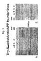
- Two primers pairs were designed Thy-1 Prom (GAATCCAAGTCGGAACTCTT, SEQ ID NO: 6) together with APP-SQ6 (TGTCAGGAACGAGAAGGGCA, SEQ ID NO: 7), and also APP-SQ3 (GCCGACCGAGGACTGA-CCAC, SEQ ID NO: 8) together with APP-SQ7 (GACACCGATGGGTAGTGAA, SEQ ID NO: 9) ( Fig. 1 ).
- SwedishArcticAPP transgenic mice were anesthetized with 0.4ml Avertin (25mg/ml) checked for loss of spinal reflexes and then perfused intracardially with 0.9% saline-solution.
- the brain was prepared and cut in two hemispheres; one of them was immersed in 4% PFA (paraformaldehyde)/1 ⁇ SPB (Sorensons Phosphate Buffer, 23mM KH 2 PO 4 , 70.5mM Na 2 HPO 4 ⁇ 2H 2 O, 5mM NaN 3 , pH7.4) over night, 4°C. Thereafter the brain was sequentially transferred and immersed in 10%, 20% and 30% (weight/volume) Sucrose/0.1 ⁇ SPB-solution each over night. The sucrose procedure was done to better preserve tissue morphology following freezing. The brain was kept in 30% sucrose-solution until the cryostat sections were cut ( Figs. 3-10 ).
- the left hemispheres of the brains were dissected from the different founder lines and weighed ( Fig. 9 ) (as well as the other organs measured).
- the brain tissue was extracted in 0.2% Tween-20 in 1xPBS with protease inhibitor tablets (cat 1836153, Roche, one tablet is tablet is sufficient for 10ml extraction solution) ( Fig. 3 ) .
- the extraction ratio was 1:10 (tissue weight: extraction buffer) and the tissue was extracted by 2x10 strokes on ice.
- the extraction solutions were centrifuged at 17900g at 4°C for 15min. The supernatants were divided into aliquots and stored at -20°C.
- the brain tissues used for western blot were homogenized in 1:10 (tissue extraction volume ratio) in 100 mM Na 2 CO 3 with 50 mM NaCl (pH11.5) with protease inhibitors, centrifuged at 100,000g at +4°C for 1 hr and the supernatants stored frozen at -80°C prior to analysis. The pellet was reextracted in 2% SDS and briefly sonicated, centrifuged as previously described. The SDS-insoluble pellet was finally reextracted in 70% formic acid ( Fig. 6 ).
- a Nitrocellulose filter was prewet in ddH 2 O and then equilibrated in 1 ⁇ Transfer-buffer (30mM Tris-base, 230mM Glycine, pH8.3) with 20% methanol. The transfer set was assembled in transfer-buffer and the transfer was run at 55V, 4°C over night. Prior to the antibody incubations the nitrocellulose-filter was boiled in 1xPBS for 5min, to stabilize and increase the exposure of epitopes in A ⁇ . The filter was then blocked in freshly prepared 1% w/v nonfat dry milk, 0.1% Tween-20 in 1 ⁇ TBS-buffer (100mM Tris base, 0.9% NaCl, pH 7.5) for 1hr at room-temperature.
- 1 ⁇ Transfer-buffer 30mM Tris-base, 230mM Glycine, pH8.3
- the transfer set was assembled in transfer-buffer and the transfer was run at 55V, 4°C over night. Prior to the antibody incubations the nitrocellulose-filter was boiled
- the filter was incubated with primary antibody (0.5 ⁇ g/ml 6E10 or 2 ⁇ g/ml 22C11) in 0.1% Tween-20 in 1 ⁇ TBS-buffer for 1hr at room-temperature. This was followed by washing 3-4 times (5min) in room-tempered 0.1% Tween-20 in 1 ⁇ TBS-buffer.
- the secondary antibody 0.2 ⁇ g/ml anti-mouseIgG/IgM-HRP (Pierce), in room-tempered 1% w/v nonfat dry milk, 0.1% Tween-20 in 1 ⁇ TBS-buffer and the filter was incubated in this solution for 30min.
- a ⁇ ELISA SDS-soluble brain tissue extracts were analyzed for A ⁇ 1-40 and A ⁇ 1-42 levels with ELISA using Amyloid Beta 1-40 and 1-42 ELISA kits (Signet Laboratories), according to manufacturer's instructions.
- a M.O.M. kit from Vector was utilized for the immunostaining .
- the frozen fixed tissue sections were incubated in concentrated formic acid (96%) for 5min at RT and then rinsed in water for 10min.
- the sections were incubated with H 2 O 2 (0.3%) in 50% DAKO-block/ 50% 1 ⁇ PBS for 15min at room-temperature to block endogenous peroxidase activity.
- the brain sections were once again rinsed in 1 ⁇ PBS before the incubation with M.O.M.
- Mouse IgG Blocking Reagent for 1hr to block unspecific binding.
- the sections were permeabilized with 1 ⁇ PBS (pH7.4) +0.4% Triton X-100) for 5min and briefly rinsed twice in 1 ⁇ PBS (pH7.4) to increase the surface tension.
- M.O.M. Mouse Diluent was used for the 5min incubation to block unspecific binding and excess were wiped away.
- kit ABC-complex (avidin-biotin-complex) were let to proceed, this was followed by a rinse in 1xPBS. Thereafter a horse radish peroxidase based substrate kit (NOVA Red, Vector) was used to develop the staining 10min. Finally the sections were briefly washed in ddH 2 O, dehydrated in 70%, 95%, 99.5% etOH, allowed to air-dry, dehydrated in Xylene and mounted in DPX (Dibutyl Phthalate Xylene, VWR) mounting medium for light microscopy. All the incubations above, unless stated otherwise, were carried out in room-temperature and on a shaking platform ( Figs. 4-5 , 7-8 and 10 ).
- Congo Red staining was accomplished by incubating tissue sections with saturated alkaline sodium chloride solution (10mM NaOH) for 20min followed by Congo Red (0.2% w/v) in saturated alkaline sodium chloride solution (10mM NaOH) for 15min and dehydration in 70%, 95%, 99.5% etOH. Tissue sections were allowed to air-dry, dehydrated in Xylene and mounted in DPX (Dibutyl Phthalate Xylene, VWR) mounting medium for light microscopy under polarized light.
- the images of 6E10 A ⁇ -immunoreactive staining were captured at 400X magnification in a Leica microscope with a cooled color CCD-camera at defined light and filter settings.
- the captured images of intraneuronal A ⁇ aggregates in the CA1 pyramidal neurons of the dorsal hippocampus were converted to greyscale images, processed with a delineation function to sharpen edges and allow an accurate segmentation.
- the images were segmented with an autothreshold command (Qwin, Leica).
- the results are expressed as area fraction (stained area tot /measured area tot , expressed in %) and presented as mean ⁇ S.E.M among the tissue section analyzed from each individual transgenic mouse.
- APP amyloid precursor protein
- the targeted epitopes are amino acids 66-81, for 22C11, and amino acids 672-687, for 6E10.
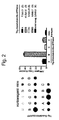
- the intensity of the spots has been analyzed with the Scion Image software and relative APP overexpression in the different founder lines has been calculated. Equal loading of the gels has been confirmed with Coomassie straining and total protein analysis.
- the relative APP expression can be estimated with antibody 22C11 which enables detection of both endogenous murine APP and human transgene APP.
- Thy-SwedishArcticAPP founder line B was found to display 3-fold APP-overexpression ( Fig. 3 ).
- Sequential chemical extraction of brain tissue from 2months old Thy-SwedishArctic transgenic mouse shows that most A ⁇ is soluble i.e. it can be recovered by gentle chemical extraction in carbonate buffer and that little A ⁇ remains in the tissue upon reextraction in 1% SDS or 70% formic acid i.e. as the insoluble A ⁇ ( Fig 6, a ).
- a ⁇ 1-40 Fig 6, b
- a ⁇ 1-42 levels ( Fig 6, c ), as measured by ELISA, in 2months old Thy-SwedishArctic transgenic mouse are reduced as compared to Thy-Swedish transgenic mouse that are of the same age and express the same amount of the transgene (the human APP protein.
- total A ⁇ levels i.e. both A ⁇ 1-40 and A ⁇ 1-42 measured together with western blot is five-fold increased in brain tissues from 2months old Thy-SwedishArctic transgenic mice as compared to Thy-Swedish transgenic mouse that are of the same age and express the same amount of the transgene (the human APP protein) ( Fig 6, d ).
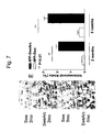
- cortical neurons contain intraneuronal A ⁇ aggregates at 15 months of age in the Thy-Swedish APP mouse (Fig. 5, c). No A ⁇ immunostaining was found in nontransgenic mice (Fig. 5, d).
- the A ⁇ -immunopositive staining is resistant to pre-treatment with concentrated formic acid, which is a typical characteristic of amyloid i.e. A ⁇ aggregates with a ⁇ -sheet structure.
- Scale bar measures 20 ⁇ m ( Fig.
- Image analysis show 11-fold (2 months; 1.91 ⁇ 0.16 (4) as compared to 0.17 ⁇ 0.02 (3); mean ⁇ S.E.M (n)) and 7-fold (5 months; 2.66 ⁇ 0.28 (3) as compared to 0.38 ⁇ .10 (4); mean ⁇ S.E.M (n) increase in percentage area covered by intraneuronal A ⁇ immunostaining in Thy-SwedishArctic APP transgenic mouse as compared to Thy-Swedish APP transgenic mouse ( Fig. 7, e ).
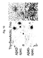
- Extracellular senile plaques were also present in the caudal part of hippocampus of Thy-SweArcticAPP transgenic mouse at this age, as shown with A ⁇ 42 and A ⁇ 40 specific antibodies ( Fig. 10 , a-b).
- the A ⁇ -immunoreactivity was resistant to and enhanced by pretreatment with concentrated formic acid.
- the arrows (in Fig. 10 , a-b) points to A ⁇ -immunoreactive deposits which are displayed at higher magnification (middle images adjacent to 10, a and b).
- Combined Congo Red and GFAP-immunostaining shows robust astrogliotic reaction surrounding a compact amyloid plaque ( Fig. 10, c ), which display classical gold-green birefringence in polarized light ( Fig. 10, d ).
- the brains were dissected and divided into its two hemispheres. Scattergram showing mean and distribution among individuals of left hemisphere brain weight.
- the brain tissue was later biochemically analysed for human APP and A ⁇ synthesis.
- the left hemisphere was initially weighed on a balance, to serve as a measure of atrophic degeneration of the brain ( Fig. 9 ).
Landscapes
- Health & Medical Sciences (AREA)
- Life Sciences & Earth Sciences (AREA)
- Chemical & Material Sciences (AREA)
- Engineering & Computer Science (AREA)
- Genetics & Genomics (AREA)
- Organic Chemistry (AREA)
- Zoology (AREA)
- Biotechnology (AREA)
- General Health & Medical Sciences (AREA)
- Biomedical Technology (AREA)
- Environmental Sciences (AREA)
- Wood Science & Technology (AREA)
- Biochemistry (AREA)
- Biophysics (AREA)
- Bioinformatics & Cheminformatics (AREA)
- Molecular Biology (AREA)
- General Engineering & Computer Science (AREA)
- Veterinary Medicine (AREA)
- Neurology (AREA)
- Animal Behavior & Ethology (AREA)
- Proteomics, Peptides & Aminoacids (AREA)
- Plant Pathology (AREA)
- Medicinal Chemistry (AREA)
- Microbiology (AREA)
- Toxicology (AREA)
- Physics & Mathematics (AREA)
- Animal Husbandry (AREA)
- Biodiversity & Conservation Biology (AREA)
- Gastroenterology & Hepatology (AREA)
- Investigating Or Analysing Biological Materials (AREA)
- Eye Examination Apparatus (AREA)
- Acyclic And Carbocyclic Compounds In Medicinal Compositions (AREA)
Abstract
Description
- The present invention relates to a transgenic mouse model of Alzheimer's disease and related neurological disorders. The present invention also relates to method of producing said transgenic mouse, and to methods of screening for therapeutic or diagnostic agents useful in treatment or diagnosis of Alzheimer's disease.
- Alzheimer's disease (AD) is a progressive and irreversible neurodegenerative disorder causing cognitive, memory and behavioral impairments . It is the most common cause of dementia in the elderly population affecting roughly 5% of the population above 65 years and 20% above 80 years of age. AD is characterized by an insidious onset and progressive deterioration in multiple cognitive functions. The neuropathology involves both extracellular and intracellular argyrophillic proteineous deposits. The extracellular deposits, referred to as neuritic plaques, mainly consist in amyloid-beta (Aβ) peptides surrounded by dystrophic neurites (swollen, distorted neuronal processes). The Aβ peptides within these extracellular deposits are fibrillar in their character with a β-pleated sheet structure. Aβ in these deposits can be stained with certain dyes e.g. Congo Red and display a fibrillar ultrastructure. These characteristics, adopted by Aβ peptides in its fibrillar structure of neuritic plaques, are the dentition of the generic term amyloid. Frequent neuritic plaques and neurofibrillary tangles deposits in the brain are diagnostic criteria for AD, as carried out when the patient has died. AD brains also display macroscopic brain atrophy, nerve cell loss, local inflammation (microgliosis and astrocytosis) and often congophilic amyloid angiopathy (CAA) in cerebral vessel walls.
- Two forms of Aβ peptides, Aβ40 and Aβ42, are the dominant species of AD neuritic plaques (Masters et. al., 1985), while Aβ40 is the prominent species in cerebrovascular amyloid associated with AD (Glenner and Wong, 1984). Enzymatic activities allow these Aβ to be continuously formed from a larger protein called the amyloid precursor protein (APP) in both healthy and AD afflicted subjects in all cells of the body. Two major APP processing events (β- and γ-secretase activities enables Aβ-peptide production through enzymatic cleavage, while a third one called α-secretase activitites prevents Aβ-peptide by cleavage inside the Aβ-peptide sequence (reviewed in Selkoe, 1994;
US5604102 ). The Aβ42 is forty two amino acid long peptide i.e. two amino acids longer at the C-terminus, as compared to Aβ40. The Aβ42 peptide is more hydrophobic, and does more easily aggregate into larger structures of Aβ peptides such as Aβ dimers, Aβ tetramers, Aβ oligomers, Aβ protofibrils or Aβ fibrils. Aβ fibrils are hydrophobic and insoluble, while the other structures are all less hydrophobic and soluble. All these higher molecular structures of Aβ peptides are individually defined based on their biophysical and structural appearance e.g. in electron microscopy, and their biochemical characteristics e.g. by analysis with size-exclusion chromatography/western blot. These Aβ peptides, particularly Aβ42, will gradually assemble into a various higher molecular structures of Aβ during the life span. AD, which is a strongly age-dependent disorder, will occur earlier in life if this assembly process occurs more rapidly in the brain of that individual. This is the core of the "amyloid cascade hypothesis" of AD which claims that APP processing, the Aβ42 levels and their assembly into higher molecular structures are central cause of all AD pathogenesis. All other neuropathology of AD brain and the symptoms of AD such as dementia are somehow caused by Aβ peptides or assembly forms thereof. The strongest evidence for the "amyloid cascade hypothesis" comes from genetic studies of individuals in families afflicted by early onset of familial AD as a dominant trait. These studies have revealed that rare mutations in the APP gene are sufficient to generate severe forms of AD. The mutations are clustered in and around Val 717 slightly downstream of the Aβ1-42 C-terminus (Goate et al., 1991, Chartier-Harlan, et al., 1991, Murrell, et al., 1991) and a unique double mutation (670-671) immediately upstream of the Aβ N-terminus in a Swedish family (Mullan, et al., 1992;US5795963 ). The APP mutations, which frames the Aβ peptide sequence, were later found to either increase both Aβ40 and Aβ42 production (the "Swedish" mutation; Citron, et al., 1992, Cai et al., 1993), or to increase the ratio of Aβ42/ Aβ40 production and also to generate Aβ peptides that are C-terminally extended to incorporate the pathogenic mutation in the Aβ peptide e.g. Aβ50 (the "717"-mutations are at position 46; Suzuki et al., 1994; Roher et al., 2003). Thus the "717" mutations, in addition to wild-type Aβ40 and wild-type Aβ42, also generate London Aβ peptides (V717I) and Indiana Aβ peptides (V7171F, Aβ46 and longer forms of Aβ) which rapidly forms Aβ fibrils. In contrast, the Swedish mutation only generates increased levels of wild-type Aβ40 and Aβ42 peptides. Early onset familial AD is more often caused by mutations in presenilin 1 (onchromosome 14;US5986054 ;US5840540 ;US5449604 ) and in some cases by mutations in presenilin 2 (chromosome 1). Presenilin 1 andpresenilin 2 are both polytopic transmembrane proteins that, together with three other proteins nicastrin, aph1 and pen-2, constitute the necessary functional core of the γ-secretase complex that enables Aβ-peptide formation through enzymatic cleavage of APP (Edbauer et al., 2003). All AD pathogenic mutations in presenilin 1 andpresenilin 2 proteins significantly increase Aβ1-42 overproduction (Schuener et al., 1996). Apolipoprotein E (ApoE) is, besides age, the most important risk factor for late-onset AD. There are three variants of the ApoE protein in humans, due to single amino acid substitutions in the ApoE protein. The ApoE4 variant confers increased risk of AD, while the ApoE2 variant is protective as compared to the predominant ApoE3 variant (Strittmatter et al., 1993; Corder et al., 1993). These protein changes are not deterministic, but confer enhanced or decreased susceptibility to develop AD in a population. The ability of the ApoE variants to facilitate amyloid deposition in APP transgenic mice models of AD is greatest for ApoE4, intermediate for ApoE3 and lowest for ApoE2, suggesting that the AD pathogenic mechanism of ApoE is to enhance Aβ-peptide assembly and/or amyloid deposition (Fagan et al., 2002). Other proteins such as a1-antichymotrypsin (Nilsson et al., 2001) and ApoJ/clusterin (DeMattos et al., 2002) also enhance Aβ-peptide assembly and/or amyloid deposition in APP transgenic mice, similar to ApoE. Neprilysin (NEP) and insulin-degrading enzyme (IDE) degrade Aβ peptides and are likely implicated in AD. However, none of these proteins has been proven to be involved in AD by human genetics. A key issue in future AD research is to better understand how enhanced levels Aβ or aggregates thereof cause dementia and functional loss in AD patients. It has been a long-standing belief that the insoluble amyloid fibrils, the main component of the neuritic plaque, are the pathogenic species in AD brain. High concentrations of Aβ fibrils have been shown to be cytotoxic in cell culture models of nerve cells in the brain (Pike et al., 1991; Lorenzo and Yankner et al., 1994). However, the hypothesis of the amyloid fibril as the main neurotoxic species is inconsistent with the poor correlation between neuritic plaque density and AD dementia score and also with the modest signs of neurodegeneration in current APP transgenic mice. Soluble neurotoxic Aβ-intermediate species and their appropriate subcellular site of formation and distribution could be the missing link that will better explain the amyloid hypothesis. This idea has gained support from recent discovery of the Arctic (E693) APP mutation, which causes early-onset AD (W00203911; Nilsberth et al., 2001 - An animal model of AD with the features of the human disease is much needed to better understand AD pathogenesis and to evaluate the efficace of new therapeutic agents. The ideal animal model of AD should generate the complete neuropathology of AD and the clinical phenotype e.g. progressive memory and cognitive dysfunctions. Major progress in this direction has been accomplished using transgenic overexpression of APP harboring AD pathogenic mutations. Current APP transgenic models of AD display important features of AD pathogenesis such as age-dependent and region-specific formation of both diffuse and neuritic plaques in the brain. The amyloid pathology is associated with hyperphosphorylated tau, local inflammation (microgliosis and astrocytosis) and to a variable extent with congophilic amyloid angiopathy (CAA). These models have been generated by very high transgene expression of human APP, particularly in nerve cells of the brain. The transgenes always carries an AD pathogenic mutation. Thus a "717"-APP-mutation (V717F; Games et al. 1995;
US2002104104 ;US5720936 ;US581 1633 ) or the "Swedish" mutation (KM670/671NL; Hsiao et al., 1996; Sturchler-Pierrat et al., 1997;WO 09803644 US2002049988 ;US6245964 ;US5850003 ;US5877399 ;US5777194 ) have been used. Double transgenic mice containing both mutant APP and mutant presenilin-1 transgenes develop accelerated amyloid plaques formation, but the animals still display modest mental impairment and still fail to display NFTs, nerve cell and brain atrophy (Holcomb et al., 1998;US5898094 ;US2003131364 ). Furthermore the current APP transgenic models likely have low levels of soluble intermediates in the Aβ fibrillization process such as Aβ protofibrils, which might be of great importance for AD pathogenesis. Several AD pathogenic mutations have previously been combined in one single transgene e.g. the "Swedish" mutation (KM670/671NL) and the"717"-APP-mutation (Indiana, V717F) have been used to enhance and increase formation of fibrillar Aβ peptides and neuritic plaque formation (Janus et al., 2001). Similarly the "Swedish" (KM670/671NL), the "Arctic" (E693G) and a "717"-APP-mutation (London, V717I) have been combined and used in an attempt to generate earlier and increased plaque formation (Teppner et al., 2003), like those of Swedish+Indiana APP transgenic models (Janus et al., 2001), since the London Aβ peptides will strongly facilitate Aβ fibril formation (Teppner et al., 2003; Roher et al., 2003). The unique characteristics of Arctic Aβ40 and Arctic A42 to form an abundance of stable protofibrils have been demonstrated (Nilsberth et al., 2001; Lashuel et al., 2003). The marked difference in pathology in human AD brain between carriers of the London APP mutation (Lantos et al., 1992; Cairns et al., 1993) and Arctic APP mutation reinforce the distinction in the chemical characteristics of London Aβ peptides and Arctic Aβ peptides for neuropathology. - The following references are presently found to be most relevant:
- Stenh C. et al. disclose in "Metabolic consequences of the arctic (E693G) APP alzheimer mutation", Society for Neuroscience. Abstract Viewer and Itenary Planner 2002, 32nd Annual Meeting of the Society for Neuroscience, November 02-07, 2002, Abstract No. 296.6 and in Neuroreport 13, 1857-60 (2002) a transfected tumorigenic cell-line harboring APP cDNA with both the "Swedish" (KM670/671NL) and "Arctic" (E693G) mutations.
- Hsiao et al., Science 274, 99-102 (1996) disclose a transgenic mouse harboring the "Swedish" (KM670/671NL) alone.
- Mullan et al., Nature Genet. 1, 345-347 (1992) discloses the dominant inheritance of the "Swedish" (KM670/671NL) in a family with Alzheimer's disease.
- Nilsberth et al., Nat Neurosci. 4, 887-893 (2001) discloses the dominant inheritance of the "Arctic" (E693G) in a family with Alzheimer's disease.
- Teppner et al., 6th Internat. Conf. AD/PD, Seville, Spain, board no 52 (2003), disloses a preliminary attempt to generate a transgenic mouse harboring the "Swedish" (KM670/671NL), "Arctic" (E693G) and "London" (V717I) mutations. No pathology is described.
- Roher et al., J Biol Chem. 279(7): 5829-36 (2004), discloses that Aβ peptides extend beyond amino acid 42, e.g. Aβ1-46 and Aβ1-50, in Alzheimer brain tissue from patient carrying a "London"-type mutation (V717F).
- Kang et al., Nature 325, 733-6 (1987) describes the cloning of human APP695 cDNA.
- In view of the shortcomings of prior art models, the object of the invention is to provide a transgenic mouse model that displays early phenotypes of Alzheimer's disease (AD) pathology that can be quantified. This would allow a more rapid and cost-efficient screening of pharmacological agents in the pharmaceutical and biotech industry.
- Is disclosed a mouse model for AD and related neurological disorders having pathologies of enhanced Aβ-40 and/or Aβ-42 Arctic peptides and Aβ Arctic protofibril production and an early soluble oligomeric and protofibrillar Aβ Arctic peptide-driven pathology, including Aβ aggregation inside neurons of the brain.
- The Aβ-immunopositive intraneuronal staining (punctate and strong) was resistant to pretreatment with concentrated formic acid, which is a typical characteristic of amyloid, i.e. Aβ aggregates with a β-sheet structure (protofibrils), and was localized to the pyramidal cell layer of CA1 in the hippocampus and in scattered neurons of the lower lamina in the cerebral cortex as well as other neurons in the brain.
- A AD transgenic mouse, that exhibits early and enhanced intracellular Aβ aggregation, which can be reliably measured is disclosed. This intracellular Aβ aggregation occurs prior to and gradually increase in amount before the onset of extracellular plaque formation. The early and enhanced soluble intraneuronal Aβ aggregation is a pathological AD phenotype that goes beyond previously described APP transgenic mouse models. This AD phenotype is present in the mouse model according to the present invention much earlier than in any AD marker found in previous animal models.
- Also is disclosed a means for identification of agents that interfere, delay or inhibit the Alzheimer disease process at an early stage. Such agents would be of significant clinical importance for treatment of early stage Alzheimer's disease or prevention of its manifestation. The provision of the mouse model according to the present invention can greatly shorten the time required for screening for such agents.
- Thus the measurement of the extent of intracellular Aβ aggregation allows one to predict the later extracellular Aβ deposition well in advance. This prediction can be made as early as 1-2 months into the development of AD neuropathology. With prior art techniques, this is possible only after 15 months. The present invention can thus be used to more rapidly and cost-efficiently screen for agents that are able to prevent, inhibit and reverse AD neuropathology at an earlier stage.
- The transgenic mouse model provided by the invention also display reduced brain weight, which suggests atrophic changes in the brain as is normally observed in human brain afflicted by AD pathogenesis.
- The transgenic mouse expresses one transgene comprising a DNA sequence encoding a heterologous Amyloid Precursor Protein (APP) comprising the Arctic mutation (E693G) and one further AD (Alzheimer's disease) pathogenic mutation selected from the group consisting of KM670/671DF, KM670/671DY, KM670/671EF, KM670/671EY, KM670/671NL, KM670/671NY, KM670/671NF, KM670/671KL, KM670/671DL and KM670/671EL, and no further APP mutations, wherein said transgene is operably linked to a promoter effective for expression of said gene in the brain tissue of said mouse, which results in increased amounts of intracellular soluble Aβ aggregates, including Aβ peptides.
- According to another embodiment, the transgenic mouse according to present invention may further comprise a homologously integrated targeting construct for at least one of the neprilysin or insulin-degrading enzyme (IDE) genes, which disrupts these genes through gene ablation (knock-out) and enhances Aβ-40 and/or Aβ-42 Arctic peptide production.
- According to a presently preferred embodiment, the transgenic animal is a mouse harboring a transgene encoding amyloid precursor protein (APP) consisting of the Arctic mutation (E693G) and the Swedish mutation KM670/671NL), and no further APP mutations.
- The transgenic mouse AD model is defined in claim 1.
- According to another aspect, the present invention also relates to a method of preparing said transgenic mouse. The method of producing said transgenic mouse is defined in claim 7.
- According to another aspect, the present invention also relates to a method of a screening, wherein the transgenic mouse is used for screening for agents useful for treating, preventing or inhibiting Alzheimer's disease. Said method is defined in claim 11.
- According to another aspect, the present invention also relates to a method of a screening, wherein the transgenic mouse is used for screening for diagnostic agents for Alzheimer's disease. Said method is defined in claim 12.
- The present invention provides a mouse model for AD and related neurological disorders having pathologies of enhanced Aβ protofibril formation and intraneuronal Aβ peptide aggregation.
- The transgenic mice and progeny thereof, typically producing the Arctic Aβ peptides in brain tissue, can be used as a model for a variety of diseases and for drug screening, testing various compounds, evaluation of diagnostic markers as well as other applications.
-
-
Fig. 1 : Ethidium bromide-stained DNA gel showing the presence of positive PCR-signal of DNA-fragments having a length of 428bp with the upstream (A) primer pair and 441bp with the downstream (B) primer pair. Genomic DNA from different founder mice have been analyzed and PCR-positive Thy-SwvedishArcticAPP founders have been assigned founder line numbers A,B,C and D, as denoted above the gels. DNA molecular weight standard ("mw-std.") shows the lengths of various predefined DNA-fragments. The two primer pairs frames the whole coding region of transgene APP and the basal promoter of the Thy-1 promoter. -
Fig. 2 : Slot-blot phosphor-imager screen reflecting radioactive emission from cRNA-probes hybridized to genomic DNA samples from individual mice from the different founder lines (Thy-SwedishArcticAPP line A, B, C and D) and nontransgenic mice, as denoted for each individual mouse above the corresponding photographic signal (left) and quantitative estimates of these signals to measure copy number for the different founder lines of Thy-SwedishArctic-APPmice (right) -
Fig. 3 : Graph depicting the APP protein with the kunitz domain (hatched) which enables alternative splicing of APP. The Aβ peptides domain (black) resides partly inside the transmembrane domain. The locations of the epitopes of the APP antibodies used in the experiment are indicated. In the APP770 protein isoform the epitopes are located between aa 66-81 (22C11) and aa 672-687 (6E10). The 22C11 antibody detects both human and endogenous murine APP, while the 6E10 antibody detects only human APP. Western blot showing threefold relative overexpression of APP in brain of Thy-SwedishArctic-APP transgenic mouse, founder line B. Coomassie staining ("Cooma.") is a measure of total protein loaded onto the gel (A). The presence of human APP and Arctic Aβ peptides in brain of Thy-SwedishArctic-APP transgenic mouse, founder line B ("B") and absence in brain of nontransgenic mouse ("ntr") (B) was verified by staining with 6E10 antibody. As said antibody only detects the presence of human APP, the functionality of the transgene is thus verified. -
Fig. 4 : APP protein in young Thy-SwedishArctic-APP transgenic mouse APP protein expression in the brain of a 1month old Thy-SwedishArcticAPP mouse, founder line B (a - left hemisphere and b - hippocampus) and a nontransgenic mouse (c -hippocampus) stained with 6E10 (epitope 1-16 in Aβ, this antibody is specific for human APP and Aβ). The staining visualizes neuronal distribution of APP protein synthesis in the brain. -
Fig. 5 : Punctate intraneuronal Aβ immunostaining showing Aβ aggregation in the cerebral cortex in the Thy-SwedishArctic APP mouse (a, marked by arrows) according to the present invention and a Swedish APP transgenic mouse (b and c). The mice had equal APP expression and anatomic expression pattern (both of these parameters as well as the age of the mouse strongly influence AD phenotypes in any transgenic mouse model). Little and very faint intraneuronal Aβ was found in a 2 months old Thy-Swedish APP mouse (b). Some cortical neurons contain intraneuronal Aβ aggregates at 15 months of age in the Thy-Swedish APP transgenic mouse (c), but still much weaker and less frequent than in the Thy-SwedishArctic APP transgenic mouse at 2 months of age (a). No Aβ immunostaining was found in nontransgenic mice (d). (e) represents an overview of Aβ-aggregates in the right hemisphere of a brain of a Thy-SwedishArctic-APP transgenic mouse. The arrows points to the pronounced formic acid-resistant Aβ-immunoreactive staining in CA1 pyramidal neurons of Thy-SwedishArctic APP. Scale bar measures 20 µm (a-d). -
Fig. 6 : Aβ protein in 2 months old Thy-SwedishArctic-APP mouse (founder line B) and Thy-Swedish-APP transgenic mouse. Sequential chemical extraction of brain tissue shows that most Aβ in Thy-SwedishArctic-APP mouse is soluble, i.e. it can be recovered by gentle chemical extraction in carbonate buffer and that little Aβ remains in the tissue upon reextraction in 1% SDS or 70% formic acid, i.e. as insoluble Aβ (a, FA=formic acid). Aβ1-40 (b) and Aβ1-42 (c) levels, as measured by ELISA, in 2 months old Thy-SwedishArctic transgenic mouse, are reduced as compared to in Thy-Swedish transgenic mouse of the same age and which expresses the same amount of the transgene (the human APP protein). In contrast, total Aβ levels, i.e. both Aβ1-40 and Aβ1-42 measured together with western blot, exhibits a five-fold increase in brain tissue from 2 months old Thy-SwedishArctic transgenic mouse as compared to Thy-Swedish transgenic mouse of the same age and which expresses the same amount of the transgene (the human APP protein) (d). The results (b-d) strongly suggest that soluble Aβ aggregates such as protofibrils are present in the brain of 2 months old Thy-SwedishArctic transgenic mouse, since western blot is a denaturing method where soluble Aβ aggregates are dissociated into their individual components, and single Aβ peptides give higher a numerical value. In contrast ELISA is non-denaturing technique, whereby each soluble Aβ aggregate will be measured as one single unit and the numerical value will be lower. -
Fig. 7 : Punctate intraneuronal Aβ (marked by arrows in A-D) is very strong and frequent in Thy-SwedishArctic APP at both 2 months (B) and 5 months (D) of age. In contrast, in Thy-Swedish APP (matched for transgene APP expression) intraneuronal immunostaining Aβ at both 2 months (A) and 5 months (C) of age is infrequent and faint. Quantitation image analysis (E) shows 7-fold or more increase in punctate intraneuronal Aβ immunostaining in Thy-SwedishArctic APP (solid bars) as compared to Thy-Swedish APP (open bars). -
Fig. 8 : Graph showing that an increase in intraneuronal Aβ aggregation predates an increase in extracellular Aβ plaque deposition by at least 2 months. Area fraction of intraneuronal Aβ aggregation in the CA1 pyramidal neurons (left y-axis) and frequency of extracellular Aβ plaque deposition in the hippocampus (right y-axis, logarithmic scale) was quantified in a cohort of Thy SwedishArctic APP transgenic mice of various ages. Each solid square represent intraneuronal Aβ (% area fraction) from a single mouse, while the corresponding open square often located beneath represents Aβ plaque frequency in the same mouse. The results represent mean ± S.E.M. of the analysis of several tissue sections from individual transgenic mice. -
Fig. 9 : Scattergram showing the group mean (line) and distribution among individuals of left hemisphere brain weight as dissected from cohorts of Thy-SwedishArcticAPP and Thy-SwedishAPP transgenic mice at 2 months of age. Thy-SwedishArcticAPP transgenic mice display reduced brain weight (221±9mg; n=9), as compared to Thy-SwedishAPP transgenic mice (239±5mg; n=8), which suggests atrophic changes in the brains of Thy-SwedishArctic APP transgenic mice, as is normally observed in human brain afflicted by AD pathogenesis. -
Fig. 10 : Extracellular senile plaques in the hippocampus of a Thy-SwedishArcticAPP transgenic mouse at 7 months of age. The Aβ-immunoreactivity was observed with two different antibodies that were specific for the short amino acid fragments in the C-terminal ends of Aβ42 (a) and Aβ40 (b) and thus do not detect APP or APP-fragments (Näslund et al., 2000). The Aβ-immunoreactivity was resistant to and enhanced by pretreatment with concentrated formic acid. The arrows points to Aβ-immunoreactive deposits which are displayed at higher magnification (images between a and b). Combined Congo Red and GFAP-immunostaining shows robust astrogliosis surrounding a compact amyloid plaque (c), which displays classical gold-green birefringence in polarized light (d). - The transgenes according to the present invention comprise a polynucleotide sequence, more specifically a heterologous APP polypeptide comprising the herein described mutations, and are operably linked to a transcription promoter capable of producing expression of the heterologous APP polypeptide in the brain tissue of the transgenic mouse.
- Said promoter can be constitutive or inducible, and can affect the expression of a polynucleotide in a tissue-specific manner. Tissue-specific promoters include, without limitation, neuron specific enolase (NSE) promoter, neurofilament light chain (NF-L) and neurofilament heavy chain (NF-H) promoter, prion protein (PrP) promoter, tyrosine hydroxylase promoter, platelet-derived growth factor (PDGF) promoter, thy1- glycoprotein promoter, β-actin promoter, ubiquitin promoter, simian virus 40 (SV40) promoter, and gene-specific promoters such as the APP promoter.
- The amyloid precursor proteins (APP) comprise a group of ubiquitously expressed transmembrane glycoproteins whose heterogeneity arises from both alternative splicing and post-translational processing [Selkoe, D. J. (1994) NCBI accession nr P05067, SEQ ID NO: 1]. Apart from the 751- and 770-residue splice forms which are highly expressed in non-neuronal cells throughout the body, neurons most abundantly express the 695-residue isoform. All isoforms are the precursors of various metabolites that result from different proteolytic cleavage induced by physiological or pathological conditions. The APP itself, as used according to the principles of this invention, can be any of the alternative splice forms of this molecule and may be used either as a glycosylated or non-glycosylated form.
- In a further embodiment, the animal comprises a targeting construct homologously integrated into an endogenous chromosomal location so as to enhance Aβ-40 and/or Aβ-42 Arctic peptide levels by impaired clearance e.g. through gene ablation (knock-out) of neprilysin and/or insulin-degrading enzyme (IDE) genes in tissues of such transgenic animal harboring the Arctic mutation (E693G) and thereby generate a similar phenotype as that described in the invention i.e. early and enhanced intracellular Aβ aggregation.
- Prior to transfection, said further transgenes are crossed with the transgene comprising the Arctic mutation.
- The invention further provides a transgenic mouse, which harbors one copy of a transgene of the invention, either homologously or non-homologously integrated into an endogenous chromosomal location so as to produce Arctic Aβ peptides. Such transgenic animals are usually produced by introducing the transgene or targeting construct into a fertilized egg or embryonic stem (ES) cell, typically by microinjection, electroporation, lipofection, or biolistics.
- The transgenic mice according to the present invention have at least one inactivated endogenous APP allele, are preferably homozygous for inactivated APP alleles, and are substantially incapable of directing the efficient expression of endogenous (i.e., wild-type) APP.
- In a preferred embodiment, a transgenic mouse is homozygous for inactivated endogenous APP alleles and substantially incapable of producing murine APP encoded by a endogenous (i.e., naturally-occurring) APP gene. Such a transgenic mouse, having inactivated endogenous APP genes, is a preferred host recipient for a transgene encoding a heterologous APP polypeptide, preferably a human Arctic mutation and the Swedish APP mutation (KM670/671NL) (APP770 numbering) to enhance both Aβ-40 and Aβ-42 Arctic peptide production.
- Said Swedish mutation may be replaced with similar mutations such as KM670/671DL, KM670/671DF, KM670/671DY, KM670/671EL, KM670/671EF, M670/671EY, KM670/671NY, KM670/671NF, KM670/671KL (APP770 numbering).
- However, the Swedish mutation (KM670/671NL) is presently the mutation that is most preferably combined with the Arctic mutation.
- Compounds that are found to have an effect on the Aβ Arctic peptide expression, or to promote or inhibit any of the diverse biochemical effects of Aβ Arctic peptides and/or aggregated forms of Aβ Arctic peptides such as Aβ protofibrils, are then further tested and used in treatment of AD and/or related neurological disorders.
- In accordance with another aspect of the invention, the transgenic animal or its progeny can be used as starting points for rational drug design to provide ligands, therapeutic drugs or other types of small chemical molecules as well as proteins, antibodies or natural products. Alternatively, small molecules or other compounds as previously described and identified by the above-described screening assays can serve as "lead compounds" in rational drug design.
- Standard molecular biology techniques known in the art and not specifically described were generally followed as in Sambrook et al., Molecular Cloning: A Laboratory Manual, Cold Springs Harbor Laboratory, New York (1989, 1992), and in Ausubel et al., Current Protocols in Molecular Biology, John Wiley and Sons, Baltimore, Md. (1989). Standard transgenic techniques for introduction of a foreign gene into fertilized eggs from mouse known in the art and not specifically described were generally followed as in Nagy et al., Manipulating the Mouse Embryo: A laboratory manual, Cold Springs Harbor Laboratory, New York (1986, 1994, 2002), ISBN 0-87969-574-9. (
Figs. 1 and2 ). General methods in immunohistochemistry: Standard methods known in the art and not specifically described were generally followed as in Stites et al. (eds), Basic and Clinical Immunology (8th Edition), Appleton & Lange, Norwalk, Conn. (1994) and Johnstone & Thorpe, Immunochemistry in Practice, Blackwell Scientific Publications, Oxford, 1982 (Figs. 4-5 ,7-8 and10) . - The transgenic constructs used for this study contain the murine Thy-1 expression vector and human APP cDNAs. The APP695 isoform, which is predominant APP isoform in the brain, was used. Modifications in human APP cDNA clone (Kang et al., 1987) between NruI(+145nt) and and SmaI(+3100) was made with enzymatic primer extension using the Transfomer mutagenesis kit (Clontech). The following primers were used:
- CACTCGGTGCC CCGCGCGCGGCCGCCATGCTGCCCGGTTTGGC (SEQ ID NO: 2) and CATAAATAAATTAAATAAAATAACCGCGGCCGCAGAAACATACAAGCTGTCAG (SEQ ID NO: 3) to incorporate flanking Notl-sites and a Kozak sequence for improved initiation of translation.
- CAAATATCAAGACGGAGGAGATATCTGAAGTGAATCTGGATGCAGAATTCCGAC (SEQ ID NO: 4) to introduce the KM670/671NL mutation and CAAAAATTGGTGTTCTTTGCAGGAGATGTGGGTTCAAACAAAG (SEQ ID NO: 5) to introduce the E693G mutation. Clones were initially selected through PCR followed by restriction enzyme digestion and the selected clones were checked by DNA sequencing throughout the whole coding region of the amyloid precursor protein (APP). Correct clones were finally digested with NotI, blunt-end ligated into the XhoI-site of the Thy1 expression cassette. The construct DNA was linearized with NotI as to allow the back-bone vector sequences to be removed from the expression cassette. After purification from β-agarose gel (SeaPlaque GTG) with β-agarase (Invitrogen) and phenol-chloroform extraction the linearized DNA construct (2µg/ml) was microinjected into pronuclear oocytes of hybrid mouse line B6-CBA-F1 (B&M, Denmark). The pronuclear microinjection technique is preferred. Transcription units obtained from a recombinant DNA construct of the invention were injected into pronuclei of animal embryos and the obtained founder transgenics were bred to establish the transgenic line.
- The resulting offspring were genotyped by cutting tail tips from weanlings, extracting DNA using a Qiagen DNA extraction kit and analyzed with PCR across the coding sequence of APP and the basal promoter of Thy-1 glycoprotein. Two primers pairs were designed Thy-1 Prom (GAATCCAAGTCGGAACTCTT, SEQ ID NO: 6) together with APP-SQ6 (TGTCAGGAACGAGAAGGGCA, SEQ ID NO: 7), and also APP-SQ3 (GCCGACCGAGGACTGA-CCAC, SEQ ID NO: 8) together with APP-SQ7 (GACACCGATGGGTAGTGAA, SEQ ID NO: 9) (
Fig. 1 ). - SwedishArcticAPP transgenic mice were anesthetized with 0.4ml Avertin (25mg/ml) checked for loss of spinal reflexes and then perfused intracardially with 0.9% saline-solution. The brain was prepared and cut in two hemispheres; one of them was immersed in 4% PFA (paraformaldehyde)/1×SPB (Sorensons Phosphate Buffer, 23mM KH2PO4, 70.5mM Na2HPO4×2H2O, 5mM NaN3, pH7.4) over night, 4°C. Thereafter the brain was sequentially transferred and immersed in 10%, 20% and 30% (weight/volume) Sucrose/0.1×SPB-solution each over night. The sucrose procedure was done to better preserve tissue morphology following freezing. The brain was kept in 30% sucrose-solution until the cryostat sections were cut (
Figs. 3-10 ). - The left hemispheres of the brains were dissected from the different founder lines and weighed (
Fig. 9 ) (as well as the other organs measured). The brain tissue was extracted in 0.2% Tween-20 in 1xPBS with protease inhibitor tablets (cat 1836153, Roche, one tablet is tablet is sufficient for 10ml extraction solution) (Fig. 3 ).The extraction ratio was 1:10 (tissue weight: extraction buffer) and the tissue was extracted by 2x10 strokes on ice. The extraction solutions were centrifuged at 17900g at 4°C for 15min. The supernatants were divided into aliquots and stored at -20°C. Alternatively, the brain tissues used for western blot were homogenized in 1:10 (tissue extraction volume ratio) in 100 mM Na2CO3 with 50 mM NaCl (pH11.5) with protease inhibitors, centrifuged at 100,000g at +4°C for 1 hr and the supernatants stored frozen at -80°C prior to analysis. The pellet was reextracted in 2% SDS and briefly sonicated, centrifuged as previously described. The SDS-insoluble pellet was finally reextracted in 70% formic acid (Fig. 6 ). All samples (~40µg protein each) were denatured by adding 1% mercaptoethanol and 1×Sample buffer (final concentration), the samples were mixed and boiled for 5min and then loaded on 4-20% Tris-Glycine gel (InVitrogen). 1×Sample buffer contains 10% Glycerol, 2% SDS, 50mM Tris-HCl and Bromophenol blue (diluted x40 from a 1.5% stock). The SDS-PAGE running buffer used includes 250mM Tris-base, 1.9M Glycine and 35mM SDS (Sodium Dodecyl Sulfate). The gel was run at 95V. A Nitrocellulose filter was prewet in ddH2O and then equilibrated in 1×Transfer-buffer (30mM Tris-base, 230mM Glycine, pH8.3) with 20% methanol. The transfer set was assembled in transfer-buffer and the transfer was run at 55V, 4°C over night. Prior to the antibody incubations the nitrocellulose-filter was boiled in 1xPBS for 5min, to stabilize and increase the exposure of epitopes in Aβ. The filter was then blocked in freshly prepared 1% w/v nonfat dry milk, 0.1% Tween-20 in 1×TBS-buffer (100mM Tris base, 0.9% NaCl, pH 7.5) for 1hr at room-temperature. After blocking, the filter was incubated with primary antibody (0.5µg/ml 6E10 or 2µg/ml 22C11) in 0.1% Tween-20 in 1 ×TBS-buffer for 1hr at room-temperature. This was followed by washing 3-4 times (5min) in room-tempered 0.1% Tween-20 in 1 ×TBS-buffer. The secondary antibody, 0.2µg/ml anti-mouseIgG/IgM-HRP (Pierce), in room-tempered 1% w/v nonfat dry milk, 0.1% Tween-20 in 1×TBS-buffer and the filter was incubated in this solution for 30min. The filter was then washed three more times in 0.1% Tween-20 in 1×TBS-buffer, and last there was a final rinse in 1×TBS-buffer without Tween before the 5min incubation in SuperSignal (Pierce-ECL). All incubations were let to proceed on a shaking platform. The blot filter was finally incubated against an ECL-Hyperfilm (Amersham) (Fig. 3 , 6). Aβ ELISA: SDS-soluble brain tissue extracts were analyzed for Aβ1-40 and Aβ1-42 levels with ELISA using Amyloid Beta 1-40 and 1-42 ELISA kits (Signet Laboratories), according to manufacturer's instructions. To ensure equal epitope recognition between Arctic and wt Aβ by the antibodies used in the ELISA, dilution series of synthetic Aβ1-40 Arctic and Aβ1-40 wt in their monomeric form were analyzed with the Amyloid Beta 1-40 ELISA kit. - The brain hemispheres from the founder lines mounted on a freezing stage and 25µm sections were cut with a sledge-microtome and stored at +°4C until use. For the immunostaining a M.O.M. kit from Vector was utilized. The frozen fixed tissue sections were incubated in pre-heated citrate-buffer (25mM, pH7.3) for 5min at 85°C. This was followed by a rinse in 1×PBS. The frozen fixed tissue sections were incubated in concentrated formic acid (96%) for 5min at RT and then rinsed in water for 10min. After that the sections were incubated with H2O2 (0.3%) in 50% DAKO-block/ 50% 1×PBS for 15min at room-temperature to block endogenous peroxidase activity. The brain sections were once again rinsed in 1×PBS before the incubation with M.O.M. Mouse IgG Blocking Reagent for 1hr to block unspecific binding. Then the sections were permeabilized with 1×PBS (pH7.4) +0.4% Triton X-100) for 5min and briefly rinsed twice in 1×PBS (pH7.4) to increase the surface tension. M.O.M. Mouse Diluent was used for the 5min incubation to block unspecific binding and excess were wiped away. Incubation with 0.2µg/ml 6E10, 14µg/ml GFAP (clone G-A-5; 1x1500) 1.5µg/ml Aβ42 and 1.7µg/ml Aβ40 antibodies (primary antibodies) in MOM-diluent/ 0.1% Triton X-100 was let to proceed over night at +4°C. After another wash in 1xPBS buffer the sections were incubated with M.O.M. Biotinylated Anti-mouse or Anti-rabbit IgG reagent in M.O.M. Diluent/0.1% Triton X-100 for 8min. The sections were once more rinsed in 1×PBS buffer. A 30min long incubation with the M.O.M. kit ABC-complex (avidin-biotin-complex) were let to proceed, this was followed by a rinse in 1xPBS. Thereafter a horse radish peroxidase based substrate kit (NOVA Red, Vector) was used to develop the staining 10min. Finally the sections were briefly washed in ddH2O, dehydrated in 70%, 95%, 99.5% etOH, allowed to air-dry, dehydrated in Xylene and mounted in DPX (Dibutyl Phthalate Xylene, VWR) mounting medium for light microscopy. All the incubations above, unless stated otherwise, were carried out in room-temperature and on a shaking platform (
Figs. 4-5 ,7-8 and10 ). Congo Red staining was accomplished by incubating tissue sections with saturated alkaline sodium chloride solution (10mM NaOH) for 20min followed by Congo Red (0.2% w/v) in saturated alkaline sodium chloride solution (10mM NaOH) for 15min and dehydration in 70%, 95%, 99.5% etOH. Tissue sections were allowed to air-dry, dehydrated in Xylene and mounted in DPX (Dibutyl Phthalate Xylene, VWR) mounting medium for light microscopy under polarized light. - Equally spaced coronal tissue sections along the rostral-caudal axis of the hippocampus, 4-5 tissue sections from each animal, were investigated by capturing four different image fields from each separate tissue section. The images of 6E10 Aβ-immunoreactive staining were captured at 400X magnification in a Leica microscope with a cooled color CCD-camera at defined light and filter settings. The captured images of intraneuronal Aβ aggregates in the CA1 pyramidal neurons of the dorsal hippocampus were converted to greyscale images, processed with a delineation function to sharpen edges and allow an accurate segmentation. The images were segmented with an autothreshold command (Qwin, Leica). The results are expressed as area fraction (stained areatot/measured areatot, expressed in %) and presented as mean ± S.E.M among the tissue section analyzed from each individual transgenic mouse.
- The results from PCR genotyping are seen to the right (
Fig.1 ). Both sets of primers identified 4 founder mice (out of 13) having the mThyl-SwedishArctic-hAPP construct and these four founder lines were established; Thy-SwedishArcticAPP lines A-D. DNA-fragments of 428bp lengths with upstream (A) and of 441bp length with downstream (B) primer pairs could be detected. Offspring from each founder line were genotyped the same way (Fig. 1 ). - Copy numbers were analyzed on individual transgene positive offspring using slot blot. The four Thy-SwedishArcticAPP founder line incorporated varying number of DNA copies, with founder line B having the highest copy number (41±2), taking into account that the nontransgenic mice have two copies of the endogenous Thy1 gene (
Fig. 2 ). - Human APP and Aβ synthesis from brain extracts of the different Thy-SwedishArctic founder lines are shown. The drawing illustrates the amyloid precursor protein (APP) and the epitopes within APP that are targeted by the antibodies. In the APP770 protein isoform, the targeted epitopes are amino acids 66-81, for 22C11, and amino acids 672-687, for 6E10. The intensity of the spots has been analyzed with the Scion Image software and relative APP overexpression in the different founder lines has been calculated. Equal loading of the gels has been confirmed with Coomassie straining and total protein analysis. The relative APP expression can be estimated with antibody 22C11 which enables detection of both endogenous murine APP and human transgene APP. In
contrast antibody 6E 10 only detects human transgene APP and Aβ peptides. Thy-SwedishArcticAPP founder line B was found to display 3-fold APP-overexpression (Fig. 3 ). Sequential chemical extraction of brain tissue from 2months old Thy-SwedishArctic transgenic mouse shows that most Aβ is soluble i.e. it can be recovered by gentle chemical extraction in carbonate buffer and that little Aβ remains in the tissue upon reextraction in 1% SDS or 70% formic acid i.e. as the insoluble Aβ (Fig 6, a ). Aβ1-40 (Fig 6, b ). and Aβ1-42 levels (Fig 6, c ), as measured by ELISA, in 2months old Thy-SwedishArctic transgenic mouse are reduced as compared to Thy-Swedish transgenic mouse that are of the same age and express the same amount of the transgene (the human APP protein. In contrast total Aβ levels i.e. both Aβ1-40 and Aβ1-42 measured together with western blot is five-fold increased in brain tissues from 2months old Thy-SwedishArctic transgenic mice as compared to Thy-Swedish transgenic mouse that are of the same age and express the same amount of the transgene (the human APP protein) (Fig 6, d ). The results (Fig 6 , b-d) strongly suggest that soluble Aβ aggregates such as protofibrils are present in the brain of 2months old Thy-SwedishArctic transgenic mouse, since western blot is a denaturing method where soluble Aβ aggregates are dissociated into their individual components i.e. single Aβ peptides thereby giving higher a numerical measurement. In contrast ELISA is a non-denaturing and each soluble Aβ aggregates will be measured as one single unit and for the total number of their individual components. - The results from the APP immunohistochemistry are presented is seen in a one month old Thy-SwedishArcticAPP, founder line B mouse (
Fig. 4 , a-b), while only diffuse background staining is apparent in a nontransgenic littermatefig. 4, c ). Punctate intraneuronal Aβ immunostaining showing Aβ aggregation in the cerebral cortex of a 2 months old Thy-SwedishArctic APP mouse (Fig. 5, a), marked by arrows). Little and very faint intraneuronal Aβ in 2 months old Thy-Swedish APP mouse with an equal APP expression(Fig. 5, b). Some cortical neurons contain intraneuronal Aβ aggregates at 15 months of age in the Thy-Swedish APP mouse(Fig. 5, c). No Aβ immunostaining was found in nontransgenic mice(Fig. 5, d). We find intraneuronal Aβ-immunopositive inclusions in the pyramidal cell layer of CA1 in the hippocampus and in scattered neurons of the lower lamina in the cerebral cortex in Thy-SweArcAPP transgenic mice (Fig. 5, e ). The Aβ-immunopositive staining is resistant to pre-treatment with concentrated formic acid, which is a typical characteristic of amyloid i.e. Aβ aggregates with a β-sheet structure. Scale bar measures 20 µm (Fig. 5 , a-d). Punctate intraneuronal Aβ immunostaining (marked by arrows inFig. 5 , a-d) showing Aβ aggregation in the hippocampus of a 2 months old (Fig. 7, b ) and 5 months old (Fig. 7, d ) Thy-SwedishArctic APP transgenic mouse. Little and very faint intraneuronal Aβ in 2 months old (Fig. 7, a ) and 5 months old (Fig. 7, c ) Thy-Swedish APP mouse with an equal APP expression. Image analysis show 11-fold (2 months; 1.91±0.16 (4) as compared to 0.17±0.02 (3); mean±S.E.M (n)) and 7-fold (5 months; 2.66±0.28 (3) as compared to 0.38±.10 (4); mean±S.E.M (n) increase in percentage area covered by intraneuronal Aβ immunostaining in Thy-SwedishArctic APP transgenic mouse as compared to Thy-Swedish APP transgenic mouse (Fig. 7, e ). Area fraction of intraneuronal Aβ aggregation in the CA1 pyramidal neurons (left y-axis) and frequency of extracellular Aβ plaque deposition in the hippocampus (right y-axis, logarithmic scale) was quantified in a cohort of Thy-SwedishArctic APP transgenic mice of various ages. Each solid square represent intraneuronal Aβ (% area fraction) from a single mouse, while the corresponding open square often located beneath represent Aβ plaque frequency in the same mouse. The results represent mean ± S.E.M. of the analysis of several tissue sections from individual transgenic mice (Fig. 8 ). Extracellular senile plaques were also present in the caudal part of hippocampus of Thy-SweArcticAPP transgenic mouse at this age, as shown with Aβ42 and Aβ40 specific antibodies (Fig. 10 , a-b). The Aβ-immunoreactivity was resistant to and enhanced by pretreatment with concentrated formic acid. The arrows (inFig. 10 , a-b) points to Aβ-immunoreactive deposits which are displayed at higher magnification (middle images adjacent to 10, a and b). Combined Congo Red and GFAP-immunostaining shows robust astrogliotic reaction surrounding a compact amyloid plaque (Fig. 10, c ), which display classical gold-green birefringence in polarized light (Fig. 10, d ). - The brains were dissected and divided into its two hemispheres. Scattergram showing mean and distribution among individuals of left hemisphere brain weight. The brain tissue was later biochemically analysed for human APP and Aβ synthesis. The left hemisphere was initially weighed on a balance, to serve as a measure of atrophic degeneration of the brain (
Fig. 9 ). -
- Cai et al. , Science 259, 514-516 (1993)
- Cairns et al., Neurosci Lett. 149, 137-40 (1993).
- Chartier-Harlan, et al., Nature 353, 844-846 (1991)
- Chishti et al., J Biol Chem. 276, 21562-21570 (2001)
- Citron, et al., Nature 360, 672-674 (1992).
- Corder et al., Science 261, 921-3. (1993)
- DeMattos et al., Proc. Natl. Acad. Sci. USA 99, 10843-10848 (2002).
- Edbauer et al., Nature Cell. Biol. 5, 486-488 (2003).
- Fagan et al., Neurobiol. Dis. 9, 305-318 (2002).
- Games et al., Nature 373, 523-527 (1995).
- Glenner and Wong, Biochem Biophys Res Commun 120, 885-890 (1984).
- Goate et al., Nature 349, 704-706 (1991).
- Holcomb et al., Nat. Med. 4, 97-100 (1998).
- Hsiao et al., Science 274, 99-102 (1996).
- Kang et al., Nature 325, 733-6. 1987)
- Klyubin et al., J. Physiol 551P, C32, commun. (2003)
- Lashuel et al., J. Mol Biol., 332, 795-808 (2003).
- Lantos et al., Neurosci Lett. 137, 221-4 (1992).
- Lorenzo and Yankner et al., Proc. Natl.Acad.Sci USA 91, 12243-12247 (1994).
- Masters et. al., Proc. Natl. Acad. Sci. USA 82, 4245-4249 (1985).
- Mullan et al., Nature Genet. 1, 345-347 (1992).
- Murrell, et al., Science, 254, 97-99 (1991).
- Nilsberth et al., Nat. Neurosci. 4, 887-893 (2001).
- Nilsson et al., J. Neurosci. 21, 1444-1451 (2001).
- Näslund et al., JAMA 283, 1571-1577 (2000).
- Pike et al., Brain Res. 563, 311-314 (1991).
- Roher et al., J Biol Chem. Nov 26, Epub ahead of print (2003)
- Scheuner et al., Nat. Med. 2, 864-870 (1996).
- Selkoe, D. J., Ann. Rev. Cell Biol. 10,373-403 (1994).
- Selkoe, Annu. Rev. Neurosci. 17, 489-517,(1994).
- Strittmatter et al., Proc. Natl Acad Sci. USA 90, 1977-81. (1993).
- Sturchler-Pierrat et al., Proc. Natl. Acad. Sci. USA 94, 13287-13292 (1997).
- Suzuki et al., Science 264, 1336-1340 (1994).
- Teppner et al., 6th Internat. Conf. AD/PD, Seville, Spain, board no 52 (2003)
- Walsh et al., Nature 416, 535-539 (2001).
Claims (12)
- A transgenic mouse expressing one transgene comprising a DNA sequence encoding a heterologous Amyloid Precursor Protein (APP) comprising the Arctic mutation (E693G) and one further AD (Alzheimer's disease) pathogenic mutation selected from the group consisting of KM670/671DF, KM670/671DY, KM670/671EF, KM670/671EY, KM670/671NL, KM670/671NY, KM670/671NF, KM670/671KL, KM670/671DL and KM670/671EL, and no further APP mutations, wherein said transgene is operably linked to a promoter effective for expression of said gene in the brain tissue of said mouse, which results in increased amounts of intracellular soluble A13 aggregates, including Aβ peptides.
- The transgenic mouse according to claim 1, wherein the transgene is integrated in the genomic DNA.
- The transgenic mouse according to any of claims 1 or 2 wherein the endogenous APP is expressed or non expressed.
- The transgenic mouse according to any of claims 1-3, wherein said further AD pathogenic mutation is the APP mutation KM670/671NL.
- The transgenic mouse according to claim 4, wherein the transgenic mouse expresses only one transgene.
- The transgenic mouse according to any of claims 1-5, additionally comprising a homologously integrated targeting construct for at least one of the neprilysin or insulin-degrading enzyme (IDE) genes, which disrupts these genes through gene ablation (knock-out) and enhances Aβ-40 and/or Aβ-42 Arctic peptide levels.
- A method of producing the transgenic mouse according to any of claims 1-6, comprising integrating in the genomic DNA one transgene comprising a DNA sequence encoding a heterologous Amyloid Precursor Protein (APP) comprising the Arctic mutation (E693G) and one further AD (Alzheimer's disease) pathogenic mutation selected from the group consisting of KM670/671DF, KM670/671DY, KM670/671EF, KM670/671EY, KM670/671NL, KM670/671NY, KM670/671NF, KM670/671KL, KM670/671DL and KM670/671EL and no further APP mutations, wherein said transgene is operably linked to a promoter effective for expression of said gene in the brain tissue of said mouse.
- The method according to claim 7 wherein the endogenous APP is made non-expressive.
- The method according to any of claims 7 or 8, wherein said further AD pathogenic mutation is the APP mutation KM670/671NL.
- The method according to any of claims 7-9, additionally comprising homologously integrating a targeting construct for at least one of the neprilysin or insulin-degrading enzyme (IDE) genes.
- A method of screening, wherein the transgenic mouse according to any of claims 1-6 is used for screening for agents useful for treating, preventing or inhibiting Alzheimer's disease.
- A method of screening, wherein the transgenic mouse according to any of claims 1-6 is used for screening for diagnostic agents for Alzheimer's disease.
Applications Claiming Priority (2)
| Application Number | Priority Date | Filing Date | Title |
|---|---|---|---|
| SE0400707A SE0400707D0 (en) | 2004-03-22 | 2004-03-22 | Transgenic animal model |
| PCT/SE2005/000383 WO2005089539A1 (en) | 2004-03-22 | 2005-03-17 | Transgenic model for alzheimer’s disease |
Publications (2)
| Publication Number | Publication Date |
|---|---|
| EP1729569A1 EP1729569A1 (en) | 2006-12-13 |
| EP1729569B1 true EP1729569B1 (en) | 2011-06-01 |
Family
ID=32067463
Family Applications (1)
| Application Number | Title | Priority Date | Filing Date |
|---|---|---|---|
| EP05722229A Expired - Lifetime EP1729569B1 (en) | 2004-03-22 | 2005-03-17 | Transgenic model for alzheimer's disease |
Country Status (10)
| Country | Link |
|---|---|
| US (1) | US7709695B1 (en) |
| EP (1) | EP1729569B1 (en) |
| JP (1) | JP4914343B2 (en) |
| AT (1) | ATE511348T1 (en) |
| AU (1) | AU2005224992B2 (en) |
| CA (1) | CA2560551A1 (en) |
| DK (1) | DK1729569T3 (en) |
| ES (1) | ES2367310T3 (en) |
| SE (1) | SE0400707D0 (en) |
| WO (1) | WO2005089539A1 (en) |
Families Citing this family (10)
| Publication number | Priority date | Publication date | Assignee | Title |
|---|---|---|---|---|
| EP2082749A3 (en) | 2000-07-07 | 2010-06-30 | Bioarctic Neuroscience AB | Prevention and treatment of Alzheimer's disease |
| SE0401601D0 (en) | 2004-06-21 | 2004-06-21 | Bioarctic Neuroscience Ab | Protofibril specific antibodies and uses thereof |
| BRPI0709050B1 (en) | 2006-03-23 | 2018-12-26 | Bioarctic Neuroscience Ab | antibody or fragment thereof, composition, methods for detecting ab protofibrils in vitro, and use of the antibody |
| HRP20160177T1 (en) | 2008-04-14 | 2016-03-25 | Alzinova Ab | STABLE AMYLOID BETA MONOMERS AND OLIGOMERS |
| KR101634244B1 (en) * | 2014-06-19 | 2016-06-29 | 한경대학교 산학협력단 | Method for preparing grk5 knockout mouse using talen |
| HRP20210680T1 (en) | 2014-07-10 | 2021-06-11 | Bioarctic Ab | Improved a-beta protofibril binding antibodies |
| US11168369B2 (en) | 2014-11-25 | 2021-11-09 | The Brigham And Women's Hospital, Inc. | Method of identifying and treating a person having a predisposition to or afflicted with a cardiometabolic disease |
| US10683552B2 (en) | 2014-11-25 | 2020-06-16 | Presidents And Fellows Of Harvard College | Clonal haematopoiesis |
| AU2021225696B2 (en) * | 2020-02-28 | 2024-11-14 | Rznomics Inc. | Trans-splicing ribozyme specific to APOE4 RNA and use thereof |
| CN113957095A (en) * | 2021-10-22 | 2022-01-21 | 江苏集萃药康生物科技股份有限公司 | A construction method for constructing Alzheimer's disease animal model, nucleic acid composition and application thereof |
Family Cites Families (18)
| Publication number | Priority date | Publication date | Assignee | Title |
|---|---|---|---|---|
| JPH07506720A (en) | 1992-01-07 | 1995-07-27 | アテナ ニューロサイエンシーズ, インコーポレイテッド | Transgenic animal model of Alzheimer's disease |
| US5604102A (en) | 1992-04-15 | 1997-02-18 | Athena Neurosciences, Inc. | Methods of screening for β-amyloid peptide production inhibitors |
| US5455169A (en) | 1992-06-04 | 1995-10-03 | Alzheimer's Institute Of America, Inc. | Nucleic acids for diagnosing and modeling Alzheimer's disease |
| US5449604A (en) | 1992-10-21 | 1995-09-12 | University Of Washington | Chromosome 14 and familial Alzheimers disease genetic markers and assays |
| JPH09507746A (en) | 1993-10-27 | 1997-08-12 | アテナ ニューロサイエンシズ,インコーポレイティド | Transgenic animal containing APP allele with Swedish mutation |
| US5877399A (en) | 1994-01-27 | 1999-03-02 | Johns Hopkins University | Transgenic mice expressing APP-Swedish mutation develop progressive neurologic disease |
| JPH07238035A (en) * | 1994-02-28 | 1995-09-12 | Kyowa Hakko Kogyo Co Ltd | β-amyloid proteolytic agent |
| US5777194A (en) | 1995-04-26 | 1998-07-07 | Cephalon, Inc. | Gene-targeted mice with humanized Aβ sequence and Swedish FAD mutation |
| US5986054A (en) | 1995-04-28 | 1999-11-16 | The Hospital For Sick Children, Hsc Research And Development Limited Partnership | Genetic sequences and proteins related to alzheimer's disease |
| US6717031B2 (en) | 1995-06-07 | 2004-04-06 | Kate Dora Games | Method for selecting a transgenic mouse model of alzheimer's disease |
| EP0920495B1 (en) | 1996-07-24 | 2008-05-07 | Novartis AG | Transgenic animal model for alzheimer disease |
| US5898094A (en) | 1996-10-21 | 1999-04-27 | University Of South Florida | Transgenic mice expressing APPK670N,M671L and a mutant presenilin transgenes |
| US20030131364A1 (en) | 1999-04-27 | 2003-07-10 | Karen Duff | Method for producing transgenic animal models with modulated phenotype and animals produced therefrom |
| EP2082749A3 (en) * | 2000-07-07 | 2010-06-30 | Bioarctic Neuroscience AB | Prevention and treatment of Alzheimer's disease |
| US7196163B2 (en) * | 2001-05-22 | 2007-03-27 | Merk & Co., Inc. | Assays using amyloid precursor proteins with modified β-secretase cleavage sites to monitor β-secretase activity |
| WO2002102412A2 (en) | 2001-06-18 | 2002-12-27 | Mcgill University | Selective inhibition of intracellular amyloid-beta neurotoxicity in human neurons |
| AU2003285151A1 (en) * | 2002-11-04 | 2004-06-07 | Bioarctic Neuroscience Ab | Methods for the identification of agents that modulate the structure and processing of beta-amyloid precursor protein |
| US20040255341A1 (en) * | 2003-10-21 | 2004-12-16 | Envivo Pharmaceuticals, Inc. | Transgenic flies expressing Abeta42-Arctic |
-
2004
- 2004-03-22 SE SE0400707A patent/SE0400707D0/en unknown
-
2005
- 2005-03-17 WO PCT/SE2005/000383 patent/WO2005089539A1/en not_active Ceased
- 2005-03-17 DK DK05722229.1T patent/DK1729569T3/en active
- 2005-03-17 JP JP2007504909A patent/JP4914343B2/en not_active Expired - Fee Related
- 2005-03-17 AT AT05722229T patent/ATE511348T1/en active
- 2005-03-17 US US10/593,639 patent/US7709695B1/en not_active Expired - Fee Related
- 2005-03-17 CA CA002560551A patent/CA2560551A1/en not_active Abandoned
- 2005-03-17 ES ES05722229T patent/ES2367310T3/en not_active Expired - Lifetime
- 2005-03-17 AU AU2005224992A patent/AU2005224992B2/en not_active Ceased
- 2005-03-17 EP EP05722229A patent/EP1729569B1/en not_active Expired - Lifetime
Non-Patent Citations (4)
| Title |
|---|
| 6th.International Congress on AD/PD, Sevilla, Spain, 2003 Poster session 9-5-2003 Board No 38. * |
| CHENG I.H. ET AL: "Accelerating amyloid-beta fibrillization reduces oligomer levels and functional deficits in Alzheimer disease mouse models", BIOL CHEM., vol. 282, no. 33, 4 June 2007 (2007-06-04), pages 23818 - 23828 * |
| HSIAO KAREN ET AL: "Correlative memory deficits,A-beta elevation, and amyloid plaques in transgenic mice", SCIENCE, vol. 274, no. 5284, 1996, pages 99 - 102 * |
| TEPPNER, I. ET AL.: "Establishment of a new transgenic Alzheimer's disease mouse model carrying human APP with Swedish, London and Arctic mutations.", ABSTRACTS FROM 6. TH. INTERNATIONAL CONGRESS ON AD/PD, SEVILLA,SPAIN,, 1 May 2003 (2003-05-01), pages 38 * |
Also Published As
| Publication number | Publication date |
|---|---|
| WO2005089539A1 (en) | 2005-09-29 |
| EP1729569A1 (en) | 2006-12-13 |
| SE0400707D0 (en) | 2004-03-22 |
| AU2005224992B2 (en) | 2010-05-06 |
| ES2367310T3 (en) | 2011-11-02 |
| DK1729569T3 (en) | 2011-09-19 |
| JP2007530035A (en) | 2007-11-01 |
| AU2005224992A1 (en) | 2005-09-29 |
| US7709695B1 (en) | 2010-05-04 |
| CA2560551A1 (en) | 2005-09-29 |
| ATE511348T1 (en) | 2011-06-15 |
| JP4914343B2 (en) | 2012-04-11 |
Similar Documents
| Publication | Publication Date | Title |
|---|---|---|
| Lim et al. | FTDP-17 mutations in tau transgenic mice provoke lysosomal abnormalities and Tau filaments in forebrain | |
| Smit et al. | Reactive astrocytes as treatment targets in Alzheimer's disease—systematic review of studies using the APPswePS1dE9 mouse model | |
| Moechars et al. | Expression in brain of amyloid precursor protein mutated in the alpha‐secretase site causes disturbed behavior, neuronal degeneration and premature death in transgenic mice. | |
| Jankowsky et al. | APP processing and amyloid deposition in mice haplo-insufficient for presenilin 1 | |
| EP0946712B1 (en) | Transgenic animal models with modulated phenotype for alzheimer's disease | |
| US7186881B2 (en) | Testing compounds for effects on synaptophysin in transgenic mice expressing an Alzheimer's disease FAD DNA sequence | |
| JPH09511388A (en) | Nonhuman transgenic mammals with progressive neurological disease | |
| Jakobsen et al. | Expression of the Alzheimer’s disease mutations AβPP695sw and PSEN1M146I in double-transgenic Göttingen minipigs | |
| JP2001517065A (en) | Methods for identifying therapeutic agents for Alzheimer's disease using transgenic animal models | |
| US7709695B1 (en) | Transgenic mouse expressing arctic mutation E693G | |
| US20030148356A1 (en) | Novel APP mutation associated with an unusual Alzheimer's disease pathology | |
| JP5070236B2 (en) | Transgenic animal model of neurodegenerative disorder | |
| JP2001514528A (en) | Transgenic animals expressing unnatural wild-type and familial Alzheimer's disease mutant presenilin 1 protein on native presenilin 1 null background | |
| Vidal et al. | The Psen1-L166P-knock-in mutation leads to amyloid deposition in human wild-type amyloid precursor protein YAC transgenic mice | |
| Renbaum et al. | Monogenic determinants of familial Alzheimer's disease: presenilin-2 mutations | |
| Pawlik et al. | Overexpression of human cystatin C in transgenic mice does not affect levels of endogenous brain amyloid β peptide | |
| EP2207885B1 (en) | Quadruple transgenic non-human animal | |
| Czech et al. | Alzheimer’s disease and transgenic mice | |
| US20130133090A1 (en) | Transgenic mammalls modified in bri protein expression | |
| US9204623B2 (en) | Transgenic mouse model expressing amyloid β 4-42 peptide | |
| Chiocco | Beta-secretase transgenic mice: Effects of BACE1 and BACE2 on Alzheimer's disease pathogenesis | |
| Lee et al. | The Neuronal Adaptor Protein X11␣ Reduces A Levels in the Brains of Alzheimer’s APPswe Tg2576 Transgenic Mice* Received for publication, January 16, 2003, and in revised form, September 11, 2003 Published, JBC Papers in Press, September 11, 2003, DOI 10.1074/jbc. M300503200 | |
| Steffen | Analysis of animal models of neurodegenerative diseases with protein deposits | |
| und Halbach | Analysis of animal models of neurodegenerative diseases with protein deposits | |
| Trojanowski et al. | Tau and a-Synuclein in Neurodegenerative Diseases |
Legal Events
| Date | Code | Title | Description |
|---|---|---|---|
| PUAI | Public reference made under article 153(3) epc to a published international application that has entered the european phase |
Free format text: ORIGINAL CODE: 0009012 |
|
| 17P | Request for examination filed |
Effective date: 20061020 |
|
| AK | Designated contracting states |
Kind code of ref document: A1 Designated state(s): AT BE BG CH CY CZ DE DK EE ES FI FR GB GR HU IE IS IT LI LT LU MC NL PL PT RO SE SI SK TR |
|
| DAX | Request for extension of the european patent (deleted) | ||
| 17Q | First examination report despatched |
Effective date: 20080110 |
|
| RTI1 | Title (correction) |
Free format text: TRANSGENIC MODEL FOR ALZHEIMER'S DISEASE |
|
| GRAP | Despatch of communication of intention to grant a patent |
Free format text: ORIGINAL CODE: EPIDOSNIGR1 |
|
| GRAS | Grant fee paid |
Free format text: ORIGINAL CODE: EPIDOSNIGR3 |
|
| GRAA | (expected) grant |
Free format text: ORIGINAL CODE: 0009210 |
|
| RAP1 | Party data changed (applicant data changed or rights of an application transferred) |
Owner name: BIOARCTIC NEUROSCIENCE AB |
|
| AK | Designated contracting states |
Kind code of ref document: B1 Designated state(s): AT BE BG CH CY CZ DE DK EE ES FI FR GB GR HU IE IS IT LI LT LU MC NL PL PT RO SE SI SK TR |
|
| REG | Reference to a national code |
Ref country code: GB Ref legal event code: FG4D |
|
| REG | Reference to a national code |
Ref country code: CH Ref legal event code: EP |
|
| REG | Reference to a national code |
Ref country code: IE Ref legal event code: FG4D |
|
| REG | Reference to a national code |
Ref country code: DE Ref legal event code: R096 Ref document number: 602005028324 Country of ref document: DE Effective date: 20110714 |
|
| REG | Reference to a national code |
Ref country code: NL Ref legal event code: T3 |
|
| REG | Reference to a national code |
Ref country code: DK Ref legal event code: T3 |
|
| REG | Reference to a national code |
Ref country code: SE Ref legal event code: TRGR |
|
| PG25 | Lapsed in a contracting state [announced via postgrant information from national office to epo] |
Ref country code: LT Free format text: LAPSE BECAUSE OF FAILURE TO SUBMIT A TRANSLATION OF THE DESCRIPTION OR TO PAY THE FEE WITHIN THE PRESCRIBED TIME-LIMIT Effective date: 20110601 |
|
| REG | Reference to a national code |
Ref country code: ES Ref legal event code: FG2A Ref document number: 2367310 Country of ref document: ES Kind code of ref document: T3 Effective date: 20111102 |
|
| PG25 | Lapsed in a contracting state [announced via postgrant information from national office to epo] |
Ref country code: GR Free format text: LAPSE BECAUSE OF FAILURE TO SUBMIT A TRANSLATION OF THE DESCRIPTION OR TO PAY THE FEE WITHIN THE PRESCRIBED TIME-LIMIT Effective date: 20110902 Ref country code: CY Free format text: LAPSE BECAUSE OF FAILURE TO SUBMIT A TRANSLATION OF THE DESCRIPTION OR TO PAY THE FEE WITHIN THE PRESCRIBED TIME-LIMIT Effective date: 20110601 Ref country code: SI Free format text: LAPSE BECAUSE OF FAILURE TO SUBMIT A TRANSLATION OF THE DESCRIPTION OR TO PAY THE FEE WITHIN THE PRESCRIBED TIME-LIMIT Effective date: 20110601 |
|
| PG25 | Lapsed in a contracting state [announced via postgrant information from national office to epo] |
Ref country code: PT Free format text: LAPSE BECAUSE OF FAILURE TO SUBMIT A TRANSLATION OF THE DESCRIPTION OR TO PAY THE FEE WITHIN THE PRESCRIBED TIME-LIMIT Effective date: 20111003 Ref country code: IS Free format text: LAPSE BECAUSE OF FAILURE TO SUBMIT A TRANSLATION OF THE DESCRIPTION OR TO PAY THE FEE WITHIN THE PRESCRIBED TIME-LIMIT Effective date: 20111001 Ref country code: EE Free format text: LAPSE BECAUSE OF FAILURE TO SUBMIT A TRANSLATION OF THE DESCRIPTION OR TO PAY THE FEE WITHIN THE PRESCRIBED TIME-LIMIT Effective date: 20110601 Ref country code: CZ Free format text: LAPSE BECAUSE OF FAILURE TO SUBMIT A TRANSLATION OF THE DESCRIPTION OR TO PAY THE FEE WITHIN THE PRESCRIBED TIME-LIMIT Effective date: 20110601 |
|
| PG25 | Lapsed in a contracting state [announced via postgrant information from national office to epo] |
Ref country code: PL Free format text: LAPSE BECAUSE OF FAILURE TO SUBMIT A TRANSLATION OF THE DESCRIPTION OR TO PAY THE FEE WITHIN THE PRESCRIBED TIME-LIMIT Effective date: 20110601 Ref country code: SK Free format text: LAPSE BECAUSE OF FAILURE TO SUBMIT A TRANSLATION OF THE DESCRIPTION OR TO PAY THE FEE WITHIN THE PRESCRIBED TIME-LIMIT Effective date: 20110601 Ref country code: RO Free format text: LAPSE BECAUSE OF FAILURE TO SUBMIT A TRANSLATION OF THE DESCRIPTION OR TO PAY THE FEE WITHIN THE PRESCRIBED TIME-LIMIT Effective date: 20110601 |
|
| PLBE | No opposition filed within time limit |
Free format text: ORIGINAL CODE: 0009261 |
|
| STAA | Information on the status of an ep patent application or granted ep patent |
Free format text: STATUS: NO OPPOSITION FILED WITHIN TIME LIMIT |
|
| 26N | No opposition filed |
Effective date: 20120302 |
|
| REG | Reference to a national code |
Ref country code: DE Ref legal event code: R097 Ref document number: 602005028324 Country of ref document: DE Effective date: 20120302 |
|
| PGFP | Annual fee paid to national office [announced via postgrant information from national office to epo] |
Ref country code: IT Payment date: 20120328 Year of fee payment: 8 |
|
| PG25 | Lapsed in a contracting state [announced via postgrant information from national office to epo] |
Ref country code: MC Free format text: LAPSE BECAUSE OF NON-PAYMENT OF DUE FEES Effective date: 20120331 |
|
| REG | Reference to a national code |
Ref country code: CH Ref legal event code: PL |
|
| PG25 | Lapsed in a contracting state [announced via postgrant information from national office to epo] |
Ref country code: LI Free format text: LAPSE BECAUSE OF NON-PAYMENT OF DUE FEES Effective date: 20120331 Ref country code: CH Free format text: LAPSE BECAUSE OF NON-PAYMENT OF DUE FEES Effective date: 20120331 |
|
| PGFP | Annual fee paid to national office [announced via postgrant information from national office to epo] |
Ref country code: FI Payment date: 20130314 Year of fee payment: 9 Ref country code: IE Payment date: 20130314 Year of fee payment: 9 Ref country code: DK Payment date: 20130325 Year of fee payment: 9 Ref country code: ES Payment date: 20130319 Year of fee payment: 9 |
|
| PG25 | Lapsed in a contracting state [announced via postgrant information from national office to epo] |
Ref country code: BG Free format text: LAPSE BECAUSE OF FAILURE TO SUBMIT A TRANSLATION OF THE DESCRIPTION OR TO PAY THE FEE WITHIN THE PRESCRIBED TIME-LIMIT Effective date: 20110901 |
|
| PGFP | Annual fee paid to national office [announced via postgrant information from national office to epo] |
Ref country code: AT Payment date: 20130327 Year of fee payment: 9 |
|
| PGFP | Annual fee paid to national office [announced via postgrant information from national office to epo] |
Ref country code: BE Payment date: 20130319 Year of fee payment: 9 |
|
| PGFP | Annual fee paid to national office [announced via postgrant information from national office to epo] |
Ref country code: NL Payment date: 20130318 Year of fee payment: 9 |
|
| PG25 | Lapsed in a contracting state [announced via postgrant information from national office to epo] |
Ref country code: TR Free format text: LAPSE BECAUSE OF FAILURE TO SUBMIT A TRANSLATION OF THE DESCRIPTION OR TO PAY THE FEE WITHIN THE PRESCRIBED TIME-LIMIT Effective date: 20110601 |
|
| PG25 | Lapsed in a contracting state [announced via postgrant information from national office to epo] |
Ref country code: LU Free format text: LAPSE BECAUSE OF NON-PAYMENT OF DUE FEES Effective date: 20120317 |
|
| PG25 | Lapsed in a contracting state [announced via postgrant information from national office to epo] |
Ref country code: HU Free format text: LAPSE BECAUSE OF FAILURE TO SUBMIT A TRANSLATION OF THE DESCRIPTION OR TO PAY THE FEE WITHIN THE PRESCRIBED TIME-LIMIT Effective date: 20050317 |
|
| REG | Reference to a national code |
Ref country code: DK Ref legal event code: EBP Effective date: 20140331 |
|
| REG | Reference to a national code |
Ref country code: NL Ref legal event code: V1 Effective date: 20141001 |
|
| PG25 | Lapsed in a contracting state [announced via postgrant information from national office to epo] |
Ref country code: FI Free format text: LAPSE BECAUSE OF NON-PAYMENT OF DUE FEES Effective date: 20140317 |
|
| REG | Reference to a national code |
Ref country code: AT Ref legal event code: MM01 Ref document number: 511348 Country of ref document: AT Kind code of ref document: T Effective date: 20140317 |
|
| REG | Reference to a national code |
Ref country code: IE Ref legal event code: MM4A |
|
| PG25 | Lapsed in a contracting state [announced via postgrant information from national office to epo] |
Ref country code: IE Free format text: LAPSE BECAUSE OF NON-PAYMENT OF DUE FEES Effective date: 20140317 |
|
| PG25 | Lapsed in a contracting state [announced via postgrant information from national office to epo] |
Ref country code: NL Free format text: LAPSE BECAUSE OF NON-PAYMENT OF DUE FEES Effective date: 20141001 Ref country code: AT Free format text: LAPSE BECAUSE OF NON-PAYMENT OF DUE FEES Effective date: 20140317 |
|
| PG25 | Lapsed in a contracting state [announced via postgrant information from national office to epo] |
Ref country code: IT Free format text: LAPSE BECAUSE OF NON-PAYMENT OF DUE FEES Effective date: 20140317 |
|
| REG | Reference to a national code |
Ref country code: ES Ref legal event code: FD2A Effective date: 20150427 |
|
| PG25 | Lapsed in a contracting state [announced via postgrant information from national office to epo] |
Ref country code: DK Free format text: LAPSE BECAUSE OF NON-PAYMENT OF DUE FEES Effective date: 20140331 |
|
| PG25 | Lapsed in a contracting state [announced via postgrant information from national office to epo] |
Ref country code: ES Free format text: LAPSE BECAUSE OF NON-PAYMENT OF DUE FEES Effective date: 20140318 |
|
| REG | Reference to a national code |
Ref country code: FR Ref legal event code: PLFP Year of fee payment: 12 |
|
| PGFP | Annual fee paid to national office [announced via postgrant information from national office to epo] |
Ref country code: DE Payment date: 20160324 Year of fee payment: 12 |
|
| PGFP | Annual fee paid to national office [announced via postgrant information from national office to epo] |
Ref country code: GB Payment date: 20160322 Year of fee payment: 12 Ref country code: SE Payment date: 20160322 Year of fee payment: 12 |
|
| PGFP | Annual fee paid to national office [announced via postgrant information from national office to epo] |
Ref country code: FR Payment date: 20160331 Year of fee payment: 12 |
|
| REG | Reference to a national code |
Ref country code: DE Ref legal event code: R082 Ref document number: 602005028324 Country of ref document: DE |
|
| PG25 | Lapsed in a contracting state [announced via postgrant information from national office to epo] |
Ref country code: BE Free format text: LAPSE BECAUSE OF NON-PAYMENT OF DUE FEES Effective date: 20140331 |
|
| REG | Reference to a national code |
Ref country code: DE Ref legal event code: R119 Ref document number: 602005028324 Country of ref document: DE |
|
| REG | Reference to a national code |
Ref country code: SE Ref legal event code: EUG |
|
| GBPC | Gb: european patent ceased through non-payment of renewal fee |
Effective date: 20170317 |
|
| PG25 | Lapsed in a contracting state [announced via postgrant information from national office to epo] |
Ref country code: SE Free format text: LAPSE BECAUSE OF NON-PAYMENT OF DUE FEES Effective date: 20170318 |
|
| REG | Reference to a national code |
Ref country code: FR Ref legal event code: ST Effective date: 20171130 |
|
| PG25 | Lapsed in a contracting state [announced via postgrant information from national office to epo] |
Ref country code: DE Free format text: LAPSE BECAUSE OF NON-PAYMENT OF DUE FEES Effective date: 20171003 Ref country code: FR Free format text: LAPSE BECAUSE OF NON-PAYMENT OF DUE FEES Effective date: 20170331 |
|
| PG25 | Lapsed in a contracting state [announced via postgrant information from national office to epo] |
Ref country code: GB Free format text: LAPSE BECAUSE OF NON-PAYMENT OF DUE FEES Effective date: 20170317 |