EP1721965B1 - Verfahren zur Herstellung und Anwendung von Protozoenkulturen - Google Patents
Verfahren zur Herstellung und Anwendung von Protozoenkulturen Download PDFInfo
- Publication number
- EP1721965B1 EP1721965B1 EP06450073A EP06450073A EP1721965B1 EP 1721965 B1 EP1721965 B1 EP 1721965B1 EP 06450073 A EP06450073 A EP 06450073A EP 06450073 A EP06450073 A EP 06450073A EP 1721965 B1 EP1721965 B1 EP 1721965B1
- Authority
- EP
- European Patent Office
- Prior art keywords
- protozoa
- blastocystis
- cells
- culture
- histomonas meleagridis
- Prior art date
- Legal status (The legal status is an assumption and is not a legal conclusion. Google has not performed a legal analysis and makes no representation as to the accuracy of the status listed.)
- Active
Links
Images

Classifications
-
- A—HUMAN NECESSITIES
- A61—MEDICAL OR VETERINARY SCIENCE; HYGIENE
- A61K—PREPARATIONS FOR MEDICAL, DENTAL OR TOILETRY PURPOSES
- A61K39/00—Medicinal preparations containing antigens or antibodies
- A61K39/002—Protozoa antigens
-
- C—CHEMISTRY; METALLURGY
- C12—BIOCHEMISTRY; BEER; SPIRITS; WINE; VINEGAR; MICROBIOLOGY; ENZYMOLOGY; MUTATION OR GENETIC ENGINEERING
- C12N—MICROORGANISMS OR ENZYMES; COMPOSITIONS THEREOF; PROPAGATING, PRESERVING, OR MAINTAINING MICROORGANISMS; MUTATION OR GENETIC ENGINEERING; CULTURE MEDIA
- C12N1/00—Microorganisms; Compositions thereof; Processes of propagating, maintaining or preserving microorganisms or compositions thereof; Processes of preparing or isolating a composition containing a microorganism; Culture media therefor
- C12N1/10—Protozoa; Culture media therefor
Definitions
- the invention relates to the production and use of protozoan cultures of Histomonas meleagridis, Tetratrichomonas gallinarum and Blastocystis sp.
- guinea fowl, pheasant, peacock and quail are described as other susceptible species of birds, while water fowl are considered non-susceptible (McDougald, 2003). Due to the pathogenesis and the high prevalence of the disease in turkeys, this species is certainly one of the most susceptible hosts.
- turkeys were infected with both Histomonas meleagridis and Tetratrichomonas gallinarum cultures followed by histological examination. After mixed infection, both pathogens were detected microscopically in the faeces. This indicates the danger that several cultures are isolated when re-isolation of protozoan cultures, as a selective medium is not known.
- Bishop (1938) also describes the isolation of trichomonas, chilomastix and blastocystis from the cecum of a chicken.
- GB 902 760 describes methods of producing attenuated endoparasites which use ultraviolet radiation to attenuate them.
- FR 2 853 550 relates to a food additive for administration to animals comprising cinnamic acid, aldehyde and thiosulphate. It is shown that a mixture containing these agents is able to effectively combat protozoa such as fetratrichomonas gallinarum and Histomonas meleagridis .
- Tan Kevin et al. (Experimental Parasitology 96 (1) (2000): 9-15) relates to the cultivation of Blastocystis hominis cultures on solid agar.
- the US 5,824,537 relates, inter alia, to a method for the in vitro cloning of Babesia genus parasites wherein individual parasite cells are obtained from a suspension of infected erythrocytes, which individual cells may eventually be used to produce a culture.
- Clark et al. (Clinical Microbiology Reviews 15 (3) (2002): 329-341 is a review of methods for culturing protozoa, among other things, describing the breeding of blastocystin hominis .
- the diagnosis of histomoniasis is particularly complicated by the fact that the parasite dies quickly in the outside world and thus can no longer be detected.
- special conditions are necessary to ensure the survival of the trophozoite (Dwyer, 1970, McDougald and Galloway, 1973).
- the direct pathogen detection can thus only by means of Histology be performed.
- several methods are described, in particular the periodic acid Schiff reaction (PAS reaction), by means of which the pathogen unspoiled in the tissue can be detected (Kemp and REID, 1966).
- PAS reaction periodic acid Schiff reaction
- this method is laborious and time consuming, underscoring the need for alternatives.
- the object of the invention is to produce clonal cultures of Histomonas meleagridis, Tetratrichomonas gallinarum and Blastocystis sp. To establish and thus establish established cultures in vitro and in vivo to overcome the disadvantages of the prior art, in particular the inhomogeneity of the previously known protozoan cultures from poultry. In addition, it is a task to develop specific diagnostics, so that a reliable pathogen detection and a delimitation between the pathogens is possible.
- the present invention provides a clonal protozoan culture comprising protozoan cells of a single protozoan species, characterized in that the protozoan species is selected from the group consisting of Histomonas meleagridis, Tetratrichomonas gallinarum and Blastocystis sp., Which culture comprises symbiotic bacteria or their Contains lysate.
- clonal protozoan culture refers to a culture of protozoan cells derived from a single protozoan cell containing either Histomonas meleagridis, Tetratrichomonas gallinarum or Blastocystis sp. is, descended and thus formed from this.
- the culture thus consists of protozoan cells, all derived from a single cell by multiplication.
- the protozoan cells are thus copies of a single starting cell given above.
- the culture contains protozoan cells of one of these protozoan species, but not protozoal cells of the two other protozoan species (eg Histomonas meleagridis but not Tetratrichomonas gallinarum and Blastocystis sp., Tetratrichomonas gallinarum but not Histomonas meleagridis and Blastocystis sp .).
- the singling is preferably carried out by micromanipulation, wherein after this singling the protozoa in an appropriate growth medium (the growth medium should be a carbohydrate source, especially a starch, such as rice or corn starch, and optionally salts, especially Earle's salts, amino acids, especially L-glutamine, and antibiotics / antimycotics, especially penicillin, streptomycin and amphotericin) may be increased to eventually give a clonal protozoan culture.
- the growth medium should be a carbohydrate source, especially a starch, such as rice or corn starch, and optionally salts, especially Earle's salts, amino acids, especially L-glutamine, and antibiotics / antimycotics, especially penicillin, streptomycin and amphotericin
- the protozoan culture according to the invention contains symbiotic bacteria and / or their lysate.
- symbiotic bacteria and / or their lysate are in the protozoan culture according to the invention, since these are used in the production of the cultures.
- those bacteria (and / or lysates) are used, which are also in vivo in symbiosis with the protozoa.
- it is also possible to remove the bacteria substantially completely from the protozoan culture eg by centrifugation or filtration), provided that the protozoan cells are used, for example, to study growth conditions, to prepare vaccines and antibodies.
- the bacteria are enterobacteria, in particular coliform bacteria, staphylococci, streptococci or mixtures thereof.
- Histomonas meleagridis, Tetratrichomonas gallinarum and Blastocystis sp. preferably be obtained from feces sets the bacterial culture preferably consists of the therein Bacteria species together. It has been found according to the invention that, in addition to enterobacteria, staphylococci and streptococci are also present in feces of poultry, which can be used individually or in combination for the cultivation of protozoan cells.
- Another aspect of the present invention relates to a vaccine formulation comprising inactivated or live clonal protozoan cells of a single protozoan species selected from the group consisting of Histomonas meleagridis, Tetratrichomonas gallinarum and Blastocystis sp. or an antibody directed against clonal protozoan cells of a single protozoan species selected from the group consisting of Histomonas meleagridis, Tetratrichomonas gallinarum and Blastocystis sp.
- clonal protozoan cultures or cells enable targeted vaccine production that can be used on the one hand for active and, on the other hand, passive immunization of poultry.
- active vaccines it is necessary to have as homogeneous an antigen as possible.
- the use of the clonal cultures according to the invention enables a significantly improved and standardisable industrial production of this vaccine, especially with regard to better reproducibility and lower batch variance.
- an active vaccine used to immunize poultry against Histomonas meleagridis should essentially comprise only Histomonas meleagridis cells. If this is not the case, it may eventually lead to a selection process that does not necessarily favor the desired species. This means that it is quite possible that the poultry is not or insufficiently immunized against the desired pathogen but against one or more other antigens found in the culture (eg other protozoa).
- active vaccines on the other hand, it is important to use mostly antibodies that specifically target the antigens to be controlled (eg, protozoan cells). Therefore, it is crucial that as pure as possible starting cultures are used in the production of such vaccines.
- the inventive are particularly suitable clonal protozoan cultures.
- the vaccine formulation preferably comprises an adjuvant.
- adjuvants are used in the preparation of active vaccines, the adjuvant preferably being selected from the group consisting of Freund's complete adjuvant, Freund's incomplete adjuvant, aluminum hydroxide, Bordetella pertussis, saponin, muramyl dipeptide, ethylene Vinyl acetate copolymer, oil, preferably a vegetable oil or a mineral oil, especially peanut oil or silicone oil, and combinations thereof as well as those described in U.S. Pat Singh et al. (Nat Biotech 17 (1999), 1075-1081 ) described adjuvants.
- Suitable adjuvants which can be used according to the invention are well known to the person skilled in the art (see, for example, US Pat O'Hagan, Arthur T. (2000) "Vaccine Adjuvants: Preparation Methods and Research Protocols", Humana Press ; US 5,885,589 ; EP 109,942 ).
- the clonal protozoan cells are preferably attenuated by passengers, by radiation, in particular by UV and radioactive radiation, by chemical substances or by viruses.
- these cells can be attenuated.
- An attenuation can be carried out according to the invention by several methods. It has been found according to the invention that, in particular, the abovementioned attenuation methods are particularly suitable for reducing the pathogenicity of protozoan cells. In particular, attenuation by repeated passaging (at least 10, preferably at least 50, more preferably at least 100 passages) is preferably used.
- the protozoa When passengers, the protozoa are first propagated in a culture vessel and after reaching a certain cell density (eg after 12 hours, preferably after 24 hours, even more preferably after 48 hours, cultivation) in a ratio of, for example, 1:10, 1:50, 1:20 or 1: 5 transferred to a new medium.
- a certain cell density eg after 12 hours, preferably after 24 hours, even more preferably after 48 hours, cultivation
- the clonal protozoan cells are inactivated by heat, formalin, beta-propiolactone, or combinations thereof.
- the vaccine formulation is preferably adapted for intravenous, subcutaneous, intramuscular, oral or intraclaval administration.
- the starting sample is preferably faeces or tissue.
- the starting sample is provided by a poultry selected from the group consisting of turkey, chicken, pheasant, peacock, quail and guinea fowl.
- Histomonas meleagridis, Tetratrichomonas gallinarum and Blastocystis sp. are parasites that primarily affect the above poultry species. Therefore, the protozoa are preferably isolated from these animals.
- the carbohydrate source is preferably starch, especially rice, corn or potato starch.
- the bacteria are preferably enterobacteria, in particular coliform bacteria, staphylococci, streptococci or mixtures thereof.
- the culture medium further contains salts, in particular Earle's salts, amino acids, in particular L-glutamine, and antibiotics / antimycotics, in particular penicillin, streptomycin and amphotericin.
- salts in particular Earle's salts
- amino acids in particular L-glutamine
- antibiotics / antimycotics in particular penicillin, streptomycin and amphotericin.
- the purity of the culture is determined by a nucleic acid amplification or hybridization technique or by an immunological technique with antibodies directed against clonal protozoan cells of a single protozoan species selected from the group consisting of Histomonas meleagridis, Tetratrichomonas gallinarum and Blastocystis sp. certainly.
- the determination of the purity serves primarily to confirm the presence of the parasite to be isolated or the absence of additional cells not desired in clonal protozoan culture of other protozoan species.
- Several methods may be used to determine purity, but nucleic acid amplification and hybridization techniques and immunological techniques are preferred.
- the knowledge of conserved or species-specific nucleic acid regions within the protozoan genomes allows specific primers or probes to be identified (eg by sequence overlays), prepared and used (eg PCR, Southern blot, in situ hybridization). Such techniques are well known to those skilled in the art (see, for example, "Current Protocols in Molecular Biology ", Ed. Fred M.
- the oligonucleotides 5'-GAAAGCATCTATCAAGTGGAA-3 'and 5'-GATCTTTTCAAATTAGCTTTAAA-3' are used to determine Histomonas meleagridis, 5'-GCAATTGTTTCTCCAGAAGTG-3 'and 5'-GATGGCTCTCTTTGAGCTTG-3' to determine Tetratrichomonas gallinarum, 5'-TAACCGTAGTAATTCTAGGGC -3 'and 5'-AACGTTAATATACGCTATTGG-3' for the determination of Blastocystis sp. used.
- oligonucleotides have been found to be particularly suitable for determining the individual protozoan species, since these bind to those genome regions which code for the ribosomal RNA (rRNA), which in turn is used for the phylogenetic characterization of organisms.
- rRNA ribosomal RNA
- the oligonucleotides were generated by sequence overlays of publicly available rRNA sequence data Histomonas meleagridis (AF293d56), Tetratrichomonas gallinarum (AF124608) and Blastocystis sp. (AF408427).
- a clonal protozoan culture is obtainable, which protozoan species is selected from the group consisting of Histomonas meleagridis, Tetratrichomonas gallinarum and Blastocyctis sp.
- clonal protozoan culture also makes it possible to provide a method which is suitable for investigating the influence of substances or substance mixtures on the growth behavior and on the survival capability of specific protozoa. Since it was previously not possible to produce clonal protozoan cultures, which include only a single protozoan species ( Histomonas meleagridis, Tetratrichomonas gallinarum and Blastocystis sp. ), No studies could be carried out to determine the extent to which certain substances on the metabolism of a affect concrete protozoan species. Such a method is of crucial importance in the research of new poultry therapeutics that are resistant to Histomonas meleagridis, Tetratrichomonas gallinarum and Blastocystis sp. are effective.
- the effect of substances on protozoa is most conveniently determined by the change in cell number (see, e.g., Zenner et al., 2003, Caillait et al., 2002).
- the number of protozoa present in a culture is first determined and, if appropriate, adjusted to a certain number.
- the clonal culture is cultured in the presence of substances that could potentially interfere with the growth of the protozoa.
- the number of protozoa is again determined and compared with the starting number or with a negative control (culture medium without the addition of potential inhibitors).
- a decrease in the number of cells compared to the original sample or a reduced number of protozoa compared to the negative control shows that the substance has a negative influence on the protozoan species.
- the number of parasites is determined simply by determining cell number (e.g., live-dead staining).
- the provision of clonal protozoan cultures makes it possible to establish means (e.g., antibodies) that can be used to specifically detect protozoa in a sample.
- the sample is a tissue, faeces, culture or environmental sample (e.g., a sample from a poultry house or enclosure) isolated.
- the oligonucleotides 5 -GAAAGCATCTATCAAGTGGAA-3 'and 5'-GATCTTTTCAAATTAGCTTTAAA-3' are used to determine Histomonas meleagridis, 5'-GCAATTGTTTCTCCAGAAGTG-3 'and 5'-GATGGCTCTCTTTGAGCTTG-3' to determine Tetratrichomonas gallinarum, 5'-TAACCGTAGTAATTCTAGGGC- 3 'and 5 -AACGTTAATATACGCTATTGG-3' for the determination of Blastocystis sp. used.
- the oligonucleotides can be used both as a primer / primer pair to amplify a genome segment by e.g. PCR can also be used as hybridization probes.
- the kit according to the invention can be used to Histomonas meleagridis, Tetratrichomonas gallinarum and Blastocystis sp. or to detect the presence of antibodies to these protozoan cells in a sample. Detection of antibodies in a sample (protozoan cells are provided and incubated with a sample) is used, for example, to monitor the immunization history of a poultry or merely to determine whether the poultry already has sufficient protective antibody titers to Histomonas meleagridis, Tetratrichomonas gallinarum or Blastocystis sp. having.
- the clonal protozoan cells or the antibodies are immobilized on a solid support.
- the means for detection are conjugated, preferably labeled, antibodies, in particular radioactive, enzyme- or fluorescence-labeled antibodies.
- Another aspect of the present invention relates to a feed or feed additive comprising a vaccine formulation according to the invention.
- a feed which is used to vaccinate poultry for Histomonas meleagridis, Tetratrichomonas gallinarum or Blastocystis sp. has proved to be particularly advantageous, as this vaccination of the animals can be made simple and efficient.
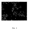
- FIG. 1 shows the light microscopy (x 400, line: 10 microns) of the starting culture A with Histomonas meleagridis (a), Tetratrichomonas gallinarum (b) and Blastocystis sp (c).
- FIG. 2 shows clonal cultures of: Histomonas meleagridis (A); Tetratrichomonas gallinarum (B) and Blastocystis sp. (C) after micromanipulation.
- FIG. 3 shows the specificity of the developed PCR assays.
- Hm Histomonas meleagridis
- Tg Tetratrichomonas gallinarum
- B1 Blastocystis sp.
- M molecular weight marker (100-bp ladder)
- 1 Histomonas meleagridis
- 2 Tetratrichomonas gallinarum
- 3 Blastocystis sp .
- N Negative control.
- the starting point for the work were 2 mixed cultures (A and B), which were created from the manure of turkeys.
- the first culture was from a 10-week-old turkey from a population of 5000 animals, of which 40% died and the remainder were killed (Hess et al., 2004). About 1 g of the caecum together with some mucosa were transferred into 9 ml of medium 199 + Earle's salts + L-glutamine + 25 mM HEPES + L-amino acids (Gibco TM, Invitrogen).
- the culture of origin B was made similar to culture A. However, as a starting material, cecal material from a bronze pute was used as a starting material by a hobbyist in which 2 of 6 animals died. For the micromanipulation culture B after passage 7 was used.
- micromanipulation was carried out in concrete as follows:
- the two cultures of origin (A and B) from the faeces of turkeys were diluted 1: 100 and fitted with a capillary (1.0 mm outer diameter, Hilgenberg, Germany) mounted on a puller (P97, Sutter, USA) and on an outer diameter of 25 microns (Microforge deFonbrune, Bachofer, Germany) was pulled. Parasites were placed in 50 ⁇ l of culture medium and transferred to an Eppendorf tube. The entire procedure was performed with a Narishige Micromanipulator (Narishige, Japan) and a microscope (Diaphot 300, Nikon, Austria) under 400X magnification. To monitor the insulation, a CCD camera and a monitor (Sony, Japan) connected to the microscope were used.
- microorganisms were incubated at 40 ° C for up to 4 days in Eppendorf tubes. Light microscopy was used to check the growth on the 2nd, 3rd and 4th day. Each positive or suspect colony was transferred to 9.7 ml of standard medium in a 50 ml plastic tube (Sarstedt).
- seed medium a suitable growth medium
- seed medium a suitable growth medium
- the bacterial accompanying flora of the respective culture of origin was determined.
- approximately 100 ⁇ l of the source colony were plated on commercial Columbia (+ 5% blood), McConkey, Salmonella Detection and Identification Media (SMID) and Sabouraud Agar plates (all: bioMerieux, Marcy l'Etoile, France).
- the bacterial flora was identified using the API-20 E microsystem (bio Merieux, Marcy l'Etoile, France). E.coli, staphylococci and streptococci have been identified as major bacterial strains. With a Impfinate was one Part of the bacteria taken from the Columbia blood agar plate and placed in the culture medium prior to micromanipulation.
- RNA portions of the ribosomal RNA were sequenced and entered into a publicly available sequence database (accession numbers: AJ920322 - AJ920324).
- Example 2 Determination of the growth behavior of clonal cultures.
- the parasites were kept in the Eppendorf tube for 3-4 days each until transferred to a 50 ml tube, which in turn contained 9 ml of medium. Subsequently, after 3 to 4 days, 1 ml of the culture was transferred to a new tube in which 9 ml of the standard medium were initially introduced, thus an identical procedure as in the original cultures A and B. The further cultivation was then carried out analogously to the method of the starting cultures. At various times counts were made to determine the growth rate of the protozoa. Trypan blue (0.4%) was used for live-death staining (Table 1). ⁇ b> Table 1 ⁇ / b>.
- a growth profile was prepared at different times, as shown in the following table (1).
- Example 3 Sensitivity of the cultures for the standard preparation dimetridazole, as a reference for the testing of substances in vitro.
- the clonal Histomonas culture was treated with the imidazole derivative dimetridazole.
- a total of three 50 ml tubes of the cloned Histomonas culture were centrifuged on the 3rd day of culture at 380 xg. Subsequently, the supernatant was removed and discarded. The pellets of the 3 tubes were pooled and the histomonads counted. Subsequently, a concentration of 100,000 / ml was set by means of cell culture medium. In each case 1 ml of histomonad culture was transferred to an Eppendorf tube and incubated together with 0.4 mg of dimetridazole at 40 ° C. At 24h intervals, a count of parasites is performed to examine the effect of a test substance as exemplified in Table 2.
- Example 4 Reproduction of histomoniasis or blackhead disease in 14 day old turkeys, as the basis for establishing an in vivo model suitable for testing substances in vivo.
- Example 5 Establishment of Molecular Diagnostic Methods for specific detection of Histomonas meleagridis, Tetratrichomonas gallinarum and Blastocystis sp.
- the clonal cultures were used to establish molecular detection methods, with the specific ability to detect the respective pathogen.
- oligonucleotides were designed, with the help of which specifically the respective pathogen can be detected.
- oligonucleotides were designed which allow the specific detection of the particular pathogen by means of hybridization or amplification of the genome fragment (PCR).
- PCR genome fragment
Landscapes
- Health & Medical Sciences (AREA)
- Life Sciences & Earth Sciences (AREA)
- Chemical & Material Sciences (AREA)
- Engineering & Computer Science (AREA)
- Biotechnology (AREA)
- Medicinal Chemistry (AREA)
- Tropical Medicine & Parasitology (AREA)
- Organic Chemistry (AREA)
- Bioinformatics & Cheminformatics (AREA)
- General Health & Medical Sciences (AREA)
- Genetics & Genomics (AREA)
- Microbiology (AREA)
- Wood Science & Technology (AREA)
- Zoology (AREA)
- Biochemistry (AREA)
- Epidemiology (AREA)
- Biomedical Technology (AREA)
- Virology (AREA)
- Immunology (AREA)
- Mycology (AREA)
- Pharmacology & Pharmacy (AREA)
- General Engineering & Computer Science (AREA)
- Animal Behavior & Ethology (AREA)
- Public Health (AREA)
- Veterinary Medicine (AREA)
- Micro-Organisms Or Cultivation Processes Thereof (AREA)
- Medicines Containing Antibodies Or Antigens For Use As Internal Diagnostic Agents (AREA)
- Pharmaceuticals Containing Other Organic And Inorganic Compounds (AREA)
- Medicines Containing Material From Animals Or Micro-Organisms (AREA)
Description
- Die Erfindung betrifft die Herstellung und Anwendung von Protozoenkulturen von Histomonas meleagridis, Tetratrichomonas gallinarum und Blastocystis sp..
- Die Schwarzkopfkrankheit, Typhlohepatitis oder Histomoniasis bei Puten wurde von Smith (1895) zum ersten Mal beschrieben. Wesentliche Erkenntnisse zur Ätiologie konnten durch die Untersuchungen von Tyzzer (1920) gewonnen werden. In dieser Arbeit werden zum ersten Mal Flagellen und Pseudopodien, als morphologisches Hauptkriterium des Erregers, beschrieben, was den Erreger als Protozoen einstufte. Die Morphologie des Parasiten und das Vorkommen bei Puten führten zur Bezeichnung Histomonas meleagridis. Die Erkenntnis, dass der Parasit bei Hühnern ebenfalls vorkommen kann, führte zur Empfehlung diese Tierarten getrennt zu halten. Als weitere empfängliche Vogelarten werden insbesondere Perlhuhn, Fasan, Pfau und Wachtel beschrieben, während das Wassergeflügel als nicht empfänglich gilt (McDougald, 2003). Durch die Pathogenese und die hohe Prävalenz der Erkrankung bei Puten ist diese Spezies sicherlich als einer der empfänglichsten Wirte anzusehen.
- In den letzten beiden Jahren kam es in Europa zu einem verstärkten Auftreten der Erkrankung. Hintergrund ist insbesondere das Verbot geeigneter Prophylaktika und Therapeutika, mit deren Hilfe die Erkrankung über Jahrzehnte gezielt bekämpft wurde. Nichtsdestotrotz wurde die prophylaktische Verwendung von Therapeutika bei Puten, insbesondere von Nifursol, Imidazolderivaten, wie Nitroimidazol und Dimetridazol, als Futterzusatzstoff in der Europäischen Union verboten (siehe EEC1798/1995, EEC2205/2001 und EEC1756/2002), da die Unbedenklichkeit dieser Stoffe (Karzinogenität und Genotoxizität) für den Verbraucher nicht auszuschließen ist. Auch das in Nordamerika zur Prophylaxe eingesetzte Nitarsone (4-Nitrophenylarsonic acid) besitzt in Europa keine Zulassung. In Folge dieses absoluten Prophylaxe- und Therapienotstandes mussten in verschiedenen Ländern Europas, nach Ausbruch der Erkrankung in Putenbeständen, in den letzten beiden Jahren Keulungsmaßnahmen durchgeführt werden (Hess et al., 2004).
- Bei Legehennen verläuft die Erkrankung milder, durch die zunehmende Alternativhaltung kommt es aber auch hier zu einem vermehrten Auftreten (Hafez et al., 2001; Esquenet et al., 2003).
- Grundlegende Arbeiten über die Vermehrung von Histomonas meleagridis in Zellkulturmedien wurden von Dwyer (1970) und später von Stepkowski (1978) durchgeführt. Als Grundlage dient beiden Autoren Kotmaterial oder Isolate aus Kot, die unter agnotobiotischen Bedingungen zu vermehren sind. Mithin ist eine Vermehrung des Erregers nur in einem entsprechenden Milieu möglich, was hauptsächlich durch die Begleitflora bestimmt wird.
- Insgesamt haben obige Kulturen den Nachteil, dass sie genetisch nicht einheitlich sind, da sie aus mehreren Erregern ein und derselben Spezies bestehen. Darüber hinaus ist nicht gewährleistet, dass sich auch andere Protozoen, die morphologisch schwierig zu unterscheiden sind, in der Kultur befinden. Das Vorkommen von verschiedenen Protozoen im Darm des Geflügels ist vielfach beschrieben (McDougald, 2003), was diese Problematik nachdrücklich unterstreicht.
- Als Folge der wenig charakterisierten Kulturen sind Ergebnisse von in vitro und in vivo Experimenten, in denen solche Kulturen verwendet werden, zurückhaltend zu beurteilen. Die verlässliche Testung von Substanzen, sowohl in vitro als auch in vivo, fordert eine genetisch einheitliche Kultur, um zu verhindern, dass bestimmte Effekte auf das Vorhandensein verschiedener Subpopulationen oder andere Protozoenspezies, zurückzuführen sind. Die Ergebnisse von Tierversuchen, die in jüngster Vergangenheit durchgeführt wurden, spiegeln diese Heterogenität wieder (Hu und McDougald, 2003; Hu et al., 2004; Landmann, 2004).
- Die Heterogenität von Kulturen bedingt, dass bestimmte Subpopulationen phänotypisch dominieren, was falsche Rückschlüsse auf die generelle Empfindlichkeit eines Erregers gibt. Dieses Problem ist beispielhaft für den Erreger der Malaria, Plasmodium falciparum, beschrieben (Rosario, 1981; Trager et al., 1981; Burkot et al., 1984; LeBras et al., 1983). Als Konsequenz hiervon besteht die Gefahr, dass im Rahmen von in vitro Tests lediglich eine Selektion und Vermehrung von resistenten Stämmen erfolgt, was keinen Rückschluss auf die Empfindlichkeit eines Erregers zulässt. Hinweise über mögliche unterschiedliche Subpopulationen von Histomonas meleagridis gibt es aus der Arbeit von Gerbod et al. (2001). Die Autoren haben von 4 Isolaten Teile der ribosomalen RNA - dieser Genabschnitt wird oft für phylogenetische Analysen benutzt - sequenziert und eine leichte Heterogenität an einigen Positionen festgestellt. Die Autoren schließen daraus, dass dies ein Hinweis für unterschiedliche Subpopulationen sein könnte. Gesicherte Hinweise über Subpopulationen innerhalb einer der drei oben erwähnten Protozoenspezies gibt es von Tetratrichomonas gallinarum, wie von Cepicka et al. (2005) berichtet wird. Auch von Blastocystis sp. ist die genetische Vielfalt ausführlich dokumentiert, auch im Hinblick auf eine mögliche Zoonoseproblematik (Noel et al., 2003; Noel et al., 2003).
- In Kulda et al. (1974) wurden Infektionsexperimente mit Tetratrichomonas gallinarum durchgeführt und dabei wurde mit einem Isolat die für Histomonas meleagridis typische Schwarzkopfkrankheit ausgelöst. Es wurde gezeigt, dass diese Infektionskultur von Tetratrichomonas gallinarum mit Histomonas meleagridis kontaminiert war, basierend auf der mikroskopischen Kotuntersuchung.
- In Goedbloed and Bool (1962) wurden Puten sowohl mit einer Kultur von Histomonas meleagridis als auch Tetratrichomonas gallinarum infiziert und anschließend histologische Untersuchungen durchgeführt. Nach gemischter Infektion wurden beide Erreger mikroskopisch im Kot nachgewiesen. Dies zeigt die Gefahr auf, dass bei Re-Isolierung von Protozoenkulturen mehrere Kulturen isoliert werden, da ein selektives Medium nicht bekannt ist.
- Bayon and Bishop (1937) beschreiben die Isolierung von Histomonas meleagridis aus der Leber eines Huhns, welches zur Diagnostik verwendet wurde. Aus dem Kot des Tieres wurde eine Mischung aus Blastocystis, Trichomonas und Chilomastix isoliert, aber keine Histomonaden. Auch dies zeigt eindeutig auf, dass es sich bei Protozoenkulturen meist um Co-Infektion bzw. Co-Isolierung handelt.
- Bishop (1938) beschreibt auch die Isolierung von Trichomonas, Chilomastix und Blastocystis aus dem Zäkum eines Huhns.
- In Norton et al. (1999) wird die Präsenz von Histomonas meleagridis und Tetratrichomonas gallinarum nach Infektion mit dem Nematoden Ascaridia dissimilis beschrieben.
- Dies zeigt auf, dass klonale Protozoenkulturen bis dato nicht bekannt waren und ebenso nicht zur Verfügung gestellt werden konnten.
- In der
EP 600 396 A2 - In der GB 902 760 werden Verfahren zur Herstellung von attenuierten Endoparasiten beschrieben, bei denen ultraviolette Strahlung dazu verwendet wird, diese zu attenuieren.
-
FR 2 853 550 - Tan Kevin et al. (Experimental Parasitology 96(1) (2000): 9-15) betrifft die Kultivierung von Blastocystis hominis Kulturen auf festem Agar.
- Die
US 5 824 537 betrifft unter anderem ein Verfahren zur in vitro-Klonierung von Parasiten des Genus Babesia, wobei einzelne Parasiten-Zellen von einer Suspension von infizierten Erythrozyten gewonnen werden, wobei diese einzelnen Zellen schließlich zur Herstellung einer Kultur verwendet werden können. - Clark et al. (Clinical Microbiology Reviews 15(3) (2002): 329-341 ist ein Übersichtsartikel über Verfahren zur Kultivierung von Protozoen, wobei unter anderem die Züchtung von Blastocystin hominis beschrieben wird.
- Bedingt durch die genetische Vielfalt, aber insbesondere durch das gleichzeitige Vorkommen mehrerer Protozoen in einem Tier, sind exakte diagnostische Methoden unabdingbar. Dies wird dadurch verstärkt, dass offensichtlich Tetratrichomonas gallinarum und Histomonas meleagridis ähnliche Symptome und morphologische Veränderungen hervorrufen können (Tyzzer, 1920; Allen, 1941).
- Die Diagnostik der Histomoniasis wird besonders dadurch erschwert, dass der Parasit in der Außenwelt rasch abstirbt und dadurch nicht mehr nachzuweisen ist. Es besteht zwar die Möglichkeit der Kultivierung des Erregers aus körperwarmen Tieren, jedoch sind besondere Bedingungen notwendig, um das Überleben des Trophozoiten zu gewährleisten (Dwyer, 1970; McDougald and Galloway, 1973). Der direkte Erregernachweis kann somit nur mittels Histologie durchgeführt werden. Hierfür sind mehrere Methoden beschrieben, insbesondere die Perjodsäure-Schiff-Reaktion (PAS-Reaktion), mittels derer der im Gewebe unbegeißelte Erreger nachgewiesen werden kann (Kemp und REID, 1966). Allerdings ist diese Methode arbeits- und zeitaufwendig, was die Notwendigkeit nach Alternativen unterstreicht. Erschwert wird die Histologie auch dadurch, dass der Erreger von Makrophagen und Pilzzellen nicht leicht differenziert werden kann, was den Bedarf nach besseren Diagnosemöglichkeiten zusätzlich unterstreicht. Esquenet et al. (2003) beschreiben den zytologischen Nachweis mittels Direktfärbung von Abklatschpräparaten.
- Molekulare diagnostische Nachweismethoden zum sensitiven und spezifischen Nachweis der drei Erreger sind bisher nicht vorhanden.
- Aufgabe der Erfindung ist es, klonale Kulturen von Histomonas meleagridis, Tetratrichomonas gallinarum und Blastocystis sp. zu etablieren und solchermaßen etablierte Kulturen in vitro und in vivo anzuwenden, um die Nachteile des Standes der Technik, insbesondere die Inhomogenität der bisher bekannten Protozoenkulturen aus Geflügel, zu überwinden. Darüber hinaus ist es eine Aufgabe, spezifische Diagnostika zu entwickeln, damit ein sicherer Erregernachweis und eine Abgrenzung zwischen den Erregern möglich ist.
- Demgemäß wird mit der vorliegenden Erfindung eine klonale Protozoenkultur umfassend Protozoenzellen einer einzigen Protozoenspezies zur Verfügung gestellt, welche dadurch gekennzeichnet ist, dass die Protozoenspezies ausgewählt ist aus der Gruppe bestehend aus Histomonas meleagridis, Tetratrichomonas gallinarum und Blastocystis sp., wobei die Kultur symbiotische Bakterien oder deren Lysat enthält.
- Gemäß der vorliegenden Erfindung wird als "klonale Protozoenkultur" eine Kultur von Protozoenzellen bezeichnet, die von einer einzelnen Protozoenzelle, die entweder Histomonas meleagridis, Tetratrichomonas gallinarum oder Blastocystis sp. ist, abstammt und somit aus dieser gebildet wurde. Die Kultur besteht somit aus Protozoenzellen, die alle durch Vermehrung von einer einzigen Zelle abstammen. Die Protozoenzellen sind somit Kopien einer einzigen oben angeführten Ausgangszelle. In der klonalen Kultur können sich zwar andere Organismen, wie Bakterien, befinden, erfindungswesentlich ist aber, dass nur eine einzige Protozoenspezies ausgewählt aus der Gruppe bestehend aus Histomonas meleagridis, Tetratrichomonas gallinarum und Blastocystis sp. in der klonalen Kultur vorhanden ist, d.h. die Kultur enthält Protozoenzellen einer dieser Protozoenspezies, nicht aber Protozoenzellen der beiden jeweils anderen Protozoenspezien (z.B. Histomonas meleagridis nicht aber Tetratrichomonas gallinarum und Blastocystis sp., Tetratrichomonas gallinarum nicht aber Histomonas meleagridis und Blastocystis sp.).
- Die Bereitstellung einer derartigen klonalen Kultur konnte bisher im Stand der Technik - trotz des seit mindestens 20 bis 30 Jahren bestehenden Bedürfnisses (siehe oben Dywer oder Stepkowski) - nicht erfolgen und ermöglicht nunmehr aber eine Vielzahl von Anwendungen, die von der Herstellung von Impfstoffen und Therapeutika gegen die hierin angeführten Protozoenspezien bis hin zur näheren Erforschung und Charakterisierung derselben in Geflügel parasitischen Protozoenspezien reichen.
- Bei der Herstellung von erfindungsgemäßen klonalen Protozoenkulturen ist es einerseits entscheidend das Wachstum der Protozoenzellen durch Vorsehen entsprechender Umgebungsbedingungen (z.B. Temperatur, Anwesenheit von symbiotischen Bakterien, Kohlenstoffquellen) in vitro zu ermöglichen und andererseits die Protozoen zu vereinzeln. Die Vereinzelung erfolgt vorzugsweise durch Mikromanipulation, wobei nach dieser Vereinzelung die Protozoen in einem entsprechenden Wachstumsmedium (das Wachstumsmedium sollte dabei eine Kohlenhydratquelle, insbesondere eine Stärke, wie Reis- oder Maisstärke, und gegebenenfalls Salze, insbesondere Earle's Salze, Aminosäuren, insbesondere L-Glutamin, und Antibiotika/Antimykotika, insbesondere Penicillin, Streptomycin und Amphotericin, umfassen) vermehrt werden, um schließlich eine klonale Protozoenkultur zu ergeben.
- Die erfindungsgemäße Protozoenkultur enthält symbiotische Bakterien und/oder deren Lysat.
- Es ist bekannt, dass sich Histomonas meleagridis, Tetratrichomonas gallinarum und Blastocystis sp. in Anwesenheit von symbiotischen Bakterien oder deren Stoffwechselprodukte (z.B. in Form von Lysat) in vitro effizient vermehren lassen. Aus diesem Grund befinden sich vorzugsweise symbiotische Bakterien und/oder deren Lysat in der erfindungsgemäßen Protozoenkultur, da diese bei der Herstellung der Kulturen eingesetzt werden. Dabei werden vorzugsweise jene Bakterien (und/oder Lysate) verwendet, die sich auch in vivo in Symbiose mit den Protozoen befinden. Selbstverständlich ist es auch möglich, die Bakterien im Wesentlichen vollständig aus der Protozoenkultur zu entfernen (z.B. durch Zentrifugation oder Filtration), sofern die Protozoenzellen beispielsweise zur Untersuchung von Wachstumsbedingungen, zur Herstellung von Impfstoffen und Antikörpern verwendet werden.
- Gemäß einer bevorzugten Ausführungsform sind die Bakterien Enterobakterien, insbesondere coliforme Bakterien, Staphylokokken, Streptokokken oder Mischungen davon.
- Da Histomonas meleagridis, Tetratrichomonas gallinarum und-Blastocystis sp. vorzugsweise aus Kot gewonnen werden, setzt sich die Bakterienkultur vorzugsweise aus den darin befindlichen Bakterienspezien zusammen. Es wurde erfindungsgemäß festgestellt, dass im Kot von Geflügel neben Enterobakterien auch Staphylokokken und Streptokokken vorhanden sind, die einzeln oder in Kombination zur Kultivierung von Protozoenzellen eingesetzt werden können.
- Ein weiterer Aspekt der vorliegenden Erfindung betrifft eine Impfstoffformulierung umfassend inaktivierte oder lebend klonale Protozoenzellen einer einzigen Protozoenspezies ausgewählt aus der Gruppe bestehend aus Histomonas meleagridis, Tetratrichomonas gallinarum und Blastocystis sp. oder einen Antikörper gerichtet gegen klonale Protozoenzellen einer einzigen Protozoenspezies ausgewählt aus der Gruppe bestehend aus Histomonas meleagridis, Tetratrichomonas gallinarum und Blastocystis sp..
- Die Bereitstellung von klonalen Protozoenkulturen bzw. -zellen ermöglicht eine gezielte Impfstoffherstellung, die einerseits zur aktiven und andererseits zur passiven Immunisierung von Geflügel eingesetzt werden kann. Bei der Herstellung von aktiven Impfstoffen ist es notwendig ein möglichst homogenes Antigen zu haben. Im konkreten Fall ist es wünschenswert, eine klonale Protozoenkultur zur Verfügung zu stellen, die im Wesentlichen nur eine Protozoenspezies enthält, gegen die ein Tier geimpft werden soll. Die Verwendung der erfindungsgemäßen klonalen Kulturen ermöglicht eine entscheidend verbesserte und standardisierbare industrielle Herstellung dieses Impfstoffes, vor allem im Hinblick auf bessere Reproduzierbarkeit und geringere Chargen-Varianz. Beispielsweise sollte ein aktiver Impfstoff, der zur Immunisierung von Geflügel gegen Histomonas meleagridis eingesetzt wird, im Wesentlichen nur Histomonas meleagridis Zellen umfassen. Ist dies nicht der Fall, kann es unter Umständen dazu führen, dass ein Selektionsprozess eintritt, der notwendigerweise nicht die gewünschte Spezies bevorzugt. D.h. es ist dann durchaus möglich, dass das Geflügel nicht oder unzureichend gegen den gewünschten Erreger immunisiert wird sondern gegen einen oder mehrere andere in der Kultur befindliche Antigene (z.B. andere Protozoen). Bei passiven Impfstoffen hingegen ist es wichtig, dass größtenteils Antikörper verwendet werden, die spezifisch gegen die zu bekämpfenden Antigene (z.B. Protozoenzellen) wirken. Daher ist es entscheidend, dass bei der Herstellung derartiger Impfstoffe möglichst reine Ausgangskulturen verwendet werden. Hierzu eignen sich besonders die erfindungsgemäßen klonalen Protozoenkulturen.
- Die Impfstoffformulierung umfasst vorzugsweise ein Adjuvans.
- Um die Immunantwort gegen ein Antigen zu fördern, werden bei der Herstellung aktiver Impfstoffe Adjuvantien verwendet, wobei das Adjuvans vorzugsweise ausgewählt ist aus der Gruppe bestehend aus Freund's komplettes Adjuvans, Freund's inkomplettes Adjuvans, Aluminiumhydroxid, Bordetella pertussis, Saponin, Muramyl-Dipeptid, Ethylen-Vinyl-Acetat Copolymer, Öl, vorzugsweise ein pflanzliches Öl oder ein Mineralöl, insbesondere Erdnussöl oder Silikonöl, und Kombinationen davon sowie die in Singh et al. (Nat. Biotech 17 (1999), 1075-1081) beschriebenen Adjuvantien.
- Geeignete Adjuvantien, die erfindungsgemäß verwendet werden können, sind dem Fachmann hinreichend bekannt (siehe z.B. O'Hagan, Derek T. (2000) "Vaccine Adjuvants: Preparation Methods and Research Protocols", Humana Press;
US 5,885,589 ;EP 109 942 - Die klonalen Protozoenzellen werden vorzugsweise durch Passagieren, durch Strahlung, insbesondere durch UV- und radioaktive Strahlung, durch chemische Substanzen oder durch Viren attenuiert.
- Um die Pathogenität der im Impfstoff eingesetzten Protozoenzellen zu reduzieren, können diese Zellen attenuiert werden. Eine Attenuierung kann erfindungsgemäß durch mehrere Methoden erfolgen. Es hat sich erfindungsgemäß herausgestellt, dass vor allem die oben angeführten Attenuierungsverfahren sich besonders gut eignen die Pathogenität der Protozoenzellen zu reduzieren. Insbesondere wird die Attenuierung durch mehrmaliges Passagieren (mindestens 10, vorzugsweise mindestens 50, noch mehr bevorzugt mindestens 100 Passagen) bevorzugt verwendet. Beim Passagieren werden die Protozoen zunächst in einem Kulturgefäß vermehrt und nach dem Erreichen einer bestimmten Zelldichte (z.B. nach 12 Stunden, vorzugsweise nach 24 Stunden, noch mehr bevorzugt nach 48 Stunden, Kultivierung) in einem Verhältnis von beispielsweise 1:10, 1:50, 1:20 oder 1:5 in ein neues Medium überführt.
- Gemäß einer bevorzugten Ausführungsform sind die klonalen Protozoenzellen durch Hitze, Formalin, beta-Propiolaktonoder Kombinationen davon inaktiviert.
- Alternativ bzw. in Ergänzung zur Attenuierung der Protozoenzellen können diese auch durch mehrere Verfahren inaktiviert werden. Durch die Inaktivierung der Zellen wird das Risiko einer Infektion des geimpften Tieres drastisch reduziert.
- Die Impfstoffformulierung ist vorzugsweise zur intravenösen, subkutanen, intramuskulären, oralen oder intrakloakalen Verabreichung adaptiert.
- Ein weiterer Aspekt der vorliegenden Erfindung betrifft ein Verfahren zur Herstellung einer klonalen Protozoenkultur umfassend Protozoenzellen einer einzigen Protozoenspezies ausgewählt aus der Gruppe bestehend aus Histomonas meleagridis, Tetratrichomonas gallinarum und Blastocystis sp. umfassend die Schritte:
- a) Bereitstellen einer Ausgangsprobe umfassend Protozoenzellen mindestens einer Protozoenspezies ausgewählt aus der Gruppe bestehend aus Histomonas meleagridis, Tetratrichomonas gallinarum und Blastocystis sp.,
- b) Kultivieren der Zellen der Ausgangsprobe in einem Kulturmedium umfassend eine Kohlenhydratquelle und Bakterien mindestens einer Bakterienspezies, die in situ symbiotisch mit den Protozoenzellen leben,
- c) Vereinzeln der Protozoenzellen mittels Mikromanipulation,
- d) Kultivieren der vereinzelten Protozoenzellen in einem Kulturmedium wie unter b) definiert und
- e) gegebenenfalls Bestimmen der Reinheit der klonalen Protozoenkultur.
- Die Ausgangsprobe ist vorzugsweise Kot oder Gewebe.
- Da Histomonas meleagridis, Tetratrichomonas gallinarum und Blastocystis sp. im Darm von Geflügel einen Teil ihres Lebenszyklus verbringen, ist besonders Kot geeignet, um diese Protozoen zu isolieren und daraus klonale Protozoenkulturen herzustellen.
- Gemäß einer bevorzugten Ausführungsform wird die Ausgangsprobe von einem Geflügel ausgewählt aus der Gruppe bestehend aus Pute, Huhn, Fasan, Pfau, Wachtel und Perlhuhn bereitgestellt.
- Histomonas meleagridis, Tetratrichomonas gallinarum und Blastocystis sp. sind Parasiten, die vornehmlich die obigen Geflügelarten befallen. Daher werden die Protozoen vorzugsweise aus diesen Tieren isoliert.
- Die Kohlenhydratquelle ist vorzugsweise Stärke, insbesondere Reis-, Mais- oder Kartoffelstärke.
- Es hat sich herausgestellt, dass Histomonas meleagridis, Tetratrichomonas gallinarum und Blastocystis sp. vor allem Stärke als Kohlenstoffquelle bevorzugen und diese durch Aufnahme in das Innere der Zelle abzubauen vermögen.
- Die Bakterien sind vorzugsweise Enterobakterien, insbesondere coliforme Bakterien, Staphylokokken, Streptokokken oder Mischungen davon.
- Es hat sich erfindungsgemäß herausgestellt, dass Histomonas meleagridis, Tetratrichomonas gallinarum und Blastocystis sp. insbesondere durch Symbiose mit den oben angeführten Bakterien (oder deren Lysate) kultivierbar sind. Ohne die Anwesenheit dieser Bakterienkulturen, die vorzugsweise aus dem Kot von Geflügel isoliert werden, können die Protozoen ex vivo nicht überleben und nicht kultiviert werden. Daher ist es entscheidend, dass die Bakterien sowohl vor als auch nach der Vereinzelung bei jedem ex vivo-Kultivierungsschritt, sofern die Lebensfähigkeit der Protozoen erwünscht ist, anwesend sind.
- Vorzugsweise enthält das Kulturmedium weiters Salze, insbesondere Earle's Salze, Aminosäuren, insbesondere L-Glutamin, und Antibiotika/Antimykotika, insbesondere Penicillin, Streptomycin und Amphotericin.
- Gemäß einer bevorzugten Ausführungsform wird die Reinheit der Kultur mittels einer Nukleinsäureamplifkations- oder Hybridisierungstechnik oder mittels einer immunologischen Technik mit Antikörpern gerichtet gegen klonale Protozoenzellen einer einzigen Protozoenspezies ausgewählt aus der Gruppe bestehend aus Histomonas meleagridis, Tetratrichomonas gallinarum und Blastocystis sp. bestimmt.
- Die Bestimmung der Reinheit dient vor allem dazu die Anwesenheit des zu isolierden Parasiten bzw. die Abwesenheit zusätzlicher nicht in der klonalen Protozoenkultur erwünschten Zellen anderer Protozoenspezies zu bestätigen. Zur Bestimmung der Reinheit können mehrere Methoden verwendet werden, wobei aber Nukleinsäureamplifikations- und Hybridisierungstechniken und immunologische Techniken bevorzugt sind. Die Kenntnis konservierter bzw. Spezies-spezifischer Nukleinsäureregionen innerhalb der Protozoen-Genome ermöglicht es spezifische Primer bzw. Sonden zu identifizieren (z.B. durch Sequenz-Überlagerungen), herzustellen und einzusetzen (z.B. PCR, Southern-Blot, In Situ Hybridisierung). Derartige Techniken sind dem Fachmann hinreichend bekannt (siehe z.B. "Current Protocols in Molecular Biology", Ed Fred M. Ausubel et al., Wiley Inc.). Aufgrund der Möglichkeit, mit Hilfe der vorliegenden Erfindung klonale Protozoenkulturen mit einer einzigen Protozoenspezies herzustellen, ist es des Weiteren möglich, spezifische Antikörper gegen diese Protozoenspezies herzustellen und hierin einzusetzen, um die Reinheit der Kulturen zu bestimmen (z.B. mittels ELISA, RIA).
- Vorzugsweise werden die Oligonukleotide 5'-GAAAGCATCTATCAAGTGGAA-3' und 5'-GATCTTTTCAAATTAGCTTTAAA-3' zur Bestimmung von Histomonas meleagridis, 5'-GCAATTGTTTCTCCAGAAGTG-3' und 5'-GATGGCTCTCTTTGAGCTTG-3' zur Bestimmung von Tetratrichomonas gallinarum, 5'-TAACCGTAGTAATTCTAGGGC-3' und 5'-AACGTTAATATACGCTATTGG-3' zur Bestimmung von Blastocystis sp. verwendet.
- Diese Oligonukleotide erwiesen sich als besonders geeignet die einzelnen Protozoenspezien zu bestimmen, da diese an jene Genomregionen, die für die ribosomale RNA (rRNA) codieren, die wiederum zur phylogenetischen Charakterisierung von Organismen herangezogen wird, binden. Die Oligonukleotide wurden aufgrund von Sequenz-Überlagerungen von öffentlich zugänglichen rRNA-Sequenzdaten Histomonas meleagridis (AF293d56), Tetratrichomonas gallinarum (AF124608) und Blastocystis sp. (AF408427) ermittelt.
- Nach dem erfindungsgemäßen Verfahren ist eine klonale Protozoenkultur: erhältlich, welche Protozoenspezies ausgewählt ist aus der Gruppe bestehend aus Histomonas meleagridis, Tetratrichomonas gallinarum und Blastocyctis sp..
- Ein weiterer Aspekt der vorliegenden Erfindung betrifft ein Verfahren zur Bestimmung des Einflusses von Substanzen oder Substanzgemischen auf Protozoenzellen einer Protozoenspezies ausgewählt aus der Gruppe bestehend aus Histomonas meleagridis, Tetratrichomonas gallinarum und Blastocystis sp. umfassend die Schritte:
- Bereitstellen einer erfindungsgemäßen klonalen Protozoenkultur,
- gegebenenfalls Einstellen der Zellzahl der Protozoenkultur,
- Inkontaktbringen und Kultivieren der klonalen Protozoenkultur mit einem erfindungsgemäßen Kulturmedium,
- Zugabe mindestens einer Substanz oder mindestens eines Substanzgemisches, die/das potentiell einen Einfluss auf Protozoenzellen ausüben kann,
- Bestimmen der Protozoenzellzahl in der Protozoenkultur,
- Vergleichen der Protozoenzellzahl und der Begleitflora vor und nach Zugabe der mindestens einen Substanz oder des mindestens einen Substanzgemisches.
- Das Bereitstellen einer klonalen Protozoenkultur ermöglicht es des Weiteren ein Verfahren zur Verfügung zu stellen, das geeignet ist, den Einfluss von Substanzen bzw. Substanzgemischen auf das Wachstumsverhalten und auf die Überlebensfähigkeit spezifischer Protozoen zu untersuchen. Da es bis dato nicht möglich war klonale Protozoenkulturen, die nur eine einzige Protozoenspezies (Histomonas meleagridis, Tetratrichomonas gallinarum und Blastocystis sp.) umfasst, herzustellen, konnten auch keine Untersuchungen durchgeführt werden, mit denen gezeigt werden konnte inwieweit sich bestimmte Substanzen auf den Stoffwechsel einer konkreten Protozoenspezies auswirken. Ein solches Verfahren ist von entscheidender Bedeutung bei der Erforschung von neuen Geflügel-Therapeutika, die gegen Histomonas meleagridis, Tetratrichomonas gallinarum und Blastocystis sp. wirksam sind.
- Es hat sich gezeigt, dass die Wirkung von Substanzen auf Protozoen am geeignetsten durch die Veränderung der Zellzahl bestimmt wird (siehe z.B. Zenner et al., 2003; Caillait et al., 2002). Dazu wird zunächst die Anzahl der in einer Kultur vorhandenen Protozoen bestimmt und gegebenfalls auf eine bestimmte Anzahl eingestellt. Anschließend wird die klonale Kultur in Anwesenheit von Substanzen, die das Wachstum der Protozoen potentiell stören könnten, kultiviert. Nach einer Inkubationszeit von mindestens 12 Stunden, vorzugsweise mindestens 24 Stunden, wird die Anzahl der Protozoen neuerlich bestimmt und mit der Ausgangsanzahl bzw. mit einer Negativ-Kontrolle (Kulturmedium ohne Zusatz von potentiellen Inhibitoren) verglichen. Eine Abnahme der Zellzahl gegenüber der Ausgangsprobe bzw. eine reduzierte Anzahl von Protozoen gegenüber der Negativ-Kontrolle zeigt, dass die Substanz einen negativen Einfluss auf die Protozoenspezies hat. Die Anzahl der Parasiten wird einfach Bestimmung der Zellzahl bestimmt (z.B. lebend-tot Färbung).
- Ein weiterer Aspekt der vorliegenden Erfindung betrifft ein Verfahren zum Nachweis einer Protozoenspezies ausgewählt aus der Gruppe bestehend aus Histomonas meleagridis, Tetratrichomonas gallinarum und Blastocystis sp. in einer Probe umfassend die Schritte:
- Bereitstellen einer Probe,
- Inkontaktbringen der Probe mit zumindest einen für Histomonas meleagridis, Tetratrichomonas gallinarum oder Blastocystis sp. spezifischen Oligonukleotid oder einen Antikörper gerichtet gegen Histomonas meleagridis, Tetratrichomonas gallinarum oder Blastocystis sp.,
- gegebenenfalls Unterwerfen des Proben/Oligonukleotid-Gemisches einer Nukleinsäureamplifikationstechnik,
- Detektion eines Nukleinsäureamplifikationsprodukts, einer Oligonukleotide/Protozoenzellen-Bindung oder einer Protozoenzellen/Antikörper-Bindung.
- Die Bereitstellung klonaler Protozoenkulturen ermöglicht es Mittel (z.B. Antikörper) zu etablieren, die dazu verwendet werden können, Protozoen spezifisch in einer Probe nachzuweisen. Vorzugsweise ist die Probe eine Gewebe-, Kot-, Kultur- oder Umweltprobe (z.B. eine Probe aus einem Geflügelstall oder -gehege) isoliert.
- Vorzugsweise werden die Oligonukleotide 5 -GAAAGCATCTATCAAGTGGAA-3' und 5'-GATCTTTTCAAATTAGCTTTAAA-3' zur Bestimmung von Histomonas meleagridis, 5'-GCAATTGTTTCTCCAGAAGTG-3' und 5'-GATGGCTCTCTTTGAGCTTG-3' zur Bestimmung von Tetratrichomonas gallinarum, 5'-TAACCGTAGTAATTCTAGGGC-3' und 5 -AACGTTAATATACGCTATTGG-3' zur Bestimmung von Blastocystis sp. verwendet.
- Die Oligonukleotide können sowohl als Primer/Primerpaar zur Amplifikation eines Genomabschnitts mittels z.B. PCR als auch als Hybridisierungssonden verwendet werden.
- Ein weiterer Aspekt der vorliegenden Erfindung betrifft ein Set zur Detektion von Histomonas meleagridis, Tetratrichomonas gallinarum und Blastocystis sp. oder Antikörper gerichtet gegen einer dieser Zellen in einer Probe umfassend:
- a) klonale Protozoenzellen einer einzigen Protozoenspezies ausgewählt aus der Gruppe bestehend aus Histomonas meleagridis, Tetratrichomonas gallinarum und Blastocystis sp. oder einen Antikörper gerichtet gegen Protozoenzellen einer einzigen Protozoenspezies ausgewählt aus der Gruppe bestehend aus Histomonas meleagridis, Tetratrichomonas gallinarum und Blastocystis sp. und
- b) Mittel zur Detektion der Protozoenzellen/Antikörper-Bindung.
- Das erfindungsgemäße Set kann dazu verwendet werden Histomonas meleagridis, Tetratrichomonas gallinarum und Blastocystis sp. oder aber auch das Vorhandensein von Antikörpern gegen diese Protozoenzellen in einer Probe nachzuweisen. Der Nachweis von Antikörpern in einer Probe (Protozoenzellen werden bereitgestellt und mit einer Probe inkubiert) dient beispielsweise dazu den Immunisierungsverlauf eines Geflügels zu beobachten oder lediglich um festzustellen, ob das Geflügel bereits einen ausreichenden schützenden Antikörpertiter gegen Histomonas meleagridis, Tetratrichomonas gallinarum oder Blastocystis sp. aufweist.
- Vorzugsweise sind die klonalen Protozoenzellen oder die Antikörper an einen festen Träger immobilisiert.
- Es ist von Vorteil, wenn entweder die Protozoenzellen oder die Antikörper gegen diese Protozoenzellen auf einem festen Träger gebunden sind.
- Gemäß einer bevorzugten Ausführungsform sind die Mittel zur Detektion konjugierte, vorzugsweise markierte Antikörper, insbesondere radioaktiv, Enzym- oder Fluoreszenz-markierte Antikörper.
- Verfahren zur Detektion von Antikörper/Ligand-Bindungen sind dem Fachmann hinreichend bekannt (siehe z.B. "Immunochemical Protocols", 3rd Edition (2005), ed. Burns, Robert, Humana Press).
- Ein weiterer Aspekt der vorliegenden Erfindung betrifft ein Futtermittel oder einen Futtermittelzusatzstoff umfassend eine erfindungsgemäße Impfstoffformulierung.
- Ein Futtermittel, welches zur Impfung von Geflügel gegen Histomonas meleagridis, Tetratrichomonas gallinarum oder Blastocystis sp. führt, hat sich als besonders vorteilhaft erwiesen, da dadurch die Impfung der Tiere einfach und effizient gestaltet werden kann.
- Die vorliegende Erfindung wird weiters durch die nachfolgenden Figuren und Beispiele beschrieben, ohne jedoch auf diese beschränkt zu sein.
-
Figur 1 zeigt die lichtmikroskopische Darstellung (x 400; Strich: 10 µm) der Ausgangskultur A mit Histomonas meleagridis (a), Tetratrichomonas gallinarum (b) and Blastocystis sp (c). -
Figur 2 zeigt klonale Kulturen von: Histomonas meleagridis (A); Tetratrichomonas gallinarum (B) and Blastocystis sp. (C) nach der Mikromanipulation. -
Figur 3 zeigt die Spezifität der entwickelten PCR-Assays. In keinem der Bilder sind Kreuzreaktionen, obwohl jeweils DNA Extrakte von 105 Mikroorganismen von Histomonas meleagridis (Hm),Tetratrichomonas gallinarum (Tg) and Blastocystis sp. (B1) für diese Untersuchung benutzt wurden. M: Molekulargewichtsmarker (100-bp ladder), 1: Histomonas meleagridis, 2: Tetratrichomonas gallinarum 3: Blastocystis sp.; N: Negativ Kontrolle. Verwendete Primer: A: Hmf / Hmr (Länge des PCR Produktes: 574 bp); B: Tgf / Tgr (527bp), C: Blf / Blr (457 bp). Oligonukleotide entsprechend der Tabelle 4. - Ausgangspunkt für die Arbeiten waren 2 Mischkulturen (A und B), die aus dem Kot von Puten angelegt wurden.
- Die erste Kultur stammte aus einer 10 Wochen alten Pute, aus einem Bestand mit 5000 Tieren, wovon 40% starben und der Rest getötet wurde (Hess et al., 2004). Etwa 1 g des Blinddarmkots zusammen mit etwas Schleimhaut wurden in 9 ml Medium 199 + Earle's Salzen + L-Glutamine + 25 mM HEPES + L - Amino acids (Gibco™, Invitrogen) überführt. Zusätzlich wurden 11 mg Reisstärke (Sigma Aldrich), 10% fötales Kälberserum (Gibco™, Invitrogen), Antibiotika (200 IE Penicillin and 200 µg Streptomycin per ml Medium) und ein Antimykotikum (2,5 µg Amphotericin B/ml Medium) hinzugegeben. Dieses Medium wurde in allen nachfolgenden Untersuchungen als Standardmedium benutzt. Passagen wurden jeden zweiten Tag durchgeführt, wobei 1 ml des Mediums in ein neues steriles 50 ml Röhrchen (Sarstedt) überführt wurden, welches wiederum 9 ml des Standardmediums enthielt. Bei Passage 117 wurde die Kultur für die Mikromanipulation verwendet.
- Die Ursprungskultur B wurde ähnlich wie Kultur A hergestellt. Allerdings wurde als Ausgangsmaterial Blinddarmmaterial von einer Bronzepute von einem Hobbybetrieb, in dem 2 von 6 Tieren starben, als Ausgangsmaterial verwendet. Für die Mikromanipulation wurde die Kultur B nach Passage 7 verwendet.
- Um einzelne Zellen aus den Vorläuferkulturen A und B zu isolieren, wurde die Mikromanipulation verwendet, wie es schon für andere Protozoen beschrieben ist. So hat Farri et al. (Transactions of the Royal Society of Tropical Medicine and Hygiene 72(2) (1978): 205-206) diese Methode benutzt, um Entamoeba histolytica zu klonieren. Odula et al. (Experimental Parasitology 66(1988): 86-95) benutzten die Methode um den Erreger der Malaria, Plasmodium falciparum zu klonieren. Letztlich konnten Bushek et al. (J. Eucaryotic Microbiol. 47(2000): 164-166) klonale Kulturen des für Austern pathogenen Mikroorganismus Perkinsus marinus etablieren.
- Alle Arbeiten nutzen die Eigenschaft der Mikromanipulation, dass einzelne Zellen manipuliert und somit separiert werden können.
- Die Mikromanipulation wurde im Konkreten wie folgt durchgeführt:
- Die beiden Ursprungskulturen (A und B) aus dem Kot von Puten wurden 1:100 verdünnt und mit einer Kapillare (1,0 mm äußerer Durchmesser, Hilgenberg, Deutschland), die auf einem Puller aufgebracht war (P97, Sutter, USA) und auf einen äußeren Durchmesser von 25 µm (Microforge deFonbrune, Bachofer, Deutschland) gezogen wurde. Parasiten wurden in 50 µl Kulturmedium gebracht und in ein Eppendorf-Röhrchen transferiert. Der gesamte Ablauf wurde mit einem Narishige Micromanipulator (Narishige, Japan) und einem Mikroskop (Diaphot 300, Nikon, Austria) unter 400-facher Vergrößerung durchgeführt. Zur Überwachung der Isolierung wurde eine CCD Kamera und ein Monitor (Sony, Japan), welche mit dem Mikroskop verbunden waren, eingesetzt. Nach der Isolierung wurden die Mikroorganismen bei 40°C bis zu 4 Tage in Eppendorf-Röhrchen bebrütet. Lichtmikroskopisch wurde das Wachstum am 2., 3. und 4. Tag überprüft. Jede positive oder verdächtige Kolonie wurde in 9,7 ml Standard Medium in ein 50 ml Plastikröhrchen (Sarstedt) überführt.
- Entscheidend für die Kultivierung ist die Verwendung eines entsprechenden Wachstumssubstrates ("Seedermedium"). Dies ist notwendig, da zumindest Histomonas meleagridis, in Abwesenheit von Bakterien und einer Kohlenhydratquelle, insbesondere Stärke, wie Reisstärke, somit axenisch, nicht wächst. Um ein entsprechendes Wachstumssubstrat herzustellen, wurde die bakterielle Begleitflora der jeweiligen Ursprungskultur bestimmt. Zu diesem Zweck wurden ca. 100 µl der Ursprungskolonie auf kommerziellen Columbia (+5% blood), McConkey, Salmonella Detection and Identification Media (SMID) und Sabouraud Agar Platten (alle: bioMerieux, Marcy l'Etoile, France) ausgestrichen. Die Identifizierung der Bakterienflora erfolgte mit dem API-20 E Microsystem (bio Merieux, Marcy l'Etoile, Frankreich). Als hauptsächliche bakterielle Begleitflora wurden E.coli, Staphylokokken und Streptokokken identifiziert. Mit einer Impföse wurde ein Teil der Bakterien von der Columbia Blutagarplatte abgenommen und in das Anzuchtmedium vor der Mikromanipulation gegeben.
- Um eine weitere Identifikation der Erreger durchzuführen, wurden Teile der ribosomalen RNA sequenziert und in eine öffentlich zugängliche Sequenzdatenbank eingegeben (Zugriffsnummern: AJ920322 - AJ920324).
- Nach der Isolierung wurden die Parasiten für je 3-4 Tage im Eppendorf-Röhrchen gehalten, bis sie in ein 50 ml Röhrchen, welches wiederum 9 ml Medium enthielt, überführt wurden. Anschließend wurden nach 3 - 4 Tagen 1 ml der Kultur in ein neues Röhrchen überführt, in welchem 9 ml des Standardmediums vorgelegt waren, mithin ein identisches Vorgehen wie bei den Ursprungskulturen A und B. Die weitere Kultivierung erfolgte dann analog dem Verfahren der Ausgangskulturen. Zu verschiedenen Zeitpunkten wurden Zählungen durchgeführt, um die Wachstumsrate der Einzeller zu bestimmen. Dabei wurde mittels Trypanblau (0,4%) eine Lebend- Totfärbung durchgeführt (Tabelle 1).
Tabelle 1. Um das Wachstumsverhalten der klonierten Erreger zu überprüfen, wurde zu unterschiedlichen Zeitpunkten ein Wachstumsprofil erstellt, wie es in der folgenden Tabelle (1) dargestellt ist. Organismus Passagenzahl *Tag 0 #24h 48h 72h 96h Histomonas meleagridis 62 1,0 x 10⁴ 2,75 x 10⁵ 3,30 x 10⁵ 7,45 x 10⁵ 3,25 x 10⁵ Tetratrichomonas gallinarum 114 1,0 x 10⁴ 7,5 x 10⁴ 8,050 x 106 8,450 x 106 4,650 x 106 Blastocystis sp. 74 6,4 x 10⁴ 1,91 x 10⁶ 7,275 x 106 3,675 x 106 4,0 x 10⁵ *Anzahl lebender Protozoen in 1 ml Medium, welche in 9 ml frisches Medium überführt wurden.
"Anzahl der Protozoen/ml zu verschiedenen Zeitpunkten nach der Inkubation - Um die Möglichkeit der Substanztestung zu überprüfen, wurde die klonale Histomonas Kultur mit dem Imidazolderivat Dimetridazol versetzt.
- Insgesamt drei 50ml Röhrchen der klonierten Histomonas Kultur wurden am 3. Tag der Anzucht bei 380 x g zentrifugiert. Anschließend wurde der Überstand abgenommen und verworfen. Die Pellets der 3 Röhrchen wurden gepoolt und die Histomonaden gezählt. Anschließend wurde mittels Zellkulturmedium eine Konzentration von 100.000/ml eingestellt. Jeweils 1 ml Histomonadenkultur wurde in ein Eppendorf-Röhrchen überführt und zusammen mit 0,4 mg Dimetridazol bei 40°C inkubiert. In 24h Abständen wird eine Zählung der Parasiten durchgeführt, um die Wirkung einer Testsubstanz zu untersuchen, wie in Tabelle 2 beispielhaft dargestellt wird.
Tabelle 2: Wachstumsverlauf des klonierten Erregers Histomonas meleagridis, als Modell für die Substanztestung in vitro aZeitpunkt 0 b24h 48h Zusatz von 0,4mg Dimetridazol 100.000 c -- -- Kontrolle (ohne Zusatz) 100.000 570.000 605.000 aAnzahl lebender Protozoen in 1 ml Medium
bAnzahl der Protozoen/ml zu verschiedenen Zeitpunkten nach der Inkubation
c keine lebende Parasiten vorhanden - Um die Infektiosität der Kulturen zu überprüfen, wurde ein Tierversuch durchgeführt, wobei 14 Tage alte Puten intrakloakal infiziert wurden.
- Kommerzielle Puten (18 Tiere) wurden als Eintagsküken eingestallt und mit Standardfutter versorgt. Im Alter von 14 Tagen wurden die 18 Tiere aufgeteilt dergestalt, dass 14 Tiere in einem Raum verblieben und 4 Tiere in einen weiteren Raum verbracht wurden. Von den 14 Tieren wurden 10 Tiere via Kloake mit je 200.000 Histomonaden (Passagenzahl: 7x bis zur Klonierung; 7x nach der Klonierung) infiziert. Die Tiere wurden täglich klinisch untersucht und sämtliche Auffälligkeiten (insbesondere Durchfälle) notiert. Tabelle 2 gibt eine Übersicht über den Verlauf der Erkrankung bei den infizierten und den Kontakttieren.
Tabelle 3: Durchfallgeschehen und Krankheitsverlauf der Histomoniasis in 14 Tage alten Puten. Kontrolltiere Infizierte Tiere Kontakttiere Tag/ Tier 1 2 3 4 5 6 7 8 9 10 11 12 13 14 15 16 17 18 6 -a - - - - - - - - - - - - - - - - - 7 - - - - - - - - x - - - - - - - - - 8 - - - - - - - - - x - - - x - - - - 9 - - - - xb - - x x x - - x - - - - - 10 - - - - x x x - x x x - x - - x - x 11 - - - - †c † x † † † x † † - - x † - 12 - - - - † x † x x - 13 - - - - † † - x 14 Tiere getötet † † a -: kein Durchfall
b x: typisch safranfarbener Durchfall
c †: Tier verstorben - Die Tatsache, dass sämtliche Tiere (infizierte Tiere und Kontakttiere) zwischen dem 11. und 14. Tag nach der Infektion versterben, unterstreicht die Effizienz des etablierten Tiermodells.
- Die klonalen Kulturen wurden verwendet, um molekulare Nachweismethoden zu etablieren, mit der Eigenschaft den jeweiligen Erreger spezifisch nachzuweisen.
- Zu diesem Zweck wurden Oligonukleotide konstruiert, mit deren Hilfe spezifisch der jeweilige Erreger nachgewiesen werden kann.
- Basierend auf den genomischen Unterschieden im Bereich der ribosomalen RNA wurden Oligonukleotide konstruiert, die den spezifischen Nachweis des jeweiligen Erregers mittels Hybridisierung oder Amplifikation des Genomfragmentes (PCR) ermöglichen.
Tabelle 4: Oligonukleotide zum spezifischen Nachweis von Histomonas meleagridis, Tetratrichomans gallinarum und Blastocystis sp.. Primer Sequenz Nukleotid Positiona,b,c Amplifikat Hmf 5'- GAA AGC ATC TAT CAA GTG GAA -3' 768 - 788 bpa 574 bp Hmr 5'- GAT CTT TTC AAA TTA GCT TTA AA -3' 1342 - 1320 bpa Tgf 5'- GCA ATT GTT TCT CCA GAA GTG - 3' 152 - 171 bpb 527 bp Tgr 5'- GAT GGC TCT CTT TGA GCT TG - 3' 660 - 679 bpb Blf 5'- TAA CCG TAG TAA TTC TAG GGC -3' 120 - 140 bpc 457 bp Blr 5'- AAC GTT AAT ATA CGC TAT TGG -3' 557 - 577 bpc a: nach Accession number: AF293056
b: nach Accession number: AF124608
c: nach Accession number: AF408427 -
- Allen, E.A., (1941) Am. J. Vet. Res., 2:214-217.
- Bayon and Bishop, (1937) Nature, 139:370-371.
- Bishop A. (1938) Parasitology, 30: 181-194.
- Burkot, T.R., et al. (1984) Trans. R. Soc. Trop. Med. Hyg. 78:339-341.
- Callait, M.P., et al. (2002) Poult Sci. 81:1122-7.
- Cepicka, I., et al. (2005) Vet. Parasitol. 128:11-21.
- Dwyer, D.M., (1970) J. Parasitol. 56:191-192.
- Esquenet, C., et al. (2003) Avian Pathol. 32:303-306.
- Gerbod, D., et al. (2001) J. Eukaryot. Microbiol. 48:498-504.
- Goedbloed and Bool, (1962) Avian Dis., 6:302-315.
- Hafez, H. M., et al. (2001) Tierärztl. Praxis 3:168.
- Hess, M., et al. (2004) 5th International Symposium on Turkey Diseases, Berlin, 16.-19.06.2004, 254-257.
- Hu, J., et al. (2004) Avian Dis. 48:746-750.
- Hu, J.H., McDougald, L.R., (2003) Avian Dis. 47:489-492.
- Kemp, R.L., Reid, W.M., (1966) Avian Dis. 10:357-363.
- Kulda et al. (1974) Acta Vet. Brno, 43:53-64.
- Landmann, W.J.M., et al. (2004) 5th International Symposium on Turkey Diseases, Berlin, 16.-19.06.2004, 258-268.
- LeBras, J., et al. (1983) Exp. Parasitol. 56:9-14.
- McDougald, L.R., Galloway, R.B., (1973) Avian Dis. 17:847-850.
- McDougald, L.R., (2003). Other protozoan diseases of the intestinal tract - Histomoniasis (Blackhead). In Saif, Y.M., Barnes, H.J., Glisson, J.R., Fadly, A.M., McDougald, L.R., Swayne, D.E., (Ed.), Diseases of Poultry, Iowa State Press, Ames, Iowa p 1001-1008.
- Noel, C., et al. (2003) Mol. Biochem. Parasitol. 126:119-123.
- Noel, C., et al. (2005) J. Clin. Microbiol. 43:348-355.
- Norton et al. (1999) Avian Diseases, 43:342-348.
- Rosario, V., (1981) Science 212:1037-1038.
- Smith, T., (1895). An infectious disease among turkeys caused by Protozoa (infectious enterohepatitis). US Dept. Agric. Bur. Anim. Indust., Bull. No. 8, 7.
- Stepkowski, S., Klimont, S., (1979). Obserwacje nad hodowlg in vitro Histomonas meleagridis (Smith, 1895), Med. Wet. 8:502-505.
- Trager, W., et al. (1981) Proc. Natl. Acad. Sci. 78:6527-6530.
- Tyzzer, E.E., (1920) J. Parasitol. 6:124-130.
- Zenner, L., et al. (2003) Parasite. 10:153-7.
Claims (19)
- Klonale Protozoenkultur umfassend Protozoenzellen einer einzigen Protozoenspezies, wobei die Protozoenspezies ausgewählt ist aus der Gruppe bestehend aus Histomonas meleagridis, Tetratrichomonas gallinarum und Blastocystis sp., dadurch gekennzeichnet, dass die Kultur symbiotische Bakterien oder deren Lysat enthält.
- Protozoenkultur nach Anspruch 1, dadurch gekennzeichnet, dass die Bakterien Enterobakterien, insbesondere coliforme Bakterien, Staphylokokken, Streptokokken oder Mischungen davon sind.
- Impfstoffformulierung umfassend inaktivierte oder lebend klonale Protozoenzellen einer einzigen Protozoenspezies ausgewählt aus der Gruppe bestehend aus Histomonas meleagridis, Tetratrichomonas gallinarum und Blastocystis sp. oder einen Antikörper gerichtet gegen klonale Protozoenzellen einer einzigen Protozoenspezies ausgewählt aus der Gruppe bestehend aus Histomonas meleagridis, Tetratrichomonas gallinarum und Blastocystis sp..
- Impfstoffformulierung nach Anspruch 3, dadurch gekennzeichnet, dass der Impfstoff ein Adjuvans umfasst, welches vorzugsweise ausgewählt ist aus der Gruppe bestehend aus Freund's komplettes Adjuvans, Freund's inkomplettes Adjuvans, Aluminiumhydroxid, Bordetella pertussis, Saponin, Muramyl-Dipeptid, Ethylen-Vinyl-Acetat Copolymer, Öl, vorzugsweise ein pflanzliches Öl oder ein Mineralöl, insbesondere Erdnussöl oder Silikonöl, und Kombinationen davon.
- Impfstoffformulierung nach Anspruch 3 oder 4, dadurch gekennzeichnet, dass die klonalen Protozoenzellen durch Passagieren, durch Strahlung, insbesondere durch UV- und radioaktive Strahlung, durch chemische Substanzen oder durch Viren oder durch Hitze, Formalin, beta-Propiolakton oder Kombinationen davon inaktiviert sind.
- Impfstoffformulierung nach einem der Ansprüche 3 bis 5, dadurch gekennzeichnet, dass der Impfstoff zur intravenösen, subkutanen, intramuskulären, oralen oder intrakloakalen Verabreichung adaptiert ist.
- Verfahren zur Herstellung einer klonalen Protozoenkultur umfassend Protozoenzellen einer einzigen Protozoenspezies ausgewählt aus der Gruppe bestehend aus Histomonas meleagridis, Tetratrichomonas gallinarum und Blastocystis sp. umfassend die Schritte:a) Bereitstellen einer Ausgangsprobe umfassend Protozoenzellen mindestens einer Protozoenspezies ausgewählt aus der Gruppe bestehend aus Histomonas meleagridis, Tetratrichomonas gallinarum und Blastocystis sp.,b) Kultivieren der Zellen der Ausgangsprobe in einem Kulturmedium umfassend eine Kohlenhydratquelle und Bakterien mindestens einer Bakterienspezies, die in situ symbiotisch mit den Protozoenzellen leben, oder Lysat davon,c) Vereinzeln der Protozoenzellen mittels Mikromanipulation,d) Kultivieren der vereinzelten Protozoenzellen in einem Kulturmedium wie unter b) definiert unde) gegebenenfalls Bestimmen der Reinheit der klonalen Protozoenkultur.
- Verfahren nach Anspruch 7, dadurch gekennzeichnet, dass die Ausgangsprobe eine Kot-, Gewebe-, Kultur- oder Umweltprobe ist.
- Verfahren nach Anspruch 7 oder 8, dadurch gekennzeichnet, dass die Ausgangsprobe von einem Geflügel ausgewählt aus der Gruppe bestehend aus Pute, Huhn, Fasan, Pfau, Wachtel und Perlhuhn bereitgestellt wird.
- Verfahren nach einem der Ansprüche 7 bis 9, dadurch gekennzeichnet, dass die Kohlenhydratquelle Stärke, insbesondere Reis-, Mais- oder Kartoffelstärke, ist.
- Verfahren nach einem der Ansprüche 7 bis 10, dadurch gekennzeichnet, dass die Bakterien Enterobakterien, insbesondere coliforme Bakterien, Staphylokokken, Streptokokken oder Mischungen davon sind.
- Verfahren nach einem der Ansprüche 7 bis 11, dadurch gekennzeichnet, dass das Kulturmedium weiters Salze, insbesondere Earle's Salze, Aminosäuren, insbesondere L-Glutamin, und Antibiotika/Antimykotika, insbesondere Penicillin, Streptomycin und Amphotericin enthält.
- Verfahren nach einem der Ansprüche 7 bis 12, dadurch gekennzeichnet, dass die Reinheit der Kultur mittels einer Nukleinsäureamplifkations- oder Hybridisierungstechnik oder mittels einer immunologischen Technik mit Antikörpern gerichtet gegen klonale Protozoenzellen einer einzigen Protozoenspezies ausgewählt aus der Gruppe bestehend aus Histomonas meleagridis, Tetratrichomonas gallinarum und Blastocystis sp. bestimmt wird.
- Verfahren nach Anspruch 13, dadurch gekennzeichnet, dass die Oligonukleotide 5'-GAAAGCATCTATCAAGTGGAA-3' und 5'-GATCTTTTCAAATTAGCTTTAAA-3' zur Bestimmung von Histomonas meleagridis, 5'-GCAATTGTTTCTCCAGAAGTG-3' und 5'-GATGGCTCTCTTTGAGCTTG-3' zur Bestimmung von Tetratrichomonas gallinarum, 5'-TAACCGTAGTAATTCTAGGGC-3' und 5'-AACGTTAATATACGCTATTGG-3' zur Bestimmung von Blastocystis sp. verwendet werden.
- Verfahren zur Bestimmung des Einflusses von Substanzen oder Substanzgemischen auf Protozoenzellen einer Protozoenspezies ausgewählt aus der Gruppe bestehend aus Histomonas meleagridis, Tetratrichomonas gallinarum und Blastocystis sp. umfassend die Schritte:- Bereitstellen einer klonalen Protozoenkultur nach Anspruch 1 oder 2,- gegebenenfalls Einstellen der Zellzahl der Protozoenkultur,- Inkontaktbringen und Kultivieren der klonalen Protozoenkultur mit einem Kulturmedium wie in den Ansprüchen 7 und 10 bis 12 definiert,- Zugabe mindestens einer Substanz oder mindestens eines Substanzgemisches, die/das potentiell einen Einfluss auf Protozoenzellen ausüben kann,- Bestimmen der Protozoenzellzahl in der Protozoenkultur,- Vergleichen der Protozoenzellzahl vor und nach Zugabe der mindestens einen Substanz oder des mindestens einen Substanzgemisches.
- Verfahren zum Nachweis einer Protozoenspezies ausgewählt aus der Gruppe bestehend aus Histomonas meleagridis, Tetratrichomonas gallinarum und Blastocystis sp. in einer Probe umfassend die Schritte:- Bereitstellen einer Probe,- Inkontaktbringen der Probe mit zumindest einen für Histomonas meleagridis, Tetratrichomonas gallinarum oder Blastocystis sp. spezifischen Oligonukleotid oder einem Antikörper gerichtet gegen Histomonas meleagridis, Tetratrichomonas gallinarum oder Blastocystis sp.,- gegebenenfalls Unterwerfen des Proben/Oligonukleotid-Gemisches einer Nukleinsäureamplifikationstechnik,- Detektion eines Nukleinsäureamplifikationsprodukts, einer Oligonukleotid/Protozoenzellen-Bindung oder einer Protozoenzellen/Antikörper-Bindung.
- Verfahren nach Anspruch 16, dadurch gekennzeichnet, dass die Oligonukleotide 5'-GAAAGCATCTATCAAGTGGAA-3' und 5'-GATCTTTTCAAATTAGCTTTAAA-3' zur Bestimmung von Histomonas meleagridis, 5'-GCAATTGTTTCTCCAGAAGTG-3' und 5'-GATGGCTCTCTTTGAGCTTG-3' zur Bestimmung von Tetratrichomonas gallinarum, 5'-TAACCGTAGTAATTCTAGGGC-3' und 5'-AACGTTAATATACGCTATTGG-3' zur Bestimmung von Blastocystis sp. verwendet werden.
- Set zur Detektion von Histomonas meleagridis, Tetratrichomonas gallinarum und Blastocystis sp. oder Antikörper gerichtet gegen einer dieser Zellen in einer Probe umfassend:a) klonale Protozoenzellen einer einzigen Protozoenspezies ausgewählt aus der Gruppe bestehend aus Histomonas meleagridis, Tetratrichomonas gallinarum und Blastocystis sp. oder einen Antikörper gerichtet gegen Protozoenzellen einer einzigen Protozoenspezies ausgewählt aus der Gruppe bestehend aus Histomonas meleagridis, Tetratrichomonas gallinarum und Blastocystis sp. undb) Mittel zur Detektion der Protozoenzellen/Antikörper-Bindung.
- Futtermittel oder Futtermittelzusatzstoff umfassend einen Impfstoff nach einem der Ansprüche 3 bis 6.
Priority Applications (2)
| Application Number | Priority Date | Filing Date | Title |
|---|---|---|---|
| PL06450073T PL1721965T3 (pl) | 2005-05-12 | 2006-05-12 | Sposób wytwarzania i zastosowanie kultur pierwotniaków |
| SI200631306T SI1721965T1 (sl) | 2005-05-12 | 2006-05-12 | Postopek za pripravo in uporabo protozojskih kultur |
Applications Claiming Priority (1)
| Application Number | Priority Date | Filing Date | Title |
|---|---|---|---|
| AT0081505A AT502702B1 (de) | 2005-05-12 | 2005-05-12 | Verfahren zur herstellung und anwendung von protozoenkulturen |
Publications (2)
| Publication Number | Publication Date |
|---|---|
| EP1721965A1 EP1721965A1 (de) | 2006-11-15 |
| EP1721965B1 true EP1721965B1 (de) | 2012-01-11 |
Family
ID=36636474
Family Applications (1)
| Application Number | Title | Priority Date | Filing Date |
|---|---|---|---|
| EP06450073A Active EP1721965B1 (de) | 2005-05-12 | 2006-05-12 | Verfahren zur Herstellung und Anwendung von Protozoenkulturen |
Country Status (9)
| Country | Link |
|---|---|
| EP (1) | EP1721965B1 (de) |
| AT (2) | AT502702B1 (de) |
| DK (1) | DK1721965T3 (de) |
| ES (1) | ES2377508T3 (de) |
| HR (1) | HRP20120274T1 (de) |
| PL (1) | PL1721965T3 (de) |
| PT (1) | PT1721965E (de) |
| RS (1) | RS52118B (de) |
| SI (1) | SI1721965T1 (de) |
Families Citing this family (3)
| Publication number | Priority date | Publication date | Assignee | Title |
|---|---|---|---|---|
| EP2187215A1 (de) | 2008-11-12 | 2010-05-19 | Veterinärmedizinische Universität Wien | Histomonas meleagridis-Test |
| EP2682457A1 (de) * | 2012-07-02 | 2014-01-08 | Veterinärmedizinische Universität Wien | Herstellung und Anwendung von Protozoenkulturen aus Histomonas meleagridis (H. meleagridis) |
| EP4112717A1 (de) | 2021-06-29 | 2023-01-04 | Veterinärmedizinische Universität Wien | Abgeschwächter histomonas-meleagridis-stamm und impfstoff |
Family Cites Families (4)
| Publication number | Priority date | Publication date | Assignee | Title |
|---|---|---|---|---|
| GB902760A (en) * | 1958-02-14 | 1962-08-09 | Wellcome Found | Improvements in or relating to the production of vaccines |
| US5512288A (en) * | 1992-12-04 | 1996-04-30 | University Technologies International, Inc. | Giardia vaccine |
| FR2724392B1 (fr) * | 1994-09-08 | 1996-12-13 | Virbac Lab | Procedes de culture, d'attenuation de la virulence et de clonage in vitro de parasites du genre babesia et leurs applications |
| FR2853550B1 (fr) * | 2003-04-14 | 2007-08-17 | Phytosynthese | Complement alimentaire destine a l'animal, en particulier la volaille |
-
2005
- 2005-05-12 AT AT0081505A patent/AT502702B1/de not_active IP Right Cessation
-
2006
- 2006-05-12 AT AT06450073T patent/ATE541032T1/de active
- 2006-05-12 RS RS20120038A patent/RS52118B/sr unknown
- 2006-05-12 DK DK06450073.9T patent/DK1721965T3/da active
- 2006-05-12 ES ES06450073T patent/ES2377508T3/es active Active
- 2006-05-12 EP EP06450073A patent/EP1721965B1/de active Active
- 2006-05-12 PT PT06450073T patent/PT1721965E/pt unknown
- 2006-05-12 SI SI200631306T patent/SI1721965T1/sl unknown
- 2006-05-12 PL PL06450073T patent/PL1721965T3/pl unknown
-
2012
- 2012-03-27 HR HR20120274T patent/HRP20120274T1/hr unknown
Also Published As
| Publication number | Publication date |
|---|---|
| PL1721965T3 (pl) | 2012-04-30 |
| ES2377508T3 (es) | 2012-03-28 |
| AT502702B1 (de) | 2008-05-15 |
| SI1721965T1 (sl) | 2012-05-31 |
| ATE541032T1 (de) | 2012-01-15 |
| AT502702A1 (de) | 2007-05-15 |
| RS52118B (sr) | 2012-08-31 |
| PT1721965E (pt) | 2012-04-03 |
| EP1721965A1 (de) | 2006-11-15 |
| HRP20120274T1 (hr) | 2012-04-30 |
| DK1721965T3 (da) | 2012-04-02 |
Similar Documents
| Publication | Publication Date | Title |
|---|---|---|
| Burge et al. | Quantifying ostreid herpesvirus (OsHV-1) genome copies and expression during transmission | |
| Panna et al. | Isolation and molecular detection of Pasteurella multocida Type A from naturally infected chickens, and their histopathological evaluation in artificially infected chickens in Bangladesh | |
| Shoaib | Mycoplasmosis in poultry, a perpetual problem | |
| De Massis et al. | Isolation of Brucella suis biovar 2 from a wild boar in the Abruzzo Region of Italy | |
| Muhammad et al. | Molecular detection of Mycoplasma gallisepticum in different poultry breeds of Abbottabad and Rawalpindi, Pakistan | |
| Marois et al. | Experimental evidence of indirect transmission of Mycoplasma synoviae | |
| Irgang et al. | Experimental tenacibaculosis infection in adult conger eel (Genypterus chilensis, Guichenot 1948) by immersion challenge with Tenacibaculum dicentrarchi. | |
| Piekara-Stepinska et al. | Suitability of selected culture media for Blastocystis spp. | |
| EP1721965B1 (de) | Verfahren zur Herstellung und Anwendung von Protozoenkulturen | |
| RU2456343C2 (ru) | Новые бактерии, вызывающие заболевания домашней птицы, и полученная из них вакцина | |
| Fiorentin et al. | Apparent eradication of Mycoplasma synoviae in broiler breeders subjected to intensive antibiotic treatment directed to control Escherichia coli | |
| Malke | Production of aposymbiotic cockroaches by means of lysozyme | |
| Crespo et al. | Salpingitis in Pekin ducks associated with concurrent infection with Tetratrichomonas sp. and Escherichia coli | |
| Castro et al. | First description of Edwardsiella tarda in Senegalese sole, Solea senegalensis (Kaup) | |
| Senthilnathan et al. | Isolation and molecular confirmation of Mycoplasma synoviae infection from broiler breeder farms in Tamil Nadu. | |
| Tajima et al. | Studies on a bacterial disease of sea urchin Strongylocentrotus intermedius occurring at low water temperatures | |
| Shivachandra et al. | Detection of Pasteurella multocida in experimentally infected embryonated chicken eggs by PCR assay | |
| Akhtaruzzaman et al. | Salmonellosis in layer chickens: Molecular detection and histopathological features of Salmonella spp. from laying hens | |
| Zhou et al. | Molecular epidemiological investigation and identification of Mycoplasma gallisepticum in large-scale duck farms | |
| Pate et al. | Genotyping of Mycobacterium avium subsp. avium isolates from domestic animals in Slovenia by IS901 RFLP | |
| Simuunza | Differential diagnosis of tick-borne diseases and population genetic analysis of Babesia bovis and Babesia bigemina | |
| Fekry et al. | Molecular characterization of mycoplasma species isolated from broilers and breeder chickens by real time PCR. | |
| SU1712405A1 (ru) | Штамм бактерий JeRSINIa реSтIS, предназначенный дл проведени генетических и эпизоотологических наблюдений | |
| Durkwa et al. | Management of concurrent Coccidiosis and Staphylococcosis in 14-week-old Isa Brown pullets: A case report. | |
| Bwala et al. | Management of mycoplasmosis in a laying flock in Vom, Plateau State, Nigeria |
Legal Events
| Date | Code | Title | Description |
|---|---|---|---|
| PUAI | Public reference made under article 153(3) epc to a published international application that has entered the european phase |
Free format text: ORIGINAL CODE: 0009012 |
|
| AK | Designated contracting states |
Kind code of ref document: A1 Designated state(s): AT BE BG CH CY CZ DE DK EE ES FI FR GB GR HU IE IS IT LI LT LU LV MC NL PL PT RO SE SI SK TR |
|
| AX | Request for extension of the european patent |
Extension state: AL BA HR MK YU |
|
| 17P | Request for examination filed |
Effective date: 20070514 |
|
| 17Q | First examination report despatched |
Effective date: 20070614 |
|
| AKX | Designation fees paid |
Designated state(s): AT BE BG CH CY CZ DE DK EE ES FI FR GB GR HU IE IS IT LI LT LU LV MC NL PL PT RO SE SI SK TR |
|
| AXX | Extension fees paid |
Extension state: YU Payment date: 20070514 Extension state: HR Payment date: 20070514 Extension state: BA Payment date: 20070514 Extension state: AL Payment date: 20070514 Extension state: MK Payment date: 20070514 |
|
| GRAP | Despatch of communication of intention to grant a patent |
Free format text: ORIGINAL CODE: EPIDOSNIGR1 |
|
| GRAJ | Information related to disapproval of communication of intention to grant by the applicant or resumption of examination proceedings by the epo deleted |
Free format text: ORIGINAL CODE: EPIDOSDIGR1 |
|
| GRAP | Despatch of communication of intention to grant a patent |
Free format text: ORIGINAL CODE: EPIDOSNIGR1 |
|
| GRAS | Grant fee paid |
Free format text: ORIGINAL CODE: EPIDOSNIGR3 |
|
| GRAA | (expected) grant |
Free format text: ORIGINAL CODE: 0009210 |
|
| AK | Designated contracting states |
Kind code of ref document: B1 Designated state(s): AT BE BG CH CY CZ DE DK EE ES FI FR GB GR HU IE IS IT LI LT LU LV MC NL PL PT RO SE SI SK TR |
|
| AX | Request for extension of the european patent |
Extension state: AL BA HR MK YU |
|
| REG | Reference to a national code |
Ref country code: GB Ref legal event code: FG4D Free format text: NOT ENGLISH |
|
| REG | Reference to a national code |
Ref country code: CH Ref legal event code: EP Ref country code: CH Ref legal event code: NV Representative=s name: KELLER & PARTNER PATENTANWAELTE AG |
|
| REG | Reference to a national code |
Ref country code: AT Ref legal event code: REF Ref document number: 541032 Country of ref document: AT Kind code of ref document: T Effective date: 20120115 |
|
| REG | Reference to a national code |
Ref country code: IE Ref legal event code: FG4D |
|
| REG | Reference to a national code |
Ref country code: DE Ref legal event code: R096 Ref document number: 502006010837 Country of ref document: DE Effective date: 20120315 |
|
| REG | Reference to a national code |
Ref country code: RO Ref legal event code: EPE |
|
| REG | Reference to a national code |
Ref country code: HR Ref legal event code: TUEP Ref document number: P20120274 Country of ref document: HR |
|
| REG | Reference to a national code |
Ref country code: ES Ref legal event code: FG2A Ref document number: 2377508 Country of ref document: ES Kind code of ref document: T3 Effective date: 20120328 |
|
| REG | Reference to a national code |
Ref country code: DK Ref legal event code: T3 |
|
| REG | Reference to a national code |
Ref country code: PT Ref legal event code: SC4A Free format text: AVAILABILITY OF NATIONAL TRANSLATION Effective date: 20120321 |
|
| REG | Reference to a national code |
Ref country code: NL Ref legal event code: T3 |
|
| REG | Reference to a national code |
Ref country code: HR Ref legal event code: T1PR Ref document number: P20120274 Country of ref document: HR Ref country code: PL Ref legal event code: T3 |
|
| REG | Reference to a national code |
Ref country code: EE Ref legal event code: FG4A Ref document number: E006582 Country of ref document: EE Effective date: 20120319 |
|
| REG | Reference to a national code |
Ref country code: GR Ref legal event code: EP Ref document number: 20120400736 Country of ref document: GR Effective date: 20120518 |
|
| REG | Reference to a national code |
Ref country code: SK Ref legal event code: T3 Ref document number: E 11468 Country of ref document: SK |
|
| PG25 | Lapsed in a contracting state [announced via postgrant information from national office to epo] |
Ref country code: IS Free format text: LAPSE BECAUSE OF FAILURE TO SUBMIT A TRANSLATION OF THE DESCRIPTION OR TO PAY THE FEE WITHIN THE PRESCRIBED TIME-LIMIT Effective date: 20120511 Ref country code: BG Free format text: LAPSE BECAUSE OF FAILURE TO SUBMIT A TRANSLATION OF THE DESCRIPTION OR TO PAY THE FEE WITHIN THE PRESCRIBED TIME-LIMIT Effective date: 20120411 |
|
| PG25 | Lapsed in a contracting state [announced via postgrant information from national office to epo] |
Ref country code: CY Free format text: LAPSE BECAUSE OF FAILURE TO SUBMIT A TRANSLATION OF THE DESCRIPTION OR TO PAY THE FEE WITHIN THE PRESCRIBED TIME-LIMIT Effective date: 20120111 |
|
| PG25 | Lapsed in a contracting state [announced via postgrant information from national office to epo] |
Ref country code: SE Free format text: LAPSE BECAUSE OF FAILURE TO SUBMIT A TRANSLATION OF THE DESCRIPTION OR TO PAY THE FEE WITHIN THE PRESCRIBED TIME-LIMIT Effective date: 20120111 |
|
| PLBE | No opposition filed within time limit |
Free format text: ORIGINAL CODE: 0009261 |
|
| STAA | Information on the status of an ep patent application or granted ep patent |
Free format text: STATUS: NO OPPOSITION FILED WITHIN TIME LIMIT |
|
| 26N | No opposition filed |
Effective date: 20121012 |
|
| PG25 | Lapsed in a contracting state [announced via postgrant information from national office to epo] |
Ref country code: MC Free format text: LAPSE BECAUSE OF NON-PAYMENT OF DUE FEES Effective date: 20120531 |
|
| REG | Reference to a national code |
Ref country code: HU Ref legal event code: AG4A Ref document number: E015021 Country of ref document: HU |
|
| REG | Reference to a national code |
Ref country code: DE Ref legal event code: R097 Ref document number: 502006010837 Country of ref document: DE Effective date: 20121012 |
|
| PGFP | Annual fee paid to national office [announced via postgrant information from national office to epo] |
Ref country code: EE Payment date: 20130507 Year of fee payment: 8 Ref country code: CH Payment date: 20130523 Year of fee payment: 8 Ref country code: LU Payment date: 20130523 Year of fee payment: 8 |
|
| PGFP | Annual fee paid to national office [announced via postgrant information from national office to epo] |
Ref country code: LV Payment date: 20130221 Year of fee payment: 8 |
|
| PG25 | Lapsed in a contracting state [announced via postgrant information from national office to epo] |
Ref country code: LU Free format text: LAPSE BECAUSE OF NON-PAYMENT OF DUE FEES Effective date: 20140512 |
|
| REG | Reference to a national code |
Ref country code: CH Ref legal event code: PL |
|
| REG | Reference to a national code |
Ref country code: EE Ref legal event code: MM4A Ref document number: E006582 Country of ref document: EE Effective date: 20140531 |
|
| PG25 | Lapsed in a contracting state [announced via postgrant information from national office to epo] |
Ref country code: EE Free format text: LAPSE BECAUSE OF NON-PAYMENT OF DUE FEES Effective date: 20140531 Ref country code: LI Free format text: LAPSE BECAUSE OF NON-PAYMENT OF DUE FEES Effective date: 20140531 Ref country code: CH Free format text: LAPSE BECAUSE OF NON-PAYMENT OF DUE FEES Effective date: 20140531 |
|
| PG25 | Lapsed in a contracting state [announced via postgrant information from national office to epo] |
Ref country code: LV Free format text: LAPSE BECAUSE OF NON-PAYMENT OF DUE FEES Effective date: 20140512 |
|
| REG | Reference to a national code |
Ref country code: HR Ref legal event code: ODRP Ref document number: P20120274 Country of ref document: HR Payment date: 20150429 Year of fee payment: 10 |
|
| PGFP | Annual fee paid to national office [announced via postgrant information from national office to epo] |
Ref country code: LT Payment date: 20150429 Year of fee payment: 10 Ref country code: SK Payment date: 20150506 Year of fee payment: 10 Ref country code: SI Payment date: 20150429 Year of fee payment: 10 Ref country code: CZ Payment date: 20150430 Year of fee payment: 10 Ref country code: RO Payment date: 20150506 Year of fee payment: 10 Ref country code: FI Payment date: 20150525 Year of fee payment: 10 |
|
| PG25 | Lapsed in a contracting state [announced via postgrant information from national office to epo] |
Ref country code: BE Free format text: LAPSE BECAUSE OF NON-PAYMENT OF DUE FEES Effective date: 20160531 |
|
| REG | Reference to a national code |
Ref country code: FR Ref legal event code: PLFP Year of fee payment: 11 |
|
| REG | Reference to a national code |
Ref country code: HR Ref legal event code: PBON Ref document number: P20120274 Country of ref document: HR Effective date: 20160512 |
|
| REG | Reference to a national code |
Ref country code: LT Ref legal event code: MM4D Effective date: 20160512 |
|
| PG25 | Lapsed in a contracting state [announced via postgrant information from national office to epo] |
Ref country code: CZ Free format text: LAPSE BECAUSE OF NON-PAYMENT OF DUE FEES Effective date: 20160512 Ref country code: RO Free format text: LAPSE BECAUSE OF NON-PAYMENT OF DUE FEES Effective date: 20160512 Ref country code: SK Free format text: LAPSE BECAUSE OF NON-PAYMENT OF DUE FEES Effective date: 20160512 Ref country code: FI Free format text: LAPSE BECAUSE OF NON-PAYMENT OF DUE FEES Effective date: 20160512 Ref country code: LT Free format text: LAPSE BECAUSE OF NON-PAYMENT OF DUE FEES Effective date: 20160512 |
|
| REG | Reference to a national code |
Ref country code: SK Ref legal event code: MM4A Ref document number: E 11468 Country of ref document: SK Effective date: 20160512 |
|
| PG25 | Lapsed in a contracting state [announced via postgrant information from national office to epo] |
Ref country code: BE Free format text: LAPSE BECAUSE OF NON-PAYMENT OF DUE FEES Effective date: 20160531 |
|
| PGRI | Patent reinstated in contracting state [announced from national office to epo] |
Ref country code: BE Effective date: 20161126 |
|
| REG | Reference to a national code |
Ref country code: SI Ref legal event code: KO00 Effective date: 20170216 |
|
| PG25 | Lapsed in a contracting state [announced via postgrant information from national office to epo] |
Ref country code: SI Free format text: LAPSE BECAUSE OF NON-PAYMENT OF DUE FEES Effective date: 20160513 |
|
| REG | Reference to a national code |
Ref country code: FR Ref legal event code: PLFP Year of fee payment: 12 |
|
| REG | Reference to a national code |
Ref country code: FR Ref legal event code: PLFP Year of fee payment: 13 |
|
| PGFP | Annual fee paid to national office [announced via postgrant information from national office to epo] |
Ref country code: PL Payment date: 20200305 Year of fee payment: 15 Ref country code: IE Payment date: 20200306 Year of fee payment: 15 Ref country code: GR Payment date: 20200309 Year of fee payment: 15 Ref country code: PT Payment date: 20200309 Year of fee payment: 15 Ref country code: NL Payment date: 20200312 Year of fee payment: 15 |
|
| PGFP | Annual fee paid to national office [announced via postgrant information from national office to epo] |
Ref country code: DK Payment date: 20200527 Year of fee payment: 15 |
|
| PGFP | Annual fee paid to national office [announced via postgrant information from national office to epo] |
Ref country code: BE Payment date: 20200518 Year of fee payment: 15 Ref country code: HU Payment date: 20200306 Year of fee payment: 15 |
|
| PGFP | Annual fee paid to national office [announced via postgrant information from national office to epo] |
Ref country code: TR Payment date: 20210330 Year of fee payment: 16 |
|
| REG | Reference to a national code |
Ref country code: DK Ref legal event code: EBP Effective date: 20210531 |
|
| REG | Reference to a national code |
Ref country code: NL Ref legal event code: MM Effective date: 20210601 |
|
| PG25 | Lapsed in a contracting state [announced via postgrant information from national office to epo] |
Ref country code: PT Free format text: LAPSE BECAUSE OF NON-PAYMENT OF DUE FEES Effective date: 20211112 |
|
| REG | Reference to a national code |
Ref country code: BE Ref legal event code: MM Effective date: 20210531 |
|
| PG25 | Lapsed in a contracting state [announced via postgrant information from national office to epo] |
Ref country code: GR Free format text: LAPSE BECAUSE OF NON-PAYMENT OF DUE FEES Effective date: 20211207 |
|
| PG25 | Lapsed in a contracting state [announced via postgrant information from national office to epo] |
Ref country code: IE Free format text: LAPSE BECAUSE OF NON-PAYMENT OF DUE FEES Effective date: 20210512 Ref country code: DK Free format text: LAPSE BECAUSE OF NON-PAYMENT OF DUE FEES Effective date: 20210531 |
|
| PG25 | Lapsed in a contracting state [announced via postgrant information from national office to epo] |
Ref country code: NL Free format text: LAPSE BECAUSE OF NON-PAYMENT OF DUE FEES Effective date: 20210601 |
|
| PG25 | Lapsed in a contracting state [announced via postgrant information from national office to epo] |
Ref country code: BE Free format text: LAPSE BECAUSE OF NON-PAYMENT OF DUE FEES Effective date: 20210531 |
|
| PGFP | Annual fee paid to national office [announced via postgrant information from national office to epo] |
Ref country code: IT Payment date: 20220531 Year of fee payment: 17 Ref country code: GB Payment date: 20220523 Year of fee payment: 17 Ref country code: FR Payment date: 20220503 Year of fee payment: 17 Ref country code: ES Payment date: 20220601 Year of fee payment: 17 Ref country code: DE Payment date: 20220525 Year of fee payment: 17 |
|
| PGFP | Annual fee paid to national office [announced via postgrant information from national office to epo] |
Ref country code: AT Payment date: 20220510 Year of fee payment: 17 |
|
| PG25 | Lapsed in a contracting state [announced via postgrant information from national office to epo] |
Ref country code: PL Free format text: LAPSE BECAUSE OF NON-PAYMENT OF DUE FEES Effective date: 20210512 |
|
| PG25 | Lapsed in a contracting state [announced via postgrant information from national office to epo] |
Ref country code: HU Free format text: LAPSE BECAUSE OF NON-PAYMENT OF DUE FEES Effective date: 20210513 |
|
| REG | Reference to a national code |
Ref country code: DE Ref legal event code: R119 Ref document number: 502006010837 Country of ref document: DE |
|
| REG | Reference to a national code |
Ref country code: AT Ref legal event code: MM01 Ref document number: 541032 Country of ref document: AT Kind code of ref document: T Effective date: 20230512 |
|
| GBPC | Gb: european patent ceased through non-payment of renewal fee |
Effective date: 20230512 |
|
| PG25 | Lapsed in a contracting state [announced via postgrant information from national office to epo] |
Ref country code: AT Free format text: LAPSE BECAUSE OF NON-PAYMENT OF DUE FEES Effective date: 20230512 |
|
| PG25 | Lapsed in a contracting state [announced via postgrant information from national office to epo] |
Ref country code: IT Free format text: LAPSE BECAUSE OF NON-PAYMENT OF DUE FEES Effective date: 20230512 Ref country code: DE Free format text: LAPSE BECAUSE OF NON-PAYMENT OF DUE FEES Effective date: 20231201 Ref country code: GB Free format text: LAPSE BECAUSE OF NON-PAYMENT OF DUE FEES Effective date: 20230512 |
|
| PG25 | Lapsed in a contracting state [announced via postgrant information from national office to epo] |
Ref country code: FR Free format text: LAPSE BECAUSE OF NON-PAYMENT OF DUE FEES Effective date: 20230531 |
|
| REG | Reference to a national code |
Ref country code: ES Ref legal event code: FD2A Effective date: 20240628 |
|
| PG25 | Lapsed in a contracting state [announced via postgrant information from national office to epo] |
Ref country code: ES Free format text: LAPSE BECAUSE OF NON-PAYMENT OF DUE FEES Effective date: 20230513 |
|
| PG25 | Lapsed in a contracting state [announced via postgrant information from national office to epo] |
Ref country code: ES Free format text: LAPSE BECAUSE OF NON-PAYMENT OF DUE FEES Effective date: 20230513 |
|
| PG25 | Lapsed in a contracting state [announced via postgrant information from national office to epo] |
Ref country code: TR Free format text: LAPSE BECAUSE OF NON-PAYMENT OF DUE FEES Effective date: 20220512 |