EP1716869B1 - Drug for preventing or treating heart diseases comprising cd9 gene - Google Patents
Drug for preventing or treating heart diseases comprising cd9 gene Download PDFInfo
- Publication number
- EP1716869B1 EP1716869B1 EP04808124A EP04808124A EP1716869B1 EP 1716869 B1 EP1716869 B1 EP 1716869B1 EP 04808124 A EP04808124 A EP 04808124A EP 04808124 A EP04808124 A EP 04808124A EP 1716869 B1 EP1716869 B1 EP 1716869B1
- Authority
- EP
- European Patent Office
- Prior art keywords
- gene
- egf
- heart
- myocardial
- drug
- Prior art date
- Legal status (The legal status is an assumption and is not a legal conclusion. Google has not performed a legal analysis and makes no representation as to the accuracy of the status listed.)
- Expired - Lifetime
Links
- 208000019622 heart disease Diseases 0.000 title claims abstract description 34
- 239000003814 drug Substances 0.000 title claims abstract description 28
- 229940079593 drug Drugs 0.000 title claims abstract description 27
- 108090000623 proteins and genes Proteins 0.000 title description 30
- 101150036788 CD9 gene Proteins 0.000 claims abstract description 23
- 208000001871 Tachycardia Diseases 0.000 claims abstract description 21
- 230000006794 tachycardia Effects 0.000 claims abstract description 18
- 206010007572 Cardiac hypertrophy Diseases 0.000 claims abstract description 14
- 208000006029 Cardiomegaly Diseases 0.000 claims abstract description 14
- 239000013604 expression vector Substances 0.000 claims abstract description 12
- 239000004480 active ingredient Substances 0.000 claims abstract description 7
- 239000013603 viral vector Substances 0.000 claims abstract description 7
- 101000871708 Homo sapiens Proheparin-binding EGF-like growth factor Proteins 0.000 claims description 88
- 102100033762 Proheparin-binding EGF-like growth factor Human genes 0.000 claims description 87
- 208000010125 myocardial infarction Diseases 0.000 claims description 44
- 102000003745 Hepatocyte Growth Factor Human genes 0.000 claims description 21
- 108090000100 Hepatocyte Growth Factor Proteins 0.000 claims description 21
- 102400000345 Angiotensin-2 Human genes 0.000 claims description 18
- 101800000733 Angiotensin-2 Proteins 0.000 claims description 18
- CZGUSIXMZVURDU-JZXHSEFVSA-N Ile(5)-angiotensin II Chemical compound C([C@@H](C(=O)N[C@@H]([C@@H](C)CC)C(=O)N[C@@H](CC=1NC=NC=1)C(=O)N1[C@@H](CCC1)C(=O)N[C@@H](CC=1C=CC=CC=1)C([O-])=O)NC(=O)[C@@H](NC(=O)[C@H](CCCNC(N)=[NH2+])NC(=O)[C@@H]([NH3+])CC([O-])=O)C(C)C)C1=CC=C(O)C=C1 CZGUSIXMZVURDU-JZXHSEFVSA-N 0.000 claims description 18
- 241000701161 unidentified adenovirus Species 0.000 claims description 18
- 241000700605 Viruses Species 0.000 claims description 11
- 241000702421 Dependoparvovirus Species 0.000 claims description 2
- 241000991587 Enterovirus C Species 0.000 claims description 2
- 241000713666 Lentivirus Species 0.000 claims description 2
- 241000711408 Murine respirovirus Species 0.000 claims description 2
- 241000700584 Simplexvirus Species 0.000 claims description 2
- 241000700618 Vaccinia virus Species 0.000 claims description 2
- 241001529453 unidentified herpesvirus Species 0.000 claims description 2
- 241001430294 unidentified retrovirus Species 0.000 claims description 2
- 239000008194 pharmaceutical composition Substances 0.000 claims 2
- 210000002216 heart Anatomy 0.000 abstract description 30
- 238000000034 method Methods 0.000 abstract description 22
- 238000001415 gene therapy Methods 0.000 abstract description 14
- 206010019280 Heart failures Diseases 0.000 abstract description 10
- 206010003119 arrhythmia Diseases 0.000 abstract description 9
- 230000006793 arrhythmia Effects 0.000 abstract description 9
- 208000037265 diseases, disorders, signs and symptoms Diseases 0.000 abstract description 9
- 201000010099 disease Diseases 0.000 abstract description 8
- 230000002265 prevention Effects 0.000 abstract description 5
- 208000031229 Cardiomyopathies Diseases 0.000 abstract description 3
- 208000002330 Congenital Heart Defects Diseases 0.000 abstract description 3
- 208000015210 hypertensive heart disease Diseases 0.000 abstract description 3
- 208000031225 myocardial ischemia Diseases 0.000 abstract description 3
- 210000004027 cell Anatomy 0.000 description 106
- 101000738354 Homo sapiens CD9 antigen Proteins 0.000 description 94
- 102100037904 CD9 antigen Human genes 0.000 description 91
- 230000002107 myocardial effect Effects 0.000 description 72
- 241000699670 Mus sp. Species 0.000 description 30
- 230000014509 gene expression Effects 0.000 description 26
- 206010020880 Hypertrophy Diseases 0.000 description 16
- 241000699666 Mus <mouse, genus> Species 0.000 description 15
- 239000013598 vector Substances 0.000 description 13
- 230000009471 action Effects 0.000 description 12
- 239000013612 plasmid Substances 0.000 description 11
- 230000002861 ventricular Effects 0.000 description 10
- 210000004072 lung Anatomy 0.000 description 8
- 102000004169 proteins and genes Human genes 0.000 description 8
- 238000010186 staining Methods 0.000 description 8
- 241000700159 Rattus Species 0.000 description 7
- 230000015572 biosynthetic process Effects 0.000 description 7
- 239000002299 complementary DNA Substances 0.000 description 7
- 230000001575 pathological effect Effects 0.000 description 7
- WSFSSNUMVMOOMR-UHFFFAOYSA-N Formaldehyde Chemical compound O=C WSFSSNUMVMOOMR-UHFFFAOYSA-N 0.000 description 6
- 230000000692 anti-sense effect Effects 0.000 description 6
- 230000000694 effects Effects 0.000 description 6
- 230000004217 heart function Effects 0.000 description 6
- 230000035755 proliferation Effects 0.000 description 6
- 102000007469 Actins Human genes 0.000 description 5
- 108010085238 Actins Proteins 0.000 description 5
- 238000010171 animal model Methods 0.000 description 5
- 230000037396 body weight Effects 0.000 description 5
- 230000008859 change Effects 0.000 description 5
- 238000002592 echocardiography Methods 0.000 description 5
- 238000005516 engineering process Methods 0.000 description 5
- 210000005240 left ventricle Anatomy 0.000 description 5
- 210000000651 myofibroblast Anatomy 0.000 description 5
- 239000012071 phase Substances 0.000 description 5
- 108020003175 receptors Proteins 0.000 description 5
- 102000005962 receptors Human genes 0.000 description 5
- 230000008054 signal transmission Effects 0.000 description 5
- 230000004083 survival effect Effects 0.000 description 5
- 102000018251 Hypoxanthine Phosphoribosyltransferase Human genes 0.000 description 4
- 108010091358 Hypoxanthine Phosphoribosyltransferase Proteins 0.000 description 4
- 238000011156 evaluation Methods 0.000 description 4
- 239000002502 liposome Substances 0.000 description 4
- 238000001000 micrograph Methods 0.000 description 4
- 239000003068 molecular probe Substances 0.000 description 4
- 210000004165 myocardium Anatomy 0.000 description 4
- 210000001087 myotubule Anatomy 0.000 description 4
- 230000008569 process Effects 0.000 description 4
- 239000000243 solution Substances 0.000 description 4
- 239000000126 substance Substances 0.000 description 4
- 102100034608 Angiopoietin-2 Human genes 0.000 description 3
- 201000006058 Arrhythmogenic right ventricular cardiomyopathy Diseases 0.000 description 3
- 206010003210 Arteriosclerosis Diseases 0.000 description 3
- 206010007558 Cardiac failure chronic Diseases 0.000 description 3
- 241000282693 Cercopithecidae Species 0.000 description 3
- 101000924533 Homo sapiens Angiopoietin-2 Proteins 0.000 description 3
- 206010028980 Neoplasm Diseases 0.000 description 3
- 229930040373 Paraformaldehyde Natural products 0.000 description 3
- 206010049447 Tachyarrhythmia Diseases 0.000 description 3
- 230000001154 acute effect Effects 0.000 description 3
- 210000000709 aorta Anatomy 0.000 description 3
- 208000011775 arteriosclerosis disease Diseases 0.000 description 3
- 238000003556 assay Methods 0.000 description 3
- 201000011510 cancer Diseases 0.000 description 3
- 210000000170 cell membrane Anatomy 0.000 description 3
- 230000001684 chronic effect Effects 0.000 description 3
- 230000001447 compensatory effect Effects 0.000 description 3
- 102000052116 epidermal growth factor receptor activity proteins Human genes 0.000 description 3
- 108700015053 epidermal growth factor receptor activity proteins Proteins 0.000 description 3
- 230000001939 inductive effect Effects 0.000 description 3
- 238000005259 measurement Methods 0.000 description 3
- 239000012528 membrane Substances 0.000 description 3
- YOHYSYJDKVYCJI-UHFFFAOYSA-N n-[3-[[6-[3-(trifluoromethyl)anilino]pyrimidin-4-yl]amino]phenyl]cyclopropanecarboxamide Chemical compound FC(F)(F)C1=CC=CC(NC=2N=CN=C(NC=3C=C(NC(=O)C4CC4)C=CC=3)C=2)=C1 YOHYSYJDKVYCJI-UHFFFAOYSA-N 0.000 description 3
- 229920002866 paraformaldehyde Polymers 0.000 description 3
- 230000026731 phosphorylation Effects 0.000 description 3
- 238000006366 phosphorylation reaction Methods 0.000 description 3
- 239000000523 sample Substances 0.000 description 3
- 230000001629 suppression Effects 0.000 description 3
- 238000012546 transfer Methods 0.000 description 3
- 230000000007 visual effect Effects 0.000 description 3
- 238000001262 western blot Methods 0.000 description 3
- 108091032973 (ribonucleotides)n+m Proteins 0.000 description 2
- NHBKXEKEPDILRR-UHFFFAOYSA-N 2,3-bis(butanoylsulfanyl)propyl butanoate Chemical compound CCCC(=O)OCC(SC(=O)CCC)CSC(=O)CCC NHBKXEKEPDILRR-UHFFFAOYSA-N 0.000 description 2
- OPIFSICVWOWJMJ-AEOCFKNESA-N 5-bromo-4-chloro-3-indolyl beta-D-galactoside Chemical compound O[C@@H]1[C@@H](O)[C@@H](O)[C@@H](CO)O[C@H]1OC1=CNC2=CC=C(Br)C(Cl)=C12 OPIFSICVWOWJMJ-AEOCFKNESA-N 0.000 description 2
- CURLTUGMZLYLDI-UHFFFAOYSA-N Carbon dioxide Chemical compound O=C=O CURLTUGMZLYLDI-UHFFFAOYSA-N 0.000 description 2
- HEDRZPFGACZZDS-UHFFFAOYSA-N Chloroform Chemical compound ClC(Cl)Cl HEDRZPFGACZZDS-UHFFFAOYSA-N 0.000 description 2
- 102000029816 Collagenase Human genes 0.000 description 2
- 108060005980 Collagenase Proteins 0.000 description 2
- 206010009944 Colon cancer Diseases 0.000 description 2
- 241000588724 Escherichia coli Species 0.000 description 2
- LFQSCWFLJHTTHZ-UHFFFAOYSA-N Ethanol Chemical compound CCO LFQSCWFLJHTTHZ-UHFFFAOYSA-N 0.000 description 2
- KFZMGEQAYNKOFK-UHFFFAOYSA-N Isopropanol Chemical compound CC(C)O KFZMGEQAYNKOFK-UHFFFAOYSA-N 0.000 description 2
- 102000043136 MAP kinase family Human genes 0.000 description 2
- 108091054455 MAP kinase family Proteins 0.000 description 2
- 108010052285 Membrane Proteins Proteins 0.000 description 2
- 102000018697 Membrane Proteins Human genes 0.000 description 2
- 206010027457 Metastases to liver Diseases 0.000 description 2
- GQPLMRYTRLFLPF-UHFFFAOYSA-N Nitrous Oxide Chemical compound [O-][N+]#N GQPLMRYTRLFLPF-UHFFFAOYSA-N 0.000 description 2
- 108700026244 Open Reading Frames Proteins 0.000 description 2
- 230000006978 adaptation Effects 0.000 description 2
- 210000004102 animal cell Anatomy 0.000 description 2
- 238000000137 annealing Methods 0.000 description 2
- 239000008346 aqueous phase Substances 0.000 description 2
- 239000002775 capsule Substances 0.000 description 2
- 238000006243 chemical reaction Methods 0.000 description 2
- 101150115304 cls-2 gene Proteins 0.000 description 2
- 230000004186 co-expression Effects 0.000 description 2
- 229960002424 collagenase Drugs 0.000 description 2
- 238000012790 confirmation Methods 0.000 description 2
- 238000012937 correction Methods 0.000 description 2
- 210000004748 cultured cell Anatomy 0.000 description 2
- 238000012258 culturing Methods 0.000 description 2
- 230000034994 death Effects 0.000 description 2
- 230000018109 developmental process Effects 0.000 description 2
- 230000004069 differentiation Effects 0.000 description 2
- 230000010339 dilation Effects 0.000 description 2
- 238000002474 experimental method Methods 0.000 description 2
- 230000006870 function Effects 0.000 description 2
- 238000011532 immunohistochemical staining Methods 0.000 description 2
- 230000006872 improvement Effects 0.000 description 2
- 238000001727 in vivo Methods 0.000 description 2
- 230000005764 inhibitory process Effects 0.000 description 2
- 238000002347 injection Methods 0.000 description 2
- 239000007924 injection Substances 0.000 description 2
- 238000011835 investigation Methods 0.000 description 2
- 210000003734 kidney Anatomy 0.000 description 2
- 238000002372 labelling Methods 0.000 description 2
- 239000007788 liquid Substances 0.000 description 2
- 230000004807 localization Effects 0.000 description 2
- 238000004519 manufacturing process Methods 0.000 description 2
- 239000002609 medium Substances 0.000 description 2
- 210000000056 organ Anatomy 0.000 description 2
- 239000012188 paraffin wax Substances 0.000 description 2
- 239000002245 particle Substances 0.000 description 2
- 230000007170 pathology Effects 0.000 description 2
- DHRLEVQXOMLTIM-UHFFFAOYSA-N phosphoric acid;trioxomolybdenum Chemical compound O=[Mo](=O)=O.O=[Mo](=O)=O.O=[Mo](=O)=O.O=[Mo](=O)=O.O=[Mo](=O)=O.O=[Mo](=O)=O.O=[Mo](=O)=O.O=[Mo](=O)=O.O=[Mo](=O)=O.O=[Mo](=O)=O.O=[Mo](=O)=O.O=[Mo](=O)=O.OP(O)(O)=O DHRLEVQXOMLTIM-UHFFFAOYSA-N 0.000 description 2
- 230000001737 promoting effect Effects 0.000 description 2
- 108091008146 restriction endonucleases Proteins 0.000 description 2
- 210000000329 smooth muscle myocyte Anatomy 0.000 description 2
- 239000007787 solid Substances 0.000 description 2
- UCSJYZPVAKXKNQ-HZYVHMACSA-N streptomycin Chemical compound CN[C@H]1[C@H](O)[C@@H](O)[C@H](CO)O[C@H]1O[C@@H]1[C@](C=O)(O)[C@H](C)O[C@H]1O[C@@H]1[C@@H](NC(N)=N)[C@H](O)[C@@H](NC(N)=N)[C@H](O)[C@H]1O UCSJYZPVAKXKNQ-HZYVHMACSA-N 0.000 description 2
- 230000035488 systolic blood pressure Effects 0.000 description 2
- 238000012360 testing method Methods 0.000 description 2
- 230000001225 therapeutic effect Effects 0.000 description 2
- 238000012800 visualization Methods 0.000 description 2
- PRDFBSVERLRRMY-UHFFFAOYSA-N 2'-(4-ethoxyphenyl)-5-(4-methylpiperazin-1-yl)-2,5'-bibenzimidazole Chemical compound C1=CC(OCC)=CC=C1C1=NC2=CC=C(C=3NC4=CC(=CC=C4N=3)N3CCN(C)CC3)C=C2N1 PRDFBSVERLRRMY-UHFFFAOYSA-N 0.000 description 1
- 239000005541 ACE inhibitor Substances 0.000 description 1
- 229920000936 Agarose Polymers 0.000 description 1
- 206010002091 Anaesthesia Diseases 0.000 description 1
- 206010003658 Atrial Fibrillation Diseases 0.000 description 1
- 108091003079 Bovine Serum Albumin Proteins 0.000 description 1
- 238000009010 Bradford assay Methods 0.000 description 1
- 206010049765 Bradyarrhythmia Diseases 0.000 description 1
- 206010059027 Brugada syndrome Diseases 0.000 description 1
- 229940127291 Calcium channel antagonist Drugs 0.000 description 1
- 241000283707 Capra Species 0.000 description 1
- 208000024172 Cardiovascular disease Diseases 0.000 description 1
- 208000017667 Chronic Disease Diseases 0.000 description 1
- 208000003322 Coinfection Diseases 0.000 description 1
- 206010056370 Congestive cardiomyopathy Diseases 0.000 description 1
- 102000004127 Cytokines Human genes 0.000 description 1
- 108090000695 Cytokines Proteins 0.000 description 1
- 102000012410 DNA Ligases Human genes 0.000 description 1
- 108010061982 DNA Ligases Proteins 0.000 description 1
- MYMOFIZGZYHOMD-UHFFFAOYSA-N Dioxygen Chemical compound O=O MYMOFIZGZYHOMD-UHFFFAOYSA-N 0.000 description 1
- 229940097420 Diuretic Drugs 0.000 description 1
- 239000006144 Dulbecco’s modified Eagle's medium Substances 0.000 description 1
- 102000004190 Enzymes Human genes 0.000 description 1
- 108090000790 Enzymes Proteins 0.000 description 1
- WQZGKKKJIJFFOK-GASJEMHNSA-N Glucose Natural products OC[C@H]1OC(O)[C@H](O)[C@@H](O)[C@@H]1O WQZGKKKJIJFFOK-GASJEMHNSA-N 0.000 description 1
- 101800001649 Heparin-binding EGF-like growth factor Proteins 0.000 description 1
- 102400001369 Heparin-binding EGF-like growth factor Human genes 0.000 description 1
- 101100112795 Homo sapiens CD9 gene Proteins 0.000 description 1
- 101500025336 Homo sapiens Heparin-binding EGF-like growth factor Proteins 0.000 description 1
- 101000945496 Homo sapiens Proliferation marker protein Ki-67 Proteins 0.000 description 1
- 206010020772 Hypertension Diseases 0.000 description 1
- 102100034343 Integrase Human genes 0.000 description 1
- 108010072255 Integrin alpha3beta1 Proteins 0.000 description 1
- 108010020437 Ki-67 Antigen Proteins 0.000 description 1
- 102000009875 Ki-67 Antigen Human genes 0.000 description 1
- 241001465754 Metazoa Species 0.000 description 1
- 101100460719 Mus musculus Noto gene Proteins 0.000 description 1
- 206010028851 Necrosis Diseases 0.000 description 1
- 239000004677 Nylon Substances 0.000 description 1
- 208000031481 Pathologic Constriction Diseases 0.000 description 1
- 229930182555 Penicillin Natural products 0.000 description 1
- JGSARLDLIJGVTE-MBNYWOFBSA-N Penicillin G Chemical compound N([C@H]1[C@H]2SC([C@@H](N2C1=O)C(O)=O)(C)C)C(=O)CC1=CC=CC=C1 JGSARLDLIJGVTE-MBNYWOFBSA-N 0.000 description 1
- 102000003992 Peroxidases Human genes 0.000 description 1
- ISWSIDIOOBJBQZ-UHFFFAOYSA-N Phenol Chemical compound OC1=CC=CC=C1 ISWSIDIOOBJBQZ-UHFFFAOYSA-N 0.000 description 1
- 208000000418 Premature Cardiac Complexes Diseases 0.000 description 1
- 102100034836 Proliferation marker protein Ki-67 Human genes 0.000 description 1
- 108010076504 Protein Sorting Signals Proteins 0.000 description 1
- 206010037423 Pulmonary oedema Diseases 0.000 description 1
- 108010092799 RNA-directed DNA polymerase Proteins 0.000 description 1
- 206010038748 Restrictive cardiomyopathy Diseases 0.000 description 1
- 241000714474 Rous sarcoma virus Species 0.000 description 1
- 208000007888 Sinus Tachycardia Diseases 0.000 description 1
- 238000000692 Student's t-test Methods 0.000 description 1
- 208000018452 Torsade de pointes Diseases 0.000 description 1
- 208000002363 Torsades de Pointes Diseases 0.000 description 1
- 229920004890 Triton X-100 Polymers 0.000 description 1
- 208000009729 Ventricular Premature Complexes Diseases 0.000 description 1
- 206010047281 Ventricular arrhythmia Diseases 0.000 description 1
- 208000027418 Wounds and injury Diseases 0.000 description 1
- 230000004913 activation Effects 0.000 description 1
- 238000000246 agarose gel electrophoresis Methods 0.000 description 1
- 230000002776 aggregation Effects 0.000 description 1
- 238000004220 aggregation Methods 0.000 description 1
- 230000032683 aging Effects 0.000 description 1
- 230000037005 anaesthesia Effects 0.000 description 1
- 238000004458 analytical method Methods 0.000 description 1
- 229940044094 angiotensin-converting-enzyme inhibitor Drugs 0.000 description 1
- 230000004872 arterial blood pressure Effects 0.000 description 1
- 238000010009 beating Methods 0.000 description 1
- 239000002876 beta blocker Substances 0.000 description 1
- 229940097320 beta blocking agent Drugs 0.000 description 1
- 230000033228 biological regulation Effects 0.000 description 1
- 230000005540 biological transmission Effects 0.000 description 1
- 210000004369 blood Anatomy 0.000 description 1
- 239000008280 blood Substances 0.000 description 1
- 108010006025 bovine growth hormone Proteins 0.000 description 1
- 208000006218 bradycardia Diseases 0.000 description 1
- AIYUHDOJVYHVIT-UHFFFAOYSA-M caesium chloride Chemical compound [Cl-].[Cs+] AIYUHDOJVYHVIT-UHFFFAOYSA-M 0.000 description 1
- 239000000480 calcium channel blocker Substances 0.000 description 1
- 239000001506 calcium phosphate Substances 0.000 description 1
- 229910000389 calcium phosphate Inorganic materials 0.000 description 1
- 235000011010 calcium phosphates Nutrition 0.000 description 1
- 239000001569 carbon dioxide Substances 0.000 description 1
- 229910002092 carbon dioxide Inorganic materials 0.000 description 1
- 210000001054 cardiac fibroblast Anatomy 0.000 description 1
- 210000004413 cardiac myocyte Anatomy 0.000 description 1
- 210000001715 carotid artery Anatomy 0.000 description 1
- -1 carrier Substances 0.000 description 1
- 125000002091 cationic group Chemical group 0.000 description 1
- 229920006317 cationic polymer Polymers 0.000 description 1
- 230000021164 cell adhesion Effects 0.000 description 1
- 238000004113 cell culture Methods 0.000 description 1
- 230000001413 cellular effect Effects 0.000 description 1
- 238000010367 cloning Methods 0.000 description 1
- 210000004351 coronary vessel Anatomy 0.000 description 1
- 230000006378 damage Effects 0.000 description 1
- 230000006866 deterioration Effects 0.000 description 1
- 238000011161 development Methods 0.000 description 1
- 230000035487 diastolic blood pressure Effects 0.000 description 1
- 230000003205 diastolic effect Effects 0.000 description 1
- 201000011304 dilated cardiomyopathy 1A Diseases 0.000 description 1
- 238000010790 dilution Methods 0.000 description 1
- 239000012895 dilution Substances 0.000 description 1
- 208000035475 disorder Diseases 0.000 description 1
- 239000002934 diuretic Substances 0.000 description 1
- 230000001882 diuretic effect Effects 0.000 description 1
- 230000002526 effect on cardiovascular system Effects 0.000 description 1
- 238000001962 electrophoresis Methods 0.000 description 1
- 229940088598 enzyme Drugs 0.000 description 1
- 230000004720 fertilization Effects 0.000 description 1
- 239000012091 fetal bovine serum Substances 0.000 description 1
- 210000002950 fibroblast Anatomy 0.000 description 1
- 239000012634 fragment Substances 0.000 description 1
- 239000008103 glucose Substances 0.000 description 1
- 238000005469 granulation Methods 0.000 description 1
- 230000003179 granulation Effects 0.000 description 1
- 239000003102 growth factor Substances 0.000 description 1
- BCQZXOMGPXTTIC-UHFFFAOYSA-N halothane Chemical compound FC(F)(F)C(Cl)Br BCQZXOMGPXTTIC-UHFFFAOYSA-N 0.000 description 1
- 229960003132 halothane Drugs 0.000 description 1
- 230000035876 healing Effects 0.000 description 1
- 210000003958 hematopoietic stem cell Anatomy 0.000 description 1
- 230000023597 hemostasis Effects 0.000 description 1
- 230000006801 homologous recombination Effects 0.000 description 1
- 238000002744 homologous recombination Methods 0.000 description 1
- 102000043530 human CD9 Human genes 0.000 description 1
- 206010020871 hypertrophic cardiomyopathy Diseases 0.000 description 1
- 238000012744 immunostaining Methods 0.000 description 1
- 238000000338 in vitro Methods 0.000 description 1
- 208000015181 infectious disease Diseases 0.000 description 1
- 230000002401 inhibitory effect Effects 0.000 description 1
- 208000014674 injury Diseases 0.000 description 1
- 230000003834 intracellular effect Effects 0.000 description 1
- 238000001990 intravenous administration Methods 0.000 description 1
- 230000001788 irregular Effects 0.000 description 1
- 230000000302 ischemic effect Effects 0.000 description 1
- 210000001985 kidney epithelial cell Anatomy 0.000 description 1
- 230000003902 lesion Effects 0.000 description 1
- 210000004185 liver Anatomy 0.000 description 1
- 201000007270 liver cancer Diseases 0.000 description 1
- 208000014018 liver neoplasm Diseases 0.000 description 1
- 208000004731 long QT syndrome Diseases 0.000 description 1
- 230000007774 longterm Effects 0.000 description 1
- 239000003550 marker Substances 0.000 description 1
- 230000007246 mechanism Effects 0.000 description 1
- 108020004999 messenger RNA Proteins 0.000 description 1
- 235000013336 milk Nutrition 0.000 description 1
- 239000008267 milk Substances 0.000 description 1
- 210000004080 milk Anatomy 0.000 description 1
- 210000004115 mitral valve Anatomy 0.000 description 1
- 239000000203 mixture Substances 0.000 description 1
- 238000010369 molecular cloning Methods 0.000 description 1
- 230000000877 morphologic effect Effects 0.000 description 1
- 229940126619 mouse monoclonal antibody Drugs 0.000 description 1
- 210000003205 muscle Anatomy 0.000 description 1
- 230000017074 necrotic cell death Effects 0.000 description 1
- 239000013642 negative control Substances 0.000 description 1
- 230000027405 negative regulation of phosphorylation Effects 0.000 description 1
- 239000001272 nitrous oxide Substances 0.000 description 1
- 210000004967 non-hematopoietic stem cell Anatomy 0.000 description 1
- 229920001778 nylon Polymers 0.000 description 1
- 229910052760 oxygen Inorganic materials 0.000 description 1
- 239000001301 oxygen Substances 0.000 description 1
- 229940049954 penicillin Drugs 0.000 description 1
- 108040007629 peroxidase activity proteins Proteins 0.000 description 1
- 239000000546 pharmaceutical excipient Substances 0.000 description 1
- 150000003904 phospholipids Chemical class 0.000 description 1
- 229920000642 polymer Polymers 0.000 description 1
- 229920001184 polypeptide Polymers 0.000 description 1
- 239000013641 positive control Substances 0.000 description 1
- 230000009090 positive inotropic effect Effects 0.000 description 1
- 239000002243 precursor Substances 0.000 description 1
- 238000002360 preparation method Methods 0.000 description 1
- 230000003449 preventive effect Effects 0.000 description 1
- 210000001948 pro-b lymphocyte Anatomy 0.000 description 1
- 102000004196 processed proteins & peptides Human genes 0.000 description 1
- 108090000765 processed proteins & peptides Proteins 0.000 description 1
- 238000012545 processing Methods 0.000 description 1
- 230000002250 progressing effect Effects 0.000 description 1
- 238000002731 protein assay Methods 0.000 description 1
- 208000005333 pulmonary edema Diseases 0.000 description 1
- 238000011002 quantification Methods 0.000 description 1
- 230000001105 regulatory effect Effects 0.000 description 1
- 230000008439 repair process Effects 0.000 description 1
- 238000011160 research Methods 0.000 description 1
- 238000003757 reverse transcription PCR Methods 0.000 description 1
- 238000012552 review Methods 0.000 description 1
- 108700038288 rhodamine-phalloidin Proteins 0.000 description 1
- 238000000926 separation method Methods 0.000 description 1
- 210000002966 serum Anatomy 0.000 description 1
- 239000012679 serum free medium Substances 0.000 description 1
- 235000020183 skimmed milk Nutrition 0.000 description 1
- 238000002415 sodium dodecyl sulfate polyacrylamide gel electrophoresis Methods 0.000 description 1
- 239000002904 solvent Substances 0.000 description 1
- 210000000952 spleen Anatomy 0.000 description 1
- 230000036262 stenosis Effects 0.000 description 1
- 208000037804 stenosis Diseases 0.000 description 1
- 229960005322 streptomycin Drugs 0.000 description 1
- 208000011580 syndromic disease Diseases 0.000 description 1
- 230000009885 systemic effect Effects 0.000 description 1
- 238000012353 t test Methods 0.000 description 1
- 230000017423 tissue regeneration Effects 0.000 description 1
- 230000009466 transformation Effects 0.000 description 1
- QORWJWZARLRLPR-UHFFFAOYSA-H tricalcium bis(phosphate) Chemical compound [Ca+2].[Ca+2].[Ca+2].[O-]P([O-])([O-])=O.[O-]P([O-])([O-])=O QORWJWZARLRLPR-UHFFFAOYSA-H 0.000 description 1
- 238000011144 upstream manufacturing Methods 0.000 description 1
- 208000003663 ventricular fibrillation Diseases 0.000 description 1
- 210000000596 ventricular septum Anatomy 0.000 description 1
- 206010047302 ventricular tachycardia Diseases 0.000 description 1
- 210000001835 viscera Anatomy 0.000 description 1
Images









Classifications
-
- C—CHEMISTRY; METALLURGY
- C07—ORGANIC CHEMISTRY
- C07K—PEPTIDES
- C07K14/00—Peptides having more than 20 amino acids; Gastrins; Somatostatins; Melanotropins; Derivatives thereof
- C07K14/435—Peptides having more than 20 amino acids; Gastrins; Somatostatins; Melanotropins; Derivatives thereof from animals; from humans
- C07K14/705—Receptors; Cell surface antigens; Cell surface determinants
- C07K14/70596—Molecules with a "CD"-designation not provided for elsewhere
-
- A—HUMAN NECESSITIES
- A61—MEDICAL OR VETERINARY SCIENCE; HYGIENE
- A61K—PREPARATIONS FOR MEDICAL, DENTAL OR TOILETRY PURPOSES
- A61K38/00—Medicinal preparations containing peptides
- A61K38/16—Peptides having more than 20 amino acids; Gastrins; Somatostatins; Melanotropins; Derivatives thereof
- A61K38/17—Peptides having more than 20 amino acids; Gastrins; Somatostatins; Melanotropins; Derivatives thereof from animals; from humans
- A61K38/1703—Peptides having more than 20 amino acids; Gastrins; Somatostatins; Melanotropins; Derivatives thereof from animals; from humans from vertebrates
- A61K38/1709—Peptides having more than 20 amino acids; Gastrins; Somatostatins; Melanotropins; Derivatives thereof from animals; from humans from vertebrates from mammals
-
- A—HUMAN NECESSITIES
- A61—MEDICAL OR VETERINARY SCIENCE; HYGIENE
- A61P—SPECIFIC THERAPEUTIC ACTIVITY OF CHEMICAL COMPOUNDS OR MEDICINAL PREPARATIONS
- A61P9/00—Drugs for disorders of the cardiovascular system
-
- A—HUMAN NECESSITIES
- A61—MEDICAL OR VETERINARY SCIENCE; HYGIENE
- A61P—SPECIFIC THERAPEUTIC ACTIVITY OF CHEMICAL COMPOUNDS OR MEDICINAL PREPARATIONS
- A61P9/00—Drugs for disorders of the cardiovascular system
- A61P9/04—Inotropic agents, i.e. stimulants of cardiac contraction; Drugs for heart failure
-
- A—HUMAN NECESSITIES
- A61—MEDICAL OR VETERINARY SCIENCE; HYGIENE
- A61P—SPECIFIC THERAPEUTIC ACTIVITY OF CHEMICAL COMPOUNDS OR MEDICINAL PREPARATIONS
- A61P9/00—Drugs for disorders of the cardiovascular system
- A61P9/06—Antiarrhythmics
-
- A—HUMAN NECESSITIES
- A61—MEDICAL OR VETERINARY SCIENCE; HYGIENE
- A61P—SPECIFIC THERAPEUTIC ACTIVITY OF CHEMICAL COMPOUNDS OR MEDICINAL PREPARATIONS
- A61P9/00—Drugs for disorders of the cardiovascular system
- A61P9/10—Drugs for disorders of the cardiovascular system for treating ischaemic or atherosclerotic diseases, e.g. antianginal drugs, coronary vasodilators, drugs for myocardial infarction, retinopathy, cerebrovascula insufficiency, renal arteriosclerosis
-
- C—CHEMISTRY; METALLURGY
- C12—BIOCHEMISTRY; BEER; SPIRITS; WINE; VINEGAR; MICROBIOLOGY; ENZYMOLOGY; MUTATION OR GENETIC ENGINEERING
- C12N—MICROORGANISMS OR ENZYMES; COMPOSITIONS THEREOF; PROPAGATING, PRESERVING, OR MAINTAINING MICROORGANISMS; MUTATION OR GENETIC ENGINEERING; CULTURE MEDIA
- C12N15/00—Mutation or genetic engineering; DNA or RNA concerning genetic engineering, vectors, e.g. plasmids, or their isolation, preparation or purification; Use of hosts therefor
- C12N15/09—Recombinant DNA-technology
- C12N15/63—Introduction of foreign genetic material using vectors; Vectors; Use of hosts therefor; Regulation of expression
- C12N15/79—Vectors or expression systems specially adapted for eukaryotic hosts
- C12N15/85—Vectors or expression systems specially adapted for eukaryotic hosts for animal cells
- C12N15/86—Viral vectors
-
- A—HUMAN NECESSITIES
- A61—MEDICAL OR VETERINARY SCIENCE; HYGIENE
- A61K—PREPARATIONS FOR MEDICAL, DENTAL OR TOILETRY PURPOSES
- A61K48/00—Medicinal preparations containing genetic material which is inserted into cells of the living body to treat genetic diseases; Gene therapy
-
- C—CHEMISTRY; METALLURGY
- C12—BIOCHEMISTRY; BEER; SPIRITS; WINE; VINEGAR; MICROBIOLOGY; ENZYMOLOGY; MUTATION OR GENETIC ENGINEERING
- C12N—MICROORGANISMS OR ENZYMES; COMPOSITIONS THEREOF; PROPAGATING, PRESERVING, OR MAINTAINING MICROORGANISMS; MUTATION OR GENETIC ENGINEERING; CULTURE MEDIA
- C12N2710/00—MICROORGANISMS OR ENZYMES; COMPOSITIONS THEREOF; PROPAGATING, PRESERVING, OR MAINTAINING MICROORGANISMS; MUTATION OR GENETIC ENGINEERING; CULTURE MEDIA dsDNA viruses
- C12N2710/00011—Details
- C12N2710/10011—Adenoviridae
- C12N2710/10311—Mastadenovirus, e.g. human or simian adenoviruses
- C12N2710/10341—Use of virus, viral particle or viral elements as a vector
- C12N2710/10343—Use of virus, viral particle or viral elements as a vector viral genome or elements thereof as genetic vector
-
- Y—GENERAL TAGGING OF NEW TECHNOLOGICAL DEVELOPMENTS; GENERAL TAGGING OF CROSS-SECTIONAL TECHNOLOGIES SPANNING OVER SEVERAL SECTIONS OF THE IPC; TECHNICAL SUBJECTS COVERED BY FORMER USPC CROSS-REFERENCE ART COLLECTIONS [XRACs] AND DIGESTS
- Y02—TECHNOLOGIES OR APPLICATIONS FOR MITIGATION OR ADAPTATION AGAINST CLIMATE CHANGE
- Y02A—TECHNOLOGIES FOR ADAPTATION TO CLIMATE CHANGE
- Y02A50/00—TECHNOLOGIES FOR ADAPTATION TO CLIMATE CHANGE in human health protection, e.g. against extreme weather
- Y02A50/30—Against vector-borne diseases, e.g. mosquito-borne, fly-borne, tick-borne or waterborne diseases whose impact is exacerbated by climate change
Definitions
- the present invention relates to a drug for preventing or treating heart diseases, more specifically, to a drug which is used for gene therapy and prevents or treats heart diseases by transferring a CD9 gene into heart.
- Heart diseases such as myocardial infarction, cardiomypathy, arrhythmia, heart failure and the like are one of three major causes for death in Japan, generating a significant problem in medical care.
- drugs such as a diuretic, ⁇ -blocker, ACE inhibitor, calcium antagonist and the like are used for treatment of heart diseases, however, these drugs do not radically treat heart diseases.
- CD9 is a membrane protein having a molecular weight of 27 kDa classified into one of transmembrane 4-times type protein super family (TM4SF).
- TM4SF transmembrane 4-times type protein super family
- kidney epithelial cells also are improved in survival by co-expression of HB-EGF and CD9 in an ischemic disorder model ( Takamura T et al; Kidney Int.
- HB-EGF is expressed in normal aorta ( Miyagawa J et al; J Clin Invest. 95, 404-411, 1995 ) and coronary ( Nakata A et al; Circulation 94, 2778-2786, 1996 ), on the other and, CD9 expresses in arteriosclerosis lesion and some inner membrane smooth muscle cells, and co-expression of CD-9 and proHB-EGF promotes proliferation of smooth muscle cells by proHB-EGF ( Nishida M et al; Arterioscler Thromb Vasc Biol.
- CD9 is possibly an important molecule for regulating an activity of HB-GF assisting a balance of proliferation and transformation of fibroblasts in a process of arteriosclerosis and tissue repair ( Kirkland G et al; J Am Soc Nephrol. 9, 1464-73, 1998 ). Further, the present inventors have a finding that excess expression of HB-EGF in a myocardial infarction model animal promotes compensatory hypertrophy of a myocardial cell, while promotes proliferation of myofibroblasts and enhances fibering, thereby, promoting decrease in a cardiac function, that is, HB-EGF is a central factor playing an important role in progressing of pathology.
- CD9 suppresses cardiac hypertrophy, or suppresses tachycardia.
- the present invention has an object of providing a drug which radically prevents or treats heart diseases. Another object is to provide a method for radically preventing or treating heart diseases.
- the present invention relates to a drug for use in preventing or treating heart diseases, containing an expression vector containing a CD9 gene as the active ingredient.
- the heart disease is myocardial infarction, or a heart disease associated with cardiac hypertrophy and/or tachycardia
- Heart diseases include diseases causative of heart failure, ischemic heart diseases, cardiomyopathy, hypertensive heart diseases, valvular disease, congenital heart diseases, mayocarditis, arrhythmia and diseases associated with arrhythmia, cardiac hypertrophy and/or tachycardia.
- Cardiac hypertrophy or tachycardia may be caused by at least one of HB-EGF (heparin binding epidermal growth factor), HGF (hepatocyte growth factor) or angiotensin 2.
- HB-EGF heparin binding epidermal growth factor
- HGF hepatocyte growth factor
- the expression vector is a viral vector or a non-viral vector.
- Prevention or treatment may be carried out by a gene therapy of transferring a CD9 gene, or administration of an expression inducing substance for expressing endogenous CD9. Further, the gene therapy may use a drug containing an expression vector containing a CD9 gene as the active ingredient.
- the CD9 gene used in the drug of the present invention means a gene capable of expressing CD9.
- the CD9 gene may be a gene in which its gene sequence is partially deleted, substituted, inserted or, other base is added, providing a polypeptide to be expressed has substantially the same effect as CD9.
- a human CD9 gene described in Strausberg R. L. et al; Proc. Natl. Acd. Sci. U.S.A. 99, (26) 16899-16903, 2002 is exemplified (Gene Bank, accession No. is AAH11988, messenger RNA No. is BC011988).
- the expression vector containing a CD9 gene includes virus vector, non-virus vector, plasmid and the like.
- virus vector include adenovirus, adeno-associated virus, retrovirus, herpesvirus, herpes simplex virus, lentivirus, Sendai virus, poxvirus, poliovirus, symbis virus, vaccinia virus and the like.
- non-virus vector include cationic liposome, membrane fusing liposome, cationic polymer and the like.
- the liposome is a capsule composed of phospholipid having a particle size of several 10 to several 100 nm, and a plasmid containing a CD9 gene in it can be filled in this capsule.
- the expression vector containing a CD9 gene can be produced by using conventional gene engineering technologies, cell culture technologies and virus technologies (e.g., " Current Protocols in Molecular Biology, F. Ausubel et al. ed., (1994), John Wiley & Sons, Inc.”, “Molecular Cloning (A Laboratory Manual), Third Edition. Volume 1-3 . Josseph Sambrook & David W. Russel ed., Cold Spring Harbor Laboratory Press (Cold Spring Harbor, New York) 2001 “, “Culture of Animal Cells ; A Manual of Basic Technique, R. Freshney ed., 2nd edition (1987), Willey-Liss “, “ Frank K. Greham ed., Manipulation of adenovirus vectors, Chapter 11.
- an expression vector containing a CD9 gene as the active ingredient and pharmaceutically acceptable auxiliaries such as excipient, carrier, solvent and the like are mixed, and the drug can be used in the form of various preparations such as injection and the like.
- the administration form of the drug of the present invention is not particularly restricted, and for example, injection, catheter, balloon catheter and the like can be adopted.
- an expression inducing substance for expressing endogenous CD9, systemic intravenous administration, or oral administration and the like can be adopted.
- the amount of a CD9 gene dosed by the drug of the present invention can be appropriately controlled in view of pathological condition, age, body weight and the like of a person to be receiving dosage, and when adenovirus is used as a virus vector in a clinical test of a conventional gene therapy for a patient suffering from a heart disease, safety has been confirmed using 2 ⁇ 10 10 in terms of virus infectiosity titer pfu (plaque forming unit), therefore, this amount is a rough standard for the dosage.
- liposome is used as a non-virus vector, a clinical test of a gene therapy for a person suffering from a heart disease has been conducted safely with a DNA amount of 2 mg, therefore, this amount is a rough standard for the dosage.
- the drug of the present invention is used in a gene therapy, and can be applied widely for patients suffering from heart failure.
- Typical pathology of heart failure include hypertrophy and dilation of heart, and hypertrophy of heart is believed to be morphological adaptation against various loads, however, it is known that, this has a limitation, and adaptation failure (compensation failure) occurs, thus, hypertrophy of a myocardial cell itself directly deteriorates the pathological condition of heart failure or other heart diseases (see, ardiology, ISHIKAWA Kyozo chief ed., 1995, Igakushoin).
- heart diseases include ischemic heart diseases (for example, myocardial infarction), cardiomyopathy (for example, congestive cardiomyopathy, hypertrophic cardiomyopathy, restrictive cardiomyopathy), hypertensive heart diseases, valvular disease (for example, atresia of aorta valve, mitral valve and the like, or stenosis thereof), congenital heart diseases, mayocarditis and the like.
- ischemic heart diseases for example, myocardial infarction
- cardiomyopathy for example, congestive cardiomyopathy, hypertrophic cardiomyopathy, restrictive cardiomyopathy
- hypertensive heart diseases for example, atresia of aorta valve, mitral valve and the like, or stenosis thereof
- congenital heart diseases mayocarditis and the like.
- Arrhythmia includes bradyarrhythmia and tachyarrhythmia, and examples of tachyarrhythmia includes superventricular arrhythmia (for example, sinus tachycardia, superventricular extrasystole, atrial fibrillation, artial flutter, superventricular tachycardia), ventricular arrhythmia (for example, ventricular extrasystole, ventricular tachycardia, ventricular fibrillation, torsade de pointes).
- superventricular arrhythmia for example, sinus tachycardia, superventricular extrasystole, atrial fibrillation, artial flutter, superventricular tachycardia
- ventricular arrhythmia for example, ventricular extrasystole, ventricular tachycardia, ventricular fibrillation, torsade de pointes.
- tachyarrhythmia Diseases showing generation of tachyarrhythmia as main pathological condition include WPW syndrome, long QT syndrome, Brugada syndrome, arrhythmia-inducing right ventricular dysplasia (ARVD, ARVC) (regarding arrhythmia described above, see, "Arrhythmia” (revision, Visual Text of Cardiovascular Disease 1, KASANUKI Hiroshi ed., 2000, Medical View )).
- the drug can be suitably used for heart diseases associated with cardiac hypertrophy or tachycardia.
- Heart diseases can be radically treated by inhibiting pathological hypertrophy of a myocardial cell and pathological frequent pulse of a myocardial cell (tachycardia) by expression of HB-EGF, further, pathological myocardial cell enlargement or pathological myocardial cell tachycardia (tachycardia) by expression of HGF (hepatocyte growth factor) or angiotensin 2, and normalizing the function of myocardial cells.
- HGF hepatocyte growth factor
- angiotensin 2 angiotensin 2
- the drug can be used prophylactically for preventing cardiac hypertrophy and tachycardia associated with chronic diseases such as hypertension, chronic myocardial infarction and the like.
- heart diseases By allowing CD9 to express in a body, heart diseases can be prevented or treated, and the prevention and treatment of heart diseases can be carried out by a gene therapy using the drug of the present invention.
- Fig. 1 shows a result of agarose electrophoresis showing expression of CD9, HB-EGF and HB-EGF receptor.
- H whole heart
- CM myocardial cell
- CF cardiac fibroblast
- C1 mouse lung
- C2 HepG2 cell
- NC negative control
- HPRT internal control
- Fig. 2 shows a graph showing a gene transferring efficiency of Ad. LacZ to a myocardial cell of a neonatal mouse, and a micrograph of a cultured myocardial cell stained with X-gal.
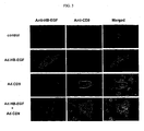
- Fig. 3 shows micrographs of myocardial cells obtained by each gene-transferring Ad. HB-EGF, Ad. CD9 or Ad. HB-EGF + Ad.
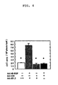
- FIG. 4 shows a graph showing areas of myocardial cells after each gene-transferring Ad. HB-EGF, Ad. CD9 or Ad. HB-EGF + Ad. CD9 to myocardial cells of a neonatal mouse.
- Fig. 5 shows a graph showing beat number of myocardial cells after each gene-transferring Ad. HB-EGF, Ad. CD9 or Ad. HB-EGF + Ad. CD9 to myocardial cells of a neonatal mouse.
- Fig. 4 shows a graph showing areas of myocardial cells after each gene-transferring Ad. HB-EGF, Ad. CD9 or Ad. HB-EGF + Ad. CD9 to myocardial cells of a neonatal mouse.
- Fig. 5 shows a graph showing beat number of myocardial cells after each gene-transferring Ad. HB-EGF, Ad. CD9 or Ad. HB-EGF + Ad. CD9 to myocardial cells of a neonatal mouse.
- Fig. 4 shows
- FIG. 6 shows micrographs of myocardial cells when human recombinant HB-EGF (rHB), angiotensin 2 (Ang2) or human recombinant HGF (rHGF) is allowed to act on myocardial cells of a neonatal mouse expressed by gene-transferring Ad. CD9.
- rHB human recombinant HB-EGF
- Ang2 angiotensin 2
- rHGF human recombinant HGF
- Fig. 7 shows a graph showing cell areas of myocardial cells when human recombinant HB-EGF (rHB), angiotensin 2 (Ang2) or human recombinant HGF (rHGF) is allowed to act on myocardial cells of a neonatal mouse expressed by gene-transferring Ad.
- solid means gene-transferring of CD9.
- Fig. 8 shows a graph showing beat number of myocardial cells when human recombinant HB-EGF (rHB), angiotensin 2 (Ang2) or human recombinant HGF (rHGF) is allowed to act on myocardial cells of a neonatal mouse expressed by gene-transferring Ad.
- CD9. solid means gene-transferring of CD9.
- Fig. 9 shows photograph images showing analysis by Western blotting of the extent of phosphorylation in signal transmission depending on the presence or absence of expression of CD9.
- Fig. 10 shows a survival curve in a chronic heart failure model of adult mice 8-week after myocardial infarction.
- Fig. 11 shows micrographs of border zones of myocardial cells in a chronic heart failure model of adult mice 8-week after myocardial infarction.
- Fig. 12 shows a graph showing the diameter of a myocardial cell in border zone and remote zone in a chronic heart failure model of adult mice 8-week after myocardial infarction.
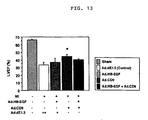
- Fig. 13 shows a graph showing LVEF of echocardiography of adult mice 1-week after myocardial infarction.
- sham represents a normal mouse not revealing myocardial infarction
- MI represents myocardial infarction (also in the following Figs 14 to 23 ).
- Fig. 14 shows a graph showing LVDd of echocardiography of adult mice 1-week after myocardial infarction.
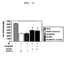
- Fig. 15 shows a graph showing + dp/dt by a catheter in adult mice 1-week after myocardial infarction.
- Fig. 16 shows a graph showing LVSP by a catheter in adult mice 1-week after myocardial infarction.
- Fig. 17 shows a graph showing LVEDP by a catheter in adult mice 1-week after myocardial infarction.
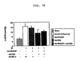
- Fig. 18 shows a graph showing heart weight after correction of the body weight of adult mice 1-week after myocardial infarction.
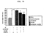
- Fig. 19 shows lung weight after correction of the body weight of adult mice 1-week after myocardial infarction.
- Fig. 20 shows a graph showing the area of a myocardial infarction region of adult mice 1-week after myocardial infarction.
- Fig. 21 shows a graph showing the fibered area of adult mice 1-week after myocardial infarction.
- Fig. 22 shows a graph showing grown cells of cardiac muscle of adult mice 1-week after myocardial infarction.
- Fig. 23 shows a graph showing myofibroblasts of cardiac muscle of adult mice 1-week after myocardial infarction.
- Adenovirus vectors used in the examples were produced as described below.
- Plasmid pADL.1/RSV ( B. Fang et al., Gene Therapy (1994), 1, 247-254 ) was a plasmid produced by incorporating, from upstream, a 0-455 base portion from 3' side of human type 5 adenovirus, Rous sarcoma virus long-term repeat (RSV) promoter, multi cloning site, poly A signal sequence of Bovine growth hormone, and a 3328-5788 base portion from 3' side of human type V adenovirus, into pBR322 plasmid, and offered from Shu-Hsia Chen (Mount Sinai University).
- RSV Rous sarcoma virus long-term repeat
- pADL.1/RSV plasmid was digested with restriction enzymes Hind III and Not I and purified, to give a vector to be used for ligation.
- plasmids pRcHBEGF and pRcCD9 containing cDNA of the whole length of open reading frame of human HB-EGF and cDNA of the whole length of open reading frame of monkey CD9, respectively, in plasmid pRc/CMV (Invitrogen), were offered from Mekata Eisuke (Osaka University).
- HB-EGF cDNA and CD9 cDNA were excised, respectively, using restriction enzymes Hind III and Not I, and these were subjected to agarose gel electrophoresis, the intended DNA fragment was excised and purified to give an insert to be used for ligation.
- treated pADL.1/RSV vector and HB-EGF cDNA, CD9 cDNA inserts were subjected to a ligation reaction with T4 DNA ligase, to obtain pADL.1/RSV-HB-EGF, and pADL.1/RSV-CD9.
- pADL.1/RSV-HB-EGF and pADL.1/RSV-CD9 were, together with pJM17 (Microbix Biosystems Inc.) of a plasmid containing a gene other than E1 region of human type V adenovirus, co-infected in 293 cells by a calcium phosphate method.
- pJM17 Merix Biosystems Inc.
- This plaque was picked up, and correct non-proliferation type recombinant adenoviruses Ad.HB-EGF and Ad.CD9 expressing intended HB-EGF and CD9 were confirmed by immune staining using an anti-HB-EGF antibody (M-18:sc-1414, SANTA CRUZ) and an anti-CD9 antibody (ALB6, IMMUNOTECH), and the like, then, viruses were proliferated by 293 cells, and concentrated and purified by a gradient centrifugal method for cesium chloride.
- Ad.LacZ expressing a LacZ gene of E. coli used for gene transferring was produced in the same method as descriebd above, and details of the Ad.LacZ production method are described in Proc. Natl. Acad. Sci. U.S.A. (1995) 92, 2577-2581 .
- Ad.dE1.3 contains no incorporated CD9 and HB-EGF, thus, this is a control recombinant adenovirus not expressing these genes at all.
- HB-EGF is described in Higashiyama, S., et al. Science 251, 936-939 (1991 ), and its accession No. of Gene Bank is N60278.
- CD9 monkey CD9 was used, and the monkey CD9 is reported as DRAP27 (diphtheria toxin receptor-associated protein) ( Mitamura T, et al: J cell Biol., 118(6): 1389-1399, 1992 , Gene Bank accession No.
- Example 1 expression of CD9 and the like in whole heart and myocardial cell of rat and mouse
- a sense primer (5'-CCGTGATGCTGAAGCTCTTT-3', SEQ ID No. 1) and an anti-sense primer (5'-CCAAGACTGTAGTGTGGTCAT-3', SEQ ID No. 2)( Yoshizumi M., et al.: J. Biol. Chem., 267, 9467-9469, 1992 ) of HB-EGF, and a sense primer (5'-AGCAAGTGCATCAAATACC-3', SEQ ID No. 3) and an anti-sense primer (5'-AATCACCTCATCCTTGTGG-3', SEQ ID No. 4) of CD9 were synthesized by requested Hokkaido System Science Co., Ltd.
- a group of receptors of HB-EGF referred to Sundaresan S, et al: Endocrinology 139: 4756-4764, 1988 , and a sense primer (5'-ACAACTGTGAAGTGGTCCT-3', SEQ ID No. 5) and an anti-sense primer (5'-TTCCTGTAAGTTCCGCAT-3', SEQ ID No. 6) of EGFR, a sense primer (5'-AGCTGGTGACACAGCTTA-3', SEQ ID No. 7) and an anti-sense primer (5'-TGGTTGGGACTCTTGAC-3', SEQ ID No. 8) of ErbB2, a sense primer (5'-GACCTAGACCTAGACTT-3', SEQ ID No.
- HepG2 cells of lung of the above-mentioned neonatal mouse and human liver cancer cells were used.
- a PCR method for HB-EGF and CD9 using Promega Taq (Promega catalogue No. M1865), a cycle of heat denaturing at 94°C for 30 seconds, annealing at 56°C for 1 minute and elongation reaction at 72°C for 1 minute was repeated 38 times, and in a PCR method for a group of receptors of HB-EGF and HPRT, TAKARA Ex Taq (TAKARA catalogue No. RR001A) were used, and the analogous procedure was performed at an annealing temperature of 55°C.
- TAKARA TP-400 was used as PCR thermal cycler.
- HB-EGF As shown in Fig. 1 , expression of HB-EGF was observed in heart both in adult rats and neonatal mice, and particularly, expression was stronger in a cultured myocardial cell, thus, it is recognized that HB-EGF was highly expressed in a myocardial cell itself in heart. Though it is recognized that ErbB2 and ErbB4 were expressed strongly in a cultured myocardial cell both in adult rats and neonatal mice, expression of EGFR was not clear in adult rats and strong expression thereof was recognized in neonatal mice. Expression of ErbB3 was not recognized in both adult rats and neonatal mice.
- Myocardial cells ( Aoyama T, et al: Cardiovasc Res., 55: 787-798, 2002 ) isolated from a 1-day old neonatal BALB/C mouse by collagenase type 2 (WOR: CLS 2, Funakoshi catalogue No. 45004177) were disseminated on a 4-well chamber slide (Nunc: Lab-Tek, Permanox, catalogue No. 15 77437) coated with mouse laminin (Biomedical Technologies Inc. catalogue No. BT-276) at a concentration of 5 ⁇ 50 5 /500 ⁇ l/well, and maintained by a low glucose DMEM medium (SIGMA catalogue No.
- the cultured myocardial cells of neonatal mice obtained above were fixed with 4% paraformaldehyde for 10 minutes, a cell membrane was perforated with 0.05% Triton X100, and blocked with 10% skimmed milk (Snow Brand Milk Products Co., Ltd.) for 60 minutes. Thereafter, a primary antibody (mouse monoclonal antibody CD9 (clone ALB6), IMMUNOTECH catalogue No. 0117) diluted 50-fold (2 ug/ml) and a goat polyclonal antibody HB-EGF (M-18)(SANTA CRUZ catalogue No.
- sc-144) diluted 100-fold were reacted for 1 hour, and visualization of HB-EGF was carried out while labeling with anti-goat Alexa 568 (MOLECULAR PROBES catalogue No. A-11057) and visualization of CD9 was carried out while labeling with anti-mouse Alexa 488 (MOLECULAR PROBES catalogue No. A-11029). Nucleus staining was carried out for 5 minutes with Hoechst 33342 (MOLECULAR PROBES catalogue No. H-3570) diluted 1000-fold. F-actin was labeled with rhodamine phalloidin (MOLECULAR PROBES catalogue No. R-415) diluted 500-fold and recognized.
- an expression pattern when only HB-EGF was singly gene-transferred was stained in the form of grain in a cell, and when only CD9 was singly gene-transferred, the surface of cell membrane was stained.
- HB-EGF and CD9 were co-expressed, change of localization patterns of the proteins was confirmed. That is, when CD9 is singly gene-transferred, an expression protein of the transferred CD9 gene localizes on a membrane which is an original expression site of endogenous CD9, however, when HB-EGF and CD9 were strongly expressed together, it was found that a CD9 protein moves into a cell together with a HB-EGF protein.
- CD9, HB-EGF or CD9 + HB-EGF (each at MOI 10) is strongly expressed each using Ad.CD9, Ad.HB-EGF, or Ad.CD9 + Ad.HB-EGF, and change of a myocardial cell after 24 hours was investigated morphologically and physiologically.
- a myocardial cell was fixed with 4% paraformaldehyde for 10 minutes, the cell area was measured using PIXEL count (Adobe Photoshop).
- the cell area of the myocardial cell containing gene-transferred HB-EGF showed a significant increase (*p ⁇ 0.05 vs Control) based on the control (HB-EGF and CD9 genes were not transferred), and the cell area of the myocardial cell containing single gene-transferred CD9 showed no significant change.
- the cell area of the myocardial cell of CD9 + HB-EGF genes transferred group significantly suppressed increase by HB-EGF.
- the beat of myocardial cells each containing gene-transferred CD9, HB-EGF, or CD9 + HB-EGF described above was recorded using a video microscope observation system (OLYMPUS microscope IX 70 + CCD camera CS 900).
- a video microscope observation system OLEDUS microscope IX 70 + CCD camera CS 900.
- the beat number of a myocardial cell containing gene-transferred HB-EGF showed a significant increase (130 ⁇ 10/min. against control 70 ⁇ 8/min.), however, the beat number of a myocardial cell containing gene-transferred CD9 + HB-EGF was approximately the same (76 ⁇ 10/min.) as the control, indicating inhibition of the action of HB-EGF by CD9 (*p ⁇ 0.05 vs control).
- a myocardial primary culture cell of a neonatal mouse was infected with Ad.CD9 at MOI 30 to cause gene transferring, CD9 was strongly expressed, and 24 hours later, human recombinant HB-EGF (rHB-EGF, R&D system, catalogue No. 259-HE)(concentration 10 ng/ml), angiotensin 2 (SIGMA, catalogue No. A-9525) (concentration 100 nM) and human recombinant HGF (rHGF, R&D system, catalogue No. 294-HG) (concentration 10 ng/ml), respectively were added into the culture solution, and allowed to act on myocardial cells for 24 hours.
- human recombinant HB-EGF rHB-EGF, R&D system, catalogue No. 259-HE
- SIGMA angiotensin 2
- HGF human recombinant HGF
- the myocardial cell (see, photograph of Anti-CD9) containing gene-transferred CD9 shown in Fig. 6 suppressed the hypertrophy action of a myocardial cell by human recombinant HB-EGF, angiotensin 2 and human recombinant HGF, as is understood from photograph images mergeing a CD9 immunostained image and a F-actin stained image.
- the product was washed three times with a serum-free culture solution, human recombinant HB-EGF, angiotensin 2 and human recombinant HGF, respectively were added in the same amount as in quantification of hypertrophy of a myocardial cell, and 3 hours later, 20 or more myocardial cells were observed each for 5 minutes or more using a cultured cell video observation system (OLYMPUS microscope product NO. IX 70 and CCD camera product No. CS 900), and the beat number in 1 minute was measured.
- a cultured cell video observation system OLEDUS microscope product NO. IX 70 and CCD camera product No. CS 900
- Fig. 6 when any one of human recombinant HB-EGF, angiotensin 2 and human recombinant HGF was added, formation of muscle fiber and hypertrophy of a myocardial cell could be confirmed in all cases, however, in a myocardial cell in which CD9 had been previously strongly expressed and its expression had been confirmed, hypertrophy of a myocardial cell and formation of muscle fiber were suppressed.
- the cell area was statistically significant as shown in Fig. 7 .
- the beat number of a myocardial cell showed significant increase by the action of human recombinant HB-EGF, angiotensin 2 and human recombinant HGF as shown in Fig. 8 , and it was confirmed that this action is canceled by CD9 (*p ⁇ 0.05 vs Control, **p ⁇ 0.001 vs Control).
- CD9 suppresses a hypertrophy action of a myocardial cell and a heart positive inotropic action also in vitro.
- phosphorylation of MAPK MAP kinase
- a protein was extracted by a Bradford method using a protein assay kit (Biorad, catalogue No. 500-0002JA), and allowed to migrate each in an amount of 3 ⁇ g on SDS-PAGE, and recognized by Western Blotting.
- phosphorylation of active-ERK, active-p70 S6K and active-p38 was suppressed (active-p38 was suppressed slightly) by the action of CD9 by using pERK(E-4) (SANTACRUZ catalogue No.
- mice C57/BL6 were subjected to tracheotomy, anesthetized generally (GOH nitrous oxide/oxygen/halothane) by an anesthesia machine (Kimura Medical Instrument Co., Ltd. product No. compact-15), then, subjected to thoracotomy.
- a coronary artery was ligated permanently with 2-0 nylon thread, to produce myocardial infarction animal models.
- a PBS solution containing each 1 ⁇ 10 11 particles of Ad.HB-EGF, Ad.dE1.3 and Ad.HB-EGF + Ad.CD9 dissolved in 100 ul of PBS was directly sprayed on the epicardium side of a cardiac muscle, and thoracotomy was performed.
- LVEF left ventricular ejection fraction
- LDLd left ventricular diastolic diameter
- LSd left ventricular systolic diameter
- LSt left ventricular septum thickness
- PWt posterior wall thickness
- SPR 407 was invasively inserted into aorta and left ventricle, and left ventricular systolic pressure (LVSP), left ventricular end diastolic pressure (LVEDP), left ventricular maximum positivity dP/dt and left ventricular maximum negativity dP/dt were measured (PowerLab system ver 4.2 ADInstruments). After blood collection, internal organs (heart, lung, liver, kidney, spleen) were collected.
- LVSP left ventricular systolic pressure
- LVEDP left ventricular end diastolic pressure
- dP/dt left ventricular maximum positivity dP/dt
- dP/dt left ventricular maximum negativity dP/dt
- Fig. 10 shows a survival curve until 8 weeks in the chronic phase.
- mice containing simultaneously gene-transferred CD9 showed suppression of compensatory hypertrophy of heart as compared with mice containing gene-transferred HB-EGF.
- cardiac muscle cell diameter was measured in border zone (Border: boundary region between a myocardial infarction part and a normal part) and remote zone (Remote: normal cardiac muscle part remote from a myocardial infarction part) (20 or more each samples for each zone) using LUZEX F system (manufactured by NIRECO).
- Fig. 12 compensatory hypertrophy of heart was observed also in the HB-EGF group in the border zone, which was also observed in the control group, and by simultaneous gene-transferring of CD9, its hypertrophy action of heart was suppressed (*p ⁇ 0.05 vs control, **p ⁇ 0.001 vs control).
- results of measurement of the cardiac function by echocardiography and a catheter of a model one week after myocardial infarction produced for investigating a reason for 80% death of the HB-EGF + dE 1.3 group in an acute phase within 2 weeks are as described below.
- the cardiac function of the CD9 gene-transferred group was significantly excellent as compared with the control group and the HB-EGF gene-transferred group (*p ⁇ 0.05 vs control), in left ventricular ejection fraction (LVEF)(see, Fig. 13 ) in cardiac function evaluation by echocardiography, and in left ventricular maximum positivity dP/dt and left ventricular systolic pressure (LVSP)( Fig. 15 and Fig.
- mice of 1 week or more after myocardial infarction formation were tenderly sacrificed in all the groups.
- the body weights of mice after sacrificing were measured, and the weight of heart and the weight of lung were divided by its body weight, and the weight of heart and the weight of lung were corrected.
- Increase in the weight of heart means excess hypertrophy and dilation of heart, indicating deterioration of the cardiac function such as heart failure and the like.
- Increase in the weight of lung means hemostasis of lung, indicating pulmonary edema, namely, heart failure.
- the CD9 transferred group shows significant suppression of the organ weight, indicating no heart failure condition (*p ⁇ 0.05 vs control, **p ⁇ 0.001 vs control).
- mice of 1 week or more after myocardial infarction formation were tenderly sacrificed in all the groups. After sacrificing, heart was removed and sliced to obtain sections which were fixed by formalin treatment, then, the section was subjected to Masson trichrome staining as an index for fibering.
- MI area myocardial infarction region area
- the ratio of a heart necrosis part in the whole left ventricle was calculated.
- the fibered area particularly the ratio of a positive part by Masson trichrome staining based on the area of all left ventricle sections was calculated.
- the CD9 transferred group showed a significant decrease, revealing a therapeutic effect, in both the myocardial infarction region area (MI area) and the fibered area (*p ⁇ 0.05 vs control).
- mice of 1 week or more after myocardial infarction formation were tenderly sacrificed in all the groups. After sacrificing, heart was removed and fixed by formalin treatment, then, embedded in paraffin, and sliced. The heart slices were stained using a monoclonal antibody to a Ki-67 antigen (clone: MIB-1, Dako Cytomation, catalogue No. M7240, antibody dilution 25-fold) as an index for grown cells and DAKO EPOS/HRP (Enhanced Polymer One-step Staining/peroxidase) kit (clone: 1A4, Dako Cytomation, catalogue No.
- Ki-67 antigen clone: MIB-1, Dako Cytomation, catalogue No. M7240, antibody dilution 25-fold
- DAKO EPOS/HRP Enhanced Polymer One-step Staining/peroxidase
- the CD9 transferred group showed a significant decrease in both the grown cell and myofibroblast, clarifying that CD9 suppresses irregular and excess repair of the grown cell, suppression of granulation and proliferation of a myofibroblast (*p ⁇ 0.05 vs control, **p ⁇ 0.001 vs control).
- the drug of the present invention is used for gene therapy, and can prevent or treat myocardial infarction, or heart diseases associated with cardiac hypertrophy and tachycardia by transferring a CD9 gene into heart.
- the prevention or treatment can radically prevent or treat the heart diseases of the invention by allowing a CD9 gene to express in heart.
Landscapes
- Health & Medical Sciences (AREA)
- Life Sciences & Earth Sciences (AREA)
- Chemical & Material Sciences (AREA)
- Engineering & Computer Science (AREA)
- Bioinformatics & Cheminformatics (AREA)
- Organic Chemistry (AREA)
- General Health & Medical Sciences (AREA)
- Genetics & Genomics (AREA)
- Zoology (AREA)
- Medicinal Chemistry (AREA)
- Public Health (AREA)
- Veterinary Medicine (AREA)
- Animal Behavior & Ethology (AREA)
- Pharmacology & Pharmacy (AREA)
- Cardiology (AREA)
- Biotechnology (AREA)
- Wood Science & Technology (AREA)
- General Chemical & Material Sciences (AREA)
- Chemical Kinetics & Catalysis (AREA)
- Nuclear Medicine, Radiotherapy & Molecular Imaging (AREA)
- General Engineering & Computer Science (AREA)
- Immunology (AREA)
- Biomedical Technology (AREA)
- Heart & Thoracic Surgery (AREA)
- Gastroenterology & Hepatology (AREA)
- Biochemistry (AREA)
- Biophysics (AREA)
- Proteomics, Peptides & Aminoacids (AREA)
- Molecular Biology (AREA)
- Plant Pathology (AREA)
- Microbiology (AREA)
- Physics & Mathematics (AREA)
- Toxicology (AREA)
- Marine Sciences & Fisheries (AREA)
- Cell Biology (AREA)
- Virology (AREA)
- Epidemiology (AREA)
- Vascular Medicine (AREA)
- Urology & Nephrology (AREA)
- Hospice & Palliative Care (AREA)
Abstract
Description
- The present invention relates to a drug for preventing or treating heart diseases, more specifically, to a drug which is used for gene therapy and prevents or treats heart diseases by transferring a CD9 gene into heart.
- Heart diseases such as myocardial infarction, cardiomypathy, arrhythmia, heart failure and the like are one of three major causes for death in Japan, generating a significant problem in medical care. Conventionally, drugs such as a diuretic, β-blocker, ACE inhibitor, calcium antagonist and the like are used for treatment of heart diseases, however, these drugs do not radically treat heart diseases.
- CD9 is a membrane protein having a molecular weight of 27 kDa classified into one of transmembrane 4-times type protein super family (TM4SF). Regarding the action of CD9 in an organism, there are reports that it is a cell-specific marker derived from pre B (Masellis-Smith A et al; J Immunol. 144 15, 1607-1613, 1990), that it expresses in various cells irrespective of hematopoietic cells or non-hematopoietic cells (Maecker H. T et al.; FASEB J. 11, 428-442, 1997, Berditchevski F; J Cell Sci. 114, 4143-4151, 2001), that it relates to aggregation and differentiation of pre B cells (Masellis-Smith A et al; J Immunol. 144, 1607-1613, 1990), that it relates to activation of platelets (Jennings LK et al.; J Biol Chem. 265, 3815-3822, 1990), that it relates to survival and adhesion of various cells typically including cancer cells (Hashida H, et al.: Br J Cancer., 89: 158-67, 2003), that it is a membrane protein indispensable for fertilization (Miyado K et al.; Science 287, 321-324, 2000), that it causes association of a plurality of proteins in a cell membrane, acts as a molecule for coordinating and promoting a mutual action between proteins, and relates to phenomena of injury healing such as cell adhesion, proliferation, differentiation, immune, hemostatis and the like (Berditchevski F; J Cell Sci. 114, 4143-4151, 2001, Klein-Soyer C et al; Arterioscler Thromb Vasc Biol. 20, 360-3).
- On the other hand, there is also a report regarding a relation between CD9 and HB-EGF (heparin binding epidermal growth factor). For example, there are reports on formation of a complex with HB-EGF or integrin α3β1 (Nakamura Y, et al: J Histochem Cytochem 49: 439-444, 2001), regulation of an activity as a juxtacrine factor of proHB-EGF as a precursor of HB-EGF (Higashiyama S., et al,: J. Cell Biol. 128, 929-938. 1995, Iwamoto R, Mekada E: Cytokine & Growth Factor Reviews. 11: 335-344, 2000), and possibility of representation of variety of an activity of HB-EGF by relating to a process of processing of HB-EGF (Nakagawa T et al; J Biol Chem. 271, 30858-30863, 1996, Nakamura K et al; J Biol Chem. 275, 18284-18290, 2000). There are also reports that kidney epithelial cells also are improved in survival by co-expression of HB-EGF and CD9 in an ischemic disorder model (Takamura T et al; Kidney Int. 55, 71-81, 1999), that, in a process of arteriosclerosis, HB-EGF is expressed in normal aorta (Miyagawa J et al; J Clin Invest. 95, 404-411, 1995) and coronary (Nakata A et al; Circulation 94, 2778-2786, 1996), on the other and, CD9 expresses in arteriosclerosis lesion and some inner membrane smooth muscle cells, and co-expression of CD-9 and proHB-EGF promotes proliferation of smooth muscle cells by proHB-EGF (Nishida M et al; Arterioscler Thromb Vasc Biol. 20, 1236-1243, 2000), thus, CD9 is possibly an important molecule for regulating an activity of HB-GF assisting a balance of proliferation and transformation of fibroblasts in a process of arteriosclerosis and tissue repair (Kirkland G et al; J Am Soc Nephrol. 9, 1464-73, 1998). Further, the present inventors have a finding that excess expression of HB-EGF in a myocardial infarction model animal promotes compensatory hypertrophy of a myocardial cell, while promotes proliferation of myofibroblasts and enhances fibering, thereby, promoting decrease in a cardiac function, that is, HB-EGF is a central factor playing an important role in progressing of pathology.
- As described above, there are a lot of reports on the action of CD9 and a relation of CD9 and HB-EGF, however, there is no report on a fact that CD9 suppresses cardiac hypertrophy, or suppresses tachycardia.
- The present invention has an object of providing a drug which radically prevents or treats heart diseases. Another object is to provide a method for radically preventing or treating heart diseases.
- The present invention relates to a drug for use in preventing or treating heart diseases, containing an expression vector containing a CD9 gene as the active ingredient. In the present invention, the heart disease is myocardial infarction, or a heart disease associated with cardiac hypertrophy and/or tachycardia Heart diseases include diseases causative of heart failure, ischemic heart diseases, cardiomyopathy, hypertensive heart diseases, valvular disease, congenital heart diseases, mayocarditis, arrhythmia and diseases associated with arrhythmia, cardiac hypertrophy and/or tachycardia. Cardiac hypertrophy or tachycardia may be caused by at least one of HB-EGF (heparin binding epidermal growth factor), HGF (hepatocyte growth factor) or
angiotensin 2. - In the above-mentioned present invention, the expression vector is a viral vector or a non-viral vector.
- Prevention or treatment may be carried out by a gene therapy of transferring a CD9 gene, or administration of an expression inducing substance for expressing endogenous CD9. Further, the gene therapy may use a drug containing an expression vector containing a CD9 gene as the active ingredient.
- The CD9 gene used in the drug of the present invention means a gene capable of expressing CD9. The CD9 gene may be a gene in which its gene sequence is partially deleted, substituted, inserted or, other base is added, providing a polypeptide to be expressed has substantially the same effect as CD9. As the CD9 gene, a human CD9 gene described in Strausberg R. L. et al; Proc. Natl. Acd. Sci. U.S.A. 99, (26) 16899-16903, 2002 is exemplified (Gene Bank, accession No. is AAH11988, messenger RNA No. is BC011988).
- The expression vector containing a CD9 gene includes virus vector, non-virus vector, plasmid and the like. Examples of the virus vector include adenovirus, adeno-associated virus, retrovirus, herpesvirus, herpes simplex virus, lentivirus, Sendai virus, poxvirus, poliovirus, symbis virus, vaccinia virus and the like. Examples of the non-virus vector include cationic liposome, membrane fusing liposome, cationic polymer and the like. The liposome is a capsule composed of phospholipid having a particle size of several 10 to several 100 nm, and a plasmid containing a CD9 gene in it can be filled in this capsule.
- The expression vector containing a CD9 gene can be produced by using conventional gene engineering technologies, cell culture technologies and virus technologies (e.g., "Current Protocols in Molecular Biology, F. Ausubel et al. ed., (1994), John Wiley & Sons, Inc.", "Molecular Cloning (A Laboratory Manual), Third Edition. Volume 1-3. Josseph Sambrook & David W. Russel ed., Cold Spring Harbor Laboratory Press (Cold Spring Harbor, New York) 2001 ", "Culture of Animal Cells; A Manual of Basic Technique, R. Freshney ed., 2nd edition (1987), Willey-Liss", "Frank K. Greham ed., Manipulation of adenovirus vectors, Chapter 11. p 109-p 128", "E.J. Murray ed., Methods in Molecular Biology, Vol. 7: Gene Transfer and Expression Protocols (1991)", "Chen, S-H, et al., Combination gene therapy for liver metastases of colon carcinoma in vivo. Proc. Natl. A-cad. Sci. USA. (1995) 92, 2477-2581", and the like).
- In the drug of the present invention, an expression vector containing a CD9 gene as the active ingredient and pharmaceutically acceptable auxiliaries such as excipient, carrier, solvent and the like are mixed, and the drug can be used in the form of various preparations such as injection and the like. The administration form of the drug of the present invention is not particularly restricted, and for example, injection, catheter, balloon catheter and the like can be adopted. In the case of an expression inducing substance for expressing endogenous CD9, systemic intravenous administration, or oral administration and the like can be adopted.
- The amount of a CD9 gene dosed by the drug of the present invention can be appropriately controlled in view of pathological condition, age, body weight and the like of a person to be receiving dosage, and when adenovirus is used as a virus vector in a clinical test of a conventional gene therapy for a patient suffering from a heart disease, safety has been confirmed using 2×10¹⁰ in terms of virus infectiosity titer pfu (plaque forming unit), therefore, this amount is a rough standard for the dosage. When liposome is used as a non-virus vector, a clinical test of a gene therapy for a person suffering from a heart disease has been conducted safely with a DNA amount of 2 mg, therefore, this amount is a rough standard for the dosage.
- The drug of the present invention is used in a gene therapy, and can be applied widely for patients suffering from heart failure.
- Typical pathology of heart failure include hypertrophy and dilation of heart, and hypertrophy of heart is believed to be morphological adaptation against various loads, however, it is known that, this has a limitation, and adaptation failure (compensation failure) occurs, thus, hypertrophy of a myocardial cell itself directly deteriorates the pathological condition of heart failure or other heart diseases (see, ardiology, ISHIKAWA Kyozo chief ed., 1995, Igakushoin). Examples of heart diseases include ischemic heart diseases (for example, myocardial infarction), cardiomyopathy (for example, congestive cardiomyopathy, hypertrophic cardiomyopathy, restrictive cardiomyopathy), hypertensive heart diseases, valvular disease (for example, atresia of aorta valve, mitral valve and the like, or stenosis thereof), congenital heart diseases, mayocarditis and the like. Arrhythmia includes bradyarrhythmia and tachyarrhythmia, and examples of tachyarrhythmia includes superventricular arrhythmia (for example, sinus tachycardia, superventricular extrasystole, atrial fibrillation, artial flutter, superventricular tachycardia), ventricular arrhythmia (for example, ventricular extrasystole, ventricular tachycardia, ventricular fibrillation, torsade de pointes). Diseases showing generation of tachyarrhythmia as main pathological condition include WPW syndrome, long QT syndrome, Brugada syndrome, arrhythmia-inducing right ventricular dysplasia (ARVD, ARVC) (regarding arrhythmia described above, see, "Arrhythmia" (revision, Visual Text of )). In the invention, the drug can be suitably used for heart diseases associated with cardiac hypertrophy or tachycardia. Heart diseases can be radically treated by inhibiting pathological hypertrophy of a myocardial cell and pathological frequent pulse of a myocardial cell (tachycardia) by expression of HB-EGF, further, pathological myocardial cell enlargement or pathological myocardial cell tachycardia (tachycardia) by expression of HGF (hepatocyte growth factor) or
angiotensin 2, and normalizing the function of myocardial cells. The drug of the present invention can be used for acute heart diseases since it performs an effect in several hours after administration. In a gene therapy, a therapeutic effect for a long period of time is expected by gene expression for a long period of time, thus, the drug can be used prophylactically for preventing cardiac hypertrophy and tachycardia associated with chronic diseases such as hypertension, chronic myocardial infarction and the like. - By allowing CD9 to express in a body, heart diseases can be prevented or treated, and the prevention and treatment of heart diseases can be carried out by a gene therapy using the drug of the present invention.
-
Fig. 1 shows a result of agarose electrophoresis showing expression of CD9, HB-EGF and HB-EGF receptor. H: whole heart, CM: myocardial cell, CF: cardiac fibroblast, C1; mouse lung, C2: HepG2 cell, NC: negative control, HPRT: internal control.Fig. 2 shows a graph showing a gene transferring efficiency of Ad. LacZ to a myocardial cell of a neonatal mouse, and a micrograph of a cultured myocardial cell stained with X-gal.Fig. 3 shows micrographs of myocardial cells obtained by each gene-transferring Ad. HB-EGF, Ad. CD9 or Ad. HB-EGF + Ad. CD9 to myocardial cells of a neonatal mouse and effecting immunohistochemical staining on these cells.Fig. 4 shows a graph showing areas of myocardial cells after each gene-transferring Ad. HB-EGF, Ad. CD9 or Ad. HB-EGF + Ad. CD9 to myocardial cells of a neonatal mouse.Fig. 5 shows a graph showing beat number of myocardial cells after each gene-transferring Ad. HB-EGF, Ad. CD9 or Ad. HB-EGF + Ad. CD9 to myocardial cells of a neonatal mouse.Fig. 6 shows micrographs of myocardial cells when human recombinant HB-EGF (rHB), angiotensin 2 (Ang2) or human recombinant HGF (rHGF) is allowed to act on myocardial cells of a neonatal mouse expressed by gene-transferring Ad. CD9. -
Fig. 7 shows a graph showing cell areas of myocardial cells when human recombinant HB-EGF (rHB), angiotensin 2 (Ang2) or human recombinant HGF (rHGF) is allowed to act on myocardial cells of a neonatal mouse expressed by gene-transferring Ad. CD9. In the graph, solid means gene-transferring of CD9.Fig. 8 shows a graph showing beat number of myocardial cells when human recombinant HB-EGF (rHB), angiotensin 2 (Ang2) or human recombinant HGF (rHGF) is allowed to act on myocardial cells of a neonatal mouse expressed by gene-transferring Ad. CD9. In the graph, solid means gene-transferring of CD9. -
Fig. 9 shows photograph images showing analysis by Western blotting of the extent of phosphorylation in signal transmission depending on the presence or absence of expression of CD9.Fig. 10 shows a survival curve in a chronic heart failure model of adult mice 8-week after myocardial infarction.Fig. 11 shows micrographs of border zones of myocardial cells in a chronic heart failure model of adult mice 8-week after myocardial infarction.Fig. 12 shows a graph showing the diameter of a myocardial cell in border zone and remote zone in a chronic heart failure model of adult mice 8-week after myocardial infarction.Fig. 13 shows a graph showing LVEF of echocardiography of adult mice 1-week after myocardial infarction. In this figure, sham represents a normal mouse not revealing myocardial infarction, and MI represents myocardial infarction (also in the followingFigs 14 to 23 ).Fig. 14 shows a graph showing LVDd of echocardiography of adult mice 1-week after myocardial infarction.Fig. 15 shows a graph showing + dp/dt by a catheter in adult mice 1-week after myocardial infarction.Fig. 16 shows a graph showing LVSP by a catheter in adult mice 1-week after myocardial infarction.Fig. 17 shows a graph showing LVEDP by a catheter in adult mice 1-week after myocardial infarction. -
Fig. 18 shows a graph showing heart weight after correction of the body weight of adult mice 1-week after myocardial infarction.Fig. 19 shows lung weight after correction of the body weight of adult mice 1-week after myocardial infarction.Fig. 20 shows a graph showing the area of a myocardial infarction region of adult mice 1-week after myocardial infarction.Fig. 21 shows a graph showing the fibered area of adult mice 1-week after myocardial infarction.Fig. 22 shows a graph showing grown cells of cardiac muscle of adult mice 1-week after myocardial infarction.Fig. 23 shows a graph showing myofibroblasts of cardiac muscle of adult mice 1-week after myocardial infarction. - The present invention will be illustrated by the following examples. In the examples, gene engineering technologies and cell culturing technologies handling plasmids, DNA, various enzymes, E. coli, cultured cells and the like were carried out according to methods described in the above-mentioned "Current Protocols in Molecular Biology, F. Ausubel et al. ed., (1994), John Wiley & Sons, Inc." and "Culture of Animal Cells; A Manual of Basic Technique, R. Freshney ed., 2nd edition (1987), Willey-Liss", unless otherwise stated. Unless otherwise stated, general handling of adenovirus was carried out according to methods described in the above-mentioned "Frank L. Graham ed., Manipulation of adenovirus vectors, Chapter 11. p 109-p 128" and "E.J. Murray ed., Methods in Molecular Biology, Vol. 7: Gene Transfer and Expression Protocols (1991)", and production of adenovirus was carried out according to a method described in the above-mentioned "Chen, S-H, et al., Combination gene therapy for liver metastases of colon carcinoma in vivo. Proc. Natl. Acad. Sci. USA. (1995) 92, 2477-2581". Regarding treatment effects and phenomena, a significant difference between some groups was first analyzed by Anova assay, subsequently, an assay of an individual significant difference between each two groups was analyzed by Student t-test (asymmetrical t test between two groups). A significant difference in survival ratio was analyzed using Kaplan-Meier assay.
- Adenovirus vectors used in the examples were produced as described below.
- Plasmid pADL.1/RSV (B. Fang et al., Gene Therapy (1994), 1, 247-254) was a plasmid produced by incorporating, from upstream, a 0-455 base portion from 3' side of
human type 5 adenovirus, Rous sarcoma virus long-term repeat (RSV) promoter, multi cloning site, poly A signal sequence of Bovine growth hormone, and a 3328-5788 base portion from 3' side of human type V adenovirus, into pBR322 plasmid, and offered from Shu-Hsia Chen (Mount Sinai University). pADL.1/RSV plasmid was digested with restriction enzymes Hind III and Not I and purified, to give a vector to be used for ligation. On the other hand, plasmids pRcHBEGF and pRcCD9, containing cDNA of the whole length of open reading frame of human HB-EGF and cDNA of the whole length of open reading frame of monkey CD9, respectively, in plasmid pRc/CMV (Invitrogen), were offered from Mekata Eisuke (Osaka University). From both the plasmids pRcHBEGF and pRcCD9, the whole lengths of HB-EGF cDNA and CD9 cDNA were excised, respectively, using restriction enzymes Hind III and Not I, and these were subjected to agarose gel electrophoresis, the intended DNA fragment was excised and purified to give an insert to be used for ligation. Thus treated pADL.1/RSV vector and HB-EGF cDNA, CD9 cDNA inserts were subjected to a ligation reaction with T4 DNA ligase, to obtain pADL.1/RSV-HB-EGF, and pADL.1/RSV-CD9. Further, pADL.1/RSV-HB-EGF and pADL.1/RSV-CD9 were, together with pJM17 (Microbix Biosystems Inc.) of a plasmid containing a gene other than E1 region of human type V adenovirus, co-infected in 293 cells by a calcium phosphate method. By this, a plaque containing correct intended adenovirus produced by homologous recombination emerged 10 to 14 days after co-infection. This plaque was picked up, and correct non-proliferation type recombinant adenoviruses Ad.HB-EGF and Ad.CD9 expressing intended HB-EGF and CD9 were confirmed by immune staining using an anti-HB-EGF antibody (M-18:sc-1414, SANTA CRUZ) and an anti-CD9 antibody (ALB6, IMMUNOTECH), and the like, then, viruses were proliferated by 293 cells, and concentrated and purified by a gradient centrifugal method for cesium chloride. - A recombinant adenovirus Ad.LacZ expressing a LacZ gene of E. coli used for gene transferring was produced in the same method as descriebd above, and details of the Ad.LacZ production method are described in Proc. Natl. Acad. Sci. U.S.A. (1995) 92, 2577-2581. Ad.dE1.3 contains no incorporated CD9 and HB-EGF, thus, this is a control recombinant adenovirus not expressing these genes at all. pADL.1/RSV and pJM17 containing no incorporated CD9 and HB-EGF were co-infected in 293 cells as described above, and subjected to the same method and process to produce Ad.dE1.3. HB-EGF is described in Higashiyama, S., et al. Science 251, 936-939 (1991), and its accession No. of Gene Bank is N60278. As CD9, monkey CD9 was used, and the monkey CD9 is reported as DRAP27 (diphtheria toxin receptor-associated protein) (Mitamura T, et al: J cell Biol., 118(6): 1389-1399, 1992, Gene Bank accession No. is BAA01569), and the gene sequence thereof is believed to be almost the same and its function is also believed to be the same as human CD9. Evaluation of cell area of a myocardial cell and evaluation of beat number of a myocardial cell conducted in the following experiment system are based on a fact that cardiac hypertrophy is defined as hypertrophy of a myocardial cell and tachycardia is defined as increase in beat number of a myocardial cell (see, worldwide most famous and authoritative text "Braun-wald's HEART DISEASE A textbook of cardiovascular medicine, 6th edition, 2001").
- Each endogenous expression of CD9, HB-EGF or a group of receptors of HB-EGF (EGFR, ErbB2, ErbB3, ErbB4) in the whole heart and myocardial cells was checked using a RT-PCR method. Myocardial cells and whole heart from 8-week old adult SD rats and 1-day old neonatal BALB/C mice were enzymatically treated with collagenase type 2 (WOR:
CLS 2, Funakoshi catalogue No. 45004177), and isolated. Then, 1 ml of sepazole RNA I Super (NACALAI TESQUE catalogue No. 304-86) was added to homogenize, and the homogenized mixture was separated into an aqueous phase and a phenol phase by chloroform, and the aqueous phase was precipitated with isopropanol. After centrifugal separation, the product was suspended with 70% ethanol, and further centrifugally separated, to extract RNA. 1 µg of total RNA was reverse-transcribed with Super Script II reverse transcriptase (Invitrogen), and cDNA was amplified by a PCR method. - A sense primer (5'-CCGTGATGCTGAAGCTCTTT-3', SEQ ID No. 1) and an anti-sense primer (5'-CCAAGACTGTAGTGTGGTCAT-3', SEQ ID No. 2)(Yoshizumi M., et al.: J. Biol. Chem., 267, 9467-9469, 1992) of HB-EGF, and a sense primer (5'-AGCAAGTGCATCAAATACC-3', SEQ ID No. 3) and an anti-sense primer (5'-AATCACCTCATCCTTGTGG-3', SEQ ID No. 4) of CD9 were synthesized by requested Hokkaido System Science Co., Ltd. A group of receptors of HB-EGF referred to Sundaresan S, et al: Endocrinology 139: 4756-4764, 1988, and a sense primer (5'-ACAACTGTGAAGTGGTCCT-3', SEQ ID No. 5) and an anti-sense primer (5'-TTCCTGTAAGTTCCGCAT-3', SEQ ID No. 6) of EGFR, a sense primer (5'-AGCTGGTGACACAGCTTA-3', SEQ ID No. 7) and an anti-sense primer (5'-TGGTTGGGACTCTTGAC-3', SEQ ID No. 8) of ErbB2, a sense primer (5'-GACCTAGACCTAGACTT-3', SEQ ID No. 9) and an anti-sense primer (5'-TCTGATGACTCTGATGC-3', SEQ ID No. 10) of ErbB3, and a sense primer (5'-CATCTACACATCCAGAACA-3', SEQ ID No. 11) and an anti-sense primer (5'-AAACATCTCAGCCGTTGCA-3', SEQ ID No. 12) of ErbB4 were synthesized by requested Hokkaido System Science Co., Ltd. As an internal control, hypoxanthine phosphoribosyltransferase (HPRT) was used.
- As a positive control, HepG2 cells of lung of the above-mentioned neonatal mouse and human liver cancer cells (offered by Medical Cellular Resource Center, Institute of Development, Aging and Cancer, Tohoku University) were used. In a PCR method for HB-EGF and CD9, using Promega Taq (Promega catalogue No. M1865), a cycle of heat denaturing at 94°C for 30 seconds, annealing at 56°C for 1 minute and elongation reaction at 72°C for 1 minute was repeated 38 times, and in a PCR method for a group of receptors of HB-EGF and HPRT, TAKARA Ex Taq (TAKARA catalogue No. RR001A) were used, and the analogous procedure was performed at an annealing temperature of 55°C. As PCR thermal cycler, TAKARA TP-400 was used.
- As shown in
Fig. 1 , expression of HB-EGF was observed in heart both in adult rats and neonatal mice, and particularly, expression was stronger in a cultured myocardial cell, thus, it is recognized that HB-EGF was highly expressed in a myocardial cell itself in heart. Though it is recognized that ErbB2 and ErbB4 were expressed strongly in a cultured myocardial cell both in adult rats and neonatal mice, expression of EGFR was not clear in adult rats and strong expression thereof was recognized in neonatal mice. Expression of ErbB3 was not recognized in both adult rats and neonatal mice. On the other hand, though expression of CD9 was recognized in the whole heart of adult rats, expression thereof was not clear in its cultured myocardial cell, and expression thereof was not recognized both in the whole heart and cultured myocardial cells of neonatal mice. Thus, it was clarified that ErbB2 and ErbB4 included in a group of receptors of HB-EGF are relatively highly expressed in myocardial cells, while CD9 is scarcely expressed in myocardial cells. - Myocardial cells (Aoyama T, et al: Cardiovasc Res., 55: 787-798, 2002) isolated from a 1-day old neonatal BALB/C mouse by collagenase type 2 (WOR:
CLS 2, Funakoshi catalogue No. 45004177) were disseminated on a 4-well chamber slide (Nunc: Lab-Tek, Permanox, catalogue No. 15 77437) coated with mouse laminin (Biomedical Technologies Inc. catalogue No. BT-276) at a concentration of 5×505/500 µl/well, and maintained by a low glucose DMEM medium (SIGMA catalogue No. D6046) containing penicillin (100 units/ml) and streptomycin (100 µg/ml), 5% inactivated Australian fetal bovine serum, and cultured in a humidified culture vessel under 95% air -5% carbon dioxide at 37°C. Gene transferring efficiency of an adenovirus vector into the resultant primary culture myocardial cell, and expression thereof were confirmed. That is, primary culture myocardial cells were infected with Ad.LacZ at MOI (multiplicity of infection: 1 MOI = 1 plaque forming unit/cell) diluted to 300, 100, 30, 10, 3 and 0, and 48 hours after, X-gal staining was effected. As shown inFig. 2 , an excellent gene transferring efficiency of 96% atMOI MOI 10 were obtained. Based on this result, adenovirus was infected atMOI 10 to 30, in the following experiment, and expressed sufficiently for 24 hours, then, the medium was exchanged, further, washed with serum-free medium liquid and used. When HB-EGF,angiotensin 2 or HGF is allowed act, culturing in serum-free culture liquid was carried out for further 24 hours. - The cultured myocardial cells of neonatal mice obtained above were fixed with 4% paraformaldehyde for 10 minutes, a cell membrane was perforated with 0.05% Triton X100, and blocked with 10% skimmed milk (Snow Brand Milk Products Co., Ltd.) for 60 minutes. Thereafter, a primary antibody (mouse monoclonal antibody CD9 (clone ALB6), IMMUNOTECH catalogue No. 0117) diluted 50-fold (2 ug/ml) and a goat polyclonal antibody HB-EGF (M-18)(SANTA CRUZ catalogue No. sc-144) diluted 100-fold were reacted for 1 hour, and visualization of HB-EGF was carried out while labeling with anti-goat Alexa 568 (MOLECULAR PROBES catalogue No. A-11057) and visualization of CD9 was carried out while labeling with anti-mouse Alexa 488 (MOLECULAR PROBES catalogue No. A-11029). Nucleus staining was carried out for 5 minutes with Hoechst 33342 (MOLECULAR PROBES catalogue No. H-3570) diluted 1000-fold. F-actin was labeled with rhodamine phalloidin (MOLECULAR PROBES catalogue No. R-415) diluted 500-fold and recognized. Recording of an observed image was performed by a cofocal laser microscope (Carl Zeiss product No. LSM 510). Sufficient transferring efficiency at
MOI 10 was confirmed, then, expression and localization after gene transferring into a myocardial cell by Ad.HB-EGF, Ad.CD9, and Ad.HB-EGF + Ad.CD9 were confirmed by fluorescent immunohistostaining. The results are shown in Table 3. - As shown in
Fig. 3 , an expression pattern when only HB-EGF was singly gene-transferred was stained in the form of grain in a cell, and when only CD9 was singly gene-transferred, the surface of cell membrane was stained. However, when HB-EGF and CD9 were co-expressed, change of localization patterns of the proteins was confirmed. That is, when CD9 is singly gene-transferred, an expression protein of the transferred CD9 gene localizes on a membrane which is an original expression site of endogenous CD9, however, when HB-EGF and CD9 were strongly expressed together, it was found that a CD9 protein moves into a cell together with a HB-EGF protein. - On the myocardial primary culture cell of a neonatal mouse obtained above, CD9, HB-EGF or CD9 + HB-EGF (each at MOI 10) is strongly expressed each using Ad.CD9, Ad.HB-EGF, or Ad.CD9 + Ad.HB-EGF, and change of a myocardial cell after 24 hours was investigated morphologically and physiologically. A myocardial cell was fixed with 4% paraformaldehyde for 10 minutes, the cell area was measured using PIXEL count (Adobe Photoshop).
- As shown in
Fig. 4 , the cell area of the myocardial cell containing gene-transferred HB-EGF showed a significant increase (*p<0.05 vs Control) based on the control (HB-EGF and CD9 genes were not transferred), and the cell area of the myocardial cell containing single gene-transferred CD9 showed no significant change. On the other hand, the cell area of the myocardial cell of CD9 + HB-EGF genes transferred group significantly suppressed increase by HB-EGF. - Next, the beat of myocardial cells each containing gene-transferred CD9, HB-EGF, or CD9 + HB-EGF described above was recorded using a video microscope observation system (
OLYMPUS microscope IX 70 + CCD camera CS 900). As a result, as shown inFig. 5 , the beat number of a myocardial cell containing gene-transferred HB-EGF showed a significant increase (130±10/min. againstcontrol 70±8/min.), however, the beat number of a myocardial cell containing gene-transferred CD9 + HB-EGF was approximately the same (76±10/min.) as the control, indicating inhibition of the action of HB-EGF by CD9 (*p<0.05 vs control). - A myocardial primary culture cell of a neonatal mouse was infected with Ad.CD9 at
MOI 30 to cause gene transferring, CD9 was strongly expressed, and 24 hours later, human recombinant HB-EGF (rHB-EGF, R&D system, catalogue No. 259-HE)(concentration 10 ng/ml), angiotensin 2 (SIGMA, catalogue No. A-9525) (concentration 100 nM) and human recombinant HGF (rHGF, R&D system, catalogue No. 294-HG) (concentration 10 ng/ml), respectively were added into the culture solution, and allowed to act on myocardial cells for 24 hours. Thereafter, the myocardial cells were fixed with 4% paraformaldehyde, the presence or absence of transferring of CD9 was confirmed by immunostaining, and the extent of formation of muscle fiber was checked by F-actin staining. The myocardial cell (see, photograph of Anti-CD9) containing gene-transferred CD9 shown inFig. 6 suppressed the hypertrophy action of a myocardial cell by human recombinant HB-EGF,angiotensin 2 and human recombinant HGF, as is understood from photograph images mergeing a CD9 immunostained image and a F-actin stained image. - Regarding myocardial cells obtained by allowing CD9 to strongly express and each adding human recombinant HB-EGF,
angiotensin 2 or human recombinant HGF to the culture solution, the area ratio of the myocardial cell in each group to the area of a myocardial cell of control containing no gene-transferred CD9 was analyzed, the area of control being 1. Regarding change in beat number of a myocardial cell, Ad.CD9 or Ad.dE1.3 (control) was allowed to act sufficiently atMOI 30, from 24 hours before each addition of human recombinant HB-EGF,angiotensin 2, or human recombinant HGF. After confirmation of the presence of a beating cell, the product was washed three times with a serum-free culture solution, human recombinant HB-EGF,angiotensin 2 and human recombinant HGF, respectively were added in the same amount as in quantification of hypertrophy of a myocardial cell, and 3 hours later, 20 or more myocardial cells were observed each for 5 minutes or more using a cultured cell video observation system (OLYMPUS microscope product NO.IX 70 and CCD camera product No. CS 900), and the beat number in 1 minute was measured. - As shown in
Fig. 6 , when any one of human recombinant HB-EGF,angiotensin 2 and human recombinant HGF was added, formation of muscle fiber and hypertrophy of a myocardial cell could be confirmed in all cases, however, in a myocardial cell in which CD9 had been previously strongly expressed and its expression had been confirmed, hypertrophy of a myocardial cell and formation of muscle fiber were suppressed. The cell area was statistically significant as shown inFig. 7 . For example, human recombinant HB-EGF alone: 2.8±0.5-fold to control and CD9 + human recombinant HB-EGF: 0.7±0.1-fold to control, and also in the case ofangiotensin 2 and rHGF, CD9 suppressed hypertrophy of a myocardial cell likewise (*p<0.05 vs Control, **p<0.001 vs Control). The beat number of a myocardial cell showed significant increase by the action of human recombinant HB-EGF,angiotensin 2 and human recombinant HGF as shown inFig. 8 , and it was confirmed that this action is canceled by CD9 (*p<0.05 vs Control, **p<0.001 vs Control). By this, it has become apparent that CD9 suppresses a hypertrophy action of a myocardial cell and a heart positive inotropic action also in vitro. - Next, for investigating the mechanism of CD9, phosphorylation of MAPK (MAP kinase) of intracellular information transmission was carried out. From myocardial cells of which observation of beat had been completed, a protein was extracted by a Bradford method using a protein assay kit (Biorad, catalogue No. 500-0002JA), and allowed to migrate each in an amount of 3 µg on SDS-PAGE, and recognized by Western Blotting. As a result, as shown in
Fig. 9 , phosphorylation of active-ERK, active-p70 S6K and active-p38 was suppressed (active-p38 was suppressed slightly) by the action of CD9 by using pERK(E-4) (SANTACRUZ catalogue No. sc-7383), p-p70 S6K(A-6) (SANTACRUZ catalogue No. sc-8416) and p-p38(D-8) (SANTACRUZ catalogue No. sc-7973), and it was guessed that signal transmission was inhibited. Inhibition of signal transmission was observed not only by the action of human recombinant HB-EGF but also by the action ofangiotensin 2 and human recombinant HGF likewise, suggesting correlation to a signal transmission route of HB-EGF,angiotensin 2 and HGF. Color development was labeled with a secondary antibody of HRP, and light was emitted by super signal West Pico chemical fluorescent color developing substance (PIERCE catalogue No. 34077). As a protein of internal control, α-tublin (DM1A) (SIGMA catalogue No. T-9026) was detected by Western blotting likewise. - 8 to 12-week adult male mice C57/BL6 were subjected to tracheotomy, anesthetized generally (GOH nitrous oxide/oxygen/halothane) by an anesthesia machine (Kimura Medical Instrument Co., Ltd. product No. compact-15), then, subjected to thoracotomy. A coronary artery was ligated permanently with 2-0 nylon thread, to produce myocardial infarction animal models. A PBS solution containing each 1×10¹¹ particles of Ad.HB-EGF, Ad.dE1.3 and Ad.HB-EGF + Ad.CD9 dissolved in 100 ul of PBS was directly sprayed on the epicardium side of a cardiac muscle, and thoracotomy was performed. 4 days later, serum was collected, and 1 week later (1 week model after myocardial infarction) and 8 weeks later, models were tenderly sacrificed and evaluated. In the
myocardial infarction 1 week model, left ventricular ejection fraction (LVEF), left ventricular diastolic diameter (LVDd), left ventricular systolic diameter (LVSd), left ventricular septum thickness (LVSt) and posterior wall thickness (PWt) were measured by echocardiography (AROKA, probe: 7.5 MHz product No. SSD-2000). From right carotid artery, an artery pressure monitor column (Millar Instruments catalogue No. SPR 407) was invasively inserted into aorta and left ventricle, and left ventricular systolic pressure (LVSP), left ventricular end diastolic pressure (LVEDP), left ventricular maximum positivity dP/dt and left ventricular maximum negativity dP/dt were measured (PowerLab system ver 4.2 ADInstruments). After blood collection, internal organs (heart, lung, liver, kidney, spleen) were collected. -
Fig. 10 shows a survival curve until 8 weeks in the chronic phase. Mice (HB-EGF + de1.3) containing only gene-transferred HB-EGF died in the acute phase (within approximately 2 weeks), when CD9 was gene-transferred simultaneously, mice (HB-EGF + CD9) could survive (p<0.05). - Next, a histological investigation was carried out. Heart of a mouse in chronic phase 8 weeks was sliced, and fixed with 10% formalin. After paraffin embedding, the sample was cut, then, stained with HE. As shown in
Fig. 11 , mice containing simultaneously gene-transferred CD9 showed suppression of compensatory hypertrophy of heart as compared with mice containing gene-transferred HB-EGF. Using each section, cardiac muscle cell diameter was measured in border zone (Border: boundary region between a myocardial infarction part and a normal part) and remote zone (Remote: normal cardiac muscle part remote from a myocardial infarction part) (20 or more each samples for each zone) using LUZEX F system (manufactured by NIRECO). As shown inFig. 12 , compensatory hypertrophy of heart was observed also in the HB-EGF group in the border zone, which was also observed in the control group, and by simultaneous gene-transferring of CD9, its hypertrophy action of heart was suppressed (*p<0.05 vs control, **p<0.001 vs control). - Results of measurement of the cardiac function by echocardiography and a catheter of a model one week after myocardial infarction produced for investigating a reason for 80% death of the HB-EGF + dE 1.3 group in an acute phase within 2 weeks are as described below. The cardiac function of the CD9 gene-transferred group was significantly excellent as compared with the control group and the HB-EGF gene-transferred group (*p<0.05 vs control), in left ventricular ejection fraction (LVEF)(see,
Fig. 13 ) in cardiac function evaluation by echocardiography, and in left ventricular maximum positivity dP/dt and left ventricular systolic pressure (LVSP)(Fig. 15 andFig. 16 ) in invasive circulation kinetics evaluation by a catheter. As told that the size of left ventricle does not change in a short period of 1 week after myocardial infarction, no difference was observed in LVDd between the CD9 gene-transferred group and the control and HB-EGF gene-transferred groups (seeFig. 14 ). Similarly, a significant difference was not observed in a short period of time of 1 week after myocardial infarction, also regarding IVSt and PWt as an index for the thickness of a wall of left ventricle (not shown). Though LVEDP showed no significant differences, there was a tendency that it was lower in the CD9 gene-transferred group (seeFig. 17 , *P<0.05 vs control). - These results show that gene-transferring of CD9 into heart suppresses cardiac hypertrophy and tachycardia, and is effective for prevention and improvement of heart diseases, thus, the drug of the present invention can also be applied in other heart failure models and the like.
- Mice of 1 week or more after myocardial infarction formation were tenderly sacrificed in all the groups. The body weights of mice after sacrificing were measured, and the weight of heart and the weight of lung were divided by its body weight, and the weight of heart and the weight of lung were corrected. Increase in the weight of heart means excess hypertrophy and dilation of heart, indicating deterioration of the cardiac function such as heart failure and the like. Increase in the weight of lung means hemostasis of lung, indicating pulmonary edema, namely, heart failure. As a result, as shown in
Fig. 18 andFig. 19 , the CD9 transferred group shows significant suppression of the organ weight, indicating no heart failure condition (*p<0.05 vs control, **p<0.001 vs control). - Mice of 1 week or more after myocardial infarction formation were tenderly sacrificed in all the groups. After sacrificing, heart was removed and sliced to obtain sections which were fixed by formalin treatment, then, the section was subjected to Masson trichrome staining as an index for fibering. Regarding the myocardial infarction region area (MI area), the ratio of a heart necrosis part in the whole left ventricle was calculated. Regarding the fibered area, particularly the ratio of a positive part by Masson trichrome staining based on the area of all left ventricle sections was calculated. As a result, as shown in
Fig. 20 andFig. 21 , the CD9 transferred group showed a significant decrease, revealing a therapeutic effect, in both the myocardial infarction region area (MI area) and the fibered area (*p<0.05 vs control). - Mice of 1 week or more after myocardial infarction formation were tenderly sacrificed in all the groups. After sacrificing, heart was removed and fixed by formalin treatment, then, embedded in paraffin, and sliced. The heart slices were stained using a monoclonal antibody to a Ki-67 antigen (clone: MIB-1, Dako Cytomation, catalogue No. M7240, antibody dilution 25-fold) as an index for grown cells and DAKO EPOS/HRP (Enhanced Polymer One-step Staining/peroxidase) kit (clone: 1A4, Dako Cytomation, catalogue No. U7033) of a monoclonal antibody to anti-smooth muscle fiber actin (α-smooth muscle actin (SMA)) as an index for myofibroblast. Staining positive cells were observed using a microscope, and positive cells were counted per unit visual field in 10 visual fields for 1 sample in every group by observation at a high magnification of 200-fold (High Power Field (HPF)), and averaged and graphed. As a result, as shown in
Fig. 22 andFig. 23 , the CD9 transferred group showed a significant decrease in both the grown cell and myofibroblast, clarifying that CD9 suppresses irregular and excess repair of the grown cell, suppression of granulation and proliferation of a myofibroblast (*p<0.05 vs control, **p<0.001 vs control). - The drug of the present invention is used for gene therapy, and can prevent or treat myocardial infarction, or heart diseases associated with cardiac hypertrophy and tachycardia by transferring a CD9 gene into heart. The prevention or treatment can radically prevent or treat the heart diseases of the invention by allowing a CD9 gene to express in heart.
-
- <110> NAGOYA INDUSTRIAL SCIENCE RESEARCH INSTITUTE
- <120> PREVENTIVE OR CURATIVE MEDICINE THAT CONSISTS OF CD9 GENE FOR CARDIAC DISEASES
- <130> PCT04TL2
- <150>
JP2003-432279
<151> 2003-12-26 - <160> 12
- <170> PatentIn version 3.1
- <210> 1
<211> 20
<212> DNA
<213> Artificial - <220>
<223> Inventor: Kosai, Kenichiro
Inventor: Ushikoshi, Hiroaki - <220>
<223> Description of Artificial Sequence : PCR primer - <400> 1
ccgtgatgct gaagctcttt 20 - <210> 2
<211> 21
<212> DNA
<213> Artificial - <220>
<223> Description of Artificial Sequence : PCR primer - <400> 2
ccaagactgt agtgtggtca t 21 - <210> 3
<211> 19
<212> DNA
<213> Artificial - <220>
<223> Description of Artificial Sequence : PCR primer - <400> 3
agcaagtgca tcaaatacc 19 - <210> 4
<211> 19
<212> DNA
<213> Artificial - <220>
<223> Description of Artificial Sequence : PCR primer - <400> 4
aatcacctca tccttgtgg 19 - <210> 5
<211> 19
<212> DNA
<213> Artificial - <220>
<223> Description of Artificial Sequence : PCR primer - <400> 5
acaactgtga agtggtcct 19 - <210> 6
<211> 18
<212> DNA
<213> Artificial - <220>
<223> Description of Artificial Sequence : PCR primer - <400> 6
ttcctgtaag ttccgcat 18 - <210> 7
<211> 18
<212> DNA
<213> Artificial - <220>
<223> Description of Artificial Sequence : PCR primer - <400> 7
agctggtgac acagctta 18 - <210> 8
<211> 17
<212> DNA
<213> Artificial - <220>
<223> Description of Artificial Sequence : PCR primer - <400> 8
tggttgggac tcttgac 17 - <210> 9
<211> 17
<212> DNA
<213> Artificial - <220>
<223> Description of Artificial Sequence : PCR primer - <400> 9
gacctagacc tagactt 17 - <210> 10
<211> 17
<212> DNA
<213> Artificial - <220>
<223> Description of Artificial Sequence : PCR primer - <400> 10
tctgatgact ctgatgc 17 - <210> 11
<211> 19
<212> DNA
<213> Artificial - <220>
<223> Description of Artificial Sequence : PCR primer - <400> 11
catctacaca tccagaaca 19 - <210> 12
<211> 19
<212> DNA
<213> Artificial - <220>
<223> Description of Artificial Sequence : PCR primer - <400> 12
aaacatctca gccgttgca 19
Claims (8)
- A drug for use in preventing or treating a myocardial infarction, comprising an expression vector containing a CD9 gene as the active ingredient.
- A drug for use in preventing or treating a heart disease associated with cardiac hypertrophy and/or tachycardia, comprising an expression vector containing a CD9 gene as the active ingredient.
- The drug for use in preventing or treating a heart disease according to Claim 2, wherein cardiac hypertrophy or tachycardia is caused by at least one of HB-EGF (heparin binding epidermal growth factor), HGF (hepatocyte growth factor) or angiotensin 2.
- The drug for use in preventing or treating a heart disease according to any of Claims 1 to 3, wherein the expression vector is a viral vector or a non-viral vector.
- The drug for use in preventing or treating a heart disease according to Claim 4, wherein the viral vector is adenovirus, adeno-associated virus, retrovirus, herpesvirus, herpes simplex virus, lentivirus, Sendai virus, poxvirus, poliovirus, symbis virus or vaccinia virus.
- Use of a CD9 gene for preparing a pharmaceutical composition for preventing or treating a myocardial infarction.
- Use of a CD9 gene for preparing a pharmaceutical composition for preventing or treating a heart disease associated with cardiac hypertrophy and/or tachycardia.
- The use according to Claim 7, wherein cardiac hypertrophy or tachycardia is caused by at least one of HB-EGF (heparin binding epidermal growth factor), HGF (hepatocyte growth factor) or angiotensin 2.
Applications Claiming Priority (2)
| Application Number | Priority Date | Filing Date | Title |
|---|---|---|---|
| JP2003432279 | 2003-12-26 | ||
| PCT/JP2004/019774 WO2005063302A1 (en) | 2003-12-26 | 2004-12-24 | Drug for preventing or treating heart diseases comprising cd9 gene |
Publications (3)
| Publication Number | Publication Date |
|---|---|
| EP1716869A1 EP1716869A1 (en) | 2006-11-02 |
| EP1716869A4 EP1716869A4 (en) | 2007-05-23 |
| EP1716869B1 true EP1716869B1 (en) | 2011-02-09 |
Family
ID=34736470
Family Applications (1)
| Application Number | Title | Priority Date | Filing Date |
|---|---|---|---|
| EP04808124A Expired - Lifetime EP1716869B1 (en) | 2003-12-26 | 2004-12-24 | Drug for preventing or treating heart diseases comprising cd9 gene |
Country Status (6)
| Country | Link |
|---|---|
| US (1) | US7803780B2 (en) |
| EP (1) | EP1716869B1 (en) |
| JP (1) | JP4736088B2 (en) |
| AT (1) | ATE497784T1 (en) |
| DE (1) | DE602004031381D1 (en) |
| WO (1) | WO2005063302A1 (en) |
Families Citing this family (3)
| Publication number | Priority date | Publication date | Assignee | Title |
|---|---|---|---|---|
| AU2013200106B2 (en) * | 2006-04-21 | 2015-10-01 | Intervet International Bv | Pestivirus Species |
| KR20200122601A (en) * | 2019-04-18 | 2020-10-28 | 황정후 | Composition for preventing or treating neurodegenerative disease and method for screening agents using CD9 |
| US11905249B2 (en) | 2021-01-25 | 2024-02-20 | Jackson State University | Covalent inhibitors of equine encephalitis virus |
Family Cites Families (2)
| Publication number | Priority date | Publication date | Assignee | Title |
|---|---|---|---|---|
| US6638502B1 (en) * | 1997-04-28 | 2003-10-28 | Gencell Sas | Adenovirus-mediated intratumoral delivery of an angiogenesis antagonist for the treatment of tumors |
| US7060433B1 (en) * | 1999-11-12 | 2006-06-13 | Rigel Pharmaceuticals, Inc. | Methods and compositions for screening using diphtheria toxin constructs |
-
2004
- 2004-12-24 WO PCT/JP2004/019774 patent/WO2005063302A1/en not_active Ceased
- 2004-12-24 US US10/584,109 patent/US7803780B2/en not_active Expired - Fee Related
- 2004-12-24 EP EP04808124A patent/EP1716869B1/en not_active Expired - Lifetime
- 2004-12-24 DE DE602004031381T patent/DE602004031381D1/en not_active Expired - Lifetime
- 2004-12-24 JP JP2005516732A patent/JP4736088B2/en not_active Expired - Fee Related
- 2004-12-24 AT AT04808124T patent/ATE497784T1/en not_active IP Right Cessation
Also Published As
| Publication number | Publication date |
|---|---|
| EP1716869A4 (en) | 2007-05-23 |
| ATE497784T1 (en) | 2011-02-15 |
| US7803780B2 (en) | 2010-09-28 |
| WO2005063302A1 (en) | 2005-07-14 |
| JP4736088B2 (en) | 2011-07-27 |
| US20070161584A1 (en) | 2007-07-12 |
| EP1716869A1 (en) | 2006-11-02 |
| JPWO2005063302A1 (en) | 2007-07-19 |
| DE602004031381D1 (en) | 2011-03-24 |
Similar Documents
| Publication | Publication Date | Title |
|---|---|---|
| US6057300A (en) | Methods for treating cancers and restenosis with p21 | |
| JP5795670B2 (en) | Compositions and methods for manipulating PIM-1 activity in circulatory cells | |
| RU2733318C2 (en) | Therapeutic application of bone morphogenetic proteins | |
| EP0998307B1 (en) | METHOD FOR TREATING VASCULAR PROLIFERATIVE DISEASES WITH p27 AND FUSIONS THEREOF | |
| Jin et al. | Elabela gene therapy promotes angiogenesis after myocardial infarction | |
| JP2001523103A (en) | Intratumoral delivery of angiogenic antagonists for tumor therapy by adenovirus | |
| Li et al. | Lefty1 ameliorates post-infarction fibrosis by suppressing p-Smad2 and p-ERK1/2 signaling pathways | |
| US7803780B2 (en) | Drug for preventing or treating heart diseases comprising CD9 gene | |
| EP1307582B1 (en) | Nucleic acid constructs, vascular cells transformed therewith, pharmaceutical compositions and methods utilizing same for inducing angiogenesis | |
| WO2019027299A2 (en) | Pharmaceutical composition for preventing or treating vascular disorders including mesenchymal stem cell expressing hepatocyte growth factor as active ingredient | |
| KR20050111593A (en) | Method of treating ischemic disease | |
| Wang et al. | Recombinant human hepatocyte growth factor transfection alleviates hyperkinetic pulmonary artery hypertension in rabbit models | |
| US20020198139A1 (en) | Method of preventing acute pulmonary cell injury | |
| Kim et al. | Angiopoietin-1 and Tie2-Based Dual Cell Therapy Enhances Antiangiogenic Barrier Function in a Retina-Mimetic Model for Neovascular Retinal Disease | |
| JP4843767B2 (en) | Angiogenesis inhibitors using cancer cell-specific gene expression | |
| US20080095835A1 (en) | Methods for treating cancers and restenosis with P21 | |
| CN117547621A (en) | Application of activating GATA5 and ISL1 in cardiac injury repair after myocardial infarction | |
| US20060079446A1 (en) | Methods and compositions for treating hypercholesterolemia | |
| Unzu-Ezquerro et al. | Helper-dependent adenoviral liver gene therapy protects against induced attacks and corrects protein folding stress in acute intermittent porphyria mice | |
| US20100104541A1 (en) | Method for Preventing and Treating Cardiovascular Diseases with BRCA1 | |
| JP2005052001A (en) | Gene therapeutic agent |
Legal Events
| Date | Code | Title | Description |
|---|---|---|---|
| PUAI | Public reference made under article 153(3) epc to a published international application that has entered the european phase |
Free format text: ORIGINAL CODE: 0009012 |
|
| 17P | Request for examination filed |
Effective date: 20060726 |
|
| AK | Designated contracting states |
Kind code of ref document: A1 Designated state(s): AT BE BG CH CY CZ DE DK EE ES FI FR GB GR HU IE IS IT LI LT LU MC NL PL PT RO SE SI SK TR |
|
| DAX | Request for extension of the european patent (deleted) | ||
| RIC1 | Information provided on ipc code assigned before grant |
Ipc: A61K 45/00 20060101ALI20070412BHEP Ipc: A61K 48/00 20060101AFI20050722BHEP Ipc: A61P 9/04 20060101ALI20070412BHEP Ipc: A61P 9/10 20060101ALI20070412BHEP Ipc: A61P 9/00 20060101ALI20070412BHEP Ipc: A61P 9/06 20060101ALI20070412BHEP Ipc: A61K 38/00 20060101ALI20070412BHEP Ipc: A61K 35/76 20060101ALI20070412BHEP Ipc: C12N 15/861 20060101ALI20070412BHEP |
|
| A4 | Supplementary search report drawn up and despatched |
Effective date: 20070423 |
|
| 17Q | First examination report despatched |
Effective date: 20071127 |
|
| GRAP | Despatch of communication of intention to grant a patent |
Free format text: ORIGINAL CODE: EPIDOSNIGR1 |
|
| GRAS | Grant fee paid |
Free format text: ORIGINAL CODE: EPIDOSNIGR3 |
|
| GRAA | (expected) grant |
Free format text: ORIGINAL CODE: 0009210 |
|
| AK | Designated contracting states |
Kind code of ref document: B1 Designated state(s): AT BE BG CH CY CZ DE DK EE ES FI FR GB GR HU IE IS IT LI LT LU MC NL PL PT RO SE SI SK TR |
|
| REG | Reference to a national code |
Ref country code: GB Ref legal event code: FG4D |
|
| REG | Reference to a national code |
Ref country code: CH Ref legal event code: EP |
|
| REG | Reference to a national code |
Ref country code: IE Ref legal event code: FG4D |
|
| REF | Corresponds to: |
Ref document number: 602004031381 Country of ref document: DE Date of ref document: 20110324 Kind code of ref document: P |
|
| REG | Reference to a national code |
Ref country code: DE Ref legal event code: R096 Ref document number: 602004031381 Country of ref document: DE Effective date: 20110324 |
|
| REG | Reference to a national code |
Ref country code: NL Ref legal event code: VDEP Effective date: 20110209 |
|
| LTIE | Lt: invalidation of european patent or patent extension |
Effective date: 20110209 |
|
| PG25 | Lapsed in a contracting state [announced via postgrant information from national office to epo] |
Ref country code: ES Free format text: LAPSE BECAUSE OF FAILURE TO SUBMIT A TRANSLATION OF THE DESCRIPTION OR TO PAY THE FEE WITHIN THE PRESCRIBED TIME-LIMIT Effective date: 20110520 Ref country code: PT Free format text: LAPSE BECAUSE OF FAILURE TO SUBMIT A TRANSLATION OF THE DESCRIPTION OR TO PAY THE FEE WITHIN THE PRESCRIBED TIME-LIMIT Effective date: 20110609 Ref country code: GR Free format text: LAPSE BECAUSE OF FAILURE TO SUBMIT A TRANSLATION OF THE DESCRIPTION OR TO PAY THE FEE WITHIN THE PRESCRIBED TIME-LIMIT Effective date: 20110510 Ref country code: LT Free format text: LAPSE BECAUSE OF FAILURE TO SUBMIT A TRANSLATION OF THE DESCRIPTION OR TO PAY THE FEE WITHIN THE PRESCRIBED TIME-LIMIT Effective date: 20110209 Ref country code: SE Free format text: LAPSE BECAUSE OF FAILURE TO SUBMIT A TRANSLATION OF THE DESCRIPTION OR TO PAY THE FEE WITHIN THE PRESCRIBED TIME-LIMIT Effective date: 20110209 |
|
| PG25 | Lapsed in a contracting state [announced via postgrant information from national office to epo] |
Ref country code: SI Free format text: LAPSE BECAUSE OF FAILURE TO SUBMIT A TRANSLATION OF THE DESCRIPTION OR TO PAY THE FEE WITHIN THE PRESCRIBED TIME-LIMIT Effective date: 20110209 Ref country code: PL Free format text: LAPSE BECAUSE OF FAILURE TO SUBMIT A TRANSLATION OF THE DESCRIPTION OR TO PAY THE FEE WITHIN THE PRESCRIBED TIME-LIMIT Effective date: 20110209 Ref country code: NL Free format text: LAPSE BECAUSE OF FAILURE TO SUBMIT A TRANSLATION OF THE DESCRIPTION OR TO PAY THE FEE WITHIN THE PRESCRIBED TIME-LIMIT Effective date: 20110209 Ref country code: FI Free format text: LAPSE BECAUSE OF FAILURE TO SUBMIT A TRANSLATION OF THE DESCRIPTION OR TO PAY THE FEE WITHIN THE PRESCRIBED TIME-LIMIT Effective date: 20110209 Ref country code: BE Free format text: LAPSE BECAUSE OF FAILURE TO SUBMIT A TRANSLATION OF THE DESCRIPTION OR TO PAY THE FEE WITHIN THE PRESCRIBED TIME-LIMIT Effective date: 20110209 Ref country code: AT Free format text: LAPSE BECAUSE OF FAILURE TO SUBMIT A TRANSLATION OF THE DESCRIPTION OR TO PAY THE FEE WITHIN THE PRESCRIBED TIME-LIMIT Effective date: 20110209 Ref country code: BG Free format text: LAPSE BECAUSE OF FAILURE TO SUBMIT A TRANSLATION OF THE DESCRIPTION OR TO PAY THE FEE WITHIN THE PRESCRIBED TIME-LIMIT Effective date: 20110509 Ref country code: CY Free format text: LAPSE BECAUSE OF FAILURE TO SUBMIT A TRANSLATION OF THE DESCRIPTION OR TO PAY THE FEE WITHIN THE PRESCRIBED TIME-LIMIT Effective date: 20110209 |
|
| PG25 | Lapsed in a contracting state [announced via postgrant information from national office to epo] |
Ref country code: DK Free format text: LAPSE BECAUSE OF FAILURE TO SUBMIT A TRANSLATION OF THE DESCRIPTION OR TO PAY THE FEE WITHIN THE PRESCRIBED TIME-LIMIT Effective date: 20110209 Ref country code: EE Free format text: LAPSE BECAUSE OF FAILURE TO SUBMIT A TRANSLATION OF THE DESCRIPTION OR TO PAY THE FEE WITHIN THE PRESCRIBED TIME-LIMIT Effective date: 20110209 |
|
| PG25 | Lapsed in a contracting state [announced via postgrant information from national office to epo] |
Ref country code: SK Free format text: LAPSE BECAUSE OF FAILURE TO SUBMIT A TRANSLATION OF THE DESCRIPTION OR TO PAY THE FEE WITHIN THE PRESCRIBED TIME-LIMIT Effective date: 20110209 Ref country code: CZ Free format text: LAPSE BECAUSE OF FAILURE TO SUBMIT A TRANSLATION OF THE DESCRIPTION OR TO PAY THE FEE WITHIN THE PRESCRIBED TIME-LIMIT Effective date: 20110209 Ref country code: RO Free format text: LAPSE BECAUSE OF FAILURE TO SUBMIT A TRANSLATION OF THE DESCRIPTION OR TO PAY THE FEE WITHIN THE PRESCRIBED TIME-LIMIT Effective date: 20110209 |
|
| PLBE | No opposition filed within time limit |
Free format text: ORIGINAL CODE: 0009261 |
|
| STAA | Information on the status of an ep patent application or granted ep patent |
Free format text: STATUS: NO OPPOSITION FILED WITHIN TIME LIMIT |
|
| 26N | No opposition filed |
Effective date: 20111110 |
|
| REG | Reference to a national code |
Ref country code: DE Ref legal event code: R097 Ref document number: 602004031381 Country of ref document: DE Effective date: 20111110 |
|
| PG25 | Lapsed in a contracting state [announced via postgrant information from national office to epo] |
Ref country code: IT Free format text: LAPSE BECAUSE OF FAILURE TO SUBMIT A TRANSLATION OF THE DESCRIPTION OR TO PAY THE FEE WITHIN THE PRESCRIBED TIME-LIMIT Effective date: 20110209 |
|
| PG25 | Lapsed in a contracting state [announced via postgrant information from national office to epo] |
Ref country code: MC Free format text: LAPSE BECAUSE OF NON-PAYMENT OF DUE FEES Effective date: 20111231 |
|
| REG | Reference to a national code |
Ref country code: CH Ref legal event code: PL |
|
| REG | Reference to a national code |
Ref country code: FR Ref legal event code: ST Effective date: 20120831 |
|
| REG | Reference to a national code |
Ref country code: IE Ref legal event code: MM4A |
|
| REG | Reference to a national code |
Ref country code: DE Ref legal event code: R119 Ref document number: 602004031381 Country of ref document: DE Effective date: 20120703 |
|
| PG25 | Lapsed in a contracting state [announced via postgrant information from national office to epo] |
Ref country code: DE Free format text: LAPSE BECAUSE OF NON-PAYMENT OF DUE FEES Effective date: 20120703 Ref country code: IE Free format text: LAPSE BECAUSE OF NON-PAYMENT OF DUE FEES Effective date: 20111224 Ref country code: CH Free format text: LAPSE BECAUSE OF NON-PAYMENT OF DUE FEES Effective date: 20111231 Ref country code: LI Free format text: LAPSE BECAUSE OF NON-PAYMENT OF DUE FEES Effective date: 20111231 |
|
| PG25 | Lapsed in a contracting state [announced via postgrant information from national office to epo] |
Ref country code: FR Free format text: LAPSE BECAUSE OF NON-PAYMENT OF DUE FEES Effective date: 20120102 |
|
| PG25 | Lapsed in a contracting state [announced via postgrant information from national office to epo] |
Ref country code: LU Free format text: LAPSE BECAUSE OF NON-PAYMENT OF DUE FEES Effective date: 20111224 |
|
| PG25 | Lapsed in a contracting state [announced via postgrant information from national office to epo] |
Ref country code: IS Free format text: LAPSE BECAUSE OF FAILURE TO SUBMIT A TRANSLATION OF THE DESCRIPTION OR TO PAY THE FEE WITHIN THE PRESCRIBED TIME-LIMIT Effective date: 20110209 |
|
| PG25 | Lapsed in a contracting state [announced via postgrant information from national office to epo] |
Ref country code: TR Free format text: LAPSE BECAUSE OF FAILURE TO SUBMIT A TRANSLATION OF THE DESCRIPTION OR TO PAY THE FEE WITHIN THE PRESCRIBED TIME-LIMIT Effective date: 20110209 |
|
| PG25 | Lapsed in a contracting state [announced via postgrant information from national office to epo] |
Ref country code: HU Free format text: LAPSE BECAUSE OF FAILURE TO SUBMIT A TRANSLATION OF THE DESCRIPTION OR TO PAY THE FEE WITHIN THE PRESCRIBED TIME-LIMIT Effective date: 20110209 |
|
| REG | Reference to a national code |
Ref country code: GB Ref legal event code: 732E Free format text: REGISTERED BETWEEN 20141204 AND 20141211 |
|
| PGFP | Annual fee paid to national office [announced via postgrant information from national office to epo] |
Ref country code: GB Payment date: 20201222 Year of fee payment: 17 |
|
| GBPC | Gb: european patent ceased through non-payment of renewal fee |
Effective date: 20211224 |
|
| PG25 | Lapsed in a contracting state [announced via postgrant information from national office to epo] |
Ref country code: GB Free format text: LAPSE BECAUSE OF NON-PAYMENT OF DUE FEES Effective date: 20211224 |