EP1704818A2 - Détermination de l'état clinique d'un sujet - Google Patents
Détermination de l'état clinique d'un sujet Download PDFInfo
- Publication number
- EP1704818A2 EP1704818A2 EP06251571A EP06251571A EP1704818A2 EP 1704818 A2 EP1704818 A2 EP 1704818A2 EP 06251571 A EP06251571 A EP 06251571A EP 06251571 A EP06251571 A EP 06251571A EP 1704818 A2 EP1704818 A2 EP 1704818A2
- Authority
- EP
- European Patent Office
- Prior art keywords
- measurement signal
- transform
- signal
- subject
- normalized
- Prior art date
- Legal status (The legal status is an assumption and is not a legal conclusion. Google has not performed a legal analysis and makes no representation as to the accuracy of the status listed.)
- Granted
Links
- 238000005259 measurement Methods 0.000 claims abstract description 68
- 238000000034 method Methods 0.000 claims abstract description 59
- 238000010606 normalization Methods 0.000 claims abstract description 54
- 230000020341 sensory perception of pain Effects 0.000 claims abstract description 35
- 230000001419 dependent effect Effects 0.000 claims abstract description 27
- 229940035676 analgesics Drugs 0.000 claims description 8
- 239000000730 antalgic agent Substances 0.000 claims description 8
- 238000004364 calculation method Methods 0.000 claims description 5
- 230000009084 cardiovascular function Effects 0.000 claims description 3
- 238000004590 computer program Methods 0.000 claims description 3
- 238000012544 monitoring process Methods 0.000 abstract description 18
- 206010002091 Anaesthesia Diseases 0.000 description 41
- 230000037005 anaesthesia Effects 0.000 description 41
- 238000009826 distribution Methods 0.000 description 28
- 229940079593 drug Drugs 0.000 description 27
- 239000003814 drug Substances 0.000 description 27
- 230000029058 respiratory gaseous exchange Effects 0.000 description 26
- 208000002193 Pain Diseases 0.000 description 24
- 230000036407 pain Effects 0.000 description 23
- 210000002216 heart Anatomy 0.000 description 19
- 230000000147 hypnotic effect Effects 0.000 description 17
- 230000004044 response Effects 0.000 description 16
- 238000001356 surgical procedure Methods 0.000 description 16
- 230000006978 adaptation Effects 0.000 description 15
- 210000003403 autonomic nervous system Anatomy 0.000 description 15
- 230000036772 blood pressure Effects 0.000 description 15
- 230000001473 noxious effect Effects 0.000 description 15
- 230000002739 subcortical effect Effects 0.000 description 15
- 230000006870 function Effects 0.000 description 14
- 230000000638 stimulation Effects 0.000 description 14
- 230000036592 analgesia Effects 0.000 description 13
- 230000001054 cortical effect Effects 0.000 description 13
- 230000000694 effects Effects 0.000 description 13
- 230000003502 anti-nociceptive effect Effects 0.000 description 12
- 230000017531 blood circulation Effects 0.000 description 11
- 238000000537 electroencephalography Methods 0.000 description 11
- 238000002567 electromyography Methods 0.000 description 11
- 230000000202 analgesic effect Effects 0.000 description 10
- 230000003044 adaptive effect Effects 0.000 description 9
- 238000002565 electrocardiography Methods 0.000 description 9
- 230000007246 mechanism Effects 0.000 description 9
- 230000003040 nociceptive effect Effects 0.000 description 9
- 230000008569 process Effects 0.000 description 9
- 210000004556 brain Anatomy 0.000 description 8
- 238000012377 drug delivery Methods 0.000 description 8
- 230000008052 pain pathway Effects 0.000 description 8
- 230000001734 parasympathetic effect Effects 0.000 description 8
- 230000001629 suppression Effects 0.000 description 8
- 206010039897 Sedation Diseases 0.000 description 7
- 230000002093 peripheral effect Effects 0.000 description 7
- 230000036280 sedation Effects 0.000 description 7
- 230000001537 neural effect Effects 0.000 description 6
- 210000001519 tissue Anatomy 0.000 description 6
- 230000009466 transformation Effects 0.000 description 6
- 238000003491 array Methods 0.000 description 5
- 210000000748 cardiovascular system Anatomy 0.000 description 5
- 210000003169 central nervous system Anatomy 0.000 description 5
- 239000002131 composite material Substances 0.000 description 5
- 230000001186 cumulative effect Effects 0.000 description 5
- 239000003326 hypnotic agent Substances 0.000 description 5
- 230000036961 partial effect Effects 0.000 description 5
- 208000003443 Unconsciousness Diseases 0.000 description 4
- QVGXLLKOCUKJST-UHFFFAOYSA-N atomic oxygen Chemical compound [O] QVGXLLKOCUKJST-UHFFFAOYSA-N 0.000 description 4
- 230000005540 biological transmission Effects 0.000 description 4
- BTCSSZJGUNDROE-UHFFFAOYSA-N gamma-aminobutyric acid Chemical compound NCCCC(O)=O BTCSSZJGUNDROE-UHFFFAOYSA-N 0.000 description 4
- 239000007789 gas Substances 0.000 description 4
- 239000000842 neuromuscular blocking agent Substances 0.000 description 4
- 239000001301 oxygen Substances 0.000 description 4
- 229910052760 oxygen Inorganic materials 0.000 description 4
- 210000005259 peripheral blood Anatomy 0.000 description 4
- 239000011886 peripheral blood Substances 0.000 description 4
- 230000010349 pulsation Effects 0.000 description 4
- 230000033764 rhythmic process Effects 0.000 description 4
- 210000000278 spinal cord Anatomy 0.000 description 4
- 230000002791 sympathovagal effect Effects 0.000 description 4
- SJLBIPLIGYWGJV-UHFFFAOYSA-N N-nitroso-N-methyl-4-aminobutyric acid Chemical compound O=NN(C)CCCC(O)=O SJLBIPLIGYWGJV-UHFFFAOYSA-N 0.000 description 3
- 230000001174 ascending effect Effects 0.000 description 3
- 230000000903 blocking effect Effects 0.000 description 3
- 210000004369 blood Anatomy 0.000 description 3
- 239000008280 blood Substances 0.000 description 3
- 210000000133 brain stem Anatomy 0.000 description 3
- 230000008859 change Effects 0.000 description 3
- 230000007423 decrease Effects 0.000 description 3
- 238000005315 distribution function Methods 0.000 description 3
- 238000005516 engineering process Methods 0.000 description 3
- 230000004217 heart function Effects 0.000 description 3
- 230000037120 immobility Effects 0.000 description 3
- 230000001965 increasing effect Effects 0.000 description 3
- 210000003205 muscle Anatomy 0.000 description 3
- 230000007372 neural signaling Effects 0.000 description 3
- 230000037361 pathway Effects 0.000 description 3
- 230000008447 perception Effects 0.000 description 3
- 230000028327 secretion Effects 0.000 description 3
- 238000009423 ventilation Methods 0.000 description 3
- OGNSCSPNOLGXSM-UHFFFAOYSA-N (+/-)-DABA Natural products NCCC(N)C(O)=O OGNSCSPNOLGXSM-UHFFFAOYSA-N 0.000 description 2
- 208000000044 Amnesia Diseases 0.000 description 2
- 208000031091 Amnestic disease Diseases 0.000 description 2
- CURLTUGMZLYLDI-UHFFFAOYSA-N Carbon dioxide Chemical compound O=C=O CURLTUGMZLYLDI-UHFFFAOYSA-N 0.000 description 2
- 102000003840 Opioid Receptors Human genes 0.000 description 2
- 108090000137 Opioid Receptors Proteins 0.000 description 2
- 230000006986 amnesia Effects 0.000 description 2
- 238000004458 analytical method Methods 0.000 description 2
- 238000013459 approach Methods 0.000 description 2
- 230000037007 arousal Effects 0.000 description 2
- 210000001367 artery Anatomy 0.000 description 2
- 230000008901 benefit Effects 0.000 description 2
- 230000033228 biological regulation Effects 0.000 description 2
- 230000007177 brain activity Effects 0.000 description 2
- 230000000747 cardiac effect Effects 0.000 description 2
- 230000002490 cerebral effect Effects 0.000 description 2
- 238000001514 detection method Methods 0.000 description 2
- 230000002526 effect on cardiovascular system Effects 0.000 description 2
- 230000002996 emotional effect Effects 0.000 description 2
- 238000011156 evaluation Methods 0.000 description 2
- 230000000763 evoking effect Effects 0.000 description 2
- 230000001815 facial effect Effects 0.000 description 2
- 229960003692 gamma aminobutyric acid Drugs 0.000 description 2
- 238000002695 general anesthesia Methods 0.000 description 2
- 239000003193 general anesthetic agent Substances 0.000 description 2
- 230000000004 hemodynamic effect Effects 0.000 description 2
- 238000001802 infusion Methods 0.000 description 2
- 230000002401 inhibitory effect Effects 0.000 description 2
- 210000004072 lung Anatomy 0.000 description 2
- 238000007726 management method Methods 0.000 description 2
- 230000001404 mediated effect Effects 0.000 description 2
- 238000012806 monitoring device Methods 0.000 description 2
- 210000000653 nervous system Anatomy 0.000 description 2
- 229940021182 non-steroidal anti-inflammatory drug Drugs 0.000 description 2
- 229940005483 opioid analgesics Drugs 0.000 description 2
- 229940124636 opioid drug Drugs 0.000 description 2
- 230000002980 postoperative effect Effects 0.000 description 2
- 238000012545 processing Methods 0.000 description 2
- 108020003175 receptors Proteins 0.000 description 2
- 102000005962 receptors Human genes 0.000 description 2
- 230000000241 respiratory effect Effects 0.000 description 2
- 230000001953 sensory effect Effects 0.000 description 2
- 210000003625 skull Anatomy 0.000 description 2
- 230000001515 vagal effect Effects 0.000 description 2
- 230000002792 vascular Effects 0.000 description 2
- 210000003462 vein Anatomy 0.000 description 2
- 208000000094 Chronic Pain Diseases 0.000 description 1
- 101000637031 Homo sapiens Trafficking protein particle complex subunit 9 Proteins 0.000 description 1
- 206010061218 Inflammation Diseases 0.000 description 1
- 206010029315 Neuromuscular blockade Diseases 0.000 description 1
- 208000004301 Sinus Arrhythmia Diseases 0.000 description 1
- 102100031926 Trafficking protein particle complex subunit 9 Human genes 0.000 description 1
- 206010057362 Underdose Diseases 0.000 description 1
- 208000027418 Wounds and injury Diseases 0.000 description 1
- 230000001133 acceleration Effects 0.000 description 1
- 230000004913 activation Effects 0.000 description 1
- 230000001154 acute effect Effects 0.000 description 1
- 230000001919 adrenal effect Effects 0.000 description 1
- 239000003994 anesthetic gas Substances 0.000 description 1
- 206010003119 arrhythmia Diseases 0.000 description 1
- 230000006793 arrhythmia Effects 0.000 description 1
- 230000002238 attenuated effect Effects 0.000 description 1
- 230000002567 autonomic effect Effects 0.000 description 1
- 238000010009 beating Methods 0.000 description 1
- 230000003925 brain function Effects 0.000 description 1
- 238000004422 calculation algorithm Methods 0.000 description 1
- 229910002092 carbon dioxide Inorganic materials 0.000 description 1
- 239000001569 carbon dioxide Substances 0.000 description 1
- UBAZGMLMVVQSCD-UHFFFAOYSA-N carbon dioxide;molecular oxygen Chemical compound O=O.O=C=O UBAZGMLMVVQSCD-UHFFFAOYSA-N 0.000 description 1
- 238000003759 clinical diagnosis Methods 0.000 description 1
- 210000003792 cranial nerve Anatomy 0.000 description 1
- 230000006378 damage Effects 0.000 description 1
- 230000003247 decreasing effect Effects 0.000 description 1
- 238000010586 diagram Methods 0.000 description 1
- 201000010099 disease Diseases 0.000 description 1
- 208000037765 diseases and disorders Diseases 0.000 description 1
- 208000037265 diseases, disorders, signs and symptoms Diseases 0.000 description 1
- 230000000857 drug effect Effects 0.000 description 1
- 230000004064 dysfunction Effects 0.000 description 1
- 210000000624 ear auricle Anatomy 0.000 description 1
- 230000002124 endocrine Effects 0.000 description 1
- 230000029142 excretion Effects 0.000 description 1
- 210000001097 facial muscle Anatomy 0.000 description 1
- 238000001914 filtration Methods 0.000 description 1
- 210000001061 forehead Anatomy 0.000 description 1
- 208000019622 heart disease Diseases 0.000 description 1
- 208000011316 hemodynamic instability Diseases 0.000 description 1
- 230000003054 hormonal effect Effects 0.000 description 1
- 230000004054 inflammatory process Effects 0.000 description 1
- 239000003983 inhalation anesthetic agent Substances 0.000 description 1
- 208000014674 injury Diseases 0.000 description 1
- 210000003734 kidney Anatomy 0.000 description 1
- 238000002483 medication Methods 0.000 description 1
- 230000001095 motoneuron effect Effects 0.000 description 1
- 210000004165 myocardium Anatomy 0.000 description 1
- 210000004126 nerve fiber Anatomy 0.000 description 1
- 230000002232 neuromuscular Effects 0.000 description 1
- 210000000715 neuromuscular junction Anatomy 0.000 description 1
- 210000002569 neuron Anatomy 0.000 description 1
- 239000002858 neurotransmitter agent Substances 0.000 description 1
- 210000000929 nociceptor Anatomy 0.000 description 1
- 108091008700 nociceptors Proteins 0.000 description 1
- 235000015097 nutrients Nutrition 0.000 description 1
- 210000000056 organ Anatomy 0.000 description 1
- 230000003285 pharmacodynamic effect Effects 0.000 description 1
- 230000035479 physiological effects, processes and functions Effects 0.000 description 1
- 230000035790 physiological processes and functions Effects 0.000 description 1
- 208000028173 post-traumatic stress disease Diseases 0.000 description 1
- 238000007781 pre-processing Methods 0.000 description 1
- 230000002035 prolonged effect Effects 0.000 description 1
- 230000009325 pulmonary function Effects 0.000 description 1
- 238000002106 pulse oximetry Methods 0.000 description 1
- 238000011084 recovery Methods 0.000 description 1
- 230000002829 reductive effect Effects 0.000 description 1
- 238000011160 research Methods 0.000 description 1
- 230000003938 response to stress Effects 0.000 description 1
- 230000004043 responsiveness Effects 0.000 description 1
- 239000000932 sedative agent Substances 0.000 description 1
- 230000001624 sedative effect Effects 0.000 description 1
- 230000035807 sensation Effects 0.000 description 1
- 230000035945 sensitivity Effects 0.000 description 1
- 230000009155 sensory pathway Effects 0.000 description 1
- 230000019491 signal transduction Effects 0.000 description 1
- 230000011664 signaling Effects 0.000 description 1
- 230000003595 spectral effect Effects 0.000 description 1
- 238000001228 spectrum Methods 0.000 description 1
- 238000010183 spectrum analysis Methods 0.000 description 1
- 239000002438 stress hormone Substances 0.000 description 1
- 210000000225 synapse Anatomy 0.000 description 1
- 238000012360 testing method Methods 0.000 description 1
- 230000007704 transition Effects 0.000 description 1
- 230000032258 transport Effects 0.000 description 1
- 230000000472 traumatic effect Effects 0.000 description 1
- 230000006442 vascular tone Effects 0.000 description 1
- 238000012795 verification Methods 0.000 description 1
Images


Classifications
-
- A—HUMAN NECESSITIES
- A61—MEDICAL OR VETERINARY SCIENCE; HYGIENE
- A61B—DIAGNOSIS; SURGERY; IDENTIFICATION
- A61B5/00—Measuring for diagnostic purposes; Identification of persons
- A61B5/48—Other medical applications
- A61B5/4821—Determining level or depth of anaesthesia
-
- A—HUMAN NECESSITIES
- A61—MEDICAL OR VETERINARY SCIENCE; HYGIENE
- A61B—DIAGNOSIS; SURGERY; IDENTIFICATION
- A61B5/00—Measuring for diagnostic purposes; Identification of persons
- A61B5/02—Detecting, measuring or recording pulse, heart rate, blood pressure or blood flow; Combined pulse/heart-rate/blood pressure determination; Evaluating a cardiovascular condition not otherwise provided for, e.g. using combinations of techniques provided for in this group with electrocardiography or electroauscultation; Heart catheters for measuring blood pressure
-
- A—HUMAN NECESSITIES
- A61—MEDICAL OR VETERINARY SCIENCE; HYGIENE
- A61B—DIAGNOSIS; SURGERY; IDENTIFICATION
- A61B5/00—Measuring for diagnostic purposes; Identification of persons
- A61B5/08—Detecting, measuring or recording devices for evaluating the respiratory organs
- A61B5/0816—Measuring devices for examining respiratory frequency
-
- A—HUMAN NECESSITIES
- A61—MEDICAL OR VETERINARY SCIENCE; HYGIENE
- A61B—DIAGNOSIS; SURGERY; IDENTIFICATION
- A61B5/00—Measuring for diagnostic purposes; Identification of persons
- A61B5/48—Other medical applications
- A61B5/4824—Touch or pain perception evaluation
-
- A—HUMAN NECESSITIES
- A61—MEDICAL OR VETERINARY SCIENCE; HYGIENE
- A61B—DIAGNOSIS; SURGERY; IDENTIFICATION
- A61B5/00—Measuring for diagnostic purposes; Identification of persons
- A61B5/02—Detecting, measuring or recording pulse, heart rate, blood pressure or blood flow; Combined pulse/heart-rate/blood pressure determination; Evaluating a cardiovascular condition not otherwise provided for, e.g. using combinations of techniques provided for in this group with electrocardiography or electroauscultation; Heart catheters for measuring blood pressure
- A61B5/021—Measuring pressure in heart or blood vessels
-
- A—HUMAN NECESSITIES
- A61—MEDICAL OR VETERINARY SCIENCE; HYGIENE
- A61B—DIAGNOSIS; SURGERY; IDENTIFICATION
- A61B5/00—Measuring for diagnostic purposes; Identification of persons
- A61B5/02—Detecting, measuring or recording pulse, heart rate, blood pressure or blood flow; Combined pulse/heart-rate/blood pressure determination; Evaluating a cardiovascular condition not otherwise provided for, e.g. using combinations of techniques provided for in this group with electrocardiography or electroauscultation; Heart catheters for measuring blood pressure
- A61B5/024—Detecting, measuring or recording pulse rate or heart rate
- A61B5/02416—Detecting, measuring or recording pulse rate or heart rate using photoplethysmograph signals, e.g. generated by infrared radiation
-
- A—HUMAN NECESSITIES
- A61—MEDICAL OR VETERINARY SCIENCE; HYGIENE
- A61B—DIAGNOSIS; SURGERY; IDENTIFICATION
- A61B5/00—Measuring for diagnostic purposes; Identification of persons
- A61B5/02—Detecting, measuring or recording pulse, heart rate, blood pressure or blood flow; Combined pulse/heart-rate/blood pressure determination; Evaluating a cardiovascular condition not otherwise provided for, e.g. using combinations of techniques provided for in this group with electrocardiography or electroauscultation; Heart catheters for measuring blood pressure
- A61B5/026—Measuring blood flow
- A61B5/0261—Measuring blood flow using optical means, e.g. infrared light
-
- A—HUMAN NECESSITIES
- A61—MEDICAL OR VETERINARY SCIENCE; HYGIENE
- A61B—DIAGNOSIS; SURGERY; IDENTIFICATION
- A61B5/00—Measuring for diagnostic purposes; Identification of persons
- A61B5/24—Detecting, measuring or recording bioelectric or biomagnetic signals of the body or parts thereof
- A61B5/316—Modalities, i.e. specific diagnostic methods
- A61B5/318—Heart-related electrical modalities, e.g. electrocardiography [ECG]
Definitions
- the present invention relates generally to the determination of the clinical state of a subject.
- One application of the invention is the determination of the nociceptive or antinociceptive state of a patient.
- Pain is an unpleasant sensory or emotional experience that is associated with actual or potential tissue damaging stimuli. It is always an individual and subjective sensation, which may be acute (nociceptive), elicited by noxious stimuli, or chronic pain that has outlived its usefulness to preserve tissue integrity.
- the perception of pain takes mainly place at cortex, and it may be suppressed in deep sedation and anesthesia by the general (global) inhibitory effects of sedative drugs and anesthetic agents.
- the responses to noxious stimulus may also be suppressed when the pain signal pathway is sufficiently suppressed at the subcortical level, often in the region of the brainstem and spinal cord. Both cortical and subcortical mechanisms play a role in pain management in modern surgical anesthesia or intensive care.
- Analgesia refers to absence of pain or loss of sensitivity to stimulation that would normally be painful. Analgesic state is independent of the level of consciousness of the patient.
- Noxious stimuli such as pin pricks or inflammation exceeding a certain threshold stimulus level in nociceptive nerve fibers (nociceptors), cause a nociception, i.e. a neuronal signal or perception that denotes the induced pain or injury.
- Nociception is transmitted in the Central Nervous System (CNS) via several different ascending pathways causing responses that can be cortical pain responses or subcortical stress responses.
- NSAIDs Non-Steroidal AntiInflammatory Drugs
- opioids selectively affect the pain pathways in the region of the spinal cord or the brainstem.
- Local or regional anesthetic agents for instance those used in epidural analgesia, block both the pain and the sensory pathways in the spinal cord region.
- Antinociception normally refers to the blocking or suppression of nociception in the pain pathways at the subcortical level. It may be described as subcortical analgesia, in distinction to preventing the perception of pain at the cortex, i.e. cortical analgesia.
- the autonomic nervous system is the 'unconscious' nervous system, which controls and regulates virtually all of our basic body functions, such as cardiac function, blood circulation and glandural secretion.
- the main parts of the ANS are the parasympathetical and sympathetical nervous branches.
- the sympathetical nervous system usually prepares us for high stress situations by speeding up the body functions. Under conditions of normal ANS regulation, the parasympathetical system restores the normal conditions in blood circulation by slowing down the heart rate. Pain, discomfort, and surgical stress may activate the sympathetical branch of the ANS and cause an increase in blood pressure, heart rate and adrenal secretions.
- Neuromonitoring is a subfield of clinical patient monitoring focused on measuring various aspects of brain function and on changes therein caused by drugs commonly used to induce and maintain anesthesia in an operation room or sedation in patients under critical or intensive care.
- Electroencephalography is a well-established method for assessing brain activity by recording and analyzing the weak biopotential signals generated in the cortex of the brain with electrodes attached on the skin of the skull surface.
- the EEG has been in wide use for decades in basic research of the neural systems of the brain, as well as in clinical diagnosis of various neurophysiological diseases and disorders.
- Electromyography is a method for recording electrical biopotentials of muscles.
- the electrodes are attached on the surface of the skin at a muscle group.
- An EMG signal is often recorded from the skull of the patient, whereby the recorded signal indicates both the activity of the facial muscle (fEMG) and the brain (EEG).
- fEMG facial muscle
- EEG brain
- the frequencies of the EMG spectrum are usually high and above the frequencies of brain activity, the signal components can be separated by methods of signal processing or spectral analysis from the EEG signal.
- Electrocardiography is another well-established method for assessing cardiac function by recording and analyzing the biopotential signals generated in the heart. Electrodes are attached on the skin of the chest with more peripheral references.
- the ECG is commonly used for diagnosing cardiac dysfunctions, various cardiac and circulatory diseases, and arrhythmias.
- Heart rate (HR) often derived from the ECG waveform, is one of the most important parameters characterizing the condition of a patient.
- Respiration rate is another vital sign, which is often monitored even in basic patient care.
- monitoring of the respiration is often combined with monitoring of gas exchange, which includes monitoring of inhaled and exhaled oxygen, carbon dioxide and anesthetic gases.
- gas exchange which includes monitoring of inhaled and exhaled oxygen, carbon dioxide and anesthetic gases.
- airway pressure (AWP) and gas flows are also measured in order to improve the safety and quality of the ventilation.
- Blood pressure (maintaining blood circulation) is yet another vital sign obtained from a patient. It may be monitored either non-invasively (NIBP) or invasively (InvBP) using catheters inserted in the arteries or veins. The latter techniques are continuous and they allow a detailed monitoring of the regulation of the cardiac-circulatory and pulmonary functions.
- NIBP non-invasively
- InvBP invasively
- Pulse oximetry is a well-established technique for measuring oxygen saturation (Sp02) in arterial blood.
- Sp02 is an important parameter, nowadays often called as the fourth vital sign, which relates to the adequacy of oxygen supply to peripheral tissues and organs.
- Pulse oximeters also display a photoplethysmographic (PPG) pulse waveform, which can be related to tissue blood volume and blood flow, i.e. the blood circulation, at the site of the measurement, typically in finger or ear.
- PPG photoplethysmographic
- the amplitude of a PPG waveform is a sensitive indicator of patient discomfort and pain, but it also reacts to non-noxious stimulations.
- HRV heart rate variability
- the assessment or measurement of the suppression of the sub-cortical activity, the ANS and the integrity of subcortical evaluations is far more unsatisfactory.
- the monitoring of the adequacy of anesthesia or sedation thus - in addition to monitoring the hypnotic state of brains by EEG - call for a multi-parameter approach, which combine parameters describing the overall responsiveness of the patient to "unconscious" stimulations. This may be defined by means of the hemodynamic, motor, and endocrine stability.
- a promising basis for searching a multi-parameter monitoring method for sub-cortical activity can thus possibly be found from the subtle features in the common vital signs, the heart rate, the respiration rate, the blood circulation, and the blood pressure.
- the sub-cortical integrity of the afferent input, ANS evaluations, and efferent autonomic output is best researched in unconscious subjects with noxious stimulations and their responses, as these are mainly processed and modulated in the brainstem and spinal levels.
- the responses can also be modulated (attenuated) by analgesic or antinociceptive drugs, which influence the pain pathways at the sub-cortical levels.
- a successful monitoring method shall thus demonstrate a clear relationship and correlation between both the effect of the analgesics on the suppression of the nociceptive responses and the intensity of the noxious stimulations on the strength or amount of the responses in the parameters.
- U.S. Patent 6,801,803 discloses a method and device for ascertaining the cerebral state of a patient.
- a measure derived from EMG signal data enhances and confirms the determination of the hypnotic state made using EEG signal data.
- the EMG data may be computed more frequently than the EEG data, this renders ascertaining changes in the hypnotic state of the patient more rapid.
- the (facial) EMG thus alone reflects the suppression of the nociceptive pathways.
- State entropy (SE) which is calculated in the low frequency band up to 32 Hz, is dominated by the cortical EEG activity, while response entropy (RE), which also includes the high frequencies, represents both the cortical and muscle activity.
- SE State entropy
- RE response entropy
- the difference RE-SE is, therefore, a measure of the (f)EMG power, which will increase at nociception and which, therefore, may be a good measure of the suppression of the pain pathways.
- the above-mentioned dependency on the medication of the patient may render the method unusable in certain situations.
- the (facial) electromyography signal is affected by neuro-muscular blocking agents (NMBAs), which suppress signaling at the nerve-muscle junctions, the EMG component of the measurement may vanish and render the method unusable, if the medication of the patient includes neuro-muscular blocking agents.
- NMBAs neuro-muscular blocking agents
- the difference RE-SE is not specific to the suppression of the pain pathways but also reflects the overall motoric activity following any arousals - that is emotional or normal sensory evoked arousals, too. For instance, when the patient is awake and not perceiving any pain or discomfort, the RE-SE difference is typically about 8-10 per cent. At deep hypnosis it is obvious that only painful stimulations can cause RE to differ from SE, but it is difficult to tell at which level the transition to the only-nociception induced varying difference in the deep anesthesia takes place.
- EP patent 0553162 proposes a method and apparatus for assessing the depth of anesthesia by using respiratory sinus arrhythmia (RSA) as a measure of the state of the brain.
- RSA respiratory sinus arrhythmia
- the document describes a method in which a parameter indicative of clustering of the heart beat pattern is determined from the ECG waveform relative to the beginning of each respiration cycle. This parameter is then compared with a reference value calculated using a test for randomness. The parameter is then compared with the reference value to derive a measurement of the depth of anesthesia.
- the clustering is proportional to the RSA, which decreases with deepening anesthesia.
- the heart rate changes influencing the clustering are primarily controlled by the parasympathetical branch of the ANS, and therefore, the depth of anesthesia is related to the parasympathetical activity. This, however, correlates poorly with sympathetical effects, i.e. with the pain and nociception, and therefore also poorly with the adequacy of analgesia. Furthermore, the clustering takes place differently in artificial over-pressure ventilation and in spontaneously breathing patients, as the heart rate always accelerates during the low pressure period of the respiration cycle and decelerates during the high pressure phase. The low pressure period occurs during the inspiration in case of spontaneously breathing patients and during the expiration in case of artificial ventilation.
- the proposed method works reasonably well for spontaneously breathing patients, who in addition have a large RSA, such as children, but often fails in connection with artificially ventilated older patients. Pain reduces RSA amplitudes, as does the deepening of anesthesia. As a result, a low value of clustering may suggest too deep an anesthesia, leading to a decrease in the level of hypnosis. This may, however, lead to a worse situation, as a result of which the patient may even wake up, especially if surgical stimulations are intense.
- U.S. Patent 6,120,443 also suggests a method based on a heart beat interval (ECG R-to-R peak interval, RRI) analysis to assess the depth of anesthesia.
- ECG R-to-R peak interval, RRI heart beat interval
- the degree of randomness of the heart beats is described by means of a fractal dimension of the series of the R-R Intervals, mathematically describing the correlation within the RRI series.
- High correlation is indicative of a low fractal dimension and of only very few (CNS) processes, which add irregularities in the RRI series.
- Low correlation and thus high randomness equals high fractal dimension, which implies that the anesthesia is light and that many processes influence the RRI series.
- the methods for calculating the fractal dimensions are computationally heavy.
- the suggested method suffers from the fact that the degree of both hypnosis and analgesia affect the fractal dimension.
- the orthogonality of the two measures corresponding to the cortical and subcortical activity is thus poor.
- the method was primarily suggested for measuring the hypnosis of the patient, it is probable that it will also correlate with the degree of the surgical stress, which increases hemodynamic instabilities and the fractal dimension of the RRI series. Using this method, it is thus difficult to tell, which type of a drug, an opioid or a hypnotic, is primarily needed, and whether the drug concentration should be added or reduced.
- European patent application EP1273265 describes a simpler method for analyzing an RRI and a blood pressure (BP) time series. Furthermore, the method tries to make a clear distinction between the sympathetical and parasympathetical cardiovascular responses.
- the sympathetical responses correlating with the surgical stress increase the heart rate and blood pressure.
- the acceleration index of the heart rate and the index for the increase of the blood pressure are calculated using a filter, a kind of edge filter, which detects the increasing slopes in the values of RRI or BP, but neglects the decreasing values. The document suggests that these indices may be used as a measure of the adequacy of analgesia.
- the method lacks the specificity to noxious stimuli and detects also the variations caused by respiration and other increasing slopes resulting from normal sympathetical activation without noxious stimulation. For instance, when the patient is in light anesthesia, both the sympathetical and parasympathetical branch of the ANS is active and the indices show erroneously high values suggesting insufficient analgesia.
- U.S. Patent application 2005/0010116 discloses a method and an apparatus for monitoring the condition of a patient under anesthesia or sedation. In this method, a mathematical index for probability of patient comfort is calculated.
- the probability index is obtained as a combination of physiological parameters extracted from a plethysmographic waveform, an ECG waveform, and/or EMG power measured from patient forehead.
- the parameters in the probability index are referred to a certain reference value determined over a certain time window or at certain reference event. Since the index is only indicative of the probability of nociception, it cannot provide quantitative information of the level of nociception or of changes in the said level. Therefore, this algorithm is also unsuitable for automatically controlling the delivery of analgetics.
- the present invention seeks to eliminate the above drawbacks by using a novel approach which enables the monitoring of the trend of the clinical state, such as the level of nociception, of a patient, and which also enables the verification of the current clinical state against a fixed scale.
- Various embodiment of the present invention seek to provide a novel mechanism for monitoring the clinical state of a subject.
- the clinical state here refers to a physiological status of the subject, which is indicative of a need or effect of a treatment or intervention, where the term physiological relates to physiology, the science dealing with the functions of living matter and beings.
- Various embodiments of the present invention further seek to provide a mechanism that allows a quantitative measure of the current clinical state of a subject to be obtained continuously by means of a fixed scale which directly indicates the clinical state for various subjects and which is suitable in various measuring environments, thereby also making the mechanism suitable for automatic drug delivery systems.
- a measure of the clinical state of a patient is generated directly based on a signal containing desired physiological information obtained from a patient by applying to said signal a normalization transform dependent on predetermined history data, such as previously measured values of the same signal.
- a normalization transform here refers to a transform that scales the input signal values to a predetermined output value range, whereby the signal values output from the normalization transform are within the predetermined value range.
- the physiological signal(s) or parameter(s) obtained from the patient is/are indicative of the cardiovascular function of the patient, especially of a pulsative component of a peripheral blood circulation of the patient, since changes in the pain state of the patient are reflected in the said signals or parameters.
- the physiological signal(s) or parameter(s) obtained from the patient may be any other signals or parameters indicative of the physiological feature pertaining to which the clinical state is to be determined.
- the transform applied to the input signal is a normalization transform and typically dependent on subject-specific history data
- the said signal may be transformed to an index signal that provides a fixed diagnostic scale whose readings are independent of the subject in question.
- the output signal of the transform thus directly forms a subject-specific diagnostic index indicative of the clinical state of the subject.
- one aspect of the invention is providing a method for ascertaining the clinical state of a subject.
- the method includes the steps of acquiring a first measurement signal containing first type of physiological data obtained from a subject, applying a first normalization transform to the first measurement signal, the first normalization transform being dependent on predetermined history data, whereby a first normalized measurement signal having a predetermined value range is obtained, and forming a diagnostic index dependent on the first normalized measurement signal, the diagnostic index serving as a measure of the clinical state of the subject.
- the apparatus includes means for acquiring a first measurement signal containing first type of physiological data obtained from a subject, first transform means for applying a first normalization transform to the first measurement signal, the first normalization transform being dependent on predetermined history data, whereby the first transform means are configured to output a first normalized measurement signal having a predetermined value range, and indicator means for indicating the clinical state of the subject as an index dependent on the first normalized measurement signal.
- the specificity of the index may be improved by determining a combined index based on the output signal of the normalization transform and at least one further signal, each of which is based on a different physiological signal or parameter and subjected to a normalization transform.
- the solution of various embodiments of the present invention provide an uncomplicated method for monitoring the clinical state of a subject and for accomplishing a diagnostic scale, such as a nociception scale, on which a certain reading corresponds to the same level for all patients.
- a diagnostic scale such as a nociception scale
- the ability to continuously obtain a quantitative measure of the current clinical state of a patient also brings along two advantages.
- the said measure serves as reliable input information for a drug delivery system allowing automatic control of drug delivery.
- the amount of drug needed may be predicted based on the changes in the said measure.
- a further aspect of the invention is that of providing a computer program product by means of which known measurement devices, such as pulse oximeters, may be upgraded to enable monitoring of the clinical state of the patient.
- the program product includes a first program code portion configured to retrieve physiological data obtained from a subject, a second program code portion configured to apply a first normalization transform to the physiological data, wherein the second program code portion is configured to output a first normalized measurement signal having a predetermined value range, and a third program code portion configured to indicate the clinical state of the subject as an index dependent on the first normalized measurement signal.
- Still further aspects of the invention are those of providing a method and a system for controlling the clinical state of a patient.
- the control method includes the steps of acquiring a first measurement signal containing first type of physiological data obtained from a subject, applying a first normalization transform to the first measurement signal, the first normalization transform being dependent on predetermined history data, whereby a first normalized measurement signal having a predetermined value range is obtained, forming a diagnostic index dependent on the first normalized measurement signal, the diagnostic index serving as a measure of the clinical state of the subject, and controlling administration of at least one drug to the patient, the controlling step being performed based on the diagnostic index.
- the system in turn includes means for acquiring a first measurement signal containing first type of physiological data obtained from a subject, first transform means for applying a first normalization transform to the first measurement signal, the first normalization transform being dependent on predetermined history data, whereby the first transform means are configured to output a first normalized measurement signal having a predetermined value range, drug delivery means for administering at least one drug to the patient, and control means for controlling the drug delivery means based on an index dependent on the first normalized measurement signal.
- FIG. 1 illustrates the concept of the quality of anesthesia.
- the quality of anesthesia includes five different components: hypnosis (i.e. unconsciousness), amnesia, antinociception, immobility, and the stability of the ANS.
- the first two components, the hypnosis and amnesia are of cortical origin and are indicative of cortical activities and processes.
- the suppression of the cortical activity is obtained by drugs, which typically affect neural signaling globally in the brain.
- the drugs may activate the natural inhibitory GABA (gamma-aminobutyric acid) receptor system in the brains or prevent, by an unknown mechanism, neural signaling in the synapses between the neurons. For this reason, the drugs often also affect other parts than the cortex in the brain, thereby also suppressing subcortical activity and processes.
- GABA gamma-aminobutyric acid
- the other components in the anesthesia model which are indicative of subcortex related activity in the patient, are much more specific and often relate to altering, modulating or blocking neural signaling at certain receptor or neurotransmitter level. These components can be affected selectively by different specific drugs. For instance, antinociception, i.e. the suppression of the neural transmission in the pain pathways, is achieved by opioid drugs, which affect the opioid/enkephalin receptors and activate the descending pathways, which block or modulate the nociceptive stimuli in the spinal cord. Furthermore, the NMBA drugs block the neural transmission in peripheral neuro-muscular junctions, which results in one kind of specific immobility of a patient.
- the stability of the ANS and the antinociception are closely related, since noxious stimulation in deep anesthesia causes hemodynamic and hormonal instability.
- the stability of the ANS is therefore also advanced by opioid drugs and by several other drugs, which may affect specifically the parasympathetical or sympathetical activities of the ANS.
- FIG. 1 also shows the drugs associated with each component of the anesthesia model by showing numbered circles, in which number one refers to hypnotics, number two to opioids, and number three to NMBAs.
- number one refers to hypnotics
- number two refers to opioids
- number three to NMBAs.
- adequacy of anesthesia is often managed only by a gas anesthetic agent or other hypnotic agent, which dominantly (and globally) affects the cortical activity, and by an opioid, which selectively modulates the pain pathways at subcortical level, and by a NMBA drug, which induces immobility by blocking neuronal transmission in peripheral nerve-muscle junctions.
- the effects of the hypnotic agent may be monitored, for instance, by the above-described methods based on calculation of spectral entropy and the neuro-muscular blockade by an NMT (NeuroMuscular Transmission) measurement.
- the hypnotic and NMBA drugs can then be administered based on these measurements.
- Various embodiments of the present invention provide a mechanism for obtaining a continuous measure of the amount of nociception in order to assess the antinociceptive component of anesthesia in a patient.
- Pain-related responses may be monitored by detecting changes in the activity of the sympathetical branch of the ANS of the patient.
- sympathetical responses i.e. short-lived signal changes which eventually return back to the base level of the signal
- Pain-related responses may be monitored by detecting changes in the activity of the sympathetical branch of the ANS of the patient.
- sympathetical responses i.e. short-lived signal changes which eventually return back to the base level of the signal
- pain may be detected if the rate of accepted responses exceeds a certain threshold value.
- the physiological signal involved may be normalized.
- FIG. 2 illustrates the basic embodiment of the present invention, in which an index of nociception, which is directly indicative of the amount of nociception, is formed by means of one normalized signal.
- signal data indicative of the function of the cardiovascular system of the patient is first obtained from the patient (step 20), since changes in the level of nociception are reflected in such signals.
- signals include a plethysmographic signal, such as a photoplethysmographic (PPG) signal, a blood pressure (BP) signal, an ECG signal, or a Laser Doppler flow signal in peripheral tissues.
- PPG photoplethysmographic
- BP blood pressure
- ECG ECG signal
- Laser Doppler flow signal in peripheral tissues.
- the cardiovascular system includes the heart, veins, arteries, and blood. Its main function is to transport oxygen and nutrients to all areas of the body and carry away carbon dioxide to the lungs and other wastes to the kidneys for excretion.
- the functions of the cardiovascular system induce a plurality of physiological signals that may be recorded to obtain information of the cardiovascular status of the subject.
- physiological signals include signals indicative of the peripheral blood circulation of the subject, such as a plethysmographic signal or a blood pressure signal.
- Blood pressure pulsation caused by the beating heart or air pressure variations in the lungs, for example, is mediated to the peripheries of the body through the vascular system.
- the tone of the vascular system regulates the conduction of the pulsation.
- Changes in the vascular tone form an independent source of pulsation detected in the peripheries of the body.
- Typical peripheral locations for the recording of the pulsation are finger tips and ear lobes. Therefore, most of the signals indicative of the function of the cardiovascular system, such as a PPG signal, a BP signal, or a Laser Doppler flow signal are also indicative of the pulsative component of the peripheral blood circulation.
- the measurement of the signal waveform data may be implemented in a conventional manner, i.e. while the patient connected to a patient monitoring system, the signal waveform data is recorded and stored in a memory of a monitoring device. In order for the method to be quick enough, the measurement is such that new signal values are received frequently, for example at about 100 samples/sec.
- the recorded waveform data may then be pre-processed at step 21 for filtering out some of the frequency components of the signal or for rejecting artifacts, for example. This step is not necessary, but may be performed to improve the quality of the signal data.
- the pulse amplitude of the waveform signal is extracted for each pulse beat at step 22, whereby a time series of the amplitude of the pulsative component of the peripheral blood circulation is obtained.
- the said time series is then subjected to a normalization process in step 23.
- the normalization process here refers to a process that scales the input signal values to a predetermined output value range, such as 0 to 100.
- the normalization process is further patient-adaptive, i.e. it adapts to the patient in question.
- the normalization transform is made dependent on time series data recorded previously for the same patient.
- the normalization transform may be dependent on the mean and variance of the amplitude of the pulsative component, which are defined based on data measured earlier during a measuring period of a predetermined length, such as 5 minutes, or from a certain event to the present, such as since the beginning of the surgery.
- a third characteristic feature of a typical normalization transform is that it emphasizes slow changes in the input signal. This is accomplished by making output values that correspond to the mean or center of the input value range relatively more sensitive to input value changes than the values in the tail regions. This mechanism enhances small changes or trends in the input values and damps large, jump-like responses in the input signal.
- the transform is thus especially suitable for detecting relative slow changes, i.e. trends, in the patient status, such as drug affected changes in the level of antinociception.
- a typical transform applied to the input signal at step 23 has three different properties:
- a transform fulfilling the above criteria may be accomplished by various techniques, which include the use of a parameterized function transform or the use of a so-called histogram transform.
- the said two techniques are described in the following.
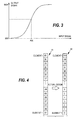
- FIG. 3 illustrates typical input-output characteristics of the normalization transform.
- A is typically a positive constant determining the scale of the index values, while B may be a patient-specific parameter, which determines the distribution of the output index values within the scale from zero to A.
- the transform forces the input signal to a predetermined output value range between a minimum value MIN and a maximum value MAX.
- MIN equals to 0
- MAX equals to A.
- the adaptation to the patient may be accomplished by defining one or more patient-specific parameters and making the transform dependent on the said at least one parameter.
- the mean ⁇ x> and standard deviation ⁇ of the amplitude of the pulsative component may be determined prior to the surgery based on the time series data measured during measuring period of a predetermined length, such as 5 minutes.
- the transform may then be made dependent on the parameters determined.
- the above third feature i.e. the emphasis of the slow changes may be accomplished by making the transform such that the slope of the transform curve is steepest around the input values that are most common, i.e. around the mean value AVE.
- the output values of the transform are then used as a measure of the pain state of the patient.
- the output value of the transform forms the index of nociception, which is indicative of the level of (anti)nociception in the patient.
- the index is not necessarily updated as frequently as a new output value is obtained, but the index may be calculated, for example, as an average over a certain number of output values output from the transform.
- the time series of the amplitude of a signal indicative of the function of the cardiovascular system of the patient is transformed to a surrogate signal that has a predetermined value range and predetermined distribution characteristics for all patients.
- the transformation thus "forces" the values of the time series to a certain value range regardless of the incoming values, and a certain index value, such as 60, is indicative of the same amount of nociception for all patients.
- a patient-adaptive normalization transform may be implemented by using a parameterized transform including at least one patient-specific parameter.
- Patient-specific parameter value(s) are measured prior to the actual measurement of the index of nociception, i.e. prior to the surgical procedure of a patient or during the surgical procedure but before the latest index value.
- the patient-specific values are then substituted for the said parameter(s) to obtain a patient-specific normalization transform.
- the patient-specific normalization transform is then used in step 23 to transform the incoming time series to the index of nociception.
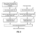
- a histogram transform an input array 41 and an output array 42 are formed, as is shown in FIG. 4.
- the input array (buffer) comprises i input elements, also termed bins, storing, respectively, i input values sorted in ascending or descending order, while the output array (buffer) comprises fixed index values sorted in ascending or descending order and stored in i output elements.
- the values of the time series of the amplitude of the pulsative component, i.e. the values obtained from step 22, are thus forced to the value range of the output buffer. This may be implemented in three different ways depending on whether full adaptation, partial adaptation, or no adaptation to the incoming signal is utilized. These embodiments of the histogram transform are discussed briefly in the following.
- the latest signal values of the time series of the amplitude of the pulsative component are stored in the input array so that the value of each new data point obtained in the time series replaces the oldest value in the input array.
- the oldest value in the input array is deleted, and the remaining values and the new value are sorted to form a new input array.
- the output value of the transform is then obtained by means of the output array as the index value that corresponds to the location of the new value in the input array. In this way, the level of the signal may change but the output values remain between the lowest and highest indices, i.e. in this example between 0 and 100.
- the time series obtained from the output array 32 may thus be such that the mean value is constant and the variability of the amplitude is limited to a certain range and certain distribution around the mean value. For instance, a Gaussian or even distribution with a desired mean value may be used for the values output from the transform.
- Full adaptation may also be implemented by a parameterized transform. If a histogram transform is used, no patient-specific parameters are needed, since the sorting of previous signal data makes the histogram transform patient-adaptive. If a parameterized transform is used, no sorting of input data is needed.
- the input array In case no adaptation to the incoming signal is used in the histogram transform, the input array remains the same regardless of the incoming signal values.
- the input array may be formed based on values measured from a large patient group, which yields a wider distribution of input values than what is typically obtained for one patient.
- the input array may store i fixed values representing the distribution of the values of the amplitude of the pulsative component among a (large) group of patients.
- different input arrays may still be defined for different patient groups, such as children or adults, and different input arrays may further be defined for each patient group according to various other parameters, such as the type of the sensor used or the peripheral site of the sensor (ear, finger, toe).
- Similar non-adaptive transforms may be implemented by means of the parameterized transform.
- the transform includes one or more parameters, whose value(s) depend on the patient group in question, and possible also on other parameters, such as the sensor type or site.
- Partial adaptation to the incoming signal refers to the combination of the above two methods.
- An embodiment of the partial adaptation is illustrated in Fig. 5a to 5d.
- FIG. 5a illustrates a parameter distribution curve 51 for a large number of patients representing a certain patient group in general anesthesia.
- the size of the patient group may be very large representing about 1000 patients, for example.
- the range of the parameter values, in the figures from 0 to 300, is advantageously selected to be much wider than the actual range obtained during a surgery of an individual patient.
- the same parameter i.e. in this case the amplitude of the pulsative component of the blood circulation, is measured and a histogram distribution is created using the same parameter value bins as in the large population average.
- This distribution for the individual patient may contain a fixed number of values, e.g. 300, and the distribution may be updated using the full adaptation method described above. It is also possible that a cumulative distribution of the parameter values of the individual patient is collected and that the distribution counts so obtained are scaled down to a predetermined match in total counts to the patient group distribution. In such a case, the individual patient distribution may represent the parameter values since the beginning of the surgery till the current moment during surgery.
- An example of a normalized patient-specific distribution curve 52 obtained during a surgery is presented in Fig. 5b
- the normalized patient-specific distribution is then added in a predetermined proportion to the normalized patient group distribution, and an average total distribution curve 53 is formed, as shown in FIG. 5c.
- the two normalized distributions are weighted equally in the total distribution.
- For calculating the input parameter value array for the partially adapted histogram transform a cumulative sum of the average total distribution is then constructed as shown in Fig. 5d. If the histogram transform arrays are 101 element long, for example, the new values for the input bins of the histogram transform can be obtained by projecting the cumulative sum values 0, 1, 2,....,100 of the Y-axis to the parameter value axis (X-axis), as is shown by dashed lines in FIG. 5d.
- the X-axis values obtained in this way form the input values of the input array for the histogram transform.
- the actual histogram transform is then executed without adaptation.
- input values for the input array are thus obtained by adding a group distribution curve to the patient-specific distribution curve and then defining the input values for the input array by means of the cumulative distribution function of the summed distribution curve.
- the input values of the input array remain fixed for a predetermined update interval, which can typically represent about 100 new individual parameter values.
- the proportions of the adaptive and non-adaptive values in the combined input may vary. The same applies to the size of the steps between consecutive (fixed) values stored in the input or output arrays. For example, in the example presented in connection with Fig. 5a to 5d each consecutive input array bin contained one percent of the input values. However, the steps may also be such that a certain other percentage of values is within each step (i.e. in each bin), in which case the step may be smaller around one range of the input values and correspondingly larger around another range of the input values.
- the median value of the output signal i.e. the center index of the output array, may be transformed to a desired output value, such as 50.
- the distribution of the output values may be specified according to the needs of the application.
- the normalization transform to be used at each time may depend on the patient, i.e. to which patient group the patient belongs, on the sensor used, and/or on the location of the sensor (finger or ear).
- H(patient adaptive transformation) A*H1(large patient population) + B*H2(history data since the beginning of the surgery) + C*H3(history data over the last M minutes), in which the first reference histogram H1 is for a large population group of similar patients (adults, children, infants, etc.) or for a particular type of sensor or equipment (PPG finger sensor, PPG ear sensor, etc.), the second reference histogram H2 is for the parameter values recorded since the beginning of surgery or anesthesia (long history data), and the third reference histogram H3 is for the parameter values recorded over the last M minutes, such as 5 minutes (short history data).
- the principal reason for the usage of a patient population histogram is that it contains the widest distribution of the parameter values, and thereby represents the allowable parameter range of the input values.
- the history data since the beginning of the anesthesia or surgery substantially sets each patient to the same norm.
- the histogram pertaining to the last M minutes allows a fast adaptation to trend-like parameter changes within one patient and thereby sets each phase of the surgery to a same equivalent norm, regardless of the absolute average value of the parameter in the time window of M minutes.
- Partial adaptation may also be implemented by a parameterized transform.
- the transform includes both patient-specific and group-specific parameters in a fashion similar to the above histogram transformation.
- the parameters may be defined in various ways. For instance, the mean parameter in the function transform may be determined as a weighted mean of the mean values of the parameter in a large patient group data, in the long history data, and in the short history data of M minutes, while the variance parameter in the function transform may be determined as a weighted sum of the variances obtained from the two groups of history data.
- the output signal of the normalization transform thus directly indicates the level of the antinociceptive component of anesthesia in the patient.
- the said output signal is termed the first normalized signal and the basic embodiments are said to form the first group of embodiments.
- the specificity of the index of nociception to noxious stimulation and to analgesic drug concentration may be improved by producing a composite indication based on the first normalized signal and at least one other normalized signal made commensurable with the first normalized signal. This is discussed in the following.
- the composite indication is formed based on two normalized signals.
- the time domain is taken into account by producing the composite indication based on the first normalized signal and a normalized pulse interval, which is in this context termed the second normalized signal.
- the pulse interval here refers to the beat-to-beat interval of the physiological signal in question.
- the physiological signal may be a plethysmographic signal, an ECG signal, or a blood pressure signal.
- a plethysmographic signal or a blood pressure signal may be used to obtain the normalized pulsative component, the same signal data may be used to derive the time series of the pulse interval.
- the signal may be supplied from step 21 directly to step 62, in which the time series of the pulse interval is generated.
- step 62 in which the time series of the pulse interval is generated.
- a pre-processing step 61 similar to step 21 may precede step 62.
- the time series of the pulse interval is then subjected to a normalization transform at step 63 to obtain a time series of a normalized pulse interval.
- the transform applied to the pulse interval is typically similar to the transform applied to the amplitude of the pulsative component in step 23.
- the transform is thus typically a fully or partially adaptive normalization transform, which may be implemented as a parameterized transform or as a histogram transform.
- the normalized pulsative component and the normalized pulse interval are then combined at step 64 to form a composite indicator that serves as the index of nociception. This may be performed by calculating a weighted average of the two normalized values for each data point pair obtained from the two time series.
- the specificity of the index of nociception to noxious stimulation and to analgesic drug concentration may further be improved by adding a third normalized parameter to the group of normalized parameters whose weighted average forms the index of nociception.
- the third normalized parameter may be indicative of the variability of either the first or the second normalized signal or the corresponding non-normalized signal.
- the input signal to the input branch corresponding to the third normalized parameter may thus be supplied from any of steps 21 to 23 if the variability of the first signal is utilized or from any of steps 61 to 63 if the variability of the second signal is utilized.
- a time series or waveform of the third normalized parameter is then produced at step 72 by calculating, based on the input signal, a parameter which is indicative of the variability in the input signal.
- the parameter calculated may be, for example, the ratio of the low frequency variability to the high frequency variability, i.e. the so-called sympatho-vagal balance, or a ratio similar to the sympatho-vagal balance.
- the parameter calculated may be, for example, the power of respiratory variability.
- a signal indicative of the respiration rhythm of a patient may be utilized to calculate the parameter at step 72 in order to extract the frequency components needed for the calculation of the parameter indicative of variability.
- a desired signal component may be extracted by forming a time series phase-locked to the respiration rhythm of the patient.
- the signal indicative of the respiration rhythm of the patient does not necessarily have to carry phase information, but the desired signal component may also be separated by means of a filter controlled by the respiration rate of the patient.
- Different methods for using a signal indicative of the respiration rhythm of the patient for extracting desired signal components are described in Applicant's copending U.S. Patent application, Serial No. 10/954,040, filed on September, 29,2004 .
- a further signal transformation may be still needed for the third normalized parameter obtained from step 72 to make the said parameter commensurable with the first and second normalized parameters.
- This is then performed at step 73, if necessary.
- the transform performed at step 73 is typically non-adaptive, i.e. it is not dependent on previous signal data of the same patient. Instead, this normalization transform may depend on various other parameters, such as the type of the sensor and/or the location of the sensor. It may also represent an average distribution of the parameter values in a large patient population subjected to a certain clinical procedure, such as general anesthesia. Therefore, the normalization transform to be used may be selected at step 73 based on said other parameters or based on patient population.
- the normalized pulsative component, the normalized pulse interval, and the normalized variability signal are then combined at step 74 to form a composite indicator that serves as the index of nociception. This may be performed by calculating a weighted average of the three normalized values obtained simultaneously in the three time series output respectively from the three transforms.
- the normalized respiration rate of the patient may be used instead of the normalized variability signal as the third normalized signal.
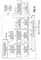
- FIG. 8 illustrates an embodiment 80, in which the normalized respiration rate of the patient is used as a third component of the weighted average. The use of the respiration rate is based on the fact that the respiration rate changes when the patient experiences pain.
- the respiration rate is derived at step 82 and the time series of the respiration rate is normalized at step 83.
- the respiration rate may be derived from an input signal, which may be a plethysmographic signal or an ECG signal.
- the input signal to step 82 may therefore be received from steps 20/21 or from step 61.
- the input signal may also be a respiration gas signal or a signal received from a particular respiration sensor attached to the patient.
- the normalization transform applied to the respiration rate is typically similar to those applied to the amplitude of the pulsative component and/or to the pulse interval at steps 23 and 63, respectively.
- the index of nociception is calculated as the weighted average of the normalized pulsative component, the normalized pulse interval, and the normalized respiration rate.
- the invention may be utilized to improve specificity in connection with the determination of any diagnostic index indicative of a desired clinical state of the patient.
- the normalizing transform may be used in the above-described manner before the complexity measures forming the components of the diagnostic index are calculated.
- normalization may be used to improve specificity in connection with the determination of any diagnostic index, which is determined based on one or more physiological parameters or signals indicative of the physiological state in question.
- FIG. 9 illustrates one embodiment of the system or apparatus according to the invention.
- the physiological signal(s) obtained from one or more sensors attached to a patient 100 are supplied to an amplifier stage 91, which amplifies the signal(s) before they are sampled and converted into digitized format in an A/D converter 92.
- the digitized signals are supplied to a computer unit 93 which may comprise one or more processors.
- the computer unit is provided with a memory or database 95 holding the digitized signal data obtained from the sensor(s).
- the computer unit may produce the time series needed, apply the normalization transform to each time series, and determine the diagnostic index based on the normalized signal value(s).
- the memory may store the transform(s) to be used, the patient-specific and/or group-specific parameter values needed for parameterized transforms, and/or the group-specific input arrays of the histogram transforms to be used.
- one computer unit or processor may perform the above steps, the processing of the data may also be distributed among different units/processors (servers) within a network, such as a hospital LAN (local area network).
- the apparatus of the invention may thus also be implemented as a distributed system.
- the computer unit may display the results through at least one monitor 94 connected to the computer unit, and it may further supply the diagnostic index as input data to a device or system 96 delivering drugs to the patient, thereby enabling automatic control of the desired clinical state of the patient.
- at least one analgesic may be delivered based on the index of nociception.
- the computer unit may act as a controlling entity controlling the administration of the drugs from the delivery system 96 to the patient.
- the computer unit may supply the diagnostic index to another computer unit or microprocessor (not shown), which then acts as the controlling entity controlling the drug delivery system 96.
- the said controlling entity is provided with the control data needed for the administration, such as the pharmacodynamic and pharmacokinetic properties of the drugs to be administered.
- the drug delivery system may comprise separate delivery units for one or more drugs to be administered, such as delivery unit for an analgesic drug and/or a delivery unit for a hypnotic drug.
- the computer unit may also act as decision-support tool for the physician, such as an anesthesiologist, who then controls the drug delivery system based on the information provided by the computer unit.
- the system of FIG. 9 may be provided with a monitor that displays the analgesic concentration during a surgery, for example, and also the recommended upper and lower limits for the said concentration.
- the recommended upper limit 101 and lower limit 102 of the analgesic concentration may further be controlled based on the index of nociception. If the index of nociception, shown as a dashed line, is too high (underdose), both limits shift up for a new infusion target and if the index is too low (overdose), both limits shift down.
- the recommendations concerning the use of the analgesic may be controlled based on the index of nociception and the controlled recommendations may be displayed to the anesthesiologist.
- a conventional measurement device such as a pulse oximeter, may also be upgraded to enable the device to determine the diagnostic index in the above-described manner based on the signal data that the device measures from the patient.
- Such an upgrade may be implemented by delivering to the measurement device a software module that enables the device to determine the diagnostic index in the above-described manner.
- the software module may be delivered, for example, on a data carrier, such as a CD or a memory card.
- the software module which is provided with an interface to the memory storing the signal data measured by the measurement device, may apply any of the above-described normalization transforms to the signal data. It is also possible that a measurement device in which the determination of the diagnostic index is based on one or two normalized signals is upgraded by adding a further normalized signal to the group of normalized signals whose weighted average forms the diagnostic index.
Applications Claiming Priority (1)
| Application Number | Priority Date | Filing Date | Title |
|---|---|---|---|
| US11/089,528 US8715193B2 (en) | 2005-03-24 | 2005-03-24 | Determination of the clinical state of a subject |
Publications (3)
| Publication Number | Publication Date |
|---|---|
| EP1704818A2 true EP1704818A2 (fr) | 2006-09-27 |
| EP1704818A3 EP1704818A3 (fr) | 2006-11-02 |
| EP1704818B1 EP1704818B1 (fr) | 2011-05-11 |
Family
ID=36691426
Family Applications (1)
| Application Number | Title | Priority Date | Filing Date |
|---|---|---|---|
| EP06251571A Active EP1704818B1 (fr) | 2005-03-24 | 2006-03-23 | Détermination de l'état clinique d'un sujet |
Country Status (3)
| Country | Link |
|---|---|
| US (1) | US8715193B2 (fr) |
| EP (1) | EP1704818B1 (fr) |
| AT (1) | ATE508680T1 (fr) |
Cited By (5)
| Publication number | Priority date | Publication date | Assignee | Title |
|---|---|---|---|---|
| WO2010022047A2 (fr) * | 2008-08-18 | 2010-02-25 | Beckman Coulter, Inc. | Systèmes équivalents décimaux normalisés et procédés |
| EP2233075A1 (fr) * | 2009-03-26 | 2010-09-29 | General Electric Company | Procédé et appareil pour déterminer un indicateur de l'état du système nerveux autonomique |
| US8657755B2 (en) | 2009-05-12 | 2014-02-25 | Angiologix, Inc. | System and method of measuring changes in arterial volume of a limb segment |
| EP3316767A4 (fr) * | 2015-07-05 | 2018-10-31 | Medasense Biometrics Ltd. | Appareil, système et procédé de surveillance de la douleur |
| US10238306B2 (en) | 2006-02-20 | 2019-03-26 | Everist Genomics, Inc. | Method for non-evasively determining an endothelial function and a device for carrying out said method |
Families Citing this family (74)
| Publication number | Priority date | Publication date | Assignee | Title |
|---|---|---|---|---|
| US7758503B2 (en) | 1997-01-27 | 2010-07-20 | Lynn Lawrence A | Microprocessor system for the analysis of physiologic and financial datasets |
| US8932227B2 (en) | 2000-07-28 | 2015-01-13 | Lawrence A. Lynn | System and method for CO2 and oximetry integration |
| US9468378B2 (en) | 1997-01-27 | 2016-10-18 | Lawrence A. Lynn | Airway instability detection system and method |
| US9042952B2 (en) | 1997-01-27 | 2015-05-26 | Lawrence A. Lynn | System and method for automatic detection of a plurality of SPO2 time series pattern types |
| US20070191697A1 (en) | 2006-02-10 | 2007-08-16 | Lynn Lawrence A | System and method for SPO2 instability detection and quantification |
| US9521971B2 (en) | 1997-07-14 | 2016-12-20 | Lawrence A. Lynn | System and method for automatic detection of a plurality of SPO2 time series pattern types |
| US20060195041A1 (en) | 2002-05-17 | 2006-08-31 | Lynn Lawrence A | Centralized hospital monitoring system for automatically detecting upper airway instability and for preventing and aborting adverse drug reactions |
| US9053222B2 (en) | 2002-05-17 | 2015-06-09 | Lawrence A. Lynn | Patient safety processor |
| US7455643B1 (en) | 2003-07-07 | 2008-11-25 | Nellcor Puritan Bennett Ireland | Continuous non-invasive blood pressure measurement apparatus and methods providing automatic recalibration |
| US7447541B2 (en) * | 2004-06-30 | 2008-11-04 | Instrumentarium Corporation | Monitoring subcortical responsiveness of a patient |
| EP1955190B1 (fr) | 2005-11-10 | 2018-08-29 | In Silico Biosciences, Inc. | Procédé et dispositif de modélisation informatique du cerveau humain devant permettre de prévoir les effets de médicaments |
| US7668579B2 (en) | 2006-02-10 | 2010-02-23 | Lynn Lawrence A | System and method for the detection of physiologic response to stimulation |
| CA2711445A1 (fr) * | 2007-01-17 | 2008-07-24 | Universite De Montreal | Procede et systeme d'administration d'un anesthesique |
| US9398863B2 (en) | 2007-06-20 | 2016-07-26 | General Electric Company | Detection of anomalies in measurement of level of hypnosis |
| US8512240B1 (en) * | 2007-11-14 | 2013-08-20 | Medasense Biometrics Ltd. | System and method for pain monitoring using a multidimensional analysis of physiological signals |
| US11259708B2 (en) | 2007-11-14 | 2022-03-01 | Medasense Biometrics Ltd. | System and method for pain monitoring using a multidimensional analysis of physiological signals |
| US9064036B2 (en) * | 2008-04-24 | 2015-06-23 | The Invention Science Fund I, Llc | Methods and systems for monitoring bioactive agent use |
| CA2722773C (fr) | 2008-05-07 | 2015-07-21 | Lawrence A. Lynn | Moteur de recherche de modele d'echec medical |
| US8660799B2 (en) | 2008-06-30 | 2014-02-25 | Nellcor Puritan Bennett Ireland | Processing and detecting baseline changes in signals |
| US8398556B2 (en) | 2008-06-30 | 2013-03-19 | Covidien Lp | Systems and methods for non-invasive continuous blood pressure determination |
| US8506498B2 (en) | 2008-07-15 | 2013-08-13 | Nellcor Puritan Bennett Ireland | Systems and methods using induced perturbation to determine physiological parameters |
| US9687161B2 (en) | 2008-09-30 | 2017-06-27 | Nellcor Puritan Bennett Ireland | Systems and methods for maintaining blood pressure monitor calibration |
| US8532751B2 (en) | 2008-09-30 | 2013-09-10 | Covidien Lp | Laser self-mixing sensors for biological sensing |
| US9314168B2 (en) | 2008-09-30 | 2016-04-19 | Nellcor Puritan Bennett Ireland | Detecting sleep events using localized blood pressure changes |
| US9301697B2 (en) | 2008-09-30 | 2016-04-05 | Nellcor Puritan Bennett Ireland | Systems and methods for recalibrating a non-invasive blood pressure monitor |
| US8515513B2 (en) * | 2008-11-05 | 2013-08-20 | Covidien Lp | System and method for facilitating observation of monitored physiologic data |
| US8216136B2 (en) | 2009-03-05 | 2012-07-10 | Nellcor Puritan Bennett Llc | Systems and methods for monitoring heart rate and blood pressure correlation |
| US20100256515A1 (en) * | 2009-04-03 | 2010-10-07 | Egeth Marc J | Communication with and consciousness-assessment of anesthetized surgery patients |
| US8290730B2 (en) | 2009-06-30 | 2012-10-16 | Nellcor Puritan Bennett Ireland | Systems and methods for assessing measurements in physiological monitoring devices |
| US9198582B2 (en) * | 2009-06-30 | 2015-12-01 | Nellcor Puritan Bennett Ireland | Determining a characteristic physiological parameter |
| US8628477B2 (en) | 2009-07-31 | 2014-01-14 | Nellcor Puritan Bennett Ireland | Systems and methods for non-invasive determination of blood pressure |
| US9220440B2 (en) | 2009-09-21 | 2015-12-29 | Nellcor Puritan Bennett Ireland | Determining a characteristic respiration rate |
| US9066660B2 (en) | 2009-09-29 | 2015-06-30 | Nellcor Puritan Bennett Ireland | Systems and methods for high-pass filtering a photoplethysmograph signal |
| US8463347B2 (en) | 2009-09-30 | 2013-06-11 | Nellcor Puritan Bennett Ireland | Systems and methods for normalizing a plethysmograph signal for improved feature analysis |
| US9451887B2 (en) | 2010-03-31 | 2016-09-27 | Nellcor Puritan Bennett Ireland | Systems and methods for measuring electromechanical delay of the heart |
| US8898037B2 (en) | 2010-04-28 | 2014-11-25 | Nellcor Puritan Bennett Ireland | Systems and methods for signal monitoring using Lissajous figures |
| US8825428B2 (en) | 2010-11-30 | 2014-09-02 | Neilcor Puritan Bennett Ireland | Methods and systems for recalibrating a blood pressure monitor with memory |
| US9357934B2 (en) | 2010-12-01 | 2016-06-07 | Nellcor Puritan Bennett Ireland | Systems and methods for physiological event marking |
| US9259160B2 (en) | 2010-12-01 | 2016-02-16 | Nellcor Puritan Bennett Ireland | Systems and methods for determining when to measure a physiological parameter |
| US9693709B2 (en) | 2011-09-23 | 2017-07-04 | Nellcot Puritan Bennett Ireland | Systems and methods for determining respiration information from a photoplethysmograph |
| US9402554B2 (en) | 2011-09-23 | 2016-08-02 | Nellcor Puritan Bennett Ireland | Systems and methods for determining respiration information from a photoplethysmograph |
| US8880576B2 (en) | 2011-09-23 | 2014-11-04 | Nellcor Puritan Bennett Ireland | Systems and methods for determining respiration information from a photoplethysmograph |
| US9119597B2 (en) | 2011-09-23 | 2015-09-01 | Nellcor Puritan Bennett Ireland | Systems and methods for determining respiration information from a photoplethysmograph |
| US9675274B2 (en) | 2011-09-23 | 2017-06-13 | Nellcor Puritan Bennett Ireland | Systems and methods for determining respiration information from a photoplethysmograph |
| US8755871B2 (en) | 2011-11-30 | 2014-06-17 | Covidien Lp | Systems and methods for detecting arrhythmia from a physiological signal |
| US9060695B2 (en) | 2011-11-30 | 2015-06-23 | Covidien Lp | Systems and methods for determining differential pulse transit time from the phase difference of two analog plethysmographs |
| US9693736B2 (en) | 2011-11-30 | 2017-07-04 | Nellcor Puritan Bennett Ireland | Systems and methods for determining respiration information using historical distribution |
| US8870783B2 (en) | 2011-11-30 | 2014-10-28 | Covidien Lp | Pulse rate determination using Gaussian kernel smoothing of multiple inter-fiducial pulse periods |
| US9247896B2 (en) | 2012-01-04 | 2016-02-02 | Nellcor Puritan Bennett Ireland | Systems and methods for determining respiration information using phase locked loop |
| US9179876B2 (en) | 2012-04-30 | 2015-11-10 | Nellcor Puritan Bennett Ireland | Systems and methods for identifying portions of a physiological signal usable for determining physiological information |
| US9241670B2 (en) | 2012-09-11 | 2016-01-26 | Covidien Lp | Methods and systems for conditioning physiological information using a normalization technique |
| US9560978B2 (en) | 2013-02-05 | 2017-02-07 | Covidien Lp | Systems and methods for determining respiration information from a physiological signal using amplitude demodulation |
| US9554712B2 (en) | 2013-02-27 | 2017-01-31 | Covidien Lp | Systems and methods for generating an artificial photoplethysmograph signal |
| US9687159B2 (en) | 2013-02-27 | 2017-06-27 | Covidien Lp | Systems and methods for determining physiological information by identifying fiducial points in a physiological signal |
| US20140275887A1 (en) * | 2013-03-15 | 2014-09-18 | Nellcor Puritan Bennett Ireland | Systems And Methods For Monitoring Respiratory Depression |
| JP6446030B2 (ja) | 2013-04-24 | 2018-12-26 | フレゼニウス カービ ドイチュラント ゲーエムベーハー | 薬剤注入装置を制御する制御装置を操作する方法 |
| US10039463B1 (en) | 2013-06-27 | 2018-08-07 | Vital Connect, Inc. | Signal quality metric for cardiovascular time series |
| US10022068B2 (en) | 2013-10-28 | 2018-07-17 | Covidien Lp | Systems and methods for detecting held breath events |
| US9848820B2 (en) | 2014-01-07 | 2017-12-26 | Covidien Lp | Apnea analysis system and method |
| US9955894B2 (en) | 2014-01-28 | 2018-05-01 | Covidien Lp | Non-stationary feature relationship parameters for awareness monitoring |
| CA2932826A1 (fr) | 2014-02-20 | 2015-08-27 | Covidien Lp | Systemes et procedes de filtration de pics d'auto-correlation et de detection d'harmoniques |
| US10405757B2 (en) | 2014-02-25 | 2019-09-10 | Icu Medical, Inc. | Patient monitoring system with gatekeeper signal |
| US10206632B2 (en) * | 2014-07-25 | 2019-02-19 | The Trustees Of Dartmouth College | Systems and methods for cardiovascular-dynamics correlated imaging |
| US10111617B2 (en) | 2014-09-22 | 2018-10-30 | Covidien Lp | Systems and methods for EEG monitoring |
| EP3364860A4 (fr) | 2015-10-19 | 2019-09-18 | ICU Medical, Inc. | Système de surveillance hémodynamique avec unité d'affichage détachable |
| US10835174B2 (en) | 2016-01-12 | 2020-11-17 | Covidien Lp | System and method for monitoring cerebral activity |
| US10660577B2 (en) * | 2016-04-01 | 2020-05-26 | Cardiac Pacamakers, Inc. | Systems and methods for detecting worsening heart failure |
| CN110430804A (zh) * | 2017-01-16 | 2019-11-08 | 皇家飞利浦有限公司 | 组合了二氧化碳测定和氧饱和度的生理监测决策支持系统 |
| EP3634211A4 (fr) | 2017-06-07 | 2021-03-17 | Covidien LP | Systèmes et procédés de détection d'accidents vasculaires cérébraux |
| EP3563759A1 (fr) * | 2018-05-01 | 2019-11-06 | Koninklijke Philips N.V. | Appareil permettant de déterminer une contrainte et/ou le niveau de douleur |
| KR102626585B1 (ko) | 2018-08-20 | 2024-01-17 | 삼성전자주식회사 | 혈압 추정 장치 및 방법 |
| CN112006658B (zh) * | 2020-08-21 | 2022-08-02 | 思澜科技(成都)有限公司 | 一种麻醉状态监测方法及装置 |
| DE102021117940A1 (de) | 2021-07-12 | 2023-01-12 | Michael Becker | Infusionsvorrichtung |
| CN115633969B (zh) * | 2022-11-11 | 2024-01-05 | 重庆玺德尔医疗器械有限公司 | 基于脑电信号和脉搏信号的麻醉疼痛伤害感受评估方法 |
Citations (4)
| Publication number | Priority date | Publication date | Assignee | Title |
|---|---|---|---|---|
| EP1273265A1 (fr) * | 2001-07-04 | 2003-01-08 | Instrumentarium Corporation | Surveillance de l'état d'un patient sous anesthésie ou sédation |
| US20040215263A1 (en) * | 2003-04-23 | 2004-10-28 | Nathalie Virag | Detection of vasovagal syncope |
| FR2859911A1 (fr) * | 2003-09-18 | 2005-03-25 | Jean Claude Barthelemy | Utilisation des ondelettes pour analyser la variabilite de la frequence cardiaque, appliquee au suivi de l'anesthesie generale en temps reel |
| EP1642525A1 (fr) * | 2004-09-29 | 2006-04-05 | GE Healthcare Finland Oy | Surveillance en temps réel de la condition du système neurovégétatif |
Family Cites Families (8)
| Publication number | Priority date | Publication date | Assignee | Title |
|---|---|---|---|---|
| US5083571A (en) * | 1988-04-18 | 1992-01-28 | New York University | Use of brain electrophysiological quantitative data to classify and subtype an individual into diagnostic categories by discriminant and cluster analysis |
| GB9022623D0 (en) | 1990-10-18 | 1990-11-28 | Univ Manchester | Depth of anaesthesia monitoring |
| FR2747027B1 (fr) | 1996-04-09 | 1998-05-29 | Cohen Laroque Emmanuel S | Procede de determination de la profondeur d'anesthesie et dispositif pour la mise en oeuvre de ce procede |
| US6684090B2 (en) * | 1999-01-07 | 2004-01-27 | Masimo Corporation | Pulse oximetry data confidence indicator |
| US6801803B2 (en) | 2000-10-16 | 2004-10-05 | Instrumentarium Corp. | Method and apparatus for determining the cerebral state of a patient with fast response |
| US20030105409A1 (en) * | 2001-11-14 | 2003-06-05 | Donoghue John Philip | Neurological signal decoding |
| EP1551279A1 (fr) | 2002-10-14 | 2005-07-13 | Instrumentarium Corporation | Procede et appareil de detection de nociception lors de l'anesthesie ou de la sedation, basee sur un plethysmographe de pulsations |
| US7367949B2 (en) | 2003-07-07 | 2008-05-06 | Instrumentarium Corp. | Method and apparatus based on combination of physiological parameters for assessment of analgesia during anesthesia or sedation |
-
2005
- 2005-03-24 US US11/089,528 patent/US8715193B2/en active Active
-
2006
- 2006-03-23 EP EP06251571A patent/EP1704818B1/fr active Active
- 2006-03-23 AT AT06251571T patent/ATE508680T1/de not_active IP Right Cessation
Patent Citations (4)
| Publication number | Priority date | Publication date | Assignee | Title |
|---|---|---|---|---|
| EP1273265A1 (fr) * | 2001-07-04 | 2003-01-08 | Instrumentarium Corporation | Surveillance de l'état d'un patient sous anesthésie ou sédation |
| US20040215263A1 (en) * | 2003-04-23 | 2004-10-28 | Nathalie Virag | Detection of vasovagal syncope |
| FR2859911A1 (fr) * | 2003-09-18 | 2005-03-25 | Jean Claude Barthelemy | Utilisation des ondelettes pour analyser la variabilite de la frequence cardiaque, appliquee au suivi de l'anesthesie generale en temps reel |
| EP1642525A1 (fr) * | 2004-09-29 | 2006-04-05 | GE Healthcare Finland Oy | Surveillance en temps réel de la condition du système neurovégétatif |
Cited By (7)
| Publication number | Priority date | Publication date | Assignee | Title |
|---|---|---|---|---|
| US10238306B2 (en) | 2006-02-20 | 2019-03-26 | Everist Genomics, Inc. | Method for non-evasively determining an endothelial function and a device for carrying out said method |
| WO2010022047A2 (fr) * | 2008-08-18 | 2010-02-25 | Beckman Coulter, Inc. | Systèmes équivalents décimaux normalisés et procédés |
| WO2010022047A3 (fr) * | 2008-08-18 | 2010-05-27 | Beckman Coulter, Inc. | Systèmes équivalents décimaux normalisés et procédés |
| US8204713B2 (en) | 2008-08-18 | 2012-06-19 | Blick Kenneth E | Normalized decimal equivalent systems and methods |
| EP2233075A1 (fr) * | 2009-03-26 | 2010-09-29 | General Electric Company | Procédé et appareil pour déterminer un indicateur de l'état du système nerveux autonomique |
| US8657755B2 (en) | 2009-05-12 | 2014-02-25 | Angiologix, Inc. | System and method of measuring changes in arterial volume of a limb segment |
| EP3316767A4 (fr) * | 2015-07-05 | 2018-10-31 | Medasense Biometrics Ltd. | Appareil, système et procédé de surveillance de la douleur |
Also Published As
| Publication number | Publication date |
|---|---|
| EP1704818A3 (fr) | 2006-11-02 |
| US20060217614A1 (en) | 2006-09-28 |
| US8715193B2 (en) | 2014-05-06 |
| ATE508680T1 (de) | 2011-05-15 |
| EP1704818B1 (fr) | 2011-05-11 |
Similar Documents
| Publication | Publication Date | Title |
|---|---|---|
| EP1704818B1 (fr) | Détermination de l'état clinique d'un sujet | |
| US8574156B2 (en) | Determination of the clinical state of a subject | |
| US7447541B2 (en) | Monitoring subcortical responsiveness of a patient | |
| US7407485B2 (en) | Monitoring pain-related responses of a patient | |
| EP1704817B1 (fr) | Appareil pour déterminer stress clinique d'un patient | |
| EP1704819B1 (fr) | Détermination de l'état anesthésique d'un patient | |
| EP1495715B1 (fr) | Appareil et méthode basé sur la combinaison de trois signaux physiologiques afin d' évaluer l' analgésie pendant une anesthésie or une sedation | |
| US7407486B2 (en) | Method and an apparatus for pulse plethysmograph based detection of nociception during anesthesia or sedation | |
| JP6761468B2 (ja) | 覚醒、鎮静及び全身麻酔中の意識、痛み及び侵害受容のレベルを評価するデバイス及び方法 | |
| EP1757226B1 (fr) | Mesure de la réponse d'un patient sous anesthésie | |
| EP3232917B1 (fr) | Appareil pour l'évaluation du niveau de douleur et de nociception au cours d'une anesthésie générale à l'aide d'un électroencéphalogramme, d'une cardiographie d'impédance pléthysmographique, de la variabilité de la fréquence cardiaque et de la concentration ou biophase des analgésiques | |
| US20230248294A1 (en) | A method and system for measurement of a level of anxiety combined with and/or correlated with a level of a modified state of consciousness and/or a level of pain | |
| EP3954290A1 (fr) | Procédé et système pour mesurer un niveau d'anxiété | |
| Venn et al. | Monitoring of the depth of sedation in the intensive care unit | |
| TWI494082B (zh) | 多維麻醉深度信號監控方法 | |
| Jensen et al. | An AEP/EEG hybrid index for monitoring the hypnotic depth during general anesthesia | |
| TWM451978U (zh) | 多維麻醉深度信號擷取裝置 | |
| Baik et al. | Comparison of heart rate variability with pulse transit time during general anesthesia |
Legal Events
| Date | Code | Title | Description |
|---|---|---|---|
| PUAI | Public reference made under article 153(3) epc to a published international application that has entered the european phase |
Free format text: ORIGINAL CODE: 0009012 |
|
| AK | Designated contracting states |
Kind code of ref document: A2 Designated state(s): AT BE BG CH CY CZ DE DK EE ES FI FR GB GR HU IE IS IT LI LT LU LV MC NL PL PT RO SE SI SK TR |
|
| AX | Request for extension of the european patent |
Extension state: AL BA HR MK YU |
|
| PUAL | Search report despatched |
Free format text: ORIGINAL CODE: 0009013 |
|
| AK | Designated contracting states |
Kind code of ref document: A3 Designated state(s): AT BE BG CH CY CZ DE DK EE ES FI FR GB GR HU IE IS IT LI LT LU LV MC NL PL PT RO SE SI SK TR |
|
| AX | Request for extension of the european patent |
Extension state: AL BA HR MK YU |
|
| 17P | Request for examination filed |
Effective date: 20070502 |
|
| AKX | Designation fees paid |
Designated state(s): AT BE BG CH CY CZ DE DK EE ES FI FR GB GR HU IE IS IT LI LT LU LV MC NL PL PT RO SE SI SK TR |
|
| 17Q | First examination report despatched |
Effective date: 20070622 |
|
| GRAP | Despatch of communication of intention to grant a patent |
Free format text: ORIGINAL CODE: EPIDOSNIGR1 |
|
| GRAS | Grant fee paid |
Free format text: ORIGINAL CODE: EPIDOSNIGR3 |
|
| GRAA | (expected) grant |
Free format text: ORIGINAL CODE: 0009210 |
|
| AK | Designated contracting states |
Kind code of ref document: B1 Designated state(s): AT BE BG CH CY CZ DE DK EE ES FI FR GB GR HU IE IS IT LI LT LU LV MC NL PL PT RO SE SI SK TR |
|
| REG | Reference to a national code |
Ref country code: GB Ref legal event code: FG4D |
|
| REG | Reference to a national code |
Ref country code: CH Ref legal event code: EP |
|
| REG | Reference to a national code |
Ref country code: IE Ref legal event code: FG4D |
|
| REG | Reference to a national code |
Ref country code: DE Ref legal event code: R096 Ref document number: 602006021819 Country of ref document: DE Effective date: 20110622 |
|
| REG | Reference to a national code |
Ref country code: NL Ref legal event code: T3 |
|
| REG | Reference to a national code |
Ref country code: SE Ref legal event code: TRGR |
|
| PG25 | Lapsed in a contracting state [announced via postgrant information from national office to epo] |
Ref country code: LT Free format text: LAPSE BECAUSE OF FAILURE TO SUBMIT A TRANSLATION OF THE DESCRIPTION OR TO PAY THE FEE WITHIN THE PRESCRIBED TIME-LIMIT Effective date: 20110511 Ref country code: PT Free format text: LAPSE BECAUSE OF FAILURE TO SUBMIT A TRANSLATION OF THE DESCRIPTION OR TO PAY THE FEE WITHIN THE PRESCRIBED TIME-LIMIT Effective date: 20110912 |
|
| PG25 | Lapsed in a contracting state [announced via postgrant information from national office to epo] |
Ref country code: BE Free format text: LAPSE BECAUSE OF FAILURE TO SUBMIT A TRANSLATION OF THE DESCRIPTION OR TO PAY THE FEE WITHIN THE PRESCRIBED TIME-LIMIT Effective date: 20110511 Ref country code: GR Free format text: LAPSE BECAUSE OF FAILURE TO SUBMIT A TRANSLATION OF THE DESCRIPTION OR TO PAY THE FEE WITHIN THE PRESCRIBED TIME-LIMIT Effective date: 20110812 Ref country code: SI Free format text: LAPSE BECAUSE OF FAILURE TO SUBMIT A TRANSLATION OF THE DESCRIPTION OR TO PAY THE FEE WITHIN THE PRESCRIBED TIME-LIMIT Effective date: 20110511 Ref country code: FI Free format text: LAPSE BECAUSE OF FAILURE TO SUBMIT A TRANSLATION OF THE DESCRIPTION OR TO PAY THE FEE WITHIN THE PRESCRIBED TIME-LIMIT Effective date: 20110511 Ref country code: IS Free format text: LAPSE BECAUSE OF FAILURE TO SUBMIT A TRANSLATION OF THE DESCRIPTION OR TO PAY THE FEE WITHIN THE PRESCRIBED TIME-LIMIT Effective date: 20110911 Ref country code: CY Free format text: LAPSE BECAUSE OF FAILURE TO SUBMIT A TRANSLATION OF THE DESCRIPTION OR TO PAY THE FEE WITHIN THE PRESCRIBED TIME-LIMIT Effective date: 20110511 Ref country code: LV Free format text: LAPSE BECAUSE OF FAILURE TO SUBMIT A TRANSLATION OF THE DESCRIPTION OR TO PAY THE FEE WITHIN THE PRESCRIBED TIME-LIMIT Effective date: 20110511 Ref country code: AT Free format text: LAPSE BECAUSE OF FAILURE TO SUBMIT A TRANSLATION OF THE DESCRIPTION OR TO PAY THE FEE WITHIN THE PRESCRIBED TIME-LIMIT Effective date: 20110511 Ref country code: ES Free format text: LAPSE BECAUSE OF FAILURE TO SUBMIT A TRANSLATION OF THE DESCRIPTION OR TO PAY THE FEE WITHIN THE PRESCRIBED TIME-LIMIT Effective date: 20110822 |
|
| PG25 | Lapsed in a contracting state [announced via postgrant information from national office to epo] |
Ref country code: EE Free format text: LAPSE BECAUSE OF FAILURE TO SUBMIT A TRANSLATION OF THE DESCRIPTION OR TO PAY THE FEE WITHIN THE PRESCRIBED TIME-LIMIT Effective date: 20110511 Ref country code: CZ Free format text: LAPSE BECAUSE OF FAILURE TO SUBMIT A TRANSLATION OF THE DESCRIPTION OR TO PAY THE FEE WITHIN THE PRESCRIBED TIME-LIMIT Effective date: 20110511 |
|
| PG25 | Lapsed in a contracting state [announced via postgrant information from national office to epo] |
Ref country code: DK Free format text: LAPSE BECAUSE OF FAILURE TO SUBMIT A TRANSLATION OF THE DESCRIPTION OR TO PAY THE FEE WITHIN THE PRESCRIBED TIME-LIMIT Effective date: 20110511 Ref country code: PL Free format text: LAPSE BECAUSE OF FAILURE TO SUBMIT A TRANSLATION OF THE DESCRIPTION OR TO PAY THE FEE WITHIN THE PRESCRIBED TIME-LIMIT Effective date: 20110511 Ref country code: RO Free format text: LAPSE BECAUSE OF FAILURE TO SUBMIT A TRANSLATION OF THE DESCRIPTION OR TO PAY THE FEE WITHIN THE PRESCRIBED TIME-LIMIT Effective date: 20110511 Ref country code: SK Free format text: LAPSE BECAUSE OF FAILURE TO SUBMIT A TRANSLATION OF THE DESCRIPTION OR TO PAY THE FEE WITHIN THE PRESCRIBED TIME-LIMIT Effective date: 20110511 |
|
| PLBE | No opposition filed within time limit |
Free format text: ORIGINAL CODE: 0009261 |
|
| STAA | Information on the status of an ep patent application or granted ep patent |
Free format text: STATUS: NO OPPOSITION FILED WITHIN TIME LIMIT |
|
| 26N | No opposition filed |
Effective date: 20120214 |
|
| PG25 | Lapsed in a contracting state [announced via postgrant information from national office to epo] |
Ref country code: IT Free format text: LAPSE BECAUSE OF FAILURE TO SUBMIT A TRANSLATION OF THE DESCRIPTION OR TO PAY THE FEE WITHIN THE PRESCRIBED TIME-LIMIT Effective date: 20110511 |
|
| REG | Reference to a national code |
Ref country code: DE Ref legal event code: R097 Ref document number: 602006021819 Country of ref document: DE Effective date: 20120214 |
|
| PG25 | Lapsed in a contracting state [announced via postgrant information from national office to epo] |
Ref country code: MC Free format text: LAPSE BECAUSE OF NON-PAYMENT OF DUE FEES Effective date: 20120331 |
|
| REG | Reference to a national code |
Ref country code: CH Ref legal event code: PL |
|
| REG | Reference to a national code |
Ref country code: FR Ref legal event code: ST Effective date: 20121130 |
|
| REG | Reference to a national code |
Ref country code: IE Ref legal event code: MM4A |
|
| PG25 | Lapsed in a contracting state [announced via postgrant information from national office to epo] |
Ref country code: FR Free format text: LAPSE BECAUSE OF NON-PAYMENT OF DUE FEES Effective date: 20120402 Ref country code: IE Free format text: LAPSE BECAUSE OF NON-PAYMENT OF DUE FEES Effective date: 20120323 Ref country code: LI Free format text: LAPSE BECAUSE OF NON-PAYMENT OF DUE FEES Effective date: 20120331 Ref country code: CH Free format text: LAPSE BECAUSE OF NON-PAYMENT OF DUE FEES Effective date: 20120331 |
|
| PG25 | Lapsed in a contracting state [announced via postgrant information from national office to epo] |
Ref country code: BG Free format text: LAPSE BECAUSE OF FAILURE TO SUBMIT A TRANSLATION OF THE DESCRIPTION OR TO PAY THE FEE WITHIN THE PRESCRIBED TIME-LIMIT Effective date: 20110811 |
|
| PG25 | Lapsed in a contracting state [announced via postgrant information from national office to epo] |
Ref country code: TR Free format text: LAPSE BECAUSE OF FAILURE TO SUBMIT A TRANSLATION OF THE DESCRIPTION OR TO PAY THE FEE WITHIN THE PRESCRIBED TIME-LIMIT Effective date: 20110511 |
|
| PGFP | Annual fee paid to national office [announced via postgrant information from national office to epo] |
Ref country code: SE Payment date: 20140327 Year of fee payment: 9 |
|
| PG25 | Lapsed in a contracting state [announced via postgrant information from national office to epo] |
Ref country code: LU Free format text: LAPSE BECAUSE OF NON-PAYMENT OF DUE FEES Effective date: 20120323 |
|
| PG25 | Lapsed in a contracting state [announced via postgrant information from national office to epo] |
Ref country code: HU Free format text: LAPSE BECAUSE OF FAILURE TO SUBMIT A TRANSLATION OF THE DESCRIPTION OR TO PAY THE FEE WITHIN THE PRESCRIBED TIME-LIMIT Effective date: 20060323 |
|
| PG25 | Lapsed in a contracting state [announced via postgrant information from national office to epo] |
Ref country code: SE Free format text: LAPSE BECAUSE OF NON-PAYMENT OF DUE FEES Effective date: 20150324 |
|
| REG | Reference to a national code |
Ref country code: SE Ref legal event code: EUG |
|
| PGFP | Annual fee paid to national office [announced via postgrant information from national office to epo] |
Ref country code: NL Payment date: 20230221 Year of fee payment: 18 |
|
| PGFP | Annual fee paid to national office [announced via postgrant information from national office to epo] |
Ref country code: GB Payment date: 20230221 Year of fee payment: 18 Ref country code: DE Payment date: 20230221 Year of fee payment: 18 |
|
| P01 | Opt-out of the competence of the unified patent court (upc) registered |
Effective date: 20230528 |
|
| PGFP | Annual fee paid to national office [announced via postgrant information from national office to epo] |
Ref country code: NL Payment date: 20240220 Year of fee payment: 19 |