EP1682223B1 - Bone-treatment instrument - Google Patents
Bone-treatment instrument Download PDFInfo
- Publication number
- EP1682223B1 EP1682223B1 EP04789764A EP04789764A EP1682223B1 EP 1682223 B1 EP1682223 B1 EP 1682223B1 EP 04789764 A EP04789764 A EP 04789764A EP 04789764 A EP04789764 A EP 04789764A EP 1682223 B1 EP1682223 B1 EP 1682223B1
- Authority
- EP
- European Patent Office
- Prior art keywords
- bone
- insertion member
- light
- bore
- treatment
- Prior art date
- Legal status (The legal status is an assumption and is not a legal conclusion. Google has not performed a legal analysis and makes no representation as to the accuracy of the status listed.)
- Expired - Lifetime
Links
- 210000000988 bone and bone Anatomy 0.000 title claims abstract description 86
- 238000011282 treatment Methods 0.000 title claims description 75
- 238000003780 insertion Methods 0.000 claims abstract description 58
- 230000037431 insertion Effects 0.000 claims abstract description 58
- 241000124008 Mammalia Species 0.000 claims abstract description 14
- 238000002560 therapeutic procedure Methods 0.000 claims description 12
- 239000013307 optical fiber Substances 0.000 claims description 6
- 229940079593 drug Drugs 0.000 abstract description 53
- 239000003814 drug Substances 0.000 abstract description 53
- 238000002428 photodynamic therapy Methods 0.000 abstract description 50
- 239000000835 fiber Substances 0.000 abstract description 36
- 239000003504 photosensitizing agent Substances 0.000 abstract description 9
- 230000002165 photosensitisation Effects 0.000 abstract description 8
- 210000004027 cell Anatomy 0.000 description 65
- 241001465754 Metazoa Species 0.000 description 63
- 206010028980 Neoplasm Diseases 0.000 description 47
- 230000003902 lesion Effects 0.000 description 34
- 230000000694 effects Effects 0.000 description 31
- 241000700159 Rattus Species 0.000 description 29
- 210000000278 spinal cord Anatomy 0.000 description 25
- 210000001519 tissue Anatomy 0.000 description 23
- 239000007924 injection Substances 0.000 description 20
- 238000002347 injection Methods 0.000 description 20
- 238000000034 method Methods 0.000 description 16
- 230000003287 optical effect Effects 0.000 description 16
- 206010033799 Paralysis Diseases 0.000 description 14
- 206010061289 metastatic neoplasm Diseases 0.000 description 14
- 238000001727 in vivo Methods 0.000 description 12
- 238000010603 microCT Methods 0.000 description 12
- 241000282887 Suidae Species 0.000 description 10
- 206010027476 Metastases Diseases 0.000 description 9
- 238000003384 imaging method Methods 0.000 description 9
- 238000000338 in vitro Methods 0.000 description 9
- 230000001394 metastastic effect Effects 0.000 description 9
- 210000000689 upper leg Anatomy 0.000 description 9
- 206010006187 Breast cancer Diseases 0.000 description 8
- 208000026310 Breast neoplasm Diseases 0.000 description 8
- WSFSSNUMVMOOMR-UHFFFAOYSA-N Formaldehyde Chemical compound O=C WSFSSNUMVMOOMR-UHFFFAOYSA-N 0.000 description 8
- 238000004458 analytical method Methods 0.000 description 8
- BDAGIHXWWSANSR-UHFFFAOYSA-N methanoic acid Natural products OC=O BDAGIHXWWSANSR-UHFFFAOYSA-N 0.000 description 8
- UCSJYZPVAKXKNQ-HZYVHMACSA-N streptomycin Chemical compound CN[C@H]1[C@H](O)[C@@H](O)[C@H](CO)O[C@H]1O[C@@H]1[C@](C=O)(O)[C@H](C)O[C@H]1O[C@@H]1[C@@H](NC(N)=N)[C@H](O)[C@@H](NC(N)=N)[C@H](O)[C@H]1O UCSJYZPVAKXKNQ-HZYVHMACSA-N 0.000 description 8
- 210000000115 thoracic cavity Anatomy 0.000 description 8
- 239000006146 Roswell Park Memorial Institute medium Substances 0.000 description 7
- 210000002303 tibia Anatomy 0.000 description 7
- 208000018084 Bone neoplasm Diseases 0.000 description 6
- 238000000339 bright-field microscopy Methods 0.000 description 6
- 238000000386 microscopy Methods 0.000 description 6
- 206010055113 Breast cancer metastatic Diseases 0.000 description 5
- 206010027468 Metastases to spine Diseases 0.000 description 5
- 210000001185 bone marrow Anatomy 0.000 description 5
- 230000006378 damage Effects 0.000 description 5
- 239000012091 fetal bovine serum Substances 0.000 description 5
- 238000000799 fluorescence microscopy Methods 0.000 description 5
- 238000007490 hematoxylin and eosin (H&E) staining Methods 0.000 description 5
- 239000007925 intracardiac injection Substances 0.000 description 5
- 210000003141 lower extremity Anatomy 0.000 description 5
- 230000007434 lytic lesion Effects 0.000 description 5
- 238000002601 radiography Methods 0.000 description 5
- 238000001959 radiotherapy Methods 0.000 description 5
- 210000002966 serum Anatomy 0.000 description 5
- OSWFIVFLDKOXQC-UHFFFAOYSA-N 4-(3-methoxyphenyl)aniline Chemical compound COC1=CC=CC(C=2C=CC(N)=CC=2)=C1 OSWFIVFLDKOXQC-UHFFFAOYSA-N 0.000 description 4
- 108091003079 Bovine Serum Albumin Proteins 0.000 description 4
- 241000282412 Homo Species 0.000 description 4
- 108060001084 Luciferase Proteins 0.000 description 4
- 229930182555 Penicillin Natural products 0.000 description 4
- JGSARLDLIJGVTE-MBNYWOFBSA-N Penicillin G Chemical compound N([C@H]1[C@H]2SC([C@@H](N2C1=O)C(O)=O)(C)C)C(=O)CC1=CC=CC=C1 JGSARLDLIJGVTE-MBNYWOFBSA-N 0.000 description 4
- 238000002679 ablation Methods 0.000 description 4
- 238000002835 absorbance Methods 0.000 description 4
- 231100000433 cytotoxic Toxicity 0.000 description 4
- 230000001472 cytotoxic effect Effects 0.000 description 4
- 230000034994 death Effects 0.000 description 4
- 235000019253 formic acid Nutrition 0.000 description 4
- 230000012010 growth Effects 0.000 description 4
- 238000003364 immunohistochemistry Methods 0.000 description 4
- 230000003834 intracellular effect Effects 0.000 description 4
- 230000004807 localization Effects 0.000 description 4
- 210000004072 lung Anatomy 0.000 description 4
- 229940049954 penicillin Drugs 0.000 description 4
- 239000000523 sample Substances 0.000 description 4
- 239000000243 solution Substances 0.000 description 4
- 229960005322 streptomycin Drugs 0.000 description 4
- 231100000338 sulforhodamine B assay Toxicity 0.000 description 4
- 238000003210 sulforhodamine B staining Methods 0.000 description 4
- 238000001356 surgical procedure Methods 0.000 description 4
- 230000008685 targeting Effects 0.000 description 4
- 230000004614 tumor growth Effects 0.000 description 4
- YTZALCGQUPRCGW-ZSFNYQMMSA-N verteporfin Chemical compound N1C(C=C2C(=C(CCC(O)=O)C(C=C3C(CCC(=O)OC)=C(C)C(N3)=C3)=N2)C)=C(C=C)C(C)=C1C=C1C2=CC=C(C(=O)OC)[C@@H](C(=O)OC)[C@@]2(C)C3=N1 YTZALCGQUPRCGW-ZSFNYQMMSA-N 0.000 description 4
- 206010027926 Monoplegia Diseases 0.000 description 3
- 241000021375 Xenogenes Species 0.000 description 3
- 238000013459 approach Methods 0.000 description 3
- 230000002146 bilateral effect Effects 0.000 description 3
- 230000029918 bioluminescence Effects 0.000 description 3
- 238000005415 bioluminescence Methods 0.000 description 3
- 239000002639 bone cement Substances 0.000 description 3
- 201000011510 cancer Diseases 0.000 description 3
- 150000001875 compounds Chemical class 0.000 description 3
- 230000002596 correlated effect Effects 0.000 description 3
- 210000002758 humerus Anatomy 0.000 description 3
- 238000011532 immunohistochemical staining Methods 0.000 description 3
- 210000005240 left ventricle Anatomy 0.000 description 3
- 238000013310 pig model Methods 0.000 description 3
- 238000011552 rat model Methods 0.000 description 3
- 238000010186 staining Methods 0.000 description 3
- 230000004083 survival effect Effects 0.000 description 3
- 238000013042 tunel staining Methods 0.000 description 3
- 229960003895 verteporfin Drugs 0.000 description 3
- MHIITNFQDPFSES-UHFFFAOYSA-N 25,26,27,28-tetrazahexacyclo[16.6.1.13,6.18,11.113,16.019,24]octacosa-1(25),2,4,6,8(27),9,11,13,15,17,19,21,23-tridecaene Chemical class N1C(C=C2C3=CC=CC=C3C(C=C3NC(=C4)C=C3)=N2)=CC=C1C=C1C=CC4=N1 MHIITNFQDPFSES-UHFFFAOYSA-N 0.000 description 2
- LCSKNASZPVZHEG-UHFFFAOYSA-N 3,6-dimethyl-1,4-dioxane-2,5-dione;1,4-dioxane-2,5-dione Chemical group O=C1COC(=O)CO1.CC1OC(=O)C(C)OC1=O LCSKNASZPVZHEG-UHFFFAOYSA-N 0.000 description 2
- ZGXJTSGNIOSYLO-UHFFFAOYSA-N 88755TAZ87 Chemical compound NCC(=O)CCC(O)=O ZGXJTSGNIOSYLO-UHFFFAOYSA-N 0.000 description 2
- 206010002199 Anaphylactic shock Diseases 0.000 description 2
- 208000008035 Back Pain Diseases 0.000 description 2
- 206010061728 Bone lesion Diseases 0.000 description 2
- 206010006895 Cachexia Diseases 0.000 description 2
- KCXVZYZYPLLWCC-UHFFFAOYSA-N EDTA Chemical compound OC(=O)CN(CC(O)=O)CCN(CC(O)=O)CC(O)=O KCXVZYZYPLLWCC-UHFFFAOYSA-N 0.000 description 2
- LFQSCWFLJHTTHZ-UHFFFAOYSA-N Ethanol Chemical compound CCO LFQSCWFLJHTTHZ-UHFFFAOYSA-N 0.000 description 2
- 108010076876 Keratins Proteins 0.000 description 2
- 102000011782 Keratins Human genes 0.000 description 2
- 239000005089 Luciferase Substances 0.000 description 2
- GQPLMRYTRLFLPF-UHFFFAOYSA-N Nitrous Oxide Chemical compound [O-][N+]#N GQPLMRYTRLFLPF-UHFFFAOYSA-N 0.000 description 2
- 208000007542 Paresis Diseases 0.000 description 2
- FAPWRFPIFSIZLT-UHFFFAOYSA-M Sodium chloride Chemical compound [Na+].[Cl-] FAPWRFPIFSIZLT-UHFFFAOYSA-M 0.000 description 2
- 102000004142 Trypsin Human genes 0.000 description 2
- 108090000631 Trypsin Proteins 0.000 description 2
- 238000000540 analysis of variance Methods 0.000 description 2
- 208000003455 anaphylaxis Diseases 0.000 description 2
- 230000003466 anti-cipated effect Effects 0.000 description 2
- 230000003115 biocidal effect Effects 0.000 description 2
- 210000004369 blood Anatomy 0.000 description 2
- 239000008280 blood Substances 0.000 description 2
- 230000037182 bone density Effects 0.000 description 2
- 210000000038 chest Anatomy 0.000 description 2
- 230000000875 corresponding effect Effects 0.000 description 2
- 230000001054 cortical effect Effects 0.000 description 2
- 230000007423 decrease Effects 0.000 description 2
- 230000003111 delayed effect Effects 0.000 description 2
- 230000005284 excitation Effects 0.000 description 2
- BCQZXOMGPXTTIC-UHFFFAOYSA-N halothane Chemical compound FC(F)(F)C(Cl)Br BCQZXOMGPXTTIC-UHFFFAOYSA-N 0.000 description 2
- 229960003132 halothane Drugs 0.000 description 2
- 230000023597 hemostasis Effects 0.000 description 2
- 238000011534 incubation Methods 0.000 description 2
- 210000004705 lumbosacral region Anatomy 0.000 description 2
- 210000004373 mandible Anatomy 0.000 description 2
- 239000000203 mixture Substances 0.000 description 2
- 210000004165 myocardium Anatomy 0.000 description 2
- 230000017074 necrotic cell death Effects 0.000 description 2
- 108010013121 palladium-bacteriopheophorbide Proteins 0.000 description 2
- 239000012188 paraffin wax Substances 0.000 description 2
- 210000000578 peripheral nerve Anatomy 0.000 description 2
- 238000002203 pretreatment Methods 0.000 description 2
- 230000000541 pulsatile effect Effects 0.000 description 2
- 229910052705 radium Inorganic materials 0.000 description 2
- HCWPIIXVSYCSAN-UHFFFAOYSA-N radium atom Chemical compound [Ra] HCWPIIXVSYCSAN-UHFFFAOYSA-N 0.000 description 2
- 230000009467 reduction Effects 0.000 description 2
- 230000000250 revascularization Effects 0.000 description 2
- 210000004872 soft tissue Anatomy 0.000 description 2
- 230000004936 stimulating effect Effects 0.000 description 2
- 239000012588 trypsin Substances 0.000 description 2
- 230000002792 vascular Effects 0.000 description 2
- 230000002861 ventricular Effects 0.000 description 2
- XLYOFNOQVPJJNP-UHFFFAOYSA-N water Substances O XLYOFNOQVPJJNP-UHFFFAOYSA-N 0.000 description 2
- 208000016261 weight loss Diseases 0.000 description 2
- 230000004580 weight loss Effects 0.000 description 2
- OPCHFPHZPIURNA-MFERNQICSA-N (2s)-2,5-bis(3-aminopropylamino)-n-[2-(dioctadecylamino)acetyl]pentanamide Chemical compound CCCCCCCCCCCCCCCCCCN(CC(=O)NC(=O)[C@H](CCCNCCCN)NCCCN)CCCCCCCCCCCCCCCCCC OPCHFPHZPIURNA-MFERNQICSA-N 0.000 description 1
- CPKVUHPKYQGHMW-UHFFFAOYSA-N 1-ethenylpyrrolidin-2-one;molecular iodine Chemical compound II.C=CN1CCCC1=O CPKVUHPKYQGHMW-UHFFFAOYSA-N 0.000 description 1
- 208000020084 Bone disease Diseases 0.000 description 1
- CPELXLSAUQHCOX-UHFFFAOYSA-M Bromide Chemical compound [Br-] CPELXLSAUQHCOX-UHFFFAOYSA-M 0.000 description 1
- 208000005243 Chondrosarcoma Diseases 0.000 description 1
- IGXWBGJHJZYPQS-SSDOTTSWSA-N D-Luciferin Chemical compound OC(=O)[C@H]1CSC(C=2SC3=CC=C(O)C=C3N=2)=N1 IGXWBGJHJZYPQS-SSDOTTSWSA-N 0.000 description 1
- 206010011985 Decubitus ulcer Diseases 0.000 description 1
- CYCGRDQQIOGCKX-UHFFFAOYSA-N Dehydro-luciferin Natural products OC(=O)C1=CSC(C=2SC3=CC(O)=CC=C3N=2)=N1 CYCGRDQQIOGCKX-UHFFFAOYSA-N 0.000 description 1
- MYMOFIZGZYHOMD-UHFFFAOYSA-N Dioxygen Chemical compound O=O MYMOFIZGZYHOMD-UHFFFAOYSA-N 0.000 description 1
- 206010015548 Euthanasia Diseases 0.000 description 1
- 201000008808 Fibrosarcoma Diseases 0.000 description 1
- BJGNCJDXODQBOB-UHFFFAOYSA-N Fivefly Luciferin Natural products OC(=O)C1CSC(C=2SC3=CC(O)=CC=C3N=2)=N1 BJGNCJDXODQBOB-UHFFFAOYSA-N 0.000 description 1
- YQEZLKZALYSWHR-UHFFFAOYSA-N Ketamine Chemical compound C=1C=CC=C(Cl)C=1C1(NC)CCCCC1=O YQEZLKZALYSWHR-UHFFFAOYSA-N 0.000 description 1
- ONIBWKKTOPOVIA-BYPYZUCNSA-N L-Proline Chemical compound OC(=O)[C@@H]1CCCN1 ONIBWKKTOPOVIA-BYPYZUCNSA-N 0.000 description 1
- DDWFXDSYGUXRAY-UHFFFAOYSA-N Luciferin Natural products CCc1c(C)c(CC2NC(=O)C(=C2C=C)C)[nH]c1Cc3[nH]c4C(=C5/NC(CC(=O)O)C(C)C5CC(=O)O)CC(=O)c4c3C DDWFXDSYGUXRAY-UHFFFAOYSA-N 0.000 description 1
- 206010027452 Metastases to bone Diseases 0.000 description 1
- 241000699666 Mus <mouse, genus> Species 0.000 description 1
- 208000003076 Osteolysis Diseases 0.000 description 1
- 206010031252 Osteomyelitis Diseases 0.000 description 1
- 208000002193 Pain Diseases 0.000 description 1
- ONIBWKKTOPOVIA-UHFFFAOYSA-N Proline Natural products OC(=O)C1CCCN1 ONIBWKKTOPOVIA-UHFFFAOYSA-N 0.000 description 1
- 206010060862 Prostate cancer Diseases 0.000 description 1
- 208000000236 Prostatic Neoplasms Diseases 0.000 description 1
- 238000011661 RNU nude rat Methods 0.000 description 1
- 206010037764 Radiation myelopathy Diseases 0.000 description 1
- 241000700157 Rattus norvegicus Species 0.000 description 1
- 108700008625 Reporter Genes Proteins 0.000 description 1
- 206010068771 Soft tissue neoplasm Diseases 0.000 description 1
- 206010064390 Tumour invasion Diseases 0.000 description 1
- 208000027418 Wounds and injury Diseases 0.000 description 1
- 230000002411 adverse Effects 0.000 description 1
- 230000003444 anaesthetic effect Effects 0.000 description 1
- 230000036592 analgesia Effects 0.000 description 1
- 239000003242 anti bacterial agent Substances 0.000 description 1
- 229940088710 antibiotic agent Drugs 0.000 description 1
- 230000006907 apoptotic process Effects 0.000 description 1
- 238000003556 assay Methods 0.000 description 1
- QVGXLLKOCUKJST-UHFFFAOYSA-N atomic oxygen Chemical compound [O] QVGXLLKOCUKJST-UHFFFAOYSA-N 0.000 description 1
- 238000011888 autopsy Methods 0.000 description 1
- 230000001580 bacterial effect Effects 0.000 description 1
- 230000004888 barrier function Effects 0.000 description 1
- 229940064804 betadine Drugs 0.000 description 1
- RMRJXGBAOAMLHD-IHFGGWKQSA-N buprenorphine Chemical compound C([C@]12[C@H]3OC=4C(O)=CC=C(C2=4)C[C@@H]2[C@]11CC[C@]3([C@H](C1)[C@](C)(O)C(C)(C)C)OC)CN2CC1CC1 RMRJXGBAOAMLHD-IHFGGWKQSA-N 0.000 description 1
- 229960001736 buprenorphine Drugs 0.000 description 1
- 230000009400 cancer invasion Effects 0.000 description 1
- IVUMCTKHWDRRMH-UHFFFAOYSA-N carprofen Chemical compound C1=CC(Cl)=C[C]2C3=CC=C(C(C(O)=O)C)C=C3N=C21 IVUMCTKHWDRRMH-UHFFFAOYSA-N 0.000 description 1
- 229960003184 carprofen Drugs 0.000 description 1
- 230000003833 cell viability Effects 0.000 description 1
- 239000003153 chemical reaction reagent Substances 0.000 description 1
- 230000008045 co-localization Effects 0.000 description 1
- 238000002591 computed tomography Methods 0.000 description 1
- 238000012790 confirmation Methods 0.000 description 1
- 230000003247 decreasing effect Effects 0.000 description 1
- 238000011161 development Methods 0.000 description 1
- 230000037213 diet Effects 0.000 description 1
- 235000005911 diet Nutrition 0.000 description 1
- 229910001882 dioxygen Inorganic materials 0.000 description 1
- 201000010099 disease Diseases 0.000 description 1
- 208000037265 diseases, disorders, signs and symptoms Diseases 0.000 description 1
- 238000002224 dissection Methods 0.000 description 1
- 231100000673 dose–response relationship Toxicity 0.000 description 1
- 238000000295 emission spectrum Methods 0.000 description 1
- 238000000695 excitation spectrum Methods 0.000 description 1
- 230000005281 excited state Effects 0.000 description 1
- 239000013604 expression vector Substances 0.000 description 1
- 210000003195 fascia Anatomy 0.000 description 1
- 230000001744 histochemical effect Effects 0.000 description 1
- 238000002513 implantation Methods 0.000 description 1
- 230000006698 induction Effects 0.000 description 1
- 208000015181 infectious disease Diseases 0.000 description 1
- 238000001802 infusion Methods 0.000 description 1
- 230000000977 initiatory effect Effects 0.000 description 1
- 208000014674 injury Diseases 0.000 description 1
- 238000007912 intraperitoneal administration Methods 0.000 description 1
- 229960003299 ketamine Drugs 0.000 description 1
- 210000002414 leg Anatomy 0.000 description 1
- 230000000670 limiting effect Effects 0.000 description 1
- 230000033001 locomotion Effects 0.000 description 1
- 208000029791 lytic metastatic bone lesion Diseases 0.000 description 1
- 238000007431 microscopic evaluation Methods 0.000 description 1
- 238000012986 modification Methods 0.000 description 1
- 230000004048 modification Effects 0.000 description 1
- 238000010172 mouse model Methods 0.000 description 1
- 210000003205 muscle Anatomy 0.000 description 1
- 210000005036 nerve Anatomy 0.000 description 1
- 230000000926 neurological effect Effects 0.000 description 1
- 210000002569 neuron Anatomy 0.000 description 1
- 239000001272 nitrous oxide Substances 0.000 description 1
- 230000000010 osteolytic effect Effects 0.000 description 1
- 239000001301 oxygen Substances 0.000 description 1
- 229910052760 oxygen Inorganic materials 0.000 description 1
- 230000036407 pain Effects 0.000 description 1
- 238000007427 paired t-test Methods 0.000 description 1
- 206010033675 panniculitis Diseases 0.000 description 1
- 230000035515 penetration Effects 0.000 description 1
- 210000003200 peritoneal cavity Anatomy 0.000 description 1
- 230000002980 postoperative effect Effects 0.000 description 1
- 230000002829 reductive effect Effects 0.000 description 1
- 230000000717 retained effect Effects 0.000 description 1
- 239000000344 soap Substances 0.000 description 1
- 239000011780 sodium chloride Substances 0.000 description 1
- 241000894007 species Species 0.000 description 1
- 210000001032 spinal nerve Anatomy 0.000 description 1
- 238000012453 sprague-dawley rat model Methods 0.000 description 1
- 238000003153 stable transfection Methods 0.000 description 1
- 210000004304 subcutaneous tissue Anatomy 0.000 description 1
- 239000000126 substance Substances 0.000 description 1
- 238000011477 surgical intervention Methods 0.000 description 1
- 239000000725 suspension Substances 0.000 description 1
- 230000009885 systemic effect Effects 0.000 description 1
- 238000012360 testing method Methods 0.000 description 1
- 239000003104 tissue culture media Substances 0.000 description 1
- 231100000331 toxic Toxicity 0.000 description 1
- 230000002588 toxic effect Effects 0.000 description 1
- 238000001890 transfection Methods 0.000 description 1
- YNJBWRMUSHSURL-UHFFFAOYSA-N trichloroacetic acid Chemical compound OC(=O)C(Cl)(Cl)Cl YNJBWRMUSHSURL-UHFFFAOYSA-N 0.000 description 1
- 210000004881 tumor cell Anatomy 0.000 description 1
- 210000003462 vein Anatomy 0.000 description 1
- 229940061392 visudyne Drugs 0.000 description 1
- BPICBUSOMSTKRF-UHFFFAOYSA-N xylazine Chemical compound CC1=CC=CC(C)=C1NC1=NCCCS1 BPICBUSOMSTKRF-UHFFFAOYSA-N 0.000 description 1
- 229960001600 xylazine Drugs 0.000 description 1
Images











Classifications
-
- A—HUMAN NECESSITIES
- A61—MEDICAL OR VETERINARY SCIENCE; HYGIENE
- A61N—ELECTROTHERAPY; MAGNETOTHERAPY; RADIATION THERAPY; ULTRASOUND THERAPY
- A61N5/00—Radiation therapy
- A61N5/06—Radiation therapy using light
- A61N5/0613—Apparatus adapted for a specific treatment
- A61N5/062—Photodynamic therapy, i.e. excitation of an agent
-
- A—HUMAN NECESSITIES
- A61—MEDICAL OR VETERINARY SCIENCE; HYGIENE
- A61N—ELECTROTHERAPY; MAGNETOTHERAPY; RADIATION THERAPY; ULTRASOUND THERAPY
- A61N5/00—Radiation therapy
- A61N5/06—Radiation therapy using light
- A61N5/0601—Apparatus for use inside the body
-
- A—HUMAN NECESSITIES
- A61—MEDICAL OR VETERINARY SCIENCE; HYGIENE
- A61B—DIAGNOSIS; SURGERY; IDENTIFICATION
- A61B17/00—Surgical instruments, devices or methods
- A61B17/56—Surgical instruments or methods for treatment of bones or joints; Devices specially adapted therefor
- A61B17/58—Surgical instruments or methods for treatment of bones or joints; Devices specially adapted therefor for osteosynthesis, e.g. bone plates, screws or setting implements
- A61B17/68—Internal fixation devices, including fasteners and spinal fixators, even if a part thereof projects from the skin
- A61B17/84—Fasteners therefor or fasteners being internal fixation devices
- A61B17/86—Pins or screws or threaded wires; nuts therefor
- A61B17/864—Pins or screws or threaded wires; nuts therefor hollow, e.g. with socket or cannulated
Definitions
- This invention relates generally to treatment of bone, bone tumors and lesions, and diseases of the bone.
- Photodynamic therapy can directly target lesions.
- PDT ablates tissue with a non-thermal specific wavelength of light delivered to the targeted tissue.
- a photosensitizing compound is administered prior to the light.
- the light activates the compound to a chemically excited state, the energy of which is then transferred to molecular oxygen producing reactive oxygen-derived species that are toxic to the surrounding tissue [8,18] [19,20].
- the drug is preferentially taken up and retained in tumor tissue compared to normal tissue [21,22] making the treatment somewhat specific.
- This therapy has been used in lung [9,10], intraperitoneal [11] and prostate cancer [12].
- Benzoporphyrin derivative monoacid ring A (BPD-MA) is a photosensitizer that can be used to either target the neo-vasculature or produce intracellular cytotoxic effects based on the drug-light interval [6,7,23,24,25,26].
- the vascular targets are primarily affected if the drug light interval is short (15 minutes or less) while the intracellular effects are seen in tissue if the drug light interval is long (3 hours) [6,24].
- United States Patent 6,622,731 to Daniel et al. discloses a device that may be configured to employ photodynamic therapy to treat bone tumors.
- the device includes an optical energy delivery device, which is positioned in an introducer.
- the energy delivery device may be coupled to an actuator to allow for advancement of the energy delivery device out of the introducer.
- European Patent Application EP 0 812 574 A2 to Mueller et al. discloses a device for use in a method for stimulating revascularization of myocardium tissue in the ventricular wall of a human heart.
- the method includes the steps of moving the distal end of an optical fiber element forwardly through the epicardium of the heart and into the myocardium and emitting relatively low power burst of laser energy from the distal end to form stimulus injury zones that promote capillary growth and tissue revascularization.
- Each penetration of the optical fiber element may produce multiple stimulus zones spaced radially from or in line with the axis of the optical fiber element.
- the device facilitates movement of the optical fiber element and control of the laser emissions in a predetermined manner.
- At least one embodiment of the invention provides a device for enabling light-based therapy for a treatment area of a mammal as defined in claim 1 of the appended claims.
- the device includes a light conduit for delivering the light-based therapy.
- the device also includes an insertion member for insertion at or near the treatment area, the insertion member comprising a first bore with a first diameter sized for receiving the light conduit.
- the device also includes a locking member releasably connectable to the insertion member for holding the light conduit in place during treatment.
- the insertion member further includes securing means on at least a portion of the insertion member for securing the insertion member at or near the treatment area.
- photodynamic therapy is used in bone.
- the method involves administering a photosensitizing drug to a mammal having bone tumors or other bone disease.
- a "bone tumor” refers to a primary or metastatic tumor associated with bone, that is, a tumor on or in a bone.
- a non-exhaustive list of photosensitizing drugs includes benzoporphyrin derivative monoacid ring A (BPD-MA) (also known as verteporfin and Visudyne®), palladium-bacteriopheophorbide (known as TOOKAD®), and 5-aminolaevulinic acid (ALA).
- BPD-MA benzoporphyrin derivative monoacid ring A
- TOOKAD® palladium-bacteriopheophorbide
- ALA 5-aminolaevulinic acid
- Non-thermal light of a specific wavelength suitable for stimulating the photosensitizing drug is applied to the drug within the bone.
- the photosensitizing drug may target the neo-vasculature and/or produce intracellular cytotoxic effects and/or inhibit growth of or destroy the tumor cells in some other manner.
- a fiber optic cable is inserted into the mammal and placed adjacent a bone lesion.
- a cannulated bone screw is secured into the bone.
- the cannulated bone screw has a head, an externally-threaded shaft and a frustro-conical tip.
- a fiber optic cable sheath is located inside the cannula of the screw and extends from the tip of the bone screw beyond the screw to at least the skin of the mammal.
- Photodynamic therapy can be used on one or more occasion by administering a photosensitizing drug, inserting a fiber optic cable into the fiber optic cable sheath, and delivering the light through the fiber optic cable.
- the bone screw and the fiber optic cable sheath remain in place from one PDT treatment to the next. Once it has been decided not to use PDT treatment at the site of the bone screw anymore, the fiber optic cable sheath is removed and bone cement is injected into the bone screw.
- the drug uptake studies indicated that the spinal cord had minimal uptake of BPD-MA suggesting that it would not be susceptible to damage during treatment.
- the fluorescence microscopy however, indicated that BPD-MA is taken up into the spinal cord.
- the drug has a delayed uptake (greater than 1 hour) and suggested that the optimal drug light interval prior to PDT would be less than 1 hour or greater than 24 hours.
- the model used in this study was a purely metastatic model involving a human breast cancer cell line. Engebraaten and Fodstad, 1999, demonstrated lesions within all vertebrae and of rnu / rnu rats between 14 and 21 days [13]. In this study metastases occurred in the spine, long bones and lung. Fine detail radiography revealed lesions within long bones as early as 14 days post tumor injection. Rapid weight loss was seen after 18 days post injection followed by overt tumors in the mandible and distal femur and proximal tibia. Paralysis and death secondary to tumor burden occurred typically around day 21 and 23 respectively. Micro-CT scanning revealed multiple large lytic lesions within most vertebrae and long bones of affected animals.
- bioluminescence allowed determination of the growth of the lesions, location of the tumor and a way of targeting the tumor.
- bioluminescence has been shown to be quantitative in determining tumor ablation in vitro as well as in vivo [30,31].
- growth of the tumor could be assessed, lesions could be detected early, then targeted and early treatment could be administered.
- the in vivo results demonstrate the effectiveness of this therapy in treating a human metastatic breast cancer in both the vertebrae and long bones.
- the results indicate that PDT can ablate tumor tissue within bone and that the in vivo structure of bone and bone marrow is not a limiting factor for this therapy.
- the bioluminescent data indicated that a 99.8% reduction in tumor growth was obtained with one treatment. The largest area of effect had a diameter of 2.2 cm and was created by a 200um fiber optical cable with a treatment time of 16 minutes.
- the results also indicate that the area of tumor ablated is directly proportional to the amount of light supplied to the targeted area. A shorter treatment time (3 minutes) produced a smaller effect than longer treatment times with a 66% reduction in the tumor growth with the 25J group.
- the spinal cord in the rat is very sensitive to PDT as the drug is taken up into the cord by 3 hours.
- a window of opportunity would exist based on the delay of transport of the drug across the drug-spinal cord barrier, yet, even at short drug-light intervals when drug was not seen in the spinal cord by fluorescence microscopy paralysis was seen.
- treatment with light only or drug only did not affect the tumor, bone, bone marrow or spinal cord.
- the nerve roots and peripheral nerves were not affected following treatment at the 3 hour drug light interval at any light dose. No paralysis was seen when treatment was administered with a 24 hour drug light interval.
- PDT with BPD-MA could be an effective treatment directed against metastatic tumors in bone.
- This treatment would be used preferentially to treat tumors within the vertebrae through a trans or para-pedicular approach and implemented to treat multiple vertebrae.
- the size of the lesions produced in the rat spine defined an area of effect that is well suited for lesions within human vertebrae. The area of effect can be varied easily allowing for safe operating parameters around the spinal cord.
- the inventor anticipates the effect on the spinal cord will be negligible. Studies in larger animals are required to establish the safety of this treatment in the spine.
- FIGURE 13 is a photograph from these studies.
- In vivo studies in a pig model demonstrated the feasibility of a procedure using a cannulated bone screw to stabilize a large bone and to facilitate delivery of light to the bone.
- the in vivo studies in the pig model also measured the attenuation of light within a vertebral body, and from the derived data, safe doses of light and drug were determined.
- MT-1 cells a human breast cancer cell line, provided courtesy of Dr. O. Engebraaten, Norwegian Radium Hospital, Oslo, Norway, were grown and maintained in RPMI media containing penicillin and streptomycin with 10% fetal bovine serum at 37 degrees Celsius. Once the cells had reached subconfluence they were resuspended in free RPMI. The cells were harvested using a 0.05% trypsin -0.05mM EDTA solution. Cells were then counted with a hemocytometer and plated at 2x10 5 cells/ml in an inverted microscopy slide chamber in PBS. BPD-MA was then added to the suspension at a concentration of 1 ug/ml.
- the cells were then visualized under bright field and fluorescence microscopy (Zeiss Axiophot) using an excitation/emission filter of 490 nm/ emission respectively. Uptake of drug into the cells was monitored overtime using live video photogrphay with a CCD camera attached to the microscope.
- FIGURE 2 shows the in vitro uptake of BPD-MA in MT-1 cells.
- A) is a bright field microscopy image of MT-1 cell (63x) and C) is an overlay of the bright field and fluorescent image showing colocalization of BPD-MA within MT-1 cells (63x).
- the MT-1 cells began fluorescing at 45 minutes following incubation with BPD.
- MT-1 cells were grown and maintained in RPMI media containing penicillin and streptomycin with 10% fetal bovine serum at 37 degrees Celsius. Once the cells had reached subconfluence they were resuspended in free RPMI. The cells were harvested using 0.05% trypsin -0.05 mM EDTA solution. Cells were then counted with a hemocytometer and plated at 2x10 5 cells/ml in a 96 well plate. BPD-MA was then added to the individual wells at a concentration of 1 ug/ml or 10 ug/ml.
- a 690 nm light was administered at 150 mW to the individual wells at a fluence of either 100 J/cm 2 or 25 J/cm 2 following an 8 hour incubation period.
- Control wells included those that did not contain cells, those that contained cells but no BPD-MA, those that contained cells and drug but did not receive a light does and those that contained cells and received a light dose but were not incubated with BPD-MA.
- Following treatment the cells were allowed to survive for 24 hours following which an SRB assay was performed to establish the number of viable cells remaining. Briefly, cells were fixed in 10% trichloroacetic acid and stained with a sulpharodamine bromide solution selectively staining viable cells. A spectrophotometer was then used to assess the absorbance of 540 nm light within individual wells of the 96 well plate which was correlated to the number of remaining cells following treatment.
- BPD-MA was administered to 10 rats (Sprague-Dawley, 150gm) through a tail vein injection. Animals were then sacrificed using C0 2 inhalation overdose at 16 minutes, 3 hrs, 6 hrs and 24 hrs following injection. Control animals without BPD-MA were euthanized in a similar fashion. Serum samples and spinal cord tissue samples were harvested from the animals at the time of euthanasia. A segment of spine was also fixed in 10% formalin for 7 days followed by decalcification for 7 days in 10% formic acid for fluoroscopic microscopic analysis. A control assay determined that BPD-MA fluorescence was not affected by formic acid. BPD-MA concentration within the serum and spinal cord tissue was then determined using fluorimetry.
- the samples were solubilized and the samples were tested with excitation and emission spectra specific to BPD-MA.
- the fluorescence of the drug within the tissue was correlated to the specific uptake of the drug within the tissue. Fluorescent microscopy was also used to visualize the presence or absence of BPD-MA within the vertebrae and spinal cord at 1 hour, 3 hours and 24 hours.
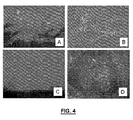
- FIGURE 4A shows the uptake of BPD-MA in the vertebrae at 15 minutes
- FIGURE 4B shows the uptake of BPD-MA in the vertebrae at 3 hours
- FIGURE 4C shows the uptake of BPD-MA in the spinal cord at 15 mintues
- FIGURE 4D shows the uptake of BPD-MA in the spinal cord at 3 hours.
- Sagittal and coronal sections from the Sprague-Dawley rat vertebrae examined under fluorescent microscopy indicated that there was delayed uptake into the spinal cord with no drug being present at 1 hour, yet, at 1 hour the bone marrow contained signal.
- the spinal cord and bone marrow contained an intense signal with neuronal cell bodies being labeled within the cord.
- the fluorescence of the drug within the spinal cord and vertebrae returned to baseline levels.
- the chest of each animal was then prepared with alcohol and 2 x10 6 cells of MT-1 were injected into the left ventricle using a 1 ml syringe with a 26g needle. Pulsatile blood in the syringe was ensured prior to each injection.
- the animals were placed back into their cages and fed water and rat chow ad libitum and kept on a constant light dark cycle.
- the animals were then imaged by fine detail radiography at 14 and 21 days post injection.
- the animals were examined for overt tumors, paralysis and cachexia following injection.
- the animals were sacrificed using CO 2 inhalation for compassionate reasons between day 23 and day 30 depending on individual tumor burden. Vertebrae and long bones were then harvested and fixed in 10% formalin for 7 days. Micro-CT images were obtained and then the samples were decalcified in 10% formic acid for 7 days.
- the tissue was then blocked, paraffin embedded and analyzed under light microscopy using H&E staining.
- the MT-1 cells were grown and maintained in RPMI media containing penicillin and streptomycin with 10% fetal bovine serum at 37 degrees celcius.
- the MT-1 cells were then transfected with pCl-neo mammalian expression vector (Promega) using the Transfectam Reagent (Promega) transfection kit. Positive colonies were selected by adding 1000ug/ml G418 antibiotic (Promega) to the tissue culture media. Cell colonies with luciferase activity were identified using the Xenogen IVIS system (Alameda, California). Individual cells were then isolated from high photon emitting colonies and plated. The cells were then grown to subconfluence in RPMI media with antibiotics and 10% FBS in 1000 ug/ml of G418 antibiotic to ensure stable transfection.
- Results Of the initial 10 animals injected with MT-1 cells 7 of the 10 animals developed metastatic disease. The mean survival of the animals with tumors was 25 days. Four of the animals showed palpable tumors in the femur and tibias as well as the lower mandible. Two animals developed hind leg paralysis secondary to metastatic disease. All animals with tumors became cachexic. The affected animals appeared well until day 18 after which the animals developed rapid weight loss and overt tumors.
- High resolution radiography indicated lesions within the humerus, femur and tibia as early as day 14 in some animals.
- FIGURE 6 shows a lateral view of rat vertebrae and femur at 21 days post left intracardiac tumor injection.
- lesions could not be detected in the vertebrae of any animals by day 21 by high resolution radiography (Faxitron).
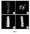
- FIGURES 7A-D show micro-CT analysis of rat vertebrae 21 days following intracardiac injection of MT-1 cells.
- Figures 7A-D show sagittal, transverse, coronal 3D reconstruction and sagittal 3D reconstruction, respectively.
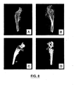
- Micro-CT analysis of the thoracic and lumbar spines of these animals showed multiple lytic lesions within the vertebrae. Similar lytic lesions were identified in the humerus, tibia and femur.
- FIGURES 8A-D show micro-CT analysis of rat tibia 21 days following intracardiac injection of MT-1 cells.
- Figures 8A-D show sagittal, transverse, coronal 3D reconstruction and sagittal 3D reconstruction, respectively.
- the mean area of the lytic lesions within the lumbar vertebrae was 2.92 mm 2 and 2.14 mm 2 in the thoracic vertebrae.
- the lesions approximated 1/3 of the vertebral body size in both the lumbar and thoracic vertebrae imaged, as summarized in TABLE 2.
- FIGURES 9A-C show histochemical and immunohistochemical staining of MT-1 cells within vertebrae.
- Figures 9A-C show H&E staining (10x); Keratin staining (brown) indicating tumor invasion of the bone and bone marrow (5x); and Human EGF-r immunohistochemistry (brown) of MT-1 cells within a vertebrae (20x), respectively.
- T indicates tumor
- BM indicates bone marrow.
- Four of the animals had the highest signals within the chest cavity and gross dissection revealed large metastatic tumors within the lung.
- Six animals showed a diffuse weak bioluminescent signal localized to the thoracic and lumbar spine. Histological confirmation of tumors within the spine was verified in only two of these animals with one animal having metastases identified in the spinal cord. One animal died a few hours following intracardiac tumor injection.
- An 18g needle was placed on the cortex of the targeted vertebrae or long bone with the use of a mini C-arm image intensifier.
- a 300mW diode laser coupled to a 200um fiber was used to deliver 690 nm light.
- BPD-MA was administered intravenously at a dose of 2mg/kg prior to the administration of the light dose.
- Drug light intervals included 1 hour, 3 hours and 24 hours.
- Light doses ranged from 25J to 150J.
- Light was delivered at 150mW for all treatments and the effects of different drug light intervals and different light doses using a fixed drug concentration were evaluated.
- the animals were examined for paralysis post treatment. The animals were sacrificed using CO 2 inhalation. Vertebrae were then harvested and fixed in 10% formalin for 7 days.
- Micro-CT images were obtained and then the samples were decalcified in 10% formic acid for 7 days. The tissue was then blocked and paraffin embedded and 10um sections were cut. The sections were analyzed under light microscopy using H&E staining as well as TUNEL and human EGF-r immunohistochemistry. The area of effect as denoted by necrosis and apoptosis was identified and measured using a Nikon slide scanner and Image Pro software.
- FIGURES 1A-C show stereotactic targeting of bioluminescent metastatic lesions using a mini-C-arm image intensifier.
- Figure 1A is a bioluminescent overlay image of a nude rat with metastatic lesions on a radio lucent stereotactic jig.
- FIGURE 1B is a fluoroscan image of markers placed along the grid of the stereotactic jig in correlation to the bioluminescent metastases and resulting in localization of the lesion.
- FIGURE 1C shows placement of the fiber optic cable sheath (needle cannula) adjacent to the targeted lesion.
- the lesions were then targeted and treated with 25J or 150J at a 3 hour drug light interval.
- the animals were then re-imaged with the Xenogen IVIS system 48 hours post treatment in the stereotactic frame.
- Signal intensity photons/second/cm 2
- the animals were then sacrificed using CO 2 inhalation and the spines and long bones were harvested. Samples were placed in 10% formalin for 7 days and micro-CT images of the vertebrae and long bones were obtained.
- the vertebrae were examined histologically using H&E staining and immunohistologically with human EGF-r and TUNEL stains as described earlier. ANOVA Bonferroni and two-tailed paired T-test statistical techniques were applied.
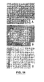
- FIGURES 14A-C show brightfield microscopy of H&E sections containing tumor following PDT treatment.
- Figure 14A shows untreated tumor tissue (10x); and FIGURES 14B-C show treated tumor tissue within a vertebral body at 0x and 20x magnification, respectively.
- FIGURES 15A-F show brightfield microscopy of rat vertebrae following PDT treatment.
- FIGURE 15A shows an H&E section of section of rat spine (the arrows delineate the diameter of the rostral caudal dimension of the area of effect and demonstrate the position of the high powered images in the right column relative to each other.
- FIGURE 15B shows human EGF-r immunohistochemistry of a contiguous section (2.5x).
- FIGURE 15C shows TUNEL staining of a contiguous section (2.5x).
- FIGURE 15D shows H&E of the same section as in FIGURE 15A , but 10x indicating the boundary of affected and unaffected tumor tissue.
- FIGURE 15E shows EGF-r immunohistochemistry at the same site as in D) but on a contiguous section (10x).
- FIGURE 15F shows TUNEL staining at the same site as in FIGURES 15D and E but on a contiguous section (10x). Histological analysis with H&E of the tissue indicated ablation of the tumor tissue. TUNEL staining of the treated tissue was positive at the periphery of the treatment area while necrosis was predominant centrally.
- FIGURES 10A-F and 12A-D show pre and post-PDT treatment bioluminescent imaging of MT-1luc+ metastatic lesions in rnu/rnu rats with BPD-MA.
- FIGURES 10A,C,E show pre-treatment bioluminescent imaging.
- FIGURES 10B,D,F show bioluminescent imaging of the animals imaged in the FIGURES 10A,C,E , respectively, 48 hours following PDT treatment with BPD-MA at 150 J with a drug light interval of 3 hours.
- FIGURES 12C,D show pre-treatment bioluminescent imaging.
- FIGURES 12A,B show bioluminescent imaging of the animals imaged in FIGURES 12C,D 48 hours following PDT treatment with BPD-MA at 25 J with a drug light interval of 3 hours.
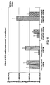
- targeted lesions treated with 150J of light with a 3 hour drug light interval reduced the signal from the targeted site by 87% and decreased tumor growth by 99.8% as compared to control lesions emitted 48 hours following treatment.
- Table 5A shows bioluminescent imaging of the animals imaged in FIGURES 12C,D 48 hours following PDT treatment with BPD-MA at 25 J with a drug light interval of 3 hours.
- the device 10 may be used to facilitate PDT.
- the device 10 is shown unassembled in FIGURE 16 .
- the device 10 comprises an insertion member 12, a locking member 14 and a gripping means 16.
- the insertion member 12 has a first shaft 18 having a first bore 20 through at least a portion thereof with a first diameter sized for receiving a light conduit (not shown).
- the insertion member 12 also includes a first head portion 22 near the proximal end of the first shaft 18.
- the first head portion 22 includes a second bore 24 extending therethrough.
- the second bore 24 has a second diameter that is larger than the first diameter of the first bore 22.
- the second bore 24 of the head 22 ends at a land.
- the first bore may have a 2.49 mm diameter.
- the insertion member 12 includes securing means in the form of external threads 26 on at least a portion of the first shaft 18.
- the external threads 26 cover the entire shaft 18 but this is not necessary.
- the external threads 26 may not be used; rather another means may be used to secure the insertion member 12 into bone such as longitudinal ribs and the like.
- the external threads 26 preferably have low torque and high holding. This is important since the insertion member 12 may need to be secured into bones that may be weak. Therefore, the external threads 26 of the shaft 18 may also have a fine thread.
- the external threads 26 may comprise a single thread and the thread(s) need not necessarily be continuous.
- the distal end of the first shaft 18 may have a frusto-conical tip 28 which facilitates inserting the insertion member 12 into hard substances such as bone.
- the frustoconical tip 28 is not mandatory.
- FIGURE 18 shows the first bore 20 extending throughout the entire length of the insertion member 12.
- the inventors have found that in some cases it may be preferable to have the light conduit extend a bit past the distal tip of the insertion member as shown in FIGURE 20 . This is so that the tip of the light conduit can extend into the treatment region for better treatment.
- the inventors have also found that it is preferable to have shafts that are shorter for the insertion member 12 since it may be difficult to insert the tip of the insertion member 12 into the treatment area. In this case, the light conduit may be extended into the treatment area as mentioned.
- the locking member 14 is releasably connectable to the insertion member 12.
- the locking member 14 includes a second shaft 30 having a third bore 32 therethrough with a third diameter.
- the third diameter is less than the second diameter but is sized for receiving the light conduit.
- the torque to remove the locking member 14 is less than the torque to remove the insertion member 12.
- the second bore 24 of the head portion 22 of the insertion member 12 includes internal threads (not shown) and the second shaft 30 of the locking member 14 includes corresponding external threads 34 on at least a portion of the second shaft 30 for releasably engaging the head portion 22 of the insertion member 12.
- the locking member 14 also includes a second head portion 36 with a fourth bore 38.
- the head portion 36 may be shaped to accommodate use with a drill for inserting the locking member 14 into the insertion member 12. Accordingly, the fourth bore 38 may be sized a bit larger than the third bore 32 in the shaft 30.
- the gripping means 16 is disposed within the second bore 24 of the insertion member 12 for holding the optical conduit in place when the locking member 14 is connected to the insertion member 12.
- the gripping means 16 may be a flexible gasket seal such as an O-ring.
- the gripping means 16 also includes a bore for allowing the optical conduit to pass therethrough. The gripping means 16 is able to freely pass inside the threads of the second bore 24 of the head 22 of the insertion member 12.
- FIGURE 20 shown therein an isometric view of the device 10 being assembled.
- An optical conduit 40 such as an optical fiber for example, is threaded through the locking member 14, the gripping means 16 and finally the insertion member 12.
- the tip of the optical conduit 40 may extend past the tip of the insertion member 12 as shown in FIGURE 20 .
- the insertion member 12 is already located in the treatment area which may be bone in this case.
- the locking member 14 is connected to the insertion member 12 with the gripping means 16 disposed therebetween.
- the locking member 14 As the locking member 14 is put into place, it pushes the gripping means to the land in the head 22 of the insertion member 12 which leads to the application of a compressive force to the gripping means 16. This causes the gripping means 16 to deform radially inward and apply a constrictive force on the optical conduit 40. Care is taken to ensure that the constrictive force is sufficient to hold the optical conduit 40 in place but not strong enough to damage the optical conduit 40.
- the device 10 may be used in the treatment of cancer in bone.
- the device 10 may also be used in all instances in which PDT treatment is used such as in the PDT treatment of osteomyelitis (SA bacterial) infection of bone. Accordingly, the device may be used in the PDT treatment of non-cancerous lesions in bone.
- SA bacterial osteomyelitis
- the gripping means 16 may include a portion of the locking member 14 and a portion of the insertion member 12.
- the head member 24' of the insertion member 12 may include a bore 24' with tapered ends 42 and the gripping member 14 may have a shaft 34' with several teeth or flaps 44.
- the flaps 44 flex inwards when the locking member 14 is connected or inserted into the insertion member 12. The flexing of the flaps 44 provides a constrictive force to hold the optical conduit 40 in place.
- the device 10 may be a cannulated bone screw assembly.
- the insertion member 12 may be a cannulated bone screw
- the locking member 14 may be a cannulated locking screw
- the gripping means 16 may be a flexible, elastometric O-ring.
- pigs 50 kg pigs were used.
- the pigs were preanesthetized with ketamine/xylazine 20/2mg/Kg IM for induction.
- An IV line was established and 0.9% saline solution was run at 5 ml/Kg/hr.
- Verteporfin was administered through the IV line using an infusion pump.
- the pigs were intubated and maintained on a ventilator with isofluorane / nitrous oxide inhalant by animal care professionals.
- the pigs were placed prone on a radiolucent surgical table with appropriate bolsters for support and padding.
- a sterile prep was made with betadine soap and scrub and prep solution.
- a small stab incision was made over the lumbothoracic spine of the pig.
- a customized guide pin was inserted through the incision onto the bone. Placement into the vertebral body through a transpedicular and parapedicular approach was facilitated by the use of a CT-Carm. The L1 and L2 vertebrae were targeted.
- a cannulated exchange sheath was placed over the guide rod and inserted through the incision.
- the guide rod was then removed, and a fiber optic cable sheath was then inserted down the cannula of the exchange sheath.
- a cannulated bone screw assembly which is one exemplary embodiment of a device for facilitating light-based therapy, and in particular photo-dynamic therapy, in accordance with the invention, was then placed around the cannulated exchange sheath and inserted through the incision, as shown in FIGURE 23 .
- the cannulated bone screw assembly was pushed down the cannulated exchange sheath.
- the cannulated bone screw was secured into bone. In this study, the cannulated bone screw was secured into the pedicle of the vertebral body.
- FIGURE 24 shows a vertebrae with a bone screw in place and a fiber optic probe inserted.
- Detector and diffusing fibers were placed in the same transverse plane.
- the diffusing fiber was moved incrementally with respect to the detector fiber and differences in power were measured at five different distances from the detector source using a computer aided photomultiplier tube.
- a 690 nm light was delivered into the vertebrae at L1 at light doses ranging from 25 J - 100 J of energy (four animals received one dose of either 25 J, 50 J, 75 J or 100 J and one control animal received no energy).
- the light dose took into account the optical properties of the 1 st and 2 nd lumbar vertebrae, such that the does refers to the dose at the site of the bone tumor.
- Hemostasis was maintained with cautery. Hemostasis was obtained prior to closure. Closure of the fascia was done with #1 vicryl followed by 2-0 vicryl for the subcutaneous tissue followed by 2-0 proline suture for skin. A sterile bandage was applied over the incision.
- the pigs were extubated and allowed activity as tolerated with diet as tolerated.
- the pigs were given post-op analgesia (buprenorphine 0.005 - 0.01 mg/Kg IM or carprofen 0.1 - 1 mg/Kg IM) as necessary.
- the pigs were observed for development of hindleg paralysis and sacrificed 24 hours post-op if no paralysis occurred. In the even that paralysis occurred, the animal was sacrificed as necessary.
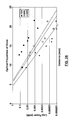
- a CT scan intraoperatively allowed the exact distance between the delivery fiber and the detector fiber to be determined. This enabled the determination of the attenuation of light within the vertebrae, as shown in FIGURES 27 and 28 .
- the treatment of the vertebrae with PDT allowed the area of effect of the treatment to be correlated with the light attenuation data, thus allowing a dose response curve for vertebral bone to be determined. It is anticipated that in humans, a light dosage in the range of 150 J/cm will be used.
- Pigs were used in this study because the bone density and structure of their vertebrae resembles that of humans. Therefore, pig vertebrae have similar optical properties to humans. In particular, the anatomical dimensions and structure of the porcine vertebrae (thoracic, lumbar inclusive) are similar to that of human. Accordingly, pig vertebrae allow one to test whether PDT could be effective in a large bone with healthy or diseased marrow. The data shows that PDT is capable of producing a volume effect that would be compatible with treating a vertebral bodies in humans.
- the cannulated bone screw and the fibre optic cable sheath may remain in the body for repeated treatments with photodynamic therapy. This ensures that therapy is provided to the same part of the treatment area across multiple treatments.
- the cannulated locking screw is unlocked and removed from the mammal.
- the cannulated exchange sheath is reinserted in the mammal around the fiber optic cable sheath.
- the fiber optic sheath is removed from the mammal.
- the cannulated bone screw is also removed from the mammal. Bone cement is injected through the cannulated exchange sheath, and the cannulated exchange sheath is slowly removed as the bone cement dries.
- FIGURE 29 shows some of the equipment used in the in vivo pig study.
- a fiber optic cable To the left of the fiber optic cable is a fiber optic cable sheath.
- a guide pin To the left of the guide pin is a cannulated exchange sheath.
- a cannulated bone screw assembly To the left of the cannulated exchange sheath is a cannulated bone screw assembly.
- various embodiments of the device developed herein facilitates the delivery of light energy into a treatment area, in this case bone, as part of a PDT treatment to allow single or repeated treatment regiments. If repeated treatment regiments are used, then advantageously, the various embodiments of the device enable for the repeated delivery of light energy with enough precision to ensure that the delivery of light energy to the treatment site is substantially reproducible with the device.
Landscapes
- Health & Medical Sciences (AREA)
- Biomedical Technology (AREA)
- Engineering & Computer Science (AREA)
- Life Sciences & Earth Sciences (AREA)
- Nuclear Medicine, Radiotherapy & Molecular Imaging (AREA)
- Pathology (AREA)
- Radiology & Medical Imaging (AREA)
- Animal Behavior & Ethology (AREA)
- General Health & Medical Sciences (AREA)
- Public Health (AREA)
- Veterinary Medicine (AREA)
- Biophysics (AREA)
- Radiation-Therapy Devices (AREA)
- Medicinal Preparation (AREA)
- Processing Of Meat And Fish (AREA)
Abstract
Description
- This application claims priority from Canadian Patent Application Serial No.
2,446,663 filed on October 24, 2003 . - This invention relates generally to treatment of bone, bone tumors and lesions, and diseases of the bone.
- Approximately 1.3 million cases of cancer were diagnosed in North America in 2001. Over 50% of these have the potential to metastasize to bone [14]. Each year, over one hundred thousand bone metastases are identified. Post-mortem autopsy results from patients with primary cancer indicate that 60% of spines examined had metastatic lesions. An estimated twenty to forty thousand cases of metastatic breast cancer lesions alone occur in the spine each year [1,2]. Metastatic lesions to the spine result in intractable back pain, loss of bowel and bladder function, paresis and paralysis. The lesions can affect singular or multiple vertebral bodies.
- In the ambulatory patient the mainstay of treatment is radiation therapy, while surgery is reserved for those experiencing collapse or neurological compromise. Unfortunately radiation therapy provides only limited relief from pain, does not provide stability to the spine and adversely affects the soft tissue such that the morbidity and mortality of surgical intervention is increased threefold [3,4,5]. Results of radiation therapy for the treatment of spinal metastases have shown that only one third have complete relief of their back pain [15]. Radiation therapy is limited by the number of times it can be administered as it affects the integrity of the soft tissues and can induce radiation myelopathy. As the longevity of patients with spinal metastases increases (average survival 2 years with breast cancer, mean 1 year survival of 78%) [16] so does the likelihood of lesion recurrence and the necessity for spinal surgery. Recurrence with radiation therapy is estimated to be 33% [1,17]. Spinal surgery for patients with spinal metastases carries a 30 - 40% risk of morbidity and a 7 - 16% risk of mortality.
- Photodynamic therapy (PDT) can directly target lesions. PDT ablates tissue with a non-thermal specific wavelength of light delivered to the targeted tissue. A photosensitizing compound is administered prior to the light. The light activates the compound to a chemically excited state, the energy of which is then transferred to molecular oxygen producing reactive oxygen-derived species that are toxic to the surrounding tissue [8,18] [19,20]. There are reports that the drug is preferentially taken up and retained in tumor tissue compared to normal tissue [21,22] making the treatment somewhat specific. This therapy has been used in lung [9,10], intraperitoneal [11] and prostate cancer [12].
- There are several photosensitizing compounds now available with minimal systemic side effect profiles. Benzoporphyrin derivative monoacid ring A (BPD-MA) is a photosensitizer that can be used to either target the neo-vasculature or produce intracellular cytotoxic effects based on the drug-light interval [6,7,23,24,25,26]. The vascular targets are primarily affected if the drug light interval is short (15 minutes or less) while the intracellular effects are seen in tissue if the drug light interval is long (3 hours) [6,24]. There are several reports of its use in soft tissue tumors in the murine model [20,23,25,27] as well as of its use in an orthotopic chondrosarcoma [6] and fibrosarcoma tumor models [24,26]. Results from these studies showed a significant effect at both the 15 minute and 3 hour drug light interval was achieved with 33% of the lesions being completely ablated at 4 weeks post treatment. The greatest effect was seen with the shorter interval demonstrating the potency of BPD-MA to affect the neo-vasculature.
- To date, there have not been any published reports of the use of this therapy in bone or in the spine. To date, there have not been any published reports of PDT use in an in vivo metastatic breast cancer model affecting bone. To date, there have not been any published reports of the pharmacokinetics of BPD-MA for bone. There is a paucity of literature on the optical properties of bone.
- Takeuchi et al., 1997 [28], reported on the optical properties of cancellous and cortical bone in comparison to muscle, fat and saline. Cortical bone has a high attenuation to light while cancellous bone does not.
- United States Patent
6,622,731 to Daniel et al. discloses a device that may be configured to employ photodynamic therapy to treat bone tumors. The device includes an optical energy delivery device, which is positioned in an introducer. The energy delivery device may be coupled to an actuator to allow for advancement of the energy delivery device out of the introducer. - European
Patent Application EP 0 812 574 A2 to Mueller et al. discloses a device for use in a method for stimulating revascularization of myocardium tissue in the ventricular wall of a human heart. The method includes the steps of moving the distal end of an optical fiber element forwardly through the epicardium of the heart and into the myocardium and emitting relatively low power burst of laser energy from the distal end to form stimulus injury zones that promote capillary growth and tissue revascularization. Each penetration of the optical fiber element may produce multiple stimulus zones spaced radially from or in line with the axis of the optical fiber element. The device facilitates movement of the optical fiber element and control of the laser emissions in a predetermined manner. - In a first aspect, at least one embodiment of the invention provides a device for enabling light-based therapy for a treatment area of a mammal as defined in
claim 1 of the appended claims. - The device includes a light conduit for delivering the light-based therapy. The device also includes an insertion member for insertion at or near the treatment area, the insertion member comprising a first bore with a first diameter sized for receiving the light conduit. The device also includes a locking member releasably connectable to the insertion member for holding the light conduit in place during treatment. The insertion member further includes securing means on at least a portion of the insertion member for securing the insertion member at or near the treatment area.
- For a better understanding of the present invention, and to show more clearly how it may be carried into effect, reference will now be made, by way of example, to the accompanying drawings which show some embodiments of the present invention, and in which:
-
FIGURE 1 shows stereotactic targeting of bioluminescent metastatic lesions in a rat using a mini-C-arm image intensifier; -
FIGURE 2 shows the in vitro uptake of BPD-MA in MT-1 cells; -
FIGURE 3 is a plot of the BPD-MA drug uptake in serum and spinal cord of the nude rat; -
FIGURE 4 shows fluorescence microscopy of BPD-MA uptake in the spinal cord and vertebrae of the nude rat; -
FIGURE 5 is a bar graph of the in vitro cell viability following BPD-MA PDT; -
FIGURE 6 is a high definition radiographic (Faxitron) lateral view of rat vertebrae and femur at 21 days post left intracardiac tumor injection; -
FIGURE 7 shows micro-CT analysis of rat vertebrae 21 days following intracardiac injection of MT-1 cells; -
FIGURE 8 shows micro-CT analysis of rat tibia 21 days following intracardiac injection of MT-1 cells; -
FIGURE 9 shows histological and immunohistochemical staining of MT-1 cells within vertebrae; -
FIGURES 10 and12 show pre and post-PDT treatment bioluminescent imaging of MT-1luc+ metastatic lesions in rnu/rnu rats with BPD-MA; -
FIGURE 11 is a bar graph of the effect of PDT on bioluminescent tumor in rats; -
FIGURE 13 is a photograph of light passing through a human cadaver vertebrae; -
FIGURE 14 shows brightfield microscopy of H&E sections containing tumor following PDT treatment; -
FIGURE 15 shows brightfield microscopy of rat vertebrae following PDT treatment; -
FIGURE 16 shows a side view of a disassembled device for use with providing light-based therapy in accordance with the invention; -
FIGURE 17 shows a side cross-section view of the components of the disassembled device ofFIGURE 16 ; -
FIGURE 18 shows a cross-sectional view of an insertion member of the device; -
FIGURE 19 shows a cross-sectional view of a locking member of the device; -
FIGURE 20 shows an isometric view of the components of the device being assembled with an optical conduit; -
FIGURE 21 shows an alternative embodiment of the insertion and locking members of the device; -
FIGURE 22 shows an inserted guide pin and the insertion of another guide pin with a cannulated drill; -
FIGURE 23 shows an inserted cannulated exchange sheath and the insertion of a cannulated bone screw assembly; -
FIGURE 24 shows inserted fibre optic cable sheaths and inserted fiber optic cables in a transpedicular placement; -
FIGURES 25 and26 show a vertebrae with a bone screw in place and a fiber optic probe inserted; -
FIGURES 27 and28 are graphs showing some of the results of the light attenuation studies in bone; and, -
FIGURE 29 shows some of the equipment used in the in vivo pig study. - It will be appreciated that for simplicity and clarity of illustration, elements shown in the figures have not necessarily been drawn to scale. For example, the dimensions of some of the elements may be exaggerated relative to other elements for clarity. Further, where considered appropriate, reference numerals may be repeated among the figures to indicate corresponding or analogous elements. In addition, numerous specific details are set forth in order to provide a thorough understanding of the invention. However, it will be understood by those of ordinary skill in the art that the invention may be practiced without these specific details. In other instances, well-known methods, procedures and components have not been described in detail so as not to obscure the invention.
- In some embodiments of the invention, photodynamic therapy (PDT) is used in bone. The method involves administering a photosensitizing drug to a mammal having bone tumors or other bone disease. A "bone tumor" refers to a primary or metastatic tumor associated with bone, that is, a tumor on or in a bone. A non-exhaustive list of photosensitizing drugs includes benzoporphyrin derivative monoacid ring A (BPD-MA) (also known as verteporfin and Visudyne®), palladium-bacteriopheophorbide (known as TOOKAD®), and 5-aminolaevulinic acid (ALA). Non-thermal light of a specific wavelength suitable for stimulating the photosensitizing drug is applied to the drug within the bone. The photosensitizing drug may target the neo-vasculature and/or produce intracellular cytotoxic effects and/or inhibit growth of or destroy the tumor cells in some other manner.
- For small bones a fiber optic cable is inserted into the mammal and placed adjacent a bone lesion. For larger bones such as, for example, pig and human vertebrae, a cannulated bone screw is secured into the bone. The cannulated bone screw has a head, an externally-threaded shaft and a frustro-conical tip. A fiber optic cable sheath is located inside the cannula of the screw and extends from the tip of the bone screw beyond the screw to at least the skin of the mammal. Photodynamic therapy can be used on one or more occasion by administering a photosensitizing drug, inserting a fiber optic cable into the fiber optic cable sheath, and delivering the light through the fiber optic cable. The bone screw and the fiber optic cable sheath remain in place from one PDT treatment to the next. Once it has been decided not to use PDT treatment at the site of the bone screw anymore, the fiber optic cable sheath is removed and bone cement is injected into the bone screw.
- The results of in vitro studies indicated that cells from a human metastatic breast cancer cell line (MT-1) were susceptible to PDT with BPD-MA while the cells were not sensitive to the drug or the light individually. This is consistent with reports in the literature [23,29]. Furthermore, uptake into MT-1 cells was demonstrated directly with real time fluorescent microscopy indicating that these cells were susceptible to intracellular cytotoxic effects of PDT.
- In vivo studies in a rat model demonstrated the effectiveness of PDT on bone tumors from a human metastatic breast cancer cell line. A bioluminescent metastatic model in the nude rat was developed to facilitate the localization and targeting of the lesions.
- As part of these studies, the drug uptake studies indicated that the spinal cord had minimal uptake of BPD-MA suggesting that it would not be susceptible to damage during treatment. The fluorescence microscopy however, indicated that BPD-MA is taken up into the spinal cord. Furthermore it showed that the drug has a delayed uptake (greater than 1 hour) and suggested that the optimal drug light interval prior to PDT would be less than 1 hour or greater than 24 hours.
- The model used in this study was a purely metastatic model involving a human breast cancer cell line. Engebraaten and Fodstad, 1999, demonstrated lesions within all vertebrae and of rnu/rnu rats between 14 and 21 days [13]. In this study metastases occurred in the spine, long bones and lung. Fine detail radiography revealed lesions within long bones as early as 14 days post tumor injection. Rapid weight loss was seen after 18 days post injection followed by overt tumors in the mandible and distal femur and proximal tibia. Paralysis and death secondary to tumor burden occurred typically around day 21 and 23 respectively. Micro-CT scanning revealed multiple large lytic lesions within most vertebrae and long bones of affected animals. Subsequent histological analysis confirmed the presence of the tumor within the vertebrae and long bones and demonstrated the invasiveness of the tumor. The bioluminescence allowed determination of the growth of the lesions, location of the tumor and a way of targeting the tumor. In addition, bioluminescence has been shown to be quantitative in determining tumor ablation in vitro as well as in vivo [30,31]. Furthermore by imaging the tumor with bioluminescence, growth of the tumor could be assessed, lesions could be detected early, then targeted and early treatment could be administered.
- The in vivo results demonstrate the effectiveness of this therapy in treating a human metastatic breast cancer in both the vertebrae and long bones. The results indicate that PDT can ablate tumor tissue within bone and that the in vivo structure of bone and bone marrow is not a limiting factor for this therapy. The bioluminescent data indicated that a 99.8% reduction in tumor growth was obtained with one treatment. The largest area of effect had a diameter of 2.2 cm and was created by a 200um fiber optical cable with a treatment time of 16 minutes. The results also indicate that the area of tumor ablated is directly proportional to the amount of light supplied to the targeted area. A shorter treatment time (3 minutes) produced a smaller effect than longer treatment times with a 66% reduction in the tumor growth with the 25J group. Based on work by Richter et al., [22] in the mouse, the inventor hypothesized that 24 hour drug-light interval would be selective for tumor ablation. Yet, this was not the case. There was no difference between control animals and those animals treated with a 24 hour drug light interval with respect to ablation of tumor tissue.
- The spinal cord in the rat is very sensitive to PDT as the drug is taken up into the cord by 3 hours. We anticipated that a window of opportunity would exist based on the delay of transport of the drug across the drug-spinal cord barrier, yet, even at short drug-light intervals when drug was not seen in the spinal cord by fluorescence microscopy paralysis was seen. This indicates that in the rat model the effect was vascular in nature while the spinal cord is also susceptible to cytotoxic damage as drug light intervals of 3 hours had an effect. Note that treatment with light only or drug only did not affect the tumor, bone, bone marrow or spinal cord. The nerve roots and peripheral nerves were not affected following treatment at the 3 hour drug light interval at any light dose. No paralysis was seen when treatment was administered with a 24 hour drug light interval.
- In summary the results suggest that PDT with BPD-MA could be an effective treatment directed against metastatic tumors in bone. This treatment would be used preferentially to treat tumors within the vertebrae through a trans or para-pedicular approach and implemented to treat multiple vertebrae. The size of the lesions produced in the rat spine defined an area of effect that is well suited for lesions within human vertebrae. The area of effect can be varied easily allowing for safe operating parameters around the spinal cord. However, in larger vertebrae in which the fiber optic cable is implanted within the bone the inventor anticipates the effect on the spinal cord will be negligible. Studies in larger animals are required to establish the safety of this treatment in the spine.
- Studies of light attenuation in human cadaver vertebrae demonstrated the feasibility of PDT in large bones.
FIGURE 13 is a photograph from these studies. - In vivo studies in a pig model demonstrated the feasibility of a procedure using a cannulated bone screw to stabilize a large bone and to facilitate delivery of light to the bone. The in vivo studies in the pig model also measured the attenuation of light within a vertebral body, and from the derived data, safe doses of light and drug were determined.
- These studies, as well as a detailed description of the surgical procedure using the cannulated bone screw, will now be described in more detail.
- Method: MT-1 cells, a human breast cancer cell line, provided courtesy of Dr. O. Engebraaten, Norwegian Radium Hospital, Oslo, Norway, were grown and maintained in RPMI media containing penicillin and streptomycin with 10% fetal bovine serum at 37 degrees Celsius. Once the cells had reached subconfluence they were resuspended in free RPMI. The cells were harvested using a 0.05% trypsin -0.05mM EDTA solution. Cells were then counted with a hemocytometer and plated at 2x10⁵ cells/ml in an inverted microscopy slide chamber in PBS. BPD-MA was then added to the suspension at a concentration of 1 ug/ml. The cells were then visualized under bright field and fluorescence microscopy (Zeiss Axiophot) using an excitation/emission filter of 490 nm/ emission respectively. Uptake of drug into the cells was monitored overtime using live video photogrphay with a CCD camera attached to the microscope.
- Results:
FIGURE 2 shows the in vitro uptake of BPD-MA in MT-1 cells. A) is a bright field microscopy image of MT-1 cell (63x) and C) is an overlay of the bright field and fluorescent image showing colocalization of BPD-MA within MT-1 cells (63x). The MT-1 cells began fluorescing at 45 minutes following incubation with BPD. - Method: MT-1 cells were grown and maintained in RPMI media containing penicillin and streptomycin with 10% fetal bovine serum at 37 degrees Celsius. Once the cells had reached subconfluence they were resuspended in free RPMI. The cells were harvested using 0.05% trypsin -0.05 mM EDTA solution. Cells were then counted with a hemocytometer and plated at 2x10⁵ cells/ml in a 96 well plate. BPD-MA was then added to the individual wells at a concentration of 1 ug/ml or 10 ug/ml. A 690 nm light was administered at 150 mW to the individual wells at a fluence of either 100 J/cm2 or 25 J/cm2 following an 8 hour incubation period. Control wells included those that did not contain cells, those that contained cells but no BPD-MA, those that contained cells and drug but did not receive a light does and those that contained cells and received a light dose but were not incubated with BPD-MA. Following treatment the cells were allowed to survive for 24 hours following which an SRB assay was performed to establish the number of viable cells remaining. Briefly, cells were fixed in 10% trichloroacetic acid and stained with a sulpharodamine bromide solution selectively staining viable cells. A spectrophotometer was then used to assess the absorbance of 540 nm light within individual wells of the 96 well plate which was correlated to the number of remaining cells following treatment.
- Results: The effect of PDT on in vitro MT-1 cells was demonstrated using an SRB assay. A significant difference was seen between the absorbance in the untreated wells versus the wells treated with light and drug. No significant difference was seen between untreated wells and wells treated with light only or drug only (Table 1). As shown in
FIGURE 5 , there was no difference between the two drug concentrations (10 ug/ml and 1 ug/ml BPD-MA) with respect to the treatment group and there was no significant difference in absorbance between BPD-MA wells treated with either 100 J/cm2 or 25 J/cm2.Table 1a. SRB Assay Group Drug Dose Light Dose Standard Mean (absorbance) Standard Deviation 95 % Confidence Interval 1 1 ug/ml x 200J/cm2 .2923 .0794 .2419 2 1 ug/ ml x 25J/cm2 .1946 .0618 .1553 3 10ug/ml x 200J/cm2 .2558 .0865 .2009 4 10ug/ml x 25 J/cm2 .2049 .0523 .1717 5 No cells No cells .0206 .0269 .0035 6 / 200J/cm2 1.4954 .2884 1.3122 7 1 ug/ml / 1.2392 .3417 .8806 8 Cells Only Cells Only 1.5992 .2800 1.4212 / indicates absence of variable Table 1b. SRB Assay ANOVA with Bonferroni for multiple comparisons (* indicate significant difference between groups) Group Mean 5 2 4 3 1 7 6 8 5 .0206 2 .1946 4 .2049 3 .2558 * 1 .2923 * 7 1.2392 * * * * * 6 1.4954 * * * * * 8 1.5992 * * * * * * - Method: BPD-MA was administered to 10 rats (Sprague-Dawley, 150gm) through a tail vein injection. Animals were then sacrificed using C02 inhalation overdose at 16 minutes, 3 hrs, 6 hrs and 24 hrs following injection. Control animals without BPD-MA were euthanized in a similar fashion. Serum samples and spinal cord tissue samples were harvested from the animals at the time of euthanasia. A segment of spine was also fixed in 10% formalin for 7 days followed by decalcification for 7 days in 10% formic acid for fluoroscopic microscopic analysis. A control assay determined that BPD-MA fluorescence was not affected by formic acid. BPD-MA concentration within the serum and spinal cord tissue was then determined using fluorimetry. Briefly, the samples were solubilized and the samples were tested with excitation and emission spectra specific to BPD-MA. The fluorescence of the drug within the tissue was correlated to the specific uptake of the drug within the tissue. Fluorescent microscopy was also used to visualize the presence or absence of BPD-MA within the vertebrae and spinal cord at 1 hour, 3 hours and 24 hours.
- Results: Fluorimetry was used to determine the specific uptake of the BPD in the spinal cord at 15 minutes, 3 and 24 hours post injection. As shown in
FIGURE 3 , the specific uptake studies indicated that there was rapid increase in the serum drug concentration over 15 minutes but began to decline after 3 hours and returned to baseline by 24 hours post injection. BPD-MA fluorescence was evident within the spinal cord at 3 hours post administration.FIGURES 4A-D show fluorescence microscopy of BPD-MA uptake in the spinal cord and vertebrae.FIGURE 4A shows the uptake of BPD-MA in the vertebrae at 15 minutes,FIGURE 4B shows the uptake of BPD-MA in the vertebrae at 3 hours,FIGURE 4C shows the uptake of BPD-MA in the spinal cord at 15 mintues, andFIGURE 4D shows the uptake of BPD-MA in the spinal cord at 3 hours. Sagittal and coronal sections from the Sprague-Dawley rat vertebrae examined under fluorescent microscopy indicated that there was delayed uptake into the spinal cord with no drug being present at 1 hour, yet, at 1 hour the bone marrow contained signal. At 3 hours the spinal cord and bone marrow contained an intense signal with neuronal cell bodies being labeled within the cord. At 24 hours the fluorescence of the drug within the spinal cord and vertebrae returned to baseline levels. - Method: Ten nude rnu/rnu (Harlan) female rats (4-6 weeks of age) were used in this part of the study. The animals were injected with MT-1 cells, a human breast cancer cell line, provided courtesy of Dr. O. Engebraaten, Norwegian Radium Hospital (Oslo, Norway) who had previously shown [13] that injection of these cells into the left ventricle in 4 week old nude rats produced spinal and boney metastases in all animals injected. The cells were grown and maintained in RPMI media containing penicillin and streptomycin with 10% fetal bovine serum at 37 degrees Celsius. The protocol was in accordance with standards of the Canadian Council on Animal Care.
- The chest of each animal was then prepared with alcohol and 2 x10⁶ cells of MT-1 were injected into the left ventricle using a 1 ml syringe with a 26g needle. Pulsatile blood in the syringe was ensured prior to each injection. The animals were placed back into their cages and fed water and rat chow ad libitum and kept on a constant light dark cycle. The animals were then imaged by fine detail radiography at 14 and 21 days post injection. The animals were examined for overt tumors, paralysis and cachexia following injection. The animals were sacrificed using CO2 inhalation for compassionate reasons between day 23 and
day 30 depending on individual tumor burden. Vertebrae and long bones were then harvested and fixed in 10% formalin for 7 days. Micro-CT images were obtained and then the samples were decalcified in 10% formic acid for 7 days. The tissue was then blocked, paraffin embedded and analyzed under light microscopy using H&E staining. - Method: In brief, the MT-1 cells were grown and maintained in RPMI media containing penicillin and streptomycin with 10% fetal bovine serum at 37 degrees celcius. The MT-1 cells were then transfected with pCl-neo mammalian expression vector (Promega) using the Transfectam Reagent (Promega) transfection kit. Positive colonies were selected by adding 1000ug/ml G418 antibiotic (Promega) to the tissue culture media. Cell colonies with luciferase activity were identified using the Xenogen IVIS system (Alameda, California). Individual cells were then isolated from high photon emitting colonies and plated. The cells were then grown to subconfluence in RPMI media with antibiotics and 10% FBS in 1000 ug/ml of G418 antibiotic to ensure stable transfection.
- Results: Of the initial 10 animals injected with MT-1 cells 7 of the 10 animals developed metastatic disease. The mean survival of the animals with tumors was 25 days. Four of the animals showed palpable tumors in the femur and tibias as well as the lower mandible. Two animals developed hind leg paralysis secondary to metastatic disease. All animals with tumors became cachexic. The affected animals appeared well until
day 18 after which the animals developed rapid weight loss and overt tumors. - High resolution radiography (Faxitron) indicated lesions within the humerus, femur and tibia as early as
day 14 in some animals. For example,FIGURE 6 shows a lateral view of rat vertebrae and femur at 21 days post left intracardiac tumor injection. However, lesions could not be detected in the vertebrae of any animals by day 21 by high resolution radiography (Faxitron). -
FIGURES 7A-D show micro-CT analysis of rat vertebrae 21 days following intracardiac injection of MT-1 cells. In particular,Figures 7A-D show sagittal, transverse, coronal 3D reconstruction and sagittal 3D reconstruction, respectively. Micro-CT analysis of the thoracic and lumbar spines of these animals showed multiple lytic lesions within the vertebrae. Similar lytic lesions were identified in the humerus, tibia and femur. -
FIGURES 8A-D show micro-CT analysis of rat tibia 21 days following intracardiac injection of MT-1 cells. In particular,Figures 8A-D show sagittal, transverse, coronal 3D reconstruction and sagittal 3D reconstruction, respectively. - The mean area of the lytic lesions within the lumbar vertebrae was 2.92 mm2 and 2.14 mm2 in the thoracic vertebrae. The lesions approximated 1/3 of the vertebral body size in both the lumbar and thoracic vertebrae imaged, as summarized in TABLE 2.
Table 2. Vertebral Body Size and Tumor Size (microCT) Vertebral Body Osteolytic Lesion (Tumor) Sagittal (mm2) Coronal (mm2) Sagittal (mm2) Coronal (mm2) Lumbar (n =3) 9.4 7.09 2.92 2.3 Thoracic (n = 3) 6.64 6.89 2.14 1.69 - Histological analysis of the vertebrae confirmed the presence of osteolytic tumor within the long bones and vertebrae of the affected animals. Of the twenty animals inoculated with MT-1luc+ cells similar results were found. All animals showed localization of bioluminescent signal to the spine or long bones by day 21. However, the bioluminescent signal intensity was quite variable. Nine of the twenty animals had either gross visible tumors or cachexia. Bioluminescent imaging of these animals showed a similar pattern of metastases among these animals. A high signal was obtained from the lumbar and thoracic spine, the humerus, femur and tibia in addition to the lung. Micro-CT scans of these animals indicated gross lytic lesions within the vertebral bodies and long bones. Subsequent histological staining with H&E, keratin and immunohistochemical staining for human EGF-r confirmed the presence of human breast cancer cells within the aforementioned sites.
-
FIGURES 9A-C show histochemical and immunohistochemical staining of MT-1 cells within vertebrae. In particular,Figures 9A-C show H&E staining (10x); Keratin staining (brown) indicating tumor invasion of the bone and bone marrow (5x); and Human EGF-r immunohistochemistry (brown) of MT-1 cells within a vertebrae (20x), respectively. "T" indicates tumor, while "BM" indicates bone marrow. Four of the animals had the highest signals within the chest cavity and gross dissection revealed large metastatic tumors within the lung. Six animals showed a diffuse weak bioluminescent signal localized to the thoracic and lumbar spine. Histological confirmation of tumors within the spine was verified in only two of these animals with one animal having metastases identified in the spinal cord. One animal died a few hours following intracardiac tumor injection. - Method: Thirty rnu/rnu nude rats were used in this part of the study. The rats received a left ventricular intracardiac injection with MT-1 cells as described earlier. At day 21 post injection each animal was anesthetized with 2% halothane / air mixture and placed on a custom made radiolucent stereotactic jig in the left lateral decubitus position. Prior to this it was established by histological methods that animals with tumors contained tumor in most vertebral bodies and long bones. Because lesions within the vertebral bodies could not be detected by fine detail radiography, T12 and L4 vertebrae were selected as representative levels for treatment. An 18g needle was placed on the cortex of the targeted vertebrae or long bone with the use of a mini C-arm image intensifier. A 300mW diode laser coupled to a 200um fiber was used to deliver 690 nm light. BPD-MA was administered intravenously at a dose of 2mg/kg prior to the administration of the light dose. Drug light intervals included 1 hour, 3 hours and 24 hours. Light doses ranged from 25J to 150J. Light was delivered at 150mW for all treatments and the effects of different drug light intervals and different light doses using a fixed drug concentration were evaluated. The animals were examined for paralysis post treatment. The animals were sacrificed using CO2 inhalation. Vertebrae were then harvested and fixed in 10% formalin for 7 days. Micro-CT images were obtained and then the samples were decalcified in 10% formic acid for 7 days. The tissue was then blocked and paraffin embedded and 10um sections were cut. The sections were analyzed under light microscopy using H&E staining as well as TUNEL and human EGF-r immunohistochemistry. The area of effect as denoted by necrosis and apoptosis was identified and measured using a Nikon slide scanner and Image Pro software.
- Method: Twenty nude rnu/rnu (Harlan) female rats (4-6 weeks of age) were used in this part of the study. The protocol was in accordance with the Canadian Council on Animal Care prior to initiation. Prior to injection each animal was anesthetized with 2% halothane / air mixture in a sterile environment. Twenty animals were used for injection. The chest was prepared with alcohol and 2 x10⁶ MT-1luc+ cells, a human breast cancer cell line carrying the luciferase reporter gene, were injected into the left ventricle using a 1 ml syringe with a 26g needle. Pulsatile blood in the syringe was ensured prior to each injection. The animals were placed back into their cages and fed water and rat chow ad libitum. At
day 18 the animals were imaged using the Xenogen IVIS system. To do this 100mg/kg luciferin was injected into the peritoneal cavity of each animal and each animal was imaged 5 minutes following injection for a 1 to 5 minute image acquisition period. The animals were then imaged on the same custom made stereotactic radiolucent jig using a mini-C-arm image intensifier which allowed correlation of the bioluminescent signal to the vertebrae.FIGURES 1A-C show stereotactic targeting of bioluminescent metastatic lesions using a mini-C-arm image intensifier.Figure 1A is a bioluminescent overlay image of a nude rat with metastatic lesions on a radio lucent stereotactic jig.FIGURE 1B is a fluoroscan image of markers placed along the grid of the stereotactic jig in correlation to the bioluminescent metastases and resulting in localization of the lesion.FIGURE 1C shows placement of the fiber optic cable sheath (needle cannula) adjacent to the targeted lesion. - The lesions were then targeted and treated with 25J or 150J at a 3 hour drug light interval. The animals were then re-imaged with the Xenogen IVIS system 48 hours post treatment in the stereotactic frame. Signal intensity (photons/second/cm2) at the targeted site before treatment was compared to signal intensity following treatment and compared to lesions not treated using Igor Pro software (Alameda, California). The animals were then sacrificed using CO2 inhalation and the spines and long bones were harvested. Samples were placed in 10% formalin for 7 days and micro-CT images of the vertebrae and long bones were obtained. The vertebrae were examined histologically using H&E staining and immunohistologically with human EGF-r and TUNEL stains as described earlier. ANOVA Bonferroni and two-tailed paired T-test statistical techniques were applied.
- Results: As summarized in TABLE 3, of the thirty animals injected, twenty-five were analyzed. As summarized in TABLE 4, light doses ranging from 25J to 150J had an ablative effect on both normal bone marrow and tumor tissue. The region of effect ranged from 2.5mm to 22mm in the rostral-caudal dimension.
Table 3. Treatment Groups n = Death Following Tumor Injection Death Following Verteporfin Injection Anesthetic Overdose During Imaging / PDT Death During PDT MT-1 20 1 1 1 0 MT- 1luc+ 30 1 2 1 1 Table 4. Effect of Different Light Doses on Vertbrae with Metastases 25J* 50J* 75J* 75J** 100J* 150J* Area (mm2) 8.435 15.04 52.38 82.47 45.49 80.53 Rostral-Caudal (mm) 4.59 5.66 11.67 17.47 13.8 13.05 Antero-Posterior (mm) 2.62 3.89 5.09 5.05 5.05 5.78 *Treatment given at drug-light interval of 3 hrs
** Treatment given at drug-light interval of 0.75 hrs - The effect varied in direct proportion to the amount of light given with the greatest effect being seen with 150J. However, a 75J light dose administered at a 1 hour drug light interval produced a similar effect (Table 5).
-
FIGURES 14A-C show brightfield microscopy of H&E sections containing tumor following PDT treatment. In particular,Figure 14A shows untreated tumor tissue (10x); andFIGURES 14B-C show treated tumor tissue within a vertebral body at 0x and 20x magnification, respectively.FIGURES 15A-F show brightfield microscopy of rat vertebrae following PDT treatment.FIGURE 15A shows an H&E section of section of rat spine (the arrows delineate the diameter of the rostral caudal dimension of the area of effect and demonstrate the position of the high powered images in the right column relative to each other.FIGURE 15B shows human EGF-r immunohistochemistry of a contiguous section (2.5x).FIGURE 15C shows TUNEL staining of a contiguous section (2.5x).FIGURE 15D shows H&E of the same section as inFIGURE 15A , but 10x indicating the boundary of affected and unaffected tumor tissue.FIGURE 15E shows EGF-r immunohistochemistry at the same site as in D) but on a contiguous section (10x).FIGURE 15F shows TUNEL staining at the same site as inFIGURES 15D and E but on a contiguous section (10x). Histological analysis with H&E of the tissue indicated ablation of the tumor tissue. TUNEL staining of the treated tissue was positive at the periphery of the treatment area while necrosis was predominant centrally. - The use of a custom made stereotactic radiolucent jig facilitated localization and targeted treatment of bioluminescent metastases.
FIGURES 10A-F and12A-D show pre and post-PDT treatment bioluminescent imaging of MT-1luc+ metastatic lesions in rnu/rnu rats with BPD-MA.FIGURES 10A,C,E show pre-treatment bioluminescent imaging.FIGURES 10B,D,F show bioluminescent imaging of the animals imaged in theFIGURES 10A,C,E , respectively, 48 hours following PDT treatment with BPD-MA at 150 J with a drug light interval of 3 hours.FIGURES 12C,D show pre-treatment bioluminescent imaging.FIGURES 12A,B show bioluminescent imaging of the animals imaged inFIGURES 12C,D 48 hours following PDT treatment with BPD-MA at 25 J with a drug light interval of 3 hours. As summarized inFIGURE 11 and TABLE 5a, targeted lesions treated with 150J of light with a 3 hour drug light interval reduced the signal from the targeted site by 87% and decreased tumor growth by 99.8% as compared to control lesions emitted 48 hours following treatment.Table 5A. Bioluminescent Data Mean Count Rx Area Mean Count UnRx Area P = Pre-PDT 1.02 x 10⁴ 1.98 x 10³ < 0.01 Post-PDT 1.42 x 10³ 1.65 x 10⁵ <0.01 PostRx/PreRx 0.139 83.3 *for drug light interval of 3 hrs 150J (n = 5) - As summarized in
FIGURE 11 and TABLE 5b, targeted lesions treated with 25J of light with a 3 hour drug light interval showed a decrease in tumor growth of 66% compared to that of the control lesions.Table 5B. Bioluminescent Data Mean Count Rx Area Mean Count UnRx Area P = Pre-PDT 1.86 x10⁴ 4.05 x 10³ <0.05 Post-PDT 2.33 x 10⁴ 1.5 x 10⁴ <0.05 PostRx/PreRx 1.25 3.7 * for drug-light interval of 3 hrs 25 J (n = 2) - No effect was seen when light was administered at a 24 hour drug light interval or in control animals when light alone or drug alone was administered. As summarized in TABLE 6, hind leg paralysis was seen in animals when treated at the 3 hour drug light interval at T12 at light doses between 50J and 150J but not at 25J. No paralysis was seen at the twenty four hour drug light time interval in animals treated with 150J at T12. No paralysis was seen in animals treated with 150J at the L5 level of the spine. No paralysis was seen in hind legs of rats following treatment with 150J directed at the distal femur.
Table 6. Effect of Light Dose on the Rat Spinal Cord, Spinal Nerves and Peripheral Nerves Light Energy (J) Vertebral Level Treated Drug Light Interval (hr) Hind Leg Paralysis N = 25 T12 3 0 5 50 T12 3 2 unilateral 5 75 T12 3 1 bilateral 4 100 T12 3 2 unilateral 3 125 T12 3 1 bilateral 2 150 T12 3 3 bilateral 4 150 T12 0.25 4 4 150 T12 24 0 5 150 L4 3 0 5 150 Distal Femur 3 0 3 Control* T12 3 0 3 * Control -150J light delivered without drug. - Referring now to
FIGURES 16-19 , shown therein, in accordance with the invention, is an exemplary embodiment of adevice 10 for enabling light-based therapy for a treatment area of a mammal. In at least one embodiment, thedevice 10 may be used to facilitate PDT. Thedevice 10 is shown unassembled inFIGURE 16 . Thedevice 10 comprises an insertion member 12, a lockingmember 14 and a grippingmeans 16. The insertion member 12 has afirst shaft 18 having afirst bore 20 through at least a portion thereof with a first diameter sized for receiving a light conduit (not shown). The insertion member 12 also includes afirst head portion 22 near the proximal end of thefirst shaft 18. Thefirst head portion 22 includes asecond bore 24 extending therethrough. Thesecond bore 24 has a second diameter that is larger than the first diameter of thefirst bore 22. The second bore 24 of thehead 22 ends at a land. In one exemplary case, the first bore may have a 2.49 mm diameter. - The insertion member 12 includes securing means in the form of
external threads 26 on at least a portion of thefirst shaft 18. In this example, theexternal threads 26 cover theentire shaft 18 but this is not necessary. In another embodiment, theexternal threads 26 may not be used; rather another means may be used to secure the insertion member 12 into bone such as longitudinal ribs and the like. In the event thatexternal threads 26 are used and the insertion member 12 is being inserted into bone, then theexternal threads 26 preferably have low torque and high holding. This is important since the insertion member 12 may need to be secured into bones that may be weak. Therefore, theexternal threads 26 of theshaft 18 may also have a fine thread. Theexternal threads 26 may comprise a single thread and the thread(s) need not necessarily be continuous. - In an embodiment of the invention, the distal end of the
first shaft 18 may have a frusto-conical tip 28 which facilitates inserting the insertion member 12 into hard substances such as bone. However, thefrustoconical tip 28 is not mandatory. - Although
FIGURE 18 shows thefirst bore 20 extending throughout the entire length of the insertion member 12. The inventors have found that in some cases it may be preferable to have the light conduit extend a bit past the distal tip of the insertion member as shown inFIGURE 20 . This is so that the tip of the light conduit can extend into the treatment region for better treatment. The inventors have also found that it is preferable to have shafts that are shorter for the insertion member 12 since it may be difficult to insert the tip of the insertion member 12 into the treatment area. In this case, the light conduit may be extended into the treatment area as mentioned. - The locking
member 14 is releasably connectable to the insertion member 12. In one embodiment, the lockingmember 14 includes asecond shaft 30 having athird bore 32 therethrough with a third diameter. The third diameter is less than the second diameter but is sized for receiving the light conduit. The torque to remove the lockingmember 14 is less than the torque to remove the insertion member 12. - In one embodiment, the
second bore 24 of thehead portion 22 of the insertion member 12 includes internal threads (not shown) and thesecond shaft 30 of the lockingmember 14 includes correspondingexternal threads 34 on at least a portion of thesecond shaft 30 for releasably engaging thehead portion 22 of the insertion member 12. - The locking
member 14 also includes asecond head portion 36 with afourth bore 38. Thehead portion 36 may be shaped to accommodate use with a drill for inserting the lockingmember 14 into the insertion member 12. Accordingly, thefourth bore 38 may be sized a bit larger than thethird bore 32 in theshaft 30. - In use, the gripping
means 16 is disposed within thesecond bore 24 of the insertion member 12 for holding the optical conduit in place when the lockingmember 14 is connected to the insertion member 12. In one embodiment, the grippingmeans 16 may be a flexible gasket seal such as an O-ring. The gripping means 16 also includes a bore for allowing the optical conduit to pass therethrough. The gripping means 16 is able to freely pass inside the threads of thesecond bore 24 of thehead 22 of the insertion member 12. - Referring now to
FIGURE 20 , shown therein an isometric view of thedevice 10 being assembled. Anoptical conduit 40, such as an optical fiber for example, is threaded through the lockingmember 14, the grippingmeans 16 and finally the insertion member 12. The tip of theoptical conduit 40 may extend past the tip of the insertion member 12 as shown inFIGURE 20 . It should be noted that, prior to assembling thedevice 10, the insertion member 12 is already located in the treatment area which may be bone in this case. To secure theoptical conduit 40 in place, the lockingmember 14 is connected to the insertion member 12 with the gripping means 16 disposed therebetween. As the lockingmember 14 is put into place, it pushes the gripping means to the land in thehead 22 of the insertion member 12 which leads to the application of a compressive force to the grippingmeans 16. This causes the gripping means 16 to deform radially inward and apply a constrictive force on theoptical conduit 40. Care is taken to ensure that the constrictive force is sufficient to hold theoptical conduit 40 in place but not strong enough to damage theoptical conduit 40. - The
device 10 may be used in the treatment of cancer in bone. Thedevice 10 may also be used in all instances in which PDT treatment is used such as in the PDT treatment of osteomyelitis (SA bacterial) infection of bone. Accordingly, the device may be used in the PDT treatment of non-cancerous lesions in bone. - Referring now to
Figure 21 , in another alternative embodiment of the device, the grippingmeans 16 may include a portion of the lockingmember 14 and a portion of the insertion member 12. For instance, the head member 24' of the insertion member 12 may include a bore 24' with tapered ends 42 and the grippingmember 14 may have a shaft 34' with several teeth or flaps 44. In this case, there is no need for aflexible seal member 16 since theflaps 44 flex inwards when the lockingmember 14 is connected or inserted into the insertion member 12. The flexing of theflaps 44 provides a constrictive force to hold theoptical conduit 40 in place. - In one embodiment of the invention, the
device 10 may be a cannulated bone screw assembly. In particular, the insertion member 12 may be a cannulated bone screw, the lockingmember 14 may be a cannulated locking screw and the grippingmeans 16 may be a flexible, elastometric O-ring. - In this study, 50 kg pigs were used. The pigs were preanesthetized with ketamine/
xylazine 20/2mg/Kg IM for induction. An IV line was established and 0.9% saline solution was run at 5 ml/Kg/hr. Verteporfin was administered through the IV line using an infusion pump. The pigs were intubated and maintained on a ventilator with isofluorane / nitrous oxide inhalant by animal care professionals. The pigs were placed prone on a radiolucent surgical table with appropriate bolsters for support and padding. A sterile prep was made with betadine soap and scrub and prep solution. A small stab incision was made over the lumbothoracic spine of the pig. - As shown in
FIGURE 22 , using a cannulated drill, a customized guide pin was inserted through the incision onto the bone. Placement into the vertebral body through a transpedicular and parapedicular approach was facilitated by the use of a CT-Carm. The L1 and L2 vertebrae were targeted. - A cannulated exchange sheath was placed over the guide rod and inserted through the incision. The guide rod was then removed, and a fiber optic cable sheath was then inserted down the cannula of the exchange sheath. A cannulated bone screw assembly, which is one exemplary embodiment of a device for facilitating light-based therapy, and in particular photo-dynamic therapy, in accordance with the invention, was then placed around the cannulated exchange sheath and inserted through the incision, as shown in
FIGURE 23 . The cannulated bone screw assembly was pushed down the cannulated exchange sheath. The cannulated bone screw was secured into bone. In this study, the cannulated bone screw was secured into the pedicle of the vertebral body. The cannulated exchange sheath was then removed, leaving the cannulated bone screw assembly and fiber optic cable sheath behind. The cannulated locking screw was tightened into the head of the bone screw, thus compressing the O-ring and locking the fiber optic cable sheath in place. A fiber optic cable was passed down the fiber optic cable sheath, as shown inFIGURE 24. FIGURES 25 and26 show a vertebrae with a bone screw in place and a fiber optic probe inserted. - Detector and diffusing fibers were placed in the same transverse plane. The diffusing fiber was moved incrementally with respect to the detector fiber and differences in power were measured at five different distances from the detector source using a computer aided photomultiplier tube.
- A 690 nm light was delivered into the vertebrae at L1 at light doses ranging from 25 J - 100 J of energy (four animals received one dose of either 25 J, 50 J, 75 J or 100 J and one control animal received no energy). The light dose took into account the optical properties of the 1st and 2nd lumbar vertebrae, such that the does refers to the dose at the site of the bone tumor.
- Hemostasis was maintained with cautery. Hemostasis was obtained prior to closure. Closure of the fascia was done with #1 vicryl followed by 2-0 vicryl for the subcutaneous tissue followed by 2-0 proline suture for skin. A sterile bandage was applied over the incision.
- The pigs were extubated and allowed activity as tolerated with diet as tolerated. The pigs were given post-op analgesia (buprenorphine 0.005 - 0.01 mg/Kg IM or carprofen 0.1 - 1 mg/Kg IM) as necessary. The pigs were observed for development of hindleg paralysis and sacrificed 24 hours post-op if no paralysis occurred. In the even that paralysis occurred, the animal was sacrificed as necessary.
- The end points of the study included: a) in vivo attenuation of 690 nm light in vertebrae; b) observation for hindleg paralysis secondary to the treatment; c) the spinal cord was harvested and examined histologically using H&E staining to determine structural damage from the treatment; and d) a sample of vertebral body was harvested and imaged with micro-CT to determine the bone density for each vertebral body in which the light attenuation studies were conducted.
- Five pigs were used in the study. Ten vertebrae were targeted, each vertebrae having two probes placed (one on the right and one on the left side) making for a total of 20 insertions of the customized implantation device.
- There was one complication with respect to inserting the guide rod in one vertebrae (1/20) leading to post-operative paresis. One pig developed anaphylactic shock and died, one pig developed anaphylactic shock and was resuscitated and survived without complications. In the other 3 pigs (6 vertebrae) there were no complications. There were no complications attributed to the PDT, for example, no hindleg paralysis.
- The use of a CT scan intraoperatively allowed the exact distance between the delivery fiber and the detector fiber to be determined. This enabled the determination of the attenuation of light within the vertebrae, as shown in
FIGURES 27 and28 . The treatment of the vertebrae with PDT allowed the area of effect of the treatment to be correlated with the light attenuation data, thus allowing a dose response curve for vertebral bone to be determined. It is anticipated that in humans, a light dosage in the range of 150 J/cm will be used. - Pigs were used in this study because the bone density and structure of their vertebrae resembles that of humans. Therefore, pig vertebrae have similar optical properties to humans. In particular, the anatomical dimensions and structure of the porcine vertebrae (thoracic, lumbar inclusive) are similar to that of human. Accordingly, pig vertebrae allow one to test whether PDT could be effective in a large bone with healthy or diseased marrow. The data shows that PDT is capable of producing a volume effect that would be compatible with treating a vertebral bodies in humans.
- The cannulated bone screw and the fibre optic cable sheath may remain in the body for repeated treatments with photodynamic therapy. This ensures that therapy is provided to the same part of the treatment area across multiple treatments. Once it has been decided not to use PDT treatment at the site of the bone screw anymore, the following method is used. The cannulated locking screw is unlocked and removed from the mammal. The cannulated exchange sheath is reinserted in the mammal around the fiber optic cable sheath. The fiber optic sheath is removed from the mammal. Optionally, the cannulated bone screw is also removed from the mammal. Bone cement is injected through the cannulated exchange sheath, and the cannulated exchange sheath is slowly removed as the bone cement dries.
-
FIGURE 29 shows some of the equipment used in the in vivo pig study. At the far right is a fiber optic cable. To the left of the fiber optic cable is a fiber optic cable sheath. To the left of the fiber optic cable sheath is a guide pin. To the left of the guide pin is a cannulated exchange sheath. To the left of the cannulated exchange sheath is a cannulated bone screw assembly. - It should be noted that various embodiments of the device developed herein facilitates the delivery of light energy into a treatment area, in this case bone, as part of a PDT treatment to allow single or repeated treatment regiments. If repeated treatment regiments are used, then advantageously, the various embodiments of the device enable for the repeated delivery of light energy with enough precision to ensure that the delivery of light energy to the treatment site is substantially reproducible with the device.
- It should be understood that various modifications can be made to the embodiments described and illustrated herein, without departing from the invention, the scope of which is defined in the appended claims.
-
- 1. Tombolini, V., et al., Radiation therapy of spinal metastases: results with different fractionations. Tumori, 1994. 80(5): p. 353-6.
- 2. Walsh, G.L., et al., Anterior approaches to the thoracic spine in patients with cancer: indications and results. Ann Thorac Surg, 1997. 64(6): p. 1611-8.
- 3. Milker-Zabel, S., et al., Clinical results of retreatment of vertebral bone metastases by stereotactic conformal radiotherapy and intensity-modulated radiotherapy. Int J Radiat Oncol Biol Phys, 2003. 55(1): p. 162-7.
- 4. Katagiri, H., et al., Clinical results of nonsurgical treatment for spinal metastases. Int J Radiat Oncol Biol Phys, 1998. 42(5): p. 1127-32.
- 5. Ryu, S., et al., Image-guided and intensity modulated radiosurgery for patients with spinal metastasis. Cancer, 2003. 97(8): p. 2013-8.
- 6. Fingar, V.H., et al., Analysis of acute vascular damage after photodynamic therapy using benzoporphyrin derivative (BPD). Br J Cancer, 1999. 79(11-12): p.1702-8.
- 7. Takeuchi, Y., et al., Induction of intensive tumor suppression by antiangiogenic photodynamic therapy using polycation-modified lipsomal photosensitizer. Cancer, 2003. 97(8): p. 2027-34.
- 8. Rousset, N., et al., Cellular distribution and phototoxicity of benzoporphyrin derivative and Photofrin. Res Exp Med (Berl), 2000. 199(6): p. 341-57.
- 9. Wiedmann, M., et al., Neoadjuvant photodynamic therapy as a new approach to treating hilar cholangiocarcinoma: a phase II pilot study. Cancer, 2003. 97(11): p. 2783-90.
- 10. Sutedja, G. and P.E. Postmus, The role of photodynamic therapy in the management of stage IlII NSCLC. Lung Cancer, 2001. 34 Suppl 3: p. S35-8.
- 11. Hendren, S.K., et al., Phase II trial of debulking surgery and photodynamic therapy for disseminated intraperitoneal tumors. Ann Surg Oncol, 2001. 8(1): p. 65-71.
- 12. Nathan, T.R., et al., Photodynamic therapy for prostate cancer recurrence after radiotherapy: a phase I study. J Urol, 2002. 168(4 Pt 1): p. 1427-32.
- 13. Engebraaten, O. and O. Fodstad, Site-specific experimental metastasis patterns of two human breast cancer cell lines in nude rats. Int J Cancer, 1999. 82(2): p. 219-25.
- 14. Weber, K.L. and M.C. Gebhardt, What's new in muscculoskeletal oncology. J Bone Joint Surg Am, 2003. 85-A(4): p.761-7.
- 15. Faul, C.M. and J.C. Flickinger, The use of radiation in the management of spinal metastases. J Neurooncol, 1995. 23(2): p.149-61.
- 16. Wedin, R., H.C. Bauer, and L.E. Rutqvist, Surgical treatment for skeletal breast cancer metastases: a population-based study of 641 patients. Cancer, 2001. 92(2): p. 257-62.
- 17. Sundaresan, N., et al., Treatment of neoplastic spinal cord compression: results of a prospective study. Neurosurgery, 1991. 29(5): p. 645-50.
- 18. Rousset, N., et al., Effects of photodynamic therapy on adhesion molecules and metastasis. J Photochem Photobiol B, 1999. 52(1-3): p. 65-73.
- 19. Richter, A.M., et al., Photosensitizing efficiency of two regioisomers of the benzoporphyrin derivative monoacid ring A (BPD-MA). Biochem Pharmacol, 1992. 43(11): p. 2349-58.
- 20. Richter, A.M., et al., Photosensitizing potency of structural analogues of benzoporphyrin derivative (BPD) in a mouse tumour model. Br J Cancer, 1991. 63(1): p. 87-93.
- 21. Jamieson, C.H., W.N. McDonald, and J.G. Levy, Preferential uptake of benzoporphyrin derivative by leukimic versus normal cells. Leuk Res, 1990. 14(3): p. 209-19.
- 22. Richter, A.M., et al., Biodistribution of tritiated benzoporphyrin derivative (3H-BPD-MA), a new potent photosensitizer, in normal and tumor-bearing mice. J Photochem Photobiol B, 1990. 5(2): p. 231-44.
- 23. Gluck, S., et al., The selective uptake of benzoporphyrin derivative mono-acid ring A (BPD-MA) in differential cell kill of multiple myeloma cells in vitro. Photochem Photobiol, 1996, 63(6): p. 846-53.
- 24. Kurohane, K., et al., Photodynamic therapy targeted to tumor-induced angiogenic vessels. Cancer Lett, 2001. 167(1): p. 49-56.
- 25. Momma, T., et al., Photodynamic therapy of orthotopic prostate cancer with benzoporphyrin derivative: local control and distant metastasis. Cancer Res, 1998. 58(23): p. 5425-31.
- 26. Cincotta, L. et al., Benzophenothiazine and benzoporphyrin derivative combination phototherapy effectively eradicates large murine sarcomas. Photochem Photobiol, 1996. 63(2): p. 229-37.
- 27. Richter, A.M., et al., Liposomal delivery of a photosensitizer, benzoporphyrin derivative monoacid ring A (BPD), to tumor tissue in a mouse tumor model. Photochem Photobiol, 1993. 57(6): p. 1000-6.
- 28. Takeuchi, A., et al., A new method of bone tissue measurement based upon light scattering. J Bone Miner Res, 1997. 12(2): p. 261-6.
- 29. Casas, A., et al., In vitro photosensitisation of a murine mammary adenocarcinoma cell line with Verteporfin. Cell Mol Biol (Noisy-le-grand), 2002. 48(8): p. 931-7.
- 30. Rehemtulla, A., et al., Rapid and quantitative assessment of cancer treatment response using in vivo bioluminescence imaging. Neoplasia, 2000. 2(6): p. 491-5.
- 31. Wetterwald, A., et al., Optical imaging of cancer metastasis to bone marrow: a mouse model of minimal residual disease. Am J Pathol, 2002. 160(3): p. 1143-53.
Claims (9)
- A device (10) for providing light-based therapy for a treatment area in bone of a mammal, wherein the device (10) comprises:a) a light conduit (40) for delivering the light-based therapy;b) an insertion member (12) for insertion into the bone of the mammal at or near the treatment area, the insertion member (12) comprising a first bore (20) with a first diameter sized for receiving the light conduit (40) ; and,c) a locking member (14) releasably connectable to the insertion member (12) for holding the light conduit (40) in place during treatment; whereby the insertion member (12) further comprises securing means on at least a portion of the insertion member (12) for securing the insertion member (12) at or near the treatment area in the bone of the mammal.
- The device of claim 1, wherein:a) the insertion member (12) comprises:(i) a first shaft (18) having the first bore (20) through at least a portion thereof, the first shaft (18) also having the securing means on at least an external portion thereof; and,(ii) a first head portion (22) near the proximal end of the first shaft (18) having a second bore (24) extending therethrough, the second bore (24) having a second diameter larger than the first diameter;b) the locking member (14) comprises a second shaft (30) having a third bore (32) therethrough with a third diameter, the third diameter being less than the second diameter but being sized for receiving the light conduit (40); and,c) the device further comprises a gripping means (16) disposed within the second bore (24) of the insertion member (12) for holding the light conduit (40) in place when the locking member (14) is connected to the insertion member (12).
- The device of claim 2, wherein the securing means comprises external threads (26) on at least a portion of the first shaft (18).
- The device of claim 3, wherein the external thread (26) of the first shaft (18) is of low torque and high holding.
- The device of any of claims 2 to 4, wherein the second bore (24) of the head portion (22) of the insertion member (12) comprises internal threads and the second shaft (30) of the locking member (14) comprises corresponding external threads (34) on at least a portion of the second shaft (30) for releasably engaging the head portion (22) of the insertion member (12).
- The device of any of claims 2 to 5, wherein the distal end of the first shaft (18) has a frustoconical tip (28).
- The device of any of claims 2 to 6, wherein the gripping means (16) is a flexible seal.
- The device of any one of claims 2 to 6, wherein the gripping means (16) comprises several flaps (44) located near the distal end of the locking member (14) and a tapered end (42) of the second bore (24') in the first head portion (22') comprises a taper for applying a compressive inward force to the several flaps (44) to grip the light conduit (40) when the locking member (14) is inserted within the insertion member (12).
- The device of any preceding claim, wherein the light conduit (40) is an optical fiber.
Applications Claiming Priority (2)
| Application Number | Priority Date | Filing Date | Title |
|---|---|---|---|
| CA002446663A CA2446663A1 (en) | 2003-10-24 | 2003-10-24 | Bone-treatment instrument and method |
| PCT/CA2004/001857 WO2005039479A2 (en) | 2003-10-24 | 2004-10-22 | Bone-treatment instrument and method |
Publications (3)
| Publication Number | Publication Date |
|---|---|
| EP1682223A2 EP1682223A2 (en) | 2006-07-26 |
| EP1682223A4 EP1682223A4 (en) | 2007-05-09 |
| EP1682223B1 true EP1682223B1 (en) | 2010-12-22 |
Family
ID=34468723
Family Applications (1)
| Application Number | Title | Priority Date | Filing Date |
|---|---|---|---|
| EP04789764A Expired - Lifetime EP1682223B1 (en) | 2003-10-24 | 2004-10-22 | Bone-treatment instrument |
Country Status (6)
| Country | Link |
|---|---|
| US (1) | US7771426B2 (en) |
| EP (1) | EP1682223B1 (en) |
| AT (1) | ATE492314T1 (en) |
| CA (2) | CA2446663A1 (en) |
| DE (1) | DE602004030707D1 (en) |
| WO (1) | WO2005039479A2 (en) |
Cited By (1)
| Publication number | Priority date | Publication date | Assignee | Title |
|---|---|---|---|---|
| DE102014200749A1 (en) | 2014-01-17 | 2015-07-23 | Robert Bosch Gmbh | Vessel for receiving medical fluids |
Families Citing this family (9)
| Publication number | Priority date | Publication date | Assignee | Title |
|---|---|---|---|---|
| US8568395B2 (en) * | 2004-05-03 | 2013-10-29 | Woodwelding Ag | Light diffuser and process for producing the same |
| US8249696B2 (en) * | 2007-12-19 | 2012-08-21 | Depuy Spine, Inc. | Smart pedicle tool |
| EP2790599B1 (en) | 2011-12-14 | 2016-08-10 | DePuy Synthes Products, LLC | Device for compression across fractures |
| KR101946000B1 (en) * | 2012-03-28 | 2019-02-08 | 삼성전자주식회사 | Robot system and Control Method thereof for surgery |
| TW201422268A (en) | 2012-12-07 | 2014-06-16 | Ind Tech Res Inst | Injection device and heting device thereof |
| CA2949060A1 (en) | 2014-05-12 | 2015-11-19 | DePuy Synthes Products, Inc. | Sacral fixation system |
| US10064670B2 (en) | 2014-05-12 | 2018-09-04 | DePuy Synthes Products, Inc. | Sacral fixation system |
| US10993748B2 (en) * | 2015-09-30 | 2021-05-04 | Koninklijke Philips N.V. | System for implanting in bone tissue |
| US20230355295A1 (en) | 2022-05-09 | 2023-11-09 | Covidien Lp | Electrosurgical systems and methods for time domain reflectometry based tissue sensing |
Family Cites Families (20)
| Publication number | Priority date | Publication date | Assignee | Title |
|---|---|---|---|---|
| US4681103A (en) * | 1985-03-11 | 1987-07-21 | Diasonics, Inc. | Ultrasound guided surgical instrument guide and method |
| US5024226A (en) * | 1989-08-17 | 1991-06-18 | Critikon, Inc. | Epidural oxygen sensor |
| FR2657772A1 (en) | 1990-02-06 | 1991-08-09 | Technomed Int Sa | USE OF A PULSED LASER WHICH SUBSTANTIALLY DOES NOT PRODUCE THERMAL CLEARANCE FOR THE MANUFACTURE OF AN APPARATUS FOR THE DESTRUCTION OF A BONE CEMENT, ENDOSCOPE AND OPTICAL FIBER FOR ITS IMPLEMENTATION. |
| DE4233744A1 (en) * | 1992-10-07 | 1994-04-14 | Hager & Meisinger Gmbh | Laser hand=held tool for medical treatment of teeth, bone and soft tissue - has interchangeable tubular tip insert contg. optical fibre and lens |
| US5351692A (en) * | 1993-06-09 | 1994-10-04 | Capistrano Labs Inc. | Laparoscopic ultrasonic probe |
| US5445608A (en) * | 1993-08-16 | 1995-08-29 | James C. Chen | Method and apparatus for providing light-activated therapy |
| US5417688A (en) * | 1993-12-22 | 1995-05-23 | Elstrom; John A. | Optical distal targeting system for an intramedullary nail |
| US5725377A (en) * | 1996-03-29 | 1998-03-10 | Lemler; Jeffrey Richard | Dental implant apparatus |
| DE69735146T2 (en) * | 1996-05-09 | 2006-09-28 | Olympus Corporation | Surgical tool for holding a cavity |
| CA2207570A1 (en) | 1996-06-13 | 1997-12-13 | Eclipse Surgical Technologies, Inc. | Intraoperative myocardial device and stimulation procedure |
| US5942534A (en) * | 1996-10-10 | 1999-08-24 | The General Hospital Corporation | Photodynamic therapy for the treatment of osteoarthritis |
| US6436100B1 (en) * | 1998-08-07 | 2002-08-20 | J. Lee Berger | Cannulated internally threaded bone screw and reduction driver device |
| US6602274B1 (en) * | 1999-01-15 | 2003-08-05 | Light Sciences Corporation | Targeted transcutaneous cancer therapy |
| DE19928449C1 (en) | 1999-06-23 | 2001-03-08 | Geot Ges Fuer Elektro Oseto Th | Bone screw with device for electrical stimulation |
| US6726373B2 (en) | 2000-05-05 | 2004-04-27 | Hubbell Incorporated | Strain relief connector for fiber optic cable and method of making same |
| EP1363700A4 (en) * | 2001-01-11 | 2005-11-09 | Rita Medical Systems Inc | Bone-treatment instrument and method |
| DE10136129A1 (en) * | 2001-07-27 | 2003-02-20 | Biedermann Motech Gmbh | Bone screw and fastening tool for this |
| US20030204267A1 (en) * | 2002-04-30 | 2003-10-30 | Hazebrouck Stephen A. | Modular limb preservation system |
| US6953458B2 (en) * | 2002-12-20 | 2005-10-11 | Trimedyne, Inc. | Device and method for delivery of long wavelength laser energy to a tissue site |
| US7794452B2 (en) * | 2004-10-18 | 2010-09-14 | Warsaw Orthopedic, Inc. | Device and method for positioning and attaching a member such as an implant or jig |
-
2003
- 2003-10-24 CA CA002446663A patent/CA2446663A1/en not_active Abandoned
-
2004
- 2004-10-22 WO PCT/CA2004/001857 patent/WO2005039479A2/en not_active Ceased
- 2004-10-22 AT AT04789764T patent/ATE492314T1/en not_active IP Right Cessation
- 2004-10-22 EP EP04789764A patent/EP1682223B1/en not_active Expired - Lifetime
- 2004-10-22 DE DE602004030707T patent/DE602004030707D1/en not_active Expired - Lifetime
- 2004-10-22 CA CA2543421A patent/CA2543421C/en not_active Expired - Fee Related
- 2004-10-22 US US10/969,910 patent/US7771426B2/en not_active Expired - Fee Related
Cited By (1)
| Publication number | Priority date | Publication date | Assignee | Title |
|---|---|---|---|---|
| DE102014200749A1 (en) | 2014-01-17 | 2015-07-23 | Robert Bosch Gmbh | Vessel for receiving medical fluids |
Also Published As
| Publication number | Publication date |
|---|---|
| WO2005039479A3 (en) | 2005-07-21 |
| CA2543421A1 (en) | 2005-05-06 |
| DE602004030707D1 (en) | 2011-02-03 |
| ATE492314T1 (en) | 2011-01-15 |
| WO2005039479A2 (en) | 2005-05-06 |
| EP1682223A2 (en) | 2006-07-26 |
| US7771426B2 (en) | 2010-08-10 |
| US20050143732A1 (en) | 2005-06-30 |
| CA2543421C (en) | 2011-02-08 |
| EP1682223A4 (en) | 2007-05-09 |
| WO2005039479A9 (en) | 2007-03-29 |
| CA2446663A1 (en) | 2005-04-24 |
Similar Documents
| Publication | Publication Date | Title |
|---|---|---|
| US6416531B2 (en) | Application of light at plural treatment sites within a tumor to increase the efficacy of light therapy | |
| Du et al. | Preliminary results of interstitial motexafin lutetium‐mediated PDT for prostate cancer | |
| Hayata et al. | Hematoporphyrin derivative and laser photoradiation in the treatment of lung cancer | |
| Bown | Phototherapy of tumors | |
| US4957481A (en) | Photodynamic therapeutic technique | |
| US9149652B2 (en) | Method for transporting radiation, applicator as well as radiation therapy device | |
| Qiang et al. | Photodynamic therapy for malignant and non-malignant diseases: clinical investigation and application | |
| AU2015204075B2 (en) | Device and method for use of photodynamic therapy | |
| CA2543421C (en) | Bone-treatment instrument and method | |
| Rovers et al. | Effective treatment of liver metastases with photodynamic therapy, using the second-generation photosensitizer meta-tetra (hydroxyphenyl) chlorin (mTHPC), in a rat model | |
| Chen et al. | Laser dosimetry studies in the prostate | |
| Burch et al. | Photodynamic therapy for the treatment of vertebral metastases in a rat model of human breast carcinoma | |
| CN209984809U (en) | Photodynamic therapy device for glioma | |
| Barr et al. | Normal tissue damage following photodynamic therapy: are there biological advantages | |
| Sroka et al. | In-vivo investigations of photodynamic therapy by means of 5-ALA induced PPIX on canine prostates | |
| JP5265186B2 (en) | Drugs for treating or diagnosing vascular lesions in skin or subcutaneous soft tissue by light irradiation | |
| KUKIELCZAK et al. | HAMSTER MELANOMA USING MEROCYANINE 540 | |
| Burch et al. | Photodynamic therapy for the treatment of metastatic lesions in bone: Studies in rat and porcine models | |
| JP2007521890A (en) | Photodynamic therapy for the treatment of prostate conditions | |
| Seguin | Treatment of Canine Osseous Tumors with Photodynamic Therapy: A Pilot Study | |
| Burch et al. | · PHOTODYNAMIC THERAPY FOR THE TREATMENT OF SPINAL METASTASES: A PRECLINICAL STUDY | |
| DeLaney | Photodynamic therapy | |
| Chen | Next-generation light delivery system for multitreatment extended-duration photodynamic therapy (MED-PDT) | |
| Chen et al. | Effects of TOOKAD-PDT on canine prostates pre-treated with ionizing radiation | |
| KR20250059098A (en) | Anticancer treatment technology using UV-C light source |
Legal Events
| Date | Code | Title | Description |
|---|---|---|---|
| PUAI | Public reference made under article 153(3) epc to a published international application that has entered the european phase |
Free format text: ORIGINAL CODE: 0009012 |
|
| 17P | Request for examination filed |
Effective date: 20060420 |
|
| AK | Designated contracting states |
Kind code of ref document: A2 Designated state(s): AT BE BG CH CY CZ DE DK EE ES FI FR GB GR HU IE IT LI LU MC NL PL PT RO SE SI SK TR |
|
| DAX | Request for extension of the european patent (deleted) | ||
| A4 | Supplementary search report drawn up and despatched |
Effective date: 20070413 |
|
| R17D | Deferred search report published (corrected) |
Effective date: 20070329 |
|
| RIC1 | Information provided on ipc code assigned before grant |
Ipc: A61B 18/14 20060101ALI20070405BHEP Ipc: A61N 5/06 20060101AFI20060620BHEP |
|
| 17Q | First examination report despatched |
Effective date: 20071121 |
|
| GRAP | Despatch of communication of intention to grant a patent |
Free format text: ORIGINAL CODE: EPIDOSNIGR1 |
|
| GRAC | Information related to communication of intention to grant a patent modified |
Free format text: ORIGINAL CODE: EPIDOSCIGR1 |
|
| RTI1 | Title (correction) |
Free format text: BONE-TREATMENT INSTRUMENT |
|
| GRAS | Grant fee paid |
Free format text: ORIGINAL CODE: EPIDOSNIGR3 |
|
| GRAA | (expected) grant |
Free format text: ORIGINAL CODE: 0009210 |
|
| AK | Designated contracting states |
Kind code of ref document: B1 Designated state(s): AT BE BG CH CY CZ DE DK EE ES FI FR GB GR HU IE IT LI LU MC NL PL PT RO SE SI SK TR |
|
| REG | Reference to a national code |
Ref country code: GB Ref legal event code: FG4D |
|
| REG | Reference to a national code |
Ref country code: CH Ref legal event code: EP |
|
| REG | Reference to a national code |
Ref country code: IE Ref legal event code: FG4D |
|
| REF | Corresponds to: |
Ref document number: 602004030707 Country of ref document: DE Date of ref document: 20110203 Kind code of ref document: P |
|
| REG | Reference to a national code |
Ref country code: DE Ref legal event code: R096 Ref document number: 602004030707 Country of ref document: DE Effective date: 20110203 |
|
| REG | Reference to a national code |
Ref country code: NL Ref legal event code: VDEP Effective date: 20101222 |
|
| PG25 | Lapsed in a contracting state [announced via postgrant information from national office to epo] |
Ref country code: AT Free format text: LAPSE BECAUSE OF FAILURE TO SUBMIT A TRANSLATION OF THE DESCRIPTION OR TO PAY THE FEE WITHIN THE PRESCRIBED TIME-LIMIT Effective date: 20101222 Ref country code: BG Free format text: LAPSE BECAUSE OF FAILURE TO SUBMIT A TRANSLATION OF THE DESCRIPTION OR TO PAY THE FEE WITHIN THE PRESCRIBED TIME-LIMIT Effective date: 20110322 Ref country code: CY Free format text: LAPSE BECAUSE OF FAILURE TO SUBMIT A TRANSLATION OF THE DESCRIPTION OR TO PAY THE FEE WITHIN THE PRESCRIBED TIME-LIMIT Effective date: 20101222 Ref country code: SE Free format text: LAPSE BECAUSE OF FAILURE TO SUBMIT A TRANSLATION OF THE DESCRIPTION OR TO PAY THE FEE WITHIN THE PRESCRIBED TIME-LIMIT Effective date: 20101222 Ref country code: FI Free format text: LAPSE BECAUSE OF FAILURE TO SUBMIT A TRANSLATION OF THE DESCRIPTION OR TO PAY THE FEE WITHIN THE PRESCRIBED TIME-LIMIT Effective date: 20101222 Ref country code: SI Free format text: LAPSE BECAUSE OF FAILURE TO SUBMIT A TRANSLATION OF THE DESCRIPTION OR TO PAY THE FEE WITHIN THE PRESCRIBED TIME-LIMIT Effective date: 20101222 |
|
| PG25 | Lapsed in a contracting state [announced via postgrant information from national office to epo] |
Ref country code: EE Free format text: LAPSE BECAUSE OF FAILURE TO SUBMIT A TRANSLATION OF THE DESCRIPTION OR TO PAY THE FEE WITHIN THE PRESCRIBED TIME-LIMIT Effective date: 20101222 Ref country code: GR Free format text: LAPSE BECAUSE OF FAILURE TO SUBMIT A TRANSLATION OF THE DESCRIPTION OR TO PAY THE FEE WITHIN THE PRESCRIBED TIME-LIMIT Effective date: 20110323 Ref country code: ES Free format text: LAPSE BECAUSE OF FAILURE TO SUBMIT A TRANSLATION OF THE DESCRIPTION OR TO PAY THE FEE WITHIN THE PRESCRIBED TIME-LIMIT Effective date: 20110402 Ref country code: PT Free format text: LAPSE BECAUSE OF FAILURE TO SUBMIT A TRANSLATION OF THE DESCRIPTION OR TO PAY THE FEE WITHIN THE PRESCRIBED TIME-LIMIT Effective date: 20110422 Ref country code: CZ Free format text: LAPSE BECAUSE OF FAILURE TO SUBMIT A TRANSLATION OF THE DESCRIPTION OR TO PAY THE FEE WITHIN THE PRESCRIBED TIME-LIMIT Effective date: 20101222 Ref country code: BE Free format text: LAPSE BECAUSE OF FAILURE TO SUBMIT A TRANSLATION OF THE DESCRIPTION OR TO PAY THE FEE WITHIN THE PRESCRIBED TIME-LIMIT Effective date: 20101222 |
|
| PG25 | Lapsed in a contracting state [announced via postgrant information from national office to epo] |
Ref country code: PL Free format text: LAPSE BECAUSE OF FAILURE TO SUBMIT A TRANSLATION OF THE DESCRIPTION OR TO PAY THE FEE WITHIN THE PRESCRIBED TIME-LIMIT Effective date: 20101222 Ref country code: NL Free format text: LAPSE BECAUSE OF FAILURE TO SUBMIT A TRANSLATION OF THE DESCRIPTION OR TO PAY THE FEE WITHIN THE PRESCRIBED TIME-LIMIT Effective date: 20101222 Ref country code: RO Free format text: LAPSE BECAUSE OF FAILURE TO SUBMIT A TRANSLATION OF THE DESCRIPTION OR TO PAY THE FEE WITHIN THE PRESCRIBED TIME-LIMIT Effective date: 20101222 Ref country code: SK Free format text: LAPSE BECAUSE OF FAILURE TO SUBMIT A TRANSLATION OF THE DESCRIPTION OR TO PAY THE FEE WITHIN THE PRESCRIBED TIME-LIMIT Effective date: 20101222 |
|
| PLBE | No opposition filed within time limit |
Free format text: ORIGINAL CODE: 0009261 |
|
| STAA | Information on the status of an ep patent application or granted ep patent |
Free format text: STATUS: NO OPPOSITION FILED WITHIN TIME LIMIT |
|
| PG25 | Lapsed in a contracting state [announced via postgrant information from national office to epo] |
Ref country code: DK Free format text: LAPSE BECAUSE OF FAILURE TO SUBMIT A TRANSLATION OF THE DESCRIPTION OR TO PAY THE FEE WITHIN THE PRESCRIBED TIME-LIMIT Effective date: 20101222 |
|
| 26N | No opposition filed |
Effective date: 20110923 |
|
| PG25 | Lapsed in a contracting state [announced via postgrant information from national office to epo] |
Ref country code: IT Free format text: LAPSE BECAUSE OF FAILURE TO SUBMIT A TRANSLATION OF THE DESCRIPTION OR TO PAY THE FEE WITHIN THE PRESCRIBED TIME-LIMIT Effective date: 20101222 |
|
| REG | Reference to a national code |
Ref country code: DE Ref legal event code: R097 Ref document number: 602004030707 Country of ref document: DE Effective date: 20110923 |
|
| PG25 | Lapsed in a contracting state [announced via postgrant information from national office to epo] |
Ref country code: MC Free format text: LAPSE BECAUSE OF NON-PAYMENT OF DUE FEES Effective date: 20111031 |
|
| REG | Reference to a national code |
Ref country code: CH Ref legal event code: PL |
|
| PG25 | Lapsed in a contracting state [announced via postgrant information from national office to epo] |
Ref country code: LI Free format text: LAPSE BECAUSE OF NON-PAYMENT OF DUE FEES Effective date: 20111031 Ref country code: CH Free format text: LAPSE BECAUSE OF NON-PAYMENT OF DUE FEES Effective date: 20111031 |
|
| REG | Reference to a national code |
Ref country code: IE Ref legal event code: MM4A |
|
| PG25 | Lapsed in a contracting state [announced via postgrant information from national office to epo] |
Ref country code: IE Free format text: LAPSE BECAUSE OF NON-PAYMENT OF DUE FEES Effective date: 20111022 |
|
| PG25 | Lapsed in a contracting state [announced via postgrant information from national office to epo] |
Ref country code: LU Free format text: LAPSE BECAUSE OF NON-PAYMENT OF DUE FEES Effective date: 20111022 |
|
| PG25 | Lapsed in a contracting state [announced via postgrant information from national office to epo] |
Ref country code: TR Free format text: LAPSE BECAUSE OF FAILURE TO SUBMIT A TRANSLATION OF THE DESCRIPTION OR TO PAY THE FEE WITHIN THE PRESCRIBED TIME-LIMIT Effective date: 20101222 |
|
| PG25 | Lapsed in a contracting state [announced via postgrant information from national office to epo] |
Ref country code: HU Free format text: LAPSE BECAUSE OF FAILURE TO SUBMIT A TRANSLATION OF THE DESCRIPTION OR TO PAY THE FEE WITHIN THE PRESCRIBED TIME-LIMIT Effective date: 20101222 |
|
| REG | Reference to a national code |
Ref country code: FR Ref legal event code: PLFP Year of fee payment: 12 |
|
| PGFP | Annual fee paid to national office [announced via postgrant information from national office to epo] |
Ref country code: DE Payment date: 20151020 Year of fee payment: 12 Ref country code: GB Payment date: 20151026 Year of fee payment: 12 |
|
| PGFP | Annual fee paid to national office [announced via postgrant information from national office to epo] |
Ref country code: FR Payment date: 20151023 Year of fee payment: 12 |
|
| REG | Reference to a national code |
Ref country code: DE Ref legal event code: R119 Ref document number: 602004030707 Country of ref document: DE |
|
| GBPC | Gb: european patent ceased through non-payment of renewal fee |
Effective date: 20161022 |
|
| REG | Reference to a national code |
Ref country code: FR Ref legal event code: ST Effective date: 20170630 |
|
| PG25 | Lapsed in a contracting state [announced via postgrant information from national office to epo] |
Ref country code: DE Free format text: LAPSE BECAUSE OF NON-PAYMENT OF DUE FEES Effective date: 20170503 Ref country code: GB Free format text: LAPSE BECAUSE OF NON-PAYMENT OF DUE FEES Effective date: 20161022 Ref country code: FR Free format text: LAPSE BECAUSE OF NON-PAYMENT OF DUE FEES Effective date: 20161102 |