EP1668029B1 - Auf ctl-spezifischen schizont-antigenen basierender east-coast-fieber-impfstoff - Google Patents
Auf ctl-spezifischen schizont-antigenen basierender east-coast-fieber-impfstoff Download PDFInfo
- Publication number
- EP1668029B1 EP1668029B1 EP04786063.0A EP04786063A EP1668029B1 EP 1668029 B1 EP1668029 B1 EP 1668029B1 EP 04786063 A EP04786063 A EP 04786063A EP 1668029 B1 EP1668029 B1 EP 1668029B1
- Authority
- EP
- European Patent Office
- Prior art keywords
- parva
- cells
- antibody
- seq
- polypeptide
- Prior art date
- Legal status (The legal status is an assumption and is not a legal conclusion. Google has not performed a legal analysis and makes no representation as to the accuracy of the status listed.)
- Expired - Lifetime
Links
Images





Classifications
-
- G—PHYSICS
- G01—MEASURING; TESTING
- G01N—INVESTIGATING OR ANALYSING MATERIALS BY DETERMINING THEIR CHEMICAL OR PHYSICAL PROPERTIES
- G01N33/00—Investigating or analysing materials by specific methods not covered by groups G01N1/00 - G01N31/00
- G01N33/48—Biological material, e.g. blood, urine; Haemocytometers
- G01N33/50—Chemical analysis of biological material, e.g. blood, urine; Testing involving biospecific ligand binding methods; Immunological testing
- G01N33/53—Immunoassay; Biospecific binding assay; Materials therefor
- G01N33/569—Immunoassay; Biospecific binding assay; Materials therefor for microorganisms, e.g. protozoa, bacteria, viruses
- G01N33/56905—Protozoa
-
- A—HUMAN NECESSITIES
- A61—MEDICAL OR VETERINARY SCIENCE; HYGIENE
- A61K—PREPARATIONS FOR MEDICAL, DENTAL OR TOILETRY PURPOSES
- A61K39/00—Medicinal preparations containing antigens or antibodies
- A61K39/002—Protozoa antigens
-
- C—CHEMISTRY; METALLURGY
- C07—ORGANIC CHEMISTRY
- C07K—PEPTIDES
- C07K14/00—Peptides having more than 20 amino acids; Gastrins; Somatostatins; Melanotropins; Derivatives thereof
- C07K14/435—Peptides having more than 20 amino acids; Gastrins; Somatostatins; Melanotropins; Derivatives thereof from animals; from humans
- C07K14/44—Peptides having more than 20 amino acids; Gastrins; Somatostatins; Melanotropins; Derivatives thereof from animals; from humans from protozoa
-
- G—PHYSICS
- G01—MEASURING; TESTING
- G01N—INVESTIGATING OR ANALYSING MATERIALS BY DETERMINING THEIR CHEMICAL OR PHYSICAL PROPERTIES
- G01N33/00—Investigating or analysing materials by specific methods not covered by groups G01N1/00 - G01N31/00
- G01N33/48—Biological material, e.g. blood, urine; Haemocytometers
- G01N33/50—Chemical analysis of biological material, e.g. blood, urine; Testing involving biospecific ligand binding methods; Immunological testing
- G01N33/5005—Chemical analysis of biological material, e.g. blood, urine; Testing involving biospecific ligand binding methods; Immunological testing involving human or animal cells
- G01N33/5008—Chemical analysis of biological material, e.g. blood, urine; Testing involving biospecific ligand binding methods; Immunological testing involving human or animal cells for testing or evaluating the effect of chemical or biological compounds, e.g. drugs, cosmetics
- G01N33/5044—Chemical analysis of biological material, e.g. blood, urine; Testing involving biospecific ligand binding methods; Immunological testing involving human or animal cells for testing or evaluating the effect of chemical or biological compounds, e.g. drugs, cosmetics involving specific cell types
- G01N33/5047—Cells of the immune system
- G01N33/505—Cells of the immune system involving T-cells
-
- A—HUMAN NECESSITIES
- A61—MEDICAL OR VETERINARY SCIENCE; HYGIENE
- A61K—PREPARATIONS FOR MEDICAL, DENTAL OR TOILETRY PURPOSES
- A61K39/00—Medicinal preparations containing antigens or antibodies
- A61K2039/55—Medicinal preparations containing antigens or antibodies characterised by the host/recipient, e.g. newborn with maternal antibodies
- A61K2039/552—Veterinary vaccine
Definitions
- Theilerioses are a group of disease syndromes affecting cattle, sheep, goats and domestic buffalo caused by tick-borne haemo-protozoan parasites in the genus Theileria.
- the most economically important diseases include Mediterranean fever, East Coast fever (ECF) and malignant theileriosis.
- Mediterranean fever caused by Theileria annulata occurs in North Africa, southern Europe, Near East, Middle East and many parts of Far East Asia with a population of 200 million cattle and buffalo at risk.
- ECF caused by T. parva, affects 30 million cattle in eastern, central and southern Africa.
- Malignant theileriosis caused by T. lestoquardi affects sheep and goats in southeastern Europe, North Africa, the Near and Middle East and southern Russia and neighbouring States.
- T. parva causes a lympho-proliferative disorder in which schizont-infected lymphoblasts are responsible for the pathogenesis of the disease.
- anaemic disease caused by T. lestoquardi and T. sergenti is due to piroplasm-infected erythrocytes while both the schizont and piroplasm of T. annulata are pathogenic resulting in lympho-proliferative and anaemic syndromes, respectively.
- ITM infection and treatment method
- ITM has a number of practical limitations that hinder its application as a sustainable control measure against ECF in those geographical areas most affected by the disease. Being live, it requires a cold refrigerator chain, which is impractical in Africa. ITM can also cause clinical disease if drug application is inadequate and it has the potential to introduce new parasite strains in areas under the vaccination campaign. The cost (US$10-20 per immunisation) of this current vaccine is well beyond the budget of poor farmers with cattle afflicted by ECF, due to both the cost of the drugs and the requirement for a trained veterinarian to administer the vaccine. Because of these concerns associated with the ITM vaccine protocol, a great deal of investment has been put in research work to develop a vaccine that will be sustainable and affordable.
- Antigens of many parasitic protozoans that induce a protective antibody response against the development of disease have been identified.
- the major merozoite surface protein of Plasmodium species has been shown to be a target of varying degrees of protective immunity against the asexual blood stages in rodent and human malaria.
- Vaccination of mice with purified P230, the major merozoite surface protein of the rodent malaria Plasmodium yoelii has resulted in reduced parasitemias in comparison to controls upon intravenous challenge with a lethal dose ofparasitized erythrocytes ( Holder et al. 1981. Nature 294:361 ). Mice have also been protected against P.
- T. parva sporozoite A 67 kDa glycoprotein (p67) from the surface of the T. parva sporozoite has been isolated ( U.S. Patent Number 5273744 ) and used in a variety of immunization protocols, with little success reported so far, in the development of pratical levels of immune-mediated disease resistance.
- cattle recovering from a single infection with T. parva sporozoites resist infection upon homologous challenge. Such animals have weak antibody and T cell responses to p67.
- T. parva antigens that can induce antigen-specific class I MHC-restricted CD8 + cytotoxic T lymphocytes (CTLs).
- the present invention provides an isolated polypeptide, comprising a sequence shown by one of SEQ ID NO: 1, SEQ ID NO: 6, SEQ ID NO: 7 or SEQ ID NO: 9.
- the present invention provides a pharmaceutical composition, comprising one or more polypeptides of the present invention and a pharmaceutically acceptable carrier.
- the present invention provides an immunogenic composition, comprising one or more polypeptides of the present invention and, optionally, an adjuvant.
- the present invention provides, in a further aspect, an isolated polynucleotide comprising:
- the present invention provides, in another aspect, a pharmaceutical composition
- a pharmaceutical composition comprising the polynucleotide of the present invention and a pharmaceutically acceptable carrier or excipient.
- the present invention provides a recombinant construct, comprising a polynucleotide of the present invention, operably linked to an expression control sequence.
- the present invention provides a vector comprising the recombinant construct of the present invention.
- the present invention provides, in another aspect, a host cell comprising a vector of the present invention.
- the present invention provides a method for producing a polypeptide which stimulates a T. parva-antigen specific cytotoxic lymphocyte (CTL), comprising culturing a host cell of the present inveniton under conditions effective for producing a polypeptide encoded by the polynucleotide, and harvesting the polypeptide.
- CTL cytotoxic lymphocyte
- the present invention provides an antibody specific for the polypeptide of the present invention.
- the present invention provides, in a further aspect, a kit for detecting the presence of T. parva in a sample suspected of containing T. parva, or for purifying T. parva from a sample containing T. parva, comprising an antibody of the present invention.
- the present invention provides, in another aspect, an in vitro method for detecting a pathogenic protozoan infection in a subject, comprising contacting peripheral blood monocytes from the subject with peptide-antigen pulsed cytotoxic T lymphocytes, wherein the cytotoxic T lymphocytes are obtained from an animal to which has been administered:
- the present invention provides a method for detecting T. parva in a sample suspected of containing T. parva, comprising detecting in the sample a polynucleotide of the present invention.
- the present invention provides a method for preparing a polyclonal antibody, comprising immunizing an animal with one or more polypeptides of the present invention or with a host cell of the present invention.
- the present invention provides, in another aspect, a method for preparing a monoclonal antibody, comprising:
- the present invention provides a method for identifying T. parva in a sample suspected of containing T. parva, comprising contacting the sample with an antibody of the present invention, under conditions effective for the antibody to bind specifically to its cognate antigen, and detecting the presence of bound antibody.
- the present description relates, e.g ., to methods for the identification of parasite antigens, such as Theileria parva antigens, that trigger antigen-specific cytotoxic T lymphocyte (CTLs) responses, for inducing immunoprotection against T. parva in bovine species and compositions identified by this method that can be used to generate a protective response in cattle, to resist the development of ECF disease, when challenged with infectious T. parva material.
- parasite antigens such as Theileria parva antigens
- CTLs cytotoxic T lymphocyte
- the method includes steps wherein the stimulation of responding lymphocytes to cells transfected by cDNAs encoding parasite antigen is measured in a high throughput manner, by the release of soluble factors, such as gamma interferon, using either an antibody-elispot assay or a bioassay employing endothelial cells.
- soluble factors such as gamma interferon
- Candidate antigens and epitopes and the nucleotide sequences that encode or result in the production of these candidate antigens and/or epitopes, identified using these method steps, can be used to stimulate CTLs and to successfully immunize cattle against infection with subsequent exposure to T. parva expressing such antigens.
- an isolated polypeptide comprising a sequence represented by one of SEQ ID NO:1 through SEQ ID NO:7; SEQ ID NO:9; or SEQ ID NO: 14 through SEQ ID NO: 17.
- Such isolated polypeptides are sometimes referred to herein as "polypeptides of the description.”
- an isolated polypeptide of the description is in detectable amounts in isolates of T. parva; and/or comprises a T. parva antigen.
- Another aspect of the invention is a pharmaceutical composition, which comprises one or more polypeptides of the invention and a pharmaceutically acceptable carrier or excipient.
- Another aspect of the invention is an immunogenic composition, which comprises one or more polypeptides of the invention and, optionally, an adjuvant
- the immunogenic composition may stimulate cytotoxic T cells specific to the polypeptide; and/or comprise an epitope that stimulates Thelieria parva (T. parva)- specific cytotoxic T cells.
- a vaccine which comprises one or more polypeptides of the invention and, optionally, an adjuvant. In an embodiment of the description the vaccine protects an animal against T. parva infection.
- Another aspect of the description is an isolated polynucleotide comprising:
- aspects of the invention include a pharmaceutical composition comprising a polynucleotide of the invention and a pharmaceutically acceptable carrier or excipient; a recombinant construct comprising a polynucleotide of the invention, which is operably linked to an expression control sequence; and a vector comprising such a construct.
- a vector of the invention may further comprise one or more sequences encoding a selectable marker; and the vector may comprise a plasmid, a bacteriophage, a minichromosome or a eukaryotic virus vector.
- a host cell e.g ., a prokaryotic cell or a eukaryotic cell
- a method for producing a polypeptide which stimulates a T. parva -antigen specific cytotoxic lymphocyte (CTL) comprising culturing a host cell of the invention under conditions effective for producing a polypeptide encoded by the polynucleotide, and harvesting the polypeptide.
- CTL cytotoxic lymphocyte
- Another aspect of the invention is an antibody (e.g ., a polyclonal antibody or a monoclonal antibody) specific for a polypeptide of the invention.
- the antibody is coupled to a carrier and/or a label.
- Another aspect of the invention is a method for preparing a polyclonal antibody, comprising immunizing an animal with one or more polypeptides of the invention, or with cells comprising polynucleotides or vectors of the invention.
- Another aspect of the invention is a method for preparing a monoclonal antibody, comprising (a) immunizing an animal with a polypeptide of the invention; (b) recovering cells from the animal which produce antibody that binds to the polypeptide; (c) preparing a hybridoma with the cells isolated in (b), and (d) recovering a monoclonal antibody from the hybridoma that binds to the polypeptide in (a).
- Another aspect of the description is a method for preparing a monoclonal antibody, comprising: (a) immunizing an animal with a host cell comprising a polynucleotide or vector of the invention; (b) recovering cells from the animal which produce antibody that binds to a polypeptide produced by the host cell; (c) preparing hybridomas with the cells isolated in (b), and (d) recovering a monoclonal antibody from the hybridoma that binds to the polypeptide in (b).
- kits for detecting the presence of T. parva in a sample suspected of containing T. parva, or for purifying T. parva from a sample containing T. parva, comprising an antibody of the invention.
- the kit may further comprise means for performing an enzyme-linked or Western blot assay to detect the presence of T. parva ; and/or means for binding the antibody to T. parva in the sample, and for releasing the organism from the antibody.
- Another aspect of the description is a method for protecting an animal against infection by T. parva, comprising administering to the animal a polypeptide of the invention, under conditions effective for the animal to generate a protective antibody against the polypeptide, or effective for the animal to generate T. parva -antigen-specific CTLs.
- Another aspect of the description is a method for protecting an animal against infection by T. parva, comprising administering to the animal a cell comprising a polynucleotide or vector of the invention, under conditions effective for the animal to generate a protective antibody against a polypeptide encoded by the polynucleotide (expressed from the polynucleotide), or effective for the animal to generate T. parva -antigen-specific CD4+ helper and CD8+ CTL responses.
- Another aspect of the description is a method for detecting a pathogenic protozoan infection in a subject, comprising contacting peripheral blood monocytes from the subject with peptide-antigen pulsed cytotoxic T lymphocytes, wherein the cytotoxic T lymphocytes are obtained from an animal to which has been administered a polypeptide of the invention, under conditions effective for the animal to generate T. parva -antigen-specific CTLs, or effective for the animal to generate T. parva -antigen-specific CD4+ helper and CD8+ cytotoxic T lymphocyte responses.
- Another aspect of the invention is a method for detecting T. parva in a sample suspected of containing T. parva, comprising detecting in the sample a polynucleotide of the invention. Any of the methods of the invention, including these detection methods, may be high throughput methods.
- Another aspect of the invention is a method for identifying T. parva in a sample suspected of containing T. parva, comprising contacting the sample with an antibody of the invention, under conditions effective for the antibody to bind specifically to its cognate antigen, and detecting the presence of bound antibody.
- the detection is carried out by enzyme immunoassay, radioimmunoassay, fluorescence immunoassay, flocculation, particle agglutination, flow microfluorimetry, a competition assay, or in situ chromogenic assay; the antibody is either a monoclonal antibody or a polyclonal antibody; the assay is quantitative; or the assay is high throughput.
- Another aspect of the description is a method for the identification of parasite antigens that are targets of cytotoxic T lymphocytes, comprising co-culturing immortalized fibroblast cell lines transfected with pooled cDNA harvested from a pathogen, with clones of lines of cytotoxic T cells, generated in an animal that has been immunized, by infection and treatment with the pathogen and assaying the supernatant from the co-culture for the presence of a soluble factor.
- the soluble factor is a cytokine, e.g . a gamma interferon
- the pathogen is a protozoan organism ( e.g . an organism in the genus Theileria, such as T.
- the method may further comprise assaying the supernatent of co-cultured cells, for the presence of a soluble factor, secreted by the cytotoxic T cells.
- Another aspect of the description is a method for a multi-matrix (e.g., a three-way matrix) resolution for identification of a single cDNA clone from a pool of cDNAs, in high throughput procedures, comprising:
- the screening assay causes the release of gamma interferon by CD8+ cytotoxic T cells; or the soluble factor is a cytokine, e.g. gammy interferon.
- the method may further comprise assaying the supernatant of co-cultured cells, for the presence of a soluble factor, secreted by the cytotoxic T cells.
- CD8 + cytotoxic T lymphocytes are responsible for protecting cattle against a lethal challenge with Theileria parva ( T. parva ), sporozoites (Morrison, Taracha, and McKeever, 1995). These CTLs are directed at schizont-infected cells, which they recognize and lyse. Schizont antigens that are recognized by these CD8 + CTLs are the prime candidates for inclusion into an effective sub-unit vaccine for the prevention of East Coast Fever disease (ECF).
- ECF East Coast Fever disease
- the present invention provides polynucleotides, and novel methods for their identification, which encode proteins and peptides that are targets of antigen-specific cytotoxic CD8+ T lymphocytes which have been shown to be protective against ECF infection in adoptive cell transfer experiments.
- Experimental immunization of cattle with these antigens or recombinant viral vector cell systems producing these antigens have stimulated the production of antigen-specific T cell responses in the immunized animals.
- the proteins identified by the novel method can also be used to as antigens to stimulate the production of antibodies.
- Such antibodies are useful for the detection of the presence of a stimulating antigen, in animals tissues and fluids. Presence of such antigen would indicate infection of the animal by organisms producing or bearing, the antigen.
- infection of animal by Theileria parva T. parva
- T. parva can be detected by antibodies produced as a result of using peptides encoded by polynucleotide sequences as antigens to stimulate the production of such antibodies
- the antigens encoded by polynucleotide sequences, cloned from expressed genes of T. parva are identified by the use of immortalized cloned bovine skin fibroblast cell lines to stimulate the activation of cytotoxic CD8+ T lymphocytes by antigen when stimulation is measured by the release of soluble factors, more specifically gamma interferon.
- the subject description also relates to compositions isolated by methods including one or more of these particular steps and the use of these compositions: to stimulate or induce cytotoxic T cells, as diagnostic reagents for the detection of disease, or an immune response, in kits or high throughput "chip" methods for the detection of or expression of, identical or homologous nucleic acids.
- the antigens identified by the novel method are useful for immunization of animals for the production of CTLs that recognize T. parva.
- the present description identifies a group of polynucletide sequences that encode T. parva antigens useful for a variety of applications.
- the nucleic acids of the invention may comprise recombinant nucleic acid.
- recombinant nucleic acid herein is meant nucleic acid, originally formed in vitro or in a cell in culture, in general, by the manipulation of nucleic acid by endonucleases and/or exonucleases and/or polymerases and/or ligases and/or recombinases, to produce a nucleic acid not normally found in nature.
- an isolated nucleic acid, in a linear form, or an expression vector formed in vitro by ligating DNA molecules that are not normally joined are both considered recombinant for the purposes of this invention.
- nucleic acid once a recombinant nucleic acid is made and reintroduced into a host cell or organism, it will replicate non-recombinantly, i.e., using the in vivo cellular machinery of the host cell rather than in vitro manipulations; however, such nucleic acids, once produced recombinantly, although subsequently replicated non-recombinantly, are still considered recombinant for the purposes of the invention.
- nucleotide sequences may be produced which are based upon the sequences provided herein and corresponding peptides, polypeptides, or proteins. Some of these nucleotide sequences will bear only minimal homology to the sequences disclosed herein; however the subject description specifically contemplates each and every possible variation of nucleotide sequence that could be made by selecting combinations based on possible codon choices. These combinations are made in accordance with the standard triplet genetic code as applied to the nucleotide sequence of naturally occurring peptide, polypeptide, or protein, and all such variations are to be considered as being specifically disclosed herein.
- Recombinant nucleotide variants are alternate polynucleotides which encode a particular protein. They may be synthesized, for example, by making use of the "redundancy" in the genetic code. Various codon substitutions, such as the silent changes which produce specific restriction sites or codon usage-specific mutations, may be introduced to optimize cloning into a plasmid or viral vector or expression in a particular prokaryotic or eukaryotic host system, respectively.
- the polynucleotides of the subject invention can be produced entirely by synthetic chemistry.
- the nucleic acid sequence can be used alone or joined with a preexisting sequence and inserted into one of the many available DNA vectors and their respective host cells using techniques well known in the art.
- synthetic chemistry may be used to introduce specific mutations into the nucleotide sequence.
- a portion of sequence in which a mutation is desired can be synthesized and recombined with a portion of an existing genomic or recombinant sequence.
- Peptides and polypeptides of the invention can also be produced, entirely or in part, by synthetic chemistry, using conventional procedures.
- Nucleotide sequences encoding a peptide, polypeptide, or protein may be joined to a variety of other nucleotide sequences by means of well established recombinant DNA techniques ( Sambrook J. et al. (1989) Molecular Cloning: A Laboratory Manual, Cold Spring Harbor Laboratory, Cold Spring Harbor, N.Y .; or Ausubel F. M. et al. (1989) Current Protocols in Molecular Biology, John Wiley & Sons, New York City ).
- Useful sequences include an assortment of cloning vectors such as plasmids, cosmids, lambda phage derivatives, phagemids, and the like.
- Vectors of interest include vectors for replication, expression, probe generation, sequencing, and the like. In general, vectors of interest may contain an origin of replication functional in at least one organism, convenient restriction endonuclease sensitive sites, and selectable markers for one or more host cell systems.
- Another aspect of the subject description is to provide for hybridization probes which are capable of hybridizing with naturally occurring antigen sequences or nucleotide sequences encoding the disclosed peptide, polypeptide, or protein.
- the stringency of the hybridization conditions will determine whether the probe identifies only the native nucleotide sequence or sequences of closely related molecules. If degenerate nucleotide sequences of the subject description are used for the detection of related sequences, they should preferably contain at least 50% of the nucleotides of the sequences presented herein.
- Probes are nucleic acid sequences of variable length, preferably between at least about 10 and as many as about 6,000 nucleotides, depending on use. They are used in the detection of identical, similar, or complementary nucleic acid sequences. Longer length probes are usually obtained from a natural or recombinant source, are highly specific and much slower to hybridize than oligomers. They may be single- or double-stranded and designed to have specificity in PCR, hybridization membrane-based, or ELISA-Like technologies.
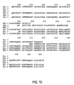
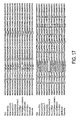
- Hybridization probes of the subject description may be derived from the nucleotide sequences of the attached List Sequences and the Sequences provided as SEQ ID NO: 18-22 (see, e.g., Figures 9 , 10 , and 17 ), or from surrounding or included genomic sequences comprising untranslated regions such as promoters, enhancers and introns. Such hybridization probes may be labeled with appropriate reporter molecules. Means for producing specific hybridization probes include oligolabelling, nick translation, end-labeling or PCR amplification using a labeled nucleotide. Alternatively, the cDNA sequence may be cloned into a vector for the production of mRNA probe.
- RNA polymerase such as T7, T3 or SP6 and labelled nucleotides.
- a number of companies (such as Pharmacia Biotech, Piscataway, N.J.; Promega, Madison, Wis.; US Biochemical Corp, Cleveland, Ohio; etc.) supply commercial kits and protocols for these procedures.
- the nucleotide sequences can be used to generate probes for mapping the native genomic sequence.
- the sequence may be mapped to a particular chromosome or to a specific region of the chromosome using well known techniques. These include in situ hybridization to chromosomal spreads, flow-sorted chromosomal preparations, or artificial chromosome constructions such as yeast artificial chromosomes (YACs), bacterial artificial chromosomes (BACs), bacterial P1 constructions or single chromosome cDNA libraries.
- nucleotide sequences of the subject invention may also be used to detect differences in the chromosomal location of nucleotide sequences due to translocation, inversion, or recombination.
- nucleotide sequences as disclosed herein may be used to produce an amino acid sequence using well known methods of recombinant DNA technology.
- Goeddel Gene Expression Technology, Methods and Enzymology [1990] Vol 185, Academic Press, San Diego, Calif .
- the amino acid or peptide may be expressed in a variety of host cells, either prokaryotic or eukaryotic.
- Some expression vectors are viral vectors such as vaccinia-based vectors and adeno-associated viral vectors, among others (Dunachie and Hill, 2003).
- Host cells may be from the same species from which the nucleotide sequence was derived or from a different species.
- Still further aspects of the invention use these purified peptides to produce antibodies or other molecules able to bind to the peptides. These antibodies or binding agents can then be used for the screening of cells in order to localize the cellular distribution of the peptides or proteins.
- the antibodies are also useful for the affinity purification of recombinantly produced peptides or proteins. Such antibodies are also useful as diagnostic reagents for the detection of protozoan diseases or in antibody-mediated tests and assays.
- T. parva antigens can also be used as a diagnostic aid or in methods that measure the presence of T. parva cytotoxic lymphocytes in immunized or nfected animals by co-culturing mononuclear cells harvested from an animal suspected to have an infection due to T. parva, with a T. parva antigen-specifc cytotoxic T cell under standard culture conditions for mammalian cell cultures. Mononuclear cells would have been incubated in an culture medium containing an indicator that is released upon cell death, such as 51 Cr. This is a method that is well-known in the art.
- the disclosed nucleotide sequences can be used individually, or in panels, in tests or assays to detect levels of peptide, polypeptide, or protein expression.
- the form of such qualitative or quantitative methods may include northern analysis, dot blot or other membrane based technologies, dip stick, pin or chip technologies, PCR, ELISAs or other multiple sample format technologies.
- oligonucleotide or “oligomer” is a stretch of nucleotide residues which has a sufficient number of bases to be used in a polymerase chain reaction (PCR). These short sequences are based on (or designed from) genomic or cDNA sequences and are used to amplify, confirm, or reveal the presence of an identical, similar or complementary DNA or RNA in a particular cell or tissue. Oligonucleotides or oligomers comprise portions of a DNA sequence having at least about 10 nucleotides and as many as about 50 nucleotides, preferably about 15 to 30 nucleotides. They can be chemically synthesized and may be used as probes.
- a "complementary" DNA or RNA is a sequence that is 100% identical to the strand to which it is complementary.
- polynucleotides of the subject invention can themselves be used as probes. Additional polynucleotide sequences can be added to the ends of (or internally in) the exemplified polynucleotide sequences so that polynucleotides that are longer than the exemplified polynucleotides can also be used as probes. Thus, isolated polynucleotides comprising one or more of the exemplified sequences are within the scope of the subject invention. Polynucleotides that have less nucleotides than the exemplified polynucleotides can also be used and are contemplated within the scope of the present description.
- polynucleotides of the subject invention can be used to find additional, homologous (wholly or partially) genes.
- Probes of the subject description may be composed of DNA, RNA, or PNA (peptide nucleic acid).
- the probe will normally have at least about 10 bases, more usually at least about 17 bases, and may have up to about 100 bases or more. Longer probes can readily be utilitzed, and such probes can be, for example, several kilobases in length.
- the probe sequence is designed to be at least substantially complementary to a portion of a gene encoding a protein of interest. The probe need not have perfect complementarity to the sequence to which it hybridizes.
- the probes may be labeled utilizing techniques that are well known to those skilled in this art.
- One approach for the use of the subject description as probes entails first identifying DNA segments that are homologous with the disclosed nucleotide sequences using, for example, Southern blot analysis of a gene bank.
- Southern blot analysis of a gene bank.
- One hybridization procedure useful according to the subject description typically includes the initial steps of isolating the DNA sample of interest and purifying it chemically. Either lysed cells or total fractionated nucleic acid isolated from cells can be used. Cells can be treated using known techniques to liberate their DNA (and/or RNA). The DNA sample can be cut into pieces with an appropriate restriction enzyme. The pieces can be interest can be through electrophoresis in a gel, usually agarose or acrylamide. The pieces of interest can be transferred to an immobilizing membrane.
- the probe and sample can then be combined in a hybridization buffer solution and held at an appropriate temperature until annealing occurs. Thereafter, the membrane is washed free of extraneous materials, leaving the sample and bound probe molecules typically detected and quantified by autoradiography and/or liquid scintillation counting.
- the probe molecule and nucleic acid sample hybridize by forming a strong non-covalent bond between the two molecules, it can be reasonably assumed that the probe and sample are essentially identical or very similar.
- the probe's detectable label provides a means for determining in a known manner whether hybridization has occurred.
- the particular probe is labeled with any suitable label known to those skilled in the art, including radioactive and non-radioactive labels.
- Typical radioactive labels include 32 P, 35 S, or the like.
- Non-radioactive labels include, for example, ligands such as biotin or thyroxine, as well as enzymes such as hydrolases or peroxidases, or the various chemiluminescers such as luciferin, or fluorescent compounds like fluorescein and its derivatives.
- the probes can be made inherently fluorescent as described in International Application No. WO 93/16094 .
- hybridization is conducted under moderate to high stringency conditions by techniques well known in the art, as described, for example, in Keller, G. H., M. M. Manak (1987) DNA Probes, Stockton Press, New York, N.Y., pp. 169-170 .
- moderate to high stringency conditions for hybridization refers to conditions that achieve the same, or about the same, degree of specificity of hybridization as the conditions described herein. Examples of moderate to high stringency conditions are provided herein. Specifically, hybridization of immobilized DNA on Southern blots with 35 P-labeled gene-specific probes is performed using standard methods (Maniatis et al.). In general, hybridization and subsequent washes are carried out under moderate to high stringency conditions that allow for detection of target sequences with homology to sequences exemplified herein. For double-stranded DNA gene probes, hybridization can be carried out overnight at 20-25° C.
- Tm melting temperature
- Washes can typically be carried out as follows:
- Tm melting temperature
- salt and/or temperature can be altered to change stringency.
- a labeled DNA fragment of greater than about 70 or so bases in length, the following conditions can be used:
- polynucleotide sequences of the subject description include mutations (both single and multiple), deletions, and insertions in the described sequences, and combinations thereof, wherein said mutations, insertions, and deletions permit formation of stable hybrids with a target polynucleotide of interest. Mutations, insertions, and deletions can be produced in a given polynucleotide sequence using standard methods known in the art. Other methods may become known in the future.
- the mutational, insertional, and deletional variants of the polypeptide sequences of the description can be used in the same manner as the exemplified polynucleotide sequences so long as the variants have substantial secjuence similarity with the original sequence.
- substantial sequence similarity refers to the extent of nucleotide similarity that is sufficient to enable the variant polynucleotide to function in the same capacity as the original sequence. Preferably, this similarity is greater than 50%; more preferably, this similarity is greater than 75%; and most preferably, this similarity is greater than 90%.
- the degree of similarity needed for the variant to function in its intended capacity will depend upon the intended use of the sequence. It is well within the skill of a person trained in this art to make mutational, insertional, and deletional mutations that are designed to improve the function of the sequence or otherwise provide a methodological advantage.
- PCR Polymerase Chain Reaction
- PCR is a repetitive, enzymatic, primed synthesis of a nucleic acid sequence. This procedure is well known and commonly used by those skilled in this art (see U.S. Pat. Nos. 4,683,195 , 4,683,202 , and 4,800,159; Saiki et al., 1985 ).
- PCR is based on the enzymatic amplification of a DNA fragment of interest that is flanked by two oligonucleotide primers that hybridize to opposite strands of the target sequence. The primers are oriented with the 3' ends pointing towards each other.
- thermostable DNA polymerase such as Taq polymerase, which is isolated from the thermophilic bacterium Thermus aquaticus, the amplification process can be completely automated.
- Other enzymes that can be used are known to those skilled in the art.
- polynucleotide sequences of the subject invention can be used as, and/or used in the design of, primers for PCR amplification.
- a certain degree of mismatch can be tolerated between primer and template. Therefore, mutations, deletions, and insertions (especially additions of nucleotides to the 5' end) of the exemplified polynucleotides can be used in this manner. Mutations, insertions and deletions can be produced in a given primer by methods known to an ordinarily skilled artisan.
- Full length genes may be cloned utilizing partial nucleotide sequence and various methods known in the art.
- Gobinda et al. (1993; PCR Methods Applic 2:318-22 ) disclose "restriction-site PCR" as a direct method which uses universal primers to retrieve unknown sequence adjacent to a known locus.
- genomic DNA is amplified in the presence of primer to linker and a primer specific to the known region.
- the amplified sequences are subjected to a second round of PCR with the same linker primer and another specific primer internal to the first one.
- Products of each round of PCR are transcribed with an appropriate RNA polymerase and sequenced using reverse transcriptase.
- Inverse PCR can be used to acquire unknown sequences starting with primers based on a known region ( Triglia T. et al. (1988) Nucleic Acids Res 16:8186 ).
- the method uses several restriction enzymes to generate a suitable fragment in the known region of a gene. The fragment is then circularized by intramolecular ligation and used as a PCR template. Divergent primers are designed from the known region. The multiple rounds of restriction enzyme digestions and ligations that are necessary prior to PCR make the procedure slow and expensive (Gobinda et al. [1993] supra).
- Capture PCR ( Lagerstrom M. et al. (1991) PCR Methods Applic 1:111-19 ) is a method for PCR amplification of DNA fragments adjacent to a known sequence in eucaryotic and YAC DNA. As noted by Gobinda et al. (1993,supra), capture PCR also requires multiple restriction enzyme digestions and ligations to place an engineered double-stranded sequence into an unknown portion of the DNA molecule before PCR. Although the restriction and ligation reactions are carried out simultaneously, the requirements for extension, immobilization and two rounds of PCR and purification prior to sequencing render the method cumbersome and time consuming.
- PromoterFinder.TM is a kit available from Clontech Laboratories, Inc. (Palo Alto, Calif.) which uses PCR and primers derived from p53 to walk in genomic DNA. Nested primers and special PromoterFinder.TM. libraries are used to detect upstream sequences such as promoters and regulatory elements. This process avoids the need to screen libraries and is useful in finding intron/exon junctions.
- PCR method replaces methods which use labeled probes to screen plasmid libraries and allow one researcher to process only about 3-5 genes in 14-40 days.
- the first step which can be performed in about two days, any two of a plurality of primers are designed and synthesized based on a known partial sequence.
- step 2 which takes about six to eight hours, the sequence is extended by PCR amplification of a selected library.
- Steps 3 and 4 which take about one day, are purification of the amplified cDNA and its ligation into an appropriate vector.
- Step 5 which takes about one day, involves transforming and growing up host bacteria.
- step 6 which takes approximately five hours, PCR is used to screen bacterial clones for extended sequence.
- the final steps which take about one day, involve the preparation and sequencing of selected clones.
- the preferred library may be one that has been size-selected to include only larger cDNAs or may consist of single or combined commercially available libraries, e.g., from Clontech Laboratories, Inc. (Palo Alto, Calif.).
- the cDNA library may have been prepared with oligo (dT) or random priming. Random primed libraries are preferred in that they will contain more sequences which contain 5' ends of genes. A randomly primed library may be particularly useful if an oligo (dT) library does not yield a complete gene. It must be noted that the larger and more complex the protein, the less likely it is that the complete gene will be found in a single plasmid.
- capillary electrophoresis Another method for analyzing either the size or the nucleotide sequence of PCR products is capillary electrophoresis.
- Systems for rapid sequencing are available from Perkin Elmer (Foster City Calif.), Beckman Instruments (Fullerton, Calif.), and other companies.
- Capillary sequencing employs flowable polymers for electrophoretic separation, four different fluorescent dyes (one for each nucleotide) which are laser activated, and detection of the emitted wavelengths by a charge coupled devise camera. Output/light intensity is converted to electrical signal using appropriate software (eg. Genotyper.TM. and Sequence Navigators.TM. from Perkin Elmer) and the entire process from loading of samples to computer analysis and electronic data display is computer controlled.
- appropriate software eg. Genotyper.TM. and Sequence Navigators.TM. from Perkin Elmer
- Capillary electrophoresis provides greater resolution and is many times faster than standard gel based procedures. It is particularly suited to the sequencing of small pieces of DNA which might be present in limited amounts in a particular sample. The reproducible sequencing of up to 350 bp of M13 phage DNA in 30 min has been reported ( Ruiz-Martinez M. C. et al. [1993] Anal Chem 65:2851-8 ).
- Polynucleotides of the subject invention can be defined according to several parameters. One characteristic is the biological activity of the protein products as identified herein.
- the proteins and genes of the subject description can be further defined by their amino acid and nucleotide sequences.
- the sequences of the molecules can be defined in terms of homology to certain exemplified sequences as well as in terms of the ability to hybridize with, or be amplified by, certain exemplified probes and primers. Additional primers and probes can readily be constructed by those skilled in the art such that alternate polynucleotide sequences encoding the same amino acid sequences can be used to identify and/or characterize additional genes.
- the proteins of the subject invention can also be identified based on their immunoreactivity with certain antibodies.
- polynucleotides and proteins of the subject description include portions, fragments, variants, and mutants of the full-length sequences as well as fusions and chimerics, so long as the encoded protein retains the characteristic biological activity of the proteins identified herein.
- variants or variantations refer to nucleotide sequences that encode the same proteins or which encode equivalent proteins having equivalent biological activity.
- equivalent proteins refers to proteins having the same or essentially the same biological activity as the exemplified proteins.
- genes may be readily constructed using standard techniques such as site-directed mutagenesis and other methods of making point mutations and by DNA shuffling, for example.
- gene and protein fragments can be made using commercially available exonucleases, endonucleases, and proteases according to standard procedures.
- enzymes such as Ba131 can be used to systematically cut off nucleotides from the ends of genes.
- genes that encode fragments may be obtained using a variety of restriction enzymes. Proteases may be used to directly obtain active fragments of these proteins.
- molecular techniques for cloning polynucleotides and producing gene constructs of interest are also well known in the art. In vitro evaluation techniques, such as MAXYGEN's "Molecular Breeding" can also be applied to practice the subject invention.
- a "selectable marker” is a gene whose expression allows one to identify cells that have been transformed or transfected with a vector containing the marker gene.
- Reporter molecules are chemical moieties used for labeling a nucleic or amino acid sequence. They include, but are not limited to, radionuclides, enzymes, fluorescent, chemi-luminescent, or chromogenic agents. Reporter molecules associate with, establish the presence of, and may allow quantification of a particular nucleic or amino acid sequence.
- a "portion" or “fragment” of a polynucleotide or nucleic acid comprises all or any part of the nucleotide sequence having fewer nucleotides than about 6 kb, preferably fewer than about 1 kb which can be used as a probe.
- probes may be labeled with reporter molecules using nick translation, Klenow fill-in reaction, PCR or other methods well known in the art. After pretesting to optimize reaction conditions and to eliminate false positives, nucleic acid probes may be used in Southern, northern or in situ hybridizations to determine whether target DNA or RNA is present in a biological sample, cell type, tissue, organ or organism.
- a "polypeptide” comprises a protein, oligopeptide or peptide fragments thereof.
- a "mutant, variant, or modified polypeptide” means any polypeptide encoded by a nucleotide sequence that has been mutated through insertions, deletions, substitutions, or the like.
- Chimeric molecules are polynucleotides or polypeptides which are created by combining one or more nucleotide or peptide sequences (or their parts). In the case of nucleotide sequences, such combined sequences may be introduced into an appropriate vector and expressed to give rise to a chimeric polypeptide which may be expected to be different from the native molecule in one or more of the following characteristics: cellular location, distribution, ligand-binding affinities, interchain affinities, degradation/turnover rate, signaling, etc.
- Active is that state which is capable of being useful or of carrying out some role. It specifically refers to those forms, fragments, or domains of an amino acid sequence which display the biologic and/or immunogenic activity characteristic of the naturally occurring peptide, polypeptide, or protein.
- the present description relates to active fragments of polypeptides ( e.g ., represented by SEQ ID NO: 1-5). Each of these active fragments retains at least one epitope of the larger polypeptide.
- Naturally occurring refers to a polypeptide produced by cells which have not been genetically engineered or which have been genetically engineered to produce the same sequence as that naturally produced.
- derivative refers to those polypeptides which have been chemically modified by such techniques as ubiquitination, labeling, pegylation (derivatization with polyethylene glycol), and chemical insertion or substitution of amino acids such as ornithine which do not normally occur in proteins.
- Recombinant polypeptide variant refers to any polypeptide which differs from naturally occurring peptide, polypeptide, or protein by amino acid insertions, deletions and/or substitutions.
- substitutions are defined as one for one amino acid replacements. They are conservative in nature when the substituted amino acid has similar structural and/or chemical properties. Examples of conservative replacements are substitution of a leucine with an isoleucine or valine, an aspartate with a glutamate, or a threonine with a serine.
- Amino acid "insertions” or “deletions” are changes to or within an amino acid sequence. They typically fall in the range of about 1 to 5 amino acids. The variation allowed in a particular amino acid sequence may be experimentally determined by producing the peptide synthetically or by systematically making insertions, deletions, or substitutions of nucleotides in the sequence using recombinant DNA techniques.
- oligopeptide is a short stretch of amino acid residues and may be expressed from an oligonucleotide. Such sequences comprise a stretch of amino acid residues of at least about 5 amino acids and often about 17 or more amino acids, typically at least about 9 to 13 amino acids, and of sufficient length to display biologic and/or immunogenic activity.
- a "standard” is a quantitative or qualitative measurement for comparison. Preferably, it is based on a statistically appropriate number of samples and is created to use as a basis of comparison when performing diagnostic assays, running clinical trials, or following patient treatment profiles.
- the samples of a particular standard may be normal or similarly abnormal.
- an antibody or “specific binding parts” means any fragment of an antibody molecule that will bind antigen or other ligands such as lectins or other molecules. "Specific binding parts” is meant to include, but not be limited to antibody fragments such as Fab fragments, Fab'(2) fragments, Fc region fragments, Complimentarity determing regions (CDRs), Fv fragments, single chain Fv (scFv) fragments, and antigen binding site fragments.
- an "antigen” is a macromolecule that is recognized by antibodies or immune cells and can trigger an immune response.
- an antigen is a protein or a polysaccharide, but it can be any type of molecule, even small molecules if coupled to a large carrier.
- An "antigen specific" cytotoxic T cell is a T lymphocyte that can recognize and kill another cell that is expressing an antigen, usually in conjunction with a type or class of molecules referred to by those familiar with the art, as Major Histocompatiblity Complex (MHC) Class I molecules.
- MHC Major Histocompatiblity Complex
- Such Cytotoxic T cells are also referred to as "CD8 + " (CD8 positive) or CTLs, by those familiar with the art.
- An epitope or antigenic determinant is a region or section of an antigen that is approximately the minimal length of antigenic sequence that will elicit an immune response, as measured by the development of an antibody response, the development of T antigen-specific T cells, or other measureable immune cell response.
- an epitope could be a site on an antigen recognized by antibody.
- a T cell "epitope" is an antigenic determinant recognized and bound by the T-cell receptor. Epitopes recognized by the T-cell receptor are often located in the inner, unexposed side of the antigen, and become accessible to the T-cell receptors after proteolytic processing of the antigen.
- An "effective" immunization protocol is one in which an animal is protected against infection, at least to a measurable degree, when exposed to the specific infectious agent for which it was immunized.
- an “ELISpot Assay” is a shorted name for "Enzyme-linked ImmunoSpot Assay", which refers to an antibody based method to detect secretion of soluble factors released by cells. Originally developed as a method to detect antibody-secreting B-cells, later the method was adapted to determine T-cell reaction to a specific antigen, often represented as number of activated cells per million.
- Effective conditions for culturing cells or cell lines means those cuture conditions that allow the cells to respond in a manner than mimics or is the same as the response would be if the cells were responding in vivo.
- Hardvesting cells or other materials means to separate and collect those cells from a mixture of from one source for use in another protocol.
- a Bovine MHC class II bioassay means an assay that measures the presence of a T cell-derived soluble factor, such as interferon (IFN- ⁇ ) release from T cells responding to specific stimulation through the ability of IFN- ⁇ to induce and up-regulate expression of class II molecules on bovine endothelial cells.
- T cell-derived soluble factor such as interferon (IFN- ⁇ ) release from T cells responding to specific stimulation through the ability of IFN- ⁇ to induce and up-regulate expression of class II molecules on bovine endothelial cells.
- Bovine endothelial cells do not constutively express class II molecules unless triggered by external signals such as IFN- ⁇ .
- DNA sequences can encode the amino acid sequences encoded by the polynucleotide sequences disclosed herein. It is well within the skill of a person trained in the art to create these alternative DNA sequences encoding proteins having the same, or essentially the same, amino acid sequence. These variant DNA sequences are within the scope of the subject description. As used herein, reference to "essentially the same" sequence refers to sequences that have amino acid substitutions, deletions, additions, or insertions that do not materially affect biological activity. Fragments retaining the characteristic biological activity are also included in this definition.
- the subject description comprises variant or equivalent proteins (and nucleotide sequences coding for equivalent proteins) having the same or similar biological activity of proteins encoded by the exemplified polynucleotides.
- Equivalent proteins will have amino acid similarity with an exemplified protein (or peptide).
- the amino acid identity will typically be greater than 60%.
- the amino acid identity will be greater than 75%. More preferably, the amino acid identity will be greater than 80%, and even more preferably greater than 90%. Most preferably, amino acid identity will be greater than 95%.
- the polynucleotides that encode the subject polypeptides will also have corresponding identities in these preferred ranges.) These identities are as determined using standard alignment techniques for determining amino acid identity.
- amino acids may be placed in the following classes: non-polar, uncharged polar, basic, and acidic. Conservative substitutions whereby an amino acid of one class is replaced with another amino acid of the same type fall within the scope of the subject description so long as the substitution does not materially alter the biological activity of the compound.
- non-conservative substitutions can also be made.
- isolated polynucleotides and/or purified proteins refers to these molecules when they are not associated with the other molecules with which they would be found in nature. Thus, reference to “isolated” and/or “purified” signifies the involvement of the "hand of man” as described herein. Reference to “heterologous” proteins, genes, and gene constructs, also signifies the involvement of the "hand of man.”
- the invention also provides vectors containing the nucleic acid molecules described herein.
- the term "vector” refers to a vehicle, preferably a nucleic acid molecule, which can transport the nucleic acid molecules.
- the vector is a nucleic acid molecule, the nucleic acid molecules are covalently linked to the vector nucleic acid.
- the vector includes a plasmid, single or double stranded phage, a single or double stranded RNA or DNA viral vector, or artificial chromosome, such as a BAC, PAC, YAC, OR MAC.
- a vector can be maintained in the host cell as an extrachromosomal element where it replicates and produces additional copies of the nucleic acid molecules.
- the vector may integrate into the host cell genome and produce additional copies of the nucleic acid molecules when the host cell replicates.
- the invention provides vectors for the maintenance (cloning vectors) or vectors for expression (expression vectors) of the nucleic acid molecules.
- the vectors can function in prokaryotic or eukaryotic cells or in both (shuttle vectors).
- Expression vectors contain cis-acting regulatory regions that are operably linked in the vector to the nucleic acid molecules such that transcription of the nucleic acid molecules is allowed in a host cell.
- the nucleic acid molecules can be introduced into the host cell with a separate nucleic acid molecule capable of affecting transcription.
- the second nucleic acid molecule may provide a trans-acting factor interacting with the cis-regulatory control region to allow transcription of the nucleic acid molecules from the vector.
- a trans-acting factor may be supplied by the host cell.
- a trans-acting factor can be produced from the vector itself. It is understood, however, that in some embodiments, transcription and/or translation of the nucleic acid molecules can occur in a cell-free system.
- the regulatory sequence to which the nucleic acid molecules described herein can be operably linked include promoters for directing mRNA transcription. These include, but are not limited to, the left promoter from bacteriophage .lambda., the lac, TRP, and TAC promoters from E. Coli, the early and late promoters from SV40, the CMV immediate early promoter, the adenovirus early and late promoters, and retrovirus long-terminal repeats.
- expression vectors may also include regions that modulate transcription, such as repressor binding sites and enhancers.
- regions that modulate transcription include the SV40 enhancer, the cytomegalovirus immediate early enhancer, polyoma enhancer, adenovirus enhancers, and retrovirus LTR enhancers.
- expression vectors can also contain sequences necessary for transcription termination and, in the transcribed region a ribosome binding site for translation.
- Other regulatory control elements for expression include initiation and termination codons as well as polyadenylation signals.
- the person of ordinary skill in the art would be aware of the numerous regulatory sequences that are useful in expression vectors. Such regulatory sequences are described, for example, in Sambrook et al., Molecular Cloning: A Laboratory Manual. 2nd. ed., Cold Spring Harbor Laboratory Press, Cold Spring Harbor, N.Y., (1989 ).
- a variety of expression vectors can be used to express a nucleic acid molecule.
- Such vectors include chromosomal, episomal, and virus-derived vectors, for example vectors derived from bacterial plasmids, from bacteriophage, from yeast episomes, from yeast chromosomal elements, including yeast artificial chromosomes, from viruses such as baculoviruses, papovaviruses such as SV40, Vaccinia viruses including the modified vaccinia virus Ankara strain (MVA), adenoviruses, poxviruses including fowlpox virus (FP9), pseudorabies viruses, and retroviruses.
- MVA modified vaccinia virus Ankara strain
- FP9 fowlpox virus
- pseudorabies viruses pseudorabies viruses
- Vectors may also be derived from combinations of these sources such as those derived from plasmid and bacteriophage genetic elements, e.g. cosmids and phagemids.
- Appropriate cloning and expression vectors for prokaryotic and eukaryotic hosts are described in Sambrook et al., Molecular Cloning: A Laboratory Manual. 2nd. ed., Cold Spring Harbor Laboratory Press, Cold Spring Harbor, N.Y., (1989 ).
- the regulatory sequence may provide constitutive expression in one or more host cells (i.e. tissue specific) or may provide for inducible expression in one or more cell types such as by temperature, nutrient additive, or exogenous factor such as a hormone or other ligand.
- host cells i.e. tissue specific
- inducible expression in one or more cell types such as by temperature, nutrient additive, or exogenous factor such as a hormone or other ligand.
- a variety of vectors providing for constitutive and inducible expression in prokaryotic and eukaryotic hosts are well known to those of ordinary skill in the art.
- the nucleic acid molecules can be inserted into the vector nucleic acid by well-known methodology.
- the DNA sequence that will ultimately be expressed is joined to an expression vector by cleaving the DNA sequence and the expression vector with one or more restriction enzymes and then ligating the fragments together. Procedures for restriction enzyme digestion and ligation are well known to those of ordinary skill in the art.
- Bacterial cells include, but are not limited to, E. coli, Streptomyces, and Salmonella typhimurium.
- Eukaryotic cells include, but are not limited to, yeast, insect cells such as Drosophila, animal cells such as COS and CHO cells, and plant cells.
- Fusion vectors can increase the expression of a recombinant protein, increase the solubility of the recombinant protein, and aid in the purification of the protein by acting for example as a ligand for affinity purification.
- a proteolytic cleavage site may be introduced at the junction of the fusion moiety so that the desired peptide can ultimately be separated from the fusion moiety.
- Proteolytic enzymes include, but are not limited to, factor Xa, thrombin, and enteroenzyme.
- Typical fusion expression vectors include pGEX ( Smith et al., Gene 67:31-40 (1988 )), pMAL (New England Biolabs, Beverly, Mass.) and pRIT5 (Pharmacia, Piscataway, N.J.) which fuse glutathione S-transferase (GST), maltose E binding protein, or protein A, respectively, to the target recombinant protein.
- GST glutathione S-transferase
- suitable inducible non-fusion E. coli expression vectors include pTrc ( Amann et al, Gene 69:301-315 (1988 )) and pET 11d ( Studier et al, Gene Expression Technology: Methods in Enzymology 185:60-89 (1990 )).
- Recombinant protein expression can be maximized in host bacteria by providing a genetic background wherein the host cell has an impaired capacity to proteolytically cleave the recombinant protein.
- the sequence of the nucleic acid molecule of interest can be altered to provide preferential codon usage for a specific host cell, for example E. coli. ( Wada et al., Nucleic Acids Res. 20:2111-2118 (1992 )).
- the nucleic acid molecules can also be expressed by expression vectors that are operative in yeast.
- yeast e.g., S. cerevisiae
- vectors for expression in yeast include pYepSec1 ( Baldari, et al, EMBO J. 6:229-234 (1987 )), pMFa ( Kujan et al, Cell 30:933-943(1982 )), pJRY88 ( Schultz et al, Gene 54:113-123 (1987 )), and pYES2 (Invitrogen Corporation, San Diego, Calf.).
- the nucleic acid molecules can also be expressed in insect cells using, for example, baculovirus expression vectors.
- Baculovirus vectors available for expression of proteins in cultured insect cells include the pAc series ( Smith et al, Mol. Cell Biol. 3:2156-2165 (1983 )) and the pVL series ( Lucklow et al, Virology 170:31-39 (1989 )).
- nucleic acid molecules described herein are expressed in mammalian cells using mammalian expression vectors.
- mammalian expression vectors include pCDM8 ( Seed, B. Nature 329:840(1987 )) and pMT2PC ( Kaufinan et al., EMBO J. 6:187-195 (1987 )).
- the expression vectors listed herein are provided by way of example only of the well-known vectors available to those of ordinary skill in the art that would be useful to express the nucleic acid molecules.
- the person of ordinary skill in the art would be aware of other vectors suitable for maintenance propagation or expression of the nucleic acid molecules described herein. These are found for example in Sambrook, J., Fritsh, E. F., and Maniatis, T. Molecular Cloning: A Laboratory Manual. 2nd, ed., Cold Spring Harbor Laboratory, Cold Spring Harbor Laboratory Press, Cold Spring Harbor, N.Y., 1989 .
- the description also encompasses vectors in which the nucleic acid sequences described herein are cloned into the vector in reverse orientation, but operably linked to a regulatory sequence that permits transcription of antisense RNA.
- an antisense transcript can be produced to all, or to a portion, of the nucleic acid molecule sequences described herein, including both coding and non-coding regions. Expression of this antisense RNA is subject to each of the parameters described above in relation to expression of the sense RNA (regulatory sequences, constitutive or inducible expression, tissue-specific expression).
- the invention also relates to recombinant host cells containing the vectors described herein.
- Host cells therefore include prokaryotic cells, lower eukaryotic cells such as yeast, other eukaryotic cells such as insect cells, and higher eukaryotic cells such as mammalian cells.
- the recombinant host cells are prepared by introducing the vector constructs described herein into the cells by techniques readily available to the person of ordinary skill in the art. These include, but are not limited to, calcium phosphate transfection, DEAE-dextran-mediated transfection, cationic lipid-mediated transfection, electroporation, transduction, infection, lipofection, and other techniques such as those found in Sambrook, et al. (Molecular Cloning: A Laboratory Manual. 2nd, ed., Cold Spring Harbor Laboratory, Cold Spring Harbor Laboratory Press, Cold Spring Harbor, N.Y., 1989 ).
- Host cells can contain more than one vector.
- different nucleotide sequences can be introduced on different vectors of the same cell.
- the nucleic acid molecules can be introduced either alone or with other nucleic acid molecules that are not related to the nucleic acid molecules such as those providing trans-acting factors for expression vectors.
- the vectors can be introduced independently, co-introduced or joined to the nucleic acid molecule vector.
- bacteriophage and viral vectors these can be introduced into cells as packaged or encapsulated virus by standard procedures for infection and transduction.
- Viral vectors can be replication-competent or replication-defective. In the case in which viral replication is defective, replication will occur in host cells providing functions that complement the defects.
- Vectors generally include selectable markers that enable the selection of the subpopulation of cells that contain the recombinant vector constructs.
- the marker can be contained in the same vector that contains the nucleic acid molecules described herein or may be on a separate vector. Markers include tetracycline or ampicillin-resistance genes for prokaryotic host cells and dihydrofolate reductase or neomycin resistance for eukaryotic host cells. However, any marker that provides selection for a phenotypic trait will be effective.
- RNA derived from the DNA constructs described herein can also be used to produce these proteins using RNA derived from the DNA constructs described herein.
- secretion of the peptide is desired, which is difficult to achieve with multi-transmembrane domain containing proteins such as MHC Class I-binding peptides, appropriate secretion signals are incorporated into the vector.
- the signal sequence can be endogenous to the peptides or heterologous to these peptides.
- the expressed protein can be isolated from the host cell by standard disruption procedures, including freeze thaw, sonication, mechanical disruption, use of lysing agents and the like.
- the peptide can then be recovered and purified by well-known purification methods including ammonium sulfate precipitation, acid extraction, anion or cationic exchange chromatography, phosphocellulose chromatography, hydrophobic-interaction chromatography, affinity chromatography, hydroxylapatite chromatography, lectin chromatography, or high performance liquid chromatography.
- the peptides can have various glycosylation patterns, depending upon the cell, or maybe non-glycosylated as when produced in bacteria.
- the peptides may include an initial modified methionine in some cases as a result of a host-mediated process.
- heterologous gene or polynucleotide there are many methods for introducing a heterologous gene or polynucleotide into a host cell or cells under conditions that allow for stable maintenance and expression of the gene or polynucleotide. These methods are well known to those skilled in the art. Synthetic genes, such as, for example, those genes modified to enhance expression in a heterologous host (such as by preferred codon usage or by the use of adjoining, downstream, or upstream enhancers) that are functionally equivalent to the genes (and which encode equivalent proteins) can also be used to transfect hosts. Methods for the production of synthetic genes are known in the art. Recombinant hosts can be used for the expression or propagation of genes and polynucleotides of the present invention.
- T. parva nucleic acids can be expressed in a recombinant baculovirus (BV) possessing an optimized promoter and translation initiation region operably linked to the T. parva nucleic acid.
- BV baculovirus
- an optimized promoter and translation initiation region are operably linked to the T. parva nucleic acid.
- insect host cells can be transformed with baculovirus expressing T. parva nucleic acids.
- Tn5 Trichoplusia ni or High Five.TM.
- host cells can be transformed with baculovirus expressing T. parva nucleic acids.
- recombinant protein is a protein made using recombinant techniques, i.e. through the expression of a recombinant nucleic acid as depicted above.
- a recombinant protein is distinguished from naturally occurring protein by at least one or more characteristics.
- the protein may be isolated or purified away from some or all of the proteins and compounds with which it is normally associated in its wild type host, and thus may be substantially pure.
- an isolated protein is unaccompanied by at least some of the material with which it is normally associated in its natural state, preferably constituting at least about 0.5%, more preferably at least about 5% by weight of the total protein in a given sample.
- a substantially pure protein comprises at least about 75% by weight of the total protein, with at least about 80% being preferred, and at least about 90% being particularly preferred.
- the definition includes the production of a protein from one organism in a different organism or host cell.
- the protein may be made at a significantly higher concentration than is normally seen, through the use of an inducible promoter or high expression promoter, such that the protein is made at increased concentration levels.
- the protein may be in a form not normally found in nature, as in the addition of an epitope tag or amino acid substitutions, insertions and/or deletions, as discussed below.
- T. parva antigen polypeptides include T. parva polypeptide variants. These variants fall into one or more of three classes: substitutional, insertional or deletional variants. These variants ordinarily are prepared by site specific mutagenesis of nucleotides in the DNA encoding a T . parva antigen polypeptide, using cassette or PCR mutagenesis, scanning mutagenesis, gene shuffling or other techniques well known in the art, to produce DNA encoding the variant, and thereafter expressing the DNA in recombinant cell culture as outlined above. However, variant T. parva polypeptide fragments having up to about 100-150 residues may be prepared by in vitro synthesis using established techniques. Amino acid sequence variants are characterized by the predetermined nature of the variation, a feature that sets them apart from naturally occurring allelic or interspecies variation of the T . parva antigen polypeptide amino acid sequence.
- the mutation per se need not be predetermined.
- random mutagenesis may be conducted at the target codon or region and the expressed T. parva antigen polypeptide variants can be screened for the optimal combination of desired activity.
- Techniques for making mutations at predetermined sites in DNA having a known sequence are well known.
- the variations can be made using uligonucleotide-mediated site-directed mutagenesis [ Carter et al., Nucl. Acids Res., 13:4331 (1986 ); Zoller et al., Nucl.
- Alanine is typically a preferred scanning amino acid among this group because it eliminates the side-chain beyond the beta-carbon and is less likely to alter the main-chain conformation of the variant [ Cunmingham and Wells, Science, 244: 1081-1085 (1989 )]. Alanine is also typically preferred because it is the most common amino acid. Further, it is frequently found in both buried and exposed positions [ Creighton, The Proteins, (W.H. Freeman & Co., N.Y .); Chothia, J. Mol. Biol.,150:1 (1976 )]. If alanine substitution does not yield adequate amounts of variant, an isoteric amino acid can be used. Screening of the mutants or variants is done using Elispot and/or bioassays of T. parva antigen polypeptide activities and/or properties as described herein.
- the present: description further provides fragments of the antigen peptides, in addition to proteins and peptides that comprise and consist of such fragments, particularly those comprising the residues identified in SEQ ID NO:9-17.
- the fragments to which the description pertains are not to be construed as encompassing fragments that may be disclosed publicly prior to the present invention.
- a fragment comprises at least about 8, 10, 12, 14, 16, or more contiguous amino acid residues from an antigen peptide.
- Such fragments can be chosen based on the ability to retain one or more of the biological activities of the antigen peptide or could be chosen for the ability to perform a function, e.g. bind a substrate or act as an immunogen.
- Particularly important fragments are biologically active fragments, peptides that are, for example, about 8 or more amino acids in length.
- Such fragments will typically comprise a domain or motif of the antigen peptide, e.g., active site, a transmembrane domain or a substrate-binding domain.
- fragments include, but are not limited to, domain or motif containing fragments, soluble peptide fragments, and fragments containing immunogenic structures.
- Predicted domains and functional sites are readily identifiable by computer programs well known and readily available to those of skill in the art (e.g., PROSITE analysis).
- Polypeptides often contain amino acids other than the 20 amino acids commonly referred to as the 20 naturally occurring amino acids. Further, many amino acids, including the terminal amino acids, may be modified by natural processes, such as processing and other post-translational modifications, or by chemical modification techniques well known in the art. Common modifications that occur naturally in antigen peptides are described in basic texts, detailed monographs, and the research literature, and they are well known to those of skill in the art.
- Known modifications include, but are not limited to, acetylation, acylation, ADP-ribosylation, animation, covalent attachment of flavin, covalent attachment of a heme moiety, covalent attachment of a nucleotide or nucleotide derivative, covalent attachment of a lipid or lipid derivative, covalent attachment of phosphotidylinositol, cross-linking, cyclization, disulfide bond formation, demethylation, formation of covalent crosslinks, formation of cystine, formation of pyroglutamate, formylation, gamma carboxylation, glycosylation, GPI anchor formation, hydroxylation, iodination, methylation, myristoylation, oxidation, proteolytic processing, phosphorylation, prenylation, racemization, selenoylation, sulfation, transfer-RNA mediated addition of amino acids to proteins such as arginylation, and ubiquitination.
- the antigen peptides of the present description also encompass derivatives or analogs in which a substituted amino acid residue is not one encoded by the genetic code, in which a substituent group is included, in which the mature antigen peptide is fused with another compound, such as a compound to increase the half-life of the antigen peptide (for example, polyethylene glycol), or in which the additional amino acids are fused to the mature antigen peptide, such as a leader or secretory sequence or a sequence for purification of the mature antigen. peptide or a pro-protein sequence.
- amino acid sequence or oligopeptide used for antibody induction does not require biological activity, it must be immunogenic.
- a peptide, polypeptide, or protein used to induce specific antibodies may have an amino acid sequence consisting of at least five amino acids and preferably at least 10 amino acids. Short stretches of amino acid sequence may be genetically or chemically fused with those of another protein such as keyhole limpet hemocyanin, and the chimeric peptide used for antibody production. Alternatively, the oligopeptide may be of sufficient length to contain an entire domain.
- Antibodies specific for peptides, polypeptides, or proteins may be produced by inoculation of an appropriate animal with an antigenic fragment of the peptide, polypeptide, or protein.
- Antibody production includes not only the stimulation of an immune response by injection into animals, but also analogous processes such as the production of synthetic antibodies, the screening of recombinant immunoglobulin libraries for specific-binding molecules ( Orlandi R et al. [1989] PNAS 86:3833-3837 , or Huse W. D. et al. [1989] Science 256:1275-1281 ), or the in vitro stimulation of lymphocyte populations. Current technology ( Winter G. and Milstein C.
- the description also provides antibodies that selectively bind to one of the peptides of the present invention, a protein comprising such a peptide, as well as variants and fragments thereof.
- an antibody selectively binds a target peptide when it binds the target peptide and does not significantly bind to unrelated proteins.
- An antibody is still considered to selectively bind a peptide even if it also binds to other proteins that are not substantially homologous with the target peptide so long as such proteins share homology with a fragment or domain of the peptide target of the antibody. In this case, it would be understood that antibody binding to the peptide is still selective despite some degree of cross-reactivity.
- an antibody is defined in terms consistent with that recognized within the art: they are multi-subunit proteins produced by a mammalian organism in response to an antigen challenge.
- the antibodies of the present description include polyclonal antibodies and monoclonal antibodies, as well as fragments of such antibodies, including, but not limited to, Fab orF(ab') 2 , and Fv fragments.
- an isolated peptide is used as an immunogen and is administered to a mammalian organism, such as a rat, rabbit or mouse.
- a mammalian organism such as a rat, rabbit or mouse.
- the full-length protein, an antigenic peptide fragment or a fusion protein can be used.
- Particularly important fragments are those covering functional domains, such as the domains identified in the figures, and domain of sequence homology or divergence amongst the family, such as those that can readily be identified using protein alignment methods and as presented in the Figures.
- Antibodies are preferably prepared from regions or discrete fragments of the antigen proteins. Antibodies can be prepared from any region of the peptide as described herein. However, preferred regions will include those involved in function/activity and/or antigen/binding partner interaction.
- An antigenic fragment will typically comprise at least 8 contiguous amino acid residues.
- the antigenic peptide can comprise, however, at least 10, 12, 14, 16 or more amino acid residues.
- Such fragments can be selected on a physical property, such as fragments correspond to regions that are located on the surface of the protein, e. g., hydrophilic regions or can be selected based on sequence uniqueness.
- Detection on an antibody of the present description can be facilitated by coupling (i.e., physically linking) the antibody to a detectable substance.
- detectable substances include various enzymes, prosthetic groups, fluorescent materials, luminescent materials, bioluminescent materials, and radioactive materials.
- suitable enzymes include horseradish peroxidase, alkaline phosphatase, .beta.-galactosidase, or acetylcholinesterase;
- suitable prosthetic group complexes include streptavidin/biotin and avidin/biotin;
- suitable fluorescent materials include umbelliferone, fluorescein, fluorescein isothiocyanate, rhodamine, dichlorotnazinylamme fluorescein, dansyl chloride or phycoerythrin;
- an example of a luminescent material includes luminol;
- examples of bioluminescent materials include luciferase, luciferin, and acquorin, and examples of suitable radioactive material include 125 I, 131 I, 35 S or 3 H.
- the antibodies can be used to isolate one of the proteins of the present invention by standard techniques, such as affinity chromatography or immunoprecipitation.
- the antibodies can facilitate the purification of the natural protein from cells and recombinantly produced protein expressed in host cells.
- such antibodies are useful to detect the presence of one of the proteins of the present invention in cells or tissues to determine the pattern of expression of the protein among various tissues in an organism and over the course of schizont development.
- Such antibodies can be used to detect protein in situ, in vitro, or in a cell lysate or supernatant in order to evaluate the abundance and pattern of expression.
- such antibodies can be used to assess abnormal tissue distribution or abnormal expression during development or progression of a protozoan infection. Antibody detection of circulating fragments of the full length protein can be used to identify turnover.
- the antibodies can be used to assess expression in disease states such as in active stages of the disease or in lymphocytes harvested from infected animals.
- Experimental data as provided in FIG. 16 indicates expression in bovine lymphocytes, infected with T. parva. If a disease is characterized by a specific mutation in the protein, antibodies specific for this mutant protein can be used to assay for the presence of the specific mutant protein.
- the diagnostic uses can be applied, not only in genetic testing, but also in monitoring a treatment modality. Accordingly, where treatment is ultimately aimed at preventing or controlling infection, antibodies directed against the protein or relevant fragments can be used to monitor therapeutic efficacy.
- the antibodies are also useful for inhibiting protein function, for example, blocking the binding of the antigenic peptide to a binding partner such as a substrate. These uses can also be applied in a therapeutic context in which treatment involves inhibiting the protein's function.
- An antibody can be used, for example, to block binding, thus modulating (agonizing or antagonizing) the peptides activity.
- Antibodies can be prepared against specific fragments containing sites required for function or against intact protein that is associated with a cell or cell membrane. See SEQ ID NO: 1-7 and 9-17 and Fig. 9 and 10 , for structural information relating to the proteins of the present description.
- kits for using antibodies to detect the presence of a protein in a biological sample can comprise antibodies such as a labeled or labelable antibody and a compound or agent for detecting protein in a biological sample; means for determining the amount of protein in the sample; means for comparing the amount of protein in the sample with a standard; and instructions for use.
- Such a kit can be supplied to detect a single protein or epitope or can be configured to detect one of a multitude of epitopes, such as in an antibody detection array. Arrays are described in detail below for nuleic acid arrays and similar methods have been developed for antibody arrays.
- the proteins of the present invention can be used in assays related to the functional information provided in the Figures; for example, to stimulate CD8+ cytotoxic T cell lines, to raise antibodies or to elicit another immune response; as a reagent (including the labeled reagent) in assays designed to quantitatively determine levels of the protein (or its binding partner or ligand) in biological fluids; and as markers for tissues in which the corresponding protein is preferentially expressed (either constitutively or at a particular stage of tissue differentiation or development or in a disease state).
- the protein binds or potentially binds to another protein or ligand (such as, for example, in a enzyme-effector protein interaction or enzyme-ligand interaction)
- the protein can be used to identify the binding partner/ligand so as to develop a system to identify inhibitors of the binding interaction. Any or all of these uses are capable of being developed into reagent grade or kit format for commercialization as commercial products.
- antigens isolated from T. parva and their protozoan orthologs serve as targets for identifying agents for use in mammalian therapeutic applications, e.g. an animal drug, particularly in modulating a biological or pathological response in a cell, tissue, or protozoan, that expresses an immunogenic antigen.
- Experimental data as provided in FIG. 16 indicates that the antigens of the present description are expressed in infected host lymphocytes, as indicated by flow cytometric analysis. Such uses can readily be determined using the information provided herein, that which is known in the art, and routine experimentation.
- astsays to detect a pathogen, e.g ., by detecting the presence and/or expression of a polypeptide of the invention in the pathogen, it is sometimes desirable to immobilize either the antigen protein, or fragment, or its target molecule to facilitate separation of complexes from uncomplexed forms of one or both of the proteins, as well as to accommodate automation of the assay.
- a fusion protein can be provided which adds a domain that allows the protein to be bound to a matrix.
- glutathione-S-transferase fusion proteins can be adsorbed onto glutathione sepharose beads (Sigma Chemical, St. Louis, Mo.) or glutathione derivabed microtitre plates, which are then combined with the cell lysates (e.g., 35 S-labeled) and the candidate compound, and the mixture incubated under conditions conducive to complex formation (e.g., at physiological conditions for salt and pH).
- the beads are washed to remove any unbound label, and the matrix immobilized and radiolabel determined directly, or in the supernatant after the complexes are dissociated.
- the complexes can be dissociated from the matrix, separated by SDS-PAGE, and the level of antigen-binding protein found in the bead fraction quantitated from the gel using standard electrophoretic techniques.
- the polypeptide or its target molecule can be immobilized utilizing conjugation of biotin and streptavidin using techniques well known in the art.
- antibodies reactive with the protein but which do not interfere with binding of the protein to its target molecule can be derivatized to the wells of the plate, and the protein trapped in the wells by antibody conjugation.
- Preparations of an antigen-binding protein and a candidate compound are incubated in the antigen protein-presenting wells and the amount of complex trapped in the well can be quantitated.
- Methods for detecting such complexes include immunodetection of complexes using antibodies reactive with the antigen protein target molecule, or which are reactive with antigen protein and compete with the target molecule, as well as enzyme-linked assays which rely on detecting an enzymatic activity associated with the presence of the target molecule.
- the antigen proteins of the present invention are also useful to provide a target for diagnosing a disease or a disease mediated by the peptide. Accordingly, the description provides methods for detecting the presence, or levels of, the protein (or encoding RNA) in a cell, tissue, or organism. Experimental data as provided in FIG. 16 indicates expression in T. parva-infected bovine lymphocytes. The method involves contacting a biological sample with a compound capable of interacting with the antigen protein such that the interaction can be detected. Such an assay can be provided in a single detection format or a multi-detection format such as an antibody chip array.
- a biological sample includes tissues, cells and biological fluids isolated from a subject, as well as tissues, cells and fluids present within a subject.
- the peptides of the present invention also provide targets for diagnosing active parasite activity or disease, in an animal having a variant peptide, particularly activities and conditions that are known for other members of the family of proteins to which the present one belongs.
- the peptide can be isolated from a biological sample and assayed for the presence of a genetic mutation that results in aberrant peptide. This includes amino acid substitution, deletion, insertion, rearrangement, (as the result of aberrant splicing events), and inappropriate post-translational modification.
- Analytic methods include altered electrophoretic mobility, altered tryptic peptide digest, altered enzyme activity in cell-based or cell-free assay, alteration in substrate or antibody-binding pattern, altered isoelectric point, direct amino acid sequencing, and any other of the known assay techniques useful for detecting mutations in a protein.
- Such an assay can be provided in a single detection format or a multi-detection format such as an antibody chip array.
- peptide detection techniques include enzyme linked immunosorbent assays (ELISAs), Western blots, immunoprecipitations and immunofluorescence using a detection reagent, such as an antibody or protein binding agent, as well as the methods that represent embodiments of the present invention.
- a detection reagent such as an antibody or protein binding agent
- the peptide can be detected in vivo in a subject by introducing into the subject a labeled anti-peptide antibody or other types of detection agent.
- the antibody can be labeled with a radioactive marker whose presence and location in a subject can be detected by standard imaging techniques.
- Such methods can be used to detect the allelic variant of a peptide expressed in an infected subject and methods which detect fragments of a peptide in a sample.
- the diagnostic uses can be applied, not only in genetic testing, but also in monitoring a treatment modality. Accordingly, where treatment is ultimately aimed at preventing or controlling infection, antibodies directed against the protein or relevant fragments can be used to monitor therapeutic efficacy.
- kits for using antibodies to detect the presence of a protein in a biological sample can comprise antibodies such as a labeled or labelable antibody and a compound or agent for detecting protein in a biological sample; means for determining the amount of protein in the sample; means for comparing the amount of protein in the sample with a standard; and instructions for use.
- Such a kit can be supplied to detect a single protein or epitope or can be configured to detect one of a multitude of epitopes, such as in an antibody detection array. Arrays are described in detail below for nucleic acid arrays and similar methods have been developed for antibody arrays.
- the present invention further provides isolated nucleic acid molecules that encode a T. parva antigen peptide or protein of the present invention (cDNA, transcript and genomic sequences).
- Such nucleic acid molecules will consist of, consist essentially of, or comprise a nucleotide sequence that encodes one of the enzyme peptides of the present description, an allelic variant thereof, or an ortholog or paralog thereof.
- an "isolated" nucleic acid molecule is one that is separated from other nucleic acid present in the natural source of the nucleic acid.
- an “isolated” nucleic acid is free of sequences which naturally flank the nucleic acid (i.e., sequences located at the 5' and 3' ends of the nucleic acid) in the genomic DNA of the organism from which the nucleic acid is derived.
- flanking nucleotide sequences for example up to about 5KB, 4KB, 3KB, 2KB, or 1KB or less, particularly contiguous peptide encoding sequences and peptide encoding sequences within the same gene but separated by introns in the genomic sequence.
- nucleic acid is isolated from remote and unimportant flanking sequences such that it can be subjected to the specific manipulations described herein such as recombinant expression, preparation of probes and primers, and other uses specific to the nucleic acid sequences.
- an "isolated" nucleic acid molecule such as a transcript/cDNA molecule
- a transcript/cDNA molecule can be substantially free of other cellular material, or culture medium when produced by recombinant techniques, or chemical precursors or other chemicals when chemically synthesized.
- the nucleic acid molecule can be fused to other coding or regulatory sequences and still be considered isolated.
- recombinant DNA molecules contained in a vector are considered isolated.
- isolated DNA molecules include recombinant DNA molecules maintained in heterologous host cells or purified (partially or substantially) DNA molecules in solution.
- isolated RNA molecules include in vivo or in vitro RNA transcripts of the isolated DNA molecules of the present invention.
- Isolated nucleic acid molecules according to the present invention further include such molecules produced synthetically.
- the nucleic acid may be double stranded, single stranded, or contain portions of both double stranded or single stranded sequence.
- the depiction of a single strand also defines the sequence of the other strand (“Crick"); thus the sequences depicted in the figures also include the complement of the sequence.
- RNAi molecules of the subject description are not limited to those that are targeted to the full-length polynucleotide or gene.
- Gene product can be inhibited with a RNAi molecule that is targeted to a portion or fragment of the exemplified polynucleotides; high homology (90-95%) or greater identity is also preferred, but not necessarily essential, for such applications.
- nucleic acid molecules that consist of, consist essentially of, or comprise, the nucleotide sequence shown in SEQ ID NO: 18-22 or any nucleic acid molecule that encodes a protein encoded by SEQ ID NO: 18-22 (as shown, e.g., in Figures 9 , 10 , and 17 ).
- Consisting essentially of when used in the context ofbiopolymers, refers to a sequence which is intermediate between the number of residues (amino acids or, in the present case, nucleotides) encompassed by the term “consisting of” and the longer length encompassed by the term “comprising.” Residues in addition to the residues encompassed by “consisting of” language do not affect the basic and novel characteristics (e.g ., in the case of a polynucleotide, the ability to encode a functional peptide, or to bind specifically to a nucleic acid of interest) of the molecule encompassed by the "consisting of" language.
- the description further provides nucleic acid molecules that encode fragments of the peptides of the present description as well as nucleic acid molecules that encode obvious variants of the enzyme proteins of the present description that are described above.
- nucleic acid molecules may be naturally occurring, such as allelic variants (same locus), paralogs (different locus), and orthologs (different organism), or may be constructed by recombinant DNA methods or by chemical synthesis.
- non-naturally occurring variants may be made by mutagenesis techniques, including those applied to nucleic acid molecules, cells, or organisms. Accordingly, as discussed above, the variants can contain nucleotide substitutions, deletions, inversions and insertions. Variation can occur in either or both the coding and non-coding regions. The variations can produce both conservative and non- conservative amino acid substitutions.
- An epitope coding region comprises a contiguous nucleotide sequence greater than 12 or more nucleotides. Further, a fragment could at least 30, 40, 50, 100, 250 or 500 nucleotides in length. The length of the fragment will be based on its intended use. For example, the fragment can encode epitope bearing regions of the peptide, or can be useful as DNA probes and primers. Such fragments can be isolated using the known nucleotide sequence to synthesize an oligonucleotide probe. A labeled probe can then be used to screen a cDNA library, genomic DNA library, or mRNA to isolate nucleic acid corresponding to the coding region. Further, primers can be used in PCR reactions to clone specific regions of gene.
- a probe/primer typically comprises substantially a purified oligonucleotide or oligonucleotide pair.
- the oligonucleotide typically comprises a region of nucleotide sequence that hybridizes under stringent conditions to at least about 12, 20, 25, 40, 50 or more consecutive nucleotides.
- Orthologs, homologs, and allelic variants can be identified using methods well known in the art. As described in the Peptide Section, these variants comprise a nucleotide sequence encoding a peptide that is typically 60-70%, 70-80%, 80-90%, and more typically at least about 90-95% or more homologous to the nucleotide sequence shown in the Figure sheets or a fragment of this sequence. Such nucleic acid molecules can readily be identified as being able to hybridize under moderate to stringent conditions, to the nucleotide sequence shown in the Figure sheets or a fragment of the sequence. Allelic variants can readily be determined by genetic locus of the encoding gene. The gene encoding the novel enzyme of the present description is located on a genome component that has been mapped to the T. parva chromosomel and 2 which is supported by multiple lines of evidence, such as STS and BAC map data.
- the nucleic acid molecules of the present invention are useful as probes, primers, chemical intermediates, and in biological assays.
- the nucleic acid molecules are useful as a hybridization probe for messenger RNA, transcript/cDNA and genomic DNA to isolate full-length cDNA and genomic clones encoding the peptide described in SEQ ID NO:9-17 and to isolate cDNA and genomic clones that correspond to variants (alleles, orthologs, etc.) producing the same or related peptides shown in SEQ ID NO:1-7 and 9-17.
- deletions were identified at 2 different nucleotide positions.
- the probe can correspond to any sequence along the entire length of the nucleic acid molecules provided in the SEQ ID information. Accordingly, it could be derived from 5' noncoding regions, the coding region, and 3' noncoding regions. However, as discussed, fragments are not to be construed as encompassing fragments disclosed prior to the present invention.
- the nucleic acid molecules are also useful as primers for PCR to amplify any given region of a nucleic acid molecule and are useful to synthesize antisense molecules of desired length and sequence.
- the nucleic acid molecules are also useful for constructing recombinant vectors.
- Such vectors include expression vectors that express a portion of, or all of, the peptide sequences.
- Vectors also include insertion vectors, used to integrate into another nucleic acid molecule sequence, such as into the cellular genome, to alter in situ expression of a gene and/or gene product.
- an endogenous coding sequence can be replaced via homologous recombination with all or part of the coding region containing one or more specifically introduced mutations.
- the nucleic acid molecules are also useful for expressing antigenic portions of the proteins.
- the nucleic acid molecules are also useful as probes for determining the chromosomal positions of the nucleic acid molecules by means of in situ hybridization methods.
- the genes encoding the novel antigens of the present description are located on a genome component that has been mapped to T. parva chromosomes 1 (Tp1 & Tp4) and 2 (Tp5, Tp7 & Tp8).
- nucleic acid molecules are also useful in making vectors containing the gene regulatory regions of the nucleic acid molecules of the present description.
- the nucleic acid molecules are also useful for designing antigens corresponding to all, or a part, of the mRNA produced from the nucleic acid molecules described herein.
- nucleic acid molecules are also useful for making vectors that express part, or all, of the peptides.
- the nucleic acid molecules are also useful for constructing host cells expressing a part, or all, of the nucleic acid molecules and peptides.
- the nucleic acid molecules are also useful as hybridization probes for determining the presence, level, form and distribution of nucleic acid expression. Such probes could be used to detect the presence of, or to determine levels of, a specific nucleic acid molecule in cells, tissues, and in organisms.
- the nucleic acid whose level is determined can be DNA or RNA. Accordingly, probes corresponding to the peptides described herein can be used to assess expression and/or gene copy number in a given cell, tissue, or organism. These uses are relevant for diagnosis of disorders involving an increase or decrease in antigen protein expression relative to normal results.
- In vitro techniques for detection of mRNA include Northern hybridizations and in situ hybridizations.
- In vitro techniques for detecting DNA includes Southern hybridizations and in situ hybridization.
- Probes can be used as a part of a diagnostic test kit for identifying cells or tissues that express an T. parva antigen protein, such as by measuring a level of an antigen-encoding nucleic acid in a sample of cells from a subject e.g., mRNA or genomic DNA, or determining if an antigen gene has been mutated.
- kits for detecting the presence of an antigen nucleic acid in a biological sample can comprise reagents such as a labeled or labelable nucleic acid or agent capable of detecting antigen nucleic acid in a biological sample; means for determining the amount of antigen nucleic acid in the sample; and means for comparing the amount of antigen nucleic acid in the sample with a standard.
- the compound or agent can be packaged in a suitable container.
- the kit can further comprise instructions for using the kit to detect antigen protein mRNA or DNA.
- the present description further provides nucleic acid detection kits, such as arrays or microarrays of nucleic acid molecules that are based on the sequence information provided in SEQ ID NO: 18-31.
- Arrays or “Microarrays” refers to an array of distinct polynucleotides or oligonucleotides synthesized on a substrate, such as paper, nylon or other type of membrane, filter, chip, glass slide, or any other suitable solid support.
- the microarray is prepared and used according to the methods described in U.S. Pat. No. 5,837,832, Chee et aL , PCT application W095/11995 (Chee et al. ), Lockhart, D. J. et al. (1996; Nat. Biotech. 1.4:1675-1680 ) and Schena, M. et al. (1996; Proc. Natl. Acad. Sci. 93: 10614-10619 ).
- such arrays are produced by the methods described by Brown et al., U.S. Pat. No. 5,807,522 .
- the microarray or detection kit is preferably composed of a large number of unique, single-stranded nucleic acid sequences, usually either synthetic antisense oligonucleotides or fragments of cDNAs, fixed to a solid support.
- the oligonucleotides are preferably about 6-60 nucleotides in length, more preferably 15-30 nucleotides in length, and most preferably about 20-25 nucleotides in length. For a certain type of microarray or detection kit, it may be preferable to use oligonucleotides that are only 7-20 nucleotides in length.
- the microarray or detection kit may contain oligonucleotides that cover the known 5', or 3', sequence, sequential oligonucleotides which cover the full length sequence; or unique oligonucleotides selected from particular areas along the length of the sequence.
- Polynucleotides used in the microarray or detection kit may be oligonucleotides that are specific to a gene or genes of interest.
- oligonucleotides In order to produce oligonucleotides to a known sequence for a microarray or detection kit, the gene(s) of interest (or an ORF identified in the present description) is typically examined using a computer algorithm which starts at the 5' or at the 3' end of the nucleotide sequence. Typical algorithms will then identify oligomers of defined length that are unique to the gene, have a GC content within a range suitable for hybridization, and lack predicted secondary structure that may interfere with hybridization. In certain situations it may be appropriate to use pairs of oligonucleotides on a microarray or detection kit. The "pairs" will be identical, except for one nucleotide that preferably is located in the center of the sequence.
- the second oligonucleotide in the pair serves as a control.
- the number of oligonucleotide pairs may range from two to one million.
- the oligomers are synthesized at designated areas on a substrate using a light-directed chemical process.
- the substrate may be paper, nylon or other type of membrane, filter, chip, glass slide or any other suitable solid support.
- an oligonucleotide may be synthesized on the surface of the substrate by using a chemical coupling procedure and an ink jet application apparatus, as described in PCT application W095/251116 (Baldeschweiler et al. ).
- a "gridded" array analogous to a dot (or slot) blot may be used to arrange and link cDNA fragments or oligonucleotides to the surface of a substrate using a vacuum system, thermal, UV, mechanical or chemical bonding procedures.
- An array such as those described above, may be produced by hand or by using available devices (slot blot or dot blot apparatus), materials (any suitable solid support), and machines (including robotic instruments), and may contain 8, 24, 96,384,1536, 6144 or more oligonucleotides, or any other number between two and one million which lends itself to the efficient use of commercially available instrumentation.
- RNA or DNA from a biological sample is made into hybridization probes.
- the mRNA is isolated, and cDNA is produced and used as a template to make antisense RNA (aRNA).
- aRNA is amplified in the presence of fluorescent nucleotide, and labeled probes are incubated with the microarray or detection kit so that the probe sequences hybridize to complementary oligonucleotides of the microarray or detection kit. Incubation conditions are adjusted so that hybridization occurs with precise complementary matches or with various degrees of less complementarity. After removal of nonhybridized probes, a scanner is used to determine the levels and patterns of fluorescence.
- the scanned images are examined to determine degree of complementarity and the relative abundance of each oligonucleotide sequence on the microarray or detection kit.
- the biological samples may be obtained from any bodily fluids (such as blood, urine, saliva, phlegm, gastric juices, etc.), cultured cells, biopsies, or other tissue preparations.
- a detection system may be used to measure the absence, presence, and amount of hybridization for all of the distinct sequences simultaneously. This data may be used for large-scale correlation studies on the sequences, expression patterns, mutations, variants, or polymorphisms among samples.
- the present description provides methods to identify the presence or expression of the antigen proteins/peptides of the present invention.
- such methods comprise incubating a test sample with one or more nucleic acid molecules and assaying for binding of the nucleic acid molecule with components within the test sample.
- assays will typically involve arrays comprising many genes, at least one of which is a gene of the present description and or alleles of the enzyme gene of the present description.
- the figures and associated information below provide information on epitope sequence and micro-variation in strains of T . parva, that have been found in the gene encoding the antigen of the present description.
- Incubation conditions depend on the format employed in the assay, the detection methods employed, and the type and nature of the nucleic acid molecule used in the assay.
- any one of the commonly available hybridization, amplification or array assay .formats can readily be adapted to employ the novel fragments of the T. parva genome disclosed herein. Examples of such assays can be found in Chard, T, An Introduction to Radioimmunoassay and Related Techniques, Elsevier Science Publishers, Amsterdam, The Netherlands (1986 ); Bullock, G. R. et al., Techniques in Immunocytochemistry, Academic Press, Orlando, Fla. Vol. 1 (1982 ), Vol. 2 (1983 ), Vol. 3 (1985 ); Tijssen, P., Practice and Theory of Enzyme Immunoassays: Laboratory Techniques in Biochemistry and Molecular Biology, Elsevier Science Publishers, Amsterdam, The Netherlands (1985 ).
- test samples of the present description include cells, protein or membrane extracts of cells.
- the test sample used in the above-described method will vary based on the assay format, nature of the detection method and the tissues, cells or extracts used as the sample to be assayed. Methods for preparing nucleic acid extracts or of cells are well known in the art and can be readily be adapted in order to obtain a sample that is compatible with the system utilized.
- kits which contain the necessary reagents to carry out the assays of the present description.
- the description provides a compartmentalized kit to receive, in close confinement, one or more containers which comprises: (a) a first container comprising one of the nucleic acid molecules that can bind to a fragment of the T. parva genome disclosed herein; and (b) one or more other containers comprising one or more of the following: wash reagents, reagents capable of detecting presence of a bound nucleic acid.
- a compartmentalized kit includes any kit in which reagents are contained in separate containers.
- Such containers include small glass containers, plastic containers, strips of plastic, glass or paper, or arraying material such as silica.
- Such containers allows one to efficiently transfer reagents from one compartment to another compartment such that the samples and reagents are not cross-contaminated, and the agents or solutions of each container can be added in a quantitative fashion from one compartment to another.
- Such containers will include a container which will accept the test sample, a container which contains the nucleic acid probe, containers which contain wash reagents (such as phosphate buffered saline, Tris-buffers, etc.), and containers which contain the reagents used to detect the bound probe.
- wash reagents such as phosphate buffered saline, Tris-buffers, etc.
- T. parva antigens of the present description have particular potential for induction of in vivo, antigen-specific CD8+ cytotoxic T cells for prophylactic immunization of cattle for the prevention of East Coast Fever disease.
- CTLs specific to the following metazoan parasites may also be induced by compositions identified in accordance with the methods of the present description: Plasmodium falciparum (which causes malaria), Schistosoma mansoni (which causes schistosomiasis), and Trypanosoma cruzi (which causes Chagas' disease), Giardia lamblia, Entoemeba histolytica, Cryptospiridium spp., Leishmania spp., Brugia spp., Wuchereria spp., Onchocerca spp., Strongyloides spp., Coccidia, Haemanchus spp., Ostertagia spp., Trichomonas spp., Dirofilaria spp.
- nucleic acid may refer to either DNA or RNA, or molecules which contain both deoxy and ribonucleotides.
- the nucleic acids include genomic DNA, cDNA, and oligonucleotides including sense and anti-sense nucleic acids.
- Such nucleic acids may also contain modifications in the ribose-phosphate backbone to increase stability and half life of such molecules in physiological environments.
- TpM Theileria parva Muguga-infected cell lines
- PBMC Peripheral blood mononuclear cells
- Infected cell lines were maintained in RPMI-1640 supplemented with 10% Fetaclone II (Hyclone, UK; tested for BVDV & Mycoplamsa spp.), 100iU/ml Penicillin, 100 ⁇ g/ml Streptomycin, 50 ⁇ g/ml Gentamycin, 5x10 -5 M 2-mercaptoethanol and 2mM L-Glutamine and passaged 1/5 three times a week.
- TpM in log growth phase were harvested and placed in 50 ml Falcon tubes and spun down at 1500 rpm for 10 minutes at room temperature.
- the cell pellet was resuspended in 50 ml of plain culture medium (RPMI 1640 with 25mM HEPES) and a cell count was [erformed.
- the suspensions were spun down again to pellet the cells.
- the cell pellet was resuspended in the residual medium.
- the cell suspension was mixed with diluted antibody (anti-class I; IL-A19; dilution 1:500 or IL-A88, dilution 1:100), that had been diluted in plain culture medium to yield a final concentration of 1x10 8 cells per ml.
- Cell suspensions were then incubated on ice for 30 minutes. Excess antibody was removed by washing that entailed adding plain culture medium to 50 ml and centrifuging the suspension at 1000 rpm for 10 minutes at room temperature.
- the cell pellet was resuspended in 2-3 ml of plain culture medium, and freshly thawed rabbit serum (Complement, previously tested for toxicity) was added to a final dilution of 1:3 to 1:5 and to a final concentration of 5x10 7 or 1x10 8 cells per ml.
- the suspension was Incubated at 37 °C for 30-90 minutes either in a water bath with shaking at 15 minutes interval until lysis was indicated to be complete (lysis checked by trypan blue differential staining using microscope).
- the suspension were spun at low speed (800 rpm) for 3 minutes to remove large cell debris. Then the schizont-rich supernatant was transferred into a fresh tube and centrifuged at 2,500 rpm for 10 minutes at 4 °C.
- Poly(A + )RNA was isolated from frozen pellets containing approximately 3x10 9 schizonts (from F100 TpM) using FastTrack 2.0 kit (Invitrogen). Frozen schizont pellets were thawed rapidly at room temperature then resuspended and lysed in 15 ml FastTrack 2.0 lysis buffer. The purification of poly(A + )RNA was done following the recommendations of the manufacturer (Invitrogen). The total amount of poly(A + )RNA isolated and dissolved in 20 ⁇ l of sterile distilled water, as estimated by ethidium-bromide-stained agarose gel, was 2 ⁇ g.
- the 20 ⁇ l poly(A + )RNA in a microcentrifuge tube was precipitated on dry-ice for 20 minutes after addition of 2 ⁇ l of 3 M sodium acetate buffer at pH 5.2, 50 ⁇ l of absolute ethanol.
- the frozen solution was thawed and centrifuged at 14,000 rpm for 15 minutes at 4 °C.
- the ethanol was removed and the RNA precipitate was resuspended in 1 ml of 70% ethanol and re-centrifuged. After removing the ethanol, the RNA was air-dried at room temperature and re-dissolved in 6 ⁇ l of water. It was then used to construct a unidirectional cDNA library using Invitrogen cDNA synthesis kit (Cat. No. 11917-010).
- schizont poly(A + )RNA (2 ⁇ g in 6 ⁇ l water) was mixed with 2 ⁇ l (1 ⁇ g) of oligo(dT) Not I primer-adapter and incubated at 70 °C for 10 minutes. The mixture was spun down briefly then 4 ⁇ l 5x first strand buffer, 2 ⁇ l 0.1 M DTT, 1 ⁇ l 10 mM dNTPs mix and 1 ⁇ l [ ⁇ - 32 P]dCTP (1 ⁇ Ci/ ⁇ l) were added, mixed and incubated at 37 °C for 2 minutes. After adding 4 ⁇ l SuperScript II RT, the reaction was mixed gently and incubated at 37 °C for 1 hour. The reaction was placed on ice.
- T4 DNA polymerase Two ⁇ l (10 U) T4 DNA polymerase was added to the reaction, mixed and incubated for additional 5 minutes at 16 °C. The reaction was stopped by placing the tube on ice and adding 10 ⁇ l 0.5 M EDTA. One hundred and fifty microlitres of phenol-chloroform solution was added to the cDNA reaction, and then the mixture was vortexed thoroughly for 30 seconds and spun in a microcentrifuge at 14,000 rpm for 10 minutes at 4 °C. The top aqueous phase was transferred in a fresh tube and extracted with 150 ⁇ l chloroform as above. The top aqueous phase was transferred in a fresh tube to which 75 ⁇ l 7.5 M ammonium acetate and 450 ⁇ l absolute ethanol were added and mixed.
- the tube was immediately centrifuged at 14,000 rpm for 20 minutes at room temperature. The supernatant was carefully removed and the cDNA pellet was gently overlaid with 0.5 ml of 70% ethanol. The tube was centrifuged at 14,000 rpm for 2 minutes at room temperature. The supernatant was removed and the cDNA pellet was air-dried at room temperature and then redissolved in 18 ⁇ l H 2 O.
- the ligation was done using Invitrogen kit.
- the ligation reaction was prepared by adding to the tube containing the 18 ⁇ l of ds-cDNA the following reagents: 10 ⁇ l 5x Adapter buffer, 10 ⁇ l BstX I adapter (at 1 ⁇ g/ ⁇ l in H 2 O) 7 ⁇ l 0.1M DTT and 5 ⁇ l T4 DNA ligase.
- the reaction was mix gently and incubated at 16 °C for 20 hours.
- the ligation reaction was stopped by incubating the tube at 70 °C for 10 minutes then chilling on ice.
- fraction A fraction A
- fraction B 46 ng
- fraction C 95 ng
- fraction D 82 ng.
- Five microlitres of each fraction was run on a 1% agarose gel in 0.5x TAE buffer at 7 V/m.
- the gel was then incubated in 10% TCA containing 1% NaPPi for 20 minutes.
- the gel was placed on a piece of Whatman 3M paper and overlaid with a pad of absorbent paper and a 500g weight for 10 minutes.
- the pad of absorbent paper and weight were removed and the gel was dried in a gel dryer under vacuum for 20 minutes.
- the dried gel was exposed to autoradiographic film at -80°C overnight ( Figure 1 ).
- Each of the 4 cDNA fractions was adjusted to 150 ⁇ l by adding H 2 O and 1 ⁇ l (10 ⁇ g yeast t-RNA). Then 75 ⁇ l of 7.5 M NH4OAc, followed by 450 ⁇ l of ice-cold absolute ethanol were added. The mixture was vortexed thoroughly and immediately centrifuged at room temperature for 20 min at 14,000 rpm. The supernatant was removed carefully (and disposed properly), and then the pellet was overlaid with 0.5 ml of ice-cold 70% ethanol and centrifuged for 2 min at 14,000 rpm. The supernatant was removed carefully (and disposed properly).
- the cDNA was air-dried at room temperature and re-dissolved in H 2 O as follow: fraction A in 14 ⁇ l, fraction B in 28 ⁇ l, fraction in 70 ⁇ l and fraction D in 56 ⁇ l.
- Bacteria E. coli DH5 ⁇ cells containing expression plasmid pcDNA3 were inoculated in 50 ml of 2x YT medium (tryptic soy broth 16 g/l; yeast extract, 10 g/l; NaCl, 5.0 g/l) containing 100 ⁇ g/ml ampicillin and grown overnight at 37°C with vigorous shaking.
- Bacteria culture was transferred in a 50 ml Falcon centrifuge tube and the cells were pelleted by centrifugation at 3,000 rpm in a minifuge (Heraeus Christ) for 10 minutes at room temperature. The supernatant was discarded and plasmid DNA was purified from bacteria pellet using the QIAprep miniprep (Qiagen)) DNA purification system. Plasmid DNA was eluted in H 2 O. Twenty microgram of plasmid DNA (8 ⁇ l) was digested with 15 ⁇ l (10 U/ ⁇ l) of the restriction enzyme Not I (Promega) in 300 ⁇ l reaction mixture containing 1x buffer D (Promega) at 37 °C for 3 hours.
- Not I Promega
- Digested and dephosphorylated plasmid vector was purified using CHROMA SPIN-1000 column (Clontech). The concentration of the vector was adjusted at 50 ⁇ g/ml in water. 1 ⁇ l (50 ng) of pcDNA3 was mixed with 1 ⁇ l (1 U) T4 DNA ligase (Promega), 1 ⁇ l 10x ligation buffer and 7 ⁇ l H 2 O and incubated at 16 °C for 16 hours. The reaction was stopped by heating at 70 °C for 10 minutes. Then 10 ⁇ l of H 2 O is added to give a final concentration of 2.5 ng/ ⁇ l.
- Fraction E was a control ligation reaction of 40 ⁇ l containing 40 ng pcDNA3 and no ds-cDNAs. The ligation reactions were incubated at 4 °C for 16 hours. Water was added to each ligation mix to a final volume of 150 ⁇ l.
- 1 ⁇ l of 10 ⁇ g/ ⁇ l yeast t-RNA was added to each ligation mix to a final volume of 150 ⁇ l.
- Each ligation was extracted with 150 ⁇ l phenol-chloroform by vortexing for 30 seconds and spinning at 14,000 rpm for 10 minutes at 4 °C.
- the top aqueous phase was reextracted with 150 ⁇ l chloroform.
- the top aqueous phase was transferred in a fresh tube and the DNA was precipitated by adding 0.1 volume of 3 M NaAc, pH 5.2, and 2.5 volumes (375 ⁇ l) of ice-cold absolute ethanol, followed by mixing briefly and incubating for15 minutes on dry-ice.
- the tubes were thawed at room temperature and centrifuged at 14,000 rpm for 10 minutes at 4 °C to pellet the DNA.
- the DNA pellet was washed with 1 ml of 70% ethanol, followed by centrifugation at 14,000 rpm for 5 minutes at 4 °C. The supernatant was removed and the DNA pellet was air-dried at Room temperature and re-dissolved in H 2 O as follow: fraction A, 5 ⁇ l; fraction B, 10 ⁇ l; fraction C, 25 ⁇ l; fraction D, 20 ⁇ l; and fraction E, 10 ⁇ l. Frozen electrocompetent DH5 ⁇ E. coli were thawed on ice and maintained on ice with the cuvettes.
- Table 1 Titration of cDNA library. The number of independent colonies in each fraction. Fractions Number of independent colonies A 200 B 12,800 C 355,000 D 298,000 Total library 666,000
- the content of a small tube (50 ⁇ l of bacteria) was resuspended in 950 ⁇ l of LB medium, mixed, diluted 10 fold several times and plated on agar plates containing ampicillin.
- Colonies were scraped from the agar plate in pools of about 50 or 10 colonies and resuspended in 3 - 4 ml LB medium with Ampicillin. Bacteria suspensions were cultivated at 37°C for 3 - 4 hrs with shaking. Then 0.8 ml of each culture was mixed with 0.8 ml LB medium containing 40% glycerol and frozen as a glycerol at -80°C while the remaining was used for plasmid purification. Plasmid DNA was eluted in 200 ⁇ l of water then transferred in a well of a 96 wells microwells plate and stored at -20°C until use. For the transfection each pool of cDNAs was adjusted at 100 ng/ ⁇ ul.
- Competent E . coli DH5 ⁇ (200 ⁇ ul) were transformed with 1 ⁇ ul (100 ng) of DNA from the positive pool. The transformation was diluted and plated on agar plates to a density of 2000 colonies per 150 cm 2 and grown overnight at 37°C.
- Plasmid DNA was then isolated from each bacteria culture by minipreps. Plasmid DNA were eluted in 200 ⁇ l of water and then transferred in a well of a 96 wells microwells plate and stored at -20 °C. The working concentration was adjusted at 100 ng/ ⁇ l and stored at -20 °C until use.
- a three-way matrix was used as follow: 256 individual colonies were picked and seeded in 200 ⁇ l of 2x YT medium containing ampicillin in 4 flat- bottom microwells plates. This gave 64 colonies per plate. Bacteria were grown at 37 °C overnight.
- Competent E. coli cells (200 ⁇ ul) were transformed with 1 ⁇ l (100 ng) of DNA from the positive pool of 50 cDNAs.
- Transformed E. coli were diluted and plated on agar plates to a density of 2000 colonies per 150 cm 2 and grown overnight at 37°C.
- Two hundred fifty six (approximately 5x coverage of 50 cDNA per positive pool) individual colonies were picked and seeded in 200 ⁇ l of 2x YT medium containing ampicillin in 4 flat-bottom microwells plates to have 64 single bacteria colony cultures per plate arrayed in a 8 x 8 format and numbered from 1 to 64 as tabulated below (this is one of 4 plates containing 64 E. coli cultures).
- Table 3 A 3-way matrix for rapid resolution of positive pools. In this format each colony is represented three times.
- Plasmid DNA were eluted in 200 ⁇ l of water and then transferred in a well of a 96 wells microwells plate and stored at -20 °C. The working concentration was adjusted at 100 ng/ ⁇ ul and stored at-20 °C until use.
- the 7 cattle used in this Example were pure bred Bos indicus (Boran) pure bred Bos taurus (Friesian) or crossbreeds.
- Cattle were immunised by 'infection and treatment' against the Muguga stock of T. parva by simultaneous inoculation of sporozoites and long-acting oxytetracycline at 20mg/kg BW (Radley et al 1975).
- Cryopreserved sporozoites (Stabilate # 4133) were thawed and diluted 1/20 as previously described. Animals were given a subcutaneous injection of 1ml of diluted sporozoites 2cm above the right parotid lymph node.
- PBMC Prior to immunisation, venous blood was collected from the eleven animals, PBMC were purified and infected in vitro with T. parva (Muguga) sporozoites (Goddeeris & Morrison, 1988).
- PBMC Venous blood from immunised animals was collected from 4 weeks post-immunisation.
- PBMC were prepared as described previously (Goddeeris & Morrison, 1988). PBMC were adjusted to 4x10 6 /ml in CTL medium (RPMI-1640 without HEPES supplemented with 10% FBS (HyClone; tested for BVDV & mycoplamsa spp.), L-glutamine, 2-mercaptoethanol and antibiotics as described above) and 1ml/well added to 24 well plates (Costar, Coming, NY, USA). PBMC were co-cultured with irradiated (50Gy) autologous T.
- parva infected cells TpM
- TpM parva infected cells
- Viable cells were harvested as described above, adjusted to 2 x10 6 /well and stimulated with 4 x10 5 /well irradiated TpM and 2 x10 6 /well irradiated autologous PBMC as filler cells.
- Viable cells were harvested from TpM stimulated lines 7 days post-stimulation (effector cells) and resuspended in cytotoxicity medium at 2x10 7 /ml. Two-fold doubling dilutions of effector cells were distributed in duplicate (100 ⁇ l/well) to 96-well half area (A/2) flat-bottom culture plates (Costar, Coming, NY, USA) resulting in a range of effector cell concentrations of 4x10 6 to 2.5x10 5/ well. Target cells were added to each well containing effector cells (50 ⁇ l/well) resulting in target cell ratios ranging from 80:1 to 5:1.
- MHC class I restricted lysis was assessed by the capacity of monoclonal antibodies recognising bovine MHC class I to inhibit lysis.
- Cells were prepared for the cytotoxicity assay as described above except that target cells were resuspended at double the density (2x10 6 /ml) and 25 ⁇ l added first to the plate. 25 ⁇ l of either cytotoxicity medium or monoclonal antibody (mAb; IL-A88 diluted 1/15 in cytotoxicity medium was added to target cells and incubated for 30min at room temperature. Serial dilutions of effector cells were then added as described above.
- TpM stimulated cultures were harvested day 7 post-infection and CD8 + T cells were isolated either by positive selection using flow cytometry (FACStar Plus, BD Biosciences, San Jose, CA, USA) or negative selection using Dynabeads (Dynal Biotech, Bromborough, UK).
- Negative selection Cells were adjusted to 2x10 7 /ml in sterile mAbs IL-A12 (specific for bovine CD4) and GB21A (specific for bovine ⁇ TCR) diluted 1/100 and incubated for 30 minutes at 4°C.
- CD8+ T cells enriched by either of the methods described above were adjusted to 30, 10, and 3 cell/ml and distributed into 96-well, round bottom culture plates containing 2 x 10 4 irradiated autologous TpM, 5 x 10 3 irradiated autologous PBMC and 5U/ml recombinant human IL-2 (HuIL-2; Sigma, Poole, UK) in a final volume of 200 ⁇ l/well. Plates were incubated at 37°C in humidified incubator containing 5% CO 2 in air. Wells showing significant cell growth were selected for analysis of lysis of autologous TpM in an ( 111 In) Indium oxine release assay.
- the 111 In release assay was performed as described above for 51 Cr release assay except that targets cells were labelled by addition of 5 ⁇ Ci 111 In/1 x 10 6 cells and incubation for 15 minutes at 37°C. Cells were washed five times with cytotoxicity medium and resuspended at 1x10 5 /ml and added 50 ⁇ l/well to 96-well V-bottom 96 well plates. 100 ⁇ l cells from wells showing significant growth were transferred to 96-well V-bottom culture plates (Greiner) and centrifuged (180xg) for 5 min. Cells were resuspended 100 ⁇ l/well in cytotoxicity medium and transferred to wells containing labelled target cells. Plates were centrifuged as described above to pellet cells before being incubated for 4 hours at 37°C in 5% CO 2 humidified atmosphere.
- the remaining cells were harvested from each well and resuspend them in culture medium (without HEPES) at a concentration estimated to be between 500 and 5000 cells/ml.
- 100 ⁇ l of cell suspension was distributed into 96 well, round bottom culture plates.
- post-stimulation clones and polyclonal CTL lines were subcultured in 96 well, round bottom culture plates by co-culturing 5000 CTL/well with 25,000 autologous irradiated TpM and 5U/ml HuIL-2 in a final volume of 200 ⁇ l/well.
- a skin biopsy was taken aseptically from the ears of cattle and placed in a 50ml falcon tube containing Alsever's solution.
- the biopsy was placed into sterile petri dishes (Sterilin) containing 1ml of 0.25% Trypsin-EDTA.
- sterile scalpel blade the sample was cut into small pieces and placed into a 50 ml falcon tube containing Trypsin-EDTA.
- the preparation was placed in a shaking incubator for 1-1.5 hr at 37°C with gentle continuous shaking. This facilitates detachment of cells. This digestion was stopped by addition of 1ml heat-inactivated FBS.
- the cell suspension was centrifuged for 10 min at 200xg and the cell pellet re-suspended in 6 ml of Dulbecco's minimum essential medium, DMEM (Gibco-BRL, Paisley, UK) supplemented with 10% heat-inactivated fetal bovine serum (FBS) (Hyclone, Logan, UT), 400 IU/ml penicillin, 300 ⁇ g/ml streptomycin and 2 mM L-glutamine.
- FBS heat-inactivated fetal bovine serum
- This cell suspension was seeded in 5ml amounts into 25-cm 2 flasks and incubated at 37°C in a 5% CO 2 humidified atmosphere.
- Cells were maintained in complete DMEM containing 10% FBS, 200 IU/ml penicillin, 100 ⁇ g/ml streptomycin and 2 mM L-glutamine and passaged every 3 days at a ratio of 1:3. Cells were expanded in 75-cm 2 tissue culture flasks (Costar) until confluent yielding 3-4 ⁇ 10 6 cells/flask. For consistency a liquid nitrogen cell bank was prepared with 2 ⁇ 10 6 cells/freezing vial in 10% DMSO in FBS.
- Bovine testicular vein or pulmonary artery EC lines were established as described by Byrom and Yunker (1990) with the modifications of Mwangi et al (1998). Briefly, the vein was placed in wash buffer consisting of Ca 2+ - and Mg 2+ -free PBS supplemented with 400 IU/ml penicillin, 400 ⁇ g/ml streptomycin and 5 ⁇ g/ml fungizone. The vessel was then slit longitudinally, washed twice before being cut into 1-cm 2 pieces and then placed lumen side down on a drop of collagenase (1 mg/ml) and incubated for 1 h at 37°C.
- wash buffer consisting of Ca 2+ - and Mg 2+ -free PBS supplemented with 400 IU/ml penicillin, 400 ⁇ g/ml streptomycin and 5 ⁇ g/ml fungizone.
- the vessel was then slit longitudinally, washed twice before being cut into 1-cm 2 pieces and then placed lumen side down
- the cell suspension was centrifuged for 5 min at 200 ⁇ g and re-suspended in 24 ml 2 ⁇ complete DMEM (Gibco-BRL, Paisley, UK) supplemented with 20% heat-inactivated fetal bovine serum (FBS) (Hyclone, Logan, UT), 400 IU/ml penicillin, 300 ⁇ g/ml streptomycin, 5 mg/ml fungizone, 300 ⁇ g/ml endothelial growth supplement (Sigma, St Louis, MO) and 2 mM L-glutamine.
- FBS heat-inactivated fetal bovine serum
- penicillin 300 ⁇ g/ml streptomycin
- 5 mg/ml fungizone 100 ⁇ g/ml endothelial growth supplement
- 2 mM L-glutamine 2 mM L-glutamine.
- One ml of the cell suspension was seeded in each well of a 24-well tissue culture plate (Costar, Cambridge, MA,
- Cells were maintained in complete DMEM containing 10% FBS, 200 IU/ml penicillin, 100 ⁇ g/ml streptomycin, 2.5 mg/ml fungizone, 2 mM L-glutamine and passaged every 3 days at a ratio of 1:3.
- Cells were expanded in 75-cm 2 tissue culture flasks (Costar) until confluent yielding between 3 ⁇ 10 6 and 4 ⁇ 10 6 cells per flask.
- a liquid nitrogen cell bank was prepared with 2x 10 6 cells per freezing vial. Cells were raised from nitrogen into 75-cm 2 flasks and used between the fourth and tenth passages.
- Cells were immortalized with the SV40 early region gene by transfection of an expression plasmid psvNeo (ATCC code 37150).
- the product is supplied as a freeze dried to which 300ul of 2XYT with ampicillin was added and fully resuspended. The 300ul suspension was sub-cultured into 6mls and divided into 3mls each. The cultures were incubated overnight at 37°C and then subcultured again into 50mls of 2XYT with ampicillin overnight at 37°C. Maxiprep of psvNeo plasmid were made for use in immortalizations. Plasmid DNA for transfection was standardized to 2mg/ml.
- the DNA mix was prepared by mixing 5 ⁇ l DNA with 495 ⁇ l of DMEM with antibiotics but no serum.
- a working dilution of Fugene 6 transfection reagent (Roche) was prepared by mixing 15 ⁇ l of Fugene with 485 ⁇ l of DMEM. Mixing was done using the 2ml sterile non-pyrogenic, DNAse and RNAse free cryopreservation tubes. The diluted Fugene was added to the diluted DNA drop wise with constant tapping at the end of the tube. The Fugene-DNA complex was allowed to form at room temperature for 30 min.
- Cells were cultured in 6-well plates (Costar) at a density of 2 x 10 5 cells per well and grown to confluence overnight at 37°C, 5% CO 2 in a humidified incubator. Culture medium was removed completely from the monolayers to be transfected. The Fugene-DNA complex was then added gently onto the monolayer. Plates were incubated at 37°C, 5% CO 2 in a humidified atmosphere for 3-4 hours. The transfection complex was removed; fresh DMEM medium containing 10% fetal calf serum was added and then cultured for a further 72h.
- COS-7 cells and iSF were maintained in T75 and T150 TC flasks with DMEM supplemented with 10% FCS, 2mM L-glutamine and antibiotics (TC medium) as described above. Cells are split 1:4 every 3 days. The day prior to transfection; cells were harvested by the removal of medium, washing in PBS and incubation in 0.25% Trypsin-EDTA for 5 min at 37°C. Once cells had detached TC medium was added and cells removed. Cells were washed by centrifugation at 1200 rpm for 10min and resuspended in TC medium. A viable cell count taken, density adjusted to 2.0x10 5 /ml, cells dispensed, 100 ⁇ l/well, into 96 well flat-bottom TC plates and incubated for overnight at 37°C in a CO 2 (5%) humidified incubator.
- DNA was prepared for either single transfections of SF or double transfections (co-transfection of schizont cDNA and BoLA class I cDNA) of COS cells. 6 ⁇ l of schizont cDNA and 6 ⁇ l of BoLA class I cDNA (for co-transfection) at 50ng/ ⁇ l in dH 2 O were added to 150 ⁇ l unsupplemented DMEM in wells of a 96-well round-bottom plate. FuGENE 6 transfection reagent was pre-warmed to 37°C. 0.9 ⁇ l or 0.45 ⁇ l FuGENE 6 was added to each well for double and single transfections respectively. The well contents mixed by shaking on a Dynatech Varishaker for 1 min and incubated at RT for 20 min.
- the medium from the 96 well plates containing adherent COS cells and SF was removed and each transfection complex added to triplicate wells (50 ⁇ l/well). The cells were then incubated for 4 hours at 37°C in a CO 2 (5%) humidified incubator. The transfection complex was removed and replaced with 200 ⁇ l/well DMEM supplemented as described above and incubate for 24 or 48 hours at 37°C in a CO 2 (5%) humidified incubator.
- the cells were centrifuged at 1200 rpm for 3 min; supernatant removed and resuspended 50 ⁇ l/well in CTL medium Schizont specific CTL, generated and maintained as described above, were harvested 7-14 days post-stimulation, transferred to polycarbonate tubes, pelleted at 1200 rpm for 10min and resuspended at 2x10 5 /ml in CTL medium supplemented with 5U/ml HuIL-2 (Sigma).
- ELISpot plates (Millipore corporation, Bedford, MA, USA) were coated 50 ⁇ l/well with 2 ⁇ g/ml of murine anti-bovine IFN- ⁇ mAb (CC302; Serotec, UK) and incubated overnight at 4°C. Wells were washed twice with unsupplemented RPMI-1640 and blocked 200 ⁇ l/well with RPMI-1640 supplemented with 10% FBS by incubating at 37°C for 2 hours. The blocking medium was removed and replaced with 50 ⁇ l/well CTL and 100 ⁇ l/well transfected cells. As a positive control, irradiated TpM are serially diluted in COS cells or SF with each at a density of 4x10 5 /ml.
- Wells were then incubated with 100 ⁇ l/well rabbit anti-bovine IFN- ⁇ antisera diluted 1/1500 in PBS-T supplemented with 0.2% BSA (PBS/BSA) for 1 hour at room temperature. Wells were washed 4 times with PBS-T before being incubated for 1 hour at room temperature with 100 ⁇ l/well murine monoclonal anti-rabbit IgG conjugated to alkaline phosphatase (Sigma) diluted 1/2000 in PBS-TBSA. Sigma Fast BCIP/NBT buffered substrate (Sigma) was by dissolving 1 tablet/10ml dH 2 O and passing it through a 0.2 ⁇ m filter.
- Endothelial cells were detached from confluent 75-cm 2 tissue culture flasks by treatment with trypsin/EDTA as described. 2.5 ⁇ 10 4 cells were seeded into each well of a 48-well plate in 1 ml of complete DMEM and incubated overnight. Cell-free test supernatants derived from either co-culture of T. parva -specific CD8 + T cell lines and SF transfected with test genes or recombinant bovine IFN- ⁇ (rBoIFN- ⁇ , the kind gift of Ciba-Geigy, Basel, Switzerland) were dispensed in duplicate wells in a final volume of 160 ⁇ l per well.
- rBoIFN- ⁇ bovine IFN- ⁇
- T. parva -specific CD8 + CTL lines were generated and maintained using methods initially described.
- Test supernatants were collected from 96-well flat-bottomed microtitre plates (Costar) 48 hours after restimulation of resting T cell lines (2 ⁇ 10 4 per well) with 4 ⁇ 10 5 COS-7 cells co-transfected with the KN104 gene and test gene(s) or confluent autologous SF transfected with the test gene(s) in a final volume of 200 ⁇ l for 24 h. These tests were set-up at least in duplicates. Where indicated, class I MHC was blocked using a specific antibody (IL-A88) to check for MHC class I restriction of the CTL lines.
- IL-A88 specific antibody
- Additional negative control supernatants were derived from co-culture of CTL with untransfected immortalized SF or COS-7 cells. Positive control supernatants were obtained from co-culture of CTL with varying proportions of irradiated autologous TpM.
- Autologous iSF or COS-7 cells were seeded in 6-well plates (Costar) at a density of 2.5x10 5 /well and incubated for 2 hours at 37°C to allow cells to adhere.
- For single transfections of iSF 2 ⁇ g of test or control cDNA was added to 1ml unsupplemented DMEM containing 3 ⁇ l FuGENE 6 transfection reagent and incubated for 40min.
- For COS-7 cells 2 ⁇ g of test or control cDNA-was added with 2 ⁇ g of BoLA class I cDNA to 1ml DMEM containing 6 ⁇ ul FuGENE. 1ml of DNA/Fugene complex was added per well of adherent COS-7 or iSF.
- Transfected cells were harvested by removal of medium, washing in PBS, detachment by Trypsin-EDTA and washing in complete DMEM. Transfected cells and TpM were labelled with 51 Chromium and the ability of schizont-specific CTL lines (day 6-8 post-stimulation) to lyse these targets.was assessed as described above.
- the recombinant Tp1 plasmid DNA was cut with restriction enzyme Apa I to generate Exonuclease III resistant 3'-protruding termini.
- the linearized plasmid DNA was then purified with phenol-chloroform before digestion with the restriction enzyme Xho I that will cut at the single and unique vector cloning site Xho I located between Not I and Apa I sites (NB: the Tp1 cDNA insert does not contain neither Apa I nor Xho I sites). Digestion of the DNA with Xho I generated an exonuclease III sensitive recessed 3'-terminus (only at the 3'side of the cDNA insert).
- the linearized plasmid DNA was purified as described previously, 100 ⁇ g of DNA was digested at 37 °C with 1 unit of Exonuclease III in 20 ⁇ ul reactions at varying times (from 0 to 30 minutes). Reactions were stopped by 10 minutes incubation at 75 °C, DNA was ethanol-precipitated, washed with 70% ethanol, air-dried and re-dissolved in 20 ⁇ ul of 1x Mung Bean nuclease. Five units of Mung Bean nuclease were added to each reaction, mixed and incubated at 30 °C for 1 hour to remove single-stranded extensions and create ligatable blunt ends.
- the DNA was again purified with phenol-chloroform, precipitated with ethanol, washed with 70% ethanol, air-dried and re-dissolved in 10 ⁇ l of sterile distilled water.
- 5 ⁇ ul of DNA was mixed with 1 ⁇ ul of 10x T4 ligation buffer, 3 ⁇ l of water and 1 ⁇ l of T4 DNA ligase, and incubated at 16 °C overnight.
- Competent E . coli DH5 ⁇ was transformed with 2 ⁇ l of the ligation and plated on agar plates as previously described. Plasmid DNA was isolated from single bacteria colonies as described and the inserts were excised by double digestion with BamH I and Bbs I (an isoschizomer of Bbv II).
- Plasmid clones with different cDNA inserts were selected for IFN- ⁇ ELISpot screening. Oligonucleotide primers were generated and used in PCR to clone small overlapping fragments of a 600 bp region containing the CTGAMMA epitope, into the mammalian expression plasmid vector pTargeT (Promega). Clones generated were then analysed by IFN-GAMMA ⁇ ELISpot.
- Peptide libraries (Cleaved PepSets; Mimotopes, Clayton, Australia) were generated for the 66 amino acid portion of Tp1 shown to contain the HD6 restricted CTL epitope.
- the PepSet libraries contained every 12mer, 11mer, 10mer and 9mer offset by 2 amino acids from the protein sequences.
- the peptides were prepared by truncations of the 12mers at the N-terminus and were supplied lyophilised with each tube containing a nominal 12mer and the 9, 10, 11mer truncations with the same C-terminus.
- Peptides were dissolved in 400 ⁇ ul 50% (v/v) DNA synthesis grade acetonitrile/water (Applied Biosystems, Warrington, UK).
- peptides were prepared at 10 ⁇ g/ml in complete RPMI-1640 and 10 ⁇ ul added to triplicate wells of an ELISpot plate, coated, washed and blocked as described above.
- Autologous iSF or P815 cells stably expressing the BoLA class I HD6 (P815-HD6) or JSP-1 (P815-JSP-1) were adjusted to a density of 4x10 5 /ml and 50 ⁇ ul added to wells containing peptides.
- Peptide-pulsed iSF and BoLA class I P815 transfectants were prepared as targets for 51 Chromium release assays by incubating 2x10 6 iSF or P815 cells overnight in T25 tissue culture flasks (Costar) with Tp1 peptides diluted to 1 ⁇ g/ml in complete DMEM. Cells were harvested, labelled and assayed as described above.
- CD8 + T cells and CD14 + monocytes were purified from PBMC by MACS magnetic cell sorting according to the manufacturers instructions (Miltenyi Biotec, Gergisch Gladbach, Germany).
- CD8 + T cells were sorted indirectly using a monoclonal antibody specific for bovine CD8 (IL-A105) followed by incubation with goat anti-mouse IgG microbeads (Miltenyi Biotec).
- CD14 monocytes were sorted directly by incubation with CD14 microbeads (Miltenyi Biotec).
- PBMC and CD8 + T cells were added to wells (2.5x10 5 /well) of coated/blocked ELISpot plates and stimulated with autologous TpM (2.5x10 4 / well) or Tp1 peptides (1 ⁇ g/ml final concentration). Purified monocytes were additionally added (2.5x10 4 /well) to wells containing peptide and CD8 + T cells. ELISpot plates were incubated and developed as described above.
- PBMC peripheral blood mononuclear cells
- IFN-GAMMA- ⁇ ELISpot The ability of the IFN-GAMMA- ⁇ ELISpot to detect the recognition of TpM by CTL was first assessed using a CD8 + polyclonal CTL line from animal F100. Fourteen days post-stimulation, CTL were added (5000/well) to coated/blocked ELISpot wells containing 25,000 irradiated autologous schizont and the formation of IFN-GAMMA- ⁇ spots assessed after a 20-hour incubation. CO-culture of CTL with TpM resulted in significant release of IFN-GAMMA-gamma.
- TpM were titrated in COS-7 or iSF and co-cultured with CTL in IFN-GAMMA- ⁇ ELISpot plates.
- the stimulator population was fixed at an input of 40,000/well, with only the proportions of TpM and COS-7/SF varying. This cell input was adopted since it was thought to mirror the numbers of APC that would be co-cultured with CTL after transient transfection.
- Initially different CTL inputs were tried against TpM titrations with the aim of identifying the minimum CTL input required to detect significant responses to 1-3% TpM.
- a CTL input of 10,000/well was determined to be optimal since it could elicit significant responses to less than 1% TpM and it was practically feasible to raise such CTL numbers for screening experiments. Further titration experiments were performed with CTL clones from F100 to confirm that with these CTL and APC inputs the IFN-GAMMA- ⁇ ELISpot was meeting or exceeding the desired sensitivity level. With all the clones tested the IFN-GAMMA- ⁇ the ELISpot could still detect recognition of target cells when they constituted only 0.1 % of the total cell population.
- the ELISpot assay worked well with little background noise and met the sensitivity requirements when TpM were titrated in COS-7 cells but it was important to determine that the assay performed as well when the TpM were titrated in autologous SF. Neither primary nor iSF significantly affected the background levels or the sensitivity of the ELISpot assay.
- Bovine vascular EC were stained for surface class II MHC expression following culture for 48 h in the presence of media containing recombinant IFN-GAMMA- ⁇ .
- the sensitivity to rBoIFN-gamma ⁇ was determined and found to be between 100 and 10 pg/ml.
- Comparisons were made between IFN-GAMMA- ⁇ bioassay and ELISpot by co-culturing T.parva-specific CTL with fixed number of autologous iSF containing varying proportions of target autologous TpM and skin fibroblasts. There was a good correlation between the two assays. Both assays detected production of IFN-GAMMA- ⁇ in co-cultures, containing as low as 1% TpM.
- a CD8 + polyclonal T cell line from Friesian bull BV115 was generated and maintained using an HD6 expressing 4229 TpM (4229 TpM).
- This CTL line termed BV115 (4229 TpM) CD8+ polyclonal CTL line, lysed both autologous TpM and 4229 TpM showing that the line included schizont specific HD6 restricted CTL.
- This CTL line was used to screen the 1000 pools of 50 schizont cDNA (B & C series) following co-transfection of COS-7 cells with schizont cDNA pools and a pcDNA3 construct expressing the full length cDNA for HD6.
- the CTL line responded to 10 cDNA pools and the results of screening pools B1 - B200 are shown in Figure 2 .
- Six of the ten putative positive pools were subjected to resolution and by way of illustration the results of screening the 48 resolved pools from the putative positive pool B162 are shown in Figure 3 .
- the 3-way matrix was decoded and the single cDNA were screened, the results from screening the single cDNA originating from pool B162 are shown in Figure 4 .
- Tp1 The response to the other resloved cDNA pools were due to this same cDNA, which was termed Tp1.
- Specific PCR primers were designed and the remaining putative positive pools were found to be positive for Tp1 cDNA.
- HD6 restriction recognition of Tp1 was assessed by co-transfection of Tp1 with HD6 or another BoLA class I cDNA, KN104 or transfection of Tp1 alone, Figure 5 shows that only when COS-7 cells were co-transfected with HD6 and Tp1 did the CTL secrete IFN- ⁇ .
- the ability of autologous immortalised skin fibroblasts to present Tp1 was assessed by transfection and compared to co-transfected COS-7 cells.
- FIG 6 shows that immortalised skin fibroblasts presented Tp1 to the CTL although the response was less than induced by the COS-7 cells, probably due to the reduced transfection efficiency of skin fibroblasts. Recognition of Tp1 transfected COS-7 cells and immortalised skin fibroblasts was also confirmed by the use of the IFN- ⁇ bioassay ( Figure 7 ).
- BV115 (4229 TpM) CD8 polyclonal CTL line was then assessed using a 51 Chromium release assay.
- COS-7 cells co-transfected with HD6 and Tp1, and Tp1 transfected autologous iSF were labelled and used as target cells.
- the lysis of Tp1 expressing targets was inhibited by the presence of a blocking monoclonal antibody against BoLA class I.
- the CTL did not lyse cells transfected with the irrelevant T. parva antigen PIM.
- Tp1 Del1 to De16 six plasmids containing Tp1 inserts of differing sizes (Tp1 Del1 to De16; denoted as SEQ ID NO: 6, 7, 32,33, 34, and 35 in Figure 9 .), were co-transfected with HD6 into COS-7 cells and recognition of plasmid clones by BV115 (4229 TpM) CD8 polyclonal CTL line assessed by IFN-GAMMA- ⁇ ELISpot. Positive ELISpot responses were observed against clones Tp1 Del1 (SEQ ID. NO: 6) and Tp1 Del2 (SEQ ID NO:7). Both ELISpot positive and negative plasmid clones were sequenced and compared.
- Figure 9 shows a comparison of the predicted amino acid sequence of Tp1 (SEQ ID NO;1) with the deleted clones (SEQ ID NO: SEQ ID NO: 6, 7, 32, 33, 34,and 35).
- the sequence comparison located the HD6 restricted CTL epitope within a 101 amino acid fragment of Tp1.
- Oligonucleotide primers were generated and used in PCR to clone a 600bp fragment of Tp1, overlapping the epitope-encoding region (Tp1.6; SEQ ID NO: 40).
- IFN-GAMMA ⁇ ELISpot confirmed CTL recognition of this portion of Tp1 and small overlapping fragments (Tp1.1-Tp1.5; SEQ ID NO: 35 through SEQ ID NO:40) of the 600 bp region were cloned into the mammalian expression plasmid vector pTargeT. Clones were then analysed by ELISpot and the CTL epitope was further narrowed down to a 200 bp DNA fragment corresponding to 66 amino acids ( Figure 10 ).
- Figure 11 shows the results of screening the cleaved Tp1 Pepset with BV115 (4229 TpM) CD8 polyclonal CTL line and BV115 CTL clones.
- Tp4 SEQ ID NO:2
- Tp5 SEQ ID NO:3
- Tp7 SEQ ID NO:4
- Tp8 SEQ ID NO:5
- the corresponding nucleotide sequence for SEQ ID NO:14 was determined to be the sequence represented in SEQ ID NO:28).
- Experiments determining the minimal length of the Tp5 that would stimulate CTLS were designed to test a set of 5 overlapping peptides in the region of SEQ ID NO: 15. These 5 peptides included the 11-mer polypeptide of SEQ ID NO:15 (amino acids numbers 8 to 95 of SEQ ID NO: 3); an 11-mer polypeptide of amino acids numbers 85 to 95 of SEQ ID NO:3 ; an 11-mer polypeptide of amino acid numbers 87 to 97 of SEQ ID NO:3; and an 8-mer polypeptide of amino acids numbers 88 to 95 of SEQ ID NO:3.
- the minimum Tp7 epitope is represented by the polypeptide of SEQ ID NO: 16 (and corresponding nucleotide sequence of SEQ ID NO:30). This was determined with an overlapping set of 7 polypeptides including SEQ ID NO:16 (amino acid numbers 206 to 214 in SEQ ID NO:4); an 11-mer polypeptide (amino acid numbers 204 to 214 of SEQ ID NO:4); a 10-mer polypeptide (amino acid numbers 205 to 213 of SEQ ID NO:4); a 9-mer polypeptide (amino acid number 204 to 212 of SEQ ID NO:4); an 8-mer polypeptide (amino acid numbers 204 to 211 of SEQ ID NO:4); and an 8-mer poly peptide (amino acid numbers 207 to 214 of SEQ ID NO;4).
- the minimum epitope is represented by the polypeptide of SEQ ID NO: 17 (and corresponding nucleotide sequence of SEQ ID NO:30). This was determined by using a set of 4 overlapping polypeptides including SE ID NO:17 (amino acid numbers 269 to 277 with SEQ ID NO:5); a 9-mer polypeptide (amino acid numbers 270 to 278 of SEQ ID NO:5); a 10-mer polypeptide (amino acid numbers 268 to 277 of SEQ ID NO:5), and an 11-mer polypeptide (amino acid numbers 268 to 278 of SEQ ID NO:5). Table 4 T.
- parva antigenic polypeptides CTL (antigen)epitopes aa sequence DNA sequence Tp1 (SEQ ID NO: 1) SEQ. ID. NO:9 VGYPKVKEEML SEQ. ID. NO:23 GTAGGGTATCCAAAGGTTAAAGAAGAAATGCTA Tp4 (SEQ ID NO: 2) SEQ. ID. NO:14 TGASIQTTL SEQ. ID. NO:28 ACTGGTGCTTCTATTCAAACCACTCTC Tp5 (SEQ ID NO: 3) SEQ. ID. NO:15 SKADVIAKY SEQ. ID. NO:29 AGCAAGGCTGACGTGATCGCAAAGTAC Tp7 (SEQ ID NO: 4) SEQ. ID.
- EFISFPISL SEQ. ID. NO:30 GAGTTCATTTCATTCCCAATCTCGCTC Tp8 (SEQ ID NO: 5)
- SEQ. ID. NO:17 -CGAELNHFL SEQ. ID. NO:31 TGCGGTGCTGAATTGAACCACTTCTTG
- Tp1 was cloned from the Marikebuni stock of T. parva. The deduced amino acid sequence was compared to that of Tp1 from the Muguga stock and found to be 95.8% identical ( Figure 13 ). The ability of CTL to recognise the Marikabuni and Muguga Tp1 was assessed using the ELISpot. BV115 (4229 TpM) CD8 polyclonal CTL line only recognised the Muguga Tp1. This is explained by a double amino acid substitution at the C-terminus of the predicted HD6 restricted epitope.
- CTL target antigens namely Tp4, Tp5, Tp7 and Tp8, have been identified through screening and resolution of the schizont cDNA library using either the ELISpot assay or bioassay. These assays utilised CTLs generated from five cattle representing four class I MHC genotypes. Cells transfected with these four antigens were recognised and lysed specifically by CTLs. Further assays have been performed to define the CTL epitope in Tp5. Experiments have been carried out which demonstrate that Tp5 is recognised by ex vivo CD8 + T cells obtained from an immune animal (BV050). These results are summarised in Table 5. Table 5: A summary of results showing the identification of further CTL target antigens.
- T. parva candidate antigen genes were amplified by PCR using Taq polymerase (Promega, Madison, WI USA 53711) from the original full-length genes in their respective plasmids in pcDNA3. Both the forward and reverse primers contained restriction enzyme sites (Table 6). The PCR products were digested with the respective restriction enzymes, and ligated into bacterial His-tag expression vectors, pQE30 (Qiagen, 28159 Avenue Stanford, Valencia, CA91355) or PET28 (Novagen, Madison, WI 53719 USA). The plasmids were transformed into E. coli strain DH5 ⁇ (Life Technologies, Carlsbad, CA 92008, USA).
- Single BL21 DE3 bacterial colonies bearing the recombinant plasmids were isolated and cultured in 2XYT (formula) at 37°C to an OD 600 of 0.6, and protein expression induced by addition of IPTG to a final concentration of 1mM, and further cultured for 4 h.
- the cells were harvested by centrifugation at 4000 g for 20 min.
- Recombinant proteins were isolated by either the native or denaturing nickel-nitrilotriacetic (Ni-NTA) agarose according to the manufacturer's protocol (Qiagen, 28159 Avenue Stanford, Valencia, CA91355), dialyzed against PBS and stored at -20 °C.
- Ni-NTA nickel-nitrilotriacetic
- Protein concentration was determined by the BCA Protein Assay reagent (PIERCE, Rockford, IL 61105, USA). Bacterial cells or purified proteins were applied to a 12% SDS-PAGE gel under denaturing conditions. Proteins were electroblotted onto nitrocellulose sheets (Schleicher and Schull, Dassel, Germany). Mouse His-tag antibody (SIGMA) was used as the primary antibody, while anti mouse horse-raddish peroxidase conjugate (SIGMA) was used as the secondary antibody followed detection with 3,3'-Diamnobenzidine and hydrogen peroxide.
- SIGMA Mouse His-tag antibody
- SIGMA anti mouse horse-raddish peroxidase conjugate
- the reading frame of the segments Tp1, Tp4 and Tp5 amplified and cloned into the bacterial expression vectors harbored no substitutions compared to the original gene sequences.
- Recombinant protein containing His-tag were produced from all the constructs ( Figure 15 ), and all can be determined by immunoblotting using His-tag antibody.
- Tp1 forward (IL #12588: 5'-ATG GCC ACT TCA ATT GCA TTT GCC-3' (SEQ ID NO: 41) and reverse (IL #12589: 5'-TTA AAT GAA ATA TTT ATG AGC TTC-3') (SEQ ID NO: 42) primers were designed and used in PCR to amplify a 430 bp region containing the BV115 CTL epitope from genomic DNA of T. parva (Kakuzi521, Kilifi KL2, Kilifi KL1, Kilifi BR305, Nyairo IL02, Nyairo IL17, D409 Tp Mariakani, Buffalo7344c1, Zambia 2 and Kenya) isolated from infected animals in different regions. PCR products were sequenced and their deduced amino acid sequences were compared with Tp1 ( Figure 17 ). The result showed variations among the Tp1 studied. This strongly indicated that Tp1 is polymorphic.
- Cattle trials are performed to assess the vaccine potential of identified CTL target antigens utilizing a recombinant canarypox virus (patented by Merial Ltd) as an antigen delivery method.
- the first of these trials is testing one of the candidate antigens and involves use of 16 cattle. Animals are inoculated intramuscularly with 1 ml of vaccine (1x10 8 pfu of virus) and boosted similarly after 4 weeks. Following a further 4 weeks, cattle are subjected to an LD 100 challenge with T.parva sporozoites by administering subcutaneously 1 ml of diluted stabilated infective material. Animals are monitored parasitologically and clinically over a period of 2-3 weeks to determine whether the vaccine has protected. It is expected that the vaccines are protective.
- Immunological assays are performed following immunization and challenge to evaluate antigen-specific CTL responses and relate these to the outcome to challenge.
Landscapes
- Health & Medical Sciences (AREA)
- Life Sciences & Earth Sciences (AREA)
- Immunology (AREA)
- Chemical & Material Sciences (AREA)
- Engineering & Computer Science (AREA)
- Biomedical Technology (AREA)
- Molecular Biology (AREA)
- Medicinal Chemistry (AREA)
- General Health & Medical Sciences (AREA)
- Cell Biology (AREA)
- Hematology (AREA)
- Urology & Nephrology (AREA)
- Microbiology (AREA)
- Tropical Medicine & Parasitology (AREA)
- Biochemistry (AREA)
- General Physics & Mathematics (AREA)
- Toxicology (AREA)
- Analytical Chemistry (AREA)
- Food Science & Technology (AREA)
- Biotechnology (AREA)
- Organic Chemistry (AREA)
- Pathology (AREA)
- Physics & Mathematics (AREA)
- Public Health (AREA)
- Biophysics (AREA)
- Pharmacology & Pharmacy (AREA)
- Epidemiology (AREA)
- Animal Behavior & Ethology (AREA)
- Bioinformatics & Cheminformatics (AREA)
- Veterinary Medicine (AREA)
- Zoology (AREA)
- Gastroenterology & Hepatology (AREA)
- Mycology (AREA)
- Genetics & Genomics (AREA)
- Proteomics, Peptides & Aminoacids (AREA)
- Virology (AREA)
- Peptides Or Proteins (AREA)
- Micro-Organisms Or Cultivation Processes Thereof (AREA)
- Medicines Containing Antibodies Or Antigens For Use As Internal Diagnostic Agents (AREA)
- Preparation Of Compounds By Using Micro-Organisms (AREA)
Claims (35)
- Isoliertes Polypeptid, umfassend eine Sequenz, die in einer von SEQ ID NO:1, SEQ ID NO:6, SEQ ID NO:7 oder SEQ ID NO:9 gezeigt ist.
- Arzneimittel, umfassend ein oder mehrere Polypeptide nach Anspruch 1 und einen pharmazeutisch verträglichen Träger.
- Immunogene Zusammensetzung, umfassend ein oder mehrere Polypeptide nach Anspruch 1 und, gegebenenfalls, ein Adjuvans.
- Immunogene Zusammensetzung nach Anspruch 3, die cytotoxische T-Zellen stimuliert, die spezifisch für das Polypeptid sind.
- Immunogene Zusammensetzung nach Anspruch 3, die ein Epitop umfasst, welches Theileria parva (T. parva-)-spezifische cytotoxische T-Zellen stimuliert.
- Polypeptid nach Anspruch 1, das in nachweisbaren Mengen in Isolaten von T.
parva vorhanden ist. - Polypeptid nach Anspruch 1, umfassend ein T. parva-Antigen.
- Isoliertes Polynucleotid, umfassend:(a) eine Sequenz, die in SEQ ID NO:18 oder SEQ ID NO:23 gezeigt ist;(b) eine Sequenz, die zu mindestens 90 % identisch mit der Sequenz von (a) ist;(c) eine Sequenz, die ein Polypeptid codiert, das in SEQ ID NO:1, SEQ ID NO:6, SEQ ID NO:7 oder SEQ ID NO:9 gezeigt ist; oder(d) ein Komplement von einem Beliebigen von (a), (b) oder (c).
- Arzneimittel, umfassend das Polynucleotid nach Anspruch 8 und einen pharmazeutisch verträglichen Träger oder Exzipienten.
- Fiekombinantes Konstrukt, umfassend ein Polynucleotid nach Anspruch 8, das funktionell mit einer Expressionskontrollsequenz verknüpft ist.
- Vektor, der das rekombinante Konstrukt nach Anspruch 10 umfasst.
- Vektor nach Anspruch 11, der weiterhin eine oder mehrere Sequenzen, die einen selektierbaren Marker codieren, umfasst.
- Vektor nach Anspruch 11 oder 12, der ein Plasmid, einen Bakteriophagen, ein Minichromosom oder einen eukaryotischen Virus-Vektor umfasst.
- Wirtszelle, die einen Vektor nach einem der Ansprüche 11 bis 13 umfasst.
- Wirtszelle nach Anspruch 14, die prokaryotisch ist.
- Wirtszelle nach Anspruch 14, die eukaryotisch ist.
- Verfahren zur Herstellung eines Polypeptids, das T. parva-Antigen-spezifische cytotoxische Lymphocyten (CTL) stimuliert, umfassend das Züchten einer Wirtszelle nach einem der Ansprüche 14 bis 16 unter Bedingungen, die zur Herstellung eines Polypeptids, das durch das Polynucleotid codiert wird, wirksam sind, und Ernten des Polypeptids.
- Antikörper, der spezifisch für das Polypeptid nach Anspruch 1 ist.
- Antikörper nach Anspruch 18, der ein polyclonaler Antikörper ist.
- Antikörper nach Anspruch 18, der ein monoclonaler Antikörper ist.
- Antikörper nach einem der Ansprüche 18 bis 20, der an einen Träger und/oder eine Markierung gekoppelt ist.
- Kit zum Nachweis des Vorhandenseins von T. parva in einer Probe, die vermutlich T. parva enthält, oder zur Aufreinigung von T. parva aus einer Probe, die T. parva enthält, umfassend einen Antikörper nach einem der Ansprüche 18 bis 21.
- Kit nach Anspruch 22, das weiterhin Mittel zum Durchführen eines enzymgekoppelten oder Western-Blot-Assays zum Nachweis des Vorhandenseins von T. parva umfasst.
- Kit nach Anspruch 22 oder 23, das weiterhin Mittel zum Binden des Antikörpers an T. parva in der Probe und zum Freisetzen des Organismus von dem Antikörper umfasst.
- In-vitro-Verfahren zum Nachweis einer pathogenen Protozoen-Infektion in einem Individuum, umfassend das Inkontaktbringen von Monozyten des peripheren Bluts des Individuums mit Peptidantigen-gepulsten cytotoxischen T-Lymphocyten, wobei die cytotoxischen T-Lymphocyten von einem Tier erhalten wurden, dem Folgendes verabreicht wurde:(i) ein Polypeptid nach Anspruch 1, unter Bedingungen, die wirksam sind, dass das Tier T. parva-Antigen-spezifische CTLs erzeugt; oder(ii) eine Wirtszelle nach Anspruch 16, unter Bedingungen, die wirksam sind, dass das Tier T. parva-Antigen-spezifische CD4+-Helfer- und cytotoxische CD8+-T-Lymphocytenantworten erzeugt.
- Verfahren zum Nachweis von T. parva in einer Probe, die vermutlich T. parva enthält, umfassend das Nachweisen eines Polynucleotids nach Anspruch 8 in der Probe.
- Verfahren nach Anspruch 26, das ein Hochdurchsatzverfahren ist.
- Verfahren zur Herstellung eines polyclonalen Antikörpers, umfassend das Immunisieren eines Tiers mit einem oder mehreren Polypeptiden nach Anspruch 1 oder mit einer Wirtszelle nach Anspruch 16.
- Verfahren zur Herstellung eines monoclonalen Antikörpers, umfassend:(a) Immunisieren eines Tiers mit einem Polypeptid nach Anspruch 1 oder mit einer Wirtszelle nach Anspruch 16,(b) Gewinnen von Zellen aus dem Tier, die Antikörper produzieren, die an das Polypeptid binden,(c) Herstellen eines Hybridoms mit den Zellen, die in (b) isoliert wurden, und(d) Gewinnen eines monoclonalen Antikörpers aus dem Hybridom, der an das Polypeptid in (a) bindet.
- Verfahren zur Identifizierung von T. parva in einer Probe, die vermutlich T.
parva enthält, umfassend das Inkontaktbringen der Probe mit einem Antikörper nach einem der Ansprüche 18 bis 21, unter Bedingungen, die wirksam sind, dass der Antikörper spezifisch an sein entsprechendes Antigen bindet, und Nachweisen des Vorhandenseins von gebundenem Antikörper. - Verfahren nach Anspruch 30, wobei der Nachweis mit einem Enzym-Immunassay, einem Radioimmunassay, einem Fluoreszenzimmunassay, Flockung, Partikelagglutination, Durchflussmikrofluorimetrie, einem kompetitiven Assay oder einem chromogenen in-situ-Assay durchgeführt wird.
- Verfahren nach Anspruch 30 oder 31, wobei der Antikörper ein polyclonaler Antikörper ist.
- Verfahren nach Anspruch 30 oder 31, wobei der Antikörper ein monoclonaler Antikörper ist.
- Verfahren nach Anspruch 30 oder 31, das quantitativ ist.
- Verfahren nach Anspruch 30 oder 31, das ein Hochdurchsatzverfahren ist.
Applications Claiming Priority (2)
| Application Number | Priority Date | Filing Date | Title |
|---|---|---|---|
| US48675003P | 2003-07-14 | 2003-07-14 | |
| PCT/US2004/022605 WO2005007691A2 (en) | 2003-07-14 | 2004-07-14 | East coast fever vaccine based on ctl-specific schizont antigens |
Publications (2)
| Publication Number | Publication Date |
|---|---|
| EP1668029A2 EP1668029A2 (de) | 2006-06-14 |
| EP1668029B1 true EP1668029B1 (de) | 2013-12-25 |
Family
ID=34079290
Family Applications (1)
| Application Number | Title | Priority Date | Filing Date |
|---|---|---|---|
| EP04786063.0A Expired - Lifetime EP1668029B1 (de) | 2003-07-14 | 2004-07-14 | Auf ctl-spezifischen schizont-antigenen basierender east-coast-fieber-impfstoff |
Country Status (5)
| Country | Link |
|---|---|
| EP (1) | EP1668029B1 (de) |
| AP (1) | AP2006003513A0 (de) |
| CA (1) | CA2537896C (de) |
| WO (1) | WO2005007691A2 (de) |
| ZA (1) | ZA200601330B (de) |
Families Citing this family (2)
| Publication number | Priority date | Publication date | Assignee | Title |
|---|---|---|---|---|
| US20060210537A1 (en) * | 2005-03-17 | 2006-09-21 | Audonnet Jean-Christophe F | Theileria parva DNA vaccines |
| CN118440193A (zh) * | 2024-05-28 | 2024-08-06 | 中国农业科学院兰州兽医研究所(中国动物卫生与流行病学中心兰州分中心) | 抗环形泰勒虫spag蛋白的单克隆抗体的制备及应用 |
Family Cites Families (5)
| Publication number | Priority date | Publication date | Assignee | Title |
|---|---|---|---|---|
| US5273744A (en) * | 1989-06-14 | 1993-12-28 | International Laboratory For Research On Animal Diseases | Vaccines for the protection of animals against theileria infection |
| WO1995011995A1 (en) | 1993-10-26 | 1995-05-04 | Affymax Technologies N.V. | Arrays of nucleic acid probes on biological chips |
| US5837832A (en) | 1993-06-25 | 1998-11-17 | Affymetrix, Inc. | Arrays of nucleic acid probes on biological chips |
| US6015880A (en) | 1994-03-16 | 2000-01-18 | California Institute Of Technology | Method and substrate for performing multiple sequential reactions on a matrix |
| US5807522A (en) | 1994-06-17 | 1998-09-15 | The Board Of Trustees Of The Leland Stanford Junior University | Methods for fabricating microarrays of biological samples |
-
2004
- 2004-07-14 EP EP04786063.0A patent/EP1668029B1/de not_active Expired - Lifetime
- 2004-07-14 ZA ZA200601330A patent/ZA200601330B/xx unknown
- 2004-07-14 WO PCT/US2004/022605 patent/WO2005007691A2/en not_active Ceased
- 2004-07-14 AP AP2006003513A patent/AP2006003513A0/xx unknown
- 2004-07-14 CA CA2537896A patent/CA2537896C/en not_active Expired - Lifetime
Non-Patent Citations (1)
| Title |
|---|
| NIALL D. MACHUGH ET AL: "CD8+ T-cell responses to Theileria parva are preferentially directed to a single dominant antigen: Implications for parasite strain-specific immunity", EUROPEAN JOURNAL OF IMMUNOLOGY, vol. 39, no. 9, 1 September 2009 (2009-09-01), pages 2459 - 2469, XP055021150, ISSN: 0014-2980, DOI: 10.1002/eji.200939227 * |
Also Published As
| Publication number | Publication date |
|---|---|
| WO2005007691A2 (en) | 2005-01-27 |
| AP2006003513A0 (en) | 2006-02-28 |
| CA2537896C (en) | 2014-03-25 |
| ZA200601330B (en) | 2010-10-27 |
| WO2005007691A8 (en) | 2005-11-10 |
| WO2005007691A3 (en) | 2005-08-18 |
| CA2537896A1 (en) | 2005-01-27 |
| EP1668029A2 (de) | 2006-06-14 |
Similar Documents
| Publication | Publication Date | Title |
|---|---|---|
| EP1989224B1 (de) | Immunogene peptide und verwendungsverfahren dafür | |
| US8916168B2 (en) | Leishmania sterol 24-c-methyltransferase compositions for the prevention, treatment and diagnosis of leishmaniasis | |
| Breed et al. | Immunity to Eimeria tenella in chickens: phenotypical and functional changes in peripheral blood T-cell subsets | |
| US20160279211A1 (en) | Lutzomyia longipalpis polypeptides and methods of use | |
| ZA200504415B (en) | Malaria vaccine | |
| Shen et al. | A chimeric Plasmodium vivax merozoite surface protein antibody recognizes and blocks erythrocytic P. cynomolgi Berok merozoites in vitro | |
| JP5999703B2 (ja) | HLA−DR1拘束性Tax特異的CD4+T細胞エピトープ | |
| US6447779B1 (en) | Compounds for the diagnosis of Chlamydial infection | |
| WO2001085949A2 (en) | Compounds and methods for the diagnosis and treatment of ehrlichia infection | |
| CA2537896C (en) | East coast fever vaccine based on ctl-specific schizont antigens | |
| US20160158329A1 (en) | Vaccines comprising leishmania polypeptides for the treatment and diagnosis of leishmaniasis | |
| O'Dea et al. | Processing of the Plasmodium chabaudi chabaudi AS merozoite surface protein 1 in vivo and in vitro | |
| CN1954217A (zh) | 衍生自gp41的多肽、含有所述多肽的疫苗组合物以及用于治疗个体HIV病毒感染的用途 | |
| EP1670820B1 (de) | Antigene für einen east coast fever-impfstoff | |
| CA2539816C (en) | Antigens for an east coast fever vaccine | |
| US6514697B1 (en) | Methods for detection of Crytosporidium species and isolates and for diagnosis of Cryptosporidium infections | |
| CA2540736A1 (en) | Polypeptides of leishmania major and polynucleotides encoding same and vaccinal, therapeutical and diagnostic applications thereof | |
| US20020169285A1 (en) | Leishmania antigens for use in the therapy and diagnosis of leishmaniasis | |
| MXPA06001765A (en) | East cost fever vaccine based on ctl-specific schizont antigens | |
| US20030104003A1 (en) | Novel surface protein of the malaria parasite plasmodium falciparum | |
| US20060234317A1 (en) | Method of screening | |
| JPH06505865A (ja) | トキソプラズマ・ゴンディの蛋白抗原のクローニング及び発現 | |
| WO2002053016A2 (en) | Compounds and methods for the diagnosis and treatment of babesia infection | |
| US20030109689A1 (en) | Compounds and methods for the diagnosis and treatment of B. microti infection | |
| US9238684B2 (en) | SPANX-B polypeptides and their use |
Legal Events
| Date | Code | Title | Description |
|---|---|---|---|
| PUAI | Public reference made under article 153(3) epc to a published international application that has entered the european phase |
Free format text: ORIGINAL CODE: 0009012 |
|
| 17P | Request for examination filed |
Effective date: 20060214 |
|
| AK | Designated contracting states |
Kind code of ref document: A2 Designated state(s): AT BE BG CH CY CZ DE DK EE ES FI FR GB GR HU IE IT LI LU MC NL PL PT RO SE SI SK TR |
|
| AX | Request for extension of the european patent |
Extension state: AL HR LT LV MK |
|
| 17Q | First examination report despatched |
Effective date: 20090310 |
|
| GRAP | Despatch of communication of intention to grant a patent |
Free format text: ORIGINAL CODE: EPIDOSNIGR1 |
|
| INTG | Intention to grant announced |
Effective date: 20130719 |
|
| GRAS | Grant fee paid |
Free format text: ORIGINAL CODE: EPIDOSNIGR3 |
|
| GRAA | (expected) grant |
Free format text: ORIGINAL CODE: 0009210 |
|
| AK | Designated contracting states |
Kind code of ref document: B1 Designated state(s): AT BE BG CH CY CZ DE DK EE ES FI FR GB GR HU IE IT LI LU MC NL PL PT RO SE SI SK TR |
|
| AX | Request for extension of the european patent |
Extension state: AL HR LT LV MK |
|
| REG | Reference to a national code |
Ref country code: GB Ref legal event code: FG4D |
|
| REG | Reference to a national code |
Ref country code: CH Ref legal event code: EP |
|
| REG | Reference to a national code |
Ref country code: AT Ref legal event code: REF Ref document number: 646614 Country of ref document: AT Kind code of ref document: T Effective date: 20140115 |
|
| REG | Reference to a national code |
Ref country code: IE Ref legal event code: FG4D |
|
| REG | Reference to a national code |
Ref country code: DE Ref legal event code: R096 Ref document number: 602004044101 Country of ref document: DE Effective date: 20140213 |
|
| PG25 | Lapsed in a contracting state [announced via postgrant information from national office to epo] |
Ref country code: SE Free format text: LAPSE BECAUSE OF FAILURE TO SUBMIT A TRANSLATION OF THE DESCRIPTION OR TO PAY THE FEE WITHIN THE PRESCRIBED TIME-LIMIT Effective date: 20131225 Ref country code: FI Free format text: LAPSE BECAUSE OF FAILURE TO SUBMIT A TRANSLATION OF THE DESCRIPTION OR TO PAY THE FEE WITHIN THE PRESCRIBED TIME-LIMIT Effective date: 20131225 |
|
| REG | Reference to a national code |
Ref country code: CH Ref legal event code: NV Representative=s name: BOHEST AG, CH |
|
| REG | Reference to a national code |
Ref country code: NL Ref legal event code: VDEP Effective date: 20131225 |
|
| REG | Reference to a national code |
Ref country code: AT Ref legal event code: MK05 Ref document number: 646614 Country of ref document: AT Kind code of ref document: T Effective date: 20131225 |
|
| REG | Reference to a national code |
Ref country code: LT Ref legal event code: MG9D |
|
| PG25 | Lapsed in a contracting state [announced via postgrant information from national office to epo] |
Ref country code: BE Free format text: LAPSE BECAUSE OF FAILURE TO SUBMIT A TRANSLATION OF THE DESCRIPTION OR TO PAY THE FEE WITHIN THE PRESCRIBED TIME-LIMIT Effective date: 20131225 Ref country code: EE Free format text: LAPSE BECAUSE OF FAILURE TO SUBMIT A TRANSLATION OF THE DESCRIPTION OR TO PAY THE FEE WITHIN THE PRESCRIBED TIME-LIMIT Effective date: 20131225 |
|
| REG | Reference to a national code |
Ref country code: CH Ref legal event code: PCAR Free format text: NEW ADDRESS: HOLBEINSTRASSE 36-38, 4051 BASEL (CH) |
|
| PG25 | Lapsed in a contracting state [announced via postgrant information from national office to epo] |
Ref country code: SK Free format text: LAPSE BECAUSE OF FAILURE TO SUBMIT A TRANSLATION OF THE DESCRIPTION OR TO PAY THE FEE WITHIN THE PRESCRIBED TIME-LIMIT Effective date: 20131225 Ref country code: CY Free format text: LAPSE BECAUSE OF FAILURE TO SUBMIT A TRANSLATION OF THE DESCRIPTION OR TO PAY THE FEE WITHIN THE PRESCRIBED TIME-LIMIT Effective date: 20131225 Ref country code: ES Free format text: LAPSE BECAUSE OF FAILURE TO SUBMIT A TRANSLATION OF THE DESCRIPTION OR TO PAY THE FEE WITHIN THE PRESCRIBED TIME-LIMIT Effective date: 20131225 Ref country code: RO Free format text: LAPSE BECAUSE OF FAILURE TO SUBMIT A TRANSLATION OF THE DESCRIPTION OR TO PAY THE FEE WITHIN THE PRESCRIBED TIME-LIMIT Effective date: 20131225 Ref country code: CZ Free format text: LAPSE BECAUSE OF FAILURE TO SUBMIT A TRANSLATION OF THE DESCRIPTION OR TO PAY THE FEE WITHIN THE PRESCRIBED TIME-LIMIT Effective date: 20131225 Ref country code: PL Free format text: LAPSE BECAUSE OF FAILURE TO SUBMIT A TRANSLATION OF THE DESCRIPTION OR TO PAY THE FEE WITHIN THE PRESCRIBED TIME-LIMIT Effective date: 20131225 Ref country code: AT Free format text: LAPSE BECAUSE OF FAILURE TO SUBMIT A TRANSLATION OF THE DESCRIPTION OR TO PAY THE FEE WITHIN THE PRESCRIBED TIME-LIMIT Effective date: 20131225 Ref country code: NL Free format text: LAPSE BECAUSE OF FAILURE TO SUBMIT A TRANSLATION OF THE DESCRIPTION OR TO PAY THE FEE WITHIN THE PRESCRIBED TIME-LIMIT Effective date: 20131225 |
|
| REG | Reference to a national code |
Ref country code: DE Ref legal event code: R097 Ref document number: 602004044101 Country of ref document: DE |
|
| PG25 | Lapsed in a contracting state [announced via postgrant information from national office to epo] |
Ref country code: DK Free format text: LAPSE BECAUSE OF FAILURE TO SUBMIT A TRANSLATION OF THE DESCRIPTION OR TO PAY THE FEE WITHIN THE PRESCRIBED TIME-LIMIT Effective date: 20131225 |
|
| PLBE | No opposition filed within time limit |
Free format text: ORIGINAL CODE: 0009261 |
|
| STAA | Information on the status of an ep patent application or granted ep patent |
Free format text: STATUS: NO OPPOSITION FILED WITHIN TIME LIMIT |
|
| 26N | No opposition filed |
Effective date: 20140926 |
|
| REG | Reference to a national code |
Ref country code: DE Ref legal event code: R097 Ref document number: 602004044101 Country of ref document: DE Effective date: 20140926 |
|
| PG25 | Lapsed in a contracting state [announced via postgrant information from national office to epo] |
Ref country code: SI Free format text: LAPSE BECAUSE OF FAILURE TO SUBMIT A TRANSLATION OF THE DESCRIPTION OR TO PAY THE FEE WITHIN THE PRESCRIBED TIME-LIMIT Effective date: 20131225 |
|
| REG | Reference to a national code |
Ref country code: FR Ref legal event code: PLFP Year of fee payment: 12 |
|
| PG25 | Lapsed in a contracting state [announced via postgrant information from national office to epo] |
Ref country code: BG Free format text: LAPSE BECAUSE OF FAILURE TO SUBMIT A TRANSLATION OF THE DESCRIPTION OR TO PAY THE FEE WITHIN THE PRESCRIBED TIME-LIMIT Effective date: 20131225 |
|
| PG25 | Lapsed in a contracting state [announced via postgrant information from national office to epo] |
Ref country code: GR Free format text: LAPSE BECAUSE OF FAILURE TO SUBMIT A TRANSLATION OF THE DESCRIPTION OR TO PAY THE FEE WITHIN THE PRESCRIBED TIME-LIMIT Effective date: 20140326 Ref country code: IT Free format text: LAPSE BECAUSE OF FAILURE TO SUBMIT A TRANSLATION OF THE DESCRIPTION OR TO PAY THE FEE WITHIN THE PRESCRIBED TIME-LIMIT Effective date: 20131225 Ref country code: PT Free format text: LAPSE BECAUSE OF FAILURE TO SUBMIT A TRANSLATION OF THE DESCRIPTION OR TO PAY THE FEE WITHIN THE PRESCRIBED TIME-LIMIT Effective date: 20131225 |
|
| REG | Reference to a national code |
Ref country code: FR Ref legal event code: PLFP Year of fee payment: 13 |
|
| PG25 | Lapsed in a contracting state [announced via postgrant information from national office to epo] |
Ref country code: TR Free format text: LAPSE BECAUSE OF FAILURE TO SUBMIT A TRANSLATION OF THE DESCRIPTION OR TO PAY THE FEE WITHIN THE PRESCRIBED TIME-LIMIT Effective date: 20131225 Ref country code: HU Free format text: LAPSE BECAUSE OF FAILURE TO SUBMIT A TRANSLATION OF THE DESCRIPTION OR TO PAY THE FEE WITHIN THE PRESCRIBED TIME-LIMIT; INVALID AB INITIO Effective date: 20040714 |
|
| REG | Reference to a national code |
Ref country code: FR Ref legal event code: PLFP Year of fee payment: 14 |
|
| REG | Reference to a national code |
Ref country code: FR Ref legal event code: PLFP Year of fee payment: 15 |
|
| PGFP | Annual fee paid to national office [announced via postgrant information from national office to epo] |
Ref country code: LU Payment date: 20230719 Year of fee payment: 20 |
|
| PGFP | Annual fee paid to national office [announced via postgrant information from national office to epo] |
Ref country code: MC Payment date: 20230724 Year of fee payment: 20 Ref country code: IE Payment date: 20230719 Year of fee payment: 20 Ref country code: GB Payment date: 20230719 Year of fee payment: 20 Ref country code: CH Payment date: 20230801 Year of fee payment: 20 |
|
| PGFP | Annual fee paid to national office [announced via postgrant information from national office to epo] |
Ref country code: FR Payment date: 20230725 Year of fee payment: 20 Ref country code: DE Payment date: 20230719 Year of fee payment: 20 |
|
| REG | Reference to a national code |
Ref country code: DE Ref legal event code: R071 Ref document number: 602004044101 Country of ref document: DE |
|
| REG | Reference to a national code |
Ref country code: CH Ref legal event code: PL |
|
| REG | Reference to a national code |
Ref country code: GB Ref legal event code: PE20 Expiry date: 20240713 |
|
| REG | Reference to a national code |
Ref country code: IE Ref legal event code: MK9A |
|
| PG25 | Lapsed in a contracting state [announced via postgrant information from national office to epo] |
Ref country code: IE Free format text: LAPSE BECAUSE OF EXPIRATION OF PROTECTION Effective date: 20240714 |
|
| PG25 | Lapsed in a contracting state [announced via postgrant information from national office to epo] |
Ref country code: GB Free format text: LAPSE BECAUSE OF EXPIRATION OF PROTECTION Effective date: 20240713 |
|
| PG25 | Lapsed in a contracting state [announced via postgrant information from national office to epo] |
Ref country code: IE Free format text: LAPSE BECAUSE OF EXPIRATION OF PROTECTION Effective date: 20240714 Ref country code: GB Free format text: LAPSE BECAUSE OF EXPIRATION OF PROTECTION Effective date: 20240713 |