EP1654387B1 - Materials and methods for capture of pathogens and removal of aurintricarboxylic acid from a sample - Google Patents
Materials and methods for capture of pathogens and removal of aurintricarboxylic acid from a sample Download PDFInfo
- Publication number
- EP1654387B1 EP1654387B1 EP04801986A EP04801986A EP1654387B1 EP 1654387 B1 EP1654387 B1 EP 1654387B1 EP 04801986 A EP04801986 A EP 04801986A EP 04801986 A EP04801986 A EP 04801986A EP 1654387 B1 EP1654387 B1 EP 1654387B1
- Authority
- EP
- European Patent Office
- Prior art keywords
- sample
- nucleic acid
- ata
- blood
- urea
- Prior art date
- Legal status (The legal status is an assumption and is not a legal conclusion. Google has not performed a legal analysis and makes no representation as to the accuracy of the status listed.)
- Not-in-force
Links
Images


Classifications
-
- C—CHEMISTRY; METALLURGY
- C07—ORGANIC CHEMISTRY
- C07K—PEPTIDES
- C07K17/00—Carrier-bound or immobilised peptides; Preparation thereof
- C07K17/02—Peptides being immobilised on, or in, an organic carrier
- C07K17/10—Peptides being immobilised on, or in, an organic carrier the carrier being a carbohydrate
-
- C—CHEMISTRY; METALLURGY
- C07—ORGANIC CHEMISTRY
- C07C—ACYCLIC OR CARBOCYCLIC COMPOUNDS
- C07C51/00—Preparation of carboxylic acids or their salts, halides or anhydrides
- C07C51/42—Separation; Purification; Stabilisation; Use of additives
-
- C—CHEMISTRY; METALLURGY
- C07—ORGANIC CHEMISTRY
- C07C—ACYCLIC OR CARBOCYCLIC COMPOUNDS
- C07C65/00—Compounds having carboxyl groups bound to carbon atoms of six—membered aromatic rings and containing any of the groups OH, O—metal, —CHO, keto, ether, groups, groups, or groups
- C07C65/32—Compounds having carboxyl groups bound to carbon atoms of six—membered aromatic rings and containing any of the groups OH, O—metal, —CHO, keto, ether, groups, groups, or groups containing keto groups
- C07C65/40—Compounds having carboxyl groups bound to carbon atoms of six—membered aromatic rings and containing any of the groups OH, O—metal, —CHO, keto, ether, groups, groups, or groups containing keto groups containing singly bound oxygen-containing groups
-
- C—CHEMISTRY; METALLURGY
- C12—BIOCHEMISTRY; BEER; SPIRITS; WINE; VINEGAR; MICROBIOLOGY; ENZYMOLOGY; MUTATION OR GENETIC ENGINEERING
- C12M—APPARATUS FOR ENZYMOLOGY OR MICROBIOLOGY; APPARATUS FOR CULTURING MICROORGANISMS FOR PRODUCING BIOMASS, FOR GROWING CELLS OR FOR OBTAINING FERMENTATION OR METABOLIC PRODUCTS, i.e. BIOREACTORS OR FERMENTERS
- C12M33/00—Means for introduction, transport, positioning, extraction, harvesting, peeling or sampling of biological material in or from the apparatus
-
- C—CHEMISTRY; METALLURGY
- C12—BIOCHEMISTRY; BEER; SPIRITS; WINE; VINEGAR; MICROBIOLOGY; ENZYMOLOGY; MUTATION OR GENETIC ENGINEERING
- C12N—MICROORGANISMS OR ENZYMES; COMPOSITIONS THEREOF; PROPAGATING, PRESERVING, OR MAINTAINING MICROORGANISMS; MUTATION OR GENETIC ENGINEERING; CULTURE MEDIA
- C12N15/00—Mutation or genetic engineering; DNA or RNA concerning genetic engineering, vectors, e.g. plasmids, or their isolation, preparation or purification; Use of hosts therefor
- C12N15/09—Recombinant DNA-technology
- C12N15/10—Processes for the isolation, preparation or purification of DNA or RNA
- C12N15/1003—Extracting or separating nucleic acids from biological samples, e.g. pure separation or isolation methods; Conditions, buffers or apparatuses therefor
- C12N15/1006—Extracting or separating nucleic acids from biological samples, e.g. pure separation or isolation methods; Conditions, buffers or apparatuses therefor by means of a solid support carrier, e.g. particles, polymers
- C12N15/101—Extracting or separating nucleic acids from biological samples, e.g. pure separation or isolation methods; Conditions, buffers or apparatuses therefor by means of a solid support carrier, e.g. particles, polymers by chromatography, e.g. electrophoresis, ion-exchange, reverse phase
-
- C—CHEMISTRY; METALLURGY
- C12—BIOCHEMISTRY; BEER; SPIRITS; WINE; VINEGAR; MICROBIOLOGY; ENZYMOLOGY; MUTATION OR GENETIC ENGINEERING
- C12Q—MEASURING OR TESTING PROCESSES INVOLVING ENZYMES, NUCLEIC ACIDS OR MICROORGANISMS; COMPOSITIONS OR TEST PAPERS THEREFOR; PROCESSES OF PREPARING SUCH COMPOSITIONS; CONDITION-RESPONSIVE CONTROL IN MICROBIOLOGICAL OR ENZYMOLOGICAL PROCESSES
- C12Q1/00—Measuring or testing processes involving enzymes, nucleic acids or microorganisms; Compositions therefor; Processes of preparing such compositions
- C12Q1/02—Measuring or testing processes involving enzymes, nucleic acids or microorganisms; Compositions therefor; Processes of preparing such compositions involving viable microorganisms
- C12Q1/04—Determining presence or kind of microorganism; Use of selective media for testing antibiotics or bacteriocides; Compositions containing a chemical indicator therefor
-
- C—CHEMISTRY; METALLURGY
- C12—BIOCHEMISTRY; BEER; SPIRITS; WINE; VINEGAR; MICROBIOLOGY; ENZYMOLOGY; MUTATION OR GENETIC ENGINEERING
- C12Q—MEASURING OR TESTING PROCESSES INVOLVING ENZYMES, NUCLEIC ACIDS OR MICROORGANISMS; COMPOSITIONS OR TEST PAPERS THEREFOR; PROCESSES OF PREPARING SUCH COMPOSITIONS; CONDITION-RESPONSIVE CONTROL IN MICROBIOLOGICAL OR ENZYMOLOGICAL PROCESSES
- C12Q1/00—Measuring or testing processes involving enzymes, nucleic acids or microorganisms; Compositions therefor; Processes of preparing such compositions
- C12Q1/02—Measuring or testing processes involving enzymes, nucleic acids or microorganisms; Compositions therefor; Processes of preparing such compositions involving viable microorganisms
- C12Q1/24—Methods of sampling, or inoculating or spreading a sample; Methods of physically isolating an intact microorganisms
-
- C—CHEMISTRY; METALLURGY
- C12—BIOCHEMISTRY; BEER; SPIRITS; WINE; VINEGAR; MICROBIOLOGY; ENZYMOLOGY; MUTATION OR GENETIC ENGINEERING
- C12Q—MEASURING OR TESTING PROCESSES INVOLVING ENZYMES, NUCLEIC ACIDS OR MICROORGANISMS; COMPOSITIONS OR TEST PAPERS THEREFOR; PROCESSES OF PREPARING SUCH COMPOSITIONS; CONDITION-RESPONSIVE CONTROL IN MICROBIOLOGICAL OR ENZYMOLOGICAL PROCESSES
- C12Q1/00—Measuring or testing processes involving enzymes, nucleic acids or microorganisms; Compositions therefor; Processes of preparing such compositions
- C12Q1/56—Measuring or testing processes involving enzymes, nucleic acids or microorganisms; Compositions therefor; Processes of preparing such compositions involving blood clotting factors, e.g. involving thrombin, thromboplastin, fibrinogen
-
- C—CHEMISTRY; METALLURGY
- C12—BIOCHEMISTRY; BEER; SPIRITS; WINE; VINEGAR; MICROBIOLOGY; ENZYMOLOGY; MUTATION OR GENETIC ENGINEERING
- C12Q—MEASURING OR TESTING PROCESSES INVOLVING ENZYMES, NUCLEIC ACIDS OR MICROORGANISMS; COMPOSITIONS OR TEST PAPERS THEREFOR; PROCESSES OF PREPARING SUCH COMPOSITIONS; CONDITION-RESPONSIVE CONTROL IN MICROBIOLOGICAL OR ENZYMOLOGICAL PROCESSES
- C12Q1/00—Measuring or testing processes involving enzymes, nucleic acids or microorganisms; Compositions therefor; Processes of preparing such compositions
- C12Q1/68—Measuring or testing processes involving enzymes, nucleic acids or microorganisms; Compositions therefor; Processes of preparing such compositions involving nucleic acids
- C12Q1/6806—Preparing nucleic acids for analysis, e.g. for polymerase chain reaction [PCR] assay
-
- G—PHYSICS
- G01—MEASURING; TESTING
- G01N—INVESTIGATING OR ANALYSING MATERIALS BY DETERMINING THEIR CHEMICAL OR PHYSICAL PROPERTIES
- G01N1/00—Sampling; Preparing specimens for investigation
- G01N1/28—Preparing specimens for investigation including physical details of (bio-)chemical methods covered elsewhere, e.g. G01N33/50, C12Q
- G01N1/34—Purifying; Cleaning
-
- G—PHYSICS
- G01—MEASURING; TESTING
- G01N—INVESTIGATING OR ANALYSING MATERIALS BY DETERMINING THEIR CHEMICAL OR PHYSICAL PROPERTIES
- G01N33/00—Investigating or analysing materials by specific methods not covered by groups G01N1/00 - G01N31/00
- G01N33/48—Biological material, e.g. blood, urine; Haemocytometers
- G01N33/50—Chemical analysis of biological material, e.g. blood, urine; Testing involving biospecific ligand binding methods; Immunological testing
- G01N33/86—Chemical analysis of biological material, e.g. blood, urine; Testing involving biospecific ligand binding methods; Immunological testing involving blood coagulating time or factors, or their receptors
Definitions
- This invention relates to the removal of aurintricarboxylic acid (ATA) from a sample.
- ATA aurintricarboxylic acid
- ATA is a polymeric anion that has been demonstrated in the literature to be a potential ribonuclease inhibitor. The compound has been decribed previously as an additive to sample lysis buffers where the objective is to extract RNA species from tissue samples. The nucleic acid extract derived from such procedures has been shown to be suitable for hybridization and gel electrophoresis analysis. However, ATA is a potent inhibitor of reverse transcriptase, which is essential for the polymerase chain reaction (PCR) detection of RNA species.
- PCR polymerase chain reaction
- nucleic acid extraction method utilize chaotropic salts such as guanidine thiocyanate in the presence of capture matrices such as silica or precipitation methods to concentrate nucleic acids out of crude samples.
- ATA in a proteinase K lysis buffer is potentially superior to 1) chaotropic salts (since they tend to reduce the efficiency of proteinase K-driven protein hydrolysis as evidenced by PCR results); 2) protein-based ribonuclease inhibitors (since these inhibitors would be broken down by proteinase K); and 3) EDTA (which only indirectly inhibits nucleases via chelation of the divalent cations used by those nucleases). In fact, divalent cations must be added to RNA preparations where enzymatic DNA hydrolysis is conducted.
- a method for nucleic acid extraction from a sample comprising aurintricarboxylic acid (ATA) and a chaotropic salt comprises adding a composition comprising urea and diethylenetriaminepentaacetate (DTPA) to the sample prior to isolation of the nucleic acid, whereby the nucleic acid is freed of ATA.
- a composition comprising urea and diethylenetriaminepentaacetate (DTPA)
- the invention also concerns materials and methods for heating a solution of urea, DTPA, optionally containing EDTA, sodium citrate, and sodium chloride, to at least 600oC for 4 hours, followed by drying and combination with proteinase K and optionally methyl 6-O-(N-heptylcarbamoyl)-a-D-glucopyranoside, and the use of this reagent to allow ATA removal from nucleic acid extracts made with existing prior art methods based on chaotropic salts, followed by centrifugation or methods described herein, to allow downstream hybridization of RNA species directly out of treated whole blood samples.
- the invention also concerns the urea/DTPA reagent that is heat-treated to above 600oC for 4 hours during production and used in sample treatment as described above, followed by the combination of urease to break down the urea followed by RNA array analysis.
- Aurintricarboxylic acid is a polymeric anion that has been demonstrated in the literature to be a potent ribonuclease inhibitor.
- the compound has been described previously as an additive to sample lysis buffers where the objective is to extract RNA species from tissue samples.
- the nucleic acid extract derived from such procedures has been shown to be suitable for hybridization and gel electrophoresis analysis.
- ATA is a potent inhibitor of reverse transcriptase, which is essential for the polymerase chain reaction (PCR) detection of RNA species. Published procedures to remove ATA from nucleic acid containing compositions have revolved around chromatographic procedures that eliminate or remove only a portion of the ATA.
- ATA in a proteinase K lysis buffer is potentially superior to 1) chaothrophic salts (since they tend to reduce the efficiency of proteinase K driven protein hydrolysis as evidenced by PCR results); 2) protein based ribonuclease inhibitors (since these inhibitors would be broken down by proteinase K); and 3) EDTA (which only indirectly inhibits nucleases via chelation of the divalent cations used by those nucleases).
- divalent cations must be added to RNA preparations where enzymatic DNA hydrolysis is conducted. What has not been demonstrated in prior art is a method where, once added, the complete downstream removal of ATA from nucleic acid extracts can be achieved to the point that down stream reverse transcriptase PCR (RT-PCR) will function.
- Also not previously described in the art is a way to cause a calcium release at the site of pathogen capture via bioactive peptide or annealing of RNA species so as to trigger the conversion of reporter molecule labeled fibrinogen to insoluble fibrin at the site of pathogen capture via bioactive peptide or annealing of RNA species upon the matrix of the hyaluronic acid polymeric waveguide.
- ATA also serves an important function in the protection of bacterial DNA when that bacteria is present in a blood sample processed with reagents containing high levels ( ⁇ 100 U/ml) of DNase I as is used in various embodiments contained within U.S. application number 10/604,779 .
- the present invention is utilized in combination with blood sample treatment technology described in U.S.
- the present invention concerns methods and materials for extracting infectious pathogens from a volume of a sample, such as blood, and includes the steps of creating a fibrin aggregate confining the pathogens and introducing a fibrin lysis reagent to expose the pathogens for analysis and DNAse to facilitate DNA extraction.
- the fibrin lysis reagents may be composed of DNAse, plasminogen and streptokinase frozen in coincident relation until the fibrin lysis reagent is needed whereby streptokinase enzymatically reacts with plasminogen to form plasmin upon thawing and introduction into the fibrin sample.
- the plasminogen is suspended in an aqueous salt solution prior to freezing including NaCl and Na 3 PO 4 .
- the fibrin lysis reagent is preferably composed of DNAse and Phospholipase A 2 .
- the DNAse enzyme is used to facilitate the chemical and physical disruption of pelleted blood elements that result from the previously described protocol.
- Phospholipase A 2 is used to help human DNA digestion by destroying phospholipid bilayers and, hence, destruction of the nuclear membrane.
- the subject invention concerns materials and methods for efficiently removing ATA from a sample, such as a sample consisting of a nucleic acid composition.
- the subject methods provide a nucleic acid composition sufficiently free of ATA such that a RT-PCR reaction and other reactions involving reverse transcriptase can be performed.
- the subject invention also concerns materials and methods for a mixture of ATA, magnesium chloride, potassium phosphate, and sodium chloride that is dried and combined with other dried components such as those described herein.
- the subject invention also concerns materials and methods for heating a solution of urea, diethylenetriaminepentaacetate (DTPA), optionally containing EDTA, sodium citrate, and sodium chloride, to at least 600 ° C for 4 hours followed by drying and combination with proteinase K and optionally Methyl 6-O- (N-heptylcarbamoyl)- ⁇ -D-glucopyranoside and the use of this reagent to allow ATA removal from nucleic acid extracts made with existing prior art methods based on chaotrophic salts or nucleic acid precipitation followed by centrifugation or methods described herein to allow downstream hybridization of RNA species directly out of treated whole blood samples.
- DTPA diethylenetriaminepentaacetate
- the subject invention also concerns the urea/DTPA reagent that was heat treated to above 600 ° C for 4 hours during production and used in sample treatment as described above followed by the combination of urease to break down the urea followed by RNA array analysis.
- the subject invention also concerns materials and methods for pathogen capture using bioactive peptides functionalized on hyaluronic acid as described herein where the hyaluronic acid in turn acts as a polymeric waveguide.
- the subject invention also concerns methods to cause a calcium release at the site of pathogen capture via bioactive peptide or annealing of RNA species so as to trigger the conversion of reporter molecule labeled fibrinogen to insoluble fibrin at the site of pathogen capture via bioactive peptide or annealing of RNA species upon the matrix of the hyaluronic acid polymeric waveguide.
- the subject invention also concerns materials and methods where the hyaluronic acid matrix that is cross linked utilizing biotin and strepavidin and functionalized with bioactive peptides, such as those described herein, can be subsequently broken down with hyaluronidase in order to facilitate pathogen elution.
- the present invention will be described in the context of extracting infectious pathogens from a biological sample, such as a volume of blood, and includes the steps of creating a fibrin aggregate confining the pathogens and introducing a fibrin lysis reagent to expose the pathogens for analysis and DNAse to facilitate DNA extraction.
- the fibrin lysis reagents may be composed of DNAse, plasminogen and streptokinase frozen in coincident relation until the fibrin lysis reagent is needed whereby streptokinase enzymatically reacts with plasminogen to form plasmin upon thawing and introduction into the fibrin sample.
- the plasminogen is suspended in an aqueous salt solution prior to freezing including NaCl and Na 3 PO 4 .
- the fibrin lysis reagent is preferably composed of DNAse and Phospholipase A 2 .
- the DNAse enzyme is used to facilitate the chemical and physical disruption of pelleted blood elements that result from the previously described protocol.
- Phospholipase A 2 is used to help human DNA digestion by destroying phospholipid bilayers and, hence, destruction of the nuclear membrane.
- the present invention utilizes resuspension of the dried enzymes in a buffer solution using Potassium Phosphate as an aide to blood element solublization. It is imperative that the streptokinase and plasminogen are not mixed with the buffer solution until immediately prior to the addition of the blood sample.
- the Potassium Phosphate pH range is about 7.8 to 8.0, which is different from prior art that claims an effective pH range of 7.2 to 7.6.
- Prior art uses phosphate ion solutions with lower pH to act as a true buffer; however, the current method allows for optimal Phospholipase A 2 activity and Magnesium solubility.
- Magnesium is present in the buffer solution as the divalent cation driving the activity of Phospholipase A 2 in the presence of DNase.
- Prior art uses calcium as the classic divalent cation for driving Phospholipase A 2 activity, however, calcium is not compatible with the phosphate ions essential for blood element solublization.
- An embodiment of the present invention includes concentrating and extracting particles such as prions, toxins, metabolic markers, cancerous matter, disease state markers, bacteria, virus, and fungi from a volume of blood by introducing an enzyme-detergent combination to expose pathogens in the blood sample and analyzing the blood sample for the particles now readily identifiable via the extraction.
- the enzyme-detergent may be a fibrin lysis reagent comprising plasminogen and streptokinase.
- the plasminogen and streptokinase may be frozen in coincident relation until the fibrin lysis reagent is needed.
- the streptokinase then reacts with the plasminogen to form plasmin upon thawing.
- the plasminogen may be suspended in an aqueous salt solution prior to freezing. Suitable salt solutions may include NaCl, NaPO 4 or the like.
- the particles may be replicated via polymerase chain reactions (PCR).
- the enzyme-detergent may include dried streptokinase and dried plasminogen as the fibrin lysis reagents.
- the dried reagents may then be mixed and distributed into disposable test containers. This embodiment may be particularly useful for field-testing in locations where sophisticated laboratory equipment and controls are unavailable.
- the plasminogen may be combined with Phospholipase A2. DNase, Endonuclease, Lipase, and combinations thereof.
- the dried enzyme-detergent combination may be suspended in pellets of trehalose buffer and packaged into tubes as a dry reagent. The dried reagents may then be resuspended in a buffer, added to a 1-10ml volume of blood and incubated for about 5-20 minutes at room temperature.
- the dried reagent is comprised of about 1,500-4,500 KU Phospholipase A2, about 5,000-10,000 U Streptokinase, about 2-10 U Plasminogen, about 200-3,650 U DNase, about 200-4,000 U Endonuclease, and about 10,000-100,000 Lipase.
- the solution may be centrifuged for approximately 20 minutes at 5,000-5,500 x g at a temperature of 10-20°C, the supernatant decanted, and the pellet washed.
- the pellet may be washed three times with a 10-20 mM solution of Ecotine/20 mM HEPES ph 7.7 and/or a 10-20 mM solution of sucrose/20 mM HEPES ph 7.7.
- the resultant sample may then be applied to a commercially available nucleic acid extraction method.
- Digesting the sample may include lysis and DNase inactivation or lysis and Endonuclease inactivation. 12.5-25 mg proteinase K, 1-105% SDS (sodium dodecyl sulfate), 10-200 mM aurintricarboxylic acid, and 10- 20mM sodium citrate buffer pH 7.8-8.4 may be utilized, the solution allowed to incubate at room temperature for 10 minutes. The sample may then be filtered with a 0.22-0.45 ⁇ m filter unit, washed with a 10-200 mM Aurintricarboxylic Acid, digested with lysis and DNase inactivation and/or Endonuclease inactivation, and purified.
- Digesting the sample may include the steps of combining 12.5-25 mg proteinease K, 1-1.5% SDS, 10-200 mM aurintricarboxylic acid, and 10-20 mM sodium citrate buffer, incubating at room temperature for 10 minutes, and eluting the lysate from the filter surface by addition of 3.5-4.2 M guanidine isothiocyanate pH 6.4.
- the solutions may be applied directly to a biosensor device wherein, responsive to the presence of the pathogens in the blood sample, the patient develops pathogenic or native disease state markers that allow for the capture and detection of these markers by the biosensor device.
- the solution may be applied directly to a liquid chromatography mass spectrometry device whereby, responsive to the presence of the pathogens in the blood sample, the patient develops pathogenic or native disease state markers that allow for the detection of mass signatures associated with the structural components of the pathogens using the mass spectrometry device.
- the buffer can comprise detergent and salts. This may be achieved by aiding blood element solublization by introducing 10-30 mM Potassium Phosphate at a pH range of 7.8 to 8.0, driving Phospholipase A2 activity by adding 10-80 mM Magnesium Chloride as the divalent cation, adding 20-150 mM Sodium Chloride, and including 10-200 mM Aurintricarboxylic Acid during the DNase incubation process.
- the buffer may also include 1.0-1.2% Triton X-100.
- Additional steps may include combining 20-35 mM methyl 6-O-(N-heptylcarbamoyl)- ⁇ -D-glucopyranoside and 0.05-0.1% Saponin; and storing the enzymes by using a trehalose buffer. Storing the enzymes is accomplished by using a trehalose buffer in combination with methyl 6-O-(N-heptylcarbamoyl)- ⁇ -D-glucopyranoside.
- the trehalose storage buffer comprises 10 mM Potassium Phosphate, 0.01-0.04% Triton X-100, 1-5 mM Dithiothreitol, and 0.3-0.5 M Trehalose.
- a blood draw 30 is performed on a patient.
- a solution of PBS, pH 7.4 and 1.2% Triton X-100 is added, the blood is vortexed and centrifuged 40 creating pellet 60 in a 15 ml tube 50 .
- resins, metal hydroxides, and/or nano materials may be added with the PBS/Triton X-100 solution to capture particles such as bacteria, virus, fungi, cancerous cells, prions, toxins and the like to contribute greater density to these particles. The increase in particle density allows lower speeds to run during centrifugation.
- a fibrin lysis reagent 70 of the invention is added to tube 50 dissolving the fibrin aggregate and leaving pathogens 65 exposed for analysis. Pathogens 65 are vortexed, centrifuged, and subject to lysis to extract the pathogen DNA. The DNA is then replicated 90 and analyzed 100 for the identity of the suspected pathogen.
- a device would be used to obviate the need for a centrifuge.
- the device will use flexible electrodes similar to a fish gill to collect particles (such as bacteria, virus, cancerous cells, prions, or toxins).
- the electrodes will also be used to collect resins and nano materials that have these particles attached to them.
- the device will resemble a bubble on a surface.
- An electrical potential will be used to accelerate pathogen capture.
- the device can be compressed to allow efficient removal of the contents.
- the device would preferably have the following properties: (1) a rigid base layer and flexible top layer; (2) flexible gills to be mounted on either the top or bottom layer; (3) Strepavidin and hyaluronic acid strands functionalized with bioactive peptides, antibodies, aptomers, molecular imprinted polymers, or metals that attract particles such as bacteria, virus, fungi, toxins, metabolic markers, disease state markers, or chemical agents are to be deposited on the flexible gill electrodes; (4) the flexible layer will have electrodes deposited on it; (5) counter electrodes for the gill electrodes will reside on the opposite side; (6) the average dead volume of the device is 300 micro liters - it is preferred that there is to be no residual material in the device after squeezing out the material from the device; and (7) polyimide will form the flexible portion and the electrodes will be made of Pt, Au, or carbon.
- the device is preferably used as follows: (1) flow liquid into the device and apply voltage at this time; (2) add chemicals and heat the device; and (3) squeeze out the device to remove all contents.
- the device is used to prepare a sample for analysis of particles (such as bacteria, virus, cancerous cells, prions, or toxins) using spectrophotometric, mass spectroscopy, antibodies, culture, or nucleic acid (e.g. PCR, NASBA, TMA) based detection systems.
- particles such as bacteria, virus, cancerous cells, prions, or toxins
- a filtering device may be used to filter out the particles from blood treated with the Triton X-100 / PBS/ magnesium solutions with enzymes selected from the group of streptokinase, plasminogen, phospholipase A 2 , DNase, and lipase.
- a filtering device may also be used to filter out the particles from blood treated with a combination of methyl 6-O-(N-heptylcarbamoyl)- ⁇ -D-glucopyranoside, Saponin, and PBS / magnesium plus enzymes selected from the group of streptokinase, plasminogen, phospholipase A 2 , DNase, and lipase. After washing away the enzyme and detergent treatment reagents and any residual broken down blood components, the particle is ready for analysis or further processing.
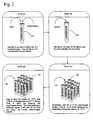
- Protocol 1 The preparation of one embodiment of a fibrin lysis reagent is shown as Protocol 1 in Figure 2 wherein NaCl, MnCl, DTT, DNAse, and plasminogen are added to mixing tube 110. Sodium phosphate is then added to mixing tube 110 and the solution is distributed into 1.5 ml reagent tubes 120 placed on ice. The reagent tubes 120 are frozen to -75°C for approximately 20 minutes. Approximately 2,700 U of streptokinase 130 is added to the wall of reagent tubes 120 just above the frozen plasminogen solution.
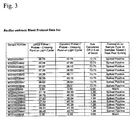
- Figures 3-6 provide PCR results derived from testing blood samples seeded with encapsulated vegetative avirulent Bacillus anthracis were grown according to CDC protocol # CDC.DFA.1.2, stored in 15% glycerol TSB, and frozen at -75°C. Stocks of avirulent Yersinia pestis grown in TSB at 37°C, frozen in 15% glycerol TSB, and frozen at -75°C. Bacterial counts were tested at the time of harvest and retested at the time of sample spike.
- Figures for average Bacillus anthracis CFU per six ml of human blood are derived from post-freezing testing given the large standard deviation encountered in side-by-side post freezing dilution events. No significant cellular death is recognized or expected. A 30% cellular death rate is the highest that is reasonably expected in the worst circumstances. A conservative approach would be to increase all calculated Bacillus anthracis CFU by 30%.
- Figures for average Yersinia pestis CFU per six ml of blood are derived from pre-freezing testing.
- the low standard deviation of pre-freezing count replicates and concordance with post-freezing testing allows use of the pre-freezing bacteria count numbers. This is a conservative approach that can be utilized given the now predictable results that are derived from storing and diluting this organism.
- the present invention reproducibly generates analyte DNA appropriate for PCR testing of pathogens, such as Bacillus anthracis, using patient blood samples that are up to 3 months old.
- Sensitivity is 100% at ⁇ 10 CFU / ml of human blood when using 6 ml of blood collected in a Becton Dickinson Vacutainer (Tables 1 and 2).
- This protocol also allows detection of Yersinia pestis at 100% sensitivity at ⁇ 10 CFU / ml for at least one of four oligo sets according to the more limited data gathered for this organism (Table 3). It should be noted that CDC does not consider samples positive for Y. pestis unless two oligo sets produce an acceptable PCR signal.
- Figure 7 shows a preferred method of the setup of extraction reagents according to the invention.
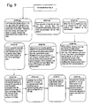
- Figures 8-9 show a method of bacterial recovery and fibrin lysis according to the invention.
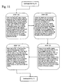
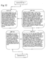
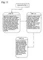
- Figures 10-13 show a preferred method of bacterial lysis and nucleic acid extraction according to the invention.
- the individual enzymes of streptokinase and plasminogen are made into dried powders, mixed, then distributed to disposable tubes.
- Phospholipase A 2 , plasminogen, DNase or Endonuclease, and lipase are suspended and dried in pellets of trehalose buffer.
- Phospholipase A 2 is preferred, any enzyme that will destroy nuclear membrane while keeping bacterial cell wall or viral coats intact may also be used.
- Streptokinase is likewise suspended and dried in pellets of trehalose buffer. At least one pellet of the plasminogen and one pellet of the streptokinase are packaged into tubes as dried reagents.
- Dried reagents of the invention can be resuspended in a 10 ml buffer solution comprising about 10-30 mM Potassium Phosphate, about 10-80 mM Magnesium Chloride, about 20-150 mM Sodium Chloride, about 10-200 mM Aurintricarboxylic Acid and about 1.0-1.2% Triton X-100.
- Aurintricarboxylic Acid is evidenced to provide a level of protection to bacterial nucleic acid without impeding human DNA digestion. The use of Aurintricarboxylic Acid is not described in prior methods of human DNA digestion.
- Methyl 6-O-(N-heptylcarbamoyl)- ⁇ -D-glucopyranoside and Saponin can be substituted for Triton X-100.
- the methyl 6-O-(N-heptylcarbomoyl)- ⁇ -D-glucopyranoside is used at 20-35 mM and the saponin is used at 0.05-0.19 concentration.
- the methyl 6-O-(N-heptylcarbamoyl)- ⁇ -D-glucopyranoside is stored with the phospholipase A 2 , plasminogen, DNase I, and lipase in a Trehalose storage buffer.
- the Trehalose storage buffer can comprise 10 mM Potassium Phosphate pH 7.4, 0.01-0.04% Triton X-100 or methyl 6-O-(N-heptylcarbamoyl)- ⁇ -D-glucopyranoside, 1-5 mM Dithiothreitol, and 0.3-0.5 Trehalose.
- the buffer and enzyme mix are then immediately combined with a 10 ml blood sample, which may be scaled down to 1 ml.
- the sample is then incubated at room temperature for 5-10 minutes.
- the aforementioned components aide blood element solublization through minimizing certain particulates that would otherwise clog filters, impair biosensors or mass spectrometry devices, and impede nucleic acid extraction. Solublization occurs while human DNA is efficiently digested and as viral and/or bacterial DNA remain intact.
- a preferred enzyme combination is comprised of Streptokinase, Plasminogen, DNase or Endonuclease, Phospholipase A 2 , and Lipase.
- enzyme combinations comprising of Streptokinase, Plasminogen, DNase or Endonuclease, and Phospholipase A 2 may also be used.
- Streptokinase, Plasminogen, DNase or Endonuclease may be used, as well as, DNase or Endonuclease, Phospholipase A 2 and Lipase.
- DNase or Endonuclease in combination with Phospholipase A 2 is yet another alternative. The efficacy of the three latter combinations was found to be equal.
- a preferred enzyme combination is comprised of Streptokinase, Plasminogen, DNase or Endonuclease, Phospholipase A 2 , and Lipase.
- enzyme combinations comprising of Streptokinase, Plasminogen, DNase or Endonuclease, and Phospholipase A 2 may also be used.
- Streptokinase, Plasminogen, DNase or Endonuclease may be used.
- the sample is centrifuged for a period of about 20 minutes at 5,000-5,500 x g at a temperature between 10-22°C after incubation.
- the supernatant is then decanted and the pellet washed three times with a 10-20 mM solution of Ecotine/20 mM HEPES pH 7.7 and/or a 20-30 mM solution of Sucrose/20 mM HEPES pH 7.7.
- Protocol 2 sample is centrifuged in similar fashion and the supernatant decanted, followed by sample lysis and DNase or Endonuclease inactivation using about 12.5-25 mg Proteinase K, about 1-1.5% Sodium Dodecyl Sulfate (SDS), about 10-200 mM Aurintricarboxylic Acid and about 10-20 mM Sodium Citrate buffer pH 7.8-8.4.
- SDS Sodium Dodecyl Sulfate
- the sample is allowed to incubate at room temperature for about 10 minutes.
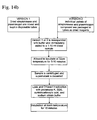
- the digested sample may then be applied to any commercially available nucleic acid extraction method, shown in Figure 14b .
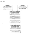
- Protocol 3 the sample is filtered with a 0.22-0.45 ⁇ m filter unit and washed with 10-20 ml of about 10-200 mM Aurintricarboxylic Acid, followed by sample lysis and DNase or Endonuclease inactivation.
- Sample lysis and DNase or Endonuclease inactivation is accomplished by using about 12.5-25 mg Proteinase K, about 1-1.5% SDS, about 10-200 mM Aurintricarboxylic acid, and about 10-20 mM Sodium Citrate buffer.
- the sample is then incubated at room temperature for about 10 minutes. Addition of about 3.5-4.2 M Guanidine Isothiocyanate pH 6.4 is necessary to elute the lysate from the filter surface.
- the nucleic acid extract may then be further purified using a commercially available method.
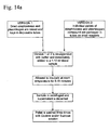
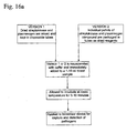
- Protocol 4 Another alternative, referred to as Protocol 4 and shown as Figure 16a , applies the sample directly to a biosensor device that will capture and detect bacteria, virus, fungi, toxins, prions, chemical agents, metabolic markers or native disease state markers developed by the patient's own body in response to these pathogens and agents present in the blood sample.
- the sample is applied directly to a liquid chromatography mass spectrometry device that will detect mass signatures of structural components that comprise bacteria, virus, toxins, prions, and chemical agents present in the blood sample or native disease state markers developed by the patients own body in response to these pathogens and agents present in the blood sample.
- the subject invention concerns materials and methods that can be used for the selective removal of ATA from a sample, such as a blood sample containing nucleic acid.
- ATA is used in procedures for extracting and purifying RNA from cells, viruses, etc., because of its activity as a ribonuclease inhibitor.
- the potent ribonuclease inhibitor ATA will be present during the portion of the nucleic acid extraction process where protein hydrolysis is allowed to proceed at optimal conditions ( i . e ., with ATA and chaotrophic salts such as guanidine thiocyanate).
- Compositions for removing ATA can be provided in either solution form or dry, solid form. Preferably, compositions are provided in a dry solid form to which a liquid or fluid is subsequently added. In an exemplified embodiment, a composition of the invention is used in combination with the lysis reagents described herein.
- a method of the invention comprises contacting a sample that comprises ATA and, optionally, nucleic acid, with a urea/DTPA composition of the invention.
- the sample can comprise any combination of reagents as described in a lysis buffer of the invention.
- a urea/DTPA composition of the invention can be prepared by combining urea with DTPA and optionally EDTA, sodium citrate, and enough of a base, such as sodium hydroxide to achieve pH 8.0 as defined in Table 1.
- the mixture is heated to above about 600 ° C for about 4 hours, dried, ground to a powder, and optionally combined with proteinase K and methyl 6-O-(N-heptylcarbamoyl)- ⁇ -D-glucopyranoside.
- the urea/DTPA reagent is preferably provided in a dried form so as to minimize the downstream sample volumes and obviate the procedure of having to add proteinase K (PK) in a separate step (since PK is not stable for long periods of time in 6.0 to 7.5 M urea).
- the ground urea/DTPA reagent is dried under vacuum and added (at 360 mg /ml blood sample) to blood treated previously with lysis reagents described herein.
- the sample treated with urea/DTPA is incubated about 5 - 10 minutes at about 65 ° C.
- samples treated with ATA and a urea/DTPA reagent of the invention are combined with prior art nucleic acid extraction protocols where binding matrices such as silica or other materials that bind nucleic acids in the presence of chaotrophic salts or where precipitation and centrifugation is used, the ATA will not co-purify with the nucleic acid extract.
- the proteinase K can be inactivated by exposure to temperatures above about 80 ° C for 5 - 10 or more minutes, the sample then cooled to below about 40 ° C, wherein urease is then added to about 1,000 - 100,000 U / ml to break down the urea.
- the sample can then be applied directly to a nucleic acid array device.
- ATA that was present prior to the proteinase K digestion step did not have a negative impact on the PCR kinetics using the nucleic acid extracts that were prepared using the subject methods. Table 1.
- a buffer comprising only ATA can be added to the cells as a first step and subsequently treated as outlined above.
- urea can be added to about 6.0 - 7.5 M to an ATA containing sample, and then combined with prior art chaotrophic salt based binding buffers and silica binding matrices, conduct the protocol according to the literature citation or manufacturer specifications with the exception of heating the chaotrophic salt based binding and wash buffer to about 55 - 65 ° C prior to use with the sample.
- the reaction of urea with the aurintricarboxylic acid (ATA) plus the combination of this solution with chaotrophic salt at about 55 - 65 ° C followed by application to a silica based nucleic acid capture matrix allows the selective binding of nucleic acid to the matrix and exclusion of ATA (passed out in the column flow through).
- blood samples can be treated with ATA containing mixtures described herein combination with pathogen capture using bioactive peptides functionalized on hyaluronic acid also as described herein where the hyaluronic acid in turn acts as a polymeric waveguide.
- the hyaluronic acid is labeled with biotin via carboxyl groups or amines and the excess unbound biotin is subsequently removed via dialysis. Strepavidin is cross-linked and the excess unbound cross linker is removed via dialysis.
- the cross-linked strepavidin is added in 100 - 10,000 molar excess to the biotinylated hyaluronic acid and incubated about 4 - 10 hours with or without an applied electrophoretic or dielectrophoretic field.
- the strepavidin is added in the described ratios, incubated for about 1 - 4 hours with mixing, combined with a photo- activated cross-linking reagent, and cross-linked within as lithography system in order to generate structures positioned within a sample flow path.
- a calcium release at the site of pathogen capture via bioactive peptide or annealing of RNA species is in the presence of thrombin (about 10 - 500 ⁇ g per milliliter) used to trigger the local conversion of reporter molecule labeled fibrinogen (about 10 - 500 ⁇ g per milliliter) to an insoluble fibrin aggregate at the site of pathogen capture via bioactive peptide or annealing of RNA species upon the matrix of the hyaluronic acid polymeric waveguide.
- Reporter molecules can be any molecule that can be detected and include, for example, fluorescent molecules (fluorescein, etc.), radioactive molecules, enzymes, antigens, and the like.
- bioactive peptides include native and modified non-specific virus binding peptides most optimally, such as lactoferrin or fatty acid modified lactoferrin, and native and modified non-specific bacteria binding peptides, most optimally, such as Cecropin P1, but also including, for example, protamine, Buforin I, Buforin II, Defensin, D-Magainin II, Cecrpin A, Cecropin B, Lectin PA-1, and Tritrpticin.
- the modified peptides may be altered in terms of amino acid content and include the salts, esters, amides, and acylated forms thereof.
- bioactive peptides functionalized upon the hyaluronic acid act as pathogen capture moieties.
- pathogen or biomarker capture the hyaluronic acid is broken down using about 1,000 - 1,000,000 units of hyaluronidase / ml of sample within the device.
- the ATA, magnesium chloride, and potassium phosphate components described in the lysis buffer of the present invention are combined, brought to about pH 9.2 - 10 in batches of 100 ml, and heated to boiling until a dry residue forms.
- the dry residue is ground up, dried further under vacuum, and added to the other enzyme, detergent, and Trehalose components, such as those described herein.
Abstract
Description
- This invention relates to the removal of aurintricarboxylic acid (ATA) from a sample.
- ATA is a polymeric anion that has been demonstrated in the literature to be a potential ribonuclease inhibitor. The compound has been decribed previously as an additive to sample lysis buffers where the objective is to extract RNA species from tissue samples. The nucleic acid extract derived from such procedures has been shown to be suitable for hybridization and gel electrophoresis analysis. However, ATA is a potent inhibitor of reverse transcriptase, which is essential for the polymerase chain reaction (PCR) detection of RNA species.
- Published procedures to remove ATA from nucleic acid-containing compositions have revolved around chromatographic procedures that eliminate or remove only a portion of the ATA. For example, prior art nucleic acid extraction method utilize chaotropic salts such as guanidine thiocyanate in the presence of capture matrices such as silica or precipitation methods to concentrate nucleic acids out of crude samples.
- The use of ATA in a proteinase K lysis buffer is potentially superior to 1) chaotropic salts (since they tend to reduce the efficiency of proteinase K-driven protein hydrolysis as evidenced by PCR results); 2) protein-based ribonuclease inhibitors (since these inhibitors would be broken down by proteinase K); and 3) EDTA (which only indirectly inhibits nucleases via chelation of the divalent cations used by those nucleases). In fact, divalent cations must be added to RNA preparations where enzymatic DNA hydrolysis is conducted.
- According to the present invention, a method for nucleic acid extraction from a sample comprising aurintricarboxylic acid (ATA) and a chaotropic salt, comprises adding a composition comprising urea and diethylenetriaminepentaacetate (DTPA) to the sample prior to isolation of the nucleic acid, whereby the nucleic acid is freed of ATA.
- Preferred features of the invention are given in
claims 2 to 15. - The invention also concerns materials and methods for heating a solution of urea, DTPA, optionally containing EDTA, sodium citrate, and sodium chloride, to at least 600ºC for 4 hours, followed by drying and combination with proteinase K and optionally methyl 6-O-(N-heptylcarbamoyl)-a-D-glucopyranoside, and the use of this reagent to allow ATA removal from nucleic acid extracts made with existing prior art methods based on chaotropic salts, followed by centrifugation or methods described herein, to allow downstream hybridization of RNA species directly out of treated whole blood samples.
- The invention also concerns the urea/DTPA reagent that is heat-treated to above 600ºC for 4 hours during production and used in sample treatment as described above, followed by the combination of urease to break down the urea followed by RNA array analysis.
- Aurintricarboxylic acid (ATA) is a polymeric anion that has been demonstrated in the literature to be a potent ribonuclease inhibitor. The compound has been described previously as an additive to sample lysis buffers where the objective is to extract RNA species from tissue samples. The nucleic acid extract derived from such procedures has been shown to be suitable for hybridization and gel electrophoresis analysis. However, ATA is a potent inhibitor of reverse transcriptase, which is essential for the polymerase chain reaction (PCR) detection of RNA species. Published procedures to remove ATA from nucleic acid containing compositions have revolved around chromatographic procedures that eliminate or remove only a portion of the ATA.
- The use of ATA in a proteinase K lysis buffer is potentially superior to 1) chaothrophic salts (since they tend to reduce the efficiency of proteinase K driven protein hydrolysis as evidenced by PCR results); 2) protein based ribonuclease inhibitors (since these inhibitors would be broken down by proteinase K); and 3) EDTA (which only indirectly inhibits nucleases via chelation of the divalent cations used by those nucleases). In fact, divalent cations must be added to RNA preparations where enzymatic DNA hydrolysis is conducted. What has not been demonstrated in prior art is a method where, once added, the complete downstream removal of ATA from nucleic acid extracts can be achieved to the point that down stream reverse transcriptase PCR (RT-PCR) will function.
- Also not previously described in the art is a way to utilize ATA in a lyses buffer to treat a large volume (1-10ml) whole blood sample and after several reagents addition steps move directly to RNA array hybridization using the entire blood sample for one analysis event hence bypassing RNA extraction and amplification.
- Also not previously described in the art is a way to use blood samples treated with ATA containing mixtures in combination with pathogen capture using bioactive peptides functionalized on hyaluronic acid detailed in
U.S. where the hyaluronic acid in turn acts as a polymeric waveguide.application number 10/604,779 - Also not previously described in the art is a way to cause a calcium release at the site of pathogen capture via bioactive peptide or annealing of RNA species so as to trigger the conversion of reporter molecule labeled fibrinogen to insoluble fibrin at the site of pathogen capture via bioactive peptide or annealing of RNA species upon the matrix of the hyaluronic acid polymeric waveguide.
- ATA also serves an important function in the protection of bacterial DNA when that bacteria is present in a blood sample processed with reagents containing high levels (≥100 U/ml) of DNase I as is used in various embodiments contained within
U.S. . In order to achieve RNA detection capabilities that are superior to what can be achieved with technology described inapplication number 10/604,779U.S. , and to do so without additional steps or requirements, the present invention is utilized in combination with blood sample treatment technology described inapplication number 10/604,779U.S. and prior art nucleic acid extraction methods that utilize chaotrophic salts such as guanidine thiocyanate in the presence of capture matrices such as silica or methods that utilize precipitation methods to concentrate nucleic acids out of crude samples.application number 10/604,779 - Accordingly, there remains a need in the art for: 1) a method of destroying and making soluble the spectrum of blood element components (erythrocytes, leukocytes, nuclear membranes, fibrin, and host nucleic acid) without damaging analyte particles (bacteria, virus, fungi, toxin, metabolic markers, disease state markers, or chemical agents) in order to expose and rapidly concentrate (via centrifugation, filtration, or capture) the analyte particles from large volumes of blood, 2) removal of the host DNA and the matrix associated biomass present in the large volume blood sample using a single step enzyme detergent cocktail that is amenable to automation and portable systems, and 3) an analyte particle concentration method that can be coupled to existing manual or automated processes for nucleic acid extraction, biosensor testing, or liquid chromatography separation and mass spectrometry analysis.
- The present invention concerns methods and materials for extracting infectious pathogens from a volume of a sample, such as blood, and includes the steps of creating a fibrin aggregate confining the pathogens and introducing a fibrin lysis reagent to expose the pathogens for analysis and DNAse to facilitate DNA extraction. The fibrin lysis reagents may be composed of DNAse, plasminogen and streptokinase frozen in coincident relation until the fibrin lysis reagent is needed whereby streptokinase enzymatically reacts with plasminogen to form plasmin upon thawing and introduction into the fibrin sample. Preferably, the plasminogen is suspended in an aqueous salt solution prior to freezing including NaCl and Na3PO4. The fibrin lysis reagent is preferably composed of DNAse and Phospholipase A2. The DNAse enzyme is used to facilitate the chemical and physical disruption of pelleted blood elements that result from the previously described protocol. Phospholipase A2 is used to help human DNA digestion by destroying phospholipid bilayers and, hence, destruction of the nuclear membrane.
- The subject invention concerns materials and methods for efficiently removing ATA from a sample, such as a sample consisting of a nucleic acid composition. The subject methods provide a nucleic acid composition sufficiently free of ATA such that a RT-PCR reaction and other reactions involving reverse transcriptase can be performed.
- The subject invention also concerns materials and methods for a mixture of ATA, magnesium chloride, potassium phosphate, and sodium chloride that is dried and combined with other dried components such as those described herein.
- The subject invention also concerns materials and methods for heating a solution of urea, diethylenetriaminepentaacetate (DTPA), optionally containing EDTA, sodium citrate, and sodium chloride, to at least 600 ° C for 4 hours followed by drying and combination with proteinase K and optionally Methyl 6-O- (N-heptylcarbamoyl)-α-D-glucopyranoside and the use of this reagent to allow ATA removal from nucleic acid extracts made with existing prior art methods based on chaotrophic salts or nucleic acid precipitation followed by centrifugation or methods described herein to allow downstream hybridization of RNA species directly out of treated whole blood samples.
- The subject invention also concerns the urea/DTPA reagent that was heat treated to above 600 ° C for 4 hours during production and used in sample treatment as described above followed by the combination of urease to break down the urea followed by RNA array analysis.
- The subject invention also concerns materials and methods for pathogen capture using bioactive peptides functionalized on hyaluronic acid as described herein where the hyaluronic acid in turn acts as a polymeric waveguide.
- The subject invention also concerns methods to cause a calcium release at the site of pathogen capture via bioactive peptide or annealing of RNA species so as to trigger the conversion of reporter molecule labeled fibrinogen to insoluble fibrin at the site of pathogen capture via bioactive peptide or annealing of RNA species upon the matrix of the hyaluronic acid polymeric waveguide.
- The subject invention also concerns materials and methods where the hyaluronic acid matrix that is cross linked utilizing biotin and strepavidin and functionalized with bioactive peptides, such as those described herein, can be subsequently broken down with hyaluronidase in order to facilitate pathogen elution.
-
-
Figure 1 is a diagrammatic view of a method for pathogen analysis. -
Figure 2 is a diagrammatic view of the preparation of the fibrin lysis reagent according toProtocol 1. -
Figure 3 is a table providing data on Bacillus anthracis blood protocol. -
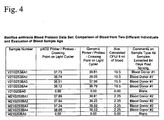
Figure 4 is a table providing data on a comparison of two blood samples from different individuals. -
Figure 5 is a table providing data on an evaluation by a Department of Health laboratorian. -
Figure 6 is a table providing data on Yersinia pestis blood protocol. -
Figure 7 is a diagrammatic view of the setup of extraction reagents according toProtocol 1. -
Figures 8-9 are diagrammatic views of bacterial recovery and fibrin lysis according toProtocol 1. -
Figure 10-13 are diagrammatic views of bacterial lysis and nucleic acid extraction according toProtocol 1. -
Figure 14a is a diagrammatic view of the steps of extracting reagents according toProtocol 2. -
Figure 14b is a diagrammatic view of the steps of extracting reagents according toProtocol 2. -
Figure 15 is a diagrammatic view of the steps of extracting reagents according toProtocol 3. -
Figure 16a is a diagrammatic view of the steps of extracting reagents according to Protocol 4. -
Figure 16b is a diagrammatic view of the steps of extracting reagents according to Protocol 4. -
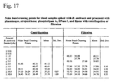
Figure 17 is a table providing data on noise band crossing points for blood samples spiked with B. anthracis and processed with plasminogen, streptokinase, phospholipase A2, DNase I, and lipase with centrifugation or filtration. -
Figure 18 shows sedimentation and solublization of tissue aggregates from 6 ml blood samples exposed to various detergent and enzyme treatments. -
Figure 19 shows filtration characteristics of 6 ml blood samples exposed to various detergent and enzyme treatments. - The present invention will be described in the context of extracting infectious pathogens from a biological sample, such as a volume of blood, and includes the steps of creating a fibrin aggregate confining the pathogens and introducing a fibrin lysis reagent to expose the pathogens for analysis and DNAse to facilitate DNA extraction. The fibrin lysis reagents may be composed of DNAse, plasminogen and streptokinase frozen in coincident relation until the fibrin lysis reagent is needed whereby streptokinase enzymatically reacts with plasminogen to form plasmin upon thawing and introduction into the fibrin sample. Preferably, the plasminogen is suspended in an aqueous salt solution prior to freezing including NaCl and Na3PO4. The fibrin lysis reagent is preferably composed of DNAse and Phospholipase A2. The DNAse enzyme is used to facilitate the chemical and physical disruption of pelleted blood elements that result from the previously described protocol. Phospholipase A2 is used to help human DNA digestion by destroying phospholipid bilayers and, hence, destruction of the nuclear membrane.
- The present invention utilizes resuspension of the dried enzymes in a buffer solution using Potassium Phosphate as an aide to blood element solublization. It is imperative that the streptokinase and plasminogen are not mixed with the buffer solution until immediately prior to the addition of the blood sample. The Potassium Phosphate pH range is about 7.8 to 8.0, which is different from prior art that claims an effective pH range of 7.2 to 7.6. Prior art uses phosphate ion solutions with lower pH to act as a true buffer; however, the current method allows for optimal Phospholipase A2 activity and Magnesium solubility. Magnesium is present in the buffer solution as the divalent cation driving the activity of Phospholipase A2 in the presence of DNase. Prior art uses calcium as the classic divalent cation for driving Phospholipase A2 activity, however, calcium is not compatible with the phosphate ions essential for blood element solublization.
- An embodiment of the present invention includes concentrating and extracting particles such as prions, toxins, metabolic markers, cancerous matter, disease state markers, bacteria, virus, and fungi from a volume of blood by introducing an enzyme-detergent combination to expose pathogens in the blood sample and analyzing the blood sample for the particles now readily identifiable via the extraction. The enzyme-detergent may be a fibrin lysis reagent comprising plasminogen and streptokinase. The plasminogen and streptokinase may be frozen in coincident relation until the fibrin lysis reagent is needed. The streptokinase then reacts with the plasminogen to form plasmin upon thawing. The plasminogen may be suspended in an aqueous salt solution prior to freezing. Suitable salt solutions may include NaCl, NaPO4 or the like. To enhance analysis, the particles may be replicated via polymerase chain reactions (PCR).
- By introducing DNase, the process is facilitated by the conversion of DNA into short fragments thereby contributing to a more rapid and efficient protein hydrolysis process during DNA extraction and lowering the burden of inhibitory human DNA. Similarly, introduction of Endonuclease produces a similar advantage.
- As an alternative to freezing, the enzyme-detergent may include dried streptokinase and dried plasminogen as the fibrin lysis reagents. The dried reagents may then be mixed and distributed into disposable test containers. This embodiment may be particularly useful for field-testing in locations where sophisticated laboratory equipment and controls are unavailable.
- The plasminogen may be combined with Phospholipase A2. DNase, Endonuclease, Lipase, and combinations thereof. The dried enzyme-detergent combination may be suspended in pellets of trehalose buffer and packaged into tubes as a dry reagent. The dried reagents may then be resuspended in a buffer, added to a 1-10ml volume of blood and incubated for about 5-20 minutes at room temperature. More specifically, the dried reagent is comprised of about 1,500-4,500 KU Phospholipase A2, about 5,000-10,000 U Streptokinase, about 2-10 U Plasminogen, about 200-3,650 U DNase, about 200-4,000 U Endonuclease, and about 10,000-100,000 Lipase.
- The solution may be centrifuged for approximately 20 minutes at 5,000-5,500 x g at a temperature of 10-20°C, the supernatant decanted, and the pellet washed. The pellet may be washed three times with a 10-20 mM solution of Ecotine/20 mM HEPES ph 7.7 and/or a 10-20 mM solution of sucrose/20 mM HEPES ph 7.7. The resultant sample may then be applied to a commercially available nucleic acid extraction method.
- Digesting the sample may include lysis and DNase inactivation or lysis and Endonuclease inactivation. 12.5-25 mg proteinase K, 1-105% SDS (sodium dodecyl sulfate), 10-200 mM aurintricarboxylic acid, and 10- 20mM sodium citrate buffer pH 7.8-8.4 may be utilized, the solution allowed to incubate at room temperature for 10 minutes. The sample may then be filtered with a 0.22-0.45 µm filter unit, washed with a 10-200 mM Aurintricarboxylic Acid, digested with lysis and DNase inactivation and/or Endonuclease inactivation, and purified.
- Digesting the sample may include the steps of combining 12.5-25 mg proteinease K, 1-1.5% SDS, 10-200 mM aurintricarboxylic acid, and 10-20 mM sodium citrate buffer, incubating at room temperature for 10 minutes, and eluting the lysate from the filter surface by addition of 3.5-4.2 M guanidine isothiocyanate pH 6.4.
- The solutions may be applied directly to a biosensor device wherein, responsive to the presence of the pathogens in the blood sample, the patient develops pathogenic or native disease state markers that allow for the capture and detection of these markers by the biosensor device. Alternatively, the solution may be applied directly to a liquid chromatography mass spectrometry device whereby, responsive to the presence of the pathogens in the blood sample, the patient develops pathogenic or native disease state markers that allow for the detection of mass signatures associated with the structural components of the pathogens using the mass spectrometry device.
- The buffer can comprise detergent and salts. This may be achieved by aiding blood element solublization by introducing 10-30 mM Potassium Phosphate at a pH range of 7.8 to 8.0, driving Phospholipase A2 activity by adding 10-80 mM Magnesium Chloride as the divalent cation, adding 20-150 mM Sodium Chloride, and including 10-200 mM Aurintricarboxylic Acid during the DNase incubation process. The buffer may also include 1.0-1.2% Triton X-100. Additional steps may include combining 20-35 mM methyl 6-O-(N-heptylcarbamoyl)-α-D-glucopyranoside and 0.05-0.1% Saponin; and storing the enzymes by using a trehalose buffer. Storing the enzymes is accomplished by using a trehalose buffer in combination with methyl 6-O-(N-heptylcarbamoyl)-α-D-glucopyranoside. The trehalose storage buffer comprises 10 mM Potassium Phosphate, 0.01-0.04% Triton X-100, 1-5 mM Dithiothreitol, and 0.3-0.5 M Trehalose.
- In
Figure 1 , ablood draw 30 is performed on a patient. A solution of PBS, pH 7.4 and 1.2% Triton X-100 is added, the blood is vortexed and centrifuged 40 creatingpellet 60 in a 15ml tube 50. Preferably, resins, metal hydroxides, and/or nano materials may be added with the PBS/Triton X-100 solution to capture particles such as bacteria, virus, fungi, cancerous cells, prions, toxins and the like to contribute greater density to these particles. The increase in particle density allows lower speeds to run during centrifugation. - The supernatant is decanted leaving a fibrin aggregate. A
fibrin lysis reagent 70 of the invention is added totube 50 dissolving the fibrin aggregate and leavingpathogens 65 exposed for analysis.Pathogens 65 are vortexed, centrifuged, and subject to lysis to extract the pathogen DNA. The DNA is then replicated 90 and analyzed 100 for the identity of the suspected pathogen. - In an alternative embodiment of the invention, a device would be used to obviate the need for a centrifuge. The device will use flexible electrodes similar to a fish gill to collect particles (such as bacteria, virus, cancerous cells, prions, or toxins). The electrodes will also be used to collect resins and nano materials that have these particles attached to them. The device will resemble a bubble on a surface. An electrical potential will be used to accelerate pathogen capture. The device can be compressed to allow efficient removal of the contents. The device would preferably have the following properties: (1) a rigid base layer and flexible top layer; (2) flexible gills to be mounted on either the top or bottom layer; (3) Strepavidin and hyaluronic acid strands functionalized with bioactive peptides, antibodies, aptomers, molecular imprinted polymers, or metals that attract particles such as bacteria, virus, fungi, toxins, metabolic markers, disease state markers, or chemical agents are to be deposited on the flexible gill electrodes; (4) the flexible layer will have electrodes deposited on it; (5) counter electrodes for the gill electrodes will reside on the opposite side; (6) the average dead volume of the device is 300 micro liters - it is preferred that there is to be no residual material in the device after squeezing out the material from the device; and (7) polyimide will form the flexible portion and the electrodes will be made of Pt, Au, or carbon. The device is preferably used as follows: (1) flow liquid into the device and apply voltage at this time; (2) add chemicals and heat the device; and (3) squeeze out the device to remove all contents. The device is used to prepare a sample for analysis of particles (such as bacteria, virus, cancerous cells, prions, or toxins) using spectrophotometric, mass spectroscopy, antibodies, culture, or nucleic acid (e.g. PCR, NASBA, TMA) based detection systems.
- A filtering device may be used to filter out the particles from blood treated with the Triton X-100 / PBS/ magnesium solutions with enzymes selected from the group of streptokinase, plasminogen, phospholipase A2, DNase, and lipase. A filtering device may also be used to filter out the particles from blood treated with a combination of methyl 6-O-(N-heptylcarbamoyl)-α-D-glucopyranoside, Saponin, and PBS / magnesium plus enzymes selected from the group of streptokinase, plasminogen, phospholipase A2, DNase, and lipase. After washing away the enzyme and detergent treatment reagents and any residual broken down blood components, the particle is ready for analysis or further processing.
- The preparation of one embodiment of a fibrin lysis reagent is shown as
Protocol 1 inFigure 2 wherein NaCl, MnCl, DTT, DNAse, and plasminogen are added to mixingtube 110. Sodium phosphate is then added to mixingtube 110 and the solution is distributed into 1.5ml reagent tubes 120 placed on ice. Thereagent tubes 120 are frozen to -75°C for approximately 20 minutes. Approximately 2,700 U ofstreptokinase 130 is added to the wall ofreagent tubes 120 just above the frozen plasminogen solution. -
Figures 3-6 provide PCR results derived from testing blood samples seeded with encapsulated vegetative avirulent Bacillus anthracis were grown according to CDC protocol # CDC.DFA.1.2, stored in 15% glycerol TSB, and frozen at -75°C. Stocks of avirulent Yersinia pestis grown in TSB at 37°C, frozen in 15% glycerol TSB, and frozen at -75°C. Bacterial counts were tested at the time of harvest and retested at the time of sample spike. - Figures for average Bacillus anthracis CFU per six ml of human blood are derived from post-freezing testing given the large standard deviation encountered in side-by-side post freezing dilution events. No significant cellular death is recognized or expected. A 30% cellular death rate is the highest that is reasonably expected in the worst circumstances. A conservative approach would be to increase all calculated Bacillus anthracis CFU by 30%.
- Figures for average Yersinia pestis CFU per six ml of blood are derived from pre-freezing testing. The low standard deviation of pre-freezing count replicates and concordance with post-freezing testing allows use of the pre-freezing bacteria count numbers. This is a conservative approach that can be utilized given the now predictable results that are derived from storing and diluting this organism.
- The present invention reproducibly generates analyte DNA appropriate for PCR testing of pathogens, such as Bacillus anthracis, using patient blood samples that are up to 3 months old. Sensitivity is 100% at <10 CFU / ml of human blood when using 6 ml of blood collected in a Becton Dickinson Vacutainer (Tables 1 and 2). This protocol also allows detection of Yersinia pestis at 100% sensitivity at <10 CFU / ml for at least one of four oligo sets according to the more limited data gathered for this organism (Table 3). It should be noted that CDC does not consider samples positive for Y. pestis unless two oligo sets produce an acceptable PCR signal.
- In accordance with
Protocol 1,Figure 7 shows a preferred method of the setup of extraction reagents according to the invention.Figures 8-9 show a method of bacterial recovery and fibrin lysis according to the invention.Figures 10-13 show a preferred method of bacterial lysis and nucleic acid extraction according to the invention. - In an alternative embodiment, as shown in
Figures 14-16b , the individual enzymes of streptokinase and plasminogen are made into dried powders, mixed, then distributed to disposable tubes. In another embodiment, Phospholipase A2, plasminogen, DNase or Endonuclease, and lipase are suspended and dried in pellets of trehalose buffer. Although Phospholipase A2 is preferred, any enzyme that will destroy nuclear membrane while keeping bacterial cell wall or viral coats intact may also be used. Streptokinase is likewise suspended and dried in pellets of trehalose buffer. At least one pellet of the plasminogen and one pellet of the streptokinase are packaged into tubes as dried reagents. - Dried reagents of the invention can be resuspended in a 10 ml buffer solution comprising about 10-30 mM Potassium Phosphate, about 10-80 mM Magnesium Chloride, about 20-150 mM Sodium Chloride, about 10-200 mM Aurintricarboxylic Acid and about 1.0-1.2% Triton X-100. Aurintricarboxylic Acid is evidenced to provide a level of protection to bacterial nucleic acid without impeding human DNA digestion. The use of Aurintricarboxylic Acid is not described in prior methods of human DNA digestion. Methyl 6-O-(N-heptylcarbamoyl)-α-D-glucopyranoside and Saponin can be substituted for Triton X-100. In one embodiment, the methyl 6-O-(N-heptylcarbomoyl)-α-D-glucopyranoside is used at 20-35 mM and the saponin is used at 0.05-0.19 concentration. The methyl 6-O-(N-heptylcarbamoyl)-α-D-glucopyranoside is stored with the phospholipase A2, plasminogen, DNase I, and lipase in a Trehalose storage buffer. Substitution of the Triton X-100 with the methyl 6-O-(N-heptylcarbamoyl)-α-D-glucopyranoside and saponin solution allows for the efficient activity of Phospholipase A2, provides the action of breaking up protein aggregates without denaturation, and is more genial to bacterial walls than Triton X-100. Use of Saponin and methyl 6-O-(N-heptylcarbamoyl)-α-D-glucopyranoside in this combination is not described in the prior art. The Trehalose storage buffer can comprise 10 mM Potassium Phosphate pH 7.4, 0.01-0.04% Triton X-100 or methyl 6-O-(N-heptylcarbamoyl)-α-D-glucopyranoside, 1-5 mM Dithiothreitol, and 0.3-0.5 Trehalose. The buffer and enzyme mix are then immediately combined with a 10 ml blood sample, which may be scaled down to 1 ml. The sample is then incubated at room temperature for 5-10 minutes. The aforementioned components aide blood element solublization through minimizing certain particulates that would otherwise clog filters, impair biosensors or mass spectrometry devices, and impede nucleic acid extraction. Solublization occurs while human DNA is efficiently digested and as viral and/or bacterial DNA remain intact.
- In accordance with
Protocol 2 and 4, a preferred enzyme combination is comprised of Streptokinase, Plasminogen, DNase or Endonuclease, Phospholipase A2, and Lipase. Alternatively, enzyme combinations comprising of Streptokinase, Plasminogen, DNase or Endonuclease, and Phospholipase A2 may also be used. In another alternative combination, Streptokinase, Plasminogen, DNase or Endonuclease may be used, as well as, DNase or Endonuclease, Phospholipase A2 and Lipase. DNase or Endonuclease in combination with Phospholipase A2 is yet another alternative. The efficacy of the three latter combinations was found to be equal. - In accordance with
Protocol 3, a preferred enzyme combination is comprised of Streptokinase, Plasminogen, DNase or Endonuclease, Phospholipase A2 , and Lipase. Alternatively, enzyme combinations comprising of Streptokinase, Plasminogen, DNase or Endonuclease, and Phospholipase A2 may also be used. In another alternative combination, Streptokinase, Plasminogen, DNase or Endonuclease may be used. - As shown in
Figure 14 withProtocol 2, the sample is centrifuged for a period of about 20 minutes at 5,000-5,500 x g at a temperature between 10-22°C after incubation. The supernatant is then decanted and the pellet washed three times with a 10-20 mM solution of Ecotine/20 mM HEPES pH 7.7 and/or a 20-30 mM solution of Sucrose/20 mM HEPES pH 7.7. - Alternatively after incubation, the
Protocol 2 sample is centrifuged in similar fashion and the supernatant decanted, followed by sample lysis and DNase or Endonuclease inactivation using about 12.5-25 mg Proteinase K, about 1-1.5% Sodium Dodecyl Sulfate (SDS), about 10-200 mM Aurintricarboxylic Acid and about 10-20 mM Sodium Citrate buffer pH 7.8-8.4. The sample is allowed to incubate at room temperature for about 10 minutes. The digested sample may then be applied to any commercially available nucleic acid extraction method, shown inFigure 14b . - Yet in another alternative, referred to as
Protocol 3 and depicted inFigure 15 , the sample is filtered with a 0.22-0.45 µm filter unit and washed with 10-20 ml of about 10-200 mM Aurintricarboxylic Acid, followed by sample lysis and DNase or Endonuclease inactivation. Sample lysis and DNase or Endonuclease inactivation is accomplished by using about 12.5-25 mg Proteinase K, about 1-1.5% SDS, about 10-200 mM Aurintricarboxylic acid, and about 10-20 mM Sodium Citrate buffer. The sample is then incubated at room temperature for about 10 minutes. Addition of about 3.5-4.2 M Guanidine Isothiocyanate pH 6.4 is necessary to elute the lysate from the filter surface. The nucleic acid extract may then be further purified using a commercially available method. - Another alternative, referred to as Protocol 4 and shown as
Figure 16a , applies the sample directly to a biosensor device that will capture and detect bacteria, virus, fungi, toxins, prions, chemical agents, metabolic markers or native disease state markers developed by the patient's own body in response to these pathogens and agents present in the blood sample. - In yet.another Protocol 4 alternative shown in
Figure 16b , the sample is applied directly to a liquid chromatography mass spectrometry device that will detect mass signatures of structural components that comprise bacteria, virus, toxins, prions, and chemical agents present in the blood sample or native disease state markers developed by the patients own body in response to these pathogens and agents present in the blood sample. - The subject invention concerns materials and methods that can be used for the selective removal of ATA from a sample, such as a blood sample containing nucleic acid. Typically, ATA is used in procedures for extracting and purifying RNA from cells, viruses, etc., because of its activity as a ribonuclease inhibitor. In one embodiment of the claimed invention, the potent ribonuclease inhibitor ATA will be present during the portion of the nucleic acid extraction process where protein hydrolysis is allowed to proceed at optimal conditions (i.e., with ATA and chaotrophic salts such as guanidine thiocyanate). Compositions for removing ATA can be provided in either solution form or dry, solid form. Preferably, compositions are provided in a dry solid form to which a liquid or fluid is subsequently added. In an exemplified embodiment, a composition of the invention is used in combination with the lysis reagents described herein.
- In one embodiment, a method of the invention comprises contacting a sample that comprises ATA and, optionally, nucleic acid, with a urea/DTPA composition of the invention. In one embodiment, the sample can comprise any combination of reagents as described in a lysis buffer of the invention. A urea/DTPA composition of the invention can be prepared by combining urea with DTPA and optionally EDTA, sodium citrate, and enough of a base, such as sodium hydroxide to achieve pH 8.0 as defined in Table 1. In one embodiment, the mixture is heated to above about 600 ° C for about 4 hours, dried, ground to a powder, and optionally combined with proteinase K and methyl 6-O-(N-heptylcarbamoyl)-α-D-glucopyranoside. The urea/DTPA reagent is preferably provided in a dried form so as to minimize the downstream sample volumes and obviate the procedure of having to add proteinase K (PK) in a separate step (since PK is not stable for long periods of time in 6.0 to 7.5 M urea). The ground urea/DTPA reagent is dried under vacuum and added (at 360 mg /ml blood sample) to blood treated previously with lysis reagents described herein. The sample treated with urea/DTPA is incubated about 5 - 10 minutes at about 65 ° C. When samples treated with ATA and a urea/DTPA reagent of the invention are combined with prior art nucleic acid extraction protocols where binding matrices such as silica or other materials that bind nucleic acids in the presence of chaotrophic salts or where precipitation and centrifugation is used, the ATA will not co-purify with the nucleic acid extract. In another embodiment, the proteinase K can be inactivated by exposure to temperatures above about 80 ° C for 5 - 10 or more minutes, the sample then cooled to below about 40 ° C, wherein urease is then added to about 1,000 - 100,000 U / ml to break down the urea. The sample can then be applied directly to a nucleic acid array device.
- Using the subject methods in conjunction with PCR, 10 CFU Bacillus anthracis per 10 ml of blood can be detected. Also, there was no difference in the RT-PCR kinetics derived from PBS samples where 1 ng of MS2 RNA was seeded into nucleic acid extracts made with and without ATA. Also, 1,000 pfu polio sabin III virus / 8 ml SPS whole blood was detected by RT-PCR when lysis reagents described herein were combined with the urea/DTPA reagent and protocol described above. By using a urea/DTPA reagent of the invention, ATA that was present prior to the proteinase K digestion step did not have a negative impact on the PCR kinetics using the nucleic acid extracts that were prepared using the subject methods.
Table 1. Contents of powdered Urea/DTPA reagent (upon addition of 1 ml sample to 360 mg reagent Urea 6.0 - 7.5 M Methyl 6-O- (N-heptylcarbamoyl)-α-D-glucopyranoside 10 - 20 mg / ml Proteinase K 600 - 1,000 µg / ml EDTA 20 - 70 mM DTPA 20 - 70 mM Sodium Citrate 120 mM Sodium Hydroxide add to pH 8.0 - In another embodiment, if a sample is not processed with lysis reagents, such as those described herein, then a buffer comprising only ATA can be added to the cells as a first step and subsequently treated as outlined above.
- In another embodiment, urea can be added to about 6.0 - 7.5 M to an ATA containing sample, and then combined with prior art chaotrophic salt based binding buffers and silica binding matrices, conduct the protocol according to the literature citation or manufacturer specifications with the exception of heating the chaotrophic salt based binding and wash buffer to about 55 - 65 ° C prior to use with the sample. The reaction of urea with the aurintricarboxylic acid (ATA) plus the combination of this solution with chaotrophic salt at about 55 - 65 ° C followed by application to a silica based nucleic acid capture matrix allows the selective binding of nucleic acid to the matrix and exclusion of ATA (passed out in the column flow through). It is the combination of reaction with urea and heat that provides for the exclusion of ATA from the silica capture matrix while nucleic acid binds readily. The above described urea/DTPA reagent produced by heating above 600 ° C during production eliminates the need for this chaotrophic salt heat step and allows for more complete removal of the ATA.
- In another embodiment, blood samples can be treated with ATA containing mixtures described herein combination with pathogen capture using bioactive peptides functionalized on hyaluronic acid also as described herein where the hyaluronic acid in turn acts as a polymeric waveguide. The hyaluronic acid is labeled with biotin via carboxyl groups or amines and the excess unbound biotin is subsequently removed via dialysis. Strepavidin is cross-linked and the excess unbound cross linker is removed via dialysis. The cross-linked strepavidin is added in 100 - 10,000 molar excess to the biotinylated hyaluronic acid and incubated about 4 - 10 hours with or without an applied electrophoretic or dielectrophoretic field. Alternatively, the strepavidin is added in the described ratios, incubated for about 1 - 4 hours with mixing, combined with a photo- activated cross-linking reagent, and cross-linked within as lithography system in order to generate structures positioned within a sample flow path. In this system a calcium release at the site of pathogen capture via bioactive peptide or annealing of RNA species is in the presence of thrombin (about 10 - 500 µg per milliliter) used to trigger the local conversion of reporter molecule labeled fibrinogen (about 10 - 500 µg per milliliter) to an insoluble fibrin aggregate at the site of pathogen capture via bioactive peptide or annealing of RNA species upon the matrix of the hyaluronic acid polymeric waveguide. Reporter molecules can be any molecule that can be detected and include, for example, fluorescent molecules (fluorescein, etc.), radioactive molecules, enzymes, antigens, and the like. As used herein, bioactive peptides include native and modified non-specific virus binding peptides most optimally, such as lactoferrin or fatty acid modified lactoferrin, and native and modified non-specific bacteria binding peptides, most optimally, such as Cecropin P1, but also including, for example, protamine, Buforin I, Buforin II, Defensin, D-Magainin II, Cecrpin A, Cecropin B, Lectin PA-1, and Tritrpticin. The modified peptides may be altered in terms of amino acid content and include the salts, esters, amides, and acylated forms thereof.
- In another embodiment, the bioactive peptides functionalized upon the hyaluronic acid (that is cross linked via biotin and strepavidin) act as pathogen capture moieties. Upon pathogen or biomarker capture, the hyaluronic acid is broken down using about 1,000 - 1,000,000 units of hyaluronidase / ml of sample within the device.
- In another embodiment, the ATA, magnesium chloride, and potassium phosphate components described in the lysis buffer of the present invention are combined, brought to about pH 9.2 - 10 in batches of 100 ml, and heated to boiling until a dry residue forms. The dry residue is ground up, dried further under vacuum, and added to the other enzyme, detergent, and Trehalose components, such as those described herein.
Claims (15)
- A method for nucleic acid extraction from a sample comprising aurintricarboxylic acid (ATA) and a chaotropic salt, comprising adding a composition comprising urea and diethylenetriaminepentaacetate (DTPA) to the sample prior to isolation of the nucleic acid, whereby the nucleic acid is freed of ATA.
- A method according to claim 1, wherein the composition further comprises sodium citrate.
- A method according to claim 1 or claim 2, wherein the composition further comprises proteinase K.
- A method according to any preceding claim, wherein the composition further comprises methyl 6-O-(N-heptylcarbamoyl)-α-D-glucopyranoside.
- A method according to any preceding claim, wherein the composition further comprises ethylenediaminetetraacetate.
- A method according to any preceding claim, wherein the pH of the sample is brought to about 8.0.
- A method according to any preceding claim, wherein the composition further comprises sodium hydroxide.
- A method according to any preceding claim, wherein the composition is a dry powder.
- A method according to any preceding claim, wherein the concentration of urea in the sample is 6.0 to 7.5M.
- A method according to any preceding claim, wherein the sample is subsequently contacted with urease.
- A method according to claim 10, wherein 1,000 to 100,000 units of the urease are added per ml of sample.
- A method according to claim 1, which comprises the prior step of heating a solution comprising urea and DTPA to at least 600°C.
- A method according to claim 12, wherein the urea and DTPA are heated for about 4 hours.
- A method according to any preceding claim, wherein the nucleic acid extraction comprises washing and/or binding steps, and the sample is heated to between 55 and 65°C during the washing and/or binding steps.
- A method according to any preceding claim, wherein the sample is blood.
Priority Applications (7)
| Application Number | Priority Date | Filing Date | Title |
|---|---|---|---|
| EP09159214A EP2103694B1 (en) | 2003-08-15 | 2004-08-16 | Materials and methods for capture of pathogens and removal of aurintricarboxylic acid from a sample |
| SI200431179T SI1654387T1 (en) | 2003-08-15 | 2004-08-16 | Materials and methods for capture of pathogens and removal of aurintricarboxylic acid from a sample |
| PL04801986T PL1654387T3 (en) | 2003-08-15 | 2004-08-16 | Materials and methods for capture of pathogens and removal of aurintricarboxylic acid from a sample |
| DK09159214.7T DK2103694T3 (en) | 2003-08-15 | 2004-08-16 | Materials and Methods for Capturing Pathogens and Removing aurintricarboxylic Acid from a Sample |
| PL09159214T PL2103694T3 (en) | 2003-08-15 | 2004-08-16 | Materials and methods for capture of pathogens and removal of aurintricarboxylic acid from a sample |
| EP09165293.3A EP2112232B1 (en) | 2003-08-15 | 2004-08-16 | Methods for extracting pathogen nucleic acid from a blood sample using aurintricarboxylic acid |
| CY20091100540T CY1110678T1 (en) | 2003-08-15 | 2009-05-21 | MATERIALS AND METHODS FOR TRADING PATHOGENES AND DETERMINATION OF CHRYSINOTRIC CARBONIC ACID FROM A SAMPLE |
Applications Claiming Priority (3)
| Application Number | Priority Date | Filing Date | Title |
|---|---|---|---|
| US10/604,779 US7998699B2 (en) | 2002-08-15 | 2003-08-15 | Early detection of pathogens in blood |
| US48189204P | 2004-01-14 | 2004-01-14 | |
| PCT/US2004/026606 WO2005021799A2 (en) | 2003-08-15 | 2004-08-16 | Materials and methods for capture of pathogens and removal of aurintricarboxylic acid from a sample |
Related Child Applications (2)
| Application Number | Title | Priority Date | Filing Date |
|---|---|---|---|
| EP09159214A Division EP2103694B1 (en) | 2003-08-15 | 2004-08-16 | Materials and methods for capture of pathogens and removal of aurintricarboxylic acid from a sample |
| EP09165293.3A Division EP2112232B1 (en) | 2003-08-15 | 2004-08-16 | Methods for extracting pathogen nucleic acid from a blood sample using aurintricarboxylic acid |
Publications (2)
| Publication Number | Publication Date |
|---|---|
| EP1654387A2 EP1654387A2 (en) | 2006-05-10 |
| EP1654387B1 true EP1654387B1 (en) | 2009-05-06 |
Family
ID=34840999
Family Applications (3)
| Application Number | Title | Priority Date | Filing Date |
|---|---|---|---|
| EP09159214A Not-in-force EP2103694B1 (en) | 2003-08-15 | 2004-08-16 | Materials and methods for capture of pathogens and removal of aurintricarboxylic acid from a sample |
| EP09165293.3A Not-in-force EP2112232B1 (en) | 2003-08-15 | 2004-08-16 | Methods for extracting pathogen nucleic acid from a blood sample using aurintricarboxylic acid |
| EP04801986A Not-in-force EP1654387B1 (en) | 2003-08-15 | 2004-08-16 | Materials and methods for capture of pathogens and removal of aurintricarboxylic acid from a sample |
Family Applications Before (2)
| Application Number | Title | Priority Date | Filing Date |
|---|---|---|---|
| EP09159214A Not-in-force EP2103694B1 (en) | 2003-08-15 | 2004-08-16 | Materials and methods for capture of pathogens and removal of aurintricarboxylic acid from a sample |
| EP09165293.3A Not-in-force EP2112232B1 (en) | 2003-08-15 | 2004-08-16 | Methods for extracting pathogen nucleic acid from a blood sample using aurintricarboxylic acid |
Country Status (13)
| Country | Link |
|---|---|
| US (12) | US7416843B2 (en) |
| EP (3) | EP2103694B1 (en) |
| JP (2) | JP4662932B2 (en) |
| AT (2) | ATE430812T1 (en) |
| AU (2) | AU2004269340B2 (en) |
| DE (1) | DE602004020999D1 (en) |
| DK (2) | DK1654387T3 (en) |
| ES (1) | ES2325445T3 (en) |
| NO (1) | NO20061189L (en) |
| PL (2) | PL2103694T3 (en) |
| PT (2) | PT2103694E (en) |
| SI (1) | SI1654387T1 (en) |
| WO (1) | WO2005021799A2 (en) |
Families Citing this family (24)
| Publication number | Priority date | Publication date | Assignee | Title |
|---|---|---|---|---|
| US7998699B2 (en) | 2002-08-15 | 2011-08-16 | University Of South Florida | Early detection of pathogens in blood |
| DK1654387T3 (en) | 2003-08-15 | 2009-06-22 | Univ South Florida | Materials and Methods for Capturing Pathogens and Removing aurintricarboxylic Acid from a Sample |
| CY1110678T1 (en) * | 2003-08-15 | 2015-06-10 | University Of South Florida | MATERIALS AND METHODS FOR TRADING PATHOGENES AND DETERMINATION OF CHRYSINOTRIC CARBONIC ACID FROM A SAMPLE |
| KR101397386B1 (en) | 2005-12-13 | 2014-05-26 | 엑스테라 메디컬 코퍼레이션 | Method for extracorporeal removal of a pathogenic microbe, an inflammatory cell or an inflammatory protein from blood |
| CA2576682A1 (en) | 2006-03-08 | 2007-09-08 | Martinrea International Inc. | Electrolyte delivery system |
| WO2008118566A2 (en) * | 2007-02-28 | 2008-10-02 | Ge Healthcare Bio-Sciences Corp. | Ambient temperature stable chemical/biological reagents on membranes or filters |
| JP5925693B2 (en) | 2009-12-01 | 2016-05-25 | エクステラ・メディカル・コーポレーション | Method for removing cytokines from blood using surface-immobilized polysaccharides |
| EP2333105A1 (en) | 2009-12-08 | 2011-06-15 | Koninklijke Philips Electronics N.V. | Selective lysis of cells |
| US20110225809A1 (en) * | 2010-03-17 | 2011-09-22 | Alan Francis Daher | Apparatus for removably attaching an item to a surface |
| CA2811493C (en) * | 2010-09-15 | 2020-06-16 | Spinomix S.A. | Method for separating target molecules or particles from fibrinogen-containing samples including blood components |
| WO2012112724A1 (en) | 2011-02-15 | 2012-08-23 | Exthera Medical, Llc | Device and method for removal of blood-borne pathogens, toxins and inflammatory cytokines |
| JP2013055888A (en) * | 2011-09-07 | 2013-03-28 | Toyobo Co Ltd | Quick and easy bacterium detection from blood culture sample |
| EP2861273B1 (en) | 2012-06-13 | 2017-08-23 | ExThera Medical Corporation | Use of heparin and carbohydrates to treat cancer |
| FR3001464B1 (en) | 2013-01-25 | 2016-02-26 | Biomerieux Sa | METHOD FOR SPECIFIC ISOLATION OF NUCLEIC ACIDS OF INTEREST |
| FR3002452B1 (en) * | 2013-02-28 | 2016-02-12 | Dermaconcept Jmc | TOPIC ANTIMICROBIAL DERMATOLOGICAL COMPOSITION |
| EP3620218A1 (en) | 2013-06-24 | 2020-03-11 | ExThera Medical Corporation | Blood filtration system containing mannose coated substrate |
| JP6806563B2 (en) | 2013-11-08 | 2021-01-06 | エクステラ・メディカル・コーポレーション | Diagnosis of infectious diseases using adsorption medium |
| MX2016013873A (en) | 2014-04-24 | 2017-03-09 | Exthera Medical Corp | Method for removing bacteria from blood using high flow rate. |
| ES2821698T3 (en) | 2014-09-22 | 2021-04-27 | Exthera Medical Corp | Portable hemoperfusion device |
| CN104749009B (en) * | 2015-03-30 | 2018-05-04 | 上海云泽生物科技有限公司 | immunosuppressant drug extraction reagent for immunoassay |
| US11911551B2 (en) | 2016-03-02 | 2024-02-27 | Exthera Medical Corporation | Method for treating drug intoxication |
| US10786615B2 (en) | 2016-03-02 | 2020-09-29 | Exthera Medical Corporation | Method for treating drug intoxication |
| US11168323B2 (en) | 2017-06-01 | 2021-11-09 | Nantomics Llc | DNA stabilization of RNA |
| US20220354932A1 (en) * | 2019-09-06 | 2022-11-10 | The Trustees Of Indiana University | Novel compositions for disrupting biofilms |
Family Cites Families (33)
| Publication number | Priority date | Publication date | Assignee | Title |
|---|---|---|---|---|
| US1541947A (en) | 1922-03-21 | 1925-06-16 | Electric Water Sterilizer & Oz | Electrolytic cell |
| US3130038A (en) | 1961-03-13 | 1964-04-21 | Int Minerals & Chem Corp | Granular fertilizers |
| US3245780A (en) | 1961-04-13 | 1966-04-12 | Phillip A Hunt Chemical Corp | Nickel stripping ammoniacal solution containing a nitro benzene compound |
| US3378443A (en) | 1966-04-05 | 1968-04-16 | American Cyanamid Co | Anhydrous liquid suspensions of biologics |
| US4693972A (en) * | 1984-01-16 | 1987-09-15 | Becton, Dickinson And Company | Composition and method for rapid detection of microorganisms in clinical samples |
| US4636524A (en) | 1984-12-06 | 1987-01-13 | Biomatrix, Inc. | Cross-linked gels of hyaluronic acid and products containing such gels |
| US6027877A (en) | 1993-11-04 | 2000-02-22 | Gene Check, Inc. | Use of immobilized mismatch binding protein for detection of mutations and polymorphisms, purification of amplified DNA samples and allele identification |
| EP0763754B1 (en) | 1995-09-13 | 2003-01-08 | Seikagaku Kogyo Kabushiki Kaisha (Seikagaku Corporation) | Photocured crosslinked-hyaluronic acid contact lens |
| US6825047B1 (en) | 1996-04-03 | 2004-11-30 | Applera Corporation | Device and method for multiple analyte detection |
| US5925552A (en) | 1996-04-25 | 1999-07-20 | Medtronic, Inc. | Method for attachment of biomolecules to medical devices surfaces |
| US6103525A (en) | 1996-10-17 | 2000-08-15 | The Regents Of The University Of California | Hybridoma cell lines producing monoclonal antibodies that bind to human plasma hyaluronidase |
| SE9604610D0 (en) | 1996-12-16 | 1996-12-16 | Noviscens Ab | Medical composition |
| US6368871B1 (en) | 1997-08-13 | 2002-04-09 | Cepheid | Non-planar microstructures for manipulation of fluid samples |
| JP4196236B2 (en) * | 1998-03-17 | 2008-12-17 | 東洋紡績株式会社 | Nucleic acid amplification reagent and sequence-specific nucleic acid amplification method |
| US6091979A (en) | 1998-07-07 | 2000-07-18 | Children's Medical Center Corporation | Subdural electrode arrays for monitoring cortical electrical activity |
| US6592814B2 (en) | 1998-10-02 | 2003-07-15 | Johnson & Johnson Vision Care, Inc. | Biomedical devices with antimicrobial coatings |
| AU3347000A (en) | 1999-01-19 | 2000-08-01 | Children's Hospital Of Philadelphia, The | Hydrogel compositions for controlled delivery of virus vectors and methods of use thereof |
| ATE419528T1 (en) | 1999-04-28 | 2009-01-15 | Eidgenoess Tech Hochschule | POLYIONIC COATINGS FOR ANALYTICAL AND SENSOR DEVICES |
| US7306924B2 (en) | 2000-04-17 | 2007-12-11 | Purdue Research Foundation | Biosensor and related method |
| US6632610B2 (en) * | 2000-10-12 | 2003-10-14 | Gensat S.A. | Methods of identification and isolation of polynucleotides containing nucleic acid differences |
| US7001724B1 (en) * | 2000-11-28 | 2006-02-21 | Applera Corporation | Compositions, methods, and kits for isolating nucleic acids using surfactants and proteases |
| US7192445B2 (en) | 2000-12-06 | 2007-03-20 | Astra Tech Ab | Medical prosthetic devices and implants having improved biocompatibility |
| DE10108483A1 (en) | 2001-02-22 | 2002-09-05 | Bayer Ag | Phosphorus-containing polymers for optical signal converters |
| AU2002258490A1 (en) | 2001-03-12 | 2003-06-17 | Clemson University | Polysaccharide-based polmerizable hydrogels |
| CN100494360C (en) | 2001-03-22 | 2009-06-03 | 博奥生物有限公司 | Process for separating cells and its application |
| EP1463796B1 (en) | 2001-11-30 | 2013-01-09 | Fluidigm Corporation | Microfluidic device and methods of using same |
| US7482116B2 (en) * | 2002-06-07 | 2009-01-27 | Dna Genotek Inc. | Compositions and methods for obtaining nucleic acids from sputum |
| US7998699B2 (en) | 2002-08-15 | 2011-08-16 | University Of South Florida | Early detection of pathogens in blood |
| US7402279B2 (en) | 2002-10-31 | 2008-07-22 | Agilent Technologies, Inc. | Device with integrated microfluidic and electronic components |
| JP4166587B2 (en) | 2003-01-24 | 2008-10-15 | 株式会社サキコーポレーション | Appearance inspection apparatus and volume inspection method |
| US7476363B2 (en) | 2003-04-03 | 2009-01-13 | Fluidigm Corporation | Microfluidic devices and methods of using same |
| JP4773348B2 (en) | 2003-07-12 | 2011-09-14 | アクセラー8 テクノロジー コーポレイション | Sensitive and rapid biodetection method |
| DK1654387T3 (en) | 2003-08-15 | 2009-06-22 | Univ South Florida | Materials and Methods for Capturing Pathogens and Removing aurintricarboxylic Acid from a Sample |
-
2004
- 2004-08-16 DK DK04801986T patent/DK1654387T3/en active
- 2004-08-16 AT AT04801986T patent/ATE430812T1/en active
- 2004-08-16 DE DE602004020999T patent/DE602004020999D1/en active Active
- 2004-08-16 AU AU2004269340A patent/AU2004269340B2/en not_active Ceased
- 2004-08-16 JP JP2006523971A patent/JP4662932B2/en not_active Expired - Fee Related
- 2004-08-16 PL PL09159214T patent/PL2103694T3/en unknown
- 2004-08-16 PT PT09159214T patent/PT2103694E/en unknown
- 2004-08-16 SI SI200431179T patent/SI1654387T1/en unknown
- 2004-08-16 PT PT04801986T patent/PT1654387E/en unknown
- 2004-08-16 PL PL04801986T patent/PL1654387T3/en unknown
- 2004-08-16 EP EP09159214A patent/EP2103694B1/en not_active Not-in-force
- 2004-08-16 ES ES04801986T patent/ES2325445T3/en active Active
- 2004-08-16 DK DK09159214.7T patent/DK2103694T3/en active
- 2004-08-16 EP EP09165293.3A patent/EP2112232B1/en not_active Not-in-force
- 2004-08-16 EP EP04801986A patent/EP1654387B1/en not_active Not-in-force
- 2004-08-16 WO PCT/US2004/026606 patent/WO2005021799A2/en active Application Filing
- 2004-08-16 AT AT09159214T patent/ATE510931T1/en active
-
2005
- 2005-01-14 US US11/035,667 patent/US7416843B2/en not_active Expired - Lifetime
-
2006
- 2006-03-14 NO NO20061189A patent/NO20061189L/en not_active Application Discontinuation
-
2008
- 2008-05-08 US US12/117,505 patent/US8465966B2/en not_active Expired - Fee Related
-
2010
- 2010-05-11 JP JP2010108850A patent/JP5253451B2/en not_active Expired - Fee Related
- 2010-08-05 AU AU2010207760A patent/AU2010207760B2/en not_active Ceased
-
2012
- 2012-12-21 US US13/725,196 patent/US8592200B2/en not_active Expired - Fee Related
- 2012-12-21 US US13/725,113 patent/US8916698B2/en not_active Expired - Fee Related
- 2012-12-21 US US13/725,924 patent/US8815502B2/en not_active Expired - Fee Related
- 2012-12-21 US US13/725,651 patent/US20130130302A1/en not_active Abandoned
- 2012-12-21 US US13/725,435 patent/US8759479B2/en not_active Expired - Fee Related
- 2012-12-21 US US13/725,303 patent/US8574866B2/en not_active Expired - Fee Related
- 2012-12-21 US US13/725,568 patent/US8927243B2/en not_active Expired - Fee Related
- 2012-12-21 US US13/725,495 patent/US8962243B2/en not_active Expired - Fee Related
-
2013
- 2013-01-04 US US13/734,367 patent/US8785176B2/en not_active Expired - Fee Related
- 2013-03-28 US US13/852,938 patent/US8735090B2/en not_active Expired - Lifetime
Also Published As
Similar Documents
| Publication | Publication Date | Title |
|---|---|---|
| AU2010207760B2 (en) | Materials and methods for capture of pathogens and removal of aurintricarboxylic acid from a sample | |
| US7993870B2 (en) | Early detection of pathogens in blood | |
| JP2013005819A (en) | Material and method for capture of pathogen and removal of aurintricarboxylic acid from sample | |
| AU2012216623B2 (en) | Materials and methods for capture of pathogens and removal of aurintricarboxylic acid from a sample | |
| AU2013203143B2 (en) | Materials and methods for capture of pathogens and removal of aurintricarboxylic acid from a sample |
Legal Events
| Date | Code | Title | Description |
|---|---|---|---|
| PUAI | Public reference made under article 153(3) epc to a published international application that has entered the european phase |
Free format text: ORIGINAL CODE: 0009012 |
|
| 17P | Request for examination filed |
Effective date: 20060307 |
|
| AK | Designated contracting states |
Kind code of ref document: A2 Designated state(s): AT BE BG CH CY CZ DE DK EE ES FI FR GB GR HU IE IT LI LU MC NL PL PT RO SE SI SK TR |
|
| DAX | Request for extension of the european patent (deleted) | ||
| 17Q | First examination report despatched |
Effective date: 20061113 |
|
| GRAP | Despatch of communication of intention to grant a patent |
Free format text: ORIGINAL CODE: EPIDOSNIGR1 |
|
| GRAS | Grant fee paid |
Free format text: ORIGINAL CODE: EPIDOSNIGR3 |
|
| GRAA | (expected) grant |
Free format text: ORIGINAL CODE: 0009210 |
|
| RAP1 | Party data changed (applicant data changed or rights of an application transferred) |
Owner name: UNIVERSITY OF SOUTH FLORIDA |
|
| AK | Designated contracting states |
Kind code of ref document: B1 Designated state(s): AT BE BG CH CY CZ DE DK EE ES FI FR GB GR HU IE IT LI LU MC NL PL PT RO SE SI SK TR |
|
| REG | Reference to a national code |
Ref country code: GB Ref legal event code: FG4D |
|
| REG | Reference to a national code |
Ref country code: CH Ref legal event code: EP |
|
| REG | Reference to a national code |
Ref country code: CH Ref legal event code: NV Representative=s name: ISLER & PEDRAZZINI AG |
|
| REG | Reference to a national code |
Ref country code: IE Ref legal event code: FG4D |
|
| REF | Corresponds to: |
Ref document number: 602004020999 Country of ref document: DE Date of ref document: 20090618 Kind code of ref document: P |
|
| REG | Reference to a national code |
Ref country code: DK Ref legal event code: T3 |
|
| REG | Reference to a national code |
Ref country code: SE Ref legal event code: TRGR |
|
| REG | Reference to a national code |
Ref country code: GR Ref legal event code: EP Ref document number: 20090401541 Country of ref document: GR |
|
| REG | Reference to a national code |
Ref country code: PT Ref legal event code: SC4A Free format text: AVAILABILITY OF NATIONAL TRANSLATION Effective date: 20090713 |
|
| REG | Reference to a national code |
Ref country code: RO Ref legal event code: EPE |
|
| REG | Reference to a national code |
Ref country code: ES Ref legal event code: FG2A Ref document number: 2325445 Country of ref document: ES Kind code of ref document: T3 |
|
| REG | Reference to a national code |
Ref country code: EE Ref legal event code: FG4A Ref document number: E003517 Country of ref document: EE Effective date: 20090805 |
|
| REG | Reference to a national code |
Ref country code: PL Ref legal event code: T3 |
|
| REG | Reference to a national code |
Ref country code: HU Ref legal event code: AG4A Ref document number: E006072 Country of ref document: HU |
|
| PLBE | No opposition filed within time limit |
Free format text: ORIGINAL CODE: 0009261 |
|
| STAA | Information on the status of an ep patent application or granted ep patent |
Free format text: STATUS: NO OPPOSITION FILED WITHIN TIME LIMIT |
|
| 26N | No opposition filed |
Effective date: 20100209 |
|
| PGFP | Annual fee paid to national office [announced via postgrant information from national office to epo] |
Ref country code: CH Payment date: 20110825 Year of fee payment: 8 Ref country code: DK Payment date: 20110826 Year of fee payment: 8 |
|
| PGFP | Annual fee paid to national office [announced via postgrant information from national office to epo] |
Ref country code: CZ Payment date: 20110805 Year of fee payment: 8 |
|
| PGFP | Annual fee paid to national office [announced via postgrant information from national office to epo] |
Ref country code: LU Payment date: 20120831 Year of fee payment: 9 |
|
| PGFP | Annual fee paid to national office [announced via postgrant information from national office to epo] |
Ref country code: FI Payment date: 20120829 Year of fee payment: 9 Ref country code: GB Payment date: 20120828 Year of fee payment: 9 Ref country code: RO Payment date: 20120807 Year of fee payment: 9 Ref country code: SE Payment date: 20120829 Year of fee payment: 9 Ref country code: EE Payment date: 20120806 Year of fee payment: 9 Ref country code: IE Payment date: 20120827 Year of fee payment: 9 Ref country code: MC Payment date: 20120801 Year of fee payment: 9 |
|
| PGFP | Annual fee paid to national office [announced via postgrant information from national office to epo] |
Ref country code: GR Payment date: 20120830 Year of fee payment: 9 Ref country code: TR Payment date: 20120806 Year of fee payment: 9 Ref country code: SI Payment date: 20120810 Year of fee payment: 9 Ref country code: SK Payment date: 20120731 Year of fee payment: 9 Ref country code: PL Payment date: 20120801 Year of fee payment: 9 Ref country code: BG Payment date: 20120827 Year of fee payment: 9 |
|
| PGFP | Annual fee paid to national office [announced via postgrant information from national office to epo] |
Ref country code: FR Payment date: 20120830 Year of fee payment: 9 Ref country code: BE Payment date: 20120827 Year of fee payment: 9 Ref country code: DE Payment date: 20120829 Year of fee payment: 9 Ref country code: ES Payment date: 20120827 Year of fee payment: 9 Ref country code: IT Payment date: 20120822 Year of fee payment: 9 Ref country code: HU Payment date: 20120803 Year of fee payment: 9 |
|
| PGFP | Annual fee paid to national office [announced via postgrant information from national office to epo] |
Ref country code: NL Payment date: 20120825 Year of fee payment: 9 Ref country code: CY Payment date: 20120820 Year of fee payment: 9 Ref country code: PT Payment date: 20120216 Year of fee payment: 9 |
|
| PGFP | Annual fee paid to national office [announced via postgrant information from national office to epo] |
Ref country code: AT Payment date: 20120801 Year of fee payment: 9 |
|
| REG | Reference to a national code |
Ref country code: PT Ref legal event code: MM4A Free format text: LAPSE DUE TO NON-PAYMENT OF FEES Effective date: 20140217 |
|
| BERE | Be: lapsed |
Owner name: UNIVERSITY OF SOUTH FLORIDA Effective date: 20130831 |
|
| REG | Reference to a national code |
Ref country code: NL Ref legal event code: V1 Effective date: 20140301 |
|
| REG | Reference to a national code |
Ref country code: CH Ref legal event code: PL Ref country code: DK Ref legal event code: EBP Effective date: 20130831 |
|
| REG | Reference to a national code |
Ref country code: SE Ref legal event code: EUG |
|
| REG | Reference to a national code |
Ref country code: AT Ref legal event code: MM01 Ref document number: 430812 Country of ref document: AT Kind code of ref document: T Effective date: 20130816 Ref country code: EE Ref legal event code: MM4A Ref document number: E003517 Country of ref document: EE Effective date: 20130831 |
|
| GBPC | Gb: european patent ceased through non-payment of renewal fee |
Effective date: 20130816 |
|
| REG | Reference to a national code |
Ref country code: GR Ref legal event code: ML Ref document number: 20090401541 Country of ref document: GR Effective date: 20140305 |
|
| PG25 | Lapsed in a contracting state [announced via postgrant information from national office to epo] |
Ref country code: CH Free format text: LAPSE BECAUSE OF NON-PAYMENT OF DUE FEES Effective date: 20130831 Ref country code: HU Free format text: LAPSE BECAUSE OF NON-PAYMENT OF DUE FEES Effective date: 20130817 Ref country code: CZ Free format text: LAPSE BECAUSE OF NON-PAYMENT OF DUE FEES Effective date: 20130816 Ref country code: EE Free format text: LAPSE BECAUSE OF NON-PAYMENT OF DUE FEES Effective date: 20130831 Ref country code: BG Free format text: LAPSE BECAUSE OF NON-PAYMENT OF DUE FEES Effective date: 20130831 Ref country code: LI Free format text: LAPSE BECAUSE OF NON-PAYMENT OF DUE FEES Effective date: 20130831 Ref country code: NL Free format text: LAPSE BECAUSE OF NON-PAYMENT OF DUE FEES Effective date: 20140301 Ref country code: MC Free format text: LAPSE BECAUSE OF NON-PAYMENT OF DUE FEES Effective date: 20130902 Ref country code: RO Free format text: LAPSE BECAUSE OF NON-PAYMENT OF DUE FEES Effective date: 20130816 Ref country code: SE Free format text: LAPSE BECAUSE OF NON-PAYMENT OF DUE FEES Effective date: 20130817 Ref country code: SK Free format text: LAPSE BECAUSE OF NON-PAYMENT OF DUE FEES Effective date: 20130816 Ref country code: FI Free format text: LAPSE BECAUSE OF NON-PAYMENT OF DUE FEES Effective date: 20130816 Ref country code: DE Free format text: LAPSE BECAUSE OF NON-PAYMENT OF DUE FEES Effective date: 20140301 |
|
| REG | Reference to a national code |
Ref country code: SK Ref legal event code: MM4A Ref document number: E 5852 Country of ref document: SK Effective date: 20130816 |
|
| REG | Reference to a national code |
Ref country code: IE Ref legal event code: MM4A |
|
| REG | Reference to a national code |
Ref country code: DE Ref legal event code: R119 Ref document number: 602004020999 Country of ref document: DE Effective date: 20140301 |
|
| REG | Reference to a national code |
Ref country code: FR Ref legal event code: ST Effective date: 20140430 |
|
| PG25 | Lapsed in a contracting state [announced via postgrant information from national office to epo] |
Ref country code: IT Free format text: LAPSE BECAUSE OF NON-PAYMENT OF DUE FEES Effective date: 20130816 Ref country code: CY Free format text: LAPSE BECAUSE OF NON-PAYMENT OF DUE FEES Effective date: 20130816 Ref country code: SI Free format text: LAPSE BECAUSE OF NON-PAYMENT OF DUE FEES Effective date: 20130817 Ref country code: BE Free format text: LAPSE BECAUSE OF NON-PAYMENT OF DUE FEES Effective date: 20130831 Ref country code: GR Free format text: LAPSE BECAUSE OF NON-PAYMENT OF DUE FEES Effective date: 20140305 Ref country code: AT Free format text: LAPSE BECAUSE OF NON-PAYMENT OF DUE FEES Effective date: 20130816 |
|
| REG | Reference to a national code |
Ref country code: SI Ref legal event code: KO00 Effective date: 20140401 |
|
| PG25 | Lapsed in a contracting state [announced via postgrant information from national office to epo] |
Ref country code: PT Free format text: LAPSE BECAUSE OF NON-PAYMENT OF DUE FEES Effective date: 20140217 |
|
| PG25 | Lapsed in a contracting state [announced via postgrant information from national office to epo] |
Ref country code: GB Free format text: LAPSE BECAUSE OF NON-PAYMENT OF DUE FEES Effective date: 20130816 Ref country code: IE Free format text: LAPSE BECAUSE OF NON-PAYMENT OF DUE FEES Effective date: 20130816 |
|
| PG25 | Lapsed in a contracting state [announced via postgrant information from national office to epo] |
Ref country code: BG Free format text: LAPSE BECAUSE OF NON-PAYMENT OF DUE FEES Effective date: 20140630 Ref country code: FR Free format text: LAPSE BECAUSE OF NON-PAYMENT OF DUE FEES Effective date: 20130902 |
|
| REG | Reference to a national code |
Ref country code: ES Ref legal event code: FD2A Effective date: 20140908 |
|
| PG25 | Lapsed in a contracting state [announced via postgrant information from national office to epo] |
Ref country code: DK Free format text: LAPSE BECAUSE OF NON-PAYMENT OF DUE FEES Effective date: 20130831 |
|
| PG25 | Lapsed in a contracting state [announced via postgrant information from national office to epo] |
Ref country code: BG Free format text: LAPSE BECAUSE OF NON-PAYMENT OF DUE FEES Effective date: 20140930 |
|
| PG25 | Lapsed in a contracting state [announced via postgrant information from national office to epo] |
Ref country code: PL Free format text: LAPSE BECAUSE OF NON-PAYMENT OF DUE FEES Effective date: 20130816 Ref country code: ES Free format text: LAPSE BECAUSE OF NON-PAYMENT OF DUE FEES Effective date: 20130817 |
|
| REG | Reference to a national code |
Ref country code: PL Ref legal event code: LAPE |
|
| PG25 | Lapsed in a contracting state [announced via postgrant information from national office to epo] |
Ref country code: LU Free format text: LAPSE BECAUSE OF NON-PAYMENT OF DUE FEES Effective date: 20130816 |
|
| PG25 | Lapsed in a contracting state [announced via postgrant information from national office to epo] |
Ref country code: TR Free format text: LAPSE BECAUSE OF NON-PAYMENT OF DUE FEES Effective date: 20130816 |
|
| PG25 | Lapsed in a contracting state [announced via postgrant information from national office to epo] |
Ref country code: BG Free format text: LAPSE BECAUSE OF NON-PAYMENT OF DUE FEES Effective date: 20140228 |