EP1650565B1 - Surface of base material being inhibited in non-specific adsorption - Google Patents
Surface of base material being inhibited in non-specific adsorption Download PDFInfo
- Publication number
- EP1650565B1 EP1650565B1 EP04771169A EP04771169A EP1650565B1 EP 1650565 B1 EP1650565 B1 EP 1650565B1 EP 04771169 A EP04771169 A EP 04771169A EP 04771169 A EP04771169 A EP 04771169A EP 1650565 B1 EP1650565 B1 EP 1650565B1
- Authority
- EP
- European Patent Office
- Prior art keywords
- group
- polymer
- peg
- main chain
- oligomer
- Prior art date
- Legal status (The legal status is an assumption and is not a legal conclusion. Google has not performed a legal analysis and makes no representation as to the accuracy of the status listed.)
- Active
Links
- 238000001179 sorption measurement Methods 0.000 title abstract description 32
- 239000000463 material Substances 0.000 title description 3
- 229920001223 polyethylene glycol Polymers 0.000 claims abstract description 84
- 229920000642 polymer Polymers 0.000 claims abstract description 77
- 239000000758 substrate Substances 0.000 claims abstract description 60
- 239000012491 analyte Substances 0.000 claims abstract description 30
- 239000000126 substance Substances 0.000 claims abstract description 25
- 102000004169 proteins and genes Human genes 0.000 claims abstract description 22
- 108090000623 proteins and genes Proteins 0.000 claims abstract description 22
- 239000002202 Polyethylene glycol Substances 0.000 claims abstract description 18
- 125000003827 glycol group Chemical group 0.000 claims abstract description 12
- 239000007788 liquid Substances 0.000 claims abstract description 9
- PCHJSUWPFVWCPO-UHFFFAOYSA-N gold Chemical compound [Au] PCHJSUWPFVWCPO-UHFFFAOYSA-N 0.000 claims description 54
- 239000002245 particle Substances 0.000 claims description 46
- 238000000034 method Methods 0.000 claims description 28
- YBJHBAHKTGYVGT-ZKWXMUAHSA-N (+)-Biotin Chemical compound N1C(=O)N[C@@H]2[C@H](CCCCC(=O)O)SC[C@@H]21 YBJHBAHKTGYVGT-ZKWXMUAHSA-N 0.000 claims description 22
- -1 vinylsulfonyl group Chemical group 0.000 claims description 21
- 239000004816 latex Substances 0.000 claims description 20
- 229920000126 latex Polymers 0.000 claims description 20
- 239000006249 magnetic particle Substances 0.000 claims description 20
- 239000010931 gold Substances 0.000 claims description 17
- 229910052737 gold Inorganic materials 0.000 claims description 17
- 239000000427 antigen Substances 0.000 claims description 16
- 102000036639 antigens Human genes 0.000 claims description 16
- 108091007433 antigens Proteins 0.000 claims description 16
- 239000007864 aqueous solution Substances 0.000 claims description 16
- 239000007790 solid phase Substances 0.000 claims description 12
- 239000011616 biotin Substances 0.000 claims description 11
- 229960002685 biotin Drugs 0.000 claims description 11
- 235000020958 biotin Nutrition 0.000 claims description 11
- 125000002887 hydroxy group Chemical group [H]O* 0.000 claims description 9
- 125000003178 carboxy group Chemical group [H]OC(*)=O 0.000 claims description 8
- 125000002485 formyl group Chemical group [H]C(*)=O 0.000 claims description 6
- 125000004435 hydrogen atom Chemical group [H]* 0.000 claims description 6
- 125000002496 methyl group Chemical group [H]C([H])([H])* 0.000 claims description 6
- 229920000747 poly(lactic acid) Polymers 0.000 claims description 6
- VYPSYNLAJGMNEJ-UHFFFAOYSA-N silicon dioxide Inorganic materials O=[Si]=O VYPSYNLAJGMNEJ-UHFFFAOYSA-N 0.000 claims description 6
- 125000003277 amino group Chemical class 0.000 claims description 5
- 125000001841 imino group Chemical group [H]N=* 0.000 claims description 5
- 108020004707 nucleic acids Proteins 0.000 claims description 5
- 150000007523 nucleic acids Chemical class 0.000 claims description 5
- 102000039446 nucleic acids Human genes 0.000 claims description 5
- 125000002924 primary amino group Chemical group [H]N([H])* 0.000 claims description 5
- 125000003396 thiol group Chemical group [H]S* 0.000 claims description 5
- 238000002965 ELISA Methods 0.000 claims description 4
- 108090000790 Enzymes Proteins 0.000 claims description 4
- 102000004190 Enzymes Human genes 0.000 claims description 4
- 125000005372 silanol group Chemical group 0.000 claims description 4
- 239000002105 nanoparticle Substances 0.000 claims description 3
- 230000010399 physical interaction Effects 0.000 claims description 3
- 239000004065 semiconductor Substances 0.000 claims description 3
- 125000000020 sulfo group Chemical group O=S(=O)([*])O[H] 0.000 claims description 3
- 108090001008 Avidin Proteins 0.000 claims description 2
- 102000004856 Lectins Human genes 0.000 claims description 2
- 108090001090 Lectins Proteins 0.000 claims description 2
- 108091034117 Oligonucleotide Proteins 0.000 claims description 2
- 239000005556 hormone Substances 0.000 claims description 2
- 229940088597 hormone Drugs 0.000 claims description 2
- 238000009396 hybridization Methods 0.000 claims description 2
- 239000002523 lectin Substances 0.000 claims description 2
- 238000002493 microarray Methods 0.000 claims description 2
- 239000010453 quartz Substances 0.000 claims description 2
- 102000005962 receptors Human genes 0.000 claims description 2
- 108020003175 receptors Proteins 0.000 claims description 2
- 239000000377 silicon dioxide Substances 0.000 claims description 2
- 239000002985 plastic film Substances 0.000 claims 1
- 229920006255 plastic film Polymers 0.000 claims 1
- 239000012535 impurity Substances 0.000 abstract description 3
- 238000003556 assay Methods 0.000 abstract description 2
- 239000000243 solution Substances 0.000 description 36
- 108091003079 Bovine Serum Albumin Proteins 0.000 description 33
- 229940098773 bovine serum albumin Drugs 0.000 description 33
- FAPWRFPIFSIZLT-UHFFFAOYSA-M Sodium chloride Chemical class [Na+].[Cl-] FAPWRFPIFSIZLT-UHFFFAOYSA-M 0.000 description 31
- 239000011369 resultant mixture Substances 0.000 description 22
- HEMHJVSKTPXQMS-UHFFFAOYSA-M Sodium hydroxide Chemical compound [OH-].[Na+] HEMHJVSKTPXQMS-UHFFFAOYSA-M 0.000 description 21
- 235000018102 proteins Nutrition 0.000 description 20
- DHMQDGOQFOQNFH-UHFFFAOYSA-N Glycine Chemical compound NCC(O)=O DHMQDGOQFOQNFH-UHFFFAOYSA-N 0.000 description 18
- AHLWZBVXSWOPPL-RGYGYFBISA-N 20-deoxy-20-oxophorbol 12-myristate 13-acetate Chemical compound C([C@]1(O)C(=O)C(C)=C[C@H]1[C@@]1(O)[C@H](C)[C@H]2OC(=O)CCCCCCCCCCCCC)C(C=O)=C[C@H]1[C@H]1[C@]2(OC(C)=O)C1(C)C AHLWZBVXSWOPPL-RGYGYFBISA-N 0.000 description 17
- 239000011521 glass Substances 0.000 description 17
- 241001602688 Pama Species 0.000 description 16
- 229920001296 polysiloxane Polymers 0.000 description 16
- OKKJLVBELUTLKV-UHFFFAOYSA-N Methanol Chemical compound OC OKKJLVBELUTLKV-UHFFFAOYSA-N 0.000 description 15
- 239000011780 sodium chloride Substances 0.000 description 15
- 238000004381 surface treatment Methods 0.000 description 15
- XLYOFNOQVPJJNP-UHFFFAOYSA-N water Substances O XLYOFNOQVPJJNP-UHFFFAOYSA-N 0.000 description 15
- 238000006243 chemical reaction Methods 0.000 description 13
- 238000005259 measurement Methods 0.000 description 13
- 238000002360 preparation method Methods 0.000 description 13
- IAZDPXIOMUYVGZ-UHFFFAOYSA-N Dimethylsulphoxide Chemical compound CS(C)=O IAZDPXIOMUYVGZ-UHFFFAOYSA-N 0.000 description 11
- VEXZGXHMUGYJMC-UHFFFAOYSA-N Hydrochloric acid Chemical compound Cl VEXZGXHMUGYJMC-UHFFFAOYSA-N 0.000 description 11
- 230000001745 anti-biotin effect Effects 0.000 description 11
- 230000000903 blocking effect Effects 0.000 description 11
- BCHIXGBGRHLSBE-UHFFFAOYSA-N (4-methyl-2-oxochromen-7-yl) dihydrogen phosphate Chemical compound C1=C(OP(O)(O)=O)C=CC2=C1OC(=O)C=C2C BCHIXGBGRHLSBE-UHFFFAOYSA-N 0.000 description 10
- 239000002981 blocking agent Substances 0.000 description 10
- 239000012064 sodium phosphate buffer Substances 0.000 description 10
- 238000005406 washing Methods 0.000 description 10
- UHOVQNZJYSORNB-UHFFFAOYSA-N Benzene Chemical compound C1=CC=CC=C1 UHOVQNZJYSORNB-UHFFFAOYSA-N 0.000 description 9
- 239000004471 Glycine Substances 0.000 description 9
- 230000035945 sensitivity Effects 0.000 description 9
- WYURNTSHIVDZCO-UHFFFAOYSA-N Tetrahydrofuran Chemical compound C1CCOC1 WYURNTSHIVDZCO-UHFFFAOYSA-N 0.000 description 8
- 239000011324 bead Substances 0.000 description 8
- 230000006641 stabilisation Effects 0.000 description 8
- 238000011105 stabilization Methods 0.000 description 8
- 102100023635 Alpha-fetoprotein Human genes 0.000 description 7
- LFQSCWFLJHTTHZ-UHFFFAOYSA-N Ethanol Chemical compound CCO LFQSCWFLJHTTHZ-UHFFFAOYSA-N 0.000 description 7
- 229920000578 graft copolymer Polymers 0.000 description 7
- 239000000203 mixture Substances 0.000 description 7
- HAXFWIACAGNFHA-UHFFFAOYSA-N aldrithiol Chemical compound C=1C=CC=NC=1SSC1=CC=CC=N1 HAXFWIACAGNFHA-UHFFFAOYSA-N 0.000 description 6
- 230000008859 change Effects 0.000 description 6
- 239000006185 dispersion Substances 0.000 description 6
- 238000001228 spectrum Methods 0.000 description 6
- 238000001262 western blot Methods 0.000 description 6
- 239000002033 PVDF binder Substances 0.000 description 5
- 125000004036 acetal group Chemical group 0.000 description 5
- 125000000217 alkyl group Chemical group 0.000 description 5
- 238000001514 detection method Methods 0.000 description 5
- 125000005647 linker group Chemical group 0.000 description 5
- 238000004519 manufacturing process Methods 0.000 description 5
- 229920002981 polyvinylidene fluoride Polymers 0.000 description 5
- YLQBMQCUIZJEEH-UHFFFAOYSA-N tetrahydrofuran Natural products C=1C=COC=1 YLQBMQCUIZJEEH-UHFFFAOYSA-N 0.000 description 5
- LENZDBCJOHFCAS-UHFFFAOYSA-N tris Chemical compound OCC(N)(CO)CO LENZDBCJOHFCAS-UHFFFAOYSA-N 0.000 description 5
- LMDZBCPBFSXMTL-UHFFFAOYSA-N 1-ethyl-3-(3-dimethylaminopropyl)carbodiimide Chemical compound CCN=C=NCCCN(C)C LMDZBCPBFSXMTL-UHFFFAOYSA-N 0.000 description 4
- HZAXFHJVJLSVMW-UHFFFAOYSA-N 2-Aminoethan-1-ol Chemical compound NCCO HZAXFHJVJLSVMW-UHFFFAOYSA-N 0.000 description 4
- 241000283707 Capra Species 0.000 description 4
- 241000283973 Oryctolagus cuniculus Species 0.000 description 4
- 239000011248 coating agent Substances 0.000 description 4
- 238000000576 coating method Methods 0.000 description 4
- 238000011156 evaluation Methods 0.000 description 4
- 239000012528 membrane Substances 0.000 description 4
- 239000008363 phosphate buffer Substances 0.000 description 4
- 239000000047 product Substances 0.000 description 4
- 238000002198 surface plasmon resonance spectroscopy Methods 0.000 description 4
- 238000012360 testing method Methods 0.000 description 4
- 238000005160 1H NMR spectroscopy Methods 0.000 description 3
- QTBSBXVTEAMEQO-UHFFFAOYSA-N Acetic acid Chemical compound CC(O)=O QTBSBXVTEAMEQO-UHFFFAOYSA-N 0.000 description 3
- 108010088751 Albumins Proteins 0.000 description 3
- 102000009027 Albumins Human genes 0.000 description 3
- RTZKZFJDLAIYFH-UHFFFAOYSA-N Diethyl ether Chemical compound CCOCC RTZKZFJDLAIYFH-UHFFFAOYSA-N 0.000 description 3
- IAYPIBMASNFSPL-UHFFFAOYSA-N Ethylene oxide Chemical compound C1CO1 IAYPIBMASNFSPL-UHFFFAOYSA-N 0.000 description 3
- ZMXDDKWLCZADIW-UHFFFAOYSA-N N,N-Dimethylformamide Chemical compound CN(C)C=O ZMXDDKWLCZADIW-UHFFFAOYSA-N 0.000 description 3
- 238000005481 NMR spectroscopy Methods 0.000 description 3
- 229920003171 Poly (ethylene oxide) Polymers 0.000 description 3
- 239000007983 Tris buffer Substances 0.000 description 3
- 230000002378 acidificating effect Effects 0.000 description 3
- 150000001299 aldehydes Chemical group 0.000 description 3
- 239000003795 chemical substances by application Substances 0.000 description 3
- 239000004205 dimethyl polysiloxane Substances 0.000 description 3
- 235000013870 dimethyl polysiloxane Nutrition 0.000 description 3
- 239000012153 distilled water Substances 0.000 description 3
- 238000001035 drying Methods 0.000 description 3
- 230000000694 effects Effects 0.000 description 3
- 108010006205 fluorescein isothiocyanate bovine serum albumin Proteins 0.000 description 3
- 238000004108 freeze drying Methods 0.000 description 3
- 238000005227 gel permeation chromatography Methods 0.000 description 3
- 239000010410 layer Substances 0.000 description 3
- 238000001840 matrix-assisted laser desorption--ionisation time-of-flight mass spectrometry Methods 0.000 description 3
- 238000012986 modification Methods 0.000 description 3
- 230000004048 modification Effects 0.000 description 3
- LSHROXHEILXKHM-UHFFFAOYSA-N n'-[2-[2-[2-(2-aminoethylamino)ethylamino]ethylamino]ethyl]ethane-1,2-diamine Chemical compound NCCNCCNCCNCCNCCN LSHROXHEILXKHM-UHFFFAOYSA-N 0.000 description 3
- 229920000435 poly(dimethylsiloxane) Polymers 0.000 description 3
- 230000002829 reductive effect Effects 0.000 description 3
- 210000002966 serum Anatomy 0.000 description 3
- 239000011550 stock solution Substances 0.000 description 3
- 239000006228 supernatant Substances 0.000 description 3
- 238000000733 zeta-potential measurement Methods 0.000 description 3
- NIXOWILDQLNWCW-UHFFFAOYSA-M Acrylate Chemical compound [O-]C(=O)C=C NIXOWILDQLNWCW-UHFFFAOYSA-M 0.000 description 2
- XKRFYHLGVUSROY-UHFFFAOYSA-N Argon Chemical compound [Ar] XKRFYHLGVUSROY-UHFFFAOYSA-N 0.000 description 2
- 241000252506 Characiformes Species 0.000 description 2
- HEDRZPFGACZZDS-UHFFFAOYSA-N Chloroform Chemical compound ClC(Cl)Cl HEDRZPFGACZZDS-UHFFFAOYSA-N 0.000 description 2
- 241000283074 Equus asinus Species 0.000 description 2
- LYCAIKOWRPUZTN-UHFFFAOYSA-N Ethylene glycol Chemical compound OCCO LYCAIKOWRPUZTN-UHFFFAOYSA-N 0.000 description 2
- 108010010803 Gelatin Proteins 0.000 description 2
- NQTADLQHYWFPDB-UHFFFAOYSA-N N-Hydroxysuccinimide Chemical group ON1C(=O)CCC1=O NQTADLQHYWFPDB-UHFFFAOYSA-N 0.000 description 2
- CBENFWSGALASAD-UHFFFAOYSA-N Ozone Chemical compound [O-][O+]=O CBENFWSGALASAD-UHFFFAOYSA-N 0.000 description 2
- 229920002845 Poly(methacrylic acid) Polymers 0.000 description 2
- 239000004793 Polystyrene Substances 0.000 description 2
- PPBRXRYQALVLMV-UHFFFAOYSA-N Styrene Chemical compound C=CC1=CC=CC=C1 PPBRXRYQALVLMV-UHFFFAOYSA-N 0.000 description 2
- 230000002411 adverse Effects 0.000 description 2
- 108010026331 alpha-Fetoproteins Proteins 0.000 description 2
- 239000002585 base Substances 0.000 description 2
- 238000005119 centrifugation Methods 0.000 description 2
- 150000001875 compounds Chemical class 0.000 description 2
- 238000012790 confirmation Methods 0.000 description 2
- 238000001816 cooling Methods 0.000 description 2
- 229940088598 enzyme Drugs 0.000 description 2
- 230000005284 excitation Effects 0.000 description 2
- 238000002474 experimental method Methods 0.000 description 2
- 239000010419 fine particle Substances 0.000 description 2
- 239000000499 gel Substances 0.000 description 2
- 239000008273 gelatin Substances 0.000 description 2
- 229920000159 gelatin Polymers 0.000 description 2
- 235000019322 gelatine Nutrition 0.000 description 2
- 235000011852 gelatine desserts Nutrition 0.000 description 2
- 229920001519 homopolymer Polymers 0.000 description 2
- 230000008105 immune reaction Effects 0.000 description 2
- 230000003993 interaction Effects 0.000 description 2
- 239000011259 mixed solution Substances 0.000 description 2
- 230000007935 neutral effect Effects 0.000 description 2
- CXQXSVUQTKDNFP-UHFFFAOYSA-N octamethyltrisiloxane Chemical compound C[Si](C)(C)O[Si](C)(C)O[Si](C)(C)C CXQXSVUQTKDNFP-UHFFFAOYSA-N 0.000 description 2
- 238000004987 plasma desorption mass spectroscopy Methods 0.000 description 2
- 229920003023 plastic Polymers 0.000 description 2
- 239000004033 plastic Substances 0.000 description 2
- 239000004626 polylactic acid Substances 0.000 description 2
- 229920002223 polystyrene Polymers 0.000 description 2
- 239000010970 precious metal Substances 0.000 description 2
- 230000008569 process Effects 0.000 description 2
- 125000006239 protecting group Chemical group 0.000 description 2
- 238000000746 purification Methods 0.000 description 2
- 230000009467 reduction Effects 0.000 description 2
- 238000011160 research Methods 0.000 description 2
- 238000002415 sodium dodecyl sulfate polyacrylamide gel electrophoresis Methods 0.000 description 2
- 239000002904 solvent Substances 0.000 description 2
- 241000894007 species Species 0.000 description 2
- 238000003756 stirring Methods 0.000 description 2
- 125000002088 tosyl group Chemical group [H]C1=C([H])C(=C([H])C([H])=C1C([H])([H])[H])S(*)(=O)=O 0.000 description 2
- 125000005369 trialkoxysilyl group Chemical group 0.000 description 2
- 229910000859 α-Fe Inorganic materials 0.000 description 2
- AUTOLBMXDDTRRT-JGVFFNPUSA-N (4R,5S)-dethiobiotin Chemical compound C[C@@H]1NC(=O)N[C@@H]1CCCCCC(O)=O AUTOLBMXDDTRRT-JGVFFNPUSA-N 0.000 description 1
- QRXMUCSWCMTJGU-UHFFFAOYSA-L (5-bromo-4-chloro-1h-indol-3-yl) phosphate Chemical compound C1=C(Br)C(Cl)=C2C(OP([O-])(=O)[O-])=CNC2=C1 QRXMUCSWCMTJGU-UHFFFAOYSA-L 0.000 description 1
- RYHBNJHYFVUHQT-UHFFFAOYSA-N 1,4-Dioxane Chemical compound C1COCCO1 RYHBNJHYFVUHQT-UHFFFAOYSA-N 0.000 description 1
- YYSCJLLOWOUSHH-UHFFFAOYSA-N 4,4'-disulfanyldibutanoic acid Chemical compound OC(=O)CCCSSCCCC(O)=O YYSCJLLOWOUSHH-UHFFFAOYSA-N 0.000 description 1
- DEQPBRIACBATHE-FXQIFTODSA-N 5-[(3as,4s,6ar)-2-oxo-1,3,3a,4,6,6a-hexahydrothieno[3,4-d]imidazol-4-yl]-2-iminopentanoic acid Chemical compound N1C(=O)N[C@@H]2[C@H](CCCC(=N)C(=O)O)SC[C@@H]21 DEQPBRIACBATHE-FXQIFTODSA-N 0.000 description 1
- XSXHTPJCSHZYFJ-MNXVOIDGSA-N 5-[(3as,4s,6ar)-2-oxo-1,3,3a,4,6,6a-hexahydrothieno[3,4-d]imidazol-4-yl]-n-[(5s)-5-amino-6-hydrazinyl-6-oxohexyl]pentanamide Chemical compound N1C(=O)N[C@@H]2[C@H](CCCCC(=O)NCCCC[C@H](N)C(=O)NN)SC[C@@H]21 XSXHTPJCSHZYFJ-MNXVOIDGSA-N 0.000 description 1
- QRXMUCSWCMTJGU-UHFFFAOYSA-N 5-bromo-4-chloro-3-indolyl phosphate Chemical compound C1=C(Br)C(Cl)=C2C(OP(O)(=O)O)=CNC2=C1 QRXMUCSWCMTJGU-UHFFFAOYSA-N 0.000 description 1
- VHUUQVKOLVNVRT-UHFFFAOYSA-N Ammonium hydroxide Chemical compound [NH4+].[OH-] VHUUQVKOLVNVRT-UHFFFAOYSA-N 0.000 description 1
- JQFDNXQKDDCWNQ-UHFFFAOYSA-N C=CC(=O)OC#N.CCCCCCCCCCCCCCCCOC(=O)C(=C)C#N Chemical compound C=CC(=O)OC#N.CCCCCCCCCCCCCCCCOC(=O)C(=C)C#N JQFDNXQKDDCWNQ-UHFFFAOYSA-N 0.000 description 1
- 108010058936 Cohn fraction V Proteins 0.000 description 1
- 239000004971 Cross linker Substances 0.000 description 1
- KCXVZYZYPLLWCC-UHFFFAOYSA-N EDTA Chemical compound OC(=O)CN(CC(O)=O)CCN(CC(O)=O)CC(O)=O KCXVZYZYPLLWCC-UHFFFAOYSA-N 0.000 description 1
- 229910021578 Iron(III) chloride Inorganic materials 0.000 description 1
- PEEHTFAAVSWFBL-UHFFFAOYSA-N Maleimide Chemical compound O=C1NC(=O)C=C1 PEEHTFAAVSWFBL-UHFFFAOYSA-N 0.000 description 1
- 102000016943 Muramidase Human genes 0.000 description 1
- 108010014251 Muramidase Proteins 0.000 description 1
- 108010062010 N-Acetylmuramoyl-L-alanine Amidase Proteins 0.000 description 1
- 229910020889 NaBH3 Inorganic materials 0.000 description 1
- 239000000020 Nitrocellulose Substances 0.000 description 1
- 206010067482 No adverse event Diseases 0.000 description 1
- 239000004677 Nylon Substances 0.000 description 1
- 239000004743 Polypropylene Substances 0.000 description 1
- 229920001213 Polysorbate 20 Polymers 0.000 description 1
- 241000183024 Populus tremula Species 0.000 description 1
- 239000002262 Schiff base Substances 0.000 description 1
- 150000004753 Schiff bases Chemical class 0.000 description 1
- 238000004833 X-ray photoelectron spectroscopy Methods 0.000 description 1
- 238000002835 absorbance Methods 0.000 description 1
- 238000000862 absorption spectrum Methods 0.000 description 1
- 230000004913 activation Effects 0.000 description 1
- 230000004520 agglutination Effects 0.000 description 1
- 125000003172 aldehyde group Chemical group 0.000 description 1
- 150000001412 amines Chemical class 0.000 description 1
- 235000011114 ammonium hydroxide Nutrition 0.000 description 1
- 150000001450 anions Chemical group 0.000 description 1
- 229910052786 argon Inorganic materials 0.000 description 1
- 239000012298 atmosphere Substances 0.000 description 1
- 238000004166 bioassay Methods 0.000 description 1
- 239000013060 biological fluid Substances 0.000 description 1
- 230000015572 biosynthetic process Effects 0.000 description 1
- LWISPDYGRSGXME-YDHLFZDLSA-N biotin peg2 amine Chemical compound N1C(=O)N[C@@H]2[C@H](CCCCC(=O)NCCOCCOCCN)SC[C@@H]21 LWISPDYGRSGXME-YDHLFZDLSA-N 0.000 description 1
- 229920001400 block copolymer Polymers 0.000 description 1
- 210000004369 blood Anatomy 0.000 description 1
- 239000008280 blood Substances 0.000 description 1
- 239000012888 bovine serum Substances 0.000 description 1
- 239000000872 buffer Substances 0.000 description 1
- 239000007853 buffer solution Substances 0.000 description 1
- 125000004432 carbon atom Chemical group C* 0.000 description 1
- BVKZGUZCCUSVTD-UHFFFAOYSA-N carbonic acid Chemical compound OC(O)=O BVKZGUZCCUSVTD-UHFFFAOYSA-N 0.000 description 1
- 150000001732 carboxylic acid derivatives Chemical class 0.000 description 1
- 239000001913 cellulose Substances 0.000 description 1
- 229920002678 cellulose Polymers 0.000 description 1
- 239000003153 chemical reaction reagent Substances 0.000 description 1
- 239000003638 chemical reducing agent Substances 0.000 description 1
- 238000003759 clinical diagnosis Methods 0.000 description 1
- 238000007796 conventional method Methods 0.000 description 1
- 229920001577 copolymer Polymers 0.000 description 1
- 230000008878 coupling Effects 0.000 description 1
- 238000010168 coupling process Methods 0.000 description 1
- 238000005859 coupling reaction Methods 0.000 description 1
- 235000018417 cysteine Nutrition 0.000 description 1
- 150000001945 cysteines Chemical class 0.000 description 1
- 239000008367 deionised water Substances 0.000 description 1
- 229910021641 deionized water Inorganic materials 0.000 description 1
- 238000003745 diagnosis Methods 0.000 description 1
- 201000010099 disease Diseases 0.000 description 1
- 208000037265 diseases, disorders, signs and symptoms Diseases 0.000 description 1
- 238000002296 dynamic light scattering Methods 0.000 description 1
- 239000000839 emulsion Substances 0.000 description 1
- 125000001495 ethyl group Chemical group [H]C([H])([H])C([H])([H])* 0.000 description 1
- 229940117927 ethylene oxide Drugs 0.000 description 1
- 239000000284 extract Substances 0.000 description 1
- 238000000605 extraction Methods 0.000 description 1
- 125000000524 functional group Chemical group 0.000 description 1
- 150000002343 gold Chemical class 0.000 description 1
- 125000005842 heteroatom Chemical group 0.000 description 1
- QOSATHPSBFQAML-UHFFFAOYSA-N hydrogen peroxide;hydrate Chemical compound O.OO QOSATHPSBFQAML-UHFFFAOYSA-N 0.000 description 1
- 230000007062 hydrolysis Effects 0.000 description 1
- 238000006460 hydrolysis reaction Methods 0.000 description 1
- 230000002209 hydrophobic effect Effects 0.000 description 1
- 125000004356 hydroxy functional group Chemical group O* 0.000 description 1
- 238000003018 immunoassay Methods 0.000 description 1
- 238000011534 incubation Methods 0.000 description 1
- 230000002401 inhibitory effect Effects 0.000 description 1
- 239000003999 initiator Substances 0.000 description 1
- RBTARNINKXHZNM-UHFFFAOYSA-K iron trichloride Chemical compound Cl[Fe](Cl)Cl RBTARNINKXHZNM-UHFFFAOYSA-K 0.000 description 1
- BAUYGSIQEAFULO-UHFFFAOYSA-L iron(2+) sulfate (anhydrous) Chemical compound [Fe+2].[O-]S([O-])(=O)=O BAUYGSIQEAFULO-UHFFFAOYSA-L 0.000 description 1
- 229910000359 iron(II) sulfate Inorganic materials 0.000 description 1
- 125000001449 isopropyl group Chemical group [H]C([H])([H])C([H])(*)C([H])([H])[H] 0.000 description 1
- 150000002632 lipids Chemical class 0.000 description 1
- 238000004020 luminiscence type Methods 0.000 description 1
- 235000018977 lysine Nutrition 0.000 description 1
- 150000002669 lysines Chemical class 0.000 description 1
- 229960000274 lysozyme Drugs 0.000 description 1
- 239000004325 lysozyme Substances 0.000 description 1
- 235000010335 lysozyme Nutrition 0.000 description 1
- 229910052751 metal Inorganic materials 0.000 description 1
- 239000002184 metal Substances 0.000 description 1
- 229910044991 metal oxide Inorganic materials 0.000 description 1
- 150000004706 metal oxides Chemical class 0.000 description 1
- 238000001465 metallisation Methods 0.000 description 1
- 125000000956 methoxy group Chemical group [H]C([H])([H])O* 0.000 description 1
- 125000001280 n-hexyl group Chemical group C(CCCCC)* 0.000 description 1
- JPXMTWWFLBLUCD-UHFFFAOYSA-N nitro blue tetrazolium(2+) Chemical compound COC1=CC(C=2C=C(OC)C(=CC=2)[N+]=2N(N=C(N=2)C=2C=CC=CC=2)C=2C=CC(=CC=2)[N+]([O-])=O)=CC=C1[N+]1=NC(C=2C=CC=CC=2)=NN1C1=CC=C([N+]([O-])=O)C=C1 JPXMTWWFLBLUCD-UHFFFAOYSA-N 0.000 description 1
- 229920001220 nitrocellulos Polymers 0.000 description 1
- 229920001778 nylon Polymers 0.000 description 1
- 238000005457 optimization Methods 0.000 description 1
- 239000003960 organic solvent Substances 0.000 description 1
- 125000001181 organosilyl group Chemical group [SiH3]* 0.000 description 1
- 238000001139 pH measurement Methods 0.000 description 1
- 238000010647 peptide synthesis reaction Methods 0.000 description 1
- 125000001997 phenyl group Chemical group [H]C1=C([H])C([H])=C(*)C([H])=C1[H] 0.000 description 1
- 229920000314 poly p-methyl styrene Polymers 0.000 description 1
- 229920001553 poly(ethylene glycol)-block-polylactide methyl ether Polymers 0.000 description 1
- 229920000768 polyamine Polymers 0.000 description 1
- 239000000256 polyoxyethylene sorbitan monolaurate Substances 0.000 description 1
- 235000010486 polyoxyethylene sorbitan monolaurate Nutrition 0.000 description 1
- 229920001155 polypropylene Polymers 0.000 description 1
- 239000004810 polytetrafluoroethylene Substances 0.000 description 1
- 229920001343 polytetrafluoroethylene Polymers 0.000 description 1
- USHAGKDGDHPEEY-UHFFFAOYSA-L potassium persulfate Chemical compound [K+].[K+].[O-]S(=O)(=O)OOS([O-])(=O)=O USHAGKDGDHPEEY-UHFFFAOYSA-L 0.000 description 1
- 238000001556 precipitation Methods 0.000 description 1
- 206010063401 primary progressive multiple sclerosis Diseases 0.000 description 1
- 125000001436 propyl group Chemical group [H]C([*])([H])C([H])([H])C([H])([H])[H] 0.000 description 1
- 230000005588 protonation Effects 0.000 description 1
- 238000011084 recovery Methods 0.000 description 1
- 238000007151 ring opening polymerisation reaction Methods 0.000 description 1
- 150000003839 salts Chemical class 0.000 description 1
- 238000004062 sedimentation Methods 0.000 description 1
- 238000000935 solvent evaporation Methods 0.000 description 1
- 238000004611 spectroscopical analysis Methods 0.000 description 1
- QAOWNCQODCNURD-UHFFFAOYSA-N sulfuric acid Substances OS(O)(=O)=O QAOWNCQODCNURD-UHFFFAOYSA-N 0.000 description 1
- 239000002344 surface layer Substances 0.000 description 1
- 238000004506 ultrasonic cleaning Methods 0.000 description 1
- 125000000391 vinyl group Chemical group [H]C([*])=C([H])[H] 0.000 description 1
- 229920003169 water-soluble polymer Polymers 0.000 description 1
Images


Classifications
-
- C—CHEMISTRY; METALLURGY
- C12—BIOCHEMISTRY; BEER; SPIRITS; WINE; VINEGAR; MICROBIOLOGY; ENZYMOLOGY; MUTATION OR GENETIC ENGINEERING
- C12Q—MEASURING OR TESTING PROCESSES INVOLVING ENZYMES, NUCLEIC ACIDS OR MICROORGANISMS; COMPOSITIONS OR TEST PAPERS THEREFOR; PROCESSES OF PREPARING SUCH COMPOSITIONS; CONDITION-RESPONSIVE CONTROL IN MICROBIOLOGICAL OR ENZYMOLOGICAL PROCESSES
- C12Q1/00—Measuring or testing processes involving enzymes, nucleic acids or microorganisms; Compositions therefor; Processes of preparing such compositions
- C12Q1/68—Measuring or testing processes involving enzymes, nucleic acids or microorganisms; Compositions therefor; Processes of preparing such compositions involving nucleic acids
- C12Q1/6813—Hybridisation assays
- C12Q1/6834—Enzymatic or biochemical coupling of nucleic acids to a solid phase
- C12Q1/6837—Enzymatic or biochemical coupling of nucleic acids to a solid phase using probe arrays or probe chips
-
- G—PHYSICS
- G01—MEASURING; TESTING
- G01N—INVESTIGATING OR ANALYSING MATERIALS BY DETERMINING THEIR CHEMICAL OR PHYSICAL PROPERTIES
- G01N33/00—Investigating or analysing materials by specific methods not covered by groups G01N1/00 - G01N31/00
- G01N33/48—Biological material, e.g. blood, urine; Haemocytometers
- G01N33/50—Chemical analysis of biological material, e.g. blood, urine; Testing involving biospecific ligand binding methods; Immunological testing
- G01N33/53—Immunoassay; Biospecific binding assay; Materials therefor
- G01N33/543—Immunoassay; Biospecific binding assay; Materials therefor with an insoluble carrier for immobilising immunochemicals
-
- C—CHEMISTRY; METALLURGY
- C12—BIOCHEMISTRY; BEER; SPIRITS; WINE; VINEGAR; MICROBIOLOGY; ENZYMOLOGY; MUTATION OR GENETIC ENGINEERING
- C12Q—MEASURING OR TESTING PROCESSES INVOLVING ENZYMES, NUCLEIC ACIDS OR MICROORGANISMS; COMPOSITIONS OR TEST PAPERS THEREFOR; PROCESSES OF PREPARING SUCH COMPOSITIONS; CONDITION-RESPONSIVE CONTROL IN MICROBIOLOGICAL OR ENZYMOLOGICAL PROCESSES
- C12Q1/00—Measuring or testing processes involving enzymes, nucleic acids or microorganisms; Compositions therefor; Processes of preparing such compositions
- C12Q1/68—Measuring or testing processes involving enzymes, nucleic acids or microorganisms; Compositions therefor; Processes of preparing such compositions involving nucleic acids
- C12Q1/6813—Hybridisation assays
- C12Q1/6834—Enzymatic or biochemical coupling of nucleic acids to a solid phase
-
- G—PHYSICS
- G01—MEASURING; TESTING
- G01N—INVESTIGATING OR ANALYSING MATERIALS BY DETERMINING THEIR CHEMICAL OR PHYSICAL PROPERTIES
- G01N27/00—Investigating or analysing materials by the use of electric, electrochemical, or magnetic means
- G01N27/26—Investigating or analysing materials by the use of electric, electrochemical, or magnetic means by investigating electrochemical variables; by using electrolysis or electrophoresis
- G01N27/28—Electrolytic cell components
- G01N27/30—Electrodes, e.g. test electrodes; Half-cells
- G01N27/327—Biochemical electrodes, e.g. electrical or mechanical details for in vitro measurements
- G01N27/3271—Amperometric enzyme electrodes for analytes in body fluids, e.g. glucose in blood
-
- G—PHYSICS
- G01—MEASURING; TESTING
- G01N—INVESTIGATING OR ANALYSING MATERIALS BY DETERMINING THEIR CHEMICAL OR PHYSICAL PROPERTIES
- G01N33/00—Investigating or analysing materials by specific methods not covered by groups G01N1/00 - G01N31/00
- G01N33/48—Biological material, e.g. blood, urine; Haemocytometers
- G01N33/50—Chemical analysis of biological material, e.g. blood, urine; Testing involving biospecific ligand binding methods; Immunological testing
- G01N33/53—Immunoassay; Biospecific binding assay; Materials therefor
- G01N33/543—Immunoassay; Biospecific binding assay; Materials therefor with an insoluble carrier for immobilising immunochemicals
- G01N33/54366—Apparatus specially adapted for solid-phase testing
-
- G—PHYSICS
- G01—MEASURING; TESTING
- G01N—INVESTIGATING OR ANALYSING MATERIALS BY DETERMINING THEIR CHEMICAL OR PHYSICAL PROPERTIES
- G01N33/00—Investigating or analysing materials by specific methods not covered by groups G01N1/00 - G01N31/00
- G01N33/48—Biological material, e.g. blood, urine; Haemocytometers
- G01N33/50—Chemical analysis of biological material, e.g. blood, urine; Testing involving biospecific ligand binding methods; Immunological testing
- G01N33/53—Immunoassay; Biospecific binding assay; Materials therefor
- G01N33/543—Immunoassay; Biospecific binding assay; Materials therefor with an insoluble carrier for immobilising immunochemicals
- G01N33/54393—Improving reaction conditions or stability, e.g. by coating or irradiation of surface, by reduction of non-specific binding, by promotion of specific binding
Definitions
- This invention relates to the surface of substrate, in particular biosensor chip etc., specifically to the surface of substrate which has been treated with uncrosslinked polymer based on polyethylene glycol chain segment, and also to a biosensor which has said surface.
- Immunodiagnosis, biosensor and the like for detecting a certain substance from among biomolecules such as protein or lipid have widely been applied as a means for the early detection or diagnosis of diseases.
- the non-specific adsorption of co-existent biomolecules onto the surface of biosensor which occurs simultaneously with a specific reaction interferes as background noise to prevent the achievement of high sensitivity.
- diagnostic particles furthermore, not only the problem of background caused by non-specific adsorption but also dispersion stabilization in biological fluid or diluted liquid thereof has been a great issue.
- substrate which has, on its surface, a brush-like structure of water-soluble polymer such as polyethylene glycol not only retrains non-specific adsorption onto sensor surface but also improves dispersion stabilization of nanoparticles, and, thus, have provided materials as a new tool of biodiagnosis.
- a surface with brush-like structure of poly(ethylene oxide) having an increased density e.g., WO 03/076933 A1
- biosensor surface which carries a poly(ethylene glycol) segment-containing polymer derivative e.g., Japanese Patent KOKAI Publication No. 2003-149245
- dispersion-stabilized functional metal fine particles and semi-conductor fine particles e.g., Japanese Patent KOKAI Publication No. 2002-080903
- biomolecules such as antibody are bonded to the tip of brush structure to serve as a system to sense, with high sensitivity, specific reaction such as antigen recognition.
- brush surface is very liable to prevent the adsorption of protein or the like, and, also for some other reasons, it is sometimes difficult to increase the amount of protein such as antibody supported on the tip of brush, which has been a bar to the achievement of high sensitivity.
- bio-specific bonding pair e.g., the affinity between streptoavidin and biotin
- a biotinated antibody is bonded to a solid phase which has previously supported streptoavidin, and said solid phase is thereby coated with biotinated polyethylene glycol (Japanese Patent KOKAI Publication No. Hei 11-211727 ).
- PEG linear poly(ethylene glycol) molecules
- free OPSS or NHS groups in the NHS-PEG-OPSS films can be used to immobilize biomolecules with accessible cysteines or lysines via formation of disulfide bonds or amide bonds, respectively.
- the PEG films formed by M-PEG-OPSS and PEG-(OPSS) 2 resist the adsorption of albumin.
- biomolecules are bonded to a PEG layer after this PEG layer has been formed on a gold surface.
- the PEG layer needs to be formed first, which is inconvenient.
- it is hard to raise the amount of protein because the PEG surface effectively inhibits the adsorption thereof.
- Peracchia et al. (Pharmaceutical Research, Vol. 15, pp. 550-556 (1998 )) describe pegylated nanoparticles from a novel methoxypoly-ethylene glycol cyanoacrylate-hexadecyl cyanoacrylate amphiphilic copolymer. These particles are formed by nanoprecipitation or by emulsion/ solvent evaporation.
- this document relates to graft polymers which have PEG side chains. The polymer is bonded to the substrate by hydrophobic bonding. This polymer is sometimes insufficient in bondability or immobilizability onto solid phase surfaces to which the non-specific adsorption of protein or the like needs to be restrained. In other cases, when immobilizability is enhanced, it may adversely affect the specific bondability of antibody to antigen.
- US patent 5,919,712 describes methods and apparatus for evanescent light fluoroimmunoassays.
- a preferred biosensor described in this document has patches of capture molecules each specific for a different analyte disposed adjacent within a single reservoir. The capture molecules are immobilized to the patches by site-specific coupling of thiol groups on the capture molecules to photo-affinity crosslinkers.
- This document describes surface modification with use of amino group-terminated PEG. Biomolecules such as antibodies are bonded to the tip of a "brush” constructed therefrom. The resulting system is utilized for sensing, at high sensitivity, specific reactions, such as antigen recognition. Thus, the surface needs to be constructed in advance, which is inconvenient. Furthermore, in some cases, it is difficult to raise the amount of protein, such as antibody, supported on the tip of the "brush” because the surface thereof inhibits the adsorption of protein and the like, which may prevent the achievement of high sensitivity.
- Inventors of this invention made researches for the purpose of providing a surface more stable than those of the above-mentioned conventional methods, which is capable of inhibiting non-specific adsorption, and which is easily prepared.
- solid phase e.g., surface originated in gold, polystyrene or polyvinylidene fluoride
- PEG chain brush can be immobilized with no special bonding means, and with no adverse effects produced on the specific bondability of antibody or antigen.
- this invention provides a substrate surface on which either a substance to detect analyte or an analyte per se is immobilized, which surface is formed by a treatment of substrate surface with a liquid which contains uncrosslinked polymer based on polyethylene glycol chain segment, said treatment conducted either simultaneously with the immobilization of said substance or analyte or after said substance or analyte has been immobilized on said surface, wherein the above-mentioned uncrosslinked polymer based on polyethylene glycol chain segment is represented by formula (I) as follows: R 1 -L 1 -(CH 2 CH 2 O) n -L 2 -X (I) wherein R 1 denotes hydrogen atom, methyl, formyl which may be protected, amino which may be protected, carboxy which may be protected, hydroxyl which may be protected or vinylsulfonyl group; L 1 and L 2 independently denote valence bond or linker; X denotes functional group or functional part to form covalent bond or a bond via physical
- this invention provides a method to produce a substrate surface which comprises (A) preparing a substrate surface, and (B) bringing both an aqueous solution of a substance to detect analyte which has been so modified as to be immobilizable on said substrate surface and a liquid which contains the uncrosslinked polymer based on polyethylene glycol chain segment as defined above into contact with said substrate surface either simultaneously or in succession, under a condition under which both of said substance and uncrosslinked polymer are quite immobilizable on said substrate surface of (A).
- this invention provides a biosensor which is equipped with the above-mentioned substrate surface.
- Examples of a substance to detect analyte or an analyte per se as referred to in this invention include bio-specific bonding pair such as antigen or hapten and antibody; oligonucleotide and nucleic acid which hybridizes therewith under stringent condition; enzyme, its substrate sugar and lectin; hormone and its receptor protein; and avidin (including streptoavidin) and biotin (including desthiobiotin, iminobiotin and aminobiotin).
- bio-specific bonding pair such as antigen or hapten and antibody
- oligonucleotide and nucleic acid which hybridizes therewith under stringent condition
- enzyme its substrate sugar and lectin
- hormone and its receptor protein include avidin (including streptoavidin) and biotin (including desthiobiotin, iminobiotin and aminobiotin).
- avidin including streptoavidin
- biotin including desthiobiotin, iminobiotin and aminobiotin
- Substrate surface on which such a substance (which may include analyte) as mentioned above has been immobilized is a surface, in a solid phase, of bioassay chip or biosensor with which to detect said substance. Any material is usable for such a surface so long as it serves to achieve the objectives of this invention.
- substrate surface include electrochemical sensor surface (e.g., made from precious metal, metal oxide, etc.), surface plasmon resonance (SPR) sensor surface (e.g., made from precious metal), quartz sensor surface, microplate surface (e.g., made from polystyrene, polypropylene or polytetrafluoroethylene) for solid phase enzyme-linked immunoassay (ELISA), plastic surface (e.g., made from cellulose derivatives such as nitrocellulose, polyvinylidene fluoride or nylon) for protein blotting or nucleic acid blotting, microarray surface (e.g., made from glass or plastics) for the hybridization of nucleic acid, glass-made surface and silicone-made one (e.g., treated with polydimethyl siloxane), which are usually employed in this field.
- substrate and substrate surface become one include gold particle surface, semiconductor particle surface, magnetic particle surface, silica particle surface, fine porous particle surface and surface of particle of latex which contains
- One binding partner or the other of a specific bonding pair which has been "so modified as to be immobilizable on said substrate surface” means, when the substrate surface is a gold-deposited membrane, a partner to whose terminal a mercapto group has been introduced in a manner well known in this art.
- the substrate surface of this invention is produced by treating a substrate surface with a liquid which contains uncrosslinked polymer based on polyethylene glycol chain segment either simultaneously with the immobilization onto the surface, or after the immobilization onto the surface, of a substance to detect analyte or analyte per se (e.g., one binding partner of specific bonding pair) which has been so modified as to be immobilizable on said substrate surface.
- a substrate surface on which a substance to detect analyte or analyte per se has previously been immobilized is treated with a liquid which contains uncrosslinked polymer based on polyethylene glycol chain segment.
- said substrate surface on which a substance to detect analyte or analyte per se has previously been immobilized includes all surfaces that have been used or suggested for use in this field, such as those which are recited above.
- X denotes main chain portion of oligo or polyimino having plural numbers of imino group (-NH-) in the main chain, which has a formula as follows: -(CH 2 CH 2 NH) m -R 2 wherein R 2 denotes hydrogen atom or lower alkyl (e.g., straight or branched chain-alkyl having one to six carbon atoms, such as methyl, ethyl, propyl, isopropyl, n -hexyl, etc., which applies to the following); and m denotes an integer of 1 to 2,000.
- R 2 denotes hydrogen atom or lower alkyl (e.g., straight or branched chain-alkyl having one to six carbon atoms, such as methyl, ethyl, propyl, isopropyl, n -hexyl, etc., which applies to the following); and m denotes an integer of 1 to 2,000.
- X denotes main chain portion of oligomer or polymer which has, on side chain, mono- or di-lower alkyl-substituted amino group, of a formula as follows: wherein R 3 , R 4 , R 5 and R 6 each independently denote hydrogen atom or lower alkyl group; 1 denotes an integer of 1 to 2,000; L 3 is selected from the group consisting of -COO(CH 2 ) p -, -CONH(CH 2 ) p - and -CONR 7 (CH 2 ) p - wherein p denotes an integer of 1 to 10; and R 7 denotes a lower alkyl which may have a hetero atom.
- Polymers having X as explained above are produced by the method as mentioned in Y. Nagasaki et al., Macromol. Chem. Rapid Commun. 1997, 18, 972 ., or in Japanese Patent Application No. 2003-49000 .
- X denotes main chain portion of oligomer or polymer which has, on side chain, silanol group, of a formula as follows: wherein R 3 , R 4 and L 3 mean the same as defined above; R 8 denotes a lower alkyl, in particular methyl, or a hydrogen atom.
- Polymers having X as defined above are produced either from trialkoxysilyl as prepared by the method of the above-mentioned Y. Nagasaki et al., or of U.S. Patent 5,929,177 , or from the hydrolysis of said trialkoxysilyl where necessary.
- X denotes main chain portion of oligomer or polymer which has, on side chain, carboxyl group, represented by a formula as follows: wherein R 3 , R 4 and 1 mean the same as defined above.
- X denotes main chain portion of oligo or polylactide represented by a formula as follows: wherein q denotes an integer of 2 to 10,000.
- Polymers having X as explained above are mentioned in U.S. Patent 5,925, 730 for instance.
- Other polymers can be produced in accordance with production processes of polymers having various kinds of X, or by modification of the processes.
- L 1 in formula (I) denotes a linker
- typical examples thereof include, although not restrictive, connecting groups as follows: -(CH 2 ) p -O-, -(CH 2 ) q -COO-, or -(CH 2 ) r -S- wherein p, q and r independently denote an integer of 0 to 8.
- These linkers are structurally incorporated, in the direction as mentioned, into the portion of L 1 in the above-mentioned formula (I).
- L 2 denotes a linker
- typical examples thereof include, although not restrictive, connecting groups as follows: -(CH 2 ) k -, -CO(CH 2 ) l -, wherein k and l each denote an integer of 1 to 6.
- These linkers are structurally incorporated, in the direction as mentioned, into the portion of L 2 in the above-mentioned formula (I).
- Amino which may be protected, carboxyl which may be protected and hydroxy which may be protected mean groups which are either protected by protective groups known in the field of peptide synthesis or the like, or unprotected.
- protected amino group includes maleimide
- protective group for hydroxyl includes p -toluenesulfonyl group.
- the substrate surface of the present invention is prepared in a process which comprises preparing a substrate surface, and bringing both an aqueous solution (including aqueous solution buffered with PBS or the like) of the above-mentioned substance to detect analyte or analyte per se modified and the above-mentioned polymer-containing liquid (water-miscible organic solvent such as methanol, ethanol, tetrahydrofuran, dimethylformamide and dimethylsulfoxide, and aqueous solution which may be buffered with PBS or the like) simultaneously into contact with said substrate surface, under a sufficient condition under which both of said substance and polymer are quite immobilizable onto said substrate surface.
- an aqueous solution including aqueous solution buffered with PBS or the like
- the above-mentioned polymer-containing liquid water-miscible organic solvent such as methanol, ethanol, tetrahydrofuran, dimethylformamide and dimethylsulfoxide,
- the above-mentioned polymer is usually used at a proportion of 10 -6 to 10 3 mg/cm 2 , preferably 10 -4 to 10 2 mg/cm 2 , more desirably 10 -3 to 10 mg/cm 2 , on the basis of area of substrate surface.
- the obtained polymer was monomadal, and had a number average molecular weight of 6,067 which almost agreed with theoretical molecular weight of 6,000.
- acetal-PEG-OH obtained in the above was dissolved in 20 ml of 90 % aqueous solution of acetic acid, and the resulting mixture was stirred for five hours. Subsequently, the mixture was adjusted to pH 8 with use of 10N HCl, and was then dialyzed (molecular weight cut-off: 3500; water was replaced after 1, 2, 4, 6, 8 and 12 hours) against pure water for one day, and was subsequently subjected to drying under reduced pressure and benzene freeze drying, for recovery. The yield of thus recovered product was 0.88 g (88 %).
- the obtained polymer was monomadal, and had a number average molecular weight of 6,056 which almost agreed with theoretical molecular weight of 6,054.
- reaction solution was dialyzed (molecular weight cut-off: 1000; water was replaced after 3, 6, 18, 24, 30, 42 and 48 hours) against pure water for two days. Subsequently, concentration was adjusted properly by drying under reduced pressure, and, then, the product was recovered by benzene freeze drying. The yield of thus obtained product was 75 mg (30 %).
- the obtained polymer was monomadal, and had a number average molecular weight of 5,800 which almost agreed with theoretical molecular weight of 6,270.
- a 200 ml flask was fed with 4 mL of styrene, 45 mL of water and 0.024 g of potassium persulfate, and the resulting mixture was polymerized for 28 hours at 70°C and 350 rps. It was confirmed by TEM and the measurement of dynamic light scattering that thus obtained latex was monodisperse with an average particle size of 1 ⁇ m.
- the chip was washed once with 50 mM sodium phosphate buffer (pH 7.4, 1 M NaCl), and was then dipped in 50 mM sodium hydroxide for 30 seconds, and was again washed three times with 50 mM sodium phosphate buffer (pH 7.4, 1 M NaCl). This operation was repeated twice.
- the chip was washed once with 50 mM sodium phosphate buffer (pH 7.4, 1 M NaCl), and was then dipped in 50 mM sodium hydroxide for 30 seconds, and, subsequently, the surface of gold chip was again washed three times with 50 mM sodium phosphate buffer (pH 7.4, 1 M NaCl).
- Gold substrate manufactured by Nippon Laser Electronics LAB which had been washed with ozone was dipped in 1 mM 4,4'-dithiodibutyric acid (solvent: ethanol) for at least 12 hours in a Petri dish at room temperature, and was thereafter washed with ethanol.
- EDC 1-ethyl-3-(3-dimethylaminopropyl) carbodiimide
- NHS N-hydroxysuccinimide
- activated substrate was set in a surface plasmon sensor (SPR manufactured by Nippon Laser Electronics LAB), and, then, 1 ⁇ M human IgG was immobilized on surface layer under a condition of 25 °C, 5 ⁇ L/min, 60 ⁇ L.
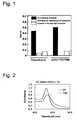
- non-specific adsorptivity of lysozyme on the surface which had been blocked with ethanolamine was 4 ⁇ 10 -2 (°)
- the surface which had been blocked with acetal-PEG/PAMA (5660/2780) inhibited non-specific adsorption almost completely. It was also confirmed that anti-human IgG antibody which had been brought into contact was efficiently detected.
- Example 2 Preparation of streptoavidin gold colloid particles, and stabilization with acetal-PEG-PAMA (4500/3200) (to be referred to as SAGCPEG/PAMA (4500/3200))
- streptoavidin-supporting PEGated gold colloid was examined by molecule recognition test with agglutination test.
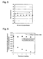
- biotin-introduced BSA as prepared in Referential Example 6 was added to PEG-modified streptoavidin gold colloid particles, the shift of peak top at absorption spectrum (800 nm - 400 nm) was observed, and, thus, interaction between streptoavidin on the surface of gold colloid and biotin was confirmed ( Fig. 2 ).
- Streptoavidin-supporting PEGated gold colloid was prepared in just the same manner as in Example 2 except that streptoavidin was replaced with BSA.
- the dispersion stability of thus obtained BSA-supporting PEGated gold colloid was remarkably high as in Example 2.
- Streptoavidin-supporting PEGated gold colloid was prepared in just the same manner as in Example 3 except that streptoavidin was replaced with BSA.
- the dispersion stability of thus obtained BSA-supporting PEGated gold colloid was remarkably high as in Example 2.
- Fig. 3 shows the results of spr.
- BSA-supporting gold colloid was hardly recognized on biotinated spr surface.
- Streptoavidin-supporting PEGated gold colloid having a PEG chain of 10,000 also gave small spr signal.
- Example 5 Preparation of anti-biotin antibody-supporting gold colloid particles, and stabilization with acetal-PEG-PAMA (5660/2780)
- Anti-biotin antibody-supporting PEGated gold colloid was prepared in just the same manner as in Example 2 except that streptoavidin and acetal-PEG-PAMA (4500/3200) were replaced with anti-biotin antibody and acetal-PEG-PAMA (5660/2780). It was confirmed that thus obtained anti-biotin antibody-supporting PEGated gold colloid had been dispersion-stabilized.
- Anti-biotin antibody-supporting PEGated gold colloid was prepared in just the same manner as in Example 3 except that streptoavidin was replaced with anti-biotin antibody. It was confirmed that thus obtained anti-biotin antibody-supporting PEGated gold colloid had been dispersion-stabilized.
- Example 7 Confirmation of molecule recognition of anti-biotin antibody-supporting PEGated gold colloid
- Fig. 4 shows the results of spr.
- Example 8 PEG blocking on the surface of magnetic particles
- the zeta potential of untreated latex was - 40 mV, a negative value. It was confirmed, on the other hand, that the surface potential of block-coated latex had almost completely been shielded, and that the coating was perfect.
- acetal-PEG/PAMA coated magnetic latex was evaluated with regard to non-specific adsorption of protein.
- Magnetic latex in an amount of 2.5 mg was treated with 3.2 mg of acetal-PEG/PAMA under the same condition as mentioned above.
- untreated magnetic latex in an amount of 2.5 mg was prepared.
- Fig. 6 shows the amount of FITC-BSA adsorbed on the surface of particles, corresponding to the times of washing. Washing did not peel protein off particles which had not been coated with block polymer, whereas protein was peeled off coated particles almost completely after four times of washing, and, thus, it was confirmed that non-specific adsorption was inhibited.
- Example 9 Method of preparation of magnetic particles having PEG brush on their surface (2)
- Magnetic particles in an amount of 0.5 mg were surface-treated with acetal-PEG/PAMA in the same amount as the magnetic particles, and then in an amount 10 times as much as the magnetic particles.
- Untreated particles had a high sedimentation rate, and was therefore unable to be measured for zeta potential.
- Polymer-treated particles on the other hand, had a zeta potential of - 4 mV and + 1 mV, and, thus, it was confirmed that surface had been shielded.
- Example 10 Method wherein magnetic particles (dynabeads) having tosyl group are made to support antibody, and are thereafter surface-blocked with acetal-PEG/polyamine
- Fig. 7 shows results of evaluation of the ability to detect antigen which had been bonded to dynabeads
- Fig. 8 shows S/N ratio of the systems.
- ALP enzyme
- PEHA-Phenyl-PEG-OH at the concentration of 0, 0.5, 1.0, 2.0, 3.0 and 4.0 wt% was mixed with a solution of JSR magnetic particles (supporting anti-AFP rabbit antibody, 7 ug/mg beads (0.35 ug/test), carboxylic acid surface particles, magnetic particles: 10 mg/ml) at a proportion of 1:1. After stirred with vortex, the resultant mixture was rotated overnight at 4°C, and, thus, surface treatment was conducted. Then, the mixture was diluted to the concentration of 1/10 with 1 % BSA.
- anti-AFP-monoclonal antibody stock solution on the market (Wako 016-14511) was diluted to the concentration of 1/3000) was added, and the resultant mixture was shaken for one hour, and was thus allowed to react. Then, the particles were separated by magnet, and were washed (in the same manner as above). Thereafter, anti-mouse alkaliphosphatase IgG antibody (stock solution of anti-mouse (goat)-alkaliphosphatase conjugate (Sigma A3688) was diluted to the concentration of 1/5000; 1 % BSA/PBS) was added, and the resultant mixture was shaken for one hour, and was thus allowed to react.
- anti-mouse alkaliphosphatase IgG antibody stock solution of anti-mouse (goat)-alkaliphosphatase conjugate (Sigma A3688) was diluted to the concentration of 1/5000; 1 % BSA/PBS) was added, and the resultant mixture was shaken for one hour, and was
- AFP ⁇ -feto protein; Aspen Bio Inc. 105S (Lot 990628V1SS) 500 K IU/mg
- AFP ⁇ -feto protein; Aspen Bio Inc. 105S (Lot 990628V1SS) 500 K IU/mg
- 50 % NRS, 1 % BSA PBS and NHS 50 % NRS, 1 % BSA PBS and NHS
- measurement was conducted in otherwise the same manner as in Example 11. It was confirmed that AFP was detected in spite of the blocking with PEHA-Phenyl-PEG-OH.
- Example 13 Blocking of dynabeads (surface tosyl group) with PEHA-Phenyl-PEG-OH
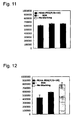
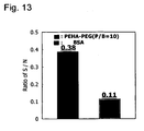
- the amount of antibody detected is shown in Fig. 11
- the amount of non-specific adsorption is shown in Fig. 12 . It was confirmed that, even when blocking was conducted with PEHA-Phenyl-PEG-OH, detection sensitivity did not decrease, and that non-specific adsorption was remarkably restrained.
- Fig. 13 shows S/N ratio. It is more effective than albumin blocking.
- Detection was conducted by fluorescence-labeled streptoavidin and with use of anti-BSA antibody (rabbit) as a primary antibody, and of biotin-labeled anti-rabbit antibody (donkey). Results are shown in Fig. 14 , from which it is seen that the polymer of the present invention achieved excellent non-specific adsorption-restraining effect as compared with conventionally used gelatin.
- Example 15 Coating treatment of glass surface with PEG
- PEG graft chain was constructed on glass substrate surface with use of a surface treating agent of the present invention.
- Coating treatment 2 (adsorption in hot water bath and under acidic conditions; activation of molecular motion)
- adsorption in hot water bath and under acidic conditions activation of molecular motion
- ionic intensity at the time of ⁇ potential measurement and for the purpose of accelerating protonation of PAMA
- Measurement of ⁇ potential Measurement was conducted by laser Doppler method with use of LEZA-600 apparatus of Otsuka Electronics Co., Ltd. For the measurement of pH dependency of ⁇ potential, pH value was raised from acidic side, i.e., pH 3, 5, 7, 8, 9 up to 10. Measurement was made two or three times at each pH, and a stable value was employed as a measurement value.
- pH Dependency of ⁇ potential Fig. 15 shows a comparison of pH dependency of ⁇ potential between the surface of unmodified glass and the surface of PEG-modified glass which had undergone adsorption treatment at 25, 50 and 80°C. The use of acetal-PEG-b-PAMA gave a surface on which surface potential change caused by pH was small, and which was hardly susceptible to outer environment, as compared with unmodified glass.
- AFP ⁇ -feto protein
- samples there were prepared 1 AFP diluted to 8,000 IU/ml with normal human serum which had been diluted to 1/10 strength; and 2 AFP diluted to 8,000 IU/ml with a cell extract having a protein concentration of 20 ⁇ g/ml.
- BSA bovine serum albumin
- PEG-PLA methoxy-polyethyleneglycol/polylactic acid block polymer
- anti-AFP antibody (rabbit) as a primary antibody
- alkaliphosphatase-labeled anti-rabbit antibody (donkey) as a secondary antibody
- 5-bromo-4-chloro-3-indolylphosphate/nitroblue tetrazolium BCIP/NBPT
- Results are shown in Fig. 16 , from which it is seen that the surface treating agent MethoxyPEG-PLA5000 of the present invention achieved the reduction of background owing to its excellent non-specific adsorption-restraining effect, as compared with conventionally used BSA.
- Example 17 (for reference and comparison only): Surface treatment of microcircuit with PEG segment-containing graft copolymer
- Silicone surface which had been prepared in the above-mentioned method was washed, and was then made to react with polymethoxysilylpropyl methacrylate-PEG graft copolymer (PTSPM-g-PEG 1100 ) and PEG homopolymer (PEG 1100 ) each for four hours at room temperature, and, thus, silicone surface was modified with PEG brush.
- PTSPM-g-PEG 1100 polymethoxysilylpropyl methacrylate-PEG graft copolymer
- PEG 1100 PEG homopolymer
- Example 18 (for reference and comparison only): Collective surface treating method wherein, when polydimethoxy silyl (PPMS) is formed, either PTSPM-g-PEG or PEG macromonomer having polymerizable vinyl group is applied on mold surface
- Non-specific adsorption of protein to treated surface was evaluated with use of fluorescence-labeled albumin and IgG. Results are shown by Fig. 19 and Fig. 20 .
- Surface treatment achieved remarkable reduction of adsorptivity of both proteins. Furthermore, whereas the untreated silicone surface showed a large scattering in the amount of adsorption, the treated surface restrained non-specific adsorption with good reproducibility.
- This invention provides a surface of substrate of biosensor to which the non-specific adsorption of impurity proteins such as blood and plasma which exist in sample is significantly restrained. This invention is therefore usable in biosensor-manufacturing industries or in the field of clinical diagnosis which uses biosensor.
Abstract
Description
- This invention relates to the surface of substrate, in particular biosensor chip etc., specifically to the surface of substrate which has been treated with uncrosslinked polymer based on polyethylene glycol chain segment, and also to a biosensor which has said surface.
- Immunodiagnosis, biosensor and the like for detecting a certain substance from among biomolecules such as protein or lipid have widely been applied as a means for the early detection or diagnosis of diseases. However, the non-specific adsorption of co-existent biomolecules onto the surface of biosensor which occurs simultaneously with a specific reaction interferes as background noise to prevent the achievement of high sensitivity. In the case of diagnostic particles, furthermore, not only the problem of background caused by non-specific adsorption but also dispersion stabilization in biological fluid or diluted liquid thereof has been a great issue. Inventors of this invention previously found that substrate which has, on its surface, a brush-like structure of water-soluble polymer such as polyethylene glycol not only retrains non-specific adsorption onto sensor surface but also improves dispersion stabilization of nanoparticles, and, thus, have provided materials as a new tool of biodiagnosis. As concrete examples of such inventions, a surface with brush-like structure of poly(ethylene oxide) having an increased density (e.g.,
WO 03/076933 A1 2003-149245 2002-080903 - In the above-mentioned inventions, biomolecules such as antibody are bonded to the tip of brush structure to serve as a system to sense, with high sensitivity, specific reaction such as antigen recognition. However, brush surface is very liable to prevent the adsorption of protein or the like, and, also for some other reasons, it is sometimes difficult to increase the amount of protein such as antibody supported on the tip of brush, which has been a bar to the achievement of high sensitivity. In another method, after antibody or antigen has been bonded to the surface of a solid phase, a polymer which is originated in glycosylethyl (meth)acrylate is adhered to redundant protein-binding sites on the solid phase surface, with a view to preventing the non-specific adsorption of impurity protein or the like which may be contained in sample for assay (Japanese Patent KOKAI Publication No.
Hei 10-123135 Hei 11-287802 - These polymers, however, may be sometimes insufficient in bondability or immobilizability onto solid phase surface to which the non-specific adsorption of protein or the like needs to be restrained. Otherwise, when immobilizability is enhanced, it may adversely affect the specific bondability of antibody to antigen in immune reaction. In another proposed method, the affinity of a bio-specific bonding pair, e.g., the affinity between streptoavidin and biotin, is utilized. In detail, a biotinated antibody is bonded to a solid phase which has previously supported streptoavidin, and said solid phase is thereby coated with biotinated polyethylene glycol (Japanese Patent KOKAI Publication No.
Hei 11-211727 - Lu et al. (Langmuir, Vol. 16, pp. 1711-1718 (2000)) describe the attachment of functionalized poly(ethylene glycol) films to gold surfaces and, in particular, the attachment of linear poly(ethylene glycol) (PEG) molecules (MW = 2000-5000) to gold surfaces via orthopyridyldisulfide (OPSS) terminal groups and the study thereof with X-ray photoelectron spectroscopy and other techniques. The molecules examined included a PEG molecule terminated with a methoxy group and derivatized with an OPSS group at the other end (M-PEG-OPSS), a PEG molecule derivatized with OPSS at both ends (PEG-(OPSS)2) and a PEG derivatized with an N-hydroxysuccinimide group at one end and an OPSS group at the other end (NHS-PEG-OPSS). According to the authors, for biosensor applications, free OPSS or NHS groups in the NHS-PEG-OPSS films can be used to immobilize biomolecules with accessible cysteines or lysines via formation of disulfide bonds or amide bonds, respectively. Moreover, the PEG films formed by M-PEG-OPSS and PEG-(OPSS)2 resist the adsorption of albumin. However, according to this document, biomolecules are bonded to a PEG layer after this PEG layer has been formed on a gold surface. Thus, the PEG layer needs to be formed first, which is inconvenient. Furthermore, in some cases it is hard to raise the amount of protein because the PEG surface effectively inhibits the adsorption thereof.
- Peracchia et al. (Pharmaceutical Research, Vol. 15, pp. 550-556 (1998)) describe pegylated nanoparticles from a novel methoxypoly-ethylene glycol cyanoacrylate-hexadecyl cyanoacrylate amphiphilic copolymer. These particles are formed by nanoprecipitation or by emulsion/ solvent evaporation. Thus, this document relates to graft polymers which have PEG side chains. The polymer is bonded to the substrate by hydrophobic bonding. This polymer is sometimes insufficient in bondability or immobilizability onto solid phase surfaces to which the non-specific adsorption of protein or the like needs to be restrained. In other cases, when immobilizability is enhanced, it may adversely affect the specific bondability of antibody to antigen.
-
US patent 5,919,712 describes methods and apparatus for evanescent light fluoroimmunoassays. A preferred biosensor described in this document has patches of capture molecules each specific for a different analyte disposed adjacent within a single reservoir. The capture molecules are immobilized to the patches by site-specific coupling of thiol groups on the capture molecules to photo-affinity crosslinkers. This document describes surface modification with use of amino group-terminated PEG. Biomolecules such as antibodies are bonded to the tip of a "brush" constructed therefrom. The resulting system is utilized for sensing, at high sensitivity, specific reactions, such as antigen recognition. Thus, the surface needs to be constructed in advance, which is inconvenient. Furthermore, in some cases, it is difficult to raise the amount of protein, such as antibody, supported on the tip of the "brush" because the surface thereof inhibits the adsorption of protein and the like, which may prevent the achievement of high sensitivity. - Inventors of this invention made researches for the purpose of providing a surface more stable than those of the above-mentioned conventional methods, which is capable of inhibiting non-specific adsorption, and which is easily prepared. As a result, they have found unexpectedly that, on the surface of solid phase (e.g., surface originated in gold, polystyrene or polyvinylidene fluoride) practically used for immunoassay on which antibody, antigen or the like is immobilized, PEG chain brush can be immobilized with no special bonding means, and with no adverse effects produced on the specific bondability of antibody or antigen. Furthermore, in view of the fact that the recognition ability of antibody or the like works depending on the length of PEG chain, the inventors paid consideration, in designing, to the optimization of said length of chain, and, thus, have found a method to conduct specific molecular recognition with high sensitivity.
- Thus, this invention provides a substrate surface on which either a substance to detect analyte or an analyte per se is immobilized, which surface is formed by a treatment of substrate surface with a liquid which contains uncrosslinked polymer based on polyethylene glycol chain segment, said treatment conducted either simultaneously with the immobilization of said substance or analyte or after said substance or analyte has been immobilized on said surface,
wherein the above-mentioned uncrosslinked polymer based on polyethylene glycol chain segment is represented by formula (I) as follows:
R1-L1-(CH2CH2O)n-L2-X (I)
wherein R1 denotes hydrogen atom, methyl, formyl which may be protected, amino which may be protected, carboxy which may be protected, hydroxyl which may be protected or vinylsulfonyl group;
L1 and L2 independently denote valence bond or linker;
X denotes functional group or functional part to form covalent bond or a bond via physical interaction by which to immobilize said polymer molecule onto the surface of fine porous particles; and
n denotes an integer of 2 to 20,000,
wherein X is selected from the group consisting of main chain portion of oligomer or polymer which has, on side chain, mono- or di-lower alkyl-substituted amino group; main chain portion of oligomer or polymer which has, on side chain, mercapto group; main chain portion of oligomer or polymer which has, on side chain, silanol group; main chain portion of oligomer or polymer which has, on side chain, carboxyl group; main chain portion of oligomer or polymer which has, on side chain, sulfo group; main chain portion of oligomer or polymer which has, on side chain, hydroxyl group, main chain portion of oligo or polyimino having plural numbers of imino group (-NH-) on main chain, and main chain portion of oligo or polylactide. - As another embodiment, this invention provides a method to produce a substrate surface which comprises (A) preparing a substrate surface, and (B) bringing both an aqueous solution of a substance to detect analyte which has been so modified as to be immobilizable on said substrate surface and a liquid which contains the uncrosslinked polymer based on polyethylene glycol chain segment as defined above into contact with said substrate surface either simultaneously or in succession, under a condition under which both of said substance and uncrosslinked polymer are quite immobilizable on said substrate surface of (A).
- As another embodiment, this invention provides a biosensor which is equipped with the above-mentioned substrate surface.
-
-
Figure 1 is a graph which shows both the blocking ability of a surface on which human IgG had been immobilized, and results of sensing of anti-human IgG antibody. -
Figure 2 shows the change of absorbance before and after the addition of biotinated BSA to streptoavidin-immobilized PEGlated gold colloid. -
Figure 3 is a graph which shows the change at sensor gram when streptoavidin-supporting and BSA-supporting PEGlated gold colloid solutions were brought into contact with spr sensor which had biotin-PEG brush surface. -
Figure 4 is a graph which shows the change at sensor gram when antibiotin antibody-supporting PEGlated gold colloid solution was brought into contact with spr sensor which had biotin-PEG brush surface. -
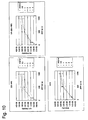
Figure 5 is a graph which shows the relation between the condition of surface treatment of magnetic latex with acetal-PEG/PAMA and zeta potential. -
Figure 6 is a graph which shows the dependency, on the times of washing, of the amount of bovine serum albumin adsorbed on magnetic latex whose surface had, or had not been, treated with acetal-PEG/ PAMA. -
Figure 7 shows a comparison of the detecting ability of anti-goat IgG when goat IgG antibody-supporting dynabeads were coated with block polymer. Five data on the left side show sensing ability of particles, and five data on the right show non-specific adsorption onto particles which support no antibody on their surface. -
Figure 8 is a graph which shows S/N as obtained fromFigure 6 . PEHA-Ph-PEG-OH exhibits the best blocking performance. -
Figure 9 is a graph which shows the result of surface treatment of JSR antibody-supporting magnetic particles with PEHA-Ph-PEG-OH. It is seen that non-specific adsorption is markedly inhibited. -
Figure 10 is a graph which shows the result of surface treatment of JSR antibody-supporting magnetic particles with PEHA-Ph-PEG-OH. It is seen that antigen-detecting ability was high enough. -
Figure 11 is a graph which shows the result of surface treatment of dynabeads antibody-supporting magnetic particles with PEHA-Ph-PEG-OH. It is seen that antigen-detecting ability was high enough. -
Figure 12 is a graph which shows the result of surface treatment of dynabeads antibody-supporting magnetic particles with PEHA-Ph-PEG-OH. It is seen that non-specific adsorption is inhibited by surface treatment. -
Figure 13 shows both the result of surface treatment of dynabeads antibody-supporting magnetic particles with PEHA-Ph-PEG-OH and S/N. -
Figure 14 are photographs in place of drawings which show whether or not fluorescence-labeled protein was non-specifically adsorbed onto PVDF membrane for western blotting. -
Figure 15 is a graph which shows the comparison of surface potential of glass surface treated with acetal-PEG-b-PAMA. -
Figure 16 is a photograph in place of drawing which shows the result of western blotting conducted in accordance with Example 16. -
Figure 17 shows a comparison of surface potential between silicone surface which was treated with polytrimethoxysilylpropyl methacrylate-PEG graft copolymer (PTSPM-g-PEG1100) and untreated silicone surface. -
Figure 18 shows the result of a comparison of surface potential of silicone which was formed from a mold of glass whose cleansed surface had been coated with polytrimethoxysilylpropyl methacrylate-PEG graft copolymer (PTSPM-g-PEG1100) and which was thereby simultaneously treated, with surface potential of untreated silicone surface and with surface potential of a surface treated with polyethylene glycol homopolymer. -
Figure 19 is a graph which shows the result of a comparison of adsorptivity of fluorescence-labeled human IgG onto a surface-treated silicone surface, with untreated surface. -
Figure 20 is a graph which shows the result of a comparison of adsorptivity of fluorescence-labeled bovine serum albumin onto a surface-treated silicone surface, with untreated surface. - Examples of a substance to detect analyte or an analyte per se as referred to in this invention include bio-specific bonding pair such as antigen or hapten and antibody; oligonucleotide and nucleic acid which hybridizes therewith under stringent condition; enzyme, its substrate sugar and lectin; hormone and its receptor protein; and avidin (including streptoavidin) and biotin (including desthiobiotin, iminobiotin and aminobiotin). Hence, a binding partner of a specific bonding pair means one of counterparts to form the above-mentioned bonding pairs.
- Substrate surface on which such a substance (which may include analyte) as mentioned above has been immobilized is a surface, in a solid phase, of bioassay chip or biosensor with which to detect said substance. Any material is usable for such a surface so long as it serves to achieve the objectives of this invention. Preferable examples of substrate surface include electrochemical sensor surface (e.g., made from precious metal, metal oxide, etc.), surface plasmon resonance (SPR) sensor surface (e.g., made from precious metal), quartz sensor surface, microplate surface (e.g., made from polystyrene, polypropylene or polytetrafluoroethylene) for solid phase enzyme-linked immunoassay (ELISA), plastic surface (e.g., made from cellulose derivatives such as nitrocellulose, polyvinylidene fluoride or nylon) for protein blotting or nucleic acid blotting, microarray surface (e.g., made from glass or plastics) for the hybridization of nucleic acid, glass-made surface and silicone-made one (e.g., treated with polydimethyl siloxane), which are usually employed in this field. Preferable examples of the case where substrate and substrate surface become one include gold particle surface, semiconductor particle surface, magnetic particle surface, silica particle surface, fine porous particle surface and surface of particle of latex which contains one of the above-mentioned particles.
- One binding partner or the other of a specific bonding pair which has been "so modified as to be immobilizable on said substrate surface" means, when the substrate surface is a gold-deposited membrane, a partner to whose terminal a mercapto group has been introduced in a manner well known in this art.
- The substrate surface of this invention is produced by treating a substrate surface with a liquid which contains uncrosslinked polymer based on polyethylene glycol chain segment either simultaneously with the immobilization onto the surface, or after the immobilization onto the surface, of a substance to detect analyte or analyte per se (e.g., one binding partner of specific bonding pair) which has been so modified as to be immobilizable on said substrate surface. Preferably, a substrate surface on which a substance to detect analyte or analyte per se has previously been immobilized is treated with a liquid which contains uncrosslinked polymer based on polyethylene glycol chain segment. In this invention, said substrate surface on which a substance to detect analyte or analyte per se has previously been immobilized includes all surfaces that have been used or suggested for use in this field, such as those which are recited above.
- Polymer which is effectively used for the above-explained surface treatment is represented by formula (I).
- In one embodiment, X denotes main chain portion of oligo or polyimino having plural numbers of imino group (-NH-) in the main chain, which has a formula as follows:
-(CH2CH2NH)m-R2
wherein R2 denotes hydrogen atom or lower alkyl (e.g., straight or branched chain-alkyl having one to six carbon atoms, such as methyl, ethyl, propyl, isopropyl, n-hexyl, etc., which applies to the following); and m denotes an integer of 1 to 2,000. - In another embodiment, X denotes main chain portion of oligomer or polymer which has, on side chain, mono- or di-lower alkyl-substituted amino group, of a formula as follows:


2003-49000 - In another embodiment, X denotes main chain portion of oligomer or polymer which has, on side chain, silanol group, of a formula as follows:

Y. Nagasaki et al., or of U.S. Patent 5,929,177 , or from the hydrolysis of said trialkoxysilyl where necessary. - In another embodiment, X denotes main chain portion of oligomer or polymer which has, on side chain, carboxyl group, represented by a formula as follows:

- In another embodiment, X denotes main chain portion of oligo or polylactide represented by a formula as follows:

- Polymers having X as explained above are mentioned in
U.S. Patent 5,925, 730 for instance. Other polymers can be produced in accordance with production processes of polymers having various kinds of X, or by modification of the processes. - When L1 in formula (I) denotes a linker, typical examples thereof include, although not restrictive, connecting groups as follows:
-(CH2)p-O-, -(CH2)q-COO-,

or -(CH2)r-S-
wherein p, q and r independently denote an integer of 0 to 8. These linkers are structurally incorporated, in the direction as mentioned, into the portion of L1 in the above-mentioned formula (I). - When L2 denotes a linker, typical examples thereof include, although not restrictive, connecting groups as follows:
-(CH2)k-, -CO(CH2)l-,

- Formyl which may be protected in the above-mentioned definition of R1 has a formula as follows:

- The substrate surface of the present invention is prepared in a process which comprises preparing a substrate surface, and bringing both an aqueous solution (including aqueous solution buffered with PBS or the like) of the above-mentioned substance to detect analyte or analyte per se modified and the above-mentioned polymer-containing liquid (water-miscible organic solvent such as methanol, ethanol, tetrahydrofuran, dimethylformamide and dimethylsulfoxide, and aqueous solution which may be buffered with PBS or the like) simultaneously into contact with said substrate surface, under a sufficient condition under which both of said substance and polymer are quite immobilizable onto said substrate surface. As said sufficient condition, there may be mentioned incubation for a time ranging from several to scores of hours at a temperature ranging from 5°C to a temperature at which the above-mentioned substance is not denatured, e.g., 55°C, although this condition may vary depending on the properties of polymer and substrate surface used. A substrate surface which has the above-mentioned substance previously immobilized thereon is also treated with polymer under almost the same condition as the above. Thus, the substrate surface of the present invention is provided. The above-mentioned polymer is usually used at a proportion of 10-6 to 103 mg/cm2, preferably 10-4 to 102 mg/cm2, more desirably 10-3 to 10 mg/cm2, on the basis of area of substrate surface.
- In the following, this invention is explained in more detail with working examples.
-

- In an egg-shaped flask in an atmosphere of argon at room temperature, 1.0 mmol of 4-hydroxymethyl benzaldehydedimethyl acetal (1.822 mol/l - THF solution, 0.55 ml) as an initiator was added to 25 ml of tetrahydrofuran (THF) as a solvent with a microsyringe, and then, 1.0 mmol of K-naphthalene (0.328 mol/l - THF solution, 3.05 ml) was added, and, thus, metallization was conducted for 10 minutes. Then, 140 mmol of ethyleneoxide (6.9 ml) was added, and the resultant mixture was stirred for two days under water-cooling, and, thus, anion ring-opening polymerization was conducted. Several drops of pure water were subsequently added to stop the reaction, and, then, purification was conducted by diethyl ether precipitation (21), chloroform extraction (three times against saturated saline water), drying under reduced pressure and benzene freeze drying. The yield of thus obtained product was 4.5 g (90 %).
- According to measurement by gel permeation chromatography, the obtained polymer was monomadal, and had a number average molecular weight of 6,067 which almost agreed with theoretical molecular weight of 6,000.
- Also measurement by MALDI-TOF-MS (matrix-assisted laser desorption ionization time of flight mass spectrometer) taught that the obtained polymer was monomadal, and had a number average molecular weight of 6,050. Moreover as a result of comparison between measured value and calculated value of the peaks, it was confirmed that this polymer was heterotelechelic polyethyleneoxide which had ethyleneoxide skeleton in its main chain, had acetal group at α-terminal and had hydroxyl group at ω-terminal.
- Also from 1H-NMR (proton nuclear magnetic resonance) spectrum of the obtained polymer in DMSO, it was confirmed that this polymer was heterotelechelic polyethyleneoxide which had ethyleneoxide skeleton in its main chain, and had acetal group at α-terminal and hydroxyl group at ω-terminal.
-

- In an egg-shaped flask, 1.0 g of acetal-PEG-OH obtained in the above was dissolved in 20 ml of 90 % aqueous solution of acetic acid, and the resulting mixture was stirred for five hours. Subsequently, the mixture was adjusted to
pH 8 with use of 10N HCl, and was then dialyzed (molecular weight cut-off: 3500; water was replaced after 1, 2, 4, 6, 8 and 12 hours) against pure water for one day, and was subsequently subjected to drying under reduced pressure and benzene freeze drying, for recovery. The yield of thus recovered product was 0.88 g (88 %). - According to measurement of gel permeation chromatography, the obtained polymer was monomadal, and had a number average molecular weight of 6,056 which almost agreed with theoretical molecular weight of 6,054.
- Also measurement by MALDI-TOF-MS taught that the obtained polymer was monomadal, and had a number average molecular weight of 6,023. Moreover as a result of comparison between measured value and calculated value of the peaks, it was confirmed that this polymer was benzaldehyde-PEG-OH wherein acetal group at α-terminal had been deprotected.
- In 1H-NMR (proton nuclear magnetic resonance) spectrum of the obtained polymer in DMSO, spectrum of aldehyde proton was observed near 10 ppm, and, also from this result, it was confirmed that this polymer had become aldehyde (formyl group), with acetal group at α-terminal deprotected.
-

- In 5 ml of methanol, 250 mg of benzaldehyde-PEG-OH was dissolved. An egg-shaped flask was fed with PEHA (5 mmol, 1.2 ml) in a
molar amount 100 times as much as PEG, and, then, said PEHA was dissolved in 20 ml of methanol. The resultant mixture was adjusted topH 6 with 5N HCl under ice cooling. To this PEHA methanol solution which was being vigorously stirred, benzaldehyde-PEG-OH methanol solution was slowly added dropwise. The resultant mixture was stirred at room temperature for four hours, and, thus, Schiff base was formed. Then, as a reducing agent, 5 mmol (amolar amount 100 times as much as PEG; about 300 mg) of NaBH3CN was added to the reaction solution three times in total at an interval of 30 minutes, and the resultant mixture was stirred for 24 hours. Thus obtained reaction solution was dialyzed (molecular weight cut-off: 1000; water was replaced after 3, 6, 18, 24, 30, 42 and 48 hours) against pure water for two days. Subsequently, concentration was adjusted properly by drying under reduced pressure, and, then, the product was recovered by benzene freeze drying. The yield of thus obtained product was 75 mg (30 %). - According to measurement of gel permeation chromatography, the obtained polymer was monomadal, and had a number average molecular weight of 5,800 which almost agreed with theoretical molecular weight of 6,270.
- In 1H-NMR (proton nuclear magnetic resonance) spectrum of the obtained polymer in D2O, spectrum of aldehyde proton which had been observed near 10 ppm in benzaldehyde-PEG-OH had disappeared, and, furthermore, there was newly observed near 3.4 ppm a spectrum which seemed to have been caused by methyl proton between amine and benzene ring. From these results, it was confirmed that PEHA-Phenyl-PEG-OH (or PEHA-Ph-PEG-OH) had been synthesized as desired.
- A 200 ml flask was fed with 4 mL of styrene, 45 mL of water and 0.024 g of potassium persulfate, and the resulting mixture was polymerized for 28 hours at 70°C and 350 rps. It was confirmed by TEM and the measurement of dynamic light scattering that thus obtained latex was monodisperse with an average particle size of 1 µm.
- A solution of this latex in an amount of 25 mL was mixed with 125 mL of water in a 300 mL flask. After pH was adjusted to 1.7 with hydrochloric acid, there were added FeCl3 (0.405 g) and FeSO4 (0.25 g), and, with vigorous stirring, the resultant mixture was adjusted to
pH 9 with ammonia water. Thus obtained ferricolloid latex was easily attracted by magnet, and, thus, it was confirmed that ferrite had been formed on the surface of latex. - Gold chip which had been washed with ozone was dipped in acetal-PEG-SH (Mn = 5,000) which had been dissolved in 50 mM sodium phosphate buffer (pH 7.4, 1 M NaCl) so that the concentration might be 1 mg/mL, and was then shaken for 30 minutes at room temperature. The chip was washed once with 50 mM sodium phosphate buffer (pH 7.4, 1 M NaCl), and was then dipped in 50 mM sodium hydroxide for 30 seconds, and was again washed three times with 50 mM sodium phosphate buffer (pH 7.4, 1 M NaCl). This operation was repeated twice. Furthermore, the chip was dipped in MeO-PEG-SH (Mn = 2,000) which had been dissolved in 50 mM sodium phosphate buffer (pH 7.4, 1 M NaCl) so that the concentration might be 1 mg/mL, and was then shaken for 30 minutes at room temperature. The chip was washed once with 50 mM sodium phosphate buffer (pH 7.4, 1 M NaCl), and was then dipped in 50 mM sodium hydroxide for 30 seconds, and, subsequently, the surface of gold chip was again washed three times with 50 mM sodium phosphate buffer (pH 7.4, 1 M NaCl).
- This operation was repeated twice, and, thus, the surface of gold chip was modified with PEG (preparation of mixed brush). Then, PEG-modified gold chip was dipped in 0.1 mol/L hydrochloric acid, and was then shaken mildly for three hours at room temperature, and, thus, acetal group at PEG terminal was converted into aldehyde group. Then, the gold chip was dipped in biocytin-hydrazide (EZ-Link®; PIERCE) which had been dissolved in 100 mM sodium phosphate buffer (pH 5.5, 1 M NaCl) so that the concentration might be 1 mg/mL, and was then shaken for three hours at room temperature, and, thus, biotin was introduced to the surface of gold chip.
- Gold substrate (manufactured by Nippon Laser Electronics LAB) which had been washed with ozone was dipped in 1
mM 4,4'-dithiodibutyric acid (solvent: ethanol) for at least 12 hours in a Petri dish at room temperature, and was thereafter washed with ethanol. A mixed solution of 25 mg of EDC (1-ethyl-3-(3-dimethylaminopropyl) carbodiimide) which had been dissolved in 1 mL of distilled water and 15 mg of NHS (N-hydroxysuccinimide) which had been dissolved in 9 ml of dioxane was added to Petri dish. Substrate which had been washed was dipped in the mixed solution, and was then mildly shaken for 30 minutes and was thus activated. - Thus activated substrate was set in a surface plasmon sensor (SPR manufactured by Nippon Laser Electronics LAB), and, then, 1 µM human IgG was immobilized on surface layer under a condition of 25 °C, 5 · L/min, 60 µL. On thus prepared IgG-immobilized surface, there were twice injected ethanolamine (pH 8.6) and 1 mg/mL acetal-polyethyleneglycol-b-poly(methacrylic acid 2-N,N-dimethylaminoethyl) (hereinafter referred to as Acetal-PEG/PAMA; PEG chain length and PAMA chain length were 5660 and 2780 respectively; hereinafter abbreviated as (5660/2780)) under a condition of 25 °C, 5 L/min, 60 µL. Thus obtained surface was measured for non-specific adsorptivity and specific adsorptivity with spr. As is shown by
Fig. 1 , non-specific adsorptivity of lysozyme on the surface which had been blocked with ethanolamine was 4 × 10⁻² (°), whereas the surface which had been blocked with acetal-PEG/PAMA (5660/2780) inhibited non-specific adsorption almost completely. It was also confirmed that anti-human IgG antibody which had been brought into contact was efficiently detected. - To 5 mg of BSA (Cohn Fraction V, WAKO) which had been dissolved in 1 ml of 50 mM carbonic acid buffer (pH 9.6), there was added 21 µl of biotin-(AC5)2-OSu (DOJINDO) which had been dissolved in DMSO so that the concentration might be 20 mg/ml, and, thus, the resulting mixture was made to react for two hours at room temperature. The mixture was gel filtrated, and unreacted biotin-(AC5)2-OSu was thereby removed, and, furthermore, the buffer was replaced with 20 mM sodium phosphate buffer (pH 7.5, 0.15 M NaCl, 1 mM EDTA). The amount of thus obtained biotin-labeled BSA was determined by HABA method. It was confirmed that about five molecules of biotin had been introduced per molecule of BSA.
- To gold colloid solution (PolyScience; average particle size: 40 nm; concentration: 0.01 %), there was added an aqueous solution of streptoavidin (ImmunoPure) in an amount of 103 times as much as the gold colloid solution, and the resultant mixture was incubated at room temperature for one hour, and, thus, streptoavidin was adsorbed on the surface of gold particles. Then, an aqueous solution of acetal-PEG-PAMA (4500/3200) was added so that the molar ratio of gold particles to polymer might be 1 : 1 × 10⁶, and the resultant mixture was made to react at 4°C overnight. Thereafter, an operation of centrifugation [4°C, 4000 × g, 30 minutes] to recover precipitated residue was repeated three times, and, thus, superfluous streptoavidin and polymer were removed.
- It was confirmed by UV-Vis spectrometry that thus obtained PEG-modified gold colloid particles showed good re-dispersibility after centrifugation purification, and existed stably in 10 mM sodium phosphate buffer (pH 7.4, 0.15 M NaCl).
- The recognition ability of thus obtained streptoavidin-supporting PEGated gold colloid was examined by molecule recognition test with agglutination test. When biotin-introduced BSA as prepared in Referential Example 6 was added to PEG-modified streptoavidin gold colloid particles, the shift of peak top at absorption spectrum (800 nm - 400 nm) was observed, and, thus, interaction between streptoavidin on the surface of gold colloid and biotin was confirmed (
Fig. 2 ). - Streptoavidin-supporting PEGated gold colloid was prepared in just the same manner as in Example 2 except that acetal-PEG-PAMA (4500/3200) was replaced with MeO-PEG-SH (Mn = 10,000) and MeO-PEG-SH (Mn = 5,000). The dispersion stability of thus obtained streptoavidin-supporting PEGated gold colloid was remarkably high as in Example 2.
- Streptoavidin-supporting PEGated gold colloid was prepared in just the same manner as in Example 2 except that streptoavidin was replaced with BSA. The dispersion stability of thus obtained BSA-supporting PEGated gold colloid was remarkably high as in Example 2.
- Streptoavidin-supporting PEGated gold colloid was prepared in just the same manner as in Example 3 except that streptoavidin was replaced with BSA. The dispersion stability of thus obtained BSA-supporting PEGated gold colloid was remarkably high as in Example 2.
- Molecule-recognizing ability of thus obtained dispersion-stabilized streptoavidin- and BSA-supporting PEGated gold colloid was confirmed with spr (BIAcore 1000). PEGated gold surface having biotin which had been prepared in Referential Example 5 was made to react with modified gold colloid particles which had been dissolved in 50 mM sodium phosphate buffer (pH 7.4, 0.15 M NaCl) containing 1 % BSA at a measurement temperature of 25°C and a flow rate of 10 · L/min, and; thus, the change of angle was measured by surface plasmon resonance method.
-
Fig. 3 shows the results of spr. BSA-supporting gold colloid was hardly recognized on biotinated spr surface. Streptoavidin-supporting PEGated gold colloid having a PEG chain of 10,000 also gave small spr signal. Gold colloid which had been treated with PEG 5,000 and PEG-PAMA (4500/3200), however, gave a very strong signal, and, thus, it was confirmed that streptoavidin supported on gold colloid efficiently interacted with biotin on the surface of spr sensor. - Anti-biotin antibody-supporting PEGated gold colloid was prepared in just the same manner as in Example 2 except that streptoavidin and acetal-PEG-PAMA (4500/3200) were replaced with anti-biotin antibody and acetal-PEG-PAMA (5660/2780). It was confirmed that thus obtained anti-biotin antibody-supporting PEGated gold colloid had been dispersion-stabilized.
- Anti-biotin antibody-supporting PEGated gold colloid was prepared in just the same manner as in Example 3 except that streptoavidin was replaced with anti-biotin antibody. It was confirmed that thus obtained anti-biotin antibody-supporting PEGated gold colloid had been dispersion-stabilized.
- Molecule-recognizing ability of thus obtained dispersion-stabilized anti-biotin antibody-supporting PEGated gold colloid was confirmed with spr (BIAcore 1000) in just the same manner as in Example 4.
-
Fig. 4 shows the results of spr. Antibody-supporting gold colloid detected a signal with high sensitivity also in the case of ace-PEG-SH (10,000) where streptoavidin-supporting PEGated gold colloid (Mn = 10,000) was unable to detect a signal. Thus, there was provided antibody-supporting gold colloid having specific recognition ability. - Ferrite-supporting latex as prepared in Referential Example 4 was surface-treated with acetal-PEG/PAMA. As shown in Table 1, a certain amount of acetal-PEG/PAMA was put in a sample tube, in which 5 ml of 10 mM phosphate buffer (pH = 7.18) was added, and, then, the resultant mixture was stirred and dissolved. To the resultant solution, magnetic latex was added, and stirred, and, subsequently, the resultant mixture was subjected to washing operation (phosphate buffer × four times), and, thus, unadsorbed PEG-b-PAMA was removed. After the washing operation was over, dispersion stability was examined, and, then, zeta potential measurement was conducted with a view to confirming surface charge of ferrite particles.
Table 1 Condition of acetal-PEG/PAMA surface treatment Run Ferrite latex (mg) Acetal-PEG/PAMA (mg) 1 0.5 0 2 0.5 0.61 3 0.5 5.13 4 0.5 24.96 Phosphate buffer: 5 mL (pH = 7.1; ionic intensity: 10 mM) - As is shown by
Fig. 5 , the zeta potential of untreated latex was - 40 mV, a negative value. It was confirmed, on the other hand, that the surface potential of block-coated latex had almost completely been shielded, and that the coating was perfect. - Thus prepared acetal-PEG/PAMA coated magnetic latex was evaluated with regard to non-specific adsorption of protein. Magnetic latex in an amount of 2.5 mg was treated with 3.2 mg of acetal-PEG/PAMA under the same condition as mentioned above. Apart from that, untreated magnetic latex in an amount of 2.5 mg was prepared. Each of these magnetic latex was mixed with 17.8 • g of solution of FITC-BSA which had been dissolved in 2 mL of phosphate buffer (pH = 7.1; I = 10 mM). After one hour, particles were separated with a magnet, and supernatant was measured with use of a spectrofluorometer for the intensity of fluorescence at emission wave length of 520 nm corresponding to excitation wave length of 490 nm, and, thus, the amount of FITC-BSA was calculated.
Fig. 6 shows the amount of FITC-BSA adsorbed on the surface of particles, corresponding to the times of washing. Washing did not peel protein off particles which had not been coated with block polymer, whereas protein was peeled off coated particles almost completely after four times of washing, and, thus, it was confirmed that non-specific adsorption was inhibited. - Surface treatment was conducted in the same manner as in Example 8 except that magnetic latex as prepared in Referential Example 4 was replaced with magnetic particles of JSR. Magnetic particles in an amount of 0.5 mg were surface-treated with acetal-PEG/PAMA in the same amount as the magnetic particles, and then in an
amount 10 times as much as the magnetic particles. Thus treated particles showed remarkably increased dispersibility, as compared with untreated latex. Untreated particles had a high sedimentation rate, and was therefore unable to be measured for zeta potential. Polymer-treated particles, on the other hand, had a zeta potential of - 4 mV and + 1 mV, and, thus, it was confirmed that surface had been shielded. - Eppendorf tube was fed with 46.9 · L of 10 mM tris buffer solution (TBS solution; pH = 8.12; 0.15 M NaCl) of dynabeads (the amount of dynabeads: 93.75 · g) and 150 µL of Goat-IgG/10 mM tris buffer solution (TBS solution; pH = 8.12) (the amount of antibody: 30 µg), and, then, the resultant mixture was allowed to react for 30 minutes at 37°C. After recovered with magnet, particles were washed three times with 10 mM TBS (pH = 8.12) with a view to removing unreacted matters.
- To magnetic particles which supported antibody in the above-mentioned manner, 150 µL of a blocking agent/TB solution was added, and, then, the resultant mixture was allowed to react for one hour at room temperature. After reaction was over, the particles were washed three times with 10 mM TBS (pH = 8.12) with a view to removing unreacted matters. After washing was over, 150 µL of Anti-Goat-IgG/TB solution was added, and, then, the resultant mixture was allowed to react for one hour at room temperature. After reaction was over, the particles were washed three times with 10 mM TBS (pH = 8.12) with a view to removing unreacted matters. After washing was over, the particles were dispensed in a white 96-well plate, and, then, 100 µL of 4-MUP (4-methylumbelliferyl phosphate; Sigma) was added as a substrate. The resultant mixture was allowed to react for 30 minutes at room temperature, and, thereafter, 35 µL of 0.5 M aqueous solution of NaOH was added to stop the reaction. After the reaction was over, particles were separated with magnet, and, then, supernatant was dispensed in a black 96-well plate, and was measured by a microplate reader for the intensity of fluorescence at emission wave length of 460 nm corresponding to excitation wave length of 355 nm. Thus, Goat-IgG which had bonded to the surface of particles was detected. Conditions of this experiment are as shown in Table 2.
Table 2 Conditions for experiment of making dynabeads support antibody, and of blocking the dynabeads Blocking agent Anti Goat IgG ALP conjugate (µL) Run Beads (µg) Goat IgG (µg) Glycine (µL) PEG-b-PAMA (µL) PEHA-PEG (µL) BSA (µL) 4-MUP (µL) 0.5M NaOH aq (µL) A-1 31.25 50 (1 wt%) 50 (P/B = 1) - - A-2 31.25 50 (10 wt%) 50 (P/B = 1) - - A-3 31.25 10 - - 50 (P/B = 1) - 50 100 35 A-4 31.25 - - 50 (P/B = 10) - A-5 31.25 - - - 50 C-1 31.25 50 (1 wt%) 50 C-2 31.25 50 (10 wt%) 50 - - C-3 31.25 - - - 50 - 50 100 35 C-4 31.25 - - 50 - C-5 31.25 - - - 50 * 10 mMTB: Trizma-base: 1.0 M HCl: distilled water: pH = 8.12
* 10 mMTBS: Trizma-base: 1.0 M HCl: distilled water: pH = 8.15 0.15 M NaCl
1 wt% glycine/TB: Glycine in an amount of 0.5 g was dissolved in 50 mL of 10 mMTB.
* 10 wt% glycine/TB: Glycine in an amount of 5 g was dissolved in 50 mL of 10 mMTB.
* BSA/TB: BSA in an amount of 15 mg was dissolved in 2 mL of 10 mMTB (pH = 8.12).
* PEG-b-PAMA/TB: PEG-b-PAMA in an amount of 1 mg was dissolved in 5 mL of 10 mMTB (pH = 8.12).
* PEHA-Ph-PEG/TB: PEHA-Ph-PEG in an amount of 1 mg was dissolved in mL of 10 mMTB (pH = 8.12).
* Anti-Goat IgG ALP conjugate: Stock solution was diluted to the strength of 1/30,000.
* 4-MUP (Substrate): 4-Methylumbelliferyl phosphate -
Fig. 7 shows results of evaluation of the ability to detect antigen which had been bonded to dynabeads, andFig. 8 shows S/N ratio of the systems. InFig. 7 , the intensity of luminescence which was caused by a reaction between enzyme (ALP) and substrate (4-MUP) in control system was in the following order: "PEG-b-PAMA + 1 wt% glycine" > "PEG-b-PAMA + 10 wt% glycine" > "PEHA-Ph-PEG (P/B = 1)" > "PEHA-Ph-PEG (P/B = 10)" > "BSA". As is seen inFig. 8 , S/N ratio of the systems was in the following order: "PEHA-Ph-PEG (P/B = 10)" > "BSA" > "PEHA-Ph-PEG (P/B = 1)" > "PEG-b-PAMA + 10 wt% glycine" > "PEG-b-PAMA + 1 wt% glycine". From this result, it is known that the system of PEHA-Ph-PEG (P/B = 10) was the best as a blocking agent. - PEHA-Phenyl-PEG-OH at the concentration of 0, 0.5, 1.0, 2.0, 3.0 and 4.0 wt% was mixed with a solution of JSR magnetic particles (supporting anti-AFP rabbit antibody, 7 ug/mg beads (0.35 ug/test), carboxylic acid surface particles, magnetic particles: 10 mg/ml) at a proportion of 1:1. After stirred with vortex, the resultant mixture was rotated overnight at 4°C, and, thus, surface treatment was conducted. Then, the mixture was diluted to the concentration of 1/10 with 1 % BSA.
- To each of 10 µL of 50 % NRS (normal rabbit serum)/PBS, 1 % BSA/PBS and NHS (normal human serum), there were added 50 µL of 1 % BSA/PBS and 50 µL of solution of magnetic particles. After stirred with vortex, the resultant mixture was shaken for one hour, and was thus allowed to react. The particles were separated by magnet, and were then washed twice with TBST (tris buffer which contained 0.15 M NaCl and 0.05 % of TWEEN 20). Subsequently, 50 uL of anti-AFP-monoclonal antibody (stock solution on the market (Wako 016-14511) was diluted to the concentration of 1/3000) was added, and the resultant mixture was shaken for one hour, and was thus allowed to react. Then, the particles were separated by magnet, and were washed (in the same manner as above). Thereafter, anti-mouse alkaliphosphatase IgG antibody (stock solution of anti-mouse (goat)-alkaliphosphatase conjugate (Sigma A3688) was diluted to the concentration of 1/5000; 1 % BSA/PBS) was added, and the resultant mixture was shaken for one hour, and was thus allowed to react. After washing, 120 uL of substrate solution of 4MUP (Sigma) was added, and, after stirring, the resultant mixture was left to stand still for 30 minutes at room temperature. Then, 40 µL of 0.5 N NaOH was added to stop reaction. To a plate (Nunc 437111), 100 uL of the resultant solution was moved, and was measured with a plate reader (Ex/Em = 355/460 nm).
- It was confirmed, as is shown by
Fig. 9 , that the treatment with PEHA-Phenyl-PEG-OH inhibited non-specific adsorption or non-specific bonding (NSB). Furthermore, high sensitivity detection was achieved, as shown byFig. 10 . - Before primary antibody of Example 11 was added, AFP (α-feto protein; Aspen Bio Inc. 105S (Lot 990628V1SS) 500 K IU/mg) was diluted to 5, 100, 500 and 1,000 IU/ml with 50 % NRS, 1 % BSA PBS and NHS, and was each added to magnetic particles. Then, measurement was conducted in otherwise the same manner as in Example 11. It was confirmed that AFP was detected in spite of the blocking with PEHA-Phenyl-PEG-OH.
-
- ① A tube was fed with a certain amount of beads solution and antigen solution, and the resultant mixture was made to react for 1.0 hour at room temperature, and, then, the beads were washed with TBS.
- ② PEHA-Phenyl-PEG-OH was added to ①, and the resultant mixture was made to react for 1.0 hour at room temperature, and, then, the beads were washed with TBS.
- ③ ② was dispensed to a 96-well plate, and, then, antibody solution was added, and the resultant mixture was made to react for 1.0 hour at room temperature, and, then, the beads were washed with TBS (the same method as in Example 12).
- ③ Substrate (4-MUP) was added to ③, and the resultant mixture was made to react for 0.5 hour at room temperature, and, then, 0.5 M aqueous solution of NaOH was added to stop the reaction.
- ④ Beads were immobilized with magnet, and supernatant was dispensed to a new 96-well plate. With use of a plate reader, the intensity of light emission at Ex/Em = 355/460 nm was measured, and, thus, antigen was detected.
- ⑤ Apart from the above, the treatment of ② to ⑤ was conducted without the treatment of ①, and, thus, the amount of non-specific adsorption was found.
- The amount of antibody detected is shown in
Fig. 11 , and the amount of non-specific adsorption is shown inFig. 12 . It was confirmed that, even when blocking was conducted with PEHA-Phenyl-PEG-OH, detection sensitivity did not decrease, and that non-specific adsorption was remarkably restrained. -
Fig. 13 shows S/N ratio. It is more effective than albumin blocking. - With use of western blot technique as a BSA-detecting system, a comparison was made between blocking agents and the polymer of the present invention. BSA which had been separated by SDS polyacrylamide gel electrophoresis (SDS-PAGE) was transferred from gel to polyvinylidene fluoride (PVDF) membrane (Immobilon-P manufactured by Nippon Millipore Co.) at a constant currency of 1 mA per 1 cm2 for two hours. Comparison on performance as a blocking agent was made among gelatin (EIA grade reagent manufactured by Bio-Rad Co.), acetal-polyethyleneglycol/polymethacrylic acid dimethylaminoethyl block polymer (acetal-PEG-b-PAMA) and methoxy-polyethyleneglycol/polylactic acid block polymer (two species of different molecular weight, i.e., methoxy-PEG-PLA5000 and methoxy-PEG-PLA500, were used). Blocking operation was conducted as follows: A membrane to which BSA had been transferred was dipped in a 1 % polymer-containing PBS solution, and was mildly shaken either for two hours at room temperature or overnight at 4°C. Detection was conducted by fluorescence-labeled streptoavidin and with use of anti-BSA antibody (rabbit) as a primary antibody, and of biotin-labeled anti-rabbit antibody (donkey). Results are shown in
Fig. 14 , from which it is seen that the polymer of the present invention achieved excellent non-specific adsorption-restraining effect as compared with conventionally used gelatin. - PEG graft chain was constructed on glass substrate surface with use of a surface treating agent of the present invention. For measurement, there were used two species of acetal-PEG-b-PAMA (sample ①: PEG Mw = 4,600, PAMA Mw = 3,800; ②: PEG Mw = 10,000, PAMA Mw = 3,800). Glass plate (15 × 30 × 1 mm) to be used for ζ potential measurement was subjected, before use, to boiling-washing for one hour at 80°C with use of a piranha solution of concentrated sulfuric acid : hydrogen peroxide water = 1 : 1. After the piranha solution was replaced with deionized water several times, the glass plate was subjected to ultrasonic cleaning for 10 minutes.
- Coating treatment ②: (adsorption in hot water bath and under acidic conditions; activation of molecular motion) In harmony with ionic intensity at the time of ζ potential measurement, and for the purpose of accelerating protonation of PAMA, there was prepared 7.5 mM aqueous solution of HCl (pH = 2.1) in a test tube, and, with use of 10 ml of this solution, 0.5 wt% acetal-PEG-b-PAMA solution was produced in hot water bath at 50°C and 80°C. Glass was dipped in this acetal-PEG-b-PAMA solution for one hour with temperature maintained, and, thus, glass surface was treated.
- Coating treatment ②: (adsorption in a high salt concentration of aqueous solution and under acidic conditions at room temperature) There was prepared 10 ml of 7.5 mM aqueous solution of HCl (pH = 2.1) which contained 1 M NaCl, and, in the aqueous solution, acetal-PEG-b-PAMA was dissolved at a concentration of 1 mg/ml. To the resultant solution, glass was dipped and left to stand still for 30 minutes at room temperature. Then, the glass was washed with 1 M NaCl-containing 7.5 mM aqueous solution of HCl (pH = 2.1), and was subsequently left to stand still for 30 minutes in the above-mentioned PEG aqueous solution, and, thus, glass surface was treated.
- Thus prepared samples were used for the evaluation of electroosmotic flow-restraining effect of surface treatment, on the basis of pH dependency of ζ potential.
- Measurement of ζ potential: Measurement was conducted by laser Doppler method with use of LEZA-600 apparatus of Otsuka Electronics Co., Ltd. For the measurement of pH dependency of ζ potential, pH value was raised from acidic side, i.e.,
pH Fig. 15 shows a comparison of pH dependency of ζ potential between the surface of unmodified glass and the surface of PEG-modified glass which had undergone adsorption treatment at 25, 50 and 80°C. The use of acetal-PEG-b-PAMA gave a surface on which surface potential change caused by pH was small, and which was hardly susceptible to outer environment, as compared with unmodified glass. - With use of alkaliphosphatase-labeled secondary antibody as a color-developing system in western blot technique, a comparison was made between the block copolymer of the present invention and conventionally used blocking agents with regard to blocking effect in a system to detect α-feto protein (hereinafter referred to as AFP). As samples, there were prepared ① AFP diluted to 8,000 IU/ml with normal human serum which had been diluted to 1/10 strength; and ② AFP diluted to 8,000 IU/ml with a cell extract having a protein concentration of 20 µg/ml. With use of these samples and in accordance with the method as mentioned in Example 14, comparison on blocking effect was made between bovine serum albumin (BSA) and methoxy-polyethyleneglycol/polylactic acid block polymer (PEG-PLA).
- For detection, there were used anti-AFP antibody (rabbit) as a primary antibody, alkaliphosphatase-labeled anti-rabbit antibody (donkey) as a secondary antibody, and 5-bromo-4-chloro-3-indolylphosphate/nitroblue tetrazolium (BCIP/NBPT) as a substrate.
- Results are shown in
Fig. 16 , from which it is seen that the surface treating agent MethoxyPEG-PLA5000 of the present invention achieved the reduction of background owing to its excellent non-specific adsorption-restraining effect, as compared with conventionally used BSA. - Abbreviation and numbers in
Fig. 16 have the following meanings: - A: BSA was used.
- ① Primary antibody diluted to 1/5000 strength, Secondary antibody diluted to 1/2500 strength;
- ② Primary antibody diluted to 1/5000 strength, Secondary antibody diluted to 1/5000 strength;
- ③ Primary antibody diluted to 1/5000 strength, Secondary antibody diluted to 1/10000 strength;
- ④ Primary antibody diluted to 1/10000 strength, Secondary antibody diluted to 1/2500 strength;
- ⑤ Primary antibody diluted to 1/10000 strength, Secondary antibody diluted to 1/5000 strength;
- ⑥ Primary antibody diluted to 1/10000 strength, Secondary antibody diluted to 1/10000 strength.
- B: MethoxyPEG-PLA5000 was used.
- ⑦ Primary antibody diluted to 1/5000 strength, Secondary antibody diluted to 1/2500 strength;
- ⑧ Primary antibody diluted to 1/5000 strength, Secondary antibody diluted to 1/5000 strength;
- ⑨ Primary antibody diluted to 1/5000 strength, Secondary antibody diluted to 1/10000 strength;
- ⑩ Primary antibody diluted to 1/10000 strength, Secondary antibody diluted to 1/2500 strength;
- ⑪ Primary antibody diluted to 1/10000 strength, Secondary antibody diluted to 1/5000 strength;
- ⑫ Primary antibody diluted to 1/10000 strength, Secondary antibody diluted to 1/10000 strength.
- Consideration was given to the modification of silicone compound surface with PEG graft polymer, and to protein adsorption.
- Preparation of silicone compound: With use of SILIGARD 184 ELASTMER KIT manufactured by Dow Corning Co., a mixture of SILICONE ELASTMER : SILICONE ELASTMER CURING AGENT = 10 : 1 (by weight) was prepared, poured into glass-made mold, and was treated for one hour at 65°C, and further for one hour at 100°C, to give a silicone base.
- Silicone surface which had been prepared in the above-mentioned method was washed, and was then made to react with polymethoxysilylpropyl methacrylate-PEG graft copolymer (PTSPM-g-PEG1100) and PEG homopolymer (PEG1100) each for four hours at room temperature, and, thus, silicone surface was modified with PEG brush.
- Comparison on ζ potential was made between surfaces which had been prepared in the above-mentioned manner. Result is shown in
Fig. 17 . As is seen inFig. 17 , the surface which had been treated by the method of the present invention showed only a small change of ζ potential caused by pH, was electrically neutral and had weak ionic interaction. - When PDMS which was to be a base was polymerized, glass surface was directly spin-coated with PEG-containing graft copolymer, i.e., PTSPM-g-PEG1100. Thus coated glass was used as a mold, by which PDMS was polymerized and simultaneously surface-treated with PEG. Then evaluation by ζ potential was conducted.
- The surface which had been treated in the above-mentioned manner was evaluated with respect to ζ potential and non-specific adsorption of bovine serum. Results are shown in
Fig. 18 . Untreated silicone surface was negatively charged, whereas silicone surface treated with PTSPM-g-PEG1100 was almost neutral in a range ofpH 4 to 8, which means that the surface had been modified with PEG brush. - Non-specific adsorption of protein to treated surface was evaluated with use of fluorescence-labeled albumin and IgG. Results are shown by
Fig. 19 andFig. 20 . Surface treatment achieved remarkable reduction of adsorptivity of both proteins. Furthermore, whereas the untreated silicone surface showed a large scattering in the amount of adsorption, the treated surface restrained non-specific adsorption with good reproducibility. - This invention provides a surface of substrate of biosensor to which the non-specific adsorption of impurity proteins such as blood and plasma which exist in sample is significantly restrained. This invention is therefore usable in biosensor-manufacturing industries or in the field of clinical diagnosis which uses biosensor.
| Name of blocking agents | Number | Beads (µg) | Antigen (µg) | blocking agents (µL) | Antibody (ALP conj.) (µL) | 4-MUP (µL) | 0.5M NaOH aq (µL) |
| PEHA-PEG | A-9 | 31.25 | 10 | ||||
| (P/B = 10) | C-9 | 31.25 | - | 50 | |||
| BSA | A-10 | 31.25 | 10 | ||||
| C-10 | 31.25 | - | 50 | 100 | 35 | ||
| no block | A-11 | 31.25 | 10 | ||||
| C-11 | 31.25 | - | - |
| * antigen solution: Antigen (Goat-IgG) was dissolved in TB. * 0.65 wt% blocking agent solution: Blocking agents (PEHA-Phenyl-PEG, BSA) were dissolved in TB. * antibody solution: Antibody (alkaliphosphatase conjugate anti-goat-IgG) was diluted with TBS to the concentration of 1/30,000. * Substrate: 4-Methylumbelliferyl phosphate (4-MUP) |
Claims (8)
- A substrate surface on which either a substance to detect analyte or an analyte per se is immobilized, which surface is formed by a treatment of substrate surface with a liquid which contains uncrosslinked polymer based on polyethylene glycol chain segment, said treatment conducted either simultaneously with the immobilization of said substance or analyte or after said substance or analyte has been immobilized on said surface,
wherein uncrosslinked polymer based on polyethylene glycol chain segment has formula (I) as follows:
R1-L1-(CH2CH2O)n-L2-X (I)
wherein R1 denotes hydrogen atom, methyl, formyl which
may be protected, amino which may be protected, carboxy which may be protected, hydroxyl which may be protected or vinylsulfonyl group;
L1 and L2 independently denote valence bond or linker;
X denotes a functional part to form covalent bond or a bond via physical interaction by which to immobilize said polymer molecule onto the surface of fine porous particles; and
n denotes an integer of 2 to 20,000,
wherein X is selected from the group consisting of main chain portion of oligomer or polymer which has, on side chain, mono- or di-lower alkyl-substituted amino group; main chain portion of oligomer or polymer which has, on side chain, mercapto group; main chain portion of oligomer or polymer which has, on side chain, silanol group; main chain portion of oligomer or polymer which has, on side chain, carboxyl group; main chain portion of oligomer or polymer which has, on side chain, sulfo group; main chain portion of oligomer or polymer which has, on side chain, hydroxyl group, main chain portion of oligo or polyimino having plural numbers of imino group (-NH-) on main chain, and main chain portion of oligo or polylactide. - A substrate surface of claim 1 wherein a substance to detect analyte or analyte per se is one binding partner to a specific bonding pair.
- A substrate surface of claim 2 wherein one binding partner to a specific bonding pair is selected from the group consisting of antigen, hapten, antibody, oligonucleotide, enzyme, substrate of enzyme, sugar, lectin, hormone, receptor protein, avidin and biotin.
- A substrate surface of claim 1 which is selected from the group consisting of electrochemical sensor surface, surface plasmon sensor surface, quartz sensor surface, microplate surface for solid phase enzyme-linked immunoassay (ELISA), plastic film surface for protein blotting or nucleic acid blotting, and microarray surface for the hybridization of nucleic acid.
- A surface of claim 1 wherein the substrate surface is selected from the group consisting of gold particle surface, semiconductor nano particle surface, magnetic particle surface, silica particle surface, fine porous particle surface and particle surface of latex which contains one of the above-mentioned particles.
- A method to produce a substrate surface of claim 1 which comprises (A) preparing a substrate surface, and (B) bringing both an aqueous solution of a substance to detect analyte or analyte per se which has been so modified as to be immobilizable on said substrate surface and a liquid which contains uncrosslinked polymer based on polyethylene glycol chain segment into contact with said substrate surface either simultaneously or in succession, under a condition under which both of said substance or analyte and uncrosslinked polymer are quite immobilizable on said substrate surface (A),
wherein uncrosslinked polymer based on polyethylene glycol chain segment has formula (I) as follows:
R1-L1-(CH2CH2O)n-L2-X (I)
wherein R1 denotes hydrogen atom, methyl, formyl which
may be protected, amino which may be protected, carboxy which may be protected, hydroxyl which may be protected or vinylsulfonyl group;
L1 and L2 independently denote valence bond or linker;
X denotes a functional part to form covalent bond or a bond via physical interaction by which to immobilize said polymer molecule onto the surface of fine porous particles; and
n denotes an integer of 2 to 20,000,
wherein X is selected from the group consisting of main chain portion of oligomer or polymer which has, on side chain, mono- or di-lower alkyl-substituted amino group; main chain portion of oligomer or polymer which has, on side chain, mercapto group; main chain portion of oligomer or polymer which has, on side chain, silanol group; main chain portion of oligomer or polymer which has, on side chain, carboxyl group; main chain portion of oligomer or polymer which has, on side chain, sulfo group; main chain portion of oligomer or polymer which has, on side chain, hydroxyl group, main chain portion of oligo or polyimino having plural numbers of imino group (-NH-) on main chain, and main chain portion of oligo or polylactide. - A biosensor which is equipped with a substrate surface of any one of claims 1 to 4.
- A substrate surface according to any one of claims 1 to 5, wherein X is main chain portion of oligo- or polyimino having plural numbers of imino (-NH-) group.
Applications Claiming Priority (3)
| Application Number | Priority Date | Filing Date | Title |
|---|---|---|---|
| JP2003307964 | 2003-07-28 | ||
| JP2003436974 | 2003-12-29 | ||
| PCT/JP2004/011123 WO2005010529A1 (en) | 2003-07-28 | 2004-07-28 | Surface of base material being inhibited in non-specific adsorption |
Publications (3)
| Publication Number | Publication Date |
|---|---|
| EP1650565A1 EP1650565A1 (en) | 2006-04-26 |
| EP1650565A4 EP1650565A4 (en) | 2006-11-02 |
| EP1650565B1 true EP1650565B1 (en) | 2009-03-25 |
Family
ID=34106973
Family Applications (1)
| Application Number | Title | Priority Date | Filing Date |
|---|---|---|---|
| EP04771169A Active EP1650565B1 (en) | 2003-07-28 | 2004-07-28 | Surface of base material being inhibited in non-specific adsorption |
Country Status (7)
| Country | Link |
|---|---|
| US (2) | US8841138B2 (en) |
| EP (1) | EP1650565B1 (en) |
| JP (1) | JP4665762B2 (en) |
| AT (1) | ATE426809T1 (en) |
| DE (1) | DE602004020231D1 (en) |
| ES (1) | ES2322061T3 (en) |
| WO (1) | WO2005010529A1 (en) |
Families Citing this family (33)
| Publication number | Priority date | Publication date | Assignee | Title |
|---|---|---|---|---|
| WO2005010529A1 (en) * | 2003-07-28 | 2005-02-03 | Tokyo University Of Science, Educational Foundation | Surface of base material being inhibited in non-specific adsorption |
| EP1780544B1 (en) | 2005-11-01 | 2009-12-09 | JSR Corporation | Magnetic particles for diagnostics |
| US20100255462A1 (en) * | 2006-01-27 | 2010-10-07 | Konica Minolta Medical & Graphic, Inc. | Fluorescent marker for living organism and fluorescent marking method for the same |
| WO2008003099A1 (en) | 2006-06-29 | 2008-01-03 | Invitrogen, Dynal As | Particles containing multi- block polymers |
| EP2069790B1 (en) * | 2006-11-01 | 2015-04-29 | Beckman Coulter, Inc. | Binding surfaces for affinity assays |
| EP1936372A1 (en) * | 2006-12-14 | 2008-06-25 | JSR Corporation | Non-specific adsorption inhibitor, probe-bonded particles, and method for producing the same |
| JP5062410B2 (en) * | 2006-12-14 | 2012-10-31 | Jsr株式会社 | Non-specific adsorption inhibitor, probe-binding particles and method for producing them |
| US20100086939A1 (en) * | 2007-03-12 | 2010-04-08 | The University Of Tokushima | Determination method for allergic disease |
| WO2008111855A1 (en) * | 2007-03-14 | 2008-09-18 | The New Zealand Institute For Plant And Food Research Limited | Biosensor, surface coating and assay |
| JP5035522B2 (en) * | 2007-03-28 | 2012-09-26 | Jsr株式会社 | Vinyl polymer, blocking agent, and method for producing probe binding particles using the same |
| EP2137533A2 (en) * | 2007-04-19 | 2009-12-30 | 3M Innovative Properties Company | Uses of water-dispersible silica nanoparticles for attaching biomolecules |
| US8597959B2 (en) | 2007-04-19 | 2013-12-03 | 3M Innovative Properties Company | Methods of use of solid support material for binding biomolecules |
| US8962342B2 (en) | 2007-06-06 | 2015-02-24 | Beckton, Dickinson And Company | Near-infrared dyes as surface enhanced raman scattering reporters |
| EP2261662B1 (en) | 2009-06-10 | 2014-01-22 | Forschungszentrum Borstel Leibniz-Zentrum für Medizin und Biowissenschaften | Novel synthetic blocking reagents |
| JP5751580B2 (en) * | 2011-04-28 | 2015-07-22 | 国立研究開発法人産業技術総合研究所 | Methoxyoligoethylene glycol-silane compound surface modification material |
| CN105606803B (en) * | 2016-01-18 | 2018-02-09 | 北京九强生物技术股份有限公司 | The latex enhancing immune turbidimetry kit of helicobacter pylori antibody content |
| CN106405071B (en) * | 2016-09-06 | 2018-01-05 | 北京华科泰生物技术有限公司 | 25 hydroxy-vitamine Ds3Fluorescence immune chromatography activation fluorescent latex microballoon |
| CN106405070B (en) * | 2016-09-06 | 2018-01-02 | 北京华科泰生物技术有限公司 | Procalcitonin fluorescence immune chromatography detection card activation fluorescent latex microballoon |
| CN106380617B (en) * | 2016-09-06 | 2018-01-16 | 北京华科泰生物技术有限公司 | The fluorescent latex microballoon of surface active and its preparation and application |
| CN106405110B (en) * | 2016-09-06 | 2018-01-16 | 北京华科泰生物技术有限公司 | Myoglobins fluorescence immune chromatography test paper bar activation fluorescent latex microballoon and application |
| CN106405109B (en) * | 2016-09-06 | 2018-01-05 | 北京华科泰生物技术有限公司 | Ferritin fluorescence immune chromatography detection card activation fluorescent latex microballoon |
| CN106405072B (en) * | 2016-09-06 | 2018-01-02 | 北京华科泰生物技术有限公司 | 25 hydroxy-vitamine D fluorescence immune chromatographies activation fluorescent latex microballoon |
| CN106370838B (en) * | 2016-09-06 | 2018-01-05 | 北京华科泰生物技术有限公司 | D dimers fluorescence immune chromatography detection card activation fluorescent latex microballoon |
| CN106405108B (en) * | 2016-09-06 | 2018-01-16 | 北京华科泰生物技术有限公司 | Whole C reactive proteins fluorescence immune chromatography activation fluorescent latex microballoon |
| CN106501502B (en) * | 2016-11-02 | 2018-01-05 | 北京华科泰生物技术有限公司 | G17 fluorescence immune chromatography activation fluorescent latex microballoon |
| WO2018173508A1 (en) | 2017-03-23 | 2018-09-27 | 東京応化工業株式会社 | Surface treatment liquid and surface treatment method |
| JP6289705B1 (en) * | 2017-03-31 | 2018-03-07 | Jsr株式会社 | Method for producing probe binding carrier, probe binding carrier and method for detecting or separating target substance |
| EP3673989A4 (en) | 2017-08-23 | 2021-04-28 | JSR Corporation | Chromatography carrier, ligand-immobilizing carrier, chromatography column, target substance purification method, and chromatography carrier production method |
| US20220001358A1 (en) | 2018-11-06 | 2022-01-06 | Jsr Corporation | Method for producing organic sulfur compound, carrier, method for producing said carrier, ligand-immobilizing carrier, chromatography column, and method for detecting or isolating target substance |
| US10704094B1 (en) | 2018-11-14 | 2020-07-07 | Element Biosciences, Inc. | Multipart reagents having increased avidity for polymerase binding |
| US10876148B2 (en) | 2018-11-14 | 2020-12-29 | Element Biosciences, Inc. | De novo surface preparation and uses thereof |
| CN111515215B (en) * | 2020-05-07 | 2021-10-29 | 甘肃金创绿丰环境技术有限公司 | Harmless treatment method for n-butyl thiophosphoric triamide hazardous waste |
| CN112710826A (en) * | 2020-11-17 | 2021-04-27 | 北京九强生物技术股份有限公司 | Coating and sealing method for improving stability of reagent |
Family Cites Families (11)
| Publication number | Priority date | Publication date | Assignee | Title |
|---|---|---|---|---|
| SE467308B (en) * | 1990-10-22 | 1992-06-29 | Berol Nobel Ab | SOLID SURFACE COATED WITH A HYDROPHILIC SURFACE WITH COVALENTLY BONDED BIOPOLYMERS, SET TO MAKE SUCH A SURFACE AND CONJUGATED THEREOF |
| US5919712A (en) * | 1993-05-18 | 1999-07-06 | University Of Utah Research Foundation | Apparatus and methods for multi-analyte homogeneous fluoro-immunoassays |
| US6297062B1 (en) * | 1996-03-07 | 2001-10-02 | Bio-Magnetics Ltd. | Separation by magnetic particles |
| DE19748489A1 (en) * | 1997-11-03 | 1999-05-06 | Roche Diagnostics Gmbh | Polyethylene glycol-derivatized biomolecules and their use in heterogeneous detection methods |
| JPH11287802A (en) * | 1998-04-03 | 1999-10-19 | Nippon Kayaku Co Ltd | Surface protective agent |
| US6127129A (en) * | 1999-05-04 | 2000-10-03 | Wisconsin Alumni Research Foundation | Process to create biomolecule and/or cellular arrays on metal surfaces and product produced thereby |
| AU5667901A (en) | 2000-05-11 | 2001-11-20 | Center For Advanced Science And Technology Incubation, Ltd. | Polymer composition for forming surface of biosensor |
| JPWO2002056020A1 (en) * | 2001-01-12 | 2004-07-02 | 片岡 一則 | Complexes of solids and polymers for biological assay systems |
| US20030040129A1 (en) * | 2001-08-20 | 2003-02-27 | Shah Haresh P. | Binding assays using magnetically immobilized arrays |
| WO2003076933A1 (en) * | 2002-03-11 | 2003-09-18 | Toudai Tlo, Ltd. | Brush-like structured surface of poly(ethylene oxide) having elevated density |
| WO2005010529A1 (en) * | 2003-07-28 | 2005-02-03 | Tokyo University Of Science, Educational Foundation | Surface of base material being inhibited in non-specific adsorption |
-
2004
- 2004-07-28 WO PCT/JP2004/011123 patent/WO2005010529A1/en active Application Filing
- 2004-07-28 EP EP04771169A patent/EP1650565B1/en active Active
- 2004-07-28 DE DE602004020231T patent/DE602004020231D1/en active Active
- 2004-07-28 ES ES04771169T patent/ES2322061T3/en active Active
- 2004-07-28 US US10/566,159 patent/US8841138B2/en active Active
- 2004-07-28 JP JP2005512122A patent/JP4665762B2/en active Active
- 2004-07-28 AT AT04771169T patent/ATE426809T1/en not_active IP Right Cessation
-
2014
- 2014-08-14 US US14/459,888 patent/US9862992B2/en active Active
Also Published As
| Publication number | Publication date |
|---|---|
| ATE426809T1 (en) | 2009-04-15 |
| ES2322061T3 (en) | 2009-06-16 |
| WO2005010529A1 (en) | 2005-02-03 |
| US8841138B2 (en) | 2014-09-23 |
| US20060240438A1 (en) | 2006-10-26 |
| JP4665762B2 (en) | 2011-04-06 |
| US9862992B2 (en) | 2018-01-09 |
| US20140378346A1 (en) | 2014-12-25 |
| JPWO2005010529A1 (en) | 2006-09-14 |
| EP1650565A1 (en) | 2006-04-26 |
| EP1650565A4 (en) | 2006-11-02 |
| DE602004020231D1 (en) | 2009-05-07 |
Similar Documents
| Publication | Publication Date | Title |
|---|---|---|
| EP1650565B1 (en) | Surface of base material being inhibited in non-specific adsorption | |
| EP1496363A1 (en) | Biochip sensor surface carrying polyethylene glycolated nanoparticles | |
| JP5164411B2 (en) | Target substance detection method and target substance detection kit | |
| US8426214B2 (en) | System and method for magnetically concentrating and detecting biomarkers | |
| JP2005180921A (en) | Surface of biosensor chip for carrying polyethylene glycol modified nanoparticles | |
| US20070105119A1 (en) | Method for detecting analytes by means of an analyte/polymeric activator bilayer arrangement | |
| EP2339342A2 (en) | Biomaterial structure, method of producing the same and uses thereof | |
| WO2005005951A2 (en) | Fluidic force discrimination | |
| KR100737689B1 (en) | A method for amplication of signals of surface plasmon resonance sensor | |
| US8349621B2 (en) | Ligand molecule-immobilized polymer, ligand molecule-immobilized particle, method of detecting target substance, and method of separating target substance | |
| US20110065212A1 (en) | Graft polymer-containing substrate, method for producing the same, target substance-detecting element, and target substance detection kit | |
| US20030171506A1 (en) | Polymer composition for forming surface of biosensor | |
| US8101405B2 (en) | Nanoparticulate affinity capture for label independent detection system | |
| EP3588075A1 (en) | High-sensitivity biosensor | |
| US20090148346A1 (en) | Substrate with binding functional group | |
| JP4517081B2 (en) | Immunosensor device | |
| Naraprawatphong et al. | Development of protein-recognition SPR devices by combination of SI-ATRP with biomolecular imprinting using protein ligands | |
| JP4911415B2 (en) | Non-specific adsorption suppression material | |
| WO2005036171A1 (en) | Method and system for detection of a target analyte | |
| Ertürk et al. | Molecular Imprinting: The Creation of Biorecognition Imprints on Biosensor Surfaces | |
| US7732158B2 (en) | Method of high-speed detection for biological analyte | |
| CN113402432A (en) | Photosensitive molecule and preparation method of surface enhanced Raman detection substrate containing photosensitive molecule | |
| Chalagalla | Design, Synthesis and Characterization of Oriented Glyco-Affinity Macroligands for Glyco-Capturing, Glycomics and Glycoproteomics Applications |
Legal Events
| Date | Code | Title | Description |
|---|---|---|---|
| PUAI | Public reference made under article 153(3) epc to a published international application that has entered the european phase |
Free format text: ORIGINAL CODE: 0009012 |
|
| 17P | Request for examination filed |
Effective date: 20060201 |
|
| AK | Designated contracting states |
Kind code of ref document: A1 Designated state(s): AT BE BG CH CY CZ DE DK EE ES FI FR GB GR HU IE IT LI LU MC NL PL PT RO SE SI SK TR |
|
| A4 | Supplementary search report drawn up and despatched |
Effective date: 20060928 |
|
| DAX | Request for extension of the european patent (deleted) | ||
| 17Q | First examination report despatched |
Effective date: 20070322 |
|
| RAP1 | Party data changed (applicant data changed or rights of an application transferred) |
Owner name: JSR CORPORATION |
|
| GRAP | Despatch of communication of intention to grant a patent |
Free format text: ORIGINAL CODE: EPIDOSNIGR1 |
|
| GRAS | Grant fee paid |
Free format text: ORIGINAL CODE: EPIDOSNIGR3 |
|
| GRAA | (expected) grant |
Free format text: ORIGINAL CODE: 0009210 |
|
| AK | Designated contracting states |
Kind code of ref document: B1 Designated state(s): AT BE BG CH CY CZ DE DK EE ES FI FR GB GR HU IE IT LI LU MC NL PL PT RO SE SI SK TR |
|
| REG | Reference to a national code |
Ref country code: GB Ref legal event code: FG4D |
|
| REG | Reference to a national code |
Ref country code: CH Ref legal event code: EP |
|
| REG | Reference to a national code |
Ref country code: IE Ref legal event code: FG4D |
|
| REF | Corresponds to: |
Ref document number: 602004020231 Country of ref document: DE Date of ref document: 20090507 Kind code of ref document: P |
|
| REG | Reference to a national code |
Ref country code: ES Ref legal event code: FG2A Ref document number: 2322061 Country of ref document: ES Kind code of ref document: T3 |
|
| PG25 | Lapsed in a contracting state [announced via postgrant information from national office to epo] |
Ref country code: FI Free format text: LAPSE BECAUSE OF FAILURE TO SUBMIT A TRANSLATION OF THE DESCRIPTION OR TO PAY THE FEE WITHIN THE PRESCRIBED TIME-LIMIT Effective date: 20090325 Ref country code: SI Free format text: LAPSE BECAUSE OF FAILURE TO SUBMIT A TRANSLATION OF THE DESCRIPTION OR TO PAY THE FEE WITHIN THE PRESCRIBED TIME-LIMIT Effective date: 20090325 |
|
| PG25 | Lapsed in a contracting state [announced via postgrant information from national office to epo] |
Ref country code: AT Free format text: LAPSE BECAUSE OF FAILURE TO SUBMIT A TRANSLATION OF THE DESCRIPTION OR TO PAY THE FEE WITHIN THE PRESCRIBED TIME-LIMIT Effective date: 20090325 Ref country code: PL Free format text: LAPSE BECAUSE OF FAILURE TO SUBMIT A TRANSLATION OF THE DESCRIPTION OR TO PAY THE FEE WITHIN THE PRESCRIBED TIME-LIMIT Effective date: 20090325 Ref country code: SE Free format text: LAPSE BECAUSE OF FAILURE TO SUBMIT A TRANSLATION OF THE DESCRIPTION OR TO PAY THE FEE WITHIN THE PRESCRIBED TIME-LIMIT Effective date: 20090625 |
|
| NLV1 | Nl: lapsed or annulled due to failure to fulfill the requirements of art. 29p and 29m of the patents act | ||
| PG25 | Lapsed in a contracting state [announced via postgrant information from national office to epo] |
Ref country code: BE Free format text: LAPSE BECAUSE OF FAILURE TO SUBMIT A TRANSLATION OF THE DESCRIPTION OR TO PAY THE FEE WITHIN THE PRESCRIBED TIME-LIMIT Effective date: 20090325 |
|
| PG25 | Lapsed in a contracting state [announced via postgrant information from national office to epo] |
Ref country code: CZ Free format text: LAPSE BECAUSE OF FAILURE TO SUBMIT A TRANSLATION OF THE DESCRIPTION OR TO PAY THE FEE WITHIN THE PRESCRIBED TIME-LIMIT Effective date: 20090325 Ref country code: EE Free format text: LAPSE BECAUSE OF FAILURE TO SUBMIT A TRANSLATION OF THE DESCRIPTION OR TO PAY THE FEE WITHIN THE PRESCRIBED TIME-LIMIT Effective date: 20090325 Ref country code: PT Free format text: LAPSE BECAUSE OF FAILURE TO SUBMIT A TRANSLATION OF THE DESCRIPTION OR TO PAY THE FEE WITHIN THE PRESCRIBED TIME-LIMIT Effective date: 20090901 |
|
| PG25 | Lapsed in a contracting state [announced via postgrant information from national office to epo] |
Ref country code: NL Free format text: LAPSE BECAUSE OF FAILURE TO SUBMIT A TRANSLATION OF THE DESCRIPTION OR TO PAY THE FEE WITHIN THE PRESCRIBED TIME-LIMIT Effective date: 20090325 Ref country code: RO Free format text: LAPSE BECAUSE OF FAILURE TO SUBMIT A TRANSLATION OF THE DESCRIPTION OR TO PAY THE FEE WITHIN THE PRESCRIBED TIME-LIMIT Effective date: 20090325 Ref country code: SK Free format text: LAPSE BECAUSE OF FAILURE TO SUBMIT A TRANSLATION OF THE DESCRIPTION OR TO PAY THE FEE WITHIN THE PRESCRIBED TIME-LIMIT Effective date: 20090325 |
|
| PG25 | Lapsed in a contracting state [announced via postgrant information from national office to epo] |
Ref country code: BG Free format text: LAPSE BECAUSE OF FAILURE TO SUBMIT A TRANSLATION OF THE DESCRIPTION OR TO PAY THE FEE WITHIN THE PRESCRIBED TIME-LIMIT Effective date: 20090625 Ref country code: DK Free format text: LAPSE BECAUSE OF FAILURE TO SUBMIT A TRANSLATION OF THE DESCRIPTION OR TO PAY THE FEE WITHIN THE PRESCRIBED TIME-LIMIT Effective date: 20090325 |
|
| PLBE | No opposition filed within time limit |
Free format text: ORIGINAL CODE: 0009261 |
|
| STAA | Information on the status of an ep patent application or granted ep patent |
Free format text: STATUS: NO OPPOSITION FILED WITHIN TIME LIMIT |
|
| PG25 | Lapsed in a contracting state [announced via postgrant information from national office to epo] |
Ref country code: MC Free format text: LAPSE BECAUSE OF NON-PAYMENT OF DUE FEES Effective date: 20090731 |
|
| REG | Reference to a national code |
Ref country code: CH Ref legal event code: PL |
|
| 26N | No opposition filed |
Effective date: 20091229 |
|
| PG25 | Lapsed in a contracting state [announced via postgrant information from national office to epo] |
Ref country code: LI Free format text: LAPSE BECAUSE OF NON-PAYMENT OF DUE FEES Effective date: 20090731 Ref country code: CH Free format text: LAPSE BECAUSE OF NON-PAYMENT OF DUE FEES Effective date: 20090731 |
|
| PG25 | Lapsed in a contracting state [announced via postgrant information from national office to epo] |
Ref country code: GR Free format text: LAPSE BECAUSE OF FAILURE TO SUBMIT A TRANSLATION OF THE DESCRIPTION OR TO PAY THE FEE WITHIN THE PRESCRIBED TIME-LIMIT Effective date: 20090626 |
|
| PG25 | Lapsed in a contracting state [announced via postgrant information from national office to epo] |
Ref country code: LU Free format text: LAPSE BECAUSE OF NON-PAYMENT OF DUE FEES Effective date: 20090728 |
|
| PG25 | Lapsed in a contracting state [announced via postgrant information from national office to epo] |
Ref country code: HU Free format text: LAPSE BECAUSE OF FAILURE TO SUBMIT A TRANSLATION OF THE DESCRIPTION OR TO PAY THE FEE WITHIN THE PRESCRIBED TIME-LIMIT Effective date: 20090926 |
|
| PG25 | Lapsed in a contracting state [announced via postgrant information from national office to epo] |
Ref country code: TR Free format text: LAPSE BECAUSE OF FAILURE TO SUBMIT A TRANSLATION OF THE DESCRIPTION OR TO PAY THE FEE WITHIN THE PRESCRIBED TIME-LIMIT Effective date: 20090325 |
|
| PG25 | Lapsed in a contracting state [announced via postgrant information from national office to epo] |
Ref country code: CY Free format text: LAPSE BECAUSE OF FAILURE TO SUBMIT A TRANSLATION OF THE DESCRIPTION OR TO PAY THE FEE WITHIN THE PRESCRIBED TIME-LIMIT Effective date: 20090325 |
|
| REG | Reference to a national code |
Ref country code: FR Ref legal event code: PLFP Year of fee payment: 13 |
|
| REG | Reference to a national code |
Ref country code: FR Ref legal event code: PLFP Year of fee payment: 14 |
|
| REG | Reference to a national code |
Ref country code: FR Ref legal event code: PLFP Year of fee payment: 15 |
|
| P01 | Opt-out of the competence of the unified patent court (upc) registered |
Effective date: 20230525 |
|
| PGFP | Annual fee paid to national office [announced via postgrant information from national office to epo] |
Ref country code: IT Payment date: 20230724 Year of fee payment: 20 Ref country code: IE Payment date: 20230719 Year of fee payment: 20 Ref country code: GB Payment date: 20230719 Year of fee payment: 20 Ref country code: ES Payment date: 20230926 Year of fee payment: 20 |
|
| PGFP | Annual fee paid to national office [announced via postgrant information from national office to epo] |
Ref country code: FR Payment date: 20230725 Year of fee payment: 20 Ref country code: DE Payment date: 20230719 Year of fee payment: 20 |