EP1649014B1 - Verfahren zur diagnose von tuberkulose mittels abstrichmikroskopie, kultur und polymerasekettenreaktion unter verwendung prozessierter klinischer proben und kit davon - Google Patents
Verfahren zur diagnose von tuberkulose mittels abstrichmikroskopie, kultur und polymerasekettenreaktion unter verwendung prozessierter klinischer proben und kit davon Download PDFInfo
- Publication number
- EP1649014B1 EP1649014B1 EP03817620A EP03817620A EP1649014B1 EP 1649014 B1 EP1649014 B1 EP 1649014B1 EP 03817620 A EP03817620 A EP 03817620A EP 03817620 A EP03817620 A EP 03817620A EP 1649014 B1 EP1649014 B1 EP 1649014B1
- Authority
- EP
- European Patent Office
- Prior art keywords
- solution
- usp
- smear
- pcr
- concentration
- Prior art date
- Legal status (The legal status is an assumption and is not a legal conclusion. Google has not performed a legal analysis and makes no representation as to the accuracy of the status listed.)
- Expired - Lifetime
Links
- 238000000034 method Methods 0.000 title claims description 376
- 201000008827 tuberculosis Diseases 0.000 title claims description 112
- 238000003745 diagnosis Methods 0.000 title claims description 36
- 238000003752 polymerase chain reaction Methods 0.000 title description 138
- 238000000386 microscopy Methods 0.000 title description 132
- 238000012545 processing Methods 0.000 claims description 106
- PJJJBBJSCAKJQF-UHFFFAOYSA-N guanidinium chloride Chemical compound [Cl-].NC(N)=[NH2+] PJJJBBJSCAKJQF-UHFFFAOYSA-N 0.000 claims description 100
- 239000008188 pellet Substances 0.000 claims description 76
- DGVVWUTYPXICAM-UHFFFAOYSA-N β‐Mercaptoethanol Chemical compound OCCS DGVVWUTYPXICAM-UHFFFAOYSA-N 0.000 claims description 47
- XLYOFNOQVPJJNP-UHFFFAOYSA-N water Chemical group O XLYOFNOQVPJJNP-UHFFFAOYSA-N 0.000 claims description 40
- 101150054168 devR gene Proteins 0.000 claims description 36
- 239000000203 mixture Substances 0.000 claims description 30
- KCXVZYZYPLLWCC-UHFFFAOYSA-N EDTA Chemical compound OC(=O)CN(CC(O)=O)CCN(CC(O)=O)CC(O)=O KCXVZYZYPLLWCC-UHFFFAOYSA-N 0.000 claims description 26
- 239000008223 sterile water Substances 0.000 claims description 20
- 230000009089 cytolysis Effects 0.000 claims description 17
- 229920004890 Triton X-100 Polymers 0.000 claims description 14
- 239000013504 Triton X-100 Substances 0.000 claims description 14
- 238000005406 washing Methods 0.000 claims description 14
- 238000005119 centrifugation Methods 0.000 claims description 13
- 235000010482 polyoxyethylene sorbitan monooleate Nutrition 0.000 claims description 12
- 229920000053 polysorbate 80 Polymers 0.000 claims description 12
- 239000001488 sodium phosphate Substances 0.000 claims description 12
- 239000000725 suspension Substances 0.000 claims description 12
- RYFMWSXOAZQYPI-UHFFFAOYSA-K trisodium phosphate Chemical compound [Na+].[Na+].[Na+].[O-]P([O-])([O-])=O RYFMWSXOAZQYPI-UHFFFAOYSA-K 0.000 claims description 12
- 241001147693 Staphylococcus sp. Species 0.000 claims description 10
- 230000007935 neutral effect Effects 0.000 claims description 10
- 229910000162 sodium phosphate Inorganic materials 0.000 claims description 10
- 239000012634 fragment Substances 0.000 claims description 9
- 208000027531 mycobacterial infectious disease Diseases 0.000 claims description 9
- 208000035143 Bacterial infection Diseases 0.000 claims description 8
- 241000187479 Mycobacterium tuberculosis Species 0.000 claims description 8
- 208000022362 bacterial infectious disease Diseases 0.000 claims description 8
- 206010062207 Mycobacterial infection Diseases 0.000 claims description 7
- 238000002156 mixing Methods 0.000 claims description 7
- 208000015181 infectious disease Diseases 0.000 claims description 5
- 238000010009 beating Methods 0.000 claims description 2
- 239000000243 solution Substances 0.000 description 241
- 239000000523 sample Substances 0.000 description 127
- 206010036790 Productive cough Diseases 0.000 description 92
- 210000003802 sputum Anatomy 0.000 description 91
- 208000024794 sputum Diseases 0.000 description 91
- 230000035945 sensitivity Effects 0.000 description 88
- 108020004414 DNA Proteins 0.000 description 86
- HEMHJVSKTPXQMS-UHFFFAOYSA-M Sodium hydroxide Chemical compound [OH-].[Na+] HEMHJVSKTPXQMS-UHFFFAOYSA-M 0.000 description 82
- 238000003556 assay Methods 0.000 description 34
- 210000001519 tissue Anatomy 0.000 description 33
- 238000003199 nucleic acid amplification method Methods 0.000 description 29
- 238000002944 PCR assay Methods 0.000 description 27
- 230000003321 amplification Effects 0.000 description 27
- 238000001514 detection method Methods 0.000 description 24
- 108091032973 (ribonucleotides)n+m Proteins 0.000 description 23
- 241000304886 Bacilli Species 0.000 description 23
- 238000007399 DNA isolation Methods 0.000 description 23
- 238000001574 biopsy Methods 0.000 description 22
- 238000012360 testing method Methods 0.000 description 22
- 238000002123 RNA extraction Methods 0.000 description 21
- 210000004027 cell Anatomy 0.000 description 21
- 108090000623 proteins and genes Proteins 0.000 description 21
- 238000011282 treatment Methods 0.000 description 21
- 239000003153 chemical reaction reagent Substances 0.000 description 20
- 238000012258 culturing Methods 0.000 description 20
- 238000002955 isolation Methods 0.000 description 19
- 102000004190 Enzymes Human genes 0.000 description 18
- 108090000790 Enzymes Proteins 0.000 description 18
- 210000004369 blood Anatomy 0.000 description 18
- 239000008280 blood Substances 0.000 description 18
- 230000000694 effects Effects 0.000 description 18
- 229940088598 enzyme Drugs 0.000 description 18
- 239000000047 product Substances 0.000 description 18
- 230000002685 pulmonary effect Effects 0.000 description 17
- 230000012010 growth Effects 0.000 description 15
- 239000000463 material Substances 0.000 description 15
- 239000002609 medium Substances 0.000 description 15
- 102000004169 proteins and genes Human genes 0.000 description 15
- 239000007787 solid Substances 0.000 description 15
- 230000035899 viability Effects 0.000 description 15
- 241000894006 Bacteria Species 0.000 description 14
- 238000005202 decontamination Methods 0.000 description 14
- 210000004910 pleural fluid Anatomy 0.000 description 14
- 230000008569 process Effects 0.000 description 14
- 230000003588 decontaminative effect Effects 0.000 description 13
- 238000005516 engineering process Methods 0.000 description 13
- 239000003112 inhibitor Substances 0.000 description 13
- PWKSKIMOESPYIA-BYPYZUCNSA-N L-N-acetyl-Cysteine Chemical compound CC(=O)N[C@@H](CS)C(O)=O PWKSKIMOESPYIA-BYPYZUCNSA-N 0.000 description 12
- 239000007788 liquid Substances 0.000 description 12
- 210000001165 lymph node Anatomy 0.000 description 11
- 210000001124 body fluid Anatomy 0.000 description 10
- 239000010839 body fluid Substances 0.000 description 10
- 230000003196 chaotropic effect Effects 0.000 description 10
- 238000006243 chemical reaction Methods 0.000 description 10
- 239000003795 chemical substances by application Substances 0.000 description 10
- 239000012568 clinical material Substances 0.000 description 10
- ZJYYHGLJYGJLLN-UHFFFAOYSA-N guanidinium thiocyanate Chemical compound SC#N.NC(N)=N ZJYYHGLJYGJLLN-UHFFFAOYSA-N 0.000 description 10
- 238000006386 neutralization reaction Methods 0.000 description 10
- 238000003757 reverse transcription PCR Methods 0.000 description 10
- 239000006228 supernatant Substances 0.000 description 10
- 230000008901 benefit Effects 0.000 description 9
- 238000011156 evaluation Methods 0.000 description 9
- 238000002360 preparation method Methods 0.000 description 9
- 241000894007 species Species 0.000 description 9
- 241001302239 Mycobacterium tuberculosis complex Species 0.000 description 8
- 210000000988 bone and bone Anatomy 0.000 description 8
- 238000007796 conventional method Methods 0.000 description 8
- 238000010790 dilution Methods 0.000 description 8
- 239000012895 dilution Substances 0.000 description 8
- 201000010099 disease Diseases 0.000 description 8
- 208000037265 diseases, disorders, signs and symptoms Diseases 0.000 description 8
- PCHJSUWPFVWCPO-UHFFFAOYSA-N gold Chemical compound [Au] PCHJSUWPFVWCPO-UHFFFAOYSA-N 0.000 description 8
- 238000010438 heat treatment Methods 0.000 description 8
- 108020004707 nucleic acids Proteins 0.000 description 8
- 102000039446 nucleic acids Human genes 0.000 description 8
- 150000007523 nucleic acids Chemical class 0.000 description 8
- 235000011008 sodium phosphates Nutrition 0.000 description 8
- 229920001213 Polysorbate 20 Polymers 0.000 description 7
- VYPSYNLAJGMNEJ-UHFFFAOYSA-N Silicium dioxide Chemical compound O=[Si]=O VYPSYNLAJGMNEJ-UHFFFAOYSA-N 0.000 description 7
- 210000002421 cell wall Anatomy 0.000 description 7
- 239000003599 detergent Substances 0.000 description 7
- 230000005764 inhibitory process Effects 0.000 description 7
- 239000000256 polyoxyethylene sorbitan monolaurate Substances 0.000 description 7
- 235000010486 polyoxyethylene sorbitan monolaurate Nutrition 0.000 description 7
- 238000010186 staining Methods 0.000 description 7
- 238000003260 vortexing Methods 0.000 description 7
- 241000283690 Bos taurus Species 0.000 description 6
- HEDRZPFGACZZDS-UHFFFAOYSA-N Chloroform Chemical compound ClC(Cl)Cl HEDRZPFGACZZDS-UHFFFAOYSA-N 0.000 description 6
- 102100034343 Integrase Human genes 0.000 description 6
- 238000012408 PCR amplification Methods 0.000 description 6
- 108010092799 RNA-directed DNA polymerase Proteins 0.000 description 6
- 239000011324 bead Substances 0.000 description 6
- 210000001175 cerebrospinal fluid Anatomy 0.000 description 6
- 238000002474 experimental method Methods 0.000 description 6
- 239000011521 glass Substances 0.000 description 6
- 235000013336 milk Nutrition 0.000 description 6
- 210000004080 milk Anatomy 0.000 description 6
- 239000008267 milk Substances 0.000 description 6
- 239000003960 organic solvent Substances 0.000 description 6
- 210000004915 pus Anatomy 0.000 description 6
- 239000007858 starting material Substances 0.000 description 6
- 102000001554 Hemoglobins Human genes 0.000 description 5
- 108010054147 Hemoglobins Proteins 0.000 description 5
- 241000186366 Mycobacterium bovis Species 0.000 description 5
- 206010061926 Purulence Diseases 0.000 description 5
- -1 Sarcosyl Chemical compound 0.000 description 5
- 238000013459 approach Methods 0.000 description 5
- 239000003172 expectorant agent Substances 0.000 description 5
- 238000000265 homogenisation Methods 0.000 description 5
- 238000011081 inoculation Methods 0.000 description 5
- 230000000510 mucolytic effect Effects 0.000 description 5
- 229940066491 mucolytics Drugs 0.000 description 5
- 229920002113 octoxynol Polymers 0.000 description 5
- 210000000056 organ Anatomy 0.000 description 5
- 230000003578 releasing effect Effects 0.000 description 5
- 210000003296 saliva Anatomy 0.000 description 5
- 230000002195 synergetic effect Effects 0.000 description 5
- 210000002700 urine Anatomy 0.000 description 5
- 208000006545 Chronic Obstructive Pulmonary Disease Diseases 0.000 description 4
- 238000003794 Gram staining Methods 0.000 description 4
- 102000016943 Muramidase Human genes 0.000 description 4
- 108010014251 Muramidase Proteins 0.000 description 4
- 241000186359 Mycobacterium Species 0.000 description 4
- 241000187480 Mycobacterium smegmatis Species 0.000 description 4
- 108010062010 N-Acetylmuramoyl-L-alanine Amidase Proteins 0.000 description 4
- 208000005228 Pericardial Effusion Diseases 0.000 description 4
- ISWSIDIOOBJBQZ-UHFFFAOYSA-N Phenol Chemical compound OC1=CC=CC=C1 ISWSIDIOOBJBQZ-UHFFFAOYSA-N 0.000 description 4
- 230000002411 adverse Effects 0.000 description 4
- 210000003567 ascitic fluid Anatomy 0.000 description 4
- 239000008366 buffered solution Substances 0.000 description 4
- 238000002405 diagnostic procedure Methods 0.000 description 4
- 239000012153 distilled water Substances 0.000 description 4
- VHJLVAABSRFDPM-QWWZWVQMSA-N dithiothreitol Chemical compound SC[C@@H](O)[C@H](O)CS VHJLVAABSRFDPM-QWWZWVQMSA-N 0.000 description 4
- 230000002357 endometrial effect Effects 0.000 description 4
- 210000003527 eukaryotic cell Anatomy 0.000 description 4
- 230000002496 gastric effect Effects 0.000 description 4
- 210000004907 gland Anatomy 0.000 description 4
- 231100001261 hazardous Toxicity 0.000 description 4
- 238000003771 laboratory diagnosis Methods 0.000 description 4
- 210000002751 lymph Anatomy 0.000 description 4
- 230000002101 lytic effect Effects 0.000 description 4
- 210000004912 pericardial fluid Anatomy 0.000 description 4
- 238000001556 precipitation Methods 0.000 description 4
- 208000008128 pulmonary tuberculosis Diseases 0.000 description 4
- 239000011347 resin Substances 0.000 description 4
- 229920005989 resin Polymers 0.000 description 4
- 230000000241 respiratory effect Effects 0.000 description 4
- 238000013207 serial dilution Methods 0.000 description 4
- 210000003491 skin Anatomy 0.000 description 4
- 239000000126 substance Substances 0.000 description 4
- 210000001179 synovial fluid Anatomy 0.000 description 4
- 231100000331 toxic Toxicity 0.000 description 4
- 238000012546 transfer Methods 0.000 description 4
- QKNYBSVHEMOAJP-UHFFFAOYSA-N 2-amino-2-(hydroxymethyl)propane-1,3-diol;hydron;chloride Chemical compound Cl.OCC(N)(CO)CO QKNYBSVHEMOAJP-UHFFFAOYSA-N 0.000 description 3
- 238000009007 Diagnostic Kit Methods 0.000 description 3
- 208000008771 Lymphadenopathy Diseases 0.000 description 3
- MUBZPKHOEPUJKR-UHFFFAOYSA-N Oxalic acid Chemical compound OC(=O)C(O)=O MUBZPKHOEPUJKR-UHFFFAOYSA-N 0.000 description 3
- 208000002151 Pleural effusion Diseases 0.000 description 3
- FAPWRFPIFSIZLT-UHFFFAOYSA-M Sodium chloride Chemical compound [Na+].[Cl-] FAPWRFPIFSIZLT-UHFFFAOYSA-M 0.000 description 3
- 230000009471 action Effects 0.000 description 3
- QVGXLLKOCUKJST-UHFFFAOYSA-N atomic oxygen Chemical compound [O] QVGXLLKOCUKJST-UHFFFAOYSA-N 0.000 description 3
- XIWFQDBQMCDYJT-UHFFFAOYSA-M benzyl-dimethyl-tridecylazanium;chloride Chemical compound [Cl-].CCCCCCCCCCCCC[N+](C)(C)CC1=CC=CC=C1 XIWFQDBQMCDYJT-UHFFFAOYSA-M 0.000 description 3
- 238000009835 boiling Methods 0.000 description 3
- 239000003638 chemical reducing agent Substances 0.000 description 3
- 239000000356 contaminant Substances 0.000 description 3
- 239000003398 denaturant Substances 0.000 description 3
- 238000004925 denaturation Methods 0.000 description 3
- 230000036425 denaturation Effects 0.000 description 3
- 230000001627 detrimental effect Effects 0.000 description 3
- 230000029087 digestion Effects 0.000 description 3
- 201000006674 extrapulmonary tuberculosis Diseases 0.000 description 3
- 230000006870 function Effects 0.000 description 3
- 238000011534 incubation Methods 0.000 description 3
- 230000002452 interceptive effect Effects 0.000 description 3
- 238000011005 laboratory method Methods 0.000 description 3
- 208000018555 lymphatic system disease Diseases 0.000 description 3
- 229960000274 lysozyme Drugs 0.000 description 3
- 239000004325 lysozyme Substances 0.000 description 3
- 235000010335 lysozyme Nutrition 0.000 description 3
- 230000000813 microbial effect Effects 0.000 description 3
- 239000006916 nutrient agar Substances 0.000 description 3
- 229910052760 oxygen Inorganic materials 0.000 description 3
- 239000001301 oxygen Substances 0.000 description 3
- 230000003389 potentiating effect Effects 0.000 description 3
- 238000003672 processing method Methods 0.000 description 3
- 230000002829 reductive effect Effects 0.000 description 3
- 238000012216 screening Methods 0.000 description 3
- 239000013049 sediment Substances 0.000 description 3
- 239000000377 silicon dioxide Substances 0.000 description 3
- 239000013589 supplement Substances 0.000 description 3
- 208000024891 symptom Diseases 0.000 description 3
- 230000002588 toxic effect Effects 0.000 description 3
- 108091093088 Amplicon Proteins 0.000 description 2
- 229920002101 Chitin Polymers 0.000 description 2
- 238000007400 DNA extraction Methods 0.000 description 2
- 241000588724 Escherichia coli Species 0.000 description 2
- 241000192125 Firmicutes Species 0.000 description 2
- KFZMGEQAYNKOFK-UHFFFAOYSA-N Isopropanol Chemical compound CC(C)O KFZMGEQAYNKOFK-UHFFFAOYSA-N 0.000 description 2
- TWRXJAOTZQYOKJ-UHFFFAOYSA-L Magnesium chloride Chemical compound [Mg+2].[Cl-].[Cl-] TWRXJAOTZQYOKJ-UHFFFAOYSA-L 0.000 description 2
- 241001467553 Mycobacterium africanum Species 0.000 description 2
- 241000186367 Mycobacterium avium Species 0.000 description 2
- 241000187484 Mycobacterium gordonae Species 0.000 description 2
- 241000186364 Mycobacterium intracellulare Species 0.000 description 2
- 241000187919 Mycobacterium microti Species 0.000 description 2
- 241000187481 Mycobacterium phlei Species 0.000 description 2
- 241000187644 Mycobacterium vaccae Species 0.000 description 2
- 241000187494 Mycobacterium xenopi Species 0.000 description 2
- 229910019093 NaOCl Inorganic materials 0.000 description 2
- 108091005804 Peptidases Proteins 0.000 description 2
- 239000004743 Polypropylene Substances 0.000 description 2
- 108010006785 Taq Polymerase Proteins 0.000 description 2
- 239000002253 acid Substances 0.000 description 2
- 238000004458 analytical method Methods 0.000 description 2
- 238000000137 annealing Methods 0.000 description 2
- 239000003242 anti bacterial agent Substances 0.000 description 2
- 229940088710 antibiotic agent Drugs 0.000 description 2
- 230000001580 bacterial effect Effects 0.000 description 2
- 230000015572 biosynthetic process Effects 0.000 description 2
- 239000007844 bleaching agent Substances 0.000 description 2
- 230000000052 comparative effect Effects 0.000 description 2
- 239000002299 complementary DNA Substances 0.000 description 2
- 239000012468 concentrated sample Substances 0.000 description 2
- 238000011109 contamination Methods 0.000 description 2
- ZMMJGEGLRURXTF-UHFFFAOYSA-N ethidium bromide Chemical compound [Br-].C12=CC(N)=CC=C2C2=CC=C(N)C=C2[N+](CC)=C1C1=CC=CC=C1 ZMMJGEGLRURXTF-UHFFFAOYSA-N 0.000 description 2
- 229960005542 ethidium bromide Drugs 0.000 description 2
- 238000000605 extraction Methods 0.000 description 2
- 210000003608 fece Anatomy 0.000 description 2
- 239000012530 fluid Substances 0.000 description 2
- 238000000799 fluorescence microscopy Methods 0.000 description 2
- 210000005256 gram-negative cell Anatomy 0.000 description 2
- 239000001963 growth medium Substances 0.000 description 2
- 238000009396 hybridization Methods 0.000 description 2
- 238000007654 immersion Methods 0.000 description 2
- 238000007834 ligase chain reaction Methods 0.000 description 2
- 150000002632 lipids Chemical class 0.000 description 2
- 238000009630 liquid culture Methods 0.000 description 2
- 108020004999 messenger RNA Proteins 0.000 description 2
- 238000012986 modification Methods 0.000 description 2
- 230000004048 modification Effects 0.000 description 2
- 210000003097 mucus Anatomy 0.000 description 2
- 230000009670 mycobacterial growth Effects 0.000 description 2
- 230000001717 pathogenic effect Effects 0.000 description 2
- 229920001155 polypropylene Polymers 0.000 description 2
- 230000009467 reduction Effects 0.000 description 2
- 230000000717 retained effect Effects 0.000 description 2
- 238000010839 reverse transcription Methods 0.000 description 2
- 238000004062 sedimentation Methods 0.000 description 2
- SUKJFIGYRHOWBL-UHFFFAOYSA-N sodium hypochlorite Chemical compound [Na+].Cl[O-] SUKJFIGYRHOWBL-UHFFFAOYSA-N 0.000 description 2
- 238000012549 training Methods 0.000 description 2
- 229910000406 trisodium phosphate Inorganic materials 0.000 description 2
- 235000019801 trisodium phosphate Nutrition 0.000 description 2
- 239000002699 waste material Substances 0.000 description 2
- 229940032912 zephiran Drugs 0.000 description 2
- CHRJZRDFSQHIFI-UHFFFAOYSA-N 1,2-bis(ethenyl)benzene;styrene Chemical compound C=CC1=CC=CC=C1.C=CC1=CC=CC=C1C=C CHRJZRDFSQHIFI-UHFFFAOYSA-N 0.000 description 1
- IIZPXYDJLKNOIY-JXPKJXOSSA-N 1-palmitoyl-2-arachidonoyl-sn-glycero-3-phosphocholine Chemical compound CCCCCCCCCCCCCCCC(=O)OC[C@H](COP([O-])(=O)OCC[N+](C)(C)C)OC(=O)CCC\C=C/C\C=C/C\C=C/C\C=C/CCCCC IIZPXYDJLKNOIY-JXPKJXOSSA-N 0.000 description 1
- 108020004465 16S ribosomal RNA Proteins 0.000 description 1
- OSBLTNPMIGYQGY-UHFFFAOYSA-N 2-amino-2-(hydroxymethyl)propane-1,3-diol;2-[2-[bis(carboxymethyl)amino]ethyl-(carboxymethyl)amino]acetic acid;boric acid Chemical compound OB(O)O.OCC(N)(CO)CO.OC(=O)CN(CC(O)=O)CCN(CC(O)=O)CC(O)=O OSBLTNPMIGYQGY-UHFFFAOYSA-N 0.000 description 1
- NKDFYOWSKOHCCO-YPVLXUMRSA-N 20-hydroxyecdysone Chemical compound C1[C@@H](O)[C@@H](O)C[C@]2(C)[C@@H](CC[C@@]3([C@@H]([C@@](C)(O)[C@H](O)CCC(C)(O)C)CC[C@]33O)C)C3=CC(=O)[C@@H]21 NKDFYOWSKOHCCO-YPVLXUMRSA-N 0.000 description 1
- 208000004881 Amebiasis Diseases 0.000 description 1
- 206010001980 Amoebiasis Diseases 0.000 description 1
- 206010007559 Cardiac failure congestive Diseases 0.000 description 1
- LZZYPRNAOMGNLH-UHFFFAOYSA-M Cetrimonium bromide Chemical compound [Br-].CCCCCCCCCCCCCCCC[N+](C)(C)C LZZYPRNAOMGNLH-UHFFFAOYSA-M 0.000 description 1
- VEXZGXHMUGYJMC-UHFFFAOYSA-M Chloride anion Chemical compound [Cl-] VEXZGXHMUGYJMC-UHFFFAOYSA-M 0.000 description 1
- 208000035473 Communicable disease Diseases 0.000 description 1
- 108020004635 Complementary DNA Proteins 0.000 description 1
- 229920000742 Cotton Polymers 0.000 description 1
- 206010011224 Cough Diseases 0.000 description 1
- 241000295636 Cryptosporidium sp. Species 0.000 description 1
- 241000205707 Cystoisospora belli Species 0.000 description 1
- 108020003215 DNA Probes Proteins 0.000 description 1
- 230000004544 DNA amplification Effects 0.000 description 1
- 239000003298 DNA probe Substances 0.000 description 1
- 102000004163 DNA-directed RNA polymerases Human genes 0.000 description 1
- 108090000626 DNA-directed RNA polymerases Proteins 0.000 description 1
- 102000016911 Deoxyribonucleases Human genes 0.000 description 1
- 108010053770 Deoxyribonucleases Proteins 0.000 description 1
- 206010013975 Dyspnoeas Diseases 0.000 description 1
- 108010067770 Endopeptidase K Proteins 0.000 description 1
- 241001481828 Glyptocephalus cynoglossus Species 0.000 description 1
- 208000000616 Hemoptysis Diseases 0.000 description 1
- 206010061218 Inflammation Diseases 0.000 description 1
- 206010023076 Isosporiasis Diseases 0.000 description 1
- 206010024229 Leprosy Diseases 0.000 description 1
- 102000003960 Ligases Human genes 0.000 description 1
- 108090000364 Ligases Proteins 0.000 description 1
- 239000001971 Middlebrook 7H10 Agar Substances 0.000 description 1
- 241000187482 Mycobacterium avium subsp. paratuberculosis Species 0.000 description 1
- 241000186363 Mycobacterium kansasii Species 0.000 description 1
- 241000186362 Mycobacterium leprae Species 0.000 description 1
- 101100387127 Mycobacterium tuberculosis (strain ATCC 25618 / H37Rv) devR gene Proteins 0.000 description 1
- 241001646725 Mycobacterium tuberculosis H37Rv Species 0.000 description 1
- BACYUWVYYTXETD-UHFFFAOYSA-N N-Lauroylsarcosine Chemical compound CCCCCCCCCCCC(=O)N(C)CC(O)=O BACYUWVYYTXETD-UHFFFAOYSA-N 0.000 description 1
- 206010028980 Neoplasm Diseases 0.000 description 1
- 241000187681 Nocardia sp. Species 0.000 description 1
- CTQNGGLPUBDAKN-UHFFFAOYSA-N O-Xylene Chemical compound CC1=CC=CC=C1C CTQNGGLPUBDAKN-UHFFFAOYSA-N 0.000 description 1
- 208000002193 Pain Diseases 0.000 description 1
- 102000035195 Peptidases Human genes 0.000 description 1
- 101710181935 Phosphate-binding protein PstS 1 Proteins 0.000 description 1
- 239000004365 Protease Substances 0.000 description 1
- 241000589774 Pseudomonas sp. Species 0.000 description 1
- 206010037660 Pyrexia Diseases 0.000 description 1
- 238000010802 RNA extraction kit Methods 0.000 description 1
- 238000011530 RNeasy Mini Kit Methods 0.000 description 1
- 102100037486 Reverse transcriptase/ribonuclease H Human genes 0.000 description 1
- 241000158504 Rhodococcus hoagii Species 0.000 description 1
- 102000006382 Ribonucleases Human genes 0.000 description 1
- 108010083644 Ribonucleases Proteins 0.000 description 1
- 241000191940 Staphylococcus Species 0.000 description 1
- 239000008051 TBE buffer Substances 0.000 description 1
- 208000007536 Thrombosis Diseases 0.000 description 1
- 238000007401 Ziehl–Neelsen staining Methods 0.000 description 1
- QCWXUUIWCKQGHC-UHFFFAOYSA-N Zirconium Chemical compound [Zr] QCWXUUIWCKQGHC-UHFFFAOYSA-N 0.000 description 1
- 150000007513 acids Chemical class 0.000 description 1
- 239000011543 agarose gel Substances 0.000 description 1
- 238000000246 agarose gel electrophoresis Methods 0.000 description 1
- 239000003513 alkali Substances 0.000 description 1
- BFNBIHQBYMNNAN-UHFFFAOYSA-N ammonium sulfate Chemical compound N.N.OS(O)(=O)=O BFNBIHQBYMNNAN-UHFFFAOYSA-N 0.000 description 1
- 229910052921 ammonium sulfate Inorganic materials 0.000 description 1
- 235000011130 ammonium sulphate Nutrition 0.000 description 1
- JPIYZTWMUGTEHX-UHFFFAOYSA-N auramine O free base Chemical compound C1=CC(N(C)C)=CC=C1C(=N)C1=CC=C(N(C)C)C=C1 JPIYZTWMUGTEHX-UHFFFAOYSA-N 0.000 description 1
- 230000009286 beneficial effect Effects 0.000 description 1
- 229960000686 benzalkonium chloride Drugs 0.000 description 1
- 230000002051 biphasic effect Effects 0.000 description 1
- 210000001185 bone marrow Anatomy 0.000 description 1
- 201000011510 cancer Diseases 0.000 description 1
- 210000000170 cell membrane Anatomy 0.000 description 1
- 230000008859 change Effects 0.000 description 1
- 239000002738 chelating agent Substances 0.000 description 1
- 230000002759 chromosomal effect Effects 0.000 description 1
- 230000002596 correlated effect Effects 0.000 description 1
- 230000000875 corresponding effect Effects 0.000 description 1
- 238000006073 displacement reaction Methods 0.000 description 1
- 238000009826 distribution Methods 0.000 description 1
- 229940079593 drug Drugs 0.000 description 1
- 239000003814 drug Substances 0.000 description 1
- 230000000459 effect on growth Effects 0.000 description 1
- 238000010828 elution Methods 0.000 description 1
- 230000006862 enzymatic digestion Effects 0.000 description 1
- 230000003203 everyday effect Effects 0.000 description 1
- 239000007789 gas Substances 0.000 description 1
- 208000027136 gram-positive bacterial infections Diseases 0.000 description 1
- 230000005484 gravity Effects 0.000 description 1
- 230000002962 histologic effect Effects 0.000 description 1
- 239000012456 homogeneous solution Substances 0.000 description 1
- 238000003018 immunoassay Methods 0.000 description 1
- 230000006872 improvement Effects 0.000 description 1
- 238000012487 in-house method Methods 0.000 description 1
- 230000000415 inactivating effect Effects 0.000 description 1
- 230000004054 inflammatory process Effects 0.000 description 1
- 230000002401 inhibitory effect Effects 0.000 description 1
- 238000003780 insertion Methods 0.000 description 1
- 230000037431 insertion Effects 0.000 description 1
- 239000000543 intermediate Substances 0.000 description 1
- 230000006799 invasive growth in response to glucose limitation Effects 0.000 description 1
- 150000002500 ions Chemical class 0.000 description 1
- 238000011901 isothermal amplification Methods 0.000 description 1
- 239000000787 lecithin Substances 0.000 description 1
- 235000010445 lecithin Nutrition 0.000 description 1
- 229940067606 lecithin Drugs 0.000 description 1
- 231100000518 lethal Toxicity 0.000 description 1
- 230000001665 lethal effect Effects 0.000 description 1
- 231100000225 lethality Toxicity 0.000 description 1
- 239000006166 lysate Substances 0.000 description 1
- 210000002540 macrophage Anatomy 0.000 description 1
- 229910001629 magnesium chloride Inorganic materials 0.000 description 1
- 201000004792 malaria Diseases 0.000 description 1
- 230000036210 malignancy Effects 0.000 description 1
- 238000013507 mapping Methods 0.000 description 1
- 239000011159 matrix material Substances 0.000 description 1
- 230000001404 mediated effect Effects 0.000 description 1
- 230000002906 microbiologic effect Effects 0.000 description 1
- 238000013048 microbiological method Methods 0.000 description 1
- 244000005706 microflora Species 0.000 description 1
- 239000011859 microparticle Substances 0.000 description 1
- 235000021243 milk fat Nutrition 0.000 description 1
- 238000013188 needle biopsy Methods 0.000 description 1
- 239000013642 negative control Substances 0.000 description 1
- 206010029410 night sweats Diseases 0.000 description 1
- 230000036565 night sweats Effects 0.000 description 1
- 231100000989 no adverse effect Toxicity 0.000 description 1
- 230000008518 non respiratory effect Effects 0.000 description 1
- 244000039328 opportunistic pathogen Species 0.000 description 1
- 235000006408 oxalic acid Nutrition 0.000 description 1
- 239000006174 pH buffer Substances 0.000 description 1
- 239000012188 paraffin wax Substances 0.000 description 1
- 239000013618 particulate matter Substances 0.000 description 1
- 244000052769 pathogen Species 0.000 description 1
- 238000002205 phenol-chloroform extraction Methods 0.000 description 1
- RXNXLAHQOVLMIE-UHFFFAOYSA-N phenyl 10-methylacridin-10-ium-9-carboxylate Chemical compound C12=CC=CC=C2[N+](C)=C2C=CC=CC2=C1C(=O)OC1=CC=CC=C1 RXNXLAHQOVLMIE-UHFFFAOYSA-N 0.000 description 1
- 239000008363 phosphate buffer Substances 0.000 description 1
- 229920003023 plastic Polymers 0.000 description 1
- 239000004033 plastic Substances 0.000 description 1
- 229920000136 polysorbate Polymers 0.000 description 1
- 238000011176 pooling Methods 0.000 description 1
- 235000019833 protease Nutrition 0.000 description 1
- 235000019419 proteases Nutrition 0.000 description 1
- 238000010791 quenching Methods 0.000 description 1
- 230000000171 quenching effect Effects 0.000 description 1
- 239000012857 radioactive material Substances 0.000 description 1
- 239000011535 reaction buffer Substances 0.000 description 1
- 238000011084 recovery Methods 0.000 description 1
- 238000011160 research Methods 0.000 description 1
- 230000004044 response Effects 0.000 description 1
- 210000003705 ribosome Anatomy 0.000 description 1
- 201000000306 sarcoidosis Diseases 0.000 description 1
- 108700004121 sarkosyl Proteins 0.000 description 1
- 239000011780 sodium chloride Substances 0.000 description 1
- 239000012064 sodium phosphate buffer Substances 0.000 description 1
- 159000000000 sodium salts Chemical class 0.000 description 1
- 238000000527 sonication Methods 0.000 description 1
- 238000001228 spectrum Methods 0.000 description 1
- 230000007480 spreading Effects 0.000 description 1
- 238000003892 spreading Methods 0.000 description 1
- 238000007619 statistical method Methods 0.000 description 1
- 230000008685 targeting Effects 0.000 description 1
- 239000002341 toxic gas Substances 0.000 description 1
- 231100000419 toxicity Toxicity 0.000 description 1
- 230000001988 toxicity Effects 0.000 description 1
- 238000013518 transcription Methods 0.000 description 1
- 230000035897 transcription Effects 0.000 description 1
- 208000037972 tropical disease Diseases 0.000 description 1
- 238000012800 visualization Methods 0.000 description 1
- 239000008096 xylene Substances 0.000 description 1
- 229910052726 zirconium Inorganic materials 0.000 description 1
Images


Classifications
-
- C—CHEMISTRY; METALLURGY
- C12—BIOCHEMISTRY; BEER; SPIRITS; WINE; VINEGAR; MICROBIOLOGY; ENZYMOLOGY; MUTATION OR GENETIC ENGINEERING
- C12Q—MEASURING OR TESTING PROCESSES INVOLVING ENZYMES, NUCLEIC ACIDS OR MICROORGANISMS; COMPOSITIONS OR TEST PAPERS THEREFOR; PROCESSES OF PREPARING SUCH COMPOSITIONS; CONDITION-RESPONSIVE CONTROL IN MICROBIOLOGICAL OR ENZYMOLOGICAL PROCESSES
- C12Q1/00—Measuring or testing processes involving enzymes, nucleic acids or microorganisms; Compositions therefor; Processes of preparing such compositions
- C12Q1/68—Measuring or testing processes involving enzymes, nucleic acids or microorganisms; Compositions therefor; Processes of preparing such compositions involving nucleic acids
- C12Q1/6876—Nucleic acid products used in the analysis of nucleic acids, e.g. primers or probes
- C12Q1/6888—Nucleic acid products used in the analysis of nucleic acids, e.g. primers or probes for detection or identification of organisms
- C12Q1/689—Nucleic acid products used in the analysis of nucleic acids, e.g. primers or probes for detection or identification of organisms for bacteria
Definitions
- a multipurpose diagnostic methodology for bacterial infections including tuberculosis includes processing of clinical specimens and the utilization of the processed material for smear microscopy, culture and polymerase chain reaction. It is applicable for all types of pulmonary and extra pulmonary tuberculosis.
- a kit useful in processing clinical specimens is also described.
- Tuberculosis kills more people in India and South-East Asia than anv other infectious disease - more than HIV, STD, malaria and tropical diseases combined. In India more than one thousand people die from TB everyday - more than 450,000 per year, 1 every minute. Rapid diagnosis of tuberculosis is becoming increasingly important to improve cure rates, reduce the risk of spreading the disease through pulmonary route, and to combat the recent emergence of drug resistant strains of M. tuberculosis bacteria and its severe implications in HIV-infected patients.
- the causative organism of the disease is Mycobacterium tuberculosis . This organism is characterized by its slow growth and the presence of a unique lipid-rich cell wall that makes it unamenable to the action of conventional antibiotics and lytic procedures. The acid-fast properties of the cell wall are utilized to rapidly detect the presence of M . tuberculosis and other mycobacterial in clinical specimens by smear microscopy.
- Nucleic acid amplification techniques were championed as providing alternative sensitive, specific and rapid approaches to tuberculosis diagnosis. These approaches have suffered in no small measure from the inability to isolate mycobacterial PCR-amplifiable DNA of adequate quality from all types of clinical material. To the best of our knowledge a DNA isolation method using environment-friendly reagents that is universally applicable on all types of clinical material is currently not available. This presents a very practical and serious impediment to the application of DNA amplification methodologies to clinical material.
- a single universal methodology includes a unique sample processing technique that alone is compatible with the most commonly used laboratory methods of TB diagnostics, namely, smear microscopy, culture and PCR. It is applicable on all types of clinical material except fecal matter, in pulmonary and extra pulmonary cases of tuberculosis.
- the method takes advantage of the unique properties of the mycobacterial cell wall to selectively remove extraneous materials, contaminating organisms and potential PCR-inhibitory material without adverse effect on M. tuberculosis acid-fast properties, viability and integrity.
- the chaotropic properties of guanidinium hydrochloride are utilized for this purpose in conjunction with detergents and neutral pH buffer.
- Smear microscopy and culture The methods for preparing samples for smear microscopy and cultivation of M. tuberculosis , involve the use of 4% NaOH; 0.5% NALC and 2% NaOH (3% NaOH is recommended for tropical countries with highly humid climates); dithiothreitol (DTT) and 2% NaOH; 13% trisodium phosphate with or without benzalkonium chloride (Zephiran); 5% oxalic acid; 1% cetylpyridium chloride and 2% NaCl. (Koneman et al . 1997). Household bleach (NaOCl) has also been used for liquefaction of sputum and preparation of smears. It requires overnight precipitation and smearing from sediment. In 3 studies performed in Ethiopia and India, the use of the NaOCl method increased the number of samples positive for acid-fast bacilli by more than 100% (Gebre et al . 1995).
- CB-18 TM C 18 -Carboxypropylbetaine
- PhAS phenol and ammonium sulfate combination
- the sensitivity of the direct method of smearing has ranged from 40 to 85 % in different laboratory settings. Involvement of a concentration step increased the sensitivity of the direct smear method from 54-57 % to 63-80% (Bruchfeld et al . 2000; Garay et al . 2000). The CDC method has shown sensitivity levels around 66 to 83% (Scott et al . 2002, Farnia et al . 2002, our study). Inclusion of a centrifugation step (4000 g for 15 min) was reported to detect ⁇ 5000 and 500 organisms/ml by smear microscopy and culture, respectively (Perera et al . 1999).
- BACTEC 460 TB Becton Dickinson Diagnostic Instruments, Sparks, MD. It detects consumption of CO 2 radiometrically. 2).
- MB/BacT Organic Teknika Corporation. Durham NC). It detects consumption of CO 2 colorimetrically. 3).
- ESP Myco System (Difco Laboratories, Detroit, Michigan). It detects change in gas pressure. 4).
- Mycobacterial Growth Indicator Tube (MGIT): Becton Dickinson, Cockeysville, MD). It detects consumption of oxygen through an oxygen-sensitive fluorescent sensor. 5).
- Septi-Check AFB System (BBL): Colonies are detected visually. All of the above systems use liquid culture media, (except for Scpti-Check which is a biphasic system having both liquid and solid media) growth supplements, antibiotics and involve sophisticated technology. Except for the Septi-Check AFB systems the other systems are automated and results can be obtained as early as 5-7 days. But with these automated systems it is not possible to study the colony morphology of the cells growing and the absence of contaminants have to be confirmed by performing AFB staining of the positive cultures.
- Clinical specimens are routinely decontaminated to remove organisms other than mycobacteria prior to culture on solid and liquid media. This is necessary as mycobacteria are slow-growing and cultures are swamped by faster-growing contaminating organisms.
- the most popularly used method of decontamination uses 2-3% NaOH along with the mucolytic agent NALC.
- the main object of the present invention is to develop an effective and economical method of processing clinical samples useful for simple, rapid, safe, sensitive, and accurate diagnosis of tuberculosis and certain gram positive bacterial infections.
- Another main object of the present invention is to develop the processed sample in the form of smear, culture, or Polymerase chain reaction (PCR) starting material, using PCR amplifiable mycobacterial DNA, and RNA.
- PCR Polymerase chain reaction
- Yet another object of the present invention is to develop a set of primers for screening patients for tuberculosis.
- Still another object of the present invention is to develop a processed clinical sample.
- Still another object of the present invention to develop a single methodology, which alone encompasses sample processing and the most common laboratory methods of TB diagnostics, namely, smear microscopy, culture and PCR/RT-PCR.
- Still another object of the present invention to develop a methodology that is applicable on all types of clinical material in pulmonary and extra pulmonary cases of tuberculosis and not just sputum.
- Still another object of the present invention is to enable the microscopic visualization of AFB from samples with minimal bacterial load.
- Still another object of the present invention is to develop a decontamination process other than NaOH treatment, which can avoid the cumbersome process of neutralization of samples and also reduce toxicity.
- Still another object is to eliminate the use of NALC or other costly mucolytic agents.
- Still another object of the present invention is to have a broad-based utility in the detection, isolation and amplification of DNA from all mycobacterial species including opportunistic pathogens.
- Still another object of the present invention is to isolate inhibitor-free PCR amplifiable DNA from a wide variety of pulmonary and extra-pulmonary samples.
- Still another object of the present invention is to avoid the use of organic solvents and enzymes for the isolation of clean PCR-amplifiable DNA from clinical material.
- Still another object of the present invention is to use chaotropic agents in the process that is not toxic and is environment friendly and safe to use.
- Still another object of the present invention is to develop a simple methodology that has minimal dependence on refrigerated high-speed centrifuge and sophisticated instrumentation.
- Still another object of the present invention is to develop a method that involves minimal transfer of sample from one tube to another during processing, thus preventing loss of sample.
- Still another object of the present invention is to develop a method that is compatible with isolation of mycobacterial RNA from clinical specimens.
- Still another object of the present invention is to develop a modular technology for the diagnosis of tuberculosis.
- Still another object of the present invention is to develop a highly sensitive method for processing clinical samples to identify the disease condition.
- an effective and economical method of processing clinical samples useful for simple, rapid, safe, sensitive diagnosis of bacterial infections such as tuberculosis and other mycobacterial infections caused by mycobacteria including M . tuberculosis and other infections caused by Gram-positive organisms like Staphylococcus sp.
- Solution 1 consisting of Universal Sample Processing (USP) solution composed of Guanidinium Hydrochloride (GuHCl) of concentration ranging between 3-6 M, Tris-Cl of concentration ranging between 40-60 mM of pH ranging between 7.3-7.7, EDTA of concentration ranging between 20-30 mM, Sarcosyl of concentration ranging between 0.3-0.8%, beta-mercaptoethanol of concentration ranging between 0.1-0.3 M, Solution 2 consisting of Sodium phosphate of concentration ranging between 65 to 70 mM of pH ranging between 6.7 to 6.8 (optionally can be replaced with sterile water), and Solution 3 consisting of Tween 80 of concentration ranging between 0.03 to 0.08%, said method comprising the steps of:
- kits useful in processing clinical samples for simple, rapid, safe, sensitive, and accurate diagnosis of bacterial infections such as tuberculosis and other mycobacterial infections caused by mycobacteria including M.tuberculosis and other infections caused by (Gram-positive organisms like Staphylococcus sp .
- said kit comprising Solution I consisting of Universal Sample Processing (USP) solution (composed of Guanidinium Hydrochloride (GuCH1) of concentration ranging between 3-6 M, Tris-C1 of concentration ranging between 40-60 mM of pH ranging between 7.3-7.7, EDTA of concentration ranging between 20-30 mM, Sarcosyl of concentration ranging between 0.3-0.8%, beta-mercaptoethanol of concentration ranging between 0.1-0.3 M), Solution 2 consisting of Sodium phosphate of concentration ranging between 65 to 70 mM of pH ranging between 6.7 to 6.8 (optionally can be replaced with water), Solution 3 consisting of Tween 80 of concentration ranging between
- the present invention relates to an effective and economical method of processing clinical samples useful for simple, rapid, safe, sensitive, and accurate diagnosis of bacterial infections using a composition
- solution 1 consisting of Universal Sample Processing (USP) solution (composed of Guanidinium Hydrochloride (GuHCl) of concentration ranging between 3-6M, Tris-Cl of concentration ranging between 40-60 mM of pH ranging between 7.3- 7.7, EDTA of concentration ranging between 20-30mM, Sarcosyl of concentration ranging between 0.3 - 0.8%, beta-mercaptoethanol of concentration ranging between 0.1-0.3 M),
- Solution 2 consisting of Sodium phosphate of concentration ranging between 65 to 70 mM of pH ranging between 6.7 to 6.8 (optional can be replaced with sterile water), and Solution 3 consisting of Tween 30 of concentration ranging between 0.03 to 0.08%
- Solution A comprising Chelex 100 suspension of concentration ranging between 8-12%
- Solution B consisting of Triton X-100 of concentration ranging
- solution 1 consisting of Universal Sample Processing (USP) solution (composed of Guanidinium Hydrochloride (GuHCl) of concentration ranging between 3-6M, Tris-Cl of concentration ranging between 40-60 mM of pH ranging between 7.3- 7.7, EDTA of concentration ranging between 20-30mM, Sarcosyl of concentration ranging between 0.3 - 0.8%, beta-mercaptoethanol of concentration ranging between 0.1-0.3 M),
- Solution 2 consisting of Sodium phosphate of concentration ranging between 65 to 70 mM of pH ranging between 6.7 to 6.8 (optional can be replaced with sterile water), and Solution 3 consisting of Tween 80 of concentration ranging between 0.03 to 0.08%
- Solution A comprising Chelex 100 suspension of concentration ranging between 8-12%
- Solution B consisting of Triton X-100 of concentration ranging between 0.0
- the processed sample can be used in the form of smear, culture, or Polymerase chain reaction (PCR) starting material, using PCR amplifiable mycobacterial DNA, and RNA.
- PCR Polymerase chain reaction
- said Universal Sample Processing (USP) solution comprises Guanidinium Hydrochloride (GuHCl) of concentration ranging between 3-6M, Tris-Cl of concentration ranging between 40-60 mM of pH ranging between 7.3- 7.7, EDTA of concentration ranging between 20-30mM, Sarcosyl of concentration ranging between 0.3 - 0.8%, beta-mercaptoethanol of concentration ranging between 0.1-0.3 M.
- GuHCl Guanidinium Hydrochloride
- Tris-Cl of concentration ranging between 40-60 mM of pH ranging between 7.3- 7.7
- EDTA of concentration ranging between 20-30mM
- Sarcosyl of concentration ranging between 0.3 - 0.8%
- beta-mercaptoethanol of concentration ranging between 0.1-0.3 M.
- the processed sample can be used for smear microscopy of mycobacteria including M. tuberculosis and Gram-positive organisms like Staphylococcus sp.
- Guanidinium hydrochloride of solution 1 lyses eukaryotic and Gram negative cells, denatures proteins, liquefies sample, and inactivates endogenous enzymes.
- Universal Sample Processing (USP) solution for processing only culture and smear samples comprises Guanidinium Hydrochloride (GuHCl) of concentration of about 4M, Tris-Cl of concentration ranging between 40-60 mM of pH ranging between 7.3- 7.7, EDTA of concentration ranging between 20-30mM, Sarcosyl of concentration ranging between 0.3 - 0.8%, beta-mercaptoethanol of concentration of about 0.1M.
- GuHCl Guanidinium Hydrochloride
- said Universal Sample Processing (USP) solution for processing culture, smear, and PCR samples comprises Guanidinium Hydrochloride (GuHCl) of concentration of about 5M, Tris-Cl of concentration ranging between 40-60 mM of pH ranging between 7.3- 7.7, EDTA of concentration ranging between 20-30mM, Sarcosyl of concentration ranging between 0.3 - 0.8%, beta-mercaptoethanol of concentration ranging between 0.1-0.2 M.
- GuHCl Guanidinium Hydrochloride
- said Universal Sample Processing (USP) solution for processing only smear comprises Guanidinium Hydrochloride (GuHCl) of concentration of about 6M, Tris-Cl of concentration ranging between 40-60 mM of pH ranging between 7.3- 7.7, EDTA of concentration ranging between 20-30mM, Sarcosyl of concentration ranging between 0.3 - 0.8%, beta-mercaptoethanol of concentration of about 0.2 M.
- GuHCl Guanidinium Hydrochloride
- Guanidinium Hydrochloride (GuHCl), Sarcosyl, and beta-mercaptoethanol of USP act in a synergistic manner to perform optimal processing of all kinds of clinical samples.
- GuHCl acts as the principal inhibitor removal component in case of specimens containing blood, by denaturing hemoglobin and removing it from the specimen.
- GuHCl acts as the principal decontaminating agent of clinical specimens.
- PCR-amplifiable mycobacterial DNA can be obtained through simple lysis by boiling in presence of Solution 3 and/or 0.03-0.1 % Triton X 100, without using Solution A, B and C in case of high bacillary load and/or lesser amount of junk containing samples.
- the processed sample is free of contaminating organisms, proteins, enzymes, and interfering substances.
- said method under smear microscopy can detect about 300-400 bacilli/ml of the sample.
- the sensitivity limit of detection
- said method has about 30 folds enhancement in sensitivity over the conventional direct smear microscopy method.
- said method in a smear shows sensitivity ranging between 97-99%.
- samples can be obtained from sources comprising all types of sputum and other body fluids comprising FNAC, pus, pleural fluid, pericardial fluid, joint aspirate, peritoneal fluids, cerebrospinal fluids, endometrial aspirate, synovial fluid, gastric aspirate, endotracheal aspirate, urine, transtracheal aspirate, bronchoalveolar lavage, laryngeal swab and nasopharyngeal swab; body tissues comprising blood, pleural tissue, bone marrow and biopsy, solid organs comprising lymph node, bone, skin, and bovine samples comprising lymph gland, milk, and blood.
- sources comprising all types of sputum and other body fluids comprising FNAC, pus, pleural fluid, pericardial fluid, joint aspirate, peritoneal fluids, cerebrospinal fluids, endometrial aspirate, synovial fluid, gastric aspirate, endo
- samples stored at about - 20°C for upto 2 months can be processed for PCR, smear-microscopy and culture.
- composition shows mucolytic, decontaminating, protein denaturant, chaotropic, liquefying, tissue softening/digesting, and mycobacteria-releasing action.
- solution 1 consisting of Universal Sample Processing (USP) solution (composed of Guanidinium Hydrochloride (GuHCl) of concentration ranging between 3-6M, Tris-Cl of concentration ranging between 40-60 mM of pH ranging between 7.3- 7.7, EDTA of concentration ranging between 20-30mM, Sarcosyl of concentration ranging between 0.3 - 0.8%, beta-mercaptoethanol of concentration ranging between 0.1-0.3 M),
- Solution 2 consisting of Sodium phosphate of concentration ranging between 65 to 70 mM of pH ranging between 6.7 to 6.8 (optional can be replaced with sterile water)
- Solution 3 consisting of Tween 80 of concentration ranging between 0.03 to 0.08%, and optionally Solution A comprising Chelex 100 suspension of concentration ranging
- the processed sample can be used in the form of smear, culture, or Polymerase chain reaction (PCR) starting material, wherein the PCR amplifiable mycobacterial DNA can be obtained by simple lysis by boiling.
- PCR Polymerase chain reaction
- said Universal Sample Processing (USP) solution comprises Guanidinium Hydrochloride (GuHCl) of concentration ranging between 3-6M, Tris-Cl of concentration ranging between 40-60 mM of pH ranging between 7.3- 7.7, EDTA of concentration ranging between 20-30mM, Sarcosyl of concentration ranging between 0.3 - 0.8%, beta-mercaptoethanol of concentration ranging between 0.1-0.3 M.
- GuHCl Guanidinium Hydrochloride
- Tris-Cl of concentration ranging between 40-60 mM of pH ranging between 7.3- 7.7
- EDTA of concentration ranging between 20-30mM
- Sarcosyl of concentration ranging between 0.3 - 0.8%
- beta-mercaptoethanol of concentration ranging between 0.1-0.3 M.
- Guanidinium hydrochloride of solution 1 lyses eukaryotic and Gram negative cells, denatures proteins, liquefies sample, and inactivates endogenous enzymes.
- Universal Sample Processing (USP) solution for processing culture and smear samples comprises Guanidinium Hydrochloride (GuHCl) of concentration of about 4M, Tris-Cl of concentration ranging between 40-60 mM of pH ranging between 7.3- 7.7, EDTA of concentration ranging between 20-30mM, Sarcosyl of concentration ranging between 0.3 - 0.8%, beta-mercaptoethanol of concentration of about 0.1M.
- GuHCl Guanidinium Hydrochloride
- said Universal Sample Processing (USP) solution for processing culture, smear, and PCR samples comprises Guanidinium Hydrochloride (GuHCl) of concentration of about 5M, Tris-Cl of concentration ranging between 40-60 mM of pH ranging between 7.3- 7.7, EDTA of concentration ranging between 20-30mM, Sarcosyl of concentration ranging between 0.3 - 0.8%, beta-mercaptoethanol of concentration ranging between 0.1-0.2 M.
- GuHCl Guanidinium Hydrochloride
- said Universal Sample Processing (USP) solution for processing smear comprises Guanidinium Hydrochloride (GuHCl) of concentration of about 6M, Tris-Cl of concentration ranging between 40-60 mM of pH ranging between 7.3- 7.7, EDTA of concentration ranging between 20-30mM, Sarcosyl of concentration ranging between 0.3 - 0.8%, beta-mercaptoethanol of concentration of about 0.2 M.
- GuHCl Guanidinium Hydrochloride
- composition comprises solution 1 consisting of Universal Sample Processing (USP) solution, Solution 2 consisting of Sodium phosphate of concentration ranging between 65 to 70 mM of pH ranging between 6.7 to 6.8, Solution 3 consisting of Tween 80 of concentration ranging between 0.03 to 0.08%, and optionally, Solution A comprising Chelex 100 suspension of concentration ranging between 8-12%, and/or Solution B consisting of Triton X-100 of concentration ranging between 0.02-0.04%, and/or Tween 20 of concentration ranging between 0.2-0.4%
- USP Universal Sample Processing
- Solution 2 consisting of Sodium phosphate of concentration ranging between 65 to 70 mM of pH ranging between 6.7 to 6.8
- Solution 3 consisting of Tween 80 of concentration ranging between 0.03 to 0.08%
- Solution A comprising Chelex 100 suspension of concentration ranging between 8-12%
- Solution B consisting of Triton X-100 of concentration ranging between 0.02-0.04%
- Tween 20 of concentration ranging between 0.2-0.4%
- Guanidinium Hydrochloride (GuHCl), Sarcosyl, and beta-mercaptoethanol of USP act in a synergistic manner to perform optimal processing of all kinds of clinical samples.
- GuHCl acts as the principal inhibitor removal component in case of specimens containing blood, by denaturing hemoglobin and removing it from the specimen.
- GuHCl acts as the principal decontaminating agent of clinical specimens.
- PCR-amplifiable mycobacterial DNA can be obtained through simple lysis by boiling in presence of Solution 3 or by adding 0.03-0.1 % Triton X 100 without using Solution A, B and C in case of high bacillary load and/or lesser amount of junk containing samples.
- the processed sample is free of contaminating organisms, proteins, enzymes, and interfering substances.
- said method under smear microscopy can detect 300-400 bacilli/ml of the sample.
- the sensitivity limit of detection
- said method has about 30 folds enhancement in sensitivity over the conventional direct smear microscopy method.
- said method in a smear shows sensitivity ranging between 97-99%.
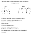
- primers devRf2, and devRr2 amplify a 308bp fragment of gene devR of microbe Mycobacterium tuberculosis.
- primers devRf3, and devRr3 amplify a 164 bp fragment of gene devR of microbe Mycobacterium tuberculosis.
- said method in PCR using primers devRf2 and devRr2 shows 2-4 folds increase in sensitivity as compared to devRf and devRr.
- samples can be obtained from sources comprising all types of sputum and other body fluids comprising FNAC, pus, pleural fluid, pericardial fluid, joint aspirate, peritoneal fluids, cerebrospinal fluids, endometrial aspirate, synovial fluid, gastric aspirate, endotracheal aspirate, urine, transtracheal aspirate, bronchoalveolar lavage, laryngeal swab and nasopharyngeal swab; body tissues comprising blood, pleural tissue, bone marrow and biopsy; solid organs comprising lymph node, bone, skin, and bovine samples comprising lymph gland, milk, and blood.
- sources comprising all types of sputum and other body fluids comprising FNAC, pus, pleural fluid, pericardial fluid, joint aspirate, peritoneal fluids, cerebrospinal fluids, endometrial aspirate, synovial fluid, gastric aspirate, endo
- samples stored at about - 20°C for upto 2 months can be processed for PCR, smear-microscopy and culture.
- composition shows mucolytic, decontaminating, protein denaturant, chaotropic, liquefying, tissue softening/digesting, and mycobacteria-releasing action.
- said method in smear enables more bacilli to be smeared on the slide thereby increasing the sensitivity and the efficiency.
- said method in smear generally converts slides that are graded as 1+ or scanty by the direct method to 2+/3+ or 2+/1+ respectively.
- samples lacking purulence or containing nasopharyngeal discharge or saliva can be processed.
- the said method can detect samples as positive which have been detected negative by direct and CDC methods of smear microscopy.
- kits useful in processing clinical samples for simple, rapid, safe, sensitive, and accurate diagnosis of microbial disease conditions comprising solution 1 consisting of Universal Sample Processing (USP) solution, Solution 2 consisting of Sodium phosphate of concentration ranging between 65 to 70 mM of pH ranging between 6.7 to 6.8 (optional, can be replaced with sterile water), Solution 3 consisting of Tween 80 of concentration ranging between 0.03 to 0.08%, optionally Solution A comprising Chelex 100 suspension of concentration ranging between 8-12%, optionally Solution B consisting of Triton X-100 of concentration ranging between 0.02-0.04%, and optionally Solution C consisting of Tween 20 of concentration ranging between 0.2-0.4%, optionally two sets of primers with devRf2 and devRr2 consisting of SEQ ID No. I and SEQ ID No. 2 respectively, and primers devRf3, and devRr3 consisting of SEQ ID No. 3, and SEQ ID No. 4
- kit is useful in processing clinical samples for detecting bacterial infections.
- kit is useful in processing clinical samples for detecting tuberculosis.
- Universal Sample Processing (USP) solution comprises Guanidinium Hydrochloride (GuHCl) of concentration ranging between 3-6M, Tris-Cl of concentration ranging between 40-60 mM of pH ranging between 7.3- 7.7, EDTA of concentration ranging between 20-30mM, Sarcosyl of concentration ranging between 0.3 - 0.8%, beta-mercaptoethanol of concentration ranging between 0.1-0.3 M.
- GuHCl Guanidinium Hydrochloride
- Tris-Cl of concentration ranging between 40-60 mM of pH ranging between 7.3- 7.7
- EDTA of concentration ranging between 20-30mM
- Sarcosyl of concentration ranging between 0.3 - 0.8%
- beta-mercaptoethanol of concentration ranging between 0.1-0.3 M.
- Universal Sample Processing (USP) solution for processing culture and smear samples comprises Guanidinium Hydrochloride (GuHCl) of concentration of about 4M, Tris-Cl of concentration ranging between 40-60 mM of pH ranging between 7.3- 7.7, EDTA of concentration ranging between 20-30mM, Sarcosyl of concentration ranging between 0.3 - 0.8%, beta-mercaptoethanol of concentration of about 0.1M.
- GuHCl Guanidinium Hydrochloride
- said Universal Sample Processing (USP) solution for processing culture, smear, and PCR samples comprises Guanidinium Hydrochloride (GuHCl) of concentration of about 5M, Tris-Cl of concentration ranging between 40-60 mM of pH ranging between 7.3- 7.7, EDTA of concentration ranging between 20-30 mM, Sarcosyl of concentration ranging between 0.3 - 0.8%, beta-mercaptoethanol of concentration ranging between 0.1-0.2 M.
- GuHCl Guanidinium Hydrochloride
- said Universal Sample Processing (USP) solution for processing smear comprises Guanidinium Hydrochloride (GuHCl) of concentration of about 6M, Tris-Cl of concentration ranging between 40-60 mM of pH ranging between 7.3- 7.7, EDTA of concentration ranging between 20-30mM. Sarcosyl of concentration ranging between 0.3 - 0.8%, beta-mercaptoethanol of concentration of about 0.2 M.
- GuHCl Guanidinium Hydrochloride
- composition comprises solution 1 consisting of Universal Sample Processing (USP) solution, Solution 2 consisting of Sodium phosphate of concentration ranging between 65 to 70 mM of pH ranging between 6.7 to 6.8 (optional, can be replaced with sterile water), Solution 3 consisting of Tween 80 of concentration ranging between 0.03 to 0.08%, and optionally, Solution A comprising Chelex 100 suspension of concentration ranging between 8-12%, and/or Solution B consisting of Triton X-100 of concentration ranging between 0.02-0.04%, and/or Tween 20 of concentration ranging between 0.2-0.4%.
- USP Universal Sample Processing
- Solution 2 consisting of Sodium phosphate of concentration ranging between 65 to 70 mM of pH ranging between 6.7 to 6.8 (optional, can be replaced with sterile water)
- Solution 3 consisting of Tween 80 of concentration ranging between 0.03 to 0.08%
- Solution A comprising Chelex 100 suspension of concentration ranging between 8-12%
- Solution B consisting of Triton X-100
- a set of primers devRf2 and devRr2 consisting of SEQ ID No. 1 and SEQ ID No. 2 respectively is used.
- a set of primers devRf3, and devRr3 consisting of SEQ ID No. 3, and SEQ ID No. 4 respectively is used.
- a method of using primers of SEQ ID NO. 1, and 2 or SEQ ID No. 3 and 4 of gene devR for screening patients of tuberculosis comprising steps of conducting Polymerase Chain Reaction (PCR) using M. tuberculosis DNA or RNA obtained from the processed sample of the subject, identifying the subjects suffering from tuberculosis.
- PCR Polymerase Chain Reaction
- samples can be obtained from sources comprising all types of sputum and other body fluids comprising FNAC, pus, pleural fluid, pericardial fluid, joint aspirate, peritoneal fluids, cerebrospinal fluids, endometrial aspirate, synovial fluid, gastric aspirate, endotracheal aspirate, urine, transtracheal aspirate, bronchoalveolar lavage, laryngeal swab and nasopharyngeal swab; body tissues comprising blood, pleural tissue, bone marrow and biopsy; solid organs comprising lymph node, bone, skin, and bovine samples comprising lymph gland, milk, and blood.
- sources comprising all types of sputum and other body fluids comprising FNAC, pus, pleural fluid, pericardial fluid, joint aspirate, peritoneal fluids, cerebrospinal fluids, endometrial aspirate, synovial fluid, gastric aspirate, endo
- the method comprises of the use of a novel Universal Sample Processing (USP) solution.
- USP Universal Sample Processing
- the USP method comprises of homogenization, liquefaction and decontamination steps carried out on the clinical specimen by treatment with USP solution, concentration of mycobacteria by centrifugation and washing steps.
- Guanidinium hydrochloride comprises the active component of USP together with a reducing agent and a detergent in a buffered solution. This chaotropic agent Guanidinium hydrochloride effectively lyses eukaryotic cells, most bacteria excluding mycobacteria, denatures proteins, including mucin and thereby liquefying sputum, and inactivates endogenous enzymes including DNases and RNases.
- the unique cell wall properties of mycobacteria render it selectively resistant to the action of chaotropic agent and the resultant bacteria are suitable for use in smear microscopy, culture, PCR, RNA isolation and procedures utilizing DNA and RNA isolated thereupon.
- the entire sample processing procedure can be completed in 1 1 ⁇ 2 - 2 hours.
- the procedure does not involve steps of phenol:chloroform extraction, protease digestion, or precipitation steps, making the procedure ideal for processing many samples at a time.
- the method is applicable on all kinds of body fluids, blood, tissue biopsies, biopsies from vertebra and other bone joints and CSF. It is also suitable for use in processing tissue biopsies and milk of bovine origin.
- the processed sample is directly used for smear microscopy, culture on solid media (such as Lowenstein Jensen (LJ) slants or Middlebrook 7H10 agar medium) or liquid medium (such as Middlebrook 7H9 medium used in MGIT system) and DNA and RNA extraction for amplification of mycobacterial DNA and RNA.
- solid media such as Lowenstein Jensen (LJ) slants or Middlebrook 7H10 agar medium
- liquid medium such as Middlebrook 7H9 medium used in MGIT system
- the duration of the methodology is as follows: 1 1 ⁇ 2 -2 hours for sample processing, 1 ⁇ 2-1 hour for smear microscopy and culture and 4 hours for nucleic acid amplification.
- the method described here will find use in any laboratory that requires highly sensitive smear microscopy of acid-fast bacilli, effective decontamination of clinical samples for culture and the isolation of mycobacterial and M. tuberculosis DNA for PCR amplification, from clinical specimens. Therefore it has potential for inclusion in TB/mycobacterial diagnostic kits.
- the claimed technology can be used in a variety of clinical settings and laboratories. It can be used for smear microscopy in laboratories having minimal equipment (a light microscope and an ambient temperature centrifuge that can go up to a speed of 3000 rpm). It can be used for smear microscopy and culture in laboratories having additional facilities for mycobacterial culture. Finally PCR and RT-PCR test can be applied in sophisticated laboratories.]
- the invention is a universal sample processing and diagnostic methodology to be used on any type of clinical specimen for the laboratory diagnosis of tuberculosis in a variety of clinical settings. It is rapid, inexpensive and easy and is compatible with smear microscopy, culture, DNA-RNA isolation and PCR/RT-PCR. It serves the quadruple purpose of (i) providing an extremely sensitive procedure for smear microscopy for the detection of M.tuberculosis, (ii) preparing the sample for culture on solid or liquid laboratory media, (iii) isolating DNA template free of PCR inhibitors and (iv) providing sensitive and specific diagnostic assays based on PCR. The methods have been evaluated on clinical specimens.
- tuberculosis and MOTT bacilli containing specimens require highly sensitive smear microscopy of acid-fast bacilli, effective decontamination of clinical samples for culture and the isolation of mycobacterial and M. tuberculosis DNA for PCR amplification, from clinical specimens. Therefore it has potential for inclusion in TB/mycobacterial diagnostic kits.
- a multipurpose method has been developed for laboratory diagnosis of tuberculosis by smear microscopy, culture and PCR.
- the method is eminently suitable for the preparation of smears for highly sensitive microscopy. It effectively decontaminates samples making them suitable for culture and yields good quality inhibitor-free mycobacterial DNA for use in PCR- based diagnostic assays. It also enables the isolation of good quality mycobacterial RNA from clinical specimens employing common RNA isolation procedures.
- the method for processing of sputum samples for smear microscopy, culture and PCR is as follows:
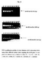
- solutions A, B and C are not required; amplifiable DNA may be isolated by directly heating the sample in Solution 3 at 90-100 °C for 30-40 minutes (or Solution 3 may be supplemented with Triton X-100 at a final concentration of 0.01 % (see Fig. 11 ).
- pleural fluid is processed within 1 hour after collection, then processing can be carried out as described in "Processing of other body fluids.” If the pleural effusion is stored for more than one hour the proteins separate out as a coagulum. Such a sample should be processed as follows:
- Simplified method of USP smear preparation Add 2-3 volumes of USP solution to the sputum (specimen volume less than or equal to 2 ml) collected in McCartney bottles. Mix by hand for 30-60 seconds to homogenize well. Allow to stand for 5-10 minutes. For highly tenacious and purulent sputum specimens, add at least 3 volumes of Solution 1, hand mix and incubate at 37°C for 20-30 min. Fill the bottle with sterile water upto 2/3 rd level and mix by inversion. Centrifuge at 3,000g for 20 minutes in an ambient temperature clinical centrifuge. Decant supernatant carefully taking care that the pellet is not dislodged/lost.
- lysis may be carried out by heating the 'Solution 3 resuspension' directly or after adding Triton X-100 (at a final concentration of 0.01 %) at 90-100 °C for 30-40 minutes.
- the supernatant may be used directly for PCR.
- the addition of the lysis reagents namely, solutions A, B and C
- the lysis reagents may be avoided in such cases; but for best results, it is recommended that the lysis reagents (Solution A, B and C) be used for isolation of PCR-amplifiable DNA (See below for details).
- RNA isolation The pellet obtained can be subjected to mycobacterial RNA isolation by any standard RNA isolation procedures or kits.
- the PCR assay targets the devR gene ( Rv3133c ) of M. tuberculosis and is specific for the organisms of the M. tuberculosis complex [Singh et al ., 1999].
- the primer pair devRf and devRr was used in the 'long target' PCR assay that amplified a 513 bp fragment from the devR gene [Singh et al ., 1999; 2000].
- the same PCR assay was used with coded sputum samples.
- the 'short target' primer pairs are, (i) devRf2, 5' TGGCAACGGCATTGAACTGT 3'and devRr2, 5' TAAGCAGGCCCAGTA GCGT 3' and (ii) devRf3, 5' ATCTGTTGTCCCGCATGCC 3' and devRr3, 5' GTCCAGC GCCCACATCTTT 3'.
- 'Long target' assay Identical parameters as for the 'short target' assay except that annealing and extension were performed at 65 °C for 90 seconds.
- IS6110 assay Published primers suggested by Eisenach et al . (1990) were used with the PCR reaction parameters: initial denaturation at 94°C for 10 minutes followed by 45 cycles of 94°C for 1 min. and 60°C for 1min. 30 seconds, followed by a final extension at 72°C for 5 minutes.
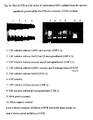
- Total reaction volume is 40 ⁇ l (30 ⁇ l cocktail and 10 ⁇ l template). Dispense 30 ⁇ l cocktail into sterile 0.2 ml or 0.5 ml vials in a DNA-free hood. Add 10 ⁇ l template in a designated area away from the PCR-setup area. Give the tubes a brief spin for 30 seconds and place them in the thermal cycler. After the completion of the reaction (parameters given above) electrophorese the amplification products in 1.5-2.3 % agarose gel in 0.5 x TBE buffer at 80 volts for 20-30 minutes. Visualize the product (load at least 35 ⁇ l) by ethidium bromide staining under UV light.
- USP solution (A) Function of USP solution components: To check for the function of each component of the USP solution, the USP solution was selectively depleted of the individual components and samples processed with the resulting solutions. USP solution consists of five components:
- a sputum specimen obtained by pooling three to four AFB positive sputa (to obtain a large volume and heterogeneous composition in terms of contaminants), was homogenized by vortexing with 3 mm glass beads for 3-5 minutes. Aliquots of 5 ml each of the homogenized sputum were distributed for processing with the solutions of different compositions. USP solution was used as a control. The following solutions of varying compositions were prepared for sample processing:
- FIG. 5a and Fig. 5b show representative sputum and tissue samples treated with USP. At the end of the process the sample is rid of contaminating organisms, proteins, enzymes and interfering substances and considerably reduced in volume.
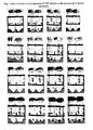
- Negative samples turn to positive in USP smear The sensitivity of the direct smear method and the USP smear method were 68.6 % and 98.2 %, respectively, with a specificity of 92.6% and 91.4 % respectively on the panel of 571 sputum sample (Data set 1). All samples diagnosed as smear positive by the direct method were also positive by the USP and CDC methods of smear microscopy. Out of the 325 direct smear-negative samples, the USP method detected 97 additional samples as smear positive, which were positive by culture also, thereby recording an enhancement in sensitivity of 29.6 %. A representative example is shown in Fig. 8 . Out of the 97 USP smear-positive samples, 14 were graded as scanty, 29 as 1+, 23 as 2+ and 31 as 3+.
- the USP smear method was also simultaneously compared with CDC method of smear microscopy in 325 out of these 571 specimens (Data Set 2). 29 specimens, which were smear negative by the CDC method, were detected as positive by the USP method of smear microscopy all of which were also culture positive.
- USP method also fared better than a popularly used concentration method of smear microscopy (CDC method) in terms of sensitivity of detection (97.1% and 80% sensitivity for USP and CDC smear microscopy, respectively).
- Sensitivity 98.2 % (95.9 %-99.3 %) Specificity: 91.4 % (86.9 %-94.4 %) PPV: 93. 9 % (90.7 %-96.1 %) NPV: 97.4 % (94.1 %-98.9 %) Diagnostic accuracy: 90.5 %
- Sensitivity 97.1 % (92.9 % - 98.9 %) Specificity: 83.3 % (76.2 % - 88.6 %) PPV: 86.4 % (80.5 % - 90.8 %) NPV: 96.3 % (91.1 % - 98.6 %) Diagnostic accuracy: 90.5 %
- Sensitivity 80.0 % (73.0 % - 85.6 %) Specificity: 89.67 % (83.5 % - 93.8 %) PPV: 89.47 % (83.2 % - 93.7 %) NPV: 80.35 % (73.5 % - 85.8 %) Diagnostic accuracy: 84.6 %
- Upgradation in smear status The USP method enhances the gradation of slides making them easier to read in a shorter period of time. Slides graded as scanty, 1+ or 2+ by the direct method of smear microscopy generally turned into 1+, 2+ or 3+ by the USP method.
- USP solution method of smearing showed a very high sensitivity in the range of 97-98 %, much better than the other two methods that were tested, with a specificity range of 83-91 %.
- USP smear microscopy showed a positivity of 97-98 % in culture positive samples vs. 80 % by CDC and 68.6 % by direct smear methods thereby recording an enhancement in sensitivity of ⁇ 30 % and 18 % over the direct and CDC method of smear microscopy respectively.
- the increase in the sensitivity was significant as the sensitivity values of all the three methods were completely non-overlapping at a 95 % confidence interval.
- the USP smear method did not miss a single sample detected by either the direct smear method or the CDC method.
- the specificity of the USP smear microscopy method when compared to the direct smear method was 91.3 % and that when compared to the CDC method was 83.3 %. The two values were overlapping at a 95 % confidence interval. (Data Sets 1 and 2).
- tuberculosis also grew somewhat faster (visible colonies were obtained at least 4-5 days earlier) than USP-treated bacteria on solid media although no appreciable differences were observed between the two treatments when bacteria were cultured in liquid system BD BACTECTM MGITTM 960 System (Becton Dickinson, USA) [see below].
- M . tuberculosis The growth rate of M . tuberculosis is equivalent after treatment with USP solution and NALC-NaOH in liquid medium (Mycobacterial growth indicator tubes): It is well known that treatment with decontaminating agents adversely affects the viability of mycobacteria and slows down their growth rate.
- USP solution treatment The effect of USP solution treatment on the growth rate of M. tuberculosis was compared to that of NALC-NaOH treatment (CDC method).
- a logarithmic phase M. tuberculosis culture grown in Middlebrook 7H9 liquid medium (with ADC supplement and 0.05% Tween 80) was divided into three sets. One set was treated with USP solution and the other set was treated with NALC-NaOH followed by neutralization with phosphate buffer (CDC protocol).
- the USP method of culture is comparable to the currently accepted methods for the culturing of M. tuberculosis from clinical samples.
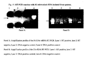
- the PCR test was performed on >700 samples of pulmonary and extra-pulmomary origin. Samples were either fresh or frozen at -20 °C for up to 2 months. Mycobacterial DNA was isolated for this purpose by the USP method. Not a single sample has showed inhibition in the PCR assay indicating that the DNA isolated from the clinical samples by the USP method was free of inhibitors.
- the PCR assay targets the devR gene of M. tuberculosis (Rv 3133c ) and is specific for the organisms of the M. tuberculosis complex [Singh et al., 1999].
- the primer pairs devRf2-devRr2 and devRf3-devRr3 showed a marked enhancement in sensitivity.
- the devRf2 and devRr2 primers [product size 308 bp] showed 2- to 4-fold more sensitivity and the devRf3 and devRr3 primers (product size, 164 bp) showed at least 10-fold more sensitivity over devRf and devRr primers (product size, 513 bp) [Table 4].
- Table 4 Comparative sensitivity of devR PCR assays using different primer pairs over serial dilution of M. tuberculosis genomic DNA. Amt.
- the sensitivity of the 'short target' assay was 98.5 %. This was substantially better than that of the 'long target' assay, whose sensitivity was 73.2 %.
- 322 sputum samples were positive by the USP smear method and also culture positive. Out of these culture positive and USP smear positive samples, long target assay failed to detect 65 samples, which were detected, by the short target assay confirming the latter assay to be a more sensitive one.
- the introduction of the 'short target' primers increased the devR assay sensitivity by 25.3 % using culture positivity, as gold standard (Data Set 3) and 2-10 folds when compared using serial dilutions of M. tuberculosis genomic DNA (Table 4).
- the overall diagnostic accuracy of the short-target assay was about 7 % higher than that of the long-target assay (Data Set 3).
- the enhanced sensitivity of the 'short target' assay was however accompanied by a lower specificity of 77 % as compared to a specificity of 91.9 % with the 'long target' assay (Data set 3).
- the apparent reduction in specificity of the 'short target' assay is likely a reflection of the enhanced sensitivity of the assay and is unlikely to be due to false positivity/ lower specificity. This is supported by the following:
- Extrapulmonary samples The USP method was evaluated for its performance in the diagnosis of tuberculous pleural effusion and lymphadenopathy in a blinded study with coded pleural fluid, pleural tissue and lymph-node biopsy specimens collected from the patients coming to the OPD of Safdarjung Hospital, New Delhi, India.
- the samples comprised of 100 specimens from 88 patients including 77 specimens from TB patients and 23 specimens from control subjects with diseases like malignancy, amoebiasis, CHF, Sarcoidosis and reactive lymphadenopathy (nonspecific inflammation).
- Inclusion criteria was based on any one or more of the following parameters:
- the devR PCR showed better sensitivity with pleural tissue and lymph node (100 % and 75 % respectively) specimens than IS6110, (75 % and 50 % respectively) probably due to absence of any copies of IS6110 in these strains (Data Sets 5 & 6).
- the USP method of specimen processing and devR PCR served as a useful modality to arrive at the definitive diagnosis of tuberculous pleural effusion and lymphadenopathy.
- Pleural Fluid Pleural tissue Lymph node Sensitivity (%) 80.9 100 75 Specificity (%) 93.3 100 66.7 PPV (%) 97.1 100 90 NPV (%) 63.6 100 40 Efficiency (%) 84.2 100 73.3
- Pleural Fluid Pleural tissue Lymph node Sensitivity (%) 93 75 50 Specificity (%) 96.15 100 75 PPV (%) 97.6 100 88.9 NPV (%) 89.3 60 27.3 Efficiency (%) 94.2 81.9 55
- MOTT tuberculosis type
- MOTT bacilli were subjected to USP treatment for 10-15 minutes at room temperature, followed by a wash with sterile triple distilled water. Each species was then subjected to smear microscopy, culture on LJ slant and PCR to check for detrimental effect of USP solution, if any.
- the MOTT species used were: M. avium, M, fortuitunm , M. gordonae, M. intracellulare, M. kansasi, M. scrofulacem, M. smegmatis, M.
- Organisms belonging to the M. tuberculosis complex like M. africanum, M. bovis, M. bovis (BCG), M. microti were also included in the study. All the organisms retained their AFB status ( Fig. 16 ) and viability. However M. smegmatis though it maintained its integrity and AFB property, lost its viability.
- Fig.17 shows two USP-treated rapid growers. Good quality PCR-amplifiable DNA was isolated from all the USP-treated Mycobacterium ( Fig. 18 ).
- the USP method is also compatible with the isolation of high quality M. tuberculosis RNA from sputum specimens.
- USP solution with the following composition was used.6M Guanidinium hydrochloride, 50 mM Tris-Cl, pH 7.5, 25 mM EDTA, 0.5% Sarcosyl and 0.2 M ⁇ - mercaptoethanol.
- the sputum specimen used was graded 3+ by the direct method of smear microscopy. To the specimen (5-7 ml), was added 1.5 times the volume of USP solution and the mixture was homogenized. To it was added 10 ml of sterile DEPC water and mixed. It was then centrifuged at 5,000 rpm for 10 minutes at room temperature. The pellet was again washed with 2-3 ml of USP solution followed by a wash with sterile DEPC water. The final processed pellet can be used as a starting material for RNA isolation by an appropriate means. For example, using the Qiagen Rneasy Mini Kit as per manufacturer's protocol. The isolated M.
- tuberculosis RNA was subjected to reverse transcription reaction using Stratrascript reverse transcriptase (RT) enzyme (Stratagene, USA) to obtain cDNA.
- the cDNA was subjected to amplification using M. tuberculosis rRNA-(23S rRNA) and mRNA ( Rv 3134c gene)-specific primers to detect the presence of M. tuberculosis RNA.
- RT negative controls in which the isolated RNA was subjected to reverse transcription reaction without the reverse transcriptase enzyme were also run for PCR to monitor for the presence of any contaminating genomic DNA in the preparation. Amplifications corresponding to both the ribosomal and messenger cDNAs were obtained ( Fig. 19 ) proving that both classes of M.
- tuberculosis RNA was isolated from the sputum specimen.
- the pellets were then resuspended in solution 3 and 10% was smeared in glass slides (in duplicates) and 40% was inoculated in nutrient agar media and incubated at 37°C to monitor for any growth. This was followed by Gram staining of the slides. Gram staining was also performed on the untreated bacteria.
- This technology is suitable for any kind of body fluid and tissue biopsy (except for fecal matter which has not been tested).
- the claimed technology can be used in a variety of clinical settings and laboratories. (1) It can be used for smear microscopy in laboratories having minimal equipment (only a vortex shaker, light microscope and ambient temperature centrifuge that can go up to a speed of 4000 rpm). (2) It can be used for smear microscopy and culture in laboratories having additional facilities for mycobacterial culture. (3) Finally PCR and RT-PCR tests can be applied in sophisticated laboratories.
Landscapes
- Chemical & Material Sciences (AREA)
- Life Sciences & Earth Sciences (AREA)
- Analytical Chemistry (AREA)
- Proteomics, Peptides & Aminoacids (AREA)
- Organic Chemistry (AREA)
- Zoology (AREA)
- Wood Science & Technology (AREA)
- Health & Medical Sciences (AREA)
- Engineering & Computer Science (AREA)
- Microbiology (AREA)
- Immunology (AREA)
- Molecular Biology (AREA)
- Biotechnology (AREA)
- Biophysics (AREA)
- Physics & Mathematics (AREA)
- Biochemistry (AREA)
- Bioinformatics & Cheminformatics (AREA)
- General Engineering & Computer Science (AREA)
- General Health & Medical Sciences (AREA)
- Genetics & Genomics (AREA)
- Measuring Or Testing Involving Enzymes Or Micro-Organisms (AREA)
Claims (11)
- Verfahren zum Verarbeiten klinischer Proben, die zur Diagnose von Folgendem nützlich sind: bakteriellen Infektionen, wie zum Beispiel Tuberkulose, und anderen mykobakteriellen Infektionen, die durch Mykobakterien, einschließlich M. tuberculosis verursacht werden und anderen Infektionen, die durch Gram-positive Organismen wie Staphylococcus sp. verursacht werden, umfassend:Verwendung einer Zusammensetzung, die Folgendes umfasst: Lösung 1, die aus Universal-Probenverarbeitungslösung (USP-Lösung; Universal Sample Processing-Lösung) besteht, die sich aus Guanidiniumhydrochlorid (GuHCl) in einer Konzentration im Bereich zwischen 3 und 6 M, Tris-Cl in einer Konzentration im Bereich zwischen 40 und 60 mM mit einem pH im Bereich zwischen 7,3 und 7,7, EDTA in einer Konzentration im Bereich zwischen 20 und 30 mM, Sarcosyl in einer Konzentration im Bereich zwischen 0,3 und 0,8 %, β-Mercaptoethanol in einer Konzentration im Bereich zwischen 0,1-0,3 M zusammensetzt, Lösung 2, die aus Natriumphosphat in einer Konzentration im Bereich zwischen 65 und 70 mM mit einem pH im Bereich zwischen 6,7 und 6,8 besteht (kann optional durch steriles Wasser ersetzt werden), und Lösung 3, die aus Tween 80 in einer Konzentration im Bereich zwischen 0,03 und 0,08 % besteht, wobei das Verfahren die folgenden Schritte umfasst:(a) Verwendung einer klinischen Probe,(b) Beimischen von 1,5 bis 2 Volumina Lösung 1 zur Probe,(c) Homogenisieren des Gemischs, während Schäumen vermieden wird,(d) Zufügen von Lösung 2 oder sterilem Wasser zum Homogenat, gefolgt von Zentrifugation zum Erhalt eines Pellets,(e) Waschen des Pellets mit Lösung 1, optional in Abhängigkeit von der Abnahme der Pelletgröße,(f) Waschen des mit Lösung 1 gewaschenen Pellets mit Wasser, und(g) Resuspendieren des mit Wasser gewaschenen Pellets von Schritt (f) in Lösung 3 zum Erhalt der verarbeiteten Probe zur Diagnose.
- Verfahren nach Anspruch 1, worin das Homogenisieren für eine Zeitdauer im Bereich zwischen 20 und 120 Sekunden durchgeführt wird.
- Verfahren nach Anspruch 1, worin die Verarbeitung in einer Zeitdauer im Bereich von insgesamt zwischen 1 und 2 Stunden abgeschlossen ist.
- Verfahren nach Anspruch 1, worin Guanidiniumhydrochlorid in einer Konzentration von ca. 4 M und β-Mercaptoethanol in einer Konzentration von ca. 0,1 M in der Universal-Probenverarbeitungslösung (USP-Lösung) zum Verarbeiten von Proben zur Kultur und zum Ausstrich vorliegt, wobei Guanidiniumhydrochlorid in einer Konzentration von ca. 5 M und das β-Mercaptoethanol in einer Konzentration von zwischen 0,1-0,2 M zum Verarbeiten von Proben zur Kultur, zum Ausstrich und zur PCR vorliegt, und wobei das Guanidiniumhydrochlorid in einer Konzentration von 6 M und das β-Mercaptoethanol in einer Konzentration von ca. 0,2 M für zum Ausstrich und zur PCR verarbeitete Proben vorliegt.
- Verfahren nach Anspruch 1 zur Diagnose einer mykobakteriellen Infektion, worin die mittels PCR amplifizierbare mykobakterielle DNA und RNA durch einfache Lyse mittels Erhitzen der Resuspension von Schritt (g) 30 bis 40 Minuten bei 90 bis 100 °C direkt oder nach dem Zufügen von Triton X-100 in einer Endkonzentration von 0,01 % erhalten werden.
- Verfahren nach Anspruch 1, worin das Verfahren ferner einen Kulturschritt umfasst, der bei einem neutralen pH abläuft.
- Verfahren nach Anspruch 1 zur Diagnose einer mykobakteriellen Infektion, worin das Verfahren ferner eine PCR umfasst, die zwei Primer-Sets verwendet, nämlich devRf2 und devRr2, die aus SEQ ID NO: 1 bzw. SEQ ID NO: 2 bestehen, und devRf3 und devRr3, die aus SEQ ID NO: 3 bzw. SEQ ID NO: 4 vom Gen devR von der Mikrobe Mycobacterium tuberculosis bestehen.
- Verfahren nach Anspruch 7, worin die Primer devRf2 und devRr2 ein 308 bp langes Fragment vom Gen devR von der Mikrobe Mycobacterium tuberculosis amplifizieren.
- Verfahren nach Anspruch 7, worin die Primer devRf3 und devRr3 ein 164 bp langes Fragment vom Gen devR von der Mikrobe Mycobacterium tuberculosis amplifizieren.
- Kit, das zur Verarbeitung klinischer Proben zur einfachen, schnellen, sicheren, empfindlichen und genauen Diagnose von Folgendem nützlich ist: bakteriellen Infektionen, wie zum Beispiel Tuberkulose, und anderen mykobakteriellen Infektionen, die durch Mykobakterien, einschließlich M. tuberculosis verursacht werden und anderen Infektionen, die durch Gram-positive Organismen wie Staphylococcus sp. verursacht werden, wobei das Kit Folgendes umfasst: Lösung 1, die aus Universal-Probenverarbeitungslösung (USP-Lösung) besteht (zusammengesetzt aus Guanidiniumhydrochlorid (GuHCl) in einer Konzentration im Bereich zwischen 3 und 6 M, Tris-Cl in einer Konzentration im Bereich zwischen 40 und 60 mM mit einem pH im Bereich zwischen 7,3 und 7,7, EDTA in einer Konzentration im Bereich zwischen 20 und 30 mM, Sarcosyl in einer Konzentration im Bereich zwischen 0,3 und 0,8 %, β-Mercaptoethanol in einer Konzentration im Bereich zwischen 0,1 und 0,3 M), Lösung 2, die aus Natriumphosphat in einer Konzentration im Bereich zwischen 65 bis 70 mM mit einem pH im Bereich zwischen 6,7 und 6,8 besteht (kann optional durch Wasser ersetzt werden), Lösung 3, die aus Tween 80 in einer Konzentration im Bereich zwischen 0,03 und 0,08 % besteht, und optional zwei Primer-Sets mit devRf2 und devRr2, die aus SEQ ID NO: 1 bzw. SEQ ID NO: 2 bestehen, und den Primern devRf3 und devRr3, die aus SEQ ID NO: 3 bzw. SEQ ID N: 4 bestehen.
- Kit nach Anspruch 10, worin Guanidiniumhydrochlorid in einer Konzentration von ca. 4 M und β-Mercaptoethanol in einer Konzentration von ca. 0,1 M in der Universal-Probenverarbeitungslösung (USP-Lösung) zum Verarbeiten von Proben zur Kultur und zum Ausstrich vorliegt, wobei Guanidiniumhydrochlorid in einer Konzentration von ca. 5 M und β-Mercaptoethanol in einer Konzentration von zwischen 0,1 und 0,2 M zur Verarbeitung von Proben zur Kultur, zum Ausstrich und zur PCR vorliegt, und das Guanidiniumhydrochlorid in einer Konzentration von 6 M vorliegt und das β-Mercaptoethanol in einer Konzentration von ca. 0,2 M für zum Ausstrich und zur PCR verarbeitete Proben vorliegt.
Priority Applications (2)
| Application Number | Priority Date | Filing Date | Title |
|---|---|---|---|
| EP07011868A EP1897957B1 (de) | 2003-07-29 | 2003-07-29 | Verfahren zur Diagnose von Tuberkulose mittels Abstrichmikroskopie, Kultur und Polymerasekettenreaktion unter Verwendung prozessierter klinischer Proben und Kit davon |
| EP09171694A EP2149614B1 (de) | 2003-07-29 | 2003-07-29 | Primer zum Nachweis von Mycobacterium tuberculosis |
Applications Claiming Priority (1)
| Application Number | Priority Date | Filing Date | Title |
|---|---|---|---|
| PCT/IB2003/003211 WO2005010186A1 (en) | 2003-07-29 | 2003-07-29 | A method for diagnosis of tuberculosis by smear microscopy, culture and polymerase chain reaction using processed clinical samples and kit thereof |
Related Child Applications (2)
| Application Number | Title | Priority Date | Filing Date |
|---|---|---|---|
| EP07011868A Division EP1897957B1 (de) | 2003-07-29 | 2003-07-29 | Verfahren zur Diagnose von Tuberkulose mittels Abstrichmikroskopie, Kultur und Polymerasekettenreaktion unter Verwendung prozessierter klinischer Proben und Kit davon |
| EP09171694A Division EP2149614B1 (de) | 2003-07-29 | 2003-07-29 | Primer zum Nachweis von Mycobacterium tuberculosis |
Publications (2)
| Publication Number | Publication Date |
|---|---|
| EP1649014A1 EP1649014A1 (de) | 2006-04-26 |
| EP1649014B1 true EP1649014B1 (de) | 2009-12-02 |
Family
ID=34090436
Family Applications (3)
| Application Number | Title | Priority Date | Filing Date |
|---|---|---|---|
| EP03817620A Expired - Lifetime EP1649014B1 (de) | 2003-07-29 | 2003-07-29 | Verfahren zur diagnose von tuberkulose mittels abstrichmikroskopie, kultur und polymerasekettenreaktion unter verwendung prozessierter klinischer proben und kit davon |
| EP09171694A Expired - Lifetime EP2149614B1 (de) | 2003-07-29 | 2003-07-29 | Primer zum Nachweis von Mycobacterium tuberculosis |
| EP07011868A Expired - Lifetime EP1897957B1 (de) | 2003-07-29 | 2003-07-29 | Verfahren zur Diagnose von Tuberkulose mittels Abstrichmikroskopie, Kultur und Polymerasekettenreaktion unter Verwendung prozessierter klinischer Proben und Kit davon |
Family Applications After (2)
| Application Number | Title | Priority Date | Filing Date |
|---|---|---|---|
| EP09171694A Expired - Lifetime EP2149614B1 (de) | 2003-07-29 | 2003-07-29 | Primer zum Nachweis von Mycobacterium tuberculosis |
| EP07011868A Expired - Lifetime EP1897957B1 (de) | 2003-07-29 | 2003-07-29 | Verfahren zur Diagnose von Tuberkulose mittels Abstrichmikroskopie, Kultur und Polymerasekettenreaktion unter Verwendung prozessierter klinischer Proben und Kit davon |
Country Status (7)
| Country | Link |
|---|---|
| US (1) | US20070111206A1 (de) |
| EP (3) | EP1649014B1 (de) |
| CN (1) | CN1856570A (de) |
| AP (3) | AP2276A (de) |
| AU (1) | AU2003247060A1 (de) |
| DE (3) | DE60335970D1 (de) |
| WO (1) | WO2005010186A1 (de) |
Families Citing this family (30)
| Publication number | Priority date | Publication date | Assignee | Title |
|---|---|---|---|---|
| EP1676113A4 (de) * | 2003-10-23 | 2006-11-15 | Salubris Inc | Dekontaminations- und anreicherungskit für mycobakterien |
| US9328326B2 (en) | 2006-08-10 | 2016-05-03 | Merck Patent Gmbh | Method for isolating cells |
| US9481912B2 (en) | 2006-09-12 | 2016-11-01 | Longhorn Vaccines And Diagnostics, Llc | Compositions and methods for detecting and identifying nucleic acid sequences in biological samples |
| US8080645B2 (en) | 2007-10-01 | 2011-12-20 | Longhorn Vaccines & Diagnostics Llc | Biological specimen collection/transport compositions and methods |
| US8652782B2 (en) * | 2006-09-12 | 2014-02-18 | Longhorn Vaccines & Diagnostics, Llc | Compositions and methods for detecting, identifying and quantitating mycobacterial-specific nucleic acids |
| US8097419B2 (en) | 2006-09-12 | 2012-01-17 | Longhorn Vaccines & Diagnostics Llc | Compositions and method for rapid, real-time detection of influenza A virus (H1N1) swine 2009 |
| US9683256B2 (en) | 2007-10-01 | 2017-06-20 | Longhorn Vaccines And Diagnostics, Llc | Biological specimen collection and transport system |
| US11041215B2 (en) | 2007-08-24 | 2021-06-22 | Longhorn Vaccines And Diagnostics, Llc | PCR ready compositions and methods for detecting and identifying nucleic acid sequences |
| AU2008293504B2 (en) | 2007-08-27 | 2012-04-12 | Longhorn Vaccines & Diagnostics, Llc | Immunogenic compositions and methods |
| US10004799B2 (en) | 2007-08-27 | 2018-06-26 | Longhorn Vaccines And Diagnostics, Llc | Composite antigenic sequences and vaccines |
| US11041216B2 (en) | 2007-10-01 | 2021-06-22 | Longhorn Vaccines And Diagnostics, Llc | Compositions and methods for detecting and quantifying nucleic acid sequences in blood samples |
| AU2008343745B2 (en) | 2007-10-01 | 2012-05-10 | Longhorn Vaccines & Diagnostics Llc | Biological specimen collection and transport system and methods of use |
| EP2255010B1 (de) | 2008-02-20 | 2018-05-30 | Streck Inc. | Thermocycler und probengefäss zur schnellen amplifikation von dna |
| WO2010132800A1 (en) * | 2009-05-14 | 2010-11-18 | Streck, Inc. | Specimen container, system, and method |
| EP2388312A1 (de) * | 2010-05-17 | 2011-11-23 | Curetis AG | Universell anwendbarer Lysepuffer und Verarbeitungsverfahren zur Lyse von Körperproben |
| CA2835654A1 (en) | 2011-06-01 | 2012-12-06 | Streck, Inc. | Rapid thermocycler system for rapid amplification of nucleic acids and related methods |
| KR20140040239A (ko) | 2011-06-19 | 2014-04-02 | 아보젠, 인크. | 샘플 수집을 위한 장치, 용액 및 방법 |
| CA2863083C (en) | 2012-01-26 | 2023-09-19 | Longhorn Vaccines And Diagnostics, Llc | Composite antigenic sequences and vaccines |
| US8735091B2 (en) * | 2012-05-17 | 2014-05-27 | Biomerieux, Inc. | Methods for inactivation and extraction of acid-fast bacteria for characterization and/or identification using mass spectrometry |
| EP2883039A1 (de) | 2012-08-10 | 2015-06-17 | Streck Inc. | Optisches echtzeitsystem für echtzeit-polymerasekettenreaktion |
| US10006861B2 (en) | 2013-06-28 | 2018-06-26 | Streck, Inc. | Devices for real-time polymerase chain reaction |
| CN103911365A (zh) * | 2014-03-13 | 2014-07-09 | 南京爱必梦生物材料有限公司 | 提取结核杆菌dna的方法及检测其多重耐药性的试剂盒 |
| CN106460030A (zh) | 2014-05-27 | 2017-02-22 | Dna真诺泰克有限公司 | 用于稳定和维持顽强微生物的生存力的组合物和方法 |
| US9976136B2 (en) | 2015-05-14 | 2018-05-22 | Longhorn Vaccines And Diagnostics, Llc | Rapid methods for the extraction of nucleic acids from biological samples |
| EP3347490A1 (de) * | 2015-09-11 | 2018-07-18 | Corning Incorporated | Zusammensetzungen und verfahren zur nukleinsäurereinigung aus blutproben |
| WO2017055494A1 (en) * | 2015-10-02 | 2017-04-06 | Roche Diagnostics Gmbh | Mycobacteria pre-analytic reagent |
| CN105890955B (zh) * | 2016-06-12 | 2018-06-19 | 李源 | 痰标本自动涂片仪 |
| CN108251414A (zh) * | 2018-01-17 | 2018-07-06 | 云健康基因科技(上海)有限公司 | 骨髓细胞基因组dna的提取方法及提取试剂盒 |
| CN108531563A (zh) * | 2018-02-05 | 2018-09-14 | 深圳市尚维高科有限公司 | 多孔微球的用途及裂解液以及裂解液的使用方法 |
| CN113417295B (zh) * | 2021-06-07 | 2022-08-12 | 海南大学 | 一种基坑微生物土重力式围护结构及其施工方法 |
Family Cites Families (4)
| Publication number | Priority date | Publication date | Assignee | Title |
|---|---|---|---|---|
| US6027883A (en) * | 1994-03-30 | 2000-02-22 | Mitokor | Optimal procedure for isolation of mutant mitochondrial alleles |
| US6027890A (en) * | 1996-01-23 | 2000-02-22 | Rapigene, Inc. | Methods and compositions for enhancing sensitivity in the analysis of biological-based assays |
| US6335193B1 (en) * | 1999-04-15 | 2002-01-01 | Padmanabhan P Nair | Isolated colonocytes |
| GB0030368D0 (en) * | 2000-12-13 | 2001-01-24 | Inst Of Molecul & Cell Biology | Dormancy-induced mycobacterium proteins |
-
2003
- 2003-07-29 EP EP03817620A patent/EP1649014B1/de not_active Expired - Lifetime
- 2003-07-29 WO PCT/IB2003/003211 patent/WO2005010186A1/en not_active Ceased
- 2003-07-29 DE DE60335970T patent/DE60335970D1/de not_active Expired - Lifetime
- 2003-07-29 AP AP2009004803A patent/AP2276A/xx active
- 2003-07-29 EP EP09171694A patent/EP2149614B1/de not_active Expired - Lifetime
- 2003-07-29 AU AU2003247060A patent/AU2003247060A1/en not_active Abandoned
- 2003-07-29 US US10/566,223 patent/US20070111206A1/en not_active Abandoned
- 2003-07-29 DE DE60331979T patent/DE60331979D1/de not_active Expired - Lifetime
- 2003-07-29 DE DE60330399T patent/DE60330399D1/de not_active Expired - Lifetime
- 2003-07-29 CN CNA038270706A patent/CN1856570A/zh active Pending
- 2003-07-29 EP EP07011868A patent/EP1897957B1/de not_active Expired - Lifetime
- 2003-07-29 AP AP2009004802A patent/AP2275A/xx active
- 2003-07-29 AP AP2006003511A patent/AP2221A/xx active
Also Published As
| Publication number | Publication date |
|---|---|
| AP2276A (en) | 2011-08-24 |
| AP2275A (en) | 2011-08-23 |
| WO2005010186A1 (en) | 2005-02-03 |
| DE60330399D1 (de) | 2010-01-14 |
| AP2009004803A0 (en) | 2009-04-30 |
| DE60335970D1 (de) | 2011-03-17 |
| AP2221A (en) | 2011-03-24 |
| AU2003247060A1 (en) | 2005-02-14 |
| DE60331979D1 (de) | 2010-05-12 |
| EP2149614A8 (de) | 2010-06-02 |
| CN1856570A (zh) | 2006-11-01 |
| EP1649014A1 (de) | 2006-04-26 |
| AP2009004802A0 (en) | 2009-04-30 |
| EP2149614B1 (de) | 2011-02-02 |
| AP2006003511A0 (en) | 2006-02-28 |
| EP1897957B1 (de) | 2010-03-31 |
| EP1897957A3 (de) | 2008-10-22 |
| EP1897957A2 (de) | 2008-03-12 |
| US20070111206A1 (en) | 2007-05-17 |
| EP2149614A1 (de) | 2010-02-03 |
Similar Documents
| Publication | Publication Date | Title |
|---|---|---|
| EP1649014B1 (de) | Verfahren zur diagnose von tuberkulose mittels abstrichmikroskopie, kultur und polymerasekettenreaktion unter verwendung prozessierter klinischer proben und kit davon | |
| Caulfield et al. | Diagnosis of active tuberculosis disease: From microscopy to molecular techniques | |
| Gopinath et al. | Multiplex PCR assay for simultaneous detection and differentiation of Mycobacterium tuberculosis, Mycobacterium avium complexes and other Mycobacterial species directly from clinical specimens | |
| Kirschner et al. | Diagnosis of mycobacterial infections by nucleic acid amplification: 18-month prospective study | |
| Miller et al. | Performance of Xpert MTB/RIF RUO assay and IS 6110 real-time PCR for Mycobacterium tuberculosis detection in clinical samples | |
| AU2014286889B2 (en) | Methods of targeted antibiotic susceptibility testing | |
| CA2950419C (en) | Composition and method for stabilizing and maintaining the viability of hardy microorganisms | |
| Haldar et al. | Improved laboratory diagnosis of tuberculosis–the Indian experience | |
| Albay et al. | The evaluation of FASTPlaqueTBTM test for the rapid diagnosis of tuberculosis | |
| Maurya et al. | Detection of 123 bp fragment of insertion element IS6110 Mycobacterium tuberculosis for diagnosis of extrapulmonary tuberculosis | |
| Raveendran et al. | Utility of multiplex real-time PCR in the diagnosis of extrapulmonary tuberculosis | |
| Phetsuksiri et al. | Comparison of loop-mediated isothermal amplification, microscopy, culture, and PCR for diagnosis of pulmonary tuberculosis | |
| Sekar et al. | The utility of IS6110 sequence based polymerase chain reaction in comparison to conventional methods in the diagnosis of extra-pulmonary tuberculosis | |
| AU2006209416A1 (en) | Method of quantitatively analysing microorganism targeting rRNA | |
| Johansen et al. | Evaluation of a new commercial assay for diagnosis of pulmonary and nonpulmonary tuberculosis | |
| Alli et al. | Direct molecular detection of Mycobacterium tuberculosis complex from clinical samples–An adjunct to cultural method of laboratory diagnosis of tuberculosis | |
| Levidiotou et al. | Four-year experience of use of the Cobas Amplicor system for rapid detection of Mycobacterium tuberculosis complex in respiratory and nonrespiratory specimens in Greece | |
| US20070065851A1 (en) | Method for detecting a microorganism in a fecal specimen | |
| Gamboa et al. | Detection and identification of mycobacteria by amplification of RNA and DNA in pretreated blood and bone marrow aspirates by a simple lysis method | |
| Seagar et al. | Comparison of two in-house Real-time PCR assays with MTB Q-PCR Alert and GenoType MTBDR plus for the rapid detection of mycobacteria in clinical specimens | |
| Loonen et al. | Acceleration of the direct identification of Staphylococcus aureus versus coagulase-negative staphylococci from blood culture material: a comparison of six bacterial DNA extraction methods | |
| Oh et al. | Improved detection and differentiation of mycobacteria with combination of Mycobacterium Growth Indicator Tube and Roche COBAS AMPLICOR System in conjunction with Duplex PCR | |
| Varghese et al. | Broad-range bacterial and fungal DNA amplification on vitreous humor from suspected endophthalmitis patients | |
| Rodrigo et al. | Rapid detection of Mycobacterium tuberculosis in respiratory specimens, blood and other non-respiratory specimens by amplification of rRNA | |
| Kaur et al. | Utility of PCR targeting IS6110 and MPT64 genes in the diagnosis of extrapulmonary tuberculosis |
Legal Events
| Date | Code | Title | Description |
|---|---|---|---|
| PUAI | Public reference made under article 153(3) epc to a published international application that has entered the european phase |
Free format text: ORIGINAL CODE: 0009012 |
|
| 17P | Request for examination filed |
Effective date: 20060130 |
|
| AK | Designated contracting states |
Kind code of ref document: A1 Designated state(s): DE FR GB IT |
|
| 17Q | First examination report despatched |
Effective date: 20061004 |
|
| DAX | Request for extension of the european patent (deleted) | ||
| RBV | Designated contracting states (corrected) |
Designated state(s): DE FR GB IT |
|
| GRAP | Despatch of communication of intention to grant a patent |
Free format text: ORIGINAL CODE: EPIDOSNIGR1 |
|
| GRAC | Information related to communication of intention to grant a patent modified |
Free format text: ORIGINAL CODE: EPIDOSCIGR1 |
|
| GRAC | Information related to communication of intention to grant a patent modified |
Free format text: ORIGINAL CODE: EPIDOSCIGR1 |
|
| GRAS | Grant fee paid |
Free format text: ORIGINAL CODE: EPIDOSNIGR3 |
|
| GRAA | (expected) grant |
Free format text: ORIGINAL CODE: 0009210 |
|
| AK | Designated contracting states |
Kind code of ref document: B1 Designated state(s): DE FR GB IT |
|
| REG | Reference to a national code |
Ref country code: GB Ref legal event code: FG4D |
|
| REF | Corresponds to: |
Ref document number: 60330399 Country of ref document: DE Date of ref document: 20100114 Kind code of ref document: P |
|
| PLBE | No opposition filed within time limit |
Free format text: ORIGINAL CODE: 0009261 |
|
| STAA | Information on the status of an ep patent application or granted ep patent |
Free format text: STATUS: NO OPPOSITION FILED WITHIN TIME LIMIT |
|
| 26N | No opposition filed |
Effective date: 20100903 |
|
| PGFP | Annual fee paid to national office [announced via postgrant information from national office to epo] |
Ref country code: DE Payment date: 20100723 Year of fee payment: 8 Ref country code: FR Payment date: 20100805 Year of fee payment: 8 Ref country code: GB Payment date: 20100511 Year of fee payment: 8 Ref country code: IT Payment date: 20100731 Year of fee payment: 8 |
|
| GBPC | Gb: european patent ceased through non-payment of renewal fee |
Effective date: 20110729 |
|
| REG | Reference to a national code |
Ref country code: FR Ref legal event code: ST Effective date: 20120330 |
|
| PG25 | Lapsed in a contracting state [announced via postgrant information from national office to epo] |
Ref country code: FR Free format text: LAPSE BECAUSE OF NON-PAYMENT OF DUE FEES Effective date: 20110801 Ref country code: DE Free format text: LAPSE BECAUSE OF NON-PAYMENT OF DUE FEES Effective date: 20120201 |
|
| REG | Reference to a national code |
Ref country code: DE Ref legal event code: R119 Ref document number: 60330399 Country of ref document: DE Effective date: 20120201 |
|
| PG25 | Lapsed in a contracting state [announced via postgrant information from national office to epo] |
Ref country code: IT Free format text: LAPSE BECAUSE OF NON-PAYMENT OF DUE FEES Effective date: 20110729 |
|
| PG25 | Lapsed in a contracting state [announced via postgrant information from national office to epo] |
Ref country code: GB Free format text: LAPSE BECAUSE OF NON-PAYMENT OF DUE FEES Effective date: 20110729 |