EP1582148A1 - Appareil à rayons X pour mammographie - Google Patents
Appareil à rayons X pour mammographie Download PDFInfo
- Publication number
- EP1582148A1 EP1582148A1 EP05101998A EP05101998A EP1582148A1 EP 1582148 A1 EP1582148 A1 EP 1582148A1 EP 05101998 A EP05101998 A EP 05101998A EP 05101998 A EP05101998 A EP 05101998A EP 1582148 A1 EP1582148 A1 EP 1582148A1
- Authority
- EP
- European Patent Office
- Prior art keywords
- filter
- kev
- ray
- mammography
- radiation
- Prior art date
- Legal status (The legal status is an assumption and is not a legal conclusion. Google has not performed a legal analysis and makes no representation as to the accuracy of the status listed.)
- Withdrawn
Links
- 238000009607 mammography Methods 0.000 title claims abstract description 12
- 239000000463 material Substances 0.000 claims abstract description 13
- 238000010521 absorption reaction Methods 0.000 claims abstract description 10
- WFKWXMTUELFFGS-UHFFFAOYSA-N tungsten Chemical compound [W] WFKWXMTUELFFGS-UHFFFAOYSA-N 0.000 claims abstract description 4
- 229910052721 tungsten Inorganic materials 0.000 claims abstract description 4
- 239000010937 tungsten Substances 0.000 claims abstract description 4
- 229910052791 calcium Inorganic materials 0.000 claims description 4
- 239000011669 selenium Substances 0.000 claims description 4
- 229910052719 titanium Inorganic materials 0.000 claims description 4
- BUGBHKTXTAQXES-UHFFFAOYSA-N Selenium Chemical compound [Se] BUGBHKTXTAQXES-UHFFFAOYSA-N 0.000 claims description 3
- 229910052804 chromium Inorganic materials 0.000 claims description 3
- 229910052742 iron Inorganic materials 0.000 claims description 3
- 229910052748 manganese Inorganic materials 0.000 claims description 3
- 229910052706 scandium Inorganic materials 0.000 claims description 3
- 229910052711 selenium Inorganic materials 0.000 claims description 3
- 239000004065 semiconductor Substances 0.000 claims description 3
- 229910052720 vanadium Inorganic materials 0.000 claims description 3
- 230000005855 radiation Effects 0.000 abstract description 16
- 239000010948 rhodium Substances 0.000 description 12
- 208000004434 Calcinosis Diseases 0.000 description 8
- 206010028980 Neoplasm Diseases 0.000 description 8
- 238000009826 distribution Methods 0.000 description 6
- 230000002308 calcification Effects 0.000 description 4
- 229910052799 carbon Inorganic materials 0.000 description 4
- 229910052739 hydrogen Inorganic materials 0.000 description 4
- 229910052757 nitrogen Inorganic materials 0.000 description 4
- 229910052760 oxygen Inorganic materials 0.000 description 4
- 238000001514 detection method Methods 0.000 description 3
- 230000002349 favourable effect Effects 0.000 description 2
- 229910052703 rhodium Inorganic materials 0.000 description 2
- 229910052709 silver Inorganic materials 0.000 description 2
- 230000002159 abnormal effect Effects 0.000 description 1
- 230000006978 adaptation Effects 0.000 description 1
- 229910052782 aluminium Inorganic materials 0.000 description 1
- 210000000481 breast Anatomy 0.000 description 1
- 238000005094 computer simulation Methods 0.000 description 1
- 230000001419 dependent effect Effects 0.000 description 1
- 230000000694 effects Effects 0.000 description 1
- 239000004744 fabric Substances 0.000 description 1
- 238000001914 filtration Methods 0.000 description 1
- 230000000762 glandular Effects 0.000 description 1
- 238000000034 method Methods 0.000 description 1
- 229910052750 molybdenum Inorganic materials 0.000 description 1
- 231100000289 photo-effect Toxicity 0.000 description 1
- MHOVAHRLVXNVSD-UHFFFAOYSA-N rhodium atom Chemical compound [Rh] MHOVAHRLVXNVSD-UHFFFAOYSA-N 0.000 description 1
- 238000009751 slip forming Methods 0.000 description 1
- 230000003595 spectral effect Effects 0.000 description 1
- 238000001228 spectrum Methods 0.000 description 1
- 229910052718 tin Inorganic materials 0.000 description 1
Images

Classifications
-
- A—HUMAN NECESSITIES
- A61—MEDICAL OR VETERINARY SCIENCE; HYGIENE
- A61B—DIAGNOSIS; SURGERY; IDENTIFICATION
- A61B6/00—Apparatus or devices for radiation diagnosis; Apparatus or devices for radiation diagnosis combined with radiation therapy equipment
- A61B6/40—Arrangements for generating radiation specially adapted for radiation diagnosis
- A61B6/4035—Arrangements for generating radiation specially adapted for radiation diagnosis the source being combined with a filter or grating
- A61B6/4042—K-edge filters
-
- A—HUMAN NECESSITIES
- A61—MEDICAL OR VETERINARY SCIENCE; HYGIENE
- A61B—DIAGNOSIS; SURGERY; IDENTIFICATION
- A61B6/00—Apparatus or devices for radiation diagnosis; Apparatus or devices for radiation diagnosis combined with radiation therapy equipment
- A61B6/50—Apparatus or devices for radiation diagnosis; Apparatus or devices for radiation diagnosis combined with radiation therapy equipment specially adapted for specific body parts; specially adapted for specific clinical applications
- A61B6/502—Apparatus or devices for radiation diagnosis; Apparatus or devices for radiation diagnosis combined with radiation therapy equipment specially adapted for specific body parts; specially adapted for specific clinical applications for diagnosis of breast, i.e. mammography
Definitions
- the invention relates to an X-ray device for mammography according to the preamble of claim 1.
- Such an X-ray device is z. From Michael Flynn, Charles Dodge, Donald Peck, Ann Swinford, "Optimal radiographic techniques for digital mammograms obtained with amorphous selenium detector", Proceedings of SPIE Vol. 5030 (2003). Based on computer simulations will be in four arbitrarily selected filter materials, Ag, Al, Mo, Rh and Sn compared. It turns out from Ag and Sn made filters as favorable. For Sn was one of The smallest dose absorbed by a patient. In spite of reduced radiation dose can be mammographic examinations Cause radiation damage, causing further reduction the radiation dose is desirable.
- the object of the invention is the disadvantages of the prior art to eliminate the technology. It should be alternative filter materials be specified with which one of a patient absorbed radiation dose at steady or improved Quality of mammographic radiographs on can be reduced.
- the filter is made of a filter material whose K-absorption edge in the range between 3.8 keV and 7.3 keV.
- K-filters Such filters are hereinafter referred to as K-filters.
- X-rays with quantum energies become smaller is strongly absorbed by the tissue as about 15 keV.
- Own K-filter for these quantum energies on average significantly higher atomic Cross sections as for higher energy quantum energies in the range of about 15 - 45 keV. This can be from the Tissue strongly absorbed X-rays and an associated Radiation exposure can be reduced.
- Mammography can be abnormal changes in the tissue a breast, such. As calcifications or tumors detected become. Due to different absorption properties, such as B. density or thickness, the X-ray radiation adapted to the respective tissue. That is with Using a K filter possible. One through a K-filter transmitted X-radiation with a maximum quantum energy less than 45 keV has a continuous spectrum with a maximum. By changing the maximum quantum energy or the thickness of the filter can be the maximum to higher or lower values, namely especially in an area which is favorable for mammography between 15 and 45 keV. This makes it possible to mammographic Optimize radiographs qualitatively. For example can be contrast and signal to noise ratio maximize.
- the X-ray tube with a peak voltage between 15 and 45 kVp operated.
- the X-ray radiation generated thereby has a for high-quality mammographic radiographs particularly advantageous maximum quantum energy between 15 and 45 keV.
- the semiconducting Material of the detector made of selenium.
- One Such a detector is used in conventional X-ray devices used for mammography. He is commercially available.
- the filter material selected from the group consisting of: Ca, Sc, Ti, V, Cr, Mn, Fe.
- the proposed filter materials absorb X-ray quanta especially with an energy less than about 15 keV strong. X-ray quanta with energies greater than about 15 keV are absorbed less strongly. Own the above filter materials Ordinal numbers between 20 and 26. For these ordinal numbers is an absorption of X-rays with a Energy in the range of about 15 - 45 keV mainly through causes a photoelectric effect occurring in the filter material. The photo effect does not cause stray radiation, which too may contribute to a dose absorbed by a patient.
- the filter materials it is possible to reduce the radiation exposure, z. B. by choosing a suitable absorption thickness of the filter, to control. Furthermore, the X-radiation transmitted through the filter to the absorption properties of the tissue, e.g. B. by a suitable Choice of maximum quantum energy to be adjusted. Under Use of the aforementioned filter materials can be mammographic X-rays with improved quality a reduced dose of radiation can be produced.
- the Filter in the beam direction has a thickness in the range of 0.05 to 1 mm up. With the suggested thickness, one for the Mammography particularly suitable radiation dose can be generated.
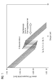
- FIG. 1 shows atomic cross-sections of the elements Ca, Sc, Ti, V, Cr, Mn, Fe for X-rays with quantum energies in the range of 1 to about 45 keV. through a double arrow is the typical area for mammography of quantum energies of about 15 to 45 keV.
- the K absorption edges lie between 4.0 and 7.1 keV. at the K absorption edges increase the atomic cross section by about an order of magnitude.
- For X-rays with quantum energies typical for mammography are the atomic cross sections on average significantly smaller than for X-rays with quantum energies less than about 15 keV.
- Quantum energies less than about 15 keV.
- Such quantum energies are strongly absorbed in the tissue and contribute significantly to a radiation dose absorbed by a patient. By filtering these quantum energies can be largely suppressed become.
- Quantum energies in the range between about 15 and 45 keV are good for mammography X-rays are especially important. You are going through an out above made filter well transmitted. This makes it possible, with minimum radiation dose, a maximum Achieve quality of x-rays.
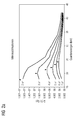
- Figs. 2a and 2b are the maxima of the quality factors marked by triangles.
- Quantum energy is D minimal and CNR maximum.
- Thickness of the fabric is the maximum at higher quantum energies.
- the maxima are dependent on whether in the tissue are contained calcifications or tumors. at same tissue thickness is the maximum for tumors at higher Quantum energies than that for calcifications.
- Figures 2a and 2b show that for high quality mammographic X-rays an adaptation of the X-ray radiation to conditions of the tissue is required.
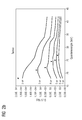
- FIG. 3 shows normalized intensity distributions of X-radiation. These were prepared according to J. Boone, "Molybdenium, rhodium and tungsten spectral models using interpolating polynomials with application to mammography ", Med. Phys. 24 (12), 1863-1874 (1997).
- W / Ti denotes an intensity distribution when using a tungsten anode in combination with a 0.4 mm thick filter made of Ti and at a peak voltage of 24 kVp.
- Rh / Rh denotes an intensity distribution for a combination of a Rh prepared anode and made of Rh, 0.18 mm thick filter, with a peak voltage of 34 kVp.
- Rh / Rh is characterized by two characteristic X-ray lines at approximately 20.0 keV K ⁇ , and 23.2 keV K ⁇ .
- W / Ti is continuously formed and has a maximum M at an optimum quantum energy of about 21 keV for the detection of calcifications. While in Rh / Rh the characteristic X-ray lines K ⁇ and K ⁇ are immutable in position, the maximum M at W / Ti can be shifted to smaller or larger quantum energies by varying the peak voltage or by choosing an appropriate thickness of the filter. In the case of an intensity distribution which is optimally adapted to a tissue to be examined, the maximum M of the respective quantum energy lies where the quality factors according to FIGS. 2a or 2b are maximal.
- the intensity distribution generated with the W / Ti system can be flexibly adjusted so that larger quality factors can be achieved.
- the W / Ti system is particularly advantageous for quantum energies > about 25 keV.
Landscapes
- Health & Medical Sciences (AREA)
- Life Sciences & Earth Sciences (AREA)
- Medical Informatics (AREA)
- Engineering & Computer Science (AREA)
- Radiology & Medical Imaging (AREA)
- Molecular Biology (AREA)
- Biophysics (AREA)
- Nuclear Medicine, Radiotherapy & Molecular Imaging (AREA)
- Optics & Photonics (AREA)
- Pathology (AREA)
- Physics & Mathematics (AREA)
- Biomedical Technology (AREA)
- Heart & Thoracic Surgery (AREA)
- High Energy & Nuclear Physics (AREA)
- Surgery (AREA)
- Animal Behavior & Ethology (AREA)
- General Health & Medical Sciences (AREA)
- Public Health (AREA)
- Veterinary Medicine (AREA)
- Dentistry (AREA)
- Oral & Maxillofacial Surgery (AREA)
- Apparatus For Radiation Diagnosis (AREA)
Applications Claiming Priority (2)
| Application Number | Priority Date | Filing Date | Title |
|---|---|---|---|
| DE102004015860 | 2004-03-31 | ||
| DE102004015860A DE102004015860A1 (de) | 2004-03-31 | 2004-03-31 | Röngtenvorrichtung für die Mammographie |
Publications (1)
| Publication Number | Publication Date |
|---|---|
| EP1582148A1 true EP1582148A1 (fr) | 2005-10-05 |
Family
ID=34877674
Family Applications (1)
| Application Number | Title | Priority Date | Filing Date |
|---|---|---|---|
| EP05101998A Withdrawn EP1582148A1 (fr) | 2004-03-31 | 2005-03-15 | Appareil à rayons X pour mammographie |
Country Status (3)
| Country | Link |
|---|---|
| US (1) | US20050243970A1 (fr) |
| EP (1) | EP1582148A1 (fr) |
| DE (1) | DE102004015860A1 (fr) |
Families Citing this family (4)
| Publication number | Priority date | Publication date | Assignee | Title |
|---|---|---|---|---|
| US7646850B2 (en) * | 2007-01-18 | 2010-01-12 | The Research Foundation Of State University Of New York | Wide-field, coherent scatter imaging for radiography using a divergent beam |
| US8971493B2 (en) | 2010-09-08 | 2015-03-03 | Siemens Medical Solutions Usa, Inc. | System for image scanning and acquisition with low-dose radiation |
| DE102011005055B4 (de) | 2011-03-03 | 2016-08-18 | Siemens Healthcare Gmbh | Verfahren zur Erstellung eines Dual-Energie-Röntgenbildes sowie entsprechendes Röntgensystem, Computerprogramm und elektronisch lesbarer Datenträger |
| US9370330B2 (en) | 2013-02-08 | 2016-06-21 | Siemens Medical Solutions Usa, Inc. | Radiation field and dose control |
Citations (3)
| Publication number | Priority date | Publication date | Assignee | Title |
|---|---|---|---|---|
| DE4204301A1 (de) * | 1991-08-05 | 1993-02-11 | Siemens Ag | Roentgenroehre mit strahlenaustrittsfenster |
| CN1243320A (zh) * | 1999-03-09 | 2000-02-02 | 山东医科大学 | 医用x光机软x射线滤波片 |
| US20030227996A1 (en) * | 2002-06-07 | 2003-12-11 | Tom Francke | Method and apparatus for detection of ionizing radiation |
Family Cites Families (1)
| Publication number | Priority date | Publication date | Assignee | Title |
|---|---|---|---|---|
| US5033075A (en) * | 1988-05-18 | 1991-07-16 | Rad/Red Laboratories Inc. | Radiation reduction filter for use in medical diagnosis |
-
2004
- 2004-03-31 DE DE102004015860A patent/DE102004015860A1/de not_active Ceased
-
2005
- 2005-03-15 EP EP05101998A patent/EP1582148A1/fr not_active Withdrawn
- 2005-03-31 US US11/096,119 patent/US20050243970A1/en not_active Abandoned
Patent Citations (3)
| Publication number | Priority date | Publication date | Assignee | Title |
|---|---|---|---|---|
| DE4204301A1 (de) * | 1991-08-05 | 1993-02-11 | Siemens Ag | Roentgenroehre mit strahlenaustrittsfenster |
| CN1243320A (zh) * | 1999-03-09 | 2000-02-02 | 山东医科大学 | 医用x光机软x射线滤波片 |
| US20030227996A1 (en) * | 2002-06-07 | 2003-12-11 | Tom Francke | Method and apparatus for detection of ionizing radiation |
Non-Patent Citations (3)
| Title |
|---|
| DATABASE WPI Section Ch Week 200028, Derwent World Patents Index; Class K08, AN 2000-318642, XP002331372 * |
| FLYNN M J ET AL: "Optimal radiographic techniques for digital mammograms obtained with an amorphous selenium detector", MEDICAL IMAGING 2003. PHYSICS OF MEDICAL IMAGING 16-18 FEB. 2003 SAN DIEGO, CA, USA, vol. 5030, June 2003 (2003-06-01), Proceedings of the SPIE - The International Society for Optical Engineering SPIE-Int. Soc. Opt. Eng USA, pages 147 - 156, XP002331371, ISSN: 0277-786X * |
| MAUDAL S: "Iron filters as a means of reducing the dose in roentgen examination of the female breast.", ACTA RADIOLOGICA: THERAPY, PHYSICS, BIOLOGY. JUN 1968, vol. 7, no. 3, June 1968 (1968-06-01), pages 238 - 240, XP008048376, ISSN: 0567-8064 * |
Also Published As
| Publication number | Publication date |
|---|---|
| US20050243970A1 (en) | 2005-11-03 |
| DE102004015860A1 (de) | 2005-11-10 |
Similar Documents
| Publication | Publication Date | Title |
|---|---|---|
| DE102012217301B4 (de) | Kombination aus Kontrastmittel und Mammographie-CT-System mit vorgegebenem Energiebereich und Verfahren zur Erzeugung tomographischer Mammographie-CT-Aufnahmen durch diese Kombination | |
| DE10052903A1 (de) | Abbildungssystem mit Strahlungsfilter zur Röntgenabbildung | |
| DE102014202330B3 (de) | Single Source DualEnergy mit zwei Filtern zur Röntgenspektrumsdifferenzierung bei Strahlerblenden mit Schlitzplatte | |
| DE19826062A1 (de) | Verfahren und Anordnung zur Detektion von Röntgenstrahlen | |
| DE102008056891A1 (de) | Computertomographiegerät, insbesondere zur Durchführung eine Spiralscans, und Verfahren zum Steuern eines Computertomographiegeräts | |
| DE102011088265B4 (de) | Verfahren zur Korrektur von aufgrund eines Streustrahlenrasters auftretenden Bildartefakten | |
| DE102016203257A1 (de) | Erzeugen von kontrastverstärkten Bilddaten auf Basis einer Multi-Energie-Röntgenbildgebung | |
| DE102009053523B4 (de) | Filter zur Filterung von Röntgenstrahlung und Röntgencomputertomograph | |
| DE102010020770A1 (de) | Verfahren zur Reduzierung von Bildartefakten, insbesondere von Metallartefakten, in CT-Bilddaten | |
| DE102007017629A1 (de) | Verfahren zur Differenzierung zwischen vier Materialien in tomographischen Aufnahmen eines 2-Energie-CT-Systems | |
| DE102011055691A1 (de) | Kompakter Mammograph und zugehöriges Mammographieverfahren | |
| DE102010041176B4 (de) | Verfahren zur Korrektur des Wertes einer an einer Röntgenröhre einzustellenden Spannung, Computertomographiegerät und Datenträger | |
| DE202014002844U1 (de) | Röntgenfilter und Röntgengerät | |
| EP1764040A2 (fr) | Procédé pour l'imagerie 3D radiologique avec des artefacts reduits, outil d'imagerie médicale et procédé pour établir un plan de traitement | |
| DD294119A5 (de) | Filter und verfahren zur verringerung des strahlendosis | |
| DE102010022851B4 (de) | Röntgenstrahlungsvorrichtung zur Erzeugung von quasimonochromatischer Röntgenstrahlung und Radiographie-Röntgenaufnahmesystem | |
| Beaman et al. | Optimum x-ray spectra for mammography | |
| DE102018214311A1 (de) | Vorrichtung zum Verändern einer räumlichen Intensitätsverteilung eines Röntgenstrahls | |
| EP1582148A1 (fr) | Appareil à rayons X pour mammographie | |
| EP4034175A1 (fr) | Représentation par images simultanées de deux zones fonctionnelles différentes | |
| DE102021205294B3 (de) | Computertomographieeinrichtung und Verfahren zum Betrieb einer Computertomographieeinrichtung | |
| DE102004053009A1 (de) | Verfahren zur Abbildung eines Aufnahmeobjektes, insbesondere einer Patientenbrust, mittels eines Röngtengerätes bzw. Röngtengerät zur Abbildung des Aufnahmeobjektes | |
| DE3851520T2 (de) | Mammographisches Gerät. | |
| DE102008037348B4 (de) | Verfahren und Röntgen-CT-System zur Erzeugung tomographischer Darstellungen aus Projektionsdaten bezüglich drei unterschiedlicher Energiebereiche | |
| DE102006014629A1 (de) | Verfahren zur Korrektur von Trunkierungsartefakten |
Legal Events
| Date | Code | Title | Description |
|---|---|---|---|
| PUAI | Public reference made under article 153(3) epc to a published international application that has entered the european phase |
Free format text: ORIGINAL CODE: 0009012 |
|
| AK | Designated contracting states |
Kind code of ref document: A1 Designated state(s): AT BE BG CH CY CZ DE DK EE ES FI FR GB GR HU IE IS IT LI LT LU MC NL PL PT RO SE SI SK TR |
|
| AX | Request for extension of the european patent |
Extension state: AL BA HR LV MK YU |
|
| AKX | Designation fees paid | ||
| STAA | Information on the status of an ep patent application or granted ep patent |
Free format text: STATUS: THE APPLICATION IS DEEMED TO BE WITHDRAWN |
|
| 18D | Application deemed to be withdrawn |
Effective date: 20060406 |
|
| REG | Reference to a national code |
Ref country code: DE Ref legal event code: 8566 |