EP1579882A1 - Procède non invasif pour l'optimisation de la respiration de poumons atélectasiques - Google Patents
Procède non invasif pour l'optimisation de la respiration de poumons atélectasiques Download PDFInfo
- Publication number
- EP1579882A1 EP1579882A1 EP04007355A EP04007355A EP1579882A1 EP 1579882 A1 EP1579882 A1 EP 1579882A1 EP 04007355 A EP04007355 A EP 04007355A EP 04007355 A EP04007355 A EP 04007355A EP 1579882 A1 EP1579882 A1 EP 1579882A1
- Authority
- EP
- European Patent Office
- Prior art keywords
- breath
- lung
- alveolar
- detected
- mean
- Prior art date
- Legal status (The legal status is an assumption and is not a legal conclusion. Google has not performed a legal analysis and makes no representation as to the accuracy of the status listed.)
- Withdrawn
Links
Images


Classifications
-
- A—HUMAN NECESSITIES
- A61—MEDICAL OR VETERINARY SCIENCE; HYGIENE
- A61B—DIAGNOSIS; SURGERY; IDENTIFICATION
- A61B5/00—Measuring for diagnostic purposes; Identification of persons
- A61B5/08—Detecting, measuring or recording devices for evaluating the respiratory organs
- A61B5/085—Measuring impedance of respiratory organs or lung elasticity
-
- A—HUMAN NECESSITIES
- A61—MEDICAL OR VETERINARY SCIENCE; HYGIENE
- A61M—DEVICES FOR INTRODUCING MEDIA INTO, OR ONTO, THE BODY; DEVICES FOR TRANSDUCING BODY MEDIA OR FOR TAKING MEDIA FROM THE BODY; DEVICES FOR PRODUCING OR ENDING SLEEP OR STUPOR
- A61M16/00—Devices for influencing the respiratory system of patients by gas treatment, e.g. mouth-to-mouth respiration; Tracheal tubes
- A61M16/021—Devices for influencing the respiratory system of patients by gas treatment, e.g. mouth-to-mouth respiration; Tracheal tubes operated by electrical means
- A61M16/022—Control means therefor
- A61M16/024—Control means therefor including calculation means, e.g. using a processor
- A61M16/026—Control means therefor including calculation means, e.g. using a processor specially adapted for predicting, e.g. for determining an information representative of a flow limitation during a ventilation cycle by using a root square technique or a regression analysis
-
- A—HUMAN NECESSITIES
- A61—MEDICAL OR VETERINARY SCIENCE; HYGIENE
- A61B—DIAGNOSIS; SURGERY; IDENTIFICATION
- A61B5/00—Measuring for diagnostic purposes; Identification of persons
- A61B5/08—Detecting, measuring or recording devices for evaluating the respiratory organs
- A61B5/083—Measuring rate of metabolism by using breath test, e.g. measuring rate of oxygen consumption
- A61B5/0836—Measuring rate of CO2 production
-
- A—HUMAN NECESSITIES
- A61—MEDICAL OR VETERINARY SCIENCE; HYGIENE
- A61M—DEVICES FOR INTRODUCING MEDIA INTO, OR ONTO, THE BODY; DEVICES FOR TRANSDUCING BODY MEDIA OR FOR TAKING MEDIA FROM THE BODY; DEVICES FOR PRODUCING OR ENDING SLEEP OR STUPOR
- A61M16/00—Devices for influencing the respiratory system of patients by gas treatment, e.g. mouth-to-mouth respiration; Tracheal tubes
- A61M16/0003—Accessories therefor, e.g. sensors, vibrators, negative pressure
- A61M2016/0015—Accessories therefor, e.g. sensors, vibrators, negative pressure inhalation detectors
- A61M2016/0018—Accessories therefor, e.g. sensors, vibrators, negative pressure inhalation detectors electrical
- A61M2016/0021—Accessories therefor, e.g. sensors, vibrators, negative pressure inhalation detectors electrical with a proportional output signal, e.g. from a thermistor
-
- A—HUMAN NECESSITIES
- A61—MEDICAL OR VETERINARY SCIENCE; HYGIENE
- A61M—DEVICES FOR INTRODUCING MEDIA INTO, OR ONTO, THE BODY; DEVICES FOR TRANSDUCING BODY MEDIA OR FOR TAKING MEDIA FROM THE BODY; DEVICES FOR PRODUCING OR ENDING SLEEP OR STUPOR
- A61M16/00—Devices for influencing the respiratory system of patients by gas treatment, e.g. mouth-to-mouth respiration; Tracheal tubes
- A61M16/0003—Accessories therefor, e.g. sensors, vibrators, negative pressure
- A61M2016/003—Accessories therefor, e.g. sensors, vibrators, negative pressure with a flowmeter
- A61M2016/0033—Accessories therefor, e.g. sensors, vibrators, negative pressure with a flowmeter electrical
- A61M2016/0036—Accessories therefor, e.g. sensors, vibrators, negative pressure with a flowmeter electrical in the breathing tube and used in both inspiratory and expiratory phase
-
- A—HUMAN NECESSITIES
- A61—MEDICAL OR VETERINARY SCIENCE; HYGIENE
- A61M—DEVICES FOR INTRODUCING MEDIA INTO, OR ONTO, THE BODY; DEVICES FOR TRANSDUCING BODY MEDIA OR FOR TAKING MEDIA FROM THE BODY; DEVICES FOR PRODUCING OR ENDING SLEEP OR STUPOR
- A61M2230/00—Measuring parameters of the user
- A61M2230/20—Blood composition characteristics
- A61M2230/202—Blood composition characteristics partial carbon oxide pressure, e.g. partial dioxide pressure (P-CO2)
-
- A—HUMAN NECESSITIES
- A61—MEDICAL OR VETERINARY SCIENCE; HYGIENE
- A61M—DEVICES FOR INTRODUCING MEDIA INTO, OR ONTO, THE BODY; DEVICES FOR TRANSDUCING BODY MEDIA OR FOR TAKING MEDIA FROM THE BODY; DEVICES FOR PRODUCING OR ENDING SLEEP OR STUPOR
- A61M2230/00—Measuring parameters of the user
- A61M2230/40—Respiratory characteristics
- A61M2230/43—Composition of exhalation
- A61M2230/432—Composition of exhalation partial CO2 pressure (P-CO2)
Definitions
- the invention refers to a method and an apparatus for determining the status of a lung ventilated by an artificial ventilator.
- WO 00/44427 A1 deals with the problem of the artificial ventilation of patients with an ailing lung.
- the basic patho-physiological mechanism of an ailing lung is the lack of surfactant (substance which reduces surface tension) which can cause a collapse of major lung fractions and a dramatically reduced gas exchange area.
- an important goal of protective ventilator therapy is a gentle and early "reopening" of the lung.
- a distressed lung may be kept open by proper choice of the airway pressure.
- the manual determination of opening and closing pressures is arduous and time consuming.
- WO 00/44427 A1 suggests to use the partial pressures of oxygen (paO 2 ) as an indicator for determining the opening and closing pressures of the lung.
- WO 00/44427 A1 has recognized that there is a significant hysteresis behaviour of the paO 2 as a function of the ventilation pressure.
- Fig. 1 shows the paO 2 hysteresis of the same healthy (left) and ailing (right) lung. While there is almost no hysteresis in the healthy lung and the choice of ventilation pressures has no visible impact on the quality of gas exchange, the hysteresis is even more severe in an ailing lung. In many cases, gas exchange may be reduced so strongly that at typical ventilation pressures, a sufficient hemoglobin oxygen saturation (> 85 mm Hg) may only be reached if high oxygen concentrations (e.g. 90 ... 100 %) are delivered to the patient.
- high oxygen concentrations e.g. 90 ... 100 %
- a ventilation strategy could be to first open the lung with a temporary high airway pressure and then ventilate on the descending branch of the hysteresis such that a sufficient tidal volume is reached and gas exchange is maintained.
- This so called recruitment maneuver has become a common strategy in operating rooms and in the intensive care medicine.
- Fig. 2 shows a typical recruitment maneuver in detail.
- the recruitment maneuver is carried out on the basis of a pressure controlled ventilation and uses two pressure levels, namely the peak inspiratory pressure (PIP) during inspiration and the positive end-expiratory pressure (PEEP) during expiration.
- PIP peak inspiratory pressure
- PEEP positive end-expiratory pressure
- the alveolar opening pressure and the alveolar closing pressure have to be identified.
- PIP and PEEP are stepwise increased by means of an incremental limb until the alveolar opening pressures have been detected with regard to PIP and PEEP (steps 2 and 3).
- the alveolar opening pressure with regard to PIP is usually about 40 cmH 2 O in normal lungs and in the range of 55-60 cmH 2 O in sick lungs.
- a decremental limb or stepwise decrease of PIP and PEEP is done (step 4) to determine the alveolar closing pressure (step 5).
- the final recruitment maneuver (step 6) is done with these new target pressures over 10 breaths and PEEP is set above the alveolar closing pressure to avoid pulmonary re-collapse.
- a volume controlled ventilation can be carried out having the advantage that the ventilated volume remains constant and that all changes of the lung status can be related to changes within the alveoli.
- WO 00/44427 A1 utilizes according to a first embodiment the endtidal CO 2 concentration (etCO 2 ) and/or the CO 2 output as feedback signals for identification of the optimal ventilator settings for ailing lungs. Both feedback signals can be measured non-invasively. etCO 2 can be obtained by measuring the CO 2 concentration at the end of an expiration cycle. CO 2 output (unit [ml CO 2/ min]) can be obtained from continuous measurements of the CO 2 concentration (unit [%]) and air flow (unit [ml/min]) and subsequent breathwise computation of during one expiration cycle.
- the hemoglobin oxygen saturation (SO 2 ) is measured non-invasively and is used as a feedback signal for identification of optimal ventilation parameters for ailing lungs.
- WO 00/44427 A1 discloses a non-invasive method for determining the alveolar opening or closing of a lung based on one of the measurement of the parameters CO 2 concentration (etCO 2 ), CO 2 output or hemoglobin oxygen saturation (SO 2 ).
- CO 2 concentration etCO 2
- CO 2 output CO 2 output
- SO 2 hemoglobin oxygen saturation
- a method according to the invention for determining the status of a lung ventilated by an artificial ventilator comprises the following steps:
- the invention is based on the hypothesis that a lung recruitment maneuver would reduce alveolar dead space.
- the basic principle of the invention is to derive a mean tracing value from the CO 2 concentration in the expired gas during a single breath which is sensitive to changes of alveolar dead space.
- the meaning of alveolar dead space with regard to the CO 2 concentration in the expired gas was already studied by R. Fletcher, G. Johnson and J. Brew in: "The Concept of Deadspace with Special Reference to Single Breath Test for Carbon Dioxide.” Br. J. Anaesth., 53, 77, 1981 and will be explained further below with regard to Fig. 3.
- Fig. 3 shows a plot of expiratory gas CO 2 concentration against expired volume, which can be obtained by combining a CO 2 concentration measurement against time and a volume rate measurement against time.
- This plot is called the CO 2 single breath test and shows three distinct phases in breath CO 2 gas concentration during the patient exhale cycle.
- Phase I represents CO 2 free gas expired from the airway conduction structures where gas exchange does not occur.
- Phase II is characterized by an S-shaped upswing and represents the transition from airway to alveolar gas.
- Phase III reflects the exhalation of unmixed gas from regions of the lung which normally are in active exchange with the alveolar tissue and thus closely resembles at least in healthy patients gas properties associated with arterial blood in contact with the lung for gas exchange, i.e. CO 2 release and O 2 absorption.
- Phase III is characterized by a horizontal level since ventilated and perfused alveolar regions are closely matched. In a diseased lung, Phase III may not appear horizontal due to a mismatch in ventilation and perfusion of this
- the plot according to Fig. 3 is formed by the exhaled partial pressure of CO 2 against the expiratory tidal volume. Its analysis can be performed, e.g., using a side-stream infrared capnometer and a pneumotachograph of the Capnomac Ultima (e.g. Datex-Engstrom Instrument, Corp., Helsinki, Finland) or a main-stream CO 2 sensor (e.g. Novametrix, USA). Furthermore, a computer is provided to record and analyze data.
- capnograph and blood gas analyzer should be calibrated using the same CO 2 concentration (5%). Airway flow can be measured and integrated to obtain volume. A corresponding device automatically normalizes airway volumes from standard condition to body temperature, ambient pressure and water vapor saturation (BTPS). Before anesthesia and ventilating a patient, induction the volume calibration can be done with e.g. a 700 ml super-syringe following the manufacturer's guidelines.
- the side-stream CO 2 signal has a time delay with respect to the flow signal.
- a corresponding software can correct the CO 2 delays automatically using mathematical algorithms.
- the VTCO 2,br or area under the curve can be computed by integrating expired flow and CO 2 in each breath. Analysis of dead space can be done on-line and/or off-line using Fowler's analysis and adding arterial PCO 2 values to the CO 2 curve of the single breath test.
- capnography is a technique to assess the arterial carbon dioxide content, expressed as partial pressure of CO 2 (paCO 2 ), and which is disclosed in detail in WO 92/04865 A1 and WO 96/24285 A1. Since alveolar dead space cannot be derived directly from the CO 2 single breath test, both WO 92/04865 A1 and WO 96/24285 A1 assume alveolar dead space as a constant variable which has to be determined or estimated by other means, e.g. by a separate blood sample. Hence, according to WO 92/04865 A1 and WO 96/24285 A1 any changes of alveolar dead space are seen as a disturbance variable, since these changes result in a faulty estimate of paCO 2 .
- Fig. 4 shows the anatomical and functional units of the lungs, namely the lung acinus.
- the meaning of the abbreviations is as follows:
- the lung acinus is constituted of the respiratory bronchioli, alveolar ducts, alveolar sacs, alveoli and pulmonary capillaries. To maintain a normal function, this acinus must be well-ventilated and perfused, i.e must be maintained in an open condition. If this acinus becomes collapsed, it loses its normal capacity for gas exchange and makes itself prone to injury during artificial ventilation, as stated previously.
- Fig. 5 shows the diffusion phenomenon for CO 2 which is defined as a passive movement of CO 2 molecules through the alveolar-capillary membrane due to a gradient of concentration or partial pressures.
- J Dmol A Dc/Dx
- J the instantaneous flux of CO 2
- Dmol the gas-phase molecular diffusivity of CO 2 in air
- A the area of gas exchange
- Dc the gas concentration gradient for CO 2 and Dx is the distance.
- the area of gas exchange depends on a normal acinar structure. Reduction in A is the consequence of pathologic three-dimensional changes in acinar morphology. Thus, a decrease in A as during lung collapse results in a decrease of the CO 2 and O 2 diffusion at the alveolar-capillary membrane. Opposite to that, the recovery of a normal acinar morphology by a recruitment maneuver normalizes A and thus diffusion.
- the three-dimensional structural changes of the lung acini are reflected in a change of alveolar dead space during a CO 2 single breath test, wherein a suitable mean tracing value is used to detect these changes.
- the mean tracing value represents a data fusion of the data samples of the CO 2 concentration using an average data algorithm. Examples of average data algorithms are a least squares linear regression, a weighted sum calculation or a FIR (finite impulse response) filter.
- the method and the apparatus according to the invention has the advantage that an averaged value within one single breath is obtained which still yields a good accuracy for determining certain lung conditions.
- An important cognition of the invention compared to WO 00/44427 A1 is the fact that a plurality of data samples from said obtained data samples are selected which enables a selective evaluation of the CO 2 concentration within one single breath.
- the data samples according to step a) are obtained in the time domain. This can be achieved by a conventional analog to digital converter. According to another preferred aspect of the invention, the obtained data samples are converted from the time domain into the volumetric domain. This is particularly useful for obtaining the well known plot of the CO 2 single breath test.
- Fig. 6 shows some possible mean tracing values within a plot of a CO 2 single breath test, which are
- VD aw airway dead space. Is the dead space created by the convective airways to the point where mixing with alveolar gas takes place (Fig. 3, Z area).
- the midpoint of phase II is the limit between anatomical dead space and alveolar gas (Fowler's method). This midpoint is calculated as 50 % of numerical data from slope II. It represents the gas inside the lung transported by convection.
- VCO 2 is the production of CO 2 per minute (ml/min) and is calculated as the product of the expired concentration of CO 2 by the alveolar minute ventilation.
- VTCO 2 or area under the curve, it represents the volume of CO 2 expired in a single breath measured by flow integration (Fig. 3, X area). It is useful to calculate CO 2 production. It represents the alveolar gas, which is in contact with pulmonary capillary blood.
- Phase I begins with the start of expiration (detection by a negative inflection on flow signal), and ends when the concentration of CO 2 in the CO 2 single breath test increases above 0,1 % from baseline (Fig. 3, Fig. 5).
- Phase II starts at the end of phase I (from 0,1 % CO 2 concentration) and continues to the intersection of the predictive slope lines for phases II and III (Fig. 3, Fig. 5) .
- Phase III or alveolar plateau begins at the intersection of the predictive slopes lines for phase II and III and terminates at the end of expiration, defined by an abrupt positive deflection on flow signal (Fig. 3, Fig. 5).
- volume of phase I Is the volume of gas contained in the phase I. It determines the largest part of the airway dead space and represents the gas in the proximal airway and the compressive gas in the ventilatory circuit (Fig. 3, Fig. 5) .
- phase II volume of phase II: Is the volume of gas contained in the phase II: The midpoint of phase II (50% of slope) is the limit between anatomical dead space and alveolar gas, and represents an interface when convective gas transport changes to diffusion transport in lung acini. Thus, phase II is part of both, airway dead space and alveolar gas. Phase II is highly influenced by the acinar emptying time: the more homogeneous the gas emptying for the acini the lower is the phase II volume (Fig. 3, Fig. 5).
- Volume of phase III This volume represents the gas inside the alveoli in contact with pulmonary capillary blood. It is considered as efficient volume for gas exchange within the tidal volume (Fig. 3, Fig. 5).
- Slope of phase II It is derived i.e. from least squares linear regression using data points collected between 25-75 % of the phase II, expressed as fraction (Fig. 6).
- the phase II slope of individual breaths is normalized by dividing the slope value by the corresponding mean alveolar fraction of CO 2 or PAECO 2 (expressed in %) . Similar to volume of phase II, the slope represents the spread of acinar expiratory times. If all acini were emptying at almost the same time the ventilation would be more homogeneous and the slope increases.
- An opposite change in slope II represents an inhomogeneous gas emptying as observed i.e during atelectasis (lung collapse) (Fig. 6).
- phase III It is derived from e.g. least squares linear regression using data points collected between 25-75 % of the phase III, expressed as fraction (Fig. 6).
- the phase III slope of individual breaths is normalized by dividing the slope value by the corresponding mean alveolar fraction of CO 2 or PAECO 2 (expressed in %).
- Phase III slope is the most useful variable to measure a recruitment effect. It is related to the ventilation/perfusion relationship (V/Q): when the V/Q ratio is more efficient the phase III slope decreases, representing a decrease in CO 2 diffusional resistance. When slope III increases, a V/Q mismatch is found.
- Mathematical models have described the variables that can change phase III slope. They are: tidal volume, respiratory rate (only in the extreme of normal values), area of gas exchange, and gas diffusivity. Maintaining all variables stable that can change phase III slope, any change in the area of gas exchange can thus affect this slope. Fick's first law of diffusion can easily explain this statement.
- Angle II-III It is defined as the angle of intersection between slope of phase II and III. Changes in this angle represent changes of the shape of the CO 2 single breath test related to the efficiency/inefficiency state of ventilation and gas exchange. When the angle decreases, as after lung recruitment, ventilation and gas exchange improve. Increasing angles II-III are related to an inhomogeneous (and worse) ventilation/perfusion relationship (Fig. 6).
- Intercept of phase II slope It is defined as the intersection of the line of phase II slope with the X axis (Fig. 6).
- Intercept of phase III slope It is defined as the intersection of the line of phase III slope with the Y axis (Fig. 6).
- VT alv It represents the portion of tidal volume that is located distal to the interface, it is true alveolar gas. This volume is constituted by the sum of VTCO 2 alv plus VD alv . VT alv is derived by Fowler analysis as VT - VD alv .
- PAECO 2 or mean expired concentration of CO 2 , constitutes the mean partial pressure of CO 2 in alveolar air. It is defined as the partial pressure of CO 2 at the middle of the slope III. This value represents the partial pressure of all CO 2 molecules in the expired volume.
- etCO 2 is the end tidal partial pressure of CO 2 (Fig. 3).
- Pet-AECO 2 is the difference between end tidal CO 2 and mean expired partial pressure. It is an index of alveolar dead space: the higher the differences between these two values, the higher is the inefficiency of lung function.
- VDBohr is the dead space formed by anatomical dead space plus part of the alveolar dead space (ml), defined as the lower portion of the alveolar dead space.
- Vdaw/VT the ratio between airway dead space to tidal volume, is an index of lung efficiency / inefficiency. The higher this index is the more inefficient the lung becomes.
- volume I, II, III and phase III slope are normalized by dividing them by the tidal volume to make comparison among different breaths possible.
- the endtidal mean slope is taken as a mean tracing value and is determined by the mean slope (either over time or over volume) of the CO 2 concentration in the expired gas towards the final stage of a single breath.
- the calculation of the endtidal mean slope can be carried out on the basis of a least squares linear regression using suitable constraints, wherein a suitable constraint could be a minimum mean square error of the regression result over a predetermined range. On the basis of such a constraint the calculation of the endtidal mean slope would be carried out as follows:
- the peak inspiratory pressure of the artificial ventilator is increased stepwise breath by breath starting from alveolar closing, wherein an alveolar opening of the lung is detected, if the resulting course of the plurality of determined first mean tracing values reaches a minimum.
- the peak inspiratory pressure of the artificial ventilator is increased stepwise breath by breath starting from alveolar opening, wherein a lung overdistension is detected, if the positive gradient of the resulting course of the plurality of determined first mean tracing values reaches a maximum.
- the peak inspiratory pressure of the artificial ventilator is decreased stepwise breath by breath starting from lung overdistension, wherein an open lung condition is detected, if the resulting course of the plurality of determined first mean tracing values reaches a minimum.
- the peak inspiratory pressure of the artificial ventilator is decreased stepwise breath by breath starting from an open lung condition, wherein an alveolar closing is detected, if the positive gradient of the resulting course of the plurality of determined first mean tracing values reaches a maximum.
- a second mean tracing value is represented by the steepest mean slope of the CO 2 concentration in the expired gas during a single breath.
- the steepest mean slope is determined by the mean slope (either over time or over volume) of the CO 2 concentration in the expired gas in the vicinity of the point of inflection.
- the calculation of the steepest mean slope can be carried out again on the basis of a least squares linear regression using suitable constraints, wherein a suitable constraint could be a minimum mean square error of the regression result over a predetermined range. On the basis of such a constraint the calculation of the steepest mean slope would be carried out as follows:
- the peak inspiratory pressure of the artificial ventilator is increased stepwise breath by breath starting from alveolar closing, wherein an alveolar opening of the lung is detected, if the resulting course of the plurality of determined second mean tracing values reaches a maximum.
- the peak inspiratory pressure of the artificial ventilator is increased stepwise breath by breath starting from alveolar opening, wherein a lung overdistension is detected, if the negative gradient of the resulting course of the plurality of determined second mean tracing values reaches a minimum.
- the peak inspiratory pressure of the artificial ventilator is decreased stepwise breath by breath starting from lung overdistension, wherein an open lung condition is detected, if the resulting course of the plurality of determined second mean tracing values reaches a maximum.
- the peak inspiratory pressure of the artificial ventilator is decreased stepwise breath by breath starting from an open lung condition, wherein an alveolar closing is detected, if the negative gradient of the resulting course of the plurality of determined second mean tracing values reaches a minimum.
- a third mean tracing value is represented by the angle between the endtidal mean slope and the steepest mean slope of the CO 2 concentration in the expired gas during a single breath.
- the peak inspiratory pressure of the artificial ventilator is increased stepwise breath by breath starting from alveolar closing, wherein an alveolar opening of the lung is detected, if the resulting course of the plurality of determined third mean tracing values reaches a minimum.
- the peak inspiratory pressure of the artificial ventilator is increased stepwise breath by breath starting from alveolar opening, wherein a lung overdistension is detected, if the positive gradient of the resulting course of the plurality of determined third mean tracing values reaches a maximum.
- the peak inspiratory pressure of the artificial ventilator is decreased stepwise breath by breath starting from lung overdistension, wherein an open lung condition is detected, if the resulting course of the plurality of determined third mean tracing values reaches a minimum.
- the peak inspiratory pressure of the artificial ventilator is decreased stepwise breath by breath starting from an open lung condition, wherein an alveolar closing is detected, if the positive gradient of the resulting course of the plurality of determined third mean tracing values reaches a maximum.
- a plurality of different types of mean tracing values are calculated in parallel and wherein from the resulting course of the plurality of different types of mean tracing values the airway pressure level at which alveolar opening or lung overdistension or lung open condition or alveolar closing occurs is detected.
- the peak inspiratory pressure is set above the airway pressure level at which alveolar opening has been detected and the positive end-expiratory pressure is set above the airway pressure level at which alveolar closing has been detected.
- Fig. 7 shows two plots of a CO 2 single breath test in the states of atelectasis and recruitment.
- an increase in the area of gas exchange due to recruitment alters the shape of the plot of a CO 2 single breath test, wherein the endtidal mean slope (slope III) decreases and the steepest mean slope (slope II) increases.
- the reversible and dynamic acinar change in morphology can be manipulated by treatment. Normalizing acinar morphology in a mechanically ventilated patient by a recruitment maneuver produces a normalisation in the physiology of the lung. A normalisation in acinar morphology by the recruitment maneuver causes an improvement in gas exchange and in gas emptying during expiration.
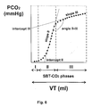
- Fig. 8 shows a plot of the airway pressures over time of a typical recruitment maneuver together with the response of the steepest mean slope (slope II) as a mean tracing value.
- the algorithm for diagnosing the lung's open-collapse state is described with regard to the steepest mean slope as follows:
- a new recruitment maneuver is done with the known opening and closing pressure.
- Fig. 9 shows a plot of the airway pressures over time of a typical recruitment maneuver together with the response of the volume of phase II as a mean tracing value. Lung recruitment, overdistension, open-lung condition and re-collapse are seen during the recruitment maneuver. The inflection point represents the change of direction of the volume of phase II from the open-lung condition to the beginning of the collapsed state. Furthermore, the inflection point represents the pulmonary closing pressure.
- ARS alveolar recruitment strategy
- OSV one-lung ventilation
- a thoracic epidural catheter was placed at T2 to T4 and a total volume of 0,1 ml/kg of bupivacaine 0,5% without epinephrine were administered.
- intravascular volume Prior to the epidural anesthesia, intravascular volume was expanded by infusing 7 ml/kg of a colloidal solution (HaemacellTM) and maintained at 8 ml kg -1 h -1 of normal saline solution.
- HaemacellTM colloidal solution
- the trachea and the left bronchus were intubated with a left double lumen tube (DLT) of the appropriate size (Broncho-CathTM, Mallinckrodt Laboratories, Atholone, Ireland). Air leakage were assessed by introducing the capnograph's side stream sensor into each lumen of the DLT while maintaining ventilation through the other lumen. Bronchoscopy confirmed the correct position of the DLT before and after positioning the patients in the lateral position. During OLV, the lumen of the non-ventilated side was left open to atmosphere.
- DLT left double lumen tube
- Lungs were ventilated with a Servo 900 C in a volume control ventilation mode and an inspired oxygen fraction (FiO 2 ) of 1.0.
- the ventilator delivered a square-wave flow with an inspiratory time of 33% with no end-inspiratory pause.
- the respiratory rate was set between 10-14 breaths/min, tidal volumes (VT) were maintained at 8 ml/kg, and PEEP was 8 cmH 2 O throughout the study.
- tidal volume was reduced to 6 ml/kg to avoid peak pressures higher than 30 cmH 2 O.
- Respiratory rate was increased to 15-18 breaths/min to maintain the same minute ventilation as during TLV.
- PIP Peak inspiratory pressure
- VTe expired tidal volume
- respiratory rate expired minute volume
- O 2 and CO 2 fractions Peak inspiratory pressure
- VCO 2 Carbon dioxide elimination
- FECO 2 mean expired alveolar fraction of CO 2
- Oxygen consumption VO 2
- RQ Respiratory quotient
- Capnograph and blood gas analyzer were calibrated using a known gas concentration of CO 2 (5%). This calibration was performed in each patient before the induction of anesthesia. Airway flow and pressure measurements are based on the measurement of kinetic gas pressure, and are performed using the Pitot effect. Flow rate is measured and integrated to obtain VT. The Capnomac device restores normal airway volumes from standard condition to body temperature, ambient pressure and water vapor saturation (BTPS) automatically. Volume calibration was done with a 700 ml super-syringe before anesthesia induction following the manufacturer's guidelines.
- BTPS water vapor saturation
- the sidestream CO 2 signal has a time delay compared to the flow signal.
- the software automatically corrected the CO 2 delay using commonly known mathematical algorithms.
- the VTCO 2,br or area under the curve was computed by integrating expired flow and FCO 2 in each breath.
- the dead space of the apparatus was 60 ml (10 ml from D-LITETM plus 50 ml from DLT connections) and was subtracted from the airway dead space value.
- the recruitment maneuver was applied selectively to the dependent lung immediately after the measurement at point b.
- the ventilator was switched to pressure control ventilation, adjusting the level of pressure to obtain the same tidal volume as during volume control ventilation. Ventilation was then allowed to equilibrate for three minutes. Thereafter, the ARS was performed based on an established concept. The critical alveolar opening pressure was assumed to be at 40 cmH 2 O as described for healthy lungs. ARS protocol:
- the ventilator was set back to volume control.
- the ARS took about 3 minutes.
- Prior to the recruitment maneuvers central venous pressure values were maintained above 10 mmHg to avoid hemodynamic side effects caused by the increased intrathoracic pressures. Hemodynamic and ventilatory variables were monitored closely while performing the ARS. If mean arterial pressure and/or heart rate changed by more than 15 % from baseline, the ARS was discontinued and 500 ml of crystalloid solution were administered. After regaining hemodynamic stability the ARS was tried again.
- L-L left lung
- R-L right lung
- L lower lobe
- U upper lobe
- M median lobe.
- Mini-CABG minimal invasive coronary by-pass graft.
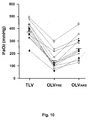
- Fig. 10 shows measurements of the partial paO 2 pressure with 12 patients at three different lung stages.
- PaO 2 was significantly higher during TLV (379 ⁇ 67 mmHg) compared to OLV PRE (144 ⁇ 73 mmHg, p ⁇ 0,001) and OLV ARS (244 ⁇ 89 mmHg, p ⁇ 0,001). During OLV the difference in PaO 2 before and after the ARS also reached significance. Hemoglobin O 2 saturation was lower at OLV PRE (95,5 ⁇ 2,6 %) as compared to TLV (98,7 ⁇ 0,4%, p ⁇ 0,001) and OLV ARS (97,8% ⁇ 0,9%, p ⁇ 0,01).
- PaCO 2 was 43 ⁇ 6 mmHg during OLV ARS but not significantly different from the other conditions. However, PaCO 2 was higher during OLV PRE (46 ⁇ 6 mmHg) compared to TLV (38 ⁇ 4 mmHg, p ⁇ 0,05). EtCO 2 and PAECO 2 were stable during the protocol without any significant differences among the measurement points. Pa-etCO 2 difference was significant higher during OLV PRE (14,2 ⁇ 4,8 mmHg) compared to TLV (8,8 ⁇ 3,2 mmHg) and OLV ARS (11,6 ⁇ 4,6 mmHg). The pHa remained in the normal range throughout the study period.
- Tidal volumes were higher during TLV (506 ⁇ 83 ml) compared to OLV PRE (377 ⁇ 45 ml) and OLV ARS (382 ⁇ 42 ml) .
- Minute ventilation was similar between OLV PRE (5,9 1/min) and OLV ARS (5,8 1/min), but both values were significantly smaller than during TLV (7 1/min).
- PIP values were higher during OLV PRE (25,3 ⁇ 1, 7 cmH 2 O) compared with TLV (20,6 ⁇ 1,7 cmH 2 O, p ⁇ 0,001) and OLV ARS (23,2 ⁇ 2 cmH 2 O, p ⁇ 0,05) with no differences between the latter two.
- Hemodynamic variables, minute CO 2 elimination, oxygen consumption and respiratory quotient were similar at all time points.
- the total time of OLV ranged from 50 to 105 minutes. No hemodynamic or ventilatory side effects related to the recruitment maneuver were detected.
- Arterial oxygenation is a common measurement used to describe the extent of lung collapse. It has been suggested that a PaO 2 higher than 450 mmHg defines an open lung condition during pure O 2 breathing. Arterial oxygenation, however, is an unspecific variable to evaluate the recruitment effect since it depends on the hemodynamic and metabolic status. As these two conditions remained stable throughout the study period, a true recruitment effect is the most likely explanation for the increases seen in PaO 2 .
- the calculated shunt values of the patients ranged from 8 to 22 % (mean 16 %), values typically seen in general anesthesia, during OLV from 18 to 45 % (mean 28 %) and during OLV ARS from 12 to 27 % (mean 21 %). After lung recruitment oxygenation was sufficient to maintain hemoglobin saturation above 95 %.
- the values of the dead space related mean tracing values are higher than normal, due to the double lumen tube, lung collapse, open-chest condition, and the use of positive pressure ventilation.
- the mean tracing values (variables) that represent efficiency of ventilation and CO 2 exchange (VCO 2,br, VD/VT, Pa-etCO 2 , VT alv , VD alv /VT alv ) were higher during TLV compared to OLV. During OLV all mean tracing values (variables) improved only after the recruitment maneuver. Even more interesting was the behaviour of the mean tracing values (variables) that show the distribution of tidal volume throughout the phases of the CO 2 single breath test. Distribution of volume was most efficient during OLV after the ARS as indicated by a decrease in phase I and II volumes and a concomitant increase in phase III volume. The absolute value of the ratio Phase III/VT observed after recruitment was even higher than during TLV.
- Phase II represents a transition between alveolar and airway gas transport.
- An increase in the cross-sectional area of the bronchial tree in the lung periphery decreases the linear velocity of the bulk flow until a point where the two transport mechanisms within the lungs (convection and diffusion) are of equal magnitude.
- This stationary diffusion front demarcates the transition between airway and alveolar gas. On expiration, this front corresponds to phase II and is used to measure VD aw in Fowler's analysis.
- phase II slope in combination with a decrease in its volume, can be considered as a more synchronous and homogeneous emptying of acini during expiration. Both, asthma and emphysema would have an opposite effect on phase II. These conditions show a wide dispersion of the transit time of gas emptying among lung units making the slope of phase II flatter and its volume higher.
- Phase III volume represents the amount of gas exposed to the capillary bed and therefore depends on an effective pulmonary perfusion and CO 2 production.
- Phase III slope is directly related to the V/Q relationship and represents the diffusional resistance for CO 2 at the alveolar-capillary membrane. Its positive slope is explained by lung pendelluft, continuous evolution of CO 2 from the blood into the acini, and a stratified inhomogeinity.
- phase III volume increased while its slope decreased compared to OLV PRE -Decrease in functional lung acini in emphysema is related to an increase in phase III slope.
- Epidural anesthesia used in open thoracotomies can cause hemodynamic and metabolic changes that could influence gas exchange. However, these conditions were stable and no differences in PaO 2 between open thoracotomies and thoracoscopies, without epidural anesthesia, were seen.
- Empirical values of 40 cmH 2 O of PIP were used as opening pressure and 8 cmH 2 O of PEEP to keep the lung open, since individual levels of these pressures for each patient are difficult to determine at the bedside.
- Lung recruitment improves gas exchange and ventilation efficiency during OLV anesthesia.
- the results suggest that one simple recruitment maneuver during OLV is enough to increase PaO 2 to safer levels thereby eliminating the need for any additional therapeutic intervention.
- VT inspired-expired tidal volume
- RR respiratory rate
- FIO 2 inspiratory time of 0,3 without pause and initially, without positive end-expiratory pressure
- Alveolar ventilation was increased or decreased by adjusting RR to reach an end-tidal CO 2 value of 34 mmHg while maintaining VT constant.
- End-expiratory lung volume was measured pushing the expiratory pause button of the Servo 900C for 6 seconds during the inspiratory pause while releasing PEEP from 5 cmH 2 O to ZEEP. Thus, a volume of gas is expelled until FRC at ambient pressure is reached.
- the EELV was then determined by subtracting the average value of the latest three normal expiratory tidal volumes before the maneuver from the volume of gas measured. This volume was recorded continuously in a computer and analyzed it off-line. The return of the expiratory flow curve to baseline at the end of the EELV-maneuver was used for checking air trapping.
- VCO 2 Carbon dioxide elimination
- Oxygen consumption VO 2
- RQ respiratory quotient
- the alveolar recruitment strategy is a maneuver assigned to treat pulmonary collapse by reaching the alveolar opening pressure for ten breaths and keeping the lung open with a PEEP level above the lung's closing pressure.
- the lung opening pressure was 40 cmH 2 O of peak inspiratory pressure (PIP) and the closing pressure lower than 5 cmH 2 O.
- PIP peak inspiratory pressure
- Normalized phase III slope decreased with PEEP ventilation and showed an additional diminution after ARS.
- Volume of phase III increased with ARS and PEEP compared with ZEEP.
- the angle between II-III showed significant differences only after the recruitment maneuver.
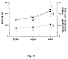
- Fig. 11 shows measurements of the end-expiratory lung volume (EELV), the partial paO 2 pressure and the compliance at three different ventilation modes.
- EELV end-expiratory lung volume
- EELV end-expiratory lung volume
- PEEP without lung recruitment showed compliance values significantly higher than ZEEP but without changes in PaO 2 .
- lung recruitment decreased those mean tracing values of the CO 2 single breath test which are related to pulmonary inefficiency and increased the ones related to efficiency.
- the increased efficiency of ventilation was associated with an increase in arterial oxygenation, expiratory lung volume and respiratory compliance, all parameters commonly used as markers of an open lung condition.
- PEEP without recruitment showed an intermediate effect between ZEEP and ARS in all mean tracing values studied.
- low levels of PEEP have a contradictory effect on arterial oxygenation and atelectasis.
- Study results agree in that the recruitment of collapsed airways is the main effect of PEEP without a recruitment maneuver.
- Atelectasis treatment requires higher airway pressures than the amount of PEEP commonly used during anesthesia to pop open collapsed alveoli due to the incomplete lung recruitment observed with the use of PEEP alone.
- lung recruitment maneuver increase both, the cross-sectional area of small airways and the alveolar-capillary area, by reversing airway and acinar collapse respectively.
- This total recruitment or open lung condition improves the diffusive CO 2 transport at the acinar level and could explain the changes observed in the CO 2 single breath test.
- Increasing CO 2 diffusion after the ARS moves the interface between convective-diffusive transport mouthward, thus decreasing the VDAW measured by Fowler's method.
- Lung recruitment was also associated with an improved efficiency in CO 2 elimination as expressed by a larger VTCO 2,br and a lower Pa-etCO 2 at constant VCO 2 and ventilator settings. These results indicate that the area of gas exchange increased and V/Q improved.
- Total lung recruitment has a positive effect on CO 2 diffusion as reflected by the changes observed in volumes and slopes of phase II-III after ARS.
- an increase in the cross-sectional area caused by airway recruitment could improve the CO 2 diffusive transport from alveoli to bronchioli.
- an increase in the area of gas exchange due to a recruitment of atelectasis improved the diffusive transport from the capillaries to the alveoli.
- the alveolar recruitment strategy improved the efficiency of ventilation in anesthetized patients. Differences observed in the CO 2 single breath test between PEEP with and without an lung recruitment maneuver can be explained by the effectiveness of the treatment of pulmonary collapse.
- Fig. 12 shows an apparatus according to the invention connected in series with the ventilator to the patient.
- the apparatus comprises a carbon dioxide sensor for measuring the expired CO 2 concentration, a pneumotachograph for measuring airway flow, a pressure sensor for measuring airway pressures, and a data processor which determines during a change of the airway pressure from the resulting course of at least one mean tracing value the airway, pressure level at which alveolar opening or lung overdistension or lung open condition or alveolar closing occurs.
- the airway pressure is increased continuously and a lung opening is detected, if the slope of phase III reaches a maximal decrease.
- the airway pressure is increased continuously and a lung opening is detected, if the intercept of the slope of phase III reaches a maximal increase.
- the airway pressure is increased continuously and a lung opening is detected, if the slope of phase II reaches a maximal increase.
- the airway pressure is increased continuously and a lung opening is detected, if the intercept of slope of phase II reaches a maximal decrease.
- the airway pressure is increased continuously and a lung opening is detected, if the angle II-III reaches a maximal decrease.
- the airway pressure is increased continuously and a lung opening is detected, if the volume of phase I reaches a maximal increase.
- the airway pressure is increased continuously and a lung opening is detected, if the volume of phase II reaches a maximal increase.
- the airway pressure is increased continuously and a lung opening is detected, if the volume of phase III reaches a maximal decrease.
- the airway pressure is increased continuously and a lung opening is detected, if the VTCO 2,br reaches a maximal increase.
- the airway pressure is increased continuously and a lung opening is detected, if the VCO 2 reaches a maximal increase.
- the airway pressure is increased continuously and a lung opening is detected, if the VDbohr reaches a maximal decrease.
- the airway pressure is increased continuously and a lung opening is detected, if the negative gradient of the resulting course of the measured etCO 2 minus the mean alveolar partial pressure of CO 2 (Pet-AECO 2 ) reaches the maximal decrease.
- the airway pressure is increased continuously and a lung overdistension is detected, if the VDaw reaches a maximal increase.
- the airway pressure is increased continuously and a lung overdistension is detected, if the Vdaw/VT reaches a maximal increase.
- the airway pressure is increased continuously and a lung overdistension is detected, if the VTCO2,br results in a decrease of about 10% from its previous value.
- the airway pressure is increased continuously and a lung overdistension is detected, if the VCO2 results in a decrease of about 10% from its previous value.
- the airway pressure is increased continuously and a lung overdistension is detected, if the PAECO2 results in a decrease of about 10% from its previous value.
- the airway pressure is increased continuously and a lung overdistension is detected, if the angle II-III results in a decrease of about 10% from its previous value.
- the airway pressure is increased continuously and a lung overdistension is detected, if the phase II slope results in a decrease of about 10% from its previous value.
- the airway pressure is decreased continuously and an open-lung condition is detected, if the VDaw resulting in a minimal value observed before the closing pressure is detected.
- the airway pressure is decreased continuously and an open-lung condition is detected, if the VDaw/VT resulting in a minimal value observed before the closing pressure is detected.
- the airway pressure is decreased continuously and an open-lung condition is detected, if the VTCO2,br resulting in a maximal value observed before the closing pressure is detected.
- the airway pressure is decreased continuously and an open-lung condition is detected, if the angle II-III resulting in a minimal value observed before the closing pressure is detected.
- the airway pressure is decreased continuously and an open-lung condition is detected, if the phase II slope resulting in a maximal value observed before the closing pressure is detected.
- the airway pressure is decreased continuously and an open-lung condition is detected, if the Volume of phase II resulting in a minimal value observed before the closing pressure is detected.
- the airway pressure is decreased continuously and an open-lung condition is detected, if the Volume of phase III resulting in a maximal value observed before the closing pressure is detected.
- the airway pressure is decreased continuously and an open-lung condition is detected, if the intercept of phase II slope resulting in a maximal value observed before the closing pressure is detected.
- the airway pressure is decreased continuously and a closing pressure of the lungs is detected, if the VDaw resulting in a change of direction (inflection point) from the open-lung condition values.
- the airway pressure is decreased continuously and a closing pressure of the lungs is detected, if the VTCO 2,br resulting in a change of direction (inflection point) from the open-lung condition values.
- the airway pressure is decreased continuously and a closing pressure of the lungs is detected, if the VCO 2 resulting in a change of direction (inflection point) from the open-lung condition values.
- the airway pressure is decreased continuously and a closing pressure of the lungs is detected, if the volume of phase II resulting in a change of direction (inflection point) from the open-lung condition values.
- the airway pressure is decreased continuously and a closing pressure of the lungs is detected, if the volume of phase III resulting in a change of direction (inflection point) from the open-lung condition values.
- the airway pressure is decreased continuously and a closing pressure of the lungs is detected, if the angle II-III resulting in a change of direction (inflection point) from the open-lung condition values.
- the airway pressure is decreased continuously and a closing pressure of the lungs is detected, if the intercept of phase II slope resulting in a change of direction (inflection point) from the open-lung condition values.
Priority Applications (5)
| Application Number | Priority Date | Filing Date | Title |
|---|---|---|---|
| EP04007355A EP1579882A1 (fr) | 2004-03-26 | 2004-03-26 | Procède non invasif pour l'optimisation de la respiration de poumons atélectasiques |
| EP05730302.6A EP1740244B1 (fr) | 2004-03-26 | 2005-03-24 | Appareil non invasif pouvant optimiser la respiration de poumons atelectasiques |
| CA2600901A CA2600901C (fr) | 2004-03-26 | 2005-03-24 | Procede et appareil non invasifs pouvant optimiser la respiration de poumons atelectasiques |
| PCT/EP2005/003181 WO2005092415A1 (fr) | 2004-03-26 | 2005-03-24 | Procede et appareil non invasifs pouvant optimiser la respiration de poumons atelectasiques |
| US11/525,244 US9173595B2 (en) | 2004-03-26 | 2006-09-22 | Non-invasive method and apparatus for optimizing the respiration of atelectatic lungs |
Applications Claiming Priority (1)
| Application Number | Priority Date | Filing Date | Title |
|---|---|---|---|
| EP04007355A EP1579882A1 (fr) | 2004-03-26 | 2004-03-26 | Procède non invasif pour l'optimisation de la respiration de poumons atélectasiques |
Publications (1)
| Publication Number | Publication Date |
|---|---|
| EP1579882A1 true EP1579882A1 (fr) | 2005-09-28 |
Family
ID=34854636
Family Applications (2)
| Application Number | Title | Priority Date | Filing Date |
|---|---|---|---|
| EP04007355A Withdrawn EP1579882A1 (fr) | 2004-03-26 | 2004-03-26 | Procède non invasif pour l'optimisation de la respiration de poumons atélectasiques |
| EP05730302.6A Active EP1740244B1 (fr) | 2004-03-26 | 2005-03-24 | Appareil non invasif pouvant optimiser la respiration de poumons atelectasiques |
Family Applications After (1)
| Application Number | Title | Priority Date | Filing Date |
|---|---|---|---|
| EP05730302.6A Active EP1740244B1 (fr) | 2004-03-26 | 2005-03-24 | Appareil non invasif pouvant optimiser la respiration de poumons atelectasiques |
Country Status (4)
| Country | Link |
|---|---|
| US (1) | US9173595B2 (fr) |
| EP (2) | EP1579882A1 (fr) |
| CA (1) | CA2600901C (fr) |
| WO (1) | WO2005092415A1 (fr) |
Cited By (17)
| Publication number | Priority date | Publication date | Assignee | Title |
|---|---|---|---|---|
| EP2211964A2 (fr) * | 2007-10-25 | 2010-08-04 | SeQual Technologies Inc. | Système de concentrateur d'oxygène portatif et procédé incluant l'administration d'un flux d'oxygène concentré |
| DE102009031826B4 (de) * | 2009-07-03 | 2011-06-22 | F. Stephan GmbH, 56412 | Beatmungseinrichtung |
| FR2965469A1 (fr) * | 2010-10-05 | 2012-04-06 | Draeger Medical Gmbh | Procede et dispositif pour l’evaluation et l’analyse automatiques d’un capnogramme et programme d’ordinateur pour l’implementation du procede, ainsi que produit programme d’ordinateur avec un tel programme d’ordinateur |
| US8202226B2 (en) | 2007-01-23 | 2012-06-19 | Kci Licensing, Inc. | Providing automated or manual guidance on dynamic patient positioning based on measured variables for ventilation control |
| RU2454932C1 (ru) * | 2011-03-11 | 2012-07-10 | Государственное образовательное учреждение высшего профессионального образования "Кубанский государственный медицинский университет" Министерства здравоохранения и социального развития Российской Федерации (ГОУ ВПО КубГМУ Минздравсоцразвития РФ) | Способ определения минутного объема вентиляции легких при проведении интраоперационной искусственной вентиляции легких в абдоминальной хирургии |
| US9233222B2 (en) | 2003-11-12 | 2016-01-12 | Draeger Medical Systems, Inc. | System for managing ventilator operation |
| WO2017200929A1 (fr) * | 2016-05-15 | 2017-11-23 | Covidien Lp | Capnographie volumétrique du flux latéral |
| WO2020080982A1 (fr) | 2018-10-17 | 2020-04-23 | Maquet Critical Care Ab | Recrutement pulmonaire dans une ventilation mécanique |
| WO2020080983A1 (fr) | 2018-10-17 | 2020-04-23 | Maquet Critical Care Ab | Mobilisation pulmonaire dans une ventilation mécanique |
| WO2020080984A1 (fr) | 2018-10-17 | 2020-04-23 | Maquet Critical Care Ab | Mobilisation pulmonaire dans une ventilation mécanique |
| WO2020080986A1 (fr) | 2018-10-17 | 2020-04-23 | Maquet Critical Care Ab | Mobilisation pulmonaire dans une ventilation mécanique |
| WO2020080985A1 (fr) | 2018-10-17 | 2020-04-23 | Maquet Critical Care Ab | Recrutement pulmonaire dans une ventilation mécanique |
| CN111712290A (zh) * | 2018-02-12 | 2020-09-25 | 提姆佩尔医疗有限责任公司 | 用于确定患者对肺泡复张手法的应答性的系统和方法 |
| WO2021130344A1 (fr) * | 2019-12-23 | 2021-07-01 | Koninklijke Philips N.V. | Système et procédé de surveillance d'échange de gaz |
| CN113082412A (zh) * | 2021-03-30 | 2021-07-09 | 湖南万脉医疗科技有限公司 | 呼吸机的吸入气氧浓度分数控制方法及系统 |
| US20210220588A1 (en) * | 2018-06-11 | 2021-07-22 | Maquet Critical Care Ab | Ventilation pattern for hemodynamic parameter determination during mechanical ventilation |
| EP3884981A4 (fr) * | 2018-11-23 | 2021-12-15 | Shenzhen Mindray Bio-Medical Electronics Co., Ltd. | Procédé et appareil de détermination de pression positive en fin d'expiration, dispositif d'aération, et support d'informations |
Families Citing this family (65)
| Publication number | Priority date | Publication date | Assignee | Title |
|---|---|---|---|---|
| US7588033B2 (en) | 2003-06-18 | 2009-09-15 | Breathe Technologies, Inc. | Methods, systems and devices for improving ventilation in a lung area |
| AU2004266693B2 (en) | 2003-08-18 | 2011-03-10 | Breathe Technologies, Inc | Method and device for non-invasive ventilation with nasal interface |
| CN101454041B (zh) | 2005-09-20 | 2012-12-12 | 呼吸科技公司 | 对患者进行呼吸支持的系统、方法和装置 |
| CN101541365A (zh) | 2006-05-18 | 2009-09-23 | 呼吸科技公司 | 气管切开方法和装置 |
| JP2009545384A (ja) | 2006-08-03 | 2009-12-24 | ブリーズ テクノロジーズ, インコーポレイテッド | 最小侵襲性呼吸補助のための方法および装置 |
| WO2008144589A1 (fr) | 2007-05-18 | 2008-11-27 | Breathe Technologies, Inc. | Procédés et dispositifs pour détecter la respiration et fournir une thérapie de ventilation |
| CN101888868B (zh) | 2007-09-26 | 2014-01-22 | 呼吸科技公司 | 用于治疗睡眠呼吸暂停的方法和设备 |
| CN101873875B (zh) | 2007-09-26 | 2014-11-12 | 呼吸科技公司 | 用于在通气治疗中提供吸气和呼气流释放的方法和装置 |
| US20090188500A1 (en) * | 2008-01-29 | 2009-07-30 | Joseph Dee Faram | Combination breathing treatment method |
| EP2257328A2 (fr) * | 2008-03-27 | 2010-12-08 | Nellcor Puritan Bennett LLC | Systèmes d'assistance respiratoire avec man uvres de recrutement pulmonaire |
| EP2363163A1 (fr) * | 2008-03-27 | 2011-09-07 | Nellcor Puritan Bennett LLC | Appareil pour l'administration contrôlée d'un gaz respiratoire à un patient à l'aide de paramètres de ventilation multiples |
| US8770193B2 (en) | 2008-04-18 | 2014-07-08 | Breathe Technologies, Inc. | Methods and devices for sensing respiration and controlling ventilator functions |
| US8776793B2 (en) | 2008-04-18 | 2014-07-15 | Breathe Technologies, Inc. | Methods and devices for sensing respiration and controlling ventilator functions |
| CA2734296C (fr) | 2008-08-22 | 2018-12-18 | Breathe Technologies, Inc. | Procedes et dispositifs pour fournir une ventilation mecanique avec une interface de voies respiratoires ouvertes |
| US8302602B2 (en) | 2008-09-30 | 2012-11-06 | Nellcor Puritan Bennett Llc | Breathing assistance system with multiple pressure sensors |
| JP5711661B2 (ja) | 2008-10-01 | 2015-05-07 | ブリーズ・テクノロジーズ・インコーポレーテッド | バイオフィードバックモニタリング及び患者の活動及び健康を改善する制御装置を有するベンチレータ |
| DE102008050497A1 (de) * | 2008-10-07 | 2010-04-08 | Ganshorn Medizin Electronic Gmbh | Verfahren zur Lungentotraummessung |
| US9132250B2 (en) | 2009-09-03 | 2015-09-15 | Breathe Technologies, Inc. | Methods, systems and devices for non-invasive ventilation including a non-sealing ventilation interface with an entrainment port and/or pressure feature |
| WO2010115166A1 (fr) | 2009-04-02 | 2010-10-07 | Breathe Technologies, Inc. | Procédés, systèmes et dispositifs de ventilation ouverte non invasive à l'aide d'ajutages de fourniture de gaz à l'air libre |
| US9962512B2 (en) | 2009-04-02 | 2018-05-08 | Breathe Technologies, Inc. | Methods, systems and devices for non-invasive ventilation including a non-sealing ventilation interface with a free space nozzle feature |
| WO2011029074A1 (fr) | 2009-09-03 | 2011-03-10 | Breathe Technologies, Inc. | Procédés, systèmes et dispositifs de ventilation non invasive comprenant une interface de ventilation non étanche avec orifice d'entraînement et/ou élément de pression |
| WO2011070472A1 (fr) * | 2009-12-07 | 2011-06-16 | Koninklijke Philips Electronics N.V. | Système de fourniture d'un traitement d'assistance tout en déterminant les concentrations d'un gaz moléculaire expiré par un sujet recevant un traitement d'assistance par pression |
| US9643186B1 (en) | 2010-03-19 | 2017-05-09 | Invoy Technologies, Llc | Breath analysis system, device and method employing nanoparticle-based sensor |
| WO2011127123A2 (fr) | 2010-04-08 | 2011-10-13 | Zoll Medical Corporation | Transmission sans fil de données de ventilation |
| US9687603B2 (en) * | 2010-04-16 | 2017-06-27 | Medtronic, Inc. | Volume monitoring for implantable fluid delivery devices |
| US8810394B2 (en) | 2010-04-16 | 2014-08-19 | Medtronic, Inc. | Reservoir monitoring for implantable fluid delivery devices |
| US9713689B2 (en) * | 2010-07-12 | 2017-07-25 | Laurent Brochard | Methods of evaluating a patient for PEEP therapy |
| NZ722192A (en) * | 2010-07-30 | 2018-01-26 | Resmed Ltd | Methods and devices with leak detection |
| CN103096981B (zh) | 2010-08-16 | 2015-07-22 | 呼吸科技公司 | 使用lox来提供通气支持的方法、系统和装置 |
| RU2013114446A (ru) | 2010-09-02 | 2014-10-10 | Конинклейке Филипс Электроникс Н.В. | Интуитивно понятное представление эффективности вентиляции |
| US20120060835A1 (en) * | 2010-09-13 | 2012-03-15 | General Electric Company | Anesthesia system and method |
| US8939152B2 (en) | 2010-09-30 | 2015-01-27 | Breathe Technologies, Inc. | Methods, systems and devices for humidifying a respiratory tract |
| US8206378B1 (en) | 2011-04-13 | 2012-06-26 | Medtronic, Inc. | Estimating the volume of fluid in therapeutic fluid delivery device reservoir |
| EP3620194B1 (fr) | 2011-05-23 | 2022-04-06 | Zoll Medical Corporation | Système de ventilation médicale équipé d'une unité de rétroaction sur la qualité de la ventilation |
| US9364624B2 (en) | 2011-12-07 | 2016-06-14 | Covidien Lp | Methods and systems for adaptive base flow |
| US9498589B2 (en) | 2011-12-31 | 2016-11-22 | Covidien Lp | Methods and systems for adaptive base flow and leak compensation |
| US9022031B2 (en) | 2012-01-31 | 2015-05-05 | Covidien Lp | Using estimated carinal pressure for feedback control of carinal pressure during ventilation |
| US9689864B2 (en) | 2012-02-01 | 2017-06-27 | Invoy Technologies, Llc | Method and apparatus for rapid quantification of an analyte in breath |
| EP2644220B1 (fr) * | 2012-03-28 | 2017-05-31 | Löwenstein Medical Technology S.A. | Dispositif destinés à l'hyperinsufflation |
| US8844526B2 (en) | 2012-03-30 | 2014-09-30 | Covidien Lp | Methods and systems for triggering with unknown base flow |
| CN104583773A (zh) | 2012-05-15 | 2015-04-29 | 尹沃伊技术有限公司 | 用于分析呼气中丙酮的方法和设备 |
| US9492629B2 (en) | 2013-02-14 | 2016-11-15 | Covidien Lp | Methods and systems for ventilation with unknown exhalation flow and exhalation pressure |
| US9981096B2 (en) | 2013-03-13 | 2018-05-29 | Covidien Lp | Methods and systems for triggering with unknown inspiratory flow |
| US10278617B1 (en) | 2013-03-15 | 2019-05-07 | Invoy Holdings, Llc | Method and apparatus for sensing ammonia in breath |
| US9299238B1 (en) | 2014-07-23 | 2016-03-29 | Invoy Technologies, Llc | Ketone measurement system capable of detecting and notifying a user of proper insertion of detachable components |
| US9808591B2 (en) | 2014-08-15 | 2017-11-07 | Covidien Lp | Methods and systems for breath delivery synchronization |
| US9950129B2 (en) | 2014-10-27 | 2018-04-24 | Covidien Lp | Ventilation triggering using change-point detection |
| US9925346B2 (en) | 2015-01-20 | 2018-03-27 | Covidien Lp | Systems and methods for ventilation with unknown exhalation flow |
| US9848075B1 (en) | 2015-05-14 | 2017-12-19 | Invoy Technologies, Llc | Communication system for pairing user devices with medical devices |
| US10694978B2 (en) | 2015-05-14 | 2020-06-30 | Invoy Holdings, Llc | Breath analysis system, device and method employing nanoparticle-based sensor |
| EP3302666A4 (fr) * | 2015-05-28 | 2019-01-30 | Fisher&Paykel Healthcare Limited | Système de traitement par un gaz |
| WO2017068465A1 (fr) | 2015-10-22 | 2017-04-27 | Koninklijke Philips N.V. | Appareil de détection de gaz |
| US10226201B2 (en) | 2015-10-29 | 2019-03-12 | Invoy Holdings, Llc | Flow regulation device for breath analysis and related method |
| TWI565945B (zh) | 2015-12-11 | 2017-01-11 | 台灣奈米碳素股份有限公司 | 一種利用氣體辨識而具肺炎感染與肺炎菌種疾病分析功能的呼吸器 |
| US10285642B2 (en) | 2016-02-03 | 2019-05-14 | Invoy Holdings, Llc | Breath analysis device with watch band that holds breath analysis cartridges |
| US10736548B2 (en) | 2016-05-18 | 2020-08-11 | Invoy Holdings, Inc. | Ketone measurement system for monitoring medical conditions |
| US10068494B2 (en) | 2016-10-14 | 2018-09-04 | Invoy Holdings, Llc | Artificial intelligence based health coaching based on ketone levels of participants |
| US10820833B2 (en) * | 2016-12-09 | 2020-11-03 | Physio-Control, Inc. | Capnograph system further detecting spontaneous patient breaths |
| US10792449B2 (en) | 2017-10-03 | 2020-10-06 | Breathe Technologies, Inc. | Patient interface with integrated jet pump |
| US20190125994A1 (en) * | 2017-10-27 | 2019-05-02 | OrangeMed, Inc. | Ventilators and systems for performing automated ventilation procedures |
| DE102019000584A1 (de) * | 2019-01-29 | 2020-07-30 | Drägerwerk AG & Co. KGaA | Beatmungsvorrichtung und Beatmungsverfahren |
| US11324954B2 (en) | 2019-06-28 | 2022-05-10 | Covidien Lp | Achieving smooth breathing by modified bilateral phrenic nerve pacing |
| DE102021000313A1 (de) | 2020-02-06 | 2021-08-12 | Löwenstein Medical Technology S.A. | Verfahren zum Betreiben eines Beatmungsgeräts zur künstlichen Beatmung eines Patienten sowie ein solches Beatmungsgerät |
| CN112057714B (zh) * | 2020-09-21 | 2023-11-10 | 王军 | 一种呼吸机 |
| CN112535471B (zh) * | 2020-11-06 | 2022-05-03 | 北京航空航天大学 | 一种基于肺的静态吸气压强-容积曲线估计肺复张特性的方法 |
Citations (7)
| Publication number | Priority date | Publication date | Assignee | Title |
|---|---|---|---|---|
| WO1992004865A1 (fr) * | 1990-09-19 | 1992-04-02 | The University Of Melbourne | Dispositif du surveillance du co2 arteriel et controleur en boucle fermee |
| WO1996024285A1 (fr) * | 1995-02-06 | 1996-08-15 | Rayburn Daniel B | Estimation non-invasive de la gazometrie du sang arteriel |
| EP0745402A1 (fr) * | 1995-06-02 | 1996-12-04 | Lachmann, Burkhard, Prof. Dr. | Appareil pour déterminer la pression pour ouvrir un poumon |
| EP0745403A1 (fr) * | 1995-06-02 | 1996-12-04 | Lachmann, Burkhard, Prof. Dr. | Dispositif et méthode pour déterminer la pression optimale pour ouvrir un poumon |
| EP0753320A1 (fr) * | 1995-07-10 | 1997-01-15 | Burkhard Prof. Dr. Lachmann | Dispositif de respiration artificielle |
| WO2000044427A1 (fr) * | 1999-01-29 | 2000-08-03 | Steffen Leonhardt | Procede non invasif pour l'optimisation de la respiration de poumons atelectasiques |
| US6402697B1 (en) * | 1999-01-21 | 2002-06-11 | Metasensors, Inc. | Non-invasive cardiac output and pulmonary function monitoring using respired gas analysis techniques and physiological modeling |
Family Cites Families (5)
| Publication number | Priority date | Publication date | Assignee | Title |
|---|---|---|---|---|
| US5632281A (en) * | 1995-02-06 | 1997-05-27 | Rayburn; Daniel B. | Non-invasive estimation of arterial blood gases |
| US5937854A (en) * | 1998-01-06 | 1999-08-17 | Sensormedics Corporation | Ventilator pressure optimization method and apparatus |
| DE19857090A1 (de) * | 1998-12-10 | 2000-06-29 | Stephan Boehm | Verfahren zur regionalen Bestimmung des alveolären Öffnens und des alveolären Schließens der Lunge |
| SE0202831D0 (sv) * | 2002-09-25 | 2002-09-25 | Siemens Elema Ab | Apparat för bestämning av rekryterbar volym i en lunga |
| SE0203431D0 (sv) * | 2002-11-20 | 2002-11-20 | Siemens Elema Ab | Method for assessing pulmonary stress and a breathing apparatus |
-
2004
- 2004-03-26 EP EP04007355A patent/EP1579882A1/fr not_active Withdrawn
-
2005
- 2005-03-24 EP EP05730302.6A patent/EP1740244B1/fr active Active
- 2005-03-24 WO PCT/EP2005/003181 patent/WO2005092415A1/fr active Application Filing
- 2005-03-24 CA CA2600901A patent/CA2600901C/fr active Active
-
2006
- 2006-09-22 US US11/525,244 patent/US9173595B2/en active Active
Patent Citations (7)
| Publication number | Priority date | Publication date | Assignee | Title |
|---|---|---|---|---|
| WO1992004865A1 (fr) * | 1990-09-19 | 1992-04-02 | The University Of Melbourne | Dispositif du surveillance du co2 arteriel et controleur en boucle fermee |
| WO1996024285A1 (fr) * | 1995-02-06 | 1996-08-15 | Rayburn Daniel B | Estimation non-invasive de la gazometrie du sang arteriel |
| EP0745402A1 (fr) * | 1995-06-02 | 1996-12-04 | Lachmann, Burkhard, Prof. Dr. | Appareil pour déterminer la pression pour ouvrir un poumon |
| EP0745403A1 (fr) * | 1995-06-02 | 1996-12-04 | Lachmann, Burkhard, Prof. Dr. | Dispositif et méthode pour déterminer la pression optimale pour ouvrir un poumon |
| EP0753320A1 (fr) * | 1995-07-10 | 1997-01-15 | Burkhard Prof. Dr. Lachmann | Dispositif de respiration artificielle |
| US6402697B1 (en) * | 1999-01-21 | 2002-06-11 | Metasensors, Inc. | Non-invasive cardiac output and pulmonary function monitoring using respired gas analysis techniques and physiological modeling |
| WO2000044427A1 (fr) * | 1999-01-29 | 2000-08-03 | Steffen Leonhardt | Procede non invasif pour l'optimisation de la respiration de poumons atelectasiques |
Cited By (23)
| Publication number | Priority date | Publication date | Assignee | Title |
|---|---|---|---|---|
| US9233222B2 (en) | 2003-11-12 | 2016-01-12 | Draeger Medical Systems, Inc. | System for managing ventilator operation |
| US8202226B2 (en) | 2007-01-23 | 2012-06-19 | Kci Licensing, Inc. | Providing automated or manual guidance on dynamic patient positioning based on measured variables for ventilation control |
| EP2211964A4 (fr) * | 2007-10-25 | 2011-06-15 | Système de concentrateur d'oxygène portatif et procédé incluant l'administration d'un flux d'oxygène concentré | |
| EP2211964A2 (fr) * | 2007-10-25 | 2010-08-04 | SeQual Technologies Inc. | Système de concentrateur d'oxygène portatif et procédé incluant l'administration d'un flux d'oxygène concentré |
| DE102009031826B4 (de) * | 2009-07-03 | 2011-06-22 | F. Stephan GmbH, 56412 | Beatmungseinrichtung |
| US8858457B2 (en) | 2010-10-05 | 2014-10-14 | Dräger Medical GmbH | Method and device for the automatic evaluation and analysis of a capnogram and computer program for implementing the method as well as computer program product with such a computer program |
| FR2965469A1 (fr) * | 2010-10-05 | 2012-04-06 | Draeger Medical Gmbh | Procede et dispositif pour l’evaluation et l’analyse automatiques d’un capnogramme et programme d’ordinateur pour l’implementation du procede, ainsi que produit programme d’ordinateur avec un tel programme d’ordinateur |
| RU2454932C1 (ru) * | 2011-03-11 | 2012-07-10 | Государственное образовательное учреждение высшего профессионального образования "Кубанский государственный медицинский университет" Министерства здравоохранения и социального развития Российской Федерации (ГОУ ВПО КубГМУ Минздравсоцразвития РФ) | Способ определения минутного объема вентиляции легких при проведении интраоперационной искусственной вентиляции легких в абдоминальной хирургии |
| WO2017200929A1 (fr) * | 2016-05-15 | 2017-11-23 | Covidien Lp | Capnographie volumétrique du flux latéral |
| US11864881B2 (en) | 2016-05-15 | 2024-01-09 | Covidien Lp | Side-stream volumetric capnography |
| US11147472B2 (en) | 2016-05-15 | 2021-10-19 | Covidien Lp | Side-stream volumetric capnography |
| CN111712290A (zh) * | 2018-02-12 | 2020-09-25 | 提姆佩尔医疗有限责任公司 | 用于确定患者对肺泡复张手法的应答性的系统和方法 |
| US20210220588A1 (en) * | 2018-06-11 | 2021-07-22 | Maquet Critical Care Ab | Ventilation pattern for hemodynamic parameter determination during mechanical ventilation |
| WO2020080983A1 (fr) | 2018-10-17 | 2020-04-23 | Maquet Critical Care Ab | Mobilisation pulmonaire dans une ventilation mécanique |
| WO2020080985A1 (fr) | 2018-10-17 | 2020-04-23 | Maquet Critical Care Ab | Recrutement pulmonaire dans une ventilation mécanique |
| US20210213221A1 (en) * | 2018-10-17 | 2021-07-15 | Maquet Critical Care Ab | Lung recruitment in mechanical ventilation |
| WO2020080986A1 (fr) | 2018-10-17 | 2020-04-23 | Maquet Critical Care Ab | Mobilisation pulmonaire dans une ventilation mécanique |
| WO2020080984A1 (fr) | 2018-10-17 | 2020-04-23 | Maquet Critical Care Ab | Mobilisation pulmonaire dans une ventilation mécanique |
| WO2020080982A1 (fr) | 2018-10-17 | 2020-04-23 | Maquet Critical Care Ab | Recrutement pulmonaire dans une ventilation mécanique |
| EP3884981A4 (fr) * | 2018-11-23 | 2021-12-15 | Shenzhen Mindray Bio-Medical Electronics Co., Ltd. | Procédé et appareil de détermination de pression positive en fin d'expiration, dispositif d'aération, et support d'informations |
| WO2021130344A1 (fr) * | 2019-12-23 | 2021-07-01 | Koninklijke Philips N.V. | Système et procédé de surveillance d'échange de gaz |
| CN113082412A (zh) * | 2021-03-30 | 2021-07-09 | 湖南万脉医疗科技有限公司 | 呼吸机的吸入气氧浓度分数控制方法及系统 |
| CN113082412B (zh) * | 2021-03-30 | 2023-11-17 | 湖南万脉医疗科技有限公司 | 呼吸机的吸入气氧浓度分数控制系统 |
Also Published As
| Publication number | Publication date |
|---|---|
| US9173595B2 (en) | 2015-11-03 |
| EP1740244B1 (fr) | 2014-09-17 |
| CA2600901C (fr) | 2015-06-02 |
| CA2600901A1 (fr) | 2005-10-06 |
| WO2005092415A1 (fr) | 2005-10-06 |
| US20070068528A1 (en) | 2007-03-29 |
| EP1740244A1 (fr) | 2007-01-10 |
Similar Documents
| Publication | Publication Date | Title |
|---|---|---|
| US9173595B2 (en) | Non-invasive method and apparatus for optimizing the respiration of atelectatic lungs | |
| Tusman et al. | Lung recruitment improves the efficiency of ventilation and gas exchange during one-lung ventilation anesthesia | |
| Tusman et al. | Alveolar recruitment improves ventilatory efficiency of the lungs during anesthesia | |
| US8613707B2 (en) | System and method for monitoring cardiac output | |
| US6648832B2 (en) | Apparatus and method for non-invasively measuring cardiac output | |
| US6955651B2 (en) | Algorithms, systems, and methods for estimating carbon dioxide stores, transforming respiratory gas measurements, and obtaining accurate noninvasive pulmonary capillary blood flow and cardiac output measurements | |
| US8460202B2 (en) | Method of measuring cardiac related parameters non-invasively via the lung during spontaneous and controlled ventilation | |
| US8176915B2 (en) | End-tidal gas estimation system and method | |
| US10595729B2 (en) | Method and apparatus for prediction of fluid responsiveness in mechanically ventilated subjects | |
| US7070569B2 (en) | Non-invasive determination of conditions in the circulatory system of a subject | |
| WO2006119546A1 (fr) | Methode capnodynamique pulmonaire pour une mesure non invasive continue d'une sortie cardiaque | |
| Proquitté et al. | Current limitations of volumetric capnography in surfactant-depleted small lungs | |
| US20210244900A1 (en) | Method for operating a ventilator for artificial ventilation of a patient, and such a ventilator | |
| Paulus | Capnography. | |
| US20170027451A1 (en) | Method and apparatus for estimating shunt | |
| Tusman et al. | Clinical monitoring by volumetric capnography | |
| Bein et al. | Effect of xenon anaesthesia on accuracy of cardiac output measurement using partial CO2 rebreathing | |
| Morales-Quinteros et al. | Monitoring ventilation | |
| Mattei et al. | Non-invasive cardiac output evaluation in postoperative cardiac surgery patients, using a new prolonged expiration-based technique | |
| Lucangelo et al. | Dead space | |
| Preiss | A New Method for the Non-Invasive Measurement of Cardiac Output During Spontaneous Ventilation | |
| Tusman et al. | Volumetric capnography for monitoring lung recruitment and PEEP titration | |
| Pinsky et al. | Dead space | |
| Ramirez et al. | Capnography: A Better Way? |
Legal Events
| Date | Code | Title | Description |
|---|---|---|---|
| PUAI | Public reference made under article 153(3) epc to a published international application that has entered the european phase |
Free format text: ORIGINAL CODE: 0009012 |
|
| AK | Designated contracting states |
Kind code of ref document: A1 Designated state(s): AT BE BG CH CY CZ DE DK EE ES FI FR GB GR HU IE IT LI LU MC NL PL PT RO SE SI SK TR |
|
| AX | Request for extension of the european patent |
Extension state: AL LT LV MK |
|
| 17P | Request for examination filed |
Effective date: 20060328 |
|
| AKX | Designation fees paid |
Designated state(s): AT BE BG CH CY CZ DE DK EE ES FI FR GB GR HU IE IT LI LU MC NL PL PT RO SE SI SK TR |
|
| 17Q | First examination report despatched |
Effective date: 20060706 |
|
| APBN | Date of receipt of notice of appeal recorded |
Free format text: ORIGINAL CODE: EPIDOSNNOA2E |
|
| RAP1 | Party data changed (applicant data changed or rights of an application transferred) |
Owner name: BOEHM, STEPHAN, DR. |
|
| APAF | Appeal reference modified |
Free format text: ORIGINAL CODE: EPIDOSCREFNE |
|
| APBR | Date of receipt of statement of grounds of appeal recorded |
Free format text: ORIGINAL CODE: EPIDOSNNOA3E |
|
| APBT | Appeal procedure closed |
Free format text: ORIGINAL CODE: EPIDOSNNOA9E |
|
| GRAP | Despatch of communication of intention to grant a patent |
Free format text: ORIGINAL CODE: EPIDOSNIGR1 |
|
| RIC1 | Information provided on ipc code assigned before grant |
Ipc: A61B 5/083 20060101ALI20110729BHEP Ipc: A61M 16/00 20060101ALI20110729BHEP Ipc: A61B 5/085 20060101AFI20110729BHEP |
|
| RTI1 | Title (correction) |
Free format text: APPARATUS FOR OPTIMIZING THE RESPIRATION FOR ATELECTATIC LUNGS |
|
| RIN1 | Information on inventor provided before grant (corrected) |
Inventor name: TUSMAN, GERARDO, DR. Inventor name: BOEHM, STEPHAN, DR. |
|
| STAA | Information on the status of an ep patent application or granted ep patent |
Free format text: STATUS: THE APPLICATION IS DEEMED TO BE WITHDRAWN |
|
| 18D | Application deemed to be withdrawn |
Effective date: 20120124 |