EP1576144B1 - Proteorhodopsin mutants with improved optical characteristics - Google Patents
Proteorhodopsin mutants with improved optical characteristics Download PDFInfo
- Publication number
- EP1576144B1 EP1576144B1 EP03811664A EP03811664A EP1576144B1 EP 1576144 B1 EP1576144 B1 EP 1576144B1 EP 03811664 A EP03811664 A EP 03811664A EP 03811664 A EP03811664 A EP 03811664A EP 1576144 B1 EP1576144 B1 EP 1576144B1
- Authority
- EP
- European Patent Office
- Prior art keywords
- proteorhodopsin
- seq
- amino acid
- mutant
- dna
- Prior art date
- Legal status (The legal status is an assumption and is not a legal conclusion. Google has not performed a legal analysis and makes no representation as to the accuracy of the status listed.)
- Expired - Lifetime
Links
- 108090000431 Proteorhodopsin Proteins 0.000 title claims abstract description 279
- 230000003287 optical effect Effects 0.000 title claims abstract description 60
- 230000035772 mutation Effects 0.000 claims abstract description 24
- 125000000539 amino acid group Chemical group 0.000 claims abstract description 21
- 125000000487 histidyl group Chemical group [H]N([H])C(C(=O)O*)C([H])([H])C1=C([H])N([H])C([H])=N1 0.000 claims abstract description 20
- 235000001014 amino acid Nutrition 0.000 claims description 108
- 150000001413 amino acids Chemical class 0.000 claims description 108
- 229940024606 amino acid Drugs 0.000 claims description 106
- 108091028043 Nucleic acid sequence Proteins 0.000 claims description 88
- 238000000034 method Methods 0.000 claims description 43
- NCYCYZXNIZJOKI-OVSJKPMPSA-N Retinaldehyde Chemical compound O=C\C=C(/C)\C=C\C=C(/C)\C=C\C1=C(C)CCCC1(C)C NCYCYZXNIZJOKI-OVSJKPMPSA-N 0.000 claims description 29
- NCYCYZXNIZJOKI-UHFFFAOYSA-N vitamin A aldehyde Natural products O=CC=C(C)C=CC=C(C)C=CC1=C(C)CCCC1(C)C NCYCYZXNIZJOKI-UHFFFAOYSA-N 0.000 claims description 29
- HNDVDQJCIGZPNO-UHFFFAOYSA-N histidine Natural products OC(=O)C(N)CC1=CN=CN1 HNDVDQJCIGZPNO-UHFFFAOYSA-N 0.000 claims description 20
- KDXKERNSBIXSRK-UHFFFAOYSA-N Lysine Natural products NCCCCC(N)C(O)=O KDXKERNSBIXSRK-UHFFFAOYSA-N 0.000 claims description 14
- DCXYFEDJOCDNAF-UHFFFAOYSA-N Asparagine Natural products OC(=O)C(N)CC(N)=O DCXYFEDJOCDNAF-UHFFFAOYSA-N 0.000 claims description 12
- DCXYFEDJOCDNAF-REOHCLBHSA-N L-asparagine Chemical compound OC(=O)[C@@H](N)CC(N)=O DCXYFEDJOCDNAF-REOHCLBHSA-N 0.000 claims description 12
- 239000004472 Lysine Substances 0.000 claims description 12
- 235000009582 asparagine Nutrition 0.000 claims description 12
- 229960001230 asparagine Drugs 0.000 claims description 12
- ZDXPYRJPNDTMRX-UHFFFAOYSA-N glutamine Natural products OC(=O)C(N)CCC(N)=O ZDXPYRJPNDTMRX-UHFFFAOYSA-N 0.000 claims description 12
- 235000004554 glutamine Nutrition 0.000 claims description 12
- ZDXPYRJPNDTMRX-VKHMYHEASA-N L-glutamine Chemical compound OC(=O)[C@@H](N)CCC(N)=O ZDXPYRJPNDTMRX-VKHMYHEASA-N 0.000 claims description 10
- KDXKERNSBIXSRK-YFKPBYRVSA-N L-lysine Chemical compound NCCCC[C@H](N)C(O)=O KDXKERNSBIXSRK-YFKPBYRVSA-N 0.000 claims description 10
- WHUUTDBJXJRKMK-UHFFFAOYSA-N Glutamic acid Natural products OC(=O)C(N)CCC(O)=O WHUUTDBJXJRKMK-UHFFFAOYSA-N 0.000 claims description 8
- QIVBCDIJIAJPQS-VIFPVBQESA-N L-tryptophane Chemical compound C1=CC=C2C(C[C@H](N)C(O)=O)=CNC2=C1 QIVBCDIJIAJPQS-VIFPVBQESA-N 0.000 claims description 8
- QIVBCDIJIAJPQS-UHFFFAOYSA-N Tryptophan Natural products C1=CC=C2C(CC(N)C(O)=O)=CNC2=C1 QIVBCDIJIAJPQS-UHFFFAOYSA-N 0.000 claims description 8
- 235000003704 aspartic acid Nutrition 0.000 claims description 8
- OQFSQFPPLPISGP-UHFFFAOYSA-N beta-carboxyaspartic acid Natural products OC(=O)C(N)C(C(O)=O)C(O)=O OQFSQFPPLPISGP-UHFFFAOYSA-N 0.000 claims description 8
- 235000013922 glutamic acid Nutrition 0.000 claims description 8
- 239000004220 glutamic acid Substances 0.000 claims description 8
- CKLJMWTZIZZHCS-REOHCLBHSA-N L-aspartic acid Chemical compound OC(=O)[C@@H](N)CC(O)=O CKLJMWTZIZZHCS-REOHCLBHSA-N 0.000 claims description 7
- 210000000170 cell membrane Anatomy 0.000 claims description 7
- 229910052739 hydrogen Inorganic materials 0.000 claims description 7
- 239000001257 hydrogen Substances 0.000 claims description 7
- WHUUTDBJXJRKMK-VKHMYHEASA-N L-glutamic acid Chemical compound OC(=O)[C@@H](N)CCC(O)=O WHUUTDBJXJRKMK-VKHMYHEASA-N 0.000 claims description 6
- 150000007523 nucleic acids Chemical group 0.000 claims description 5
- 239000000049 pigment Substances 0.000 claims description 5
- 239000004475 Arginine Substances 0.000 claims description 4
- ODKSFYDXXFIFQN-BYPYZUCNSA-P L-argininium(2+) Chemical compound NC(=[NH2+])NCCC[C@H]([NH3+])C(O)=O ODKSFYDXXFIFQN-BYPYZUCNSA-P 0.000 claims description 4
- ODKSFYDXXFIFQN-UHFFFAOYSA-N arginine Natural products OC(=O)C(N)CCCNC(N)=N ODKSFYDXXFIFQN-UHFFFAOYSA-N 0.000 claims description 4
- 230000005281 excited state Effects 0.000 claims description 4
- 238000002741 site-directed mutagenesis Methods 0.000 claims description 4
- MTCFGRXMJLQNBG-REOHCLBHSA-N (2S)-2-Amino-3-hydroxypropansäure Chemical compound OC[C@H](N)C(O)=O MTCFGRXMJLQNBG-REOHCLBHSA-N 0.000 claims description 3
- OUYCCCASQSFEME-QMMMGPOBSA-N L-tyrosine Chemical compound OC(=O)[C@@H](N)CC1=CC=C(O)C=C1 OUYCCCASQSFEME-QMMMGPOBSA-N 0.000 claims description 3
- MTCFGRXMJLQNBG-UHFFFAOYSA-N Serine Natural products OCC(N)C(O)=O MTCFGRXMJLQNBG-UHFFFAOYSA-N 0.000 claims description 3
- 230000000284 resting effect Effects 0.000 claims description 3
- 235000004400 serine Nutrition 0.000 claims description 3
- 235000002374 tyrosine Nutrition 0.000 claims description 3
- OUYCCCASQSFEME-UHFFFAOYSA-N tyrosine Natural products OC(=O)C(N)CC1=CC=C(O)C=C1 OUYCCCASQSFEME-UHFFFAOYSA-N 0.000 claims description 3
- AYFVYJQAPQTCCC-GBXIJSLDSA-N L-threonine Chemical compound C[C@@H](O)[C@H](N)C(O)=O AYFVYJQAPQTCCC-GBXIJSLDSA-N 0.000 claims description 2
- AYFVYJQAPQTCCC-UHFFFAOYSA-N Threonine Natural products CC(O)C(N)C(O)=O AYFVYJQAPQTCCC-UHFFFAOYSA-N 0.000 claims description 2
- 239000004473 Threonine Substances 0.000 claims description 2
- 239000011159 matrix material Substances 0.000 claims description 2
- 235000008521 threonine Nutrition 0.000 claims description 2
- 230000002378 acidificating effect Effects 0.000 abstract description 60
- 230000003595 spectral effect Effects 0.000 abstract description 44
- 125000003275 alpha amino acid group Chemical group 0.000 abstract description 33
- 238000010521 absorption reaction Methods 0.000 abstract description 23
- 125000000637 arginyl group Chemical group N[C@@H](CCCNC(N)=N)C(=O)* 0.000 abstract 1
- 241000894006 Bacteria Species 0.000 description 194
- 108020004414 DNA Proteins 0.000 description 120
- 210000004027 cell Anatomy 0.000 description 76
- 108090000623 proteins and genes Proteins 0.000 description 47
- 235000018102 proteins Nutrition 0.000 description 45
- 102000004169 proteins and genes Human genes 0.000 description 45
- 238000005086 pumping Methods 0.000 description 32
- 239000012528 membrane Substances 0.000 description 29
- 229920001184 polypeptide Polymers 0.000 description 24
- 102000004196 processed proteins & peptides Human genes 0.000 description 24
- 108090000765 processed proteins & peptides Proteins 0.000 description 24
- 238000005286 illumination Methods 0.000 description 19
- 102220519288 Conserved oligomeric Golgi complex subunit 3_H75N_mutation Human genes 0.000 description 17
- 102220592128 LIM homeobox transcription factor 1-beta_H77Q_mutation Human genes 0.000 description 17
- 230000008859 change Effects 0.000 description 17
- 238000001228 spectrum Methods 0.000 description 17
- 108010082845 Bacteriorhodopsins Proteins 0.000 description 15
- 238000002835 absorbance Methods 0.000 description 15
- 230000001419 dependent effect Effects 0.000 description 15
- 239000002773 nucleotide Substances 0.000 description 14
- 125000003729 nucleotide group Chemical group 0.000 description 14
- 241000588724 Escherichia coli Species 0.000 description 13
- 239000013612 plasmid Substances 0.000 description 13
- HNDVDQJCIGZPNO-YFKPBYRVSA-N L-histidine Chemical compound OC(=O)[C@@H](N)CC1=CN=CN1 HNDVDQJCIGZPNO-YFKPBYRVSA-N 0.000 description 12
- 102220592127 LIM homeobox transcription factor 1-beta_H77N_mutation Human genes 0.000 description 12
- 239000000872 buffer Substances 0.000 description 12
- 238000004519 manufacturing process Methods 0.000 description 11
- 102220535078 Lysophospholipase_H75Q_mutation Human genes 0.000 description 10
- 239000010408 film Substances 0.000 description 10
- 239000000463 material Substances 0.000 description 10
- 239000011347 resin Substances 0.000 description 10
- 229920005989 resin Polymers 0.000 description 10
- 238000004448 titration Methods 0.000 description 10
- 238000000862 absorption spectrum Methods 0.000 description 9
- 108091033319 polynucleotide Proteins 0.000 description 9
- 102000040430 polynucleotide Human genes 0.000 description 9
- 239000002157 polynucleotide Substances 0.000 description 9
- 239000013615 primer Substances 0.000 description 9
- 239000007993 MOPS buffer Substances 0.000 description 8
- 108010083204 Proton Pumps Proteins 0.000 description 8
- 238000006243 chemical reaction Methods 0.000 description 8
- 239000000543 intermediate Substances 0.000 description 8
- 239000000523 sample Substances 0.000 description 8
- 239000000126 substance Chemical class 0.000 description 8
- 230000032258 transport Effects 0.000 description 8
- 102220557845 26S proteasome regulatory subunit 8_D97N_mutation Human genes 0.000 description 7
- NLEBIOOXCVAHBD-QKMCSOCLSA-N dodecyl beta-D-maltoside Chemical compound O[C@@H]1[C@@H](O)[C@H](OCCCCCCCCCCCC)O[C@H](CO)[C@H]1O[C@@H]1[C@H](O)[C@@H](O)[C@H](O)[C@@H](CO)O1 NLEBIOOXCVAHBD-QKMCSOCLSA-N 0.000 description 7
- 238000003860 storage Methods 0.000 description 7
- KCXVZYZYPLLWCC-UHFFFAOYSA-N EDTA Chemical compound OC(=O)CN(CC(O)=O)CCN(CC(O)=O)CC(O)=O KCXVZYZYPLLWCC-UHFFFAOYSA-N 0.000 description 6
- 241000192128 Gammaproteobacteria Species 0.000 description 6
- WQZGKKKJIJFFOK-GASJEMHNSA-N Glucose Natural products OC[C@H]1OC(O)[C@H](O)[C@@H](O)[C@@H]1O WQZGKKKJIJFFOK-GASJEMHNSA-N 0.000 description 6
- 102100032709 Potassium-transporting ATPase alpha chain 2 Human genes 0.000 description 6
- FAPWRFPIFSIZLT-UHFFFAOYSA-M Sodium chloride Chemical compound [Na+].[Cl-] FAPWRFPIFSIZLT-UHFFFAOYSA-M 0.000 description 6
- HEMHJVSKTPXQMS-UHFFFAOYSA-M Sodium hydroxide Chemical compound [OH-].[Na+] HEMHJVSKTPXQMS-UHFFFAOYSA-M 0.000 description 6
- FPPNZSSZRUTDAP-UWFZAAFLSA-N carbenicillin Chemical compound N([C@H]1[C@H]2SC([C@@H](N2C1=O)C(O)=O)(C)C)C(=O)C(C(O)=O)C1=CC=CC=C1 FPPNZSSZRUTDAP-UWFZAAFLSA-N 0.000 description 6
- 229960003669 carbenicillin Drugs 0.000 description 6
- 239000013613 expression plasmid Substances 0.000 description 6
- 239000008103 glucose Substances 0.000 description 6
- 230000008569 process Effects 0.000 description 6
- 238000005119 centrifugation Methods 0.000 description 5
- 239000003599 detergent Substances 0.000 description 5
- 230000006870 function Effects 0.000 description 5
- 230000007935 neutral effect Effects 0.000 description 5
- 230000002207 retinal effect Effects 0.000 description 5
- 239000002253 acid Substances 0.000 description 4
- WIIZWVCIJKGZOK-RKDXNWHRSA-N chloramphenicol Chemical compound ClC(Cl)C(=O)N[C@H](CO)[C@H](O)C1=CC=C([N+]([O-])=O)C=C1 WIIZWVCIJKGZOK-RKDXNWHRSA-N 0.000 description 4
- 229960005091 chloramphenicol Drugs 0.000 description 4
- 238000010367 cloning Methods 0.000 description 4
- 230000005284 excitation Effects 0.000 description 4
- 239000013604 expression vector Substances 0.000 description 4
- 150000002411 histidines Chemical group 0.000 description 4
- 150000002500 ions Chemical class 0.000 description 4
- BPHPUYQFMNQIOC-NXRLNHOXSA-N isopropyl beta-D-thiogalactopyranoside Chemical compound CC(C)S[C@@H]1O[C@H](CO)[C@H](O)[C@H](O)[C@H]1O BPHPUYQFMNQIOC-NXRLNHOXSA-N 0.000 description 4
- 239000000203 mixture Substances 0.000 description 4
- 238000002360 preparation method Methods 0.000 description 4
- 230000002441 reversible effect Effects 0.000 description 4
- 102200028556 rs61754449 Human genes 0.000 description 4
- 230000000638 stimulation Effects 0.000 description 4
- TYIRBZOAKBEYEJ-UHFFFAOYSA-N 2-(1,3-dimethyl-2,6-dioxopurin-7-yl)ethyl 2-[1-methyl-5-(4-methylbenzoyl)pyrrol-2-yl]acetate Chemical compound C1=CC(C)=CC=C1C(=O)C(N1C)=CC=C1CC(=O)OCCN1C(C(=O)N(C)C(=O)N2C)=C2N=C1 TYIRBZOAKBEYEJ-UHFFFAOYSA-N 0.000 description 3
- SOFRHZUTPGJWAM-UHFFFAOYSA-N 3-hydroxy-4-[(2-methoxy-5-nitrophenyl)diazenyl]-N-(3-nitrophenyl)naphthalene-2-carboxamide Chemical compound COc1ccc(cc1N=Nc1c(O)c(cc2ccccc12)C(=O)Nc1cccc(c1)[N+]([O-])=O)[N+]([O-])=O SOFRHZUTPGJWAM-UHFFFAOYSA-N 0.000 description 3
- 239000008000 CHES buffer Substances 0.000 description 3
- 102100024692 Double-stranded RNA-specific editase B2 Human genes 0.000 description 3
- PEDCQBHIVMGVHV-UHFFFAOYSA-N Glycerine Chemical compound OCC(O)CO PEDCQBHIVMGVHV-UHFFFAOYSA-N 0.000 description 3
- 101000686486 Homo sapiens Double-stranded RNA-specific editase B2 Proteins 0.000 description 3
- 101000574982 Homo sapiens Mediator of RNA polymerase II transcription subunit 25 Proteins 0.000 description 3
- 101001033395 Homo sapiens Mediator of RNA polymerase II transcription subunit 9 Proteins 0.000 description 3
- UFHFLCQGNIYNRP-UHFFFAOYSA-N Hydrogen Chemical compound [H][H] UFHFLCQGNIYNRP-UHFFFAOYSA-N 0.000 description 3
- 102100025548 Mediator of RNA polymerase II transcription subunit 25 Human genes 0.000 description 3
- 102000008300 Mutant Proteins Human genes 0.000 description 3
- 108010021466 Mutant Proteins Proteins 0.000 description 3
- MKWKNSIESPFAQN-UHFFFAOYSA-N N-cyclohexyl-2-aminoethanesulfonic acid Chemical compound OS(=O)(=O)CCNC1CCCCC1 MKWKNSIESPFAQN-UHFFFAOYSA-N 0.000 description 3
- 108091005804 Peptidases Proteins 0.000 description 3
- 101000686495 Platymeris rhadamanthus Venom redulysin 2 Proteins 0.000 description 3
- 239000004365 Protease Substances 0.000 description 3
- 102100037486 Reverse transcriptase/ribonuclease H Human genes 0.000 description 3
- 238000000692 Student's t-test Methods 0.000 description 3
- 208000034953 Twin anemia-polycythemia sequence Diseases 0.000 description 3
- 238000003556 assay Methods 0.000 description 3
- 230000006399 behavior Effects 0.000 description 3
- ONTQJDKFANPPKK-UHFFFAOYSA-L chembl3185981 Chemical compound [Na+].[Na+].CC1=CC(C)=C(S([O-])(=O)=O)C=C1N=NC1=CC(S([O-])(=O)=O)=C(C=CC=C2)C2=C1O ONTQJDKFANPPKK-UHFFFAOYSA-L 0.000 description 3
- 238000013500 data storage Methods 0.000 description 3
- 238000001514 detection method Methods 0.000 description 3
- 238000011534 incubation Methods 0.000 description 3
- 238000005259 measurement Methods 0.000 description 3
- 238000000746 purification Methods 0.000 description 3
- WPPDXAHGCGPUPK-UHFFFAOYSA-N red 2 Chemical compound C1=CC=CC=C1C(C1=CC=CC=C11)=C(C=2C=3C4=CC=C5C6=CC=C7C8=C(C=9C=CC=CC=9)C9=CC=CC=C9C(C=9C=CC=CC=9)=C8C8=CC=C(C6=C87)C(C=35)=CC=2)C4=C1C1=CC=CC=C1 WPPDXAHGCGPUPK-UHFFFAOYSA-N 0.000 description 3
- -1 retinal Schiff-base Chemical class 0.000 description 3
- 239000011780 sodium chloride Substances 0.000 description 3
- 238000012360 testing method Methods 0.000 description 3
- 230000009466 transformation Effects 0.000 description 3
- 239000013598 vector Substances 0.000 description 3
- XLYOFNOQVPJJNP-UHFFFAOYSA-N water Substances O XLYOFNOQVPJJNP-UHFFFAOYSA-N 0.000 description 3
- QTBSBXVTEAMEQO-UHFFFAOYSA-M Acetate Chemical compound CC([O-])=O QTBSBXVTEAMEQO-UHFFFAOYSA-M 0.000 description 2
- 206010001497 Agitation Diseases 0.000 description 2
- CURLTUGMZLYLDI-UHFFFAOYSA-N Carbon dioxide Chemical compound O=C=O CURLTUGMZLYLDI-UHFFFAOYSA-N 0.000 description 2
- 108020004705 Codon Proteins 0.000 description 2
- 241000588722 Escherichia Species 0.000 description 2
- DHMQDGOQFOQNFH-UHFFFAOYSA-N Glycine Chemical compound NCC(O)=O DHMQDGOQFOQNFH-UHFFFAOYSA-N 0.000 description 2
- 102220480540 Glycogen synthase kinase-3 beta_R96A_mutation Human genes 0.000 description 2
- 101000955282 Homo sapiens Mediator of RNA polymerase II transcription subunit 27 Proteins 0.000 description 2
- TWRXJAOTZQYOKJ-UHFFFAOYSA-L Magnesium chloride Chemical compound [Mg+2].[Cl-].[Cl-] TWRXJAOTZQYOKJ-UHFFFAOYSA-L 0.000 description 2
- 102100039001 Mediator of RNA polymerase II transcription subunit 27 Human genes 0.000 description 2
- 108010052285 Membrane Proteins Proteins 0.000 description 2
- 108091034117 Oligonucleotide Proteins 0.000 description 2
- 102000006270 Proton Pumps Human genes 0.000 description 2
- 102220477122 Putative uncharacterized protein PP632_R96E_mutation Human genes 0.000 description 2
- 241000607142 Salmonella Species 0.000 description 2
- 239000002262 Schiff base Substances 0.000 description 2
- JLCPHMBAVCMARE-UHFFFAOYSA-N [3-[[3-[[3-[[3-[[3-[[3-[[3-[[3-[[3-[[3-[[3-[[5-(2-amino-6-oxo-1H-purin-9-yl)-3-[[3-[[3-[[3-[[3-[[3-[[5-(2-amino-6-oxo-1H-purin-9-yl)-3-[[5-(2-amino-6-oxo-1H-purin-9-yl)-3-hydroxyoxolan-2-yl]methoxy-hydroxyphosphoryl]oxyoxolan-2-yl]methoxy-hydroxyphosphoryl]oxy-5-(5-methyl-2,4-dioxopyrimidin-1-yl)oxolan-2-yl]methoxy-hydroxyphosphoryl]oxy-5-(6-aminopurin-9-yl)oxolan-2-yl]methoxy-hydroxyphosphoryl]oxy-5-(6-aminopurin-9-yl)oxolan-2-yl]methoxy-hydroxyphosphoryl]oxy-5-(6-aminopurin-9-yl)oxolan-2-yl]methoxy-hydroxyphosphoryl]oxy-5-(6-aminopurin-9-yl)oxolan-2-yl]methoxy-hydroxyphosphoryl]oxyoxolan-2-yl]methoxy-hydroxyphosphoryl]oxy-5-(5-methyl-2,4-dioxopyrimidin-1-yl)oxolan-2-yl]methoxy-hydroxyphosphoryl]oxy-5-(4-amino-2-oxopyrimidin-1-yl)oxolan-2-yl]methoxy-hydroxyphosphoryl]oxy-5-(5-methyl-2,4-dioxopyrimidin-1-yl)oxolan-2-yl]methoxy-hydroxyphosphoryl]oxy-5-(5-methyl-2,4-dioxopyrimidin-1-yl)oxolan-2-yl]methoxy-hydroxyphosphoryl]oxy-5-(6-aminopurin-9-yl)oxolan-2-yl]methoxy-hydroxyphosphoryl]oxy-5-(6-aminopurin-9-yl)oxolan-2-yl]methoxy-hydroxyphosphoryl]oxy-5-(4-amino-2-oxopyrimidin-1-yl)oxolan-2-yl]methoxy-hydroxyphosphoryl]oxy-5-(4-amino-2-oxopyrimidin-1-yl)oxolan-2-yl]methoxy-hydroxyphosphoryl]oxy-5-(4-amino-2-oxopyrimidin-1-yl)oxolan-2-yl]methoxy-hydroxyphosphoryl]oxy-5-(6-aminopurin-9-yl)oxolan-2-yl]methoxy-hydroxyphosphoryl]oxy-5-(4-amino-2-oxopyrimidin-1-yl)oxolan-2-yl]methyl [5-(6-aminopurin-9-yl)-2-(hydroxymethyl)oxolan-3-yl] hydrogen phosphate Polymers Cc1cn(C2CC(OP(O)(=O)OCC3OC(CC3OP(O)(=O)OCC3OC(CC3O)n3cnc4c3nc(N)[nH]c4=O)n3cnc4c3nc(N)[nH]c4=O)C(COP(O)(=O)OC3CC(OC3COP(O)(=O)OC3CC(OC3COP(O)(=O)OC3CC(OC3COP(O)(=O)OC3CC(OC3COP(O)(=O)OC3CC(OC3COP(O)(=O)OC3CC(OC3COP(O)(=O)OC3CC(OC3COP(O)(=O)OC3CC(OC3COP(O)(=O)OC3CC(OC3COP(O)(=O)OC3CC(OC3COP(O)(=O)OC3CC(OC3COP(O)(=O)OC3CC(OC3COP(O)(=O)OC3CC(OC3COP(O)(=O)OC3CC(OC3COP(O)(=O)OC3CC(OC3COP(O)(=O)OC3CC(OC3COP(O)(=O)OC3CC(OC3CO)n3cnc4c(N)ncnc34)n3ccc(N)nc3=O)n3cnc4c(N)ncnc34)n3ccc(N)nc3=O)n3ccc(N)nc3=O)n3ccc(N)nc3=O)n3cnc4c(N)ncnc34)n3cnc4c(N)ncnc34)n3cc(C)c(=O)[nH]c3=O)n3cc(C)c(=O)[nH]c3=O)n3ccc(N)nc3=O)n3cc(C)c(=O)[nH]c3=O)n3cnc4c3nc(N)[nH]c4=O)n3cnc4c(N)ncnc34)n3cnc4c(N)ncnc34)n3cnc4c(N)ncnc34)n3cnc4c(N)ncnc34)O2)c(=O)[nH]c1=O JLCPHMBAVCMARE-UHFFFAOYSA-N 0.000 description 2
- 238000007792 addition Methods 0.000 description 2
- 238000004458 analytical method Methods 0.000 description 2
- 210000004436 artificial bacterial chromosome Anatomy 0.000 description 2
- 238000013528 artificial neural network Methods 0.000 description 2
- 230000015572 biosynthetic process Effects 0.000 description 2
- 239000002131 composite material Substances 0.000 description 2
- 230000009089 cytolysis Effects 0.000 description 2
- 230000002950 deficient Effects 0.000 description 2
- 238000012217 deletion Methods 0.000 description 2
- 230000037430 deletion Effects 0.000 description 2
- 238000010790 dilution Methods 0.000 description 2
- 239000012895 dilution Substances 0.000 description 2
- 230000000694 effects Effects 0.000 description 2
- 238000002474 experimental method Methods 0.000 description 2
- 239000011521 glass Substances 0.000 description 2
- 125000000404 glutamine group Chemical group N[C@@H](CCC(N)=O)C(=O)* 0.000 description 2
- 238000005305 interferometry Methods 0.000 description 2
- 238000002955 isolation Methods 0.000 description 2
- 230000031700 light absorption Effects 0.000 description 2
- 150000002632 lipids Chemical class 0.000 description 2
- 238000012986 modification Methods 0.000 description 2
- 230000004048 modification Effects 0.000 description 2
- 238000002703 mutagenesis Methods 0.000 description 2
- 231100000350 mutagenesis Toxicity 0.000 description 2
- 239000000137 peptide hydrolase inhibitor Substances 0.000 description 2
- 238000012545 processing Methods 0.000 description 2
- 239000000047 product Substances 0.000 description 2
- KCXFHTAICRTXLI-UHFFFAOYSA-N propane-1-sulfonic acid Chemical compound CCCS(O)(=O)=O KCXFHTAICRTXLI-UHFFFAOYSA-N 0.000 description 2
- 238000012552 review Methods 0.000 description 2
- 102220068478 rs797044707 Human genes 0.000 description 2
- 238000000527 sonication Methods 0.000 description 2
- 238000006467 substitution reaction Methods 0.000 description 2
- 239000006228 supernatant Substances 0.000 description 2
- 230000002194 synthesizing effect Effects 0.000 description 2
- 239000010409 thin film Substances 0.000 description 2
- 238000000954 titration curve Methods 0.000 description 2
- 239000011534 wash buffer Substances 0.000 description 2
- NCYCYZXNIZJOKI-IOUUIBBYSA-N 11-cis-retinal Chemical compound O=C/C=C(\C)/C=C\C=C(/C)\C=C\C1=C(C)CCCC1(C)C NCYCYZXNIZJOKI-IOUUIBBYSA-N 0.000 description 1
- SXGZJKUKBWWHRA-UHFFFAOYSA-N 2-(N-morpholiniumyl)ethanesulfonate Chemical compound [O-]S(=O)(=O)CC[NH+]1CCOCC1 SXGZJKUKBWWHRA-UHFFFAOYSA-N 0.000 description 1
- 241001135756 Alphaproteobacteria Species 0.000 description 1
- CKLJMWTZIZZHCS-UHFFFAOYSA-N Aspartic acid Chemical compound OC(=O)C(N)CC(O)=O CKLJMWTZIZZHCS-UHFFFAOYSA-N 0.000 description 1
- KRKNYBCHXYNGOX-UHFFFAOYSA-K Citrate Chemical compound [O-]C(=O)CC(O)(CC([O-])=O)C([O-])=O KRKNYBCHXYNGOX-UHFFFAOYSA-K 0.000 description 1
- 241001112695 Clostridiales Species 0.000 description 1
- 239000003155 DNA primer Substances 0.000 description 1
- 241000607473 Edwardsiella <enterobacteria> Species 0.000 description 1
- 241000588914 Enterobacter Species 0.000 description 1
- 241000588921 Enterobacteriaceae Species 0.000 description 1
- 230000005526 G1 to G0 transition Effects 0.000 description 1
- 239000004471 Glycine Substances 0.000 description 1
- 241000205062 Halobacterium Species 0.000 description 1
- 101000574992 Homo sapiens Mediator of RNA polymerase II transcription subunit 26 Proteins 0.000 description 1
- 241000588748 Klebsiella Species 0.000 description 1
- XUJNEKJLAYXESH-REOHCLBHSA-N L-Cysteine Chemical compound SC[C@H](N)C(O)=O XUJNEKJLAYXESH-REOHCLBHSA-N 0.000 description 1
- ONIBWKKTOPOVIA-BYPYZUCNSA-N L-Proline Chemical compound OC(=O)[C@@H]1CCCN1 ONIBWKKTOPOVIA-BYPYZUCNSA-N 0.000 description 1
- QNAYBMKLOCPYGJ-REOHCLBHSA-N L-alanine Chemical compound C[C@H](N)C(O)=O QNAYBMKLOCPYGJ-REOHCLBHSA-N 0.000 description 1
- AGPKZVBTJJNPAG-WHFBIAKZSA-N L-isoleucine Chemical compound CC[C@H](C)[C@H](N)C(O)=O AGPKZVBTJJNPAG-WHFBIAKZSA-N 0.000 description 1
- ROHFNLRQFUQHCH-YFKPBYRVSA-N L-leucine Chemical compound CC(C)C[C@H](N)C(O)=O ROHFNLRQFUQHCH-YFKPBYRVSA-N 0.000 description 1
- FFEARJCKVFRZRR-BYPYZUCNSA-N L-methionine Chemical compound CSCC[C@H](N)C(O)=O FFEARJCKVFRZRR-BYPYZUCNSA-N 0.000 description 1
- COLNVLDHVKWLRT-QMMMGPOBSA-N L-phenylalanine Chemical compound OC(=O)[C@@H](N)CC1=CC=CC=C1 COLNVLDHVKWLRT-QMMMGPOBSA-N 0.000 description 1
- KZSNJWFQEVHDMF-BYPYZUCNSA-N L-valine Chemical compound CC(C)[C@H](N)C(O)=O KZSNJWFQEVHDMF-BYPYZUCNSA-N 0.000 description 1
- ROHFNLRQFUQHCH-UHFFFAOYSA-N Leucine Natural products CC(C)CC(N)C(O)=O ROHFNLRQFUQHCH-UHFFFAOYSA-N 0.000 description 1
- 102100025546 Mediator of RNA polymerase II transcription subunit 26 Human genes 0.000 description 1
- 102000018697 Membrane Proteins Human genes 0.000 description 1
- 102000016943 Muramidase Human genes 0.000 description 1
- 108010014251 Muramidase Proteins 0.000 description 1
- 108010062010 N-Acetylmuramoyl-L-alanine Amidase Proteins 0.000 description 1
- 101000774651 Naja atra Zinc metalloproteinase-disintegrin-like kaouthiagin-like Proteins 0.000 description 1
- 108091005461 Nucleic proteins Proteins 0.000 description 1
- 108700026244 Open Reading Frames Proteins 0.000 description 1
- 238000013494 PH determination Methods 0.000 description 1
- ONIBWKKTOPOVIA-UHFFFAOYSA-N Proline Natural products OC(=O)C1CCCN1 ONIBWKKTOPOVIA-UHFFFAOYSA-N 0.000 description 1
- 229940124158 Protease/peptidase inhibitor Drugs 0.000 description 1
- 241000588769 Proteus <enterobacteria> Species 0.000 description 1
- 108091027981 Response element Proteins 0.000 description 1
- 102100040756 Rhodopsin Human genes 0.000 description 1
- 108090000820 Rhodopsin Proteins 0.000 description 1
- 241000293869 Salmonella enterica subsp. enterica serovar Typhimurium Species 0.000 description 1
- 150000004753 Schiff bases Chemical class 0.000 description 1
- 241000607720 Serratia Species 0.000 description 1
- 241000607768 Shigella Species 0.000 description 1
- 108010006785 Taq Polymerase Proteins 0.000 description 1
- KZSNJWFQEVHDMF-UHFFFAOYSA-N Valine Natural products CC(C)C(N)C(O)=O KZSNJWFQEVHDMF-UHFFFAOYSA-N 0.000 description 1
- 241000700605 Viruses Species 0.000 description 1
- 241000607734 Yersinia <bacteria> Species 0.000 description 1
- 239000000654 additive Substances 0.000 description 1
- 238000013019 agitation Methods 0.000 description 1
- 235000004279 alanine Nutrition 0.000 description 1
- 230000004075 alteration Effects 0.000 description 1
- 230000003321 amplification Effects 0.000 description 1
- 238000000149 argon plasma sintering Methods 0.000 description 1
- 239000000823 artificial membrane Substances 0.000 description 1
- 125000000613 asparagine group Chemical group N[C@@H](CC(N)=O)C(=O)* 0.000 description 1
- 150000001540 azides Chemical class 0.000 description 1
- 230000008901 benefit Effects 0.000 description 1
- 239000012620 biological material Substances 0.000 description 1
- 230000005540 biological transmission Effects 0.000 description 1
- 239000005388 borosilicate glass Substances 0.000 description 1
- 210000004899 c-terminal region Anatomy 0.000 description 1
- 239000001569 carbon dioxide Substances 0.000 description 1
- 229910002092 carbon dioxide Inorganic materials 0.000 description 1
- 230000001413 cellular effect Effects 0.000 description 1
- 230000007248 cellular mechanism Effects 0.000 description 1
- 230000036755 cellular response Effects 0.000 description 1
- 210000003850 cellular structure Anatomy 0.000 description 1
- 210000000349 chromosome Anatomy 0.000 description 1
- 239000003086 colorant Substances 0.000 description 1
- 150000001875 compounds Chemical class 0.000 description 1
- 239000012141 concentrate Substances 0.000 description 1
- 230000003750 conditioning effect Effects 0.000 description 1
- 230000021615 conjugation Effects 0.000 description 1
- XUJNEKJLAYXESH-UHFFFAOYSA-N cysteine Natural products SCC(N)C(O)=O XUJNEKJLAYXESH-UHFFFAOYSA-N 0.000 description 1
- 235000018417 cysteine Nutrition 0.000 description 1
- 238000007405 data analysis Methods 0.000 description 1
- 238000009795 derivation Methods 0.000 description 1
- 238000010612 desalination reaction Methods 0.000 description 1
- 238000011033 desalting Methods 0.000 description 1
- 238000000502 dialysis Methods 0.000 description 1
- 229940042399 direct acting antivirals protease inhibitors Drugs 0.000 description 1
- 230000005684 electric field Effects 0.000 description 1
- 230000005611 electricity Effects 0.000 description 1
- 239000012149 elution buffer Substances 0.000 description 1
- 230000007613 environmental effect Effects 0.000 description 1
- 229940088598 enzyme Drugs 0.000 description 1
- 238000001976 enzyme digestion Methods 0.000 description 1
- CCIVGXIOQKPBKL-UHFFFAOYSA-M ethanesulfonate Chemical compound CCS([O-])(=O)=O CCIVGXIOQKPBKL-UHFFFAOYSA-M 0.000 description 1
- 229940071106 ethylenediaminetetraacetate Drugs 0.000 description 1
- 238000001914 filtration Methods 0.000 description 1
- 125000000524 functional group Chemical group 0.000 description 1
- 230000002068 genetic effect Effects 0.000 description 1
- 239000001307 helium Substances 0.000 description 1
- 229910052734 helium Inorganic materials 0.000 description 1
- SWQJXJOGLNCZEY-UHFFFAOYSA-N helium atom Chemical compound [He] SWQJXJOGLNCZEY-UHFFFAOYSA-N 0.000 description 1
- 238000005210 holographic interferometry Methods 0.000 description 1
- 238000001093 holography Methods 0.000 description 1
- 238000009396 hybridization Methods 0.000 description 1
- 230000002209 hydrophobic effect Effects 0.000 description 1
- 238000003384 imaging method Methods 0.000 description 1
- 239000012535 impurity Substances 0.000 description 1
- 230000010365 information processing Effects 0.000 description 1
- 238000003780 insertion Methods 0.000 description 1
- 230000037431 insertion Effects 0.000 description 1
- 239000002198 insoluble material Substances 0.000 description 1
- AGPKZVBTJJNPAG-UHFFFAOYSA-N isoleucine Natural products CCC(C)C(N)C(O)=O AGPKZVBTJJNPAG-UHFFFAOYSA-N 0.000 description 1
- 229960000310 isoleucine Drugs 0.000 description 1
- 239000006166 lysate Substances 0.000 description 1
- 125000003588 lysine group Chemical group [H]N([H])C([H])([H])C([H])([H])C([H])([H])C([H])([H])C([H])(N([H])[H])C(*)=O 0.000 description 1
- 239000012139 lysis buffer Substances 0.000 description 1
- 229960000274 lysozyme Drugs 0.000 description 1
- 239000004325 lysozyme Substances 0.000 description 1
- 235000010335 lysozyme Nutrition 0.000 description 1
- 229910001629 magnesium chloride Inorganic materials 0.000 description 1
- 230000007246 mechanism Effects 0.000 description 1
- 229930182817 methionine Natural products 0.000 description 1
- 238000010369 molecular cloning Methods 0.000 description 1
- 238000003199 nucleic acid amplification method Methods 0.000 description 1
- 102000039446 nucleic acids Human genes 0.000 description 1
- 108020004707 nucleic acids Proteins 0.000 description 1
- 230000003204 osmotic effect Effects 0.000 description 1
- 238000003909 pattern recognition Methods 0.000 description 1
- 239000008188 pellet Substances 0.000 description 1
- COLNVLDHVKWLRT-UHFFFAOYSA-N phenylalanine Natural products OC(=O)C(N)CC1=CC=CC=C1 COLNVLDHVKWLRT-UHFFFAOYSA-N 0.000 description 1
- 108010030416 proteoliposomes Proteins 0.000 description 1
- 230000005855 radiation Effects 0.000 description 1
- 238000000611 regression analysis Methods 0.000 description 1
- 238000011160 research Methods 0.000 description 1
- 108091008146 restriction endonucleases Proteins 0.000 description 1
- 210000001525 retina Anatomy 0.000 description 1
- 150000003726 retinal derivatives Chemical class 0.000 description 1
- 239000013535 sea water Substances 0.000 description 1
- 238000002864 sequence alignment Methods 0.000 description 1
- 238000012163 sequencing technique Methods 0.000 description 1
- 230000035939 shock Effects 0.000 description 1
- 239000008223 sterile water Substances 0.000 description 1
- 239000013589 supplement Substances 0.000 description 1
- 239000004094 surface-active agent Substances 0.000 description 1
- 238000012353 t test Methods 0.000 description 1
- 230000002277 temperature effect Effects 0.000 description 1
- 230000026683 transduction Effects 0.000 description 1
- 238000010361 transduction Methods 0.000 description 1
- 238000001890 transfection Methods 0.000 description 1
- 238000012546 transfer Methods 0.000 description 1
- 102000035160 transmembrane proteins Human genes 0.000 description 1
- 108091005703 transmembrane proteins Proteins 0.000 description 1
- GPRLSGONYQIRFK-MNYXATJNSA-N triton Chemical compound [3H+] GPRLSGONYQIRFK-MNYXATJNSA-N 0.000 description 1
- 239000004474 valine Substances 0.000 description 1
- 230000000007 visual effect Effects 0.000 description 1
- 238000005406 washing Methods 0.000 description 1
Images


Classifications
-
- C—CHEMISTRY; METALLURGY
- C07—ORGANIC CHEMISTRY
- C07H—SUGARS; DERIVATIVES THEREOF; NUCLEOSIDES; NUCLEOTIDES; NUCLEIC ACIDS
- C07H21/00—Compounds containing two or more mononucleotide units having separate phosphate or polyphosphate groups linked by saccharide radicals of nucleoside groups, e.g. nucleic acids
- C07H21/04—Compounds containing two or more mononucleotide units having separate phosphate or polyphosphate groups linked by saccharide radicals of nucleoside groups, e.g. nucleic acids with deoxyribosyl as saccharide radical
-
- C—CHEMISTRY; METALLURGY
- C12—BIOCHEMISTRY; BEER; SPIRITS; WINE; VINEGAR; MICROBIOLOGY; ENZYMOLOGY; MUTATION OR GENETIC ENGINEERING
- C12N—MICROORGANISMS OR ENZYMES; COMPOSITIONS THEREOF; PROPAGATING, PRESERVING, OR MAINTAINING MICROORGANISMS; MUTATION OR GENETIC ENGINEERING; CULTURE MEDIA
- C12N15/00—Mutation or genetic engineering; DNA or RNA concerning genetic engineering, vectors, e.g. plasmids, or their isolation, preparation or purification; Use of hosts therefor
- C12N15/01—Preparation of mutants without inserting foreign genetic material therein; Screening processes therefor
-
- C—CHEMISTRY; METALLURGY
- C07—ORGANIC CHEMISTRY
- C07K—PEPTIDES
- C07K14/00—Peptides having more than 20 amino acids; Gastrins; Somatostatins; Melanotropins; Derivatives thereof
- C07K14/195—Peptides having more than 20 amino acids; Gastrins; Somatostatins; Melanotropins; Derivatives thereof from bacteria
-
- C—CHEMISTRY; METALLURGY
- C07—ORGANIC CHEMISTRY
- C07K—PEPTIDES
- C07K14/00—Peptides having more than 20 amino acids; Gastrins; Somatostatins; Melanotropins; Derivatives thereof
- C07K14/435—Peptides having more than 20 amino acids; Gastrins; Somatostatins; Melanotropins; Derivatives thereof from animals; from humans
- C07K14/705—Receptors; Cell surface antigens; Cell surface determinants
-
- G—PHYSICS
- G11—INFORMATION STORAGE
- G11B—INFORMATION STORAGE BASED ON RELATIVE MOVEMENT BETWEEN RECORD CARRIER AND TRANSDUCER
- G11B7/00—Recording or reproducing by optical means, e.g. recording using a thermal beam of optical radiation by modifying optical properties or the physical structure, reproducing using an optical beam at lower power by sensing optical properties; Record carriers therefor
- G11B7/24—Record carriers characterised by shape, structure or physical properties, or by the selection of the material
- G11B7/241—Record carriers characterised by shape, structure or physical properties, or by the selection of the material characterised by the selection of the material
- G11B7/242—Record carriers characterised by shape, structure or physical properties, or by the selection of the material characterised by the selection of the material of recording layers
- G11B7/244—Record carriers characterised by shape, structure or physical properties, or by the selection of the material characterised by the selection of the material of recording layers comprising organic materials only
- G11B7/245—Record carriers characterised by shape, structure or physical properties, or by the selection of the material characterised by the selection of the material of recording layers comprising organic materials only containing a polymeric component
-
- G—PHYSICS
- G11—INFORMATION STORAGE
- G11B—INFORMATION STORAGE BASED ON RELATIVE MOVEMENT BETWEEN RECORD CARRIER AND TRANSDUCER
- G11B7/00—Recording or reproducing by optical means, e.g. recording using a thermal beam of optical radiation by modifying optical properties or the physical structure, reproducing using an optical beam at lower power by sensing optical properties; Record carriers therefor
- G11B7/24—Record carriers characterised by shape, structure or physical properties, or by the selection of the material
- G11B7/241—Record carriers characterised by shape, structure or physical properties, or by the selection of the material characterised by the selection of the material
- G11B7/242—Record carriers characterised by shape, structure or physical properties, or by the selection of the material characterised by the selection of the material of recording layers
- G11B7/244—Record carriers characterised by shape, structure or physical properties, or by the selection of the material characterised by the selection of the material of recording layers comprising organic materials only
- G11B7/246—Record carriers characterised by shape, structure or physical properties, or by the selection of the material characterised by the selection of the material of recording layers comprising organic materials only containing dyes
-
- G—PHYSICS
- G11—INFORMATION STORAGE
- G11B—INFORMATION STORAGE BASED ON RELATIVE MOVEMENT BETWEEN RECORD CARRIER AND TRANSDUCER
- G11B7/00—Recording or reproducing by optical means, e.g. recording using a thermal beam of optical radiation by modifying optical properties or the physical structure, reproducing using an optical beam at lower power by sensing optical properties; Record carriers therefor
- G11B7/24—Record carriers characterised by shape, structure or physical properties, or by the selection of the material
- G11B7/241—Record carriers characterised by shape, structure or physical properties, or by the selection of the material characterised by the selection of the material
- G11B7/252—Record carriers characterised by shape, structure or physical properties, or by the selection of the material characterised by the selection of the material of layers other than recording layers
- G11B7/253—Record carriers characterised by shape, structure or physical properties, or by the selection of the material characterised by the selection of the material of layers other than recording layers of substrates
- G11B7/2531—Record carriers characterised by shape, structure or physical properties, or by the selection of the material characterised by the selection of the material of layers other than recording layers of substrates comprising glass
-
- G—PHYSICS
- G11—INFORMATION STORAGE
- G11B—INFORMATION STORAGE BASED ON RELATIVE MOVEMENT BETWEEN RECORD CARRIER AND TRANSDUCER
- G11B7/00—Recording or reproducing by optical means, e.g. recording using a thermal beam of optical radiation by modifying optical properties or the physical structure, reproducing using an optical beam at lower power by sensing optical properties; Record carriers therefor
- G11B7/24—Record carriers characterised by shape, structure or physical properties, or by the selection of the material
- G11B7/241—Record carriers characterised by shape, structure or physical properties, or by the selection of the material characterised by the selection of the material
- G11B7/252—Record carriers characterised by shape, structure or physical properties, or by the selection of the material characterised by the selection of the material of layers other than recording layers
- G11B7/253—Record carriers characterised by shape, structure or physical properties, or by the selection of the material characterised by the selection of the material of layers other than recording layers of substrates
- G11B7/2532—Record carriers characterised by shape, structure or physical properties, or by the selection of the material characterised by the selection of the material of layers other than recording layers of substrates comprising metals
-
- G—PHYSICS
- G11—INFORMATION STORAGE
- G11B—INFORMATION STORAGE BASED ON RELATIVE MOVEMENT BETWEEN RECORD CARRIER AND TRANSDUCER
- G11B7/00—Recording or reproducing by optical means, e.g. recording using a thermal beam of optical radiation by modifying optical properties or the physical structure, reproducing using an optical beam at lower power by sensing optical properties; Record carriers therefor
- G11B7/24—Record carriers characterised by shape, structure or physical properties, or by the selection of the material
- G11B7/241—Record carriers characterised by shape, structure or physical properties, or by the selection of the material characterised by the selection of the material
- G11B7/252—Record carriers characterised by shape, structure or physical properties, or by the selection of the material characterised by the selection of the material of layers other than recording layers
- G11B7/253—Record carriers characterised by shape, structure or physical properties, or by the selection of the material characterised by the selection of the material of layers other than recording layers of substrates
- G11B7/2533—Record carriers characterised by shape, structure or physical properties, or by the selection of the material characterised by the selection of the material of layers other than recording layers of substrates comprising resins
- G11B7/2539—Record carriers characterised by shape, structure or physical properties, or by the selection of the material characterised by the selection of the material of layers other than recording layers of substrates comprising resins biodegradable polymers, e.g. cellulose
Definitions
- This present invention relates to the composition and use of proteorhodopsin mutants with improved optical characteristics.
- the proteorhodopsin mutants have a mutation at a conserved amino acid residue of a naturally occurring proteorhodopsin variant or its equivalent sharing high identity.
- proteorhodopsins are integral membrane proteins; they are isolated from uncultivated marine eubacteria and function as light-driven proton pumps. Upon absorption of light by the all-trans-retinal co-factor, proteorhodopsin goes through a photocycle with a number of intermediates. It is believed that upon excitation of the proteorhodopsin molecule by light stimulation, a proteorhodopsin/retinal complex is excited to an unstable intermediate energy state.
- Proteorhodopsin progresses through a series of unstable energy states that can vary in terms of energy plateaus or intermediates, e.g., an "M-like state” or “M-state”, a “K-like state” or “K-state”, an "N-like state” or “N-state”, or an “O-like state” or “O-state”. Subsequently, the complex reverts to a more stable basal state concomitant with transportation of a proton.
- Proteorhodopsin and bacteriorhodopsin have some shared characteristics, but also have clearly different properties. Proteorhodopsins are more advantageous to use in some technical applications than bacteriorhodopsins because of the ease of expressing and producing proteorhodopsins. However, the conditions where the proteorhodopsins can be used in different applications are limited because wild-type proteorhodopsins exist in two distinct spectral forms depending on the extra-cellular pH. A basic form, which is a spectral form at a higher pH, is able to achieve an M-state of excitation and transport a proton upon exposure to an optical stimulation. An acidic form, which is a spectral form at a lower pH, is unable to exhibit the M-state of excitation and does not transport a proton upon exposure to an optical stimulation.
- the properties of the two distinct pH-dependent spectral forms of the Bac31A8 proteorhodopsin have been characterized to some extent ( Dioumaev, et al., Biochem. 41:5348-58, 2002 ; Krebs, et al., BMC Physiol. 2:1-8, 2002 ; Fredrich, et al., J. Mol. Biol. 321, 821-838, 2002 ).
- the D97 residue in the Bac31A8 proteorhodopsin was previously identified (Dioumaev, et al., 2002) as being part of the titratable group(s) involved in the pH dependent change in spectral and photochemical properties.
- Bac31A8 D97N mutant protein appears only to exist as a single spectral form, the acidic form.
- An analysis of the photocycle intermediates of the Bac31A8 proteorhodopsin at different pH values showed that only the high pH ("basic") form exhibits the photocycle wherein protons are pumped across the membrane.
- this Bac31A8 D97N mutant is not very useful for most applications because the protein is unable to pump protons and form an M-state.
- proteorhodopsin genes from various sources.
- the proteorhodopsin variants appear to belong to an extensive family of globally distributed proteorhodopsin variants that maximally absorb light at different wavelengths.
- WO 01/83701 discloses specific proteorhodopsin gene and protein sequences retrieved from naturally occurring bacteria; the reference also discloses the use of these proteorhodopsin variants in a light-driven energy generation system.
- Dioumaev, et al. (Biochem. 41:5348-58 ); Krebs, et al. (BMC Physiol. 2:1-8, 2002 ); and Friedrich, et al ( J. Mol. Biol. 321, 821-38, 2002 ) disclose the properties of two distinct pH-dependent spectral forms of the Bac3 1 A8 proteorhodopsin.
- Dioumaev, et al. also disclose that essentially only the acidic form is present in the Bac31A8 D97N mutant since the N residue is non-protonatable.
- Dioumaev, et al. disclose that the D97E mutant causes minor changes in the absorbance maximum of the acidic and basic forms.
- An E108Q mutant causes the decay of the M-like state intermediate to be a hundred fold slower. Both the D97E and E108Q mutants have pH titration similar to that of the wild-type protein.
- Varo, et al. describe the results of a thorough analysis of the photocyle of the wildtype Bac31A8 proteorhodopsin; the spectral properties and lifetimes of different intermediates in the photocycles are characterized.
- WO 02/10207 discloses proton-translocating retinal protein, such as a Halobacterium salinarim bacteriorhodopsin, in which one or more positions of the amino acids that participate in proton-translocation, from the group of amino acid residues D38, R82, D85, D96, D102, D104, E194 and E204 are modified; such proton-translocating retinal proteins have a slower photocycle in comparison to with the wild-type proteins.
- proton-translocating retinal protein such as a Halobacterium salinarim bacteriorhodopsin, in which one or more positions of the amino acids that participate in proton-translocation, from the group of amino acid residues D38, R82, D85, D96, D102, D104, E194 and E204 are modified; such proton-translocating retinal proteins have a slower photocycle in comparison to with the wild-type proteins.
- the present invention is directed to a proteorhodopsin mutant having improved optical characteristics; the mutant comprises a mutation in a conserved residue such as a histidine residue of a proteorhodopsin variant.
- One improved optical characteristic of the mutant is having a lower pH (p K rh ), at which equal concentrations of the acidic and basic spectral form of the proteorhodopsin molecules are present.
- Another improved optical characteristic of the mutant is having a smaller difference in maximum absorption wavelength between the basic and the acidic form.
- the present invention also provides proteorhodopsin mutants comprising specific amino acid sequences.
- a conserved histidine residue is at, for example, amino acid position 75 of Bac31A8, or position 77 of Hot75ml, or its equivalent position of a proteorhodopsin variant. Mutations according to the present invention are substituting histidine with asparagine, glutamine, lysine, tryptophan, aspartic acid, or glutamic acid.
- the present invention also provides an isolated nucleic acid sequence encoding the proteorhodopsin mutant.
- the present invention further provides a method for preparing a proteorhodopsin mutant having improved optical characteristics.
- polypeptide may include a plurality of polypeptides.
- derived shall encompass derivation by information alone.
- an amino acid sequence can be derived from a wild-type protein by using the information of the known amino acid sequence of the wild-type protein to chemically synthesize the amino acid sequence.
- proteorhodopsin variants encompasses various naturally occurring proteorhodopsins and their homologues, either know or unknown, which are able to undergo a photocycle containing an "M-state” or "M- like state.”
- FIGURES 1-1 to 1-81 provide the nucleotide and amino acid sequences of 81 proteorhodopsin variants; these proteorhodopsin variants show between 53 to 99.6% amino acid sequence identity to each other.
- proteorhodopsin variant includes proteins having an amino acid sequence that is at least 53%, preferably 90%, more preferably 95%, most preferably 97 or 98%, identical to that of any proteorhodopsin variant described in FIGURES 1-1 to 1-81 .
- proteorhodopsin variant also encompasses fused proteorhodopsin constructed by fusing the amino acid sequences from two or more different naturally found proteorhodopsins, so that each amino acid sequence occupies an equivalent position in the resulting proteorhodopsin (even if these fused proteorhodopsin are not naturally occurring).
- proteorhodopsin mutant refers to a proteorhodopsin variant comprising one or more mutations that substitutes one or more amino acid residues and/or nucleotides by different amino acids and/or nucleotide sequences.
- basal state or “B-state” or B-like state” refers to the basal state of the photocycle of a proteorhodopsin molecule without light excitation; the basal absorption maxima of proteorhodopsin variants are in general between 480 nm and 530 nm, often between 488 and 526 nm.
- M-state refers to an excited spectral state in a photocycle as compared with the basal state; the absorption maxima of the M-state of proteorhodopsin variants in general are between 350 nm and 450 nm, often about 410 nm.
- the M-state is distinguished from other identified spectral states, the K-, N- and O-like states, which all have red-shifted absorbtion spectra (e.g. >530 nm) compared with the basal state.
- p K rh refers to the pH at which equal concentrations of the acidic and basic spectral forms of the proteorhodopsin molecule are present. Applicants derive this term from p K a , which is a term that identifies the pH at which equal concentrations of the acidic and basic forms of the molecule are present. If the p K rh of that particular proteorhodopsin is determined to be a specific value, that specific value is the pH where the basic and acidic form are present in equal concentrations. Shifting the pH of the environment relative to the p K rh will increase the concentration of the basic or acidic form present, depending upon the direction (more acidic or more basic) and the pH units shifted.
- wavelength maximum for the purpose of this application, is the wavelength of maximum absorbance for proteorhodopsin at a specific pH.
- the E. coli containing plasmid PalE6 (assigned ATCC No. PTA-3250), the E. coli containing the plasmid Hot0m1 (assigned ATCC No. PTA-3251), and the E. coli containing the plasmid Hot75m4 (assigned ATCC No. PTA-3252) were deposited with the ATCC Patent Depository (10801 University Boulevard., Manassas, VA 20110, U.S.A.) on March 30, 2001.
- the present invention is directed to a proteorhodopsin mutant having an improved optical characteristic.
- the mutant comprises a mutation in a conserved amino acid residue of a proteorhodopsin variant, which causes the spectral shifts.
- the improved optical characteristics include having a lower p K rh or a smaller difference in maximum absorption wavelength between the basic and the acidic form, in comparison with the proteorhodopsin variant from which the mutant is derived.
- Proteorhodopsin is a trans-membrane protein with a structure of seven lipid membrane-spanning ⁇ -helices which form a generally cylinder shaped channel. When folded correctly and supplied with all-trans-retinal, the seven ⁇ -helices of proteorhodpsin are arranged as a cage surrounding the all-trans-retinal. A properly folded proteorhodopsin has the property of being able to bind all-trans-retinal and undergo a photocycle wherein a proton is transported.
- the source of all-trans-retinal includes chromophore retinal and chemical derivatives of all-trans-retinal.
- Chemical derivatives of retinal include, but are not limited to, 3-methyl-5-(1-pyryl)-2E,4E-pentadienal, 3,7-dimethyl-9-(1-pyryl)-2E,4E,6E,8E-nonatetraenal, all-trans-9-(4-azido-2,3,5,6-tetrafluorophenyl)-3,7-dimethyl-2,4,6,8-nonatetraenal, 2,3-dehydro-4-oxoretinal, and like compounds.
- Proteorhodopsin is a light-activated proton pump.
- Proteorhodopsin binds all-trans-retinal to form a pigment that absorbs in the visible wavelength range of light.
- An all-trans-retinal covalently attached to a conserved lysine (K231 in Bac31A8 and K234 in Hot75M1) by a Schiff-base linkage contributes to the visible light chromophore of proteorhodopsin.
- the absorbance of light energy by this chromophore is converted through a photocycle into mechanical energy that pumps a proton from the interior to the exterior of the cellular host through the all-trans-retinal binding pocket.
- the resulting proton imbalance is then used by the cell as chemical energy.
- Proteorhodopsin has two distinct pH-dependent spectral forms: a basic form and an acidic form.
- the basic form can undergo a photocycle that includes the excited M-state (or the M intermediate) and pump a proton, H + , across the proteorhodopsin-containing membrane, from inside the cell to outside the cell.
- the basic form has a rapid photocycle and is able to transport protons out of a cell or cell vesicle.
- an excited energy state e.g. an M-state
- it proceeds back into the more basal energy or resting state, where upon it transports a proton.
- the charge or proton is transported thereby, pumping a proton through the membrane and out of the cell during this process.
- the acidic form of proteorhodopsin is unable to undergo a photocycle that includes the M-state, and is unable to pump protons.
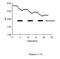
- Spectroscopic titration curves, plotting wavelength against absorbance in buffers of different pH indicate that the concentration of the acidic form and the basic form of some wild-type proteorhodopsins are about equal at a pH range about 7-8 (i.e. p K rh is about 7- 8).
- Hot75m1 in intact cells has a p K rh value of about 8.2.
- Hot75m1 exhibits a shift in the absorption maxima of about 45 nm when the pH is changed from 4.9 to 9.1. Because only one form, the basic form, gives a productive photocycle, the formation of different spectral forms limits the conditions where the proteorhodopsins can be used for certain applications.
- Different naturally occurring proteorhodopsin variants have different absoption maxima wavelengths.
- the absorption maxima wavelengths when the proteorhodopsin is in an acidic form range from about 534 to about 570 nm.
- the maximum wavelength when the proteorhodopsin is in a basic form falls within two groups: those that range from about 488 to about 494 nm, and those that range from about 516 to about 528 nm.
- Different naturally occurring proteorhodopsin variants also have different p K rh values, ranging from about 7.1 to about 8.6.
- the photochemical property of proteorhodopsin can be altered by, either one or a combination of, the pH, presence of an amphipathic molecule (such as a surfactant detergent), temperature of the environment, the presence of chemical additives in the environment (such as azide or glycerol or the like), and the water content of the medium containing the photeorhodopsin.
- an amphipathic molecule such as a surfactant detergent
- temperature of the environment such as a surfactant detergent
- chemical additives in the environment such as azide or glycerol or the like
- proteorhodopsin As described above, a useful and productive form of proteorhodopsin is the basic form. To achieve a predominantly basic spectral form of a proteorhodopsin population with a p K rh of 8.0, a two or more pH unit shift to pH 10.0 or more is contemplated to exhibit an increased relative amount of the basic form versus the acidic form.
- a basic environment is not an optimal pH for proteorhodopsins, since proteins in general tend to denature at such a high pH. Therefore, a proteorhodopsin mutant having a low p K th value is desirable.
- the proteorhodopsin mutant of the present invention preferably has an altered p K rh value that is lower (more acidic) than that of the naturally occurring proteorhodopsin.
- the proteorhodopsin mutant has a p K rh value that is lower than neutral pH. This is because at near neutral pH, the polypeptide is more stable and has a longer shelf life.
- the p K rh value of the proteorhodopsin mutant should be reduced as much as possible so that the basic form can predominate at near a neutral pH range, for example, about 5.5-9.0, or about 6.0-9.0, or about 7.1 - 8.6.
- the p K rh value of the proteorhodopsin mutant is reduced in comparison with that of the proteorhodopsin variant from which the mutant is derived.
- the p K rh value is reduced by at least 1.0, preferably, 2.0, and more preferably, 3.0 pH units.
- the value of the p K rh of proteorhodopsin mutant is reduced to lower than 7.0, preferably 6.0, more preferably 5.0, more preferably 4.0, and even more preferably 3.0.
- a proteorhodopsin mutant in general has a broad range of pH in which the basic form predominates.
- the p K rh value of the proteorhodopsin mutant is low enough such that essentially only the basic form is present in the useful pH range.
- the proteorhodopsin mutant predominates in the basic form under all pH conditions.
- the proteorhodopsin mutant has a smaller difference in maximum absorption wavelength between the basic and the acidic form, in comparison with the proteorhodopsin variant from which the mutant is derived.
- Such mutants of which the basic and acidic absorption maxima are closer to each other, are useful in applications where the environmental pH is close to the p K rh value. In such applications, both spectral forms will be present.
- a large difference in the absorption maxima of the acidic and basic forms will result in a broad composite absorption spectra (broader spectral width) because the acidic and basic absorption spectra are superimposed.
- the composite absorption spectra will be narrower (smaller peak width).
- the proteorhodopsin mutant has a difference in maximum absorption wavelength between the basic and the acidic form of less than 25 nm, preferably less than 20 nm, more preferably less than 15 nm, and most preferably, less than 10 nm.
- a proteorhodopsin variant that the mutant is derived from can be any naturally occurring proteorhodopsin.
- Proteorhodopsin variants, and nucleic acid sequences encoding thereof, have been obtained from naturally occurring members of the domain bacteria. Such members include marine bacteria, such as bacteria from the SAR86 group.
- Proteorhodopsin variants useful in the present invention include those derived from marine bacteria, as well as those derived or obtained from non-marine bacteria. There are many variant forms of proteorhodopsin; all of which can be used for the present invention.
- MED208 (SEQ ID NO: 77), REDA9 (SEQ ID NOs: 79), REDB9 (SEQ ID NO: 81), REDF9 (SEQ ID NO: 83), RED19 (SEQ ID NO: 85), RED2 (SEQ ID NO: 87), RED23 (SEQ ID NO: 89), RED27 (SEQ ID NO: 91), RED30 (SEQ ID NO: 93), RED4 (SEQ ID NO: 95), RED5 (SEQ ID NO: 97), REDr6a5a14 (SEQ ID NO: 99), and REDr6a5a6 (SEQ ID NO: 101).
- REDr7_1_4 SEQ ID NO: 103
- REDs3_7 SEQ ID NO: 105
- REDr7_1_15 SEQ ID NO: 107
- REDs3_15 SEQ ID NO: 109
- medA15r8ex6 SEQ ID NO: 111
- REDr7_1_16 SEQ ID NO: 113
- medA15r11b9 SEQ ID NO: 115
- medA15r9b5 SEQ ID NO: 117
- medA15r8b3 SEQ ID NO: 119
- medA15r11b3 SEQ ID NO: 121
- medA15_r8_1 SEQ ID NO: 123
- medA17R9_1 SEQ ID NO: 125
- medA15r8b9 SEQ ID NO: 127
- medA19_R8_16 SEQ ID NO: 129
- medA19_R8_19 SEQ ID NO: 131
- the proteorhodopsin variant, from which the mutant is derived can be a naturally occurring proteorhodopsin variant, including but not limited to those of SEQ ID NOs: 1, 3, 5, 7, 9, 11, 13, 15, 17, 19, 21, 23, 25, 27, 29, 31, 33, 35, 37, 39, 41, 43, 45, 47, 49, 51, 53, 55, 57, 59, 61, 63, 65, 67, 69, 71, 73, 75, 77, 79, 81, 83, 85, 87, 89, 91, 93, 95, 97, 99, 101, 103, 105, 107, 109, 111, 113, 115, 117, 119, 121, 123, 125, 127, 129, 131, 133, 135, 137, 139, 141, 143, 145, 147, 149, 151, 153,155, 157, 159, and 161, or other proteorhodopsin variants sharing at least 70%, or
- nucleotide and amino acid sequences of the 81 proteorhodopsin variants can be used to prepare mutants for this invention.
- nucleotide and amino acid sequences of the 81 proteorhodopsin variants can be altered by substitutions, additions or deletions to provide functionally equivalent molecules, which are suitable for preparing mutants for this invention.
- other DNA sequences that encode substantially the same amino acid sequence as depicted in figures 1-1 through 1-81 can be used in the practice of the present invention.
- the DNA sequence can be altered by a substitution of a different codon that encodes the same or a functionally equivalent amino acid residue within the sequence, thus producing a silent change.
- an amino acid residue within the sequence can be substituted by another amino acid of a similar polarity, or a similar class.
- Non-polar (hydrophobic) amino acids include alanine, leucine, isoleucine, valine, proline, phenylalanine, tryptophan, glycine and methionine.
- Polar neutral amino acids include serine, threonine, cysteine, tyrosine, asparagine, and glutamine.
- Positively charged (basic) amino acids include arginine, lysine, and histidine.
- Negatively charged (acidic) amino acids include aspartic and glutamic acid.
- a proteorhodopsin mutant comprises the amino acid sequence of Bac31A8 H75K (SEQ ID NO: 163), Bac31A8 H75N (SEQ ID NO: 165), Bac31A8 H75Q (SEQ ID NO: 167), Hot75ml H77K (SEQ ID NO: 169), Hot75ml H77N (SEQ ID NO: 171), Hot75ml H77Q (SEQ ID NO: 173), Hot 75ml H77E (SEQ ID NO: 175), Hot75ml H77W (SEQ ID NO: 177).
- Naturally occurring proteorhodopsin variants have about 234 to 249 amino acids. Comparing the amino acid sequence of different forms of proteorhodopsins, while contrasting their physical, or chemical properties, can reveal specific target regions that are likely to produce useful mutant proteins, and direct the creation of new mutants with deliberately modified functions.
- sequences determined for proteorhodopsins Bac31A8 and/or Hot75m1 are compared with sequences of known proteorhodopsins (see figures 1-1 through 1-81 ) or newly discovered proteorhodpsins in order to deduce sites for desirable mutations. To do this, the closeness of relation of the proteorhodopsins being compared is first determined.
- Closeness of relation can be measured by comparing of amino-acid sequences.
- aligning protein sequences There are many methods of aligning protein sequences. Methods defining relatedness are described in Atlas of Protein Sequence and Structure, Margaret O. Dayhoff editor, vol. 5, supplement 2, 1976, National Biomedical Research Foundation, Georgetown University Medical Center, Washington, D.C., p. 3 ff ., entitled SEARCH and ALIGN.
- related proteins can differ in the number of amino acids as well as the identity of each amino acid along the chain. That is, there can be deletions or insertions when two structures are aligned for maximum identity. For example, proteorhodopsin Bac31A8 has only 249 amino acids while proteorhodopsin Hot75m1 has 252 amino acids.
- the conserved amino acid D97 (Asp97) in the Bac31A8 proteorhodopsin is the amino acid residue that donates a proton to the retinal Schiff-base during the photocycle.
- the Bac31A8 D97N proteorhodopsin mutant is locked in the acidic form and is therefore incapable of pumping protons or undergoing a complete photocycle (Dioumaev, et al .) .
- the Bac31A8 D97E proteorhodopsin mutant which has a conserved amino acid replacement, has different wavelength spectra at acidic and basic pH compared with those of the wild-type Bac31A8 proteorhodopsin.
- Bac31A8 D97E mutant has a similar pH dependent spectral change and a similar p K rh value compared with that of wild-type Bac31A8 proteorhodopsin (Dioumaev, et al. ).
- a conservative replacement in the Asp97 position causes spectral changes, but not a change in the p K rh value. Since the nature and position of the proton-donating group is important for the proper functioning of the proteorhodopsin photocycle, it is unlikely that changes in D97 of Bac31A8 proteorhodopsin will allow for alteration of the p K rh value without disrupting the pumping of protons or the photocycle.
- the present invention provides a mutant proteorhodopsin wherein said proteorhodopsin has one or more mutations in a conserved amino acid residue, wherein said one or more mutations cause said proteorhodopsin to have an altered photochemical property, wherein said mutant proteorhodopsin, when optically stimulated, undergoes a photocycle in which a proton is transported.
- the proteorhodopsin mutant of this invention has an improved optical characteristic in comparison with the proteorhodopsins variant.
- the improved optical characteristic is a lower p K rh value or a smaller difference in maximum absorption wavelength between a basic and an acidic form.
- the present invention has identified a number of conserved amino acid residues that may interact with the proton transported during the photocycle, thus altering the pH at which the basic form of the proteorhodopsin appears, and/or altering the spectrophotometic properties of the proteorhodopsin as compared with that of the wild-type.
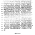
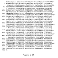
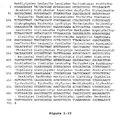
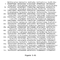
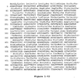
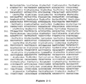
- a conserved amino acid residue of proteorhodopsin is an amino acid that is found in the equivalent position of the 81 proteorhodopsins as depicted in FIGURE 3 .
- a conserved amino acid residue which alters the photochemical property of the proteorhodopsin when substituted with a different amino acid, is important for this invention.
- FIGURE 3 shows the alignment of amino acid sequences of 81 natural proteorhodopsin variants. Examples of conserved amino acid residues (H75, R94, D227 of BAC31A8) are shown in FIGURE 3 .
- conserved amino acid residues can affect the conformation of the protein and the positioning of the all-trans-retinal molecule in relation to the proteorhodopsin protein.
- the present invention identifies that the conserved histidine of a proteorhodopsin variant is important for the proton transport during a photocycle.
- the conserved histidine residue (for example, H75 in Bac31A8 and H77 in Hot75ml) is purportedly located near both the D97 residue and the all-trans-retinal molecule. This residue in proteorhodopsin interact with D97 or with another amino acid or a water molecule that interacts with D97, but do not interact directly with all trans-retinal.
- the conserved histidine is likely to be part of the hydrogen bondable groups responsible for the spectral change at different pH values.
- Proteorhodopsin mutants which have the conserved histidine substituted with an amino acid capable of forming a hydrogen bond, have an altered photochemical property that shifts the p K rh to a lower pH (more acidic) value than the wild-type proteorhodopsin. For example, mutations at H75 in Bac31A8 proteorhodopsin and at H77 in Hot75m1 proteorhodopsin lower the p K rh value of proteorhodopsin and expand the pH range of the basic form. In some H75 or H77 proteorhodopsin mutants, only the basic form exists in the pH range tested (such as pH 4.9-9.1).
- Amino acids capable of forming a hydrogen bond and suitable for substituting histidine in this invention include asparagine, glutamine, lysine, arginine, serine, theonine, tyrosine, aspartic acid (in its protonated state at acid pH), glutamic acid (in its protonated state at acidic pH), tryptophan, and any synthetic amino acid that has a functional group that is able to contribute a hydrogen to form a hydrogen bond.
- Preferred proteorhopsin mutants have the conserved histidine residue substituted with glutamine (Q), asparagine (N), glutamic acid (E), lysine (K), aspartic acid (D), and tryptophan (W).
- the altered photochemical property of proteorhodopsin mutants can be identified by measuring the spectral shift of the proteorhodopsin in intact or whole cells, wherein the proteorhodopsin is present in the cytoplasmic membrane, or in a solubilized polypeptide form stabilized by an amphipathic molecule, or in a pure polypeptide form.
- the altered photochemical property can be identified by measuring the spectral shift of the proteorhodopsin in a membrane preparation, including but not limited to crude or partly purified membranes.
- Membrane preparations contain lipids and potentially membrane proteins other than proteorhodopsin.
- the present invention provides a method for preparing a proteorhodopsin mutant having improved optical characteristics.
- the method comprises the steps of: (a) identifying a conserved amino acid residue of a wild-type proteorhodopsin variant, (b) mutagenizing the conserved amino acid residue and obtaining proteorhodopsin mutants, (c) determining the optical characteristics of the proteorhodopsin mutants, and (d) selecting the proteorhodopsin mutant having improved optical characteristics.
- the conserved amino acid residue for example, is a histidine residue.
- the wild-type proteorhodopsin variant can by mutagenized by any method, including but not limited to site-directed mutagenisis, known to a skilled person.
- the nucleotide and amino acid sequences of various proteorhodopsin variants are deposited with Genbank under accession numbers AF349976-AF350003 and AF279106 (which contains the nucleotide sequence of a 105 kilobase genomic region that includes the gene encoding the Bac31A8 proteorhodopsin).
- the nucleotide and amino acid sequences of newly isolated proteorhodopsin variants ( Figures 1-30 to 1-51 ) are deposited with Genbank under accession numbers AY210898-AY210919.
- nucleotide and amino acid sequences of newly isolated proteorhodopsin variants are deposited with Genbank under accession numbers AY250714-AY250741.
- nucleotide and amino acid sequences of newly isolated proteorhodopsin variants are deposited with Genbank under accession numbers AY372453 and AY372455. All these sequences are specifically incorporated herein by reference in this application. Any of these natural proteorhodopsin amino acid sequences can be used to make the proteorhodopsin mutants.
- the natural proteorhodopsin variant include Hot75ml, Bac31A8, Bac40E8, Bac41B4, Bac64A5, Hot0m1, Hot75m3, Hot75m4, Hot75m8, MB0m1, MB0m2, MB20m2, MB20m5, MB20m12, MB40m1, MB40m5, MB100m5, MB100m7, MB100m9, MB100m10, PalB1, PalB2, PalB5, PalB7, PalB6, PalB8, PalE1, PalE6, PalE7, MED 26, MED27, NMD36, MED101, MED102, MED106, MED25, MED202, MED204.
- MED208 REDA9, REDB9, REDF9, RED19, RED2, RED23, RED27, RED30, RED4, RED5, REDr6a5a14, REDr6a5a6, REDr7_1_4, REDs3_7, REDr7_1_15, REDs3_15, medA15r8ex6, REDr7_1_16, medA15r11b9, medA15r9b5, medA15r8b3, medA15r11b3, medA15_r8_1, medA17R9_1, medA15r8b9, medA19_R8_16, medA19_R8_19, medA17_R8_6, medA15r9b7, medA15_R8_3, medA15r10b5, medA19_r9_9, medA15_r8ex7, medA19_R8_20, medA15
- the nucleotide sequence of any of the above proteorhodopsin genes can be obtained or derived using the method of Béjà, et al. (2000) or WO 01/83701 .
- An example of a gene encoding a proteorhodopsin variant is the bacterioplankton Bacterial Artificial Chromosome (BAC) clone Bac31A8 (also known as EBAC31A08) (see WO 01/83701 ).
- BAC Bacterial Artificial Chromosome
- One skilled in the art can obtain other genes encoding different proteorhodopsin variants using the identical techniques described above.
- One skilled in the art can also obtain or clone proteorhodopsin genes from organisms obtained from natural habitats by designing primers and using degenerate PCR, heterologous hybridization, or random sequencing of DNA.
- the proteorhodopsin is capable of proper folding and integrating into a membrane when expressed or synthesized in a suitable host.
- a suitable host is a cell that naturally expresses proteorhodopsin, such as the SAR 86 strain.
- a suitable host can also be a cell that naturally does not express proteorhodopsin.
- a suitable host includes a marine and a non-marine bacteria.
- a suitable host is deficient in any outer-membrane protease, either naturally or constructed not to express such protease.
- An example of a suitable host is an eubacteria cell; preferably, a Gram-negative bacteria; for example, a proteobacteria such as a gamma-proteobacteria.
- the gamma-proteobacteria can belong to the Enterobacteriaceae family; such as Escherichia, Edwardsiella, Citrobactor, Salmonella, Shigella, Klebsiella, Enterobacter, Serratia, Proteus, and Yersinia.
- the gamma-proteobacteria belonging to the Salmonella genus is Salmonella typhimurium.
- the gamma-proteobacteria belonging to the Escherichia genus is Escherichia coli.
- the polypeptide is expressed or synthesized in a strain that is an outer membrane protease-deficient strain.
- suitable strains include the E. coli strains: UT5600 (as disclosed by Béjà, et al. (2000), and Dioumaev, et al. ) and BL21-Codonplus-RIL.
- the gamma-proteobacteria is the SAR86 strain.
- the present invention also provides for a host cell comprising a polynucleotide encoding the proteorhodopsin mutant.
- the suitable host cell is capable of expressing or synthesizing a functional proteorhodopsin from the polynucleotide.
- a functional proteorhodopsin is a polypeptide capable of undergoing a photocycle in a suitable environment.
- the one or more mutations can be constructed by site-directed mutagenesis of a cloned proteorhodopsin gene. Specific designed mutations can be constructed or variability can be introduced to produce a variety of mutants. Such mutagenesis and other methods for manipulation of the nucleic acid and protein are well known to one skilled in the art and are described by Sambrook, et al. ( Molecular cloning: a laboratory manual. 3d ed. Cold Spring Harbor Laboratory Press, Cold Spring harbor, NY).
- the present invention provides a polynucleotide encoding the proteorhodopsin mutant.
- One skilled in the art is able to construct a nucleotide sequence using the information of the amino acid sequence of the polypeptide, based on the universal genetic code.
- the polynucleotide optionally comprises a promoter operatively linked 5' to the open reading frame encoding the polynucleotide.
- the polynucleotide further comprises appropriate promoter control, translational control and stop and other necessary sequences in order to express that polypeptide in a suitable host.
- the polynucleotide can be a chromosome, an episome, a plasmid, or any suitable expression vector.
- the polynucleotide is capable of amplification in a host cell.
- a wide variety of expression vectors can be used for introduction by transformation, conjugation, transduction or transfection of the polynucleotide into the eubactaerial host cell.
- Vectors include plasmids, such as pBR322, pMB9, pBAD-TOPO ® (from the pBAD TOPO ® TA Expression Kit, Invitrogen, La Jolla, CA), pTrcHis2-TOPO ® (from the pTrcHis2-TOPO ® TA Expression Kit, Invitrogen, La Jolla, CA) and the like; cosmids, such as pVK100, and the like; and viruses such as P22, and the like.
- plasmids such as pBR322, pMB9
- pBAD-TOPO ® from the pBAD TOPO ® TA Expression Kit, Invitrogen, La Jolla, CA
- pTrcHis2-TOPO ® from the pTrcHis2-TOPO ® TA Expression Kit, Invitrogen, La Jolla, CA
- cosmids such as pVK100, and the like
- viruses such
- proteorhodopsin polypeptide can be purified from a host cell using a variety of methods, which follow a general scheme:
- Methods of purification can differ for each intended use of the protein since requirements of yield and purity differ in each application.
- Lysis can be carried out by sonication, osmotic shock, freeze thaw or French press or a combination of above methods.
- a preferred method of lysis is freeze-thaw followed by French press.
- Insoluble materials are removed by centrifugation (e.g. 1500 xg, 30 min.) and the supernatant collected. The supernatant is further centrifuged (e.g. 150,000 xg. 1 hour) and the pellet containing primarily membrane material is collected.
- the membrane material can be solubilized with detergent.
- useful detergents include, but are not limited to, dodecyl- ⁇ -maltoside, octyl- ⁇ -glucoside, or Triton ® X-100. Dodecyl- ⁇ -maltoside (2%) is a preferred detergent.
- the solubilized membrane material is incubated with His-tag affinity resin.
- His-tag affinity resin For example, the solubilized membrane material is incubated with Talon TM resin from Clontech (Palo Alto, CA) overnight at 4 °C with gentle agitation.
- the solubilized membrane material is purified from contaminating proteins by washing the resin, for example, with three column volumes of buffer containing 0.1 % dodecyl- ⁇ -maltoside three times.
- the purified polypeptide can be removed from His-tag purification resins by a variety of methods including, but not limited to, incubation with EDTA, incubation in acidic pH, or incubation at high temperature.
- a preferred method is to incubate the resin with a buffer containing 0.5 M EDTA.
- the buffer can be exchanged by a variety of methods including, but not limited to, concentration and dilution using membrane based concentrators, dialysis, or desalting columns.
- concentration and dilution using membrane based concentrators, dialysis, or desalting columns.
- the protein is concentrated using a membrane based centrifugation concentrator device and then diluted into a desired buffer. The process is repeated to completely exchange the buffer. Then the protein is concentrated again using the same concentrator device.
- the proteorhodopsin mutant is derived from Hot75ml proteorhodopsin variant with the conserved histidine substituted with a glutamine residue.
- the polypeptide is expressed from an E. coli host cell. When analyzed for its absorbance behavior in the membrane of intact cells, the mutant polypeptide exhibits a spectral shift with the p K rh value of less than about 5.8 (as compared with 7.2 of the wild-type proteorhodopsin). The shift of p K rh is about 1.4 units.
- the proteorhodopsin mutant is derived from Hot75ml proteorhodopsin variant with the conserved histidine substituted with an asparagine residue.
- the polypeptide is expressed from an E. coli host cell. When analyzed for its absorbance behavior in the membrane of intact cells, the polypeptide exhibits a spectral shift with the p K rh value of less than about 5.1. The shift in the p K rh is about 2.1 units.
- the proteorhodopsin mutant is derived from Hot75ml proteorhodopsin variant with the conserved histidine substituted with a lysine residue.
- the polypeptide is expressed from an E. coli host cell. When analyzed for its absorbance behavior in the membrane of intact cells, the polypeptide does not exhibit a spectral shift (or does not titrate) when analyzed at a pH range from 4.9 to 9.1.
- the shape of the curves obtained for this mutant proteorhodopsin when absorbance of the polypeptide is plotted against the wavelength of the light source, is similar to the shape for the curve obtained from the basic form of the wild-type Hot75ml proteorhodopsin.
- this mutant proteorhodopsin is much closer to the maxima of absorbance for the basic form of Hot75ml (about 490 nm) than the acidic form of Hot75ml (about 540 nm). Based on this reasoning, this mutant proteorhodopsin is likely in its basic form when the pH of its environment is from 4.9 to 9.1. It is likely that the p K rh of this mutant is shifted to a pH much lower than pH 4.9.
- proteorhodopsin mutants taught herewith have many technical applications.
- the proteohodopsin mutant can be incorporated into instruments or devices having photochromic applications, photoelectric applications, and/or phototransport applications.
- the polypeptide can be used for its light absorption properties for optical data storage, interferometry and/or photonics.
- Photochromic applications include, but are not limited to, holographic film.
- the proteorhodopsin mutant can be used as a photochromic all-trans-retinal protein for optical data storage devices.
- the proteorhodopsin mutant can be used in a device for information storage, such as 2-D storage, 3-D storage, holographic storage, associative storage, or the like.
- the proteorhodopsin mutant can be used in a device for information processing, such as optical bistability/light switching, optical filtering, signal conditioning, neural networks, spatial light modulators, phaseconjugation, pattern recognition, interferometry, or the like.
- the present invention also provides a method of storing and retrieving optical data using the proteorhodopsin mutant of the invention.
- a method of storing and retrieving optical data comprises the steps of: (a) providing a film comprising the proteorhodopsin mutant immobilized in a matrix; (b) recording the optical data by exposing the film to light of a wavelength that is absorbed by the proteorhodopsin mutant in a predetermined pattern and selectively converting portions of the film to an excited state; (c) exposing the film of step (b) to light of a wavelength that is absorbed either by the basal or the excited state of the proteorhodopsin mutant; (d) detecting the optical data by an optical recording device.
- the detecting of step (d) can be conducted by any optical recording device.
- the optical recording device can be a video camera, which can be a charged coupled device (CCD).
- CCD charged coupled device
- the proteorhodopsin mutant can be used in devices for its light-induced changes in electric fields caused by proton transport, such as in ultrafast light detection, artifical retina, and/or light/motion detectors.
- the proteorhodopsin mutant can be used for its light-induced proton transport across a membrane, such as photovoltaic device.
- a photovoltaic device is a light-driven energy generator comprising the polypeptide, whereby light energy can be converted to chemical energy.
- the manufacture and use of such a light-driven energy generator is disclosed in WO 01/83701 .
- the proteorhodopsin mutant can also be used in devices for ATP generation in reactors, desalination of seawater, and/or conversion of sunlight into electricity.
- the present invention provides for a light-driven energy generator comprising the use of the proteorhodopsin mutant.
- a light-driven energy generator comprises: (a) the proteorhodopsin mutant of the present invention, (b) a cell membrane, (c) a source of all-trans-retinal, and (d) a light source, wherein the proteorhodopsin mutant integrates within the cell membrane to produce an integrated proteorhodopsin mutant, and the integrated proteorhodopsin mutant binds covalently to all-trans-retinal to produce a light absorbing pigment.
- Light-driven energy generators as applied to natural proteorhodopsin variants are disclosed by WO 01/83701 , and can be modified by replacing the proteorhodopsin with the proteorhodopsin mutants.
- the light-driven generator utilizes the proteorhodopsin mutant to convert light-energy into biochemical energy.
- the light-driven energy generator takes advantages of the functional properties of a proteorhodopsin mutant, which is expressed and correctly folded in a suitable host cell.
- the proteorhodopsin mutant can also be used in devices for 2D harmonic generation, radiation detection, biosensor applications, or the like.
- Hampp Chem . Rev .,100: 1755-76 (2000) discloses various uses and instruments and devices that utilize the photochemical properties of bacteriorhodopsin. Proteorhodopsin mutants of this invention can be used in the analogous manner as described by Hampp, which is specifically incorporated by reference.
- proteorhodopsin mutants can be used to replace bacteriorhodopsin (BR) for a variety of devices/processes that utilize bacteriorhodopsin, for example, in the following list of patents (Table 1), which are incorporated herein by reference. TABLE 1.
- Priority Patent No(s) Subject Inatomi and Isoda 26 APR 1983 JP 59197849 Photosensor Isoda 02 MAR 1984 JP 60185228 Recording and reproducing method of information Isoda and Daimon 02 MAR 1984 JP 60184246 Light recording medium Inatomi 19 NOV 1984 JP 61124384 ATP reproduction bioreactor Hikima, et al.
- 08 MAR 1990 EP 445758 Methods for improving the signal-to-noise ratio in holography when using br-based recording media US 5223355 Oyama, et al. 13 APR 1990 JP 3295278 Photoelectric conversion element Miyasaka 25 APR 1990 JP 4006420 Photoelectric conversion element Miyasaka and Kitaguchi 25 APR 1990 JP 4009400 Fixation of photosensitive chromoprotein Saito, et al. 08 JUN 1990 JP 4042585 Photoresponsive excitable synthetic membrane and its manufacture Saito, et al.
- EXAMPLE 1 Cloning of Proteorhodopsins Into Expression Vector.
- a total of 21 of the 81 known natural proteorhodopsin genes were cloned in the pTrcHis2 expression vector.
- Different natural proteorhodopsin genes were PCR amplified using Taq DNA Polymerase (Roche Applied Science) as described by the manufacturer.
- the primers used to amplify all proteorhodopsin variants were PR-u4 and PR-d2 (see Table 2) and the templates were pCR2.1 containing the relevant proteorhodopsin (Béjà, et al., 2000; Béjà, et al., 2001).
- the PCR products were cloned in the pTrcHis2 vector using the pTrcHis2 TOPO TA Expression Kit (Invitrogen, Carlsbad, CA), as described by the manufacturer. Restriction enzyme digestions of the plasmids were used to identify clones containing the insert in the correct orientation.
- the plasmids were sequenced using the primers pTrcHis Forward and pTrcHis Reverse to ensure that no sequence variations were introduced during the PCR and cloning procedure.
- the plasmids contain the pTrc promoter transcribing the proteorhodopsin gene with a C-terminal extension containing a myc epitope and six histidine residues.
- FIGURE 4 A map of one of the expression plasmids is shown in FIGURE 4 .
- PR-u4 SEQ. ID NO : 185) AAATTATTACTGATATTAGGTAGTG PR-d2 (SEQ ID NO : 186) AGCATTAGAAGATTCTTTAACAGC pTrcHis Forward (SEQ ID NO : 187) GAGGTATATATTAATGTATCG pTrcHis Reverse (SEQ ID NO : 188) GATTTAATCTGTATCAGG 31A8-m6A (SEQ ID NO : 189) TGTTACTGGTATTGCTTTCTGG aAT TACATGTACATGAGAGGGGT 31A8-m6B (SEQ ID NO : 190) ACCCCTCTCATGTACATGTA ATt CCAGAAAGCAATACCAGTAACA 31A8-m7A (SEQ ID NO : 191) TGTTACTGGT
- pTrcHis Forward and pTrcHis Reverse oligonucleotides were obtained from Invitrogen, the rest of the oligonucleotides were purchased from Operon, the primers used for site-directed mutagenesis were PAGE purified.
- Bold letters indicate the histidine or arginine codons that were mutated, lowercase letters indicate nucleotides that were changed compared to the wild-type sequence.
- proteorhodopsin expression plasmids and the control plasmid pTrcHis2-LacZ were transformed into competent cells of the strain BL21-Codonplus-RIL (Stratagene) as described by the manufacturer.
- the transformed cells were plated on LA + 0.5% glucose + 100 ⁇ g/ml carbenicillin + 25 ⁇ g/ml chloramphenicol plates and incubated overnight at 37°C. Cells from these plates were grown in 6 ml LB + 0.5% glucose + 100 ⁇ g/ml carbenicillin + 25 ⁇ g/ml chloramphenicol + 10 ⁇ M all-trans-retinal medium in glass tubes at at 37°C for 6 hours, where the cells reached early stationary phase.
- the cells constitutively express proteorhodopsins from the uninduced pTrc promoter.
- the cells were collected by centrifugation at 4,500xg for 6 minutes, the medium was discarded and the cells were resuspended in 5 ml sterile water.
- a 900 ⁇ l sample of cells were added to a 100 ⁇ l aliquot of concentrated buffer (1.0 M acetate (pH 4.9), 1.0 M MES (pH 5.8), 1.0 M MOPS (pH 6.7), 1.0 M TAPS (pH 8.1), or 1.0 M CHES (pH 9.1)).
- the spectra of the 21 natural proteorhodopsins were measured in intact cells at five different extracellular pH values using a spectrophotometer (On-Line Instrument Systems, Inc.) adapted for use with turbid samples by placing the photomultiplier detector adjacent to the sample cuvette.
- the absorption from a strain containing a control plasmid (pTrcHis2-LacZ) was subtracted from proteorhodopsin containing samples.
- pTrcHis2-LacZ plasmid
- a linear baseline determined by least-squares fitting of the first ten and last ten wavelength and absorbance values in each spectrum was subtracted from each spectrum. The spectra were then adjusted to the same minimum value by subtracting the differences in minimum values.
- Typical proteorhodopsin spectra such as Bac31A8 and Hot75ml inside intact cells at different pH values, can be constructed by plotting absorbance vs. wavelength. Two different spectral forms of the proteorhodopsin variants (Bac31A8 and Hot75ml) are observed under acidic conditions (pH 4.9) and basic conditions (pH 9.1). At intermediate pH values, spectra representing a mixture of the acidic and basic forms are observed.
- Abs Acidic and Abs Basic-Acidic were fitted at each wavelength, but the same value of p K rh was fitted for data at all wavelengths.
- the values AbsA cidic and Abs Basic- Acidic were used to reconstruct wavelength spectra for Acidic and Basic forms of proteorhodopsin and from this data the wavelength maximum at acidic and basic pH were determined (Table 3).
- the wild-type Bac31A8 proteorhodopsin in intact cells had absorbance maximum at 538 nm at acidic conditions and at 519 nm at basic conditions.
- the p K rh value of the titratable group(s) responsible for the spectral shift was determined to be 7.6.
- the wild-type Hot75ml proteorhodopsin had absorbance maximum at 546 nm at acidic conditions and at 493 nm at basic conditions.
- the value of the p K rh of the titratable group(s) responsible for the spectral shift was determined to be 8.2.
- Table 4 summarizes the templates and primers used to construct the different mutants. TABLE 4. Templates and Primers Used to Construct Mutants Mutant Name of expression plasmid Template used Primers used Bac31A8 H75N pTrcHis2-Bac31A8 H75N pTrcHis2-Bac31A8 31A8-m6A and 31A8-m6B Bac31A8 H75Q pTrcHis2-Bac31A8H75Q pTrcHis2-Bac31A8 31A8-m7A and 31A8-m7B Bac31A8 H75K pTrcHis2-Bac31A8 H75K pTrcHis2-Bac31A8 31A8-m8A and 31A8-m8B Hot75M1 H77N pTrcHis2-Hot75M1 H77N pTrcHis2-Hot75M1 H75m1-m2A and H75m1-
- the plasmids were sequenced using the pTrcHis Forward primer (Invitrogen) to ensure that the desired mutations had been introduced into the plasmids.
- proteorhodopsin mutants were expressed; their spectra in intact cells at different pH values were measured and the p K rh values were calculated as described above.
- Table 5 summarizes the p K rh values and the absorption maxima of the acidic and basic spectra of the wild-type and mutants of Bac31 A8. TABLE 5.
- p K rh and Absorption Maxima of BAC31A8 Mutants Protein p K rh Basic Max (nm) Acidic Max (nm) Bac31A8 7.6 519 538 Bac31A8 H75Q 6.0 518 540 Bac31A8 H75N NA 521 - Bac31A8 H75K NA 522 - Note: "NA” means that no significant pH titration is observed, so a p K rh value cannot be determined. "-” means that only a single absorption maximum can be reported because of the lack of titration in the pH range tested.
- the Bac31A8 H75Q mutant has a p K rh value of 6.0 compared with 7.6 observed with the wild-type protein.
- Both the Bac31A8 H75N and Bac31A8 H75K mutants show no or very little pH dependent change in spectral properties, the mutant proteins appear to be only in the basic form in the pH range tested here. It is likely caused by the p K th , value being shifted so much to be outside the pH range of 4.9-9.1.
- the p K rh value of the Bac31A8 H75N mutant could not be determined accurately from these data, but is estimated to be 5.8 or lower.
- FIGURE 5 shows the pH-dependent change in absorption spectra of the acid and the basic form of wild-type Hot75m1 and mutant Hot75m1 H77Q.
- the p K rh of the wild type is 8.2, which is lowered to 5.8 in the mutant.
- FIGURE 6 shows the fraction of the acid form of Hot75m1 and Hot75m1 H77Q at different pH.
- the basic form clearly predominates at pH 9 and above for the wild-type Hot75m1 protein and at pH 7 and above for the Hot75m1 H77Q protein.
- Table 6 summarizes the p K rh value and the maxima of the acidic and basic absorption spectra of the wild-type and six mutants. TABLE 6. p K rh and Absorption Maxima of Hot75M1 Mutants Protein p K rh Basic Max (nm) Acidic Max (nm) Hot75M1 8.2 493 546 Hot75M1 H77Q 5.8 487 516 Hot75M1 H77N 5.5 487 497 Hot75M1 H77K NA 500 - Hot75M1 H77D NA 515 - Hot75M1 H77E 5.3 487 529 Hot75M1 H77W 5.8 486 529 Note: "NA” means that no significant pH titration is observed, so a p K rh value cannot be determined. "-” means that only a single absorption maximum can be reported because of the lack of titration in the pH range tested.
- Hot75m1 H77K and Hot75m1 H77D mutants where the histidine is changed to lysine or aspartic acid, respectively, show no or very little pH dependent change in spectral properties and appear to be only in the basic form in the pH range tested here. It is likely caused by the p K rh value being shifted so much to be outside the pH range of 4.9-9.1.
- proteorhodopsins in intact cells, where the protein is present in the cytoplasmic membrane.
- proteorhodopsins in intact cells, where the protein is present in the cytoplasmic membrane.
- proteorhodopsin (wild-type or mutant) expression plasmids were transformed into competent cells of the strain BL21-Codonplus-RIL (Stcatagene) as described by the manufacturer.
- the transformed cells were plated on LA + 0.5% glucose + 100 ⁇ g/ml carbenicillin + 25 ⁇ g/ml chloramphenicol plates and incubated overnight at 37°C. Cells from these plates were grown in 200 ml LB + 0.5% glucose + 104 ⁇ g/ml carbenicillin + 10 ⁇ M all-trans-retinal medium at 37°C in baffled 500 ml shakeflasks. The cultures were inoculated directly from fresh transformation plates.
- Proteorhodopsin expression in the different cultures were induced when the OD 600 of the cultures were approximately 0.8 (after 4 hours growth) by adding 0.5 mM IPTG + 10 ⁇ M all-trans-retinal and the cultures were incubated for additional 4 hours at 37°C. The cells were harvested by centrifugation at 3,500xg for 10 minutes and stored at -80°C.
- Cells were resuspended in lysis buffer containing 40 mM MOPS pH 7.0, 20 mM MgCl 2 , 0.2 mg/ml lysozyme, 0.2 mg/ml DnaseI, 2% dodecyl- ⁇ -D-maltoside, and protease inhibitors (Complete, EDTA-free Protease Inhibitor Cocktail Tablets from Roche Applied Science; and lysed at 0°C by sonication.
- the lysates were incubated 16 hours with the resin in Talon spin columns (Clontech) that had been equilibrated with 1 ml wash buffer containing 40 mM MOPS, pH 7.0 and 0.5% dodecyl- ⁇ -D-maltoside.
- the resin was washed three times with 1 ml wash buffer containing 40 mM MOPS, pH 7.0 and 0.5% dodecyl- ⁇ -D-maltoside.
- Proteorhodopsins were eluted from the resin two times with 0.5 ml elution buffer containing 40 mM MOPS pH 7.0, 0.5% dodecyl- ⁇ -D-maltoside and 250 mM EDTA.
- the two elutates were pooled and EDTA was removed by three successive ten-fold concentrations using a Microcon YM-10 centrifugal filter unit (Millipore) and dilutions with a buffer contained 40 mM MOPS, pH 7.0 as described by the manufacturer.
- the proteorhodopsin samples were then concentrated ten-fold and stored at 4 °C.
- the purified Bac31A8 H75K mutant where the histidine is changed to lysine, shows no or very little pH dependent change in spectral properties, the protein appears to be locked in the basic form in the pH (pH 4.9-9.1) range tested here.
- Table 9 summarizes the p K rh value and the maxima of the acidic and basic absorption spectra of different variants. TABLE 9. p K rh and Absorption Maxima of Hot75M1 MUTANTS Protein P K rh Basic Max (nm) Acidic Max (nm) Hot75M1 8.4 504 538 Hot75M1 H77Q 6.3 508 531 Hot75M1 H77N 6.5 496 523 Hot75M1 H77K NA 521 - Note: “NA” means that no significant pH titration is observed, so a p K rh value cannot be determined. "-” means that only a single absorption maximum can be reported because of the lack of titration in the pH range tested.
- Hot75m1 proteorhodopsins show that when the conserved H77 histidine residue of Hot75m1 is changed to asparagine or glutamine, the p K rh value was significantly shifted towards the acidic pH.
- Purified Hot75m1 H77Q has a value of the p K rh of 6.3 and purified Hot75m1 H77N has a p K rh value of 6.5, compared with 8.4 of the wild-type protein.
- the purified Hot75m1 H77K mutant where the histidine is changed to lysine shows no or very little pH dependent change in spectral properties in the pH range (pH 4.9-9.1) tested here, and appears to have only one basic form.
- proteorhodopsin can undergo a photocycle that includes the formation of the excited M-state (or M-like state) and pumping of a proton across the proteorhodopsin-containing membrane from inside the cell to outside the cell.
- a photocycle that includes the formation of the excited M-state (or M-like state) and pumping of a proton across the proteorhodopsin-containing membrane from inside the cell to outside the cell.
- Proton pumping was measured as a change of the extracellular pH with illumination of cells expressing proteorhodopsin, caused by pumping of protons from inside the cells to the extracellular medium.
- proteorhodopsin (wild-type or mutant) expression plasmids were transformed into competent cells of the strain BL21-Codonplus-RIL (Stratagene) as described by the manufacturer.
- the transformed cells were plated on LA + 0.5% glucose + 100 ⁇ g/ml carbenicillin + 25 ⁇ g/ml chloramphenicol plates and incubated overnight at 37°C.
- Cells from these plates were grown in 50 ml LB + 0.5% glucose + 100 ⁇ g/ml carbenicillin + 10 ⁇ M all-trans-retinal medium at 37°C in baffled 250 ml shakeflasks. The cultures were inoculated directly from fresh transformation plates.
- Proteorhodopsin expression in different cultures was induced when the OD 600 of the cultures was approximately 0.8 (after 4 hours growth) by adding 0.1 mM IPTG + 10 ⁇ M all-trans-retinal, and the cultures were incubated for additional 4 hours at 37°C. The cells were harvested by centrifugation at 3,500xg for 10 minutes and stored at -80°C.
- Cells were thawed and suspended in 150 mM NaCl without buffer (sparged with helium to remove carbon dioxide). Cells were washed an additional three times with 150 mM NaCl without buffer. The cells were suspended to an OD 600 of 2.0 in 150 mM NaCl. Cell samples were adjusted to between pH 7.5 and 8.5 with 0.5 M HCl or 0.5% NaOH.
- Cell samples were magnetically stirred and were illuminated from two sides by split fiber-optic cables attached to a 150-watt illuminator (Fiber-Lite A3200) from Dolan-Jenner Industries (Lawrence, MA) with an infrared filter (03FC569) from Melles Griot (Irvine, CA) and a 450 nm cut-on colored-glass filter (51284) from Oriel Instruments (Stratford, CT).
- the manual opening and closing of a shutter controlled light.
- the incident angle of light was held constant by the use of a custom-built cell holder.
- the pH of the cell sample was determined during the 3 minutes of constant illumination.
- the pH of the cell sample was then measured for 3 minutes after illumination was stopped. This process was repeated two more times for each cell sample resulting in three repeated measurements of each cell sample.
- FIGURE 7-1 shows that the pH of the cells expressing Bac31A8 wild-type, which is an active proton pump, drifted slowly over time without illumination. However, a rapid (within a few seconds) change in pH occurred following illumination. After illumination was ended, the pH value of the cell samples returns to that before illumination. This pH change is a result of cellular mechanisms that control membrane potential.
- FIGURE 7-2 shows that the pH of cells expressing a LacZ control protein also drifted slowly over time without illumination. With illumination, the pH shifted slowly (over the entire 3 minutes) and minimally (at most 0.05 pH units), presumably from a temperature change at the pH probe. This temperature effect was reduced dramatically by the addition of the infrared filter, but it could not be completely eliminated.
- ⁇ pH max ⁇ k for cells expressing a proteorhodopsin variant is compared using the student-t test to the value of ⁇ pH max ⁇ k determined for cells expressing a LacZ control.
- the degrees of freedom, n, and the results of the student-t test at 60-99% confidence are given in Table 10. This test indicates whether a particular proteorhodopsin mutant is a proton pump at a given confidence. The negative result of this test does not indicate that a given proteorhodopsin mutant is not a proton pump; it only indicates that the assay is not sensitive enough to detect pumping by that mutant. TABLE 10.
- Proton pumping by the Bac31A8 E108Q where the photocycle is more than a hundred fold slower than that of the wild-type protein mutant, was not detected. Proton pumping was detected for Bac31A8 wild-type, H75K, H75N, and H75Q; and Hot75m1 wild-type, H77K, H77N, H77Q, H77E and H77W, which indicates that these proteins, when exposed to light, undergo a productive photocycle that includes proton pumping.
- proteorhodopsins with mutations in the conserved histidine residue are expected to be better proton pumps at low pH than wildtype proteorhodopsin, since the lower p K rh value of the mutants results in a larger fraction of the protein in a productive basic form at low pH.
- FIGURE 7-3 displays the dependence of proton-pumping rate on the extracellular pH for the Bac31A8 wild-type and the H75N mutant.
- the results show that the Bac31A8 H75N mutant is a better proton pump than the Bac31A8 wild-type at acidic pH.
- the results confirm that a mutation lowering the p K rh value results in a proteorhodopsin mutant having an extended pH range where a productive photocycle is present.
Landscapes
- Chemical & Material Sciences (AREA)
- Health & Medical Sciences (AREA)
- Life Sciences & Earth Sciences (AREA)
- Organic Chemistry (AREA)
- Genetics & Genomics (AREA)
- Biochemistry (AREA)
- Molecular Biology (AREA)
- General Health & Medical Sciences (AREA)
- Engineering & Computer Science (AREA)
- Biophysics (AREA)
- Zoology (AREA)
- Biotechnology (AREA)
- Proteomics, Peptides & Aminoacids (AREA)
- Medicinal Chemistry (AREA)
- Gastroenterology & Hepatology (AREA)
- General Engineering & Computer Science (AREA)
- Bioinformatics & Cheminformatics (AREA)
- Biomedical Technology (AREA)
- Wood Science & Technology (AREA)
- Cell Biology (AREA)
- Immunology (AREA)
- Toxicology (AREA)
- Plant Pathology (AREA)
- Physics & Mathematics (AREA)
- Microbiology (AREA)
- Peptides Or Proteins (AREA)
- Preparation Of Compounds By Using Micro-Organisms (AREA)
- Micro-Organisms Or Cultivation Processes Thereof (AREA)
- Compositions Of Macromolecular Compounds (AREA)
Abstract
Description
- This present invention relates to the composition and use of proteorhodopsin mutants with improved optical characteristics. Particularly, the proteorhodopsin mutants have a mutation at a conserved amino acid residue of a naturally occurring proteorhodopsin variant or its equivalent sharing high identity.
- Proteorhodopsins are integral membrane proteins; they are isolated from uncultivated marine eubacteria and function as light-driven proton pumps. Upon absorption of light by the all-trans-retinal co-factor, proteorhodopsin goes through a photocycle with a number of intermediates. It is believed that upon excitation of the proteorhodopsin molecule by light stimulation, a proteorhodopsin/retinal complex is excited to an unstable intermediate energy state. Proteorhodopsin progresses through a series of unstable energy states that can vary in terms of energy plateaus or intermediates, e.g., an "M-like state" or "M-state", a "K-like state" or "K-state", an "N-like state" or "N-state", or an "O-like state" or "O-state". Subsequently, the complex reverts to a more stable basal state concomitant with transportation of a proton.
- Proteorhodopsins are distantly related to bacteriorhodopsin from Halobacteriun salinarium (22-24% sequence similarity). Hampp (Appl. Microbiol. Biotechnol. 53:633-9, 2000a) reviews the structure and function of bacteriorhodopsin, and its technical applications. Hampp (Chem. Rev. 100:1755-76, 2000b) reviews the technical application of bacteriorhodopsin.
- Proteorhodopsin and bacteriorhodopsin have some shared characteristics, but also have clearly different properties. Proteorhodopsins are more advantageous to use in some technical applications than bacteriorhodopsins because of the ease of expressing and producing proteorhodopsins. However, the conditions where the proteorhodopsins can be used in different applications are limited because wild-type proteorhodopsins exist in two distinct spectral forms depending on the extra-cellular pH. A basic form, which is a spectral form at a higher pH, is able to achieve an M-state of excitation and transport a proton upon exposure to an optical stimulation. An acidic form, which is a spectral form at a lower pH, is unable to exhibit the M-state of excitation and does not transport a proton upon exposure to an optical stimulation.
- The properties of the two distinct pH-dependent spectral forms of the Bac31A8 proteorhodopsin have been characterized to some extent (Dioumaev, et al., Biochem. 41:5348-58, 2002; Krebs, et al., BMC Physiol. 2:1-8, 2002; Fredrich, et al., J. Mol. Biol. 321, 821-838, 2002). The D97 residue in the Bac31A8 proteorhodopsin was previously identified (Dioumaev, et al., 2002) as being part of the titratable group(s) involved in the pH dependent change in spectral and photochemical properties. However, the Bac31A8 D97N mutant protein appears only to exist as a single spectral form, the acidic form. An analysis of the photocycle intermediates of the Bac31A8 proteorhodopsin at different pH values showed that only the high pH ("basic") form exhibits the photocycle wherein protons are pumped across the membrane. Hence, this Bac31A8 D97N mutant is not very useful for most applications because the protein is unable to pump protons and form an M-state.
- Béjà, et al. (Science 289:1902-6, 2000) disclose the cloning of a proteorhodopsin gene from an uncultivated member of the marine γ-proteobacteria (i.e., the "SAR86" group). The proteorhodopsin was functionally expressed in E. coli and bound all-trans-retinal to form an active light-driven proton pump.
- Béjà, et al. (Nature 411:786-9, 2001) disclose the cloning of over twenty variant proteorhodopsin genes from various sources. The proteorhodopsin variants appear to belong to an extensive family of globally distributed proteorhodopsin variants that maximally absorb light at different wavelengths.
-
WO 01/83701 - Dioumaev, et al. (Biochem. 41:5348-58); Krebs, et al. (BMC Physiol. 2:1-8, 2002); and Friedrich, et al ( J. Mol. Biol. 321, 821-38, 2002) disclose the properties of two distinct pH-dependent spectral forms of the
Bac3 1 A8 proteorhodopsin. Dioumaev, et al. also disclose that essentially only the acidic form is present in the Bac31A8 D97N mutant since the N residue is non-protonatable. Further, Dioumaev, et al. disclose that the D97E mutant causes minor changes in the absorbance maximum of the acidic and basic forms. An E108Q mutant causes the decay of the M-like state intermediate to be a hundred fold slower. Both the D97E and E108Q mutants have pH titration similar to that of the wild-type protein. - Varo, et al. (Bioophysical J., 84:1202-1207 (2003)) describe the results of a thorough analysis of the photocyle of the wildtype Bac31A8 proteorhodopsin; the spectral properties and lifetimes of different intermediates in the photocycles are characterized.
-
WO 02/10207 - The present invention is directed to a proteorhodopsin mutant having improved optical characteristics; the mutant comprises a mutation in a conserved residue such as a histidine residue of a proteorhodopsin variant. One improved optical characteristic of the mutant is having a lower pH (pK rh), at which equal concentrations of the acidic and basic spectral form of the proteorhodopsin molecules are present. Another improved optical characteristic of the mutant is having a smaller difference in maximum absorption wavelength between the basic and the acidic form. The present invention also provides proteorhodopsin mutants comprising specific amino acid sequences.
- A conserved histidine residue is at, for example, amino acid position 75 of Bac31A8, or position 77 of Hot75ml, or its equivalent position of a proteorhodopsin variant. Mutations according to the present invention are substituting histidine with asparagine, glutamine, lysine, tryptophan, aspartic acid, or glutamic acid.
- The present invention also provides an isolated nucleic acid sequence encoding the proteorhodopsin mutant. The present invention further provides a method for preparing a proteorhodopsin mutant having improved optical characteristics.
-
-
FIGURE 1-1 depicts the amino acid and nucleotide sequences of Hot75m1 (SEQ ID NOs: 1 and 2). -
FIGURE 1-2 depicts the amino acid and nucleotide sequences of Bac31A8 (SEQ ID NOs: 3 and 4). -
FIGURE 1-3 depicts the amino acid and nucleotide sequences of Bac40ES (SEQ ID NOs: 5 and 6). -
FIGURE 1-4 depicts the amino acid and nucleotide sequences of Bac64A5 (SEQ ID NOs: 7 and 8). -
FIGURE 1-5 depicts the amino acid and nucleotide sequences of Hot0ml (SEQ ID NOs: 9 and 10). -
FIGURE 1-6 depicts the amino acid and nucleotide sequences of Hot75m3 (SEQ ID NOs: 11 and 12). -
FIGURE 1-7 depicts the amino acid and nucleotide sequences of Hot75m4 (SEQ ill NOs: 13 and 14). -
FIGURE 1-8 depicts the amino acid and nucleotide sequences of Hot75m8 (SEQ ID NOs: 15 and 16). -
FIGURE 1-9 depicts the amino acid and nucleotide sequences of MB0m1 (SEQ ID NOs: 17 and 18). -
FIGURE 1-10 depicts the amino acid and nucleotide sequences of MB0m2 (SEQ ID NOs: 19 and 20). -
FIGURE 1-11 depicts the amino acid and nucleotide sequences of MB20m2 (SEQ ID NOs: 21 and 22). -
FIGURE 1-12 depicts the amino acid and nucleotide sequences of MB20m5 (SEQ ID NOs: 23 and 24). -
FIGURE 1-13 depicts the amino acid and nucleotide sequences of MB20m12 (SEQ ID NOs: 25 and 26). -
FIGURE 1-14 depicts the amino acid and nucleotide sequences of MB40m1 (SEQ ID NOs: 27 and 28). -
FIGURE 1-1 depicts the amino acid and nucleotide sequences of MB40m5 (SEQ ID NOs: 29 and 30). -
FIGURE 1-16 depicts the amino acid and nucleotide sequences of MB40m12 (SEQ ID NOs: 31 and 32). -
FIGURE 1-17 depicts the amino acid and nucleotide sequences of MB100m5 (SEQ ID NOs: 33 and 34). -
FIGURE 1-18 depicts the amino acid and nucleotide sequences of MB100m7 (SEQ ID NOs: 35 and 36). -
FIGURE 1-19 depicts the amino acid and nucleotide sequences of MB100m9 (SEQ ID NOs: 37 and 38). -
FIGURE 1-20 depicts the amino acid and nucleotide sequences of MB100m10 (SEQ ID NOs: 39 and 40). -
FIGURE 1-21 depicts the amino acid and nucleotide sequences of Pa1B1 (SEQ ID NOs: 41 and 42). -
FIGURE 1-22 depicts the amino acid and nucleotide sequences of PalB2 (SEQ ID NOs: 43 and 44). -
FIGURE 1-23 depicts the amino acid and nucleotide sequences ofPalB5 (SEQ ID NOs: 45 and 46). -
FIGURE 1-24 depicts the amino acid and nucleotide sequences of PalB7 (SEQ ID NOs: 47 and 48). -
FIGURE 1-25 depicts the amino acid and nucleotide sequences of PalB6 (SEQ ID NOs: 49 and 50). -
FIGURE 1-26 depicts the amino acid and nucleotide sequences of PalB8 (SEQ ID NOs: 51 and 52). -
FIGURE 1-27 depicts the amino acid and nucleotide sequences of PalE1 (SEQ ID NOs: 53 and 54). -
FIGURE 1-28 depicts the amino acid and nucleotide sequences of PalE6 (SEQ ID NOs: 55 and 56). -
FIGURE 1-29 depicts the amino acid and nucleotide sequences of PalE7 (SEQ ID NOs: 57 and 58). -
FIGURE 1-30 depicts the amino acid and nucleotide sequences of MED26 (SEQ ID NOs: 59 and 60). -
FIGURE 1-31 depicts the amino acid and nucleotide sequences of MED27 (SEQ ID NOs: 61 and 62). -
FIGURE 1-32 depicts the amino acid and nucleotide sequences of MED36 (SEQ ID NOs: 63 and 64). -
FIGURE 1-33 depicts the amino acid and nucleotide sequences of MED101 (SEQ ID NOs: 65 and 66). -
FIGURE 1-34 depicts the amino acid and nucleotide sequences of MED102 (SEQ ID NOs: 67 and 68). -
FIGURE 1-35 depicts the amino acid and nucleotide sequences of MED106 (SEQ ID NOs: 69 and 70). -
FIGURE 1-36 depicts the amino acid and nucleotide sequences of MED25 (SEQ ID NOs: 71 and 72). -
FIGURE 1-37 depicts the amino acid and nucleotide sequences of MED202 (SEQ ID NOs: 73 and 74). -
FIGURE 1-38 depicts the amino acid and nucleotide sequences of MED204 (SEQ ID NOs: 75 and 76). -
FIGURE 1-39 depicts the amino acid and nucleotide sequences of MED208 (SEQ ID NOs: 77 and 78). -
FIGURE 1-40 depicts the amino acid and nucleotide sequences of REDA9 (SEQ ID NOs: 79 and 80). -
FIGURE 1-41 depicts the amino acid and nucleotide sequences of REDB9 (SEQ ID NOs: 81 and 82). -
FIGURE 1-42 depicts the amino acid and nucleotide sequences of REDF9 (SEQ ID NOs: 83 and 84). -
FIGURE 1-43 depicts the amino acid and nucleotide sequences of RED 19 (SEQ ID NOs: 85 and 86). -
FIGURE 1-44 depicts the amino acid and nucleotide sequences of RED2 (SEQ ID NOs: 87 and 88). -
FIGURE 1-45 depicts the amino acid and nucleotide sequences of RED23 (SEQ ID NOs: 89 and 90). -
FIGURE 1-46 depicts the amino acid and nucleotide sequences of RED27 (SEQ ID NOs: 91 and 92). -
FIGURE 1-47 depicts the amino acid and nucleotide sequences of RED30 (SEQ ID NOs: 93 and 94). -
FIGURE 1-48 depicts the amino acid and nucleotide sequences of RED4 (SEQ ID NOs: 95 and 96). -
FIGURE 1-49 depicts the amino acid and nucleotide sequences of RED5 (SEQ ID NOs: 97 and 98). -
FIGURE 1-50 depicts the amino acid and nucleotide sequences of REDr6a5a14 (SEQ ID NOs: 99 and 100). -
FIGURE 1-51 depicts the amino acid and nucleotide sequences of REDr6a5a6 (SEQ ID NOs: 101 and 102). -
FIGURE 1-52 depicts the amino acid and nucleotide sequences of REDr7_1_4 (SEQ ID NOs: 103 and 104). -
FIGURE 1-53 depicts the amino acid and nucleotide sequences of REDs3_7 (SEQ ID NOs: 105 and 106). -
FIGURE 1-54 depicts the amino acid and nucleotide sequences of REDr7_1_15 (SEQ ID NOs: 107 and 108). -
FIGURE 1-55 depicts the amino acid and nucleotide sequences of REDs3_15 (SEQ ID NOs: 109 and 110). -
FIGURE 1-56 depicts the amino acid and nucleotide sequences of medA15r8ex6 (SEQ ID NOs: 111 and 112). -
FIGURE 1-57 depicts the amino acid and nucleotide sequences of REDr7_1_16 (SEQ ID NOs: 113 and 114). -
FIGURE 1-58 depicts the amino acid and nucleotide sequences of medA15r11b9 (SEQ ID NOs: 115 and 116). -
FIGURE 1-59 depicts the amino acid and nucleotide sequences of medA15r9b5 (SEQ ID NOs: 117 and 118). -
FIGURE 1-60 depicts the amino acid and nucleotide sequences of medA15r8b3 (SEQ ID NOs: 119 and 120). -
FIGURE 1-61 depicts the amino acid and nucleotide sequences of medA15r11b3 (SEQ ID NOs: 121 and 122). -
FIGURE 1-62 depicts the amino acid and nucleotide sequences of medA15_r8_1 (SEQ ID NOs: 123 and 124). -
FIGURE 1-63 depicts the amino acid and nucleotide sequences of medA17R9_1 (SEQ ID NOs: 125 and 126). -
FIGURE 1-64 depicts the amino acid and nucleotide sequences of medA15r8b9 (SEQ ID NOs: 127 and 128). -
FIGURE 1-65 depicts the amino acid and nucleotide sequences of medA19_R8_16 (SEQ ID NOs: 129 and 130). -
FIGURE 1-66 depicts the amino acid and nucleotide sequences of medA19_R8_19 (SEQ ID NOs: 131 and 132). -
FIGURE 1-67 depicts the amino acid and nucleotide sequences of medA17_R8_6 (SEQ ID NOs: 133 and 134). -
FIGURE 1-68 depicts the amino acid and nucleotide sequences of medA15r9b7 (SEQ ID NOs: 135 and 136). -
FIGURE 1-69 depicts the amino acid and nucleotide sequences of medA15_R8_3 (SEQ ID NOs: 137 and 138). -
FIGURE 1-70 depicts the amino acid and nucleotide sequences of medA15r10b5 (SEQ ID NOs: 139 and 140). -
FIGURE 1-71 depicts the amino acid and nucleotide sequences of mcdA19_r9_9 (SEQ D7 NOs: 141 and 142). -
FIGURE 1-72 depicts the amino acid and nucleotide sequences of medA15_r8ex7 (SEQ ID NOs: 143 and 144). -
FIGURE 1-73 depicts the amino acid and nucleotide sequences of medA19_R8_20 (SEQ ID NOs: 145 and 146). -
FIGURE 1-74 depicts the amino acid and nucleotide sequences of medA15_R8cx9 (SEQ ID NOs: 147 and 148). -
FIGURE 1-75 depicts the amino acid and nucleotide sequences of medA15_r9_3 (SEQ ID NOs: 149 and 150). -
FIGURE 1-76 depicts the amino acid and nucleotide sequences of medA17_r8_15 (SEQ ID NOs: 151 and 152). -
FIGURE 1-77 depicts the amino acid and nucleotide sequences of medA17_r8_11 (SEQ ID NOs: 153 and 154). -
FIGURE 1-78 depicts the amino acid and nucleotide sequences of medA15r8b8 (SEQ ID NOs: 155 and 156). -
FIGURE 1-79 depicts the amino acid and nucleotide sequences of medA15r8ex4 (SEQ ID NOs: 157 and 158). -
FIGURE 1-80 depicts the amino acid and nucleotide sequences of ANT32C12 PR (SEQ ID NOs: 159 and 160). -
FIGURE 1-81 depicts the amino acid and nucleotide sequences of HOT2C01 PR (SEQ ID NOs: 161 and 162). -
Figures 2-1 to 2-8 show the amino acid and nucleotide sequences of proteorhodopsin mutants Bac31A8 H75K (SEQ ID NOs: 163 and 164), Bac31A8 H75N (SEQ ID NOs: 165 and 166), Bac31A8 H75Q (SEQID NOs: 167 and 168), Hot75ml H77K (SEQ ID NOs: 169 and 170), Hot75ml H77N (SEQ ID NOs: 171 and 172), Hot75ml H77Q (SEQ ID NOs: 173 and 174), Hot75ml H77E (SEQ ID NOs: 175 and 176), Hot75ml H77W (SEQ ID NOs: 177 and 178). -
FIGURES 3-1 to 3-8 depict an alignment of the amino acid sequences of 81 natural proteorhodopsin variants. The bold "H" indicates the position of a conserved histidine, which corresponds to H75 of Bac31A8. -
FIGURE 4 depicts the map of plasmid pTrcHis2-Hot75ml. -
FIGURE 5 shows the titration curves of absorption spectra of mutant Hot75ml H77Q and wildtype (WT) Hot75ml at pH 4.9, 6.0, 6.9, 8.1 and 9.1. -
FIGURE 6 shows the fraction of acidic form of Hot75ml and Hot75ml H77Q at various pH's. -
FIGURE 7-1 shows the extracellular pH changes with cells expressing Bac31A8 wild-type, with and without illumination. -
FIGURE 7-2 shows the extracellular pH changes with cells expressing a LacZ control protein, with and without illumination. -
FIGURE 7-3 shows the effect of extracellular pH on the proton-pumping rate for Bac31A8 wild-type and the H75N mutant - As used in the specification and claims, the singular "a", "an" and "the" include the plural references unless the context clearly dictates otherwise. For example, the term polypeptide may include a plurality of polypeptides.
- The term "derived" shall encompass derivation by information alone. For example, an amino acid sequence can be derived from a wild-type protein by using the information of the known amino acid sequence of the wild-type protein to chemically synthesize the amino acid sequence.
- The term "proteorhodopsin variants" encompasses various naturally occurring proteorhodopsins and their homologues, either know or unknown, which are able to undergo a photocycle containing an "M-state" or "M- like state."
FIGURES 1-1 to 1-81 provide the nucleotide and amino acid sequences of 81 proteorhodopsin variants; these proteorhodopsin variants show between 53 to 99.6% amino acid sequence identity to each other. The term "proteorhodopsin variant" includes proteins having an amino acid sequence that is at least 53%, preferably 90%, more preferably 95%, most preferably 97 or 98%, identical to that of any proteorhodopsin variant described inFIGURES 1-1 to 1-81 . The term "proteorhodopsin variant" also encompasses fused proteorhodopsin constructed by fusing the amino acid sequences from two or more different naturally found proteorhodopsins, so that each amino acid sequence occupies an equivalent position in the resulting proteorhodopsin (even if these fused proteorhodopsin are not naturally occurring). - The term "proteorhodopsin mutant" for the purpose of this application refers to a proteorhodopsin variant comprising one or more mutations that substitutes one or more amino acid residues and/or nucleotides by different amino acids and/or nucleotide sequences.
- The term "basal state" or "B-state" or B-like state" refers to the basal state of the photocycle of a proteorhodopsin molecule without light excitation; the basal absorption maxima of proteorhodopsin variants are in general between 480 nm and 530 nm, often between 488 and 526 nm.
- The term "M-state" or "M-like state" refers to an excited spectral state in a photocycle as compared with the basal state; the absorption maxima of the M-state of proteorhodopsin variants in general are between 350 nm and 450 nm, often about 410 nm. The M-state is distinguished from other identified spectral states, the K-, N- and O-like states, which all have red-shifted absorbtion spectra (e.g. >530 nm) compared with the basal state.
- The term "pK rh" for the purpose of this application, refers to the pH at which equal concentrations of the acidic and basic spectral forms of the proteorhodopsin molecule are present. Applicants derive this term from pK a, which is a term that identifies the pH at which equal concentrations of the acidic and basic forms of the molecule are present. If the pK rh of that particular proteorhodopsin is determined to be a specific value, that specific value is the pH where the basic and acidic form are present in equal concentrations. Shifting the pH of the environment relative to the pK rh will increase the concentration of the basic or acidic form present, depending upon the direction (more acidic or more basic) and the pH units shifted.
- The term "wavelength maximum" for the purpose of this application, is the wavelength of maximum absorbance for proteorhodopsin at a specific pH.
-
- IPTG, Isopropyl β-D-thiogalactopyranoside.
- MES, 2-(N-morpholino)ethanesulfonic acid.
- MOPS, (3-N-morpholino)propanesulfonic acid.
- TAPS, N-([tris(hydroxymethyl)methyl]amino)propanesulfonic acid.
- CHES, 2-(N-cyclohexlamino)ethanesulfonic acid.
- EDTA, Ethylenediaminetetraacetate.
- The E. coli containing the Bac31A8 clone (assigned ATCC No. PTA-3083), the E. coli containing the Bac40E8 clone (assigned ATCC No. PTA-3082), the E. coli containing the Bac41B4 clone (assigned ATCC No. PTA-3080), and the E. coli containing the Bac64A5 clone (assigned ATCC No. PTA-3081) were deposited with the ATCC Patent Depository (10801 University Blvd., Manassas, VA 20110, U.S.A.) on February 21, 2001.
- The E. coli containing plasmid PalE6 (assigned ATCC No. PTA-3250), the E. coli containing the plasmid Hot0m1 (assigned ATCC No. PTA-3251), and the E. coli containing the plasmid Hot75m4 (assigned ATCC No. PTA-3252) were deposited with the ATCC Patent Depository (10801 University Blvd., Manassas, VA 20110, U.S.A.) on March 30, 2001.
- The present invention is directed to a proteorhodopsin mutant having an improved optical characteristic. The mutant comprises a mutation in a conserved amino acid residue of a proteorhodopsin variant, which causes the spectral shifts. The improved optical characteristics include having a lower pK rh or a smaller difference in maximum absorption wavelength between the basic and the acidic form, in comparison with the proteorhodopsin variant from which the mutant is derived.
- Proteorhodopsin is a trans-membrane protein with a structure of seven lipid membrane-spanning α-helices which form a generally cylinder shaped channel. When folded correctly and supplied with all-trans-retinal, the seven α-helices of proteorhodpsin are arranged as a cage surrounding the all-trans-retinal. A properly folded proteorhodopsin has the property of being able to bind all-trans-retinal and undergo a photocycle wherein a proton is transported. The source of all-trans-retinal includes chromophore retinal and chemical derivatives of all-trans-retinal. Chemical derivatives of retinal include, but are not limited to, 3-methyl-5-(1-pyryl)-2E,4E-pentadienal, 3,7-dimethyl-9-(1-pyryl)-2E,4E,6E,8E-nonatetraenal, all-trans-9-(4-azido-2,3,5,6-tetrafluorophenyl)-3,7-dimethyl-2,4,6,8-nonatetraenal, 2,3-dehydro-4-oxoretinal, and like compounds. Proteorhodopsin is a light-activated proton pump. Proteorhodopsin binds all-trans-retinal to form a pigment that absorbs in the visible wavelength range of light.
- An all-trans-retinal covalently attached to a conserved lysine (K231 in Bac31A8 and K234 in Hot75M1) by a Schiff-base linkage contributes to the visible light chromophore of proteorhodopsin. The absorbance of light energy by this chromophore is converted through a photocycle into mechanical energy that pumps a proton from the interior to the exterior of the cellular host through the all-trans-retinal binding pocket. The resulting proton imbalance is then used by the cell as chemical energy.
- Proteorhodopsin has two distinct pH-dependent spectral forms: a basic form and an acidic form. The basic form can undergo a photocycle that includes the excited M-state (or the M intermediate) and pump a proton, H+, across the proteorhodopsin-containing membrane, from inside the cell to outside the cell. The basic form has a rapid photocycle and is able to transport protons out of a cell or cell vesicle. When the proteorhodopsin is present in a basic form and is stimulated by light, it first proceeds into an excited energy state, e.g. an M-state, then it proceeds back into the more basal energy or resting state, where upon it transports a proton. The charge or proton is transported thereby, pumping a proton through the membrane and out of the cell during this process. (See Dioumaev, et al. and Krebs, et al.)
- The acidic form of proteorhodopsin is unable to undergo a photocycle that includes the M-state, and is unable to pump protons. Spectroscopic titration curves, plotting wavelength against absorbance in buffers of different pH, indicate that the concentration of the acidic form and the basic form of some wild-type proteorhodopsins are about equal at a pH range about 7-8 (i.e. pK rh is about 7- 8). For example, Hot75m1 in intact cells has a pK rh value of about 8.2. Hot75m1 exhibits a shift in the absorption maxima of about 45 nm when the pH is changed from 4.9 to 9.1. Because only one form, the basic form, gives a productive photocycle, the formation of different spectral forms limits the conditions where the proteorhodopsins can be used for certain applications.
- Different naturally occurring proteorhodopsin variants have different absoption maxima wavelengths. The absorption maxima wavelengths when the proteorhodopsin is in an acidic form range from about 534 to about 570 nm. The maximum wavelength when the proteorhodopsin is in a basic form falls within two groups: those that range from about 488 to about 494 nm, and those that range from about 516 to about 528 nm. Different naturally occurring proteorhodopsin variants also have different pK rh values, ranging from about 7.1 to about 8.6.
- Without wishing to be bound by any theory, it is believed that the photochemical property of proteorhodopsin can be altered by, either one or a combination of, the pH, presence of an amphipathic molecule (such as a surfactant detergent), temperature of the environment, the presence of chemical additives in the environment (such as azide or glycerol or the like), and the water content of the medium containing the photeorhodopsin.
- As described above, a useful and productive form of proteorhodopsin is the basic form. To achieve a predominantly basic spectral form of a proteorhodopsin population with a pK rh of 8.0, a two or more pH unit shift to pH 10.0 or more is contemplated to exhibit an increased relative amount of the basic form versus the acidic form. However, such a basic environment is not an optimal pH for proteorhodopsins, since proteins in general tend to denature at such a high pH. Therefore, a proteorhodopsin mutant having a low pK th value is desirable.
- The proteorhodopsin mutant of the present invention preferably has an altered pK rh value that is lower (more acidic) than that of the naturally occurring proteorhodopsin. In one embodiment, the proteorhodopsin mutant has a pK rh value that is lower than neutral pH. This is because at near neutral pH, the polypeptide is more stable and has a longer shelf life. To allow the proteorhodopsin to undergo a photocycle wherein a proton is transported at near neutral pH, the pK rh value of the proteorhodopsin mutant should be reduced as much as possible so that the basic form can predominate at near a neutral pH range, for example, about 5.5-9.0, or about 6.0-9.0, or about 7.1 - 8.6.
- The pK rh value of the proteorhodopsin mutant is reduced in comparison with that of the proteorhodopsin variant from which the mutant is derived. The pK rh value is reduced by at least 1.0, preferably, 2.0, and more preferably, 3.0 pH units.
- In general, the value of the pK rh of proteorhodopsin mutant is reduced to lower than 7.0, preferably 6.0, more preferably 5.0, more preferably 4.0, and even more preferably 3.0.
- A proteorhodopsin mutant in general has a broad range of pH in which the basic form predominates. The range of the pH values where the basic form of proteorhodopsin predominates is increased in the mutant by 1.0, preferably by 2.0, and more preferably by 3.0, when compared with the proteorhodopsin variant that the mutant is derived from. Preferably, the pK rh value of the proteorhodopsin mutant is low enough such that essentially only the basic form is present in the useful pH range.
- In the proteorhodopsin mutant, the lower limit of the pH range where the basic form predominates is extended to at least pH 7.0, preferably 6.0, more preferably 5.0, more preferably 4.0, or 3.0. Alternatively, the proteorhodopsin mutant predominates in the basic form under all pH conditions.
- In another embodiment of the invention, the proteorhodopsin mutant has a smaller difference in maximum absorption wavelength between the basic and the acidic form, in comparison with the proteorhodopsin variant from which the mutant is derived. Such mutants, of which the basic and acidic absorption maxima are closer to each other, are useful in applications where the environmental pH is close to the pK rh value. In such applications, both spectral forms will be present. A large difference in the absorption maxima of the acidic and basic forms will result in a broad composite absorption spectra (broader spectral width) because the acidic and basic absorption spectra are superimposed. When a mutant proteorhodopsin has a smaller difference in the absorption maxima of the acidic and basic forms, the composite absorption spectra will be narrower (smaller peak width).
- The proteorhodopsin mutant has a difference in maximum absorption wavelength between the basic and the acidic form of less than 25 nm, preferably less than 20 nm, more preferably less than 15 nm, and most preferably, less than 10 nm.
- A proteorhodopsin variant that the mutant is derived from can be any naturally occurring proteorhodopsin. Proteorhodopsin variants, and nucleic acid sequences encoding thereof, have been obtained from naturally occurring members of the domain bacteria. Such members include marine bacteria, such as bacteria from the SAR86 group. Proteorhodopsin variants useful in the present invention include those derived from marine bacteria, as well as those derived or obtained from non-marine bacteria. There are many variant forms of proteorhodopsin; all of which can be used for the present invention.
- The amino acid sequences of 29 proteorhodopsin variants from various sources are shown in
Figures 1-1 to 1-29 (Baja, et al., Nature 411:786-9 (2001)). These include Hot75m1 (SEQ ID NO: 1), Bac31A8 (SEQ ID NO: 3), Bac40E8 (SEQ ID NO: 5), Bac41B4 (SEQ ID NO: 7), Bac64A5 (SEQ ID NO: 9), Hot0m1 (SEQ ID NO: 11), Hot75m3 (SEQ ID NO: 13), Hot75m4 (SEQ ID NO: 15), Hot75m8 (SEQ ID NO: 17), MB0m1 (SEQ ID NO: 19), MB0m2 (SEQ ID NO: 21), MB20m2 (SEQ ID NO: 23), MB20m5 (SEQ ID NO: 25), MB20m12 (SEQ ID NO: 27), MB40m1 (SEQ ID NO: 29), MB40m5 (SEQ ID NO: 31), MB100m5 (SEQ ID NO: 33), MB100m7 (SEQ ID NO: 35), MB100m9 (SEQ ID NO: 37), MB100m10 (SEQ ID NO: 39), PalB1 (SEQ ID NO: 41), PalB2 (SEQ ID NO: 43), PalB5 (SEQ ID NO: 45), PalB7 (SEQ ID NO: 47), PalB6 (SEQ ID NO: 49), PalB8 (SEQ ID NO: 51), PalEl (SEQ ID NO: 53), PalE6 (SEQ ID NO: 55), and PalE7 (SEQ ID NO: 57). - The amino acid sequences of another 22 proteorhodopsin variants from the Mediterranean Sea and Red Sea are shown in
Figures 1-30 to 1-51 (see Man, et al., EMBO J., 22:1725-1731 (2003)). These include MED 26 (SEQ ID NO: 59), MED 27 (SEQ ID NO: 61)), MED36 (SEQ ID NO: 63), MED101 (SEQ ID NO: 65), MED102 (SEQ ID NO: 67), MED106 (SEQ ID NOs: 69). MED25 (SEQ ID NO: 71), MED202 (SEQ ID NOs: 73), MED204 (SEQ ID NO: 75). MED208 (SEQ ID NO: 77), REDA9 (SEQ ID NOs: 79), REDB9 (SEQ ID NO: 81), REDF9 (SEQ ID NO: 83), RED19 (SEQ ID NO: 85), RED2 (SEQ ID NO: 87), RED23 (SEQ ID NO: 89), RED27 (SEQ ID NO: 91), RED30 (SEQ ID NO: 93), RED4 (SEQ ID NO: 95), RED5 (SEQ ID NO: 97), REDr6a5a14 (SEQ ID NO: 99), and REDr6a5a6 (SEQ ID NO: 101). - The amino acid sequences of another 28 proteorhodopsin variants from the Mediterranean Sea and Red Sea are shown in
Figures 1-52 to 1-79 (see Sabehi, et al., Environ. Microbiol., 5: 842-9 (2003)). These include REDr7_1_4 (SEQ ID NO: 103), REDs3_7 (SEQ ID NO: 105), REDr7_1_15 (SEQ ID NO: 107), REDs3_15 (SEQ ID NO: 109), medA15r8ex6 (SEQ ID NO: 111), REDr7_1_16 (SEQ ID NO: 113), medA15r11b9 (SEQ ID NO: 115), medA15r9b5 (SEQ ID NO: 117), medA15r8b3 (SEQ ID NO: 119), medA15r11b3 (SEQ ID NO: 121), medA15_r8_1 (SEQ ID NO: 123), medA17R9_1 (SEQ ID NO: 125), medA15r8b9 (SEQ ID NO: 127), medA19_R8_16 (SEQ ID NO: 129), medA19_R8_19 (SEQ ID NO: 131), medA17_R8_6 (SEQ ID NO: 133), medA15r9b7 (SEQ ID NO: 135), medA15_R8_3 (SEQ ID NO: 137), medA15r10b5 (SEQ ID NO: 139), medA19_r9_9 (SEQ ID NO: 141), medA15_r8ex7 (SEQ ID NO: 143), medA19_R8_20 (SEQ ID NO: 145), medA15_R8ex9 (SEQ ID NO: 147), medA15_r9_3 (SEQ ID NO: 149), medA17_r8_15 (SEQ ID NO: 151), medA17_r8_1 (SEQ ID NO: 153), medA15r8b8 (SEQ ID NO: 155), and medA15r8ex4 (SEQ ID NO: 157). - The amino acid sequences of another 2 newly isolated proteorhodopsin variants from are shown in
Figures 1-80 to 1-81 (see De La Torre, et al., Proc. Natl. Acad. Sci. U.S.A. 100: 12830-5 (2003)). These include ANT32C12 PR (SEQ ID NO: 159) and HOT1C01 PR (SEQ ID NO: 161). - The proteorhodopsin variant, from which the mutant is derived, can be a naturally occurring proteorhodopsin variant, including but not limited to those of SEQ ID NOs: 1, 3, 5, 7, 9, 11, 13, 15, 17, 19, 21, 23, 25, 27, 29, 31, 33, 35, 37, 39, 41, 43, 45, 47, 49, 51, 53, 55, 57, 59, 61, 63, 65, 67, 69, 71, 73, 75, 77, 79, 81, 83, 85, 87, 89, 91, 93, 95, 97, 99, 101, 103, 105, 107, 109, 111, 113, 115, 117, 119, 121, 123, 125, 127, 129, 131, 133, 135, 137, 139, 141, 143, 145, 147, 149, 151, 153,155, 157, 159, and 161, or other proteorhodopsin variants sharing at least 70%, or 80%, or 90%, or 95% amino acid identity with those listed sequences.
- The nucleotide and amino acid sequences of the 81 proteorhodopsin variants (
FIGURES 1-1 to 1-81 ) can be used to prepare mutants for this invention. In addition, the nucleotide and amino acid sequences of the 81 proteorhodopsin variants can be altered by substitutions, additions or deletions to provide functionally equivalent molecules, which are suitable for preparing mutants for this invention. For example, due to the degeneracy of nucleotide coding sequences, other DNA sequences that encode substantially the same amino acid sequence as depicted infigures 1-1 through 1-81 can be used in the practice of the present invention. The DNA sequence can be altered by a substitution of a different codon that encodes the same or a functionally equivalent amino acid residue within the sequence, thus producing a silent change. For example, an amino acid residue within the sequence can be substituted by another amino acid of a similar polarity, or a similar class. Non-polar (hydrophobic) amino acids include alanine, leucine, isoleucine, valine, proline, phenylalanine, tryptophan, glycine and methionine. Polar neutral amino acids include serine, threonine, cysteine, tyrosine, asparagine, and glutamine. Positively charged (basic) amino acids include arginine, lysine, and histidine. Negatively charged (acidic) amino acids include aspartic and glutamic acid. - In one embodiment of the invention, a proteorhodopsin mutant comprises the amino acid sequence of Bac31A8 H75K (SEQ ID NO: 163), Bac31A8 H75N (SEQ ID NO: 165), Bac31A8 H75Q (SEQ ID NO: 167), Hot75ml H77K (SEQ ID NO: 169), Hot75ml H77N (SEQ ID NO: 171), Hot75ml H77Q (SEQ ID NO: 173), Hot 75ml H77E (SEQ ID NO: 175), Hot75ml H77W (SEQ ID NO: 177).
- Naturally occurring proteorhodopsin variants have about 234 to 249 amino acids. Comparing the amino acid sequence of different forms of proteorhodopsins, while contrasting their physical, or chemical properties, can reveal specific target regions that are likely to produce useful mutant proteins, and direct the creation of new mutants with deliberately modified functions.
- According to the invention, the sequences determined for proteorhodopsins Bac31A8 and/or Hot75m1 are compared with sequences of known proteorhodopsins (see
figures 1-1 through 1-81 ) or newly discovered proteorhodpsins in order to deduce sites for desirable mutations. To do this, the closeness of relation of the proteorhodopsins being compared is first determined. - Closeness of relation can be measured by comparing of amino-acid sequences. There are many methods of aligning protein sequences. Methods defining relatedness are described in Atlas of Protein Sequence and Structure, Margaret O. Dayhoff editor, vol. 5, supplement 2, 1976, National Biomedical Research Foundation, Georgetown University Medical Center, Washington, D.C., p. 3 ff., entitled SEARCH and ALIGN. As known in the art, related proteins can differ in the number of amino acids as well as the identity of each amino acid along the chain. That is, there can be deletions or insertions when two structures are aligned for maximum identity. For example, proteorhodopsin Bac31A8 has only 249 amino acids while proteorhodopsin Hot75m1 has 252 amino acids. Aligning the two sequences shows that Bac31A8 has no residue corresponding to 214 of Hot75M1. Thus, the amino acid sequence of Bac31A8 would appear very different from Hot75m1 unless a gap is recorded between locations 211 and 212 of Bac31A8 (see
Figure 2 for alignment). Based on the proper amino acid sequence alignment of Bac31A8 and Hot75m1, one can predict with a high degree of confidence that substituting Q, N or K for H at location 75 of proteorhodopsin Bac31A8 will incur the same altered photochemical and spectrophotometric characteristics as substituting Q, N or K for H at location 77 of proteorhodopsin Hot75M1. - The conserved amino acid D97 (Asp97) in the Bac31A8 proteorhodopsin is the amino acid residue that donates a proton to the retinal Schiff-base during the photocycle. The Bac31A8 D97N proteorhodopsin mutant is locked in the acidic form and is therefore incapable of pumping protons or undergoing a complete photocycle (Dioumaev, et al.). The Bac31A8 D97E proteorhodopsin mutant, which has a conserved amino acid replacement, has different wavelength spectra at acidic and basic pH compared with those of the wild-type Bac31A8 proteorhodopsin. However, the Bac31A8 D97E mutant has a similar pH dependent spectral change and a similar pK rh value compared with that of wild-type Bac31A8 proteorhodopsin (Dioumaev, et al.). Thus, a conservative replacement in the Asp97 position causes spectral changes, but not a change in the pK rh value. Since the nature and position of the proton-donating group is important for the proper functioning of the proteorhodopsin photocycle, it is unlikely that changes in D97 of Bac31A8 proteorhodopsin will allow for alteration of the pK rh value without disrupting the pumping of protons or the photocycle.
- Conserved amino acid residues that are involved in the relay of protons through the all-trans-retinal binding site, but are not in direct contact with the all-trans-retinal cofactor, are likely to affect the pH dependent spectral shift and the pK rh value of proteorhodopsin, and allow for a continued pumping of protons and therefore a productive photocycle. Applicants have identified such conserved amino acid residues in proteorhodopsins.
- The present invention provides a mutant proteorhodopsin wherein said proteorhodopsin has one or more mutations in a conserved amino acid residue, wherein said one or more mutations cause said proteorhodopsin to have an altered photochemical property, wherein said mutant proteorhodopsin, when optically stimulated, undergoes a photocycle in which a proton is transported. The proteorhodopsin mutant of this invention has an improved optical characteristic in comparison with the proteorhodopsins variant. The improved optical characteristic is a lower pK rh value or a smaller difference in maximum absorption wavelength between a basic and an acidic form. The present invention has identified a number of conserved amino acid residues that may interact with the proton transported during the photocycle, thus altering the pH at which the basic form of the proteorhodopsin appears, and/or altering the spectrophotometic properties of the proteorhodopsin as compared with that of the wild-type.
- A conserved amino acid residue of proteorhodopsin is an amino acid that is found in the equivalent position of the 81 proteorhodopsins as depicted in
FIGURE 3 . A conserved amino acid residue, which alters the photochemical property of the proteorhodopsin when substituted with a different amino acid, is important for this invention.FIGURE 3 shows the alignment of amino acid sequences of 81 natural proteorhodopsin variants. Examples of conserved amino acid residues (H75, R94, D227 of BAC31A8) are shown inFIGURE 3 . Such conserved amino acid residues can affect the conformation of the protein and the positioning of the all-trans-retinal molecule in relation to the proteorhodopsin protein. - The present invention identifies that the conserved histidine of a proteorhodopsin variant is important for the proton transport during a photocycle. The conserved histidine residue (for example, H75 in Bac31A8 and H77 in Hot75ml) is purportedly located near both the D97 residue and the all-trans-retinal molecule. This residue in proteorhodopsin interact with D97 or with another amino acid or a water molecule that interacts with D97, but do not interact directly with all trans-retinal. The conserved histidine is likely to be part of the hydrogen bondable groups responsible for the spectral change at different pH values.
- Proteorhodopsin mutants, which have the conserved histidine substituted with an amino acid capable of forming a hydrogen bond, have an altered photochemical property that shifts the pK rh to a lower pH (more acidic) value than the wild-type proteorhodopsin. For example, mutations at H75 in Bac31A8 proteorhodopsin and at H77 in Hot75m1 proteorhodopsin lower the pK rh value of proteorhodopsin and expand the pH range of the basic form. In some H75 or H77 proteorhodopsin mutants, only the basic form exists in the pH range tested (such as pH 4.9-9.1). This is likely caused by the pK rh values being shifted so low that it is outside the pH value used in the experiment. It is possible that an acid form will appear at a lower pH. Such mutants in the basic form are able to undergo a photocycle that results in a proton transport and go into the M-state with photo stimulation.
- Amino acids capable of forming a hydrogen bond and suitable for substituting histidine in this invention include asparagine, glutamine, lysine, arginine, serine, theonine, tyrosine, aspartic acid (in its protonated state at acid pH), glutamic acid (in its protonated state at acidic pH), tryptophan, and any synthetic amino acid that has a functional group that is able to contribute a hydrogen to form a hydrogen bond. Preferred proteorhopsin mutants have the conserved histidine residue substituted with glutamine (Q), asparagine (N), glutamic acid (E), lysine (K), aspartic acid (D), and tryptophan (W).
- The altered photochemical property of proteorhodopsin mutants can be identified by measuring the spectral shift of the proteorhodopsin in intact or whole cells, wherein the proteorhodopsin is present in the cytoplasmic membrane, or in a solubilized polypeptide form stabilized by an amphipathic molecule, or in a pure polypeptide form. In addition, the altered photochemical property can be identified by measuring the spectral shift of the proteorhodopsin in a membrane preparation, including but not limited to crude or partly purified membranes. Membrane preparations contain lipids and potentially membrane proteins other than proteorhodopsin.
- The present invention provides a method for preparing a proteorhodopsin mutant having improved optical characteristics. The method comprises the steps of: (a) identifying a conserved amino acid residue of a wild-type proteorhodopsin variant, (b) mutagenizing the conserved amino acid residue and obtaining proteorhodopsin mutants, (c) determining the optical characteristics of the proteorhodopsin mutants, and (d) selecting the proteorhodopsin mutant having improved optical characteristics. The conserved amino acid residue, for example, is a histidine residue. The wild-type proteorhodopsin variant can by mutagenized by any method, including but not limited to site-directed mutagenisis, known to a skilled person.
- The nucleotide and amino acid sequences of various proteorhodopsin variants (
Figures 1-1 to 1-29) are deposited with Genbank under accession numbers AF349976-AF350003 and AF279106 (which contains the nucleotide sequence of a 105 kilobase genomic region that includes the gene encoding the Bac31A8 proteorhodopsin). The nucleotide and amino acid sequences of newly isolated proteorhodopsin variants (Figures 1-30 to 1-51 ) are deposited with Genbank under accession numbers AY210898-AY210919. The nucleotide and amino acid sequences of newly isolated proteorhodopsin variants (Figures 1-52 to 1-79 ) are deposited with Genbank under accession numbers AY250714-AY250741. The nucleotide and amino acid sequences of newly isolated proteorhodopsin variants (Figures 1-80 to 1-81 ) are deposited with Genbank under accession numbers AY372453 and AY372455. All these sequences are specifically incorporated herein by reference in this application. Any of these natural proteorhodopsin amino acid sequences can be used to make the proteorhodopsin mutants. For example, the natural proteorhodopsin variant include Hot75ml, Bac31A8, Bac40E8, Bac41B4, Bac64A5, Hot0m1, Hot75m3, Hot75m4, Hot75m8, MB0m1, MB0m2, MB20m2, MB20m5, MB20m12, MB40m1, MB40m5, MB100m5, MB100m7, MB100m9, MB100m10, PalB1, PalB2, PalB5, PalB7, PalB6, PalB8, PalE1, PalE6, PalE7, MED 26, MED27, NMD36, MED101, MED102, MED106, MED25, MED202, MED204. MED208, REDA9, REDB9, REDF9, RED19, RED2, RED23, RED27, RED30, RED4, RED5, REDr6a5a14, REDr6a5a6, REDr7_1_4, REDs3_7, REDr7_1_15, REDs3_15, medA15r8ex6, REDr7_1_16, medA15r11b9, medA15r9b5, medA15r8b3, medA15r11b3, medA15_r8_1, medA17R9_1, medA15r8b9, medA19_R8_16, medA19_R8_19, medA17_R8_6, medA15r9b7, medA15_R8_3, medA15r10b5, medA19_r9_9, medA15_r8ex7, medA19_R8_20, medA15_R8ex9, medA15_r9_3, medA17_r8_15, medA17_r8_11, medA15r8b8, medA15r8ex4, ANT32C12 PR and HOT2C01 PR. Preferred natural proteorhodopsin variants are Hot75m1 and Bac31A8 proteorhodopsin. - The nucleotide sequence of any of the above proteorhodopsin genes can be obtained or derived using the method of Béjà, et al. (2000) or
WO 01/83701 WO 01/83701 - The proteorhodopsin is capable of proper folding and integrating into a membrane when expressed or synthesized in a suitable host. A suitable host is a cell that naturally expresses proteorhodopsin, such as the SAR 86 strain. A suitable host can also be a cell that naturally does not express proteorhodopsin. A suitable host includes a marine and a non-marine bacteria. Preferably, a suitable host is deficient in any outer-membrane protease, either naturally or constructed not to express such protease. An example of a suitable host is an eubacteria cell; preferably, a Gram-negative bacteria; for example, a proteobacteria such as a gamma-proteobacteria. The gamma-proteobacteria can belong to the Enterobacteriaceae family; such as Escherichia, Edwardsiella, Citrobactor, Salmonella, Shigella, Klebsiella, Enterobacter, Serratia, Proteus, and Yersinia. In the embodiment, the gamma-proteobacteria belonging to the Salmonella genus is Salmonella typhimurium. In another embodiment, the gamma-proteobacteria belonging to the Escherichia genus is Escherichia coli.
- Optionally, the polypeptide is expressed or synthesized in a strain that is an outer membrane protease-deficient strain. Such suitable strains include the E. coli strains: UT5600 (as disclosed by Béjà, et al. (2000), and Dioumaev, et al.) and BL21-Codonplus-RIL. Alternatively, the gamma-proteobacteria is the SAR86 strain.
- The present invention also provides for a host cell comprising a polynucleotide encoding the proteorhodopsin mutant. The suitable host cell is capable of expressing or synthesizing a functional proteorhodopsin from the polynucleotide. A functional proteorhodopsin is a polypeptide capable of undergoing a photocycle in a suitable environment.
- The one or more mutations can be constructed by site-directed mutagenesis of a cloned proteorhodopsin gene. Specific designed mutations can be constructed or variability can be introduced to produce a variety of mutants. Such mutagenesis and other methods for manipulation of the nucleic acid and protein are well known to one skilled in the art and are described by Sambrook, et al. (Molecular cloning: a laboratory manual. 3d ed. Cold Spring Harbor Laboratory Press, Cold Spring harbor, NY).
- The present invention provides a polynucleotide encoding the proteorhodopsin mutant. One skilled in the art is able to construct a nucleotide sequence using the information of the amino acid sequence of the polypeptide, based on the universal genetic code. The polynucleotide optionally comprises a promoter operatively linked 5' to the open reading frame encoding the polynucleotide. The polynucleotide further comprises appropriate promoter control, translational control and stop and other necessary sequences in order to express that polypeptide in a suitable host.
- The polynucleotide can be a chromosome, an episome, a plasmid, or any suitable expression vector. The polynucleotide is capable of amplification in a host cell. For an eubacterial host cell, a wide variety of expression vectors can be used for introduction by transformation, conjugation, transduction or transfection of the polynucleotide into the eubactaerial host cell. Vectors include plasmids, such as pBR322, pMB9, pBAD-TOPO® (from the pBAD TOPO® TA Expression Kit, Invitrogen, La Jolla, CA), pTrcHis2-TOPO® (from the pTrcHis2-TOPO® TA Expression Kit, Invitrogen, La Jolla, CA) and the like; cosmids, such as pVK100, and the like; and viruses such as P22, and the like.
- The proteorhodopsin polypeptide can be purified from a host cell using a variety of methods, which follow a general scheme:
- 1. Lyse the host cells containing the polypeptide.
- 2. Dissolve the membrane with detergent.
- 3. Load impure polypeptide on His-tag affinity resin.
- 4. Wash resin to remove impurities.
- 5. Remove protein from column.
- 6. Exchange buffer and concentrate protein.
- Methods of purification can differ for each intended use of the protein since requirements of yield and purity differ in each application.
- Lysis can be carried out by sonication, osmotic shock, freeze thaw or French press or a combination of above methods. A preferred method of lysis is freeze-thaw followed by French press. Insoluble materials are removed by centrifugation (e.g. 1500 xg, 30 min.) and the supernatant collected. The supernatant is further centrifuged (e.g. 150,000 xg. 1 hour) and the pellet containing primarily membrane material is collected.
- The membrane material can be solubilized with detergent. Useful detergents include, but are not limited to, dodecyl-β-maltoside, octyl-β-glucoside, or Triton® X-100. Dodecyl-β-maltoside (2%) is a preferred detergent.
- The solubilized membrane material is incubated with His-tag affinity resin. For example, the solubilized membrane material is incubated with Talon™ resin from Clontech (Palo Alto, CA) overnight at 4 °C with gentle agitation.
- The solubilized membrane material is purified from contaminating proteins by washing the resin, for example, with three column volumes of buffer containing 0.1 % dodecyl-β-maltoside three times.
- The purified polypeptide can be removed from His-tag purification resins by a variety of methods including, but not limited to, incubation with EDTA, incubation in acidic pH, or incubation at high temperature. A preferred method is to incubate the resin with a buffer containing 0.5 M EDTA.
- The buffer can be exchanged by a variety of methods including, but not limited to, concentration and dilution using membrane based concentrators, dialysis, or desalting columns. In a preferred method, the protein is concentrated using a membrane based centrifugation concentrator device and then diluted into a desired buffer. The process is repeated to completely exchange the buffer. Then the protein is concentrated again using the same concentrator device.
- Further methods for the isolation of the polypeptide in the crude or partly pure form are disclosed by Dioumaev, et al. and Krebs, et al., both of which are incorporated in this application by reference.
- In one embodiment of the present invention, the proteorhodopsin mutant is derived from Hot75ml proteorhodopsin variant with the conserved histidine substituted with a glutamine residue. The polypeptide is expressed from an E. coli host cell. When analyzed for its absorbance behavior in the membrane of intact cells, the mutant polypeptide exhibits a spectral shift with the pK rh value of less than about 5.8 (as compared with 7.2 of the wild-type proteorhodopsin). The shift of pK rh is about 1.4 units.
- In another embodiment of the present invention, the proteorhodopsin mutant is derived from Hot75ml proteorhodopsin variant with the conserved histidine substituted with an asparagine residue. The polypeptide is expressed from an E. coli host cell. When analyzed for its absorbance behavior in the membrane of intact cells, the polypeptide exhibits a spectral shift with the pK rh value of less than about 5.1. The shift in the pK rh is about 2.1 units.
- In another embodiment of the present invention, the proteorhodopsin mutant is derived from Hot75ml proteorhodopsin variant with the conserved histidine substituted with a lysine residue. The polypeptide is expressed from an E. coli host cell. When analyzed for its absorbance behavior in the membrane of intact cells, the polypeptide does not exhibit a spectral shift (or does not titrate) when analyzed at a pH range from 4.9 to 9.1. The shape of the curves obtained for this mutant proteorhodopsin, when absorbance of the polypeptide is plotted against the wavelength of the light source, is similar to the shape for the curve obtained from the basic form of the wild-type Hot75ml proteorhodopsin. Also, the wavelength of the maxima absorbance of this mutant proteorhodopsin (about 495 nm) is much closer to the maxima of absorbance for the basic form of Hot75ml (about 490 nm) than the acidic form of Hot75ml (about 540 nm). Based on this reasoning, this mutant proteorhodopsin is likely in its basic form when the pH of its environment is from 4.9 to 9.1. It is likely that the pK rh of this mutant is shifted to a pH much lower than pH 4.9.
- The proteorhodopsin mutants taught herewith have many technical applications. For example, the proteohodopsin mutant can be incorporated into instruments or devices having photochromic applications, photoelectric applications, and/or phototransport applications.
- Under photochromic applications, the polypeptide can be used for its light absorption properties for optical data storage, interferometry and/or photonics. Photochromic applications include, but are not limited to, holographic film. The proteorhodopsin mutant can be used as a photochromic all-trans-retinal protein for optical data storage devices. The proteorhodopsin mutant can be used in a device for information storage, such as 2-D storage, 3-D storage, holographic storage, associative storage, or the like. The proteorhodopsin mutant can be used in a device for information processing, such as optical bistability/light switching, optical filtering, signal conditioning, neural networks, spatial light modulators, phaseconjugation, pattern recognition, interferometry, or the like.
- The present invention also provides a method of storing and retrieving optical data using the proteorhodopsin mutant of the invention. A method of storing and retrieving optical data, for example, comprises the steps of: (a) providing a film comprising the proteorhodopsin mutant immobilized in a matrix; (b) recording the optical data by exposing the film to light of a wavelength that is absorbed by the proteorhodopsin mutant in a predetermined pattern and selectively converting portions of the film to an excited state; (c) exposing the film of step (b) to light of a wavelength that is absorbed either by the basal or the excited state of the proteorhodopsin mutant; (d) detecting the optical data by an optical recording device. The detecting of step (d) can be conducted by any optical recording device. The optical recording device can be a video camera, which can be a charged coupled device (CCD). This method as applied to bacteriorhodopsin, and mutants thereof, is known in the art (see patents listed in Table 1) and can be readily modified by replacing the bacteriorhodopsin with the proteorhodopsin mutant of the invention.
- Under photoelectric applications, the proteorhodopsin mutant can be used in devices for its light-induced changes in electric fields caused by proton transport, such as in ultrafast light detection, artifical retina, and/or light/motion detectors.
- Under phototransport applications, the proteorhodopsin mutant can be used for its light-induced proton transport across a membrane, such as photovoltaic device. One such photovoltaic device is a light-driven energy generator comprising the polypeptide, whereby light energy can be converted to chemical energy. The manufacture and use of such a light-driven energy generator is disclosed in
WO 01/83701 - The present invention provides for a light-driven energy generator comprising the use of the proteorhodopsin mutant. A light-driven energy generator comprises: (a) the proteorhodopsin mutant of the present invention, (b) a cell membrane, (c) a source of all-trans-retinal, and (d) a light source, wherein the proteorhodopsin mutant integrates within the cell membrane to produce an integrated proteorhodopsin mutant, and the integrated proteorhodopsin mutant binds covalently to all-trans-retinal to produce a light absorbing pigment. Light-driven energy generators as applied to natural proteorhodopsin variants are disclosed by
WO 01/83701 - The proteorhodopsin mutant can also be used in devices for 2D harmonic generation, radiation detection, biosensor applications, or the like.
- Hampp (Chem. Rev.,100: 1755-76 (2000)) discloses various uses and instruments and devices that utilize the photochemical properties of bacteriorhodopsin. Proteorhodopsin mutants of this invention can be used in the analogous manner as described by Hampp, which is specifically incorporated by reference.
- Further, proteorhodopsin mutants can be used to replace bacteriorhodopsin (BR) for a variety of devices/processes that utilize bacteriorhodopsin, for example, in the following list of patents (Table 1), which are incorporated herein by reference.
TABLE 1. Inventor(s) Priority Patent No(s). Subject Inatomi and Isoda 26 APR 1983 JP 59197849 Photosensor Isoda 02 MAR 1984 JP 60185228 Recording and reproducing method of information Isoda and Daimon 02 MAR 1984 JP 60184246 Light recording medium Inatomi 19 NOV 1984 JP 61124384 ATP reproduction bioreactor Hikima, et al. 09 JUL 1985 JP 62011158 Biochemical element Sora, et al. 14 SEP 1985 JP 62063823 Optical sensor Arai, et al. 08 OCT 1986 JP 63092946 Recording medium and process for forming color image with same US 4965174 Arai, et al. 08 OCT 1986 JP 63092947 Protein-enzyme biochemical optical recording medium and imaging process Inoue 20 MAR 1987 JP 63231424 Optical switch formed by using BR Yamamoto, et al. 20 MAR 1987 JP 63231337 Recording medium and image forming method using same Ogawa 29 MAY 1987 JP 63299374 Color image sensor obtained from visual photosensitive material; derived from biological substances US 4896049 Inoue 20 JUL 1987 JP 1023125 Image pickup element Oesterhelt, et al. 10 SEP 1987 EP 306985 BR modifications and methods for their preparation Iwashita, et al. 29 OCT 1987 JP 1116536 Recording medium and color image forming device using same Kawada, et al. 21 DEC 1987 JP 1165186 Switching device Oyama, et al. 28 MAR 1988 JP 1245810 Ion permeating membrane and its control by light irradiation Sakakibara and Fukuda 12 OCT 1988 JP 2104600 Photo-sensitive pigment, production thereof, optical recording material and colorant Oyama, et al. 18 MAR 1989 JP 2247233 Ion transmission membrane and ion transfer method using the membrane Ikematsu 20 MAR 1989 JP 2247894 Plastic optical memory element Tokunaga and 27 MAR 1989 JP 2251949 Photoreactive composition Nakasako Watanabe 26 MAY 1989 JP 2310538 Optical switch using BR Watanabe 24 JUL 1989 JP 3054532 Optical function element Koyama 25 AUG 1989 JP 3081756 Optical recording material Takeda, et al. 28 AUG 1989 EP 417541 Process for preparing a rhodopsin-coated membrane for use in transducers Miyasaka and 13 SEP 1989 JP 3100524 Optical response element Shizukuishi US 5279932 Miyasaka 18 OCT 1989 JP 3205520 Photoelectric transducer having photosensitive chromoprotein film, i. e. BR US 5107104 Koyama and Yamaguchi 29 NOV 1989 JP 3170500 Orientation of photosensitive pigment protein Miyasaka 18 DEC 1989 JP 3188421 Optical information-converting element Koyama, et al 02 MAR 1990 JP 3252530 Color image photodetector Hampp, et al. 08 MAR 1990 EP 445758 Methods for improving the signal-to-noise ratio in holography when using br-based recording media US 5223355 Oyama, et al. 13 APR 1990 JP 3295278 Photoelectric conversion element Miyasaka 25 APR 1990 JP 4006420 Photoelectric conversion element Miyasaka and Kitaguchi 25 APR 1990 JP 4009400 Fixation of photosensitive chromoprotein Saito, et al. 08 JUN 1990 JP 4042585 Photoresponsive excitable synthetic membrane and its manufacture Saito, et al. 31 JUL 1990 JP 4088995 ATP synthesizing device Miyasaki and Koyama 28 NOV 1990 JP 3237769 Color picture image sensor Birge and Lawrence 23 JAN 1991 US 5228001 Optical random access memory Saito, et al. 18 FEB 1991 JP 4262583 Photoresponsive excitability artificial membrane and manufacture thereof Miyasaka 11 APR 1991 JP 4312078 Image information detecting method by photoelectric conversion element JP 4312079 JP 4312080 4312081 US 5260559 Birge and Govender 20 APR 1991 US 5253198 Three-dimensional optical memory Saito, et al. 21 AUG 1991 JP 05048176 Optical responsive exciting artificial film and method for manufacturing thereof Saito, et al. 12 NOV 1991 JP 5133795 Bio-element Fukuzawa and Kuwano 12 NOV 1991 JP 5130880 Hydrogen generator Fukuzawa 14 NOV 1991 JP 5227765 Micro electrostatic actuator Fuktuawa and Kuwano 14 NOV 1991 JP 5136483 Image detection method and image sensors Fukuzawa and Kuwano 14 NOV 1991 JP 5134187 Close-view microscope Fukuzawa and Kuwano 14 NOV 1991 JP 5134188 Close-view microscope Haronian and Lewis 07 FEB 1992 US 5248899 Neural network using photoelectric substance for storing or retrieving information Miyasaka and Yamaguchi 27 JAN 1992 JP 5204090 Production of BR-oriented film Miyasaka 22 MAY 1992 JP 5322645 Light receiving element Lewis, et al. 30 NOV 1992 US 5346789 Oriented biological material for optical information storage and processing WO 9311470 Miyasaka 09 DEC 19 92 JP 6174544 Photodetector Fukuzawa 17 DEC 1992 JP 6235606 Position detector Miyasaka 21 DEC 1992 JP 6186078 Photoelectric conversion material Takei and Shimzu 24 DEC 1992 JP 6194612 Photo-controlled spatial light modulator US 5618654 Tomita, et al. 09 FEB 1993 JP 6234626 Polymerizable proteoliposomes Koyama 12 APR 1993 JP 6294682 Photoelectric conversion element Takei and Shimizu 10 JUN 1993 JP 6347835 Production of violet thin film Fukuzawa, et al. 04 OCT 1993 JP 7106616 Optical energy conversion thin film and production thereof Kato and Tanaka 09 MAY 1994 JP 7301591 Moisture measuring method Dyukova and vsevolodov 26 MAY 1994 US 5518858 Photochromic compositions and materials containing BR Lewis, et al. 09 JUN 1994 US 5470690 Optical information storage on a BR-containing film Ikematsu and izeki 16 NOV 1994 JP 8146469 Optical information conversion element Birge 27 DEC 1994 US 5559732 Branched photocycle optical memory device WO 9621228 Fitzpatrick 23 FEB 1995 US 5563704 Camera and method for holographic interferometry using an erasable photosensitive photopolymer film Otomo 22 MAR 1995 JP 8261980 Ion sensor Kolodner and Rousseau 05 APR 1996 US 5781330 High efficiency optical switching and display devices Rao, et al. 05 JUN 1996 US 5757525 All-optical devices WO 9746907 Rao, et al. 09 SEP 1996 US 5854710 Optical fourier processing WO 9810315 Fiedler, et al. 25 OCT 1996 WO 9819217 Method to prepare the production of structured metalcoatings using proteins Rayfield and Hsu 06 MAY 1997 US 5825725 Method and apparatus for reversible optical data storage - The following examples further illustrate the present invention. These examples are intended merely to be illustrative of the present invention and are not to be construed as being limiting.
- A total of 21 of the 81 known natural proteorhodopsin genes were cloned in the pTrcHis2 expression vector. Different natural proteorhodopsin genes were PCR amplified using Taq DNA Polymerase (Roche Applied Science) as described by the manufacturer. The primers used to amplify all proteorhodopsin variants were PR-u4 and PR-d2 (see Table 2) and the templates were pCR2.1 containing the relevant proteorhodopsin (Béjà, et al., 2000; Béjà, et al., 2001). The PCR products were cloned in the pTrcHis2 vector using the pTrcHis2 TOPO TA Expression Kit (Invitrogen, Carlsbad, CA), as described by the manufacturer. Restriction enzyme digestions of the plasmids were used to identify clones containing the insert in the correct orientation. The plasmids were sequenced using the primers pTrcHis Forward and pTrcHis Reverse to ensure that no sequence variations were introduced during the PCR and cloning procedure. The plasmids contain the pTrc promoter transcribing the proteorhodopsin gene with a C-terminal extension containing a myc epitope and six histidine residues. A map of one of the expression plasmids is shown in
FIGURE 4 .TABLE 2. Sequences of Oligonucleotide Primers Used Primer name Sequence (5' to 3') PR-u4 (SEQ. ID NO : 185) AAATTATTACTGATATTAGGTAGTG PR-d2 (SEQ ID NO : 186) AGCATTAGAAGATTCTTTAACAGC pTrcHis Forward (SEQ ID NO : 187) GAGGTATATATTAATGTATCG pTrcHis Reverse (SEQ ID NO : 188) GATTTAATCTGTATCAGG 31A8-m6A (SEQ ID NO : 189) TGTTACTGGTATTGCTTTCTGGaATTACATGTACATGAGAGGGGT 31A8-m6B (SEQ ID NO : 190) ACCCCTCTCATGTACATGTAATtCCAGAAAGCAATACCAGTAACA 31A8-m7A (SEQ ID NO : 191) TGTTACTGGTATTGCTTTCTGGcAgTACATGTACATGAGAGGGGT 31A8-m7B (SEQ ID NO : 192) ACCCCTCTCATGTACATGTAcTgCCAGAAAGCAATACCAGTAACA 31A8-m8A (SEQ ID NO : 193) TGTTACTGGTATTGCTTTCTGGaAaTACATGTACATGAGAGGGGT 31A8-m8B (SEQ ID NO : 194) ACCCCTCTCATGTACATGTAtTtCCAGAAAGCAATACCAGTAAC H75ml-m2A (SEQ ID NO : 195) TTACTGGTATAGCTTTTTGGaATTATCTCTATATGAGAGGTGTTT H75ml-m2B (SEQ ID NO : 196) AAACACCTCTCATATAGAGATAATtCCAAA.A.AGCTATACGAGTAA H75ml-m3A (SEQ ID NO : 197) TTACTGGTATAGCTTTTTGGCAgTATCTCTATATGAGAGGTGTTT H75ml-m3B (SEQ ID NO : 198) 
H75ml-m4A (SEQ ID NO : 199) TTACTGGTATAGCTTTTTGGaAaTATCTCTATATGAGAGGTGTTT H75ml-m4B (SEQ ID NO : 200) AAACACCTCTCATATAGAGATAtTtCCAAAAAGCTATACCAGTAA H75ml-m7A (SEQ ID NO : 201) CTGGTGATACCCCAACAGTATTCgcATATATTGATTGGTTATTAA H75ml-m7B (SEQ ID NO: 202) TTAATAACCAATCAATATATgcGAATACTGTTGGGGTATCACCAG H75ml-m8A (SEQ ID NO : 203) CTGGTGATACCCCAACAGTATTCcaATATATTGATTGGTTATTAA H75ml-m8B (SEQ ID NO: 204) TTAATAACCAATCAATATATtgGAATACTGTTGGGGTATCACCAG H75ml-m9A (SEQ ID NO : 205) CTGGTGATACCCCAACAGTATTCgaATATATTGATTGGTTATTAA H75ml-m9B (SEQ ID NO : 206) TTAATAACCAATCAATATATtcGAATACTGTTGGGGTATCACCAG H75ml-ml2A (SEQ ID NO : 207) TTACTGGTATAGCTTTTTGGgATTATCTCTATATGAGAGGTGTTT H75ml-ml2B (SEQ ID NO : 208) 
H75ml-ml3A (SEQ ID NO : 209) TTACTGGTATAGCTTTTTGGgaaTATCTCTATATGAGAGGTGTTT H75ml-ml3B (SEQ ID NO : 210) AAACACCTCTCATATAGAGATAttcCCAAAAAGCTATACCAGTAA H75ml-m15A (SEQ ID NO : 211) TTACTGGTATAGCTTTTTGGtggTATCTCTATATGAGAGGTGTTT H75ml-m15B (SEQ ID NO : 212) AAACACCTCTCATATAGAGATAccaCCAAAAAGCTATACCAGTAA - The pTrcHis Forward and pTrcHis Reverse oligonucleotides were obtained from Invitrogen, the rest of the oligonucleotides were purchased from Operon, the primers used for site-directed mutagenesis were PAGE purified. Bold letters indicate the histidine or arginine codons that were mutated, lowercase letters indicate nucleotides that were changed compared to the wild-type sequence.
- The proteorhodopsin expression plasmids and the control plasmid pTrcHis2-LacZ (Invitrogen) were transformed into competent cells of the strain BL21-Codonplus-RIL (Stratagene) as described by the manufacturer. The transformed cells were plated on LA + 0.5% glucose + 100 µg/ml carbenicillin + 25 µg/ml chloramphenicol plates and incubated overnight at 37°C. Cells from these plates were grown in 6 ml LB + 0.5% glucose + 100 µg/ml carbenicillin + 25 µg/ml chloramphenicol + 10 µM all-trans-retinal medium in glass tubes at at 37°C for 6 hours, where the cells reached early stationary phase. The cells constitutively express proteorhodopsins from the uninduced pTrc promoter. The cells were collected by centrifugation at 4,500xg for 6 minutes, the medium was discarded and the cells were resuspended in 5 ml sterile water. A 900 µl sample of cells were added to a 100 µl aliquot of concentrated buffer (1.0 M acetate (pH 4.9), 1.0 M MES (pH 5.8), 1.0 M MOPS (pH 6.7), 1.0 M TAPS (pH 8.1), or 1.0 M CHES (pH 9.1)). The spectra of the 21 natural proteorhodopsins were measured in intact cells at five different extracellular pH values using a spectrophotometer (On-Line Instrument Systems, Inc.) adapted for use with turbid samples by placing the photomultiplier detector adjacent to the sample cuvette. The absorption from a strain containing a control plasmid (pTrcHis2-LacZ) was subtracted from proteorhodopsin containing samples. To correct for differences in background light scattering caused by variations in cell densities between the sample and the reference, a linear baseline determined by least-squares fitting of the first ten and last ten wavelength and absorbance values in each spectrum was subtracted from each spectrum. The spectra were then adjusted to the same minimum value by subtracting the differences in minimum values.
- Typical proteorhodopsin spectra, such as Bac31A8 and Hot75ml inside intact cells at different pH values, can be constructed by plotting absorbance vs. wavelength. Two different spectral forms of the proteorhodopsin variants (Bac31A8 and Hot75ml) are observed under acidic conditions (pH 4.9) and basic conditions (pH 9.1). At intermediate pH values, spectra representing a mixture of the acidic and basic forms are observed.
- To determine the pK rh value, the different adjusted absorbance intensities at each wavelength and pH were fitted to
equation 1 by least-squares regression analysis using the Solver function of Microsoft Excel.
- A different value of AbsAcidic and AbsBasic-Acidic were fitted at each wavelength, but the same value of pK rh was fitted for data at all wavelengths. The values AbsAcidic and AbsBasic- Acidic were used to reconstruct wavelength spectra for Acidic and Basic forms of proteorhodopsin and from this data the wavelength maximum at acidic and basic pH were determined (Table 3). The wild-type Bac31A8 proteorhodopsin in intact cells had absorbance maximum at 538 nm at acidic conditions and at 519 nm at basic conditions. The pK rh value of the titratable group(s) responsible for the spectral shift was determined to be 7.6. The wild-type Hot75ml proteorhodopsin had absorbance maximum at 546 nm at acidic conditions and at 493 nm at basic conditions. The value of the pK rh of the titratable group(s) responsible for the spectral shift was determined to be 8.2.
- Table 3 summarizes the results obtained with the 21 different proteorhodopsin genes expressed and characterized:
TABLE 3. Spectral Property of Proteorhodopsin Variants Protein Name of expression plasmid pK rh Basic Max (nm) Acidic Max (nm) Bac31A8 pTrcHis2-Bac31A8 7.6 521 538 Bac40E8 pTrcHis2-Bac40E8 7.7 519 538 Bac64A5 pTrcHis2-Bac64A5 7.6 519 538 Hot0m1 pTrcHis2-Hot0m1 8.0 518 538 Hot75m1 pTrcHis2-Hot75m1 8.2 493 546 Hot75m3 pTrcHis2-Hot75m3 7.2 488 538 Hot75m4 pTrcHis2-Hot75m4 7.6 490 538 Hot75m8 pTrcHis2-Hot75m8 8.0 493 538 MB0m1 pTrcHis2-MB0m1 7.9 518 540 MB0m2 pTrcHis2-MB0m2 7.9 523 540 MB20m2 pTrcHis2-MB20m2 7.9 523 538 MB20m5 pTrcHis2-MB20m5 8.2 526 569 MB20m12 pTrcHis2-MB20m12 7.6 524 540 MB40m1 pTrcHis2-MB40m1 7.7 519 538 MB40m5 pTrcHis2-MB40m5 8.5 525 558 MB40m12 pTrcHis2-MB40m12 7.5 523 536 MB100m5 pTrcHis2-MB100m5 7.8 523 538 MB100m7 pTrcHis2-MB100m7 8.1 524 550 MB100m9 pTrcHis2-MB100m9 7.3 524 538 MB100m10 pTrcHis2-MB100m10 7.7 524 538 PalE6 pTrcHis2-PalE6 7.1 490 542 - We have expressed and measured the spectral properties at different pH values for 21 of the 81 known proteorhodopsins. All of them have different absorption spectra at acidic and basic conditions and the pK th value is in the pH 7-8.5 range for all of the natural proteorhodopsin tested. Thus, the pH dependent shift in spectral properties is a property of the entire proteorhodopsin family.
- To test whether a conserved histidine residue (H75 in Bac31A8 and H77 in Hot75ml) is involved in forming the protonatable group responsible for the pH dependent change in the spectral properties of proteorhodopsin variants, these amino acid residues in Bac31A8 and Hot75m1 were mutagenized using the QuickChange Site-Directed Mutagenesis Kit (Stratagene) as described by the manufacturer
- Table 4 summarizes the templates and primers used to construct the different mutants.
TABLE 4. Templates and Primers Used to Construct Mutants Mutant Name of expression plasmid Template used Primers used Bac31A8 H75N pTrcHis2-Bac31A8 H75N pTrcHis2-Bac31A8 31A8-m6A and 31A8-m6B Bac31A8 H75Q pTrcHis2-Bac31A8H75Q pTrcHis2-Bac31A8 31A8-m7A and 31A8-m7B Bac31A8 H75K pTrcHis2-Bac31A8 H75K pTrcHis2-Bac31A8 31A8-m8A and 31A8-m8B Hot75M1 H77N pTrcHis2-Hot75M1 H77N pTrcHis2-Hot75M1 H75m1-m2A and H75m1-m2B Hot75M1 H77Q pTrcHis2-Hot75M1 H77Q pTrcHis2-Hot75M1 H75m1-m3A and H75m1-m3B Hot75M1 H77K pTrcHis2-Hot75M1 H77K pTrcHis2-Hot75M1 H75m1-m4A andH75m1-m4B Hot75M1 R96A pTrcHis2-Hot75m1 R96A pTrcHis2-Hot75M1 H75m1-m7A and H75m1-m7B Hot75M1 R96Q pTrcHis2-Ho175m1 R96Q pTrcHis2-Hot75M1 H75m1-m8A and H75m1-m8B Hot75M1 R96E pTrcHis2-Hot75m1 R96E pTrcHis2-Hot75M1 H75m1-m9A and H75m1-m9B Hot75M1 H77D pTrcHis2-Hot75M1 H77D pTrcHis2-Hot75M1 H75m1-m12A and H75m1-m12B Hot75M1 H77E pTrcHis2-Hot75M1 H77E pTrcHis2-Hot75M1 H75m1-m13A and H75m1-m13B Hot75M1 H77W pTrcHis2-Hot75M1 H77W pTrcHis2-Hot75M1 H75m1-m15A and H75m1-m15B - The plasmids were sequenced using the pTrcHis Forward primer (Invitrogen) to ensure that the desired mutations had been introduced into the plasmids.
- These proteorhodopsin mutants were expressed; their spectra in intact cells at different pH values were measured and the pK rh values were calculated as described above.
- Table 5 summarizes the pK rh values and the absorption maxima of the acidic and basic spectra of the wild-type and mutants of Bac31 A8.
TABLE 5. pK rh and Absorption Maxima of BAC31A8 Mutants Protein pK rh Basic Max (nm) Acidic Max (nm) Bac31A8 7.6 519 538 Bac31A8 H75Q 6.0 518 540 Bac31A8 H75N NA 521 - Bac31A8 H75K NA 522 - Note: "NA" means that no significant pH titration is observed, so a pK rh value cannot be determined. "-" means that only a single absorption maximum can be reported because of the lack of titration in the pH range tested. - These results show that asparagine or glutamine mutations in the conserved histidine residue of Bac31A8 resulted in a value of the pK rh that was shifted towards acidic pH. The Bac31A8 H75Q mutant has a pK rh value of 6.0 compared with 7.6 observed with the wild-type protein. Both the Bac31A8 H75N and Bac31A8 H75K mutants show no or very little pH dependent change in spectral properties, the mutant proteins appear to be only in the basic form in the pH range tested here. It is likely caused by the pK th, value being shifted so much to be outside the pH range of 4.9-9.1. The pK rh value of the Bac31A8 H75N mutant could not be determined accurately from these data, but is estimated to be 5.8 or lower.
- The spectra of Hot75m1 H77Q, Hot75m1 H77N, Hot75m1 H77K, Hot75m1 H77D, Hot75m1 H77E and Hot75m1 H77W at different pH values were determined.
FIGURE 5 shows the pH-dependent change in absorption spectra of the acid and the basic form of wild-type Hot75m1 and mutant Hot75m1 H77Q. The pK rh of the wild type is 8.2, which is lowered to 5.8 in the mutant.FIGURE 6 shows the fraction of the acid form of Hot75m1 and Hot75m1 H77Q at different pH. The basic form clearly predominates atpH 9 and above for the wild-type Hot75m1 protein and atpH 7 and above for the Hot75m1 H77Q protein. - Table 6 summarizes the pK rh value and the maxima of the acidic and basic absorption spectra of the wild-type and six mutants.
TABLE 6. pK rh and Absorption Maxima of Hot75M1 Mutants Protein pK rh Basic Max (nm) Acidic Max (nm) Hot75M1 8.2 493 546 Hot75M1 H77Q 5.8 487 516 Hot75M1 H77N 5.5 487 497 Hot75M1 H77K NA 500 - Hot75M1 H77D NA 515 - Hot75M1 H77E 5.3 487 529 Hot75M1 H77W 5.8 486 529 Note: "NA" means that no significant pH titration is observed, so a pK rh value cannot be determined. "-" means that only a single absorption maximum can be reported because of the lack of titration in the pH range tested. - These results show that mutation in the conserved H77 histidine residue of Hot75m1 that changes it to asparagine, glutamine, glutamic acid or tryptophan resulted in a pK rh value that was significantly shifted towards acidic pH compared with that of the wildtype Hot75m1 protein. The Hot75m1 H77Q and the Hot75m1 H77W mutants have a pK rh value of 5.8 compared with 8.2 of the wild-type protein. The pK rh value of the Hot75m1 H77N and the Hot75m1 H77E mutants were even lower, with values of the pK rh of 5.5 and 5.3, respectively. The Hot75m1 H77K and Hot75m1 H77D mutants, where the histidine is changed to lysine or aspartic acid, respectively, show no or very little pH dependent change in spectral properties and appear to be only in the basic form in the pH range tested here. It is likely caused by the pK rh value being shifted so much to be outside the pH range of 4.9-9.1.
- Thus, we have shown that mutations in the conserved histidine residue (H75 in Bac31A8 and H77 in Hot75m1) in two different proteorhodopsin variants give the same results. When the histidine is changed to an asparagine, glutamine, glutamic acid or a tryptophan, the value of the pK th of the spectral shift was significantly lower. A proteorhodopsin with the histidine changed to lysine or aspartic acid shows no significant titration in the pH range tested, and appears to be in the basic form in the pH range of 4.9-9.1.
- The results described above were obtained with proteorhodopsins in intact cells, where the protein is present in the cytoplasmic membrane. To characterize the spectral properties of proteorhodopsin variants in the absence of other cell components, we purified wild-type and different mutant Bac31A8 and Hot75m1 proteorhodopsins as described below.
- The proteorhodopsin (wild-type or mutant) expression plasmids were transformed into competent cells of the strain BL21-Codonplus-RIL (Stcatagene) as described by the manufacturer. The transformed cells were plated on LA + 0.5% glucose + 100 µg/ml carbenicillin + 25 µg/ml chloramphenicol plates and incubated overnight at 37°C. Cells from these plates were grown in 200 ml LB + 0.5% glucose + 104 µg/ml carbenicillin + 10 µM all-trans-retinal medium at 37°C in baffled 500 ml shakeflasks. The cultures were inoculated directly from fresh transformation plates. Proteorhodopsin expression in the different cultures were induced when the OD600 of the cultures were approximately 0.8 (after 4 hours growth) by adding 0.5 mM IPTG + 10 µM all-trans-retinal and the cultures were incubated for additional 4 hours at 37°C. The cells were harvested by centrifugation at 3,500xg for 10 minutes and stored at -80°C. Cells were resuspended in lysis buffer containing 40 mM MOPS pH 7.0, 20 mM MgCl2, 0.2 mg/ml lysozyme, 0.2 mg/ml DnaseI, 2% dodecyl-β-D-maltoside, and protease inhibitors (Complete, EDTA-free Protease Inhibitor Cocktail Tablets from Roche Applied Science; and lysed at 0°C by sonication. The lysates were incubated 16 hours with the resin in Talon spin columns (Clontech) that had been equilibrated with 1 ml wash buffer containing 40 mM MOPS, pH 7.0 and 0.5% dodecyl-β-D-maltoside. The resin was washed three times with 1 ml wash buffer containing 40 mM MOPS, pH 7.0 and 0.5% dodecyl-β-D-maltoside. Proteorhodopsins were eluted from the resin two times with 0.5 ml elution buffer containing 40 mM MOPS pH 7.0, 0.5% dodecyl-β-D-maltoside and 250 mM EDTA. The two elutates were pooled and EDTA was removed by three successive ten-fold concentrations using a Microcon YM-10 centrifugal filter unit (Millipore) and dilutions with a buffer contained 40 mM MOPS, pH 7.0 as described by the manufacturer. The proteorhodopsin samples were then concentrated ten-fold and stored at 4 °C.
- 5 µl purified proteorhodopsin was diluted in 500 µl of buffer containing 100 mM either citrate (pH 3.54, 3.97, 4.50, or 5.03), acetate (pH 4.88), MES (pH 5.34, 5.80, or 6.35), MOPS (pH 6.75 or 7.32), TAPS (pH 7.91 or 8.43), CHES (pH 9.00 or 9.50), or CAPS (10.12 or 10.64). Wavelength spectra from 250 nm to 650 nm were obtained on a Cary3 spectrophotometer (Varian). The value of the pK rh of the spectral shift was calculated using
equation 1 as described previously. - The spectra of detergent-solubilized and purified wild-type Bac3.1A8, Bac31A8 H75Q, Bac31A8 H75N and Bac31A8 H75K at different pH values were determined. Table 8 summarizes the pK rh value and the maxima of the acidic and basic absorption spectra of wild-type and different mutants.
TABLE 8. pK rh and Absorption Maxima of BAC31A8 Mutants Protein pK rh Basic Max (nm) Acidic Max (nm) Bac31A8 7.1 517 543 Bac31A8 H75Q 5.5 517 551 Bac31A8 H75N 5.2 517 534 Bac31A8 H75K NA 516 - Note: "NA" means that no significant pH titration is observed, so a pK rh value cannot be determined. "-" means that only a single absorption maximum can be reported because of the lack of titration in the pH range tested. - These results obtained with detergent-solubilized and purified Bac31A8 are similar to the results obtained with proteorhodopsins in intact cells. When the conserved H75 histidine residue of Bac31A8 is changed to asparagine or glutamine, the values of the pK rh of the spectral shift was significantly reduced. Purified Bac31A8 H75Q mutant has a pK rh value of 5.5 and purified Bac31A8 H75N mutant has a pK rh value of 5.2. In comparison, the purified wild-type Bac31A8 protein has a pK rh value of 7.1. The purified Bac31A8 H75K mutant, where the histidine is changed to lysine, shows no or very little pH dependent change in spectral properties, the protein appears to be locked in the basic form in the pH (pH 4.9-9.1) range tested here.
- The spectra of purified wild-type Hot75m1, Hot75m1 H77Q, Hot75m1 H77N and Hot75m1 H77K at different pH values were determined.
- Table 9 summarizes the pK rh value and the maxima of the acidic and basic absorption spectra of different variants.
TABLE 9. pK rh and Absorption Maxima of Hot75M1 MUTANTS Protein PK rh Basic Max (nm) Acidic Max (nm) Hot75M1 8.4 504 538 Hot75M1 H77Q 6.3 508 531 Hot75M1 H77N 6.5 496 523 Hot75M1 H77K NA 521 - Note: "NA" means that no significant pH titration is observed, so a pK rh value cannot be determined. "-" means that only a single absorption maximum can be reported because of the lack of titration in the pH range tested. - These results with purified wild-type and mutant Hot75m1 proteorhodopsins show that when the conserved H77 histidine residue of Hot75m1 is changed to asparagine or glutamine, the pK rh value was significantly shifted towards the acidic pH. Purified Hot75m1 H77Q has a value of the pK rh of 6.3 and purified Hot75m1 H77N has a pK rh value of 6.5, compared with 8.4 of the wild-type protein. The purified Hot75m1 H77K mutant where the histidine is changed to lysine shows no or very little pH dependent change in spectral properties in the pH range (pH 4.9-9.1) tested here, and appears to have only one basic form.
- In conclusion, mutations in a conserved histidine residue (H75 in Bac31A8 and H77 in Hot75m1) in two different proteorhodopsin variants give similar results. This was observed both when the protein is present in the cytoplasmic membrane in intact cells and with detergent-solubilized and purified protein. Changing the histidine to an asparagine or a glutamine, results in a significant lower pK rh value. Changing the histidine to a lysine results in a basic form only at pH 4.9-9.1, and likely to have a very low pK rh value.
- The basic form of proteorhodopsin can undergo a photocycle that includes the formation of the excited M-state (or M-like state) and pumping of a proton across the proteorhodopsin-containing membrane from inside the cell to outside the cell. To examine whether the proteorhodopsin variants do undergo a productive photocycle, we have determined the ability of these variants to pump protons. Proton pumping was measured as a change of the extracellular pH with illumination of cells expressing proteorhodopsin, caused by pumping of protons from inside the cells to the extracellular medium. We have also determined the pH dependence of proton pumping for Bac31A8 wild-type and the H75N mutant.
- Production and Preparation of Cells. The proteorhodopsin (wild-type or mutant) expression plasmids were transformed into competent cells of the strain BL21-Codonplus-RIL (Stratagene) as described by the manufacturer. The transformed cells were plated on LA + 0.5% glucose + 100 µg/ml carbenicillin + 25 µg/ml chloramphenicol plates and incubated overnight at 37°C. Cells from these plates were grown in 50 ml LB + 0.5% glucose + 100 µg/ml carbenicillin + 10 µM all-trans-retinal medium at 37°C in baffled 250 ml shakeflasks. The cultures were inoculated directly from fresh transformation plates. Proteorhodopsin expression in different cultures was induced when the OD600 of the cultures was approximately 0.8 (after 4 hours growth) by adding 0.1 mM IPTG + 10 µM all-trans-retinal, and the cultures were incubated for additional 4 hours at 37°C. The cells were harvested by centrifugation at 3,500xg for 10 minutes and stored at -80°C.
- Cells were thawed and suspended in 150 mM NaCl without buffer (sparged with helium to remove carbon dioxide). Cells were washed an additional three times with 150 mM NaCl without buffer. The cells were suspended to an OD600 of 2.0 in 150 mM NaCl. Cell samples were adjusted to between pH 7.5 and 8.5 with 0.5 M HCl or 0.5% NaOH.
- Equipment and methods. The values of pH of 2.0 ml cell samples in 10 ml borosilicate glass tubes were measured using a Beckman (Fullerton, CA) Phi40 pH meter with a voltage-out port connected to a National Instruments (Austin, TX) data acquisition board, which averaged the signal at one-second intervals and saved the data on a Pentium based personal computer. Cell samples were magnetically stirred and were illuminated from two sides by split fiber-optic cables attached to a 150-watt illuminator (Fiber-Lite A3200) from Dolan-Jenner Industries (Lawrence, MA) with an infrared filter (03FC569) from Melles Griot (Irvine, CA) and a 450 nm cut-on colored-glass filter (51284) from Oriel Instruments (Stratford, CT). The manual opening and closing of a shutter controlled light. The incident angle of light was held constant by the use of a custom-built cell holder. After the determination of pH for 3 minutes prior to illumination, the pH of the cell sample was determined during the 3 minutes of constant illumination. The pH of the cell sample was then measured for 3 minutes after illumination was stopped. This process was repeated two more times for each cell sample resulting in three repeated measurements of each cell sample.
-
FIGURE 7-1 shows that the pH of the cells expressing Bac31A8 wild-type, which is an active proton pump, drifted slowly over time without illumination. However, a rapid (within a few seconds) change in pH occurred following illumination. After illumination was ended, the pH value of the cell samples returns to that before illumination. This pH change is a result of cellular mechanisms that control membrane potential. -
FIGURE 7-2 shows that the pH of cells expressing a LacZ control protein also drifted slowly over time without illumination. With illumination, the pH shifted slowly (over the entire 3 minutes) and minimally (at most 0.05 pH units), presumably from a temperature change at the pH probe. This temperature effect was reduced dramatically by the addition of the infrared filter, but it could not be completely eliminated. - Data Analysis. To remove the drift of the extracellular pH values with time mathematically, the data not affected by illumination was fitted to a model-independent polynomial approximation, equation 2,

equation 3, an exponential decay model assuming that cells dynamically adjust proton flow to maintain membrane pH differential,
- Ability to Pump Protons. To determine whether a proteorhodopsin variant is capable of pumping protons, the value of ΔpHmax × k for cells expressing a proteorhodopsin variant is compared using the student-t test to the value of ΔpHmax × k determined for cells expressing a LacZ control. The degrees of freedom, n, and the results of the student-t test at 60-99% confidence are given in Table 10. This test indicates whether a particular proteorhodopsin mutant is a proton pump at a given confidence. The negative result of this test does not indicate that a given proteorhodopsin mutant is not a proton pump; it only indicates that the assay is not sensitive enough to detect pumping by that mutant.
TABLE 10. Proton Pumping Results. (Result of t-test at given Confidence Interval) Proteorhodopsin n 60% 75% 90% 95% 97.5% 99% Bac31A8 Wild- type 19 Pump Pump Pump Pump Pump Pump Bac31A8 D97N 19 Not detected Not detected Not detected Not detected Not detected Not detected Bac31A8 E10BQ 19 Not detected Not detected Not detected Not detected Not detected Not detected Bac31A8 H75K 28 Pump Pump Pump Not detected Not detected Not detected Bac31A8 H75K a 40 Pump Pump Pump Pump Pump Not detected Bac31A8 H75N 19 Pump Pump Pump Pump Pump Pump Bac31A8 H75Q 16 Pump Pump Pump Pump Pump Pump Hot75m1 Wild-type 22 Pump Pump Pump Pump Pump Pump Hot75m1 H77K 19 Pump Not detected Not detected Not detected Not detected Not detected Hot75m1 H77Ka 31 Pump Not detected Not detected Not detected Not detected Not detected Hot75m1 H77N 16 Pump Pump Pump Pump Pump Pump Hot75m1 H77Q 16 Pump Pump Pump Pump Pump Pump Hot75m1 H77D 22 Not detected Not detected Not detected Not detected Not detected Not detected Hot75m1 H77Da 34 Not detected Not detected Not detected Not detected Not detected Not detected Hot75m1 H77E 19 Pump Pump Pump Pump Pump Pump Hot75m1 H77W 16 Pump Pump Pump Pump Pump Pump Note: "Pump" indicates that proton pumping is detected at the respective confidence level. "Not detected" indicates that no strong evidence of proton pumping exists, but does not indicate that proton pumping is not occurring.a The data for the two proteins with mutations at residues central to the proton pumping mechanism, Bac31A8 D97N and Bac31A8 E108Q, were included with the non proton pumping control data to allow for a greater value of n and a greater discrimination of proton pumping capability. - Using this assay, we observe proton pumping by the Bac31A8 and Hot75m1 wild-type proteorhodopsins as expected. No detectable proton pumping was observed with the Bac31A8 D97N mutant proteorhodopsin, which appears to exist in an acidic form only (Dioumaev, et al.). Since proteorhodopsin in the acidic form is unable to pump protons (Dioumaev, et al.), this confirms the feasibility to use this assay for determining whether proteorhodopsin mutants can undergo a photocycle that includes proton pumping. Proton pumping by the Bac31A8 E108Q, where the photocycle is more than a hundred fold slower than that of the wild-type protein mutant, was not detected. Proton pumping was detected for Bac31A8 wild-type, H75K, H75N, and H75Q; and Hot75m1 wild-type, H77K, H77N, H77Q, H77E and H77W, which indicates that these proteins, when exposed to light, undergo a productive photocycle that includes proton pumping.
- Dependence of Extracellular pH on Proton Pumping. The proteorhodopsins with mutations in the conserved histidine residue are expected to be better proton pumps at low pH than wildtype proteorhodopsin, since the lower pK rh value of the mutants results in a larger fraction of the protein in a productive basic form at low pH. To confirm this, we have compared the rate of proton pumping by Bac31A8 wildtype and the H75N mutant.
- Cell samples produced in a manner similar to those used to detect proton-pumping activity were adjusted with 0.5 M HCl or 0.5% NaOH to various pH values and the rate of pumping was determined. The value of extracellular pH was determined from the average pH value over the course of the experiment.
FIGURE 7-3 displays the dependence of proton-pumping rate on the extracellular pH for the Bac31A8 wild-type and the H75N mutant. The results show that the Bac31A8 H75N mutant is a better proton pump than the Bac31A8 wild-type at acidic pH. The results confirm that a mutation lowering the pK rh value results in a proteorhodopsin mutant having an extended pH range where a productive photocycle is present. - Although the invention has been described with reference to the presently preferred embodiments, it should be understood that various modifications can be made without departing from the scope of the invention.
-
- <110> JENSEN, Rasmus B.
KELEMEN, Bradley - <120> PROTEORHODOPSIN MUTANTS WITH IMPROVED OPTICAL CHARACTERISTICS
- <130> 02716.0005.00PC00
- <150> 60/429,518
<151> 2002-11-26 - <160> 212
- <170> PatentIn version 3.2
- <210> 1
<211> 252
<212> PRT
<213> Marine eubacteria - <400> 1


- <210> 2
<211> 756
<212> DNA
<213> Marine eubacteria - <400> 2

- <210> 3
<211> 249
<212> PRT
<213> Marine eubacteria - <400> 3
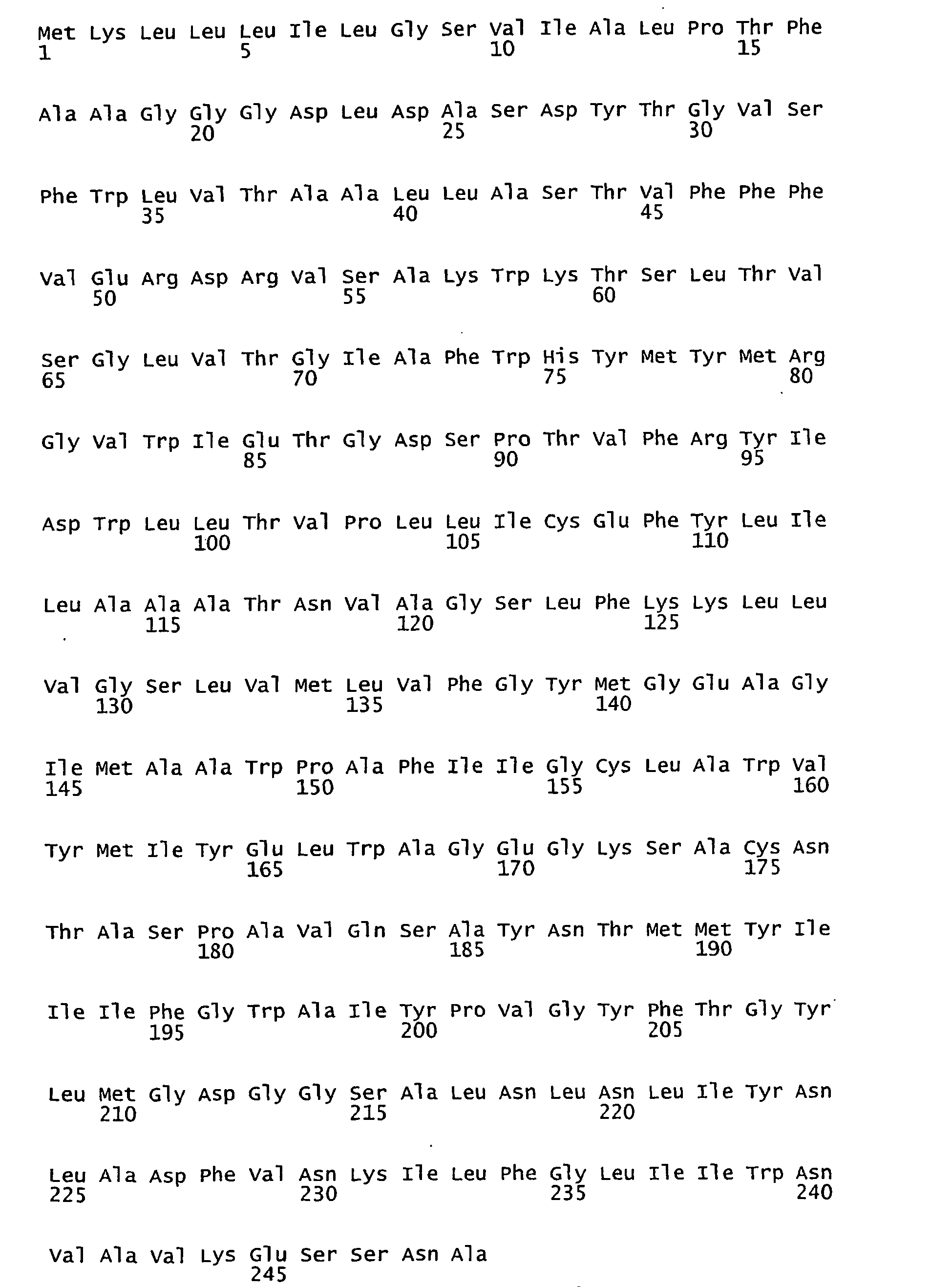
- <210> 4
<211> 750
<212> DNA
<213> Marine eubacteria - <400> 4

- <210> 5
<211> 252
<212> PRT
<213> Marine eubacteria - <400> 5


- <210> 6
<211> 252
<212> PRT
<213> Marine eubacteria - <400> 6


- <210> 7
<211> 252
<212> PRT
<213> Marine eubacteria - <400> 7


- <210> 8
<211> 757
<212> DNA
<213> Marine eubacteria - <400> 8

- <210> 9
<211> 251
<212> PRT
<213> Marine eubacteria - <400> 9

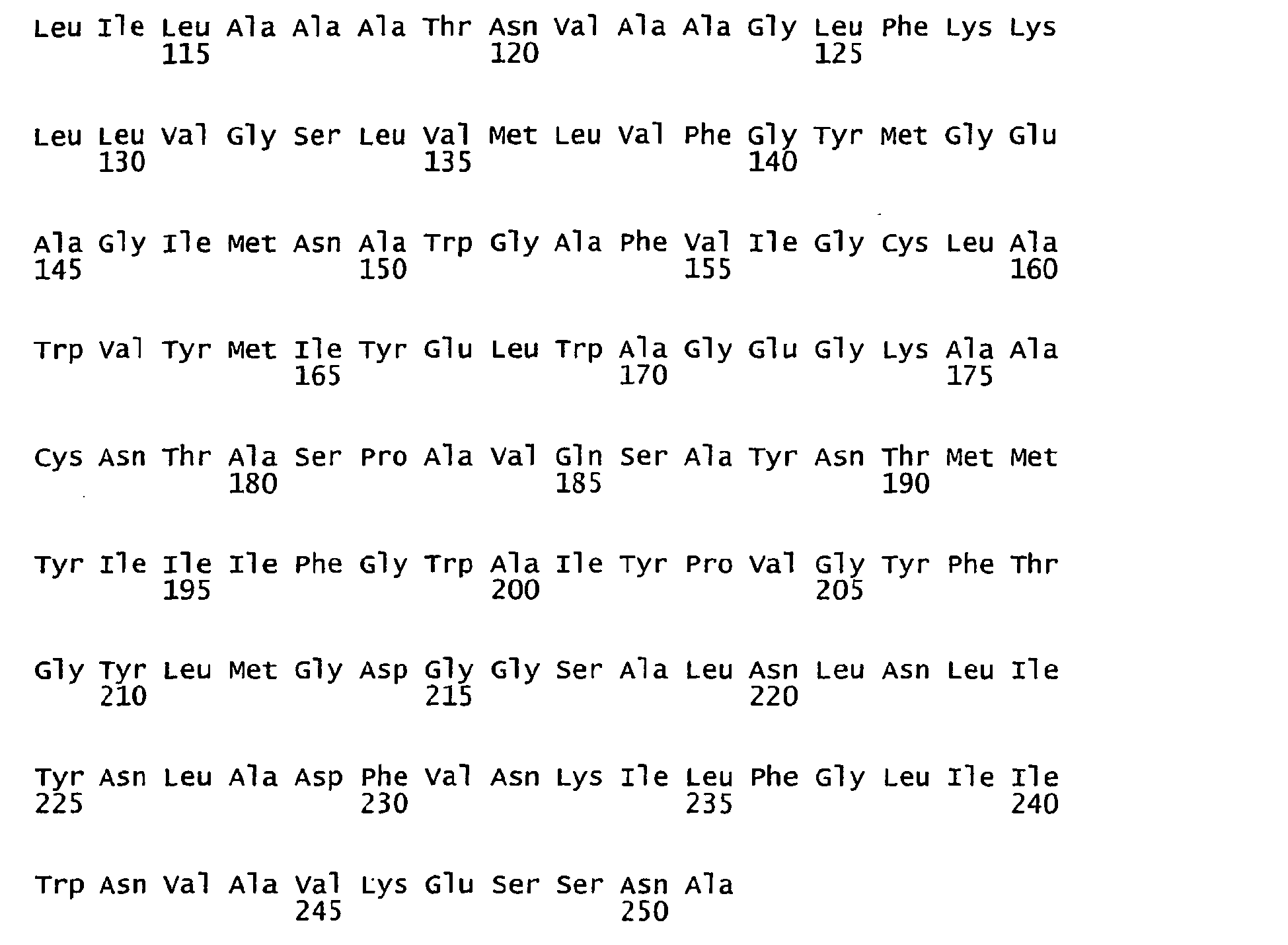
- <210> 10
<211> 753
<212> DNA
<213> Marine eubacteria - <400> 10


- <210> 11
<211> 252
<212> PRT
<213> Marine eubacteria - <400> 11

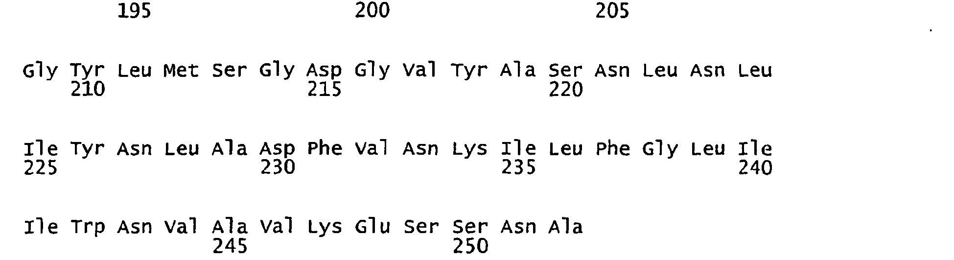
- <210> 12
<211> 757
<212> DNA
<213> Marine eubacteria - <400> 12

- <210> 13
<211> 252
<212> PRT
<213> Marine eubacteria - <400> 13


- <210> 14
<211> 756
<212> DNA
<213> Marine eubacteria - <400> 14


- <210> 15
<211> 252
<212> PRT
<213> Marine eubacteria - <400> 15
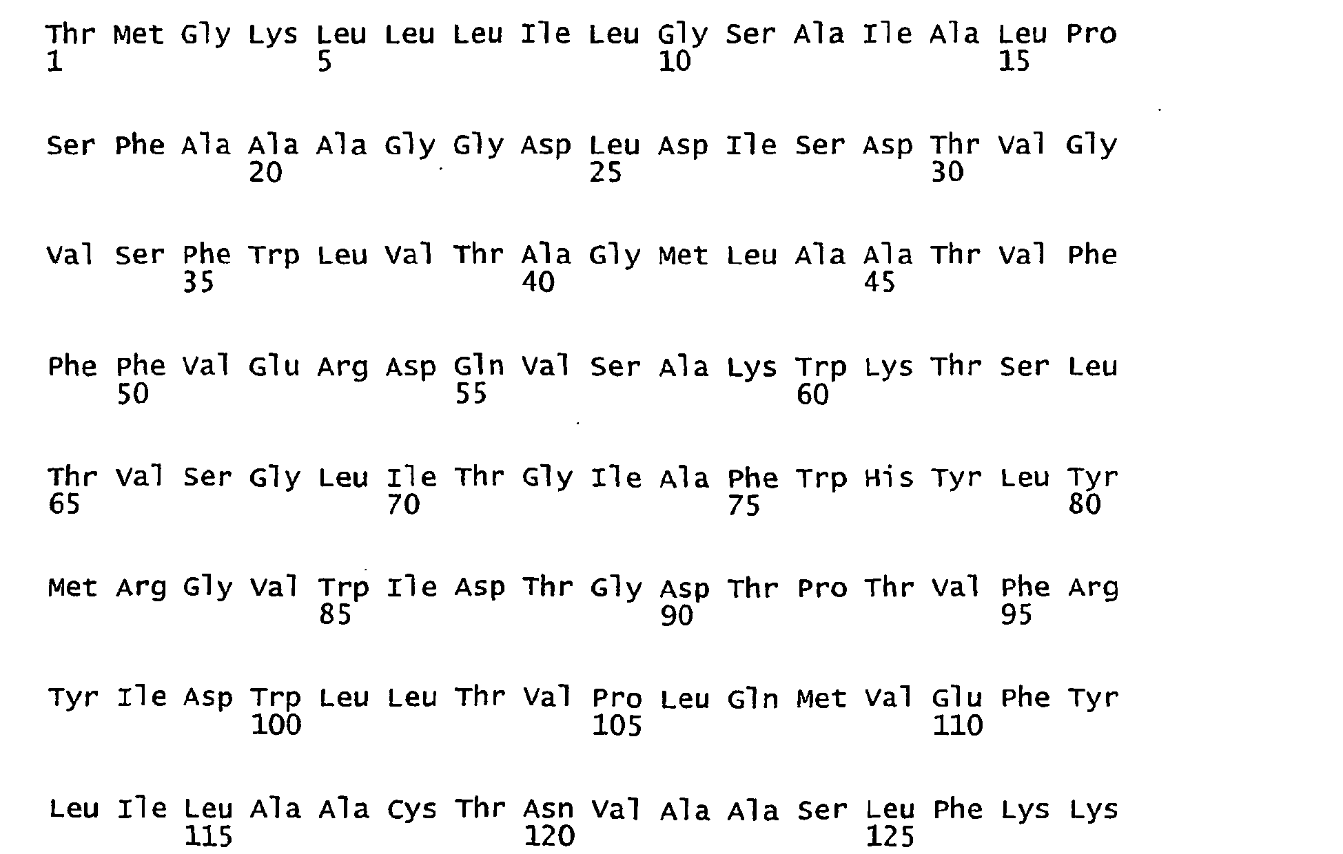

- <210> 16
<211> 756
<212> DNA
<213> Marine eubacteria - <400> 16
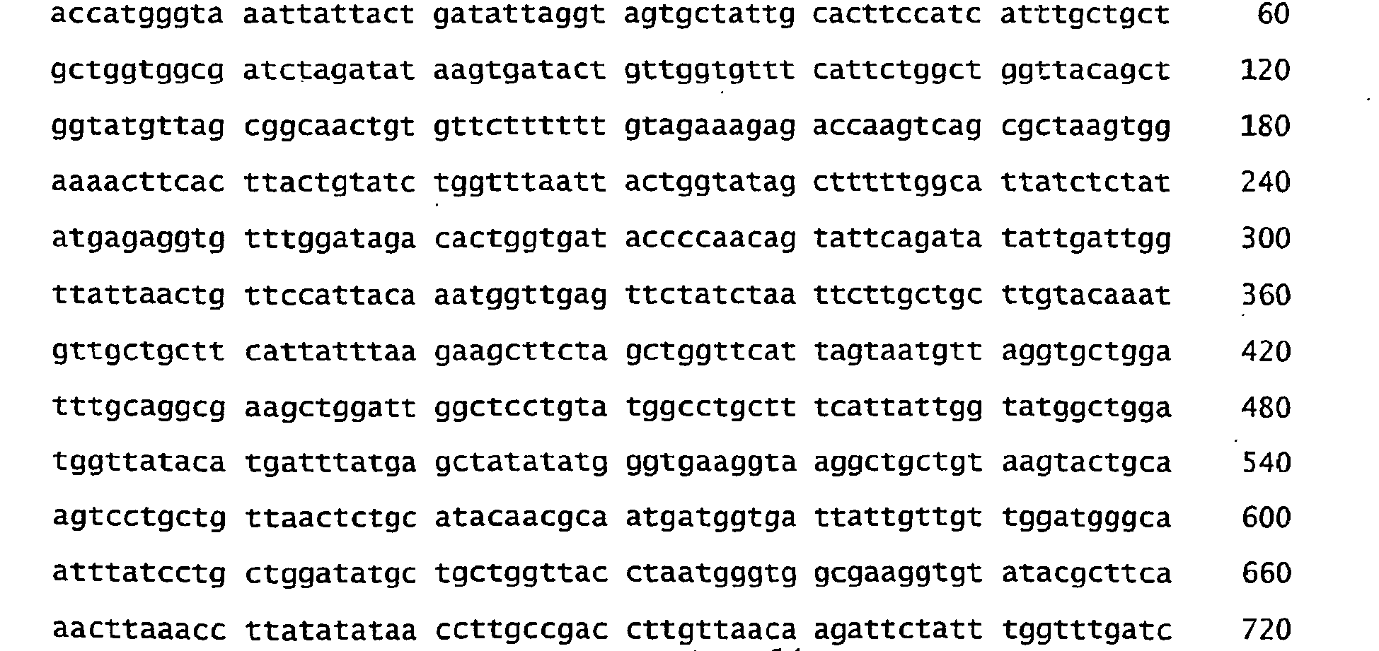

- <210> 17
<211> 251
<212> PRT
<213> Marine eubacteria - <400> 17


- <210> 18
<211> 251
<212> PRT
<213> Marine eubacteria - <400> 18

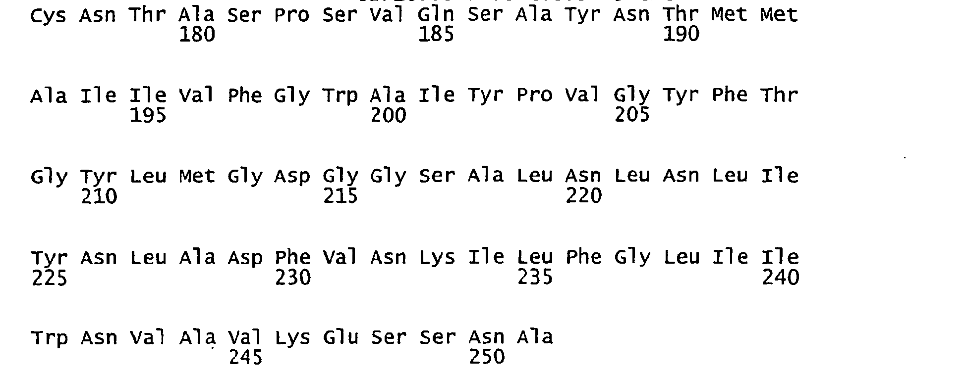
- <210> 19
<211> 753
<212> DNA
<213> Marine eubacteria - <400> 19

- <210> 20
<211> 753
<212> DNA
<213> Marine eubacteria - <400> 20


- <210> 21
<211> 251
<212> PRT
<213> Marine eubacteria - <400> 21

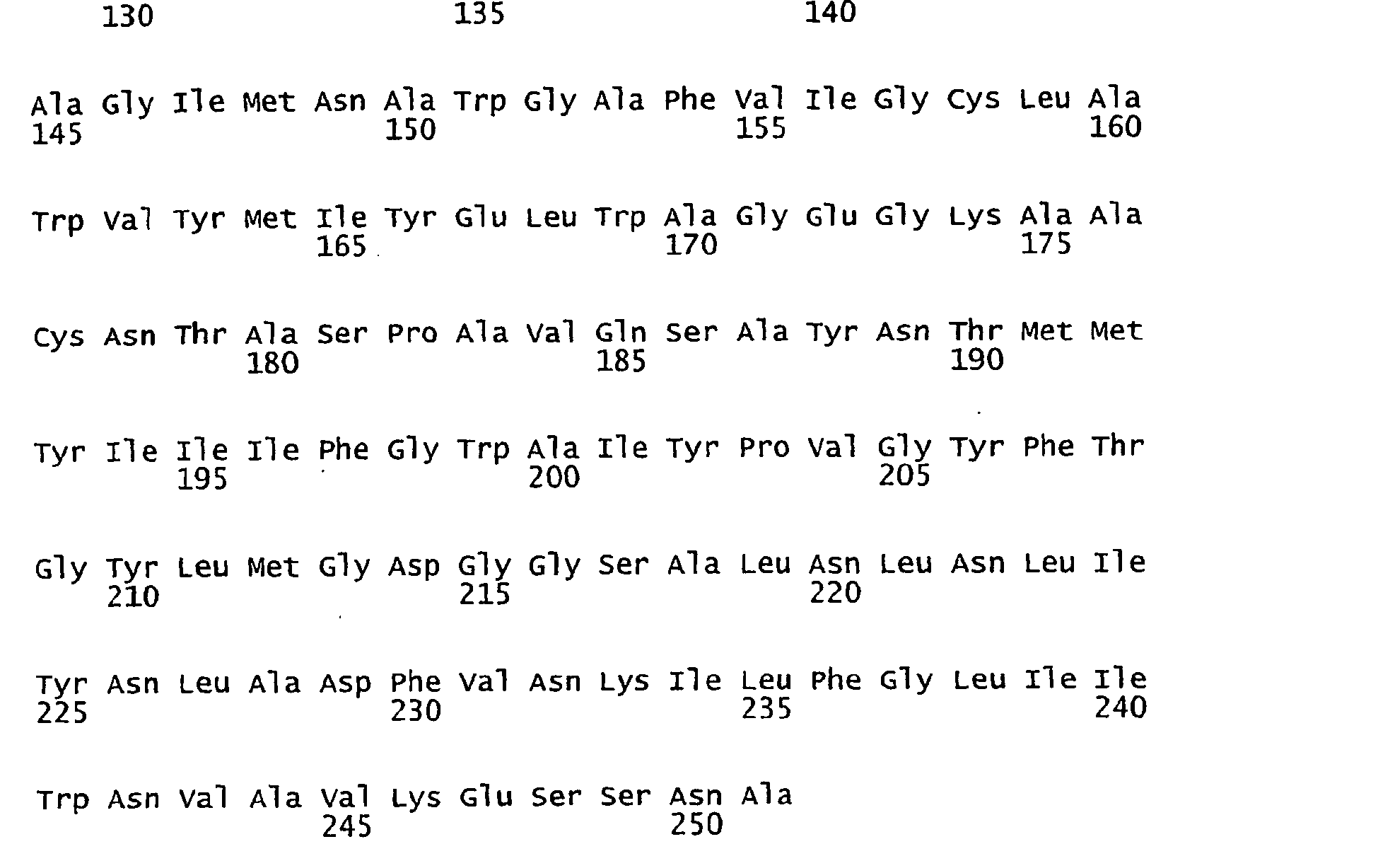
- <210> 22
<211> 753
<212> DNA
<213> Marine eubacteria - <400> 22

- <210> 23
<211> 251
<212> PRT
<213> Marine eubacteria - <400> 23


- <210> 24
<211> 753
<212> DNA
<213> Marine eubacteria - <400> 24

- <210> 25
<211> 249
<212> PRT
<213> Marine eubacteria - <400> 25
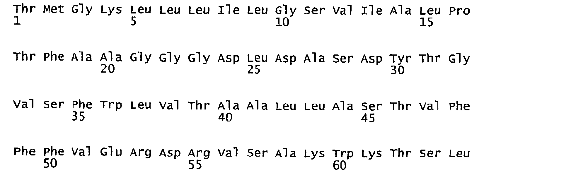
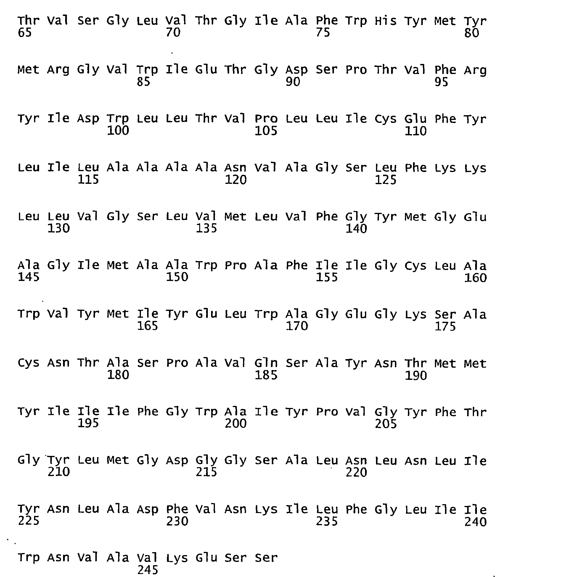
- <210> 26
<211> 748
<212> DNA
<213> Marine eubacteria - <400> 26


- <210> 27
<211> 251
<212> PRT
<213> Marine eubacteria - <400> 27


- <210> 28
<211> 753
<212> DNA
<213> Marine eubacteria - <400> 28

- <210> 29
<211> 249
<212> PRT
<213> Marine eubacteria - <400> 29


- <210> 30
<211> 748
<212> DNA
<213> Marine eubacteria - <400> 30

- <210> 31
<211> 251
<212> PRT
<213> Marine eubacteria - <400> 31

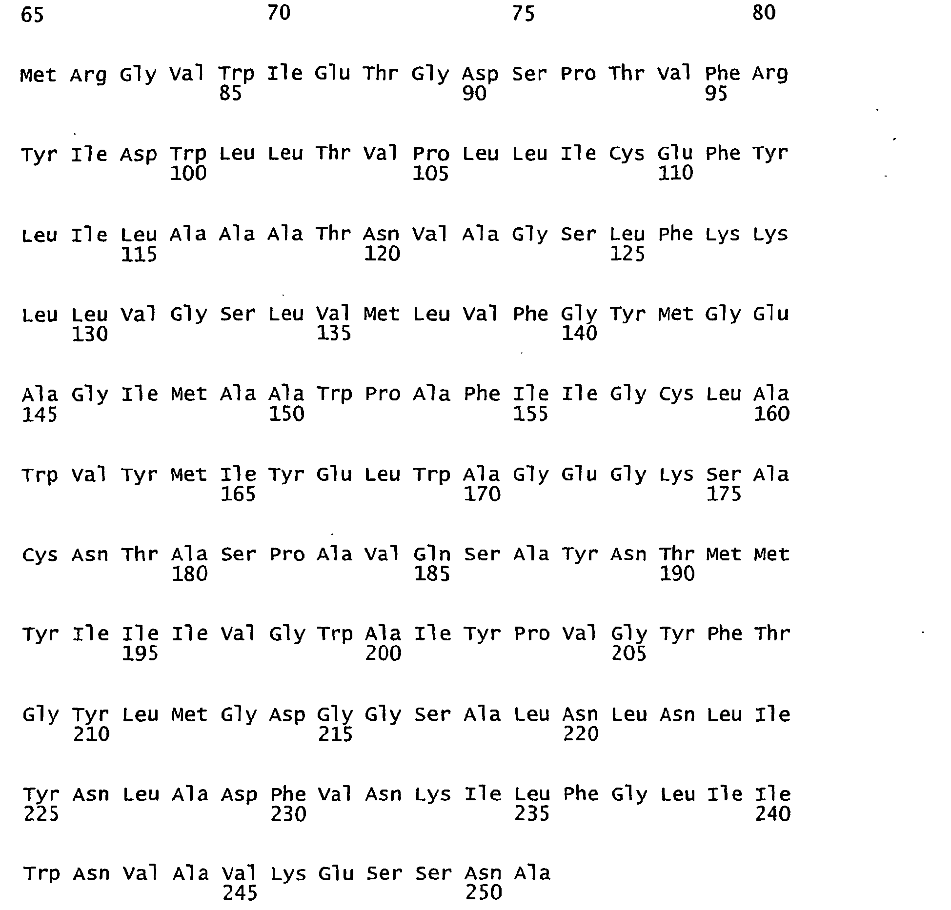
- <210> 32
<211> 753
<212> DNA
<213> Marine eubacteria - <400> 32

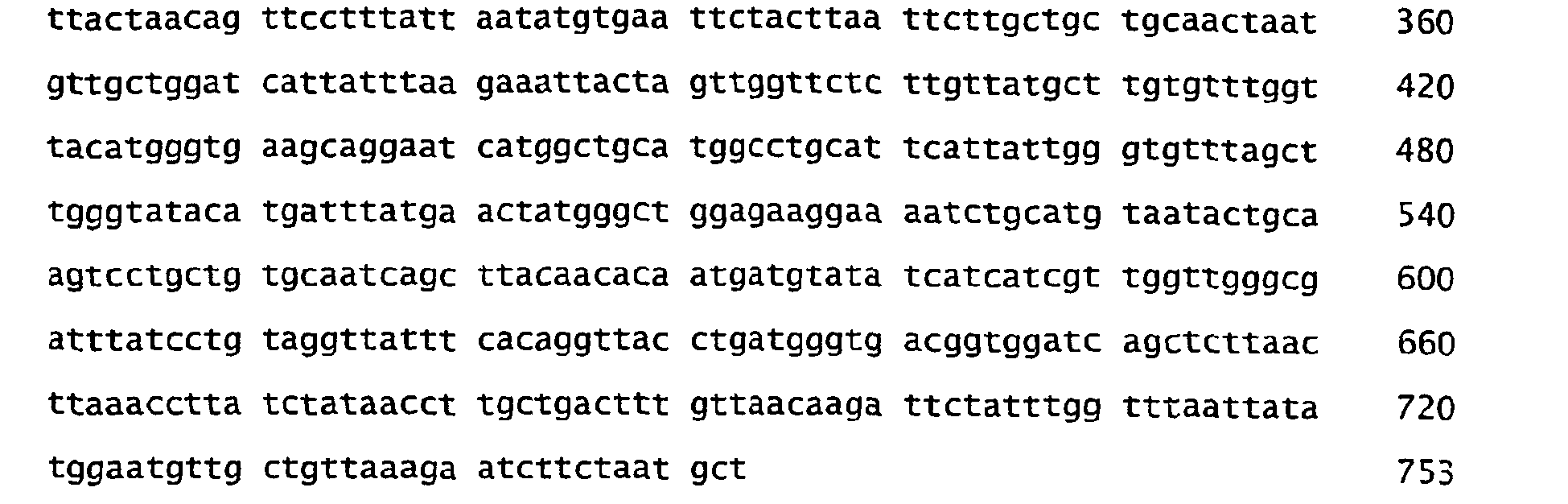
- <210> 33
<211> 251
<212> PRT
<213> Marine eubacteria - <400> 33


- <210> 34
<211> 753
<212> DNA
<213> Marine eubacteria - <400> 34

- <210> 35
<211> 251
<212> PRT
<213> Marine eubacteria - <400> 35
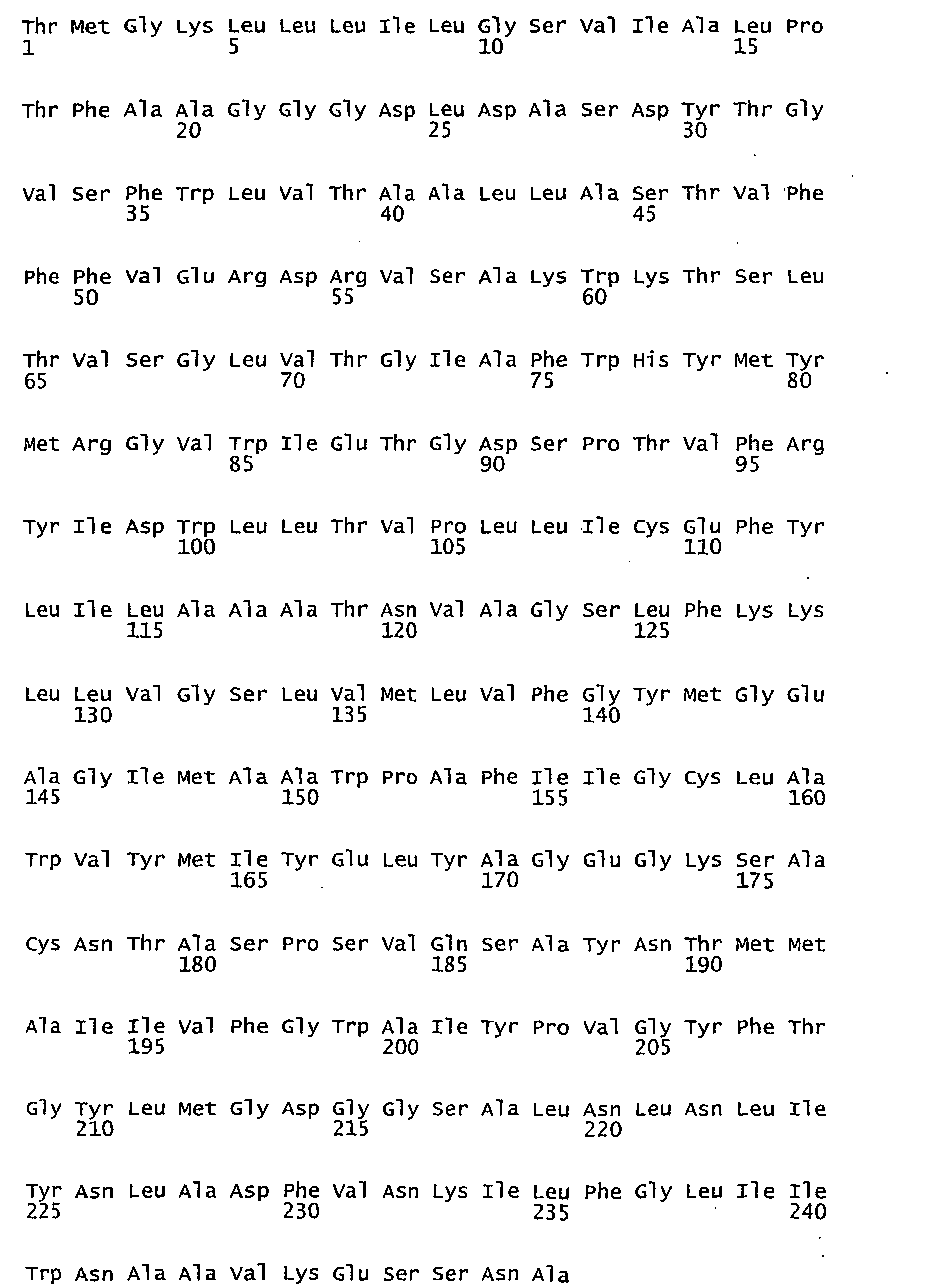

- <210> 36
<211> 753
<212> DNA
<213> Marine eubacteria - <400> 36

- <210> 37
<211> 251
<212> PRT
<213> Marine eubacteria - <400> 37


- <210> 38
<211> 753
<212> DNA
<213> Marine eubacteria - <400> 38

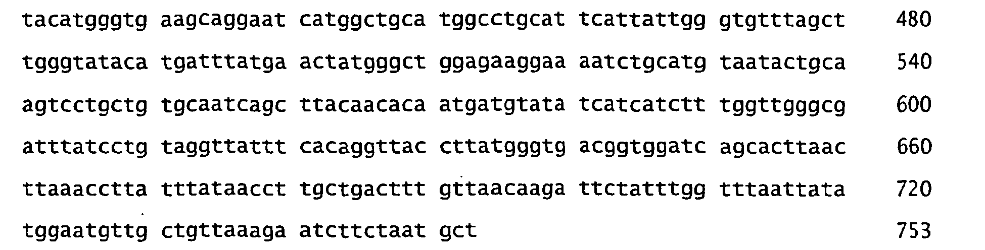
- <210> 39
<211> 251
<212> PRT
<213> Marine eubacteria - <400> 39


- <210> 40
<211> 753
<212> DNA
<213> Marine eubacteria - <400> 40
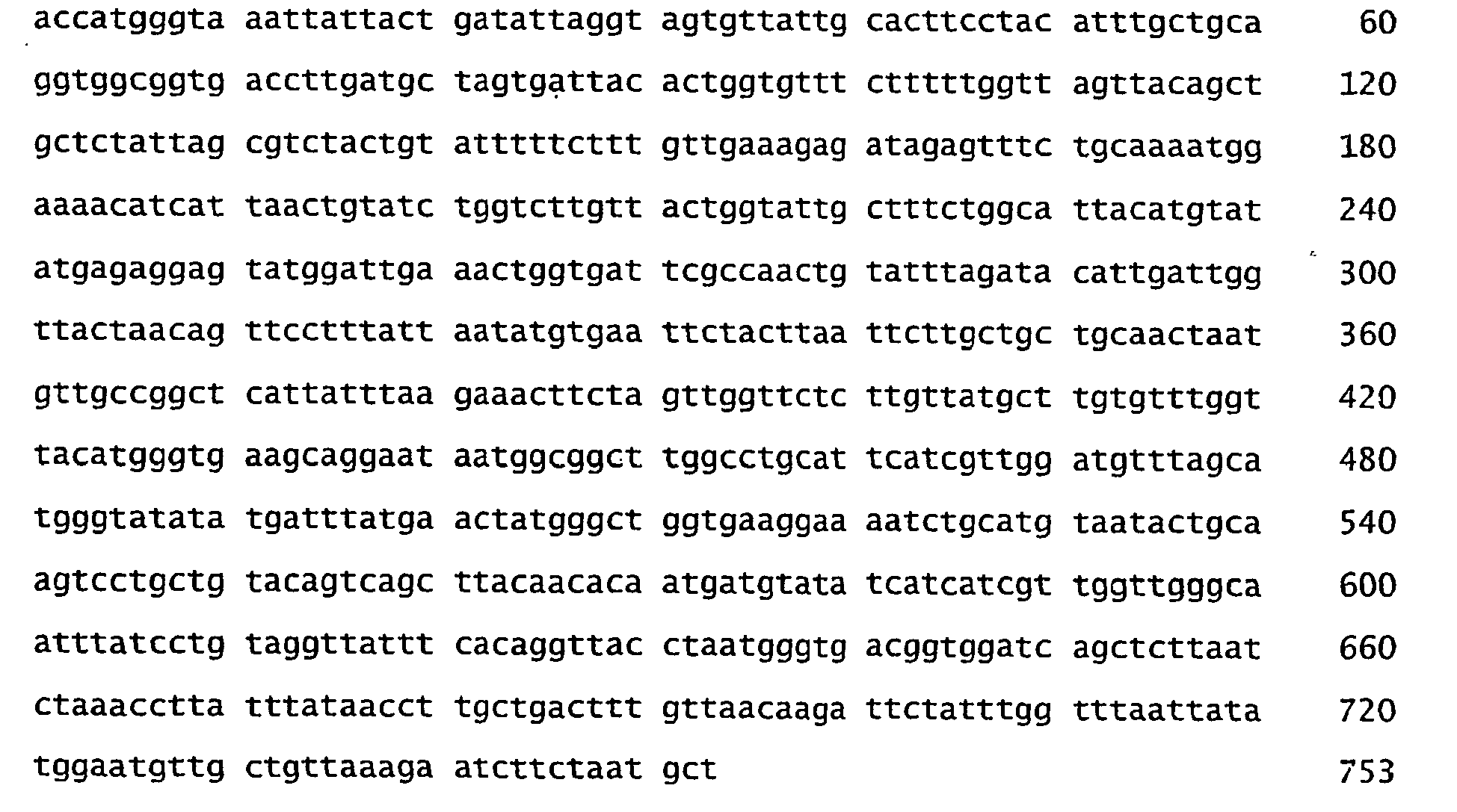
- <210> 41
<211> 252
<212> PRT
<213> Marine eubacteria - <400> 41


- <210> 42
<211> 756
<212> DNA
<213> Marine eubacteria - <400> 42

- <210> 43
<211> 252
<212> PRT
<213> Marine eubacteria - <400> 43

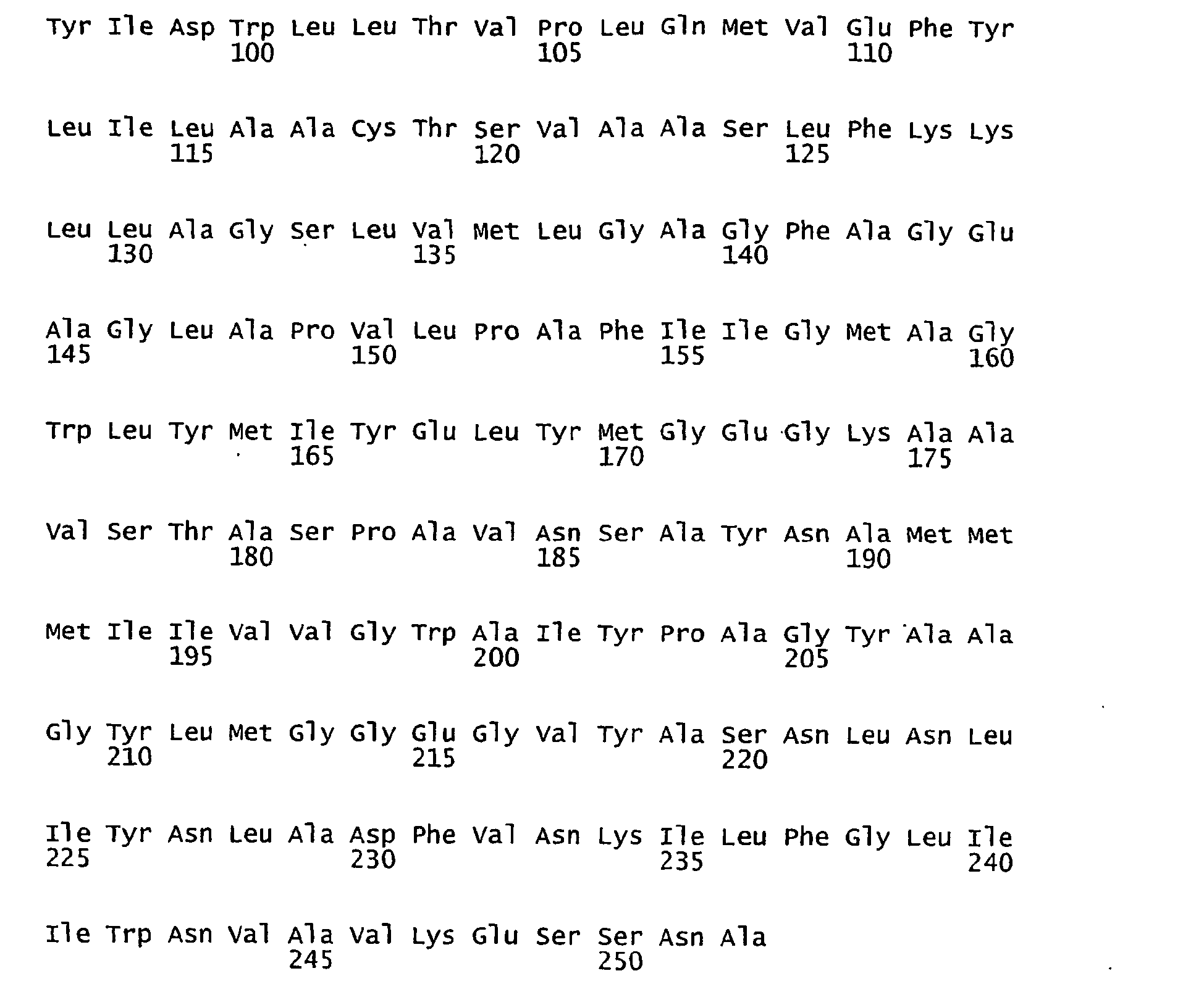
- <210> 44
<211> 756
<212> DNA
<213> Marine eubacteria - <400> 44

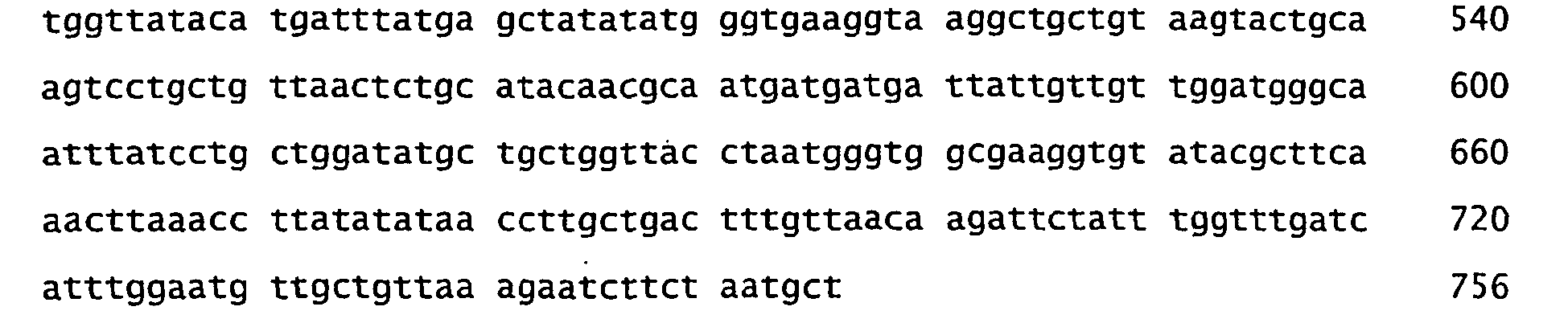
- <210> 45
<211> 252
<212> PRT
<213> Marine eubacteria - <400> 45


- <210> 46
<211> 756
<212> DNA
<213> Marine eubacteria - <400> 46

- <210> 47
<211> 252
<212> PRT
<213> Marine eubacteria - <400> 47


- <210> 48
<211> 756
<212> DNA
<213> Marine eubacteria - <400> 48

- <210> 49
<211> 252
<212> PRT
<213> Marine eubacteria - <400> 49


- <210> 50
<211> 756
<212> DNA
<213> Marine eubacteria - <400> 50

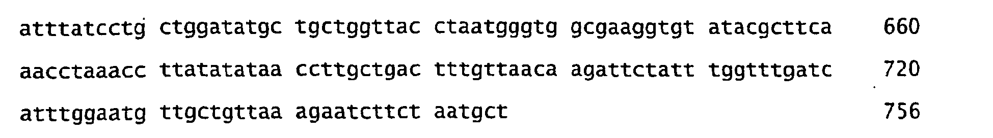
- <210> 51
<211> 252
<212> PRT
<213> Marine eubacteria - <400> 51


- <210> 52
<211> 756
<212> DNA
<213> Marine eubacteria - <400> 52

- <210> 53
<211> 252
<212> PRT
<213> Marine eubacteria - <400> 53


- <210> 54
<211> 756
<212> DNA
<213> Marine eubacteria - <400> 54

- <210> 55
<211> 252
<212> PRT
<213> Marine eubacteria - <400> 55

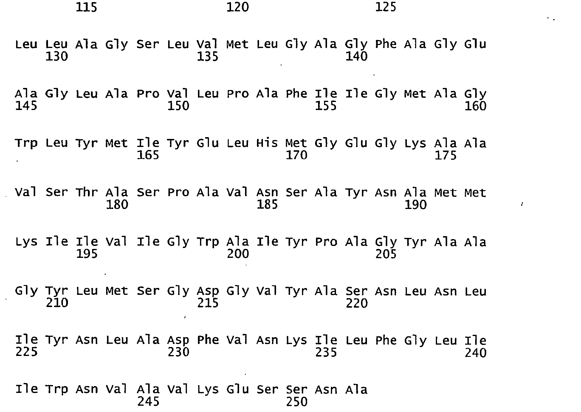
- <210> 56
<211> 756
<212> DNA
<213> Marine eubacteria - <400> 56
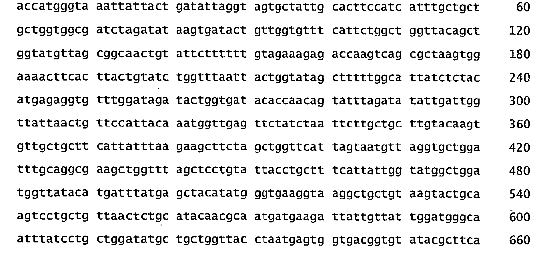

- <210> 57
<211> 252
<212> PRT
<213> Marine eubacteria - <400> 57


- <210> 58
<211> 756
<212> DNA
<213> Marine eubacteria - <400> 58

- <210> 59
<211> 250
<212> PRT
<213> Marine eubacteria - <400> 59


- <210> 60
<211> 751
<212> DNA
<213> Marine eubacteria - <400> 60


- <210> 61
<211> 250
<212> PRT
<213> Marine eubacteria - <400> 61


- <210> 62
<211> 751
<212> DNA
<213> Marine eubacteria - <400> 62

- <210> 63
<211> 250
<212> PRT
<213> Marine eubacteria - <400> 63


- <210> 64
<211> 751
<212> DNA
<213> Marine eubacteria - <400> 64

- <210> 65
<211> 250
<212> PRT
<213> Marine eubacteria - <400> 65


- <210> 66
<211> 751
<212> DNA
<213> Marine eubacteria - <400> 66


- <210> 67
<211> 250
<212> PRT
<213> Marine eubacteria - <400> 67


- <210> 68
<211> 751
<212> DNA
<213> Marine eubacteria - <400> 68

- <210> 69
<211> 250
<212> PRT
<213> Marine eubacteria - <400> 69
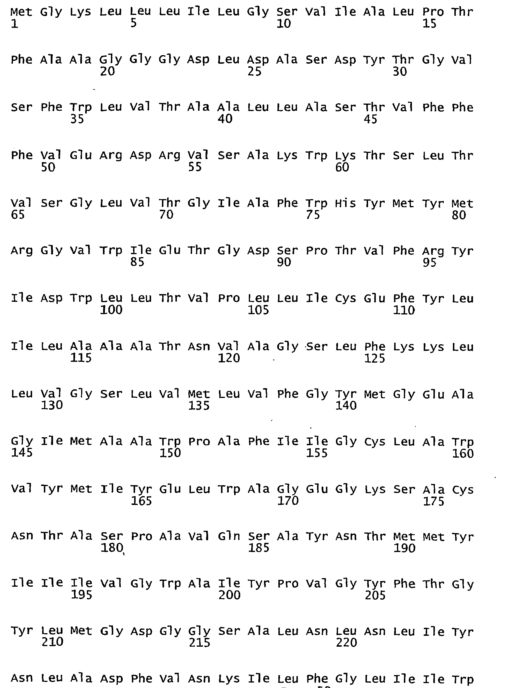

- <210> 70
<211> 751
<212> DNA
<213> Marine eubacteria - <400> 70

- <210> 71
<211> 250
<212> PRT
<213> Marine eubacteria - <400> 71


- <210> 72
<211> 751
<212> DNA
<213> Marine eubacteria - <400> 72


- <210> 73
<211> 250
<212> PRT
<213> Marine eubacteria - <400> 73


- <210> 74
<211> 751
<212> DNA
<213> Marine eubacteria - <400> 74
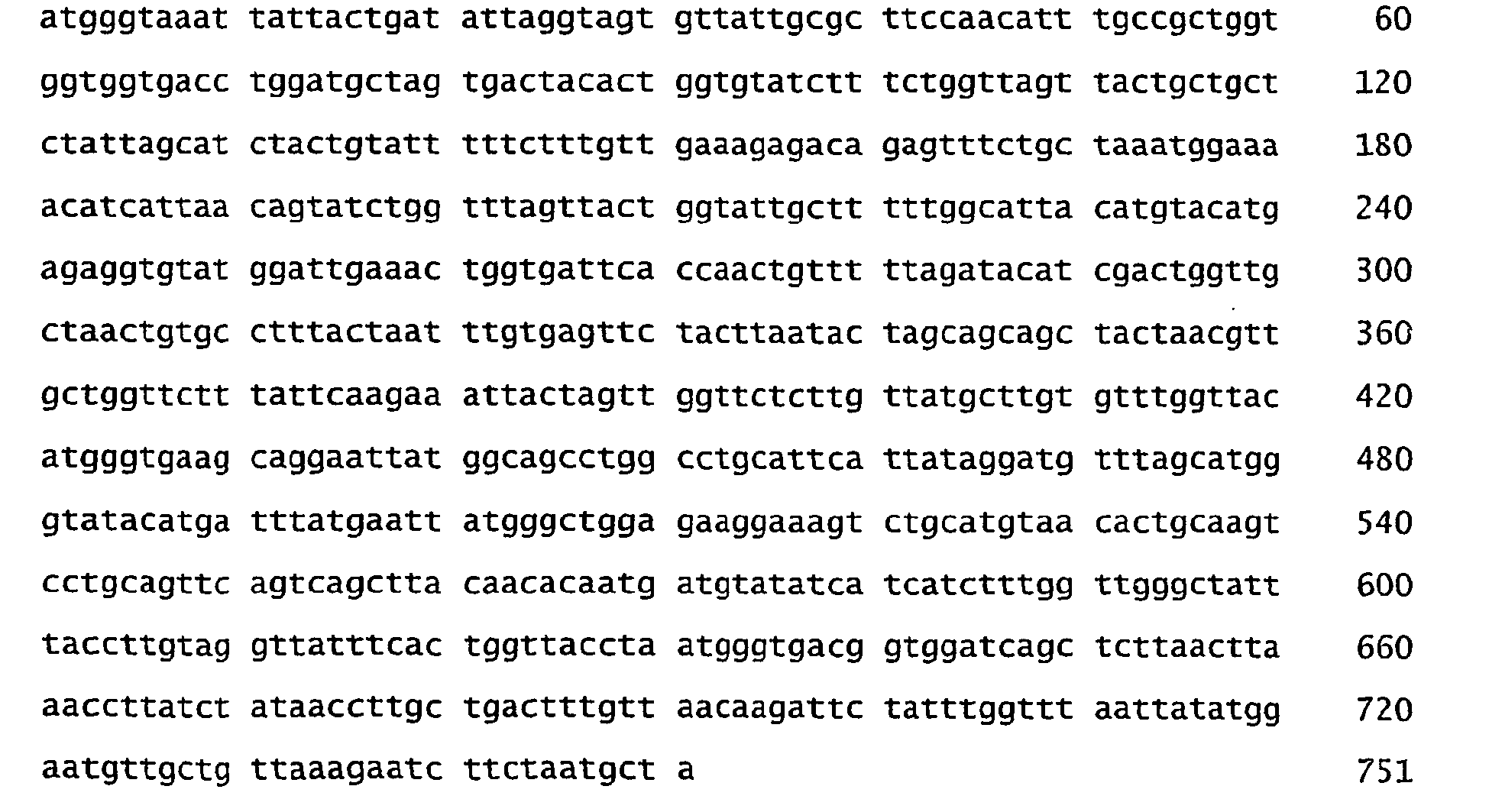
- <210> 75
<211> 250
<212> PRT
<213> Marine eubacteria - <400> 75
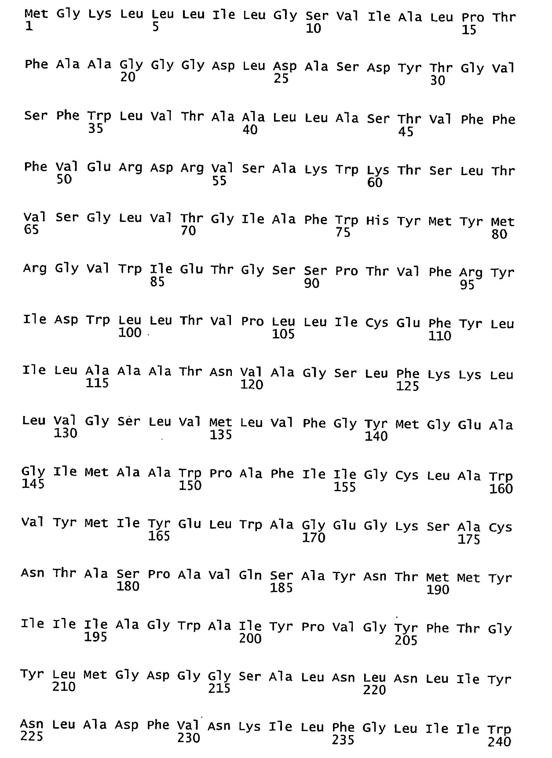

- <210> 76
<211> 751
<212> DNA
<213> Marine eubacteria - <400> 76

- <210> 77
<211> 250
<212> PRT
<213> Marine eubacteria - <400> 77


- <210> 78
<211> 751
<212> DNA
<213> Marine eubacteria - <400> 78


- <210> 79
<211> 250
<212> PRT
<213> Marine eubacteria - <400> 79


- <210> 80
<211> 750
<212> DNA
<213> Marine eubacteria - <400> 80

- <210> 81
<211> 250
<212> PRT
<213> Marine eubacteria - <400> 81

- <210> 82
<211> 750
<212> DNA
<213> Marine eubacteria - <400> 82
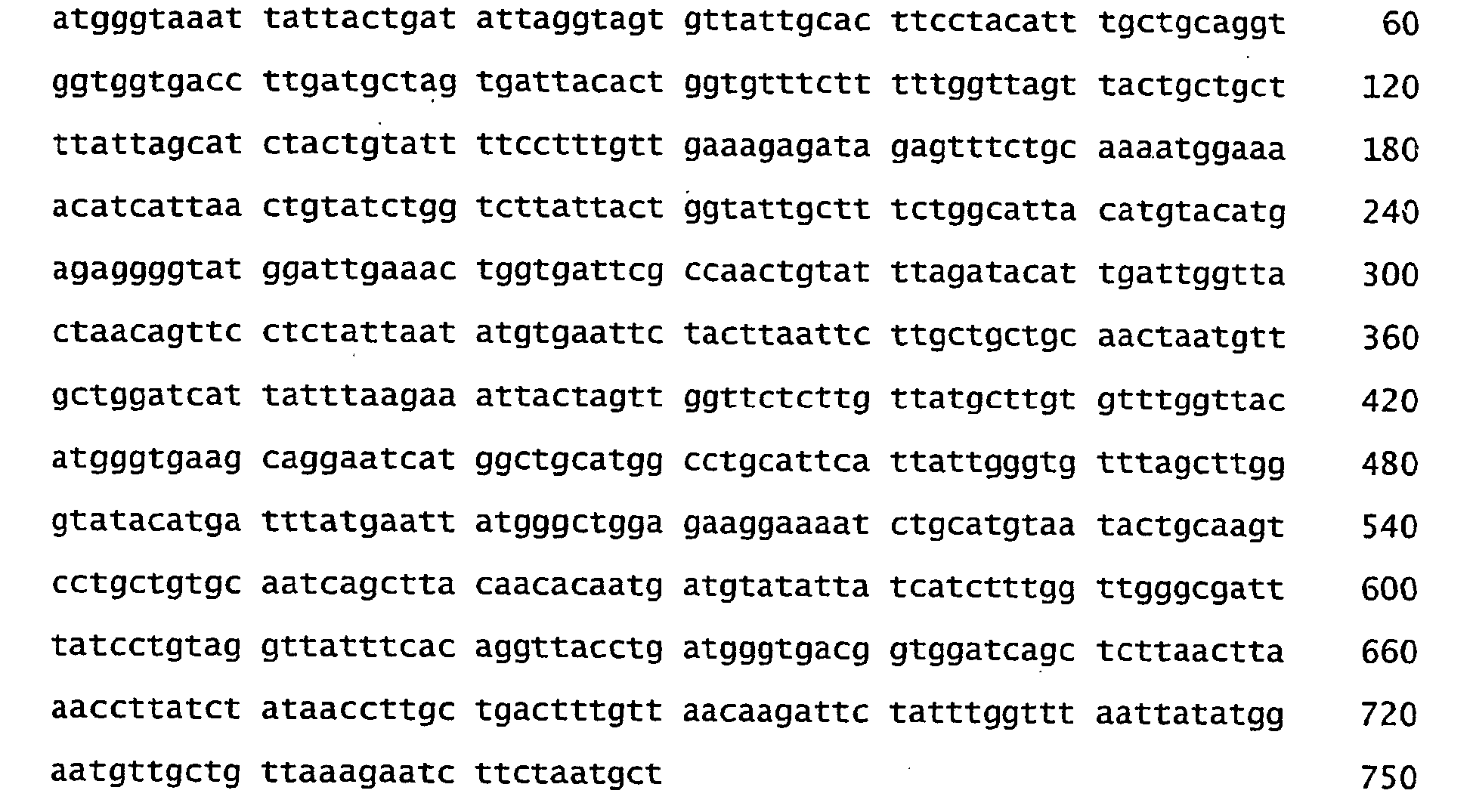
- <210> 83
<211> 250
<212> PRT
<213> Marine eubacteria - <400> 83


- <210> 84
<211> 750
<212> DNA
<213> Marine eubacteria - <400> 84
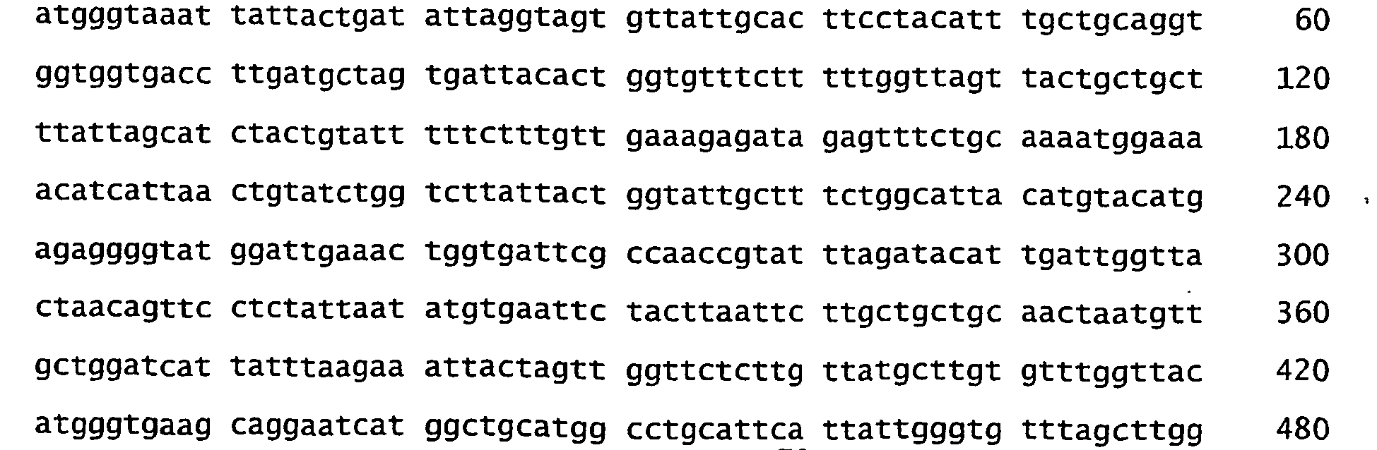

- <210> 85
<211> 250
<212> PRT
<213> Marine eubacteria - <400> 85

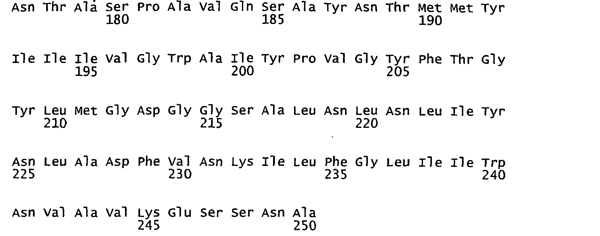
- <210> 86
<211> 751
<212> DNA
<213> Marine eubacteria - <400> 86
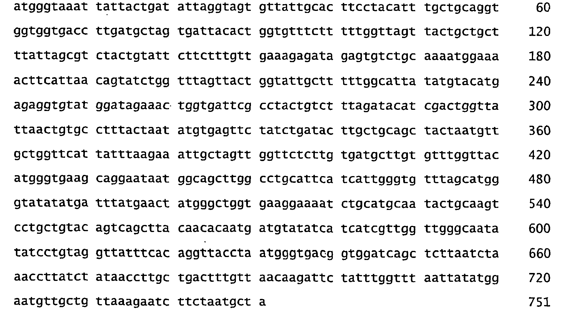
- <210> 87
<211> 250
<212> PRT
<213> Marine eubacteria - <400> 87


- <210> 88
<211> 751
<212> DNA
<213> Marine eubacteria - <400> 88

- <210> 89
<211> 250
<212> PRT
<213> Marine eubacteria - <400> 89


- <210> 90
<211> 751
<212> DNA
<213> Marine eubacteria - <400> 90

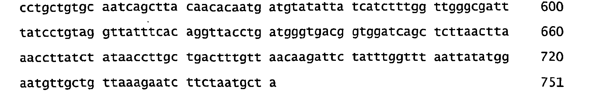
- <210> 91
<211> 250
<212> PRT
<213> Marine eubacteria - <400> 91

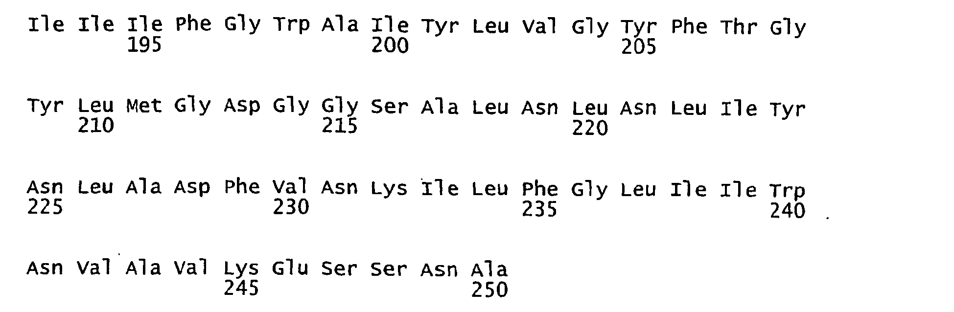
- <210> 92
<211> 751
<212> DNA
<213> Marine eubacteria - <400> 92

- <210> 93
<211> 250
<212> PRT
<213> Marine eubacteria - <400> 93


- <210> 94
<211> 751
<212> DNA
<213> Marine eubacteria - <400> 94

- <210> 95
<211> 250
<212> PRT
<213> Marine eubacteria - <400> 95

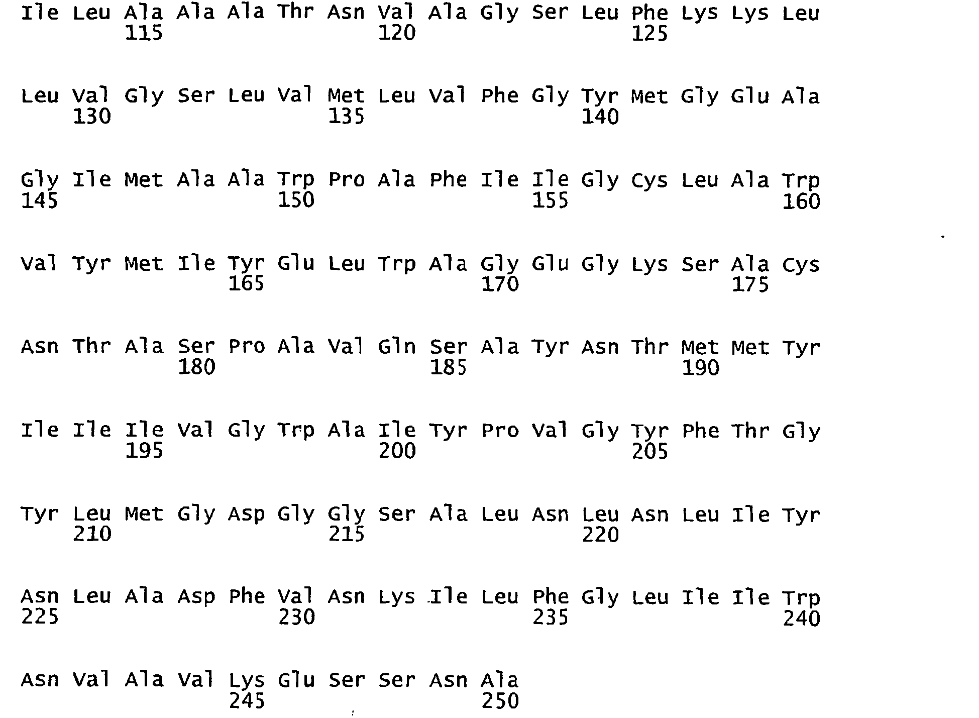
- <210> 96
<211> 751
<212> DNA
<213> Marine eubacteria - <400> 96


- <210> 97
<211> 250
<212> PRT
<213> Marine eubacteria - <400> 97


- <210> 98
<211> 751
<212> DNA
<213> Marine eubacteria - <400> 98
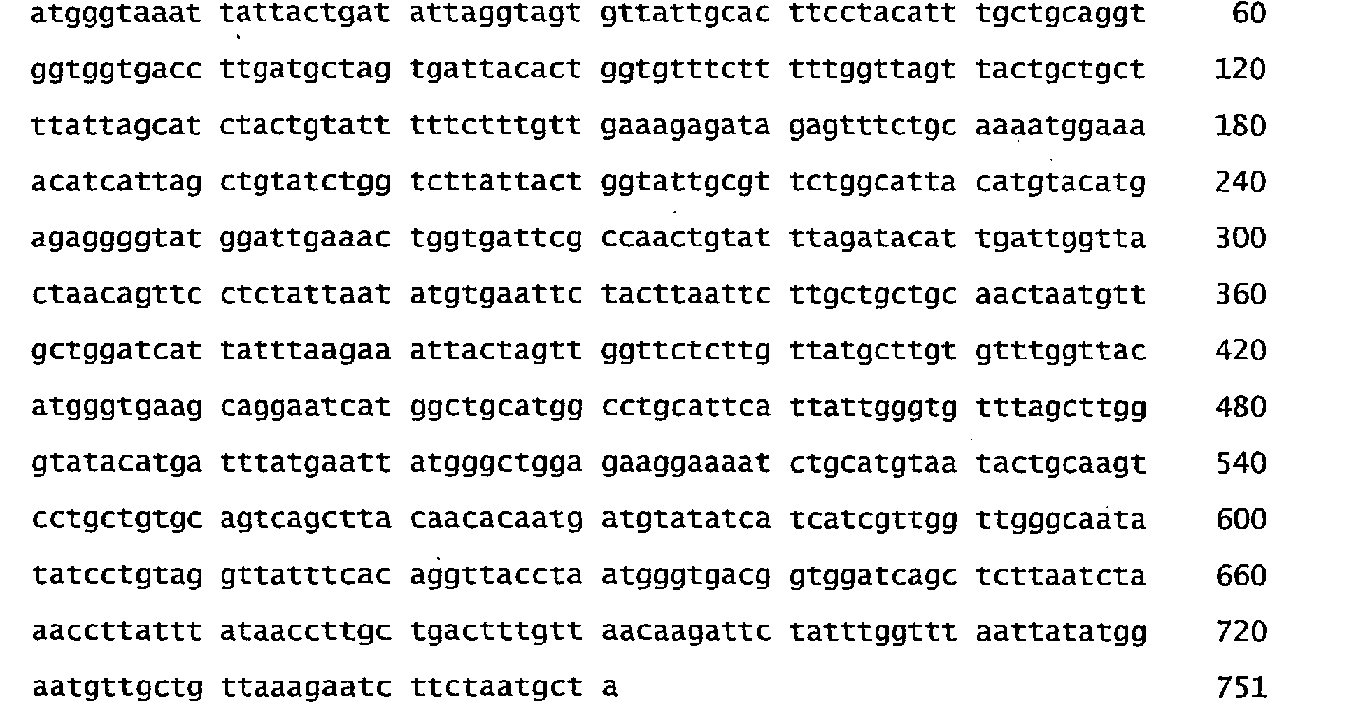
- <210> 99
<211> 250
<212> PRT
<213> Marine eubacteria - <400> 99


- <210> 100
<211> 751
<212> DNA
<213> Marine eubacteria - <400> 100


- <210> 101
<211> 250
<212> PRT
<213> Marine eubacteria - <400> 101

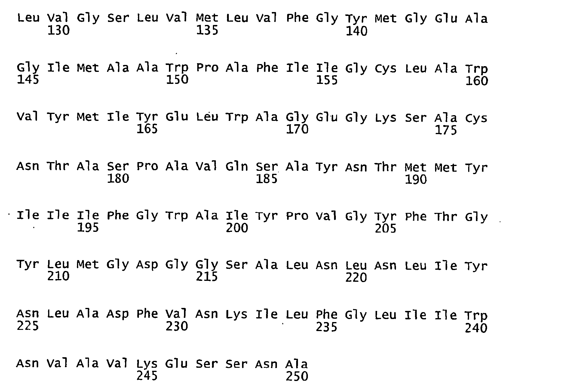
- <210> 102
<211> 751
<212> DNA
<213> Marine eubacteria - <400> 102


- <210> 103
<211> 250
<212> PRT
<213> Marine eubacteria - <400> 103


- <210> 104
<211> 751
<212> DNA
<213> Marine eubacteria - <400> 104

- <210> 105
<211> 249
<212> PRT
<213> Marine eubacteria - <400> 105


- <210> 106
<211> 748
<212> DNA
<213> Marine eubacteria - <400> 106

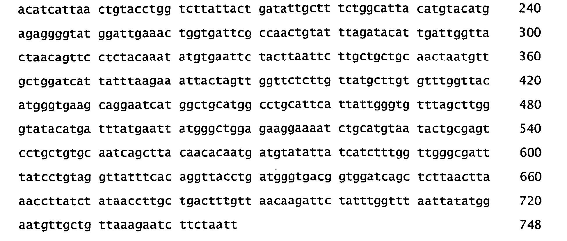
- <210> 107
<211> 250
<212> PRT
<213> Marine eubacteria - <400> 107


- <210> 108
<211> 751
<212> DNA
<213> Marine eubacteria - <400> 108

- <210> 109
<211> 250
<212> PRT
<213> Marine eubacteria - <400> 109
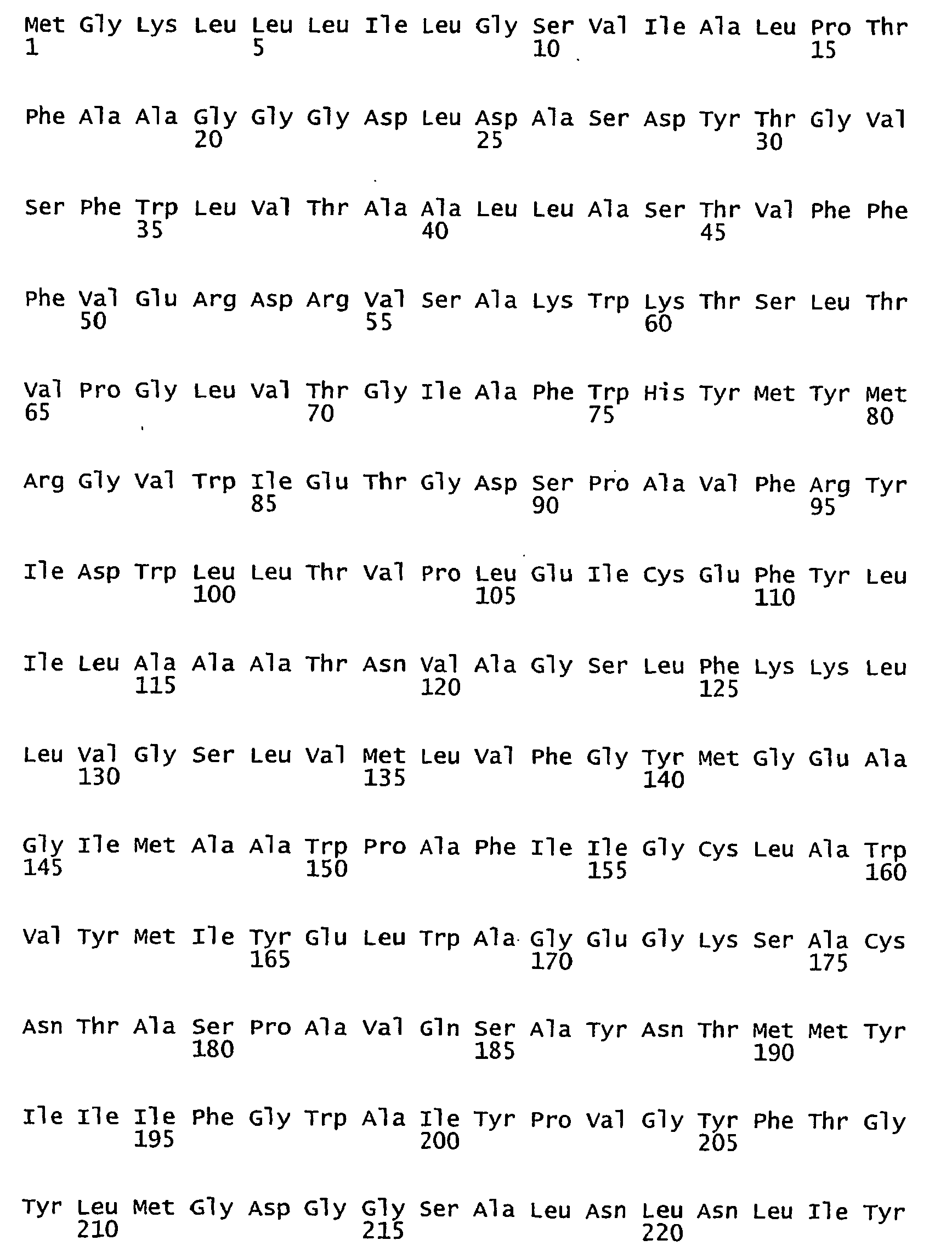

- <210> 110
<211> 751
<212> DNA
<213> Marine eubacteria - <400> 110

- <210> 111
<211> 250
<212> PRT
<213> Marine eubacteria - <400> 111


- <210> 112
<211> 751
<212> DNA
<213> Marine eubacteria - <400> 112


- <210> 113
<211> 250
<212> PRT
<213> Marine eubacteria - <400> 113

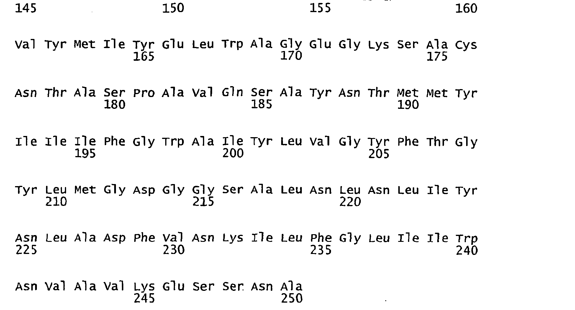
- <210> 114
<211> 751
<212> DNA
<213> Marine eubacteria - <400> 114

- <210> 115
<211> 250
<212> PRT
<213> Marine eubacteria - <400> 115


- <210> 116
<211> 751
<212> DNA
<213> Marine eubacteria - <400> 116

- <210> 117
<211> 250
<212> PRT
<213> Marine eubacteria - <400> 117

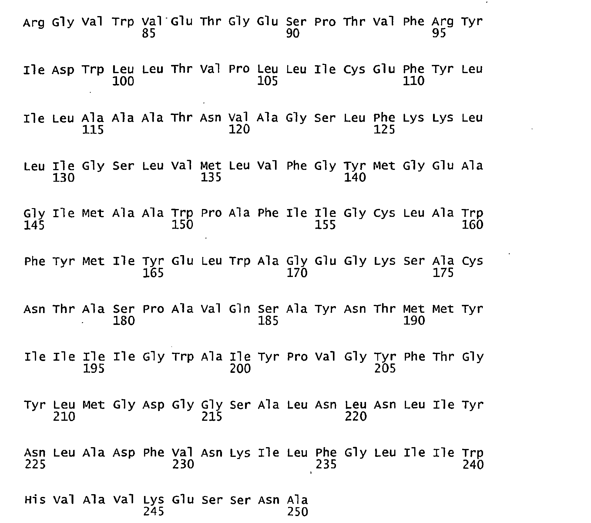
- <210> 118
<211> 751
<212> DNA
<213> Marine eubacteria - <400> 118


- <210> 119
<211> 250
<212> PRT
<213> Marine eubacteria - <400> 119
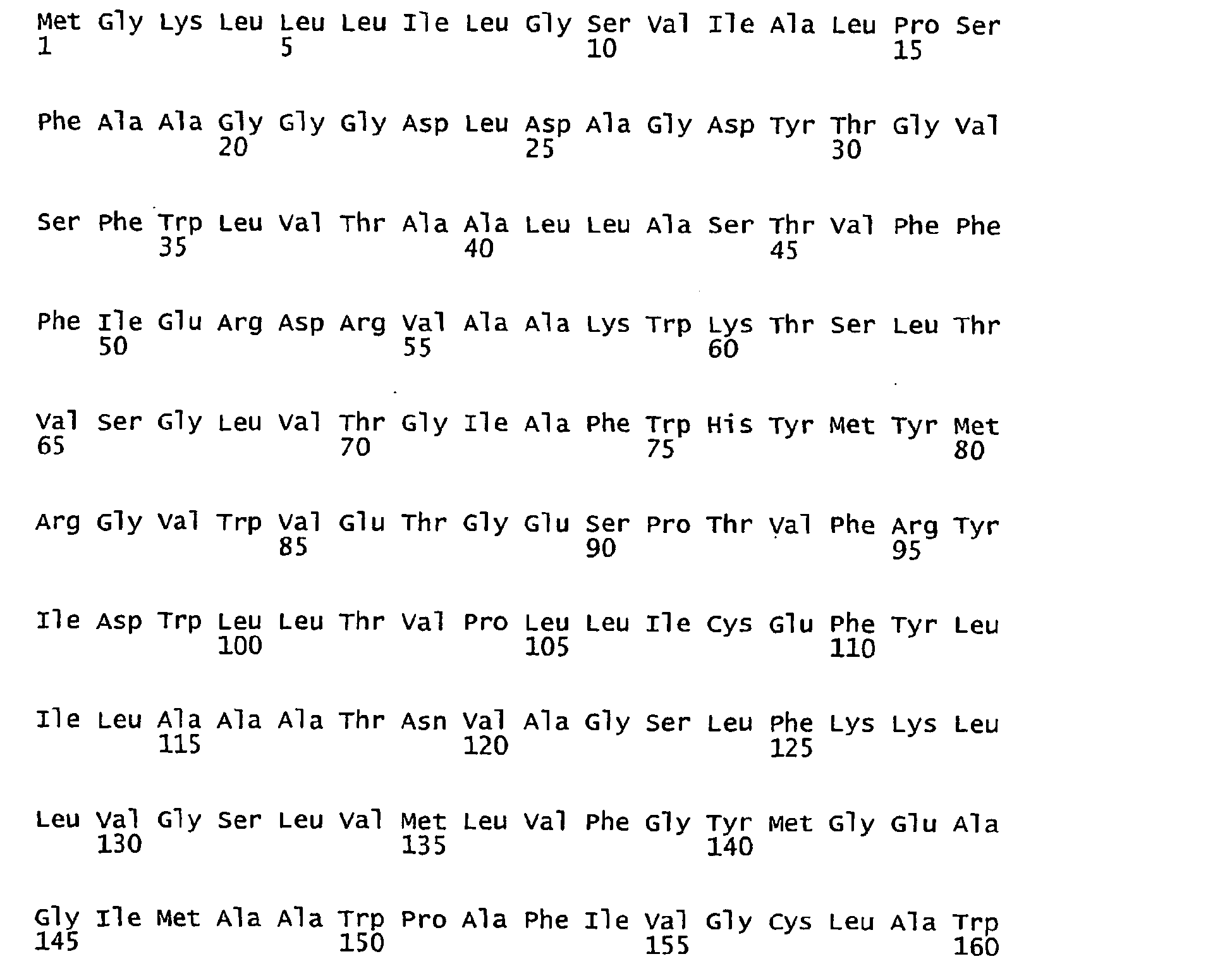

- <210> 120
<211> 751
<212> DNA
<213> Marine eubacteria - <400> 120

- <210> 121
<211> 250
<212> PRT
<213> Marine eubacteria - <400> 121

- <210> 122
<211> 751
<212> DNA
<213> Marine eubacteria - <400> 122

- <210> 123
<211> 250
<212> PRT
<213> Marine eubacteria - <400> 123

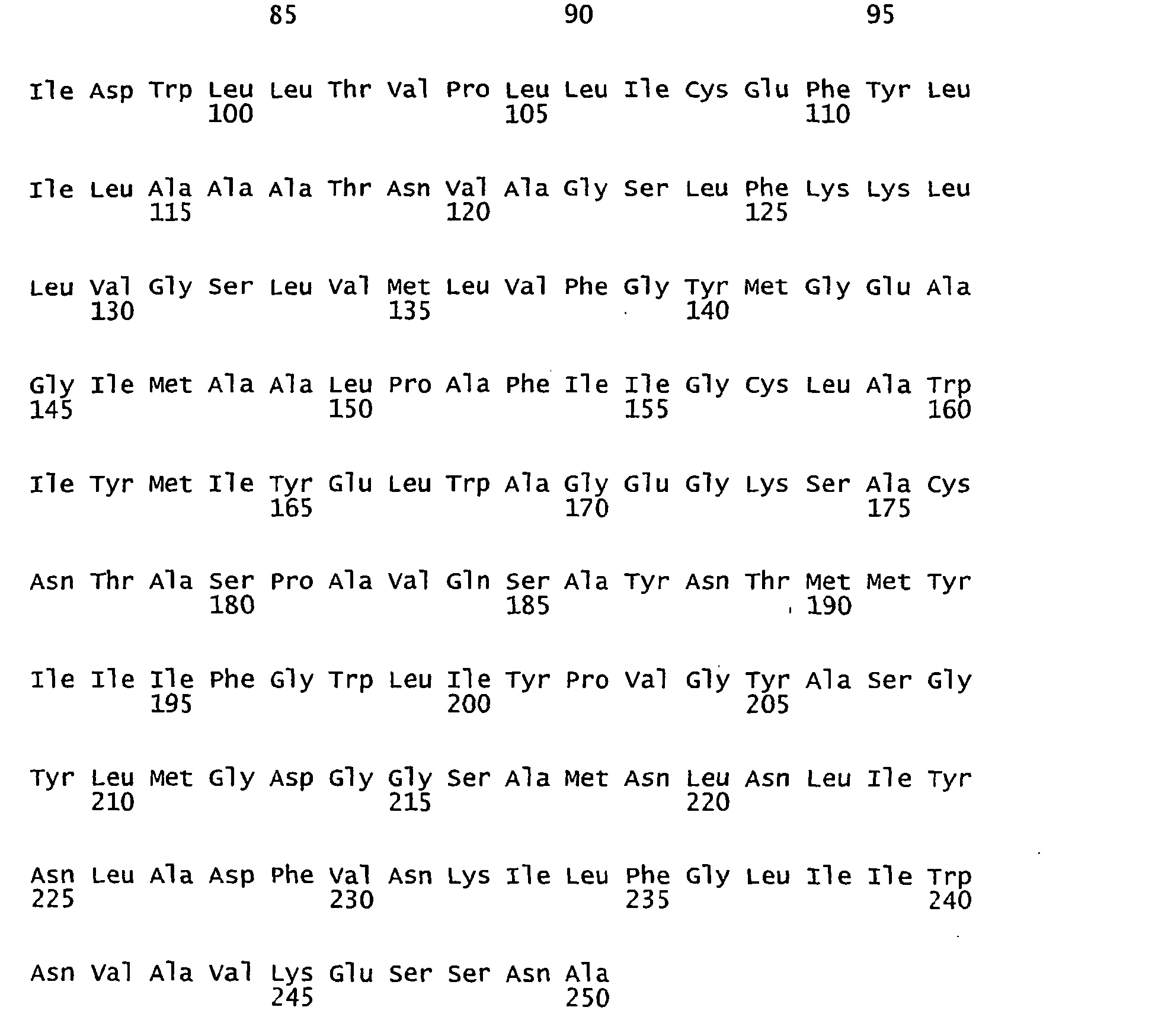
- <210> 124
<211> 751
<212> DNA
<213> Marine eubacteria - <400> 124


- <210> 125
<211> 258
<212> PRT
<213> Marine eubacteria - <400> 125

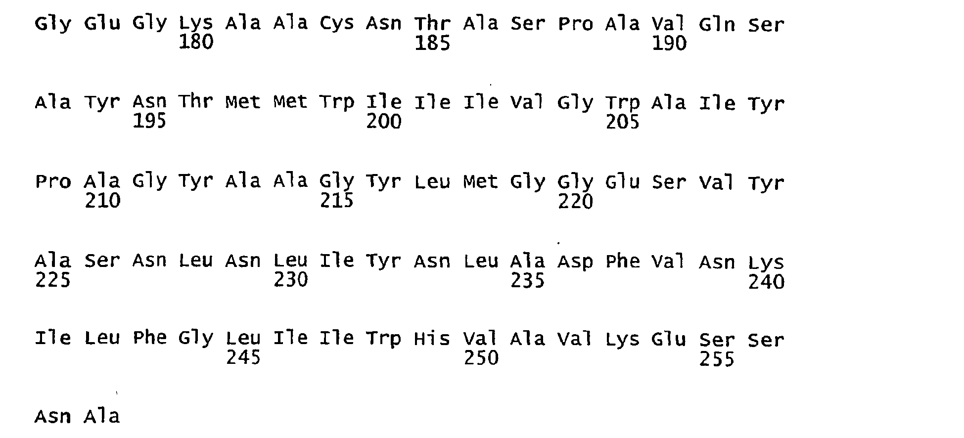
- <210> 126
<211> 775
<212> DNA
<213> Marine eubacteria - <400> 126

- <210> 127
<211> 257
<212> PRT
<213> Marine eubacteria - <400> 127


- <210> 128
<211> 772
<212> DNA
<213> Marine eubacteria - <400> 128

- <210> 129
<211> 249
<212> PRT
<213> Marine eubacteria - <400> 129
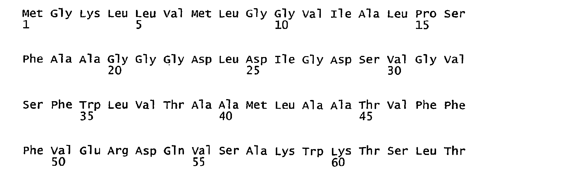

- <210> 130
<211> 748
<212> DNA
<213> Marine eubacteria - <400> 130


- <210> 131
<211> 249
<212> PRT
<213> Marine eubacteria - <400> 131


- <210> 132
<211> 748
<212> DNA
<213> Marine eubacteria - <400> 132

- <210> 133
<211> 251
<212> PRT
<213> Marine eubacteria - <400> 133


- <210> 134
<211> 754
<212> DNA
<213> Marine eubacteria - <400> 134

- <210> 135
<211> 251
<212> PRT
<213> Marine eubacteria - <400> 135


- <210> 136
<211> 754
<212> DNA
<213> Marine eubacteria - <400> 136


- <210> 137
<211> 251
<212> PRT
<213> Marine eubacteria - <400> 137


- <210> 138
<211> 754
<212> DNA
<213> Marine eubacteria - <400> 138

- <210> 139
<211> 251
<212> PRT
<213> Marine eubacteria - <400> 139

- <210> 140
<211> 754
<212> DNA
<213> Marine eubacteria - <400> 140

- <210> 141
<211> 247
<212> PRT
<213> Marine eubacteria - <400> 141


- <210> 142
<211> 742
<212> DNA
<213> Marine eubacteria - <400> 142
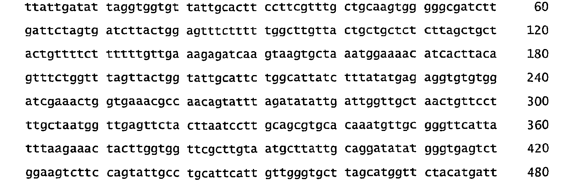

- <210> 143
<211> 251
<212> PRT
<213> Marine eubacteria - <400> 143


- <210> 144
<211> 754
<212> DNA
<213> Marine eubacteria - <400> 144
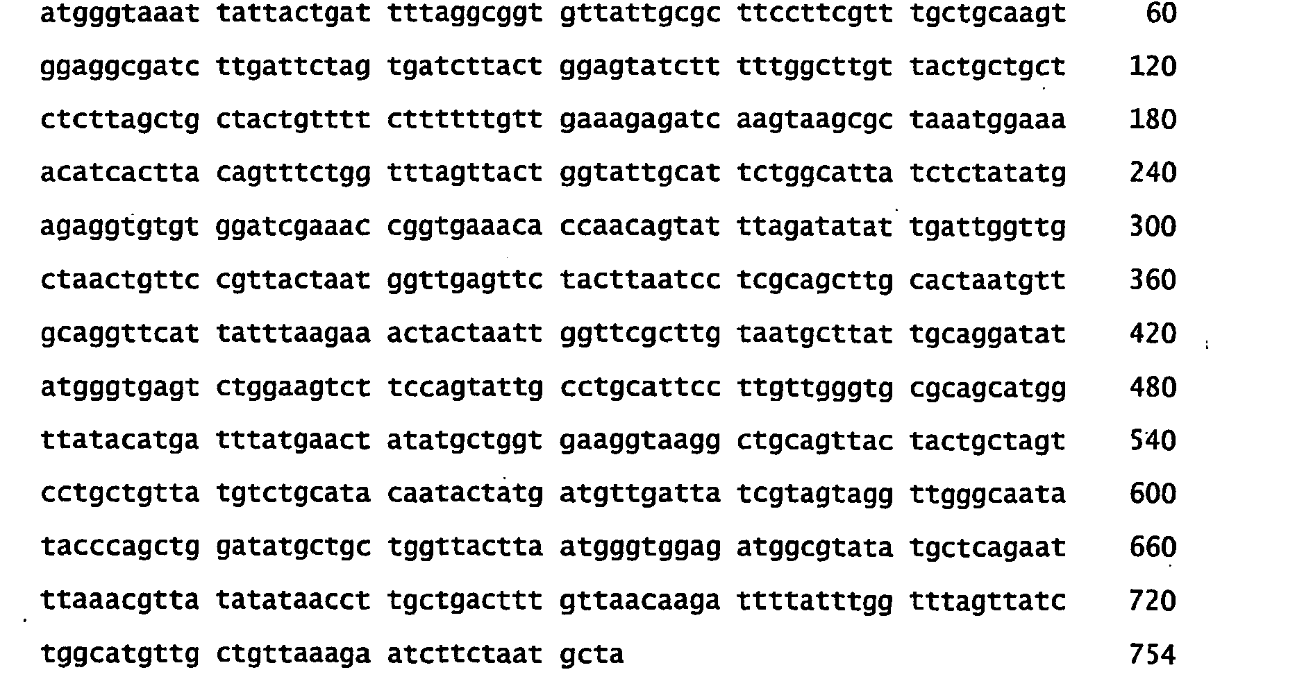
- <210> 145
<211> 250
<212> PRT
<213> Marine eubacteria - <400> 145


- <210> 146
<211> 751
<212> DNA
<213> Marine eubacteria - <400> 146

- <210> 147
<211> 251
<212> PRT
<213> Marine eubacteria - <400> 147


- <210> 148
<211> 754
<212> DNA
<213> Marine eubacteria - <400> 148


- <210> 149
<211> 251
<212> PRT
<213> Marine eubacteria - <400> 149


- <210> 150
<211> 754
<212> DNA
<213> Marine eubacteria - <400> 150

- <210> 151
<211> 254
<212> PRT
<213> Marine eubacteria - <400> 151


- <210> 152
<211> 763
<212> DNA
<213> Marine eubacteria - <400> 152

- <210> 153
<211> 254
<212> PRT
<213> Marine eubacteria - <400> 153


- <210> 154
<211> 763
<212> DNA
<213> Marine eubacteria - <400> 154


- <210> 155
<211> 254
<212> PRT
<213> Marine eubacteria - <400> 155


- <210> 156
<211> 763
<212> DNA
<213> Marine eubacteria - <400> 156

- <210> 157
<211> 254
<212> PRT
<213> Marine eubacteria - <400> 157


- <210> 158
<211> 763
<212> DNA
<213> Marine eubacteria - <400> 158


- <210> 159
<211> 250
<212> PRT
<213> Marine eubacteria - <400> 159


- <210> 160
<211> 750
<212> DNA
<213> Marine eubacteria - <400> 160


- <210> 161
<211> 230
<212> PRT
<213> Marine eubacteria - <400> 161
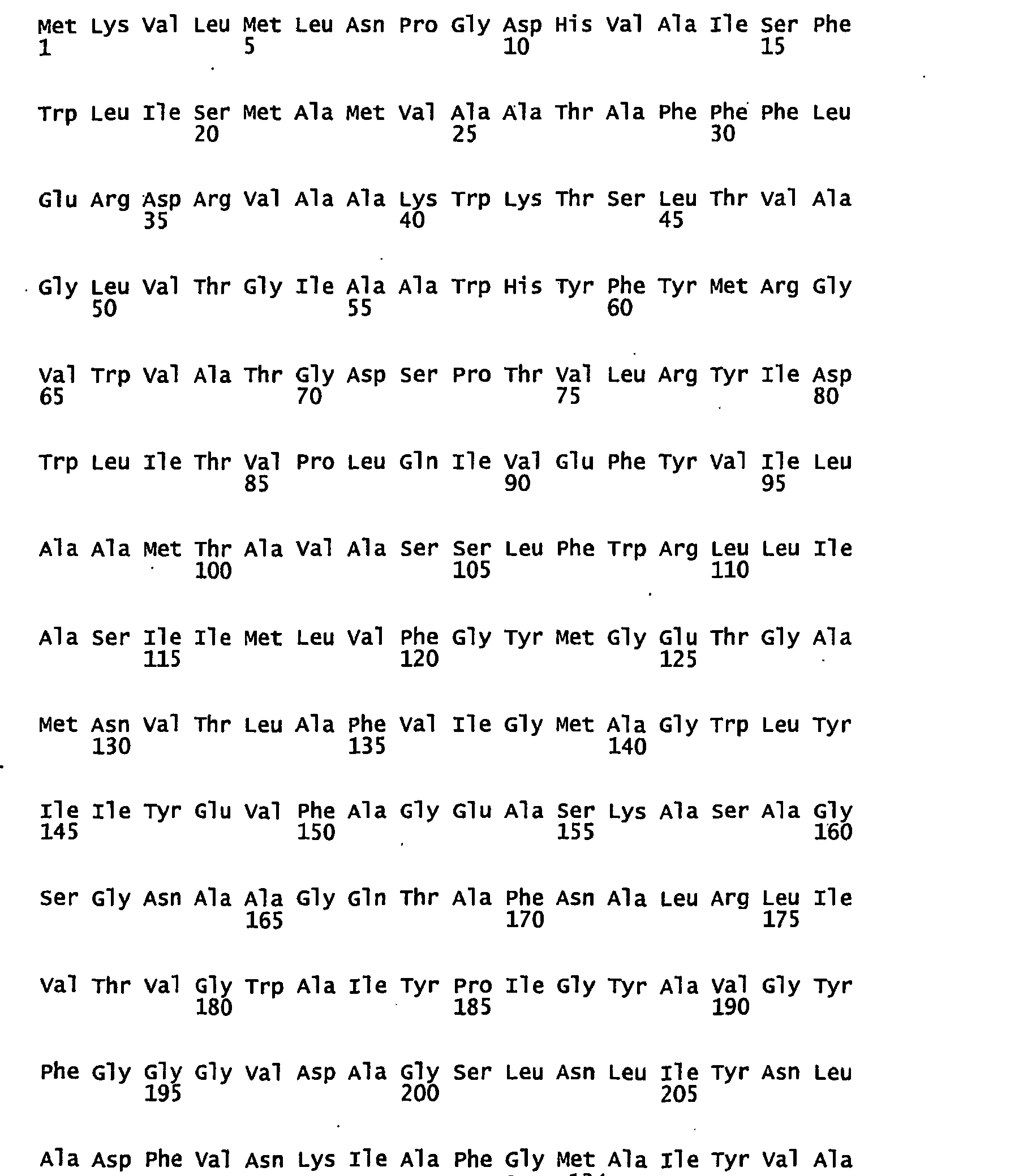

- <210> 162
<211> 690
<212> DNA
<213> Marine eubacteria - <400> 162

- <210> 163
<211> 249
<212> PRT
<213> Marine eubacteria - <400> 163


- <210> 164
<211> 750
<212> DNA
<213> Marine eubacteria - <400> 164


- <210> 165
<211> 249
<212> PRT
<213> Marine eubacteria - <400> 165


- <210> 166
<211> 750
<212> DNA
<213> Marine eubacteria - <400> 166

- <210> 167
<211> 249
<212> PRT
<213> Marine eubacteria - <400> 167

- <210> 168
<211> 750
<212> DNA
<213> Marine eubacteria - <400> 168
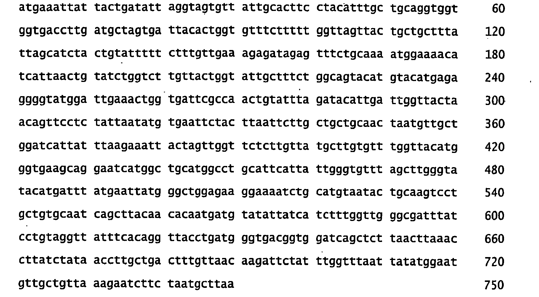
- <210> 169
<211> 252
<212> PRT
<213> Marine eubacteria - <400> 169
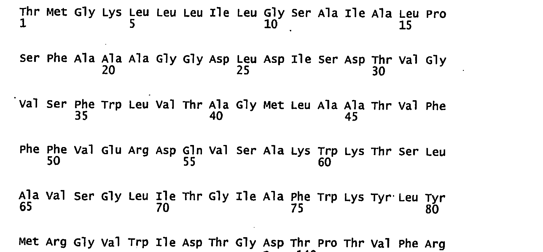

- <210> 170
<211> 756
<212> DNA
<213> Marine eubacteria - <400> 170
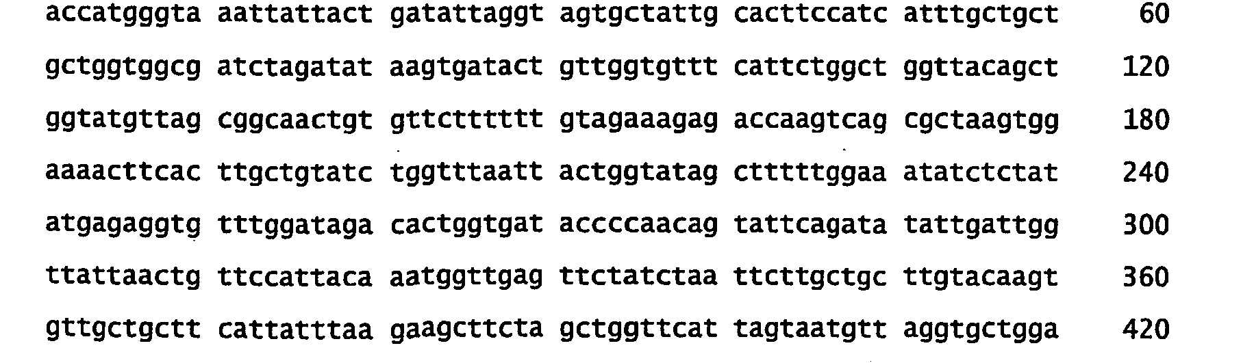

- <210> 171
<211> 252
<212> PRT
<213> Marine eubacteria - <400> 171


- <210> 172
<211> 756
<212> DNA
<213> Marine eubacteria - <400> 172

- <210> 173
<211> 252
<212> PRT
<213> Marine eubacteria - <400> 173

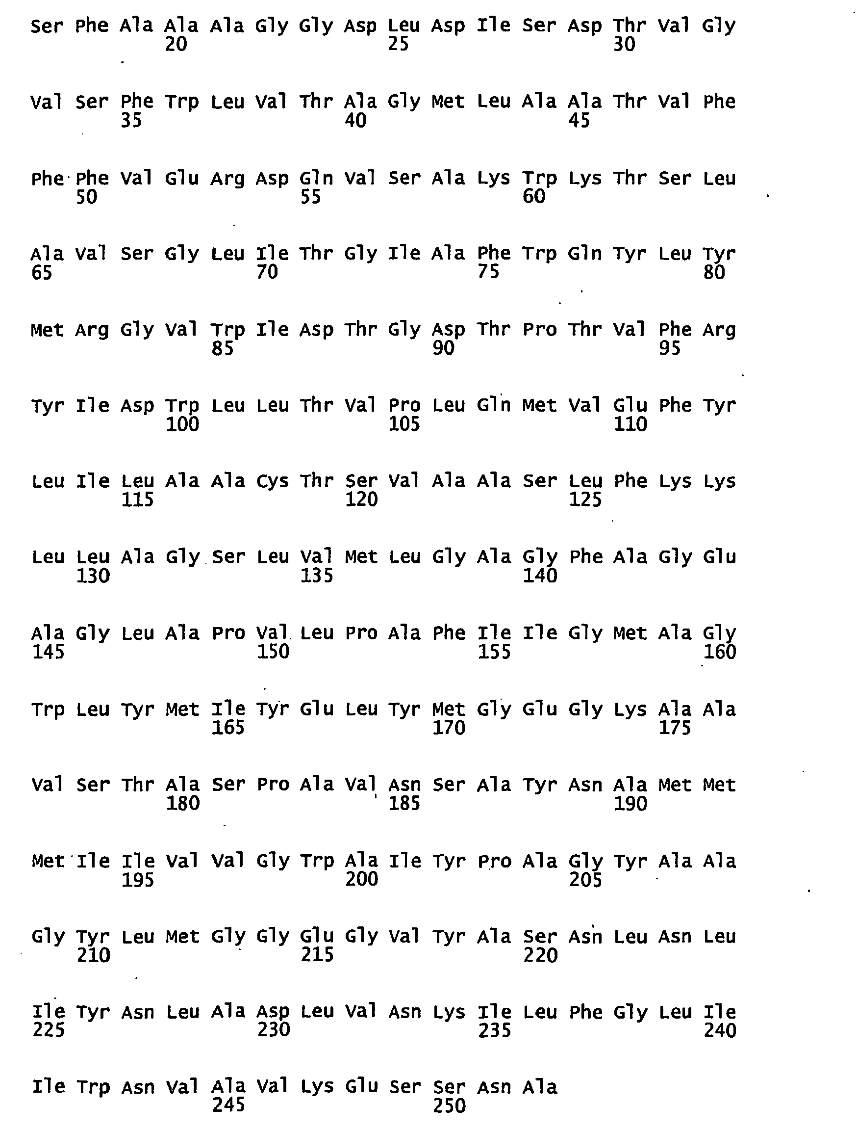
- <210> 174
<211> 756
<212> DNA
<213> Marine eubacteria - <400> 174

- <210> 175
<211> 252
<212> PRT
<213> Marine eubacteria - <400> 175


- <210> 176
<211> 756
<212> DNA
<213> Marine eubacteria - <400> 176


- <210> 177
<211> 252
<212> PRT
<213> Marine eubacteria - <400> 177
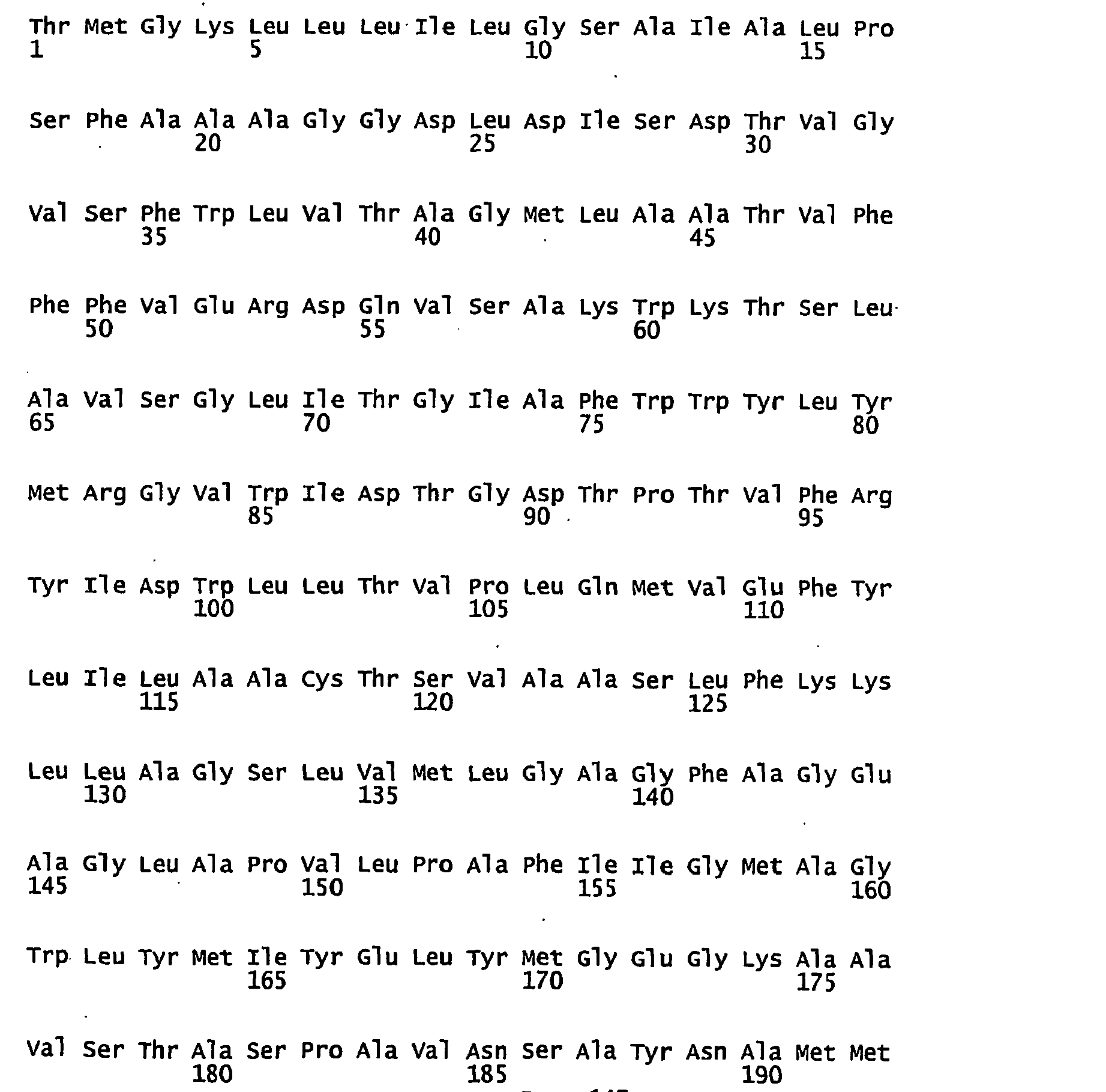

- <210> 178
<211> 756
<212> DNA
<213> Marine eubacteria - <400> 178

- <210> 179
<211> 252
<212> PRT
<213> Marine eubacteria - <400> 179


- <210> 180
<211> 756
<212> DNA
<213> Marine eubacteria - <400> 180

- <210> 181
<211> 252
<212> PRT
<213> Marine eubacteria - <400> 181
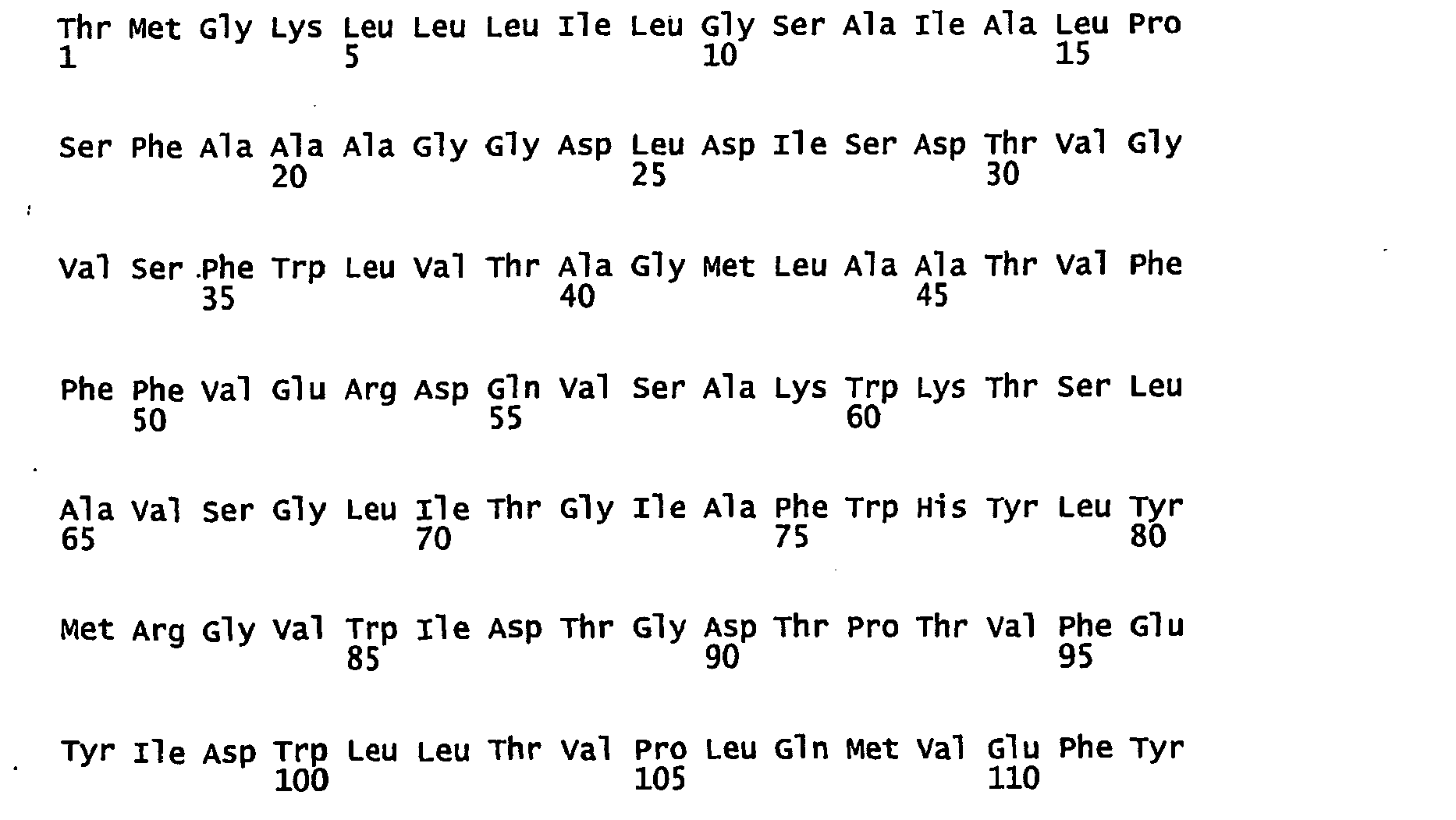

- <210> 182
<211> 756
<212> DNA
<213> Marine eubacteria - <400> 182


- <210> 183
<211> 252
<212> PRT
<213> Marine eubacteria - <400> 183


- <210> 184
<211> 756
<212> DNA
<213> Marine eubacteria - <400> 184

- <210> 185
<211> 25
<212> DNA
<213> artificial sequence - <220>
<223> Synthetic - <400> 185
aaattattac tgatattagg tagtg 25 - <210> 186
<211> 24
<212> DNA
<213> artificial sequence - <400> 186
agcattagaa gattctttaa cage 24 - <210> 187
<211> 21
<212> DNA
<213> artificial sequence - <400> 187
gaggtatata ttaatgtatc g 21 - <210> 188
<211> 18
<212> DNA
<213> artificial sequence - <400> 188
gatttaatct gtatcagg 18 - <210> 189
<211> 45
<212> DNA
<213> artificial sequence - <400> 189
tgttactggt attgctttct ggaattacat gtacatgaga ggggt 45 - <210> 190
<211> 45
<212> DNA
<213> artificial sequence - <400> 190
acccctctca tgtacatgta atttcagaaa gcaataccag taaca 45 - <210> 191
<211> 45
<212> DNA
<213> artificial sequence - <400> 191
tgttactggt attgctttct ggcagtacat gtacatgaga ggggt 45 - <210> 192
<211> 45
<212> DNA
<213> artificial sequence - <400> 192
acccctctca tgtacatgta ctgccagaaa gcaataccag taaca 45 - <210> 193
<211> 45
<212> DNA
<213> artificial sequence - <400> 193
tgttactggt attgctttct ggaaatacat gtacatgaga ggggt 45 - <210> 194
<211> 44
<212> DNA
<213> artificial sequence - <400> 194
acccctctca tgtacatgta tttccagaaa gcaataccag taac 44 - <210> 195
<211> 45
<212> DNA
<213> artificial sequence - <400> 195
ttactggtat agctttttgg aattatctct atatgagagg tgttt 45 - <210> 196
<211> 45
<212> DNA
<213> artificial sequence - <400> 196
aaacacctct catatagaga taattccaaa aagctatacc agtaa 45 - <210> 197
<211> 45
<212> DNA
<213> artificial sequence - <400> 197
ttactggtat agctttttgg cagtatctct atatgagagg tgttt 45 - <210> 198
<211> 45
<212> DNA
<213> artificial sequence - <400> 198
aaacacctct catatagaga tactgccaaa aagctatacc agtaa 45 - <210> 199
<211> 45
<212> DNA
<213> artificial sequence - <400> 199
ttactggtat agctttttgg aaatatctct atatgagagg tgttt 45 - <210> 200
<211> 45
<212> DNA
<213> artificial sequence - <400> 200
aaacacctct catatagaga tatttccaaa aagctatacc agtaa 45 - <210> 201
<211> 45
<212> DNA
<213> artificial sequence - <400> 201
ctggtgatac cccaacagta ttcgcatata ttgattggtt attaa 45 - <210> 202
<211> 45
<212> DNA
<213> artificial sequence - <400> 202
ttaataacca atcaatatat gcgaatactg ttggggtatc accag 45 - <210> 203
<211> 45
<212> DNA
<213> artificial sequence - <400> 203
ctggtgatac cccaacagta ttccaatata ttgattggtt attaa 45 - <210> 204
<211> 45
<212> DNA
<213> artificial sequence - <400> 204
ttaataacca atcaatatat tggaatactg ttggggtatc accag 45 - <210> 205
<211> 45
<212> DNA
<213> artificial sequence - <400> 205
ctggtgatac cccaacagta ttcgaatata ttgattggtt attaa 45 - <210> 206
<211> 45
<212> DNA
<213> artificial sequence - <220>
<223> synthetic - <400> 206
ttaataacca atcaatatat tcgaatactg ttggggtatc accag 45 - <210> 207
<211> 45
<212> DNA
<213> artificial sequence - <220>
<223> Synthetic - <400> 207
ttactggtat agctttttgg gattatctct atatgagagg tgttt 45 - <210> 208
<211> 45
<212> DNA
<213> artificial sequence - <220>
<223> Synthetic - <400> 208
aaacacctct catatagaga taatcccaaa aagctatacc agtaa 45 - <210> 209
<211> 45
<212> DNA
<213> artificial sequence - <220>
<223> synthetic - <400> 209
ttactggtat agctttttgg gaatatctct atatgagagg tgttt 45 - <210> 210
<211> 45
<212> DNA
<213> artificial sequence - <220>
<223> Synthetic - <400> 210
aaacacctct catatagaga tattcccaaa aagctatacc agtaa 45 - <210> 211
<211> 45
<212> DNA
<213> artificial sequence - <220>
<223> synthetic - <400> 211
ttactggtat agctttttgg tggtatctct atatgagagg tgttt 45 - <210> 212
<211> 45
<212> DNA
<213> artificial sequence - <400> 212
aaacacctct catatagaga taccaccaaa aagctatacc agtaa 45
Claims (10)
- A proteorhodopsin mutant having improved optical characteristics, said mutant comprising a mutation in the conserved histidine residue of a wild-type proteorhodopsin variant, wherein said conserved histidine residue is mutated to a hydrogen bond forming amino acid selected from the group consisting of asparagine, glutamine, lysine, arginine, tryptophan, serine, threonine, tyrosine, aspartic acid and glutamic acid, wherein the conserved histidine residue is present at the amino acid position equivalent to the position 77 of: SEQ ID NO: 1 or the position 75 of SEQ ID NO: 3 when the proteorhodopsin variant is aligned with SEQ ID NO: 1 or 3 respectively for maximum identity, and wherein said proteorhodopsin mutant has lower pK rh in comparison with the wild-type proteorhodopsin variant.
- The proteorhodopsin mutant according to claim 1, wherein said wild-type proteorhodopsin variant is a naturally occurring proteorhdopsin variant of SEQ ID NO: 1, 3, 6, 7, 8, 11, 13, 15, 17, 19, 21, 23, 25, 27, 29, 31, 33, 35, 37, 39, 41, 43, 45, 47, 49, 51, 53, 55, 57, 59, 61, 63, 65, 67, 69, 71, 73, 75, 77, 79, 81, 83, 85, 87, 89, 91, 93, 95, 97, 99, 101, 103, 105, 107, 109, 111, 113, 116, 117, 119, 121, 123, 125, 127, 129, 131, 133, 135, 137, 139, 141, 143, 145, 147, 149, 151, 153, 155, 157, 157, 169, or 161; or other proteorhodopsin variants sharing at least 90% amino acid identity thereof.
- The proteorhodopsin mutant according to claim 1, wherein said amino acid capable of forming an H-bond is asparagine, glutamine, lysine, tryptophan, aspartic acid or glutamic acid.
- The proteorhodopsin mutant according to claim 1, comprising an amino acid sequence selected from the group consisting of SEQ ID NOs: 163, 165, 167, 169, 171, 173, 175 and 177.
- A nucleic acid sequence encoding the proteorhodopsin mutant according to any one of claims 1 to 4.
- The nucleic acid sequence according to claim 5, selected from the group consisting of SEQ ID NOs: 164, 166, 168, 170, 172, 174, 176 and 178.
- A method for preparing the proteorhodopsin mutant having Improved optical characteristics according to any one of claims 1 to 4 comprising the steps of(a) ldentifying a conserved histidine amino acid residue of a wlid-type proteorhodopsin variant,(b) mutagenizing the conserved histidine amino acid residue, and obtaining proteorhodopsin mutants,(c) determining the optical characteristics of the proteorhodopsin mutants, and(d) selecting the proteorhodopsin mutant having Improved optical characteristics.
- The method according to claim 7, wherein said conserved amino acid residue is mutagenized by site-directed mutagenesis.
- A method of storing and retrieving optical data, comprising the steps of:(a) providing a film comprising a matrix having the proteorhodopsin mutant according to any one of claims 1 to 4 immobilized within,(b) exposing the film to light of a wavelength that is absorbed by the proteorhodopsin mutant at a resting state in a predetermined pattern,(c) converting selective portions of the film to an excited state and storing optical data therein,(d) exposing the film of step (c) to light of a wavelength that is absorbed by the proteorhodopsin mutant at either a resting state or an excited state, and(e) detecting the stored optical data by an optical recording device.
- A light-driven energy generator comprising:(a) the proteorhodopsin mutant according to any one of claims 1 to 4,(b) a cell membrane,(c) a source of all-trans-retinal, and(d) a light-source, wherein the proteorhodopsin mutant Integrates within the cell membrane to produce an integrated proteorhodopsin mutant, and the integrated proteorhodopsin mutant binds covalently to all-trans-retinal to produce a light absorbing pigment.
Applications Claiming Priority (3)
| Application Number | Priority Date | Filing Date | Title |
|---|---|---|---|
| US42951802P | 2002-11-26 | 2002-11-26 | |
| US429518P | 2002-11-26 | ||
| PCT/US2003/038194 WO2004063326A2 (en) | 2002-11-26 | 2003-11-26 | Proteorhodopsin mutants with improved optical characteristics |
Publications (3)
| Publication Number | Publication Date |
|---|---|
| EP1576144A2 EP1576144A2 (en) | 2005-09-21 |
| EP1576144A4 EP1576144A4 (en) | 2007-02-14 |
| EP1576144B1 true EP1576144B1 (en) | 2010-07-14 |
Family
ID=40834426
Family Applications (1)
| Application Number | Title | Priority Date | Filing Date |
|---|---|---|---|
| EP03811664A Expired - Lifetime EP1576144B1 (en) | 2002-11-26 | 2003-11-26 | Proteorhodopsin mutants with improved optical characteristics |
Country Status (10)
| Country | Link |
|---|---|
| US (1) | US7501488B2 (en) |
| EP (1) | EP1576144B1 (en) |
| JP (1) | JP4448776B2 (en) |
| KR (1) | KR101179086B1 (en) |
| AT (1) | ATE474050T1 (en) |
| AU (1) | AU2003302734C1 (en) |
| CA (1) | CA2506987A1 (en) |
| DE (1) | DE60333388D1 (en) |
| NZ (1) | NZ540563A (en) |
| WO (1) | WO2004063326A2 (en) |
Families Citing this family (11)
| Publication number | Priority date | Publication date | Assignee | Title |
|---|---|---|---|---|
| CA2506987A1 (en) | 2002-11-26 | 2004-07-29 | Genencor International, Inc. | Proteorhodopsin mutants with improved optical characteristics |
| EP1576042B1 (en) * | 2002-11-26 | 2012-05-23 | Danisco US Inc. | Optical information carrier comprising immobilized proteorhodopsin |
| EP1753448B1 (en) * | 2004-06-10 | 2012-04-25 | Danisco US Inc. | Compositions comprising various proteorhodopsins and/or bacteriorhodopsins and use thereof |
| US20090233342A1 (en) * | 2007-08-23 | 2009-09-17 | Asuncion Martinez | Heterologous expression of proteorhodopsin photosystem |
| US9057734B2 (en) | 2010-08-23 | 2015-06-16 | President And Fellows Of Harvard College | Optogenetic probes for measuring membrane potential |
| US9207237B2 (en) | 2010-08-23 | 2015-12-08 | President And Fellows Of Harvard College | Systems, methods, and workflows for optogenetics analysis |
| US9518103B2 (en) | 2014-06-18 | 2016-12-13 | President And Fellows Of Harvard College | Optogenetic probes for measuring membrane potential |
| US10077463B2 (en) | 2015-01-15 | 2018-09-18 | President And Fellows Of Harvard College | Optical selection of cells |
| KR101843840B1 (en) | 2015-02-10 | 2018-03-30 | 경북대학교 산학협력단 | Codon-optimized proteorhodopsin for photic driving energy metabolism in Non-photosynthetic microorganisms and use thereof |
| CN110483645B (en) * | 2018-05-14 | 2023-05-12 | 郑州大学第一附属医院 | Mitochondrial localized proton pump rhodopsin, its mutant protein and its application |
| JP2023057064A (en) * | 2021-10-08 | 2023-04-20 | 静岡県公立大学法人 | Novel rhodopsin mutants |
Family Cites Families (5)
| Publication number | Priority date | Publication date | Assignee | Title |
|---|---|---|---|---|
| US5825725A (en) * | 1997-05-06 | 1998-10-20 | The State Of Oregon Acting By And Through The State Board Of Higher Education On Behalf Of The University Of Oregon | Method and apparatus for reversible optical data storage |
| US20110093981A9 (en) * | 1999-05-06 | 2011-04-21 | La Rosa Thomas J | Nucleic acid molecules and other molecules associated with transcription in plants and uses thereof for plant improvement |
| US7253268B2 (en) * | 2000-05-03 | 2007-08-07 | Monterey Bay Aquarium Research Institute | Light-driven energy generation using proteorhodopsin |
| US7939220B2 (en) | 2000-07-27 | 2011-05-10 | Mib Munich Innovative Biomaterials Gmbh | Proton-translocating retinal protein |
| CA2506987A1 (en) | 2002-11-26 | 2004-07-29 | Genencor International, Inc. | Proteorhodopsin mutants with improved optical characteristics |
-
2003
- 2003-11-26 CA CA002506987A patent/CA2506987A1/en not_active Abandoned
- 2003-11-26 JP JP2004566503A patent/JP4448776B2/en not_active Expired - Fee Related
- 2003-11-26 WO PCT/US2003/038194 patent/WO2004063326A2/en not_active Ceased
- 2003-11-26 EP EP03811664A patent/EP1576144B1/en not_active Expired - Lifetime
- 2003-11-26 AT AT03811664T patent/ATE474050T1/en not_active IP Right Cessation
- 2003-11-26 NZ NZ540563A patent/NZ540563A/en not_active IP Right Cessation
- 2003-11-26 KR KR1020057009587A patent/KR101179086B1/en not_active Expired - Fee Related
- 2003-11-26 US US10/724,264 patent/US7501488B2/en not_active Expired - Fee Related
- 2003-11-26 AU AU2003302734A patent/AU2003302734C1/en not_active Ceased
- 2003-11-26 DE DE60333388T patent/DE60333388D1/en not_active Expired - Lifetime
Also Published As
| Publication number | Publication date |
|---|---|
| US20050095605A1 (en) | 2005-05-05 |
| WO2004063326A3 (en) | 2006-02-16 |
| US7501488B2 (en) | 2009-03-10 |
| ATE474050T1 (en) | 2010-07-15 |
| EP1576144A2 (en) | 2005-09-21 |
| JP2006516389A (en) | 2006-07-06 |
| KR20050101536A (en) | 2005-10-24 |
| JP4448776B2 (en) | 2010-04-14 |
| EP1576144A4 (en) | 2007-02-14 |
| AU2003302734C1 (en) | 2010-01-14 |
| NZ540563A (en) | 2008-03-28 |
| AU2003302734B2 (en) | 2009-03-26 |
| KR101179086B1 (en) | 2012-09-07 |
| AU2003302734A1 (en) | 2004-08-10 |
| DE60333388D1 (en) | 2010-08-26 |
| CA2506987A1 (en) | 2004-07-29 |
| WO2004063326A2 (en) | 2004-07-29 |
Similar Documents
| Publication | Publication Date | Title |
|---|---|---|
| EP1576144B1 (en) | Proteorhodopsin mutants with improved optical characteristics | |
| Stiel et al. | 1.8 Å bright-state structure of the reversibly switchable fluorescent protein Dronpa guides the generation of fast switching variants | |
| Brown et al. | Crystal structure of the flagellar rotor protein FliN from Thermotoga maritima | |
| Trautner et al. | The sll1951 gene encodes the surface layer protein of Synechocystis sp. strain PCC 6803 | |
| Kufryk et al. | Association of small CAB-like proteins (SCPs) of Synechocystis sp. PCC 6803 with photosystem II | |
| Wilson et al. | Sodium-substrate cotransport in bacteria | |
| Yorimitsu et al. | The conserved charged residues of the C-terminal region of FliG, a rotor component of the Na+-driven flagellar motor | |
| Kim et al. | Architecture of the flagellar switch complex of Escherichia coli: conformational plasticity of FliG and implications for adaptive remodeling | |
| Kojima et al. | Mutations targeting the C-terminal domain of FliG can disrupt motor assembly in the Na+-driven flagella of Vibrio alginolyticus | |
| Henderson et al. | Proton-solute coupling mechanism of the maltose transporter from Saccharomyces cerevisiae | |
| US7704708B2 (en) | Monomeric streptavidin muteins | |
| US7265205B2 (en) | Monomeric streptavidin muteins | |
| Fukuoka et al. | Visualization of functional rotor proteins of the bacterial flagellar motor in the cell membrane | |
| JP2006516389A5 (en) | ||
| Jorissen et al. | Heterologous Expression and Characterization of Recombinant Phytochrome from the Green Alga Mougeotia scalaris¶ | |
| Chen et al. | The blue-green sensory rhodopsin SRM from Haloarcula marismortui attenuates both phototactic responses mediated by sensory rhodopsin I and II in Halobacterium salinarum | |
| US7129329B1 (en) | Heme proteins hemAT-Hs and hemAT-Bs and their use in medicine and microsensors | |
| Bhatt et al. | Manipulating the length of the b subunit F1 binding domain in F1F0 ATP synthase from Escherichia coli | |
| Thornton et al. | Purification, characterization, and crystallization of the distal BRCT domain of the human XRCC1 DNA repair protein | |
| US7517968B2 (en) | Rapid and inexpensive method for the purification of proteorhodopsin | |
| EP1753997B1 (en) | Photochromic material comprising a proteorhodopsin apoprotein and retinal analog | |
| Homma et al. | Function of the Vibrio PomB plug region based on the stator rotation model for bacterial flagellar motor | |
| Steube | Evolution of molecular innovations in cyanobacterial light-perceiving systems | |
| AU2002301732B2 (en) | Novel PRO362 polypeptides and nucleic acids encoding same | |
| Schroeder | Phycobiliproteins: Biosynthesis and applications |
Legal Events
| Date | Code | Title | Description |
|---|---|---|---|
| PUAI | Public reference made under article 153(3) epc to a published international application that has entered the european phase |
Free format text: ORIGINAL CODE: 0009012 |
|
| 17P | Request for examination filed |
Effective date: 20050602 |
|
| AK | Designated contracting states |
Kind code of ref document: A2 Designated state(s): AT BE BG CH CY CZ DE DK EE ES FI FR GB GR HU IE IT LI LU MC NL PT RO SE SI SK TR |
|
| AX | Request for extension of the european patent |
Extension state: AL LT LV MK |
|
| PUAK | Availability of information related to the publication of the international search report |
Free format text: ORIGINAL CODE: 0009015 |
|
| DAX | Request for extension of the european patent (deleted) | ||
| RIC1 | Information provided on ipc code assigned before grant |
Ipc: C07K 14/195 20060101AFI20060418BHEP |
|
| RIC1 | Information provided on ipc code assigned before grant |
Ipc: G11C 13/04 20060101ALI20061013BHEP Ipc: G11B 20/10 20060101ALI20061013BHEP Ipc: C12N 15/31 20060101AFI20061013BHEP Ipc: C07K 14/195 20060101ALI20061013BHEP Ipc: G11B 7/24 20060101ALI20061013BHEP |
|
| A4 | Supplementary search report drawn up and despatched |
Effective date: 20070111 |
|
| 17Q | First examination report despatched |
Effective date: 20070622 |
|
| GRAP | Despatch of communication of intention to grant a patent |
Free format text: ORIGINAL CODE: EPIDOSNIGR1 |
|
| GRAC | Information related to communication of intention to grant a patent modified |
Free format text: ORIGINAL CODE: EPIDOSCIGR1 |
|
| GRAS | Grant fee paid |
Free format text: ORIGINAL CODE: EPIDOSNIGR3 |
|
| GRAA | (expected) grant |
Free format text: ORIGINAL CODE: 0009210 |
|
| AK | Designated contracting states |
Kind code of ref document: B1 Designated state(s): AT BE BG CH CY CZ DE DK EE ES FI FR GB GR HU IE IT LI LU MC NL PT RO SE SI SK TR |
|
| REG | Reference to a national code |
Ref country code: GB Ref legal event code: FG4D |
|
| REG | Reference to a national code |
Ref country code: CH Ref legal event code: EP |
|
| RAP2 | Party data changed (patent owner data changed or rights of a patent transferred) |
Owner name: DANISCO US INC. |
|
| REG | Reference to a national code |
Ref country code: IE Ref legal event code: FG4D |
|
| REF | Corresponds to: |
Ref document number: 60333388 Country of ref document: DE Date of ref document: 20100826 Kind code of ref document: P |
|
| REG | Reference to a national code |
Ref country code: CH Ref legal event code: NV Representative=s name: ING. MARCO ZARDI C/O M. ZARDI & CO. S.A. |
|
| REG | Reference to a national code |
Ref country code: NL Ref legal event code: VDEP Effective date: 20100714 |
|
| PG25 | Lapsed in a contracting state [announced via postgrant information from national office to epo] |
Ref country code: FI Free format text: LAPSE BECAUSE OF FAILURE TO SUBMIT A TRANSLATION OF THE DESCRIPTION OR TO PAY THE FEE WITHIN THE PRESCRIBED TIME-LIMIT Effective date: 20100714 Ref country code: AT Free format text: LAPSE BECAUSE OF FAILURE TO SUBMIT A TRANSLATION OF THE DESCRIPTION OR TO PAY THE FEE WITHIN THE PRESCRIBED TIME-LIMIT Effective date: 20100714 Ref country code: NL Free format text: LAPSE BECAUSE OF FAILURE TO SUBMIT A TRANSLATION OF THE DESCRIPTION OR TO PAY THE FEE WITHIN THE PRESCRIBED TIME-LIMIT Effective date: 20100714 |
|
| PG25 | Lapsed in a contracting state [announced via postgrant information from national office to epo] |
Ref country code: SI Free format text: LAPSE BECAUSE OF FAILURE TO SUBMIT A TRANSLATION OF THE DESCRIPTION OR TO PAY THE FEE WITHIN THE PRESCRIBED TIME-LIMIT Effective date: 20100714 Ref country code: PT Free format text: LAPSE BECAUSE OF FAILURE TO SUBMIT A TRANSLATION OF THE DESCRIPTION OR TO PAY THE FEE WITHIN THE PRESCRIBED TIME-LIMIT Effective date: 20101115 Ref country code: CY Free format text: LAPSE BECAUSE OF FAILURE TO SUBMIT A TRANSLATION OF THE DESCRIPTION OR TO PAY THE FEE WITHIN THE PRESCRIBED TIME-LIMIT Effective date: 20100714 Ref country code: BG Free format text: LAPSE BECAUSE OF FAILURE TO SUBMIT A TRANSLATION OF THE DESCRIPTION OR TO PAY THE FEE WITHIN THE PRESCRIBED TIME-LIMIT Effective date: 20101014 |
|
| PG25 | Lapsed in a contracting state [announced via postgrant information from national office to epo] |
Ref country code: SE Free format text: LAPSE BECAUSE OF FAILURE TO SUBMIT A TRANSLATION OF THE DESCRIPTION OR TO PAY THE FEE WITHIN THE PRESCRIBED TIME-LIMIT Effective date: 20100714 Ref country code: BE Free format text: LAPSE BECAUSE OF FAILURE TO SUBMIT A TRANSLATION OF THE DESCRIPTION OR TO PAY THE FEE WITHIN THE PRESCRIBED TIME-LIMIT Effective date: 20100714 Ref country code: GR Free format text: LAPSE BECAUSE OF FAILURE TO SUBMIT A TRANSLATION OF THE DESCRIPTION OR TO PAY THE FEE WITHIN THE PRESCRIBED TIME-LIMIT Effective date: 20101015 |
|
| PG25 | Lapsed in a contracting state [announced via postgrant information from national office to epo] |
Ref country code: DK Free format text: LAPSE BECAUSE OF FAILURE TO SUBMIT A TRANSLATION OF THE DESCRIPTION OR TO PAY THE FEE WITHIN THE PRESCRIBED TIME-LIMIT Effective date: 20100714 |
|
| PLBE | No opposition filed within time limit |
Free format text: ORIGINAL CODE: 0009261 |
|
| STAA | Information on the status of an ep patent application or granted ep patent |
Free format text: STATUS: NO OPPOSITION FILED WITHIN TIME LIMIT |
|
| PG25 | Lapsed in a contracting state [announced via postgrant information from national office to epo] |
Ref country code: IT Free format text: LAPSE BECAUSE OF FAILURE TO SUBMIT A TRANSLATION OF THE DESCRIPTION OR TO PAY THE FEE WITHIN THE PRESCRIBED TIME-LIMIT Effective date: 20100714 Ref country code: SK Free format text: LAPSE BECAUSE OF FAILURE TO SUBMIT A TRANSLATION OF THE DESCRIPTION OR TO PAY THE FEE WITHIN THE PRESCRIBED TIME-LIMIT Effective date: 20100714 Ref country code: CZ Free format text: LAPSE BECAUSE OF FAILURE TO SUBMIT A TRANSLATION OF THE DESCRIPTION OR TO PAY THE FEE WITHIN THE PRESCRIBED TIME-LIMIT Effective date: 20100714 Ref country code: RO Free format text: LAPSE BECAUSE OF FAILURE TO SUBMIT A TRANSLATION OF THE DESCRIPTION OR TO PAY THE FEE WITHIN THE PRESCRIBED TIME-LIMIT Effective date: 20100714 Ref country code: EE Free format text: LAPSE BECAUSE OF FAILURE TO SUBMIT A TRANSLATION OF THE DESCRIPTION OR TO PAY THE FEE WITHIN THE PRESCRIBED TIME-LIMIT Effective date: 20100714 |
|
| 26N | No opposition filed |
Effective date: 20110415 |
|
| PG25 | Lapsed in a contracting state [announced via postgrant information from national office to epo] |
Ref country code: ES Free format text: LAPSE BECAUSE OF FAILURE TO SUBMIT A TRANSLATION OF THE DESCRIPTION OR TO PAY THE FEE WITHIN THE PRESCRIBED TIME-LIMIT Effective date: 20101025 Ref country code: MC Free format text: LAPSE BECAUSE OF NON-PAYMENT OF DUE FEES Effective date: 20101130 |
|
| REG | Reference to a national code |
Ref country code: DE Ref legal event code: R097 Ref document number: 60333388 Country of ref document: DE Effective date: 20110415 |
|
| REG | Reference to a national code |
Ref country code: FR Ref legal event code: ST Effective date: 20110801 |
|
| PG25 | Lapsed in a contracting state [announced via postgrant information from national office to epo] |
Ref country code: IE Free format text: LAPSE BECAUSE OF NON-PAYMENT OF DUE FEES Effective date: 20101126 Ref country code: FR Free format text: LAPSE BECAUSE OF NON-PAYMENT OF DUE FEES Effective date: 20101130 |
|
| PG25 | Lapsed in a contracting state [announced via postgrant information from national office to epo] |
Ref country code: LU Free format text: LAPSE BECAUSE OF NON-PAYMENT OF DUE FEES Effective date: 20101126 Ref country code: HU Free format text: LAPSE BECAUSE OF FAILURE TO SUBMIT A TRANSLATION OF THE DESCRIPTION OR TO PAY THE FEE WITHIN THE PRESCRIBED TIME-LIMIT Effective date: 20110115 |
|
| PG25 | Lapsed in a contracting state [announced via postgrant information from national office to epo] |
Ref country code: TR Free format text: LAPSE BECAUSE OF FAILURE TO SUBMIT A TRANSLATION OF THE DESCRIPTION OR TO PAY THE FEE WITHIN THE PRESCRIBED TIME-LIMIT Effective date: 20100714 |
|
| PGFP | Annual fee paid to national office [announced via postgrant information from national office to epo] |
Ref country code: DE Payment date: 20141118 Year of fee payment: 12 Ref country code: GB Payment date: 20141126 Year of fee payment: 12 Ref country code: CH Payment date: 20141112 Year of fee payment: 12 |
|
| REG | Reference to a national code |
Ref country code: DE Ref legal event code: R119 Ref document number: 60333388 Country of ref document: DE |
|
| REG | Reference to a national code |
Ref country code: CH Ref legal event code: PL |
|
| GBPC | Gb: european patent ceased through non-payment of renewal fee |
Effective date: 20151126 |
|
| PG25 | Lapsed in a contracting state [announced via postgrant information from national office to epo] |
Ref country code: LI Free format text: LAPSE BECAUSE OF NON-PAYMENT OF DUE FEES Effective date: 20151130 Ref country code: CH Free format text: LAPSE BECAUSE OF NON-PAYMENT OF DUE FEES Effective date: 20151130 |
|
| PG25 | Lapsed in a contracting state [announced via postgrant information from national office to epo] |
Ref country code: DE Free format text: LAPSE BECAUSE OF NON-PAYMENT OF DUE FEES Effective date: 20160601 Ref country code: GB Free format text: LAPSE BECAUSE OF NON-PAYMENT OF DUE FEES Effective date: 20151126 |