EP1575502B1 - Compositions and methods involving nuclear hormone receptor site ii - Google Patents
Compositions and methods involving nuclear hormone receptor site ii Download PDFInfo
- Publication number
- EP1575502B1 EP1575502B1 EP03765637A EP03765637A EP1575502B1 EP 1575502 B1 EP1575502 B1 EP 1575502B1 EP 03765637 A EP03765637 A EP 03765637A EP 03765637 A EP03765637 A EP 03765637A EP 1575502 B1 EP1575502 B1 EP 1575502B1
- Authority
- EP
- European Patent Office
- Prior art keywords
- site
- atom
- nhr
- modulator
- binding
- Prior art date
- Legal status (The legal status is an assumption and is not a legal conclusion. Google has not performed a legal analysis and makes no representation as to the accuracy of the status listed.)
- Expired - Lifetime
Links
- 0 Cc(cc1)ccc1C1=CSC(NC(C2(C3(C4)C44c5ccccc5C3C2)/C=C/Cc2c4cccc2)=O)=*C1 Chemical compound Cc(cc1)ccc1C1=CSC(NC(C2(C3(C4)C44c5ccccc5C3C2)/C=C/Cc2c4cccc2)=O)=*C1 0.000 description 2
- VDMYZVVOWGYDCM-UHFFFAOYSA-N CC(CC1c2c3cccc2)(C3c2c1cccc2)C(Nc1ncc(C)[s]1)=O Chemical compound CC(CC1c2c3cccc2)(C3c2c1cccc2)C(Nc1ncc(C)[s]1)=O VDMYZVVOWGYDCM-UHFFFAOYSA-N 0.000 description 1
- PQBCMYCFCVEZIA-UHFFFAOYSA-N Cc(cc1)c(cccc2)c2c1-c1c[nH]c(N)n1 Chemical compound Cc(cc1)c(cccc2)c2c1-c1c[nH]c(N)n1 PQBCMYCFCVEZIA-UHFFFAOYSA-N 0.000 description 1
- DHUFMCMYVNLRRQ-UHFFFAOYSA-N Nc1nc(-c(c2c3cccc2)ccc3Br)c[s]1 Chemical compound Nc1nc(-c(c2c3cccc2)ccc3Br)c[s]1 DHUFMCMYVNLRRQ-UHFFFAOYSA-N 0.000 description 1
- KZYOELPVZVDPGC-UHFFFAOYSA-N Nc1nc(-c2c[s]c3ccccc23)c[s]1 Chemical compound Nc1nc(-c2c[s]c3ccccc23)c[s]1 KZYOELPVZVDPGC-UHFFFAOYSA-N 0.000 description 1
- YVHHQTSYHNTIBI-UHFFFAOYSA-N Nc1nc(-c2cccc3c2cccc3)c[nH]1 Chemical compound Nc1nc(-c2cccc3c2cccc3)c[nH]1 YVHHQTSYHNTIBI-UHFFFAOYSA-N 0.000 description 1
- CFKWTZWWRBCTIQ-UHFFFAOYSA-N Nc1nc(-c2cccc3c2cccc3)c[o]1 Chemical compound Nc1nc(-c2cccc3c2cccc3)c[o]1 CFKWTZWWRBCTIQ-UHFFFAOYSA-N 0.000 description 1
Images







Classifications
-
- C—CHEMISTRY; METALLURGY
- C07—ORGANIC CHEMISTRY
- C07K—PEPTIDES
- C07K14/00—Peptides having more than 20 amino acids; Gastrins; Somatostatins; Melanotropins; Derivatives thereof
- C07K14/435—Peptides having more than 20 amino acids; Gastrins; Somatostatins; Melanotropins; Derivatives thereof from animals; from humans
- C07K14/705—Receptors; Cell surface antigens; Cell surface determinants
-
- C—CHEMISTRY; METALLURGY
- C07—ORGANIC CHEMISTRY
- C07K—PEPTIDES
- C07K14/00—Peptides having more than 20 amino acids; Gastrins; Somatostatins; Melanotropins; Derivatives thereof
- C07K14/435—Peptides having more than 20 amino acids; Gastrins; Somatostatins; Melanotropins; Derivatives thereof from animals; from humans
- C07K14/705—Receptors; Cell surface antigens; Cell surface determinants
- C07K14/72—Receptors; Cell surface antigens; Cell surface determinants for hormones
-
- G—PHYSICS
- G01—MEASURING; TESTING
- G01N—INVESTIGATING OR ANALYSING MATERIALS BY DETERMINING THEIR CHEMICAL OR PHYSICAL PROPERTIES
- G01N33/00—Investigating or analysing materials by specific methods not covered by groups G01N1/00 - G01N31/00
- G01N33/48—Biological material, e.g. blood, urine; Haemocytometers
- G01N33/50—Chemical analysis of biological material, e.g. blood, urine; Testing involving biospecific ligand binding methods; Immunological testing
- G01N33/74—Chemical analysis of biological material, e.g. blood, urine; Testing involving biospecific ligand binding methods; Immunological testing involving hormones or other non-cytokine intercellular protein regulatory factors such as growth factors, including receptors to hormones and growth factors
-
- G—PHYSICS
- G16—INFORMATION AND COMMUNICATION TECHNOLOGY [ICT] SPECIALLY ADAPTED FOR SPECIFIC APPLICATION FIELDS
- G16B—BIOINFORMATICS, i.e. INFORMATION AND COMMUNICATION TECHNOLOGY [ICT] SPECIALLY ADAPTED FOR GENETIC OR PROTEIN-RELATED DATA PROCESSING IN COMPUTATIONAL MOLECULAR BIOLOGY
- G16B15/00—ICT specially adapted for analysing two-dimensional [2D] or three-dimensional [3D] molecular structures, e.g. structural or functional relations or structure alignment
- G16B15/30—Drug targeting using structural data; Docking or binding prediction
-
- G—PHYSICS
- G01—MEASURING; TESTING
- G01N—INVESTIGATING OR ANALYSING MATERIALS BY DETERMINING THEIR CHEMICAL OR PHYSICAL PROPERTIES
- G01N2500/00—Screening for compounds of potential therapeutic value
-
- G—PHYSICS
- G16—INFORMATION AND COMMUNICATION TECHNOLOGY [ICT] SPECIALLY ADAPTED FOR SPECIFIC APPLICATION FIELDS
- G16B—BIOINFORMATICS, i.e. INFORMATION AND COMMUNICATION TECHNOLOGY [ICT] SPECIALLY ADAPTED FOR GENETIC OR PROTEIN-RELATED DATA PROCESSING IN COMPUTATIONAL MOLECULAR BIOLOGY
- G16B15/00—ICT specially adapted for analysing two-dimensional [2D] or three-dimensional [3D] molecular structures, e.g. structural or functional relations or structure alignment
Definitions
- the present invention generally refers to a binding site, termed Site II, in nuclear hormone receptors.
- the present disclosure describes: machine-readable data storage media comprising structure coordinates of Site II; computer systems capable of producing three-dimensional representations of all or any part of Site II; methods used in the design and identification of ligands of Site II and of modulators of nuclear hormone receptors (NHRs); ligands of Site II; modulators of NHRs; methods of modulating NHRs; pharmaceutical compositions comprising modulators of NHRs; modulators of an NHR for use in treating diseases; methods of designing mutants; mutant NHRs or portions of mutant NHRs; methods of measuring the binding of a test molecule to Site II; and models of Site II.
- NHRs nuclear hormone receptors
- NHR nuclear hormone receptor
- NHRs stimulate transcription by binding to DNA and inducing transcription of specific genes. NHRs may also stimulate transcription by not binding to DNA itself, rather they may modulate the activity of other DNA binding proteins ( Stocklin, E., et al., Nature (1996) 383:726-8 ). The process of stimulation of transcription is called transactivation. NHRs repress transcription by interacting with other transcription factors or coactivators and inhibiting the ability of these other transcription factors or coactivators to induce transcription of specific genes.
- transrepression The process of repression of transcription is called transrepression.
- transrepression The Nuclear Receptor Factsbook, V. Laudet and H. Gronemeyer, Academic Press, p42, 2002 ).
- the glucocorticoid receptor, estrogen receptor, androgen receptor and peroxisome proliferator activated receptors ⁇ and y have been shown to repress the activity of the transcription factors AP-1 and NF- ⁇ ( Jonat, C., et al., Cell, 62, p1189-1204, (1990 ) Kallio, P.J., et al., Mol. Endocrinol., 9, p1017-1028 (1995 ), Keller, E.T., et al., J.
- the nuclear hormone receptor family includes the glucocorticoid receptor ( Hollenberg, S.M. et al.(1985) Nature, 318, p635 ), progesterone receptor ( Misrahi, M. et al. (1987) Biochem. Biophys. Res. Commun. 143, p740 ), androgen receptor (Lubahn D. B., et al (1988), estrogen receptors ( Green, S., et al.
- Glucocorticoid receptor progesterone receptor, androgen receptor, estrogen receptor, and mineralocorticoid receptor are steroid hormone receptors (SHRs).
- sequences vary amongst the various nuclear hormone receptors, they can be divided into functional domains including an N-terminal transactivation domain, a central DNA binding domain and a C-terminal ligand and dimerization domain.
- the ligands which bind these receptors act in a ligand, cell type, and promoter dependent fashion and include: glucocorticoids, progestins, retinoids, mineralocorticoids, and others.
- non-steroids can bind to nuclear hormone receptors and induce a biologic response ( Coghlan, MJ, et al, J. Med. Chem. 44, p2879, 2001 ).
- Ligand cross-talk can occur between the receptors, for example, progesterone can bind not only the progesterone receptor but the glucocorticoid receptor as well ( Zhang, S., Mol. Endocrinology 10, p24, 1996 ).
- RCSB Research Collaboratory for Structural Bioinformatics, pdb file format
- Mutagenesis of GR has defined the dexamethasone interacting surface, defined as Site I, which includes amino acids Met-560, Met-639, Gln-642 and Thr-739 ( Lind, U., et al. J. Biol. Chem. 275, p19041, 2000 ).
- a second binding site for hydroxytamoxifen with the ligand binding domain of estrogen receptor beta describes the crystal structure of estrogen receptor bound with 4-hydroxytamoxifen, in which the ligand is found in two locations: the usual steroid binding pocket and a second site located along the hydrophobic groove near the cofactor binding region. This second site is remote from the Site II location described in this application.
- the glucocorticoid receptor is a member of the nuclear hormone receptor family of transcription factors, and a member of the steroid hormone family of transcription factors. Affinity labeling of the glucocorticoid receptor protein allowed the production of antibodies against the receptor which facilitated cloning the human ( Weinberger, et al. Science 228, p740-742, 1985 , Weinberger, et al. Nature, 318, P635-641, 1985 ) and rat ( Miesfeld, R. Nature, 312, p779-781, 1985 ) glucocorticoid receptors. Subsequently, glucocorticoid receptors from other species were cloned including mouse ( Danielson, M. et al.
- GR-beta C-terminally distinct isoform of GR termed GR-beta. This isoform is identical to GR up to amino acid 727 and then diverges in the last C-terminal 15 amino acids. GR-beta is not known to bind glucocorticoids, is unable to transactivate, but does bind DNA ( Hollenberg, SM. et al.
- GR-beta binds compounds other than the typical glucocorticoids.
- Glucocorticoids which interact with GR have been used for over 50 years to treat inflammatory diseases. It has been clearly shown that glucocorticoids exert their anti-inflammatory activity via the inhibition by GR of the transcription factors NF- ⁇ B and AP-1. This inhibition is termed transrepression. It has been shown that the primary mechanism for inhibition of these transcription factors by GR is via a direct physical interaction. This interaction alters the transcription factor complex and inhibits the ability of NF- ⁇ B and AP-1 to stimulate transcription ( Jonat, C., et al. Cell, 62, p1189, 1990 , Yang-Yen, H.F., et al. Cell 62, p1205, 1990 , Diamond, M.I. et al.
- NF- ⁇ B and AP-1 are involved in regulating the expression of a number of important inflammatory and immunomodulatory genes including: TNF-alpha, IL-1, IL-2, IL-5, adhesion molecules (such as E-selectin), chemokines (such as Eoxtaxin and Rantes), Cox-2, and others.
- glucocorticoids are very effective anti-inflammatory agents, their systemic use is limited by their side effects which include diabetes, osteoporosis, glaucoma, Cushingoid syndrome, muscle loss, facial swelling, personality changes, and others.
- transactivation requires dimerization of GR and binding to a glucocorticoid response element (GRE). DNA binding is mediated via Zn fingers in the DNA binding domain ( Giguere, V. et al Cell 46, p645, 1986 ; Rusconi, S. and Yamamoto, K.R. EMBO J., 6, p1309, 1987 ). DNA sequence specific interactions are determined by the C-terminal part of the first Zn finger ( Danielsen, M., et al. Cell 57, p1131, 1989 ).
- GR target genes include MMTV, metallothionein, and tyrosine amino transferase ( Ringold, GM et al, Cell 6, p299,1975 ; Scheidereit, C., et al Nature, 304, p749, 1983 ; Hager, LJ, Palmiter RD, Nature 291, 340, 1981 ; Grange, T., et al. Oncogene, 20, p3028, 2001 ). Transrepression, as opposed to transactivation, can occur in the absence of dimerization, and as mentioned above is believed to involve the direct interaction of GR with AP-1 and NF- ⁇ B.
- Dissociated steroids Compounds which can induce transrepression of GR with none to minimal induction of transactivation have been termed "dissociated steroids" ( Vayassiere, BM, et al., Mol. Endocrinology, 11, p1249, 1997 ). Such "dissociated” compounds would be useful to treat inflammatory diseases. See Figure 1 for a graphical description of transactivation mediated by GR dimers versus transrepression mediated by GR monomers. It is possible that these "dissociated” compounds bind to GR without inducing dimerization yet allow the monomer to transrepress AP-1 and NF- ⁇ B. Another plausible explanation is that "dissociated” compounds may alter the conformation of GR to enable transrepression without inducing a DNA binding conformation.
- Ursodeoxycholic acid has recently been shown to repress NF- ⁇ B activity via a GR mediated pathway. The compound appears to be "dissociated” as it does not induce DNA binding in a cellular assay. This compound, although acting in a GR dependent fashion, does not compete for dexamethasone binding to GR. Although direct binding of UDCA to GR has not been demonstrated, mutagenesis studies suggest that the ligand binding domain (LBD) of GR is required for activity. ( Miura, T., J. Biol. Chem. 276, p47371, 2001 ). However, these studies did not delineate the specific amino acids which are involved in UDCA activity.
- a modulator of an NHR may be useful in treating NHR-associated diseases, that is diseases associated with the expression products of genes whose transcription is stimulated or repressed by NHRs.
- NHR-associated diseases that is diseases associated with the expression products of genes whose transcription is stimulated or repressed by NHRs.
- modulators of NHR that induce inhibition of AP-1 and NF- ⁇ B, as such modulators would be useful in the treatment of inflammatory and immune associated diseases and disorders, such as osteoarthritis, rheumatoid arthritis, multiple sclerosis, asthma, inflammatory bowel disease, transplant rejection, and graft vs. host disease.
- glucocorticoids are potent anti-inflammatory agents, their systemic use is limited by side effects.
- a dissociated compound that retained the anti-inflammatory efficacy of glucocorticoids while minimizing the side effects such as diabetes, osteoporosis and glaucoma would be of great benefit to a very large number of patients with inflammatory diseases.
- glucocorticoid such as diabetes, osteoporosis and glaucoma.
- the present invention relates to the following items:
- NHRs nuclear hormone receptors
- Site II is a structure defined by structure coordinates that describe conserved residue backbone atoms having a root mean square deviation of not more than 2.0 ⁇ from the conserved residue backbone atoms described by the structure coordinates of amino acids E537-V543, L566, G567, Q570-W577, S599-A607, W610, R611, R614, Q615, P625, Y663, L664 and K667 according to Table I. Table I is located under the heading for Example 21.
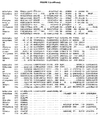
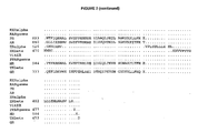
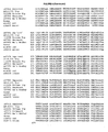
- Figure 2 shows the amino acids of Site II in various human NHRs.
- the structure coordinates of Site II in the NHRs of Figure 2 are given in Table III, located under the heading for Example 22.
- Two sets of xray structure coordinates of Site II in GR are given in Table IV, located under the heading for Example 23, and Table V, located under the heading for Example 24.
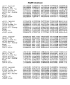
- Figure 6 shows the amino acids of Site II in the GR of various species.
- ligands of Site II modulate NHRs.
- Ligands of Site II induce transrepression.
- Ligands of Site II possess dissociated activity.
- Ligands of Site II antagonize transactivation.
- machine-readable data storage media comprising data storage material encoded with machine readable data comprising all or any part of the structure coordinates of Site II are described.
- Computer systems comprising the machine-readable data storage media of the invention capable of producing three-dimensional representations of all or any part of Site II are disclosed in the context of the present invention. Methods used in the design and identification of ligands of Site II and modulators of NHRs are also disclosed herein.
- the context of the present invention describes: methods of docking a test molecule into all or any part of the cavity circumscribed by Site II; methods comprising identifying structural and chemical features of all or any part of Site II; methods of designing a ligand of Site II comprising modeling all or any part of Site II and designing a chemical entity that has structural and chemical complementarity with all or any part of Site II; methods of evaluating the potential of a chemical entity to bind to all or any part of Site II; methods for identifying a modulator of an NHR; and methods for identifying a ligand of Site II.
- ligands of Site II are disclosed.
- Modulators of NHRs are also described herein.
- methods of modulating NHR are also described.
- compositions comprising modulators of NHRs are disclosed.
- a pharmaceutical composition comprising a modulator of an NHR for treating diseases.
- diseases include NHR-associated diseases, diseases associated with NHR transactivation, diseases associated with NHR transrepression, diseases associated with AP-1-dependent gene expression, diseases associated with NF- ⁇ B-dependent gene expression, inflammatory or immune associated diseases and disorders, diseases treatable by inducing NHR transrepression, and diseases treatable by antagonizing NHR transactivation.
- the invention provides methods of designing mutants comprising mutating Site II by making an amino acid substitution, deletion or insertion, and the resultant mutant NHRs, or portions of mutant NHRs, comprising a mutation in Site II.
- the invention provides methods of measuring the binding of a test molecule to Site II.
- the invention provides models of Site II.
- NHRs nuclear hormone receptors
- Site II is a structure defined by structure coordinates that describe conserved residue backbone atoms having a root mean square deviation of not more than 2.0 ⁇ from the conserved residue backbone atoms described by the structure coordinates of amino acids E537-V543, L566, G567, Q570-W577, S599-A607, W610, R611, R614, Q615, P625, Y663, L664 and K667 according to Table I. That is, a Site II is any structure that falls within the given root mean square deviation. Table I is located under the heading for Example 21.
- Figure 2 shows the amino acids of Site II in various human NHRs.
- the structure coordinates of Site II in the NHRs of Figure 2 are given in Table III, located under the heading for Example 22.
- Two sets of xray structure coordinates of Site II in GR are given in Table IV, located under the heading for Example 23, and Table V, located under the heading for Example 24.
- Figure 6 shows the amino acids of Site II in the GR of various species.
- Ligands of Site II modulate NHRs.
- Ligands of Site II induce transrepression.
- Ligands of Site II possess dissociated activity.
- Ligands of Site II antagonize transactivation.
- Said Site II preferably is a nuclear hormone receptor (NHR) Site II, more preferably steroid hormone receptor (SHR) Site II, most preferably a glucocorticoid receptor (GR) Site II.
- NHR nuclear hormone receptor
- SHR steroid hormone receptor
- GR glucocorticoid receptor
- Said NHR is preferably an SHR, more preferably a GR.
- NHR is selected from the group consisting of: RXR-alpha; RXR-beta; progesterone receptor (PR); androgen receptor (AR); estrogen receptor-alpha (ER-alpha); ER-beta; vitamin D receptor (VitDR); peroxisome proliferator activated receptor-gamma (PPAR-gamma); thyroid receptor-alpha (TR-alpha); TR-beta; mineralocorticoid receptor (MR); and glucocorticoid receptor (GR).
- RXR-alpha RXR-beta
- PR progesterone receptor
- AR estrogen receptor-alpha
- ER-beta ER-beta
- vitamin D receptor VitDR
- PPAR-gamma peroxisome proliferator activated receptor-gamma
- TR-alpha thyroid receptor-alpha
- TR-beta mineralocorticoid receptor (MR); and glucocorticoid receptor (GR).
- said NHR is selected from the group consisting of: RXR-alpha; RXR-beta; progesterone receptor (PR); androgen receptor (AR); vitamin D receptor (VitDR); peroxisome proliferator activated receptor- gamma (PPAR-gamma); thyroid receptor-alpha (TR-alpha); TR-beta; mineralocorticoid receptor (MR); and glucocorticoid receptor (GR).
- said NHR is selected from the group consisting of: RXR-alpha; RXR-beta; androgen receptor (AR); vitamin D receptor (VitDR); peroxisome proliferator activated receptor-gamma (PPAR-gamma); thyroid receptor-alpha (TR-alpha); TR-beta; mineralocorticoid receptor (MR); and glucocorticoid receptor (GR).
- said SHR is selected from the group consisting of: PR; AR; ER-alpha; ER-beta; MR; and GR. More preferably, said SHR is selected from the group consisting of: PR; AR; MR; and GR. Most preferably, said SHR is selected from the group consisting of: AR; MR; and GR.
- Said RXR-alpha Site II preferably is composed of amino acids L236-P244, A272-A273, Q276-W283, G305-S313, H316-R317, A320-V321, T329, L368-G369, and R372 according to Figure 2 .
- Preferably said structure coordinates of said RXR-alpha Site II define the structure of amino acids L236-P244, A272-A273, Q276-W283, G305-S313, H316-R317, A320-V321, T329, L368-G369, and R372 according to Table III, or define the structure of the conserved residue backbone atoms according to Table III.
- said structure coordinates define the same shape as the structure of the amino acids according to Table III, or as the structure of the residue backbone atoms according to Table II, but not necessarily that the structure coordinates are identical to those of the amino acids or residue backbone atoms in the table, as the structure coordinates may be of a coordinate system other than Cartesian coordinates.
- said structure coordinates of said Site II are the structure coordinates of Site II amino acids L236-P244, A272-A273, Q276-W283, G305-S313, H316-R317, A320-V321, T329, L368-G369, and R372 according to Table III.
- Said RAR-gamma Site II preferably is composed of amino acids S 194-P202, L233-A234, C237-F244, A266-R274, T277-R278, T280-E282, D290, T328-G329 and S332 according to Figure 2 .
- Preferably said structure coordinates of said RAR-gamma Site II define the structure of amino acids S 194-P202, L233-A234, C237-F244, A266-R274, T277-R278, T280-E282, D290, T328-G329 and S332 according to Table III, or define the structure of the conserved residue backbone atoms according to Table III.
- said structure coordinates of said Site II are the structure coordinates of Site II amino acids S194-P202, L233-A234, C237-F244, A266-R274, T277-R278, T280-E282, D290, T328-G329 and S332 according to Table III.
- Said PR preferably is composed of amino acids M692-V698, L721-G722, Q725-W732, S754-G762, W765-R766, K769-H770, P780, F818-L819 and K822 according to Figure 2 .
- said structure coordinates of said PR Site II define the structure of amino acids M692-V698, L721-G722, Q725-W732, S754-G762, W765-R766, K769-H770, P780, F818-L819 and K822 according to Table III, or define the structure of the conserved residue backbone atoms according to Table III.
- said structure coordinates of said Site II are the structure coordinates of Site II amino acids M692-V698, L721-G722, Q725-W732, S754-G762, W765-R766, K769-H770, P780, F818-L819 and K822 according to Table III.
- Said AR Site II preferably is composed of amino acids E678-V684, L708-G709, Q712-W719, S741-A749, W752-R753, T756-N757, P767, F805-L806 and K809 according to Figure 2 .
- said structure coordinates of said AR Site II define the structure of amino acids E678-V684, L708-G709, Q712-W719, S741-A749, W752-R753, T756-N757, P767, F805-L806 and K809 according to Table III, or define the structure of the conserved residue backbone atoms according to Table III.
- said structure coordinates of said Site II are the structure coordinates of Site II amino acids E678-V684, L708-G709, Q712-W719, S741-A749, W752-R753, T756-N757, P767, F805-L806 and K809 according to Table III.
- Said ER-alpha Site II preferably is composed of amino acids L320-I326, L348-A349, E352-W359, A381-G389, W392-R393, E396, P405, F444-V445 and K448 according to Figure 2 .
- Preferably said structure coordinates of said ER-alpha Site II define the structure of amino acids L320-I326, L348-A349, E352-W359, A381-G389, W392-R393, E396, P405, F444-V445 and K448 according to Table III, or define the structure of the conserved residue backbone atoms according to Table III.
- said structure coordinates of said Site II are the structure coordinates of Site II amino acids L320-I326, L348-A349, E352-W359, A381-G389, W392-R393, E396, P405, F444-V445 and K448 according to Table III.
- Said ER-beta Site II preferably is composed of amino acids L273-H279, L297-A298, E301-W308, C330-G338, W341-R342, D345, P354, Y393-L394 and K397 according to Figure 2 .
- Preferably said structure coordinates of said ER-beta Site II define the structure of amino acids L273-H279, L297-A298, E301-W308, C330-G338, W341-R342, D345, P354, Y393-L394 and K397 according to Table III, or define the structure of the conserved residue backbone atoms according to Table III.
- said structure coordinates of said Site II are the structure coordinates of Site II amino acids L273-H279, L297-A298, E301-W308, C330-G338, W341-R342, D345, P354, Y393-L394 and K397 according to Table III.
- Said VitDR Site II preferably is composed of amino acids L136-D144, L182-V183, S186-F193, S215-R223, E226-S227, T229-D231, G238, H279-V280 and M283 according to Figure 2 .
- Preferably said structure coordinates of said VitDR Site II define the structure of amino acids L136-D144, L182-V183, S186-F193, S215-R223, E226-S227, T229-D231, G238, H279-V280 and M283 according to Table III, or define the structure of the conserved residue backbone atoms according to Table III.
- said structure coordinates of said Site II are the structure coordinates of Site II amino acids L136-D144, L182-V183, S186-F193, S215-R223, E226-S227, T229-D231, G238, H279-V280 and M283 according to Table III.
- Said PPAR-gamma Site II preferably is composed of amino acids Y219-P227, R288-S289, A292-Y299, G321-M329, S332-L333, N335-K336, E343, L384-A385 and I388 according to Figure 2 .
- Preferably said structure coordinates of said PPAR-gamma Site II define the structure of amino acids Y219-P227, R288-S289, A292-Y299, G321-M329, S332-L333, N335-K336, E343, L384-A385 and I388 according to Table III, or define the structure of the conserved residue backbone atoms according to Table III.
- said structure coordinates of said Site II are the structure coordinates of Site II amino acids Y219-P227, R288-S289, A292-Y299, G321-M329, S332-L333, N335-K336, E343, L384-A385 and I388 according to Table III.
- Said MR Site II preferably is composed of amino acids E743-I749, L772-A773, Q776-W783, S805-A813, W816-R817, K820-H821, P831, Y869-T870 and K873 according to Figure 2 .
- Preferably said structure coordinates of said MR Site II define the structure of amino acids E743-I749, L772-A773, Q776-W783, S805-A813, W816-R817, K820-H821, P831, Y869-T870 and K873 according to Table III, or define the structure of the conserved residue backbone atoms according to Table III.
- said structure coordinates of said Site II are the structure coordinates of Site II amino acids E743-I749, L772-A773, Q776-W783, S805-A813, W816-R817, K820-H821, P831, Y869-T870 and K873 according to Table III.
- Said TR-beta Site II preferably is composed of amino acids T226-Q235, I267-I268, A271-F278, C300-R308, V311-R312, D314-E316, G324, V362-A363 and Q366 according to Figure 2 .
- Preferably said structure coordinates of said TR-beta Site II define the structure of amino acids T226-Q235, I267-I268, A271-F278, C300-R308, V311-R312, D314-E316, G324, V362-A363 and Q366 according to Table III, or define the structure of the conserved residue backbone atoms according to Table III.
- said structure coordinates of said Site II are the structure coordinates of Site II amino acids T226-Q235, I267-I268, A271-F278, C300-R308, V311-R312, D314-E316, G324, V362-A363 and Q366 according to Table III.
- Said GR Site II preferably is composed of amino acids E537-V543, L566, G567, Q570-W577, S599-A607, W610, R611, R614, Q615, P625, Y663, L664 and K667 according to Figure 2 .
- said structure coordinates of said GR Site II define the structure of amino acids E537-V543, L566, G567, Q570-W577, S599-A607, W610, R611, R614, Q615, P625, Y663, L664 and K667 according to Table I, Table III, Table IV or Table V, or define the structure of the aforementioned amino acids according to the structure coordinates disclosed in Bledsoe, et.
- said structure coordinates of said Site II are the structure coordinates of Site II amino acids E537-V543, L566, G567, Q570-W577, S599-A607, W610, R611, R614, Q615, P625, Y663, L664 and K667 according to Table I, Table III, Table IV or Table V.
- Said GR Site II is preferably selected from the group consisting of: human GR Site II composed of amino acids E537-V543, L566, G567, Q570-W577, S599-A607, W610, R611, R614, Q615, P625, Y663, L664 and K667 according to Figure 6 ; squirrel GR Site II composed of amino acids E537-V543, L566, G567, Q570-W577, S599-A607, W610, R611, R614, Q615, P625, Y663, L664 and K667 accoring to Figure 6 ; pig GR Site II composed of amino acids E501-V507, L530, G531, Q534-W541, S563-A571, W574, R575, R578, Q579, P589, Y627, L628 and K631 according to Figure 6 ; guinea pig GR Site II composed of amino acids E531-V537, L
- Said nuclear hormone receptor can be of any source, preferably human.
- Said glucocorticoid receptor can be of any source, preferably human, rat, mouse, sheep, marmoset, squirrel, pig, guinea pig, or m'az monkey. Most preferably said glucorticoid receptor is human.
- Said NHR Site II may be native or mutant.
- said NHR Site II is a native NHR Site II.
- Said SHR Site II maybe native or mutant.
- Preferably said SHR Site II is a native SHR Site II.
- Said GR Site II may be native or mutant.
- Preferably said GR Site II is a native GR Site II.
- Said Site II may be found on a protein of any source, including mammalian, fungal, bacterial and plant.
- Preferably said Site II is found on a mammalian protein, more preferably on a human protein.
- the conserved residue backbone atoms of said Site II have a root mean square deviation of less than 1.9, 1.8, 1.7, 1.6 or 1.5 ⁇ , more preferably of less than 1.4, 1.3, 1.2, 1.1, 1.03, 1.02, or 1.0 A, yet more preferably of less than 0.93, 0.92, 0.9, 0.8, 0.7, 0.6 0.5, 0.4, 0.3, 0.2, or 0.1 ⁇ , most preferably 1.02, 0.92 or 0.0 ⁇ from the conserved residue backbone atoms described by the structure coordinates of amino acids E537-V543, L566, G567, Q570-W577, S599-A607, W610, R611, R614, Q615, P625, Y663, L664 and K667 according to Table I.
- the conserved residue backbone atoms of said Site II have a root mean square deviation of less than 2.0, 1.9, 1.8, 1.7, 1.6 or 1.5 ⁇ , more preferably of less than 1.4, 1.3, 1.2, 1.1, 1.03, 1.02, or 1.0 ⁇ , yet more preferably of less than 0.93, 0.92, 0.9, 0.8, 0.7, 0.6 0.5, 0.4, 0.3, 0.2, or 0.1 ⁇ , most preferably 1.02, 0.92 or 0.0 ⁇ from the conserved residue backbone atoms described by the structure coordinates of amino acids E537-V543, L566, G567, Q570-W577, S599-A607, W610, R611, R614, Q615, P625, Y663, L664 and K667 according to Table IV.
- the conserved residue backbone atoms of said Site II have a root mean square deviation of less than 2.0, 1.9, 1.8, 1.7, 1.6 or 1.5 ⁇ , more preferably of less than 1.4, 1.3, 1.2, 1.1, 1.03, 1.02, or 1.0 ⁇ , yet more preferably of less than 0.93, 0.92, 0.9, 0.8, 0.7, 0.6 0.5, 0.4, 0.3, 0.2, or 0.1 ⁇ , most preferably 1.02, 0.92 or 0.0 ⁇ from the conserved residue backbone atoms described by the structure coordinates of amino acids E537-V543, L566, G567, Q570-W577, S599-A607, W610, R611, R614, Q615, P625, Y663, L664 and K667 according to Table V.
- Said structure coordinates may be those determined for a Site II to which a ligand is bound or to which no ligand is bound. Said structure coordinates may be those determined for a Site II of a ligand binding domain in which a Site I ligand is bound to Site I. Said structure coordinates may be those determined for a Site II of an NHR that is in monomer, dimer, or other form.
- the cavity circumscribed by Site II and the cavity circumscribed by Site I share a common wall section. That is, some amino acids are common to both Site II and Site I. However the cavity circumscribed by Site II is distinct from the cavity circumscribed by Site I, as the two cavities are on opposite sides of the common wall.
- GR Site I M560, L563, N564, L566, G567, Q570, M601, M604, A605, L608, R611, F623, M639, Q642, M646, L732, Y735, C736, T739 and E748.
- the following amino acid residues are common to both GR Site I and Site II: L566, G567, Q570, M601, M604, A605 and R611.
- the following amino acid residues are unique to GR Site II, i.e.
- GR Site I E537-V543, V571-W577, S599-W600, F602-L603, F606-A607, W610, R614, Q615, P625, Y663, L664 and K667.
- the following amino acid residues are unique to GR Site I, i.e. they are not part of GR Site II: M560, L563, N564, L608, F623, M639, Q642, M646, L732, Y735, C736, T739 and E748.
- the amino acids in other NHRs and non-human GR corresponding to the above-recited GR amino acids can be seen in Figures 2 and 6 respectively.
- Site II in NHRs as a binding site whose ligands modulate NHRs.
- a three dimensional model can be constructed on the basis of the known structure of a homologous protein (Greer et. al., 1991, Lesk, et. al., 1992, Cardozo, et. al., 1995, Sali, et. al., 1995).
- a homology model is constructed on the basis of first identifying a template, or, protein of known structure which is similar to the protein without known structure. This can be accomplished through pairwise alignment of sequences using such programs as the MODELLER module found in InsightII (Accelrys, Inc., San Diego, CA).
- a set of structure coordinates for a protein or part of a protein is a relative set of points that define a shape in three dimensions.
- the structure coordinates that define two identical or almost identical shapes may vary slightly. If variations are within an acceptable standard error as compared to the original coordinates, the resulting three-dimensional shape is considered to be equivalent.
- a ligand that bound to the structure defined by the structure coordinates of amino acids E537-V543, L566, G567, Q570-W577, S599-A607, W610, R611, R614, Q615, P625, Y663, L664 and K667 according to Table I would also be expected to bind to a site having a shape that fell within the acceptable error. Such sites with structures within the acceptable standard error are also within the scope of this invention.
- comparisons can be made between different structures and different conformations of the same structure.
- the procedure used in InsightII to compare structures is divided into four steps: 1) load the structures to be compared; 2) define the atom equivalencies in these structures; 3) perform a fitting operation; and 4) analyze the results.
- Each structure is identified by a name.
- One structure is identified as the target (i.e., the fixed structure); the second structure (i.e., moving structure) is identified as the source structure.
- Three-dimensional coordinates give the location of the centers of all atoms in a protein molecule and are typically expressed as Cartesian coordinates (eg, distances in three directions, each perpendicular to the other), or polar coordinates (eg, sets of angle/distance pairs from a universal origin), or internal coordinates (eg, sets of angle/distance pairs from one atom center to the next).
- Cartesian coordinates eg, distances in three directions, each perpendicular to the other
- polar coordinates eg, sets of angle/distance pairs from a universal origin
- internal coordinates eg, sets of angle/distance pairs from one atom center to the next.
- Variations in coordinates may also be generated because of mathematical manipulations of the structure coordinates.
- the structure coordinates set forth in Table I could be manipulated by fractionalization of the structure coordinates, integer additions or subtractions to sets of the structure coordinates, inversion of the structure coordinates or any combination of the above.
- the structure coordinates of an actual xray structure of a protein would be expected to have some variation from the homology model of that very same protein. For example, the location of sidechains may vary to some extent.
- the homology model GR Site II coordinates were compared to the GR Site II x-ray structure coordinates available from the disclosures in WO 03/015692 A2, February 27, 2003 Apolito, et. al. and Kauppi et. al., in the Journal of Biological Chemistry Online, JBC Papers In Press as DOI:10.1074/jbc.M212711200, April 9, 2003 , RCSB file: 1nhz.pdb (GR LBD bound to an antagonist, RU 486).
- Variations in structure coordinates can be due to mutations, additions, substitutions, and/or deletions of amino acids of a protein being studied.
- Variations in structure coordinates can be due to variations in proteins whose shape is being described by the structure coordinates given.
- rigid fitting operations conducted (see Example 13) between the GR LBD homology model and several closely-related NHRs known to have similar structure and function yielded Site II root mean square (rms) deviations in conserved residue backbone atom comparisons of 0.57-0.71 ⁇ . These Site II rms deviations could be greater if other variation factors described above were present in the calculations.
- GR LBDs from non-human species may also have slight variations in shape from that of human GR LBD defined by the structure coordinates in Table I.
- any structure that has a root mean square deviation of residue backbone atoms (N, C ⁇ , C, O) of less than 2.0 Angstroms ( ⁇ ) when superimposed on the relevant residue backbone atoms described by structure coordinates listed in Table I is considered to be equivalent.
- the root mean square deviation is less than 1.9, 1.8, 1.7, 1.6 or 1.5 ⁇ , more preferably of less than 1.4, 1.3, 1.2, 1.1, 1.03, 1.02, or 1.0 ⁇ , yet more preferably of less than 0.93, 0.92, 0.9, 0.8, 0.7, 0.6 0.5, 0.4, 0.3, 0.2, or 0.1 ⁇ , most preferably 1.02, 0.92 or 0.0 ⁇ .
- conserved refers to a portion of a protein backbone which is found in common between two proteins. That is, if portions of two proteins are aligned and compared using the three-dimensional coordinates of their residue backbone atoms for super-positioning, and comparison of the structure coordinates of the residue backbone atoms yields an rms of 2.0 ⁇ or less, then the residue backbone atoms are considered to be conserved between the two proteins.
- a homology model of the ligand binding domain (LBD) of GR was constructed using the known crystal structure of the progesterone receptor (PR).
- Site II in the LBD of GR was identified by the complementarity of three-dimensional shape and functional features between the Site II and compounds having AP-1 inhibitory activity. Manual docking of one such compound was performed and confirmed the identity of Site II and its role in transrepression. Binding energetics of the S enantomer of the twenty-seven compounds to Site II were calculated and correlated with AP-1 inhibitory activity of these compounds. This positive correlation further confirmed the identity and role of Site II.
- Site II As binding energetics to Site II correlates to AP-1 inhibitory activity and all compounds that were tested for binding to Site II are dissociated compounds, Site II was determined to be a target for compounds that have AP-1 inhibitory activity as well as compounds that have dissociated activity.
- the cellular transcriptional assay was performed with a titration of dexamethasone in the presence or absence of each of both enantiomers of a pair. Again, an additive effect on AP-1 inhibitory activity was seen with each of the enantiomers and dexamethasone. In contrast, the Site I antagonist RU 486 inhibited the ability of dexamethasone to induce transrepression.
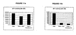
- Example 17 shows that both enantiomers R (Compound B) and S (Compound A) act in an additive fashion with saturating levels of dexamethasone to suppress AP-1 activity. Since dexamethasone binds to Site I, it is most likely that the R and S enantiomers interact with Site II to allosterically enhance the repressive activity.
- nuclear hormone receptor and “NHR,” as used herein, refer to a member of the nuclear hormone receptor family of transcription factors which bind low molecular weight ligands and stimulate or repress transcription.
- NHRs include, but are not limited to, glucocorticoid receptors (GRs), progesterone receptors (PRs), androgen receptors (ARs), estrogen receptors (ERs), mineralocorticoid receptors (MRs), retinoid receptors (RXRs and RARs), Vitamin D receptors (VitDRs), thyroid receptors (TRs), peroxisome proliferator activated receptors (PPARs), and orphan nuclear receptors (i.e.
- Nuclear hormone receptor includes orphan nuclear receptors, which are gene products that embody structural features of nuclear hormone receptors and were identified without any prior knowledge of their association with a putative ligand.
- An NHR has a modulator domain including the AF-1 domain responsible in part for transcriptional activation function. Modulator domains can also include regions for promoters and cell-specific cofactors and can interact with steroid receptor co-activators (SRCs). 2.
- An NHR has a DNA binding domain (DBD) composed of two zinc finger modules composed of 60-70 amino acids and a carboxy-terminal extension (CTE) that providesprotein-protein and protein-DNA interactions upon homo- or heterodimer receptorbinding. 3.
- DBD DNA binding domain
- CTE carboxy-terminal extension
- An NHR has a hinge region that is the hinge between the DBD and the carboxy-terminal ligand binding domain.
- the hinge region is variable is primary structure and amino acid sequence length.
- An NHR has a ligand binding domain (LBD) that contains the AF-2 motif (which corresponds to helix 12 ofNHRs) and provides a structured region whereby AF-2 (helix 12) is packed closely to the LBD core forming an interface with at least 3 other helices of the core. The interface is involved with binding of coactivator or co-repressor polypeptides.
- LBD ligand binding domain
- Nuclear hormone receptor and "NHR,” as used herein, refer to NHRs from any source, including but not limited to: glucocorticoid receptor as disclosed in Hollenberg, S.M., Weinberger, C., Ong, E.S., Cerelli, G., Oro, A., Leba, R., Thompson, E.B., Rosenfeld, M.G., Evans, R.M., Nature (1985) 318: 635-641 ; progesterone receptor as disclosed in Misrahi, M. et al. (1987) Biochem. Biophys. Res. Commun. 143, p740 ; androgen receptor as disclosed in Lubahn D.
- peroxisome proliferator activated receptor as disclosed in Greene, M.E., et al. (1995) Gene Expression 4, p281 ; RXRalpha (11bd) as disclosed in Bourguet, W., Ruff, M., Chambon, P., Gronemeyer, H., Moras, D. Nature 375 pp. 377 (1995 ); PPARgamma (2prg) as disclosed in Nolte, R. T., Wisely, G. B., Westin, S., Cobb, J. E., Lambert, M. H., Kurokawa, R., Rosenfeld, M. G., Willson, T. M., Glass, C.
- PPAR peroxisome proliferator activated receptor
- ERbeta (1l2j) as disclosed in Shiau, A. K., Barstad, D., Radek, J. T., Meyers, M. J., Nettles, K. W., Katzenellenbogen, B. S., Katzenellenbogen, J. A., Agard, D. A., Greene, G. L. Nat.Struct.Biol. 9 pp. 359 (2002 ); TRbeta (1bsx) as disclosed in Wagner, R. L., Darimont, B. D., Apriletti, J. W., Stallcup, M. R., Kushner, P. J., Baxter, J. D., Fletterick, R.
- GR PIR Accession Number QRHUGA, as dislcosed in Hollenberg, S.M., Weinberger, C., Ong, E.S.; Cerelli, G., Oro, A., Leba, R., Thompson, E.B., Rosenfeld, M.G., Evans, R.M., Nature (1985) 318: 635-641 ;
- MR PIR Accession Number A29613, as disclosed in Arriza, J.L.; Weinberger, C., Cerelli, G., Glaser, T.M., Handelin, B.L., Housman, D.E., Evans, R.M., Science (1987) 237: 268-275 .
- Orphan nuclear receptors include but are not limited to: Rev Erb(alpha) (1hlz) as disclosed in Sierk, M.L., et. al., Biochemistry (2001) 40: pp. 12833 ; Pxr (1ilh) as disclosed in Watkins, R.E., et. al., Science (2002) 292: pp. 2329 ; ERR3 (1kv6) as disclosed in Greschik, H., et. al., Mol. Cell (2002) 9: pp. 303 ; Nurrl (1ovl) as disclosed in Wang, Z., et. al., Nature (2003) 423: pp.
- NHRs including orphan nuclear receptors, include those disclosed in: The Nuclear Receptor Facts Book, V. Laudet and H. Gronemeyer, Academic Press, p345, 2002 ; and Francis et al, Annu. Rev. Physiol. 2003, 65:261-311 .
- steroid hormone receptor and "SHR,” as used herein, refer to a member of the nuclear hormone receptor family of transcription factors which bind steroids and stimulate or repress transcription.
- SHRs include, but are not limited to, glucocorticoid receptors (GRs), progesterone receptors (PRs), androgen receptors (ARs), estrogen receptors (ERs), mineralocorticoid receptors (MRs), and orphan receptors (i.e. receptors for which the ligands are not yet identified) that bind steroids.
- GRs glucocorticoid receptors
- PRs progesterone receptors
- ARs androgen receptors
- ERs estrogen receptors
- MRs mineralocorticoid receptors
- orphan receptors i.e. receptors for which the ligands are not yet identified
- glucocorticoid receptor and "GR,” as used herein, refer to a member of the nuclear hormone receptor family of transcription factors which bind glucocorticoids and stimulate or repress transcription, and to the GR-beta isoform. These terms, as used herein, refer to glucocorticoid receptor from any source, including but not limited to: human glucocorticoid receptor as disclosed in Weinberger, et al.
- binding site refers to a region of a molecule or molecular complex that, as a result of its shape, favorably associates with, i.e. binds, another molecule, such other molecule being a ligand of the binding site.
- a binding site such as Site II, is analogous to a wall and circumscribes a space referred to as a "cavity" or "pocket.”
- the ligand of the binding site situates in the cavity.
- binding in all its grammatical forms, as used herein, refers to a condition of proximity between or amongst molecules, chemical compounds or chemical entities.
- the association may be non-covalent (i.e. non-bonded or reversible), wherein the juxtaposition is energetically favored by hydrogen bonding or van der Waals or electrostatic interactions, or it may be covalent (i.e. bonded or irreversible).
- soaked refers to a process in which the protein crystal is transferred to a solution containing the compound of interest.
- the terms relate to a sufficient number of residues or the corresponding structure coordinates so as to be useful in docking or modeling a ligand in the cavity circumscribed by Site II.
- the terms comprise one or more of the following residues or the corresponding structure coordinates: E537-V543, V571-W577, S599-W600, F602-L603, F606-A607, W610, R614, Q615, P625, Y663, L664 and K667. These are the residues of Site II that are not also part of Site I.
- the terms comprise one or more of the following residues or the corresponding structure coordinates: E537-V543, V571-W577, S599-W600, F602-L603, F606-A607, W610, R614, Y663, L664 and K667.
- the terms relate to at least four amino acid residues, more preferably at least five amino acids, more preferably at least eight amino acid residues, more preferably at least fifteen amino acid residues, more preferably at least twenty amino acid residues, more preferably at least twenty-five amino acid residues, most preferably at least thirty amino acid residues.
- mutant refers to a protein, or portion of protein, having one or more amino acid deletions, insertions, inversions, repeats, or substitutions as compared to the relevant native protein or relevant portion of native protein.
- a native protein is one occurring in nature.
- a mutant Site II falls within the scope of this invention so long as the rms deviation in conserved residue backbone atoms between such mutant Site II and the the Site II residues according to Table I falls within 2.0 Angstroms.
- a mutant may have the same, similar, or altered activity as compared to the native protein.
- Activity refers to transrepression, transactivation, and ligand binding.
- Preferred mutants have at least 25% sequence identity, more preferably at least 50% sequence identity, more preferably at least 75% sequence identity, and most preferably at least 95% sequence identity to the native protein or portion of native protein.
- root mean square deviation means the square root of the arithmetic mean of the squares of the deviations from the mean. It is a way to express the deviation or variation from a trend or object.
- the "root mean square deviation” defines the variation in the backbone of a protein or portion of a protein from the relevant portion of the backbone of another protein, such as the LBD defined by the structure coordinates of Table I.
- structure coordinates refers to coordinates that specify the location of the centers of atoms in a protein molecule or molecular complex.
- the terms include, but are not limited to, Cartesian coordinates, polar coordinates, and internal coordinates.
- the structure coordinates may be generated by any means, including the building of a homology model or derivation from mathematical equations related to the patterns obtained on diffraction of a monochromatic beam of X-rays by the atoms (scattering centers) of a molecule or molecular complex in crystal form.
- the diffraction data are used to calculate an electron density map of the repeating unit of the crystal.
- the electron density maps are then used to establish the positions of the individual atoms of the molecule or molecular complex.
- molecule has the meaning generally used in the art and includes, but is not limited to, proteins, nucleic acids, and chemical compounds, including small organic compounds. "Small organic compounds” are also known as “small organic molecules” or “small molecules.”
- complex refers to a covalent or non-covalent association of a molecule with its ligand.
- a modulator may be a chemical entity.
- a ligand may be a chemical entity.
- test molecule refers to a molecule, preferably a chemical compound, that is being tested for specific characteristics.
- ligand refers to a molecule that binds to another molecule or portion of another molecule.
- modulator refers to a molecule whose presence induces an activity in the molecule that it modulates.
- a modulator can bind to the molecule that it modulates, i.e. be a ligand of the molecule it modulates.
- a preferred modulator is a ligand of the molecule that it modulates.
- Modulators include, but are not limited to, small organic molecules, chemical compounds, peptides, peptidomimetics (eg., cyclic peptides, peptide analogs, or constrained peptides) and nucleic acids. Modulators can be natural or synthetic. Preferred modulators are small organic molecules.
- modeling in all its grammatical forms, as used herein, refers to the development of a mathematical construct designed to mimic real molecular geometry and behavior in proteins and small molecules.
- These mathematical constructs include, but are not limited to: energy calculations for a given geometry of a molecule utilizing forcefields or ab initio methods known in the art; energy minimization using gradients of the energy calculated as atoms are shifted so as to produce a lower energy; conformational searching, ie, locating local energy minima; molecular dynamics wherein a molecular system (single molecule or ligand/protein complex) is propagated forward through increments of time according to Newtonian mechanics using techniques known to the art; calculations of molecular properties such as electrostatic fields, hydrophobicity and lipophilicity; calculation of solvent-accessible or other molecular surfaces and rendition of the molecular properties on those surfaces; comparison of molecules using either atom-atom correspondences or other criteria such as surfaces and properties; quantitiative structure-activity relationships in which molecular
- docking and “performing a fitting operation,” in all their grammatical forms, as used herein, refer to the computational placement of a chemical entity (eg. a potential ligand, preferably a small organic molecule) within a space (i.e. cavity) at least partially enclosed by the protein structure (i.e. binding site) so that structural and chemical feature complementarity (i.e. binding contacts) between chemical entity and binding site components can be assessed in terms of interactions typical of protein/ligand complexes.
- a chemical entity eg. a potential ligand, preferably a small organic molecule
- the structural and chemical features may include both bonded and non-bonded interactions, and more generally, the non-bonded interactions which occur in the bulk of reversible protein/ligand complexes would include forces such as hydrogen-bonding, electrostatic or charge interactions, vander Waal's interactions, and hydrophobic interactions. Such placement could be conducted manually or automatically using software designed for such purpose.
- transrepress or “transrepression,” in all their grammatical forms, is used herein to refer to the process in which an NHR represses transcription by inhibiting a transcription factor or coactivator from inducing transcription.
- the term is not limited to any specific mechanism of action, any specific transcription factor or coactivator, or any specific gene whose transcription is repressed.
- AP-1 and NF- ⁇ B are two transcription factors, among others, that can be inhibited by an NHR.
- transactivate or “transactivation,” in all their grammatical forms, is used herein to refer to the process in which an NHR stimulates transcription, either by binding to DNA and inducing transcription, or by modulating the activity of another DNA binding protein that induces transcription.
- the term is not limited to any specific mechanism of action or any specific gene whose transcription is stimulated.
- NF- ⁇ B-dependent gene expression refers to the expression of those genes that are under the regulatory control of the NF- ⁇ B transcription factor.
- genes include, but are not limited to the immune-related and inflammatory genes encoding TNF-alpha, IL-1, IL-2, IL-5, adhesion molecules (such as E-selectin), chemokines (such Eoxtaxin and Rantes), and Cox-2.
- AP-1-dependent gene expression refers to the expression of genes that are under the regulatory control of the AP-1 transcription factor.
- genes include, but are not limited to the immune-related and inflammatory genes encoding TNF-alpha, IL-1, IL-2, IL-5, adhesion molecules (such as E-selectin), chemokines (such Eoxtaxin and Rantes), and Cox-2.
- dissociated compound is used herein to refer to a modulator of an NHR that induces transrepression and induces none to minimal transactivation.
- dissociated activity refers to the activity in which a dissociated compound induces transrepression and induces none to minimal transactivation.
- treat in all grammatical forms, as used herein refers to the prevention, reduction, or amelioration, partial or complete alleviation, or cure of a disease, disorder, or condition.
- NHR-associated disease refers to a disease or disorder associated with the expression product of a gene whose transcription is stimulated or repressed by an NHR. Stimulation is through transactivation. Repression is through transrepression.
- diseases include, but are not limited to, inflammatory and immune associated diseases and disorders, diseases associated with AP-1-dependent gene expression, diseases associated with NF- ⁇ B-dependent gene expression, diseases associated with NHR transrepression, diseases associated with NHR transactivation, diseases treatable by inducing NHR transrepression, and diseases treatable by antagonizing NHR transactivation.
- SHR-associated disease refers to a disease or disorder associated with the expression product of a gene whose transcription is stimulated or repressed by an SHR. Stimulation is through transactivation. Repression is through transrepression.
- diseases include, but are not limited to, inflammatory and immune associated diseases and disorders, diseases associated with AP-1-dependent gene expression, diseases associated with NF- ⁇ B-dependent gene expression, diseases associated with SHR transrepression, diseases associated with SHR transactivation, diseases treatable by inducing SHR transrepression, and diseases treatable by antagonizing SHR transactivation.
- GR-associated disease refers to a disease or disorder associated with the expression product of a gene whose transcription is stimulated or repressed by a GR. Stimulation is through transactivation. Repression is through transrepression.
- diseases include, but are not limited to, inflammatory and immune associated diseases and disorders, diseases associated with AP-1-dependent gene expression, diseases associated with NF- ⁇ B-dependent gene expression, diseases associated with NHR transrepression, diseases associated with GR transactivation, diseases treatable by inducing GR transrepression, and diseases treatable by antagonizing GR transactivation.
- disease associated with NHR transrepression refers to a disease or disorder associated with the transcription product of a gene whose transcription is transrepressed by an NHR.
- diseases include, but are not limited to, inflammatory and immune associated diseases and disorders, diseases associated with NF- ⁇ B-dependent gene expression, diseases, diseases associated with AP-1-dependent gene expression, and diseases treatable by inducing NHR transrepression.
- disease associated with SHR transrepression refers to a disease or disorder associated with the transcription product of a gene whose transcription is transrepressed by an SHR.
- diseases include, but are not limited to, inflammatory and immune associated diseases and disorders, diseases associated with NF- ⁇ B-dependent gene expression, diseases, diseases associated with AP-1-dependent gene expression, and diseases treatable by inducing SHR transrepression.
- disease associated with GR transrepression refers to a disease or disorder associated with the transcription product of a gene whose transcription is transrepressed by a GR.
- diseases include, but are not limited to, inflammatory and immune associated diseases and disorders, diseases associated with NF- ⁇ B-dependent gene expression, diseases, diseases associated with AP-1-dependent gene expression, and diseases treatable by inducing GR transrepression.
- disease associated with NHR transactivation refers to a disease or disorder associated with the transcription product of a gene whose transcription is transactivated by an NHR.
- diseases include, but are not limited to: osteoporosis, diabetes, glaucoma, muscle loss, facial swelling, personality changes, hypertension, obesity, depression, AIDS, the condition of wound healing, prostate cancer, breast cancer, primary or secondary andrenocortical insufficiency, and Addison's disease.
- disease associated with SHR transactivation refers to a disease or disorder associated with the transcription product of a gene whose transcription is transactivated by an SHR.
- diseases include, but are not limited to: osteoporosis, diabetes, glaucoma, muscle loss, facial swelling, personality changes, hypertension, obesity, depression, and AIDS, the condition of wound healing, prostate cancer, breast cancer, primary or secondary andrenocortical insufficiency, and Addison's disease.
- disease associated with GR transactivation refers to a disease or disorder associated with the transcription product of a gene whose transcription is transactivated by a GR.
- diseases include, but are not limited to: osteoporosis, diabetes, glaucoma, muscle loss, facial swelling, personality changes, hypertension, obesity, depression, and AIDS, the condition of wound healing, primary or secondary andrenocortical insufficiency, and Addison's disease.
- disease associated with NF- ⁇ B-dependent gene expression refers to a disease or disorder associated with the expression product of a gene under the regulatory control of NF- ⁇ B.
- diseases include, but are not limited to: inflammatory and immune associated diseases and disorders; cancer and tumor disorders, such as solid tumors, lymphomas and leukemia; fungal infections such as mycosis fungoides; ischemic or reperfusion injury such as ischemic or reperfusion injury that may have been incurred during organ transplantation, myocardial infarction, stroke or other causes; and DNA and RNA viral replication diseases, such herpes simplex type 1 (HSV-1), herpes simplex type 2 (HSV-2), hepatitis (including hepatitis B and hepatitis C), cytomegalovirus, Epstein-Barr, and human immunodeficiency virus (HIV).
- HSV-1 herpes simplex type 1
- HSV-2 herpes simplex type 2
- hepatitis including hepatitis B and
- disease associated with AP-1-dependent gene expression refers to a disease or disorder associated with the expression product of a gene under the regulatory control of AP-1.
- diseases include, but are not limited to: inflammatory and immune associated diseases and disorders; cancer and tumor disorders, such as solid tumors, lymphomas and leukemia; and fungal infections such as mycosis fungoides.
- inflammatory or immune associated diseases or disorders is used herein to encompass any condition, disease, or disorder that has an inflammatory or immune component, including,but not limited to, each of the following conditions: transplant rejection (e.g., kidney, liver, heart, lung, pancreas (e.g., islet cells), bone marrow, cornea, small bowel, skin allografts, skin homografts (such as employed in bum treatment), heart valve xenografts, serum sickness, and graft vs.
- transplant rejection e.g., kidney, liver, heart, lung, pancreas (e.g., islet cells), bone marrow, cornea, small bowel, skin allografts, skin homografts (such as employed in bum treatment), heart valve xenografts, serum sickness, and graft vs.
- autoimmune diseases such as rheumatoid arthritis, psoriatic arthritis, multiple sclerosis, juvenile diabetes, asthma, inflammatory bowel disease (such as Crohn's disease and ulcerative colitus), pyoderma gangrenum, lupus (systemic lupus erythematosis), myasthenia gravis, psoriasis, dermatitis, dermatomyositis; eczema, seborrhoea, pulmonary inflammation, eye uveitis, hepatitis, Grave's disease, Hashimoto's thyroiditis, autoimmune thyroiditis, Behcet's or Sjorgen's syndrome (dry eyes/mouth), pernicious or immunohaemolytic anaemia, atherosclerosis, Addison's disease (autoimmune disease of the adrenal glands), idiopathic adrenal insufficiency, autoimmune polyglandular disease (also known as autoimmune polyglandular syndrome), glomerul
- Inflammatory or immune associated diseases or disorders also includes, but is not limited to: endocrine disorders, rheumatic disorders, collagen diseases, dermatologic disease, allergic disease, opthalmic disease, respiratory disease, hematologic disease, gastrointestinal disease, inflammatory disease, autoimmune disease, Congenital adrenal hyperplasia, Nonsuppurative thyroiditis, Hypercalcemia associated with cancer, Psoriatic arthritis, Rheumatoid arthritis, including juvenile rheumatoid arthritis, Ankylosing spondylitis, Acute and subacute bursitis, Acute nonspecific tenosynovitis, Acute gouty arthritis, Post-traumatic osteoarthritis, Synovitis of osteoarthritis, Epicondylitis, Systemic lupus erythematosus, Acute rheumatic carditis,Pemphigus, Bullous dermatitis herpetiformis, Severe erythema multiforme, Exfoliative dermatitis, Psorias
- disease treatable by inducing NHR transrepression refers to a disease that can be treated by inducing NHR transrepression.
- diseases include, but are not limited to, inflammatory and immune associated diseases and disorders.
- disease treatable by inducing SHR transrepression refers to a disease that can be treated by inducing SHR transrepression.
- diseases include, but are not limited to, inflammatory and immune associated diseases and disorders.
- disease treatable by inducing GR transrepression refers to a disease that can be treated by inducing GR transrepression.
- diseases include, but are not limited to, inflammatory and immune associated diseases and disorders.
- disease treatable by antagonizing NHR transactivation refers to a disease that can be treated by antagonizing NHR transactivation.
- diseases include, but are not limited to: osteoporosis, diabetes, glaucoma, muscle loss, facial swelling, personality changes, hypertension, obesity, depression, and AIDS, the condition of wound healing, prostate cancer, breast cancer, and primary or secondary andrenocortical insufficiency.
- disease treatable by antagonizing SHR transactivation refers to a disease that can be treated by antagonizing SHR transactivation.
- diseases include, but are not limited to: osteoporosis, diabetes, glaucoma, muscle loss, facial swelling, personality changes, hypertension, obesity, depression, and AIDS, the condition of wound healing, prostate cancer, breast cancer, and primary or secondary andrenocortical insufficiency.
- disease treatable by antagonizing GR transactivation refers to a disease that can be treated by antagonizing GR transactivation.
- diseases include, but are not limited to: osteoporosis, diabetes, glaucoma, muscle loss, facial swelling, personality changes, hypertension, obesity, depression, and AIDS, the condition of wound healing, prostate cancer, breast cancer, and primary or secondary andrenocortical insufficiency.
- Site II a second binding site in the ligand binding domain of NHRs, termed Site II, and provide herein the structure coordinates of Site II.
- the structure coordinates may be used in the design and identification of ligands of Site II and modulators of NHRs. In order to so-use structure coordinates, it may be necessary to convert them into a three-dimensional shape. This is achieved through the use of commercially available software that, in conjunction with a computer, is capable of generating three-dimensional graphical representations of molecules or portions thereof from a set of structure coordinates provided on a machine-readable data storage medium.
- machine-readable data storage medium comprising a data storage material encoded with machine-readable data comprising all or any part of structure coordinates of a Site II, wherein said Site II is a structure defined by structure coordinates that describe conserved residue backbone atoms having a root mean square deviation of not more than 2.0 ⁇ from the conserved residue backbone atoms described by the structure coordinates of amino acids E537-V543, L566, G567, Q570-W577, S599-A607, W610, R611, R614, Q615, P625, Y663, L664 and K667 according to Table I is described.
- machine-readable data storage medium comprising a data storage material encoded with machine readable data consisting of all or any part of structure coordinates of a Site II.
- the cavity circumscribed by Site II and the cavity circumscribed by Site I share a common wall section. That is, some amino acids are common to both Site II and Site I. However the cavity circumscribed by Site II is distinct from the cavity circumscribed by Site I, as the two cavities are on opposite sides of the common wall.
- dexamethasone into GR Site I (see Example 10) and determined that the following amino acid residues are in contact distance, i.e.
- GR Site I M560, L563, N564, L566, G567, Q570, M601, M604, A605, L608, R611, F623, M639, Q642, M646, L732, Y735, C736, T739 and E748.
- the following amino acid residues are common to both GR Site I and Site II: L566, G567, Q570, M601, M604, A605 and R611.
- the following amino acid residues are unique to GR Site II, i.e.
- GR Site I E537-V543, V571-W577, S599-W600, F602-L603, F606-A607, W610, R614, Q615, P625, Y663, L664 and K667.
- the following amino acid residues are unique to GR Site I, i.e. they are not part of GR Site II: M560, L563, N564, L608, F623, M639, Q642, M646, L732, Y735, C736, T739 and E748.
- the amino acids in other NHRs and non-human GR corresponding to the above-recited GR amino acids can be seen in Figures 2 and 6 respectively.
- a machine-readable data storage medium comprising a data storage material encoded with machine readable data, wherein the data: (a) comprises all or any part of the structure coordinates of a Site II, wherein said Site II is a structure defined by structure coordinates that describe conserved residue backbone atoms having a root mean square deviation of not more than 2.0 ⁇ from the conserved residue backbone atoms described by the structure coordinates of amino acids E537-V543, L566, G567, Q570-W577, S599-A607, W610, R611, R614, Q615, P625, Y663, L664 and K667 according to Table I; and (b) does not comprise structure coordinates that describe conserved residue backbone atoms having a root mean square deviation of not more than 2.0 ⁇ from the conserved residue backbone
- said data does not comprise structure coordinates that describe conserved residue backbone atoms having a root mean square deviation of not more than 2.0 ⁇ from the conserved residue backbone atoms described by the structure coordinates of all of amino acids M560, L563, N564, L608, F623, M639, Q642, M646, L732, Y735, C736, T739 and E748 according to Table I.
- the root mean square deviation of part (b) is less than 1.9, 1.8, 1.7, 1.6 or 1.5 ⁇ , more preferably of less than 1.4, 1.3, 1.2, 1.1, 1.03, 1.02, or 1.0 ⁇ , yet more preferably of less than 0.93, 0,92, 0.9, 0.8, 0.7, 0.6 0.5, 0.4, 0.3, 0.2, or 0.1 ⁇ , most preferably 1.02, 0.92 or 0.0 ⁇ .
- the machine-readable data storage media of the present invention are used in a computer.
- the computer is capable of producing a three-dimensional representation of Site II, and comprises various components, including the machine-readable storage medium, used to produce the three-dimensional representation.
- the context of the present invention further discloses a computer system capable of producing a three-dimensional representation of all or any part of a Site II, wherein said computer system comprises: (a) a machine-readable data storage medium comprising a data storage material encoded with machine readable data comprising all or any part of structure coordinates of Site II, wherein said Site II is a structure defined by structure coordinates that describe conserved residue backbone atoms having a root mean square deviation of not more than 2.0 ⁇ from the conserved residue backbone atoms described by the structure coordinates of amino acids E537-V543, L566, G567, Q570-W577, S599-A607, W610, R611, R614, Q615, P625, Y663, L664 and K667 according to Table
- a computer system as described above wherein the machine-readable data consists of all or any part of the structure coordinates of Site II.
- the context of the present invention also discloses a computer system capable of producing a three-dimensional representation of all or any part of Site II, wherein said computer system comprises: (a) a machine-readable data storage medium comprising a data storage material encoded with machine readable data, wherein the data: (i) comprises all or any part of the structure coordinates of a Site II, wherein said Site II is a structure defined by structure coordinates that describe conserved residue backbone atoms having a root mean square deviation of not more than 2.0 ⁇ from the conserved residue backbone atoms described by the structure coordinates of amino acids E537-V543, L566, G567, Q570-W577, S599-A607, W610, R611, R614, Q615, P625, Y663, L664 and K667 according to Table I; and (ii) does not comprise structure coordinates that describe conserved
- said data does not comprise structure coordinates that describe conserved residue backbone atoms having a root mean square deviation of not more than 2.0 ⁇ from the conserved residue backbone atoms described by the structure coordinates of all of amino acids M560, L563, N564, L608, F623, M639, Q642, M646, L732, Y735, C736, T739 and E748 according to Table I.
- said structure coordinates are Cartesian coordinates, polar coordinates, or internal coordinates. Most preferably said structure coordinates are Cartesian coordinates.
- said structure coordinates are of at least four amino acids, more preferably of at least five amino acids, more preferably of at least eight amino acids, more preferably of at least fifteen amino acids, more preferably of at least twenty amino acids, more preferably at least twenty-five amino acids, most preferably at least thirty amino acids.
- said structure coordinates may be those determined for a Site II to which a ligand is bound or to which no ligand is bound. Said structure coordinates may be those determined for a Site II of an NHR that is in monomer, dimer, or other form.
- One of ordinary skill in the art will recognize that there can be various embodiments of the components of the computer system.
- One embodiment of a computer system utilizes System 10 as disclosed in WO 98/11134 , the disclosure of which is incorporated herein by reference in its entirety.
- a computer system comprises a central processing unit ("CPU"), a working memory which may be, e.g, RAM (random-access memory) or “core” memory, mass storage memory (such as one or more disk drives or CD-ROM drives), one or more cathoderay tube (“CRT”) display terminals, one or more keyboards, one or more input lines, and one or more output lines, all of which are interconnected by a conventional bidirectional system bus.
- CPU central processing unit
- a working memory which may be, e.g, RAM (random-access memory) or “core” memory, mass storage memory (such as one or more disk drives or CD-ROM drives), one or more cathoderay tube (“CRT”) display terminals, one or more keyboards, one or more input
- Input hardware coupled to the computer by input lines, may be implemented in a variety of ways.
- Machine-readable data of this invention may be inputted via the use of a modem or modems connected by a telephone line or dedicated data line.
- the input hardware may comprise CD-ROM drives or disk drives.
- keyboard may also be used as an input device.
- Output hardware coupled to the computer by output lines, may similarly be implemented by conventional devices.
- output hardware may include a CRT display terminal for displaying a graphical representation of a region or domain of the present invention using a program such as QUANTA as described herein.
- Output hardware might also include a printer, so that hard copy output may be produced, or a disk drive, to store system output for later use.
- the CPU coordinates the use of the various input and output devices, coordinates data accesses from mass storage, and accesses to and from the working memory, and determines the sequence of data processing steps.
- a number of programs may be used to process the machine-readable data of this invention. Such programs are discussed in reference to the computational methods of drug discovery as described herein. Specific references to components of the hardware system are included as appropriate throughout the following description of the data storage medium.
- any magnetic data storage medium which can be encoded with machine-readable data would be sufficient for currying out the storage requirements of the system.
- the medium could be a conventional floppy diskette or hard disk, having a suitable substrate, which may be conventional, and a suitable coating, which may be conventional, on one or both sides, containing magnetic domains whose polarity or orientation could be altered magnetically, for example.
- the medium may also have an opening for receiving the spindle of a disk drive or other data storage device.
- the magnetic domains of the coating of a medium may be polarized or oriented so as to encode in a manner which may be conventional, machine readable data such as that described herein, for execution by a system such as the system described herein.
- a suitable storage medium which could also be encoded with such machine-readable data, or set of instructions, which could be carried out by a system such as the system described herein, could be an optically-readable data storage medium.
- the medium could be a conventional compact disk read only memory (CD-ROM) or a rewritable medium such as a magneto-optical disk which is optically readable and magneto-optically writable.
- the medium preferably has a suitable substrate, which may be conventional, and a suitable coating , which may be conventional, usually of one side of substrate.
- the coating is reflective and is impressed with a plurality of pits to encode the machine-readable data.
- the arrangement of pits is read by reflecting laser light off the surface of the coating.
- a protective coating which preferably is substantially transparent, is provided on top of the reflective coating.
- the coating has no pits, but has a plurality of magnetic domains whose polarity or orientation can be changed magnetically when heated above a certain temperature, as by a laser.
- the orientation of the domains can be read by measuring the polarization of laser light reflected from the coating.
- the arrangement of the domains encodes the data as described above.
- Site II as disclosed herein permits the practice of the following techniques commonly practiced in structure-based design and virtual screening.
- a test molecule i.e. potential ligand or potential modulator
- Site II a test molecule
- a fitting operation can be performed between a test molecule and Site II.
- the test molecule may be analyzed for structural and chemical feature complementarity with all or any part of Site II.
- Structural and chemical features include, but are not limited to, any one of the following: van der Waals interactions, hydrogen bonding interactions, charge interaction, hydrophobic interactions, and dipole interactions.
- the invention provides a method of docking a test molecule comprising docking the test molecule into all or any part of the cavity circumscribed by a Site II, wherein said Site II is a structure defined by structure coordinates that describe conserved residue backbone atoms having a root mean square deviation of not more than 2.0 ⁇ from the conserved residue backbone atoms described by the structure coordinates of amino acids E537-V543, L566, G567, Q570-W577, S599-A607, W610, R611, R614, Q615, P625, Y663, L664 and K667 according to Table I.
- the method may further comprise analyzing structural and chemical feature complementarity of the test molecule with all or any part of said Site II.
- a three-dimensional model can be created using methods known in the art, including, but not limited to, using software such as InsightII (Accelrys, Inc., San Diego, CA), SYBYL (Tripos Associates, St. Louis, MO), and Flo (Colin McMartin, Thistlesoft, Colebrook, CT). Docking can be performed manually or using a variety of software, including but not limited to, DOCK (Kuntz et. al. 1982), GOLD (Cambridge Crystallographic Data Center, 12 Union Road, Cambridge, UK), or Flo (Thistlesoft, High Meadow, 603 Colebrook Raod, Colebrook, CT).
- Analyzing structural and chemical feature complementarity includes any process of a) quantifying features of atomic components found within a ligand molecule and protein molecule (eg, charge, size, shape, polarizability, hyprophobicity, etc), and b) quantifying interactions between such features in the ligand molecule, the protein molecule and the protein/ligand complex , as determined using any number of approaches known in the art (eg. molecular mechanics force fields and/or quantum mechanics). Analyzing sturctural and chemical feature complementarity can, for example, be ascertained visually or by scoring functions based on computed ligand-site interactions as implemented in DOCK, GOLD or Flo.
- a three dimensional model of Site II can be used to identify structural and chemical features that may be involved in binding of ligands to Site II. Identified structural or chemical features can then be employed to design ligands or modulators of Site II or identify test molecules as ligands or modulators of Site II.
- a method of identifying structural and chemical features comprising identifying structural and chemical features of all or any part of a Site II using a three-dimensional model of all or any part of said Site II is disclosed wherein said Site II is a structure defined by structure coordinates that describe conserved residue backbone atoms having a root mean square deviation of not more than 2.0 ⁇ from the conserved residue backbone atoms described by the structure coordinates of amino acids E537-V543, L566, G567, Q570-W577, S599-A607, W610, R611, R614, Q615, P625, Y663, L664 and K667 according to Table I. Identification of structural and chemical features may be performed by means known in the art, such as through use of DOCK, GOLD or Flo.
- Modeling is the development of a mathematical construct designed to mimic real molecular geometry and behavior in proteins and small molecules.
- These mathematical constructs include, but are not limited to: energy calculations for a given geometry of a molecule utilizing forcefields or ab initio methods known in the art; energy minimization using gradients of the energy calculated as atoms are shifted so as to produce a lower energy; conformational searching, ie, locating local energy minima; molecular dynamics wherein a molecular system (single molecule or ligand/protein complex) is propagated forward through increments of time according to Newtonian mechanics using techniques known to the art; calculations of molecular properties such as electrostatic fields, hydrophobicity and lipophilicity; calculation of solvent-accessible or other molecular surfaces and rendition of the molecular properties on those surfaces; comparison of molecules using either atom-atom correspondences or other criteria such as surfaces and properties; quantitiative structure-activity relationships in which molecular features or properties dependent upon them are correlated with activity or
- a number of computer modeling systems are available in which a sequence and structure (i.e., structure coordinates) of a protein or portion of a protein can be input. Examples of such computer modeling systems include, but are not limited to, InsightII (Accelrys, Inc., San Diego, CA), SYBYL (Tripos Associates, St. Louis, MO), and Flo (Colin McMartin, Thistlesoft, Colebrook, CT).
- the computer system then generates the structural details of one or more regions in which a potential ligand binds so that complementary structural and chemical features of the potential ligands can be determined. Design in these modeling systems is generally based upon the compound being capable of structurally and chemically associating with the protein, i.e. have structural and chemical feature complementarity.
- the compound must be able to assume a conformation that allows it to associate with the protein.
- Some modeling and design systems estimate the potential inhibitory or binding effect of a potential modulator prior to actual synthesis and testing. Using modeling, compounds may be designed de novo using an empty binding site. Alternatively, compounds may be designed including some portion of a known ligand, i.e. grown in place. The known ligand may have been determined through virtual screening. Programs for design include, but are not limited to LUDI (Bohm 1992), LeapFrog (Tripos Associates, St. Louis MO) and DOCK (Kuntz et. al., 1982).
- a method of designing a ligand of Site II comprising is disclosed: (a) modeling all or any part of a Site II; and (b) based on said modeling, designing a chemical entity that has structural and chemical feature complementarity with all or any part of said Site II; wherein said Site II is a structure defined by structure coordinates that describe conserved residue backbone atoms having a root mean square deviation of not more than 2.0 ⁇ from the conserved residue backbone atoms described by the structure coordinates of amino acids E537-V543, L566, G567, Q570-W577, S599-A607, W610, R611, R614, Q615, P625, Y663, L664 and K667 according to Table I.
- the chemical entity is designed to fit spatially into all or any part of the cavity circumscribed by Site II.
- the chemical entity may be designed manually without the aid of computer software, either de novo or including some portion of a known ligand.
- the chemical entity may be designed by computer either de novo or including some portion of a known ligand. Design by computer may employ a database from which chemical entities are chosen based on the model.
- the method may further comprise: (c) docking the chemical entity into all or any part of the cavity circumscribed by said Site II; and (d) analyzing structural and chemical feature complementarity of the chemical entity with all or any part of said Site II.
- the method may further comprise analyzing structural and chemical feature complementarity of a second chemical entity with all or any part of said Site II, such as when the modeling operation grows a ligand in place.
- Also described herein is a method of designing a modulator of an NHR comprising: (a) modeling all or any part of a Site II; and (b) based on said modeling, designing a chemical entity that has structural and chemical feature complementarity with all or any part of said Site II; wherein said Site II is a structure defined by structure coordinates that describe conserved residue backbone atoms having a root mean square deviation of not more than 2.0 ⁇ from the conserved residue backbone atoms described by the structure coordinates of amino acids E537-V543, L566, G567, Q570-W577, S599-A607, W610, R611, R614, Q615, P625, Y663, L664 and K667 according to Table I.
- the chemical entity is designed to fit spatially into all or any part of the cavity circumscribed by said Site II.
- the chemical entity may be designed manually without the aid of computer software, either de novo or including some portion of a known ligand.
- the chemical entity may be designed by computer either de novo or including some portion of a known ligand. Design by computer may employ a database from which chemical entities are chosen based on the model.
- the method may further comprise: (c) docking the chemical entity into all or any part of the cavity circumscribed by said Site II; and (d) analyzing structural and chemical feature complementarity of the chemical entity with all or any part of said Site II.
- the method may further comprise analyzing structural and chemical feature complementarity of a second chemical entity with all or any part of said Site II, such as when the modeling operation grows a ligand in place.
- Virtual screening methods i.e. methods of evaluating the potential of chemical entities to bind to a given protein or portion of a protein, are well known in the art. These methods often utilize databases as sources of the chemical entities and often are employed in designing ligands. Often these methods begin by visual inspection of the binding site on the computer screen. Selected chemical entities can then be placed, i.e. docked, in one or more positions and orientations within the binding site and chemical and structural feature complementarity can be analyzed.
- molecular docking can be accomplished using software such as InsightII, ICM (Molsoft LLC, La Jolla, CA), and SYBYL, followed by energy minimization and molecular dynamics with standard molecular mechanics forcefields such as CHARM and MMFF.
- software such as InsightII, ICM (Molsoft LLC, La Jolla, CA), and SYBYL
- energy minimization and molecular dynamics with standard molecular mechanics forcefields such as CHARM and MMFF.
- Examples of computer programs which assist in the selection of chemical entities useful in the present invention include, but are not limited to, GRID (Goodford, 1985), AUTODOCK (Goodsell, 1990), and DOCK (Kuntz et. al. 1982).
- Databases of chemical entities that may be used include, but are not limited to, ACD (Molecular Designs Limited), Aldrich (Aldrich Chemical Company), NCI (National Cancer Institute), Maybridge(Maybridge Chemical Company Ltd), CCDC (Cambridge Crystallographic Data Center), CAST (Chemical Abstract Service) and Derwent (Derwent Information Limited).
- DOCK Zantz et. al. 1982
- Such a virtual screening approach may include, but is not limited to, the following steps:
- the context of the present invention describes a method for evaluating the potential of a chemical entity to bind to all or any part of Site II comprising: a) docking a chemical entity into all or any part of the cavity circumscribed by a Site II, wherein said Site II is a structure defined by structure coordinates that describe conserved residue backbone atoms having a root mean square deviation of not more than 2.0 ⁇ from the conserved residue backbone atoms described by the structure coordinates of amino acids E537-V543, L566, G567, Q570-W577, S599-A607, W610, R611, R614, Q615, P625, Y663, L664 and K667 according to Table I; and b) analyzing structural and chemical feature complementarity between the chemical entity and all or any part of said Site II.
- the chemical entity may be selected from a database.
- the method may further comprise a step in which a second chemical entity is joined to the first chemical entity that was docked and analyzed, and the resultant chemical
- Ligands designed or identified using the methods described herein can then be synthesized and screened in an NHR Site II binding assay (such as is described in Examples 15 and 18), or in an assay designed to test functional activity (such as the cellular tranrepressional assay described in Example 3 and the cellular transcriptional assay described in Example 4, and the competition assays described in Examples 11 and 12).
- assays useful in screening of potential ligands or modulators include, but are not limited to, screening in silico, in vitro assays and high throughput assays.
- test molecules may be screened, using computational means and biological assays, to identify ligands of Site II and modulators of NHRs.
- a method for identifying a modulator of an NHR is disclosed.
- the method comprises the following steps, which are preferably, but not necessarily, performed in the order given: a) docking a test molecule into all or any part of the cavity circumscribed by a Site II, wherein said Site II is a structure defined by structure coordinates that describe conserved residue backbone atoms having a root mean square deviation of not more than 2.0 ⁇ from the conserved residue backbone atoms described by the structure coordinates of amino acids E537-V543, L566, G567, Q570-W577, S599-A607, W610, R611, R614, Q615, P625, Y663, L664 and K667 according to Table I; b) analyzing structural and chemical feature complementarity between the test molecule and all or any part said Site II; and c) screening the test molecule in a biological assay of modulation of an NHR.
- a test molecule is identified as a modulator of an NHR if the structural and chemical feature complementarity and/or the modulation exceed a desired level.
- a compound which stimulates or inhibits a measured activity in a cellular assay by greater than 10% is a preferred modulator.
- the method may further comprise one or more of the following steps: d) screening the test molecule in an assay that characterizes binding to a Site II; and e) screening the test molecule in an assay that characterizes binding to Site I.
- a biological assay of modulation of an NHR includes, but is not limited to: a transrepression assay, such as described in Example 3; a transactivation assay, such as described in Example 4; a transrepression competition assay, such as described in Example 11; and a transactivation competition assay, such as described in Example 12.
- An assay that characterizes binding to Site II includes, but is not limited to, any of the assays described in Examples 15 and 18. In the context of the present invention a method for identifying a ligand of Site II is described.
- the method comprises the following steps, which are preferably, but not necessarily, performed in the order given: a) docking a test molecule into all or any part of the cavity circumscribed by a Site II, wherein said Site II is a structure defined by structure coordinates that describe conserved residue backbone atoms having a root mean square deviation of not more than 2.0 ⁇ from the conserved residue backbone atoms described by the structure coordinates of amino acids E537-V543, L566, G567, Q570-W577, S599-A607, W610, R611, R614, Q615, P625, Y663, L664 and K667 according to Table I; b) analyzing structural and chemical feature complementarity between the test molecule and all or any part said Site II; and c) screening the test molecule in an assay that characterizes binding to a Site II.
- a test molecule that binds to Site II is identified as a ligand of Site II.
- the method may further comprise one or more of the following steps: d) screening the test molecule in a biological assay of modulation of an NHR; and e) screening the test molecule in an assay that characterizes binding to Site I.
- the structure coordinates of a Site II of a first NHR may be used, while the biological assays (i.e. biological assay of modulation of an NHR, or assay that characterizes binding to Site II, or assay that characterizes binding to Site I) may be performed using a second NHR.
- the structure coordinates of a Site II are of the same NHR as the NHR used in the biological assays.
- a modulator of an NHR can induce one or more of the following four activities in the NHR. This list is not meant to be inclusive. (1) A modulator of an NHR can induce transrepression. (2) A modulator of an NHR can induce transactivation. (3) A modulator of an NHR can inhibit or antagonize the ability of another modulator from inducing transrepression. (4) A modulator of an NHR can inhibit or antagonize the ability of another modulator from inducing transactivation.
- said modulator of an NHR is a modulator of an SHR, more preferably a modulator of GR.
- a modulator of an NHR, SHR or GR that induces transrepression includes, but is not limited to, a dissociated compound.
- said modulator of an NHR induces transrepression. More preferably said modulator of an NHR is a dissociated compound. More preferably said modulator of an NHR is an SHR dissociated compound. Most preferably said modulator of an NHR is a GR dissociated compound.
- All or any part of the cavity circumscribed by Site II preferably relates to enough of the cavity so as to be useful in docking or modeling a ligand into the cavity.
- all or any part of the cavity is circumscribed by one or more of the following residues: E537-V543, V571-W577, S599-W600, F602-L603, F606-A607, W610, R614, Q615, P625, Y663, L664 and K667. These are the residues of Site II that are not also part of Site I.
- all or any part of the cavity is circumscribed by at least four amino acid residues, more preferably at least five amino acids, more preferably at least eight amino acid residues, more preferably at least fifteen amino acid residues, more preferably at least twenty amino acid residues, more preferably at least twenty-five amino acid residues, most preferably at least thirty amino acid residues.
- the structure coordinates of Site II of a first NHR may be used in the above methods when one is interested in a second NHR.
- one may use the structure coordinates of GR Site II in a method when the end goal is to evaluate the potential of a chemical entity to bind to Site II of another NHR, for instance, androgen receptor.
- a modulator of GR Site II or structural variants of a modulator of GR Site II, could bind to Site II in other NHRs.
- a single steroid can bind to multiple NHRs.
- cortisol can bind not only to GR but to the mineralocorticoid receptor as well. It is thought that this binding of cortisol occurs via Site L
- Site II in NHRs and identified ligands of Site II as modulators of NHRs and thus drug candidates. Therefore, in the context of the present invention a ligand of Site II is disclosed, wherein said Site II is a structure defined by structure coordinates that describe conserved residue backbone atoms having a root mean square deviation of not more than 2.0 ⁇ from the conserved residue backbone atoms described by the structure coordinates of amino acids E537-V543, L566, G567, Q570-W577, S599-A607, W610, R611, R614, Q615, P625, Y663, L664 and K667 according to Table I.
- a ligand can be identified by any art-recognized assay for binding to Site II, such as the assays described in Examples 15 and 18.
- Preferred ligands have been identified according to a method of the invention described herein. That is, preferred ligands were identified by a method comprising: a) docking a test molecule into all or any part of the cavity circumscribed by a Site II, wherein said Site II is a structure defined by structure coordinates that describe conserved residue backbone atoms having a root mean square deviation of not more than 2.0 ⁇ from the conserved residue backbone atoms described by the structure coordinates of amino acids E537-V543, L566, G567, Q570-W577, S599-A607, W610, R611, R614, Q615, P625, Y663, L664 and K667 according to Table I; b) analyzing the structural and chemical feature complementarity between the test molecule and all or any part said Site II; and c) screening the test molecule in an assay that characterizes binding to a Site II.
- a test molecule that binds to Site II is identified as a ligand of Site II.
- the method may further comprise one or more of the following steps: d) screening the test molecule in a biological assay of modulation of an NHR; and e) screening the test molecule in an assay that characterizes binding to Site L
- Preferred ligands are ligands of an NHR Site II, more preferably of an SHR Site II, most preferably of a GR Site II.
- a modulator of an NHR identified according to a method of the invention described herein is also disclosed.
- a modulator of an NHR wherein said modulator has been identified by a method comprising: a) docking a test molecule into all or any part of the cavity circumscribed by a Site II, wherein said Site II is a structure defined by structure coordinates that describe conserved residue backbone atoms having a root mean square deviation of not more than 2.0 ⁇ from the conserved residue backbone atoms described by the structure coordinates of amino acids E537-V543, L566, G567, Q570-W577, S599-A607, W610, R611, R614, Q615, P625, Y663, L664 and K667 according to Table I; b) analyzing the structural and chemical feature complementarity between the test molecule and all or any part said Site II; and c) screening the test molecule in a biological assay of modulation of an NHR.
- a test molecule is identified as a modulator of an NHR if the structural and chemical feature complementarity and the modulation exceed a desired level.
- the method may further comprise one or more of the following steps: d) screening the test molecule in an assay that characterizes binding to a Site II; and e) screening the test molecule in an assay that characterizes binding to Site I.
- said modulator of an NHR is a ligand of Site II.
- Preferred modulators are modulators of an NHR, more preferably of an SHR, most preferably of a GR.
- a modulator of an NHR that is a ligand of a Site II is also disclosed.
- a modulator of an NHR that is a ligand of Site II is part of the invention regardless of how the modulator was identified.
- modulator refers to a molecule whose presence induces an activity in the molecule that it modulates.
- modulators refers to all of the present inventions.
- a modulator can bind to the molecule that it modulates, i.e. be a ligand of the molecule it modulates.
- a preferred modulator is a ligand of the molecule that it modulates.
- a preferred modulator is a ligand of Site II.
- Modulators include, but are not limited to, small organic molecules, chemical compounds, peptides, peptidomimetics (eg., cyclic peptides, peptide analogs, or constrained peptides) and nucleic acids. Modulators can be natural or synthetic. Preferred modulators are small organic molecules.
- a modulator of an NHR can induce one or more of the following four activities in the NHR. This list is not meant to be inclusive. (1) A modulator of an NHR can induce transrepression. (2) A modulator of an NHR can induce transactivation. (3) A modulator of an NHR can inhibit or antagonize the ability of another modulator from inducing transrepression. (4) A modulator of an NHR can inhibit or antagonize the ability of another modulator from inducing transactivation.
- modulator of an NHR is one that induces transrepression.
- examples of this type of modulator are steroids (such as glucocorticoids and dexamethasone) and dissociated compounds, both of which are discussed further below.
- steroids such as glucocorticoids and dexamethasone
- dissociated compounds both of which are discussed further below.
- modulators are described in the Examples.
- Such a modulator is useful in treating inflammatory and immune associated diseases and disorders.
- a modulator that induces tranrespression and synergizes (i.e. has an additive effect) with another modulator that induces transrepression, such as described in Examples 11 and 17, is included in the definition of a modulator that induces transrepression.
- a dissociated compound is a modulator that induces transrepression while inducing none or minimal transactivation. That is, a dissociated compound induces activity (1) above but induces no or little activity (2) above.
- a dissociated compound also may inhibit or antagonize the ability of another modulator from causing transactivation, i.e. a dissociated compound may cause activity (4) above, such as the compound described in Examples 11 and 12, Dissociated compounds that induce AP-1 and NF- ⁇ B inhibitory activity without causing DNA-binding activity are useful in treating inflammatory and immune associated diseases and disorders, such as in immunosuppressive therapy.
- AP-1 and NF- ⁇ B are transcription factors which regulate the expression of a large number of genes involved in immune and inflammatory responses. These genes include TNF-alpha, IL-2, IL-5, E-selectin, Eoxtaxin, Rantes, Cox-2, among others.
- glucocorticoids which inhibit the activity of both AP-1 and NF- ⁇ B are one of the most potent anti-inflammatory drugs known to date. Glucocorticoids are used to treat more than 50 diseases, however, their use in patients is often limited by the side effects of osteoporosis, diabetes, glaucoma, muscle loss, facial swelling, personality changes, and others. It is thought that a compound which inhibits NF- ⁇ B and AP-1 without inducing DNA binding (i.e. without causing transactivation) would possess most of the anti-inflammatory effects of glucocorticoids without the side effects.
- modulator of is one that induces transactivation without inducing transrepression.
- a modulator that induces DNA binding and transcription may be useful in treating Addison's disease or other metabolic disorders where circulating glucocorticoid levels are lower than normal and where causing transrepression is not desireable.
- modulator is one that induces both transrepression and transactivation.
- examples of this type of modulator are steroids such as glucocorticoids and dexamethasone. Such a modulator is useful in treating inflammatory and immune associated diseases and disorders.
- modulator Another type of modulator is one that antagonizes a modulator that induces transactivation. These modulators inhibit transcription. Such a modulator is described in Example 12. These modulators may also induce transrepression.
- a modulator that antagonizes a modulator that induces transactivation may be useful in treating metabolic diseases such as diabetes, hypertension, obesity, glaucoma, depression, and AIDS, and in wound healing. It is believed that some of these diseases are, at least in part, caused by higher than normal circulating levels of glucocorticoids. Inhibiting the transactivation or DNA binding induced by the increased circulating glucocorticoids may ameliorate or attenuate some or all of these diseases.
- the GR modulator that antagonizes a modulator that induces transactivation does not also induce transrepression.
- All modulators of NHRs and ligands of Site II may be useful in elucidating the mechanism of transcriptional regulation mediated by NHRs. These modulators and ligands could be used in cellular and animal studies to determine the requirement for NHRs in the induction or inhibition of gene expression, the association of coactivators and corepressors with NHRs, and the role of chaperones in regulating NHR activity, among other experiments.
- Modulators of NHRs may be found by performing any art-recognized transrepression assay, transactivation assay, transrepression competition assay, or transactivation competition assay. Such assays include, but are not limited to, the assays described in Examples 3, 4, 11 and 12.
- a preferred modulator induces transrepression at an IC50 of between 0.1 nM and 10 ⁇ M, more preferably between 0.1 nm and 1 ⁇ M (such as between 33 nM and 275 nM, or between 15 nm and 275 nm), more preferably between 0.1 nM and 100 nM, most preferably between 0.1 nM and 10 nM.
- Transrepression may be measured by any art-recognized method, such as the cellular transrepressional assays described in Example 3.
- An IC50 is the modulator concentration which causes a 50% repression of transcription.
- a preferred modulator induces transactivation at an EC50 of greater than 1nM, preferably at greater than 100nM, more preferably at greater than 1 ⁇ M, and most preferably at greater than 40 ⁇ M. Transactivation may be measured by any method known in the art, such as the cellular transcriptional assays described in Example 4.
- An EC50 is modulator concentration required to cause a 50% stimulation of transcription.
- a preferred dissociated compound has a dissociation constant of greater than 0.1, more preferably greater than 10, more preferably greater than 100 (such as between 167 and 1000, or between 137 and 1000), most preferably greater than 1000.
- the dissociation constant is calculated by dividing the EC50 for transactivation by the IC50 for transrepression.
- a preferred modulator antagonizes at an IC50 of between 0.1 nM and 10 ⁇ M, more preferably between 0.1 nM and 1 ⁇ M, more preferably between 0.1 nM and 100 nM, most preferably between 0.1 nM and 10 nM.
- a preferred modulator induces transactivation at an IC50 of between 0.1 nM and 10 ⁇ M, more preferably between 0.1 nM and 1 ⁇ M, more preferably between 0.1 nM and 100 nM, most preferably between 0.1 nM and 10 nM.
- the modulators of the present invention may be used to modulate an NHR.
- a method of modulating an NHR comprising administering a modulator of an NHR is an amount sufficient to modulate the NHR is disclosed, wherein said modulator of an NHR is a ligand of a Site II or was identified by a method comprising: a) docking a test molecule into all or any part of the cavity circumscribed by a Site II, wherein said Site II is a structure defined by structure coordinates that describe conserved residue backbone atoms having a root mean square deviation of not more than 2.0 ⁇ from the conserved residue backbone atoms described by the structure coordinates of amino acids E537-V543, L566, G567, Q570-W577, S599-A607, W610, R611, R614, Q615, P625, Y663, L664 and K667 according to Table I; b) analyzing the structural and chemical feature complementarity structural and chemical feature complementarity between the
- a test molecule is identified as a modulator of an NHR if the structural and chemical feature complementarity and the modulation exceed a desired level.
- the method may further comprise one or more of the following steps: d) screening the test molecule in an assay that characterizes binding to a Site II; and e) screening the test molecule in an assay that characterizes binding to Site I.
- the invention provides a method of inducing transrepression comprising administering a modulator of an NHR in an amount sufficient to cause transrepression, wherein said modulator on an NHR is a ligand of Site II or was identified by the method described above.
- the invention provides a method of inhibiting AP-1-dependent gene expression comprising administering a modulator of an NHR in an amount sufficient to inhibit AP-1-dependent gene expression, wherein said modulator on an NHR is a ligand of Site II or was identified by the method described above.
- the invention provides a method of inhibiting NF- ⁇ B-dependent gene expression comprising administering a modulator of an NHR in an amount sufficient to inhibit NF- ⁇ B -dependent gene expression, wherein said modulator on an NHR is a ligand of Site II or was identified by the method described above.
- the invention provides a method of antagonizing transactivation comprising administering a modulator of an NHR in an amount sufficient to antagonize transactivation, wherein said modulator on an NHR is a ligand of Site II or was identified by the method described above.
- Preferred ligands used in the methods of the present invention were identified by a method comprising: a) docking a test molecule into all or any part of the cavity circumscribed by a Site II, wherein said Site II is a structure defined by structure coordinates that describe conserved residue backbone atoms having a root mean square deviation of not more than 2.0 ⁇ from the conserved residue backbone atoms described by the structure coordinates of amino acids E537-V543, L566, G567, Q570-W577, S599-A607, W610, R611, R614, Q615, P625, Y663, L664 and K667 according to Table I; b) analyzing the structural and chemical feature complementarity between the test molecule and all or any part said Site II; and c) screening the test molecule in an assay that characterizes binding to a Site II.
- a test molecule that binds to Site II is identified as a ligand of Site II.
- the method may further comprise one or more of the following steps: d) screening the test molecule in a biological assay of modulation of an NHR; and e) screening the test molecule in an assay that characterizes binding to Site I.
- the methods may be practiced in vitro or in vivo.
- the method may employ any number of art-recognized in vitro systems, including the assays described in Examples 3 and 4.
- In vivo methods include, but are not limited to, any of the ways described in the section below on methods of treatment.
- a pharmaceutical composition comprising: (a) a modulator of an NHR that was identified by a method comprising: (i) docking a test molecule into all or any part of the cavity circumscribed by a Site II, wherein said Site II is a structure defined by structure coordinates that describe conserved residue backbone atoms having a root mean square deviation of not more than 2.0 ⁇ from the conserved residue backbone atoms described by the structure coordinates of amino acids E537-V543, L566, G567, Q570-W577, S599-A607, W610, R611, R614, Q615, P625, Y663, L664 and K667 according to Table I; (ii) analyzing the structural and chemical feature complementarity between the test molecule and all or any part said Site II; and (iii) screening the test molecule in a biological assay of modulation of an NHR; and (b) a pharmaceutically acceptable carrier, adjuvant, excipient or vehicle is disclosed.
- a test molecule is identified as a modulator of an NHR if the structural and chemical feature complementarity and the modulation exceed a desired level.
- the method used to identify a modulator of Site II may further comprise one or more of the following steps: d) screening the test molecule in an assay that characterizes binding to a Site II; and e) screening the test molecule in an assay that characterizes binding to Site I.
- a pharmaceutical composition comprising a modulator of an NHR that is a ligand of Site II and a pharmaceutically acceptable carrier, adjuvant, excipient or vehicle is disclosed.
- Preferred ligands of Site II were identified by a method comprising: a) docking a test molecule into all or any part of the cavity circumscribed by a Site II, wherein said Site II is a structure defined by structure coordinates that describe conserved residue backbone atoms having a root mean square deviation of not more than 2.0 ⁇ from the conserved residue backbone atoms described by the structure coordinates of amino acids E537-V543, L566, G567, Q570-W577, S599-A607, W610, R611, R614, Q615, P625, Y663, L664 and K667 according to Table I; b) analyzing the structural and chemical feature complementarity between the test molecule and all or any part said Site II; and c) screening the test molecule in an assay that characterizes binding to a Site II.
- a test molecule that binds to Site II is identified as a ligand of Site II.
- the method may further comprise one or more of the following steps: d) screening the test molecule in a biological assay of modulation of an NHR; and e) screening the test molecule in an assay that characterizes binding to Site I.
- a modulator of an NHR can induce one or more of the following four activities in the NHR. This list is not meant to be inclusive. (1) A modulator of an NHR can induce transrepression. (2) A modulator of an NHR can induce transactivation. (3) A modulator of an NHR can inhibit or antagonize the ability of another modulator from inducing transrepression. (4) A modulator of an NHR can inhibit or antagonize the ability of another modulator from inducing transactivation.
- said modulator of an NHR is a modulator of an SHR, more preferably a modulator of GR.
- a modulator of an NHR, SHR or GR that induces transrepression includes, but is not limited to, a dissociated compound.
- said modulator of an NHR induces transrepression. More preferably said modulator of an NHR is a dissociated compound. More preferably said modulator of an NHR is an SHR dissociated compound. Most preferably said modulator of an NHR is a GR dissociated compound.
- the pharmaceutical composition may further comprise at least one additional therapeutic agent.
- additional therapeutic agents encompasses, but is not limited to, an agent or agents selected from the group consisting of an immunosuppressant, an anti-cancer agent, an anti-viral agent, an anti-inflammatory agent, an anti-fungal agent, an antibiotic, an anti-vascular hyperproliferation compound, an anti-diabetic agent, or an anti-depressant agent.
- pharmaceutically acceptable carrier, adjuvant or vehicle refers to a carrier, adjuvant or vehicle that may be administered to a subject, together with a modulator of the present invention, and which does not destroy the pharmacological activity thereof.
- Pharmaceutically acceptable carriers, adjuvants and vehicles that may be used in the pharmaceutical compositions of the present invention include, but are not limited to, the following: ion exchangers, alumina, aluminum stearate, lecithin, self-emulsifying drug delivery systems ("SEDDS”) such as d(-tocopherol polyethyleneglycol 1000 succinate), surfactants used in pharmaceutical dosage forms such as Tweens or other similar polymeric delivery matrices, serum proteins such as human serum albumin, buffer substances such as phosphates, glycine, sorbic acid, potassium sorbate, partial glyceride mixtures of saturated vegetable fatty acids, water, salts or electrolytes such as protamine sulfate, disodium hydrogen phosphate, potassium hydrogen phosphat
- Cyclodextrins such as ⁇ -, ⁇ - and ⁇ -cyclodextrin, or chemically modified derivatives such as hydroxyalkylcyclodextrins, including 2- and 3-hydroxypropyl- ⁇ -cyclodextrins, or other solubilized derivatives may also be used to enhance delivery of the modulators of the present invention.
- compositions of the present invention may contain other therapeutic agent(s) as described below, and may be formulated, for example, by employing conventional solid or liquid vehicles or diluents, as well as pharmaceutical additives of a type appropriate to the mode of desired administration (for example, excipients, binders, preservatives, stabilizers, flavors, etc.) according to techniques such as those well known in the art of pharmaceutical formulation.
- the modulators may be administered by any suitable means, for example, orally, such as in the form of tablets, capsules, granules or powders; sublingually; buccally; parenterally, such as by subcutaneous, intravenous, intramuscular, or intrasternal injection or infusion techniques (e.g., as sterile injectable aqueous or nonaqueous solutions or suspensions); nasally such as by inhalation spray; topically, such as in the form of a cream or ointment; or rectally such as in the form of suppositories; in dosage unit formulations containing non-toxic, pharmaceutically acceptable vehicles or diluents.
- the present modulators may, for example, be administered in a form suitable for immediate release or extended release.
- Immediate release or extended release may be achieved by the use of suitable pharmaceutical compositions comprising the present modulators, or, particularly in the case of extended release, by the use of devices such as subcutaneous implants or osmotic pumps.
- the present modulators may also be administered liposomally.
- compositions for oral administration include suspensions which may contain, for example, microcrystalline cellulose for imparting bulk, alginic acid or sodium alginate as a suspending agent, methylcellulose as a viscosity enhancer, and sweeteners or flavoring agents such as those known in the art; and immediate release tablets which may contain, for example, microcrystalline cellulose, dicalcium phosphate, starch, magnesium stearate and/or lactose and/or other excipients, binders, extenders, disintegrants, diluents and lubricants such as those known in the art.
- the present modulators may also be delivered through the oral cavity by sublingual and/or buccal administration.
- Molded tablets, compressed tablets or freeze-dried tablets are exemplary forms which may be used.
- Exemplary compositions include those formulating the present modulator(s) with fast dissolving diluents such as mannitol, lactose, sucrose and/or cyclodextrins. Also included in such formulations may be high molecular weight excipients such as celluloses (avicel) or polyethylene glycols (PEG).
- Such formulations may also include an excipient to aid mucosal adhesion such as hydroxy propyl cellulose (HPC), hydroxy propyl methyl cellulose (HPMC), sodium carboxy methyl cellulose (SCMC), maleic anhydride copolymer (e.g., Gantrez), and agents to control release such as polyacrylic copolymer (e.g., Carbopol 934).
- HPC hydroxy propyl cellulose
- HPMC hydroxy propyl methyl cellulose
- SCMC sodium carboxy methyl cellulose
- maleic anhydride copolymer e.g., Gantrez
- agents to control release such as polyacrylic copolymer (e.g., Carbopol 934).
- Lubricants, glidants, flavors, coloring agents and stabilizers may also be added for ease of fabrication and use.
- compositions for nasal aerosol or inhalation administration include solutions in saline which may contain, for example, benzyl alcohol or other suitable preservatives, absorption promoters to enhance bioavailability, and/or other solubilizing or dispersing agents such as those known in the art.
- compositions for parenteral administration include injectable solutions or suspensions which may contain, for example, suitable non-toxic, parenterally acceptable diluents or solvents, such as mannitol, 1,3-butanediol, water, Ringer's solution, an isotonic sodium chloride solution, or other suitable dispersing or wetting and suspending agents, including synthetic mono- or diglycerides, and fatty acids, including oleic acid.
- suitable non-toxic, parenterally acceptable diluents or solvents such as mannitol, 1,3-butanediol, water, Ringer's solution, an isotonic sodium chloride solution, or other suitable dispersing or wetting and suspending agents, including synthetic mono- or diglycerides, and fatty acids, including oleic acid.
- parenteral as used herein includes subcutaneous, intracutaneous, intravenous, intramuscular, intraarticular, intraarterial, intrasynovial, intrasternal, intrathecal,
- compositions for rectal administration include suppositories which may contain, for example, a suitable non-irritating excipient, such as cocoa butter, synthetic glyceride esters or polyethylene glycols, which are solid at ordinary temperatures, but liquify and/or dissolve in the rectal cavity to release the drug.
- a suitable non-irritating excipient such as cocoa butter, synthetic glyceride esters or polyethylene glycols, which are solid at ordinary temperatures, but liquify and/or dissolve in the rectal cavity to release the drug.
- compositions for topical administration include a topical carrier such as Plastibase (mineral oil gelled with polyethylene).
- a topical carrier such as Plastibase (mineral oil gelled with polyethylene).
- the effective amount of a modulator of the present invention may be determined by one of ordinary skill in the art, and includes exemplary dosage amounts for an adult human of from about 0.1 to 500 mg/kg of body weight of active modulator per day, which may be administered in a single dose or in the form of individual divided doses, such as from 1 to 5 times per day. It will be understood that the specific dose level and frequency of dosage for any particular subject may be varied and will depend upon a variety of factors including the activity of the specific modulator employed, the metabolic stability and length of action of that modulator, the species, age, body weight, general health, sex and diet of the subject, the mode and time of administration, rate of excretion, drug combination, and severity of the particular condition.
- Preferred subjects for treatment include animals, most preferably mammalian species such as humans, and domestic animals such as dogs, cats and the like, subject to NHR-associated diseases.
- the modulators of the present invention may be employed alone or in combination with each other and/or other suitable therapeutic agent(s) useful in the treatment of NHR-associated diseases, such as immunosuppressants, anti-cancer agents, anti-viral agents, anti-inflammatory agents, anti-fungal agents, antibiotics, anti-vascular hyperproliferation agents, anti-diabetic agents, or anti-depressant agents.
- suitable therapeutic agent(s) such as immunosuppressants, anti-cancer agents, anti-viral agents, anti-inflammatory agents, anti-fungal agents, antibiotics, anti-vascular hyperproliferation agents, anti-diabetic agents, or anti-depressant agents.
- Such other therapeutic agent(s) may be administered prior to, simultaneously with or following the administration of the compound(s) of the present invention.
- Exemplary such other therapeutic agents include the following: cyclosporins (e.g., cyclosporin A), CTLA4-Ig, antibodies such as anti-TNF- ⁇ (such as Remicade), anti-ICAM-3, anti-IL-2 receptor (Anti-Tac), anti-CD45RB, anti-CD2, anti-CD3 (OKT-3), anti-CD4, anti-CD80, anti-CD86, monoclonal antibody OKT3, agents blocking the interaction between CD40 and CD154 (a.k.a.
- cyclosporins e.g., cyclosporin A
- CTLA4-Ig antibodies such as anti-TNF- ⁇ (such as Remicade), anti-ICAM-3, anti-IL-2 receptor (Anti-Tac), anti-CD45RB, anti-CD2, anti-CD3 (OKT-3), anti-CD4, anti-CD80, anti-CD86, monoclonal antibody OKT3, agents blocking the interaction between CD40 and CD154 (a.k.a.
- gp39 such as antibodies specific for CD40, and/or CD154, fusion proteins constructed from CD40 and/or CD154/gp39 (e.g., CD40Ig and CD8gp39), inhibitors, such as nuclear translocation inhibitors, of NF- ⁇ B function, such as deoxyspergualin (DSG), non-steroidal antiinflammatory drugs (NSAIDs) such as ibuprofen, celecoxib, rofecoxib, cox-2 inhibitors, and aspirin, antibiotics such as penicillin, and tetracycline, steroids such as prednisone or dexamethasone, gold compounds, antiviral agents such as abacavir, antiproliferative agents such as mycophenolate, 5-fluorouracil, cisplatin, methotrexate, leflunomide, FK506 (tacrolimus, Prograf), cytotoxic drugs such as azathiprine and cyclophosphamide, TNF- ⁇ inhibitors such as
- the modulators of the present invention may be used to treat diseases.
- a pharmaceutical composition comprising at least one modulator of an NHR for treating an NHR-associated disease is disclosed wherein said modulator of an NHR was identified by the method comprising: a) docking a test molecule into all or any part of the cavity circumscribed by a Site II, wherein said Site II is a structure defined by structure coordinates that describe conserved residue backbone atoms having a root mean square deviation of not more than 2.0 ⁇ from the conserved residue backbone atoms described by the structure coordinates of amino acids E537-V543, L566, G567, Q570-W577, S599-A607, W610, R611, R614, Q615, P625, Y663, L664 and K667 according to Table I; b) analyzing the structural and chemical feature complementarity between the test molecule and all or any part said Site II; and c) screening the test molecule in a biological assay of modulation of an NHR
- a test molecule is identified as a modulator of an NHR if the structural and/or chemical feature complementarity and the modulation exceed a desired level.
- the method may further comprise one or more of the following steps: d) screening the test molecule in an assay that characterizes binding to a Site II; and e) screening the test molecule in an assay that characterizes binding to Site I.
- a pharmaceutical composition comprising at least one modulator of an NHR that is a ligand of a Site II for treating an NHR-associated disease is disclosed.
- said NHR-associated disease is an SHR-associated disease and said modulator of an NHR is a modulator of an SHR.
- said NHR- associated disease is a GR-associated disease and said modulator of an NHR is a modulator of GR.
- composition comprising at least one modulator of an NHR for treating a disease associated with NHR transactivation is disclosed, wherein said modulator of an NHR was identified by the method described above.
- composition comprising at least one modulator of an NHR that is a ligand of a Site II for treating a disease associated with NHR transactivation is disclosed.
- composition comprising at least one modulator of an NHR for treating a disease associated with NHR transrepression is disclosed, wherein said modulator of an NHR was identified by the method described above.
- composition comprising at least one modulator of an NHR that is a ligand of a Site for treating a disease associated with NHR transrepression is disclosed.
- composition comprising at least one modulator of an NHR for treating a disease associated with AP-1 dependant gene expression or NF- ⁇ B-dependent gene expression is disclosed, wherein said was identified by the method described above.
- composition comprising at least one modulator of an NHR that is a ligand of a Site II for treating a disease associated with AP-1-dependent gene expression or NF- ⁇ B-dependent gene expression is disclosed.
- composition comprising at least one modulator of an NHR for treating an inflammatory or immune associated disease or disorder is disclosed, wherein said modulator of an NHR was identified by the method described above.
- a pharmaceutical composition comprising at least one modulator of an NHR that is a ligand of a Site II for treating an inflammatory or immune disease or disorder is disclosed, wherein said inflammatory or immune disease or disorder comprise inhibiting AP-1-dependent gene expression or NF- ⁇ B-dependent gene expression by administering said modulator of an NHR in an amount effective to inhibit AP-1-dependent gene expression or NF- ⁇ B-dependent gene expression.
- composition comprising at least one modulator of an NHR that induces transrepression for treating a disease treatable by inducing NHR transrepression is disclosed, wherein said modulator of an NHR was identified by the method described above.
- a pharmaceutical composition comprising at least one modulator of an NHR that induces transrepression for treating a disease treatable by inducing NHR transrepression is disclosed, wherein said modulator of an NHR is a ligand of a Site II.
- composition comprising at least one modulator of an NHR that antagonizes transactivation for treating a disease treatable by antagonizing NHR transactivation is disclosed, wherein said modulator of an NHR was identified by the method described above.
- a pharmaceutical composition comprising at least one modulator of an NHR that antagonizes transactivation for treating a disease treatable by antagonizing NHR transactivation is disclosed, wherein said modulator of an NHR is a ligand of a Site II.
- Preferred ligands of Site II for use in treatment were identified by a method comprising: a) docking a test molecule into all or any part of the cavity circumscribed by a Site II, wherein Site II is a structure defined by structure coordinates that describe conserved residue backbone atoms having a root mean square deviation of not more than 2.0 ⁇ from the conserved residue backbone atoms described by the structure coordinates of amino acids E537-V543, L566, G567, Q570-W577, S599-A607, W610, R611, R614, Q615, P625, Y663, L664 and K667 according to Table I; b) analyzing the structural and chemical feature complementarity between the test molecule and all or any part said Site II; and c) screening the test molecule in an assay that characterizes binding to a Site II.
- a test molecule that binds to Site II is identified as a ligand of Site II.
- the method may further comprise one or more of the following steps: d) screening the test molecule in a biological assay of modulation of an NHR; and e) screening the test molecule in an assay that characterizes binding to Site L
- a preferred ligand of Site II was identified by screening a test molecule in an assay that characterizes binding to Site II.
- said NHR is an SHR, more preferably a GR.
- a pharmaceutical composition comprising a modulator of an NHR for the induction of one or more of the following four activities in the NHR is disclosed. This list is not meant to be inclusive.
- a modulator of an NHR can induce transrepression.
- a modulator of an NHR can induce transactivation.
- a modulator of an NHR can inhibit or antagonize the ability of another modulator from inducing transrepression.
- a modulator of an NHR can inhibit or antagonize the ability of another modulator from inducing transactivation.
- said modulator of an NHR is a modulator of an SHR, more preferably a modulator of GR.
- a modulator of an NHR, SHR or GR that induces transrepression includes, but is not limited to, a dissociated compound.
- said modulator of an NHR induces transrepression. More preferably said modulator of an NHR is a dissociated compound. More preferably said modulator of an NHR is an SHR dissociated compound. Most preferably said modulator of an NHR is a GR dissociated compound.
- said subject is a mammal, most preferably a human.
- therapeutic agent(s) such as those described above in the section on pharmaceutical compositions, may be employed with the modulators in the present methods. As disclosed herein such other therapeutic agent(s) may be administered prior to, simultaneously with or following the administration of the pharmaceutical composition(s) as disclosed in the context of the present invention.
- the modulators of the present invention are useful for the treatment of the aforementioned exemplary disorders irrespective of their etiology, for example, for the treatment of transplant rejection, rheumatoid arthritis, inflammatory bowel disease, and viral infections.
- Site II in NHRs as a binding site whose ligands modulate N-HRs. Now that Site II is known to be a region of interest, mutants of NHRs, and mutants of portions of NHRs, in which Site II is mutated may be made.
- the invention provides a method of designing a mutant comprising making one or more amino acid mutations in a Site II.
- the mutant so designed may be an NHR or a portion of an NHR, such as the LBD.
- the mutation(s) is a deletion or substitution of one or more of the amino acids of said Site II.
- the mutation(s) is an amino acid insertion, preferably the amino acid(s) inserted are inserted next to an amino acid of said Site II.
- a mutation involves one or more of the following amino acids in human GR: E537-V543, V571-W577, S599-W600, F602-L603, F606-A607, W610, R614, Q615, P625, Y663, L664 and K667, or one or more of the corresponding amino acids in another NHR or non-human GR as can be seen in Figures 2 and 6 respectively.
- a mutation involves one or more of the following amino acids in human GR: E537-V543, V571-W577, S599-W600, F602-L603, F606-A607, W610, R614, Y663, L664 and K667, or one or more of the corresponding amino acids in another NHR or non-human GR as can be seen in Figures 2 and 6 respectively.
- the deletion or substitution is of one or more of the aforementioned amino acids or corresponding amino acids, and preferably the insertion is next to one or more the aforementioned amino acids or corresponding amino acids.
- the method may further comprise using all or part of a model of a Site II to visualize all or part of Site II in its mutated or native form.
- a model of a Site II to visualize all or part of Site II in its mutated or native form.
- said model is a three-dimensional model.
- Mutation includes one or more amino acid deletions, insertions, inversions, repeats, or substitutions as compared to the native protein.
- a mutant may have the same, similar, or altered activity as compared to the native protein.
- Activity refers to transrepression, transactivation, and ligand binding.
- Preferred mutants have at least 25% sequence identity, more preferably 50% sequence identity, more preferably 75% sequence identity, and most preferably 95% sequence identity to the native protein.
- a mutant designed by the method of the invention that has the same or similar biological activity as the native NHR or native portion of NHR may be useful for any purpose for which the native is useful.
- a mutant designed by the method of the invention that has altered biological activity as the native may be useful in binding assays to test the ability of a potential ligand to bind to or associate with Site II.
- a mutant designed by the method of the invention that has the altered biological activity from the native may be useful in further elucidating the biological role of Site II.
- Example 16 illustrates designing mutants comprising making one or more amino acid mutations in Site II.
- mutant NHR or a mutant portion of an NHR, comprising one or more amino acid mutations in Site II is disclosed.
- Said mutant portion of an NHR preferably comprises a mutant LBD of the NHR, more preferably consists of a mutant LBD of the NHR.
- the mutation(s) is a deletion or substitution of one or more of the amino acids of Site II.
- the mutation(s) is an amino acid insertion, preferably the amino acid(s) inserted are inserted next to an amino acid of Site II.
- a mutation involves one or more of the following amino acids in human GR: E537-V543, V571-W577, S599-W600, F602-L603, F606-A607, W610, R614, Q615, P625, Y663, L664 and K667, or one or more of the corresponding amino acids in another NHR or non-human GR as can be seen in Figures 2 and 6 respectively.
- a mutation involves one or more of the following amino acids in human GR: E537-V543, V571-W577, S599-W600, F602-L603, F606-A607, W610, R614, Y663, L664 and K667, or one or more of the corresponding amino acids in another NHR or non-human GR as can be seen in Figures 2 and 6 respectively.
- the deletion or substitution is of one or more of the aforementioned amino acids or corresponding amino acids, and preferably the insertion is next to one or more the aforementioned amino acids or corresponding amino acids.
- Mutation includes one or more amino acid deletions, insertions, inversions, repeats, or substitutions as compared to the native protein.
- a mutant may have the same, similar, or altered activity as compared to the native protein.
- Activity refers to transrepression, transactivation, and ligand binding.
- Preferred mutants have at least 25% sequence identity, more preferably 50% sequence identity, more preferably 75% sequence identity, and most preferably 95% sequence identity to the native protein.
- a mutant of the present invention that has the same or similar biological activity as the native NHR, or native portion of NHR, may be useful for any purpose for which the native is useful.
- a mutant of the present invention that has altered biological activity as the native may be useful in binding assays to test the ability of a potential ligand to bind to or associate with Site II.
- a mutant of the present invention that has the altered biological activity from the native may be useful in further elucidating the biological role of Site II.
- the mutation consists of five or fewer substitutions, more preferably four or fewer substitutions, more preferably three or fewer substitutions, more preferably two or fewer substitutions, most preferably one substitution.
- a substitution is preferably a conservative amino acid substitution.
- the mutation consists of three or fewer deletions, more preferably two or fewer deletions, most preferably one deletion.
- the mutation consists of two or fewer substitutions and two or fewer deletions.
- Example 16 illustrates mutants comprising making one or more amino acid mutations in Site II.
- the context of the present invention refers to a method of measuring the binding of a test molecule to Site II comprising: (a) incubating an NHR with a ligand of Site II and said test molecule; and (b) measuring the ability of said test molecule to compete for binding to said Site II with said ligand; wherein said ability to compete is the measure of binding of said test molecule to Site II.
- the method may further comprise comparing the ability of said test molecule to modulate a native NHR and to modulate an NHR mutated in Site II.
- the ligand of Site II may be identified by any art-recognized method, such as those described in Examples 15 and 16.
- the NHR may be in a purified form, in a partially purified form, or in a cell lysate.
- the ligand can be labeled, such as radiolabeled or fluorescently labeled. Binding can be measured using any art-recognized technique, such as fluorescence quenching, fluorescence polarization, filter binding, scintillation proximity assay, among others.
- the ability to compete is determined by comparing the measured value with the labeled compound alone to the measured value in the presence of the unlabeled test molecule. A decrease in the measured signal indicates binding of the test molecule.
- test molecule to modulate a native NHR and to modulate an NHR mutated in Site II can be determined by measuring transrepression and transactivation using methods such as described in Examples 3 and 4.
- Example 18 provides a method of measuring the binding of a test molecule to Site II by: incubating said test molecule with an NHR, a Site I ligand (such as FITC-dexamethasone), and a known Site II ligand that inhibits the binding of the Site I ligand to Site 1.
- a Test molecule that does not inhibit the binding of the Site I ligand to Site I and does bind to Site II will displace the known Site II ligand, thus allowing the Site I ligand to bind to Site I.
- the binding of the Site I ligand to Site I can be measured.
- the comparison of the Site I ligand binding to Site I in the presence of the Site II ligand with and without the test molecule provides a measurement of relative binding of the test molecule to Site II.
- the Site I ligand can be labeled, such as radiolabeled or fluorescently labeled. Binding can be measured using any art-recognized technique, such as fluorescence quenching, fluorescence polarization, filter binding, scintillation proximity assay, among others.
- Site II in NHRs as a binding site whose ligands modulate NHRs.
- Site II as a region of interest in NHRs.
- models of Site II such as three-dimensional models, useful in drug design may be made.
- a model comprising all or any part of a Site II is disclosed wherein said Site II is a structure defined by structure coordinates that describe conserved residue backbone atoms having a root mean square deviation of not more than 2.0 ⁇ from the conserved residue backbone atoms described by the structure coordinates of amino acids E537-V543, L566, G567, Q570-W577, S599-A607, W610, R611, R614, Q615, P625, Y663, L664 and K667 according to Table I.
- the model consists of all or any part of Site II.
- the model comprises all or any part of a Site II, wherein said Site II is a structure defined by structure coordinates that describe conserved residue backbone atoms having a root mean square deviation of not more than 2.0 ⁇ from the conserved residue backbone atoms described by the structure coordinates of amino acids E537-V543, L566, G567, Q570-W577, S599-A607, W610, R611, R614, Q615, P625, Y663, L664 and K667 according to Table I; and (b) does not comprise structure coordinates that describe conserved residue backbone atoms having a root mean square deviation of not more than 2.0 ⁇ from the conserved residue backbone atoms described by the structure coordinates of one or more of amino acids M560, L563, N564, L608, F623, M639, Q642, M646, L732, Y735, C736, T739 and E748 according to Table I.
- Site II is a structure defined by structure coordinates that describe conserved residue back
- said data does not comprise structure coordinates that describe conserved residue backbone atoms having a root mean square deviation of not more than 2.0 ⁇ from the conserved residue backbone atoms described by the structure coordinates of all of amino acids M560, L563, N564, L608, F623, M639, Q642, M646, L732, Y735, C736, T739 and E748 according to Table I.
- the root mean square deviation of part (b) is less than 1.5 ⁇ , more preferably less that 1.0 ⁇ , yet more preferably less than 0.9, 0.8, 0.7, 0.6 0.5, 0.4, 0.3, 0.2, or 0.1 ⁇ , most preferably 0.0 ⁇ .
- a model of a Site II of the present invention may be any type of art-recognized model, including but not limited to: three-dimensional models; and steric/electrostatic field definition models that can be used to study/compute the putative interactions ligands might undergo.
- a three-dimensional model may be produced through use of structure coordinates, such as are ribbon diagrams.
- a three-dimensional model of a Site II of the present invention is useful for designing and identifying ligands and modulators of NHRs.
- step 1 (18.73 mmol, 5.0 g) in DMF (15 mL) at room temperature was added 1-acetylguanidine (57.43 mmol, 5.80 g). After 5 hours at room temperature, the reaction mixture was diluted with water (100 mL) and extracted with ethyl acetate (3 x 100 mL). The organic phases were concentrated in vacuo and the residue chromatographed on silica gel (eluted with 5% methanol in dichloromethane) to give 2.0 g (Y: 39%) of the product of step 1. MS (E+) m / z: 270 (MH + ).
- step 2 To a solution of the product of step 1 (7.43 mmol, 2.0 g) in methanol (17 mL) was added water (8.5 mL) and 12 N HCl (12.0 mL). After 1 hour at reflux the reaction mixture was concentrated in vacuo to approximately 15 mL. The resulting solution was then purified and neutralized by cation exchange SPE to give 1.66 g (Y: 99%) of the title compound 2a. MS (E+) m / z : 228 (MH + ).
- step 2 To a solution of the product of step 1 (5.50 mmol, 1.37 g) in ethyl alcohol (10 mL) was added urea (27.50 mmol, 1.65 g). After 2 hours at reflux the reaction mixture was concentrated in vacuo and the residue chromatographed on silica gel (eluted with 30% ethyl acetate in hexane) to give 100 mg (Y: 9%) of the title compound 3a. MS (E+) m / z : 211 (MH + ).
- step 1 LC/MS (m/z 217, (M-H) + ); 1 H NMR (CDCl 3 ) ⁇ 8.82 (d, 1H), 8.03 (dd, 1H), 7.90 (d, 1H), 7.44 (t, 1H), 7.26 (dd, 1H), 7.16 (s, 1H), 4.02 (s, 3H), 3.95 (s, 3H).
- step 1 To a solution of the product of step 1 (0.238 g, 1.1 mmol, 1.0 equi.) and chloroiodomethane (0.32 mL, 4.4 mmol, 4 equi.) in THF (5 mL) was added a solution of LDA (2M, 2.2 mL, 4.0 equi.) in THF (10 mL) dropwise in 30 minutes, while keeping the solution temperature at -78°C. The reaction solution was stirred at -78°C for 10 minutes. A solution of acetic acid (1.5 mL) in THF (10 mL) was added in dropwise in 10 minutes.
- LDA 2M, 2.2 mL, 4.0 equi.
- step 2 To a solution of the product of step 2 (0.23 g, 1.0 mm01, 1.0 equi.) in ethanol (5mL) at room temperature was added thiourea (90 mg, 1.2 mmol, 1.2 equi.). The reaction solution was stirred at room temperature for 2 hours, after which a yellow precipitate was formed. The reaction was quenched by addition of water and ethyl acetate. The aqueous phase was extracted with ethyl acetate (3x).
- step 2 To a solution of the product of preparation 6, step 2 (0.5 g, 2.14 mmol, 1.0 equi.), in ethanol (5 mL) at room temperature was added 1-acetylguanidine (650 mg, 6.42 mmol, 3.0 equi.). The reaction solution was stirred at room temperature for 24 hours. The reaction was quenched by addition of water and ethyl acetate. The aqueous phase was extracted with ethyl acetate (3x). The combined organic phases were dried with sodium sulfate and concentrated in vacuo to yield 0.2 g (35%) of the product of step 1: LC/MS (m/z 282, (M+H) + ).
- step 2 To a solution of the product of step 1 (0.2 g, 0.7 mmol, 1.0 equi.) in methanol (5 mL) was added water (1.0 mL) and hydrochloric acid (12N, 1.0 mL). The reaction solution was heated to reflux for 1 hour, after which the solvent was removed in vacuo. The crude mixture was purified by cation exchange SPE to give 0.12 g (70%) of the title compound 7a : LC/MS (m/z 240, (M+H) + ).
- Step 1 Ref B. Bacle and G. Levesque, Polymer Communications, 28, 36 (1987).
- step 1 Recrystallization with hexane and ethyl acetate to yield 8g (40%) of step 1, 14 :LC/MS (m/z 263 (M-H) + ); 1 H NMR (CDCl 3 ) ⁇ 7.08-7.25 (m, 8H), 4.37 (s, 1H), 4.25(t, 1H), 2.61 (dd, 1H), 1.39 (dd, 1H), 1.07 (s, 3H).
- step 1 The product of step 1, 14 was resolved into its corresponding enantiomers, 14(R) and 14(S) by chiral preparative HPLC with the following conditions, Column: Chiracel®-OJ, 5 X 50 cm, Mobile phase: trifluroacetic acid/acetonitrile: 1/1000 (vol/vol), Temperature: ambient, Flowrate: 70 mL/min, Injection: 1.5 grams in 50 mL solvent, Detection: UV (250 nm). Retention times for R-enantiomer , 30 min, S-enantiomer, 52 min.
- step 1 (20 mg, 0.075 mmol, 1.0 equi.) in acetonitrile (2 mL) was added 1-[3-(dimethylamino) propyl]-3-ethylcarbodiimide hydrochloride (DEC) (17 mg, 0.09 mmol, 1.2 equi.), 1-hydroxy-7-azabenzotriazole (HOAt) (12 mg. 0.09 mmol, 1.2 equi.), triethyl amine (0.025 mL, 0.18 mmol, 2.5 equi.), and 2-amino-4,5-dimethylthiazole hydrochloride salt (14.8 mg, 0.09 mmol, 1.2 equi.).
- DEC 1-[3-(dimethylamino) propyl]-3-ethylcarbodiimide hydrochloride
- HOAt 1-hydroxy-7-azabenzotriazole
- triethyl amine 0.025 mL, 0.18 mmol, 2.5 equi.
- Examples 2-51 were prepared from the coupling of the appropriate acids (14, 14R, 14S, 14a, 14aR, 14aS, 14b, 14bR, 14bS)) prepared as described in Preparation 14 and the appropriate amines.
- Preparations of amines not commercially available are described in the preceding preparations section of this document. All examples in the tables are racemic unless specified otherwise. Examples in the table where one enantiomer predominates or is the sole component, are designated as either R or S.
- Glucocorticoid receptor competitor assay kit Panvera Co., Madison, WI. Briefly, a cell lysate containing recombinantly expressed human full-length glucocorticoid receptor was mixed with a fluorescently labeled glucocorticoid (4nM FITC-dexamethasone) plus or minus test molecule. After one hour at room temperature, the fluorescence polarization (FP) of the samples were measured. The FP of a mixture of receptor, fluorescent probe (i.e.
- FITC-dexamethasone and 1mM dexamethasone represented background fluorescence or 100% inhibition, whereas, the FP of the mixture without dexamethasone was taken to be 100% binding.
- the percentage inhibition of test molecules were then compared to the sample with 1mM dexamethasone and expressed as % relative binding activity with dexamethasone being 100% and no inhibition is 0%.
- Test molecules were analyzed in the concentration range from 0.1nM to 40 ⁇ M.
- Site I binding assays for any NHR are conducted similarly to the above.
- An appropriate cell lysate or purified NHR is used as the source of the NHR.
- the fluorescent probe and unlabeled competitor are appropriate for the specific NHR, i.e. are ligands for the specific NHR.
- pAP-1-Luc plasmid plasmid containing 7x AP-1 DNA binding sites
- PMA phorbol myristic acid
- a luciferase reagent was added to measure luciferase enzymatic activity in the cell. After a 10 minute incubation of luciferase reagent with cells, luminescence was measured in a TopCount luminescence counter.
- Repression of AP-1 activity was calculated as the percentage decrease in the signal induced by PMA alone.
- Test molecules were analyzed in the concentration range from 0.1nM to 40 ⁇ M.
- IC50s were determined by using standard curve fitting methods such as Excel fit (Microsoft Co.).
- An IC50 is the test molecule concentration which causes a 50% repression of transcription, i.e. a 50% reduction of AP-1 activity.
- reporter and cell lines also may be used in a cellular transrepressional assay.
- a similar assay is performed in which NF- ⁇ B activity is measured.
- a plasmid containing NF- ⁇ B DNA binding sites is used, such as pNF-kB-Luc, (Stratagene, LaJolla CA), and PMA or another stimulus, such as TNF- ⁇ or lipopolysaccharide, is used to activate the NF- ⁇ B pathway.
- NF- ⁇ B assays similar to that described in Yamamoto K., et al., J Biol Chem 1995 Dec 29;270(52):31315-20 may be used.
- the cellular transrepressional assays described above may be used to measure transrepression by any NHR.
- assays may require the addition of components, such as a stimulus (eg. PMA, lipopolysaccharide, TNF- ⁇ , etc) which will induce transcription mediated by AP-1 or NF- ⁇ B.
- a stimulus eg. PMA, lipopolysaccharide, TNF- ⁇ , etc
- AR mediated transrepression may be measured by the assay described in Palvimo JJ, et al. J Biol Chem 1996 Sep 27;271(39):24151-6
- PR mediated transrepression may be measured by the assay described in Kalkhoven E., et al. J Biol Chem 1996 Mar 15;271(11):6217-24 .
- a HeLa cell line was stably transfected with a gene expressing a fusion protein consisting of the GAL4 DNA binding domain linked to the ligand binding domain of GR. Also transfected into these cells was a DNA binding site for GAL4 (5 repetitions of a 17-mer GAL4 binding site) linked to the beta-globin reporter in front of the luciferase gene. See Eisenmann, G., Cheynel, and Gronemeyer, H. (1995), Tronemeyer, H. (1995), Trontillent, Biofutur (Le Technoscope), 151 : 8.
- EC50s were calculated by using standard curve fitting methods such as Excel Fit (Microsoft Co., Redmond, WA). An EC50 is the test molecule concentration required to cause a 50% stimulation of transcription.
- a second assay which measures the ability of compounds to induce the expression of tyrosine amino transferase mRNA in liver cells was also utilized to determine the ability of compounds to induce DNA binding.
- an HTC cell line was treated with dexamethasone or test molecules for 20 hours followed by mRNA extraction and analysis by RT-PCR. Again, dexamethasone induction (100 nM) was considered 100% activation. Test molecules were analyzed in the concentration range from 0.1 nM to 40 ⁇ M.
- Cellular transcription transactivated by any NHR may be measured using an assay similar to the one described above for GR. That is, the NHR (either full length or the ligand binding domain) of interest is overexpressed in a suitable cell line such as COS.
- a plasmid which contains the DNA binding element specific for the NHR attached to a promoter and linked upstream of a reporter gene (e.g. luciferase), is co-transfected with the NHR.
- a chimeric NHR- ligand binding domain fused to GAL4, or other transcription factor could also be used to measure transactivation mediated by ligand binding to the NHR.
- the DNA binding element would be specific for the NHR fusion partner.
- An appropriate nuclear hormone is used for comparison to the test molecule.
- the cell line is treated with test molecule and reporter gene activity measured.
- Example 10 Each of the twenty-seven racemic mixtures described in Example 1 was tested in the GR Site I binding assay, the cellular transrepressional assay, and the cellular transcriptional assay. The results are given in Table II below, in Example 10.
- GR Site I binding of the compounds ranged from 20.0 - 99.1 % inhibition at 10 ⁇ M concentration.
- AP-1 inhibition in the cellular transrepressional assay ranged from 0.8 - 82.9 % at 10 ⁇ M concentration.
- EC50s for DNA binding in the cellular transcriptional were determined for some of the compounds and were greater than 40 ⁇ M for all but one.
- the EC50 for dexamethasone induction of tyrosine amino transferase mRNA in the HTC cell line is approximately 50nM.
- Two racemic mixtures of Example 1 were analyzed in the tyrosine amino transferase mRNA assay and had EC50s of greater than 40 ⁇ M.
- Each of the twenty-four enantiomers was tested in the cellular transcriptional assay. All but one of the twenty-four enantiomers were tested in the cellular transrepressional assay. Most of the twenty-four enantiomers were tested in the GR Site I binding assay. Two enantiomers (one pair) were tested in the tyrosine amino transferase mRNA assay.
- EC50s for DNA binding in the cellular transcriptional assay were greater than 40 ⁇ M for all but three of the twelve S enantiomers and for all but two of the twelve R enantiomers.
- EC50s for DNA binding in the cellular transcriptional assay were greater than 40 ⁇ M for all but one.
- EC50s for DNA binding in the cellular transcriptional assay were greater than 40 ⁇ M for all.
- IC50s for AP-1 inhibition in the cellular transrepressional assay ranged from 15 nM to 11 ⁇ M for the twelve S enantiomers, with the range for eleven of the S enantiomers being 15 nM to 275 nM.
- IC50s for AP-1 inhibition in the cellular transrepressional assay ranged from 33 nM to 11 ⁇ M for ten of the S enantiomers, with the range for nine of those S enatiomers being 33 nM to 275 nM.
- IC50s for AP-1 inhibition in the cellular transrepressional assay ranged from 222 nM to 40 ⁇ M for the twelve R enantiomers, with the range for ten of the R enantiomers being 650 nM to 40 ⁇ M.
- GR Site I binding inhibition at 10 ⁇ M ranged from 6.1% to 41% for the S enantiomers, and from 51.8% to 99% for the R enantiomers, with the range being 51.8% to 97.9% for ten of the R enantiomers. Both enantiomers of the pair tested had EC50s of greater than 40 ⁇ M in the tyrosine amino transferase mRNA assay.
- the dissociation of the twenty-four enantiomers was calculated by dividing the EC50 from the cellular transcriptional assay by the IC50 from the cellular transrepressional assay.
- the dissociation constant for the R enantiomers ranged from 62.5 to 1.0.
- the dissociation value for the S enantiomers ranged from 1000 to 0.91, with the dissociation value for eleven of the S enantiomers ranging from 1000 to 137, and the dissociation value for some of the S enantiomers ranging from 1000 to 167.
- the GR homology model of the ligand binding domain was constructed using known methodology. Specifically, the human sequence (QRHUGA obtained from the International Protein Sequence Database, pir.georgetown.edu/pirwww), residues 523-777 (SEQ ID NO:1), comprising the LBD was aligned to the human PR sequence (LBD residues 682-932) (SEQ ID NO:2) available as a xray-structure (1A28.pdb obtained from the RCSB, the Research Collaboratory for Structural Bioinformatics, www.rcsb.org/pdb) using the modeler module within InsightII (Version 2000, MSI/Accelrys).
- the classical ligand binding site i.e. Site I
- Site I is defined by the immediate space surrounding progesterone in 1A28.pdb, can be further defined by the amino acid residues in contact with progesterone, and are well known in the art.
- the analogous GR site I residues were identified as those proximate to a modeled version of dexamethasone in the GR homology model.
- GR Site I residues in contact with dexamethasone are M560, L563, N564, L566, G567, Q570, M601, M604, A605, L608, R611, F623, M639, Q642, M646, L732, Y735, C736, T739 and E748.
- the present invention is based on the discovery of an alternate binding site, herein known as Site II, present in a number of NHRs (nuclear hormone receptors), in particular in GR, which interacts with small molecule modulators.
- NHRs nuclear hormone receptors
- a ligand binding to Site II results in a transrepression signaling within cells (inhibition of AP-1 or NF- ⁇ B).
- GR Site II residues include the following (using GR numbering): E537-V543, L566, G567, Q570-W577, S599-A607, W610, R611, R614, Q615, P625, Y663, L664 and K667.
- Site II is supported by the three-dimensional complementarity of shape and functional features between the site and ligands having in vivo transrepression activity.
- An example of such complementarity is shown in Figure 3 , wherein the S-enantiomer of Compound 15 was manually docked into the GR homology model of Site II.
- Site I and Site II residues were defined as those capable of van der Waal's contact with a ligand contained by those residues.
- dexamethasone for Site I bound in a similar manner as that reported for progesterone in PR Site I
- Compound 15 for Site II manufactured as shown in Figure 3
- all residues capable of vander Waal's contact were listed as site residues. Included as well were those residues not in immediate contact with either ligand, but capable of such contact if the space between ligand and residue was occupied by a small molecule fragment.
- Binding energetics were calculated using Flo (Colin McMartin, Thistlesoft, High Meadow, 603 Colebrook Road, Colebrook, CT 06021), molecular modeling software which utilizes the Amber molecular mechanics force field to achieve a best fit between ligand and protein binding site. Both the ligand and Site II residues in contact with the ligand are allowed to undergo energy minimization and geometry optimization. The result of these operations provides an optimum ligand binding orientation and a series of calculated energies of interaction. The correlation between observed AP-1 inhibition (percent at 10 uM) and binding energetics is based on the calculated non-bonded contact interactions between ligand and protein residues. This type of calculation generally reflects the degree to which a ligand conforms to the shape of the binding site.
- Table II gives correlation data calculated for the analogues of Compound 15 using Flo Contact Energy Scores (Ecnt) after manually docking each analogue into GR Site II (Flo modeling software, Colin McMartin, ThistleSoft). %AP-1 values were determined at an inhibitor concentration of 10 ⁇ M.
- GR binding assay was performed as described in Example 2.
- DNA binding assay was performed as described in Example 4.
- AP-1 inhibition assay was performed as described in Example 3.
- Correlating structure with activity is a fundamental criterion in pointing out those aspects of the structure most relevant to activity.
- the correlation can demonstrate which properties/features of the ligands are important for activity.
- the correlation provides evidence that certain three-dimensional binding site features are important.
- the cellular transrepressional assay was performed by determining the IC50 for transrepression for dexamethasone in the presence or absence of one of the compounds (an S enantiomer) of Example 1.
- This S enantiomer is hereinafter referred to as Compound A.
- Compound A In the absence of Compound A dexamethasone yielded an IC50 of 3.4 nM with a maximum percent inhibition of 75%, whereas, in the presence of 800nM of the compound the IC50 decreased to 1.2 nM with 100% inhibition. This showed that there is an additive effect of adding the compound with dexamethasone.
- Example 11 For transactivation, the compound used in Example 11, Compound A, was an antagonist of dexamethasone activity.
- an EC50 was determined in the cellular transcriptional assay for dexamethasone in the presence or absence of the compound. In the absence, the EC50 was 3.4nM with 100% stimulation, whereas, in the presence of the compound the EC50 shifted to 8.5nM with 47% stimulation.
- Consensus alignments were carried out using ICM (Molsoft LLC, La Jolla, CA) between human GR LBD and other human NHR LBDs.
- Figure 2 shows the alignment, indicating by shading the residues of Site II in each NHR, i.e. residues corresponding to residues of GR Site II. Dots are spaceholders and do not represent amino acids. Numbers refer to the first residue in each line, are specific for each NHR and are based on the full-length NHR. For the NHRs listed below, with the exception of GR and MR, structural data was obtained from the RCSB references listed below, and the numbering system in the RCSB references was used.
- RCSB references in parentheses
- literature references for the various NHRs are as follows: RXRalpha (11bd) Bourguet, W., Ruff, M., Chambon, P., Gronemeyer, H., Moras, D. Nature 375 pp. 377 (1995 ); PPAR-gamma (2prg) Nolte, R. T., Wisely, G. B., Westin, S., Cobb, J. E., Lambert, M. H., Kurokawa, R., Rosenfeld, M. G., Willson, T. M., Glass, C.
- MR and GR structural data were obtained by homology modeling to PR using the sequences from the following references: GR, PIR Accession Number QRHUGA, Hollenberg, S.M., Weinberger, C., Ong, E.S., Cerelli, G.,Oro, A., Leba, R., Thompson, E.B., Rosenfeld, M.G., Evans, R.M., Nature (1985) 318: 635-641 ; MR, PIR Accession Number A29613, Arriza, J.L.; Weinberger, C., Cerelli, G., Glaser, T.M., Handelin, B.L., Housman, D.E., Evans, R.M., Science (1987) 237: 268-275 .
- Figure 2 is merely illustrative of the invention and is not intended to be limiting in any manner. Accordingly, it is understood that corresponding amino acid residues of other nuclear receptors such as other estrogen receptors, thyroid receptors, retinoid receptors, glucocorticoid receptors, progestin receptors, mineralocorticoid receptors, androgen receptors, peroxisome receptors and vitamin D receptors may also be used in the methods of the invention.
- other nuclear receptors such as other estrogen receptors, thyroid receptors, retinoid receptors, glucocorticoid receptors, progestin receptors, mineralocorticoid receptors, androgen receptors, peroxisome receptors and vitamin D receptors may also be used in the methods of the invention.
- Sequence alignments were prepared of the human GR and the GR from various non-human species. The sequence alignments were conducted using the program LOOK (Version 3.5.2 Molecular Applications Group, Palo Alto, CA). The alignment is shown in Figure 6 . The sequence for each GR starts at residue 1. Alignments were made based on pair-wise sequence identity. Site II residues are shaded. GR sequences were obtained from the following sources: Squirrel (Saimiri boliviensis boliviensis) (GenBank U87951) Reynolds,P.D., Pittler,S.J. and Scammell,J.G. J. Clin. Endocrinol. Metab.
- a labeled, such as radiolabeled or fluorescently labeled, known ligand of Site II is prepared.
- Several approaches can be utilized to identify a ligand of Site II that can be used as the labeled known ligand of Site II. All involve analyzing a modulator of GR to determine if it is a ligand of Site II. Three such approaches follow.
- the first approach involves making mutations in GR in site II. These Site II mutants are expressed in the transactivation and/or transrepression assays to determine if there is any alteration of the modulator's activity. It would be predicted that those amino acids which are in proximity to the compound, if mutated, should decrease the activity of the modulator in the transrepression assay, whereas, there should be no effect on the activity of dexamethsone mediated tranactivation and/or transrepression. This approach is used in Example 16.
- a second approach is to prepare a modulator with a moiety that can be crosslinked to GR, such as dexamethasone mesylate. After crosslinking, the GR is digested with proteases and analyzed by mass spectrometry. The peptide(s) with a covalently attached modulator are identified.
- a third approach is to crystallize the GR with the modulator.
- the structure of the co-crystal complex definitively shows the mode in which the modulator is binding to GR.
- a ligand is labeled and is used in the assay to measure binding of a test molecule to Site II.
- the test molecule and the labeled ligand are incubated with a cell lysate or purified complex containing GR.
- the binding of compounds to GR are measured using techniques which are standard in the art such as fluorescence quenching, fluorescence polarization, filter binding, scintillation proximity assay, among others.
- the readout, such as fluorescence polarization, of a mixture of receptor, labeled ligand and 1mM unlabeled ligand represents 100% competition, whereas, the readout of the mixture without unlabeled ligand is taken to be 100% binding.
- test molecules are then compared to the sample with 1mM unlabeled ligand and expressed as % relative binding activity with unlabeled ligand being 100% and no competition being 0%.
- Test molecules are analyzed in the concentration range from 0.1nM to 40 ⁇ M.
- a GR that has mutations in Site II may be used. It would be expected that a GR with mutations in Site II would have a diminished ability to be modulated by a ligand of Site II. Modulation by the test molecule, i.e. transrepression and transactivation, is compared between the native GR and the mutant GR.
- Site II binding may also be confirmed by demonstrating that a steroid known to bind to Site I, such as dexamethasone, does not compete for the binding of the test molecule to Site II.
- a steroid known to bind to Site I such as dexamethasone
- IC50 a fixed concentration of labeled ligand is used and a titration with unlabeled test molecule is performed. Test molecules are analyzed in the concentration range from 0.1 nM to 40 ⁇ M.
- a Site II binding assay may be prepared using any NHR in place of GR as described above. Cross-linking agents and ligands appropriate for the specific NHR are used.
- COS cells were transfected with expression vectors for either the wild type or the various mutants.
- Cells were also transfected with a highly sensitive, artificial reporter consisting of two glucocorticoid response elements upstream of the gene for luciferase, the GRE2LUC reporter.
- a highly sensitive, artificial reporter consisting of two glucocorticoid response elements upstream of the gene for luciferase, the GRE2LUC reporter.
- Dex dexamethasone
- Site II ligands Site II ligands.
- luciferase levels were measured using the PROMEGA steady glo luciferase assay system (Promega, Madison, WI).
- FIG. 8 shows that in the wild type GR, both dexamethasone and Compound A can induce transactivation via the GRE2LUC reporter.
- Compound A did not.
- Compound A is indicated by the left, darker, solid bar in each pair of bars.
- Dexamethasone is indicated by the right, lighter, hatched bar in each pair of bars.
- the control is cells transfected with DNA vector alone (no GR) plus the GRE2LUC reporter.
- Glucocorticoid receptor transactivation activity is very sensitive to mutations which may alter the conformation of the receptor. As seen in Figure 8 , some point mutations enhance the ability of dexamethasone to induce transactivation. This may occur due to, among other explanations, an increase in the ability of dexamethasone to bind to GR or increased ability to recruit co-activators. The double mutant caused a decrease in the ability of dexamethasone to induce transactivation perhaps due to a more dramatic effect on the conformation of GR and a decreased ability to bind to dexamethasone or recruit co-activators.
- a titration of dexamethasone was performed in the presence or absence of Compound A (an S enantiomer) or Compound B (the R enantiomer of Compound A).
- these compounds increase the percentage of inhibition of AP-1 relative to dexamethasone alone, suggesting there is an additive effect between these Site II compounds and dexamethasone.
- RU486 Hofmann TG, Hehner SP, Bacher S, Droge W, Schmitz ML. FEBS Lett.
- Site II ligands act in a GR dependent fashion to repress AP-1 driven transcription
- GR Site II x-ray structure coordinates were discerned from the disclosure in WO 03/015692 A2, February 27, 2003, Apolito , et. al. and are provided in Table IV under the heading for Example 23.
- Apolito discloses x-ray structure coordinates of GR LBD, but does not disclose the existence or identity of Site II.
- Table I which gives the three-dimensional structure coordinates of the GR LBD homology model.
- the format used is based on that commonly used in the RCSB (Research Collaboratory for Structural Bioinformatics, pdb file format), and the fields listed from left to right are defined as follows: record name, atom serial number, atom name, residue name, chain identifier, residue sequence number, orthogonal coordinate for x in ⁇ ngstroms, orthogonal cordinate for y in ⁇ ngstroms, orthogonal coordinate for z in ⁇ ngstroms, occupancy, and temperature factor. Table I.
- Table III which gives the structure coordinates for Site II in various NHRs based on the consensus alignments in Figure 2 .
- the format used is based on that commonly used in the RCSB (Research Collaboratory for Structural Bioinformatics, pdb file format), and the fields listed from left to right are defined as follows: record name, atom serial number, atom name, residue name, chain identifier, residue sequence number, orthogonal coordinate for x in ⁇ ngstroms, orthogonal cordinate for y in ⁇ ngstroms, orthogonal coordinate for z in ⁇ ngstroms, occupancy, and temperature factor.
- Table III AR Site II Residues (ref.
- Table IV which gives the x-ray structure coordinates for GR Site II discerned from the disclosure in WO 03/015692 A2, February 27, 2003, Apolito , et. al,.
- the format used is based on that commonly used in the RCSB (Research Collaboratory for Structural Bioinformatics, pdb file format), and the fields listed from left to right are defined as follows: record name, atom serial number, atom name, residue name, chain identifier, residue sequence number, orthogonal coordinate for x in ⁇ ngstroms, orthogonal cordinate for y in ⁇ ngstroms, orthogonal coordinate for z in ⁇ ngstroms, occupancy, and temperature factor.
- the format used is based on that commonly used in the RCSB (Research Collaboratory for Structural Bioinformatics, pdb file format), and the fields listed from left to right are defined as follows: record name, atom serial number, atom name, residue name, chain identifier, residue sequence number, orthogonal coordinate for x in ⁇ ngstroms, orthogonal cordinate for y in ⁇ ngstroms, orthogonal coordinate for z in ⁇ ngstroms, occupancy, and temperature factor.
Landscapes
- Health & Medical Sciences (AREA)
- Life Sciences & Earth Sciences (AREA)
- Chemical & Material Sciences (AREA)
- Molecular Biology (AREA)
- Engineering & Computer Science (AREA)
- Immunology (AREA)
- General Health & Medical Sciences (AREA)
- Organic Chemistry (AREA)
- Medicinal Chemistry (AREA)
- Physics & Mathematics (AREA)
- Cell Biology (AREA)
- Biochemistry (AREA)
- Biophysics (AREA)
- Biomedical Technology (AREA)
- Proteomics, Peptides & Aminoacids (AREA)
- Genetics & Genomics (AREA)
- Endocrinology (AREA)
- Gastroenterology & Hepatology (AREA)
- Zoology (AREA)
- Biotechnology (AREA)
- Hematology (AREA)
- Spectroscopy & Molecular Physics (AREA)
- Urology & Nephrology (AREA)
- Bioinformatics & Cheminformatics (AREA)
- Toxicology (AREA)
- General Physics & Mathematics (AREA)
- Bioinformatics & Computational Biology (AREA)
- Pathology (AREA)
- Food Science & Technology (AREA)
- Pharmacology & Pharmacy (AREA)
- Crystallography & Structural Chemistry (AREA)
- Microbiology (AREA)
- Analytical Chemistry (AREA)
- Evolutionary Biology (AREA)
- Medical Informatics (AREA)
- Theoretical Computer Science (AREA)
- Medicines That Contain Protein Lipid Enzymes And Other Medicines (AREA)
- Peptides Or Proteins (AREA)
- Pharmaceuticals Containing Other Organic And Inorganic Compounds (AREA)
Applications Claiming Priority (3)
| Application Number | Priority Date | Filing Date | Title |
|---|---|---|---|
| US39690702P | 2002-07-18 | 2002-07-18 | |
| US396907P | 2002-07-18 | ||
| PCT/US2003/022299 WO2004009016A2 (en) | 2002-07-18 | 2003-07-17 | Compositions and methods involving nuclear hormone receptor site ii |
Publications (4)
| Publication Number | Publication Date |
|---|---|
| EP1575502A2 EP1575502A2 (en) | 2005-09-21 |
| EP1575502A3 EP1575502A3 (en) | 2005-10-06 |
| EP1575502A4 EP1575502A4 (en) | 2007-01-03 |
| EP1575502B1 true EP1575502B1 (en) | 2010-01-20 |
Family
ID=30770956
Family Applications (1)
| Application Number | Title | Priority Date | Filing Date |
|---|---|---|---|
| EP03765637A Expired - Lifetime EP1575502B1 (en) | 2002-07-18 | 2003-07-17 | Compositions and methods involving nuclear hormone receptor site ii |
Country Status (7)
| Country | Link |
|---|---|
| US (1) | US7442554B2 (es) |
| EP (1) | EP1575502B1 (es) |
| AT (1) | ATE456100T1 (es) |
| AU (1) | AU2003251969A1 (es) |
| DE (1) | DE60331103D1 (es) |
| ES (1) | ES2337679T3 (es) |
| WO (1) | WO2004009016A2 (es) |
Families Citing this family (11)
| Publication number | Priority date | Publication date | Assignee | Title |
|---|---|---|---|---|
| GB0316290D0 (en) | 2003-07-11 | 2003-08-13 | Glaxo Group Ltd | Novel compounds |
| WO2005012263A1 (en) * | 2003-07-24 | 2005-02-10 | Pharmagene Laboratories Limited | 5-ht2b receptor antagonists |
| WO2005097113A2 (en) * | 2004-04-08 | 2005-10-20 | Pharmagene Laboratories Limited | 5-ht2b receptor antagonists |
| PE20060272A1 (es) | 2004-05-24 | 2006-05-22 | Glaxo Group Ltd | (2r,3r,4s,5r,2'r,3'r,4's,5's)-2,2'-{trans-1,4-ciclohexanodiilbis-[imino(2-{[2-(1-metil-1h-imidazol-4-il)etil]amino}-9h-purin-6,9-diil)]}bis[5-(2-etil-2h-tetrazol-5-il)tetrahidro-3,4-furanodiol] como agonista a2a |
| TWI307630B (en) | 2004-07-01 | 2009-03-21 | Glaxo Group Ltd | Immunoglobulins |
| EP1841780B1 (en) | 2005-01-10 | 2011-07-27 | Glaxo Group Limited | Androstane 17-alpha-carbonate derivatives for use in the treatment of allergic and inflammatory conditions |
| MY145343A (en) | 2005-03-25 | 2012-01-31 | Glaxo Group Ltd | Novel compounds |
| PE20061193A1 (es) | 2005-03-25 | 2006-12-02 | Glaxo Group Ltd | DERIVADOS DE 3,4-DIHIDROPIRIMIDO[4,5-d]PIRIMIDIN-2-[1H]-0NA COMO INHIBIDORES DE QUINASA p38 |
| GB0514809D0 (en) | 2005-07-19 | 2005-08-24 | Glaxo Group Ltd | Compounds |
| WO2009058944A2 (en) | 2007-11-01 | 2009-05-07 | Bristol-Myers Squibb Company | Nonsteroidal compounds useful as modulators of glucocorticoid receptor ap-1 and /or nf- kappa b activity and use thereof |
| EP3020393B1 (en) | 2009-12-16 | 2020-10-07 | 3M Innovative Properties Company | Formulations and methods for controlling mdi particle size delivery |
Family Cites Families (9)
| Publication number | Priority date | Publication date | Assignee | Title |
|---|---|---|---|---|
| US5759785A (en) * | 1992-05-14 | 1998-06-02 | Baylor College Of Medicine | Method of identifying hormone antagonists and agonists |
| US6057119A (en) | 1994-06-17 | 2000-05-02 | Vertex Pharmaceuticals, Incorporated | Crystal structure and mutants of interleukin-1β converting enzyme |
| US6236946B1 (en) * | 1995-12-13 | 2001-05-22 | Thomas S. Scanlan | Nuclear receptor ligands and ligand binding domains |
| KR20010042384A (ko) * | 1998-03-30 | 2001-05-25 | 린다 에스. 스티븐슨 | 핵 수용체 보조활성제 결합을 위한 방법 및 화합물 |
| AU2818200A (en) * | 1999-03-01 | 2000-09-21 | Karo Bio Ab | Homology models of the glucocorticoid receptor |
| US7238778B2 (en) * | 2001-07-17 | 2007-07-03 | Smithkline Beecham Corporation | Crystallized glucocorticoid receptor ligand binding domain polypeptide and screening methods employing same |
| GB0209507D0 (en) * | 2002-04-25 | 2002-06-05 | Karobio Ab | Nuclear receptor structure |
| EP1375517A1 (en) * | 2002-06-21 | 2004-01-02 | Smithkline Beecham Corporation | Structure of a glucocorticoid receptor ligand binding domain comprising an expanded binding pocket and methods employing same |
| EP1534273A4 (en) | 2002-07-18 | 2007-08-22 | Bristol Myers Squibb Co | MODULATORS OF THE GLUCOCORTICOID RECEPTOR AND METHOD |
-
2003
- 2003-07-17 DE DE60331103T patent/DE60331103D1/de not_active Expired - Lifetime
- 2003-07-17 AT AT03765637T patent/ATE456100T1/de not_active IP Right Cessation
- 2003-07-17 EP EP03765637A patent/EP1575502B1/en not_active Expired - Lifetime
- 2003-07-17 AU AU2003251969A patent/AU2003251969A1/en not_active Abandoned
- 2003-07-17 WO PCT/US2003/022299 patent/WO2004009016A2/en not_active Ceased
- 2003-07-17 US US10/621,807 patent/US7442554B2/en not_active Expired - Fee Related
- 2003-07-17 ES ES03765637T patent/ES2337679T3/es not_active Expired - Lifetime
Also Published As
| Publication number | Publication date |
|---|---|
| EP1575502A2 (en) | 2005-09-21 |
| WO2004009016A2 (en) | 2004-01-29 |
| DE60331103D1 (de) | 2010-03-11 |
| AU2003251969A1 (en) | 2004-02-09 |
| US20060223110A1 (en) | 2006-10-05 |
| AU2003251969A8 (en) | 2004-02-09 |
| ES2337679T3 (es) | 2010-04-28 |
| US7442554B2 (en) | 2008-10-28 |
| EP1575502A4 (en) | 2007-01-03 |
| ATE456100T1 (de) | 2010-02-15 |
| WO2004009016A3 (en) | 2005-10-06 |
Similar Documents
| Publication | Publication Date | Title |
|---|---|---|
| US7225083B2 (en) | Crystallographic structure of the androgen receptor ligand binding domain | |
| Zhang et al. | ROR nuclear receptors: structures, related diseases, and drug discovery | |
| AU763452B2 (en) | Nuclear receptor ligands and ligand binding domains | |
| EP0873361B1 (en) | Crystals of the ligand-binding domain of the thyroid hormone receptor complexed to a ligand | |
| US20060160135A1 (en) | SF-1 and LRH-1 modulator development | |
| US7238778B2 (en) | Crystallized glucocorticoid receptor ligand binding domain polypeptide and screening methods employing same | |
| EP1575502B1 (en) | Compositions and methods involving nuclear hormone receptor site ii | |
| Meijer et al. | Ligand-based design of allosteric retinoic acid receptor-related orphan receptor γt (RORγt) inverse agonists | |
| Jin et al. | Communication between the ligand-binding pocket and the activation function-2 domain of androgen receptor revealed by molecular dynamics simulations | |
| Noguchi et al. | Ternary complex of human ROR γ ligand‐binding domain, inverse agonist and SMRT peptide shows a unique mechanism of corepressor recruitment | |
| Albuquerque et al. | In silico evaluation of the binding energies of androgen receptor agonists in wild-type and mutational models | |
| EP1375517A1 (en) | Structure of a glucocorticoid receptor ligand binding domain comprising an expanded binding pocket and methods employing same | |
| US8536306B2 (en) | Human A2A adenosine receptor crystals and uses thereof | |
| Wu et al. | Fragment screening identifies novel allosteric binders and binding sites in the VHR (DUSP3) phosphatase | |
| US20070087352A9 (en) | Inhibitors for androgen antagonist refractory prostate cancer | |
| US8927297B2 (en) | Methods to measure dissociation rates for ligands that form reversible covalent bonds | |
| Childs et al. | Discovery of Ghrelin (1–8) analogues with Improved Stability and Functional Activity for PET Imaging | |
| Gabriel et al. | Structure‐based discovery of thiamine uptake inhibitors | |
| US7869958B2 (en) | Structure-based modulators of B1 G-protein coupled receptors | |
| Kojima et al. | Covalent Modifier Discovery Using Hydrogen/Deuterium Exchange–Mass Spectrometry | |
| Moka et al. | Computational investigation of isoquinoline alkaloid from the stems of tinospora cordifolia against polycystic ovarian syndrome | |
| JP2003525948A (ja) | 結 晶 | |
| US8455244B2 (en) | Co-crystallization of ERR-α with a ligand that forms a reversible covalent bond | |
| de Paula et al. | Tetrazoles as PPARγ ligands: A structural and computational investigation | |
| Prashantha Kumar et al. | Rigid and flexible docking studies on PPAR-γ agonists: key interactions for a better antihyperglycemic activity and in silico pharmacodynamic activity versus experimental in vivo activity |
Legal Events
| Date | Code | Title | Description |
|---|---|---|---|
| PUAI | Public reference made under article 153(3) epc to a published international application that has entered the european phase |
Free format text: ORIGINAL CODE: 0009012 |
|
| 17P | Request for examination filed |
Effective date: 20050218 |
|
| AK | Designated contracting states |
Kind code of ref document: A2 Designated state(s): AT BE BG CH CY CZ DE DK EE ES FI FR GB GR HU IE IT LI LU MC NL PT RO SE SI SK TR |
|
| AX | Request for extension of the european patent |
Extension state: AL LT LV MK |
|
| PUAK | Availability of information related to the publication of the international search report |
Free format text: ORIGINAL CODE: 0009015 |
|
| AK | Designated contracting states |
Kind code of ref document: A3 Designated state(s): AT BE BG CH CY CZ DE DK EE ES FI FR GB GR HU IE IT LI LU MC NL PT RO SE SI SK TR |
|
| AX | Request for extension of the european patent |
Extension state: AL LT LV MK |
|
| RIC1 | Information provided on ipc code assigned before grant |
Ipc: 7G 06F 19/00 A |
|
| A4 | Supplementary search report drawn up and despatched |
Effective date: 20061205 |
|
| 17Q | First examination report despatched |
Effective date: 20070420 |
|
| GRAP | Despatch of communication of intention to grant a patent |
Free format text: ORIGINAL CODE: EPIDOSNIGR1 |
|
| GRAS | Grant fee paid |
Free format text: ORIGINAL CODE: EPIDOSNIGR3 |
|
| GRAA | (expected) grant |
Free format text: ORIGINAL CODE: 0009210 |
|
| AK | Designated contracting states |
Kind code of ref document: B1 Designated state(s): AT BE BG CH CY CZ DE DK EE ES FI FR GB GR HU IE IT LI LU MC NL PT RO SE SI SK TR |
|
| AX | Request for extension of the european patent |
Extension state: AL LT LV MK |
|
| REG | Reference to a national code |
Ref country code: GB Ref legal event code: FG4D |
|
| REG | Reference to a national code |
Ref country code: CH Ref legal event code: EP |
|
| REG | Reference to a national code |
Ref country code: IE Ref legal event code: FG4D |
|
| REF | Corresponds to: |
Ref document number: 60331103 Country of ref document: DE Date of ref document: 20100311 Kind code of ref document: P |
|
| REG | Reference to a national code |
Ref country code: ES Ref legal event code: FG2A Ref document number: 2337679 Country of ref document: ES Kind code of ref document: T3 |
|
| REG | Reference to a national code |
Ref country code: NL Ref legal event code: VDEP Effective date: 20100120 |
|
| LTIE | Lt: invalidation of european patent or patent extension |
Effective date: 20100120 |
|
| PG25 | Lapsed in a contracting state [announced via postgrant information from national office to epo] |
Ref country code: AT Free format text: LAPSE BECAUSE OF FAILURE TO SUBMIT A TRANSLATION OF THE DESCRIPTION OR TO PAY THE FEE WITHIN THE PRESCRIBED TIME-LIMIT Effective date: 20100120 |
|
| PG25 | Lapsed in a contracting state [announced via postgrant information from national office to epo] |
Ref country code: PT Free format text: LAPSE BECAUSE OF FAILURE TO SUBMIT A TRANSLATION OF THE DESCRIPTION OR TO PAY THE FEE WITHIN THE PRESCRIBED TIME-LIMIT Effective date: 20100520 Ref country code: NL Free format text: LAPSE BECAUSE OF FAILURE TO SUBMIT A TRANSLATION OF THE DESCRIPTION OR TO PAY THE FEE WITHIN THE PRESCRIBED TIME-LIMIT Effective date: 20100120 |
|
| PG25 | Lapsed in a contracting state [announced via postgrant information from national office to epo] |
Ref country code: SI Free format text: LAPSE BECAUSE OF FAILURE TO SUBMIT A TRANSLATION OF THE DESCRIPTION OR TO PAY THE FEE WITHIN THE PRESCRIBED TIME-LIMIT Effective date: 20100120 Ref country code: FI Free format text: LAPSE BECAUSE OF FAILURE TO SUBMIT A TRANSLATION OF THE DESCRIPTION OR TO PAY THE FEE WITHIN THE PRESCRIBED TIME-LIMIT Effective date: 20100120 |
|
| PG25 | Lapsed in a contracting state [announced via postgrant information from national office to epo] |
Ref country code: EE Free format text: LAPSE BECAUSE OF FAILURE TO SUBMIT A TRANSLATION OF THE DESCRIPTION OR TO PAY THE FEE WITHIN THE PRESCRIBED TIME-LIMIT Effective date: 20100120 Ref country code: CY Free format text: LAPSE BECAUSE OF FAILURE TO SUBMIT A TRANSLATION OF THE DESCRIPTION OR TO PAY THE FEE WITHIN THE PRESCRIBED TIME-LIMIT Effective date: 20100120 Ref country code: BE Free format text: LAPSE BECAUSE OF FAILURE TO SUBMIT A TRANSLATION OF THE DESCRIPTION OR TO PAY THE FEE WITHIN THE PRESCRIBED TIME-LIMIT Effective date: 20100120 Ref country code: RO Free format text: LAPSE BECAUSE OF FAILURE TO SUBMIT A TRANSLATION OF THE DESCRIPTION OR TO PAY THE FEE WITHIN THE PRESCRIBED TIME-LIMIT Effective date: 20100120 Ref country code: SE Free format text: LAPSE BECAUSE OF FAILURE TO SUBMIT A TRANSLATION OF THE DESCRIPTION OR TO PAY THE FEE WITHIN THE PRESCRIBED TIME-LIMIT Effective date: 20100120 Ref country code: GR Free format text: LAPSE BECAUSE OF FAILURE TO SUBMIT A TRANSLATION OF THE DESCRIPTION OR TO PAY THE FEE WITHIN THE PRESCRIBED TIME-LIMIT Effective date: 20100421 |
|
| PGFP | Annual fee paid to national office [announced via postgrant information from national office to epo] |
Ref country code: ES Payment date: 20100813 Year of fee payment: 8 |
|
| PLBE | No opposition filed within time limit |
Free format text: ORIGINAL CODE: 0009261 |
|
| STAA | Information on the status of an ep patent application or granted ep patent |
Free format text: STATUS: NO OPPOSITION FILED WITHIN TIME LIMIT |
|
| PG25 | Lapsed in a contracting state [announced via postgrant information from national office to epo] |
Ref country code: SK Free format text: LAPSE BECAUSE OF FAILURE TO SUBMIT A TRANSLATION OF THE DESCRIPTION OR TO PAY THE FEE WITHIN THE PRESCRIBED TIME-LIMIT Effective date: 20100120 Ref country code: BG Free format text: LAPSE BECAUSE OF FAILURE TO SUBMIT A TRANSLATION OF THE DESCRIPTION OR TO PAY THE FEE WITHIN THE PRESCRIBED TIME-LIMIT Effective date: 20100420 Ref country code: CZ Free format text: LAPSE BECAUSE OF FAILURE TO SUBMIT A TRANSLATION OF THE DESCRIPTION OR TO PAY THE FEE WITHIN THE PRESCRIBED TIME-LIMIT Effective date: 20100120 |
|
| PGFP | Annual fee paid to national office [announced via postgrant information from national office to epo] |
Ref country code: DE Payment date: 20100714 Year of fee payment: 8 Ref country code: FR Payment date: 20100805 Year of fee payment: 8 Ref country code: IT Payment date: 20100724 Year of fee payment: 8 |
|
| 26N | No opposition filed |
Effective date: 20101021 |
|
| PGFP | Annual fee paid to national office [announced via postgrant information from national office to epo] |
Ref country code: GB Payment date: 20100714 Year of fee payment: 8 |
|
| PG25 | Lapsed in a contracting state [announced via postgrant information from national office to epo] |
Ref country code: DK Free format text: LAPSE BECAUSE OF FAILURE TO SUBMIT A TRANSLATION OF THE DESCRIPTION OR TO PAY THE FEE WITHIN THE PRESCRIBED TIME-LIMIT Effective date: 20100120 |
|
| PG25 | Lapsed in a contracting state [announced via postgrant information from national office to epo] |
Ref country code: MC Free format text: LAPSE BECAUSE OF NON-PAYMENT OF DUE FEES Effective date: 20100731 |
|
| REG | Reference to a national code |
Ref country code: CH Ref legal event code: PL |
|
| PG25 | Lapsed in a contracting state [announced via postgrant information from national office to epo] |
Ref country code: LI Free format text: LAPSE BECAUSE OF NON-PAYMENT OF DUE FEES Effective date: 20100731 Ref country code: CH Free format text: LAPSE BECAUSE OF NON-PAYMENT OF DUE FEES Effective date: 20100731 |
|
| PG25 | Lapsed in a contracting state [announced via postgrant information from national office to epo] |
Ref country code: IE Free format text: LAPSE BECAUSE OF NON-PAYMENT OF DUE FEES Effective date: 20100717 |
|
| GBPC | Gb: european patent ceased through non-payment of renewal fee |
Effective date: 20110717 |
|
| REG | Reference to a national code |
Ref country code: FR Ref legal event code: ST Effective date: 20120330 |
|
| PG25 | Lapsed in a contracting state [announced via postgrant information from national office to epo] |
Ref country code: FR Free format text: LAPSE BECAUSE OF NON-PAYMENT OF DUE FEES Effective date: 20110801 Ref country code: DE Free format text: LAPSE BECAUSE OF NON-PAYMENT OF DUE FEES Effective date: 20120201 |
|
| REG | Reference to a national code |
Ref country code: DE Ref legal event code: R119 Ref document number: 60331103 Country of ref document: DE Effective date: 20120201 |
|
| PG25 | Lapsed in a contracting state [announced via postgrant information from national office to epo] |
Ref country code: IT Free format text: LAPSE BECAUSE OF NON-PAYMENT OF DUE FEES Effective date: 20110717 |
|
| PG25 | Lapsed in a contracting state [announced via postgrant information from national office to epo] |
Ref country code: GB Free format text: LAPSE BECAUSE OF NON-PAYMENT OF DUE FEES Effective date: 20110717 |
|
| PG25 | Lapsed in a contracting state [announced via postgrant information from national office to epo] |
Ref country code: LU Free format text: LAPSE BECAUSE OF NON-PAYMENT OF DUE FEES Effective date: 20100717 Ref country code: HU Free format text: LAPSE BECAUSE OF FAILURE TO SUBMIT A TRANSLATION OF THE DESCRIPTION OR TO PAY THE FEE WITHIN THE PRESCRIBED TIME-LIMIT Effective date: 20100721 |
|
| PG25 | Lapsed in a contracting state [announced via postgrant information from national office to epo] |
Ref country code: TR Free format text: LAPSE BECAUSE OF FAILURE TO SUBMIT A TRANSLATION OF THE DESCRIPTION OR TO PAY THE FEE WITHIN THE PRESCRIBED TIME-LIMIT Effective date: 20100120 |
|
| REG | Reference to a national code |
Ref country code: ES Ref legal event code: FD2A Effective date: 20130606 |
|
| PG25 | Lapsed in a contracting state [announced via postgrant information from national office to epo] |
Ref country code: ES Free format text: LAPSE BECAUSE OF NON-PAYMENT OF DUE FEES Effective date: 20110718 |