EP1571969B1 - System for electrosurgical tissue treatment - Google Patents
System for electrosurgical tissue treatment Download PDFInfo
- Publication number
- EP1571969B1 EP1571969B1 EP03762238A EP03762238A EP1571969B1 EP 1571969 B1 EP1571969 B1 EP 1571969B1 EP 03762238 A EP03762238 A EP 03762238A EP 03762238 A EP03762238 A EP 03762238A EP 1571969 B1 EP1571969 B1 EP 1571969B1
- Authority
- EP
- European Patent Office
- Prior art keywords
- tissue
- electrode
- active electrode
- active
- contacting surface
- Prior art date
- Legal status (The legal status is an assumption and is not a legal conclusion. Google has not performed a legal analysis and makes no representation as to the accuracy of the status listed.)
- Expired - Lifetime
Links
- 239000012530 fluid Substances 0.000 claims abstract description 51
- 238000013022 venting Methods 0.000 claims description 4
- 238000000034 method Methods 0.000 abstract description 65
- 125000006850 spacer group Chemical group 0.000 abstract description 6
- 210000001519 tissue Anatomy 0.000 description 147
- 238000002679 ablation Methods 0.000 description 31
- BASFCYQUMIYNBI-UHFFFAOYSA-N platinum Chemical compound [Pt] BASFCYQUMIYNBI-UHFFFAOYSA-N 0.000 description 24
- 210000002381 plasma Anatomy 0.000 description 23
- 239000010410 layer Substances 0.000 description 16
- 239000000523 sample Substances 0.000 description 15
- FAPWRFPIFSIZLT-UHFFFAOYSA-M Sodium chloride Chemical compound [Na+].[Cl-] FAPWRFPIFSIZLT-UHFFFAOYSA-M 0.000 description 14
- 238000010438 heat treatment Methods 0.000 description 12
- 238000005520 cutting process Methods 0.000 description 11
- 229910052697 platinum Inorganic materials 0.000 description 11
- 238000005345 coagulation Methods 0.000 description 10
- 230000015271 coagulation Effects 0.000 description 10
- 239000007789 gas Substances 0.000 description 10
- 230000000694 effects Effects 0.000 description 9
- 230000017074 necrotic cell death Effects 0.000 description 9
- 230000008569 process Effects 0.000 description 9
- 239000000463 material Substances 0.000 description 8
- 238000001356 surgical procedure Methods 0.000 description 8
- 206010028980 Neoplasm Diseases 0.000 description 7
- 230000005684 electric field Effects 0.000 description 7
- 210000003128 head Anatomy 0.000 description 7
- 150000002500 ions Chemical class 0.000 description 7
- 239000007788 liquid Substances 0.000 description 7
- 239000011780 sodium chloride Substances 0.000 description 7
- 230000006378 damage Effects 0.000 description 6
- 238000010494 dissociation reaction Methods 0.000 description 6
- 230000005593 dissociations Effects 0.000 description 6
- 201000010260 leiomyoma Diseases 0.000 description 5
- 238000002271 resection Methods 0.000 description 5
- 102000008186 Collagen Human genes 0.000 description 4
- 108010035532 Collagen Proteins 0.000 description 4
- 125000004429 atom Chemical group 0.000 description 4
- 229920001436 collagen Polymers 0.000 description 4
- 229910052741 iridium Inorganic materials 0.000 description 4
- 210000000214 mouth Anatomy 0.000 description 4
- 239000002245 particle Substances 0.000 description 4
- 230000037361 pathway Effects 0.000 description 4
- 210000003491 skin Anatomy 0.000 description 4
- 238000003491 array Methods 0.000 description 3
- 230000008901 benefit Effects 0.000 description 3
- 210000004369 blood Anatomy 0.000 description 3
- 239000008280 blood Substances 0.000 description 3
- 230000001413 cellular effect Effects 0.000 description 3
- 238000002316 cosmetic surgery Methods 0.000 description 3
- 230000007246 mechanism Effects 0.000 description 3
- 210000003800 pharynx Anatomy 0.000 description 3
- HWLDNSXPUQTBOD-UHFFFAOYSA-N platinum-iridium alloy Chemical class [Ir].[Pt] HWLDNSXPUQTBOD-UHFFFAOYSA-N 0.000 description 3
- 230000003685 thermal hair damage Effects 0.000 description 3
- 238000012546 transfer Methods 0.000 description 3
- 210000001944 turbinate Anatomy 0.000 description 3
- 238000009834 vaporization Methods 0.000 description 3
- 230000008016 vaporization Effects 0.000 description 3
- DGAQECJNVWCQMB-PUAWFVPOSA-M Ilexoside XXIX Chemical compound C[C@@H]1CC[C@@]2(CC[C@@]3(C(=CC[C@H]4[C@]3(CC[C@@H]5[C@@]4(CC[C@@H](C5(C)C)OS(=O)(=O)[O-])C)C)[C@@H]2[C@]1(C)O)C)C(=O)O[C@H]6[C@@H]([C@H]([C@@H]([C@H](O6)CO)O)O)O.[Na+] DGAQECJNVWCQMB-PUAWFVPOSA-M 0.000 description 2
- 206010046798 Uterine leiomyoma Diseases 0.000 description 2
- 210000001188 articular cartilage Anatomy 0.000 description 2
- 230000000747 cardiac effect Effects 0.000 description 2
- 210000000845 cartilage Anatomy 0.000 description 2
- 230000015556 catabolic process Effects 0.000 description 2
- 210000004027 cell Anatomy 0.000 description 2
- 239000000919 ceramic Substances 0.000 description 2
- 239000004020 conductor Substances 0.000 description 2
- 230000008602 contraction Effects 0.000 description 2
- 210000004351 coronary vessel Anatomy 0.000 description 2
- 239000002537 cosmetic Substances 0.000 description 2
- 210000000968 fibrocartilage Anatomy 0.000 description 2
- 239000003574 free electron Substances 0.000 description 2
- 239000011521 glass Substances 0.000 description 2
- 230000023597 hemostasis Effects 0.000 description 2
- 229930195733 hydrocarbon Natural products 0.000 description 2
- 150000002430 hydrocarbons Chemical class 0.000 description 2
- 230000001965 increasing effect Effects 0.000 description 2
- 238000002684 laminectomy Methods 0.000 description 2
- 238000004519 manufacturing process Methods 0.000 description 2
- 229910052751 metal Inorganic materials 0.000 description 2
- 239000002184 metal Substances 0.000 description 2
- 210000001331 nose Anatomy 0.000 description 2
- 238000006303 photolysis reaction Methods 0.000 description 2
- 229920000642 polymer Polymers 0.000 description 2
- 210000002307 prostate Anatomy 0.000 description 2
- 231100000241 scar Toxicity 0.000 description 2
- 238000007789 sealing Methods 0.000 description 2
- 239000011734 sodium Substances 0.000 description 2
- 229910052708 sodium Inorganic materials 0.000 description 2
- 241000894007 species Species 0.000 description 2
- 230000000451 tissue damage Effects 0.000 description 2
- 231100000827 tissue damage Toxicity 0.000 description 2
- 210000004291 uterus Anatomy 0.000 description 2
- 240000000073 Achillea millefolium Species 0.000 description 1
- 235000007754 Achillea millefolium Nutrition 0.000 description 1
- 241000321096 Adenoides Species 0.000 description 1
- OYPRJOBELJOOCE-UHFFFAOYSA-N Calcium Chemical compound [Ca] OYPRJOBELJOOCE-UHFFFAOYSA-N 0.000 description 1
- 241001631457 Cannula Species 0.000 description 1
- OKTJSMMVPCPJKN-UHFFFAOYSA-N Carbon Chemical compound [C] OKTJSMMVPCPJKN-UHFFFAOYSA-N 0.000 description 1
- 239000004215 Carbon black (E152) Substances 0.000 description 1
- ZAMOUSCENKQFHK-UHFFFAOYSA-N Chlorine atom Chemical compound [Cl] ZAMOUSCENKQFHK-UHFFFAOYSA-N 0.000 description 1
- 208000032544 Cicatrix Diseases 0.000 description 1
- RYGMFSIKBFXOCR-UHFFFAOYSA-N Copper Chemical compound [Cu] RYGMFSIKBFXOCR-UHFFFAOYSA-N 0.000 description 1
- 229910052691 Erbium Inorganic materials 0.000 description 1
- PXGOKWXKJXAPGV-UHFFFAOYSA-N Fluorine Chemical compound FF PXGOKWXKJXAPGV-UHFFFAOYSA-N 0.000 description 1
- 229910052689 Holmium Inorganic materials 0.000 description 1
- 208000003618 Intervertebral Disc Displacement Diseases 0.000 description 1
- FYYHWMGAXLPEAU-UHFFFAOYSA-N Magnesium Chemical compound [Mg] FYYHWMGAXLPEAU-UHFFFAOYSA-N 0.000 description 1
- 208000031264 Nerve root compression Diseases 0.000 description 1
- 208000031481 Pathologic Constriction Diseases 0.000 description 1
- 208000012641 Pigmentation disease Diseases 0.000 description 1
- 229910000566 Platinum-iridium alloy Inorganic materials 0.000 description 1
- 208000037062 Polyps Diseases 0.000 description 1
- ZLMJMSJWJFRBEC-UHFFFAOYSA-N Potassium Chemical compound [K] ZLMJMSJWJFRBEC-UHFFFAOYSA-N 0.000 description 1
- 206010036940 Prostatic adenoma Diseases 0.000 description 1
- 229910001260 Pt alloy Inorganic materials 0.000 description 1
- 206010037779 Radiculopathy Diseases 0.000 description 1
- 229910052581 Si3N4 Inorganic materials 0.000 description 1
- XUIMIQQOPSSXEZ-UHFFFAOYSA-N Silicon Chemical compound [Si] XUIMIQQOPSSXEZ-UHFFFAOYSA-N 0.000 description 1
- 206010041235 Snoring Diseases 0.000 description 1
- 208000007097 Urinary Bladder Neoplasms Diseases 0.000 description 1
- 208000014070 Vestibular schwannoma Diseases 0.000 description 1
- 206010047675 Vocal cord polyp Diseases 0.000 description 1
- 238000009825 accumulation Methods 0.000 description 1
- 208000004064 acoustic neuroma Diseases 0.000 description 1
- 230000006978 adaptation Effects 0.000 description 1
- 210000002534 adenoid Anatomy 0.000 description 1
- 229910045601 alloy Inorganic materials 0.000 description 1
- 239000000956 alloy Substances 0.000 description 1
- WYTGDNHDOZPMIW-RCBQFDQVSA-N alstonine Natural products C1=CC2=C3C=CC=CC3=NC2=C2N1C[C@H]1[C@H](C)OC=C(C(=O)OC)[C@H]1C2 WYTGDNHDOZPMIW-RCBQFDQVSA-N 0.000 description 1
- 210000003423 ankle Anatomy 0.000 description 1
- 239000007864 aqueous solution Substances 0.000 description 1
- QVGXLLKOCUKJST-UHFFFAOYSA-N atomic oxygen Chemical compound [O] QVGXLLKOCUKJST-UHFFFAOYSA-N 0.000 description 1
- 230000015572 biosynthetic process Effects 0.000 description 1
- 230000000740 bleeding effect Effects 0.000 description 1
- 210000000988 bone and bone Anatomy 0.000 description 1
- 210000004556 brain Anatomy 0.000 description 1
- -1 but not limited to Substances 0.000 description 1
- 239000006227 byproduct Substances 0.000 description 1
- 239000011575 calcium Substances 0.000 description 1
- 229910052791 calcium Inorganic materials 0.000 description 1
- 229910052799 carbon Inorganic materials 0.000 description 1
- 238000003763 carbonization Methods 0.000 description 1
- 238000007675 cardiac surgery Methods 0.000 description 1
- 230000010261 cell growth Effects 0.000 description 1
- 239000000460 chlorine Substances 0.000 description 1
- 229910052801 chlorine Inorganic materials 0.000 description 1
- 238000000576 coating method Methods 0.000 description 1
- 238000001816 cooling Methods 0.000 description 1
- 229910052802 copper Inorganic materials 0.000 description 1
- 239000010949 copper Substances 0.000 description 1
- 230000007797 corrosion Effects 0.000 description 1
- 238000005260 corrosion Methods 0.000 description 1
- 230000001186 cumulative effect Effects 0.000 description 1
- 230000000991 decompressive effect Effects 0.000 description 1
- 230000007812 deficiency Effects 0.000 description 1
- 210000004207 dermis Anatomy 0.000 description 1
- 238000013461 design Methods 0.000 description 1
- 230000002500 effect on skin Effects 0.000 description 1
- 229920001971 elastomer Polymers 0.000 description 1
- 239000000806 elastomer Substances 0.000 description 1
- 210000001513 elbow Anatomy 0.000 description 1
- 238000010891 electric arc Methods 0.000 description 1
- 239000007772 electrode material Substances 0.000 description 1
- 239000003792 electrolyte Substances 0.000 description 1
- 238000011846 endoscopic investigation Methods 0.000 description 1
- 238000013129 endoscopic sinus surgery Methods 0.000 description 1
- 238000002674 endoscopic surgery Methods 0.000 description 1
- 210000002615 epidermis Anatomy 0.000 description 1
- UYAHIZSMUZPPFV-UHFFFAOYSA-N erbium Chemical compound [Er] UYAHIZSMUZPPFV-UHFFFAOYSA-N 0.000 description 1
- 210000003238 esophagus Anatomy 0.000 description 1
- 230000001815 facial effect Effects 0.000 description 1
- 239000000835 fiber Substances 0.000 description 1
- 238000001914 filtration Methods 0.000 description 1
- 229910052731 fluorine Inorganic materials 0.000 description 1
- 239000011737 fluorine Substances 0.000 description 1
- 238000011010 flushing procedure Methods 0.000 description 1
- 230000004907 flux Effects 0.000 description 1
- 210000002683 foot Anatomy 0.000 description 1
- 230000004927 fusion Effects 0.000 description 1
- 210000001624 hip Anatomy 0.000 description 1
- KJZYNXUDTRRSPN-UHFFFAOYSA-N holmium atom Chemical compound [Ho] KJZYNXUDTRRSPN-UHFFFAOYSA-N 0.000 description 1
- 239000001257 hydrogen Substances 0.000 description 1
- 229910052739 hydrogen Inorganic materials 0.000 description 1
- 125000004435 hydrogen atom Chemical class [H]* 0.000 description 1
- 230000001969 hypertrophic effect Effects 0.000 description 1
- 230000001939 inductive effect Effects 0.000 description 1
- 238000009413 insulation Methods 0.000 description 1
- GKOZUEZYRPOHIO-UHFFFAOYSA-N iridium atom Chemical compound [Ir] GKOZUEZYRPOHIO-UHFFFAOYSA-N 0.000 description 1
- 230000001788 irregular Effects 0.000 description 1
- 210000003127 knee Anatomy 0.000 description 1
- 210000000867 larynx Anatomy 0.000 description 1
- 210000002414 leg Anatomy 0.000 description 1
- 230000003902 lesion Effects 0.000 description 1
- 239000011777 magnesium Substances 0.000 description 1
- 229910052749 magnesium Inorganic materials 0.000 description 1
- 230000005499 meniscus Effects 0.000 description 1
- 150000002739 metals Chemical class 0.000 description 1
- 239000000203 mixture Substances 0.000 description 1
- 238000012986 modification Methods 0.000 description 1
- 230000004048 modification Effects 0.000 description 1
- 210000004400 mucous membrane Anatomy 0.000 description 1
- 210000003097 mucus Anatomy 0.000 description 1
- 210000003205 muscle Anatomy 0.000 description 1
- 230000002107 myocardial effect Effects 0.000 description 1
- 210000003928 nasal cavity Anatomy 0.000 description 1
- 210000000492 nasalseptum Anatomy 0.000 description 1
- 210000005036 nerve Anatomy 0.000 description 1
- 229910017464 nitrogen compound Inorganic materials 0.000 description 1
- 150000002830 nitrogen compounds Chemical class 0.000 description 1
- 239000012811 non-conductive material Substances 0.000 description 1
- 208000001797 obstructive sleep apnea Diseases 0.000 description 1
- 230000000399 orthopedic effect Effects 0.000 description 1
- 230000003647 oxidation Effects 0.000 description 1
- 238000007254 oxidation reaction Methods 0.000 description 1
- 239000001301 oxygen Substances 0.000 description 1
- 229910052760 oxygen Inorganic materials 0.000 description 1
- 210000002741 palatine tonsil Anatomy 0.000 description 1
- 210000003695 paranasal sinus Anatomy 0.000 description 1
- 230000000737 periodic effect Effects 0.000 description 1
- 230000019612 pigmentation Effects 0.000 description 1
- 239000004417 polycarbonate Substances 0.000 description 1
- 229920000515 polycarbonate Polymers 0.000 description 1
- 208000014515 polyp of vocal cord Diseases 0.000 description 1
- 229920001296 polysiloxane Polymers 0.000 description 1
- 239000011591 potassium Substances 0.000 description 1
- 229910052700 potassium Inorganic materials 0.000 description 1
- 230000002265 prevention Effects 0.000 description 1
- 239000000047 product Substances 0.000 description 1
- 230000005855 radiation Effects 0.000 description 1
- 230000003716 rejuvenation Effects 0.000 description 1
- 208000037803 restenosis Diseases 0.000 description 1
- 238000002435 rhinoplasty Methods 0.000 description 1
- 210000003079 salivary gland Anatomy 0.000 description 1
- 229910052594 sapphire Inorganic materials 0.000 description 1
- 239000010980 sapphire Substances 0.000 description 1
- 230000037387 scars Effects 0.000 description 1
- 206010039722 scoliosis Diseases 0.000 description 1
- 238000007493 shaping process Methods 0.000 description 1
- 210000002832 shoulder Anatomy 0.000 description 1
- 229910052710 silicon Inorganic materials 0.000 description 1
- 239000010703 silicon Substances 0.000 description 1
- HQVNEWCFYHHQES-UHFFFAOYSA-N silicon nitride Chemical compound N12[Si]34N5[Si]62N3[Si]51N64 HQVNEWCFYHHQES-UHFFFAOYSA-N 0.000 description 1
- 229920002379 silicone rubber Polymers 0.000 description 1
- 210000003625 skull Anatomy 0.000 description 1
- 229910001415 sodium ion Inorganic materials 0.000 description 1
- 210000001584 soft palate Anatomy 0.000 description 1
- 239000007787 solid Substances 0.000 description 1
- 229910001220 stainless steel Inorganic materials 0.000 description 1
- 239000010935 stainless steel Substances 0.000 description 1
- 230000036262 stenosis Effects 0.000 description 1
- 208000037804 stenosis Diseases 0.000 description 1
- 210000005176 supraglottis Anatomy 0.000 description 1
- 239000002344 surface layer Substances 0.000 description 1
- 210000005222 synovial tissue Anatomy 0.000 description 1
- 238000002207 thermal evaporation Methods 0.000 description 1
- 230000008467 tissue growth Effects 0.000 description 1
- 238000007483 tonsillectomy Methods 0.000 description 1
- 208000006601 tracheal stenosis Diseases 0.000 description 1
- 238000002054 transplantation Methods 0.000 description 1
- 230000004614 tumor growth Effects 0.000 description 1
- WFKWXMTUELFFGS-UHFFFAOYSA-N tungsten Chemical compound [W] WFKWXMTUELFFGS-UHFFFAOYSA-N 0.000 description 1
- 229910052721 tungsten Inorganic materials 0.000 description 1
- 239000010937 tungsten Substances 0.000 description 1
- 210000002396 uvula Anatomy 0.000 description 1
- 231100000216 vascular lesion Toxicity 0.000 description 1
- 210000003462 vein Anatomy 0.000 description 1
- 208000029761 vertebral disease Diseases 0.000 description 1
- 230000037303 wrinkles Effects 0.000 description 1
Images

Classifications
-
- A—HUMAN NECESSITIES
- A61—MEDICAL OR VETERINARY SCIENCE; HYGIENE
- A61B—DIAGNOSIS; SURGERY; IDENTIFICATION
- A61B18/00—Surgical instruments, devices or methods for transferring non-mechanical forms of energy to or from the body
- A61B18/04—Surgical instruments, devices or methods for transferring non-mechanical forms of energy to or from the body by heating
- A61B18/12—Surgical instruments, devices or methods for transferring non-mechanical forms of energy to or from the body by heating by passing a current through the tissue to be heated, e.g. high-frequency current
- A61B18/14—Probes or electrodes therefor
- A61B18/148—Probes or electrodes therefor having a short, rigid shaft for accessing the inner body transcutaneously, e.g. for neurosurgery or arthroscopy
-
- A—HUMAN NECESSITIES
- A61—MEDICAL OR VETERINARY SCIENCE; HYGIENE
- A61B—DIAGNOSIS; SURGERY; IDENTIFICATION
- A61B18/00—Surgical instruments, devices or methods for transferring non-mechanical forms of energy to or from the body
- A61B18/04—Surgical instruments, devices or methods for transferring non-mechanical forms of energy to or from the body by heating
- A61B18/12—Surgical instruments, devices or methods for transferring non-mechanical forms of energy to or from the body by heating by passing a current through the tissue to be heated, e.g. high-frequency current
- A61B18/14—Probes or electrodes therefor
-
- A—HUMAN NECESSITIES
- A61—MEDICAL OR VETERINARY SCIENCE; HYGIENE
- A61B—DIAGNOSIS; SURGERY; IDENTIFICATION
- A61B18/00—Surgical instruments, devices or methods for transferring non-mechanical forms of energy to or from the body
- A61B18/04—Surgical instruments, devices or methods for transferring non-mechanical forms of energy to or from the body by heating
- A61B18/042—Surgical instruments, devices or methods for transferring non-mechanical forms of energy to or from the body by heating using additional gas becoming plasma
-
- A—HUMAN NECESSITIES
- A61—MEDICAL OR VETERINARY SCIENCE; HYGIENE
- A61B—DIAGNOSIS; SURGERY; IDENTIFICATION
- A61B18/00—Surgical instruments, devices or methods for transferring non-mechanical forms of energy to or from the body
- A61B2018/00571—Surgical instruments, devices or methods for transferring non-mechanical forms of energy to or from the body for achieving a particular surgical effect
- A61B2018/00577—Ablation
-
- A—HUMAN NECESSITIES
- A61—MEDICAL OR VETERINARY SCIENCE; HYGIENE
- A61B—DIAGNOSIS; SURGERY; IDENTIFICATION
- A61B18/00—Surgical instruments, devices or methods for transferring non-mechanical forms of energy to or from the body
- A61B2018/00571—Surgical instruments, devices or methods for transferring non-mechanical forms of energy to or from the body for achieving a particular surgical effect
- A61B2018/00577—Ablation
- A61B2018/00583—Coblation, i.e. ablation using a cold plasma
-
- A—HUMAN NECESSITIES
- A61—MEDICAL OR VETERINARY SCIENCE; HYGIENE
- A61B—DIAGNOSIS; SURGERY; IDENTIFICATION
- A61B18/00—Surgical instruments, devices or methods for transferring non-mechanical forms of energy to or from the body
- A61B18/04—Surgical instruments, devices or methods for transferring non-mechanical forms of energy to or from the body by heating
- A61B18/12—Surgical instruments, devices or methods for transferring non-mechanical forms of energy to or from the body by heating by passing a current through the tissue to be heated, e.g. high-frequency current
- A61B18/1206—Generators therefor
- A61B2018/1213—Generators therefor creating an arc
-
- A—HUMAN NECESSITIES
- A61—MEDICAL OR VETERINARY SCIENCE; HYGIENE
- A61B—DIAGNOSIS; SURGERY; IDENTIFICATION
- A61B18/00—Surgical instruments, devices or methods for transferring non-mechanical forms of energy to or from the body
- A61B18/04—Surgical instruments, devices or methods for transferring non-mechanical forms of energy to or from the body by heating
- A61B18/12—Surgical instruments, devices or methods for transferring non-mechanical forms of energy to or from the body by heating by passing a current through the tissue to be heated, e.g. high-frequency current
- A61B18/14—Probes or electrodes therefor
- A61B2018/1472—Probes or electrodes therefor for use with liquid electrolyte, e.g. virtual electrodes
Definitions
- the present invention relates generally to the field of surgical devices which employ high frequency voltage to cut and ablate body tissue.
- Bipolar electrosurgical devices have an inherent advantage over monopolar devices because the return current path does not flow through the patient beyond the immediate site of application of the bipolar electrodes.
- both the active and return electrode are typically exposed so that they may both contact tissue, thereby providing a return current path from the active to the return electrode through the tissue.
- the return electrode may cause tissue desiccation or destruction at its contact point with the patient's tissue.
- bipolar and monopolar electrosurgery devices are not suitable for the precise removal (i.e., ablation) or tissue.
- conventional electrosurgical cutting devices typically operate by creating a voltage difference between the active electrode and the target tissue, causing an electrical arc to form across the physical gap between the electrode and tissue.
- rapid tissue heating occurs due to high current density between the electrode and tissue.
- This high current density causes cellular fluids to rapidly vaporize into stearn, thereby producing a "cutting effect" along the pathway of localized tissue heating.
- the tissue is parted along the pathway of evaporated cellular fluid, inducing undesirable collateral tissue damage in regions surrounding the target tissue site.
- electrosurgical procedures both monopolar and bipolar
- electrically conductive environments can be further problematic.
- many arthroscopic procedures require flushing of the region to be treated with isotonic saline, both to maintain an isotonic environment and to keep the field of view clear.
- saline which is a highly conductive electrolyte
- Such shorting causes unnecessary heating in the treatment environment and can further cause non specific tissue destruction.
- Electrosurgical techniques used for tissue ablation also suffer from an inability to control the depth of necrosis in the tissue being treated.
- Most electrosurgical devices rely on creation of an electric arc between the treating electrode and the tissue being cut or ablated to cause the desired localized heating.
- Such arcs often create very high temperatures causing a depth of necrosis greater than 500 ⁇ m, frequently greater than 800 ⁇ m, and sometimes as great as 1700 ⁇ m
- the inability to control such depth of necrosis is a significant disadvantage in using electrosurgical techniques for tissue ablation, particularly in arthroscopic procedures for ablating and/or reshaping fibrocartilage, articular cartilage, meniscal tissue, and the like.
- laser apparatus In an effort to overcome at least some of these limitations of electrosurgery, laser apparatus have been developed for use in arthroscopic and other surgical procedures. Lasers do not suffer from electrical shorting in conductive environments, and certain types of lasers allow for very controlled cutting with limited depth of necrosis. Despite these advantages, laser devices suffer from their own set of deficiencies. In the first place, laser equipment can be very expensive because of the costs associated with the laser light sources. Moreover, those lasers which permit acceptable depths of necrosis (such as excimer lasers, erbium:YAG lasers, and the like) provide a very low volumetric ablation rate, which is a particular disadvantage in cutting and ablation of fibrocartilage, articular cartilage, and meniscal tissue.
- the holmium:YAG and Nd:YAG lasers provide much higher volumetric ablation rates, but are much less able to control depth of necrosis than are the slower laser devices.
- the CO 2 lasers provide high rate of ablation and low depth of tissue necrosis, but cannot operate in a liquid-filled cavity.
- Excimer lasers which operate in an ultraviolet wavelength, cause photodissociation of human tissue, commonly referred to as cold ablation. Through this mechanism, organic molecules can be disintegrated into light hydrocarbon gases that are removed from the target site. Such photodissociation reduces the likelihood of thermal damage to tissue outside of the target site.
- excimer lasers must be operated in pulses so that ablation plumes created during operation can clear. This prevents excessive secondary heating of the plume of ablation products which can increase the likelihood of collateral tissue damage as well as a decrease in the rate of ablation. Unfortunately, the pulsed mode of operation reduces the volumetric ablation rate, which may increase the time spent in surgery.
- 6,210,405 describes an electrode assembly at one end of a shaft, the electrode assembly comprising a tissue treatment electrode and a return electrode which is insulated from the treatment electrode by means of an insulation member.
- the tissue treatment electrode has an exposed end for treating tissue
- the return electrode has a fluid contact surface which is spaced from the tissue treatment electrode in such a manner as to define a conductive fluid path that completes an electrical circuit between the tissue treatment electrode and the return electrode.
- United States Patent No. 2002/0072739 describes an electrically activated surgical device which includes a prove body, an active element and a structure for selectively electrically insulating and/or physically isolating the active element from the patient's tissue when the device is in use.
- 5,810,764 describes an electrosurgical resecting instrument having a resecting loop electrode on the distal end of a shaft and coupled to a high frequency voltage source.
- the loop electrode is configured to fit within a working end of a resectoscope and to remove small portions of tissue (e.g., chips of tissue).
- systems, apparatus and methods for selectively applying electrical energy to body tissue are also described herein.
- systems and methods are provided for applying a high frequency voltage in the presence of an electrically conductive fluid to create a relatively low-temperature plasma for ablation of tissue adjacent to, or in contact with, the plasma.
- a method comprising positioning an electrosurgical probe or catheter adjacent the target site so that one or more active electrode(s) are brought into contact with, or close proximity to, a target tissue in the presence of electrically conductive fluid.
- the electrically conductive fluid may be delivered directly to the active electrode(s) and the target tissue, or the entire target site may be submersed within the conductive fluid.
- High frequency voltage is then applied between the active electrode(s) and one or more return electrode(s) to generate a plasma adjacent to the active electrode(s) while maintaining a low temperature in the active electrode(s). At least a portion of the target tissue is volumetrically removed or ablated.
- the high frequency voltage generates electric fields around the active electrode(s) with sufficient energy to ionize the conductive fluid adjacent to the active electrode(s).
- free electrons are accelerated, and electron-atoms collisions liberate more electrons, and the process cascades until the plasma contains sufficient energy to break apart the tissue molecules, causing molecular dissociation and ablation of the target tissue.
- the electrosurgical probe comprises platinum or platinum-iridium alloy electrodes.
- the platinum-iridium electrodes comprise between approximately 1% and 30%, and preferably between 5% and 15% iridium to mechanically strengthen the electrodes.
- the platinum/platinum-iridium electrodes provide more efficient ionization of the conductive fluid, less thermal heating of surrounding tissue, and overall superior ablation. Because platinum has a low thermal conductivity and low resistivity, heat production is minimized and there is a more efficient transfer of energy into the conductive fluid to create the plasma.
- platinum/platinum-iridium electrodes have better corrosion properties and oxidation properties in the presence of the conductive fluid over other electrode materials.
- the high frequency voltage applied to the active electrode(s) is sufficient to non-thermally vaporize the electrically conductive fluid (e.g., gel or saline) between the active electrode(s) and the tissue.
- the electrically conductive fluid e.g., gel or saline
- an ionized plasma is formed and charged particles (e.g., electrons) are accelerated towards the tissue to cause the molecular breakdown or disintegration of several cell layers of the tissue.
- This molecular dissociation is accompanied by the volumetric removal of the tissue.
- the short range of the accelerated charged particles within the plasma layer confines the molecular dissociation process to the surface layer to minimize damage and necrosis to the surrounding and underlying tissue.
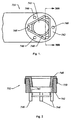
- Figures 1 to 4 illustrate an electrosurgical probe having a vent/opening that assists in prevention of accumulated heat between the tissue treatment surface and the tissue being treated.
- High frequency (RF) electrical energy is applied to one or more active electrodes in the presence of electrically conductive fluid to remove and/or modify body tissue.
- the techniques may be performed in a conventional open surgery environment or in a minimally invasive manner using cannulas or port access devices.
- the present invention is useful in procedures where the tissue site is flooded or submerged with an electrically conducting fluid, such as arthroscopic surgery of the knee, shoulder, ankle, hip, elbow, hand or foot.
- the present invention is useful in the resection and/or ablation of the meniscus and the synovial tissue within a joint during an arthroscopic procedure.
- tissues which may be treated by the system and method of the present invention include, but are not limited to, prostate tissue and leiomyomas (fibroids) located within the uterus, gingival tissues and mucosal tissues located in the mouth, tumors, scar tissue, myocardial tissue, collagenous tissue within the eye or epidermal and dermal tissues on the surface of the skin.
- the present invention is also useful for resecting tissue within accessible sites of the body that are suitable for electrode loop resection, such as the resection of prostate tissue, leiomyomas (fibroids) located within the uterus and other diseased tissue within the body.
- the present invention is particularly useful for treating tissue in the head and neck, such as the ear, mouth, pharynx, larynx, esophagus, nasal cavity and sinuses.
- the head and neck procedures may be performed through the mouth or nose using speculae or gags, or using endoscopic techniques, such as functional endoscopic sinus surgery (FESS).
- FESS functional endoscopic sinus surgery
- These procedures may include the removal of swollen tissue, chronically-diseased inflamed and hypertrophic mucus linings, polyps, turbinates and/or neoplasms from the various anatomical sinuses of the skull, the turbinates and nasal passages, in the tonsil, adenoid, epi-glottic and supra-glottic regions, and salivary glands, submucus resection of the nasal septum, excision of diseased tissue and the like.
- the present invention may be useful for collagen shrinkage, ablation and/or hemostasis in procedures for treating swollen tissue (e.g., turbinates) or snoring and obstructive sleep apnea (e.g., soft palate, such as the uvula, or tongue/pharynx stiffening, and midline glossectomies), for gross tissue removal, such as tonsillectomies, adenoidectomies, tracheal stenosis and vocal cord polyps and lesions, or for the resection or ablation of facial tumors or tumors within the mouth and pharynx, such as glossectomies, laryngectomies, acoustic neuroma procedures and nasal ablation procedures.
- the present invention is useful for procedures within the ear, such as stapedotomies, tympanostomies or the like.
- the present invention may also be useful for treating tissue or other body structures in the brain or spine.
- These procedures include tumor removal, laminectomy/disketomy procedures for treating herniated disks, decompressive laminectomy for stenosis in the lumbosacral and cervical spine, medial facetectomy, posterior lumbosacral and cervical spine fusions, treatment of scoliosis associated with vertebral disease, foraminotomies to remove the roof of the intervertebral foramina to relieve nerve root compression and anterior cervical and lumbar diskectomies.
- These procedures may be performed through open procedures, or using minimally invasive techniques, such as thoracoscopy, arthroscopy, laparascopy or the like.
- the present invention may also be useful for maintaining patency in body passages subject to occlusion by invasive tissue growth.
- the apparatus and methods of the present invention may be used to open and maintain patency in virtually any hollow body passage which may be subject to occlusion by invasive cellular growth or invasive solid tumor growth.
- Suitable hollow body passages include ducts, orifices, lumens, and the like, with exemplary body passages including the coronary arteries.
- the present invention is particularly useful for reducing or eliminating the effects of restenosis in coronary arteries by selectively removing tissue ingrowth in or around intraluminal prostheses or stents anchored therein.
- the present invention may also be useful for cosmetic and plastic surgery procedures in the head and neck.
- the present invention is particularly useful for ablation and sculpting of cartilage tissue, such as the cartilage within the nose that is sculpted during rhinoplasty procedures.
- the present invention may also be employed for skin tissue removal and/or collagen shrinkage in the epidermis or dermis tissue in the head and neck, e.g., the removal of pigmentations, vascular lesions (e.g., leg veins), scars, tattoos, etc., and for other surgical procedures on the skin, such as tissue rejuvenation, cosmetic eye procedures (blepharoplasties), wrinkle removal, tightening muscles for facelifts or browlifts, hair removal and/or transplant procedures, etc.
- tissue rejuvenation e.g., cosmetic eye procedures (blepharoplasties), wrinkle removal, tightening muscles for facelifts or browlifts, hair removal and/or transplant procedures, etc.
- the remaining disclosure will be directed specifically to the treatment of tissue structures within a joint, e.g., arthroscopic surgery, but it will be appreciated that the system and method can be applied equally well to procedures involving other tissues of the body, as well as to other procedures including open procedures, intravascular procedures, interventional cardiology procedures, urology, laparascopy, arthroscopy, thoracoscopy or other cardiac procedures, cosmetic surgery, orthopedics, gynecology, otorhinolaryngology, spinal and neurologic procedures, oncology and the like.
- Described herein is a method in which the body tissue is volumetrically removed or ablated.
- a high frequency voltage difference is applied between one or more active electrode(s) and one or more return electrode(s) to non-thermally develop a high electric field intensities in the vicinity of the target tissue.
- the high electric field intensities adjacent the active electrode(s) lead to electric field induced molecular breakdown of target tissue through molecular dissociation (rather than thermal evaporation or carbonization).
- Applicant believes that the tissue structure is volumetrically removed through molecular disintegration of larger organic molecules into smaller molecules and/or atoms, such as hydrogen, oxygen, oxides of carbon, hydrocarbons and nitrogen compounds. This molecular disintegration completely removes the tissue structure, as opposed to dehydrating the tissue material by the removal of liquid within the cells of the tissue, as is typically the case with electrosurgical desiccation and vaporization.
- the high electric field intensities may be generated by applying a high frequency voltage that is sufficient to vaporize an electrically conducting fluid over at least a portion of the active electrode(s) in the region between the distal tip of the active electrode(s) and the target tissue.
- the active electrode(s) are maintained at a low temperature to prevent thermal damage to the surrounding tissue and to improve the ionization of the electrically conductive fluid.
- the electrically conductive fluid may be a liquid or gas, such as isotonic saline or blood, delivered to the target site, or a viscous fluid, such as a gel, applied to the target site.
- the vapor layer or vaporized region Since the vapor layer or vaporized region has a relatively high electrical impedance, it increases the voltage differential between the active electrode tip and the tissue and causes ionization within the vapor layer due to the presence of an ionizable species (e.g., sodium when isotonic saline is the electrically conducting fluid).
- This ionization under the conditions described herein, induces the discharge of energetic electrons and photons from the vapor layer and to the surface of the target tissue.
- This energy may be in the form of energetic photons (e.g., ultraviolet radiation), energetic particles (e.g., electrons or ions) or a combination thereof.
- Coblation® A more detailed description of this phenomena, termed Coblation® can be found in commonly assigned U.S. Patent No. 5,697,882
- Applicant believes that the principle mechanism of tissue removal in the Coblation® mechanism of the present invention is energetic electrons or ions that have been energized in a plasma adjacent to the active electrode(s).
- a liquid is heated enough that atoms vaporize off the surface faster than they recondense, a gas is formed.
- the gas is heated enough that the atoms collide with each other and knock their electrons off in the process, an ionized gas or plasma is formed (the so-called "fourth state of matter").
- a more complete description of plasma can be found in Plasma Physics, by R.J. Goldston and P.H. Rutherford of the Plasma Physics Laboratory of Princeton University (1995).
- the electron mean free path increases to enable subsequently injected electrons to cause impact ionization within these regions of low density (i.e., vapor layers or bubbles).
- the ionic particles in the plasma layer have sufficient energy, they accelerate towards the target tissue.
- Energy evolved by the energetic electrons e.g., 3.5 eV to 5 eV
- Plasmas may be formed by heating a small of gas and ionizing the gas by driving an electric current through it, or by shining radio waves into the gas.
- these methods of plasma formation give energy to free electrons in the plasma directly, and then electron-atom collisions liberate more electrons, and the process cascades until the desired degree of ionization is achieved.
- the electrons carry the electrical current or absorb the radio waves and, therefore, are hotter than the ions.
- the electrons which are carried away from the tissue towards the return electrode, carry most of the plasma's heat with them, allowing the ions to break apart the tissue molecules in a substantially non-thermal manner.
- a method is described herein which applies high frequency (RF) electrical energy in an electrically conducting fluid environment to remove (i.e., resect, cut or ablate) a tissue structure and to seal transected vessels within the region of the target tissue.
- the present invention is particularly useful for sealing larger arterial vessels, e.g., on the order of 1 mm or greater.
- a high frequency power supply is provided having an ablation mode, wherein a first voltage is applied to an active electrode sufficient to effect molecular dissociation or disintegration of the tissue, and a coagulation mode, wherein a second, lower voltage is applied to an active electrode (either the same or a different electrode) sufficient to achieve hemostasis of severed vessels within the tissue.
- an electrosurgical instrument having one or more coagulation electrode(s) configured for sealing a severed vessel, such as an arterial vessel, and one or more active electrodes configured for either contracting the collagen fibers within the tissue or removing (ablating) the tissue, e.g., by applying sufficient energy to the tissue to effect molecular dissociation.
- the coagulation electrode(s) may be configured such that a single voltage can be applied to coagulate with the coagulation electrode(s), and to ablate with the active electrode(s).
- the power supply is combined with the coagulation instrument such that the coagulation electrode is used when the power supply is in the coagulation mode (low voltage), and the active electrode(s) are used when the power supply is in the ablation mode (higher voltage).
- the present invention comprises platinum or platinum-iridium electrodes to deliver the high frequency energy to the conductive fluid.
- the platinum/platinum-iridium electrodes have a low resistivity and low thermal conductivity so as to minimize the production of heat in the electrodes and the electrically conductive fluid. This allows more electrical energy to be applied directly into the conductive fluid.
- the tip region of the instrument may comprise many independent active electrodes designed to deliver electrical energy in the vicinity of the tip.
- the selective application of electrical energy to the conductive fluid is achieved by connecting each individual active electrode and the return electrode to a power source having independently controlled or current limited channels.
- the return electrode(s) may comprise a single tubular member of conductive material proximal to the electrode array at the tip which also serves as a conduit for the supply of the electrically conducting fluid between the active and return electrodes.
- the instrument may comprise an array of return electrodes at the distal tip of the instrument (together with the active electrodes) to maintain the electric current at the tip.
- the application of high frequency voltage between the return clectrode(s) and the electrode array results in the generation of high electric field intensities at the distal tips of the active electrodes with conduction of high frequency current from each individual active electrode to the return electrode.
- the current flow from each individual active electrode to the return electrode(s) is controlled by either active or passive means, or a combination thereof, to deliver electrical energy to the surrounding conductive fluid while minimizing energy delivery to surrounding (non-target) tissue.
- the application of a high frequency voltage between the return platinum electrode(s) and the active platinum electrode(s) for appropriate time intervals effects cutting, removing, ablating, shaping, contracting or otherwise modifying the target tissue.
- the tissue volume over which energy is dissipated i.e., a high current density exists
- the tissue volume over which energy is dissipated may be more precisely controlled, for example, by the use of a multiplicity of small active platinum electrodes whose effective diameters or principal dimensions range from about 10 mm to 0.01 mm, preferably from about 2 mm to 0.05 mm, and more preferably from about 1 mm to 0.1 mm.
- Electrode areas for both circular and non-circular terminals will have a contact area (per active electrode) below 50 mm 2 for electrode arrays and as large as 75 mm 2 for single electrode embodiments.
- the contact area of each active electrode is typically in the range from 0.0001 mm 2 to 1 mm 2 , and more preferably from 0.001 mm 2 to .5 mm 2 .
- the circumscribed area of the electrode array or active electrode is in the range from 0.25 mm 2 to 75 mm 2 , preferably from 0.5 mm 2 to 40 mm 2 .
- the array will usually include at least two isolated active electrodes, often at least five active electrodes, often greater than 10 active electrodes and even 50 or more active electrodes, disposed over the distal contact surfaces on the shaft.
- the use of small diameter active electrodes increases the electric field intensity and reduces the extent or depth of tissue heating as a consequence of the divergence of current flux lines which emanate from the exposed surface of each active electrode.
- the area of the tissue treatment surface can vary widely, and the tissue treatment surface can assume a variety of geometries, with particular areas and geometries being selected for specific applications.
- the geometries can be planar, concave, convex, hemispherical, conical, linear "in-line” array or virtually any other regular or irregular shape.
- the active electrode(s) or active electrode(s) will be formed at the distal tip of the electrosurgical instrument shaft, frequently being planar, disk-shaped, or hemispherical surfaces for use in reshaping procedures or being linear arrays for use in cutting.
- the active electrode(s) may be formed on lateral surfaces of the electrosurgical instrument shaft (e. g., in the manner of a spatula), facilitating access to certain body structures in endoscopic procedures.
- the electrode support and the fluid outlet may be recessed from an outer surface of the instrument or handpiece to confine the electrically conductive fluid to the region immediately surrounding the electrode support.
- the shaft may be shaped so as to form a cavity around the electrode support and the fluid outlet. This helps to assure that the electrically conductive fluid will remain in contact with the active electrode(s) and the return electrode(s) to maintain the conductive path therebetween. In addition, this will help to maintain a vapor layer and subsequent plasma layer between the active electrode(s) and the tissue at the treatment site throughout the procedure, which reduces the thermal damage that might otherwise occur if the vapor layer were extinguished due to a lack of conductive fluid. Provision of the electrically conductive fluid around the target site also helps to maintain the tissue temperature at desired levels.
- the active electrodes are spaced from the tissue a sufficient distance to minimize or avoid contact between the tissue and the vapor layer formed around the active electrodes.
- contact between the heated electrons in the vapor layer and the tissue is minimized as these electrons travel from the vapor layer back through the conductive fluid to the return electrode.
- the ions within the plasma will have sufficient energy, under certain conditions such as higher voltage levels, to accelerate beyond the vapor layer to the tissue.
- the tissue bonds are dissociated or broken as in previous embodiments, while minimizing the electron flow, and thus the thermal energy, in contact with the tissue.
- the electrically conducting fluid should have a threshold conductivity to provide a suitable conductive path between the return electrode and the active electrode(s).
- the electrical conductivity of the fluid (in units of milliSiemans per centimeter or mS/cm) will usually be greater than 0.2 mS/cm, preferably will be greater than 2 mS/cm and more preferably greater than 10 mS/cm.
- the electrically conductive fluid is isotonic saline, which has a conductivity of about 17 mS/cm. Applicant has found that a more conductive fluid, or one with a higher ionic concentration, will usually provide a more aggressive ablation rate.
- a saline solution with higher levels of sodium chloride than conventional saline (which is on the order of about .9% sodium chloride) e.g., on the order of greater than 1% or between about 3% and 20%, may be desirable.
- the invention may be used with different types of conductive fluids that increase the power of the plasma layer by, for example, increasing the quantity of ions in the plasma, or by providing ions that have higher energy levels than sodium ions.
- the present invention may be used with elements other than sodium, such as potassium, magnesium, calcium and other metals near the left end of the periodic chart.
- other electronegative elements may be used in place of chlorine, such as fluorine.
- the voltage difference applied between the return electrode(s) and the active electrode(s) will be at high or radio frequency, typically between about 5 kHz and 20 MHz, usually being between about 30 kHz and 2.5 MHz, preferably being between about 50 kHz and 500 kHz, often less than 350 kHz, and often between about 100 kHz and 200 kHz.
- a frequency of about 100 kHz is useful because the tissue impedance is much greater at this frequency.
- higher frequencies may be desirable (e.g., 400 kHz-600 kHz) to minimize low frequency current flow into the heart or the nerves of the head and neck.
- the RMS (root mean square) voltage applied will usually be in the range from about 5 volts to 1000 volts, preferably being in the range from about 10 volts to 500 volts, often between about 150 volts to 350 volts depending on the active electrode size, the operating frequency and the operation mode of the particular procedure or desired effect on the tissue (i.e., contraction, coagulation, cutting or ablation).
- the peak-to-peak voltage for ablation or cutting with a square wave form will be in the range of 10 volts to 2000 volts and preferably in the range of 100 volts to 1800 volts and more preferably in the range of about 300 to 1500 volts, often in the range of about 300 volts to 800 volts peak to peak (again, depending on the electrode size, the operating frequency and the operation mode).
- Lower peak-to-peak voltages will be used for tissue coagulation or collagen contraction and will typically be in the range from 50 to 1500, preferably 100 to 1000 and more preferably 120 to 400 volts peak-to-peak (again, these values are computed using a square wave form).
- Peak-to-peak voltages e.g., greater than about 700 volts peak-to-peak, may be desirable for ablation of harder material, such as bone, depending on other factors, such as the electrode geometries and the composition of the conductive fluid.
- the voltage is usually delivered in a series of voltage pulses or alternating current of time varying voltage amplitude with a sufficiently high frequency (e.g., on the order of 5 kHz to 20 MHz) such that the voltage is effectively applied continuously (as compared with e.g., lasers claiming small depths of necrosis, which are generally pulsed about 10 Hz to 20 Hz).
- the duty cycle i.e., cumulative time in any one-second interval that energy is applied
- the preferred power source of the present invention delivers a high frequency current selectable to generate average power levels ranging from several milliwatts to tens of watts per electrode, depending on the volume of target tissue being heated, and/or the maximum allowed temperature selected for the instrument tip.
- the power source allows the user to select the voltage level according to the specific requirements of a particular neurosurgery procedure, cardiac surgery, arthroscopic surgery, dermatological procedure, ophthalmic procedures, open surgery or other endoscopic surgery procedure.
- the power source may have an additional filter, for filtering leakage voltages at frequencies below 100 kHz, particularly voltages around 60 kHz.
- a power source having a higher operating frequency e.g., 300 to 600 kHz may be used in certain procedures in which stray low frequency currents may be problematic.
- the power source may be current limited or otherwise controlled so that undesired heating of the target tissue or surrounding (non-target) tissue does not occur.
- current limiting inductors are placed in series with each independent active electrode, where the inductance of the inductor is in the range of 10uH to 50,000uH, depending on the electrical properties of the target tissue, the desired tissue heating rate and the operating frequency.
- capacitor-inductor (LC) circuit structures may be employed, as described previously in U.S. Patent No. 5,697,909 , Additionally, current limiting resistors may be selected.
- these resistors will have a large positive temperature coefficient of resistance so that, as the current level begins to rise for any individual active electrode in contact with a low resistance medium (e.g., saline irrigant or blood), the resistance of the current limiting resistor increases significantly, thereby minimizing the power delivery from said active electrode into the low resistance medium (e.g., saline irrigant or blood).
- a low resistance medium e.g., saline irrigant or blood
- the invention is not limited to electrically isolated active platinum electrodes, or even to a plurality of active platinum electrodes.
- the array of active platinum electrodes may be connected to a single lead that extends through the catheter shaft to a power source of high frequency current.
- the instrument may incorporate a single electrode that extends directly through the catheter shaft or is connected to a single lead that extends to the power source.
- the active electrode(s) may have ball shapes (e.g., for tissue vaporization and desiccation), twizzle shapes (for vaporization and needle-like cutting), spring shapes (for rapid tissue debulking and desiccation), twisted metal shapes, annular or solid tube shapes or the like.
- the electrode(s) may comprise a plurality of filaments, rigid or flexible brush electrode(s) (for debulking a tumor, such as a fibroid, bladder tumor or a prostate adenoma), side-effect brush electrode(s) on a lateral surface of the shaft, coiled electrode(s) or the like.
- Figures 1 and 2 illustrate variations of the invention in which the probe 751 includes an opening 741 in the center of tissue treatment surface 742, and a plurality of active electrodes 744 around the perimeter of surface 742.
- Fig. 1 shows a bottom view of the tissue treatment surface 742 of the probe.
- the opening 741 functions as a vent to prevent heat from accumulating adjacent to the tissue treatment surface 742.
- the vent/opening 741 may be also configured such that the internal profile facilitates the transfer of heat (e.g., convective, conductive, radiative heat transfer).
- the internal profile of the opening 741 may have a draft angle, taper, or other such design to generate a venturi effect.
- the opening 741 is designed such that heat will not be trapped (e.g., the passage of the opening exits through the side of the device opposite to the tissue treatment surface.)
- the opening may have a separate exhaust port allowing for the venting of heat.
- the opening 741 may be combined with other features of the device (e.g., fluid supply source, suction ports, return electrode, etc.) while still retaining an exhaust function.
- the venting will permit increased fluid circulation causing cooling and evacuation of gas bubbles (by products of tissue ablation). Accordingly, such venting may also increase visibility at the target site.
- opening 741 Another function of the opening 741 is that the absence of material at the opening 741 prevents radiated heat (generated by the ablation process) from being reflected between the tissue being ablated and the tissue treatment surface 742 of the device.
- the vent or opening 741 allows the heat being generated by the ablation process to escape rather than being reflected back to tissue. Without a vent, the radiated heat may be reflected back onto the tissue from the tissue treatment surface of the probe causing undesired collateral heating of the tissue.
- Fig. 2 illustrates a side view of the probe of Fig. 1 .
- the device may also contain spacers 746, which, as discussed above, serve to offset the active electrodes 744 from the tissue.
- the spacers 746 may be formed from as part of an insert 748 (e.g., a silicone molded insert) that is placed within the top of the vent or opening 741 where the spacers 746 are protrusions that extend through the tissue treatment surface 742 of the device.
- Fig. 2 also shows the probe as having a return electrode 750 disposed about an outside of the probe. However, as discussed throughout this disclosure, the placement of the return electrode 750 is not limited as such.
- the return electrode 750 may be placed within the opening 741 of the device.
- a fluid source (not shown) may be placed within the opening 741, along an outside surface of the probe, or the fluid source may be external to the device.
- the opening 741 may also be coupled to a suction port (not shown) as described above.
- the devices described herein may also incorporate another feature to reduce the accumulation of heat at the site of the tissue being treated.
- heat generated by the ablation process may be reflected from the tissue to a tissue treatment surface of the device and back to the tissue.
- the devices of the present invention may incorporate probes having tissue treatment surfaces or electrode support members that are non-reflective.
- materials or coatings may be selected such that heat radiated to the tissue treatment surface or electrode support member is absorbed or transmitted as opposed to being reflected.
- glass, ceramic, silicone elastomers, polymer elastomers, silicon nitride, glass used silicon sapphire may be used as material for the electrode support member.
- Figures 3 and 4 illustrate a distal portion of another probe 800 for ablating tissue.
- Figure 3 is a perspective view and Figures 4A and 4B are top and side views respectively.
- the probe includes an atraumatic head.
- a support 820 is connected to the shaft 810.
- the support 820 may be an electrically nonconductive material such as ceramic or a polymer.
- a spacer 860 may separate and further affix the support 820 to shaft 810.
- the spacer may be rigid and formed of, for example, polycarbonate or another material.
- the support 820 has a cavity or recess for holding active electrode 840.
- the circular cavity or recess in the support 820 provides, when an electrode is contained therein, a substantially flush active (or working) surface to treat tissue.
- the active electrode may be spaced a predetermined distance from the target tissue by properly pre-selecting the depth of the cavity and size of the electrode.
- the support 820 may include one or more cut-out regions 822 for fluid to flow through As discussed above, it is desirable to provide an electroconductive fluid around the active electrode.
- the active electrode 840 is shown as a loop electrode, it may take other shapes.
- the active electrode has a shape that cooperatively fits recess in support 820.
- the shape of the recess may vary widely. It may be serpentine, rectangular, circular, etc. Additionally, more than one electrode may be included in the probe. There may be more than one cavity in the support. Also, the electrode may be made of materials such as, but not limited to, tungsten.
- the return electrode 830 is shown having a clip shape. It is connected to a power supply via one or more conducting members. Examples of materials for the return electrode are stainless steel, copper, alloys and other conducting materials. As shown, the clip surrounds the body of the support 820 but does not cover either the active (upper) or inactive (lower) sides. Accordingly, when treating a tissue in a yarrow space such that both the active and inactive sides contact tissue, tissue at only the active side is ablated because the plasma does not extend to the lower side of the support 820.
- vent aperture 850 Another aspect of the probe shown in Figures 3 to 4 is vent aperture 850. This aperture allows bubbles that are formed during a procedure to escape from the tissue treatment zone. It is desirable to vent the bubbles from the target area because the bubbles may affect visibility, temperature and the overall effectiveness of the ablation process.
Landscapes
- Health & Medical Sciences (AREA)
- Surgery (AREA)
- Engineering & Computer Science (AREA)
- Life Sciences & Earth Sciences (AREA)
- Heart & Thoracic Surgery (AREA)
- Animal Behavior & Ethology (AREA)
- Nuclear Medicine, Radiotherapy & Molecular Imaging (AREA)
- Otolaryngology (AREA)
- Veterinary Medicine (AREA)
- Public Health (AREA)
- Physics & Mathematics (AREA)
- Biomedical Technology (AREA)
- Medical Informatics (AREA)
- Molecular Biology (AREA)
- Plasma & Fusion (AREA)
- General Health & Medical Sciences (AREA)
- Neurosurgery (AREA)
- Neurology (AREA)
- Surgical Instruments (AREA)
Abstract
Description
- The present invention relates generally to the field of surgical devices which employ high frequency voltage to cut and ablate body tissue.
- Conventional electrosurgical methods are widely used since they generally reduce patient bleeding associated with tissue cutting operations and improve the surgeon's visibility. These traditional electrosurgical techniques for treatment have typically relied on thermal methods to rapidly heat and vaporize liquid within tissue and to cause cellular destruction In conventional monopolar electrosurgery, for example, electric current is directed along a defined path from the exposed or active electrode through the patient's body to the return electrode, which is externally attached to a suitable location on the patient's skin. In addition, since the defined path through the patient's body has a relatively high electrical impedance, large voltage differences must typically be applied between the active and return electrodes to generate a current suitable for cutting or coagulation of the target tissue. This current, however, may inadvertently flow along localized pathways in the body having less impedance than the defined electrical path. This situation will substantially increase the current flowing through these paths, possibly causing damage to or destroying tissue along and surrounding this pathway.
- Bipolar electrosurgical devices have an inherent advantage over monopolar devices because the return current path does not flow through the patient beyond the immediate site of application of the bipolar electrodes. In bipolar devices, both the active and return electrode are typically exposed so that they may both contact tissue, thereby providing a return current path from the active to the return electrode through the tissue. One drawback with this configuration, however, is that the return electrode may cause tissue desiccation or destruction at its contact point with the patient's tissue.
- Another limitation of conventional bipolar and monopolar electrosurgery devices is that they are not suitable for the precise removal (i.e., ablation) or tissue. For example, conventional electrosurgical cutting devices typically operate by creating a voltage difference between the active electrode and the target tissue, causing an electrical arc to form across the physical gap between the electrode and tissue. At the point of contact of the electric arcs with tissue, rapid tissue heating occurs due to high current density between the electrode and tissue. This high current density causes cellular fluids to rapidly vaporize into stearn, thereby producing a "cutting effect" along the pathway of localized tissue heating. The tissue is parted along the pathway of evaporated cellular fluid, inducing undesirable collateral tissue damage in regions surrounding the target tissue site.
- The use of electrosurgical procedures (both monopolar and bipolar) in electrically conductive environments can be further problematic. For example, many arthroscopic procedures require flushing of the region to be treated with isotonic saline, both to maintain an isotonic environment and to keep the field of view clear. However, the presence of saline, which is a highly conductive electrolyte, can cause shorting of the active electrode(s) in conventional monopolar and bipolar electrosurgery. Such shorting causes unnecessary heating in the treatment environment and can further cause non specific tissue destruction.
- Present electrosurgical techniques used for tissue ablation also suffer from an inability to control the depth of necrosis in the tissue being treated. Most electrosurgical devices rely on creation of an electric arc between the treating electrode and the tissue being cut or ablated to cause the desired localized heating. Such arcs, however, often create very high temperatures causing a depth of necrosis greater than 500 µm, frequently greater than 800 µm, and sometimes as great as 1700 µm The inability to control such depth of necrosis is a significant disadvantage in using electrosurgical techniques for tissue ablation, particularly in arthroscopic procedures for ablating and/or reshaping fibrocartilage, articular cartilage, meniscal tissue, and the like.
- In an effort to overcome at least some of these limitations of electrosurgery, laser apparatus have been developed for use in arthroscopic and other surgical procedures. Lasers do not suffer from electrical shorting in conductive environments, and certain types of lasers allow for very controlled cutting with limited depth of necrosis. Despite these advantages, laser devices suffer from their own set of deficiencies. In the first place, laser equipment can be very expensive because of the costs associated with the laser light sources. Moreover, those lasers which permit acceptable depths of necrosis (such as excimer lasers, erbium:YAG lasers, and the like) provide a very low volumetric ablation rate, which is a particular disadvantage in cutting and ablation of fibrocartilage, articular cartilage, and meniscal tissue. The holmium:YAG and Nd:YAG lasers provide much higher volumetric ablation rates, but are much less able to control depth of necrosis than are the slower laser devices. The CO2 lasers provide high rate of ablation and low depth of tissue necrosis, but cannot operate in a liquid-filled cavity.
- Excimer lasers, which operate in an ultraviolet wavelength, cause photodissociation of human tissue, commonly referred to as cold ablation. Through this mechanism, organic molecules can be disintegrated into light hydrocarbon gases that are removed from the target site. Such photodissociation reduces the likelihood of thermal damage to tissue outside of the target site. Although promising, excimer lasers must be operated in pulses so that ablation plumes created during operation can clear. This prevents excessive secondary heating of the plume of ablation products which can increase the likelihood of collateral tissue damage as well as a decrease in the rate of ablation. Unfortunately, the pulsed mode of operation reduces the volumetric ablation rate, which may increase the time spent in surgery.
United States Patent No.6,210,405 describes an electrode assembly at one end of a shaft, the electrode assembly comprising a tissue treatment electrode and a return electrode which is insulated from the treatment electrode by means of an insulation member. The tissue treatment electrode has an exposed end for treating tissue, and the return electrode has a fluid contact surface which is spaced from the tissue treatment electrode in such a manner as to define a conductive fluid path that completes an electrical circuit between the tissue treatment electrode and the return electrode.
United States Patent No.2002/0072739 describes an electrically activated surgical device which includes a prove body, an active element and a structure for selectively electrically insulating and/or physically isolating the active element from the patient's tissue when the device is in use.
United States Patent No.5,810,764 describes an electrosurgical resecting instrument having a resecting loop electrode on the distal end of a shaft and coupled to a high frequency voltage source. The loop electrode is configured to fit within a working end of a resectoscope and to remove small portions of tissue (e.g., chips of tissue). - Aspects and examples of the invention are set out in the claims.
- Also described herein are systems, apparatus and methods for selectively applying electrical energy to body tissue. In particular, systems and methods are provided for applying a high frequency voltage in the presence of an electrically conductive fluid to create a relatively low-temperature plasma for ablation of tissue adjacent to, or in contact with, the plasma.
- There is described herein a method comprising positioning an electrosurgical probe or catheter adjacent the target site so that one or more active electrode(s) are brought into contact with, or close proximity to, a target tissue in the presence of electrically conductive fluid. The electrically conductive fluid may be delivered directly to the active electrode(s) and the target tissue, or the entire target site may be submersed within the conductive fluid. High frequency voltage is then applied between the active electrode(s) and one or more return electrode(s) to generate a plasma adjacent to the active electrode(s) while maintaining a low temperature in the active electrode(s). At least a portion of the target tissue is volumetrically removed or ablated. The high frequency voltage generates electric fields around the active electrode(s) with sufficient energy to ionize the conductive fluid adjacent to the active electrode(s). Within the ionized gas or plasma, free electrons are accelerated, and electron-atoms collisions liberate more electrons, and the process cascades until the plasma contains sufficient energy to break apart the tissue molecules, causing molecular dissociation and ablation of the target tissue.
- In a specific configuration the electrosurgical probe comprises platinum or platinum-iridium alloy electrodes. Typically, the platinum-iridium electrodes comprise between approximately 1% and 30%, and preferably between 5% and 15% iridium to mechanically strengthen the electrodes. Applicants have found that the platinum/platinum-iridium electrodes provide more efficient ionization of the conductive fluid, less thermal heating of surrounding tissue, and overall superior ablation. Because platinum has a low thermal conductivity and low resistivity, heat production is minimized and there is a more efficient transfer of energy into the conductive fluid to create the plasma. As an additional benefit, Applicants have found that platinum/platinum-iridium electrodes have better corrosion properties and oxidation properties in the presence of the conductive fluid over other electrode materials.
- In some embodiments, the high frequency voltage applied to the active electrode(s) is sufficient to non-thermally vaporize the electrically conductive fluid (e.g., gel or saline) between the active electrode(s) and the tissue. Within the vaporized fluid, an ionized plasma is formed and charged particles (e.g., electrons) are accelerated towards the tissue to cause the molecular breakdown or disintegration of several cell layers of the tissue. This molecular dissociation is accompanied by the volumetric removal of the tissue. The short range of the accelerated charged particles within the plasma layer confines the molecular dissociation process to the surface layer to minimize damage and necrosis to the surrounding and underlying tissue. This process can be precisely controlled to effect the volumetric removal of tissue as thin as 10 to 150 microns with minimal heating of, or damage to, surrounding or underlying tissue structures. A more complete description of this phenomena is described in commonly assigned
U.S. Patent No. 5,697,882 . -
Figures 1 to 4 illustrate an electrosurgical probe having a vent/opening that assists in prevention of accumulated heat between the tissue treatment surface and the tissue being treated. - High frequency (RF) electrical energy is applied to one or more active electrodes in the presence of electrically conductive fluid to remove and/or modify body tissue. The techniques may be performed in a conventional open surgery environment or in a minimally invasive manner using cannulas or port access devices. The present invention is useful in procedures where the tissue site is flooded or submerged with an electrically conducting fluid, such as arthroscopic surgery of the knee, shoulder, ankle, hip, elbow, hand or foot. Specifically, the present invention is useful in the resection and/or ablation of the meniscus and the synovial tissue within a joint during an arthroscopic procedure. In addition, tissues which may be treated by the system and method of the present invention include, but are not limited to, prostate tissue and leiomyomas (fibroids) located within the uterus, gingival tissues and mucosal tissues located in the mouth, tumors, scar tissue, myocardial tissue, collagenous tissue within the eye or epidermal and dermal tissues on the surface of the skin. The present invention is also useful for resecting tissue within accessible sites of the body that are suitable for electrode loop resection, such as the resection of prostate tissue, leiomyomas (fibroids) located within the uterus and other diseased tissue within the body.
- The present invention is particularly useful for treating tissue in the head and neck, such as the ear, mouth, pharynx, larynx, esophagus, nasal cavity and sinuses. The head and neck procedures may be performed through the mouth or nose using speculae or gags, or using endoscopic techniques, such as functional endoscopic sinus surgery (FESS). These procedures may include the removal of swollen tissue, chronically-diseased inflamed and hypertrophic mucus linings, polyps, turbinates and/or neoplasms from the various anatomical sinuses of the skull, the turbinates and nasal passages, in the tonsil, adenoid, epi-glottic and supra-glottic regions, and salivary glands, submucus resection of the nasal septum, excision of diseased tissue and the like. In other procedures, the present invention may be useful for collagen shrinkage, ablation and/or hemostasis in procedures for treating swollen tissue (e.g., turbinates) or snoring and obstructive sleep apnea (e.g., soft palate, such as the uvula, or tongue/pharynx stiffening, and midline glossectomies), for gross tissue removal, such as tonsillectomies, adenoidectomies, tracheal stenosis and vocal cord polyps and lesions, or for the resection or ablation of facial tumors or tumors within the mouth and pharynx, such as glossectomies, laryngectomies, acoustic neuroma procedures and nasal ablation procedures. In addition, the present invention is useful for procedures within the ear, such as stapedotomies, tympanostomies or the like.
- The present invention may also be useful for treating tissue or other body structures in the brain or spine. These procedures include tumor removal, laminectomy/disketomy procedures for treating herniated disks, decompressive laminectomy for stenosis in the lumbosacral and cervical spine, medial facetectomy, posterior lumbosacral and cervical spine fusions, treatment of scoliosis associated with vertebral disease, foraminotomies to remove the roof of the intervertebral foramina to relieve nerve root compression and anterior cervical and lumbar diskectomies. These procedures may be performed through open procedures, or using minimally invasive techniques, such as thoracoscopy, arthroscopy, laparascopy or the like.
- The present invention may also be useful for maintaining patency in body passages subject to occlusion by invasive tissue growth. The apparatus and methods of the present invention may be used to open and maintain patency in virtually any hollow body passage which may be subject to occlusion by invasive cellular growth or invasive solid tumor growth. Suitable hollow body passages include ducts, orifices, lumens, and the like, with exemplary body passages including the coronary arteries. The present invention is particularly useful for reducing or eliminating the effects of restenosis in coronary arteries by selectively removing tissue ingrowth in or around intraluminal prostheses or stents anchored therein.
- The present invention may also be useful for cosmetic and plastic surgery procedures in the head and neck. For example, the present invention is particularly useful for ablation and sculpting of cartilage tissue, such as the cartilage within the nose that is sculpted during rhinoplasty procedures. The present invention may also be employed for skin tissue removal and/or collagen shrinkage in the epidermis or dermis tissue in the head and neck, e.g., the removal of pigmentations, vascular lesions (e.g., leg veins), scars, tattoos, etc., and for other surgical procedures on the skin, such as tissue rejuvenation, cosmetic eye procedures (blepharoplasties), wrinkle removal, tightening muscles for facelifts or browlifts, hair removal and/or transplant procedures, etc.
- For convenience, the remaining disclosure will be directed specifically to the treatment of tissue structures within a joint, e.g., arthroscopic surgery, but it will be appreciated that the system and method can be applied equally well to procedures involving other tissues of the body, as well as to other procedures including open procedures, intravascular procedures, interventional cardiology procedures, urology, laparascopy, arthroscopy, thoracoscopy or other cardiac procedures, cosmetic surgery, orthopedics, gynecology, otorhinolaryngology, spinal and neurologic procedures, oncology and the like.
- Described herein is a method in which the body tissue is volumetrically removed or ablated. In this procedure, a high frequency voltage difference is applied between one or more active electrode(s) and one or more return electrode(s) to non-thermally develop a high electric field intensities in the vicinity of the target tissue. The high electric field intensities adjacent the active electrode(s) lead to electric field induced molecular breakdown of target tissue through molecular dissociation (rather than thermal evaporation or carbonization). Applicant believes that the tissue structure is volumetrically removed through molecular disintegration of larger organic molecules into smaller molecules and/or atoms, such as hydrogen, oxygen, oxides of carbon, hydrocarbons and nitrogen compounds. This molecular disintegration completely removes the tissue structure, as opposed to dehydrating the tissue material by the removal of liquid within the cells of the tissue, as is typically the case with electrosurgical desiccation and vaporization.
- The high electric field intensities may be generated by applying a high frequency voltage that is sufficient to vaporize an electrically conducting fluid over at least a portion of the active electrode(s) in the region between the distal tip of the active electrode(s) and the target tissue. Preferably, the active electrode(s) are maintained at a low temperature to prevent thermal damage to the surrounding tissue and to improve the ionization of the electrically conductive fluid. The electrically conductive fluid may be a liquid or gas, such as isotonic saline or blood, delivered to the target site, or a viscous fluid, such as a gel, applied to the target site. Since the vapor layer or vaporized region has a relatively high electrical impedance, it increases the voltage differential between the active electrode tip and the tissue and causes ionization within the vapor layer due to the presence of an ionizable species (e.g., sodium when isotonic saline is the electrically conducting fluid). This ionization, under the conditions described herein, induces the discharge of energetic electrons and photons from the vapor layer and to the surface of the target tissue. This energy may be in the form of energetic photons (e.g., ultraviolet radiation), energetic particles (e.g., electrons or ions) or a combination thereof. A more detailed description of this phenomena, termed Coblation® can be found in commonly assigned
U.S. Patent No. 5,697,882 - Applicant believes that the principle mechanism of tissue removal in the Coblation® mechanism of the present invention is energetic electrons or ions that have been energized in a plasma adjacent to the active electrode(s). When a liquid is heated enough that atoms vaporize off the surface faster than they recondense, a gas is formed. When the gas is heated enough that the atoms collide with each other and knock their electrons off in the process, an ionized gas or plasma is formed (the so-called "fourth state of matter"). A more complete description of plasma can be found in Plasma Physics, by R.J. Goldston and P.H. Rutherford of the Plasma Physics Laboratory of Princeton University (1995). When the density of the vapor layer (or within a bubble formed in the electrically conducting liquid) becomes sufficiently low (i.e., less than approximately 1020 atoms/cm3 for aqueous solutions), the electron mean free path increases to enable subsequently injected electrons to cause impact ionization within these regions of low density (i.e., vapor layers or bubbles). Once the ionic particles in the plasma layer have sufficient energy, they accelerate towards the target tissue. Energy evolved by the energetic electrons (e.g., 3.5 eV to 5 eV) can subsequently bombard a molecule and break its bonds, dissociating a molecule into free radicals, which then combine into final gaseous or liquid species.
- Plasmas may be formed by heating a small of gas and ionizing the gas by driving an electric current through it, or by shining radio waves into the gas. Generally, these methods of plasma formation give energy to free electrons in the plasma directly, and then electron-atom collisions liberate more electrons, and the process cascades until the desired degree of ionization is achieved. Often, the electrons carry the electrical current or absorb the radio waves and, therefore, are hotter than the ions. Thus, in applicant's invention, the electrons, which are carried away from the tissue towards the return electrode, carry most of the plasma's heat with them, allowing the ions to break apart the tissue molecules in a substantially non-thermal manner.
- A method is described herein which applies high frequency (RF) electrical energy in an electrically conducting fluid environment to remove (i.e., resect, cut or ablate) a tissue structure and to seal transected vessels within the region of the target tissue. The present invention is particularly useful for sealing larger arterial vessels, e.g., on the order of 1 mm or greater. A high frequency power supply is provided having an ablation mode, wherein a first voltage is applied to an active electrode sufficient to effect molecular dissociation or disintegration of the tissue, and a coagulation mode, wherein a second, lower voltage is applied to an active electrode (either the same or a different electrode) sufficient to achieve hemostasis of severed vessels within the tissue. In other embodiments, an electrosurgical instrument is provided having one or more coagulation electrode(s) configured for sealing a severed vessel, such as an arterial vessel, and one or more active electrodes configured for either contracting the collagen fibers within the tissue or removing (ablating) the tissue, e.g., by applying sufficient energy to the tissue to effect molecular dissociation. In the latter embodiments, the coagulation electrode(s) may be configured such that a single voltage can be applied to coagulate with the coagulation electrode(s), and to ablate with the active electrode(s). In other embodiments, the power supply is combined with the coagulation instrument such that the coagulation electrode is used when the power supply is in the coagulation mode (low voltage), and the active electrode(s) are used when the power supply is in the ablation mode (higher voltage).
- In most embodiments, the present invention comprises platinum or platinum-iridium electrodes to deliver the high frequency energy to the conductive fluid. The platinum/platinum-iridium electrodes have a low resistivity and low thermal conductivity so as to minimize the production of heat in the electrodes and the electrically conductive fluid. This allows more electrical energy to be applied directly into the conductive fluid.
- The tip region of the instrument may comprise many independent active electrodes designed to deliver electrical energy in the vicinity of the tip. The selective application of electrical energy to the conductive fluid is achieved by connecting each individual active electrode and the return electrode to a power source having independently controlled or current limited channels. The return electrode(s) may comprise a single tubular member of conductive material proximal to the electrode array at the tip which also serves as a conduit for the supply of the electrically conducting fluid between the active and return electrodes. Alternatively, the instrument may comprise an array of return electrodes at the distal tip of the instrument (together with the active electrodes) to maintain the electric current at the tip. The application of high frequency voltage between the return clectrode(s) and the electrode array results in the generation of high electric field intensities at the distal tips of the active electrodes with conduction of high frequency current from each individual active electrode to the return electrode. The current flow from each individual active electrode to the return electrode(s) is controlled by either active or passive means, or a combination thereof, to deliver electrical energy to the surrounding conductive fluid while minimizing energy delivery to surrounding (non-target) tissue.
- The application of a high frequency voltage between the return platinum electrode(s) and the active platinum electrode(s) for appropriate time intervals effects cutting, removing, ablating, shaping, contracting or otherwise modifying the target tissue. The tissue volume over which energy is dissipated (i.e., a high current density exists) may be more precisely controlled, for example, by the use of a multiplicity of small active platinum electrodes whose effective diameters or principal dimensions range from about 10 mm to 0.01 mm, preferably from about 2 mm to 0.05 mm, and more preferably from about 1 mm to 0.1 mm. Electrode areas for both circular and non-circular terminals will have a contact area (per active electrode) below 50 mm2 for electrode arrays and as large as 75 mm2 for single electrode embodiments. In multiple electrode arrays, the contact area of each active electrode is typically in the range from 0.0001 mm2 to 1 mm2, and more preferably from 0.001 mm2 to .5 mm2. The circumscribed area of the electrode array or active electrode is in the range from 0.25 mm2 to 75 mm2, preferably from 0.5 mm2 to 40 mm2. In multiple electrode embodiments, the array will usually include at least two isolated active electrodes, often at least five active electrodes, often greater than 10 active electrodes and even 50 or more active electrodes, disposed over the distal contact surfaces on the shaft. The use of small diameter active electrodes increases the electric field intensity and reduces the extent or depth of tissue heating as a consequence of the divergence of current flux lines which emanate from the exposed surface of each active electrode.
- The area of the tissue treatment surface can vary widely, and the tissue treatment surface can assume a variety of geometries, with particular areas and geometries being selected for specific applications. The geometries can be planar, concave, convex, hemispherical, conical, linear "in-line" array or virtually any other regular or irregular shape. Most commonly, the active electrode(s) or active electrode(s) will be formed at the distal tip of the electrosurgical instrument shaft, frequently being planar, disk-shaped, or hemispherical surfaces for use in reshaping procedures or being linear arrays for use in cutting. Alternatively or additionally, the active electrode(s) may be formed on lateral surfaces of the electrosurgical instrument shaft (e. g., in the manner of a spatula), facilitating access to certain body structures in endoscopic procedures.
- In some embodiments, the electrode support and the fluid outlet may be recessed from an outer surface of the instrument or handpiece to confine the electrically conductive fluid to the region immediately surrounding the electrode support. In addition, the shaft may be shaped so as to form a cavity around the electrode support and the fluid outlet. This helps to assure that the electrically conductive fluid will remain in contact with the active electrode(s) and the return electrode(s) to maintain the conductive path therebetween. In addition, this will help to maintain a vapor layer and subsequent plasma layer between the active electrode(s) and the tissue at the treatment site throughout the procedure, which reduces the thermal damage that might otherwise occur if the vapor layer were extinguished due to a lack of conductive fluid. Provision of the electrically conductive fluid around the target site also helps to maintain the tissue temperature at desired levels.
- In other embodiments, the active electrodes are spaced from the tissue a sufficient distance to minimize or avoid contact between the tissue and the vapor layer formed around the active electrodes. In these embodiments, contact between the heated electrons in the vapor layer and the tissue is minimized as these electrons travel from the vapor layer back through the conductive fluid to the return electrode. The ions within the plasma, however, will have sufficient energy, under certain conditions such as higher voltage levels, to accelerate beyond the vapor layer to the tissue. Thus, the tissue bonds are dissociated or broken as in previous embodiments, while minimizing the electron flow, and thus the thermal energy, in contact with the tissue.
- The electrically conducting fluid should have a threshold conductivity to provide a suitable conductive path between the return electrode and the active electrode(s). The electrical conductivity of the fluid (in units of milliSiemans per centimeter or mS/cm) will usually be greater than 0.2 mS/cm, preferably will be greater than 2 mS/cm and more preferably greater than 10 mS/cm. In an exemplary embodiment, the electrically conductive fluid is isotonic saline, which has a conductivity of about 17 mS/cm. Applicant has found that a more conductive fluid, or one with a higher ionic concentration, will usually provide a more aggressive ablation rate. For example, a saline solution with higher levels of sodium chloride than conventional saline (which is on the order of about .9% sodium chloride) e.g., on the order of greater than 1% or between about 3% and 20%, may be desirable. Alternatively, the invention may be used with different types of conductive fluids that increase the power of the plasma layer by, for example, increasing the quantity of ions in the plasma, or by providing ions that have higher energy levels than sodium ions. For example, the present invention may be used with elements other than sodium, such as potassium, magnesium, calcium and other metals near the left end of the periodic chart. In addition, other electronegative elements may be used in place of chlorine, such as fluorine.
- The voltage difference applied between the return electrode(s) and the active electrode(s) will be at high or radio frequency, typically between about 5 kHz and 20 MHz, usually being between about 30 kHz and 2.5 MHz, preferably being between about 50 kHz and 500 kHz, often less than 350 kHz, and often between about 100 kHz and 200 kHz. In some applications, applicant has found that a frequency of about 100 kHz is useful because the tissue impedance is much greater at this frequency. In other applications, such as procedures in or around the heart or head and neck, higher frequencies may be desirable (e.g., 400 kHz-600 kHz) to minimize low frequency current flow into the heart or the nerves of the head and neck. The RMS (root mean square) voltage applied will usually be in the range from about 5 volts to 1000 volts, preferably being in the range from about 10 volts to 500 volts, often between about 150 volts to 350 volts depending on the active electrode size, the operating frequency and the operation mode of the particular procedure or desired effect on the tissue (i.e., contraction, coagulation, cutting or ablation). Typically, the peak-to-peak voltage for ablation or cutting with a square wave form will be in the range of 10 volts to 2000 volts and preferably in the range of 100 volts to 1800 volts and more preferably in the range of about 300 to 1500 volts, often in the range of about 300 volts to 800 volts peak to peak (again, depending on the electrode size, the operating frequency and the operation mode). Lower peak-to-peak voltages will be used for tissue coagulation or collagen contraction and will typically be in the range from 50 to 1500, preferably 100 to 1000 and more preferably 120 to 400 volts peak-to-peak (again, these values are computed using a square wave form). Higher peak-to-peak voltages, e.g., greater than about 700 volts peak-to-peak, may be desirable for ablation of harder material, such as bone, depending on other factors, such as the electrode geometries and the composition of the conductive fluid.
- As discussed above, the voltage is usually delivered in a series of voltage pulses or alternating current of time varying voltage amplitude with a sufficiently high frequency (e.g., on the order of 5 kHz to 20 MHz) such that the voltage is effectively applied continuously (as compared with e.g., lasers claiming small depths of necrosis, which are generally pulsed about 10 Hz to 20 Hz). In addition, the duty cycle (i.e., cumulative time in any one-second interval that energy is applied) is on the order of about 50% for the present invention, as compared with pulsed lasers which typically have a duty cycle of about 0.0001%.
- The preferred power source of the present invention delivers a high frequency current selectable to generate average power levels ranging from several milliwatts to tens of watts per electrode, depending on the volume of target tissue being heated, and/or the maximum allowed temperature selected for the instrument tip. The power source allows the user to select the voltage level according to the specific requirements of a particular neurosurgery procedure, cardiac surgery, arthroscopic surgery, dermatological procedure, ophthalmic procedures, open surgery or other endoscopic surgery procedure. For cardiac procedures and potentially for neurosurgery, the power source may have an additional filter, for filtering leakage voltages at frequencies below 100 kHz, particularly voltages around 60 kHz. Alternatively, a power source having a higher operating frequency, e.g., 300 to 600 kHz may be used in certain procedures in which stray low frequency currents may be problematic.
- The power source may be current limited or otherwise controlled so that undesired heating of the target tissue or surrounding (non-target) tissue does not occur. In a presently preferred embodiment of the present invention, current limiting inductors are placed in series with each independent active electrode, where the inductance of the inductor is in the range of 10uH to 50,000uH, depending on the electrical properties of the target tissue, the desired tissue heating rate and the operating frequency. Alternatively, capacitor-inductor (LC) circuit structures may be employed, as described previously in
U.S. Patent No. 5,697,909 , Additionally, current limiting resistors may be selected. Preferably, these resistors will have a large positive temperature coefficient of resistance so that, as the current level begins to rise for any individual active electrode in contact with a low resistance medium (e.g., saline irrigant or blood), the resistance of the current limiting resistor increases significantly, thereby minimizing the power delivery from said active electrode into the low resistance medium (e.g., saline irrigant or blood). - It should be clearly understood that the invention is not limited to electrically isolated active platinum electrodes, or even to a plurality of active platinum electrodes. For example, the array of active platinum electrodes may be connected to a single lead that extends through the catheter shaft to a power source of high frequency current. Alternatively, the instrument may incorporate a single electrode that extends directly through the catheter shaft or is connected to a single lead that extends to the power source. The active electrode(s) may have ball shapes (e.g., for tissue vaporization and desiccation), twizzle shapes (for vaporization and needle-like cutting), spring shapes (for rapid tissue debulking and desiccation), twisted metal shapes, annular or solid tube shapes or the like. Alternatively, the electrode(s) may comprise a plurality of filaments, rigid or flexible brush electrode(s) (for debulking a tumor, such as a fibroid, bladder tumor or a prostate adenoma), side-effect brush electrode(s) on a lateral surface of the shaft, coiled electrode(s) or the like.
-
Figures 1 and 2 illustrate variations of the invention in which theprobe 751 includes anopening 741 in the center oftissue treatment surface 742, and a plurality of active electrodes 744 around the perimeter ofsurface 742.Fig. 1 shows a bottom view of thetissue treatment surface 742 of the probe. In this variation, the opening 741 functions as a vent to prevent heat from accumulating adjacent to thetissue treatment surface 742. The vent/opening 741 may be also configured such that the internal profile facilitates the transfer of heat (e.g., convective, conductive, radiative heat transfer). For example, the internal profile of theopening 741 may have a draft angle, taper, or other such design to generate a venturi effect. Moreover, it is preferable that theopening 741 is designed such that heat will not be trapped (e.g., the passage of the opening exits through the side of the device opposite to the tissue treatment surface.) Alternatively, the opening may have a separate exhaust port allowing for the venting of heat. As mentioned herein, it is contemplated that theopening 741 may be combined with other features of the device (e.g., fluid supply source, suction ports, return electrode, etc.) while still retaining an exhaust function. Moreover, the venting will permit increased fluid circulation causing cooling and evacuation of gas bubbles (by products of tissue ablation). Accordingly, such venting may also increase visibility at the target site. - Another function of the
opening 741 is that the absence of material at theopening 741 prevents radiated heat (generated by the ablation process) from being reflected between the tissue being ablated and thetissue treatment surface 742 of the device. The vent oropening 741 allows the heat being generated by the ablation process to escape rather than being reflected back to tissue. Without a vent, the radiated heat may be reflected back onto the tissue from the tissue treatment surface of the probe causing undesired collateral heating of the tissue. -
Fig. 2 illustrates a side view of the probe ofFig. 1 . As shown in this view, the device may also containspacers 746, which, as discussed above, serve to offset the active electrodes 744 from the tissue. In one example, thespacers 746 may be formed from as part of an insert 748 (e.g., a silicone molded insert) that is placed within the top of the vent oropening 741 where thespacers 746 are protrusions that extend through thetissue treatment surface 742 of the device.Fig. 2 also shows the probe as having areturn electrode 750 disposed about an outside of the probe. However, as discussed throughout this disclosure, the placement of thereturn electrode 750 is not limited as such. For example, thereturn electrode 750 may be placed within theopening 741 of the device. Moreover, a fluid source (not shown) may be placed within theopening 741, along an outside surface of the probe, or the fluid source may be external to the device. Theopening 741 may also be coupled to a suction port (not shown) as described above. - The devices described herein may also incorporate another feature to reduce the accumulation of heat at the site of the tissue being treated. As discussed above, heat generated by the ablation process may be reflected from the tissue to a tissue treatment surface of the device and back to the tissue. To reduce the effects of reflected heat, the devices of the present invention may incorporate probes having tissue treatment surfaces or electrode support members that are non-reflective. To accomplish this, materials or coatings may be selected such that heat radiated to the tissue treatment surface or electrode support member is absorbed or transmitted as opposed to being reflected. For example, glass, ceramic, silicone elastomers, polymer elastomers, silicon nitride, glass used silicon sapphire, may be used as material for the electrode support member.
-
Figures 3 and 4 illustrate a distal portion of anotherprobe 800 for ablating tissue.Figure 3 is a perspective view andFigures 4A and 4B are top and side views respectively. The probe includes an atraumatic head. - Referring to
Figure 3 , asupport 820 is connected to theshaft 810. Thesupport 820 may be an electrically nonconductive material such as ceramic or a polymer. Additionally, aspacer 860 may separate and further affix thesupport 820 toshaft 810. The spacer may be rigid and formed of, for example, polycarbonate or another material. - The
support 820 has a cavity or recess for holdingactive electrode 840. The circular cavity or recess in thesupport 820 provides, when an electrode is contained therein, a substantially flush active (or working) surface to treat tissue. Furthermore, the active electrode may be spaced a predetermined distance from the target tissue by properly pre-selecting the depth of the cavity and size of the electrode. - Additionally, the
support 820 may include one or more cut-outregions 822 for fluid to flow through As discussed above, it is desirable to provide an electroconductive fluid around the active electrode. - Although the
active electrode 840 is shown as a loop electrode, it may take other shapes. Preferably, the active electrode has a shape that cooperatively fits recess insupport 820. - The shape of the recess may vary widely. It may be serpentine, rectangular, circular, etc. Additionally, more than one electrode may be included in the probe. There may be more than one cavity in the support. Also, the electrode may be made of materials such as, but not limited to, tungsten.
- The
return electrode 830 is shown having a clip shape. It is connected to a power supply via one or more conducting members. Examples of materials for the return electrode are stainless steel, copper, alloys and other conducting materials. As shown, the clip surrounds the body of thesupport 820 but does not cover either the active (upper) or inactive (lower) sides. Accordingly, when treating a tissue in a yarrow space such that both the active and inactive sides contact tissue, tissue at only the active side is ablated because the plasma does not extend to the lower side of thesupport 820. - Another aspect of the probe shown in
Figures 3 to 4 isvent aperture 850. This aperture allows bubbles that are formed during a procedure to escape from the tissue treatment zone. It is desirable to vent the bubbles from the target area because the bubbles may affect visibility, temperature and the overall effectiveness of the ablation process. - While the exemplary embodiment of the present invention has been described in detail, by way of example and for clarity of understanding, a variety of changes, adaptations, and modifications will be obvious to those of skill in the art. Therefore, the scope of the present invention is limited solely by the appended claims.
Claims (7)
- An electrosurgical instrument for applying electrical energy to tissue at a target site the instrument comprising:a tissue-contacting surface comprising a support (820) having a recess for holding an active electrode (840), active electrode (840) recessed within the recess provided in the support of the tissue-contacting surface, the active electrode having a curved configuration;a return electrode (830) spaced apart from the at least one active electrode, and connectable to a power supply by one or more conducting members, and arranged to surround the body of the support without covering the tissue contacting surface and without covering an inactive surface, opposite to the tissue contacting surface; andat least one opening (741, 850) within the tissue-contacting surface (742, 820) for venting the target site, wherein the opening provides a passage which exits through the side of the device opposite to the tissue contacting surface (742).
- The instrument of claim 1 further comprising:a fluid source for providing electrically conductive fluid between the return electrode and the at least one active electrode; andone or more connectors coupled to the at least one active electrode for connecting the active electrodes to a high frequency power supply.
- The instrument of any preceding claim further comprising:at least one cut out region (822) within the tissue-contacting surface for facilitating fluid flow to the at least one active electrode.
- The instrument of any preceding claim wherein the at least one active electrode is positioned within a cavity within the tissue-contacting surface.
- The instrument of any preceding claim wherein the at least one active electrode is flush with the tissue-contacting surface.
- The instrument of any of Claims 1 to 5 wherein the at least one active electrode is recessed below the tissue-contacting surface.
- The instrument of any preceding claim wherein the at least one active electrode has an annular configuration.
Applications Claiming Priority (3)
| Application Number | Priority Date | Filing Date | Title |
|---|---|---|---|
| US187733 | 2002-06-27 | ||
| US10/187,733 US7435247B2 (en) | 1998-08-11 | 2002-06-27 | Systems and methods for electrosurgical tissue treatment |
| PCT/US2003/020574 WO2004002293A2 (en) | 2002-06-27 | 2003-06-27 | Systems and methods for electrosurgical tissue treatment |
Publications (4)
| Publication Number | Publication Date |
|---|---|
| EP1571969A3 EP1571969A3 (en) | 2005-07-28 |
| EP1571969A2 EP1571969A2 (en) | 2005-09-14 |
| EP1571969A4 EP1571969A4 (en) | 2006-07-05 |
| EP1571969B1 true EP1571969B1 (en) | 2012-02-08 |
Family
ID=29999396
Family Applications (1)
| Application Number | Title | Priority Date | Filing Date |
|---|---|---|---|
| EP03762238A Expired - Lifetime EP1571969B1 (en) | 2002-06-27 | 2003-06-27 | System for electrosurgical tissue treatment |
Country Status (5)
| Country | Link |
|---|---|
| US (1) | US7435247B2 (en) |
| EP (1) | EP1571969B1 (en) |
| AT (1) | ATE544391T1 (en) |
| AU (1) | AU2003248766A1 (en) |
| WO (1) | WO2004002293A2 (en) |
Families Citing this family (275)
| Publication number | Priority date | Publication date | Assignee | Title |
|---|---|---|---|---|
| US6179824B1 (en) | 1993-05-10 | 2001-01-30 | Arthrocare Corporation | System and methods for electrosurgical restenosis of body lumens |
| US5683366A (en) | 1992-01-07 | 1997-11-04 | Arthrocare Corporation | System and method for electrosurgical tissue canalization |
| US7429262B2 (en) * | 1992-01-07 | 2008-09-30 | Arthrocare Corporation | Apparatus and methods for electrosurgical ablation and resection of target tissue |
| US6053172A (en) * | 1995-06-07 | 2000-04-25 | Arthrocare Corporation | Systems and methods for electrosurgical sinus surgery |
| US6159194A (en) * | 1992-01-07 | 2000-12-12 | Arthrocare Corporation | System and method for electrosurgical tissue contraction |
| US5697882A (en) | 1992-01-07 | 1997-12-16 | Arthrocare Corporation | System and method for electrosurgical cutting and ablation |
| US6024733A (en) * | 1995-06-07 | 2000-02-15 | Arthrocare Corporation | System and method for epidermal tissue ablation |
| US6063079A (en) * | 1995-06-07 | 2000-05-16 | Arthrocare Corporation | Methods for electrosurgical treatment of turbinates |
| US6770071B2 (en) * | 1995-06-07 | 2004-08-03 | Arthrocare Corporation | Bladed electrosurgical probe |
| US7297145B2 (en) * | 1997-10-23 | 2007-11-20 | Arthrocare Corporation | Bipolar electrosurgical clamp for removing and modifying tissue |
| US6832996B2 (en) * | 1995-06-07 | 2004-12-21 | Arthrocare Corporation | Electrosurgical systems and methods for treating tissue |
| US6749604B1 (en) * | 1993-05-10 | 2004-06-15 | Arthrocare Corporation | Electrosurgical instrument with axially-spaced electrodes |
| US6602248B1 (en) | 1995-06-07 | 2003-08-05 | Arthro Care Corp. | Methods for repairing damaged intervertebral discs |
| US7572251B1 (en) | 1995-06-07 | 2009-08-11 | Arthrocare Corporation | Systems and methods for electrosurgical tissue treatment |
| US20050004634A1 (en) * | 1995-06-07 | 2005-01-06 | Arthrocare Corporation | Methods for electrosurgical treatment of spinal tissue |
| US6837888B2 (en) * | 1995-06-07 | 2005-01-04 | Arthrocare Corporation | Electrosurgical probe with movable return electrode and methods related thereto |
| US6149620A (en) | 1995-11-22 | 2000-11-21 | Arthrocare Corporation | System and methods for electrosurgical tissue treatment in the presence of electrically conductive fluid |
| US7393351B2 (en) | 1995-06-07 | 2008-07-01 | Arthrocare Corporation | Apparatus and methods for treating cervical inter-vertebral discs |
| WO2003024506A2 (en) * | 2001-09-14 | 2003-03-27 | Arthrocare Corporation | Methods and apparatus for treating intervertebral discs |
| US7758537B1 (en) | 1995-11-22 | 2010-07-20 | Arthrocare Corporation | Systems and methods for electrosurgical removal of the stratum corneum |
| US7270661B2 (en) | 1995-11-22 | 2007-09-18 | Arthocare Corporation | Electrosurgical apparatus and methods for treatment and removal of tissue |
| US6805130B2 (en) * | 1995-11-22 | 2004-10-19 | Arthrocare Corporation | Methods for electrosurgical tendon vascularization |
| US6620155B2 (en) | 1996-07-16 | 2003-09-16 | Arthrocare Corp. | System and methods for electrosurgical tissue contraction within the spine |
| US6726684B1 (en) * | 1996-07-16 | 2004-04-27 | Arthrocare Corporation | Methods for electrosurgical spine surgery |
| US7357798B2 (en) * | 1996-07-16 | 2008-04-15 | Arthrocare Corporation | Systems and methods for electrosurgical prevention of disc herniations |
| WO1998007375A1 (en) * | 1996-08-22 | 1998-02-26 | The Trustees Of Columbia University | Endovascular flexible stapling device |
| US7892229B2 (en) | 2003-01-18 | 2011-02-22 | Tsunami Medtech, Llc | Medical instruments and techniques for treating pulmonary disorders |
| US7674259B2 (en) * | 2000-12-09 | 2010-03-09 | Tsunami Medtech | Medical instruments and techniques for thermally-mediated therapies |
| US8016823B2 (en) | 2003-01-18 | 2011-09-13 | Tsunami Medtech, Llc | Medical instrument and method of use |
| US7276063B2 (en) | 1998-08-11 | 2007-10-02 | Arthrocare Corporation | Instrument for electrosurgical tissue treatment |
| US7819861B2 (en) * | 2001-05-26 | 2010-10-26 | Nuortho Surgical, Inc. | Methods for electrosurgical electrolysis |
| US6902564B2 (en) | 2001-08-15 | 2005-06-07 | Roy E. Morgan | Methods and devices for electrosurgery |
| US7445619B2 (en) * | 2000-08-18 | 2008-11-04 | Map Technologies Llc | Devices for electrosurgery |
| US7771422B2 (en) * | 2002-06-06 | 2010-08-10 | Nuortho Surgical, Inc. | Methods and devices for electrosurgery |
| US20040167244A1 (en) * | 2000-08-18 | 2004-08-26 | Auge Wayne K. | Methods and compositions for fusing bone during endoscopy procedures |
| US20030158545A1 (en) | 2000-09-28 | 2003-08-21 | Arthrocare Corporation | Methods and apparatus for treating back pain |
| US7549987B2 (en) | 2000-12-09 | 2009-06-23 | Tsunami Medtech, Llc | Thermotherapy device |
| US9433457B2 (en) | 2000-12-09 | 2016-09-06 | Tsunami Medtech, Llc | Medical instruments and techniques for thermally-mediated therapies |
| US7066932B1 (en) | 2001-05-26 | 2006-06-27 | Map Technologies Llc | Biologically enhanced irrigants |
| US8235979B2 (en) * | 2001-08-15 | 2012-08-07 | Nuortho Surgical, Inc. | Interfacing media manipulation with non-ablation radiofrequency energy system and method |
| US8734441B2 (en) * | 2001-08-15 | 2014-05-27 | Nuortho Surgical, Inc. | Interfacing media manipulation with non-ablation radiofrequency energy system and method |
| US8623012B2 (en) | 2001-08-15 | 2014-01-07 | Nuortho Surgical, Inc. | Electrosurgical plenum |
| US8591508B2 (en) * | 2001-08-15 | 2013-11-26 | Nuortho Surgical, Inc. | Electrosurgical plenum |
| US8444636B2 (en) * | 2001-12-07 | 2013-05-21 | Tsunami Medtech, Llc | Medical instrument and method of use |
| WO2003068055A2 (en) | 2002-02-11 | 2003-08-21 | Arthrocare Corporation | Electrosurgical apparatus and methods for laparoscopy |
| JP4994573B2 (en) * | 2002-02-12 | 2012-08-08 | オラテック インターベンションズ インコーポレイテッド | Endoscopic high-frequency ablation device |
| US8043286B2 (en) * | 2002-05-03 | 2011-10-25 | The Board Of Trustees Of The Leland Stanford Junior University | Method and apparatus for plasma-mediated thermo-electrical ablation |
| US6780178B2 (en) * | 2002-05-03 | 2004-08-24 | The Board Of Trustees Of The Leland Stanford Junior University | Method and apparatus for plasma-mediated thermo-electrical ablation |
| US6907884B2 (en) | 2002-09-30 | 2005-06-21 | Depay Acromed, Inc. | Method of straddling an intraosseous nerve |
| US8361067B2 (en) | 2002-09-30 | 2013-01-29 | Relievant Medsystems, Inc. | Methods of therapeutically heating a vertebral body to treat back pain |
| US8808284B2 (en) | 2008-09-26 | 2014-08-19 | Relievant Medsystems, Inc. | Systems for navigating an instrument through bone |
| US8613744B2 (en) | 2002-09-30 | 2013-12-24 | Relievant Medsystems, Inc. | Systems and methods for navigating an instrument through bone |
| US7258690B2 (en) | 2003-03-28 | 2007-08-21 | Relievant Medsystems, Inc. | Windowed thermal ablation probe |
| AU2003297691A1 (en) * | 2002-12-03 | 2004-06-23 | Arthrocare Corporation | Devices and methods for selective orientation of electrosurgical devices |
| US20040127893A1 (en) * | 2002-12-13 | 2004-07-01 | Arthrocare Corporation | Methods for visualizing and treating intervertebral discs |
| US7951142B2 (en) | 2003-01-31 | 2011-05-31 | Smith & Nephew, Inc. | Cartilage treatment probe |
| US8066700B2 (en) * | 2003-01-31 | 2011-11-29 | Smith & Nephew, Inc. | Cartilage treatment probe |
| EP1596705B1 (en) * | 2003-02-05 | 2018-09-12 | Arthrocare Corporation | Temperature indicating electrosurgical apparatus |
| US7257450B2 (en) * | 2003-02-13 | 2007-08-14 | Coaptus Medical Corporation | Systems and methods for securing cardiovascular tissue |
| US8021359B2 (en) * | 2003-02-13 | 2011-09-20 | Coaptus Medical Corporation | Transseptal closure of a patent foramen ovale and other cardiac defects |
| US7736361B2 (en) * | 2003-02-14 | 2010-06-15 | The Board Of Trustees Of The Leland Stamford Junior University | Electrosurgical system with uniformly enhanced electric field and minimal collateral damage |
| US9603545B2 (en) * | 2003-02-21 | 2017-03-28 | 3Dt Holdings, Llc | Devices, systems, and methods for removing targeted lesions from vessels |
| US7972330B2 (en) | 2003-03-27 | 2011-07-05 | Terumo Kabushiki Kaisha | Methods and apparatus for closing a layered tissue defect |
| AU2004226374B2 (en) * | 2003-03-27 | 2009-11-12 | Terumo Kabushiki Kaisha | Methods and apparatus for treatment of patent foramen ovale |
| US8021362B2 (en) * | 2003-03-27 | 2011-09-20 | Terumo Kabushiki Kaisha | Methods and apparatus for closing a layered tissue defect |
| US7293562B2 (en) * | 2003-03-27 | 2007-11-13 | Cierra, Inc. | Energy based devices and methods for treatment of anatomic tissue defects |
| US6939348B2 (en) * | 2003-03-27 | 2005-09-06 | Cierra, Inc. | Energy based devices and methods for treatment of patent foramen ovale |
| US7165552B2 (en) * | 2003-03-27 | 2007-01-23 | Cierra, Inc. | Methods and apparatus for treatment of patent foramen ovale |
| US7186251B2 (en) | 2003-03-27 | 2007-03-06 | Cierra, Inc. | Energy based devices and methods for treatment of patent foramen ovale |
| US20040258000A1 (en) * | 2003-05-02 | 2004-12-23 | Jalil Kamali | Automatic mode selection in Annex C |
| US7794456B2 (en) * | 2003-05-13 | 2010-09-14 | Arthrocare Corporation | Systems and methods for electrosurgical intervertebral disc replacement |
| US7311701B2 (en) * | 2003-06-10 | 2007-12-25 | Cierra, Inc. | Methods and apparatus for non-invasively treating atrial fibrillation using high intensity focused ultrasound |
| WO2005009213A2 (en) * | 2003-07-16 | 2005-02-03 | Arthrocare Corporation | Rotary electrosurgical apparatus and methods thereof |
| US7708733B2 (en) * | 2003-10-20 | 2010-05-04 | Arthrocare Corporation | Electrosurgical method and apparatus for removing tissue within a bone body |
| US7491200B2 (en) * | 2004-03-26 | 2009-02-17 | Arthrocare Corporation | Method for treating obstructive sleep disorder includes removing tissue from base of tongue |
| US7704249B2 (en) | 2004-05-07 | 2010-04-27 | Arthrocare Corporation | Apparatus and methods for electrosurgical ablation and resection of target tissue |
| US20080132899A1 (en) * | 2004-05-17 | 2008-06-05 | Shadduck John H | Composite implant and method for treating bone abnormalities |
| US20060095138A1 (en) | 2004-06-09 | 2006-05-04 | Csaba Truckai | Composites and methods for treating bone |
| US7367975B2 (en) | 2004-06-21 | 2008-05-06 | Cierra, Inc. | Energy based devices and methods for treatment of anatomic tissue defects |
| DE102004030068B3 (en) * | 2004-06-23 | 2005-06-23 | Drägerwerk AG | Respiration mask for continuous positive airway pressure respiration device with respiration gases supplied via bandage attaching mask to head of patient |
| US7892230B2 (en) * | 2004-06-24 | 2011-02-22 | Arthrocare Corporation | Electrosurgical device having planar vertical electrode and related methods |
| US20060106459A1 (en) * | 2004-08-30 | 2006-05-18 | Csaba Truckai | Bone treatment systems and methods |
| US20060047281A1 (en) | 2004-09-01 | 2006-03-02 | Syneron Medical Ltd. | Method and system for invasive skin treatment |
| US20060095031A1 (en) * | 2004-09-22 | 2006-05-04 | Arthrocare Corporation | Selectively controlled active electrodes for electrosurgical probe |
| US7473252B2 (en) * | 2004-10-07 | 2009-01-06 | Coaptus Medical Corporation | Systems and methods for shrinking and/or securing cardiovascular tissue |
| US8048083B2 (en) * | 2004-11-05 | 2011-11-01 | Dfine, Inc. | Bone treatment systems and methods |
| US7918795B2 (en) | 2005-02-02 | 2011-04-05 | Gynesonics, Inc. | Method and device for uterine fibroid treatment |
| CA2599310C (en) * | 2005-04-11 | 2013-12-10 | Cierra, Inc. | Methods and apparatus to achieve a closure of a layered tissue defect |
| US20060259025A1 (en) * | 2005-05-16 | 2006-11-16 | Arthrocare Corporation | Conductive fluid bridge electrosurgical apparatus |
| US7632267B2 (en) * | 2005-07-06 | 2009-12-15 | Arthrocare Corporation | Fuse-electrode electrosurgical apparatus |
| US20070032785A1 (en) * | 2005-08-03 | 2007-02-08 | Jennifer Diederich | Tissue evacuation device |
| US20070093804A1 (en) * | 2005-10-17 | 2007-04-26 | Coaptus Medical Corporation | Control systems for patient devices, including devices for securing cardiovascular tissue, and associated methods |
| US20070106288A1 (en) * | 2005-11-09 | 2007-05-10 | Arthrocare Corporation | Electrosurgical apparatus with fluid flow regulator |
| ES2342790T3 (en) * | 2006-01-03 | 2010-07-14 | Alcon, Inc. | SYSTEM FOR DISPOSAL AND WITHDRAWAL OF PROTEIN FABRIC. |
| US8876746B2 (en) | 2006-01-06 | 2014-11-04 | Arthrocare Corporation | Electrosurgical system and method for treating chronic wound tissue |
| US20070161981A1 (en) * | 2006-01-06 | 2007-07-12 | Arthrocare Corporation | Electrosurgical method and systems for treating glaucoma |
| US7691101B2 (en) | 2006-01-06 | 2010-04-06 | Arthrocare Corporation | Electrosurgical method and system for treating foot ulcer |
| US7815571B2 (en) | 2006-04-20 | 2010-10-19 | Gynesonics, Inc. | Rigid delivery systems having inclined ultrasound and needle |
| US10058342B2 (en) | 2006-01-12 | 2018-08-28 | Gynesonics, Inc. | Devices and methods for treatment of tissue |
| US7874986B2 (en) | 2006-04-20 | 2011-01-25 | Gynesonics, Inc. | Methods and devices for visualization and ablation of tissue |
| US11259825B2 (en) | 2006-01-12 | 2022-03-01 | Gynesonics, Inc. | Devices and methods for treatment of tissue |
| US9357977B2 (en) | 2006-01-12 | 2016-06-07 | Gynesonics, Inc. | Interventional deployment and imaging system |
| US7879034B2 (en) * | 2006-03-02 | 2011-02-01 | Arthrocare Corporation | Internally located return electrode electrosurgical apparatus, system and method |
| US7691102B2 (en) * | 2006-03-03 | 2010-04-06 | Covidien Ag | Manifold for gas enhanced surgical instruments |
| US8920849B1 (en) | 2006-03-30 | 2014-12-30 | Kenneth P. Callison | Skin treatment method and system |
| US8206300B2 (en) | 2008-08-26 | 2012-06-26 | Gynesonics, Inc. | Ablation device with articulated imaging transducer |
| US10595819B2 (en) | 2006-04-20 | 2020-03-24 | Gynesonics, Inc. | Ablation device with articulated imaging transducer |
| US8114071B2 (en) | 2006-05-30 | 2012-02-14 | Arthrocare Corporation | Hard tissue ablation system |
| CA2668407A1 (en) | 2006-11-02 | 2008-05-15 | Peak Surgical, Inc. | Electric plasma-mediated cutting and coagulation of tissue and surgical apparatus |
| US8192424B2 (en) | 2007-01-05 | 2012-06-05 | Arthrocare Corporation | Electrosurgical system with suction control apparatus, system and method |
| US20080234673A1 (en) * | 2007-03-20 | 2008-09-25 | Arthrocare Corporation | Multi-electrode instruments |
| US7862560B2 (en) * | 2007-03-23 | 2011-01-04 | Arthrocare Corporation | Ablation apparatus having reduced nerve stimulation and related methods |
| WO2009009398A1 (en) | 2007-07-06 | 2009-01-15 | Tsunami Medtech, Llc | Medical system and method of use |
| ATE556667T1 (en) | 2007-08-23 | 2012-05-15 | Aegea Medical Inc | UTERUS THERAPY DEVICE |
| US9037229B2 (en) * | 2007-10-09 | 2015-05-19 | Syneron Medical Ltd | Magnetic patch coupling |
| US8088072B2 (en) | 2007-10-12 | 2012-01-03 | Gynesonics, Inc. | Methods and systems for controlled deployment of needles in tissue |
| WO2009072108A2 (en) * | 2007-12-05 | 2009-06-11 | Syneron Medical Ltd. | A disposable electromagnetic energy applicator and method of using it |
| ES2395454T3 (en) | 2008-01-17 | 2013-02-12 | Syneron Medical Ltd. | A device for hair removal for personal use and the method of use thereof |
| WO2009093230A2 (en) | 2008-01-24 | 2009-07-30 | Syneron Medical Ltd. | A device, apparatus, and method of adipose tissue treatment |
| US9445854B2 (en) | 2008-02-01 | 2016-09-20 | Dfine, Inc. | Bone treatment systems and methods |
| US9358063B2 (en) * | 2008-02-14 | 2016-06-07 | Arthrocare Corporation | Ablation performance indicator for electrosurgical devices |
| US9924992B2 (en) | 2008-02-20 | 2018-03-27 | Tsunami Medtech, Llc | Medical system and method of use |
| EP2810664B1 (en) | 2008-02-28 | 2019-05-29 | Dfine, Inc. | Bone Cement Composition |
| US8994270B2 (en) | 2008-05-30 | 2015-03-31 | Colorado State University Research Foundation | System and methods for plasma application |
| WO2009146439A1 (en) | 2008-05-30 | 2009-12-03 | Colorado State University Research Foundation | System, method and apparatus for generating plasma |
| US9288886B2 (en) | 2008-05-30 | 2016-03-15 | Colorado State University Research Foundation | Plasma-based chemical source device and method of use thereof |
| US8721632B2 (en) | 2008-09-09 | 2014-05-13 | Tsunami Medtech, Llc | Methods for delivering energy into a target tissue of a body |
| US8579888B2 (en) | 2008-06-17 | 2013-11-12 | Tsunami Medtech, Llc | Medical probes for the treatment of blood vessels |
| US8747400B2 (en) | 2008-08-13 | 2014-06-10 | Arthrocare Corporation | Systems and methods for screen electrode securement |
| CN102159151B (en) | 2008-09-21 | 2014-11-12 | 赛诺龙医疗公司 | Personal skin treatment method and device |
| US10028753B2 (en) | 2008-09-26 | 2018-07-24 | Relievant Medsystems, Inc. | Spine treatment kits |
| JP5688022B2 (en) | 2008-09-26 | 2015-03-25 | リリーバント メドシステムズ、インコーポレイテッド | System and method for guiding an instrument through the interior of a bone |
| US10064697B2 (en) | 2008-10-06 | 2018-09-04 | Santa Anna Tech Llc | Vapor based ablation system for treating various indications |
| US9561068B2 (en) | 2008-10-06 | 2017-02-07 | Virender K. Sharma | Method and apparatus for tissue ablation |
| US20100114082A1 (en) | 2008-10-06 | 2010-05-06 | Sharma Virender K | Method and Apparatus for the Ablation of Endometrial Tissue |
| US9561066B2 (en) | 2008-10-06 | 2017-02-07 | Virender K. Sharma | Method and apparatus for tissue ablation |
| US10695126B2 (en) | 2008-10-06 | 2020-06-30 | Santa Anna Tech Llc | Catheter with a double balloon structure to generate and apply a heated ablative zone to tissue |
| US8357150B2 (en) | 2009-07-20 | 2013-01-22 | Syneron Medical Ltd. | Method and apparatus for fractional skin treatment |
| US8355799B2 (en) | 2008-12-12 | 2013-01-15 | Arthrocare Corporation | Systems and methods for limiting joint temperature |
| US8845576B2 (en) * | 2008-12-23 | 2014-09-30 | Stryker Corporation | Electrosurgical tool |
| US8137345B2 (en) | 2009-01-05 | 2012-03-20 | Peak Surgical, Inc. | Electrosurgical devices for tonsillectomy and adenoidectomy |
| US11284931B2 (en) * | 2009-02-03 | 2022-03-29 | Tsunami Medtech, Llc | Medical systems and methods for ablating and absorbing tissue |
| US8606366B2 (en) | 2009-02-18 | 2013-12-10 | Syneron Medical Ltd. | Skin treatment apparatus for personal use and method for using same |
| US9278230B2 (en) | 2009-02-25 | 2016-03-08 | Syneron Medical Ltd | Electrical skin rejuvenation |
| US8262574B2 (en) | 2009-02-27 | 2012-09-11 | Gynesonics, Inc. | Needle and tine deployment mechanism |
| DE102009011479A1 (en) * | 2009-03-06 | 2010-09-09 | Olympus Winter & Ibe Gmbh | Surgical instrument |
| US8574187B2 (en) | 2009-03-09 | 2013-11-05 | Arthrocare Corporation | System and method of an electrosurgical controller with output RF energy control |
| US9532827B2 (en) | 2009-06-17 | 2017-01-03 | Nuortho Surgical Inc. | Connection of a bipolar electrosurgical hand piece to a monopolar output of an electrosurgical generator |
| US8257350B2 (en) | 2009-06-17 | 2012-09-04 | Arthrocare Corporation | Method and system of an electrosurgical controller with wave-shaping |
| US8323279B2 (en) | 2009-09-25 | 2012-12-04 | Arthocare Corporation | System, method and apparatus for electrosurgical instrument with movable fluid delivery sheath |
| US8317786B2 (en) | 2009-09-25 | 2012-11-27 | AthroCare Corporation | System, method and apparatus for electrosurgical instrument with movable suction sheath |
| US8222822B2 (en) | 2009-10-27 | 2012-07-17 | Tyco Healthcare Group Lp | Inductively-coupled plasma device |
| US8900223B2 (en) * | 2009-11-06 | 2014-12-02 | Tsunami Medtech, Llc | Tissue ablation systems and methods of use |
| US20110118729A1 (en) * | 2009-11-13 | 2011-05-19 | Alcon Research, Ltd | High-intensity pulsed electric field vitrectomy apparatus with load detection |
| US20110135626A1 (en) * | 2009-12-08 | 2011-06-09 | Alcon Research, Ltd. | Localized Chemical Lysis of Ocular Tissue |
| US8372067B2 (en) | 2009-12-09 | 2013-02-12 | Arthrocare Corporation | Electrosurgery irrigation primer systems and methods |
| US20110144562A1 (en) * | 2009-12-14 | 2011-06-16 | Alcon Research, Ltd. | Localized Pharmacological Treatment of Ocular Tissue Using High-Intensity Pulsed Electrical Fields |
| US20110144641A1 (en) * | 2009-12-15 | 2011-06-16 | Alcon Research, Ltd. | High-Intensity Pulsed Electric Field Vitrectomy Apparatus |
| US9161801B2 (en) * | 2009-12-30 | 2015-10-20 | Tsunami Medtech, Llc | Medical system and method of use |
| JP5553460B2 (en) | 2010-03-31 | 2014-07-16 | コロラド ステート ユニバーシティー リサーチ ファウンデーション | Liquid-gas interface plasma device |
| EP2554028B1 (en) | 2010-03-31 | 2016-11-23 | Colorado State University Research Foundation | Liquid-gas interface plasma device |
| US8747399B2 (en) | 2010-04-06 | 2014-06-10 | Arthrocare Corporation | Method and system of reduction of low frequency muscle stimulation during electrosurgical procedures |
| US8696659B2 (en) | 2010-04-30 | 2014-04-15 | Arthrocare Corporation | Electrosurgical system and method having enhanced temperature measurement |
| EP2568903A2 (en) * | 2010-05-11 | 2013-03-20 | Electromedical Associates, LLC | Brazed electrosurgical device |
| US8979838B2 (en) | 2010-05-24 | 2015-03-17 | Arthrocare Corporation | Symmetric switching electrode method and related system |
| EP2394693A1 (en) | 2010-06-10 | 2011-12-14 | Golsen Limited | Electrical impulse stimulation device for the healing of wounds |
| US8546979B2 (en) | 2010-08-11 | 2013-10-01 | Alcon Research, Ltd. | Self-matching pulse generator with adjustable pulse width and pulse frequency |
| US9943353B2 (en) | 2013-03-15 | 2018-04-17 | Tsunami Medtech, Llc | Medical system and method of use |
| US8685018B2 (en) | 2010-10-15 | 2014-04-01 | Arthrocare Corporation | Electrosurgical wand and related method and system |
| USD658760S1 (en) | 2010-10-15 | 2012-05-01 | Arthrocare Corporation | Wound care electrosurgical wand |
| US8568405B2 (en) | 2010-10-15 | 2013-10-29 | Arthrocare Corporation | Electrosurgical wand and related method and system |
| US10448992B2 (en) * | 2010-10-22 | 2019-10-22 | Arthrocare Corporation | Electrosurgical system with device specific operational parameters |
| EP2637590B1 (en) | 2010-11-09 | 2022-04-13 | Aegea Medical, Inc. | Positioning apparatus for delivering vapor to the uterus |
| US8747401B2 (en) | 2011-01-20 | 2014-06-10 | Arthrocare Corporation | Systems and methods for turbinate reduction |
| US9131597B2 (en) | 2011-02-02 | 2015-09-08 | Arthrocare Corporation | Electrosurgical system and method for treating hard body tissue |
| US9168082B2 (en) | 2011-02-09 | 2015-10-27 | Arthrocare Corporation | Fine dissection electrosurgical device |
| US9271784B2 (en) | 2011-02-09 | 2016-03-01 | Arthrocare Corporation | Fine dissection electrosurgical device |
| US9408658B2 (en) | 2011-02-24 | 2016-08-09 | Nuortho Surgical, Inc. | System and method for a physiochemical scalpel to eliminate biologic tissue over-resection and induce tissue healing |
| US9011428B2 (en) | 2011-03-02 | 2015-04-21 | Arthrocare Corporation | Electrosurgical device with internal digestor electrode |
| US8979842B2 (en) | 2011-06-10 | 2015-03-17 | Medtronic Advanced Energy Llc | Wire electrode devices for tonsillectomy and adenoidectomy |
| KR101292268B1 (en) * | 2011-08-29 | 2013-08-01 | 한림대학교 산학협력단 | Parallel driving micro plasma devices for treatment of wound area |
| US9788882B2 (en) | 2011-09-08 | 2017-10-17 | Arthrocare Corporation | Plasma bipolar forceps |
| CN104135960B (en) | 2011-10-07 | 2017-06-06 | 埃杰亚医疗公司 | A uterine treatment device |
| US10390877B2 (en) | 2011-12-30 | 2019-08-27 | Relievant Medsystems, Inc. | Systems and methods for treating back pain |
| WO2014025394A1 (en) | 2012-08-09 | 2014-02-13 | University Of Iowa Research Foundation | Catheters, catheter systems, and methods for puncturing through a tissue structure |
| US9888954B2 (en) | 2012-08-10 | 2018-02-13 | Cook Medical Technologies Llc | Plasma resection electrode |
| US10588691B2 (en) | 2012-09-12 | 2020-03-17 | Relievant Medsystems, Inc. | Radiofrequency ablation of tissue within a vertebral body |
| AU2013337680B2 (en) | 2012-11-05 | 2019-04-18 | Relievant Medsystems, Inc. | Systems and methods for creating curved paths through bone and modulating nerves within the bone |
| US9579142B1 (en) | 2012-12-13 | 2017-02-28 | Nuortho Surgical Inc. | Multi-function RF-probe with dual electrode positioning |
| EP3964151A3 (en) | 2013-01-17 | 2022-03-30 | Virender K. Sharma | Apparatus for tissue ablation |
| US9254166B2 (en) | 2013-01-17 | 2016-02-09 | Arthrocare Corporation | Systems and methods for turbinate reduction |
| US9532826B2 (en) | 2013-03-06 | 2017-01-03 | Covidien Lp | System and method for sinus surgery |
| US9713489B2 (en) | 2013-03-07 | 2017-07-25 | Arthrocare Corporation | Electrosurgical methods and systems |
| US9693818B2 (en) * | 2013-03-07 | 2017-07-04 | Arthrocare Corporation | Methods and systems related to electrosurgical wands |
| US20140257278A1 (en) * | 2013-03-07 | 2014-09-11 | Arthrocare Corporation | Methods and systems related to electrosurgical wands |
| US9283040B2 (en) * | 2013-03-13 | 2016-03-15 | The Spectranetics Corporation | Device and method of ablative cutting with helical tip |
| US9801678B2 (en) | 2013-03-13 | 2017-10-31 | Arthrocare Corporation | Method and system of controlling conductive fluid flow during an electrosurgical procedure |
| US10383691B2 (en) | 2013-03-13 | 2019-08-20 | The Spectranetics Corporation | Last catheter with helical internal lumen |
| US9555145B2 (en) | 2013-03-13 | 2017-01-31 | Covidien Lp | System and method for biofilm remediation |
| US9456872B2 (en) | 2013-03-13 | 2016-10-04 | The Spectranetics Corporation | Laser ablation catheter |
| US9259260B2 (en) | 2013-03-14 | 2016-02-16 | Megadyne Medical Products, Inc. | Fluid evacuation device |
| US9375253B2 (en) | 2013-03-14 | 2016-06-28 | Megadyne Medical Products, Inc. | Electrosurgical instrument |
| US9482658B2 (en) | 2013-03-25 | 2016-11-01 | Ohio University | Test system and method for determining thermal effects of tissue ablation on an ex vivo tissue |
| GB201308558D0 (en) | 2013-05-13 | 2013-06-19 | Creo Medical Ltd | Electrosurgical apparatus |
| US9724151B2 (en) | 2013-08-08 | 2017-08-08 | Relievant Medsystems, Inc. | Modulating nerves within bone using bone fasteners |
| US9962150B2 (en) | 2013-12-20 | 2018-05-08 | Arthrocare Corporation | Knotless all suture tissue repair |
| EP3091921B1 (en) | 2014-01-06 | 2019-06-19 | Farapulse, Inc. | Apparatus for renal denervation ablation |
| US9918790B2 (en) | 2014-01-23 | 2018-03-20 | Old Dominion University Research Foundation | Ablation of myocardial tissues with nanosecond pulsed electric fields |
| US10420607B2 (en) | 2014-02-14 | 2019-09-24 | Arthrocare Corporation | Methods and systems related to an electrosurgical controller |
| US9526556B2 (en) | 2014-02-28 | 2016-12-27 | Arthrocare Corporation | Systems and methods systems related to electrosurgical wands with screen electrodes |
| WO2015156157A1 (en) * | 2014-04-11 | 2015-10-15 | オリンパス株式会社 | Plasma treatment system |
| EP3495018B1 (en) | 2014-05-07 | 2023-09-06 | Farapulse, Inc. | Apparatus for selective tissue ablation |
| EP3142584A1 (en) | 2014-05-16 | 2017-03-22 | Iowa Approach Inc. | Methods and apparatus for multi-catheter tissue ablation |
| US10179019B2 (en) | 2014-05-22 | 2019-01-15 | Aegea Medical Inc. | Integrity testing method and apparatus for delivering vapor to the uterus |
| WO2015179666A1 (en) | 2014-05-22 | 2015-11-26 | Aegea Medical Inc. | Systems and methods for performing endometrial ablation |
| US10405924B2 (en) | 2014-05-30 | 2019-09-10 | The Spectranetics Corporation | System and method of ablative cutting and vacuum aspiration through primary orifice and auxiliary side port |
| WO2015192027A1 (en) | 2014-06-12 | 2015-12-17 | Iowa Approach Inc. | Method and apparatus for rapid and selective transurethral tissue ablation |
| EP4559428A3 (en) | 2014-06-12 | 2025-08-20 | Boston Scientific Scimed, Inc. | Method and apparatus for rapid and selective tissue ablation with cooling |
| AU2015284596A1 (en) * | 2014-06-30 | 2017-01-12 | Origin Inc. | Apparatus for applying nitric oxide to a treatment site |
| EP3206613B1 (en) | 2014-10-14 | 2019-07-03 | Farapulse, Inc. | Apparatus for rapid and safe pulmonary vein cardiac ablation |
| CN104887295A (en) * | 2015-05-07 | 2015-09-09 | 苏州博习医疗科技有限公司 | Minimally invasive surgical instrument for treating spinal canal stenosis |
| EP3141204B1 (en) * | 2015-09-10 | 2021-07-28 | Erbe Elektromedizin GmbH | Ablation system for the large-scale surface coagulation of biological tissues |
| US10130423B1 (en) | 2017-07-06 | 2018-11-20 | Farapulse, Inc. | Systems, devices, and methods for focal ablation |
| US12144541B2 (en) | 2016-01-05 | 2024-11-19 | Boston Scientific Scimed, Inc. | Systems, apparatuses and methods for delivery of ablative energy to tissue |
| US10660702B2 (en) | 2016-01-05 | 2020-05-26 | Farapulse, Inc. | Systems, devices, and methods for focal ablation |
| US10172673B2 (en) | 2016-01-05 | 2019-01-08 | Farapulse, Inc. | Systems devices, and methods for delivery of pulsed electric field ablative energy to endocardial tissue |
| US20170189097A1 (en) | 2016-01-05 | 2017-07-06 | Iowa Approach Inc. | Systems, apparatuses and methods for delivery of ablative energy to tissue |
| CN114983553A (en) | 2016-02-19 | 2022-09-02 | 埃杰亚医疗公司 | Method and apparatus for determining the integrity of a body cavity |
| US12364537B2 (en) | 2016-05-02 | 2025-07-22 | Santa Anna Tech Llc | Catheter with a double balloon structure to generate and apply a heated ablative zone to tissue |
| US11134899B2 (en) * | 2016-05-06 | 2021-10-05 | Biosense Webster (Israel) Ltd. | Catheter with shunting electrode |
| US11331140B2 (en) | 2016-05-19 | 2022-05-17 | Aqua Heart, Inc. | Heated vapor ablation systems and methods for treating cardiac conditions |
| WO2017218734A1 (en) | 2016-06-16 | 2017-12-21 | Iowa Approach, Inc. | Systems, apparatuses, and methods for guide wire delivery |
| WO2018089523A1 (en) | 2016-11-11 | 2018-05-17 | Gynesonics, Inc. | Controlled treatment of tissue and dynamic interaction with, and comparison of, tissue and/or treatment data |
| WO2018102376A1 (en) * | 2016-11-29 | 2018-06-07 | St. Jude Medical, Cardiology Division, Inc. | Electroporation systems and catheters for electroporation systems |
| GB2559595B (en) * | 2017-02-10 | 2021-09-01 | Creo Medical Ltd | Electrosurgical apparatus and electrosurgical instrument |
| US10617867B2 (en) | 2017-04-28 | 2020-04-14 | Farapulse, Inc. | Systems, devices, and methods for delivery of pulsed electric field ablative energy to esophageal tissue |
| US11992261B2 (en) | 2017-05-16 | 2024-05-28 | Megadyne Medical Products, Inc. | Locking mechanism and sliding conductor for extendable shaft |
| US11039876B2 (en) | 2017-05-16 | 2021-06-22 | Megadyne Medical Products, Inc. | Hand-held instrument with extendable shaft locking mechanism |
| US10765472B2 (en) | 2017-05-16 | 2020-09-08 | Megadyne Medical Products, Inc. | Electrosurgical instrument extension attachment |
| EP3412234B1 (en) | 2017-06-07 | 2022-11-16 | Erbe Elektromedizin GmbH | Instrument with a multi-flow instrument head for argon-plasma coagulation |
| JP7586706B2 (en) | 2017-09-12 | 2024-11-19 | ボストン サイエンティフィック サイムド,インコーポレイテッド | Systems, devices and methods for focal ventricular ablation - Patents.com |
| US11737778B2 (en) * | 2017-11-15 | 2023-08-29 | Pavel V. Efremkin | Devices and methods for intrabody surgery |
| CN109953816A (en) * | 2017-12-25 | 2019-07-02 | 四川锦江电子科技有限公司 | A multipolar plasma ablation device |
| CN109953819A (en) * | 2017-12-25 | 2019-07-02 | 四川锦江电子科技有限公司 | A multipolar plasma ablation catheter |
| CN109953815A (en) * | 2017-12-25 | 2019-07-02 | 四川锦江电子科技有限公司 | A multipolar plasma ablation catheter body |
| EP3743001B1 (en) * | 2018-01-23 | 2025-03-12 | APYX Medical Corporation | Skin status monitor for electrosurgical apparatuses |
| WO2019157359A1 (en) | 2018-02-08 | 2019-08-15 | Farapulse, Inc. | Method and apparatus for controlled delivery of pulsed electric field ablative energy to tissue |
| EP3755254A1 (en) | 2018-02-21 | 2020-12-30 | Medtronic, Inc. | Focal pulsed field ablation devices and methods |
| US20190336198A1 (en) | 2018-05-03 | 2019-11-07 | Farapulse, Inc. | Systems, devices, and methods for ablation using surgical clamps |
| CN115836908A (en) | 2018-05-07 | 2023-03-24 | 波士顿科学医学有限公司 | Systems, devices, and methods for delivering ablation energy to tissue |
| WO2019217317A1 (en) | 2018-05-07 | 2019-11-14 | Farapulse, Inc. | Systems, apparatuses, and methods for filtering high voltage noise induced by pulsed electric field ablation |
| CN116327352A (en) | 2018-05-07 | 2023-06-27 | 波士顿科学医学有限公司 | epicardial ablation catheter |
| US10610282B2 (en) * | 2018-05-09 | 2020-04-07 | John H. Shadduck | Medical ablation devices and methods |
| US20240398462A1 (en) | 2018-06-01 | 2024-12-05 | Aqua Medical, Inc. | Duodenal Ablation with Improved Depth and Consistency of Ablation |
| CA3102080A1 (en) | 2018-06-01 | 2019-12-05 | Santa Anna Tech Llc | Multi-stage vapor-based ablation treatment methods and vapor generation and delivery systems |
| JP7619937B2 (en) | 2018-09-20 | 2025-01-22 | ボストン サイエンティフィック サイムド,インコーポレイテッド | Systems, devices and methods for delivery of pulsed electric field ablation energy to endocardial tissue - Patents.com |
| US11464658B2 (en) * | 2018-10-25 | 2022-10-11 | Medtronic Vascular, Inc. | Implantable medical device with cavitation features |
| WO2021050767A1 (en) | 2019-09-12 | 2021-03-18 | Relievant Medsystems, Inc. | Systems and methods for tissue modulation |
| US10625080B1 (en) | 2019-09-17 | 2020-04-21 | Farapulse, Inc. | Systems, apparatuses, and methods for detecting ectopic electrocardiogram signals during pulsed electric field ablation |
| CN111529049B (en) * | 2019-10-11 | 2025-12-19 | 深圳钮迈科技有限公司 | Double-electrode probe and operation method thereof |
| US11065047B2 (en) | 2019-11-20 | 2021-07-20 | Farapulse, Inc. | Systems, apparatuses, and methods for protecting electronic components from high power noise induced by high voltage pulses |
| US11497541B2 (en) | 2019-11-20 | 2022-11-15 | Boston Scientific Scimed, Inc. | Systems, apparatuses, and methods for protecting electronic components from high power noise induced by high voltage pulses |
| US10842572B1 (en) | 2019-11-25 | 2020-11-24 | Farapulse, Inc. | Methods, systems, and apparatuses for tracking ablation devices and generating lesion lines |
| CN111297468B (en) * | 2020-03-17 | 2025-01-14 | 西安外科医学科技有限公司 | A soft plasma surgical knife for soft endoscopy |
| AU2021306313A1 (en) | 2020-07-10 | 2023-03-02 | Relievant Medsystems, Inc. | Vertebral denervation in conjunction with vertebral fusion |
| US12310652B2 (en) | 2020-07-24 | 2025-05-27 | Boston Scientific Scimed, Inc. | Hybrid electroporation ablation catheter |
| JP7536995B2 (en) | 2020-07-24 | 2024-08-20 | ボストン サイエンティフィック サイムド,インコーポレイテッド | Electric field application for single-shot cardiac ablation by irreversible electroporation |
| CN116157085A (en) | 2020-09-08 | 2023-05-23 | 波士顿科学医学有限公司 | Corrugated electrode for pulsed electric field ablation, system, device and method thereof |
| US12082876B1 (en) | 2020-09-28 | 2024-09-10 | Relievant Medsystems, Inc. | Introducer drill |
| JP7564353B2 (en) | 2020-09-30 | 2024-10-08 | ボストン サイエンティフィック サイムド,インコーポレイテッド | Pretreatment waveforms for irreversible electroporation |
| EP4268150A4 (en) | 2020-12-22 | 2024-12-18 | Relievant Medsystems, Inc. | PREDICTION OF CANDIDATES FOR SPINAL NEUROMODULATION |
| CN116744870A (en) | 2021-01-27 | 2023-09-12 | 波士顿科学医学有限公司 | Voltage-controlled pulse sequences for irreversible electroporation ablation |
| US12433668B1 (en) | 2021-11-08 | 2025-10-07 | Relievant Medsystems, Inc. | Impedance stoppage mitigation during radiofrequency tissue ablation procedures |
| CN115337094B (en) * | 2022-10-20 | 2023-01-31 | 北京晨息医疗科技有限公司 | Retronasal nerve ablation device and retronasal nerve ablation device thereof |
| EP4611671A1 (en) * | 2022-11-03 | 2025-09-10 | Medtronic Advanced Energy LLC | Fluid-enhanced electrosurgery with integrated irrigation and aspiration |
Family Cites Families (434)
| Publication number | Priority date | Publication date | Assignee | Title |
|---|---|---|---|---|
| US2056377A (en) * | 1933-08-16 | 1936-10-06 | Wappler Frederick Charles | Electrodic instrument |
| US2050904A (en) | 1934-11-26 | 1936-08-11 | Trice Spencer Talley | Electric hemostat or cautery |
| US2275167A (en) | 1939-04-26 | 1942-03-03 | Bierman William | Electrosurgical instrument |
| US3633425A (en) | 1970-01-02 | 1972-01-11 | Meditech Energy And Environmen | Chromatic temperature indicator |
| US3815604A (en) * | 1972-06-19 | 1974-06-11 | Malley C O | Apparatus for intraocular surgery |
| US3812858A (en) | 1972-10-24 | 1974-05-28 | Sybron Corp | Dental electrosurgical unit |
| US3828780A (en) * | 1973-03-26 | 1974-08-13 | Valleylab Inc | Combined electrocoagulator-suction instrument |
| DE2324658B2 (en) * | 1973-05-16 | 1977-06-30 | Richard Wolf Gmbh, 7134 Knittlingen | PROBE FOR COAGULATING BODY TISSUE |
| DE2354070C2 (en) | 1973-10-29 | 1975-09-11 | Feinmechanische Werke Mainz Gmbh, 6500 Mainz | Device for remote-controllable and programmable positioning of height-adjustable, rotatable and pivotable devices provided with air cooling, in particular studio spotlights |
| US3901242A (en) * | 1974-05-30 | 1975-08-26 | Storz Endoskop Gmbh | Electric surgical instrument |
| US4033351A (en) | 1974-06-14 | 1977-07-05 | Siemens Aktiengesellschaft | Bipolar cutting electrode for high-frequency surgery |
| US3939839A (en) * | 1974-06-26 | 1976-02-24 | American Cystoscope Makers, Inc. | Resectoscope and electrode therefor |
| US3987795A (en) * | 1974-08-28 | 1976-10-26 | Valleylab, Inc. | Electrosurgical devices having sesquipolar electrode structures incorporated therein |
| US4043342A (en) | 1974-08-28 | 1977-08-23 | Valleylab, Inc. | Electrosurgical devices having sesquipolar electrode structures incorporated therein |
| DE2521719C2 (en) * | 1975-05-15 | 1985-06-20 | Delma, Elektro- Und Medizinische Apparatebaugesellschaft Mbh, 7200 Tuttlingen | Electrosurgical device |
| DE2525982C3 (en) | 1975-06-11 | 1978-03-09 | Richard Wolf Gmbh, 7134 Knittlingen | Cutting electrode for resectoscopes |
| US4184492A (en) * | 1975-08-07 | 1980-01-22 | Karl Storz Endoscopy-America, Inc. | Safety circuitry for high frequency cutting and coagulating devices |
| US4040426A (en) * | 1976-01-16 | 1977-08-09 | Valleylab, Inc. | Electrosurgical method and apparatus for initiating an electrical discharge in an inert gas flow |
| US4074718A (en) * | 1976-03-17 | 1978-02-21 | Valleylab, Inc. | Electrosurgical instrument |
| US4092986A (en) * | 1976-06-14 | 1978-06-06 | Ipco Hospital Supply Corporation (Whaledent International Division) | Constant output electrosurgical unit |
| US4181131A (en) * | 1977-02-28 | 1980-01-01 | Olympus Optical Co., Ltd. | High frequency electrosurgical instrument for cutting human body cavity structures |
| US4202337A (en) * | 1977-06-14 | 1980-05-13 | Concept, Inc. | Bipolar electrosurgical knife |
| US4228800A (en) * | 1978-04-04 | 1980-10-21 | Concept, Inc. | Bipolar electrosurgical knife |
| US4326529A (en) * | 1978-05-26 | 1982-04-27 | The United States Of America As Represented By The United States Department Of Energy | Corneal-shaping electrode |
| US4240441A (en) | 1978-10-10 | 1980-12-23 | The United States Of America As Represented By The Secretary Of The Navy | Carotid thermodilution catheter |
| US4248231A (en) * | 1978-11-16 | 1981-02-03 | Corning Glass Works | Surgical cutting instrument |
| US4232676A (en) * | 1978-11-16 | 1980-11-11 | Corning Glass Works | Surgical cutting instrument |
| DE2944730A1 (en) | 1978-11-16 | 1980-05-29 | Corning Glass Works | SURGICAL INSTRUMENT |
| US4269174A (en) | 1979-08-06 | 1981-05-26 | Medical Dynamics, Inc. | Transcutaneous vasectomy apparatus and method |
| JPS614260B2 (en) * | 1980-05-13 | 1986-02-07 | Amerikan Hosupitaru Sapurai Corp | |
| US4411266A (en) | 1980-09-24 | 1983-10-25 | Cosman Eric R | Thermocouple radio frequency lesion electrode |
| US4674499A (en) * | 1980-12-08 | 1987-06-23 | Pao David S C | Coaxial bipolar probe |
| US4805616A (en) * | 1980-12-08 | 1989-02-21 | Pao David S C | Bipolar probes for ophthalmic surgery and methods of performing anterior capsulotomy |
| US4476862A (en) * | 1980-12-08 | 1984-10-16 | Pao David S C | Method of scleral marking |
| JPS57117843U (en) | 1981-01-16 | 1982-07-21 | ||
| US4381007A (en) * | 1981-04-30 | 1983-04-26 | The United States Of America As Represented By The United States Department Of Energy | Multipolar corneal-shaping electrode with flexible removable skirt |
| DE3120102A1 (en) | 1981-05-20 | 1982-12-09 | F.L. Fischer GmbH & Co, 7800 Freiburg | ARRANGEMENT FOR HIGH-FREQUENCY COAGULATION OF EGG WHITE FOR SURGICAL PURPOSES |
| US4483338A (en) * | 1981-06-12 | 1984-11-20 | Raychem Corporation | Bi-Polar electrocautery needle |
| US4582057A (en) * | 1981-07-20 | 1986-04-15 | Regents Of The University Of Washington | Fast pulse thermal cautery probe |
| US5370675A (en) | 1992-08-12 | 1994-12-06 | Vidamed, Inc. | Medical probe device and method |
| US4548207A (en) * | 1982-11-17 | 1985-10-22 | Mentor O & O, Inc. | Disposable coagulator |
| US4961422A (en) * | 1983-01-21 | 1990-10-09 | Marchosky J Alexander | Method and apparatus for volumetric interstitial conductive hyperthermia |
| US4590934A (en) * | 1983-05-18 | 1986-05-27 | Jerry L. Malis | Bipolar cutter/coagulator |
| US4593691A (en) * | 1983-07-13 | 1986-06-10 | Concept, Inc. | Electrosurgery electrode |
| JPS6036041A (en) * | 1983-08-09 | 1985-02-25 | 太田 富雄 | Dual electrode electric coagulating tweezers used in operation |
| US4682596A (en) * | 1984-05-22 | 1987-07-28 | Cordis Corporation | Electrosurgical catheter and method for vascular applications |
| USRE33925E (en) | 1984-05-22 | 1992-05-12 | Cordis Corporation | Electrosurgical catheter aned method for vascular applications |
| DE3423356C2 (en) * | 1984-06-25 | 1986-06-26 | Berchtold Medizin-Elektronik GmbH & Co, 7200 Tuttlingen | Electrosurgical high frequency cutting instrument |
| US4727874A (en) * | 1984-09-10 | 1988-03-01 | C. R. Bard, Inc. | Electrosurgical generator with high-frequency pulse width modulated feedback power control |
| US4658817A (en) | 1985-04-01 | 1987-04-21 | Children's Hospital Medical Center | Method and apparatus for transmyocardial revascularization using a laser |
| US4660571A (en) * | 1985-07-18 | 1987-04-28 | Cordis Corporation | Percutaneous lead having radially adjustable electrode |
| JPS6235429A (en) | 1985-08-06 | 1987-02-16 | Pioneer Electronic Corp | Cooling device for projection television |
| US4750488A (en) | 1986-05-19 | 1988-06-14 | Sonomed Technology, Inc. | Vibration apparatus preferably for endoscopic ultrasonic aspirator |
| US5304169A (en) * | 1985-09-27 | 1994-04-19 | Laser Biotech, Inc. | Method for collagen shrinkage |
| US5137530A (en) | 1985-09-27 | 1992-08-11 | Sand Bruce J | Collagen treatment apparatus |
| US4976709A (en) | 1988-12-15 | 1990-12-11 | Sand Bruce J | Method for collagen treatment |
| US4641649A (en) | 1985-10-30 | 1987-02-10 | Rca Corporation | Method and apparatus for high frequency catheter ablation |
| US4827911A (en) | 1986-04-02 | 1989-05-09 | Cooper Lasersonics, Inc. | Method and apparatus for ultrasonic surgical fragmentation and removal of tissue |
| US5336217A (en) | 1986-04-24 | 1994-08-09 | Institut National De La Sante Et De La Recherche Medicale (Insepm) | Process for treatment by irradiating an area of a body, and treatment apparatus usable in dermatology for the treatment of cutaneous angio dysplasias |
| IL78756A0 (en) | 1986-05-12 | 1986-08-31 | Biodan Medical Systems Ltd | Catheter and probe |
| US4736743A (en) | 1986-05-12 | 1988-04-12 | Surgical Laser Technology, Inc. | Vaporization contact laser probe |
| US4709698A (en) | 1986-05-14 | 1987-12-01 | Thomas J. Fogarty | Heatable dilation catheter |
| US4940064A (en) | 1986-11-14 | 1990-07-10 | Desai Jawahar M | Catheter for mapping and ablation and method therefor |
| US4762128A (en) | 1986-12-09 | 1988-08-09 | Advanced Surgical Intervention, Inc. | Method and apparatus for treating hypertrophy of the prostate gland |
| US4785806A (en) | 1987-01-08 | 1988-11-22 | Yale University | Laser ablation process and apparatus |
| US4765331A (en) * | 1987-02-10 | 1988-08-23 | Circon Corporation | Electrosurgical device with treatment arc of less than 360 degrees |
| US4823791A (en) * | 1987-05-08 | 1989-04-25 | Circon Acmi Division Of Circon Corporation | Electrosurgical probe apparatus |
| US4943290A (en) * | 1987-06-23 | 1990-07-24 | Concept Inc. | Electrolyte purging electrode tip |
| US4936301A (en) * | 1987-06-23 | 1990-06-26 | Concept, Inc. | Electrosurgical method using an electrically conductive fluid |
| US4785823A (en) * | 1987-07-21 | 1988-11-22 | Robert F. Shaw | Methods and apparatus for performing in vivo blood thermodilution procedures |
| US4931047A (en) * | 1987-09-30 | 1990-06-05 | Cavitron, Inc. | Method and apparatus for providing enhanced tissue fragmentation and/or hemostasis |
| EP0311295A3 (en) | 1987-10-07 | 1990-02-28 | University College London | Improvements in surgical apparatus |
| US4832048A (en) * | 1987-10-29 | 1989-05-23 | Cordis Corporation | Suction ablation catheter |
| EP0325456B1 (en) * | 1988-01-20 | 1995-12-27 | G2 Design Limited | Diathermy unit |
| US4860752A (en) | 1988-02-18 | 1989-08-29 | Bsd Medical Corporation | Invasive microwave array with destructive and coherent phase |
| US5061266A (en) | 1988-03-30 | 1991-10-29 | Hakky Said I | Laser resectoscope and method |
| US4907589A (en) | 1988-04-29 | 1990-03-13 | Cosman Eric R | Automatic over-temperature control apparatus for a therapeutic heating device |
| DE3815835A1 (en) * | 1988-05-09 | 1989-11-23 | Flachenecker Gerhard | HIGH FREQUENCY GENERATOR FOR TISSUE CUTTING AND COAGULATION IN HIGH FREQUENCY SURGERY |
| WO1989011311A1 (en) | 1988-05-18 | 1989-11-30 | Kasevich Associates, Inc. | Microwave balloon angioplasty |
| US4998933A (en) * | 1988-06-10 | 1991-03-12 | Advanced Angioplasty Products, Inc. | Thermal angioplasty catheter and method |
| US5178620A (en) * | 1988-06-10 | 1993-01-12 | Advanced Angioplasty Products, Inc. | Thermal dilatation catheter and method |
| US5374261A (en) | 1990-07-24 | 1994-12-20 | Yoon; Inbae | Multifunctional devices for use in endoscopic surgical procedures and methods-therefor |
| US4967765A (en) * | 1988-07-28 | 1990-11-06 | Bsd Medical Corporation | Urethral inserted applicator for prostate hyperthermia |
| US5249585A (en) | 1988-07-28 | 1993-10-05 | Bsd Medical Corporation | Urethral inserted applicator for prostate hyperthermia |
| US5037421A (en) * | 1989-10-06 | 1991-08-06 | Coherent, Inc., Medical Group | Mid-infrared laser arthroscopic procedure |
| US5147354B1 (en) * | 1988-08-19 | 1997-10-14 | Coherent Inc | Mid-infrared laser endoscope |
| US4920978A (en) * | 1988-08-31 | 1990-05-01 | Triangle Research And Development Corporation | Method and apparatus for the endoscopic treatment of deep tumors using RF hyperthermia |
| US5112330A (en) * | 1988-09-16 | 1992-05-12 | Olympus Optical Co., Ltd. | Resectoscope apparatus |
| GB8822492D0 (en) | 1988-09-24 | 1988-10-26 | Considine J | Apparatus for removing tumours from hollow organs of body |
| US5151098A (en) | 1990-07-23 | 1992-09-29 | Hanspeter Loertscher | Apparatus for controlled tissue ablation |
| US5191883A (en) | 1988-10-28 | 1993-03-09 | Prutech Research And Development Partnership Ii | Device for heating tissue in a patient's body |
| US4955377A (en) | 1988-10-28 | 1990-09-11 | Lennox Charles D | Device and method for heating tissue in a patient's body |
| US4966597A (en) * | 1988-11-04 | 1990-10-30 | Cosman Eric R | Thermometric cardiac tissue ablation electrode with ultra-sensitive temperature detection |
| IL92332A0 (en) | 1988-11-21 | 1990-07-26 | Technomed Int Sa | Apparatus for the surgical treatment of tissues by hyperthermia,particularly the prostate,equipped with heat-protection means preferably comprising means forming radioreflecting screen |
| US4945912A (en) | 1988-11-25 | 1990-08-07 | Sensor Electronics, Inc. | Catheter with radiofrequency heating applicator |
| WO1990007303A1 (en) | 1989-01-06 | 1990-07-12 | Angioplasty Systems, Inc. | Electrosurgical catheter for resolving atherosclerotic plaque |
| US5098431A (en) * | 1989-04-13 | 1992-03-24 | Everest Medical Corporation | RF ablation catheter |
| US5078717A (en) * | 1989-04-13 | 1992-01-07 | Everest Medical Corporation | Ablation catheter with selectively deployable electrodes |
| US5125928A (en) * | 1989-04-13 | 1992-06-30 | Everest Medical Corporation | Ablation catheter with selectively deployable electrodes |
| US4976711A (en) * | 1989-04-13 | 1990-12-11 | Everest Medical Corporation | Ablation catheter with selectively deployable electrodes |
| US4936281A (en) * | 1989-04-13 | 1990-06-26 | Everest Medical Corporation | Ultrasonically enhanced RF ablation catheter |
| US4979948A (en) * | 1989-04-13 | 1990-12-25 | Purdue Research Foundation | Method and apparatus for thermally destroying a layer of an organ |
| US5057104A (en) | 1989-05-30 | 1991-10-15 | Cyrus Chess | Method and apparatus for treating cutaneous vascular lesions |
| US5007437A (en) | 1989-06-16 | 1991-04-16 | Mmtc, Inc. | Catheters for treating prostate disease |
| US5084044A (en) * | 1989-07-14 | 1992-01-28 | Ciron Corporation | Apparatus for endometrial ablation and method of using same |
| US5009656A (en) * | 1989-08-17 | 1991-04-23 | Mentor O&O Inc. | Bipolar electrosurgical instrument |
| US5057105A (en) | 1989-08-28 | 1991-10-15 | The University Of Kansas Med Center | Hot tip catheter assembly |
| DE3930451C2 (en) | 1989-09-12 | 2002-09-26 | Leibinger Gmbh | Device for high-frequency coagulation of biological tissue |
| US5007908A (en) * | 1989-09-29 | 1991-04-16 | Everest Medical Corporation | Electrosurgical instrument having needle cutting electrode and spot-coag electrode |
| US5047026A (en) * | 1989-09-29 | 1991-09-10 | Everest Medical Corporation | Electrosurgical implement for tunneling through tissue |
| US5035696A (en) * | 1990-02-02 | 1991-07-30 | Everest Medical Corporation | Electrosurgical instrument for conducting endoscopic retrograde sphincterotomy |
| US5102410A (en) * | 1990-02-26 | 1992-04-07 | Dressel Thomas D | Soft tissue cutting aspiration device and method |
| AU7463991A (en) | 1990-03-14 | 1991-10-10 | Candela Laser Corporation | Apparatus and method of treating pigmented lesions using pulsed irradiation |
| US5088997A (en) * | 1990-03-15 | 1992-02-18 | Valleylab, Inc. | Gas coagulation device |
| US5217457A (en) * | 1990-03-15 | 1993-06-08 | Valleylab Inc. | Enhanced electrosurgical apparatus |
| US5306238A (en) * | 1990-03-16 | 1994-04-26 | Beacon Laboratories, Inc. | Laparoscopic electrosurgical pencil |
| US5047027A (en) * | 1990-04-20 | 1991-09-10 | Everest Medical Corporation | Tumor resector |
| US5171311A (en) * | 1990-04-30 | 1992-12-15 | Everest Medical Corporation | Percutaneous laparoscopic cholecystectomy instrument |
| US5312400A (en) * | 1992-10-09 | 1994-05-17 | Symbiosis Corporation | Cautery probes for endoscopic electrosurgical suction-irrigation instrument |
| US5080660A (en) * | 1990-05-11 | 1992-01-14 | Applied Urology, Inc. | Electrosurgical electrode |
| JPH0734805B2 (en) * | 1990-05-16 | 1995-04-19 | アロカ株式会社 | Blood coagulator |
| US5195958A (en) * | 1990-05-25 | 1993-03-23 | Phillips Edward H | Tool for laparoscopic surgery |
| US5103804A (en) | 1990-07-03 | 1992-04-14 | Boston Scientific Corporation | Expandable tip hemostatic probes and the like |
| US5092339A (en) | 1990-07-23 | 1992-03-03 | Geddes Leslie A | Method and apparatus for electrically compensated measurement of cardiac output |
| US5083565A (en) | 1990-08-03 | 1992-01-28 | Everest Medical Corporation | Electrosurgical instrument for ablating endocardial tissue |
| US5282799A (en) * | 1990-08-24 | 1994-02-01 | Everest Medical Corporation | Bipolar electrosurgical scalpel with paired loop electrodes |
| US5093877A (en) | 1990-10-30 | 1992-03-03 | Advanced Cardiovascular Systems | Optical fiber lasing apparatus lens |
| US5389096A (en) | 1990-12-18 | 1995-02-14 | Advanced Cardiovascular Systems | System and method for percutaneous myocardial revascularization |
| US5085659A (en) * | 1990-11-21 | 1992-02-04 | Everest Medical Corporation | Biopsy device with bipolar coagulation capability |
| US5122138A (en) * | 1990-11-28 | 1992-06-16 | Manwaring Kim H | Tissue vaporizing accessory and method for an endoscope |
| US5380316A (en) | 1990-12-18 | 1995-01-10 | Advanced Cardiovascular Systems, Inc. | Method for intra-operative myocardial device revascularization |
| DE59108752D1 (en) * | 1991-01-16 | 1997-07-24 | Erbe Elektromedizin | High frequency surgical device |
| US5261410A (en) | 1991-02-07 | 1993-11-16 | Alfano Robert R | Method for determining if a tissue is a malignant tumor tissue, a benign tumor tissue, or a normal or benign tissue using Raman spectroscopy |
| US5984919A (en) | 1991-02-13 | 1999-11-16 | Applied Medical Resources Corporation | Surgical trocar |
| US5156151A (en) | 1991-02-15 | 1992-10-20 | Cardiac Pathways Corporation | Endocardial mapping and ablation system and catheter probe |
| NZ272209A (en) | 1991-05-01 | 2001-02-23 | Univ Columbia | Myocardial revascularisation of the heart by a laser |
| US5542928A (en) | 1991-05-17 | 1996-08-06 | Innerdyne, Inc. | Method and device for thermal ablation having improved heat transfer |
| EP0766533A1 (en) | 1991-05-17 | 1997-04-09 | InnerDyne, Inc. | Method and device for thermal ablation |
| CA2109793A1 (en) | 1991-05-24 | 1992-12-10 | Stuart D. Edwards | Combination monophasic action potential/ablation catheter and high-performance filter system |
| US5195959A (en) * | 1991-05-31 | 1993-03-23 | Paul C. Smith | Electrosurgical device with suction and irrigation |
| US5301687A (en) | 1991-06-06 | 1994-04-12 | Trustees Of Dartmouth College | Microwave applicator for transurethral hyperthermia |
| US5190517A (en) * | 1991-06-06 | 1993-03-02 | Valleylab Inc. | Electrosurgical and ultrasonic surgical system |
| US5633578A (en) | 1991-06-07 | 1997-05-27 | Hemostatic Surgery Corporation | Electrosurgical generator adaptors |
| US5234428A (en) * | 1991-06-11 | 1993-08-10 | Kaufman David I | Disposable electrocautery/cutting instrument with integral continuous smoke evacuation |
| SE9101819D0 (en) | 1991-06-12 | 1991-06-12 | Hoeganaes Ab | ANNUAL BASED POWDER COMPOSITION WHICH SINCERATES GOOD FORM STABILITY AFTER SINTERING |
| DE4122219A1 (en) * | 1991-07-04 | 1993-01-07 | Delma Elektro Med App | ELECTRO-SURGICAL TREATMENT INSTRUMENT |
| US5383917A (en) | 1991-07-05 | 1995-01-24 | Jawahar M. Desai | Device and method for multi-phase radio-frequency ablation |
| US5207675A (en) * | 1991-07-15 | 1993-05-04 | Jerome Canady | Surgical coagulation device |
| US5217455A (en) | 1991-08-12 | 1993-06-08 | Tan Oon T | Laser treatment method for removing pigmentations, lesions, and abnormalities from the skin of a living human |
| EP0598780B1 (en) | 1991-08-12 | 1999-03-03 | Karl Storz GmbH & Co. | Surgical high-frequency generator for cutting tissues |
| US5217459A (en) * | 1991-08-27 | 1993-06-08 | William Kamerling | Method and instrument for performing eye surgery |
| US5370642A (en) | 1991-09-25 | 1994-12-06 | Keller; Gregory S. | Method of laser cosmetic surgery |
| US5697281A (en) | 1991-10-09 | 1997-12-16 | Arthrocare Corporation | System and method for electrosurgical cutting and ablation |
| US5697909A (en) | 1992-01-07 | 1997-12-16 | Arthrocare Corporation | Methods and apparatus for surgical cutting |
| US5273524A (en) * | 1991-10-09 | 1993-12-28 | Ethicon, Inc. | Electrosurgical device |
| US5395312A (en) | 1991-10-18 | 1995-03-07 | Desai; Ashvin | Surgical tool |
| US5662680A (en) | 1991-10-18 | 1997-09-02 | Desai; Ashvin H. | Endoscopic surgical instrument |
| US5562703A (en) | 1994-06-14 | 1996-10-08 | Desai; Ashvin H. | Endoscopic surgical instrument |
| US5423803A (en) | 1991-10-29 | 1995-06-13 | Thermotrex Corporation | Skin surface peeling process using laser |
| DE4138115A1 (en) | 1991-11-19 | 1993-05-27 | Delma Elektro Med App | MEDICAL HIGH FREQUENCY COAGULATION INSTRUMENT |
| US5192280A (en) * | 1991-11-25 | 1993-03-09 | Everest Medical Corporation | Pivoting multiple loop bipolar cutting device |
| US5197963A (en) * | 1991-12-02 | 1993-03-30 | Everest Medical Corporation | Electrosurgical instrument with extendable sheath for irrigation and aspiration |
| US5522873A (en) | 1991-12-26 | 1996-06-04 | Webster Laboratories, Inc. | Catheter having electrode with annular recess and method of using same |
| US5419767A (en) | 1992-01-07 | 1995-05-30 | Thapliyal And Eggers Partners | Methods and apparatus for advancing catheters through severely occluded body lumens |
| US5683366A (en) | 1992-01-07 | 1997-11-04 | Arthrocare Corporation | System and method for electrosurgical tissue canalization |
| US6179824B1 (en) | 1993-05-10 | 2001-01-30 | Arthrocare Corporation | System and methods for electrosurgical restenosis of body lumens |
| US6102046A (en) | 1995-11-22 | 2000-08-15 | Arthrocare Corporation | Systems and methods for electrosurgical tissue revascularization |
| US6024733A (en) | 1995-06-07 | 2000-02-15 | Arthrocare Corporation | System and method for epidermal tissue ablation |
| US6296638B1 (en) | 1993-05-10 | 2001-10-02 | Arthrocare Corporation | Systems for tissue ablation and aspiration |
| US6159194A (en) | 1992-01-07 | 2000-12-12 | Arthrocare Corporation | System and method for electrosurgical tissue contraction |
| US5681282A (en) | 1992-01-07 | 1997-10-28 | Arthrocare Corporation | Methods and apparatus for ablation of luminal tissues |
| US6210402B1 (en) | 1995-11-22 | 2001-04-03 | Arthrocare Corporation | Methods for electrosurgical dermatological treatment |
| US6770071B2 (en) | 1995-06-07 | 2004-08-03 | Arthrocare Corporation | Bladed electrosurgical probe |
| US5697882A (en) | 1992-01-07 | 1997-12-16 | Arthrocare Corporation | System and method for electrosurgical cutting and ablation |
| US6355032B1 (en) | 1995-06-07 | 2002-03-12 | Arthrocare Corporation | Systems and methods for selective electrosurgical treatment of body structures |
| US6190381B1 (en) | 1995-06-07 | 2001-02-20 | Arthrocare Corporation | Methods for tissue resection, ablation and aspiration |
| US6974453B2 (en) | 1993-05-10 | 2005-12-13 | Arthrocare Corporation | Dual mode electrosurgical clamping probe and related methods |
| US5366443A (en) | 1992-01-07 | 1994-11-22 | Thapliyal And Eggers Partners | Method and apparatus for advancing catheters through occluded body lumens |
| US6183469B1 (en) | 1997-08-27 | 2001-02-06 | Arthrocare Corporation | Electrosurgical systems and methods for the removal of pacemaker leads |
| US6142992A (en) | 1993-05-10 | 2000-11-07 | Arthrocare Corporation | Power supply for limiting power in electrosurgery |
| US5843019A (en) | 1992-01-07 | 1998-12-01 | Arthrocare Corporation | Shaped electrodes and methods for electrosurgical cutting and ablation |
| US7429262B2 (en) | 1992-01-07 | 2008-09-30 | Arthrocare Corporation | Apparatus and methods for electrosurgical ablation and resection of target tissue |
| US5891095A (en) | 1993-05-10 | 1999-04-06 | Arthrocare Corporation | Electrosurgical treatment of tissue in electrically conductive fluid |
| US5902272A (en) | 1992-01-07 | 1999-05-11 | Arthrocare Corporation | Planar ablation probe and method for electrosurgical cutting and ablation |
| US6063079A (en) | 1995-06-07 | 2000-05-16 | Arthrocare Corporation | Methods for electrosurgical treatment of turbinates |
| US6109268A (en) | 1995-06-07 | 2000-08-29 | Arthrocare Corporation | Systems and methods for electrosurgical endoscopic sinus surgery |
| US6053172A (en) | 1995-06-07 | 2000-04-25 | Arthrocare Corporation | Systems and methods for electrosurgical sinus surgery |
| US7297145B2 (en) | 1997-10-23 | 2007-11-20 | Arthrocare Corporation | Bipolar electrosurgical clamp for removing and modifying tissue |
| US5484435A (en) | 1992-01-15 | 1996-01-16 | Conmed Corporation | Bipolar electrosurgical instrument for use in minimally invasive internal surgical procedures |
| US5230334A (en) * | 1992-01-22 | 1993-07-27 | Summit Technology, Inc. | Method and apparatus for generating localized hyperthermia |
| US5267994A (en) * | 1992-02-10 | 1993-12-07 | Conmed Corporation | Electrosurgical probe |
| GB9204217D0 (en) | 1992-02-27 | 1992-04-08 | Goble Nigel M | Cauterising apparatus |
| GB9204218D0 (en) | 1992-02-27 | 1992-04-08 | Goble Nigel M | A surgical cutting tool |
| US5330518A (en) | 1992-03-06 | 1994-07-19 | Urologix, Inc. | Method for treating interstitial tissue associated with microwave thermal therapy |
| US5300099A (en) | 1992-03-06 | 1994-04-05 | Urologix, Inc. | Gamma matched, helical dipole microwave antenna |
| US5436566A (en) | 1992-03-17 | 1995-07-25 | Conmed Corporation | Leakage capacitance compensating current sensor for current supplied to medical device loads |
| US5281216A (en) | 1992-03-31 | 1994-01-25 | Valleylab, Inc. | Electrosurgical bipolar treating apparatus |
| US5300068A (en) | 1992-04-21 | 1994-04-05 | St. Jude Medical, Inc. | Electrosurgical apparatus |
| US5496314A (en) | 1992-05-01 | 1996-03-05 | Hemostatic Surgery Corporation | Irrigation and shroud arrangement for electrically powered endoscopic probes |
| US5277201A (en) * | 1992-05-01 | 1994-01-11 | Vesta Medical, Inc. | Endometrial ablation apparatus and method |
| US5318563A (en) | 1992-06-04 | 1994-06-07 | Valley Forge Scientific Corporation | Bipolar RF generator |
| US5281218A (en) | 1992-06-05 | 1994-01-25 | Cardiac Pathways Corporation | Catheter having needle electrode for radiofrequency ablation |
| US5176528A (en) * | 1992-06-11 | 1993-01-05 | Molex Incorporated | Pin and socket electrical connnector assembly |
| US5290282A (en) * | 1992-06-26 | 1994-03-01 | Christopher D. Casscells | Coagulating cannula |
| US5293868A (en) | 1992-06-30 | 1994-03-15 | American Cardiac Ablation Co., Inc. | Cardiac ablation catheter having resistive mapping electrodes |
| CA2141577A1 (en) | 1992-08-03 | 1994-02-17 | David R. Hennings | Method and apparatus for exposing a human eye to a controlled pattern of radiation spots |
| US5322507A (en) | 1992-08-11 | 1994-06-21 | Myriadlase, Inc. | Endoscope for treatment of prostate |
| US5300069A (en) * | 1992-08-12 | 1994-04-05 | Daniel Hunsberger | Electrosurgical apparatus for laparoscopic procedures and method of use |
| US5514131A (en) | 1992-08-12 | 1996-05-07 | Stuart D. Edwards | Method for the ablation treatment of the uvula |
| US5375588A (en) | 1992-08-17 | 1994-12-27 | Yoon; Inbae | Method and apparatus for use in endoscopic procedures |
| US5401272A (en) | 1992-09-25 | 1995-03-28 | Envision Surgical Systems, Inc. | Multimodality probe with extendable bipolar electrodes |
| TW259716B (en) | 1992-10-09 | 1995-10-11 | Birtcher Med Syst | |
| US5314406A (en) * | 1992-10-09 | 1994-05-24 | Symbiosis Corporation | Endoscopic electrosurgical suction-irrigation instrument |
| US5336220A (en) | 1992-10-09 | 1994-08-09 | Symbiosis Corporation | Tubing for endoscopic electrosurgical suction-irrigation instrument |
| US5342357A (en) * | 1992-11-13 | 1994-08-30 | American Cardiac Ablation Co., Inc. | Fluid cooled electrosurgical cauterization system |
| EP0719113A1 (en) | 1992-11-13 | 1996-07-03 | American Cardiac Ablation Co., Inc. | Fluid cooled electrosurgical probe |
| US5676693A (en) | 1992-11-13 | 1997-10-14 | Scimed Life Systems, Inc. | Electrophysiology device |
| DE4338758C2 (en) | 1992-11-13 | 2001-08-09 | Scimed Life Systems Inc | Catheter assembly |
| US5334193A (en) | 1992-11-13 | 1994-08-02 | American Cardiac Ablation Co., Inc. | Fluid cooled ablation catheter |
| WO1994010922A1 (en) | 1992-11-13 | 1994-05-26 | Ep Technologies, Inc. | Cardial ablation systems using temperature monitoring |
| EP0597463A3 (en) | 1992-11-13 | 1996-11-06 | Dornier Med Systems Inc | Thermotherapiesonde. |
| US5545161A (en) | 1992-12-01 | 1996-08-13 | Cardiac Pathways Corporation | Catheter for RF ablation having cooled electrode with electrically insulated sleeve |
| US5348554A (en) | 1992-12-01 | 1994-09-20 | Cardiac Pathways Corporation | Catheter for RF ablation with cooled electrode |
| US5400267A (en) | 1992-12-08 | 1995-03-21 | Hemostatix Corporation | Local in-device memory feature for electrically powered medical equipment |
| US5558671A (en) | 1993-07-22 | 1996-09-24 | Yates; David C. | Impedance feedback monitor for electrosurgical instrument |
| WO1994014383A1 (en) | 1992-12-22 | 1994-07-07 | Laser Engineering, Inc. | Handpiece for transmyocardial vascularization heart-synchronized pulsed laser system |
| US5579764A (en) | 1993-01-08 | 1996-12-03 | Goldreyer; Bruce N. | Method and apparatus for spatially specific electrophysiological sensing in a catheter with an enlarged ablating electrode |
| US5304170A (en) | 1993-03-12 | 1994-04-19 | Green Howard A | Method of laser-induced tissue necrosis in carotenoid-containing skin structures |
| US5403311A (en) | 1993-03-29 | 1995-04-04 | Boston Scientific Corporation | Electro-coagulation and ablation and other electrotherapeutic treatments of body tissue |
| US5335668A (en) | 1993-04-30 | 1994-08-09 | Medical Scientific, Inc. | Diagnostic impedance measuring system for an insufflation needle |
| US5417687A (en) | 1993-04-30 | 1995-05-23 | Medical Scientific, Inc. | Bipolar electrosurgical trocar |
| GB9309142D0 (en) | 1993-05-04 | 1993-06-16 | Gyrus Medical Ltd | Laparoscopic instrument |
| EP0697841B2 (en) | 1993-05-10 | 2007-05-23 | ArthroCare Corporation | Apparatus for surgical cutting |
| US6117109A (en) | 1995-11-22 | 2000-09-12 | Arthrocare Corporation | Systems and methods for electrosurgical incisions on external skin surfaces |
| US6832996B2 (en) | 1995-06-07 | 2004-12-21 | Arthrocare Corporation | Electrosurgical systems and methods for treating tissue |
| US6749604B1 (en) | 1993-05-10 | 2004-06-15 | Arthrocare Corporation | Electrosurgical instrument with axially-spaced electrodes |
| US6391025B1 (en) | 1993-05-10 | 2002-05-21 | Arthrocare Corporation | Electrosurgical scalpel and methods for tissue cutting |
| US6254600B1 (en) | 1993-05-10 | 2001-07-03 | Arthrocare Corporation | Systems for tissue ablation and aspiration |
| US6235020B1 (en) | 1993-05-10 | 2001-05-22 | Arthrocare Corporation | Power supply and methods for fluid delivery in electrosurgery |
| US5395368A (en) | 1993-05-20 | 1995-03-07 | Ellman; Alan G. | Multiple-wire electrosurgical electrodes |
| US5395363A (en) | 1993-06-29 | 1995-03-07 | Utah Medical Products | Diathermy coagulation and ablation apparatus and method |
| US5860974A (en) | 1993-07-01 | 1999-01-19 | Boston Scientific Corporation | Heart ablation catheter with expandable electrode and method of coupling energy to an electrode on a catheter shaft |
| GB9314391D0 (en) | 1993-07-12 | 1993-08-25 | Gyrus Medical Ltd | A radio frequency oscillator and an electrosurgical generator incorporating such an oscillator |
| DE4323585A1 (en) | 1993-07-14 | 1995-01-19 | Delma Elektro Med App | Bipolar high-frequency surgical instrument |
| CN1142747C (en) | 1993-08-23 | 2004-03-24 | 拉里·L·胡德 | Improvement of visual acuity by thermoelectric means |
| US5807395A (en) | 1993-08-27 | 1998-09-15 | Medtronic, Inc. | Method and apparatus for RF ablation and hyperthermia |
| US5980516A (en) | 1993-08-27 | 1999-11-09 | Medtronic, Inc. | Method and apparatus for R-F ablation |
| US5405376A (en) | 1993-08-27 | 1995-04-11 | Medtronic, Inc. | Method and apparatus for ablation |
| US5431649A (en) | 1993-08-27 | 1995-07-11 | Medtronic, Inc. | Method and apparatus for R-F ablation |
| US5496312A (en) | 1993-10-07 | 1996-03-05 | Valleylab Inc. | Impedance and temperature generator control |
| US5571100B1 (en) | 1993-11-01 | 1998-01-06 | Gyrus Medical Ltd | Electrosurgical apparatus |
| GB9322464D0 (en) | 1993-11-01 | 1993-12-22 | Gyrus Medical Ltd | Electrosurgical apparatus |
| US5536267A (en) | 1993-11-08 | 1996-07-16 | Zomed International | Multiple electrode ablation apparatus |
| US5487385A (en) | 1993-12-03 | 1996-01-30 | Avitall; Boaz | Atrial mapping and ablation catheter system |
| US6530922B2 (en) | 1993-12-15 | 2003-03-11 | Sherwood Services Ag | Cluster ablation electrode system |
| US5437664A (en) | 1994-01-18 | 1995-08-01 | Endovascular, Inc. | Apparatus and method for venous ligation |
| US5462545A (en) | 1994-01-31 | 1995-10-31 | New England Medical Center Hospitals, Inc. | Catheter electrodes |
| US5855277A (en) | 1994-02-03 | 1999-01-05 | Rehrig Pacific Company, Inc. | Nestable display crate for bottles with handle feature |
| US5458596A (en) | 1994-05-06 | 1995-10-17 | Dorsal Orthopedic Corporation | Method and apparatus for controlled contraction of soft tissue |
| US5743870A (en) | 1994-05-09 | 1998-04-28 | Somnus Medical Technologies, Inc. | Ablation apparatus and system for removal of soft palate tissue |
| US5505730A (en) | 1994-06-24 | 1996-04-09 | Stuart D. Edwards | Thin layer ablation apparatus |
| US5681308A (en) | 1994-06-24 | 1997-10-28 | Stuart D. Edwards | Ablation apparatus for cardiac chambers |
| US5746224A (en) | 1994-06-24 | 1998-05-05 | Somnus Medical Technologies, Inc. | Method for ablating turbinates |
| EP0768841B1 (en) | 1994-06-27 | 2003-12-03 | Boston Scientific Limited | System for controlling tissue ablation using temperature sensors |
| GB9413070D0 (en) | 1994-06-29 | 1994-08-17 | Gyrus Medical Ltd | Electrosurgical apparatus |
| DE4425015C2 (en) | 1994-07-15 | 1997-01-16 | Winter & Ibe Olympus | Endoscopic electrosurgical device |
| US5520685A (en) * | 1994-08-04 | 1996-05-28 | Alto Development Corporation | Thermally-insulated anti-clog tip for electrocautery suction tubes |
| US5810802A (en) | 1994-08-08 | 1998-09-22 | E.P. Technologies, Inc. | Systems and methods for controlling tissue ablation using multiple temperature sensing elements |
| US5876398A (en) | 1994-09-08 | 1999-03-02 | Medtronic, Inc. | Method and apparatus for R-F ablation |
| US5609151A (en) | 1994-09-08 | 1997-03-11 | Medtronic, Inc. | Method for R-F ablation |
| US5785705A (en) | 1994-10-11 | 1998-07-28 | Oratec Interventions, Inc. | RF method for controlled depth ablation of soft tissue |
| US5514130A (en) | 1994-10-11 | 1996-05-07 | Dorsal Med International | RF apparatus for controlled depth ablation of soft tissue |
| US5556397A (en) | 1994-10-26 | 1996-09-17 | Laser Centers Of America | Coaxial electrosurgical instrument |
| US5643255A (en) | 1994-12-12 | 1997-07-01 | Hicor, Inc. | Steerable catheter with rotatable tip electrode and method of use |
| GB9425781D0 (en) | 1994-12-21 | 1995-02-22 | Gyrus Medical Ltd | Electrosurgical instrument |
| US6063081A (en) | 1995-02-22 | 2000-05-16 | Medtronic, Inc. | Fluid-assisted electrocautery device |
| US5897553A (en) | 1995-11-02 | 1999-04-27 | Medtronic, Inc. | Ball point fluid-assisted electrocautery device |
| US6602248B1 (en) | 1995-06-07 | 2003-08-05 | Arthro Care Corp. | Methods for repairing damaged intervertebral discs |
| US6203542B1 (en) | 1995-06-07 | 2001-03-20 | Arthrocare Corporation | Method for electrosurgical treatment of submucosal tissue |
| US6159208A (en) | 1995-06-07 | 2000-12-12 | Arthocare Corporation | System and methods for electrosurgical treatment of obstructive sleep disorders |
| US5688267A (en) | 1995-05-01 | 1997-11-18 | Ep Technologies, Inc. | Systems and methods for sensing multiple temperature conditions during tissue ablation |
| DE19516238A1 (en) | 1995-05-03 | 1996-11-07 | Delma Elektro Med App | Method and device for generating an arc in biological tissue using high-frequency surgical means |
| US6575969B1 (en) | 1995-05-04 | 2003-06-10 | Sherwood Services Ag | Cool-tip radiofrequency thermosurgery electrode system for tumor ablation |
| US6241753B1 (en) | 1995-05-05 | 2001-06-05 | Thermage, Inc. | Method for scar collagen formation and contraction |
| US5755753A (en) | 1995-05-05 | 1998-05-26 | Thermage, Inc. | Method for controlled contraction of collagen tissue |
| US5660836A (en) | 1995-05-05 | 1997-08-26 | Knowlton; Edward W. | Method and apparatus for controlled contraction of collagen tissue |
| CA2220689A1 (en) | 1995-05-10 | 1996-11-14 | Cardiogenesis Corporation | System for treating or diagnosing heart tissue |
| US6238391B1 (en) | 1995-06-07 | 2001-05-29 | Arthrocare Corporation | Systems for tissue resection, ablation and aspiration |
| US6837888B2 (en) | 1995-06-07 | 2005-01-04 | Arthrocare Corporation | Electrosurgical probe with movable return electrode and methods related thereto |
| US6149620A (en) | 1995-11-22 | 2000-11-21 | Arthrocare Corporation | System and methods for electrosurgical tissue treatment in the presence of electrically conductive fluid |
| US7090672B2 (en) | 1995-06-07 | 2006-08-15 | Arthrocare Corporation | Method for treating obstructive sleep disorder includes removing tissue from the base of tongue |
| CA2223943A1 (en) | 1995-06-07 | 1996-12-19 | Cardiogenesis Corporation | Surgical channel forming device with penetration limiter |
| US20050004634A1 (en) | 1995-06-07 | 2005-01-06 | Arthrocare Corporation | Methods for electrosurgical treatment of spinal tissue |
| US6132451A (en) | 1995-06-07 | 2000-10-17 | Eclipse Surgical Technologies, Inc. | Optical fiber for myocardial channel formation |
| US5728091A (en) | 1995-06-07 | 1998-03-17 | Cardiogenesis Corporation | Optical fiber for myocardial channel formation |
| US6363937B1 (en) | 1995-06-07 | 2002-04-02 | Arthrocare Corporation | System and methods for electrosurgical treatment of the digestive system |
| US7179255B2 (en) | 1995-06-07 | 2007-02-20 | Arthrocare Corporation | Methods for targeted electrosurgery on contained herniated discs |
| US6837887B2 (en) | 1995-06-07 | 2005-01-04 | Arthrocare Corporation | Articulated electrosurgical probe and methods |
| US6632193B1 (en) | 1995-06-07 | 2003-10-14 | Arthrocare Corporation | Systems and methods for electrosurgical tissue treatment |
| WO2003024506A2 (en) | 2001-09-14 | 2003-03-27 | Arthrocare Corporation | Methods and apparatus for treating intervertebral discs |
| US6015406A (en) | 1996-01-09 | 2000-01-18 | Gyrus Medical Limited | Electrosurgical instrument |
| GB9600352D0 (en) | 1996-01-09 | 1996-03-13 | Gyrus Medical Ltd | Electrosurgical instrument |
| CA2224975A1 (en) | 1995-06-23 | 1997-01-09 | Gyrus Medical Limited | An electrosurgical instrument |
| GB9600377D0 (en) | 1996-01-09 | 1996-03-13 | Gyrus Medical Ltd | Electrosurgical instrument |
| DE69609473T3 (en) | 1995-06-23 | 2006-09-28 | Gyrus Medical Ltd., St. Mellons | ELECTRO-SURGICAL INSTRUMENT |
| GB9526627D0 (en) | 1995-12-29 | 1996-02-28 | Gyrus Medical Ltd | An electrosurgical instrument and an electrosurgical electrode assembly |
| US6780180B1 (en) | 1995-06-23 | 2004-08-24 | Gyrus Medical Limited | Electrosurgical instrument |
| US6293942B1 (en) | 1995-06-23 | 2001-09-25 | Gyrus Medical Limited | Electrosurgical generator method |
| US6156031A (en) | 1995-08-09 | 2000-12-05 | Eclipse Surgical Technologies | Transmyocardial revascularization using radiofrequency energy |
| US6267757B1 (en) | 1995-08-09 | 2001-07-31 | Eclipse Surgical Technologies, Inc. | Revascularization with RF ablation |
| DE19537084A1 (en) | 1995-10-05 | 1997-04-10 | Sievers Hans Hinrich Prof Dr M | Catheter for transmyocardial revasculation with guidable multi=ID main catheter |
| DE69631681T2 (en) | 1995-10-06 | 2004-07-29 | Cordis Webster, Inc., Diamond Bar | ELECTRODE CATHETER WITH DIVIDED TIP |
| US5700262A (en) | 1995-10-16 | 1997-12-23 | Neuro Navigational, L.L.C. | Bipolar electrode with fluid channels for less invasive neurosurgery |
| GB9521772D0 (en) | 1995-10-24 | 1996-01-03 | Gyrus Medical Ltd | An electrosurgical instrument |
| US7270661B2 (en) | 1995-11-22 | 2007-09-18 | Arthocare Corporation | Electrosurgical apparatus and methods for treatment and removal of tissue |
| US6896672B1 (en) | 1995-11-22 | 2005-05-24 | Arthrocare Corporation | Methods for electrosurgical incisions on external skin surfaces |
| US6228078B1 (en) | 1995-11-22 | 2001-05-08 | Arthrocare Corporation | Methods for electrosurgical dermatological treatment |
| US6805130B2 (en) | 1995-11-22 | 2004-10-19 | Arthrocare Corporation | Methods for electrosurgical tendon vascularization |
| BR9612395A (en) | 1995-12-29 | 1999-07-13 | Gyrus Medical Ltd | Electrosurgical instrument and an electrosurgical electrode set |
| EP0873087A1 (en) * | 1995-12-29 | 1998-10-28 | Microgyn, Inc. | Apparatus and method for electrosurgery |
| IL125259A (en) | 1996-01-08 | 2002-12-01 | Biosense Inc | Apparatus for myocardial revascularization |
| GB9600354D0 (en) | 1996-01-09 | 1996-03-13 | Gyrus Medical Ltd | Electrosurgical instrument |
| US6013076A (en) | 1996-01-09 | 2000-01-11 | Gyrus Medical Limited | Electrosurgical instrument |
| US6090106A (en) | 1996-01-09 | 2000-07-18 | Gyrus Medical Limited | Electrosurgical instrument |
| US5769843A (en) | 1996-02-20 | 1998-06-23 | Cormedica | Percutaneous endomyocardial revascularization |
| US5810836A (en) | 1996-03-04 | 1998-09-22 | Myocardial Stents, Inc. | Device and method for trans myocardial revascularization (TMR) |
| US5832929A (en) | 1996-03-22 | 1998-11-10 | Plc Medical Systems, Inc. | Video assisted thoracoscopic transmyocardial revascularization surgical method |
| AU5279898A (en) | 1996-03-29 | 1998-03-26 | Eclipse Surgical Technologies, Inc. | Minimally invasive method and apparatus for forming revascularization channels |
| US5980545A (en) | 1996-05-13 | 1999-11-09 | United States Surgical Corporation | Coring device and method |
| IL118352A0 (en) | 1996-05-21 | 1996-09-12 | Sudai Amnon | Apparatus and methods for revascularization |
| US5919189A (en) | 1996-05-21 | 1999-07-06 | Benderev; Theodore V. | Electrosurgical instrument and method of use |
| DE29609350U1 (en) | 1996-05-24 | 1996-08-29 | Dr.-Ing. P. Osypka mbH Gesellschaft für Medizintechnik, 79639 Grenzach-Wyhlen | Device for perforating the heart wall |
| DE19621099C2 (en) | 1996-05-24 | 1999-05-20 | Sulzer Osypka Gmbh | Device with a catheter and a needle that can be inserted into the heart wall from the inside as a high-frequency electrode |
| GB2314274A (en) | 1996-06-20 | 1997-12-24 | Gyrus Medical Ltd | Electrode construction for an electrosurgical instrument |
| GB2327351A (en) | 1997-07-18 | 1999-01-27 | Gyrus Medical Ltd | Electrosurgical instrument |
| GB9612993D0 (en) | 1996-06-20 | 1996-08-21 | Gyrus Medical Ltd | Electrosurgical instrument |
| GB2327350A (en) | 1997-07-18 | 1999-01-27 | Gyrus Medical Ltd | Electrosurgical instrument |
| US7104986B2 (en) | 1996-07-16 | 2006-09-12 | Arthrocare Corporation | Intervertebral disc replacement method |
| US6468274B1 (en) | 1996-07-16 | 2002-10-22 | Arthrocare Corporation | Systems and methods for treating spinal pain |
| US6620155B2 (en) | 1996-07-16 | 2003-09-16 | Arthrocare Corp. | System and methods for electrosurgical tissue contraction within the spine |
| US7357798B2 (en) | 1996-07-16 | 2008-04-15 | Arthrocare Corporation | Systems and methods for electrosurgical prevention of disc herniations |
| US6726684B1 (en) | 1996-07-16 | 2004-04-27 | Arthrocare Corporation | Methods for electrosurgical spine surgery |
| US6126682A (en) | 1996-08-13 | 2000-10-03 | Oratec Interventions, Inc. | Method for treating annular fissures in intervertebral discs |
| US6068628A (en) | 1996-08-20 | 2000-05-30 | Oratec Interventions, Inc. | Apparatus for treating chondromalacia |
| US5746746A (en) | 1996-08-30 | 1998-05-05 | Garito; Jon C. | Electrosurgical electrode and method for skin resurfacing |
| US5891134A (en) | 1996-09-24 | 1999-04-06 | Goble; Colin | System and method for applying thermal energy to tissue |
| EP1007111B1 (en) | 1996-10-02 | 2005-09-14 | Medtronic, Inc. | Fluid-assisted electrocautery device |
| US5800431A (en) | 1996-10-11 | 1998-09-01 | Brown; Robert H. | Electrosurgical tool with suction and cautery |
| US6030377A (en) | 1996-10-21 | 2000-02-29 | Plc Medical Systems, Inc. | Percutaneous transmyocardial revascularization marking system |
| US5893848A (en) | 1996-10-24 | 1999-04-13 | Plc Medical Systems, Inc. | Gauging system for monitoring channel depth in percutaneous endocardial revascularization |
| DE29619029U1 (en) | 1996-11-02 | 1997-04-10 | Kletke, Georg, Dr.med., 24534 Neumünster | Miocard puncture needle |
| US5895386A (en) | 1996-12-20 | 1999-04-20 | Electroscope, Inc. | Bipolar coagulation apparatus and method for arthroscopy |
| GB9626512D0 (en) | 1996-12-20 | 1997-02-05 | Gyrus Medical Ltd | An improved electrosurgical generator and system |
| US5807384A (en) | 1996-12-20 | 1998-09-15 | Eclipse Surgical Technologies, Inc. | Transmyocardial revascularization (TMR) enhanced treatment for coronary artery disease |
| WO1998027877A1 (en) | 1996-12-23 | 1998-07-02 | Advanced Coronary Intervention | Radio frequency transmyocardial revascularization |
| JP4236014B2 (en) | 1997-01-08 | 2009-03-11 | バイオセンス・ウェブスター・インコーポレイテッド | Monitoring myocardial vascular regeneration |
| US5810809A (en) | 1997-01-13 | 1998-09-22 | Enhanced Orthopaedic Technologies, Inc. | Arthroscopic shaver incorporating electrocautery |
| AU726127B2 (en) | 1997-02-06 | 2000-11-02 | Exogen, Inc. | Method and apparatus for cartilage growth stimulation |
| US5904681A (en) | 1997-02-10 | 1999-05-18 | Hugh S. West, Jr. | Endoscopic surgical instrument with ability to selectively remove different tissue with mechanical and electrical energy |
| AU6326298A (en) | 1997-02-12 | 1998-08-26 | Oratec Interventions, Inc. | Electrode for electrosurgical ablation of tissue and method of manufacturing thesame |
| AU6156198A (en) | 1997-02-12 | 1998-08-26 | Oratec Interventions, Inc. | Concave probe for arthroscopic surgery |
| US5954716A (en) | 1997-02-19 | 1999-09-21 | Oratec Interventions, Inc | Method for modifying the length of a ligament |
| US5938632A (en) | 1997-03-06 | 1999-08-17 | Scimed Life Systems, Inc. | Radiofrequency transmyocardial revascularization apparatus and method |
| US5968059A (en) | 1997-03-06 | 1999-10-19 | Scimed Life Systems, Inc. | Transmyocardial revascularization catheter and method |
| GB9708268D0 (en) | 1997-04-24 | 1997-06-18 | Gyrus Medical Ltd | An electrosurgical instrument |
| JP3391466B2 (en) | 1997-06-13 | 2003-03-31 | アースロケア コーポレイション | Electrosurgical catheter system and catheter for recanalization of occluded body lumen |
| WO1999000060A1 (en) | 1997-06-26 | 1999-01-07 | Advanced Coronary Intervention | Electrosurgical catheter for resolving obstructions by radio frequency ablation |
| US5843078A (en) | 1997-07-01 | 1998-12-01 | Sharkey; Hugh R. | Radio frequency device for resurfacing skin and method |
| US6096037A (en) | 1997-07-29 | 2000-08-01 | Medtronic, Inc. | Tissue sealing electrosurgery device and methods of sealing tissue |
| WO1999003408A1 (en) | 1997-07-18 | 1999-01-28 | Gyrus Medical Limited | An electrosurgical instrument |
| GB2327352A (en) | 1997-07-18 | 1999-01-27 | Gyrus Medical Ltd | Electrosurgical instrument |
| GB9900964D0 (en) | 1999-01-15 | 1999-03-10 | Gyrus Medical Ltd | An electrosurgical system |
| US6214001B1 (en) | 1997-09-19 | 2001-04-10 | Oratec Interventions, Inc. | Electrocauterizing tool for orthopedic shave devices |
| US7094215B2 (en) | 1997-10-02 | 2006-08-22 | Arthrocare Corporation | Systems and methods for electrosurgical tissue contraction |
| US6280441B1 (en) | 1997-12-15 | 2001-08-28 | Sherwood Services Ag | Apparatus and method for RF lesioning |
| EP0923907A1 (en) | 1997-12-19 | 1999-06-23 | Gyrus Medical Limited | An electrosurgical instrument |
| US5976127A (en) | 1998-01-14 | 1999-11-02 | Lax; Ronald | Soft tissue fixation devices |
| US6165175A (en) | 1999-02-02 | 2000-12-26 | Ethicon Endo-Surgery, Inc. | RF bipolar mesentery takedown device including improved bipolar end effector |
| US6045532A (en) | 1998-02-20 | 2000-04-04 | Arthrocare Corporation | Systems and methods for electrosurgical treatment of tissue in the brain and spinal cord |
| US6517498B1 (en) | 1998-03-03 | 2003-02-11 | Senorx, Inc. | Apparatus and method for tissue capture |
| GB9807303D0 (en) | 1998-04-03 | 1998-06-03 | Gyrus Medical Ltd | An electrode assembly for an electrosurgical instrument |
| US6047700A (en) | 1998-03-30 | 2000-04-11 | Arthrocare Corporation | Systems and methods for electrosurgical removal of calcified deposits |
| GB2335858A (en) | 1998-04-03 | 1999-10-06 | Gyrus Medical Ltd | Resectoscope having pivoting electrode assembly |
| US6327505B1 (en) | 1998-05-07 | 2001-12-04 | Medtronic, Inc. | Method and apparatus for rf intraluminal reduction and occlusion |
| US6238393B1 (en) | 1998-07-07 | 2001-05-29 | Medtronic, Inc. | Method and apparatus for creating a bi-polar virtual electrode used for the ablation of tissue |
| US6302903B1 (en) | 1998-07-07 | 2001-10-16 | Medtronic, Inc. | Straight needle apparatus for creating a virtual electrode used for the ablation of tissue |
| US7276063B2 (en) | 1998-08-11 | 2007-10-02 | Arthrocare Corporation | Instrument for electrosurgical tissue treatment |
| US6174309B1 (en) | 1999-02-11 | 2001-01-16 | Medical Scientific, Inc. | Seal & cut electrosurgical instrument |
| US6217575B1 (en) | 1999-02-24 | 2001-04-17 | Scimed Life Systems, Inc. | PMR catheter |
| US6398781B1 (en) | 1999-03-05 | 2002-06-04 | Gyrus Medical Limited | Electrosurgery system |
| US6308089B1 (en) | 1999-04-14 | 2001-10-23 | O.B. Scientific, Inc. | Limited use medical probe |
| GB9911956D0 (en) | 1999-05-21 | 1999-07-21 | Gyrus Medical Ltd | Electrosurgery system and method |
| US6409724B1 (en) | 1999-05-28 | 2002-06-25 | Gyrus Medical Limited | Electrosurgical instrument |
| US6270460B1 (en) | 1999-06-24 | 2001-08-07 | Acuson Corporation | Apparatus and method to limit the life span of a diagnostic medical ultrasound probe |
| US6237604B1 (en) | 1999-09-07 | 2001-05-29 | Scimed Life Systems, Inc. | Systems and methods for preventing automatic identification of re-used single use devices |
| US6379350B1 (en) | 1999-10-05 | 2002-04-30 | Oratec Interventions, Inc. | Surgical instrument for ablation and aspiration |
| US6510854B2 (en) | 2000-03-16 | 2003-01-28 | Gyrus Medical Limited | Method of treatment of prostatic adenoma |
| US6514250B1 (en) | 2000-04-27 | 2003-02-04 | Medtronic, Inc. | Suction stabilized epicardial ablation devices |
| AU2001263239A1 (en) | 2000-05-18 | 2001-11-26 | Nuvasive, Inc. | Tissue discrimination and applications in medical procedures |
| US7070596B1 (en) | 2000-08-09 | 2006-07-04 | Arthrocare Corporation | Electrosurgical apparatus having a curved distal section |
| US20030158545A1 (en) | 2000-09-28 | 2003-08-21 | Arthrocare Corporation | Methods and apparatus for treating back pain |
| GB0026586D0 (en) | 2000-10-31 | 2000-12-13 | Gyrus Medical Ltd | An electrosurgical system |
| US6530924B1 (en) | 2000-11-03 | 2003-03-11 | Alan G. Ellman | Electrosurgical tonsilar and adenoid electrode |
| US6432105B1 (en) * | 2000-12-04 | 2002-08-13 | Alan G. Ellman | Bipolar electrosurgical handpiece for treating tissue |
| US20020072739A1 (en) | 2000-12-07 | 2002-06-13 | Roberta Lee | Methods and devices for radiofrequency electrosurgery |
| US20030013986A1 (en) | 2001-07-12 | 2003-01-16 | Vahid Saadat | Device for sensing temperature profile of a hollow body organ |
| DE60239778D1 (en) | 2001-08-27 | 2011-06-01 | Gyrus Medical Ltd | Electrosurgical device |
| AU2002336575A1 (en) | 2001-09-14 | 2003-04-01 | Arthrocare Corporation | Electrosurgical apparatus and methods for tissue treatment and removal |
| GB2379878B (en) | 2001-09-21 | 2004-11-10 | Gyrus Medical Ltd | Electrosurgical system and method |
| WO2003028542A2 (en) | 2001-10-02 | 2003-04-10 | Arthrocare Corporation | Apparatus and methods for electrosurgical removal and digestion of tissue |
| US7041102B2 (en) | 2001-10-22 | 2006-05-09 | Surgrx, Inc. | Electrosurgical working end with replaceable cartridges |
| US20030088245A1 (en) | 2001-11-02 | 2003-05-08 | Arthrocare Corporation | Methods and apparatus for electrosurgical ventriculostomy |
| US6920883B2 (en) | 2001-11-08 | 2005-07-26 | Arthrocare Corporation | Methods and apparatus for skin treatment |
| US7004941B2 (en) | 2001-11-08 | 2006-02-28 | Arthrocare Corporation | Systems and methods for electrosurigical treatment of obstructive sleep disorders |
| WO2003068311A2 (en) | 2002-02-13 | 2003-08-21 | Arthrocare Corporation | Electrosurgical apparatus and methods for treating joint tissue |
| US20030208196A1 (en) | 2002-05-03 | 2003-11-06 | Arthrocare Corporation | Control system for limited-use device |
| US6780178B2 (en) | 2002-05-03 | 2004-08-24 | The Board Of Trustees Of The Leland Stanford Junior University | Method and apparatus for plasma-mediated thermo-electrical ablation |
| US6749608B2 (en) | 2002-08-05 | 2004-06-15 | Jon C. Garito | Adenoid curette electrosurgical probe |
| EP1545362A4 (en) | 2002-09-05 | 2006-05-03 | Arthrocare Corp | Methods and apparatus for treating intervertebral discs |
| US6620156B1 (en) | 2002-09-20 | 2003-09-16 | Jon C. Garito | Bipolar tonsillar probe |
| AU2003297691A1 (en) | 2002-12-03 | 2004-06-23 | Arthrocare Corporation | Devices and methods for selective orientation of electrosurgical devices |
| US20040127893A1 (en) | 2002-12-13 | 2004-07-01 | Arthrocare Corporation | Methods for visualizing and treating intervertebral discs |
| EP1596705B1 (en) | 2003-02-05 | 2018-09-12 | Arthrocare Corporation | Temperature indicating electrosurgical apparatus |
| US20050261754A1 (en) | 2003-02-26 | 2005-11-24 | Arthrocare Corporation | Methods and apparatus for treating back pain |
| WO2005009213A2 (en) | 2003-07-16 | 2005-02-03 | Arthrocare Corporation | Rotary electrosurgical apparatus and methods thereof |
| US7708733B2 (en) | 2003-10-20 | 2010-05-04 | Arthrocare Corporation | Electrosurgical method and apparatus for removing tissue within a bone body |
| US7491200B2 (en) | 2004-03-26 | 2009-02-17 | Arthrocare Corporation | Method for treating obstructive sleep disorder includes removing tissue from base of tongue |
| US7704249B2 (en) | 2004-05-07 | 2010-04-27 | Arthrocare Corporation | Apparatus and methods for electrosurgical ablation and resection of target tissue |
| NL1026422C2 (en) | 2004-06-15 | 2005-12-19 | Univ Eindhoven Tech | Device for creating a locally cold plasma at the location of an object. |
| US7892230B2 (en) | 2004-06-24 | 2011-02-22 | Arthrocare Corporation | Electrosurgical device having planar vertical electrode and related methods |
| US20060095031A1 (en) | 2004-09-22 | 2006-05-04 | Arthrocare Corporation | Selectively controlled active electrodes for electrosurgical probe |
| US20060259025A1 (en) | 2005-05-16 | 2006-11-16 | Arthrocare Corporation | Conductive fluid bridge electrosurgical apparatus |
| US7632267B2 (en) | 2005-07-06 | 2009-12-15 | Arthrocare Corporation | Fuse-electrode electrosurgical apparatus |
| US8114071B2 (en) | 2006-05-30 | 2012-02-14 | Arthrocare Corporation | Hard tissue ablation system |
-
2002
- 2002-06-27 US US10/187,733 patent/US7435247B2/en not_active Expired - Fee Related
-
2003
- 2003-06-27 AU AU2003248766A patent/AU2003248766A1/en not_active Abandoned
- 2003-06-27 AT AT03762238T patent/ATE544391T1/en active
- 2003-06-27 EP EP03762238A patent/EP1571969B1/en not_active Expired - Lifetime
- 2003-06-27 WO PCT/US2003/020574 patent/WO2004002293A2/en not_active Ceased
Also Published As
| Publication number | Publication date |
|---|---|
| EP1571969A2 (en) | 2005-09-14 |
| US7435247B2 (en) | 2008-10-14 |
| EP1571969A4 (en) | 2006-07-05 |
| AU2003248766A1 (en) | 2004-01-19 |
| WO2004002293A3 (en) | 2005-07-28 |
| WO2004002293A2 (en) | 2004-01-08 |
| AU2003248766A8 (en) | 2004-01-19 |
| US20030028189A1 (en) | 2003-02-06 |
| ATE544391T1 (en) | 2012-02-15 |
Similar Documents
| Publication | Publication Date | Title |
|---|---|---|
| US7276063B2 (en) | Instrument for electrosurgical tissue treatment | |
| US7435247B2 (en) | Systems and methods for electrosurgical tissue treatment | |
| US7572251B1 (en) | Systems and methods for electrosurgical tissue treatment | |
| EP1637087B1 (en) | Systems and methods for electrosurgical tissue treatment in the presence of electrically conductive fluid | |
| US6632193B1 (en) | Systems and methods for electrosurgical tissue treatment | |
| US6063079A (en) | Methods for electrosurgical treatment of turbinates | |
| US7186234B2 (en) | Electrosurgical apparatus and methods for treatment and removal of tissue | |
| US6159208A (en) | System and methods for electrosurgical treatment of obstructive sleep disorders | |
| EP1178758B1 (en) | System for electrosurgical treatment of submucosal tissue | |
| EP1460945B1 (en) | Electrosurgical apparatus for tissue treatment & removal | |
| US20060129145A1 (en) | Bipolar electrosurgical clamp for removing and modifying tissue | |
| WO2005086683A2 (en) | Tip reinforced electrosurgical device |
Legal Events
| Date | Code | Title | Description |
|---|---|---|---|
| PUAI | Public reference made under article 153(3) epc to a published international application that has entered the european phase |
Free format text: ORIGINAL CODE: 0009012 |
|
| PUAK | Availability of information related to the publication of the international search report |
Free format text: ORIGINAL CODE: 0009015 |
|
| 17P | Request for examination filed |
Effective date: 20050113 |
|
| AK | Designated contracting states |
Kind code of ref document: A2 Designated state(s): AT BE BG CH CY CZ DE DK EE ES FI FR GB GR HU IE IT LI LU MC NL PT RO SE SI SK TR |
|
| AX | Request for extension of the european patent |
Extension state: AL LT LV MK |
|
| AK | Designated contracting states |
Kind code of ref document: A3 Designated state(s): AT BE BG CH CY CZ DE DK EE ES FI FR GB GR HU IE IT LI LU MC NL PT RO SE SI SK TR |
|
| AX | Request for extension of the european patent |
Extension state: AL LT LV MK |
|
| DAX | Request for extension of the european patent (deleted) | ||
| A4 | Supplementary search report drawn up and despatched |
Effective date: 20060602 |
|
| RIC1 | Information provided on ipc code assigned before grant |
Ipc: A61B 1/00 20060101AFI20040121BHEP Ipc: A61B 18/14 20060101ALI20060529BHEP Ipc: A61B 18/18 20060101ALI20060529BHEP |
|
| RIN1 | Information on inventor provided before grant (corrected) |
Inventor name: DAHLA, ROBERT, H. Inventor name: VILLARUEL, ADELINA Inventor name: BAKER, MICHAEL, A. Inventor name: WOLOSZKO, JEAN Inventor name: GUTIERREZ, IRMA Inventor name: NGUYEN, CINDY, TRUC Inventor name: KAHLON, HARDEV |
|
| RIN1 | Information on inventor provided before grant (corrected) |
Inventor name: GUTIERREZ, IRMA Inventor name: KAHLON, HARDEV Inventor name: VILLARUEL, ADELINA Inventor name: BAKER, MICHAEL, A. Inventor name: DAHLA, ROBERT, H. Inventor name: WOLOSZKO, JEAN Inventor name: NGUYEN, CINDY, TRUC |
|
| RAP1 | Party data changed (applicant data changed or rights of an application transferred) |
Owner name: ARTHROCARE CORPORATION |
|
| 17Q | First examination report despatched |
Effective date: 20091214 |
|
| RTI1 | Title (correction) |
Free format text: SYSTEM FOR ELECTROSURGICAL TISSUE TREATMENT |
|
| GRAP | Despatch of communication of intention to grant a patent |
Free format text: ORIGINAL CODE: EPIDOSNIGR1 |
|
| RIN1 | Information on inventor provided before grant (corrected) |
Inventor name: GUTIERREZ, IRMA Inventor name: NGUYEN, CINDY, TRUC Inventor name: VILLARUEL, ADELINA Inventor name: WOLOSZKO, JEAN Inventor name: BAKER, MICHAEL, A. Inventor name: KAHLON, HARDEV Inventor name: DAHLA, ROBERT, H. |
|
| GRAS | Grant fee paid |
Free format text: ORIGINAL CODE: EPIDOSNIGR3 |
|
| GRAA | (expected) grant |
Free format text: ORIGINAL CODE: 0009210 |
|
| RAP1 | Party data changed (applicant data changed or rights of an application transferred) |
Owner name: ARTHROCARE CORPORATION |
|
| AK | Designated contracting states |
Kind code of ref document: B1 Designated state(s): AT BE BG CH CY CZ DE DK EE ES FI FR GB GR HU IE IT LI LU MC NL PT RO SE SI SK TR |
|
| REG | Reference to a national code |
Ref country code: GB Ref legal event code: FG4D |
|
| REG | Reference to a national code |
Ref country code: CH Ref legal event code: EP Ref country code: AT Ref legal event code: REF Ref document number: 544391 Country of ref document: AT Kind code of ref document: T Effective date: 20120215 |
|
| REG | Reference to a national code |
Ref country code: DE Ref legal event code: R096 Ref document number: 60339957 Country of ref document: DE Effective date: 20120329 |
|
| REG | Reference to a national code |
Ref country code: NL Ref legal event code: VDEP Effective date: 20120208 |
|
| PG25 | Lapsed in a contracting state [announced via postgrant information from national office to epo] |
Ref country code: NL Free format text: LAPSE BECAUSE OF FAILURE TO SUBMIT A TRANSLATION OF THE DESCRIPTION OR TO PAY THE FEE WITHIN THE PRESCRIBED TIME-LIMIT Effective date: 20120208 |
|
| PG25 | Lapsed in a contracting state [announced via postgrant information from national office to epo] |
Ref country code: BE Free format text: LAPSE BECAUSE OF FAILURE TO SUBMIT A TRANSLATION OF THE DESCRIPTION OR TO PAY THE FEE WITHIN THE PRESCRIBED TIME-LIMIT Effective date: 20120208 Ref country code: GR Free format text: LAPSE BECAUSE OF FAILURE TO SUBMIT A TRANSLATION OF THE DESCRIPTION OR TO PAY THE FEE WITHIN THE PRESCRIBED TIME-LIMIT Effective date: 20120509 Ref country code: FI Free format text: LAPSE BECAUSE OF FAILURE TO SUBMIT A TRANSLATION OF THE DESCRIPTION OR TO PAY THE FEE WITHIN THE PRESCRIBED TIME-LIMIT Effective date: 20120208 Ref country code: PT Free format text: LAPSE BECAUSE OF FAILURE TO SUBMIT A TRANSLATION OF THE DESCRIPTION OR TO PAY THE FEE WITHIN THE PRESCRIBED TIME-LIMIT Effective date: 20120608 |
|
| REG | Reference to a national code |
Ref country code: AT Ref legal event code: MK05 Ref document number: 544391 Country of ref document: AT Kind code of ref document: T Effective date: 20120208 |
|
| PG25 | Lapsed in a contracting state [announced via postgrant information from national office to epo] |
Ref country code: CY Free format text: LAPSE BECAUSE OF FAILURE TO SUBMIT A TRANSLATION OF THE DESCRIPTION OR TO PAY THE FEE WITHIN THE PRESCRIBED TIME-LIMIT Effective date: 20120208 |
|
| PG25 | Lapsed in a contracting state [announced via postgrant information from national office to epo] |
Ref country code: RO Free format text: LAPSE BECAUSE OF FAILURE TO SUBMIT A TRANSLATION OF THE DESCRIPTION OR TO PAY THE FEE WITHIN THE PRESCRIBED TIME-LIMIT Effective date: 20120208 Ref country code: SI Free format text: LAPSE BECAUSE OF FAILURE TO SUBMIT A TRANSLATION OF THE DESCRIPTION OR TO PAY THE FEE WITHIN THE PRESCRIBED TIME-LIMIT Effective date: 20120208 Ref country code: SE Free format text: LAPSE BECAUSE OF FAILURE TO SUBMIT A TRANSLATION OF THE DESCRIPTION OR TO PAY THE FEE WITHIN THE PRESCRIBED TIME-LIMIT Effective date: 20120208 Ref country code: EE Free format text: LAPSE BECAUSE OF FAILURE TO SUBMIT A TRANSLATION OF THE DESCRIPTION OR TO PAY THE FEE WITHIN THE PRESCRIBED TIME-LIMIT Effective date: 20120208 Ref country code: CZ Free format text: LAPSE BECAUSE OF FAILURE TO SUBMIT A TRANSLATION OF THE DESCRIPTION OR TO PAY THE FEE WITHIN THE PRESCRIBED TIME-LIMIT Effective date: 20120208 Ref country code: DK Free format text: LAPSE BECAUSE OF FAILURE TO SUBMIT A TRANSLATION OF THE DESCRIPTION OR TO PAY THE FEE WITHIN THE PRESCRIBED TIME-LIMIT Effective date: 20120208 |
|
| PG25 | Lapsed in a contracting state [announced via postgrant information from national office to epo] |
Ref country code: IT Free format text: LAPSE BECAUSE OF FAILURE TO SUBMIT A TRANSLATION OF THE DESCRIPTION OR TO PAY THE FEE WITHIN THE PRESCRIBED TIME-LIMIT Effective date: 20120208 Ref country code: SK Free format text: LAPSE BECAUSE OF FAILURE TO SUBMIT A TRANSLATION OF THE DESCRIPTION OR TO PAY THE FEE WITHIN THE PRESCRIBED TIME-LIMIT Effective date: 20120208 |
|
| PLBE | No opposition filed within time limit |
Free format text: ORIGINAL CODE: 0009261 |
|
| STAA | Information on the status of an ep patent application or granted ep patent |
Free format text: STATUS: NO OPPOSITION FILED WITHIN TIME LIMIT |
|
| 26N | No opposition filed |
Effective date: 20121109 |
|
| PG25 | Lapsed in a contracting state [announced via postgrant information from national office to epo] |
Ref country code: MC Free format text: LAPSE BECAUSE OF NON-PAYMENT OF DUE FEES Effective date: 20120630 Ref country code: AT Free format text: LAPSE BECAUSE OF FAILURE TO SUBMIT A TRANSLATION OF THE DESCRIPTION OR TO PAY THE FEE WITHIN THE PRESCRIBED TIME-LIMIT Effective date: 20120208 |
|
| REG | Reference to a national code |
Ref country code: CH Ref legal event code: PL |
|
| REG | Reference to a national code |
Ref country code: CH Ref legal event code: PL |
|
| REG | Reference to a national code |
Ref country code: DE Ref legal event code: R097 Ref document number: 60339957 Country of ref document: DE Effective date: 20121109 |
|
| REG | Reference to a national code |
Ref country code: IE Ref legal event code: MM4A |
|
| REG | Reference to a national code |
Ref country code: FR Ref legal event code: ST Effective date: 20130228 |
|
| PG25 | Lapsed in a contracting state [announced via postgrant information from national office to epo] |
Ref country code: FR Free format text: LAPSE BECAUSE OF NON-PAYMENT OF DUE FEES Effective date: 20120702 Ref country code: LI Free format text: LAPSE BECAUSE OF NON-PAYMENT OF DUE FEES Effective date: 20120630 Ref country code: IE Free format text: LAPSE BECAUSE OF NON-PAYMENT OF DUE FEES Effective date: 20120627 Ref country code: CH Free format text: LAPSE BECAUSE OF NON-PAYMENT OF DUE FEES Effective date: 20120630 Ref country code: ES Free format text: LAPSE BECAUSE OF FAILURE TO SUBMIT A TRANSLATION OF THE DESCRIPTION OR TO PAY THE FEE WITHIN THE PRESCRIBED TIME-LIMIT Effective date: 20120519 |
|
| PG25 | Lapsed in a contracting state [announced via postgrant information from national office to epo] |
Ref country code: BG Free format text: LAPSE BECAUSE OF FAILURE TO SUBMIT A TRANSLATION OF THE DESCRIPTION OR TO PAY THE FEE WITHIN THE PRESCRIBED TIME-LIMIT Effective date: 20120508 |
|
| PG25 | Lapsed in a contracting state [announced via postgrant information from national office to epo] |
Ref country code: TR Free format text: LAPSE BECAUSE OF FAILURE TO SUBMIT A TRANSLATION OF THE DESCRIPTION OR TO PAY THE FEE WITHIN THE PRESCRIBED TIME-LIMIT Effective date: 20120208 |
|
| PG25 | Lapsed in a contracting state [announced via postgrant information from national office to epo] |
Ref country code: LU Free format text: LAPSE BECAUSE OF NON-PAYMENT OF DUE FEES Effective date: 20120627 |
|
| PG25 | Lapsed in a contracting state [announced via postgrant information from national office to epo] |
Ref country code: HU Free format text: LAPSE BECAUSE OF FAILURE TO SUBMIT A TRANSLATION OF THE DESCRIPTION OR TO PAY THE FEE WITHIN THE PRESCRIBED TIME-LIMIT Effective date: 20030627 |
|
| PGFP | Annual fee paid to national office [announced via postgrant information from national office to epo] |
Ref country code: DE Payment date: 20200617 Year of fee payment: 18 |
|
| PGFP | Annual fee paid to national office [announced via postgrant information from national office to epo] |
Ref country code: GB Payment date: 20200618 Year of fee payment: 18 |
|
| REG | Reference to a national code |
Ref country code: DE Ref legal event code: R119 Ref document number: 60339957 Country of ref document: DE |
|
| GBPC | Gb: european patent ceased through non-payment of renewal fee |
Effective date: 20210627 |
|
| PG25 | Lapsed in a contracting state [announced via postgrant information from national office to epo] |
Ref country code: GB Free format text: LAPSE BECAUSE OF NON-PAYMENT OF DUE FEES Effective date: 20210627 Ref country code: DE Free format text: LAPSE BECAUSE OF NON-PAYMENT OF DUE FEES Effective date: 20220101 |