EP1565880B1 - Image processing system for automatic adaptation of a 3-d mesh model onto a 3-d surface of an object - Google Patents
Image processing system for automatic adaptation of a 3-d mesh model onto a 3-d surface of an object Download PDFInfo
- Publication number
- EP1565880B1 EP1565880B1 EP03769837A EP03769837A EP1565880B1 EP 1565880 B1 EP1565880 B1 EP 1565880B1 EP 03769837 A EP03769837 A EP 03769837A EP 03769837 A EP03769837 A EP 03769837A EP 1565880 B1 EP1565880 B1 EP 1565880B1
- Authority
- EP
- European Patent Office
- Prior art keywords
- model
- image
- resolution
- confidence
- feature
- Prior art date
- Legal status (The legal status is an assumption and is not a legal conclusion. Google has not performed a legal analysis and makes no representation as to the accuracy of the status listed.)
- Expired - Lifetime
Links
Images

Classifications
-
- G—PHYSICS
- G06—COMPUTING OR CALCULATING; COUNTING
- G06T—IMAGE DATA PROCESSING OR GENERATION, IN GENERAL
- G06T7/00—Image analysis
- G06T7/0002—Inspection of images, e.g. flaw detection
- G06T7/0012—Biomedical image inspection
-
- G—PHYSICS
- G06—COMPUTING OR CALCULATING; COUNTING
- G06T—IMAGE DATA PROCESSING OR GENERATION, IN GENERAL
- G06T17/00—Three-dimensional [3D] modelling for computer graphics
- G06T17/20—Finite element generation, e.g. wire-frame surface description, tesselation
-
- G—PHYSICS
- G06—COMPUTING OR CALCULATING; COUNTING
- G06T—IMAGE DATA PROCESSING OR GENERATION, IN GENERAL
- G06T7/00—Image analysis
- G06T7/10—Segmentation; Edge detection
- G06T7/12—Edge-based segmentation
-
- G—PHYSICS
- G06—COMPUTING OR CALCULATING; COUNTING
- G06T—IMAGE DATA PROCESSING OR GENERATION, IN GENERAL
- G06T7/00—Image analysis
- G06T7/10—Segmentation; Edge detection
- G06T7/149—Segmentation; Edge detection involving deformable models, e.g. active contour models
-
- G—PHYSICS
- G06—COMPUTING OR CALCULATING; COUNTING
- G06T—IMAGE DATA PROCESSING OR GENERATION, IN GENERAL
- G06T2207/00—Indexing scheme for image analysis or image enhancement
- G06T2207/10—Image acquisition modality
- G06T2207/10072—Tomographic images
-
- G—PHYSICS
- G06—COMPUTING OR CALCULATING; COUNTING
- G06T—IMAGE DATA PROCESSING OR GENERATION, IN GENERAL
- G06T2207/00—Indexing scheme for image analysis or image enhancement
- G06T2207/10—Image acquisition modality
- G06T2207/10116—X-ray image
-
- G—PHYSICS
- G06—COMPUTING OR CALCULATING; COUNTING
- G06T—IMAGE DATA PROCESSING OR GENERATION, IN GENERAL
- G06T2207/00—Indexing scheme for image analysis or image enhancement
- G06T2207/30—Subject of image; Context of image processing
- G06T2207/30004—Biomedical image processing
Definitions
- the invention relates to an processing system for automatic adaptation of a 3-D mesh model onto the surface of an object in a medical image.
- the invention also relates to an image processing method having steps for operating said system.
- the invention applies to methods of segmentation of a three dimensional object in a three dimensional image, which often comprises an operation of fitting a three dimensional mesh model onto said three dimensional object.
- the invention further relates to a medical imaging apparatus coupled to such an image processing system and to program products for processing medical three dimensional images produced by this apparatus or system, for the segmentation of objects that are body organs in order to study or detect organ pathologies.
- the invention finds a particular application in the field of medical imaging.
- a Simplex Mesh has constant vertex connectivity. For representing 3-D surfaces, Simplex Meshes, which are called 2-D Simplex Meshes, where each vertex is connected to three neighboring vertices, are used.
- the structure of a Simplex Mesh is dual to the structure of a triangulation as illustrated by the FIG. 1 of the cited publication. It can represent all types of orientable surface.
- the contour on a Simplex Mesh is defined as a closed polygonal chain consisting of neighboring vertices on the Simplex Mesh. The contour is restricted to not intersect itself, as far as possible. Contours are deformable models and are handled independently of the Simplex Mesh where they are embedded. Four independent transformations are defined for achieving the whole range of possible mesh transformations.
- the description of the Simplex Mesh also comprises the definition of a Simplex Angle that generalized the angle used in planar geometry; and the definition of metric parameters, which describe how the vertex is located with respect to its three neighbours.
- the dynamic of each vertex is given by a Newtonian law of motion.
- the deformation implies a force that constrains the shape to be smooth and a force that constrains the mesh to be close to the 3-D object.
- Internal forces determine the response of a physically based model to external constraints. The internal forces are expressed so that they be intrinsic viewpoint invariant and scale dependant. Similar types of constraints hold for contours.
- the cited publication provides a simple model for representing a given 3-D object. It defines the forces to be applied in order to reshape and adjust the model onto the 3-D object of interest.
- the "Simplex Mesh technique” is a robust segmentation method.
- image data processing means for automatic adaptation of the mapping of a 3-D Mesh Model onto a three dimensional surface of an obj ect represented in a gray level image
- viewing means for displaying quantified and visual indications of the confidence of such a Mesh Model adaptation onto said object surface, in a coded manner, preferably in a colour coded manner.
- the system has means for estimating the adaptation for three-dimensional cells of the Mesh Model, and means for representing said cells of the Simplex Mesh Model coloured according to a code of colours that permits quantifying the cell adaptation with respect to the corresponding zone of the three dimensional object
- the proposed image processing system is claimed in claim 1.
- the invention also relates to a medical diagnostic imaging apparatus coupled to this system for 3-D image processing.
- the medical imaging apparatus may be an X-ray medical examination apparatus or any other 3-D medical imaging apparatus.
- the invention further relates to a program product or a program package for carrying out the image processing method.
- the image processing method to be applied for example to a three dimensional digital image represented in gray levels is first described.
- the image may represent the noisy three dimensional surface of an organ called object of interest.
- this object is segmented.
- the segmented image permits the user of better studying or detecting abnormalities or diseases of the organ.
- the present image processing method comprises several steps:
- the way the three-dimensional image is acquired is not part of the invention.
- the segmentation method could be applied to three-dimensional digital images of organs that can be acquired by ultrasound systems or X-ray apparatus or by other systems known of those skilled in the art.
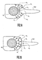
- a three dimensional object which is illustrated in FIG.1A and FIG.1B, has a complex surface denoted by OBJ.
- OBJ complex surface denoted by OBJ.
- the segmentation technique that has been previously described in relation to the publication above cited as the state of the art is used because it is robust and gives excellent results. It is an iterative method that permits of representing the object of interest using the discrete model called Simplex Mesh Model.
- the complex surface OBJ poses a problem when the segmentation method described above is applied.
- the present invention is able to solve this problem.
- the surface denoted by OBJ, represented in FIG.1A and 1B, has been chosen in order to demonstrate that the method of the invention may be applied to a great number of different and complex surfaces.
- the image segmentation method is based on utilization of discrete deformable models, like 2-Simplex Meshes Model described as prior art [Delingette].
- a three dimensional digital Simplex Mesh Model is generated as illustrated by FIG.1A or FIG.1B.
- it is a simple sphere M 0 formed by a set of small three dimensional discrete curved faces called cells, which are linked by their boundaries called the edges of the Mesh Model, and which have common nodes called the vertices of the Mesh Model.
- the segmentation operation consists in mapping the three dimensional Simplex Mesh Model M 0 of FIG.1A or FIG.1B onto a three dimensional object of interest denoted by OBJ as shown in FIG.1A and FIG.1B.
- the object of interest OBJ is a complex shape representing an organ. This organ surface can show a difficult geometry such as the one of FIG.1A and 1B, comprising a bulging surface with a tubular appendix of small diameter with respect to the rest of the organ, which extends through a wall of the organ.
- the shape of the object to be segmented is initially unknown, and therefore the initial model has to be deformed in order to fit the shape of the object of interest OBJ. It seems difficult for the sphere to deform appropriately in order to map correctly the entirety of the surface of the complex shape of this body organ considering the complexity of said shape.
- a three dimensional Mesh Model such as M 0 has constant vertex connectivity.
- a three dimensional surface is represented using the three dimensional Mesh Cells, where each given vertex is connected to three neighboring vertices.
- neighbor vertex it must be understood a vertex of a given cell that constitutes the second extremity of an edge of said cell starting at said given vertex. So, each given vertex is common to three Cells, hence is common to three angles and is the starting point of three edges.
- a Mesh Model such as M 0 can represent all types of three-dimensional surfaces using Mesh Transformations. Four independent transformations are defined for achieving the whole range of possible Mesh Transformations.
- vertices consist in inserting or in deleting vertices (nodes) in a cell; in defining angles in the cell; and in defining metric parameters that describe how the vertex is located with respect to its three neighbors.
- a law of motion defines the dynamic of each vertex.
- the deformation implies an internal force that constrains the shape of the Simplex Mesh Model to be smooth and an external force that constrains the three-dimensional Mesh Model to be close to the three dimensional object surface, in this example the surface OBJ .
- the deformation of the Mesh Model is essentially function of the distance between the Mesh Model and the image data at each stage of the deformation.
- the Mesh Model is only attracted towards data points that are relatively close.
- FIG.1A illustrates this defect by showing the Model surface turning itself inside-out.
- This refinement consists in introducing an algorithm for automatically increasing the Mesh resolution at parts of high curvature and at parts whose distance to the image data is higher than a threshold. Hence, at each iteration, the area of the spherical cells decreases. So, according to the known technique, since when the distance to the image data is large, as shown in FIG.1A, the surface of the cells decreases and the number of cells increases, the refinement algorithm would be unable to improve the result in said parts P'1, P'2.
- the present invention proposes a method for automatic adaptation of 3-D surface Model to image data in the context of model-based image segmentation, which allows to dynamically adapt the model resolution to image feature content.
- the Model resolution is locally set to higher resolution only when reliable image features are found and is locally set to lower resolution in the opposite case, when image features are far or are not reliable, for instance noisy.
- Increasing the resolution of the Mesh Model when the image features are reliable is completely contrary to the disclosure of the prior art that increases the resolution when the distance of the image features is larger than a threshold.
- the present invention permits to prevent model self-intersections, but also it improves the computational effectiveness of the whole segmentation process.
- a feature confidence parameter is defined, and model resolution is locally adapted according to it.
- the feature confidence parameter depends on the feature distance and on the quality of the image data. noisy, although close features are penalized.
- This technique allows of keeping rather coarse model resolution in absence of image features. Then this technique allows of increasing the model resolution gradually with the rise of feature confidence.
- Low local resolution in a part of the Model implies few cells of large areas, which constraints local surface curvature in that part, and thus prevents the Model surface from turning inside-out and from self-intersections.
- the Model will have large cells in part P'2 at the stage M2 of the Model deformation. This permits the Model of properly continuing its propagation toward the part P3 of the object of interest, instead of turning inside-out as in FIG.1A.
- This segmentation operation consists in deforming the original spherical shape M 0 of the Simplex Mesh Model until it is mapped onto the object of interest OBJ, making its surface as close as possible to the surface of the object of interest OBJ.
- This operation is performed by iterative steps, according to an iterative law.
- This law permits of establishing a balance between external forces that are first forces of traction of the cells OBJ of the model towards the surface of the object of reference OBJ, i. e. they force the cell surfaces to be close to the object surface; and internal forces that are regularization forces for forcing of the general surface of the Mesh Model to be smooth.
- FIG.2A illustrates another example of object of interest OBJ to be segmented using a Mesh Model that is initially a sphere.
- the object of interest has elliptic diameters in all directions.
- directions A and P the image features are far from the cells of the sphere. So, the model is attracted toward the closest image features as shown in FIG.2B.
- the size of cells in A and P direction is increased, which is performed by decimating cells.
- the object border in the directions R and L is close to the mesh and is strong. Therefore the feature confidence in these directions is high.
- the object borders in the directions A and P are thought strong but beyond the search range. Therefore, they are not "seen" by the mesh and their confidence is low.
- the cells that are nearer the image features pull the enlarge cells, which at their turn become closer and closer from the image features. As said cells become closer, they acquire more confidence. Since the image features become more reliable with respect to said cells, the resolution of the first enlarged cells is locally increased. Then, by degrees, the first enlarged cells are pulled by the image features that are more and more reliable, and their resolution is more and more increase until the deformation of the sphere is such that it fits the object of interest as illustrated by FIG.2D.
- FIG.2C and FIG.2D represent the Mesh Model M 0 that is deformed after a given number of iterative steps performed according to the above-cited iterative law.
- the surfaces of the cells of the initial Mesh Model M 0 are attracted by the surface of the object of reference OBJ by the action of the external forces, while the internal forces smooth the Mesh Model surface, in such a manner that the shape of the Mesh Model is nearer and nearer of the shape of the object of reference.
- the user may visually evaluate the confidence to bestow on the image features.
- the present invention provides a visualization of the cells that are color coded in function of the confidence parameter. This visualization with different colors allows to supervise the deformation process and to locally assess its final quality.
- FIG.2C which shows a cross section of the object of interest along the diameter RL and perpendicularly to AP
- the cells at the center of the representation are those that are farer from points A and P. So, at the center of this representation, the decimated cells of low resolution are represented for instance in red, which is a very clear gray in FIG.2C. Around these cells, the cells toward H and F directions are very near the image features and they have a high resolution. They are represented for instance in green, which is middle gray in FIG.2C.
- the automated technique of estimation of the adaptation comprises sub-steps of:
- steps 4.2) and 4.3) may be performed before the user actually displays the images of the Mesh Model by performing steps 4.4), 4.5) and 4.6) in order to obtain a visual evaluation of the goodness of fitness and take a decision to further go on with the process or not as in step 4.7).
- the goodness of adaptation has to be empirically estimating by performing a comparison between the shape of the object of reference and the Mesh Model and by visually estimating the distance between the cells of the Mesh Model and the corresponding zones of the object of reference in 2D slices.
- the user disposes of an automatic quantified estimation of the goodness of adaptation of the Mesh Model with respect to the object of interest without to have to perform himself an approximate estimation.
- the color-coded cells of the Mesh Model provide automatically the user with a numerical and visual knowledge of said goodness of adaptation.
- the confidence parameter value, related to a given cell gives a representation of likelihood said given cell be close to and aligned with a surface of the object of interest in the 3D image.
- the greater the confidence parameter value related to said cell of the Mesh Model the better said cell of the Mesh Model locally fits the surface of said object.
- this color-coded representation for each cell of the Mesh Model the user can appreciate easily and rapidly the adaptation of each cell.
- the user may decide to go on the iterative steps in order to better this fitness.
- Freezing cells means that no more calculations are applied to said cells. In particular they are no more divided. Their actual surface area and their distance with respect to the surface of the object of interest do not change anymore. Their goodness of adaptation is automatically estimated by confidence parameter value of the cells and by their color or hue. The decision that the adaptation is good is taken in function of said estimation according to the threshold previously described. The frozen cells will have the same color and shape after the further operation of adaptation refining.
- the iterative steps are stopped either when the user decides so by a simple visualization of the color-coded image of the resulting Mesh Model or by deciding that the process is automatically stopped when all the cells or a predetermined number of Cells have reached the predetermined threshold.
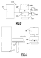
- FIG.3 shows a diagram of an image processing system 150 according to the invention for carrying out all the steps of the above-described method.
- the system has means 151 for acquiring digital image data of a sequence of images, and is coupled to computer means 153 for processing these data according to the processing steps of the method cited above.
- the medical viewing system can be used in the intervention room or near the intervention room for processing real time images. Steps of the present method can be applied on stored medical images, for example for estimating medical parameters.
- the medical viewing system provides the image data by connection 157 to the system 153.
- the system provides processed image data to display means and/or storage means.
- the display means 154 may be a screen.
- the storage means may be a memory of the system 153. Said storage means may be alternately external storage means.
- This image viewing system 153 may comprise a suitably programmed computer, or a special purpose processor having circuit means such as LUTs, Memories, Filters, Logic Operators, that are arranged to perform the functions of the method steps according to the invention.
- the system 153 may also comprise a keyboard 155 and a mouse 156. Icones may be provided on the screen to be activated by mouse-clicks, or special pushbuttons may be provided on the system, to constitute control means 158 for the user to start, to control the duration or to stop the processing means of the system at chosen stages or phases.
- Fig.4 shows the basic components of an embodiment of an image processing system in accordance to the present invention, incorporated in a medical examination apparatus.
- the medical examination apparatus typically includes a bed 110 on which the patient lies or another element for localizing the patient relative to the medical examination apparatus.
- the medical examination apparatus may be a CT scanner 151.
- the image data produced by the CT scanner 151 is fed to the system 153, such as a general-purpose computer, that carries out the steps of the method.
- the system 153 is typically associated with a visualization device, such as a monitor 154, and an input device 155, such as a keyboard, pointing device, etc. operative by the user so that he can interact with the system.
- the system 153 is programmed to implement a method of processing medical image data according to invention.
- the system 153 has computing means and memory means to perform the steps of the method.
- a computer program product having pre-programmed instructions to carry out the method may also be implemented.
- the present invention is applicable regardless of the medical imaging technology that is used to generate the initial data.
- magnetic resonance (MR) coronary angiography may be used to generate 3D medical image data in a non-invasive manner.
- MR magnetic resonance
- Various modifications can be made to the order in which processing steps are performed in the above-described specific embodiment.
- the above-described processing steps applied to medical image data can advantageously be combined with various other known processing/visualization techniques.
- the drawings and their description herein before illustrate rather than limit the invention. It will be evident that there are numerous alternatives that fall within the scope of the appended claims.
- the present invention has been described in terms of generating image data for display, the present invention is intended to cover substantially any form of visualization of the image data including, but not limited to, display on a display device, and printing. Any reference sign in a claim should not be construed as limiting the claim.
Landscapes
- Engineering & Computer Science (AREA)
- Physics & Mathematics (AREA)
- Theoretical Computer Science (AREA)
- General Physics & Mathematics (AREA)
- Computer Vision & Pattern Recognition (AREA)
- Software Systems (AREA)
- Geometry (AREA)
- General Health & Medical Sciences (AREA)
- Radiology & Medical Imaging (AREA)
- Nuclear Medicine, Radiotherapy & Molecular Imaging (AREA)
- Medical Informatics (AREA)
- Computer Graphics (AREA)
- Health & Medical Sciences (AREA)
- Quality & Reliability (AREA)
- Image Processing (AREA)
- Measuring And Recording Apparatus For Diagnosis (AREA)
- Image Analysis (AREA)
- Coating With Molten Metal (AREA)
- Heating, Cooling, Or Curing Plastics Or The Like In General (AREA)
- Processing Or Creating Images (AREA)
- Ultra Sonic Daignosis Equipment (AREA)
- Image Generation (AREA)
- Magnetic Resonance Imaging Apparatus (AREA)
- Apparatus For Radiation Diagnosis (AREA)
Priority Applications (1)
| Application Number | Priority Date | Filing Date | Title |
|---|---|---|---|
| EP03769837A EP1565880B1 (en) | 2002-11-20 | 2003-11-14 | Image processing system for automatic adaptation of a 3-d mesh model onto a 3-d surface of an object |
Applications Claiming Priority (4)
| Application Number | Priority Date | Filing Date | Title |
|---|---|---|---|
| EP02292884 | 2002-11-20 | ||
| EP02292884 | 2002-11-20 | ||
| PCT/IB2003/005168 WO2004047030A2 (en) | 2002-11-20 | 2003-11-14 | Image processing system for automatic adaptation of a 3-d mesh model onto a 3-d surface of an object |
| EP03769837A EP1565880B1 (en) | 2002-11-20 | 2003-11-14 | Image processing system for automatic adaptation of a 3-d mesh model onto a 3-d surface of an object |
Publications (2)
| Publication Number | Publication Date |
|---|---|
| EP1565880A2 EP1565880A2 (en) | 2005-08-24 |
| EP1565880B1 true EP1565880B1 (en) | 2007-02-14 |
Family
ID=32319683
Family Applications (1)
| Application Number | Title | Priority Date | Filing Date |
|---|---|---|---|
| EP03769837A Expired - Lifetime EP1565880B1 (en) | 2002-11-20 | 2003-11-14 | Image processing system for automatic adaptation of a 3-d mesh model onto a 3-d surface of an object |
Country Status (7)
| Country | Link |
|---|---|
| US (1) | US8437579B2 (https=) |
| EP (1) | EP1565880B1 (https=) |
| JP (1) | JP2006506164A (https=) |
| AT (1) | ATE354140T1 (https=) |
| AU (1) | AU2003278538A1 (https=) |
| DE (1) | DE60311865T2 (https=) |
| WO (1) | WO2004047030A2 (https=) |
Families Citing this family (29)
| Publication number | Priority date | Publication date | Assignee | Title |
|---|---|---|---|---|
| EP2176799B1 (en) * | 2007-08-01 | 2019-09-11 | Koninklijke Philips N.V. | Accessing medical image detabases using medically relevant terms |
| BRPI0908886A2 (pt) | 2008-02-15 | 2015-09-15 | Koninkl Philips Electronics Nv | aparelho, método e programa de computador pra segmentar um objeto, e, modelo de objeto |
| EP2260468B1 (en) * | 2008-04-04 | 2015-06-10 | Koninklijke Philips N.V. | Simultaneous model-based segmentation of objects satisfying pre-defined spatial relationships |
| JP5432241B2 (ja) * | 2008-04-07 | 2014-03-05 | コーニンクレッカ フィリップス エヌ ヴェ | メッシュ衝突回避 |
| GB2460411B (en) | 2008-05-27 | 2012-08-08 | Simpleware Ltd | Image processing method |
| EP2340444A1 (en) * | 2008-10-22 | 2011-07-06 | Koninklijke Philips Electronics N.V. | 3-d ultrasound imaging |
| US8265363B2 (en) * | 2009-02-04 | 2012-09-11 | General Electric Company | Method and apparatus for automatically identifying image views in a 3D dataset |
| CN103098092B (zh) * | 2010-09-17 | 2016-05-11 | 皇家飞利浦电子股份有限公司 | 选择用于图像分割的解剖变型模型 |
| US8903088B2 (en) | 2011-12-02 | 2014-12-02 | Adobe Systems Incorporated | Binding of protected video content to video player with encryption key |
| US8879731B2 (en) | 2011-12-02 | 2014-11-04 | Adobe Systems Incorporated | Binding of protected video content to video player with block cipher hash |
| US9064318B2 (en) | 2012-10-25 | 2015-06-23 | Adobe Systems Incorporated | Image matting and alpha value techniques |
| US9355649B2 (en) | 2012-11-13 | 2016-05-31 | Adobe Systems Incorporated | Sound alignment using timing information |
| US9201580B2 (en) | 2012-11-13 | 2015-12-01 | Adobe Systems Incorporated | Sound alignment user interface |
| US10638221B2 (en) | 2012-11-13 | 2020-04-28 | Adobe Inc. | Time interval sound alignment |
| KR101339552B1 (ko) * | 2012-11-15 | 2013-12-10 | 이화여자대학교 산학협력단 | 사용자 기반의 오브젝트 근사 방법 및 오브젝트 근사 시스템 |
| US9076205B2 (en) | 2012-11-19 | 2015-07-07 | Adobe Systems Incorporated | Edge direction and curve based image de-blurring |
| US10249321B2 (en) | 2012-11-20 | 2019-04-02 | Adobe Inc. | Sound rate modification |
| US9451304B2 (en) | 2012-11-29 | 2016-09-20 | Adobe Systems Incorporated | Sound feature priority alignment |
| US9135710B2 (en) * | 2012-11-30 | 2015-09-15 | Adobe Systems Incorporated | Depth map stereo correspondence techniques |
| US10455219B2 (en) | 2012-11-30 | 2019-10-22 | Adobe Inc. | Stereo correspondence and depth sensors |
| US9208547B2 (en) | 2012-12-19 | 2015-12-08 | Adobe Systems Incorporated | Stereo correspondence smoothness tool |
| US10249052B2 (en) | 2012-12-19 | 2019-04-02 | Adobe Systems Incorporated | Stereo correspondence model fitting |
| US9214026B2 (en) | 2012-12-20 | 2015-12-15 | Adobe Systems Incorporated | Belief propagation and affinity measures |
| GB2515510B (en) | 2013-06-25 | 2019-12-25 | Synopsys Inc | Image processing method |
| CN107209794B (zh) * | 2015-01-28 | 2022-02-08 | 皇家飞利浦有限公司 | 解剖结构的有限元建模 |
| US10282917B2 (en) | 2015-06-29 | 2019-05-07 | Koninklijke Philips N.V. | Interactive mesh editing |
| GB2539963A (en) * | 2015-07-03 | 2017-01-04 | Williams Justin | Thermographic imaging system |
| US10915680B2 (en) * | 2018-12-21 | 2021-02-09 | Dassault Systemes Simulia Corp. | Local control of design patterns on surfaces for enhanced physical properties |
| EP3839895A1 (en) | 2019-12-19 | 2021-06-23 | Koninklijke Philips N.V. | Mesh correction depending on mesh normal direction |
Family Cites Families (10)
| Publication number | Priority date | Publication date | Assignee | Title |
|---|---|---|---|---|
| DE69331719T2 (de) * | 1992-06-19 | 2002-10-24 | Agfa-Gevaert, Mortsel | Verfahren und Vorrichtung zur Geräuschunterdrückung |
| AU6108396A (en) | 1995-06-07 | 1996-12-30 | David M. Geshwind | Systems using motion detection, interpolation, and cross-dis solving for improving picture quality |
| AU2454397A (en) * | 1996-04-24 | 1997-11-12 | Shriners Hospital For Children | Method and apparatus for recording three-dimensional topographies |
| US6404920B1 (en) * | 1996-09-09 | 2002-06-11 | Hsu Shin-Yi | System for generalizing objects and features in an image |
| US5886702A (en) * | 1996-10-16 | 1999-03-23 | Real-Time Geometry Corporation | System and method for computer modeling of 3D objects or surfaces by mesh constructions having optimal quality characteristics and dynamic resolution capabilities |
| GB9626676D0 (en) | 1996-12-23 | 1997-02-12 | Smith & Nephew Res | Imaging method |
| US6351262B1 (en) * | 1998-06-30 | 2002-02-26 | Lucent Technologies, Inc. | Display techniques for three-dimensional virtual reality |
| JP2000175052A (ja) * | 1998-12-07 | 2000-06-23 | Xerox Corp | ピクセルマップ表現の処理方法及び装置 |
| US6968299B1 (en) * | 2000-04-14 | 2005-11-22 | International Business Machines Corporation | Method and apparatus for reconstructing a surface using a ball-pivoting algorithm |
| WO2002073536A2 (en) * | 2001-03-09 | 2002-09-19 | Koninklijke Philips Electronics N.V. | Image segmentation |
-
2003
- 2003-11-14 AU AU2003278538A patent/AU2003278538A1/en not_active Abandoned
- 2003-11-14 WO PCT/IB2003/005168 patent/WO2004047030A2/en not_active Ceased
- 2003-11-14 US US10/535,466 patent/US8437579B2/en active Active
- 2003-11-14 AT AT03769837T patent/ATE354140T1/de not_active IP Right Cessation
- 2003-11-14 DE DE60311865T patent/DE60311865T2/de not_active Expired - Lifetime
- 2003-11-14 JP JP2004553008A patent/JP2006506164A/ja not_active Withdrawn
- 2003-11-14 EP EP03769837A patent/EP1565880B1/en not_active Expired - Lifetime
Also Published As
| Publication number | Publication date |
|---|---|
| EP1565880A2 (en) | 2005-08-24 |
| AU2003278538A8 (en) | 2004-06-15 |
| AU2003278538A1 (en) | 2004-06-15 |
| DE60311865T2 (de) | 2007-10-31 |
| DE60311865D1 (de) | 2007-03-29 |
| WO2004047030A2 (en) | 2004-06-03 |
| US20060078194A1 (en) | 2006-04-13 |
| ATE354140T1 (de) | 2007-03-15 |
| WO2004047030A3 (en) | 2004-10-07 |
| US8437579B2 (en) | 2013-05-07 |
| JP2006506164A (ja) | 2006-02-23 |
Similar Documents
| Publication | Publication Date | Title |
|---|---|---|
| EP1565880B1 (en) | Image processing system for automatic adaptation of a 3-d mesh model onto a 3-d surface of an object | |
| Gibson | Constrained elastic surface nets: Generating smooth surfaces from binary segmented data | |
| US6754374B1 (en) | Method and apparatus for processing images with regions representing target objects | |
| EP1527422B1 (en) | Image processing method for displaying information relating to wall motions of a deformable 3-d object | |
| US7043062B2 (en) | Image processing method for displaying an image sequence of a deformable 3-D object with indications of the object wall motion | |
| US20040171922A1 (en) | Image processing method for interacting with a 3-d surface represented in a 3-d image | |
| JP3712234B2 (ja) | 関心領域抽出方法及び画像処理サーバ | |
| WO2012017375A2 (en) | In-plane and interactive surface mesh adaptation | |
| EP1685534B1 (en) | Three-dimensional segmentation using deformable surfaces | |
| JP4170096B2 (ja) | 対象の3次元表面上にマップされた3次元メッシュモデルの適合性評価のための画像処理装置 | |
| CN101001572A (zh) | 结节检测 | |
| Bro-Nielsen | Active nets and cubes | |
| US6392646B1 (en) | Iterative determination of the shortest path between two points on a polygonal surface | |
| EP1709591A1 (en) | Real time user interaction with deformable surfaces of segmentation | |
| CA2617227C (en) | Deformable model for segmenting patient contours versus support structures in medical images | |
| Muraki et al. | A survey of medical applications of 3D image analysis and computer graphics | |
| EP1141894B1 (en) | Method and apparatus for processing images with regions representing target objects | |
| EP4521355A1 (en) | Correcting topological defects on a surface mesh representing an organ | |
| EP4254350A1 (en) | Determination of illumination parameters in medical image rendering | |
| EP4152255A1 (en) | System and method for differentiating a tissue of interest from another part of a medical scanner image | |
| EP1262914A1 (en) | Method and apparatus for processing images with regions representing target objects | |
| CN119784956A (zh) | 一种基于vtk可视化框架的虚拟解剖方法 | |
| Caulkin et al. | Generating Realistic Mass Lesions in Digital Mammograms Using Statistical Models. | |
| Crawford-Hines et al. | A System for 3D Surface Models from 2D Sectional Imagery | |
| Koehler et al. | Knowledge-Assisted 3D Reconstruction and Visualization of Human Ribcage and Lungs |
Legal Events
| Date | Code | Title | Description |
|---|---|---|---|
| PUAI | Public reference made under article 153(3) epc to a published international application that has entered the european phase |
Free format text: ORIGINAL CODE: 0009012 |
|
| 17P | Request for examination filed |
Effective date: 20050620 |
|
| AK | Designated contracting states |
Kind code of ref document: A2 Designated state(s): AT BE BG CH CY CZ DE DK EE ES FI FR GB GR HU IE IT LI LU MC NL PT RO SE SI SK TR |
|
| AX | Request for extension of the european patent |
Extension state: AL LT LV MK |
|
| DAX | Request for extension of the european patent (deleted) | ||
| GRAP | Despatch of communication of intention to grant a patent |
Free format text: ORIGINAL CODE: EPIDOSNIGR1 |
|
| GRAS | Grant fee paid |
Free format text: ORIGINAL CODE: EPIDOSNIGR3 |
|
| GRAA | (expected) grant |
Free format text: ORIGINAL CODE: 0009210 |
|
| AK | Designated contracting states |
Kind code of ref document: B1 Designated state(s): AT BE BG CH CY CZ DE DK EE ES FI FR GB GR HU IE IT LI LU MC NL PT RO SE SI SK TR |
|
| PG25 | Lapsed in a contracting state [announced via postgrant information from national office to epo] |
Ref country code: CH Free format text: LAPSE BECAUSE OF FAILURE TO SUBMIT A TRANSLATION OF THE DESCRIPTION OR TO PAY THE FEE WITHIN THE PRESCRIBED TIME-LIMIT Effective date: 20070214 Ref country code: SI Free format text: LAPSE BECAUSE OF FAILURE TO SUBMIT A TRANSLATION OF THE DESCRIPTION OR TO PAY THE FEE WITHIN THE PRESCRIBED TIME-LIMIT Effective date: 20070214 Ref country code: FI Free format text: LAPSE BECAUSE OF FAILURE TO SUBMIT A TRANSLATION OF THE DESCRIPTION OR TO PAY THE FEE WITHIN THE PRESCRIBED TIME-LIMIT Effective date: 20070214 Ref country code: BE Free format text: LAPSE BECAUSE OF FAILURE TO SUBMIT A TRANSLATION OF THE DESCRIPTION OR TO PAY THE FEE WITHIN THE PRESCRIBED TIME-LIMIT Effective date: 20070214 Ref country code: AT Free format text: LAPSE BECAUSE OF FAILURE TO SUBMIT A TRANSLATION OF THE DESCRIPTION OR TO PAY THE FEE WITHIN THE PRESCRIBED TIME-LIMIT Effective date: 20070214 Ref country code: DK Free format text: LAPSE BECAUSE OF FAILURE TO SUBMIT A TRANSLATION OF THE DESCRIPTION OR TO PAY THE FEE WITHIN THE PRESCRIBED TIME-LIMIT Effective date: 20070214 Ref country code: LI Free format text: LAPSE BECAUSE OF FAILURE TO SUBMIT A TRANSLATION OF THE DESCRIPTION OR TO PAY THE FEE WITHIN THE PRESCRIBED TIME-LIMIT Effective date: 20070214 Ref country code: NL Free format text: LAPSE BECAUSE OF FAILURE TO SUBMIT A TRANSLATION OF THE DESCRIPTION OR TO PAY THE FEE WITHIN THE PRESCRIBED TIME-LIMIT Effective date: 20070214 |
|
| REG | Reference to a national code |
Ref country code: GB Ref legal event code: FG4D |
|
| REG | Reference to a national code |
Ref country code: CH Ref legal event code: EP |
|
| REF | Corresponds to: |
Ref document number: 60311865 Country of ref document: DE Date of ref document: 20070329 Kind code of ref document: P |
|
| REG | Reference to a national code |
Ref country code: IE Ref legal event code: FG4D |
|
| REG | Reference to a national code |
Ref country code: GB Ref legal event code: 746 Effective date: 20070404 |
|
| PG25 | Lapsed in a contracting state [announced via postgrant information from national office to epo] |
Ref country code: SE Free format text: LAPSE BECAUSE OF FAILURE TO SUBMIT A TRANSLATION OF THE DESCRIPTION OR TO PAY THE FEE WITHIN THE PRESCRIBED TIME-LIMIT Effective date: 20070514 |
|
| PG25 | Lapsed in a contracting state [announced via postgrant information from national office to epo] |
Ref country code: BG Free format text: LAPSE BECAUSE OF EXPIRATION OF PROTECTION Effective date: 20070515 |
|
| PG25 | Lapsed in a contracting state [announced via postgrant information from national office to epo] |
Ref country code: ES Free format text: LAPSE BECAUSE OF FAILURE TO SUBMIT A TRANSLATION OF THE DESCRIPTION OR TO PAY THE FEE WITHIN THE PRESCRIBED TIME-LIMIT Effective date: 20070525 |
|
| PG25 | Lapsed in a contracting state [announced via postgrant information from national office to epo] |
Ref country code: PT Free format text: LAPSE BECAUSE OF FAILURE TO SUBMIT A TRANSLATION OF THE DESCRIPTION OR TO PAY THE FEE WITHIN THE PRESCRIBED TIME-LIMIT Effective date: 20070716 |
|
| NLV1 | Nl: lapsed or annulled due to failure to fulfill the requirements of art. 29p and 29m of the patents act | ||
| REG | Reference to a national code |
Ref country code: CH Ref legal event code: PL |
|
| EN | Fr: translation not filed | ||
| PG25 | Lapsed in a contracting state [announced via postgrant information from national office to epo] |
Ref country code: SK Free format text: LAPSE BECAUSE OF FAILURE TO SUBMIT A TRANSLATION OF THE DESCRIPTION OR TO PAY THE FEE WITHIN THE PRESCRIBED TIME-LIMIT Effective date: 20070214 |
|
| PLBE | No opposition filed within time limit |
Free format text: ORIGINAL CODE: 0009261 |
|
| STAA | Information on the status of an ep patent application or granted ep patent |
Free format text: STATUS: NO OPPOSITION FILED WITHIN TIME LIMIT |
|
| PG25 | Lapsed in a contracting state [announced via postgrant information from national office to epo] |
Ref country code: RO Free format text: LAPSE BECAUSE OF FAILURE TO SUBMIT A TRANSLATION OF THE DESCRIPTION OR TO PAY THE FEE WITHIN THE PRESCRIBED TIME-LIMIT Effective date: 20070214 Ref country code: CZ Free format text: LAPSE BECAUSE OF FAILURE TO SUBMIT A TRANSLATION OF THE DESCRIPTION OR TO PAY THE FEE WITHIN THE PRESCRIBED TIME-LIMIT Effective date: 20070214 |
|
| 26N | No opposition filed |
Effective date: 20071115 |
|
| PG25 | Lapsed in a contracting state [announced via postgrant information from national office to epo] |
Ref country code: IT Free format text: LAPSE BECAUSE OF FAILURE TO SUBMIT A TRANSLATION OF THE DESCRIPTION OR TO PAY THE FEE WITHIN THE PRESCRIBED TIME-LIMIT Effective date: 20070214 Ref country code: GR Free format text: LAPSE BECAUSE OF FAILURE TO SUBMIT A TRANSLATION OF THE DESCRIPTION OR TO PAY THE FEE WITHIN THE PRESCRIBED TIME-LIMIT Effective date: 20070515 Ref country code: FR Free format text: LAPSE BECAUSE OF FAILURE TO SUBMIT A TRANSLATION OF THE DESCRIPTION OR TO PAY THE FEE WITHIN THE PRESCRIBED TIME-LIMIT Effective date: 20071005 |
|
| PG25 | Lapsed in a contracting state [announced via postgrant information from national office to epo] |
Ref country code: MC Free format text: LAPSE BECAUSE OF NON-PAYMENT OF DUE FEES Effective date: 20071130 |
|
| PG25 | Lapsed in a contracting state [announced via postgrant information from national office to epo] |
Ref country code: IE Free format text: LAPSE BECAUSE OF NON-PAYMENT OF DUE FEES Effective date: 20071114 |
|
| PG25 | Lapsed in a contracting state [announced via postgrant information from national office to epo] |
Ref country code: FR Free format text: LAPSE BECAUSE OF FAILURE TO SUBMIT A TRANSLATION OF THE DESCRIPTION OR TO PAY THE FEE WITHIN THE PRESCRIBED TIME-LIMIT Effective date: 20070214 |
|
| PG25 | Lapsed in a contracting state [announced via postgrant information from national office to epo] |
Ref country code: EE Free format text: LAPSE BECAUSE OF FAILURE TO SUBMIT A TRANSLATION OF THE DESCRIPTION OR TO PAY THE FEE WITHIN THE PRESCRIBED TIME-LIMIT Effective date: 20070214 |
|
| PG25 | Lapsed in a contracting state [announced via postgrant information from national office to epo] |
Ref country code: CY Free format text: LAPSE BECAUSE OF FAILURE TO SUBMIT A TRANSLATION OF THE DESCRIPTION OR TO PAY THE FEE WITHIN THE PRESCRIBED TIME-LIMIT Effective date: 20070214 |
|
| PG25 | Lapsed in a contracting state [announced via postgrant information from national office to epo] |
Ref country code: LU Free format text: LAPSE BECAUSE OF NON-PAYMENT OF DUE FEES Effective date: 20071114 |
|
| PG25 | Lapsed in a contracting state [announced via postgrant information from national office to epo] |
Ref country code: TR Free format text: LAPSE BECAUSE OF FAILURE TO SUBMIT A TRANSLATION OF THE DESCRIPTION OR TO PAY THE FEE WITHIN THE PRESCRIBED TIME-LIMIT Effective date: 20070214 Ref country code: HU Free format text: LAPSE BECAUSE OF FAILURE TO SUBMIT A TRANSLATION OF THE DESCRIPTION OR TO PAY THE FEE WITHIN THE PRESCRIBED TIME-LIMIT Effective date: 20070815 |
|
| REG | Reference to a national code |
Ref country code: DE Ref legal event code: R082 Ref document number: 60311865 Country of ref document: DE Representative=s name: VOLMER, GEORG, DIPL.-ING., DE |
|
| REG | Reference to a national code |
Ref country code: DE Ref legal event code: R082 Ref document number: 60311865 Country of ref document: DE Representative=s name: MEISSNER BOLTE PATENTANWAELTE RECHTSANWAELTE P, DE Effective date: 20140328 Ref country code: DE Ref legal event code: R082 Ref document number: 60311865 Country of ref document: DE Representative=s name: VOLMER, GEORG, DIPL.-ING., DE Effective date: 20140328 Ref country code: DE Ref legal event code: R082 Ref document number: 60311865 Country of ref document: DE Representative=s name: MEISSNER, BOLTE & PARTNER GBR, DE Effective date: 20140328 Ref country code: DE Ref legal event code: R081 Ref document number: 60311865 Country of ref document: DE Owner name: KONINKLIJKE PHILIPS N.V., NL Free format text: FORMER OWNER: KONINKLIJKE PHILIPS ELECTRONICS N.V., EINDHOVEN, NL Effective date: 20140328 |
|
| REG | Reference to a national code |
Ref country code: DE Ref legal event code: R082 Ref document number: 60311865 Country of ref document: DE Representative=s name: MEISSNER BOLTE PATENTANWAELTE RECHTSANWAELTE P, DE Ref country code: DE Ref legal event code: R082 Ref document number: 60311865 Country of ref document: DE Representative=s name: MEISSNER, BOLTE & PARTNER GBR, DE |
|
| PGFP | Annual fee paid to national office [announced via postgrant information from national office to epo] |
Ref country code: GB Payment date: 20221122 Year of fee payment: 20 Ref country code: DE Payment date: 20220628 Year of fee payment: 20 |
|
| REG | Reference to a national code |
Ref country code: DE Ref legal event code: R071 Ref document number: 60311865 Country of ref document: DE |
|
| REG | Reference to a national code |
Ref country code: GB Ref legal event code: PE20 Expiry date: 20231113 |
|
| PG25 | Lapsed in a contracting state [announced via postgrant information from national office to epo] |
Ref country code: GB Free format text: LAPSE BECAUSE OF EXPIRATION OF PROTECTION Effective date: 20231113 |
|
| PG25 | Lapsed in a contracting state [announced via postgrant information from national office to epo] |
Ref country code: GB Free format text: LAPSE BECAUSE OF EXPIRATION OF PROTECTION Effective date: 20231113 |