EP1554383B1 - Rekombinante mva-stamme als potentielle impfstoffe gegen p. falciparum -malaria - Google Patents
Rekombinante mva-stamme als potentielle impfstoffe gegen p. falciparum -malaria Download PDFInfo
- Publication number
- EP1554383B1 EP1554383B1 EP03769329A EP03769329A EP1554383B1 EP 1554383 B1 EP1554383 B1 EP 1554383B1 EP 03769329 A EP03769329 A EP 03769329A EP 03769329 A EP03769329 A EP 03769329A EP 1554383 B1 EP1554383 B1 EP 1554383B1
- Authority
- EP
- European Patent Office
- Prior art keywords
- msp
- vaccine according
- protein
- vaccine
- rmva
- Prior art date
- Legal status (The legal status is an assumption and is not a legal conclusion. Google has not performed a legal analysis and makes no representation as to the accuracy of the status listed.)
- Expired - Lifetime
Links
- 229960005486 vaccine Drugs 0.000 title claims abstract description 31
- 241000223960 Plasmodium falciparum Species 0.000 title claims abstract description 18
- 208000002476 Falciparum Malaria Diseases 0.000 title 1
- 206010035500 Plasmodium falciparum infection Diseases 0.000 title 1
- 201000011336 Plasmodium falciparum malaria Diseases 0.000 title 1
- 108010057081 Merozoite Surface Protein 1 Proteins 0.000 claims abstract description 90
- 239000012634 fragment Substances 0.000 claims abstract description 32
- 201000004792 malaria Diseases 0.000 claims abstract description 18
- 108020004707 nucleic acids Proteins 0.000 claims abstract description 16
- 102000039446 nucleic acids Human genes 0.000 claims abstract description 16
- 150000007523 nucleic acids Chemical class 0.000 claims abstract description 16
- 238000011321 prophylaxis Methods 0.000 claims abstract description 5
- 238000002560 therapeutic procedure Methods 0.000 claims abstract description 4
- 241001183012 Modified Vaccinia Ankara virus Species 0.000 claims description 31
- 108090000623 proteins and genes Proteins 0.000 claims description 27
- 108010076504 Protein Sorting Signals Proteins 0.000 claims description 14
- 230000028327 secretion Effects 0.000 claims description 4
- 239000003814 drug Substances 0.000 claims description 3
- 239000012528 membrane Substances 0.000 claims description 3
- 230000002829 reductive effect Effects 0.000 claims description 3
- 238000004873 anchoring Methods 0.000 claims description 2
- 230000004807 localization Effects 0.000 claims description 2
- 239000002773 nucleotide Substances 0.000 claims description 2
- 125000003729 nucleotide group Chemical group 0.000 claims description 2
- 241000700605 Viruses Species 0.000 abstract description 32
- 238000004519 manufacturing process Methods 0.000 abstract description 6
- 238000000034 method Methods 0.000 abstract description 3
- 210000004027 cell Anatomy 0.000 description 24
- 238000002649 immunization Methods 0.000 description 19
- 230000003053 immunization Effects 0.000 description 19
- 241000699670 Mus sp. Species 0.000 description 15
- 102000004169 proteins and genes Human genes 0.000 description 15
- 208000015181 infectious disease Diseases 0.000 description 13
- 241000588724 Escherichia coli Species 0.000 description 11
- 238000011161 development Methods 0.000 description 10
- 230000014509 gene expression Effects 0.000 description 10
- 241000700618 Vaccinia virus Species 0.000 description 9
- 241000282412 Homo Species 0.000 description 8
- 241001465754 Metazoa Species 0.000 description 8
- 230000028996 humoral immune response Effects 0.000 description 8
- 244000045947 parasite Species 0.000 description 8
- 239000000427 antigen Substances 0.000 description 5
- 102000036639 antigens Human genes 0.000 description 5
- 108091007433 antigens Proteins 0.000 description 5
- 230000002238 attenuated effect Effects 0.000 description 5
- 239000003550 marker Substances 0.000 description 5
- 241000282693 Cercopithecidae Species 0.000 description 4
- 108020004705 Codon Proteins 0.000 description 4
- 238000002965 ELISA Methods 0.000 description 4
- 108091028043 Nucleic acid sequence Proteins 0.000 description 4
- 206010046865 Vaccinia virus infection Diseases 0.000 description 4
- 210000004369 blood Anatomy 0.000 description 4
- 239000008280 blood Substances 0.000 description 4
- 210000004962 mammalian cell Anatomy 0.000 description 4
- 208000007089 vaccinia Diseases 0.000 description 4
- 241000700647 Variola virus Species 0.000 description 3
- 125000003275 alpha amino acid group Chemical group 0.000 description 3
- 238000004458 analytical method Methods 0.000 description 3
- 201000010099 disease Diseases 0.000 description 3
- 208000037265 diseases, disorders, signs and symptoms Diseases 0.000 description 3
- 238000002474 experimental method Methods 0.000 description 3
- 230000036039 immunity Effects 0.000 description 3
- 239000000463 material Substances 0.000 description 3
- 210000003936 merozoite Anatomy 0.000 description 3
- 244000052769 pathogen Species 0.000 description 3
- 239000000047 product Substances 0.000 description 3
- 230000010076 replication Effects 0.000 description 3
- 238000001228 spectrum Methods 0.000 description 3
- 239000006228 supernatant Substances 0.000 description 3
- 238000001262 western blot Methods 0.000 description 3
- 108091026890 Coding region Proteins 0.000 description 2
- 102000004127 Cytokines Human genes 0.000 description 2
- 108090000695 Cytokines Proteins 0.000 description 2
- 108020004414 DNA Proteins 0.000 description 2
- 230000004543 DNA replication Effects 0.000 description 2
- 241000287828 Gallus gallus Species 0.000 description 2
- 108060003951 Immunoglobulin Proteins 0.000 description 2
- 241000700629 Orthopoxvirus Species 0.000 description 2
- 241000224016 Plasmodium Species 0.000 description 2
- 241000223830 Plasmodium yoelii Species 0.000 description 2
- 108020005067 RNA Splice Sites Proteins 0.000 description 2
- 241000282695 Saimiri Species 0.000 description 2
- 230000024932 T cell mediated immunity Effects 0.000 description 2
- 230000005867 T cell response Effects 0.000 description 2
- 108020005202 Viral DNA Proteins 0.000 description 2
- 108010027570 Xanthine phosphoribosyltransferase Proteins 0.000 description 2
- 229940023860 canarypox virus HIV vaccine Drugs 0.000 description 2
- 239000000969 carrier Substances 0.000 description 2
- 230000001413 cellular effect Effects 0.000 description 2
- 238000012217 deletion Methods 0.000 description 2
- 230000037430 deletion Effects 0.000 description 2
- 210000002950 fibroblast Anatomy 0.000 description 2
- 238000003119 immunoblot Methods 0.000 description 2
- 102000018358 immunoglobulin Human genes 0.000 description 2
- 229940072221 immunoglobulins Drugs 0.000 description 2
- 230000001965 increasing effect Effects 0.000 description 2
- 230000003834 intracellular effect Effects 0.000 description 2
- 229940124735 malaria vaccine Drugs 0.000 description 2
- 210000001161 mammalian embryo Anatomy 0.000 description 2
- 230000001404 mediated effect Effects 0.000 description 2
- 210000004379 membrane Anatomy 0.000 description 2
- 238000010899 nucleation Methods 0.000 description 2
- 238000012545 processing Methods 0.000 description 2
- 238000011160 research Methods 0.000 description 2
- 210000002966 serum Anatomy 0.000 description 2
- 230000014616 translation Effects 0.000 description 2
- CJDRUOGAGYHKKD-XMTJACRCSA-N (+)-Ajmaline Natural products O[C@H]1[C@@H](CC)[C@@H]2[C@@H]3[C@H](O)[C@@]45[C@@H](N(C)c6c4cccc6)[C@@H](N1[C@H]3C5)C2 CJDRUOGAGYHKKD-XMTJACRCSA-N 0.000 description 1
- 241000282708 Aotus <primate> Species 0.000 description 1
- 241000224482 Apicomplexa Species 0.000 description 1
- 108010009575 CD55 Antigens Proteins 0.000 description 1
- 241000178270 Canarypox virus Species 0.000 description 1
- 241000700628 Chordopoxvirinae Species 0.000 description 1
- 208000035473 Communicable disease Diseases 0.000 description 1
- 102000053602 DNA Human genes 0.000 description 1
- 102000016928 DNA-directed DNA polymerase Human genes 0.000 description 1
- 102000004163 DNA-directed RNA polymerases Human genes 0.000 description 1
- 102000004533 Endonucleases Human genes 0.000 description 1
- 108010042407 Endonucleases Proteins 0.000 description 1
- 241000206602 Eukaryota Species 0.000 description 1
- 101100386242 Homo sapiens CD55 gene Proteins 0.000 description 1
- 108010054278 Lac Repressors Proteins 0.000 description 1
- 102000043129 MHC class I family Human genes 0.000 description 1
- 108091054437 MHC class I family Proteins 0.000 description 1
- 241000282560 Macaca mulatta Species 0.000 description 1
- 108010052285 Membrane Proteins Proteins 0.000 description 1
- 241000186366 Mycobacterium bovis Species 0.000 description 1
- 108700026244 Open Reading Frames Proteins 0.000 description 1
- 239000002033 PVDF binder Substances 0.000 description 1
- 241000224017 Plasmodium berghei Species 0.000 description 1
- 241000224024 Plasmodium chabaudi Species 0.000 description 1
- 241000223821 Plasmodium malariae Species 0.000 description 1
- 206010035501 Plasmodium malariae infection Diseases 0.000 description 1
- 241001505293 Plasmodium ovale Species 0.000 description 1
- 241000223810 Plasmodium vivax Species 0.000 description 1
- 206010035503 Plasmodium vivax infection Diseases 0.000 description 1
- 108010008281 Recombinant Fusion Proteins Proteins 0.000 description 1
- 102000007056 Recombinant Fusion Proteins Human genes 0.000 description 1
- 241000283984 Rodentia Species 0.000 description 1
- 240000004808 Saccharomyces cerevisiae Species 0.000 description 1
- 235000014680 Saccharomyces cerevisiae Nutrition 0.000 description 1
- 102400000368 Surface protein Human genes 0.000 description 1
- 108010008038 Synthetic Vaccines Proteins 0.000 description 1
- 108010055044 Tetanus Toxin Proteins 0.000 description 1
- 229920004890 Triton X-100 Polymers 0.000 description 1
- 239000013504 Triton X-100 Substances 0.000 description 1
- 108700005077 Viral Genes Proteins 0.000 description 1
- 108020000999 Viral RNA Proteins 0.000 description 1
- 239000002671 adjuvant Substances 0.000 description 1
- 238000010171 animal model Methods 0.000 description 1
- 239000002246 antineoplastic agent Substances 0.000 description 1
- 238000013459 approach Methods 0.000 description 1
- 230000008901 benefit Effects 0.000 description 1
- 230000015572 biosynthetic process Effects 0.000 description 1
- 238000010241 blood sampling Methods 0.000 description 1
- 210000004899 c-terminal region Anatomy 0.000 description 1
- 210000000170 cell membrane Anatomy 0.000 description 1
- 210000003711 chorioallantoic membrane Anatomy 0.000 description 1
- 238000004624 confocal microscopy Methods 0.000 description 1
- 230000008094 contradictory effect Effects 0.000 description 1
- 210000000805 cytoplasm Anatomy 0.000 description 1
- 210000001151 cytotoxic T lymphocyte Anatomy 0.000 description 1
- 229940127089 cytotoxic agent Drugs 0.000 description 1
- 230000002950 deficient Effects 0.000 description 1
- 238000001514 detection method Methods 0.000 description 1
- 239000003085 diluting agent Substances 0.000 description 1
- 231100000676 disease causative agent Toxicity 0.000 description 1
- 229940079593 drug Drugs 0.000 description 1
- 210000002472 endoplasmic reticulum Anatomy 0.000 description 1
- 210000003743 erythrocyte Anatomy 0.000 description 1
- MHMNJMPURVTYEJ-UHFFFAOYSA-N fluorescein-5-isothiocyanate Chemical compound O1C(=O)C2=CC(N=C=S)=CC=C2C21C1=CC=C(O)C=C1OC1=CC(O)=CC=C21 MHMNJMPURVTYEJ-UHFFFAOYSA-N 0.000 description 1
- 238000001502 gel electrophoresis Methods 0.000 description 1
- 229930004094 glycosylphosphatidylinositol Natural products 0.000 description 1
- 239000003102 growth factor Substances 0.000 description 1
- 230000036541 health Effects 0.000 description 1
- 210000002443 helper t lymphocyte Anatomy 0.000 description 1
- 210000005260 human cell Anatomy 0.000 description 1
- 230000028993 immune response Effects 0.000 description 1
- 230000002163 immunogen Effects 0.000 description 1
- 230000005847 immunogenicity Effects 0.000 description 1
- 238000001727 in vivo Methods 0.000 description 1
- 230000006698 induction Effects 0.000 description 1
- 230000001939 inductive effect Effects 0.000 description 1
- 230000002458 infectious effect Effects 0.000 description 1
- 230000009545 invasion Effects 0.000 description 1
- 101150066555 lacZ gene Proteins 0.000 description 1
- 238000011031 large-scale manufacturing process Methods 0.000 description 1
- 231100000518 lethal Toxicity 0.000 description 1
- 230000001665 lethal effect Effects 0.000 description 1
- 238000012423 maintenance Methods 0.000 description 1
- 230000007246 mechanism Effects 0.000 description 1
- 210000004779 membrane envelope Anatomy 0.000 description 1
- 108020004999 messenger RNA Proteins 0.000 description 1
- 238000010172 mouse model Methods 0.000 description 1
- 230000035772 mutation Effects 0.000 description 1
- 230000008520 organization Effects 0.000 description 1
- 230000036961 partial effect Effects 0.000 description 1
- 239000002245 particle Substances 0.000 description 1
- 230000001717 pathogenic effect Effects 0.000 description 1
- 229940118768 plasmodium malariae Drugs 0.000 description 1
- 229920002981 polyvinylidene fluoride Polymers 0.000 description 1
- 108090000765 processed proteins & peptides Proteins 0.000 description 1
- 230000000644 propagated effect Effects 0.000 description 1
- 238000000746 purification Methods 0.000 description 1
- 229940124551 recombinant vaccine Drugs 0.000 description 1
- 238000012552 review Methods 0.000 description 1
- 238000012216 screening Methods 0.000 description 1
- 238000010972 statistical evaluation Methods 0.000 description 1
- 238000012360 testing method Methods 0.000 description 1
- 229940118376 tetanus toxin Drugs 0.000 description 1
- 238000013518 transcription Methods 0.000 description 1
- 230000035897 transcription Effects 0.000 description 1
- 230000002103 transcriptional effect Effects 0.000 description 1
- 238000012546 transfer Methods 0.000 description 1
- 238000013519 translation Methods 0.000 description 1
- 238000011282 treatment Methods 0.000 description 1
- 238000002255 vaccination Methods 0.000 description 1
- 239000013598 vector Substances 0.000 description 1
- 108700001624 vesicular stomatitis virus G Proteins 0.000 description 1
- 230000006648 viral gene expression Effects 0.000 description 1
- 210000002845 virion Anatomy 0.000 description 1
- 230000010464 virion assembly Effects 0.000 description 1
- 230000001018 virulence Effects 0.000 description 1
Images


Classifications
-
- C—CHEMISTRY; METALLURGY
- C12—BIOCHEMISTRY; BEER; SPIRITS; WINE; VINEGAR; MICROBIOLOGY; ENZYMOLOGY; MUTATION OR GENETIC ENGINEERING
- C12N—MICROORGANISMS OR ENZYMES; COMPOSITIONS THEREOF; PROPAGATING, PRESERVING, OR MAINTAINING MICROORGANISMS; MUTATION OR GENETIC ENGINEERING; CULTURE MEDIA
- C12N15/00—Mutation or genetic engineering; DNA or RNA concerning genetic engineering, vectors, e.g. plasmids, or their isolation, preparation or purification; Use of hosts therefor
- C12N15/09—Recombinant DNA-technology
- C12N15/63—Introduction of foreign genetic material using vectors; Vectors; Use of hosts therefor; Regulation of expression
- C12N15/79—Vectors or expression systems specially adapted for eukaryotic hosts
- C12N15/85—Vectors or expression systems specially adapted for eukaryotic hosts for animal cells
- C12N15/86—Viral vectors
-
- A—HUMAN NECESSITIES
- A61—MEDICAL OR VETERINARY SCIENCE; HYGIENE
- A61K—PREPARATIONS FOR MEDICAL, DENTAL OR TOILETRY PURPOSES
- A61K39/00—Medicinal preparations containing antigens or antibodies
- A61K39/002—Protozoa antigens
- A61K39/015—Hemosporidia antigens, e.g. Plasmodium antigens
-
- A—HUMAN NECESSITIES
- A61—MEDICAL OR VETERINARY SCIENCE; HYGIENE
- A61P—SPECIFIC THERAPEUTIC ACTIVITY OF CHEMICAL COMPOUNDS OR MEDICINAL PREPARATIONS
- A61P33/00—Antiparasitic agents
- A61P33/02—Antiprotozoals, e.g. for leishmaniasis, trichomoniasis, toxoplasmosis
- A61P33/06—Antimalarials
-
- A—HUMAN NECESSITIES
- A61—MEDICAL OR VETERINARY SCIENCE; HYGIENE
- A61K—PREPARATIONS FOR MEDICAL, DENTAL OR TOILETRY PURPOSES
- A61K39/00—Medicinal preparations containing antigens or antibodies
- A61K2039/51—Medicinal preparations containing antigens or antibodies comprising whole cells, viruses or DNA/RNA
- A61K2039/525—Virus
- A61K2039/5256—Virus expressing foreign proteins
-
- C—CHEMISTRY; METALLURGY
- C12—BIOCHEMISTRY; BEER; SPIRITS; WINE; VINEGAR; MICROBIOLOGY; ENZYMOLOGY; MUTATION OR GENETIC ENGINEERING
- C12N—MICROORGANISMS OR ENZYMES; COMPOSITIONS THEREOF; PROPAGATING, PRESERVING, OR MAINTAINING MICROORGANISMS; MUTATION OR GENETIC ENGINEERING; CULTURE MEDIA
- C12N2710/00—MICROORGANISMS OR ENZYMES; COMPOSITIONS THEREOF; PROPAGATING, PRESERVING, OR MAINTAINING MICROORGANISMS; MUTATION OR GENETIC ENGINEERING; CULTURE MEDIA dsDNA viruses
- C12N2710/00011—Details
- C12N2710/24011—Poxviridae
- C12N2710/24111—Orthopoxvirus, e.g. vaccinia virus, variola
- C12N2710/24141—Use of virus, viral particle or viral elements as a vector
- C12N2710/24143—Use of virus, viral particle or viral elements as a vector viral genome or elements thereof as genetic vector
-
- Y—GENERAL TAGGING OF NEW TECHNOLOGICAL DEVELOPMENTS; GENERAL TAGGING OF CROSS-SECTIONAL TECHNOLOGIES SPANNING OVER SEVERAL SECTIONS OF THE IPC; TECHNICAL SUBJECTS COVERED BY FORMER USPC CROSS-REFERENCE ART COLLECTIONS [XRACs] AND DIGESTS
- Y02—TECHNOLOGIES OR APPLICATIONS FOR MITIGATION OR ADAPTATION AGAINST CLIMATE CHANGE
- Y02A—TECHNOLOGIES FOR ADAPTATION TO CLIMATE CHANGE
- Y02A50/00—TECHNOLOGIES FOR ADAPTATION TO CLIMATE CHANGE in human health protection, e.g. against extreme weather
- Y02A50/30—Against vector-borne diseases, e.g. mosquito-borne, fly-borne, tick-borne or waterborne diseases whose impact is exacerbated by climate change
Definitions
- vaccinia viruses of strain MVA Modified Vaccinia Virus Ankara
- the invention relates to the use of the recombinant MVA, which carry the synthetic DNA sequence of the malaria antigen and parts thereof integrated into the virus genome as a vaccine for the immunization against malaria.
- Malaria is one of the most dangerous infectious diseases in the world. According to World Health Organization (WHO) estimates, between 400 and 900 million disease episodes are registered every year. According to information from the Multilateral Initiative against Malaria (MIM), between 700,000 and 2.7 million people die every year from the infection (MIM, 2001). In 99 countries 40% of the world population is at risk of malaria. The disease is caused by unicellular protozoa of the genus Plasmodium from the phylum Apicomplexa.
- MIM Multilateral Initiative against Malaria
- Plasmodium malariae responsible for malaria quartana
- Plasmodium vivax and Plasmodium ovale
- malaria tertiana and finally Plasmodium falciparum
- Plasmodium falciparum the causative agent of malaria tropica and ultimately responsible for almost all fatal infections.
- Its distribution is currently increasing again. This is mainly due to an intense resistance of the malaria pathogens, which is promoted by the fact that the drugs used for the therapy are also recommended and used for prophylaxis.
- research is focused on the development of vaccines as malaria infections in humans employ mechanisms of immunity that manifest themselves in increased resistance to plasmodia, as in the development of various types of immunity in humans in malaria epidemic regions shows
- MSP-1 the main surface protein of merozoites, the invasive form of the blood stages of malaria pathogens, is a 190-220 kDa protein. This protein is later processed during the development of the merozoites into smaller protein fragments, which can be present and isolated until the invasion of erythrocytes by the parasites as a complex via a glycosylphosphatidyl-inositol anchor on the merozoite surface and can be isolated.
- the sequences of the MSP-1 proteins of various P. falciparum strains fall into two groups named after two representative isolates K1 and MAD20.
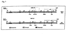
- the protein consists of several highly conserved regions, of dimorphic regions, each of which can be assigned to one of two relatively small oligomorphic blocks in the N-terminal part of the protein ( Fig. 1 ; Tanabe et al., 1987).
- mice Immunization of mice with the protein purified from P. yoelii parasites protects the animals from otherwise lethal infection (Holder and Freeman, 1981). Also, transfer of monoclonal antibodies to MSP-1 from P. yoelii mediated protection in the mouse model (Majarian et al., 1984). In addition to studies in mice, Saimiri and Aotus monkeys were also immunized with native, immunoaffinity-purified MSP-1. In these experiments, the protein recovered from P. falciparum protected partially (Perrin et al., 1984) or completely (Siddiqui et al., 1987) against the subsequent infection with the parasite.
- mice were successfully with from E. coli or Saccharomyces cerevisiae purified MSP-1-19 (Daly and Long, 1993; Ling et al., 1994; Tian et al., 1996; Hirunpetcharat et al., 1997), as well as MSP-1-19 carrying Mycobacterium bovis (Matsumoto et al., 1999).
- MSP-1-19 encoding DNA has also been used as a vaccine and protect immunized mice from infection with P.
- MSP-1 falciparum fused to a T helper cell epitope of tetanus toxin (Keitel et al., 1999) and MSP-1 blocks 3 and 4 (Saul et al., 1999, Genton et al., 2000).
- Some epidemiological studies in endemic areas have shown a correlation between antibody titers against MSP-1 and immunity to malaria in adults (Tolle et al., 1993, Riley et al., 1992, Riley et al., 1993). These studies, together with immunization studies in animals, indicate that MSP-1 is a promising candidate for the development of a malaria vaccine. In general, these studies can be divided into two approaches, either using material purified from parasites or material obtained in heterologous systems.
- MSP-1 can be purified from parasites, this is only possible on a small scale at great expense and therefore not feasible for obtaining MSP-1 according to the criteria mentioned in this way.
- Vaccinia viruses belong to the genus Orthopoxvirus in the subfamily Chordopoxvirinae.
- the smallpox viruses are complex viruses belonging to the largest known viruses with a double-stranded DNA genome of about 200 kb and a size of 250 x 350 nm. They consist of a cuboid virion, which is surrounded by a membrane envelope. In the host cell, the replication and generation of smallpox viruses occurs exclusively in the cytoplasm (for review Moss et al., 1996). Vaccinia viruses have a very broad host cell spectrum, infecting almost all cells from both humans and animals.
- the virus has lost 31 kb DNA sequence of its genome, mainly in the form of six large deletions, including at least two genes that determine the host cell spectrum and thus the replication ability of the virus (Meyer et al., 1991).
- the formation of infectious virus particles in MVA infection in most mammalian cells, including human cells, is blocked very late in the infection cycle at the virion assembly stage, ie, viral genes under the control of both early and intermediate promoters late transcription can also be expressed in non-permissive cells. This distinguishes MVA from other attenuated and replication-deficient poxviruses such. , Vaccinia virus NYVAC or canarypox virus ALVAC, whose infection is disrupted in most mammalian cells prior to viral DNA replication (Tartaglia et al., 1992, Sutter and Moss, 1992).
- vaccinia viruses have been used for malaria vaccine development using replication-competent Western Reserve and Copenhagen viruses and attenuated NYVAC, ALVAC or COPAC viruses (Kaslow et al., 1991, Etlinger and Altenburger, 1991, Aidoo et al. 1997, Allsopp et al., 1996).
- attenuated vaccinia virus MVA only recombinant viruses carrying CSP from the rodent parasite Plasmodium berghei as malaria antigen have been described (Schneider et al., 1998, Plebanski et al., 1998, Degano et al., 1999 Gilbert et al., 1999).
- vaccinia viruses containing an MSP-1 coding sequence are also described.
- the authors of a publication state that they have integrated the native MSP-1 coding sequence into the genome of the Western Standard virus type, but do not support this statement experimentally (no restriction analyzes, PCR, Nothemblot, Western blot analyzes, etc.). Immunization of Saimiri monkeys with these recombinant viruses did not result in the production of MSP-1-specific antibodies and moreover remained without any measurable influence on the humoral immune response to MSP-1 after P. falciparum infection (Pye et al., 1991). , In another publication, the vector NYVAC Pf7 is described, which expresses, inter alia, msp-1 from P.
- the term "MVA-based virus” means a MVA-derived virus having one or more mutations in non-essential regions of the virus genome.
- the essential regions of the MVA virus are all genome segments of the MVA virus required for maintenance the viral gene expression and the ability to multiply the MVA virus are necessary, these include, for example, the gene sequences coding for viral RNA polymerase subunits or the viral DNA polymerase.
- the MVA-based virus is the MVA virus.
- the vaccinia system NYVAC-Pf7 known from the prior art is based on the base virus NYVAC, which originally originated from the vaccinia virus strain Copenhagen and was attenuated by the targeted deletion of 18 open reading frames.
- NYVAC blocks DNA replication in mammalian cells (Tartaglla et al., 1992), while in MVA virus assembly is inhibited.
- MVA the late gene expression takes place, which can be used for the expression of recombinant genes. Therefore, MVA can preferentially induce a cytotoxic T cell response both during the early transcriptional phase and stimulate the humoral branch of the immune response through high protein expression during the late phase.
- MVA which has already been used extensively during the smallpox vaccination campaign, is considered to be a very safe human vaccine virus (Stickl et al., 1974).
- a recombinant MVA virus comprising at least one nucleic acid encoding a fragment or fragments of P. falciparum MSP-1 protein.
- MSP-1 amino acid sequence is available from publicly available databases. 3D7 (MAD20 isolate): CAA84556; FCB-1 (K1 isolate): CAB36903. Both from the NIH database (http://www.ncbi.nlm.nih.gov).
- the MSP-1 fragment or MSP-1 fragment-encoding nucleic acid is a nucleic acid reduced in its AT content, as described in U.S. Patent Nos. 4,378,199 and 5,629,866 DE 19640817 A1 ,
- a nucleic acid derived from the P. falciparum MSP-1 sequence and in which a majority of the Plasmodium codons have been exchanged to correspond to the codon frequency of the synthetic human gene without changing the amino acid sequence is preferred.
- the MSP-1 protein is MSP-1 of isolate 3D7, the MSP-1 protein, hereinafter referred to as "MSP-1D".
- MSP-1 protein may be the FCB1 strain, hereafter referred to as "MSP-1 F”.
- the MSP-1 protein comprises the indicated fragments of these both forms of MSP-1. This includes p42; p42 and p38; and p83, p30, p42 and p38. in particular, the combination p83 and p30 and p38 and p42 are preferred. The position of the fragments results from this illustration 1 , Also included are fragment p42 of both MSP-1 forms.
- the virus contained in the vaccine comprises a promoter suitable for expression, wherein the msp- 1-coding sequence is under the control of the promoter.
- the promoter may in this case be an MVA promoter, wherein the promoter may be an early, intermediate or later gene promoter or a combination thereof.
- MVA promoter MVA promoter
- non-MVA promoters are also included which are capable of expression in the expression system used.
- constitutive and inducible promoters can be used.
- a strong vaccinia virus promoter such as the synthetic late or early / late promoter or the hybrid vaccinia / T7 polymerase system may be used; for the induction of MHC class I restricted cytotactic T cell response in vivo , a naturally occurring early or tandem early / late promoter may be used; Furthermore, the E. coli lac repressor / operator system or the hybrid vaccinia / T7 system can be used to initiate gene expression; ( Methods in Molecular Biology, Volume 62, editors Rocky S. Tuan, Humana Press, Broder and Earl, p. 176 , with further evidence).
- the virus further comprises a selecton marker.
- the selection marker is suitable for selection and / or screening in a known manner. Suitable selection markers here include, for example, the E. coli lacZ system, the seeding system using E. coli xanthine-guanine phosphoribosyl transferase (XGPRT) gene. In addition, seeding methods can be used which alter the host cell specificity of the viruses (Staib et al., 2000). Other selection markers known in the art may be used.
- the nucleic acid is fused at the 5 end with a nucleotide sequence encoding signal peptide sequence.
- a nucleotide sequence encoding signal peptide sequence As is known in the art, P. falciparum signal and anchor sequences are not recognized upon expression in mammalian cells or are not processed correctly (Moran and Caras, 1994; Burghaus et al., 1999; Yang et al. , 1997).
- the problem of selective control of intracellular gating is solved by the use of the signal sequences of the human "Decay Accelereting Factor” (DAF) ( Fig. 2 ).
- DAF Decay Accelereting Factor
- Suitable signal peptide sequences are specific for higher eukaryotes. Examples of such signal sequences besides those of the decay accelerating factor are immunoglobulins or signal peptides of various growth factors and cytokines (von Heijne, 1985).
- the signal peptide sequence controls the secretion of the gene product, for example cytokines, antibodies etc.
- signal sequences that result in GPI anchoring of the C-terminus of the gene product to the cell surface, as in human DAF.
- peptide sequences are preferred which control the membrane-localization of the gene product, as in the case of immunoglobulins of the M-isotype or the Vesicular Stomatitis Virus-G protein.
- the virus may further contain suitable splice donor and splice acceptor sites to produce a correspondingly spliced mRNA suitable for translation within the subject being treated.
- the nucleic acid may further contain a sequence which is useful as a ribosome binding site.
- Suitable pharmacologically acceptable carriers are all carriers and diluents known in the prior art. If a certain mode of administration is intended, the pharmacologically acceptable carrier can be selected or changed in a known manner.
- the vaccine can be administered subcutaneously, intramuscularly, intravenously, transdermally, intraperitoneally or orally.
- the vaccine is indicated for the prophylaxis and / or treatment of malaria in humans and animals.
- the vaccine may further comprise MSP-1, and / or a fragment listed in claim 1 or a fragment combination thereof listed in claim 1.
- the MSP-1 protein is produced here recombinantly, in particular recombinantly in E. coli.
- the MSP-1-encoding nucleic acid is, preferably one which is reduced in its AT content is particularly preferred such a nucleic acid as in DE-19640817 A1
- Plasmodium falciparum codons are exchanged for human codons without altering the amino acid sequence here.
- the vaccine comprises both the recombinant virus and MSP-1, or a fragment thereof
- the vaccine may be in kit form. It is thus suitable for simultaneous, sequential or separate administration of the two components of the vaccine.
- Fig. 1 Primary structure of MSP-1 from P. fa / ciparum strains FCB-1 and MAD20.
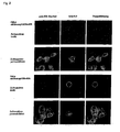
- Fig. 2 Expression of msp1d-38 / 42 in recombinant MVA-infected HeLa cells
- HeLa cells were infected with rMVA-msp1d / 38 + 42S or rMVA-msp1d / 38 + 42A and then fixed.
- the second and fourth cells each show cells that were permeabilized after fixation with Triton X-100, while the membrane of the cells in lines one and three remained intact.
- the cells treated in this way were labeled with mAb 5.2, which recognizes an MSP-1-specific, conformational epitope in the C-terminal part of the MSP-1 fragment p19 and a polyclonal serum which recognizes the ER protein Sec61beta (anti-ER marker).
- MSP-1 D-38/42 is only recognized in the case of infection with rMVA-msp1d / 38 + 42A on the surface of the infected cells.
- the ER marker served as a control for the integrity of the cell membrane.
- ER endoplasmic reticulum
- mAb monoclonal antibody
- FIG. 3 Detection of MSP-1D-42 and MSP-1D-38 / 42 in recombinant MVA infected HeLa cells by immunoblot
- a HeLa cells were infected with rMVA-msp1d / 42S, rMVA-msp1d / 42A, rMVA-msp1d / 38 + 42S and rMVA-msp1d / 38 + 42A, respectively, and incubated overnight. Samples of the supernatant and cellular fraction were separated by SDS gel electrophoresis under non-reducing conditions, transferred to a PVDF membrane and detected by mAb 5.2 as the first antibody. Only chimeras of the DAF signal sequence and the corresponding MSP-1D fragments can be detected in the supernatants of the infected cells, while intracellular expression provides a signal in all cells infected with recombinant MVA.
- FIG. 4 Development of the humoral immune response after three immunizations with rMVA-msp1d / 42S or rMVA-msp1d / 42A and immunization with MSP-1D-HX42 from E. coli
- A shows the analysis of the humoral immune response by ELISA with recombinantly produced MSP-1D-HX42 purified from E. coli as antigen.
- the mice were in each case with intervals of three weeks with 10 6 IU (first immunization, at the same time as the blood sampling S0) or 10 8 IU (1st and 2nd Boost immunized simultaneously with S1 and S2) rMVA-msp1d / 42S.
- the mice were subcutaneously injected with 5 ⁇ g of MSP-1 D-HX42 from E. coli in Freund's adjuvant incomplete after another four weeks (one week after blood collection S3).
- S0 to S5 represent the time of blood collection, while the blood S0 to S3 was taken at intervals of three weeks, the removal of S4 and S5 was carried out at intervals of 4 weeks.
- the arrows mark the times of the immunizations.
- the star marks the fourth immunization with MSP-1 D-HX42 from E. coli.
- B shows the same analysis for immunization with rMVA-msp1 d / 42A.
- Fig. 5 Development of the humoral immune response after immunizations with rMVA-msp1d / S or rMVA-msp1d / 83 + 30/38 + 42A in combination with immunization with MSP-1D from E. coli
- recombinant viruses resulting in the production of MSP-1 in its surface anchored form will be designated “A” and those for MSP-1 secretion by "S”.
- MSP-1 or the fragments and the localization of the proteins in the infected cell was confirmed by confocal microscopy and is exemplary for the infection of HeLa cells by rMVA- msp- 1d / 38 + 42S and rMVA-msp1d / 38 + 42A ( Fig. 2 ).
- Secretion of all MSP-1 variants from cells infected with recombinant MVA was detected by immunoblot analysis of cellular supernatants and is exemplified here for rMVA-msp1d / 42S and rMVA-msp1d / 38 + 42S ( Fig. 3 ).
- the recombinant MVA were tested in immunization experiments on mice for their immunogenic effect on the humoral immune response.
- msp-1 recombinant MVA high antibody titres against the parasite protein induced by ELISA were induced.
- the p42-specific antibody titers and the observed, different immunization potential of the surface-anchored or secreted proteins produced by recombinant MVA are illustrated by way of example for immunizations with rMVA-msp1d / 42S and rMVA-msp1d / 42A ( Fig. 4 ).
Landscapes
- Health & Medical Sciences (AREA)
- Life Sciences & Earth Sciences (AREA)
- Genetics & Genomics (AREA)
- Chemical & Material Sciences (AREA)
- Tropical Medicine & Parasitology (AREA)
- Engineering & Computer Science (AREA)
- Organic Chemistry (AREA)
- General Health & Medical Sciences (AREA)
- Biotechnology (AREA)
- Biomedical Technology (AREA)
- Veterinary Medicine (AREA)
- General Engineering & Computer Science (AREA)
- Bioinformatics & Cheminformatics (AREA)
- Microbiology (AREA)
- Wood Science & Technology (AREA)
- Zoology (AREA)
- Public Health (AREA)
- Medicinal Chemistry (AREA)
- Animal Behavior & Ethology (AREA)
- Pharmacology & Pharmacy (AREA)
- Epidemiology (AREA)
- Plant Pathology (AREA)
- Virology (AREA)
- Mycology (AREA)
- Physics & Mathematics (AREA)
- Biochemistry (AREA)
- Biophysics (AREA)
- Immunology (AREA)
- Molecular Biology (AREA)
- Chemical Kinetics & Catalysis (AREA)
- General Chemical & Material Sciences (AREA)
- Nuclear Medicine, Radiotherapy & Molecular Imaging (AREA)
- Medicines Containing Antibodies Or Antigens For Use As Internal Diagnostic Agents (AREA)
- Micro-Organisms Or Cultivation Processes Thereof (AREA)
- Peptides Or Proteins (AREA)
Description
- Die Herstellung rekombinanter Vaccinia-Viren des Stamms MVA (Modifiziertes Vaccinia Virus Ankara) zur rekombinanten Herstellung des vollständigen Malaria-Antigens gp190/MSP-1 des Malariaerregers Plasmodium falciparum sowie einzelner natürlich vorkommender Domänen und Teile derselben werden hier beschrieben. Ferner betrifft die Erfindung die Nutzung der rekombinanten MVA, welche die synthetische DNA-Sequenz des Malaria-Antigens sowie Teile derselben integriert in das Virus-Genom tragen als Impfstoff zur Immunisierung gegen Malaria.
- Malaria zählt zu den gefährlichsten Infektionskrankheiten der Welt. Schätzungen der Weltgesundheitsorganisation (WHO) zufolge werden jährlich 400 bis 900 Millionen Krankheitsepisoden registriert. Nach Informationen der Multilateralen Initiative gegen Malaria (MIM) sterben zwischen 700.000 und 2,7 Millionen Menschen jedes Jahr an der Infektion (MIM, 2001). Dabei sind in 99 Ländern 40% der Weltbevölkerung einem Malariarisiko ausgesetzt. Verursacht wird die Krankheit durch einzellige Protozoen der Gattung Plasmodium aus dem Phylum Apicomplexa. Es gibt vier Arten, die den Menschen infizieren: Plasmodium malariae, verantwortlich für Malaria quartana, Plasmodium vivax und Plasmodium ovale, beide verursachen Malaria tertiana und schließlich Plasmodium falciparum, der Erreger der Malaria tropica und letztlich verantwortlich für fast alle tödlichen Infektionen.
Ihre Verbreitung nimmt derzeit wieder zu. Dies ist vor allem auf eine intensive Resistenzbildung der Malaria-Erreger zurückzuführen, die dadurch gefördert wird, dass die zur Therapie benutzen Medikamente ebenfalls zur Prophylaxe empfohlen und eingesetzt werden. Neben der Suche nach neuen Chemotherapeutika konzentriert sich die Forschung auf die Entwicklung von Impfstoffen, da im Laufe von Malaria-Infektionen im Menschen Immunitätsmechanismen einsetzen, die sich in einer erhöhten Widerstandsfähigkeit gegenüber den Plasmodien äußert, wie sich in der Entwicklung verschiedener Arten von Immunität bei Menschen in Malaria-epidemischen Regionen zeigt - MSP-1, das Hauptoberflächenprotein von Merozoiten, der invasiven Form der Blutstadien der Malaria-Erreger, ist ein 190 - 220 kDa Protein. Dieses Protein wird spät während der Entwicklung der Merozoiten in kleinere Proteinfragmente prozessiert, die bis zur Invasion von Erythrozyten durch die Parasiten als Komplex verankert über einen Glycosylphosphatidyl-Inositol-Anker auf der Merozoiten-Obertläche vorliegen und isoliert werden können.
Die Sequenzen der MSP-1-Proteine verschiedener P. falciparum-Stämme fallen in zwei Gruppen, die nach zwei repräsentativen Isolaten K1 und MAD20 benannt wurden. Insgesamt besteht das Protein aus mehreren hochkonservierten Regionen, aus dimorphen Bereichen, die sich jeweils einer der beiden zuordnen lassen und aus zwei relativ kleinen oligomorphen Blöcken im N-terminalen Teil des Proteins (Fig. 1 ; Tanabe et al., 1987). - Die Immunisierung von Mäusen mit dem aus P. yoelii-Parasiten aufgereinigten Protein schütze die Tiere vor der andernfalls tödlichen Infektion (Holder and Freeman, 1981). Auch der Transfer von monoklonalen Antikörpern gegen MSP-1 von P. yoelii vermittelte Schutz im Mausmodell (Majarian et al., 1984).
Neben Studien an Mäusen wurden auch Saimiri und Aotus-Affen mit nativem, immunaffinitätsgereinigtem MSP-1 immunisiert. In diesen Versuchen schützte das aus P. falciparum gewonnene Protein partiell (Perrin et al., 1984) bzw. völlig (Siddiqui et al., 1987) gegen die folgende Infektion mit dem Parasiten.
Eine Aufreinigung von nativem Material aus Plasmodien ist allerdings aufwendig und lässt keine Produktion im großen Maßstab zu. Daher konzentriert sich die Impfstoff-Forschung auf die Entwicklung rekombinanter Vakzine.
So wurden Mäuse erfolgreich mit aus E. coli oder Saccharomyces cerevisiae gereinigtem MSP-1-19 immunisiert (Daly and Long, 1993; Ling et al., 1994; Tian et al., 1996; Hirunpetcharat et al., 1997), ebenso wie MSP-1-19 tragendes Mycobacterium bovis (Matsumoto et al., 1999). Alternativ zur Immunisierung mit nativen oder rekombinanten Proteinen wurde auch für MSP-1-19-kodierende DNA als Impfstoff eingesetzt und schütze immunisierte Mäuse vor der Infektion mit P. chabaudi (Wunderlich et al., 2000).
Die Immunisierungen von Affen mit rekombinantem MSP-1-19 und MSP-1-42 aus P. falciparum vermittelte teilweise Schutz (Kumar et al., 1995; Egan et al., 2000; Chang et al., 1996; Stowers et al., 2001). Die Interpretation von Immunsierungsversuchen an Affen ist allerdings nur bedingt möglich, da eine statistische Auswertung der Ergebnisse auf Grund der geringen Versuchstierzahlen nicht gegeben ist.
In Phase I und II Studien zu MSP-1-Fragmenten als Impfstoff wurde deren Immunogenität auch im Menschen gezeigt. Dabei handelte es sich um p19 aus P. falciparum fusioniert an ein T-Helfer-Zell-Epitope des Tetanustoxins (Keitel et al., 1999) und die MSP-1-Blöcke 3 und 4 (Saul et al., 1999; Genton et al., 2000).
Einige epidemiologische Studien in endemischen Gebieten zeigten bei Erwachsenen eine Korrelation zwischen Antikörpertitem gegen MSP-1 und der Immunität gegen Malaria (Tolle et al., 1993; Riley et al., 1992; Riley et al., 1993).
Diese Untersuchungen zusammen mit den Immunisierungsstudien an Tieren belegen, dass es sich bei MSP-1 um einen vielversprechenden Kandidaten zur Entwicklung eines Malaria-Impfstoffs handelt.
Generell lassen sich diese Studien in zwei Ansätze unterscheiden, entweder wurde aus Parasiten aufgereinigtes Material oder in heterologen Systemen gewonnenes Material eingesetzt.
Sowohl für Funktionsuntersuchungen als auch für den Einsatz als Impfstoff müssen Proteine in guter Ausbeute, hoher Qualität und reproduzierbar herzustellen sein. Zwar läßt sich MSP-1 aus Parasiten aufreinigen, allerdings ist dies nur im kleinen Maßstab unter großem Aufwand möglich und daher für die Gewinnung von MSP-1 nach den genannten Kriterien auf diesem Weg nicht durchführbar. - Vaccinia-Viren gehören zur Gattung Orthopoxvirus in der Unterfamilie der Chordopoxvirinae. Es handelt sich bei den Pocken-Viren um komplexe Viren, die mit einem doppelsträngigen DNA-Genom von ca. 200 kb und einer Größe von 250 x 350 nm zu den größten bekannten Viren gehören. Sie bestehen aus einem quaderförmigen Virion, das von einer Membranhülle umgeben ist. In der Wirtszelle verläuft die Replikation und Generation der Pocken-Viren ausschließlich im Zytoplasma (zur Übersicht Moss et al., 1996). Dabei besitzen Vaccinia-Viren ein sehr breites Wirtszellspektrum, sie infizieren nahezu alle Zellen sowohl von Menschen als auch Tieren stammend. 1953 isolierte und reinigte Anton Mayr den Dermovaccinia-Stamm Chorioallantois Vaccinia Ankara (CVA). Dieses Virus wurde über fortlaufende Passage auf Hühner-Embryo-Fibroblasten weiter vermehrt, und heraus kam ein attenuiertes Virus, das in Tier und Mensch keine Virulenz mehr zeigte (Stickl et al., 1974). Ungeachtet dessen konnte das Virus weiterhin zur Immunprophylaxe gegen durch Orthopockenviren hervorgerufenen Erkrankungen bei Mensch und Tier eingesetzt werden (Stickl et al., 1974). Nach seinem Ursprungsort wurde dieses Virus Modifiziertes Vaccinia-Virus Ankara (MVA) genannt.
Molekufargenetisch betrachtet hat das Virus während der über 570 Passagen auf Hühner-Embryo-Fibroblasten 31 kb DNA-Sequenz seines Genoms, hauptsächlich in Form sechs großer Deletionen verloren, darunter wenigstens zwei Gene, die das Wirtszellspektrum und damit die Replikationsfähigkeit des Virus bestimmen (Meyer et al., 1991). Die Bildung infektiöser Viruspartikel ist bei der MVA-Infektion in den meisten von Säugetieren stammenden Zellen, einschließlich Zellen von Menschen, erst sehr spät im Infektionszyklus auf der Stufe des Virionenzusammenbaus blockiert, d.h. virale Gene unter der Kontrolle von Promotoren sowohl zur frühen als auch intermediären und späten Transkription können auch in nicht permissiven Zellen exprimiert werden. Das unterscheidet MVA von anderen attenuierten und replikationsdefizienten Pockenviren, wie z. B. Vaccinia-Virus NYVAC oder Kanarienpockenvirus ALVAC, deren Infektion in den meisten vom Säugetier stammenden Zellen bereits vor der viralen DNA-Replikation unterbrochen ist (Tartaglia et al., 1992, Sutter and Moss, 1992). - Zur Malariaimpfstoffentwicklung wurden verschiedene rekombinante Vaccinia-Viren eingesetzt, dabei wurden replikationskompetente Viren des Typs Western Reserve und Copenhagen und attenuierte Viren der Typen NYVAC, ALVAC oder COPAC verwendet (Kaslow et al., 1991; Etlinger and Altenburger, 1991; Aidoo et al., 1997; Allsopp et al., 1996).
Im Zusammenhang mit dem attenuierten Vaccinia-Virus MVA wurden lediglich rekombinante Viren beschrieben, welche CSP aus dem Nager-Parasiten Plasmodium berghei als Malaria-Antigen tragen (Schneider et al., 1998; Plebanski et al., 1998; Degano et al., 1999; Gilbert et al., 1999). - Ferner sind rekombinante Vaccinia-Viren beschrieben, die eine MSP-1-kodierende Sequenz enthalten. Die Autoren einer Publikation geben an, die native MSP-1-kodierende Sequenz in das Genom des Virustyps Western Reserve integriert zu haben, unterstützen diese Aussage aber nicht experimentell (keine Restriktionsanalysen, PCR, Nothemblot-, Westemblotanalysen etc.). Eine Immunisierung von Saimiri-Affen mit diesen rekombinanten Viren führte nicht zur Bildung MSP-1-spezifischer Antikörper und blieb überdies auch ohne messbaren Einfluss auf die humorale Immunantwort gegen MSP-1 nach einer P. falciparum-Infektion (Pye et al., 1991).
In einer weiteren Veröffentlichung wird der Vektor NYVAC-Pf7 beschrieben, der unter anderem msp-1 aus P. falciparum exprimiert. Die Seren von zwei der sechs immunisierten Rhesusaffen zeigen in der Westernblotanalyse keine In der Publikation erkennbaren Signale gegen natives MSP-1. Die Signale von drei weiteren Tieren erkennen lediglich Teile des Proteinkompexes, und nur das Serum eines der immunisierten Tiere erkennt ein breiteres Bandenspektrum. Insgesamt erscheinen jedoch auch diese Signale schwach. Quantitative Analysen zu MSP-1-spezifischen Antikörpern durch ELISA wurden nicht angeführt (Tine et al., 1996).
In Humanversuchen der Phase I/IIa mit NYVAC-Pf7 wurden weder eine zelluläre Immunantwort gegen MSP-1 noch eine durch ELISA nachweisbare humorale Immunantwort nachgewiesen (Ockenhouse et al., 1998).
Im Gegensatz dazu weisen Tine et al. (1996) intaktes MSP-1 in der Westemblot-Analyse nach, wobei widersprüchlich erscheint, dass MSP-1 mittels P. falciparum Signalsequenzen (vgl. Publikationen von Moran and Caras, 1994, Yang et al., 1997) aus der Zelle transportiert wird. - Der vorliegenden Erfindung liegt daher die Aufgabe zu Grunde ein rekombinantes Vaccinia-Virus bereitzustellen, das fähig ist
- DNA-Sequenzen, welche MSP-1 aus P, falciparum bzw. Teibereiche desselben kodieren, stabil Integriert zu enthalten,
- diese Sequenzen effizient und reproduzierbar zu exprimieren und damit
- MSP-1 Protein in sekretierter oder Oberflächen-verankerter Form herzustellen, um
- einen Wirt zu Immunisieren und dabei
- eine zelluläre und humorale Immantwort hervorzurufen.
- Die der vorliegenden Erfindung zu Grunde liegende Aufgabe wird gelöst durch die Bereitstellung eines Vakzins, umfassend
- a) ein rekombinantes MVA-Virus, umfassend eine ein Fragment oder Fragmente von Plasmodium falciparum MSP-1 Protein kodierende Nukleinsäure; und
- b) einen pharmakologischen verträglichen Träger,
- p42;
- p42 und p38;
- p83, p30, p42 und p38.
- Erfindungsgemäß bedeutet der Ausdruck "Virus auf MVA-Basis" ein von MVA abgeleitetes Virus, das in nicht-essentiellen Regionen des Virusgenoms eine oder mehrere Mutationen aufweist Die essentiellen Regionen des MVA-Virus sind hierbei alle Genomabschnitte des MVA-Virus, die für den Erhalt der viralen Genexpression und die Vermehrungsfähigkeit des MVA-Virus notwendig sind, hierzu zählen z. b. die für virale RNA-Polymerase-Untereinheiten bzw. die virale DNA-Polymerase kodierenden Gensequenzen. Vorzugsweise ist das Virus auf MVA-Basis das MVA-Virus.
- Das aus dem Stand der Technik bekannte Vaccinia-System NYVAC-Pf7 beruht auf dem Basisvirus NYVAC, welches ursprünglich dem Vaccinia-Virus-Stamm Copenhagen entstammt und durch die gezielte Deletion von 18 offenen Leserastem attenuiert wurde. Jedoch ist bei NYVAC in Säugerzellen die DNA-Replikation blockiert (Tartaglla et al., 1992), während bei MVA die Virusassemblierung unterbunden wird. Dies hat den Vorteil, dass im Gegensatz zu NYVAC In MVA die späte Genexpression stattfindet, welche für die Expression rekombinanter Gene genützt werden kann. MVA kann daher sowohl während der frühen Transkriptionsphase bevorzugt eine zytotoxische T-Zell-Antwort induzierten als auch den humoralen Zweig der Immunantwort durch die hohe Protein-Expression während der späten Phase stimulieren.
- MVA, das bereits während der Pockenschutz-Impfkampagne umfangreich eingesetzt wurde, gilt als sehr sicheres Impfvirus am Menschen (Stickl et al., 1974).
- Effindungsgemäß wird ein rekombinantes MVA-Virus, umfassend wenigstens eine ein Fragment oder Fragmente von P. falciparum MSP-1 Protein kodierende Nukleinsäure.
- Die MSP-1-Aminosäuresequenz ist aus öffentlich zugänglichen Datenbanken erhältlich. 3D7 (MAD20-Isolat): CAA84556; FCB-1 (K1-Isolat): CAB36903. Beides aus der NIH-Datenbank (http://www.ncbi.nlm.nih.gov).
- Besonders bevorzugt ist die MSP-1-Fragment oder MSP-1 Fragmente kodierende Nukleinsäure eine in ihrem AT-Gehalt verringerte Nukleinsäure wie beschrieben in
DE 19640817 A1 . Insbesondere ist eine Nukleinsäure bevorzugt, die von der P. falciparum-MSP-1-Sequenz abgeleitet ist und bei der ein Großteil der Plasmodium-Codons so ausgetauscht wurde, das die Codonhäufigkeit des synthetischen Gens der humanen entspricht, ohne dabei die Aminosäuresequenz zu verändem. - Gemäß einer bevorzugten Ausführungsform ist das MSP-1-Protein das MSP-1 des Isolats 3D7, das MSP-1-Proteins das nachfolgend als "MSP-1D" bezeichnet wird. Alternativ kann das MSP-1-Protein des FCB1-Stammes sein, das nachfolgend als "MSP-1 F" bezeichnet wird. Das MSP-1-Protein umfasst die angegebenen Fragmente dieser beiden Formen von MSP-1. Umfasst sind hierbei p42; p42 und p38; und p83, p30, p42 und p38. insbesondere ist die Kombination p83 und p30 sowie p38 und p42 bevorzugt. Die Lage der Fragmente ergibt sich hierbei aus
Abbildung 1 . Ferner umfasst werden auch das Fragment p42 von beiden MSP-1-Formen. - In einer bevorzugten Ausführungsform umfasst das im Vakzin enthaltene Virus einen zur Expression geeigneten Promotor, wobei die msp-1-kodierende Sequenz unter der Kontrolle des Promotors steht. Der Promotor kann hierbei ein MVA-Promotor sein, wobei der Promotor ein früher, intermediärer oder später Genpromotor oder eine Kombination davon sein kann. Jedoch sind auch nicht-MVA-Promotoren umfasst, die zur Expression in dem verwendeten Expressionssystem fähig sind. Hierbei können sowohl konstitutive als auch induzierbare Promotoren verwendet werden. Zur Large Scale Protein-Produktion kann hierbei ein starker Vaccinia Virus-Promotor, wie der synthetische späte oder frühe/späte Promotor oder das HybridVaccinia/T7 Polymeresasystem verwendet werden; zur induktion von MHC-Klasse I - restringierter zytotoktischer T-Zellantwort in vivo kann bevorzugt ein natürlich vorkommender früher oder Tandem-füher/später Promotor verwendet werden; weiterhin kann das E. coli lac repressor/Operatorsystem oder das HybridVaccinia/T7-System zur initiation der Genexpression verwendet werden; (Methods in Molecular Biology, Volume 62, Herausgeber Rocky S. Tuan, Humana Press, Broder und Earl, Seite 176, mit weiteren Nachweisen).
- Gemäß einer weiteren bevorzugten Ausführungsform umfasst das Virus ferner einen Selektlonsmarker. Der Selektionsmarker ist hierbei zur Selektion und/oder zum Screening in bekannter Weise geeignet. Geeignete Selektionsmarker umfassen hierbei beispielsweise das E. coli lacZ-System, das Seiektions-System unter Verwendung von E. coli Xanthin-Guanin Phosphoribosyl Transferase (XGPRT)-Gen. Zusätzlich können Seiektionsmethoden verwendet werden, welche die Wirtszellspezifität der Viren verändern (Staib et al., 2000). Weitere im Stand der Technik bekannte Selektionsmarker können verwendet werden.
- Gemäß einer weiteren bevorzugten Ausführungsform ist die Nukleinsäure am 5 Ende mit einer Signalpeptidsequenz kodierenden Nukleotidsequenz fusioniert. Wie aus dem Stand der Technik bekannt, werden die Signal- und Anker-Sequenzen aus P. falciparum bei Expression in Säugerzellen nicht erkannt, bzw. nicht korrekt prozessiert (Moran und Caras, 1994; Burghaus et al.,1999; Yang et al., 1997).
- Das Problem der selektiven Steuerung des intrazellulären Gating wird durch die Verwendung der Signalsequenzen des humanen "Decay-Accelereting-Factors" (DAF) gelöst (
Fig. 2 ). Geeignete Signalpeptidsequenzen sind spezifisch für höhere Eukaryonten. Beispiele für solche Signalsequenzen neben denen des Decay-Accelerating-Factors sind Immunglobuline oder Signalpeptide verschiedener Wachstumsfaktoren und Zytokine (von Heijne, 1985). Gemäß einer bevorzugten Ausführungsform steuert die Signalpeptidsequenz die Sekretion des Genproduktes, beispielsweise Cytokine, Antikörper etc.. - Ferner sind Signalsequenzen bevorzugt, die zu einer GPI-Verankerung des C-Terminus des Genproduktes an der Zelloberfläche führen, wie bei den humanen DAF. Alternativ sind Peptidsequenzen bevorzugt, die die membranständige Lokalisierung des Genproduktes steuern, wie im Falle von Immunglobulinen des M-Isotyps oder des Vesicular Stomatitis Virus-G Proteins.
- Gemäß einer weiteren bevorzugten Ausführungsform kann das Virus ferner geeignete Splice Donor- und Splice Akzeptor-Stellen enthalten, so dass eine entsprechend gespleißte mRNA entsteht, die zur Translation innerhalb des zu behandelnden Individuums geeignet ist. Die Nukleinsäure kann ferner eine Sequenz enthalten, die als Ribosomenbindungsstelle geeignet ist.
- Als pharmakologisch verträgliche Träger sind hierbei sämtliche im Stand der Technik bekannten Träger und Verdünnungsmittel geeignet. Sofern eine bestimmte Applikationsart beabsichtigt ist, kann der pharmakologisch verträgliche Träger in bekannter Weise ausgewählt oder geändert werden.
- Das Vakzin kann subkutan, intramuskulär, intravenös, transdermal, intraperitoneal oder oral verabreicht werden. Das Vakzin wird zur Prophylaxe und/oder Therapie von Malaria bei Mensch und Tier angegeben.
- Gemäß einer bevorzugten Ausführungsform kann das Vakzin ferner MSP-1, und / oder ein im Anspruch 1 aufgeführtes Fragment bzw. eine im Anspruch 1 aufgeführte Fragmentkombination davon enthalten.
- Besonders bevorzugt ist das MSP-1-Protein hierbei rekombinant hergesteilt, insbesondere rekombinant in E. coli. Die MSP-1 kodierende Nukleinsäure ist, vorzugsweise eine solche, die bezüglich ihres AT-Gehaltes verringert ist Insbesondere bevorzugt ist eine solche Nukleinsäure wie in
DE-19640817 A1 beschrieben, bei der Insbesondere Plasmodium falciparum-Codons gegen humane Codons ausgetauscht sind, ohne die Aminosäure-Sequenz hierbei zu verändem. - Sofern das Vakzin sowohl das rekombinante Virus als auch MSP-1, oder ein Fragment davon umfasst, so kann das Vakzin In Kit-Form vorlieben. Es Ist somit geeignet zur simultanen, sequenziellen oder getrennten Verabreichung der beiden Komponenten des Vakzins.
- Die nachfolgenden Beispiele sollen die Erfindung veranschaulichen.
- Die Pfeile oberhalb der Sequenz kennzeichnen die Prozessierungsstellen der nativen Proteine (Holder et al., 1987), welche MSP-1 in die Fragmenten p83, p30, p38 und p42 teilen, die als Komplex auf der Parasitenoberfläche verankert sind. Im zweiten Prozessierungsschritt wird p42 zu p33 und p19 gespalten. Die Pfeile unterhalb der Darstellungen bezeichnen die einmalig vorkommenden Endonuklease-Schnittstellen der synthetischen DNA-Sequenzen. Abkürzungen: SP = Signalpeptid, GA = GPI-Anker
- HeLa-Zellen wurden mit rMVA-msp1d/38+42S bzw. rMVA-msp1d/38+42A infiziert und anschließend fixiert. Die zweite und vierte Zelle zeigt jeweils Zellen, die nach der Fixierung mit Triton X-100 permeabilisiert wurden, während die Membran der Zellen in Zeile eins und drei intakt blieb. Die so behandelten Zellen wurden mit mAk 5.2, der ein für MSP-1spezifisches, konformationelles Epitop in C-terminalen Teil des MSP-1-Fragments p19 erkennt und einem polyklonalen Serum, das das ER-Protein Sec61beta erkennt (anti-ER-Marker), als erstem Antikörper inkubiert, diese über Cy3-konjugierte anti-Maus-IgG (erkennt mAk 5.2) bzw. FITC-konjugierte anti-Kaninchen-IgG (erkennt anti-Sec61beta) farblich markiert und anschließend im konfokalen Mikroskop analysiert. Wurden die Zellen mit rMVA-msp1d/38+42S bzw. rMVA-msp1d/38+42A infiziert und permeabiliziert, so kann das Signal für MSP-1D-38/42 (mAk 5.2) kolokalisiert werden mit dem ER-Marker. Blieben die Zellen hingegen unversehrt, so wird MSP-1 D-38/42 nur im Fall der Infektion mit rMVA-msp1d/38+42A auf der Oberfläche der infizierten Zellen erkannt. Der ER-Marker fungierte hier als Kontrolle für die Unversehrtheit der Zellmembran.
Abkürzungen: ER = Endoplasmatisches Retikulum; mAk = monoklonaler Antikörper - A HeLa-Zellen wurden mit rMVA-msp1d/42S, rMVA-msp1d/42A, rMVA-msp1d/38+42S bzw. rMVA-msp1d/38+42A infiziert und über Nacht inkubiert. Proben des Überstands und der zellulären Fraktion wurden über SDS-Gel-Elektrophorese unter nicht reduzierenden Bedingungen aufgetrennt, auf eine PVDF-Membran transferiert und mittels mAb 5.2 als Erstantikörper nachgewiesen. Nur Chimäre aus DAF-Signalsequenz und den entsprechenden MSP-1D-Fragmenten lassen sich in den Überständen der infizierten Zellen nachweisen, während die intrazelluläre Expression in allen mit rekombinanten MVA infizierten Zellen ein Signal liefert.
- In A ist die Analyse der humoralen Immnunantwort mittels ELISA mit rekombinant hergestelltem MSP-1D-HX42 aufgereinigt aus E. coli als Antigen dargestellt. Die Kurven zeigen die p42 spezifische Antikörperentwicklung, gemessen an der OD405 = 1. Die Mäuse wurden jeweils im Abstand von drei Wochen mit 106 IU (Erstimmunisierung, zeitgleich mit der Blutentnahme S0) bzw. 108 IU (1. und 2. Boost, zeitgleich mit S1 und S2) rMVA-msp1d/42S immunisiert. Zusätzlich wurde den Mäusen nach weiteren vier Wochen je 5µg MSP-1 D-HX42 aus E. coli in unkompletten Freund'schem Adjuvans subcutan injiziert (eine Woche nach der Blutentnahme S3). S0 bis S5 stellen die Zeitpunkte der Blutentnahme dar, dabei wurde das Blut S0 bis S3 jeweils im Abstand von drei Wochen entnommen, die Entnahme von S4 und S5 erfolgte jeweils im Abstand von 4 Wochen. die Pfeile markieren die Zeitpunkte der Immunisierungen. Der Stern markiert die vierte Immunisierung mit MSP-1 D-HX42 aus E. coli.
B zeigt die gleiche Analyse für die Immunisierung mit rMVA-msp1 d/42A. - Die humorale Immunantwort wurde mittels ELISA unter Verwendung von rekombinant hergestelltem MSP-1 D aufgereinigt aus E. coli als Antigen ermittelt. Wie in
Fig.4 zeigen die Kurven die MSP-1 D-spezifische Antikörperentwicklung, gemessen an der OD405 = 1. Die Mäuse wurden jeweils im Abstand von 3 Wochen immunisiert (in der Abbildung durch Pfeile gekennzeichnet). Die den Gruppen zugeordneten Immunisierungsstrategien setzten sich wie folgt zusammen: - Gr. 1: 20 µg MSP-1 D (Tag 0), 108 IU rMVA-msp1d/S (Tag 21), 5 Mäuse
- Gr. 2: 20 µg MSP-1 D (Tag 0), 108 IU rMVA-msp1d/A (Tag 21), 5 Mäuse
- Gr. 3: 20 µg MSP-1 D (Tag 0), 108 IU rMVA-msp1d/S (Tag 21), 20 µg MSP-1 D (Tag 42), 10 Mäuse
- Gr. 4: 20 µg MSP-1 D (Tag 0), 108 IU rMVA-msp1d/S (Tag 21), 108 IU rMVA-msp1d/S (Tag 42), 10 Mäuse
- Gr. 5: 20 µg MSP-1 D (Tag 0), 108 IU rMVA-msp1d/83+30/38+42A (Tag 21), 20 µg MSP-1 D (Tag 42) 9 Mäuse
- Gr. 6: 20 µg MSP-1D (Tag 0), 108 IU rMVA-msp1d/83+30/38+42A (Tag 21), 108 IU rMVA-msp1d183+30/38+42A (Tag 42), 10 Mäuse
- Gr. 7: 20 µg MSP-1 D (Tag 0), 108 IU rMVA-msp1d/A (Tag 21), 20 µg MSP-1 D (Tag 42), 5 Mäuse
- Im folgenden werden rekombinante Viren, welche zur Herstellung von MSP-1 in seiner Oberflächen-verankerten Form führen, mit "A" und solche zur Sekretion von MSP-1 durch "S" gekennzeichnet.
Tabelle 1 Vollständige Liste der im Rahmen der Erfindung hergestellten Virus-Konstrukte: rMVA-msp1d rMVA-msp1f sekretiertes MSP-1 rMVA-msp1d/S rMVA-msp1d/83S rMVA-msp1d/83+30S rMVA-msp1d/42S rMVA-msp1d/38+42S Oberflächen-verankertes MSP-1 rMVA-msp1d/A rMVA-msp1f/A rMVA-msp1d/83A rMVA-msp1f/83+30/38+42A rMVA-msp1d/83+30A rMVA-msp1f/38+42A rMVA-msp1d/42A rMVA-msp1d/38+42A rMVA-msp1d/83+30/38+42A - Die Herstellung von MSP-1 bzw. der Fragmente und die Lokalisation der Proteine in der infizierten Zelle wurde mittels konfokaler Mikroskopie belegt und ist exemplarisch für die Infektion von HeLa-Zellen durch rMVA-msp-1d/38+42S und rMVA-msp1d/38+42A dargestellt (
Fig. 2 ).
Die Sekretion aller MSP-1-Varianten aus mit rekombinanten MVA infizierten Zellen wurde über Immunoblot-Analysen zellulärer Überstände nachgewiesen und ist hier exemplarische für rMVA-msp1d/42S und rMVA-msp1d/38+42S dargestellt (Fig. 3 ). - Anschließend wurden die rekombinanten MVA in Immunisierungsversuchen an Mäusen auf ihre immunogene Wirkung bezüglich der humoralen Immunantwort untersucht. Dabei induzierten für msp-1 rekombinanten MVA hohe Antikörpertiter gegen das Parasitenprotein, die über ELISA bestimmt wurden. Die p42-spezfischen Antikörpertiter und das beobachtete, unterschiedliche Immunisierungspotential der durch rekombinante MVA hergestellten Oberflächen-verankerten bzw. sekretierten Proteine wird exemplarisch für Immunisierungen mit rMVA-msp1d/42S und rMVA-msp1d/42A dargestellt (
Fig. 4 ). -
- Aidoo, M., Lalvani, A., Whittle, H. C., Hill, A. V., and Robson, K. J. (1997). Recombinant vaccinia viruses for the characterization of Plasmodium falciparum-specific cytotoxic T lymphocytes: recognition of processed antigen despite limited re-stimulation efficacy. Int Immunol 9, 731-7.
- Allsopp, C. E., Plebanski, M., Gilbert, S., Sinden, R. E., Harris, S., Frankel, G., Dougan, G., Hioe, C., Nixon, D., Paoletti, E., Layton, G., and Hill, A. V. (1996). Comparison of numerous delivery systems for the induction of cytotoxic T lymphocytes by immunization. Eur J Immunol 26, 1951-9.
- Antoine, G., Scheiflinger, F., Domer, F, and Falkner, F. G. (1998). The complete genomic sequence of the modified vaccinia Ankara strain: Comparison with other Orthopoxviruses. Virology 244, 365-396.
- Chang, S. P., Case, S. E., Gosnell, W. L., Hashimoto, A., Kramer, K. J., Tam, L. Q., Hashiro, C. Q., Nikaido, C. M., Gibson, H. L., Lee-Ng, C. T., Barr, P. J., Yokota, B. T., and Hut, G. S. (1996). A recombinant baculovirus 42-kilodalton C-terminal fragment of Plasmodium falciparum merozoite surface protein 1 protects Aotus monkeys against malaria. Infect Immun 64, 253-61.
- Daly, T. M., and Long, C. A. (1993). A recombinant 15-kilodalton carboxyl-terminal fragment of Plasmodium yoelii yoelii 17XL merozoite surface protein 1 induces a protective immune response in mice. Infect. Immun. 61, 2462-7.
- Degano, P., Schneider, J., Hannan, C. M., Gilbert, S. C., and Hill, A. V. (1999). Gene gun intradermal DNA immunization followed by boosting with modified Vaccinia virus Ankara: enhanced CD8+ T cell immunogenicity and protective efficacy in the influenza and malaria models. Vaccine 18, 623-32.
- Egan, A. F., Blackman, M. J., and Kaslow, D. C. (2000). Vaccine efficacy of recombinant Plasmodium falciparum merozoite surface protein 1 in malaria-naive, -exposed, and/or - rechallenged Aotus vociferans monkeys. Infect Immun 68, 1418-27.
- Etlinger, H. M., and Altenburger, W. (1991). Overcoming inhibition of antibody responses to a malaria recombinant vaccinia virus caused by prior exposure to wild type virus. Vaccine 9, 470-2.
- Genton, B., Al-Yaman, F., Anders, R., Saul, A., Brown, G., Pye, D., Irving, D. O., Briggs, W. R., Mai, A., Ginny, M., Adiguma, T., Rare, L, Giddy, A., Reber-Liske, R., Stuerchler, D., and Alpers, M. P. (2000). Safety and immunogenicity of a three-component blood-stage malaria vaccine in adults living in an endemic area of Papua New Guinea. Vaccine 18, 2504-11.
- Gilbert, S. C., Schneider, J., Plebanski, M., Hannan, C. M., Blanchard, T. J., Smith, G. L, and Hill, A. V. (1999). Ty virus-like particles, DNA vaccines and Modified Vaccinia Virus Ankara; comparisons and combinations. Biol Chem 380, 299-303.
- Hirunpetcharat, C., Tian, J. H., Kaslow, D. C., van Rooijen, N., Kumar, S., Berzofsky, J. A., Miller, L. H., and Good, M. F. (1997). Complete protective immunity induced in mice by immunization with the 19-kilodalton carboxyl-terminal fragment of the merozoite surface protein-1 (MSP1[19]) of Plasmodium yoelii expressed in Saccharomyces cerevisiae: correlation of protection with antigen-specific antibody titer, but not with effector CD4+ T cells. J Immunol 159, 3400-11.
- Holder, A. A., and Freeman, R. R. (1981). Immunization against blood-stage rodent malaria using purified parasite antigens. Nature. 294, 361-364.
- Holder, A. A., Sandhu, J. S., Hillman, Y., Davey, L. S., Nicholls, S. C., Cooper, H., and Lockyer, M. J. (1987). Processing of the precursor to the major merozoite surface antigens of Plasmodium falciparum. Parasitology 94, 199-208.
- Kaslow, D. C., Isaacs, S. N., Quakyi, I. A., Gwadz, R. W., Moss, B., and Keister, D. B. (1991). Induction of Plasmodium falciparum transmission-blocking antibodies by recombinant vaccinia virus. Science 252, 1310-1313.
- Keitel, W. A., Kester, K. E., Atmar, R. L., White, A. C., Bond, N. H., Holland, C. A., Krzych, U., Palmer, D. R., Egan, A., Diggs, C., Ballou, W. R., Hall, B. F., and Kaslow, D. (1999). Phase I trial of two recombinant vaccines containing the 19kd carboxy terminal fragment of Plasmodium falciparum merozoite surface protein 1 (msp-1(19)) and T helper epitopes of tetanus toxoid. Vaccine 18, 531-9.
- Kumar, S., Yadava, A., Keister, D. B., Tian, J. H., Ohl, M., Perdue-Greenfield, K. A., Miller, L. H., and Kaslow, D. C. (1995). Immunogenicity and in vivo efficacy of recombinant Plasmodium falciparum merozoite surface protein-1 in Aotus monkeys. Mol Med 1, 325-32.
- Ling, I. T., Ogun, S. A., and Holder, A. A. (1994). Immunization against malaria with a recombinant protein. Parasite Immunol 16, 63-7.
- Majarian, W. R., Daly, T. M., Weidanz, W. P., and Long, C. A. (1984). Passive immunization against murine malaria with an IgG3 monoclonal antibody. J. Immunol. 132, 3131-3137.
- Matsumoto, S., Yukitake, H., Kanbara, H., and Yamada, T. (1999). Long-lasting protective immunity against rodent malaria parasite infection at the blood stage by recombinant BCG secreting merozoite surface protein-1. Vaccine 18, 832-4.
- Meyer, H., Sutter, G., and Mayr, A. (1991). Mapping of deletions in the genome of the highly attenuated vaccinia virus MVA and their influence on virulence. J Gen Virol 72, 1031-8.
- Moran, P., and Caras, I. W. (1994). Requirements for glycosylphosphatidylinositol attachment are similar but not identical in mammalian cells and parasitic protozoa. J Cell Biol 125, 333-43.
- Moss, B., Carroll, M. W., Wyatt, L. S., Bennink, J. R., Hirsch, V. M., Goldstein, S., Elkins, W. R., Fuerst, T. R., Lifson, J. D., Piatak, M., Restifo, N. P., Overwijk, W., Chamberlain, R., Rosenberg, S. A., and Sutter, G. (1996). Host range restricted, non-replicating vaccinia virus vectors as vaccine candidates. Adv Exp Med Biol 397, 7-13.
- Ockenhouse, C. F., Sun, P. F., Lanar, D. E., Wellde, B. T., Hall, B. T., Kester, K., Stoute, J. A., Magill, A., Krzych, U., Farley, L, Wirtz, R. A., Sadoff, J. C., Kaslow, D. C., Kumar, S., Church, L W., Crutcher, J. M., Wizel, B., Hoffman, S., Lalvani, A., Hill, A. V., Tine, J. A., Guito, K. P., de Taisne, C., Anders, R., Ballou, W. R., and et al. (1998). Phase I/IIa safety, immunogenicity, and efficacy trial of NYVAC-Pf7, a pox-vectored, multiantigen, multistage vaccine candidate for Plasmodium falciparum malaria. J Infect Dis 177, 1664-73.
- Perrin, L. H., Merkli, B., Loche, M., Chizzolini, C., Smart, J., and Richle, R. (1984). Antimalarial immunity in Saimiri monkeys. Immunization with surface components of asexual blood stages. J. Exp. Med. 160, 441-451.
- Plebanski, M., Gilbert, S. C., Schneider, J., Hannan, C. M., Layton, G., Blanchard, T., Becker, M., Smith, G., Butcher, G., Sinden, R. E., and Hill, A. V. (1998). Protection from Plasmodium berghei infection by priming and boosting T cells to a single class I-restricted epitope with recombinant carriers suitable for human use. Eur J Immunol 28, 4345-55.
- Pye, D., Edwards, S. J., Anders, R. F., O'Brien, C. M., Franchina, P., Corcoran, L. N., Monger, C., Peterson, M. G., Vandenberg, K. L., Smythe, J. A., Westley, S. R., Coppel, R. L., Webster, T. L., Kemp, D. J., Hampson, A. W., and Langford, C. J. (1991). Failure of recombinant vaccinia viruses expressing Plasmodium falciparum antigens to protect Saimiri monkeys against malaria. Infect. Immun. 59, 2403-2411.
- Riley, E. M., Allen, S. J., Wheeler, J. G., Blackman, M. J., Bennett, S., Takacs, B., Schonfeld, H. J., Holder, A. A., and Greenwood, B. M. (1992). Naturally acquired cellular and humoral immune responses to the major merozoite surface antigen (PfMSP1) of Plasmodium falciparum are associated with reduced malaria morbidity. Parasite Immunol. 14, 321-337.
- Riley, E. M., Morris-Jones, S., Blackman, M. J., Greenwood, B. M., and Holder, A. A. (1993). A longitudinal study of naturally acquired cellular and humoral immune responses to a merozoite surface protein (MSP1) of Plasmodium falciparum in an area of seasonal malaria transmission. Parasite Immunol. 15, 513-524.
- Saul, A., Lawrence, G., Smillie, A., Rzepczyk, C. M., Reed, C., Taylor, D., Anderson, K., Stowers, A., Kemp, R., Allworth, A., Anders, R. F., Brown, G. V., Pye, D., Schoofs, P., Irving, D. O., Dyer, S. L., Woodrow, G. C., Briggs, W. R., Reber, R., and Sturchler, D. (1999). Human phase I vaccine trials of 3 recombinant asexual stage malaria antigens with Montanide ISA720 adjuvant. Vaccine 17, 3145-59.
- Schneider, J., Gilbert, S. C., Blanchard, T. J., Hanke, T., Robson, K. J., Hannan, C. M., Becker, M., Sinden, R., Smith, G. L., and Hill, A. V. (1998). Enhanced immunogenicity for CD8+ T cell induction and complete protective efficacy of malaria DNA vaccination by boosting with modified vaccinia virus Ankara. Nat Med 4, 397-402.
- Siddiqui, W. A., Tam, L. Q., Kramer, K. J., Hui, G. S., Case, S. E., Yamaga, K. M., Chang, S. P., Chan, E. B., and Kan, S. C. (1987). Merozoite surface coat precursor protein completely protects Aotus monkeys against Plasmodium falciparum malaria. Proc. Natl. Acad. Sci. USA 84, 3014-3018.
- Staib, C., Drexler, 1., Ohlmann, M., Wintersperger, S., Erfle, V., and Sutter, G. (2000). Transient host range selection for genetic engineering of modified vaccinia virus Ankara. Biotechniques 28, 1137-42, 1144-6, 1148.
- Stickl, H., Hochstein-Mintzel, V., Mayr, A., Huber, H. C., Schafer, H., and Holzner, A. (1974). [MVA vaccination against smallpox: clinical tests with an attenuated live vaccinia virus strain (MVA) (author's transl)]. Dtsch Med Wochenschr 99, 238-92.
- Stowers, A. W., Cioce, V., Shimp, R. L., Lawson, M., Hui, G., Muratova, O., Kaslow, D. C., Robinson, R., Long, C. A., and Miller, L. H. (2001). Efficacy of two altemate vaccines based on Plasmodium falciparum merozoite surface protein 1 in an Aotus challenge trial. Infect Immun 69, 1536,46.
- Sutter, G., and Moss, B. (1992). Nonreplicating vaccinia vector efficiently expresses recombinant genes. Proc Natl Acad Sci U S A 89, 10847-51.
- Tanabe, K., Mackay, M., Goman, M., and Scaife, J. G. (1987). Allelic dimorphism in a surface antigen gene of the malaria parasite Plasmodium falciparum. J. Mol. Biol. 195, 273-287.
- Tartaglia, J., Perkus, M. E., Taylor, J., Norton, E. K., Audonnet, J. C., Cox, W. I., Davis, S. W., van der Hoeven, J., Meignier, B., Riviere, M., and et al. (1992). NYVAC: a highly attenuated strain of vaccinia virus. Virology 188, 217-32.
- Tian, J. H., Miller, L. H., Kaslow, D. C., Ahlers, J., Good, M. F., Alling, D. W., Berzofsky, J. A., and Kumar, S. (1996). Genetic regulation of protective immune response in congenic strains of mice vaccinated with a subunit malaria vaccine. J Immunol 157, 1176-83.
- Tine, J. A., Lanar, D. E., Smith, D. M., Wellde, B. T., Schultheiss, P., Ware, L. A., Kauffman, E. B., Wirtz, R. A., De Taisne, C., Hui, G. S., Chang, S. P., Church, P., Hollingdale, M. R., Kaslow, D. C., Hoffman, S., Guito, K. P., Ballou, W. R., Sadoff, J. C., and Paoletti, E. (1996). NYVAC-Pf7: a poxvirus-vectored, multiantigen, multistage vaccine candidate for Plasmodium falciparum malaria. Infect Immun 64, 3833-44.
- Tolle, R., Fruh, K., Doumbo, O., Koita, O., N'Diaye, M., Fischer, A., Dietz, K., and Bujard, H. (1993). A prospective study of the association between the human humoral immune response to Plasmodium falciparum blood stage antigen gp190 and control of malarial infections. Infect Immun 61, 40-7.
- von Heijne, G. (1985). Signal sequences. The limits of variation. J Mol Biol 184, 99-105.
- Wunderlich, G., Moura, I. C., and del Portillo, H. A. (2000). Genetic immunization of BALB/c mice with a plasmid bearing the gene coding for a hybrid merozoite surface protein 1-hepatitis B virus surface protein fusion protects mice against lethal Plasmodium chabaudi chabaudi PC1 infection. Infect Immun 68, 5839-45.
- Yang, S., Carroll, M. W., Torres-Duarte, A. P., Moss, B., and Davidson, E. A. (1997). Addition of the MSA1 signal and anchor sequences to the malaria merozoite surface antigen 1 C-terminal region enhances immunogenicity when expressed by recombinant vaccinia virus. Vaccine 15, 1303-13.
Claims (12)
- Vakzin umfassenda) ein rekombinantes MVA-Virus, umfassend eine ein Fragment oder Fragmente von Plasmodium falciparum MSP-1-Protein kodierende Nukleinsäure; undb) einen pharmakologischen verträglichen Träger,
wobei das durch die Nukleinsäure kodierte MSP-1 Fragment bzw. die MSP-1 Fragmente ausschließlich ausgewählt ist/sind aus:• p42;• p42 und p38;• p83, p30, p42 und p38. - Vakzin nach Anspruch 1, dadurch gekennzeichnet, dass das MSP-1-Protein das MSP-1-Protein des Isolates 3D7 oder das MSP-1-Protein des FCB1-Stammes ist.
- Vakzin nach einem der Ansprüche 1 oder 2, dadurch gekennzeichnet, dass die MSP-1 kodierende Nukleinsäure gegenüber der Wildtyp-Sequenz in ihrem AT-Gehalt verringert ist.
- Vakzin nach einem der Ansprüche 1 bis 3, dadurch gekennzeichnet, dass die Nukleinsäure unter der Kontrolle eines Promotors steht.
- Vakzin nach einem der Ansprüche 1 bis 4, dadurch gekennzeichnet, dass die Nukleinsäure am 5' Ende mit einer eine Signalpeptidsequenz kodierenden Nukleotidsequenz fusioniert ist.
- Vakzin nach Anspruch 5, dadurch gekennzeichnet, dass die Signalpeptidsequenz die Sekretion des Genproduktes steuert.
- Vakzin nach Anspruch 5, dadurch gekennzeichnet, dass die Signalpeptidsequenz die membranständige Lokalisierung des Genproduktes steuert.
- Vakzin nach Anspruch 5, dadurch gekennzeichnet, dass die Signalsequenz die GPI-Verankerung des Genproduktes steuert.
- Vakzin gemäss Anspruch 8, dadurch gekennzeichnet, dass das Vakzin ferner als Bestandteil c) MSP-1 und/oder ein im Anspruch 1 aufgeführtes Fragment bzw. eine im Anspruch 1 aufgeführte Fragmentkombination davon enthält.
- Vakzin gemäss Anspruch 9, dadurch gekennzeichnet, dass die Bestandteile a) und c) simultan, sequenziell oder getrennt verabreicht werden können.
- Verwendung des Vakzins gemäss einem der Ansprüche 1 bis 7 zur Herstellung eines Medikaments zur Prophylaxe und/oder Therapie von Malaria.
- Verwendung des Vakzins gemäss einem der Ansprüche 1 bis 6 und von MSP-1 und/oder einem im Anspruch 1 aufgeführten Fragment oder einer im Anspruch 1 aufgeführten Fragmentkombination davon zur Herstellung eines Medikaments zur Prophylaxe und/oder Therapie von Malaria
Applications Claiming Priority (3)
| Application Number | Priority Date | Filing Date | Title |
|---|---|---|---|
| DE2002149390 DE10249390A1 (de) | 2002-10-23 | 2002-10-23 | Rekombinante MVA-Stämme als potentielle Impfstoffe gegen P.falciparum-Malaria |
| DE10249390 | 2002-10-23 | ||
| PCT/EP2003/010723 WO2004038024A1 (de) | 2002-10-23 | 2003-09-26 | Rekombinante mva-stämme als potentielle impfstoffe gegen p. falciparum-malaria |
Publications (2)
| Publication Number | Publication Date |
|---|---|
| EP1554383A1 EP1554383A1 (de) | 2005-07-20 |
| EP1554383B1 true EP1554383B1 (de) | 2010-01-20 |
Family
ID=32102906
Family Applications (1)
| Application Number | Title | Priority Date | Filing Date |
|---|---|---|---|
| EP03769329A Expired - Lifetime EP1554383B1 (de) | 2002-10-23 | 2003-09-26 | Rekombinante mva-stamme als potentielle impfstoffe gegen p. falciparum -malaria |
Country Status (10)
| Country | Link |
|---|---|
| US (1) | US20060127413A1 (de) |
| EP (1) | EP1554383B1 (de) |
| CN (1) | CN1708587B (de) |
| AT (1) | ATE455854T1 (de) |
| AU (1) | AU2003277909A1 (de) |
| BR (1) | BR0315579A (de) |
| DE (2) | DE10249390A1 (de) |
| DK (1) | DK1554383T3 (de) |
| ES (1) | ES2339765T3 (de) |
| WO (1) | WO2004038024A1 (de) |
Families Citing this family (7)
| Publication number | Priority date | Publication date | Assignee | Title |
|---|---|---|---|---|
| CA2652347A1 (en) * | 2006-05-15 | 2007-11-22 | Paratek Pharmaceuticals, Inc. | Methods of regulating expression of genes or of gene products using substituted tetracycline compounds |
| EP1944313A1 (de) * | 2007-01-15 | 2008-07-16 | Vakzine Projekt Management GmbH | Rekombinanter Malariaimpfstoff |
| EP2141177A1 (de) * | 2008-07-04 | 2010-01-06 | Bujard, Hermann | MSP-1-Proteinherstellung aus Plasmodium |
| KR101616572B1 (ko) | 2009-05-05 | 2016-05-11 | 카딜라 핼쓰캐어 리미티드 | 홍역-말라리아 조합 백신 |
| US9321834B2 (en) | 2013-12-05 | 2016-04-26 | Leidos, Inc. | Anti-malarial compositions |
| CA3011014A1 (en) | 2016-01-08 | 2017-07-13 | Geovax, Inc. | Compositions and methods for generating an immune response to a tumor associated antigen |
| US11311612B2 (en) * | 2017-09-19 | 2022-04-26 | Geovax, Inc. | Compositions and methods for generating an immune response to treat or prevent malaria |
Family Cites Families (6)
| Publication number | Priority date | Publication date | Assignee | Title |
|---|---|---|---|---|
| US5756101A (en) * | 1991-07-01 | 1998-05-26 | Pasteur Merieux Serums Et Vaccins | Malaria recombinant poxvirus |
| UA68327C2 (en) * | 1995-07-04 | 2004-08-16 | Gsf Forschungszentrum Fur Unwe | A recombinant mva virus, an isolated eukaryotic cell, infected with recombinant mva virus, a method for production in vitro of polypeptides with use of said cell, a method for production in vitro of virus parts (variants), vaccine containing the recombinant mva virus, a method for immunization of animals |
| DE19640817A1 (de) * | 1996-10-02 | 1998-05-14 | Hermann Prof Dr Bujard | Rekombinantes Herstellungsverfahren für ein vollständiges Malaria-Antigen gp190/MSP 1 |
| GB0023203D0 (en) * | 2000-09-21 | 2000-11-01 | Isis Innovation | Vaccination method |
| WO2001085927A1 (en) * | 2000-05-08 | 2001-11-15 | Georgetown University | Malaria vaccine and methods thereof based upon novel antigenic domain of plasmodium falciparum |
| CA2462946C (en) * | 2001-01-26 | 2014-04-29 | Jeffrey A. Lyon | Recombinant p. falciparum merozoite protein-142 vaccine |
-
2002
- 2002-10-23 DE DE2002149390 patent/DE10249390A1/de not_active Ceased
-
2003
- 2003-09-26 WO PCT/EP2003/010723 patent/WO2004038024A1/de not_active Ceased
- 2003-09-26 DK DK03769329.8T patent/DK1554383T3/da active
- 2003-09-26 ES ES03769329T patent/ES2339765T3/es not_active Expired - Lifetime
- 2003-09-26 US US10/532,067 patent/US20060127413A1/en not_active Abandoned
- 2003-09-26 EP EP03769329A patent/EP1554383B1/de not_active Expired - Lifetime
- 2003-09-26 DE DE50312368T patent/DE50312368D1/de not_active Expired - Lifetime
- 2003-09-26 CN CN038245590A patent/CN1708587B/zh not_active Expired - Fee Related
- 2003-09-26 BR BR0315579-0A patent/BR0315579A/pt active Search and Examination
- 2003-09-26 AT AT03769329T patent/ATE455854T1/de active
- 2003-09-26 AU AU2003277909A patent/AU2003277909A1/en not_active Abandoned
Non-Patent Citations (8)
Also Published As
| Publication number | Publication date |
|---|---|
| CN1708587A (zh) | 2005-12-14 |
| DE10249390A1 (de) | 2004-05-13 |
| ATE455854T1 (de) | 2010-02-15 |
| BR0315579A (pt) | 2005-08-30 |
| CN1708587B (zh) | 2010-05-05 |
| AU2003277909A8 (en) | 2004-05-13 |
| AU2003277909A1 (en) | 2004-05-13 |
| EP1554383A1 (de) | 2005-07-20 |
| DE50312368D1 (de) | 2010-03-11 |
| ES2339765T3 (es) | 2010-05-25 |
| US20060127413A1 (en) | 2006-06-15 |
| WO2004038024A1 (de) | 2004-05-06 |
| DK1554383T3 (da) | 2010-05-25 |
Similar Documents
| Publication | Publication Date | Title |
|---|---|---|
| DE69810459T2 (de) | Reagenzien zur impfung, die eine cd8 t-zell-immunantwort erzeugen | |
| Schneider et al. | Enhanced immunogenicity for CD8+ T cell induction and complete protective efficacy of malaria DNA vaccination by boosting with modified vaccinia virus Ankara | |
| Gilbert et al. | Enhanced CD8 T cell immunogenicity and protective efficacy in a mouse malaria model using a recombinant adenoviral vaccine in heterologous prime–boost immunisation regimes | |
| DE69120662T2 (de) | Rekombinante poxvirusinnenkerne | |
| US12329808B2 (en) | Compositions and methods for generating an immune response to treat or prevent malaria | |
| Gilbert et al. | Ty virus-like particles, DNA vaccines and Modified Vaccinia Virus Ankara; comparisons and combinations | |
| EP1504107A1 (de) | Rekombinantes fowlpox-virus | |
| EP1554383A1 (de) | Rekombinante mva-stamme als potentielle impfstoffe gegen p. falciparum -malaria | |
| EP1615663B1 (de) | Mittel zur behandlung von infektionen mit leishmanien | |
| EP0929677A2 (de) | Rekombinantes herstellungsverfahren für ein vollständiges malaria-antigen gp190/msp1 | |
| DE69232460T2 (de) | Die ansteckung blockierende malariaimpfstoffe | |
| NO347468B1 (no) | Et modifisert Vacciniavirus Ankara (MVA) for anvendelse ved immunisering. | |
| US12502423B2 (en) | Vaccine immunogens | |
| AU2004203141B2 (en) | Methods and reagents for vaccination which generate CD8 T cell immune response | |
| DE10249594A1 (de) | Immunisierung gegen Bestandteile des Humanen Immunschwächevirus (HIV) | |
| WO2018237339A1 (en) | Nyvac-based plasmodium malaria vaccine | |
| EP0499834A2 (de) | Ein Plasmodium falciparum Blutstadien-Antigen, seine Herstellung und Verwendung |
Legal Events
| Date | Code | Title | Description |
|---|---|---|---|
| PUAI | Public reference made under article 153(3) epc to a published international application that has entered the european phase |
Free format text: ORIGINAL CODE: 0009012 |
|
| 17P | Request for examination filed |
Effective date: 20050406 |
|
| AK | Designated contracting states |
Kind code of ref document: A1 Designated state(s): AT BE BG CH CY CZ DE DK EE ES FI FR GB GR HU IE IT LI LU MC NL PT RO SE SI SK TR |
|
| AX | Request for extension of the european patent |
Extension state: AL LT LV MK |
|
| RAP1 | Party data changed (applicant data changed or rights of an application transferred) |
Owner name: RUPRECHT-KARLS-UNIVERSITAET HEIDELBERG |
|
| RAP1 | Party data changed (applicant data changed or rights of an application transferred) |
Owner name: GSF - FORSCHUNGSZENTRUM FUER UMWELT UND GESUNDHEIT Owner name: RUPRECHT-KARLS-UNIVERSITAET HEIDELBERG |
|
| DAX | Request for extension of the european patent (deleted) | ||
| 17Q | First examination report despatched |
Effective date: 20060809 |
|
| RIN1 | Information on inventor provided before grant (corrected) |
Inventor name: MIAO, JUN Inventor name: BUJARD, HERMANN Inventor name: SUTTER, GERD Inventor name: WESTERFELD, NICOLE |
|
| RAP1 | Party data changed (applicant data changed or rights of an application transferred) |
Owner name: RUPRECHT-KARLS-UNIVERSITAET HEIDELBERG Owner name: HELMHOLTZ ZENTRUM MUENCHEN DEUTSCHES F |
|
| GRAP | Despatch of communication of intention to grant a patent |
Free format text: ORIGINAL CODE: EPIDOSNIGR1 |
|
| GRAS | Grant fee paid |
Free format text: ORIGINAL CODE: EPIDOSNIGR3 |
|
| GRAA | (expected) grant |
Free format text: ORIGINAL CODE: 0009210 |
|
| AK | Designated contracting states |
Kind code of ref document: B1 Designated state(s): AT BE BG CH CY CZ DE DK EE ES FI FR GB GR HU IE IT LI LU MC NL PT RO SE SI SK TR |
|
| REG | Reference to a national code |
Ref country code: GB Ref legal event code: FG4D Free format text: NOT ENGLISH |
|
| REG | Reference to a national code |
Ref country code: CH Ref legal event code: EP |
|
| REG | Reference to a national code |
Ref country code: IE Ref legal event code: FG4D |
|
| REF | Corresponds to: |
Ref document number: 50312368 Country of ref document: DE Date of ref document: 20100311 Kind code of ref document: P |
|
| REG | Reference to a national code |
Ref country code: NL Ref legal event code: T3 |
|
| REG | Reference to a national code |
Ref country code: CH Ref legal event code: NV Representative=s name: BOVARD AG PATENTANWAELTE |
|
| REG | Reference to a national code |
Ref country code: DK Ref legal event code: T3 Ref country code: ES Ref legal event code: FG2A Ref document number: 2339765 Country of ref document: ES Kind code of ref document: T3 |
|
| PG25 | Lapsed in a contracting state [announced via postgrant information from national office to epo] |
Ref country code: PT Free format text: LAPSE BECAUSE OF FAILURE TO SUBMIT A TRANSLATION OF THE DESCRIPTION OR TO PAY THE FEE WITHIN THE PRESCRIBED TIME-LIMIT Effective date: 20100520 |
|
| REG | Reference to a national code |
Ref country code: IE Ref legal event code: FD4D |
|
| PG25 | Lapsed in a contracting state [announced via postgrant information from national office to epo] |
Ref country code: SI Free format text: LAPSE BECAUSE OF FAILURE TO SUBMIT A TRANSLATION OF THE DESCRIPTION OR TO PAY THE FEE WITHIN THE PRESCRIBED TIME-LIMIT Effective date: 20100120 Ref country code: FI Free format text: LAPSE BECAUSE OF FAILURE TO SUBMIT A TRANSLATION OF THE DESCRIPTION OR TO PAY THE FEE WITHIN THE PRESCRIBED TIME-LIMIT Effective date: 20100120 |
|
| PG25 | Lapsed in a contracting state [announced via postgrant information from national office to epo] |
Ref country code: EE Free format text: LAPSE BECAUSE OF FAILURE TO SUBMIT A TRANSLATION OF THE DESCRIPTION OR TO PAY THE FEE WITHIN THE PRESCRIBED TIME-LIMIT Effective date: 20100120 Ref country code: CY Free format text: LAPSE BECAUSE OF FAILURE TO SUBMIT A TRANSLATION OF THE DESCRIPTION OR TO PAY THE FEE WITHIN THE PRESCRIBED TIME-LIMIT Effective date: 20100120 Ref country code: GR Free format text: LAPSE BECAUSE OF FAILURE TO SUBMIT A TRANSLATION OF THE DESCRIPTION OR TO PAY THE FEE WITHIN THE PRESCRIBED TIME-LIMIT Effective date: 20100421 Ref country code: SE Free format text: LAPSE BECAUSE OF FAILURE TO SUBMIT A TRANSLATION OF THE DESCRIPTION OR TO PAY THE FEE WITHIN THE PRESCRIBED TIME-LIMIT Effective date: 20100120 Ref country code: RO Free format text: LAPSE BECAUSE OF FAILURE TO SUBMIT A TRANSLATION OF THE DESCRIPTION OR TO PAY THE FEE WITHIN THE PRESCRIBED TIME-LIMIT Effective date: 20100120 Ref country code: IE Free format text: LAPSE BECAUSE OF FAILURE TO SUBMIT A TRANSLATION OF THE DESCRIPTION OR TO PAY THE FEE WITHIN THE PRESCRIBED TIME-LIMIT Effective date: 20100120 |
|
| PLBE | No opposition filed within time limit |
Free format text: ORIGINAL CODE: 0009261 |
|
| STAA | Information on the status of an ep patent application or granted ep patent |
Free format text: STATUS: NO OPPOSITION FILED WITHIN TIME LIMIT |
|
| PG25 | Lapsed in a contracting state [announced via postgrant information from national office to epo] |
Ref country code: SK Free format text: LAPSE BECAUSE OF FAILURE TO SUBMIT A TRANSLATION OF THE DESCRIPTION OR TO PAY THE FEE WITHIN THE PRESCRIBED TIME-LIMIT Effective date: 20100120 Ref country code: CZ Free format text: LAPSE BECAUSE OF FAILURE TO SUBMIT A TRANSLATION OF THE DESCRIPTION OR TO PAY THE FEE WITHIN THE PRESCRIBED TIME-LIMIT Effective date: 20100120 Ref country code: BG Free format text: LAPSE BECAUSE OF FAILURE TO SUBMIT A TRANSLATION OF THE DESCRIPTION OR TO PAY THE FEE WITHIN THE PRESCRIBED TIME-LIMIT Effective date: 20100420 |
|
| 26N | No opposition filed |
Effective date: 20101021 |
|
| REG | Reference to a national code |
Ref country code: CH Ref legal event code: PFA Owner name: RUPRECHT-KARLS-UNIVERSITAET HEIDELBERG Free format text: RUPRECHT-KARLS-UNIVERSITAET HEIDELBERG#GRABENGASSE 1#69117 HEIDELBERG (DE) $ HELMHOLTZ ZENTRUM MUENCHEN DEUTSCHES FORSCHUNGSZENTRUM FUER GESUNDHEIT UND UMWELT (GMBH)#INGOLSTAEDTER LANDSTRASSE 1#85764 NEUHERBERG (DE) -TRANSFER TO- RUPRECHT-KARLS-UNIVERSITAET HEIDELBERG#GRABENGASSE 1#69117 HEIDELBERG (DE) $ HELMHOLTZ ZENTRUM MUENCHEN DEUTSCHES FORSCHUNGSZENTRUM FUER GESUNDHEIT UND UMWELT (GMBH)#INGOLSTAEDTER LANDSTRASSE 1#85764 NEUHERBERG (DE) |
|
| PG25 | Lapsed in a contracting state [announced via postgrant information from national office to epo] |
Ref country code: MC Free format text: LAPSE BECAUSE OF NON-PAYMENT OF DUE FEES Effective date: 20100930 |
|
| PG25 | Lapsed in a contracting state [announced via postgrant information from national office to epo] |
Ref country code: LU Free format text: LAPSE BECAUSE OF NON-PAYMENT OF DUE FEES Effective date: 20100926 Ref country code: HU Free format text: LAPSE BECAUSE OF FAILURE TO SUBMIT A TRANSLATION OF THE DESCRIPTION OR TO PAY THE FEE WITHIN THE PRESCRIBED TIME-LIMIT Effective date: 20100721 |
|
| PG25 | Lapsed in a contracting state [announced via postgrant information from national office to epo] |
Ref country code: TR Free format text: LAPSE BECAUSE OF FAILURE TO SUBMIT A TRANSLATION OF THE DESCRIPTION OR TO PAY THE FEE WITHIN THE PRESCRIBED TIME-LIMIT Effective date: 20100120 |
|
| PGFP | Annual fee paid to national office [announced via postgrant information from national office to epo] |
Ref country code: CH Payment date: 20140925 Year of fee payment: 12 Ref country code: DK Payment date: 20140924 Year of fee payment: 12 |
|
| PGFP | Annual fee paid to national office [announced via postgrant information from national office to epo] |
Ref country code: AT Payment date: 20140630 Year of fee payment: 12 Ref country code: ES Payment date: 20140923 Year of fee payment: 12 |
|
| PGFP | Annual fee paid to national office [announced via postgrant information from national office to epo] |
Ref country code: IT Payment date: 20140924 Year of fee payment: 12 |
|
| REG | Reference to a national code |
Ref country code: FR Ref legal event code: PLFP Year of fee payment: 13 |
|
| REG | Reference to a national code |
Ref country code: DK Ref legal event code: EBP Effective date: 20150930 |
|
| PG25 | Lapsed in a contracting state [announced via postgrant information from national office to epo] |
Ref country code: IT Free format text: LAPSE BECAUSE OF NON-PAYMENT OF DUE FEES Effective date: 20150926 |
|
| REG | Reference to a national code |
Ref country code: CH Ref legal event code: PL |
|
| REG | Reference to a national code |
Ref country code: AT Ref legal event code: MM01 Ref document number: 455854 Country of ref document: AT Kind code of ref document: T Effective date: 20150926 |
|
| REG | Reference to a national code |
Ref country code: DE Ref legal event code: R082 Ref document number: 50312368 Country of ref document: DE Representative=s name: MUELLER-BORE & PARTNER PATENTANWAELTE PARTG MB, DE Ref country code: DE Ref legal event code: R081 Ref document number: 50312368 Country of ref document: DE Owner name: SUMAYA BIOTECH GMBH & CO. KG, DE Free format text: FORMER OWNERS: HELMHOLTZ ZENTRUM MUENCHEN DEUTSCHES FORSCHUNGSZENTRUM FUER GESUNDHEIT UND UMWELT (GMBH), 85764 NEUHERBERG, DE; RUPRECHT-KARLS-UNIVERSITAET HEIDELBERG, 69120 HEIDELBERG, DE |
|
| REG | Reference to a national code |
Ref country code: GB Ref legal event code: 732E Free format text: REGISTERED BETWEEN 20160602 AND 20160608 |
|
| REG | Reference to a national code |
Ref country code: GB Ref legal event code: 732E Free format text: REGISTERED BETWEEN 20160609 AND 20160615 |
|
| PG25 | Lapsed in a contracting state [announced via postgrant information from national office to epo] |
Ref country code: LI Free format text: LAPSE BECAUSE OF NON-PAYMENT OF DUE FEES Effective date: 20150930 Ref country code: CH Free format text: LAPSE BECAUSE OF NON-PAYMENT OF DUE FEES Effective date: 20150930 |
|
| REG | Reference to a national code |
Ref country code: FR Ref legal event code: TP Owner name: SUMAYA BIOTECH GMBH & CO. KG, DE Effective date: 20160721 |
|
| PG25 | Lapsed in a contracting state [announced via postgrant information from national office to epo] |
Ref country code: AT Free format text: LAPSE BECAUSE OF NON-PAYMENT OF DUE FEES Effective date: 20150926 |
|
| REG | Reference to a national code |
Ref country code: NL Ref legal event code: PD Owner name: SUMAYA BIOTECH GMBH & CO. KG, IM; DE Free format text: DETAILS ASSIGNMENT: VERANDERING VAN EIGENAAR(S), OVERDRACHT; FORMER OWNER NAME: RUPRECHT-KARLS-UNIVERSITAET HEIDELBERG Effective date: 20160906 |
|
| REG | Reference to a national code |
Ref country code: FR Ref legal event code: PLFP Year of fee payment: 14 |
|
| PG25 | Lapsed in a contracting state [announced via postgrant information from national office to epo] |
Ref country code: DK Free format text: LAPSE BECAUSE OF NON-PAYMENT OF DUE FEES Effective date: 20150930 |
|
| PG25 | Lapsed in a contracting state [announced via postgrant information from national office to epo] |
Ref country code: ES Free format text: LAPSE BECAUSE OF NON-PAYMENT OF DUE FEES Effective date: 20150927 |
|
| REG | Reference to a national code |
Ref country code: FR Ref legal event code: PLFP Year of fee payment: 15 |
|
| REG | Reference to a national code |
Ref country code: ES Ref legal event code: FD2A Effective date: 20180703 |
|
| REG | Reference to a national code |
Ref country code: FR Ref legal event code: PLFP Year of fee payment: 16 |
|
| PGFP | Annual fee paid to national office [announced via postgrant information from national office to epo] |
Ref country code: NL Payment date: 20220927 Year of fee payment: 20 Ref country code: GB Payment date: 20220922 Year of fee payment: 20 Ref country code: DE Payment date: 20220928 Year of fee payment: 20 |
|
| PGFP | Annual fee paid to national office [announced via postgrant information from national office to epo] |
Ref country code: FR Payment date: 20220924 Year of fee payment: 20 Ref country code: BE Payment date: 20220922 Year of fee payment: 20 |
|
| REG | Reference to a national code |
Ref country code: DE Ref legal event code: R071 Ref document number: 50312368 Country of ref document: DE |
|
| REG | Reference to a national code |
Ref country code: NL Ref legal event code: MK Effective date: 20230925 |
|
| REG | Reference to a national code |
Ref country code: BE Ref legal event code: MK Effective date: 20230926 |
|
| REG | Reference to a national code |
Ref country code: GB Ref legal event code: PE20 Expiry date: 20230925 |
|
| PG25 | Lapsed in a contracting state [announced via postgrant information from national office to epo] |
Ref country code: GB Free format text: LAPSE BECAUSE OF EXPIRATION OF PROTECTION Effective date: 20230925 |