EP1548112B1 - Method of amplifying nucleic acid, reagent kit for amplifying nucleic acid, method of detecting single nucleotide polymorphism and reagent kit for detecting single nucleotide polymorphism - Google Patents
Method of amplifying nucleic acid, reagent kit for amplifying nucleic acid, method of detecting single nucleotide polymorphism and reagent kit for detecting single nucleotide polymorphism Download PDFInfo
- Publication number
- EP1548112B1 EP1548112B1 EP03797599A EP03797599A EP1548112B1 EP 1548112 B1 EP1548112 B1 EP 1548112B1 EP 03797599 A EP03797599 A EP 03797599A EP 03797599 A EP03797599 A EP 03797599A EP 1548112 B1 EP1548112 B1 EP 1548112B1
- Authority
- EP
- European Patent Office
- Prior art keywords
- lane
- pcr
- carried out
- results
- shows
- Prior art date
- Legal status (The legal status is an assumption and is not a legal conclusion. Google has not performed a legal analysis and makes no representation as to the accuracy of the status listed.)
- Expired - Lifetime
Links
- 0 C*C(C12)C(C)(C)C1(C)C1(C)C2(C)C2(C)C3(C)C(C)(C)C(C(C)C)C(C)C3C(C)C12 Chemical compound C*C(C12)C(C)(C)C1(C)C1(C)C2(C)C2(C)C3(C)C(C)(C)C(C(C)C)C(C)C3C(C)C12 0.000 description 3
- ORTDZGFNIQBSPY-UHFFFAOYSA-N CCC[N](C)(C)C Chemical compound CCC[N](C)(C)C ORTDZGFNIQBSPY-UHFFFAOYSA-N 0.000 description 1
Images


Classifications
-
- C—CHEMISTRY; METALLURGY
- C12—BIOCHEMISTRY; BEER; SPIRITS; WINE; VINEGAR; MICROBIOLOGY; ENZYMOLOGY; MUTATION OR GENETIC ENGINEERING
- C12Q—MEASURING OR TESTING PROCESSES INVOLVING ENZYMES, NUCLEIC ACIDS OR MICROORGANISMS; COMPOSITIONS OR TEST PAPERS THEREFOR; PROCESSES OF PREPARING SUCH COMPOSITIONS; CONDITION-RESPONSIVE CONTROL IN MICROBIOLOGICAL OR ENZYMOLOGICAL PROCESSES
- C12Q1/00—Measuring or testing processes involving enzymes, nucleic acids or microorganisms; Compositions therefor; Processes of preparing such compositions
- C12Q1/68—Measuring or testing processes involving enzymes, nucleic acids or microorganisms; Compositions therefor; Processes of preparing such compositions involving nucleic acids
- C12Q1/6844—Nucleic acid amplification reactions
- C12Q1/6848—Nucleic acid amplification reactions characterised by the means for preventing contamination or increasing the specificity or sensitivity of an amplification reaction
-
- C—CHEMISTRY; METALLURGY
- C12—BIOCHEMISTRY; BEER; SPIRITS; WINE; VINEGAR; MICROBIOLOGY; ENZYMOLOGY; MUTATION OR GENETIC ENGINEERING
- C12Q—MEASURING OR TESTING PROCESSES INVOLVING ENZYMES, NUCLEIC ACIDS OR MICROORGANISMS; COMPOSITIONS OR TEST PAPERS THEREFOR; PROCESSES OF PREPARING SUCH COMPOSITIONS; CONDITION-RESPONSIVE CONTROL IN MICROBIOLOGICAL OR ENZYMOLOGICAL PROCESSES
- C12Q1/00—Measuring or testing processes involving enzymes, nucleic acids or microorganisms; Compositions therefor; Processes of preparing such compositions
- C12Q1/68—Measuring or testing processes involving enzymes, nucleic acids or microorganisms; Compositions therefor; Processes of preparing such compositions involving nucleic acids
- C12Q1/6844—Nucleic acid amplification reactions
- C12Q1/6858—Allele-specific amplification
Definitions
- the present invention relates to a method of amplifying nucleic acids by PCR, a reagent kit for amplifying nucleic acids by PCR, a method of detecting single nucleotide polymorphism by PCR and a use of the above reagent kit for detecting single nucleotide polymorphism by PCR.
- PCR is a technique to obtain certain DNA by mixing a template DNA, primer DNAs, a DNA polymerase, etc. in a reaction solution, and specifically amplifying a region narrowed by two kinds of the primer DNAs in the template DNA.
- Patent Document 1 a method using a RecA protein of E.coli is known (for example, Patent Document 1).
- WO 91/17267 discloses a process for nucleic acid amplification, wherein a RecA protein is used.
- the amplification reaction of this process is conducted below the temperature required for thermal dissociation of the two target strands, while in a preferred embodiment the DNA synthesis is conducted at a constant temperature above 50°C, wherein the heat-stable RecA protein of Thermus aquaticus is present in the reaction mixture.
- thermostable RecA proteins originating from different thermophilic bacteria, e.g. Thermus aquaticus and Thermus thermophilus. Furthermore, according to an evolutionary tree shown in Wetmur JG et al., the RecA proteins of Thermus aquaticus and Thermus thermophilus are closely related.
- US-A-5 510 473 describes the cloning of the Rec A gene from Thermus aquaticus YT-1. In one Example the use of the corresponding Rec A protein in an PCR reaction is described.
- an object of the present invention is to provide a nucleic acid amplification method for amplifying a desired nucleic acid while suppressing amplification of byproducts in a PCR reaction, a reagent kit for nucleic acid amplification for amplifying the desired nucleic acid while suppressing amplification of the byproducts in the PCR reaction, a method for detecting single nucleotide polymorphism utilizing amplification of the desired nucleic acid while suppressing amplification of the byproducts in the PCR reaction, and a use of the above reagent kit for detecting single nucleotide polymorphism by detecting single nucleotide polymorphism utilizing amplification of the desired nucleic acid while suppressing amplification of the byproducts in the PCR reaction.
- Means for solving such problems is a method of amplifying nucleic acids by PCR which is defined according to claim 1.
- PCR is carried out by mixing the homologous recombinant RecA protein from Thermus thermophilus and ATP- ⁇ S in a reaction solution, to amplify the desired DNA.
- amplification of byproducts can be suppressed to low levels without reducing the yield of the desired nucleic acid (the right specific PCR product).
- the primer extension reaction caused by binding of the primer DNAs to a non-specific region of the template DNA is suppressed, and thus it is possible to suppress amplification of non-specific PCR products.
- the method of amplifying nucleic acids of the present invention it is possible to amplify nucleic acids in a sufficient amount even if the concentration of the primer DNAs added to the reaction solution is reduced to low levels, and by reducing the concentration of the primer DNAs to low levels, it is possible to specifically amplify the desired nucleic acid only while suppressing amplification of byproducts.
- the specificity is high as described above, it is possible to specifically amplify the desired nucleic acid even when the temperature conditions of the primer extension reaction such as an annealing temperature are changed. That is, in the conventional method of amplifying nucleic acids, when the temperature conditions of the primer extension reaction such as an annealing temperature are set to be low, not only the desired DNA, but also byproducts are amplified in a large amount. However, according to the present invention, it is possible to amplify the desired nucleic acid more specifically.
- the present method of amplifying nucleic acids it is possible to amplify nucleic acids in a sufficient amount as compared with the conventional method even when the amount of the DNA polymerase to be added is reduced to low levels.
- the above-mentioned homologous recombinant protein is not limited if it comprises at least one of a RecA protein of Thermus thermophilus (it may be referred to as a T.th.RecA protein in the present specification).
- the homologous recombinant protein is preferably mixed in the range of 0.1 ⁇ g to 100 ⁇ g per 1 ⁇ g of the primer DNA, and more preferably 1 ⁇ g to 10 ⁇ g per 1 ⁇ g of the primer DNA. If PCR is carried out with the homologous recombinant protein in such a range, the desired nucleic acid can be amplified more efficiently and specifically.
- the template DNA is not particularly limited.
- any template DNA comprising any base sequence may be used, and the chain length is not limited by any upper limit. Accordingly, for example, even a giant DNA having a full length of 3,000Mbp of the human genome may be used. Needless to say, the origin thereof is not limited. Accordingly, it includes a DNA derived from genomes of a virus, a microorganism, an animal or a plant, or a modified DNA thereof; a plasmid, etc. contained in a microorganism, etc., or a chimera DNA formed by insertion of a heterologous DNA fragment into the plasmid, etc.
- the template DNA may be a double-stranded DNA or a single-stranded DNA.
- a cDNA obtained by the reverse transcription of a RNA may be also used as a template DNA.
- the primer DNA is not particularly limited if it is substantially complementary to a significant number of the bases located at both ends of the sequence (the region) to be amplified in the template DNA, and also the origin thereof is not limited.
- the extent of the substantial complementarity is preferably a mismatch of 3 bases or less, more preferably 2 bases or less, further preferably 1 base or less, and particularly preferably 100% of complementarity for the template DNA.
- the reason for this is that amplification of the desired nucleic acid becomes difficult with low complementarity of the primer DNA, since, as described above, binding of the primer DNA to the non-specific region of the template DNA to cause the primer extension reaction is suppressed by the presence of the homologous recombinant protein such as a T.th.RecA protein.
- the primer DNAs are preferably mixed in the range of 0.01 ⁇ M to 10 ⁇ M, and particularly preferably 0.1 ⁇ M to 1 ⁇ M in the final concentration for each of the primer DNAs. If PCR is carried out with the primer DNAs in such a range, the desired nucleic acid can be amplified more efficiently and specifically. Further, by reducing the concentration of the primer DNAs lower than that of the conventional method, it is possible to amplify more specifically the desired nucleic acid only while suppressing amplification of byproducts.
- a suitable DNA polymerase is one which is not permanently inactivated by short-time heating at a high temperature at which the DNA chain is denatured in PCR, and has activity at high temperature.
- the DNA polymerase includes, for example, a DNA polymerase derived from thermophilic bacteria such as Thermococcus litoralis , Bacillus stearothermophilus , Methanothermus fervidus , Thermus aquaticus , T.flavus , T.lacteus , T.rubens , T.rubber and T.thermophilus , a DNA polymerase derived from thermophilic Archaea such as Desulfurococcus mobilis , Methanobacterium thermoautotrophilcum , Sulfolobus solfataricus , S.acidocaldarius and Thermoplasma acidophilum , and the like.
- a DNA polymerase derived from Thermus aquaticus (a Taq DNA polymerase), a DNA polymerase derived from Thermus thermophilus (a T.th.DNA polymerase), and a DNA polymerase derived from Thermococcus litoralis are preferred in view of easy availability, etc.
- a Taq DNA polymerase for example, if a Taq DNA polymerase is used, it is preferably mixed in the range of 0.05unit to 50units per 100 ⁇ l, and more preferably 0.5unit to 5units per 100 ⁇ l. If PCR is carried out with the DNA polymerase in such a range, the desired nucleic acid can be amplified more efficiently and specifically. Further, even if the amount of the DNA polymerase to be added is reduced to low levels, it is possible to amplify nucleic acids in a sufficient amount as compared with the conventional method.
- an antibody which is specific to the above-mentioned DNA polymerase may be mixed in the PCR reaction solution in order to inhibit the activity of the above-mentioned DNA polymerase before amplifying nucleic acids.
- Such antibody includes a monoclonal antibody, a polyclonal antibody, an antibody produced by a recombination method, an antibody fragment produced by a chemical or recombination method (for example, a Fab fragment) and the like. Among them, it is particularly preferable to use a monoclonal antibody.
- the enzymatic activity of the Taq DNA polymerase at a temperature of about 20°C to about 40°C can be inhibited, and also can be inactivated by the high temperature of the thermal PCR cycle.
- PCR is generally carried out in the presence of four kinds of dNTPs, i.e., dATP, dCTP, dGTP and dTTP.
- dNTPs i.e., dATP, dCTP, dGTP and dTTP.
- PCR is generally carried out in a reaction solution containing a suitable buffer to amplify nucleic acids efficiently.
- the buffer solution can be suitably varied to obtain optimal reaction conditions according to the homologous recombinant protein, the DNA polymerase, etc. used in the PCR reaction.
- potassium chloride or magnesium chloride can be added to a TRIS buffer solution of which pH is suitably adjusted.
- the RecA protein from E.coli and the like has no resistance to the denaturing agents while the RecA protein from Thermus thermophilus has resistance to the denaturing agents and thus can be used in the present invention.
- an antibody for the homologous recombinant protein such as the RecA protein from Thermus thermophilus may be added to the PCR reaction solution.
- Another means for solving such problems is a method of amplifying nucleic acids by PCR, wherein the method is characterized by admixing in a reaction solution, a homologous recombinant protein which contains at least one of a RecA protein which causes primer extension reaction only for the primer DNA having a mismatch of 3 bases or less with the template DNA, and a modified RecA protein obtained by modification of the RecA protein and having a function similar to that of the RecA protein, and carrying out PCR.
- PCR is carried out as defined in claim 1.
- amplification of byproducts can be suppressed to low levels without decreasing the yield of the desired nucleic acid.
- the primer extension reaction caused by binding of the primer DNAs to a non-specific region of the template DNA is suppressed, and thus it is possible to suppress amplification of non-specific PCR products.
- this method of amplifying nucleic acids it is possible to amplify nucleic acids in a sufficient amount even if the concentration of the primer DNAs added to the reaction solution is reduced to low levels, and by reducing the concentration of the primer DNAs to low levels, it is possible to specifically amplify the desired nucleic acid only while suppressing amplification of byproducts.
- the specificity is high as described above, it is possible to specifically amplify the desired nucleic acid even when the temperature conditions of the primer extension reaction such as an annealing temperature are changed. That is, in the conventional method of amplifying nucleic acids, when the temperature conditions of the primer extension reaction such as the annealing temperature are set to be low, not only the desired nucleic acid, but also byproducts are amplified in a large amount. However, according to the present invention, it is possible to amplify the desired nucleic acid more specifically.
- the method of amplifying nucleic acids according to any of those described above is provided as a QQ' method of amplifying nucleic acids, characterized by adding ATP- ⁇ S to a reaction solution and carrying out PCR.
- the above-described homologous recombinant protein is mixed with a reaction solution, further ATP- ⁇ S is added and PCR is carried out.
- PCR By carrying out PCR as such, it is possible to amplify the desired nucleic acid more specifically.
- addition of ATP is also assumed to increase the specificity of PCR.
- ATP is decomposed to ADP by the homologous recombinant protein, and ADP inhibits the homologous recombinant protein from binding to the primer DNAs and the like. Therefore, it is difficult to improve specificity of PCR by addition of ATP.
- ATP- ⁇ S which is not decomposed to ADP, is added to the reaction solution.
- ATP- ⁇ S is preferably added before the addition of dNTP to the reaction solution to which T.th.RecA protein is already added.
- the reason for this is considered to be that if dNTP is added in advance, dNTP binds to T.th.RecA protein, and the ATP- ⁇ S added thereafter is difficult to bind to T.th.RecA protein.
- the concentration of ATP- ⁇ S may be varied suitably depending on the purpose, but is usually 0.01mM to 10mM, and preferably 0.1mM to 1mM.
- the method of amplifying nucleic acids according to any of those described above is preferably provided as a method of amplifying nucleic acids, characterized by a template DNA having a region of an inhibitory or suppressive secondary structure.
- the template DNA has a region of an inhibitory or suppressive secondary structure. That is, the template DNA has a region of a secondary structure which inhibits or suppresses nucleic acid amplification when usual PCR is carried out. Accordingly, in the conventional PCR, it was difficult to amplify the desired nucleic acid which has such a region efficiently and specifically.
- the present invention it is possible to amplify the desired nucleic acid efficiently and specifically even when the template DNA has the region of the inhibitory or suppressive secondary structure since PCR is carried out by mixing the above-described homologous recombinant protein.
- the reason for this is considered to be that by binding of the homologous recombinant protein to the template DNA, the inhibitory or suppressive secondary structure is released.
- the method of amplifying nucleic acids according to any of those described above is preferably provided as a method of amplifying nucleic acids, characterized by adding KCl to the reaction solution and carrying out PCR.
- KCl is added to the reaction solution and PCR is carried out.
- PCR is carried out.
- the concentration of KCl may be varied suitably depending on the purpose, but is usually 1mM to 1,000mM, and preferably 10mM to 100mM.
- the method of amplifying nucleic acids according to any of those described above is preferably provided as a method of amplifying nucleic acids, characterized by adding Mg 2+ to the reaction solution and carrying out PCR.
- Mg 2+ is added to the reaction solution and PCR is carried out.
- PCR is carried out.
- affinity of the above-mentioned homologous recombinant protein for DNA is improved.
- the concentration of Mg 2+ may be varied suitably depending on the purpose, but is usually 0.1mM to 100mM, and preferably 3mM to 10mM.
- the method of amplifying nucleic acids according to any of those described above is preferably provided as a method of amplifying nucleic acids, characterized by adding a plurality of sets of primer DNAs to the reaction solution and carrying out PCR.
- PCR is carried out in the reaction solution to which the plurality of sets of primer DNAs are added.
- the present invention it is possible to specifically amplify the desired nucleic acid only while suppressing amplification of byproducts even if the DNA concentration of each primer DNA is reduced to low levels, by the addition of the above-described homologous recombinant protein and ATP- ⁇ -S. Accordingly, a plurality of sets of primer DNAs can be mixed, and further, even if PCR is carried out as such, it is possible to specifically amplify the desired nucleic acid only corresponding to each of the primer sets while suppressing amplification of byproducts.
- Still another means for solving such problems is a reagent kit for amplifying nucleic acids by PCR, wherein the kit is defined according to claim 4.
- the reagent kit for amplifying nucleic acids of the present invention comprises a DNA polymerase, four kinds of dNTPs, a buffer solution, and a homologous recombinant T. th. RecA protein and ATP- ⁇ S.
- PCR can be easily carried out by using such a kit, by preparing a reaction solution to which the DNA polymerase, the four kinds of dNTPs, the buffer solution and the homologous recombinant T.th.RecA protein and ATP- ⁇ S are added, and by simply adding further the template DNA and the primer DNA prepared depending on the purpose to the reaction solution. Further, the presence of the homologous recombinant protein suppresses binding of the primer DNA to the non-specific region of the template DNA which causes primer extension reaction, and thus it is possible to suppress amplification of non-specific PCR products. Accordingly, the desired nucleic acid can be amplified more specifically by using the present kit.
- DNA polymerase the four kinds of dNTPs, the buffer solution and the homologous recombinant T.th.RecA protein and ATP- ⁇ S described in the present invention are similar to those described above.
- the reagent kit for amplifying nucleic acids according to the present invention is not limited if it comprises a DNA polymerase, four kinds of dNTPs, a buffer solution and a homologous recombinant T.th.RecA protein and ATP- ⁇ S. Accordingly, such components may be contained in separate vessels, or two or more components of them may be mixed in advance. It is the same for KCl and Mg 2+ , which will be described below.
- PCR is carried out by using such a kit with adding ATP- ⁇ S, it is possible to amplify the desired nucleic acid more specifically.
- the reagent kit for amplifying nucleic acids according to any of those described above is preferably provided as a reagent kit for amplifying nucleic acids, characterized by containing KCl.
- PCR is carried out by using such a kit, it is possible to amplify the desired nucleic acid more specifically.
- the reagent kit for amplifying nucleic acids according to any of those described above is preferably provided as a reagent kit for amplifying nucleic acids, characterized by containing Mg 2+ .
- PCR is carried out by using such a kit, it is possible to amplify the desired nucleic acid more specifically.
- Still another means for solving such problems is a method of detecting single nucleotide polymorphism defined according to claim 5.
- a primer DNA corresponding to a sequence comprising a base which forms single nucleotide polymorphism in the template DNA is used as one of the primer DNAs.
- PCR is carried out with the homologous recombinant T.th.RecA protein and ATP- ⁇ S mixed in the reaction solution.
- the template DNA is not completely complementary to the primer DNA corresponding to the sequence comprising the base which forms single nucleotide polymorphism, i.e., when the base which forms single nucleotide polymorphism is not complementary to the primer DNA, it is possible not to amplify or to inhibit amplification of the desired nucleic acid. Therefore, amplification of the desired nucleic acid by PCR allows detection of single nucleotide polymorphism.
- PCR is carried out by mixing the above-described homologous recombinant protein in the reaction solution, and further adding ATP- ⁇ S to the reaction solution.
- the method of detecting single nucleotide polymorphism according to any of those described above is preferably provided as a method of detecting single nucleotide polymorphism, wherein the method is characterized by adding KCl to the reaction solution and carrying out PCR.
- the method of detecting single nucleotide polymorphism according to any of those described above is preferably provided as a method of detecting single nucleotide polymorphism, wherein the method is characterized by adding Mg 2+ to the reaction solution and carrying out PCR.
- Still another means for solving such problems is a use of a reagent kit for detecting single nucleotide polymorphism, which is defined according to claim 6.
- the reagent kit for detecting single nucleotide polymorphism of the present invention comprises a DNA polymerase, four kinds of dNTPs, a buffer solution, and a homologous recombinant T.th.RecA protein and ATP- ⁇ S.
- PCR can be easily carried out by using such a kit, by preparing a reaction solution, to which the DNA polymerase, the four kinds of dNTPs, the buffer solution and the homologous recombinant T.th.RecA protein and ATP- ⁇ S are added to the reaction solution, and by simply adding further the template DNA and the primer DNA prepared depending on the purpose to the reaction solution.
- PCR By carrying out PCR as such, it is possible to amplify the desired nucleic acid only when the template DNA is completely complementary to the primer DNA corresponding to a sequence comprising a base which forms single nucleotide polymorphism.
- the template DNA is not completely complementary to the primer DNA corresponding to the sequence comprising the base which forms single nucleotide polymorphism, i.e., when the base which forms single nucleotide polymorphism is not complementary to the primer DNA, it is possible not to amplify or to inhibit amplification of the desired nucleic acid. Therefore, amplification of the desired nucleic acid by PCR allows the detection of single nucleotide polymorphism.
- single nucleotide polymorphism can be easily detected by using the reagent kit for detecting single nucleotide polymorphism of the present invention.
- DNA polymerase the four kinds of dNTPs, the buffer solution and the homologous recombinant T.th.RecA protein and ATP- ⁇ S described in the present invention are similar to those described above.
- the reagent kit for detecting single nucleotide polymorphism of the present invention is not limited if it comprises a DNA polymerase, four kinds of dNTPs, a buffer solution and a homologous recombinant T.th.RecA protein and ATP- ⁇ S. Accordingly, such components may be contained in separate vessels, or two or more components of them may be mixed in advance. It is the same for KCl and Mg 2+ , which will be described below.
- PCR is carried out by using such a kit with adding ATP- ⁇ S, it is possible to detect single nucleotide polymorphism more precisely.
- the reagent kit for detecting single nucleotide polymorphism according to any of those described above is preferably provided as a reagent kit for detecting single nucleotide polymorphism, wherein the kit is characterized by comprising KCl.
- PCR is carried out by using such a kit, it is possible to detect single nucleotide polymorphism more precisely.
- the reagent kit for amplifying nucleic acids according to any of those described above is preferably provided as a reagent kit for amplifying nucleic acids, wherein the kit is characterized by comprising Mg 2+ .
- PCR is carried out by using such a kit, it is possible to detect single nucleotide polymorphism more precisely.
- a human genome DNA (Promega) was prepared as a template DNA as shown in Fig. 1 . Further, 16 kinds of oligonucleotides (Oligonucleotides 1 to 16) were prepared as the primer DNAs.
- Each primer DNA was designed with reference to Homo sapiens PAC clone from RP5-852P6 from 7p11.2-p21, complete sequence (Genbank accession no.; AC006454). The Genbank accession no.; number refers to an access number of Gene Bank (the same in the following). Since each primer DNA has possibility of any primer design, each of the positions was shifted to design the primer DNA.
- Each primer DNA consists of a 20mer base sequence which is 100% complementary to the template DNA.
- Each primer DNA may be synthesized by a known method on the basis of the base sequence of the template DNA.
- a RecA protein of Thermus thermophilus was prepared as a homologous recombinant protein, and a DNA polymerase derived from Thermus aquaticus (TaKaRa Taq; Takara Bio, Inc.) was prepared as the DNA polymerase.
- dNTPs i.e., dATP, dCTP, dGTP and dTTP were prepared, and a buffer solution was prepared by adding potassium chloride and magnesium chloride as buffer to a pH-adjusted Tris buffer solution.
- a DNA polymerase it is convenient to have a DNA polymerase, four kinds of dNTPs, a buffer solution and a homologous recombinant protein such as T.th.RecA protein and the like prepared in advance as a reagent kit for amplifying nucleic acids.
- PCR can be easily carried out by using such a kit, by preparing a reaction solution to which the DNA polymerase, the four kinds of dNTPs, the buffer solution and the homologous recombinant protein such as T.th.RecA protein and the like are added, and by simply adding further the template DNA and the primer DNA prepared depending on the purposeto the reaction solution.
- nucleic acids were amplified by the PCR reaction. Specifically, 0.06 ⁇ M each (the final concentration) of two kinds of the oligonucleotides, 40ng of the human genome DNA, 1.0unit of Taq polymerase, 0.2mM of the dNTP mixture solution and 1.2 ⁇ g of the T.th.RecA protein were mixed with 10mM Tris-HCl Buffer (pH 8.3), 50mM KCl and 1.5mM MgCl 2 in 10 ⁇ l of a PCR reaction solution.
- PCR was carried out with 1 cycle (at 70°C for 10 minutes and at 94°C for 1 minute), 30 cycles (at 94°C for 30 seconds, at 60°C for 30 seconds, and at 68°C for 1 minute) and 1 cycle (at 68°C for 7 minutes and at 4°C for 1 minute).
- reaction solution was subjected to electrophoresis with a 1% agarose gel, the agarose gel was soaked in an ethidium bromide solution to stain the DNA in the gel, and then the stained DNA was recorded by photography.
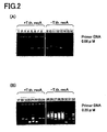
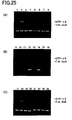
- Fig. 2 The results are shown in Fig. 2 .
- Lane 1 shows the results when Oligonucleotide 1 and Oligonucleotide 2 were added as the primer DNAs.
- Lane 2 shows the results when Oligonucleotide 3 and Oligonucleotide 4 were added as the primer DNAs.
- Lane 3 shows the results when Oligonucleotide 5 and Oligonucleotide 6 were added as the primer DNAs.
- Lane 4 shows the results when Oligonucleotide 7 and Oligonucleotide 8 were added as the primer DNAs.
- Lane 5 shows the results when Oligonucleotide 9 and Oligonucleotide 10 were added as the primer DNAs.
- Lane 6 shows the results when Oligonucleotide 11 and Oligonucleotide 12 were added as the primer DNAs.
- Lane 7 shows the results when Oligonucleotide 13 and Oligonucleotide 14 were added as the primer DNAs.
- Lane 8 shows the results when Oligonucleotide 15 and Oligonucleotide 16 were added as the primer DNAs.
- Lane 9 shows the results when PCR was carried out in the same manner as in Lane 1 without adding the T.th.RecA protein.
- Lane 10 shows the results when PCR was carried out in the same manner as in Lane 2 without adding the T.th.RecA protein.
- Lane 11 shows the results when PCR was carried out in the same manner as in Lane 3 without adding the T.th.RecA protein.
- Lane 12 shows the results when PCR was carried out in the same manner as in Lane 4 without adding the T.th.RecA protein.
- Lane 13 shows the results when PCR was carried out in the same manner as in Lane 5 without adding the T.th.RecA protein.
- Lane 14 shows the results when PCR was carried out in the same manner as in Lane 6 without adding the T.th.RecA protein.
- Lane 15 shows the results when PCR was carried out in the same manner as in Lane 7 without adding the T.th.RecA protein.
- Lane 16 shows the results when PCR was carried out in the same manner as in Lane 8 without adding the T.th.RecA protein.
- Lane 17 shows the results when PCR was carried out in the same manner as in Lane 1 by increasing the concentration of each of the primer DNAs to 0.20 ⁇ M, respectively (the final concentration).
- Lane 18 shows the results when PCR was carried out in the same manner as in Lane 2 by increasing the concentration of each of the primer DNAs to 0.20 ⁇ M, respectively (the final concentration).
- Lane 19 shows the results when PCR was carried out in the same manner as in Lane 3 by increasing the concentration of each of the primer DNAs to 0.20 ⁇ M, respectively (the final concentration).
- Lane 20 shows the results when PCR was carried out in the same manner as in Lane 4 by increasing the concentration of each of the primer DNAs to 0.20 ⁇ M, respectively (the final concentration).
- Lane 21 shows the results when PCR was carried out in the same manner as in Lane 5 by increasing the concentration of each of the primer DNAs to 0.20 ⁇ M, respectively (the final concentration).
- Lane 22 shows the results when PCR was carried out in the same manner as in Lane 6 by increasing the concentration of each of the primer DNAs to 0.20 ⁇ M, respectively (the final concentration).
- Lane 23 shows the results when PCR was carried out in the same manner as in Lane 7 by increasing the concentration of each of the primer DNAs to 0.20 ⁇ M, respectively (the final concentration).
- Lane 24 shows the results when PCR was carried out in the same manner as in Lane 8 by increasing the concentration of each of the primer DNAs to 0.20 ⁇ M, respectively (the final concentration).
- Lane 25 shows the results when PCR was carried out in the same manner as in Lane 9 by increasing the concentration of each of the primer DNAs to 0.20 ⁇ M, respectively (the final concentration).
- Lane 26 shows the results when PCR was carried out in the same manner as in Lane 10 by increasing the concentration of each of the primer DNAs to 0.20 ⁇ M, respectively (the final concentration).
- Lane 27 shows the results when PCR was carried out in the same manner as in Lane 11 by increasing the concentration of each of the primer DNAs to 0.20 ⁇ M, respectively (the final concentration).
- Lane 28 shows the results when PCR was carried out in the same manner as in Lane 12 by increasing the concentration of each of the primer DNAs to 0.20 ⁇ M, respectively (the final concentration).
- Lane 29 shows the results when PCR was carried out in the same manner as in Lane 13 by increasing the concentration of each of the primer DNAs to 0.20 ⁇ M, respectively (the final concentration).
- Lane 30 shows the results when PCR was carried out in the same manner as in Lane 14 by increasing the concentration of each of the primer DNAs to 0.20 ⁇ M, respectively (the final concentration).
- Lane 31 shows the results when PCR was carried out in the same manner as in Lane 15 by increasing the concentration of each of the primer DNAs to 0.20 ⁇ M, respectively (the final concentration).
- Lane 32 shows the results when PCR was carried out in the same manner as in Lane 16 by increasing the concentration of each of the primer DNAs to 0.20 ⁇ M, respectively (the final concentration).
- Lanes 25 to 32 in which PCR was carried out by increasing the concentrations of the primer DNAs but without adding the T.th.RecA protein byproducts were detected in an amount larger than with the results of the corresponding Lanes 9 to 16.
- T.th.RecA protein if the T.th.RecA protein is added and PCR is carried out, amplification of byproducts can be suppressed to low levels without decreasing the yield of the desired nucleic acid.
- the primer extension reaction caused by binding of the primer DNAs to a non-specific region of the template DNA is suppressed, and thus it is possible to suppress amplification of non-specific PCR products.
- PCR is carried out with the addition of T.th.RecA protein, it is possible to amplify the desired nucleic acid efficiently and specifically even when the template DNA has the region of the inhibitory or suppressive secondary structure.
- the reason for this is considered to be that the inhibitory or suppressive secondary structure is released as the homologous recombinant protein binds to the template DNA.
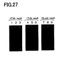
- a human genome DNA was prepared as a template DNA, and 12 kinds of oligonucleotides (Oligonucleotides 17 to 28) were prepared as the primer DNAs.
- Each primer DNA was designed with reference to Homo sapiens BAC clone from RP11-16P10 from 7, complete sequence (Genbank accession no.; AC093734, AC011786). Since each primer DNA has possibility of any primer design, each position was shifted to design the primer DNA.
- Each primer DNA consists of a 20mer base sequence which is 100% complementary to the template DNA.
- Lane 1 shows the results when Oligonucleotide 17 and Oligonucleotide 18 were added as the primer DNAs.
- Lane 2 shows the results when Oligonucleotide 19 and Oligonucleotide 20 were added as the primer DNAs.
- Lane 3 shows the results when Oligonucleotide 21 and Oligonucleotide 22 were added as the primer DNAs.
- Lane 4 shows the results when Oligonucleotide 23 and Oligonucleotide 24 are added as the primer DNA.
- Lane 5 shows the results when Oligonucleotide 25 and Oligonucleotide 26 were added as the primer DNAs.
- Lane 6 shows the results when Oligonucleotide 27 and Oligonucleotide 28 are added as the primer DNA.
- Lane 7 shows the results when PCR was carried out in the same manner as in Lane 1 without adding the T.th.RecA protein.
- Lane 8 shows the results when PCR was carried out in the same manner as in Lane 2 without adding the T.th.RecA protein.
- Lane 9 shows the results when PCR was carried out in the same manner as in Lane 3 without adding the T.th.RecA protein.
- Lane 10 shows the results when PCR was carried out in the same manner as in Lane 4 without adding the T.th.RecA protein.
- Lane 11 shows the results when PCR was carried out in the same manner as in Lane 5 without adding the T.th.RecA protein.
- Lane 12 shows the results when PCR was carried out in the same manner as in Lane 6 without adding the T.th.RecA protein.
- Lanes 7 to 12 in which PCR was carried out without adding the T.th.RecA protein not only the desired nucleic acid but also byproducts were detected in a large amount.
- a human genome DNA was prepared as a template DNA, and 8 kinds of oligonucleotides (Oligonucleotides 29 to 36) were prepared as the primer DNAs.
- Each primer DNA was designed with reference to the Human DNA sequence from clone RP5-1013A22 on chromosome 20 Contains the HNF4A (hepatic nuclear factor 4, alpha) gene, part of a novel gene encoding a protein similar to cellular retinaldehyde-binding protein, a RPL37A (ribosomal protein L37a) pseudogene, parts of 2 novel genes, ESTs, STSs and GSSs, complete sequence (Genbank accession no.; AL132772).
- HNF4A hepatic nuclear factor 4, alpha
- RPL37A ribosomal protein L37a pseudogene
- Oligonucleotide 31 and Oligonucleotide 32 were designed with reference to Homo sapiens 3q BAC RP11-529F4 (Roswell Park Cancer Institute Human BAC Library) complete sequence (Genbank accession no.; AC080007). Further, Oligonucleotide 33 and Oligonucleotide 34 were designed with reference to Homo sapiens genomic beta globin region on chromosome 11 (Genbank accession no.; NG000007). Further, Oligonucleotide 35 and Oligonucleotide 36 were designed with reference to Homo sapiens HPFH60R gene for olfactory receptor (Genbank accession no.; X81445, X91835). Each primer DNA consists of a base sequence from a 20mer to a 25mer, which is 100% complementary to the template DNA.
- a reaction solution was prepared under the same conditions as those of the above-mentioned Reference Example 1 and the like except the concentration of the primer DNAs. Subsequently, PCR was carried out with 1 cycle (at 94°C for 1 minute), 30 cycles (at 94°C for 30 seconds, at 60°C for 30 seconds, and at 68°C for 1 minute) and 1 cycle (at 68°C for 7 minutes and at 4°C for 1 minute). Then, the reaction solution was subjected to electrophoresis with a 1% agarose gel, and the results were recorded by photography and shown in Fig. 7 and Fig. 8 , in the same manner as in the above-mentioned Reference Example 1, etc.
- Lane 1 shows the results when 0.3 ⁇ M (the final concentration) of Oligonucleotide 29 and 0.3 ⁇ M (the final concentration) of Oligonucleotide 30 were added as the primer DNAS.
- Lane 2 shows the results when PCR was carried out in the same manner as in Lane 1 without adding the T.th.RecA protein.
- Lane 3 shows the results when PCR was carried out in the same manner as in Lane 1 by reducing the concentration of each of Oligonucleotide 29 and Oligonucleotide 30 to 0.1 ⁇ M (the final concentration), respectively.
- Lane 4 shows the results when PCR was carried out in the same manner as in Lane 3 without adding the T.th.RecA protein.
- Lane 5 shows the results when PCR was carried out in the same manner as in Lane 1 by reducing the concentration of each of Oligonucleotide 29 and Oligonucleotide 30 to 0.03 ⁇ M (the final concentration), respectively.
- Lane 6 shows the results when PCR was carried out in the same manner as in Lane 5 without adding the T.th.RecA protein.
- Lane 7 shows the results when PCR was carried out in the same manner as in Lane 1 by reducing the concentration of each of Oligonucleotide 29 and Oligonucleotide 30 to 0.01 ⁇ M (the final concentration), respectively.
- Lane 8 shows the results when PCR was carried out in the same manner as in Lane 7 without adding the T.th.RecA protein.
- Lane 9 shows the results when PCR was carried out in the same manner as in Lane 1 by reducing the concentration of each of Oligonucleotide 29 and Oligonucleotide 30 to 0.003 ⁇ M (the final concentration), respectively.
- Lane 10 shows the results when PCR was carried out in the same manner as in Lane 9 without adding the T.th.RecA protein.
- Lane 11 shows the results when 0.3 ⁇ M (the final concentration) of Oligonucleotide 31 and 0.3 ⁇ M (the final concentration) of Oligonucleotide 32 were added as the primer DNAs.
- Lane 12 shows the results when PCR was carried out in the same manner as in Lane 11 without adding the T.th.RecA protein.
- Lane 13 shows the results when PCR was carried out in the same manner as in Lane 11 by reducing the concentration of each of Oligonucleotide 31 and Oligonucleotide 32 to 0.1 ⁇ M (the final concentration), respectively.
- Lane 14 shows the results when PCR was carried out in the same manner as in Lane 13 without adding the T.th.RecA protein.
- Lane 15 shows the results when PCR was carried out in the same manner as in Lane 11 by reducing the concentration of each of Oligonucleotide 31 and Oligonucleotide 32 to 0.03 ⁇ M (the final concentration), respectively.
- Lane 16 shows the results when PCR was carried out in the same manner as in Lane 15 without adding the T.th.RecA protein.
- Lane 17 shows the results when PCR was carried out in the same manner as in Lane 11 by reducing the concentration of each of Oligonucleotide 31 and Oligonucleotide 32 to 0.01 ⁇ M (the final concentration), respectively.
- Lane 18 shows the results when PCR was carried out in the same manner as in Lane 17 without adding the T.th.RecA protein.
- Lane 19 shows the results when PCR was carried out in the same manner as in Lane 11 by reducing the concentration of each of Oligonucleotide 31 and Oligonucleotide 32 to 0.003 ⁇ M (the final concentration), respectively.
- Lane 20 shows the results when PCR was carried out in the same manner as in Lane 19 without adding the T.th.RecA protein.
- Lane 21 shows the results when 0.3 ⁇ M (the final concentration) of Oligonucleotide 33 and 0.3 ⁇ M (the final concentration) of Oligonucleotide 34 were added as the primer DNAs.
- Lane 22 shows the results when PCR was carried out in the same manner as in Lane 21 without adding the T.th.RecA protein.
- Lane 23 shows the results when PCR was carried out in the same manner as in Lane 21 by reducing the concentration of each of Oligonucleotide 33 and Oligonucleotide 34 to 0.1 ⁇ M (the final concentration), respectively.
- Lane 24 shows the results when PCR was carried out in the same manner as in Lane 23 without adding the T.th.RecA protein.
- Lane 25 shows the results when PCR was carried out in the same manner as in Lane 21 by reducing the concentration of each of Oligonucleotide 33 and Oligonucleotide 34 to 0.03 ⁇ M (the final concentration), respectively.
- Lane 26 shows the results when PCR was carried out in the same manner as in Lane 25 without adding the T.th.RecA protein.
- Lane 27 shows the results when PCR was carried out in the same manner as in Lane 21 by reducing the concentration of each of Oligonucleotide 33 and Oligonucleotide 34 to 0.01 ⁇ M (the final concentration), respectively.
- Lane 28 shows the results when PCR was carried out in the same manner as in Lane 27 without adding the T.th.RecA protein.
- Lane 29 shows the results when PCR was carried out in the same manner as in Lane 21 by reducing the concentration of each of Oligonucleotide 33 and Oligonucleotide 34 to 0.003 ⁇ M (the final concentration), respectively.
- Lane 30 shows the results when PCR was carried out in the same manner as in Lane 29 without adding the T.th.RecA protein.
- Lane 31 shows the results when 0.3 ⁇ M (the final concentration) of Oligonucleotide 35 and 0.3 ⁇ M (the final concentration) of Oligonucleotide 36 were added as the primer DNAs.
- Lane 32 shows the results when PCR was carried out in the same manner as in Lane 31 without adding the T.th.RecA protein.
- Lane 33 shows the results when PCR was carried out in the same manner as in Lane 31 by reducing the concentration of each of Oligonucleotide 35 and Oligonucleotide 36 to 0.1 ⁇ M (the final concentration), respectively.
- Lane 34 shows the results when PCR was carried out in the same manner as in Lane 33 without adding the T.th.RecA protein.
- Lane 35 shows the results when PCR was carried out in the same manner as in Lane 31 by reducing the concentration of each of Oligonucleotide 35 and Oligonucleotide 36 to 0.03 ⁇ M (the final concentration), respectively.
- Lane 36 shows the results when PCR was carried out in the same manner as in Lane 35 without adding the T.th.RecA protein.
- Lane 37 shows the results when PCR was carried out in the same manner as in Lane 31 by reducing the concentration of each of Oligonucleotide 35 and Oligonucleotide 36 to 0.01 ⁇ M (the final concentration), respectively.
- Lane 38 shows the results when PCR was carried out in the same manner as in Lane 37 without adding the T.th.RecA protein.
- Lane 39 shows the results when PCR was carried out in the same manner as in Lane 31 by reducing the concentration of each of Oligonucleotide 35 and Oligonucleotide 36 to 0.003 ⁇ M (the final concentration), respectively.
- Lane 40 shows the results when PCR was carried out in the same manner as in Lane 39 without adding the T.th.RecA protein.
- Lane 10 In contrast, among Lanes 2, 4, 6, 8 and 10 in which PCR was carried out without adding the T.th.RecA protein, except in Lane 10, not only the desired nucleic acid but also byproducts were detected. In Lane 10, amplification of DNA was hardly detected, possibly due to the too low concentration of the primer DNAs.
- Lane 20 In contrast, among Lanes 12, 14, 16, 18 and 20 in which PCR was carried out without adding the T.th.RecA protein, except in Lane 20, not only the desired nucleic acid but also byproducts were detected. In Lane 20, amplification of DNA was hardly detected, possibly due to the too low concentration of the primer DNAs.
- Lanes 22, 24, 26, 28 and 30 in which PCR was carried out without adding the T.th.RecA protein in Lanes 22, 24 and 26, not only the desired nucleic acid but also byproducts were detected. In Lanes 28 and 30, amplification of DNA was hardly detected, possibly due to the too low concentration of the primer DNAs.
- Lane 40 In contrast, among Lanes 32, 34, 36, 38 and 40 in which PCR was carried out without adding the T.th.RecA protein, except in Lane 40, not only the desired nucleic acid but also byproducts were detected. In Lane 40, amplification of DNA was hardly detected, possibly due to the too low concentration of the primer DNAs.
- Example 4 will be explained. Explanation of the parts which are similar to those of each of the above-mentioned Reference Examples will be omitted or simplified.
- a human genome DNA was prepared as a template DNA, and 5 kinds of oligonucleotides (Oligonucleotides 37 to 41) were prepared as the primer DNAs.
- Each primer DNA was designed with reference to Homo sapiens PAC clone RP5-1142J19 from 7q35-q36, complete sequence (Genbank accession no.; AC004975).
- Oligonucleotides 37 and 38 consist of a 20mer or a 21mer base sequence, which is 100% complementary to the template DNA.
- Oligonucleotide 39 consists of a base sequence which is different by 1 base from the template DNA, while the rest is the same as Oligonucleotide 37.
- Oligonucleotide 40 consists of a base sequence which is different by 3 base from the template DNA, while the rest is the same as Oligonucleotide 37.
- Oligonucleotide 41 consists of a base sequence which is different by 5 base from the template DNA, while the rest is the same as Oligonucleotide 37.
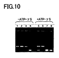
- nucleic acids were amplified by PCR reaction in the same manner as in the above-mentioned Reference Example 3 except that the concentration of each of the primer DNAs was set to 0.3 ⁇ M, (the final concentration). Subsequently, the reaction solution was subjected to electrophoresis with a 1% agarose gel, and the results were recorded by photography and shown in Fig. 10 , in the same manner as the above-mentioned Example 1, etc.
- Lane 1 shows the results when Oligonucleotide 37 and Oligonucleotide 38 were added as the primer DNAs.
- Lane 2 shows the results when Oligonucleotide 39 and Oligonucleotide 38 were added as the primer DNAs.
- Lane 3 shows the results when Oligonucleotide 40 and Oligonucleotide 38 were added as the primer DNAs.
- Lane 4 shows the results when Oligonucleotide 41 and Oligonucleotide 38 were added as the primer DNAs.
- Lane 5 shows the results when PCR was carried out in the same manner as in Lane 1 by further adding 1mM (the final concentration) ATP- ⁇ S (Roche).
- Lane 6 shows the results when PCR was carried out in the same manner as in Lane 2 by further adding 1mM (the final concentration) ATP- ⁇ S (Roche).
- Lane 7 shows the results when PCR was carried out in the same manner as in Lane 3 by further adding 1mM (the final concentration) ATP- ⁇ S (Roche).
- Lane 8 shows the results when PCR was carried out in the same manner as in Lane 4 by further adding 1mM (the final concentration) ATP- ⁇ S (Roche).
- Lanes 5 to 8 in which PCR was carried out by adding the ATP- ⁇ S in Lanes 5 and 6, amplification of the desired DNA was detected whereas byproducts were scarcely detected. In Lane 7 and Lane 8, amplification of DNA was scarcely detected.
- ATP- ⁇ S it is possible to amplify nucleic acids specifically only if there is a mismatch of 1 base or less between the primer DNA and the template DNA. Accordingly, by adding ATP- ⁇ S to the reaction solution, it is possible to amplify the desired nucleic acid further specifically.
- a reagent kit for amplifying nucleic acids is prepared with a DNA polymerase, four kinds of dNTPs, a buffer solution and a homologous recombinant protein such as T.th.RecA protein and the like, it is preferable to add ATP- ⁇ S to such a kit.
- ATP- ⁇ S a homologous recombinant protein
- the reason for this is that, as clearly shown in the above-mentioned Example 4, if ATP- ⁇ S was added and PCR was carried out, it is possible to amplify the desired nucleic acid more specifically.
- Example 4 it is possible to detect single nucleotide polymorphism.
- PCR is carried out by using a primer DNA corresponding to a sequence comprising a base which forms single nucleotide polymorphism in the template DNA as one of the primer DNAs, it is possible to amplify the desired nucleic acid only when the template DNA is completely complementary to the primer DNA corresponding to a sequence comprising a base which forms single nucleotide polymorphism.
- the template DNA is not completely complementary to the primer DNA corresponding to a sequence comprising a base which forms single nucleotide polymorphism, i.e., when the base which forms single nucleotide polymorphism is not complementary to the primer DNA, it is possible not to amplify or to inhibit amplification of the desired nucleic acid. Therefore, amplification of the desired nucleic acid by PCR allows detection of single nucleotide polymorphism.
- a DNA polymerase it is convenient to have a DNA polymerase, four kinds of dNTPs, a buffer solution and a homologous recombinant protein such as T.th.RecA protein and ATP- ⁇ S as a reagent kit prepared in advance for detecting single nucleotide polymorphism.
- a kit it is possible to detect easily single nucleotide polymorphism by PCR, just by adding the DNA polymerase, the four kinds of dNTPs, the buffer solution and the homologous recombinant protein such as T.th.RecA protein and ATP- ⁇ S to a reaction solution, and further adding the template DNA and the primer DNA prepared depending on the purpose to the reaction solution.
- ATP- ⁇ S is also added to the above-mentioned kit. As clearly shown in the above-mentioned Example 4, if ATP- ⁇ S was added and PCR was carried out, it is possible to amplify the desired nucleic acid more specifically, and thus it is possible to detect single nucleotide polymorphism more reliably.
- a human genome DNA was prepared as a template DNA, and 12 kinds of oligonucleotides (Oligonucleotides 42 to 53) were prepared as the primer DNAs.
- Oligonucleotides 42 and 43 were designed with reference to Homo sapiens PAC clone RP5-1142J19 from 7q35-q36, complete sequence (Genbank accession no.; AC004975).
- Oligonucleotides 44 and 45 were designed with reference to Homo sapiens PAC clone RP5-852P6 from 7p11.2-p21, complete sequence (Genbank accession no.; AC006454).
- Oligonucleotides 46 and 47 were designed with reference to Homo sapiens PAC clone RP5-912I13 from 7, complete sequence (Genbank accession no.; AC008060).
- Oligonucleotides 48 and 49 were designed with reference to Homo sapiens BAC clone RP11-16P10 from 7, complete sequence (Genbank accession no.; AC093734, AC011786).
- Oligonucleotides 50 and 51 were designed with reference to Homo sapiens BAC clone CTB-135C18 from 7q11.2-q22, complete sequence (Genbank accession no.; AC005164).
- Oligonucleotides 52 and 53 were designed with reference to Homo sapiens PAC clone RP5-852P6 from 7p11.2-p21, complete sequence (Genbank accession no.; AC006454). Each primer DNA consists of a base sequence from a 18mer to a 22mer, which is 100% complementary to the template DNA.
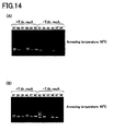
- PCR was carried out under the same conditions as those of Example 4 to amplify nucleic acids. Subsequently, the reaction solution was subjected to electrophoresis with a 1% agarose gel, and the results were recorded by photography and shown in Fig. 13 and Fig.14 , in the same manner as in Example 1, etc.
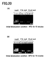
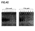
- the PCR temperature condition was set to 1 cycle (at 94°C for 1 minute), 30 cycles (at 94°C for 30 seconds, at 60°C for 30 seconds, and at 68°C for 1 minute) and 1 cycle (at 68°C for 7 minutes, and at 4°C for 1 minute), which is referred to as Temperature Condition 1.
- the annealing temperature was 60°C.
- Lane 1 shows the results when Oligonucleotide 42 and Oligonucleotide 43 were added as the primer DNAs.
- Lane 2 shows the results when Oligonucleotide 44 and Oligonucleotide 45 were added as the primer DNAs.
- Lane 3 shows the results when Oligonucleotide 46 and Oligonucleotide 47 were added as the primer DNAs.
- Lane 4 shows the results when Oligonucleotide 48 and Oligonucleotide 49 were added as the primer DNAs.
- Lane 5 shows the results when Oligonucleotide 50 and Oligonucleotide 51 were added as the primer DNAs.
- Lane 6 shows the results when Oligonucleotide 52 and Oligonucleotide 53 were added as the primer DNAs.
- Lane 7 shows the results when PCR was carried out in the same manner as in Lane 1 without adding the T.th.RecA protein.
- Lane 8 shows the results when PCR was carried out in the same manner as in Lane 2 without adding the T.th.RecA protein.
- Lane 9 shows the results when PCR was carried out in the same manner as in Lane 3 without adding the T.th.RecA protein.
- Lane 10 shows the results when PCR was carried out in the same manner as in Lane 4 without adding the T.th.RecA protein.
- Lane 11 shows the results when PCR was carried out in the same manner as in Lane 5 without adding the T.th.RecA protein.
- Lane 12 shows the results when PCR was carried out in the same manner as in Lane 6 without adding the T.th.RecA protein.
- the PCR temperature condition was set to 1 cycle (at 94°C for 1 minute), 30 cycles (at 94°C for 30 seconds, at 55°C for 30 seconds, and at 68°C for 1 minute) and 1 cycle (at 68°C for 7 minutes, and at 4°C for 1 minute), which is referred to as Temperature Condition 2.
- the annealing temperature was 55°C.
- Lane 13 shows the results when PCR was carried out in the same manner as in Lane 1 except for the above-mentioned Temperature Condition 2.
- Lane 14 shows the results when PCR was carried out in the same manner as in Lane 2 except for the above-mentioned Temperature Condition 2.
- Lane 15 shows the results when PCR was carried out in the same manner as in Lane 3 except for the above-mentioned Temperature Condition 2.
- Lane 16 shows the results when PCR was carried out in the same manner as in Lane 4 except for the above-mentioned Temperature Condition 2.
- Lane 17 shows the results when PCR was carried out in the same manner as in Lane 5 except for the above-mentioned Temperature Condition 2.
- Lane 18 shows the results when PCR was carried out in the same manner as in Lane 6 except for the above-mentioned Temperature Condition 2.
- Lane 19 shows the results when PCR was carried out in the same manner as in Lane 7 except for the above-mentioned Temperature Condition 2.
- Lane 20 shows the results when PCR was carried out in the same manner as in Lane 8 except for the above-mentioned Temperature Condition 2.
- Lane 21 shows the results when PCR was carried out in the same manner as in Lane 9 except for the above-mentioned Temperature Condition 2.
- Lane 22 shows the results when PCR was carried out in the same manner as in Lane 10 except for the above-mentioned Temperature Condition 2.
- Lane 23 shows the results when PCR was carried out in the same manner as in Lane 11 except for the above-mentioned Temperature Condition 2.
- Lane 24 shows the results when PCR was carried out in the same manner as in Lane 12 except for the above-mentioned Temperature Condition 2.
- the PCR temperature condition was set to 1 cycle (at 94°C for 1 minute), 30 cycles (at 94°C for 30 seconds, at 50°C for 30 seconds, and at 68°C for 1 minute) and 1 cycle (at 68°C for 7 minutes, and at 4°C for 1 minute), which is referred to as Temperature Condition 3.
- the annealing temperature was 50°C.
- Lane 25 shows the results when PCR was carried out in the same manner as in Lane 1 except for the above-mentioned Temperature Condition 3.
- Lane 26 shows the results when PCR was carried out in the same manner as in Lane 2 except for the above-mentioned Temperature Condition 3.
- Lane 27 shows the results when PCR was carried out in the same manner as in Lane 3 except for the above-mentioned Temperature Condition 3.
- Lane 28 shows the results when PCR was carried out in the same manner as in Lane 4 except for the above-mentioned Temperature Condition 3.
- Lane 29 shows the results when PCR was carried out in the same manner as in Lane 5 except for the above-mentioned Temperature Condition 3.
- Lane 30 shows the results when PCR was carried out in the same manner as in Lane 6 except for the above-mentioned Temperature Condition 3.
- Lane 31 shows the results when PCR was carried out in the same manner as in Lane 7 except for the above-mentioned Temperature Condition 3.
- Lane 32 shows the results when PCR was carried out in the same manner as in Lane 8 except for the above-mentioned Temperature Condition 3.
- Lane 33 shows the results when PCR was carried out in the same manner as in Lane 9 except for the above-mentioned Temperature Condition 3.
- Lane 34 shows the results when PCR was carried out in the same manner as in Lane 10 except for the above-mentioned Temperature Condition 3.
- Lane 35 shows the results when PCR was carried out in the same manner as in Lane 11 except for the above-mentioned Temperature Condition 3.
- Lane 36 shows the results when PCR was carried out in the same manner as in Lane 12 except for the above-mentioned Temperature Condition 3.
- the PCR temperature condition was set to 1 cycle (at 94°C for 1 minute), 30 cycles (at 94°C for 30 seconds, at 45°C for 30 seconds, and at 68°C for 1 minute) and 1 cycle (at 68°C for 7 minutes, and at 4°C for 1 minute), which is referred to as Temperature Condition 4.
- the annealing temperature was 45°C.
- Lane 37 shows the results when PCR was carried out in the same manner as in Lane 1 except for the above-mentioned Temperature Condition 4.
- Lane 38 shows the results when PCR was carried out in the same manner as in Lane 2 except for the above-mentioned Temperature Condition 4.
- Lane 39 shows the results when PCR was carried out in the same manner as in Lane 3 except for the above-mentioned Temperature Condition 4.
- Lane 40 shows the results when PCR was carried out in the same manner as in Lane 4 except for the above-mentioned Temperature Condition 4.
- Lane 41 shows the results when PCR was carried out in the same manner as in Lane 5 except for the above-mentioned Temperature Condition 4.
- Lane 42 shows the results when PCR was carried out in the same manner as in Lane 6 except for the above-mentioned Temperature Condition 4.
- Lane 43 shows the results when PCR was carried out in the same manner as in Lane 7 except for the above-mentioned Temperature Condition 4.
- Lane 44 shows the results when PCR was carried out in the same manner as in Lane 8 except for the above-mentioned Temperature Condition 4.
- Lane 45 shows the results when PCR was carried out in the same manner as in Lane 9 except for the above-mentioned Temperature Condition 4.
- Lane 46 shows the results when PCR was carried out in the same manner as in Lane 10 except for the above-mentioned Temperature Condition 4.
- Lane 47 shows the results when PCR was carried out in the same manner as in Lane 11 except for the above-mentioned Temperature Condition 4.
- Lane 48 shows the results when PCR was carried out in the same manner as in Lane 12 except for the above-mentioned Temperature Condition 4.
- PCR is carried out with the addition of T.th.RecA protein, it is possible to amplify the desired nucleic acid efficiently and specifically even when the template DNA has a region of the inhibitory or suppressive secondary structure.
- the reason is considered to be that the inhibitory or suppressive secondary structure is released by binding of the homologous recombinant protein to the template DNA.
- the PCR specificity is high, it is possible to specifically amplify the desired nucleic acid even when the temperature conditions of the primer extension reaction (the annealing temperature) are changed. That is, in the cases in which PCR is carried out without adding the T.th.RecA protein, when the temperature conditions of the primer extension reaction (the annealing temperature) are set to be low, not only the desired nucleic acid, but also byproducts are amplified in a large amount. However, if PCR is carried out with the addition of T.th.RecA protein, it is possible to amplify the desired nucleic acid more specifically.
- Example 6 will be explained. Explanation of the parts which are similar to those of each of the above-mentioned Examples and Reference Examples will be omitted or simplified.
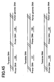
- a human genome DNA was prepared as a template DNA, and 5 kinds of oligonucleotides (Oligonucleotides 37 to 41) which are the same as those of Example 4 (see Fig. 9 ), and 5 kinds of oligonucleotides (Oligonucleotides 54 to 58) as shown in Fig. 15 , were prepared as the primer DNAs.
- Oligonucleotides 54 and 55 consist of a 22mer base sequence which is 100% complementary to the template DNA.
- Oligonucleotide 56 consists of a base sequence which is different by 1 base from the template DNA, while other parts are the same as Oligonucleotide 54.
- Oligonucleotide 57 consists of a base sequence which is different by 3 bases from the template DNA, while other parts are the same as Oligonucleotide 54.
- Oligonucleotide 58 consists of a base sequence which is different by 5 bases from the template DNA, while other parts are the same as Oligonucleotide 54.
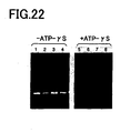
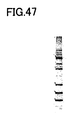
- PCR was carried out under the same conditions as those of Example 4, etc. to amplify nucleic acids. Subsequently, the reaction solution was subjected to electrophoresis with a 1% agarose gel, and the results were recorded by photography and shown in Fig. 16 in the same manner as in Example 1, etc.
- Lane 5 shows the results when Oligonucleotide 37 and Oligonucleotide 38 were added as the primer DNAs.
- Lane 6 shows the results when Oligonucleotide 39 and Oligonucleotide 38 were added as the primer DNAs.
- Lane 7 shows the results when Oligonucleotide 40 and Oligonucleotide 38 were added as the primer DNAs.
- Lane 8 shows the results when Oligonucleotide 41 and Oligonucleotide 38 were added as the primer DNAs.
- Lane 9 shows the results when PCR was carried out in the same manner as in Lane 5 by further adding 1mM (the final concentration) ATP- ⁇ S.
- Lane 10 shows the results when PCR was carried out in the same manner as in Lane 6 by further adding 1mM (the final concentration) ATP- ⁇ S.
- Lane 11 shows the results when PCR was carried out in the same manner as in Lane 7 by further adding 1mM (the final concentration) ATP- ⁇ S.
- Lane 12 shows the results when PCR was carried out in the same manner as in Lane 8 by further adding 1mM (the final concentration) ATP- ⁇ S.
- Lane 1 shows the results when PCR was carried out in the same manner as in Lane 5, except that 1mM (the final concentration) ATP- ⁇ S was added without adding the T.th.RecA protein.
- Lane 2 shows the results when PCR was carried out in the same manner as in Lane 6, except that 1mM (the final concentration) ATP- ⁇ S was added without adding the T.th.RecA protein.
- Lane 3 shows the results when PCR was carried out in the same manner as in Lane 7, except that 1mM (the final concentration) ATP- ⁇ S was added without adding the T.th.RecA protein.
- Lane 4 shows the results when PCR was carried out in the same manner as in Lane 8, except that 1mM (the final concentration) ATP- ⁇ S was added without adding the T.th.RecA protein.
- Lane 17 shows the results when Oligonucleotide 54 and Oligonucleotide 55 were added as the primer DNAs.
- Lane 18 shows the results when Oligonucleotide 56 and Oligonucleotide 55 were added as the primer DNAs.
- Lane 19 shows the results when Oligonucleotide 57 and Oligonucleotide 55 were added as the primer DNAs.
- Lane 20 shows the results when Oligonucleotide 58 and Oligonucleotide 55 were added as the primer DNAs.
- Lane 21 shows the results when 1mM (the final concentration) ATP- ⁇ S was further added and PCR was carried out in the same manner as in Lane 17.
- Lane 22 shows the results when 1mM (the final concentration) ATP- ⁇ S was further added and PCR was carried out in the same manner as in Lane 18.
- Lane 23 shows the results when 1mM (the final concentration) ATP- ⁇ S was further added and PCR was carried out in the same manner as in Lane 19.
- Lane 24 shows the results when 1mM (the final concentration) ATP- ⁇ S was further added and PCR was carried out in the same manner as in Lane 20.
- Lane 13 shows the results when PCR was carried out in the same manner as in Lane 17 except that 1mM (the final concentration) ATP- ⁇ S was added without adding the T.th.RecA protein.
- Lane 14 shows the results when PCR was carried out in the same manner as in Lane 18 except that 1mM (the final concentration) ATP- ⁇ S was added without adding the T.th.RecA protein.
- Lane 15 shows the results when PCR was carried out in the same manner as in Lane 19 except that 1mM (the final concentration) ATP- ⁇ S was added without adding the T.th.RecA protein.
- Lane 16 shows the results when PCR was carried out in the same manner as in Lane 20 except that 1mM (the final concentration) ATP- ⁇ S was added without adding the T.th.RecA protein.
- Lanes 9 to 12 in which PCR was carried out with the addition of T.th.RecA protein and the ATP- ⁇ S, in Lanes 9 to 11, amplification of the desired DNA was detected whereas byproducts were hardly detected. On the other hand, in Lane 12, nucleic acid amplification was not detected. Also, in Lane 11, amplification of the desired nucleic acid was smaller than those of Lanes 9 and 10. Further, in Lane 11, amplification of the desired nucleic acid was smaller than that of the above-mentioned Lane 7.
- Lanes 17 to 20 in which PCR was carried out with the addition of T.th.RecA protein but without the addition of ATP- ⁇ S, in Lanes 17 to 19, amplification of the desired nucleic acid was detected whereas byproducts were scarcely detected. In Lane 20, amplification of the desired nucleic acid was scarcely detected. Further, in Lane 19, amplification of the desired nucleic acid was smaller than in that of Lane 17 and Lane 18.
- Lane 21 to 24 in which PCR was carried out with the addition of T.th.RecA protein and ATP- ⁇ S, in Lane 21, amplification of the desired nucleic acid was detected, whereas byproducts were scarcely detected. On the other hand, in Lanes 22 to 24, nucleic acid amplification was scarcely detected.
- ATP- ⁇ S it is possible to amplify nucleic acids specifically only if there is a mismatch of 1 base or less between the primer DNA and the template DNA. Accordingly, by adding ATP- ⁇ S to the reaction solution, it is possible to amplify the desired nucleic acid further specifically.
- ATP- ⁇ S can increase the specificity of PCR when T.th.RecA protein is added, but ATP- ⁇ S alone in the absence of T.th.RecA protein cannot increase the specificity of PCR.
- the template DNA is not completely complementary to the primer DNA corresponding to the sequence comprising the base which forms single nucleotide polymorphism, i.e., when the base which forms single nucleotide polymorphism is not complementary to the primer DNA, it is possible not to amplify or to inhibit amplification of the desired nucleic acid. Therefore, amplification of the desired nucleic acid by PCR allows detection of single nucleotide polymorphism.
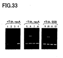
- a human genome DNA was prepared as a template DNA, and some of the oligonucleotides (Oligonucleotides 38 to 40) which were used in Example 4, were prepared as the primer DNAs (see Fig. 9 ).
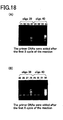
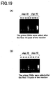
- PCR was carried out under the same conditions as those of Example 4, etc. to amplify nucleic acids. Subsequently, the reaction solution was subjected to electrophoresis with a 1% agarose gel, and the results were recorded by photography and shown in Fig. 17 to Fig. 19 , in the same manner as in Reference Example 1, etc.
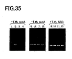
- Lane 1 shows the results when Oligonucleotide 39 and Oligonucleotide 38 were added as the primer DNAs.
- Lane 2 shows the results when PCR was carried out in the same manner as in Lane 1 except that the RecA protein of E.coli was added instead of the T.th.RecA protein.
- Lane 3 shows the results when PCR was carried out in the same manner as in Lane 1 except that the T. th. SSB protein was added instead of the T.th.RecA protein.
- Lane 4 shows the results when PCR was carried out in the same manner as in Lane 1 except that the T.th.RecA protein was not added.
- Lane 5 shows the results when Oligonucleotide 40 and Oligonucleotide 38 were added as the primer DNAs.
- Lane 6 shows the results when PCR was carried out in the same manner as in Lane 5 except that the RecA protein of E.coli was added instead of the T.th.RecA protein.
- Lane 7 shows the results when PCR was carried out in the same manner as in Lane 5 except that the T. th. SSB protein was added instead of the T.th.RecA protein.
- Lane 8 shows the results when PCR was carried out in the same manner as in Lane 5 except that the T.th.RecA protein was not added.
- Lane 9 shows the results when PCR was carried out in the same manner as in Lane 1 except that the primer DNAs were added after completing the first 1 cycle of the primer extension reaction, and then 30 cycles of the primer extension reaction was carried out.
- Lane 10 shows the results when PCR was carried out in the same manner as in Lane 2 except that the primer DNAs were added after completing the first 1 cycle of the primer extension reaction, and then 30 cycles of the primer extension reaction was carried out.
- Lane 11 shows the results when PCR was carried out in the same manner as in Lane 3 except that the primer DNAs were added after completing the first 1 cycle of the primer extension reaction, and then 30 cycles of the primer extension reaction was carried out.
- Lane 12 shows the results when PCR was carried out in the same manner as in Lane 4 except that the primer DNAs were added after completing the first 1 cycle of the primer extension reaction, and then 30 cycles of the primer extension reaction was carried out.
- Lane 13 shows the results when PCR was carried out in the same manner as in Lane 5 except that the primer DNAs were added after completing the first 1 cycle of the primer extension reaction, and then 30 cycles of the primer extension reaction was carried out.
- Lane 14 shows the results when PCR was carried out in the same manner as in Lane 6 except that the primer DNAs were added after completing the first 1 cycle of the primer extension reaction, and then 30 cycles of the primer extension reaction was carried out.
- Lane 15 shows the results when PCR was carried out in the same manner as in Lane 7 except that the primer DNAs were added after completing the first 1 cycle of the primer extension reaction, and then 30 cycles of the primer extension reaction was carried out.
- Lane 16 shows the results when PCR was carried out in the same manner as in Lane 8 except that the primer DNAs were added after completing the first 1 cycle of the primer extension reaction, and then 30 cycles of the primer extension reaction was carried out.
- Lane 17 shows the results when PCR was carried out in the same manner as in Lane 1 except that the primer DNAs were added after completing the first 3 cycles of the primer extension reaction, and then 30 cycles of the primer extension reaction was carried out.
- Lane 18 shows the results when PCR was carried out in the same manner as in Lane 2 except that the primer DNAs were added after completing the first 3 cycles of the primer extension reaction, and then 30 cycles of the primer extension reaction was carried out.
- Lane 19 shows the results when PCR was carried out in the same manner as in Lane 3 except that the primer DNAs were added after completing the first 3 cycles of the primer extension reaction, and then 30 cycles of the primer extension reaction was carried out.
- Lane 20 shows the results when PCR was carried out in the same manner as in Lane 4 except that the primer DNAs were added after completing the first 3 cycles of the primer extension reaction, and then 30 cycles of the primer extension reaction was carried out.
- Lane 21 shows the results when PCR was carried out in the same manner as in Lane 5 except that the primer DNAs were added after completing the first 3 cycles of the primer extension reaction, and then 30 cycles of the primer extension reaction was carried out.
- Lane 22 shows the results when PCR was carried out in the same manner as in Lane 6 except that the primer DNAs were added after completing the first 3 cycles of the primer extension reaction, and then 30 cycles of the primer extension reaction was carried out.
- Lane 23 shows the results when PCR was carried out in the same manner as in Lane 7 except that the primer DNAs were added after completing the first 3 cycles of the primer extension reaction, and then 30 cycles of the primer extension reaction was carried out.
- Lane 24 shows the results when PCR was carried out in the same manner as in Lane 8 except that the primer DNAs were added after completing the first 3 cycles of the primer extension reaction, and then 30 cycles of the primer extension reaction was carried out.
- Lane 25 shows the results when PCR was carried out in the same manner as in Lane 1 except that the primer DNAs were added after completing the first 6 cycles of the primer extension reaction, and then 30 cycles of the primer extension reaction was carried out.
- Lane 26 shows the results when PCR was carried out in the same manner as in Lane 2 except that the primer DNAs were added after completing the first 6 cycles of the primer extension reaction, and then 30 cycles of the primer extension reaction was carried out.
- Lane 27 shows the results when PCR was carried out in the same manner as in Lane 3 except that the primer DNAs were added after completing the first 6 cycles of the primer extension reaction, and then 30 cycles of the primer extension reaction was carried out.
- Lane 28 shows the results when PCR was carried out in the same manner as in Lane 4 except that the primer DNAs were added after completing the first 6 cycles of the primer extension reaction, and then 30 cycles of the primer extension reaction was carried out.
- Lane 29 shows the results when PCR was carried out in the same manner as in Lane 5 except that the primer DNAs were added after completing the first 6 cycles of the primer extension reaction, and then 30 cycles of the primer extension reaction was carried out.
- Lane 30 shows the results when PCR was carried out in the same manner as in Lane 6 except that the primer DNAs were added after completing the first 6 cycles of the primer extension reaction, and then 30 cycles of the primer extension reaction was carried out.
- Lane 31 shows the results when PCR was carried out in the same manner as in Lane 7 except that the primer DNAs were added after completing the first 6 cycles of the primer extension reaction, and then 30 cycles of the primer extension reaction was carried out.
- Lane 32 shows the results when PCR was carried out in the same manner as in Lane 8 except that the primer DNAs were added after completing the first 6 cycles of the primer extension reaction, and then 30 cycles of the primer extension reaction was carried out.
- Lane 33 shows the results when PCR was carried out in the same manner as in Lane 1 except that the primer DNAs were added after completing the first 10 cycles of the primer extension reaction, and then 30 cycles of the primer extension reaction was carried out.
- Lane 34 shows the results when PCR was carried out in the same manner as in Lane 2 except that the primer DNAs were added after completing the first 10 cycles of the primer extension reaction, and then 30 cycles of the primer extension reaction was carried out.
- Lane 35 shows the results when PCR was carried out in the same manner as in Lane 3 except that the primer DNAs were added after completing the first 10 cycles of the primer extension reaction, and then 30 cycles of the primer extension reaction was carried out.
- Lane 36 shows the results when PCR was carried out in the same manner as in Lane 4 except that the primer DNAs were added after completing the first 10 cycles of the primer extension reaction, and then 30 cycles of the primer extension reaction was carried out.
- Lane 37 shows the results when PCR was carried out in the same manner as in Lane 5 except that the primer DNAs were added after completing the first 10 cycles of the primer extension reaction, and then 30 cycles of the primer extension reaction was carried out.
- Lane 38 shows the results when PCR was carried out in the same manner as in Lane 6 except that the primer DNAs were added after completing the first 10 cycles of the primer extension reaction, and then 30 cycles of the primer extension reaction was carried out.
- Lane 39 shows the results when PCR was carried out in the same manner as in Lane 7 except that the primer DNAs were added after completing the first 10 cycles of the primer extension reaction, and then 30 cycles of the primer extension reaction was carried out.
- Lane 40 shows the results when PCR was carried out in the same manner as in Lane 8 except that the primer DNAs were added after completing the first 10 cycles of the primer extension reaction, and then 30 cycles of the primer extension reaction was carried out.
- Lane 41 shows the results when PCR was carried out in the same manner as in Lane 1 except that the primer DNAs were added after completing the first 15 cycles of the primer extension reaction, and then 30 cycles of the primer extension reaction was carried out.
- Lane 42 shows the results when PCR was carried out in the same manner as in Lane 2 except that the primer DNAs were added after completing the first 15 cycles of the primer extension reaction, and then 30 cycles of the primer extension reaction was carried out.
- Lane 43 shows the results when PCR was carried out in the same manner as in Lane 3 except that the primer DNAs were added after completing the first 15 cycles of the primer extension reaction, and then 30 cycles of the primer extension reaction was carried out.
- Lane 44 shows the results when PCR was carried out in the same manner as in Lane 4 except that the primer DNAs were added after completing the first 15 cycles of the primer extension reaction, and then 30 cycles of the primer extension reaction was carried out.
- Lane 45 shows the results when PCR was carried out in the same manner as in Lane 5 except that the primer DNAs were added after completing the first 15 cycles of the primer extension reaction, and then 30 cycles of the primer extension reaction was carried out.
- Lane 46 shows the results when PCR was carried out in the same manner as in Lane 6 except that the primer DNAs were added after completing the first 15 cycles of the primer extension reaction, and then 30 cycles of the primer extension reaction was carried out.
- Lane 47 shows the results when PCR was carried out in the same manner as in Lane 7 except that the primer DNAs were added after completing the first 15 cycles of the primer extension reaction, and then 30 cycles of the primer extension reaction was carried out.
- Lane 48 shows the results when PCR was carried out in the same manner as in Lane 8 except that the primer DNAs were added after completing the first 15 cycles of the primer extension reaction, and then 30 cycles of the primer extension reaction was carried out.
- Lane 5 in which PCR was carried out with the addition of T.th.RecA protein, amplification of the desired DNA was detected whereas byproducts were scarcely detected.
- Lane 6 amplification of the desired DNA was observed, but amplification of byproducts was also observed.
- Lane 7 nucleic acid amplification was scarcely detected.
- Lane 8 amplification of the desired DNA was observed, but byproducts were also amplified in a large amount.
- Lane 21 in which PCR was carried out with the addition of T.th.RecA protein, amplification of the desired DNA was detected while byproducts were scarcely detected.
- Lane 22 amplification of the desired DNA was observed, but.amplification of byproducts was also observed in some amount.
- Lane 23 nucleic acid amplification was scarcely detected.
- Lane 24 amplification of the desired DNA was observed, but byproducts were also amplified in a large amount.
- Lane 29 in which PCR was carried out with the addition of T.th.RecA protein, amplification of the desired DNA was detected whereas byproducts were scarcely detected.
- Lane 30 amplification of the desired DNA was observed, but amplification of byproducts was also observed.
- Lane 31 amplification of the desired DNA was observed whereas byproducts were scarcely detected.
- Lane 32 amplification of the desired DNA was observed, but byproducts were also amplified in a large amount.
- Lane 37 in which PCR was carried out with the addition of T.th.RecA protein, amplification of the desired DNA was detected whereas byproducts were scarcely detected.
- Lane 38 amplification of the desired DNA was observed, but amplification of byproducts was also observed.
- Lane 39 amplification of the desired DNA was observed, but byproducts were scarcely detected.
- Lane 40 amplification of the desired DNA was observed, but byproducts were also amplified in a large amount.
- Lane 45 in which PCR was carried out with the addition of T.th.RecA protein, amplification of the desired DNA was detected whereas byproducts were scarcely detected.
- Lane 46 amplification of the desired DNA was observed, but amplification of byproducts was also slightly observed.
- Lane 47 amplification of the desired DNA was observed whereas byproducts were scarcely detected.
- Lane 48 amplification of the desired DNA was observed, but byproducts were also amplified in a large amount.
- amplification of byproducts can be suppressed to low levels without decreasing the yield of the desired nucleic acid.
- the primer extension reaction caused by binding of the primer DNAs to a non-specific region of the template DNA is suppressed, and thus it is possible to suppress amplification of non-specific PCR products.
- E.coli RecA protein is easily denatured by heat applied in the PCR cycles. For this reason, the effects of the E.coli RecA protein are considered to be easily reduced. If heat denaturation occurs as the E.coli RecA protein binds to a denatured (single-stranded) template DNA, the E.coli RecA protein remains not separated from the template DNA. As a result, the PCR reaction is inhibited. Also, in the case where the E.coli SSB protein is added, it is considered that PCR is inhibited by the phenomena similar thereto.
- a human genome DNA was prepared as a template DNA, and 5 kinds of the oligonucleotides (Oligonucleotides 37 to 41), in the same manner as in Example 4, were prepared as the primer DNAs (see Fig. 9 ).
- nucleic acids were amplified by PCR in the same manner as in Example 1 except that the concentration of the primer DNAs was set to 0.3 ⁇ M.
- the PCR temperature condition was set to 1 cycle (at 70°C for 10 minutes, and at 94°C for 1 minute), 30 cycles (at 94°C for 30 seconds, at 60°C for 30 seconds, and at 68°C for 1 minute) and 1 cycle (at 68°C for 7 minutes, and at 4°C for 1 minute), which is referred to as Temperature Condition 5.
- the first temperature of the first cycle (the initial temperature) was 70°C.
- the reaction solution was subjected to electrophoresis with a 1% agarose gel, and the results were recorded by photography and shown in Fig. 20 , in the same manner as in Reference Example 1, etc.
- Lane 5 shows the results when Oligonucleotide 37 and Oligonucleotide 38 were added as the primer DNAs.
- Lane 6 shows the results when Oligonucleotide 39 and Oligonucleotide 38 were added as the primer DNAs.
- Lane 7 shows the results when Oligonucleotide 40 and Oligonucleotide 38 were added as the primer DNAs.
- Lane 8 shows the results when Oligonucleotide 41 and Oligonucleotide 38 are added as the primer DNA.
- Lane 1 shows the results when PCR was carried out in the same manner as in Lane 5 except that the T.th.RecA protein was not added.
- Lane 2 shows the results when PCR was carried out in the same manner as in Lane 6 except that the T.th.RecA protein was not added.
- Lane 3 shows the results when PCR was carried out in the same manner as in Lane 7 except that the T.th.RecA protein was not added.
- Lane 4 shows the results when PCR was carried out in the same manner as in Lane 8 except that the T.th.RecA protein was not added.
- Lane 9 shows the results when PCR was carried out in the same manner as in Lane 5 except that the RecA protein of E.coli was added instead of the T.th.RecA protein.
- Lane 10 shows the results when PCR was carried out in the same manner as in Lane 6 except that the RecA protein of E.coli was added instead of the T.th.RecA protein.
- Lane 11 shows the results when PCR was carried out in the same manner as in Lane 7 except that the RecA protein of E.coli was added instead of the T.th.RecA protein.
- Lane 12 shows the results when PCR was carried out in the same manner as in Lane 8 except that the RecA protein of E.coli was added instead of the T.th.RecA protein.
- the PCR temperature condition was set to 1 cycle (at 80°C for 10 minutes, and at 94°C for 1 minute), 30 cycles (at 94°C for 30 seconds, at 60°C for 30 seconds, and at 68°C for 1 minute) and 1 cycle (at 68°C for 7 minutes, and at 4°C for 1 minute), which is referred to as Temperature Condition 6.
- the first temperature of the first cycle (the initial temperature) was 80°C.
- Lane 13 shows the results when PCR was carried out in the same manner as in Lane 1 except for the above-mentioned Temperature Condition 6.
- Lane 14 shows the results when PCR was carried out in the same manner as in Lane 2 except for the above-mentioned Temperature Condition 6.
- Lane 15 shows the results when PCR was carried out in the same manner as in Lane 3 except for the above-mentioned Temperature Condition 6.
- Lane 16 shows the results when PCR was carried out in the same manner as in Lane 4 except for the above-mentioned Temperature Condition 6.
- Lane 17 shows the results when PCR was carried out in the same manner as in Lane 5 except for the above-mentioned Temperature Condition 6.
- Lane 18 shows the results when PCR was carried out in the same manner as in Lane 6 except for the above-mentioned Temperature Condition 6.
- Lane 19 shows the results when PCR was carried out in the same manner as in Lane 7 except for the above-mentioned Temperature Condition 6.
- Lane 20 shows the results when PCR was carried out in the same manner as in Lane 8 except for the above-mentioned Temperature Condition 6.
- Lane 21 shows the results when PCR was carried out in the same manner as in Lane 9 except for the above-mentioned Temperature Condition 6.
- Lane 22 shows the results when PCR was carried out in the same manner as in Lane 10 except for the above-mentioned Temperature Condition 6.
- Lane 23 shows the results when PCR was carried out in the same manner as in Lane 11 except for the above-mentioned Temperature Condition 6.
- Lane 24 shows the results when PCR was carried out in the same manner as in Lane 12 except for the above-mentioned Temperature Condition 6.
- Lanes 9 to 11 among Lanes 9 to 12 in which PCR was carried out with the addition of E.coli RecA protein amplification of the desired nucleic acid was observed, but byproducts were also observed.
- Lane 12 nucleic acid amplification was scarcely detected.
- Lanes 21 to 23 among Lanes 21 to 24 in which PCR was carried out with the addition of E.coli RecA protein amplification of the desired nucleic acid was observed, but byproducts were also detected. The amount of the byproducts was larger than that in the case where the T.th.RecA protein was added. In Lane 24, nucleic acid amplification was scarcely detected.
- amplification of byproducts can be suppressed to low levels without decreasing the yield of the desired nucleic acid.
- the primer extension reaction caused by binding of the primer DNAs to a non-specific region of the template DNA is suppressed, and thus it is possible to suppress amplification of non-specific PCR products.
- Example 9 will be explained. Explanation of the parts which are similar to those of each of the above-mentioned Examples and Reference Examples will be omitted or simplified.
- a human genome DNA was prepared as a template DNA, and 5 kinds of oligonucleotides (Oligonucleotides 59 to 63) were prepared as the primer DNAs.
- Each primer DNA was designed with reference to Homo sapiens BAC clone CTB-135C18 from 7q11.2-q22, complete sequence (Genbank accession no.; AC005164).
- Oligonucleotides 60 and 61 consist of a 22mer base sequence which is 100% complementary to the template DNA.
- Oligonucleotide 59 is the same as Oligonucleotide 61 except that one base is changed from C to A.
- Oligonucleotide 62 is the same as Oligonucleotide 61 except that one base is changed from C to G.
- Oligonucleotide 63 is the same as Oligonucleotide 61 except that one base is changed from C to T.
- nucleic acids were amplified by PCR reaction under the same conditions as those of Example 4. Subsequently, the reaction solution was subjected to electrophoresis with a 1% agarose gel, and the results were recorded by photography and shown in Fig. 22 in the same manner as in Reference Example 1, etc.
- Lane 1 shows the results when Oligonucleotide 59 and Oligonucleotide 60 were added as the primer DNAs.
- Lane 2 shows the results when Oligonucleotide 61 and Oligonucleotide 60 were added as the primer DNAs.
- Lane 3 shows the results when Oligonucleotide 62 and Oligonucleotide 60 were added as the primer DNAs.
- Lane 4 shows the results when Oligonucleotide 63 and Oligonucleotide 60 were added as the primer DNAs.
- Lane 5 shows the results when PCR was carried out in the same manner as in Lane 1 with further adding 1mM (the final concentration) ATP- ⁇ S.
- Lane 6 shows the results when PCR was carried out in the same manner as in Lane 2 with further adding 1mM (the final concentration) ATP- ⁇ S.
- Lane 7 shows the results when PCR was carried out in the same manner as in Lane 3 with further adding 1mM (the final concentration) ATP- ⁇ S.
- Lane 8 shows the results when PCR was carried out in the same manner as in Lane 4 with further adding 1mM (the final concentration) ATP- ⁇ S.
- Lane 6 among Lanes 5 to 8 in which PCR was carried out by adding ATP- ⁇ S amplification of the desired DNA was detected whereas byproducts were scarcely detected.
- Lanes 5, 7 and 8 nucleic acid amplification was scarcely detected.
- ATP- ⁇ S it is possible to amplify nucleic acids specifically only if the primer DNAs are 100% complementary to the template DNA. Accordingly, by adding ATP- ⁇ S to the reaction solution, it is possible to amplify the desired nucleic acid further specifically.
- the template DNA is not completely complementary to the primer DNA corresponding to the sequence comprising the base which forms single nucleotide polymorphism, i.e., when the base which forms single nucleotide polymorphism is not complementary to the primer DNA, it is possible not to amplify or to inhibit amplification of the desired nucleic acid. Therefore, amplification of the desired nucleic acid by PCR allows detection of single nucleotide polymorphism.
- Example 10 will be explained. Explanation of the parts which are similar to those of each of the above-mentioned Examples and Reference Examples will be omitted or simplified.
- a human genome DNA was prepared as a template DNA, and 5 kinds of oligonucleotides (Oligonucleotides 64 to 68) were prepared as the primer DNAs.
- Each primer DNA was designed with reference to Homo sapiens PAC clone RP5-1142J19 from 7q35-q36, complete sequence (Genbank accession no.; AC004975).
- Oligonucleotides 65 and 68 consist of a 20mer or a 21mer base sequence 100% complementary to the template DNA.
- Oligonucleotide 64 is the same as Oligonucleotide 68 except that the third base from the 3' end is changed from T to A.
- Oligonucleotide 66 is the same as Oligonucleotide 68 except that the third base from the 3' end is changed from T to C.
- Oligonucleotide 67 is the same as Oligonucleotide 68 except that the third base from the 3' end is changed from T to G.
- a human genome DNA was prepared as a template DNA, and 5 kinds of oligonucleotides (Oligonucleotides 69 to 73) were prepared as the primer DNAs.
- Each primer DNA was designed with reference to Homo sapiens BAC clone CTB-135C18 (Genbank accession no.; AC005164).
- Oligonucleotides 70 and 71 consist of a 22mer base sequence which is 100% complementary to the template DNA.
- Oligonucleotide 69 is the same as Oligonucleotide 71 except that the fourth base from the 3' end is changed from C to A.
- Oligonucleotide 72 is the same as Oligonucleotide 71 except that the fourth base from the 3' end is changed from C to G.
- Oligonucleotide 73 is the same as Oligonucleotide 71 except that the fourth base from the 3' end is changed from C to T.
- nucleic acids were amplified by PCR reaction under the same conditions as those of Example 4, etc. except that Ex Taq (Takara Bio, Inc.) was used as the DNA polymerase instead of Taq polymerase (Takara Bio, Inc.), and 1mM (the final concentration) of ATP- ⁇ S was further added.
- the ExTaq is one which is known not to recognize the base sequence at the 3' end of a primer DNA.
- the reaction solution was subjected to electrophoresis with a 1% agarose gel, and the results were recorded by photography and shown in Fig. 25 in the same manner as in Example 1, etc.
- Lane 9 shows the results when Oligonucleotide 64 and Oligonucleotide 65 were added as the primer DNAs.
- Lane 10 shows the results when Oligonucleotide 66 and Oligonucleotide 65 were added as the primer DNAs.
- Lane 11 shows the results when Oligonucleotide 67 and Oligonucleotide 65 were added as the primer DNAs.
- Lane 12 shows the results when Oligonucleotide 68 and Oligonucleotide 65 were added as the primer DNAs.
- Lane 13 shows the results when Oligonucleotide 69 and Oligonucleotide 70 were added as the primer DNAs.
- Lane 14 shows the results when Oligonucleotide 71 and Oligonucleotide 70 were added as the primer DNAs.
- Lane 15 shows the results when Oligonucleotide 72 and Oligonucleotide 70 were added as the primer DNAs.
- Lane 16 shows the results when Oligonucleotide 73 and Oligonucleotide 70 were added as the primer DNAs.
- Lane 1 shows the results when PCR was carried out in the same manner as in Lane 9 except that the T.th.RecA protein was not added.
- Lane 2 shows the results when PCR was carried out in the same manner as in Lane 10 except that the T.th.RecA protein was not added.
- Lane 3 shows the results when PCR was carried out in the same manner as in Lane 11 except that the T.th.RecA protein was not added.
- Lane 4 shows the results when PCR was carried out in the same manner as in Lane 12 except that the T.th.RecA protein was not added.
- Lane 5 shows the results when PCR was carried out in the same manner as in Lane 13 except that the T.th.RecA protein was not added.
- Lane 6 shows the results when PCR was carried out in the same manner as in Lane 14 except that the T.th.RecA protein was not added.
- Lane 7 shows the results when PCR was carried out in the same manner as in Lane 15 except that the T.th.RecA protein was not added.
- Lane 8 shows the results when PCR was carried out in the same manner as in Lane 16 except that the T.th.RecA protein was not added.
- Lane 17 shows the results when PCR was carried out in the same manner as in Lane 9 except that the T. th. SSB protein was added instead of the T.th.RecA protein.
- Lane 18 shows the results when PCR was carried out in the same manner as in Lane 10 except that the T. th. SSB protein was added instead of the T.th.RecA protein.
- Lane 19 shows the results when PCR was carried out in the same manner as in Lane 11 except that the T. th. SSB protein was added instead of the T.th.RecA protein.
- Lane 20 shows the results when PCR was carried out in the same manner as in Lane 12 except that the T. th. SSB protein was added instead of the T.th.RecA protein.
- Lane 21 shows the results when PCR was carried out in the same manner as in Lane 13 except that the T. th. SSB protein was added instead of the T.th.RecA protein.
- Lane 22 shows the results when PCR was carried out in the same manner as in Lane 14 except that the T. th. SSB protein was added instead of the T.th.RecA protein.
- Lane 23 shows the results when PCR was carried out in the same manner as in Lane 15 except that the T. th. SSB protein was added instead of the T.th.RecA protein.
- Lane 24 shows the results when PCR was carried out in the same manner as in Lane 16 except that the T. th. SSB protein was added instead of the T.th.RecA protein.
- ATP- ⁇ S it is possible to amplify nucleic acids specifically only if primer DNAs are 100% complementary to the template DNA. Accordingly, by adding ATP- ⁇ S to the reaction solution, it is possible to amplify the desired nucleic acid further specifically.
- the template DNA is not completely complementary to the primer DNA corresponding to the sequence comprising the base which forms single nucleotide polymorphism, i.e., when the base which leads single nucleotide polymorphism is not complementary to the primer DNA, it is possible not to amplify or to inhibit amplification of the desired nucleic acid. Therefore, amplification of the desired nucleic acid by PCR allows detection of single nucleotide polymorphism.
- a human genome DNA (Promega) was prepared as a template DNA, and 6 kinds of oligonucleotides (Oligonucleotides 74 to 79) were prepared as the primer DNAs.
- Oligonucleotides 74 and 75 were designed with reference to Human S100 protein beta-subunit gene (Genbank accession no.; M59486, J05600).
- Oligonucleotides 76 and 77 were designed with reference to Homo sapiens blue cone opsin gene (Genbank accession no.; L32835).
- Oligonucleotides 78 and 79 were designed with reference to Homo sapiens beta globin region (Genbank accession no.; NG000007).
- Each primer DNA consists of a base sequence from a 20-mer to a 25-mer, which is 100% complementary to the template DNA.
- nucleic acids were amplified by PCR reaction. Specifically, 0.5 ⁇ M each (the final concentration) of two kinds of the oligonucleotides, 200ng of the human genome DNA (Promega), 1.0unit of a ExTaq-HS polymerase (Takara Bio, Inc.), 0.2mM of a dNTP mixture solution, and 1.0 ⁇ g of the T.th.RecA protein, were mixed with 1XExTaq Buffer (Takara Bio, Inc.) in 50 ⁇ l of a PCR reaction solution.
- 1XExTaq Buffer Takara Bio, Inc.
- PCR was carried out with 1 cycle (at 90°C for 30 seconds), 30 cycles (at 94°C for 15 seconds, at 55°C for 30 seconds, and at 72°C for 1 minute) and 1 cycle (at 72°C for 7 minutes, and at 4°C for 1 minute).
- Lane 1 shows the results when Oligonucleotide 74 and Oligonucleotide 75 were added as the primer DNAs.
- Lane 2 shows the results when Oligonucleotide 76 and Oligonucleotide 77 were added as the primer DNAs.
- Lane 3 shows the results when Oligonucleotide 78 and Oligonucleotide 79 are added as the primer DNA.
- Lane 4 shows the results when PCR was carried out in the same manner as in Lane 1 without adding the T.th.RecA protein.
- Lane 5 shows the results when PCR was carried out in the same manner as in Lane 2 without adding the T.th.RecA protein.
- Lane 6 shows the results when PCR was carried out in the same manner as in Lane 3 without adding the T.th.RecA protein.
- Lane 7 shows the results when PCR was carried out in the same manner as in Lane 1 except that the E.coli RecA protein was added in the same amount instead of the T.th.RecA protein.
- Lane 8 shows the results when PCR was carried out in the same manner as in Lane 2 except that the E.coli RecA protein was added in the same amount instead of the T.th.RecA protein.
- Lane 9 shows the results when PCR was carried out in the same manner as in Lane 3 except that the E.coli RecA protein was added in the same amount instead of the T.th.RecA protein.
- a human genome DNA (Promega) was prepared as a template DNA, and 5 kinds of oligonucleotides (Oligonucleotides 80 to 84) were prepared as the primer DNAs.
- Each primer DNA was designed with reference to Homo sapiens BAC clone CTB-135C18 (Genbank accession no.; AC005164) and Homo sapiens chromosome 19 clone CTD-2166J9 (Genbank Accession; AC010412).
- the primer DNAs on the forward side (Oligonucleotides 80 to 83) were designed with the respective positions staggered due to possibility of arbitrary primer design.
- the primer DNAs on the reverse side (Oligonucleotide 84) were common ones.
- Each primer DNA consists of a 22-mer base sequence which is 100% complementary to the template DNA.
- PCR was carried out under the same conditions as those of Lane 1 of Reference Example 11 and the like. Subsequently, the reaction solution was subjected to electrophoresis with a 1% agarose gel, and the results were recorded by photography and shown in Fig. 29 , in the same manner as in Reference Example 1 and the like.
- Lane 1 shows the results when Oligonucleotide 80 and Oligonucleotide 84 were added as the primer DNAs.
- Lane 2 shows the results when Oligonucleotide 81 and Oligonucleotide 84 were added as the primer DNAs.
- Lane 3 shows the results when Oligonucleotide 82 and Oligonucleotide 84 were added as the primer DNAs.
- Lane 4 shows the results when Oligonucleotide 83 and Oligonucleotide 84 were added as the primer DNAs.
- Lane 5 shows the results when PCR was carried out in the same manner as in Lane 1 without adding the T.th.RecA protein.
- Lane 6 shows the results when PCR was carried out in the same manner as in Lane 2 without adding the T.th.RecA protein.
- Lane 7 shows the results when PCR was carried out in the same manner as in Lane 3 without adding the T.th.RecA protein.
- Lane 8 shows the results when PCR was carried out in the same manner as in Lane 4 without adding the T.th.RecA protein.
- Lane 9 shows the results when PCR was carried out in the same manner as in Lane 1 except that the E.coli RecA protein was added in the same amount instead of the T.th.RecA protein.
- Lane 10 shows the results when PCR was carried out in the same manner as in Lane 2 except that the E.coli RecA protein was added in the same amount instead of the T.th.RecA protein.
- Lane 11 shows the results when PCR was carried out in the same manner as in Lane 3 except that the E.coli RecA protein was added in the same amount instead of the T.th.RecA protein.
- Lane 12 shows the results when PCR was carried out in the same manner as in Lane 4 except that the E.coli RecA protein was added in the same amount instead of the T.th.RecA protein.
- Lanes 5 to 8 in which PCR was carried out without adding the T.th.RecA protein not only the desired nucleic acid but also byproducts were detected in a large amount.
- a human genome DNA (Promega) was prepared as a template DNA, and Oligonucleotides 85 and 86 were prepared as the primer DNAs.
- Each primer DNA was designed with respect to a single copy gene of the human genome DNA. Specifically, they were designed with reference to Homo sapiens PAC clone RP5-1142J19 (Genbank accession no.; AC004975).
- Each primer DNA consists of a 20mer or a 21mer base sequence, which is 100% complementary to the template DNA.
- nucleic acids were amplified by PCR reaction. Specifically, 1.0 ⁇ M each (the final concentration) of two kinds of the oligonucleotides, 200ng of the human genome DNA (Promega), 1.0unit of ExTaq-HS (Takara Bio, Inc.), 0.2mM of a dNTP mixture solution and 1.0 ⁇ g of the T.th.RecA protein, were mixed with 10mM Tris-HCl Buffer (pH 8.3), 50mM KCl and 1.5mM MgCl 2 in 50 ⁇ l of a PCR reaction solution.
- Tris-HCl Buffer pH 8.3
- PCR was carried out with 1 cycle (at 90°C for 1 minute), 30 cycles (at 94°C for 30 seconds, at 60°C for 30 seconds, and at 68°C for 1 minute) and 1 cycle (at 68°C for 7 minutes, and at 4°C for 1 minute).
- reaction solution was subjected to electrophoresis with a 1% agarose gel, the agarose gel was soaked in an ethidium bromide solution to stain DNA in the gel, and then the stained DNA was recorded by photography.
- Fig. 31 The results are shown in Fig. 31 .
- Lane 1 shows the results when PCR was carried out with the concentration of each of the primer DNAs at 1.0 ⁇ M (the final concentration), as described above.
- Lane 2 shows the results when PCR was carried out in the same manner as in Lane 1 by reducing the concentration of each of the primer DNAs to 0.3 ⁇ M (the final concentration).
- Lane 3 shows the results when PCR was carried out in the same manner as in Lane 1 by reducing the concentration of each of the primer DNAs to 0.1 ⁇ M (the final concentration).
- Lane 4 shows the results when PCR was carried out in the same manner as in Lane 1 by reducing the concentration of each of the primer DNAs to 0.03 ⁇ M (the final concentration).
- Lane 5 shows the results when PCR was carried out in the same manner as in Lane 1 by reducing the concentration of each of the primer DNAs to 0.01 ⁇ M (the final concentration).
- Lane 6 shows the results when PCR was carried out in the same manner as in Lane 1 by reducing the concentration of each of the primer DNAs to 0.003 ⁇ M (the final concentration).
- Lane 7 shows the results when PCR was carried out in the same manner as in Lane 1 by reducing the concentration of each of the primer DNAs to 0.001 ⁇ M (the final concentration).
- Lane 8 shows the results when PCR was carried out in the same manner as in Lane 1 by reducing the concentration of each of the primer DNAs to 0.0003 ⁇ M (the final concentration).
- Lane 9 shows the results when PCR was carried out in the same manner as in Lane 1 without adding the T.th.RecA protein.
- Lane 10 shows the results when PCR was carried out in the same manner as in Lane 2 without adding the T.th.RecA protein.
- Lane 11 shows the results when PCR was carried out in the same manner as in Lane 3 without adding the T.th.RecA protein.
- Lane 12 shows the results when PCR was carried out in the same manner as in Lane 4 without adding the T.th.RecA protein.
- Lane 13 shows the results when PCR was carried out in the same manner as in Lane 5 without adding the T.th.RecA protein.
- Lane 14 shows the results when PCR was carried out in the same manner as in Lane 6 without adding the T.th.RecA protein.
- Lane 15 shows the results when PCR was carried out in the same manner as in Lane 7 without adding the T.th.RecA protein.
- Lane 16 shows the results when PCR was carried out in the same manner as in Lane 8 without adding the T.th.RecA protein.
- Lanes 9 to 12 among Lanes 9 to 16 in which PCR was carried out without adding the T.th.RecA protein not only the desired nucleic acid but also byproducts were detected.
- Lanes 13 and 14 amplification of the desired nucleic acid was detected whereas byproducts were scarcely detected.
- Lanes 15 and 16 amplification of DNA was hardly detected, possibly due to the too low concentration of the primer DNAs.
- a human genome DNA (Promega) was prepared as a template DNA, and 5 kinds of oligonucleotides (Oligonucleotides 87 to 91) were prepared as the primer DNAs.
- Each primer DNA was designed with reference to Homo sapiens BAC clone CTB-135C18 (Genbank accession no.; AC005164).
- the primer DNAs at one end are ones in which the third bases from the 3' end are different, and Oligonucleotide 90 is 100% complementary to the template DNA.
- the primer DNA at the other end was a common one, and 100% complementary to the template DNA.
- Each primer DNA consists of a 22-mer base sequence which is 100% complementary to the template DNA.
- nucleic acids were amplified by PCR reaction. Specifically, 0.3 ⁇ M each (the final concentration) of two kinds of the oligonucleotides, 200ng of the human genome DNA (Promega), 1.0unit of Taq-HS (Takara Bio, Inc.), 0.2mM of a dNTP mixture solution and 1.0 ⁇ g of the T.th.RecA protein, were mixed with 10mM Tris-HCl Buffer (pH 8.3), 50mM KCl and 1.5mM MgCl 2 in 50 ⁇ l of a PCR reaction solution.
- Tris-HCl Buffer pH 8.3
- PCR was carried out with 1 cycle (at 94°C for 1 minute), 30 cycles (at 94°C for 30 seconds, at 60°C for 30 seconds, and at 68°C for 1 minute) and 1 cycle (at 68°C for 7 minutes, and at 4°C for 1 minute).
- reaction solution was subjected to electrophoresis with a 1% agarose gel, the agarose gel was soaked in an ethidium bromide solution to stain DNA in the gel, and then the stained DNA was recorded by photography.
- Fig. 33 The results are shown in Fig. 33 .
- Lane 1 shows the results when Oligonucleotide 87 and Oligonucleotide 91 were added as the primer DNAs.
- Lane 2 shows the results when Oligonucleotide 88 and Oligonucleotide 91 were added as the primer DNAs.
- Lane 3 shows the results when Oligonucleotide 89 and Oligonucleotide 91 were added as the primer DNAs.
- Lane 4 shows the results when Oligonucleotide 90 and Oligonucleotide 91 were added as the primer DNAs.
- Lane 5 shows the results when PCR was carried out in the same manner as in Lane 1 without adding the T.th.RecA protein.
- Lane 6 shows the results when PCR was carried out in the same manner as in Lane 2 without adding the T.th.RecA protein.
- Lane 7 shows the results when PCR was carried out in the same manner as in Lane 3 without adding the T.th.RecA protein.
- Lane 8 shows the results when PCR was carried out in the same manner as in Lane 4 without adding the T.th.RecA protein.
- Lane 9 shows the results when PCR was carried out in the same manner as in Lane 1 except that the T. th. SSB protein was added in the same amount instead of the T.th.RecA protein.
- Lane 10 shows the results when PCR was carried out in the same manner as in Lane 2 except that the T. th. SSB protein was added in the same amount instead of the T.th.RecA protein.
- Lane 11 shows the results when PCR was carried out in the same manner as in Lane 3 except that the T. th. SSB protein was added in the same amount instead of the T.th.RecA protein.
- Lane 12 shows the results when PCR was carried out in the same manner as in Lane 4 except that the T. th. SSB protein was added in the same amount instead of the T.th.RecA protein.
- Lane 4 in which the primer DNAs 100% complementary to the template DNA were used, substantial amplification of the desired DNA was detected whereas byproducts were scarcely detected.
- Lanes 1 to 3 where the primer DNAs whose third bases from the 3' end were different from the template DNA, amplification of the desired nucleic acid was scarcely detected.
- PCR is carried out with the addition of T.th.RecA protein
- amplification of byproducts can be suppressed to low levels without decreasing the yield of the desired nucleic acid.
- the primer extension reaction caused by binding of the primer DNAs to a non-specific region of the template DNA is suppressed, and thus it is possible to suppress amplification of non-specific PCR products.
- the template DNA is not completely complementary to the primer DNA corresponding to the sequence comprising the base which forms single nucleotide polymorphism, i.e., when the base which forms single nucleotide polymorphism is not complementary to the primer DNA, it is possible not to amplify or to inhibit amplification of the desired nucleic acid. Therefore, amplification of the desired nucleic acid by PCR allows detection of single nucleotide polymorphism.
- a human genome DNA (Promega) was prepared as a template DNA, and 5 kinds of oligonucleotides (Oligonucleotides 92 to 96) were prepared as the primer DNAs.
- Each primer DNA was designed with reference to Homo sapiens PAC clone RP5-1142J19 (Genbank accession no.; AC004975).
- the primer DNAs at one end are ones in which the third bases from the 3' end are different, and Oligonucleotide 93 is 100% complementary to the template DNA.
- the primer DNA at the other end (Oligonucleotide 96) is a common one, and 100% complementary to the template DNA.
- Each primer DNA consists of a 20mer or a 21mer base sequence.
- Lane 1 shows the results when Oligonucleotide 92 and Oligonucleotide 96 were added as the primer DNAs.
- Lane 2 shows the results when Oligonucleotide 93 and Oligonucleotide 96 were added as the primer DNAs.
- Lane 3 shows the results when Oligonucleotide 94 and Oligonucleotide 96 were added as the primer DNAs.
- Lane 4 shows the results when Oligonucleotide 95 and Oligonucleotide 96 were added as the primer DNAs.
- Lane 5 shows the results when PCR was carried out in the same manner as in Lane 1 without adding the T.th.RecA protein.
- Lane 6 shows the results when PCR was carried out in the same manner as in Lane 2 without adding the T.th.RecA protein.
- Lane 7 shows the results when PCR was carried out in the same manner as in Lane 3 without adding the T.th.RecA protein.
- Lane 8 shows the results when PCR was carried out in the same manner as in Lane 4 without adding the T.th.RecA protein.
- Lane 9 shows the results when PCR was carried out in the same manner as in Lane 1 except that the T. th. SSB protein was added in the same amount instead of the T.th.RecA protein.
- Lane 10 shows the results when PCR was carried out in the same manner as in Lane 2 except that the T. th. SSB protein was added in the same amount instead of the T.th.RecA protein.
- Lane 11 shows the results when PCR was carried out in the same manner as in Lane 3 except that the T. th. SSB protein was added in the same amount instead of the T.th.RecA protein.
- Lane 12 shows the results when PCR was carried out in the same manner as in Lane 4 except that the T. th. SSB protein was added in the same amount instead of the T.th.RecA protein.
- PCR is carried out with the addition of T.th.RecA protein
- amplification of byproducts can be suppressed to low levels without decreasing the yield of the desired nucleic acid.
- the primer extension reaction caused by binding of the primer DNAs to a non-specific region of the template DNA is suppressed, and thus it is possible to suppress amplification of non-specific PCR products.
- the template DNA is not completely complementary to the primer DNA corresponding to the sequence comprising the base which forms single nucleotide polymorphism, i.e., when the base which forms single nucleotide polymorphism is not complementary to the primer DNA, it is possible not to amplify or to inhibit amplification of the desired nucleic acid. Therefore, amplification of the desired nucleic acid by PCR allows detection of single nucleotide polymorphism.
- a human genome DNA (Promega) was prepared as a template DNA, and Oligonucleotides 97 and 98 were prepared as the primer DNAs.
- Each primer DNA was designed with reference to Human chromosome 14 DNA sequence BAC C-2240H23 (Genbank accession no.; AL356017).
- Each primer DNA consists of a 20-mer base sequence which is 100% complementary to the template DNA.
- nucleic acids were amplified by PCR reaction. Specifically, 0.5 ⁇ M each (final concentration) of two kinds of the oligonucleotides, 200ng of human cDNA (Invitrogen), 1.0unit of ExTaq-HS Polymerase (Takara Bio, Inc.), 0.2mM of a dNTP mixture solution and 1.0 ⁇ g of the T.th.RecA protein, were mixed with 1XExTaq-HS dedicated buffer (Takara Bio, Inc.) in 50 ⁇ l of a PCR reaction solution.
- 1XExTaq-HS dedicated buffer Takara Bio, Inc.
- PCR was carried out with 1 cycle (at 94°C for 30 seconds), 30 cycles (at 94°C for 15 seconds, at 55°C for 30 seconds, and at 72°C for 1 minute) and 1 cycle (at 72°C for 7 minutes, and at 4°C for 1 minute).
- Lane 1 shows the results when T.th.RecA protein was added as described above.
- Lane 2 shows the results when PCR was carried out in the same manner as in Lane 1 without adding the T.th.RecA protein.
- Lane 2 in which PCR was carried out without adding the T.th.RecA protein, amplification of the desired DNA was not detected.
- the reason is considered to be that the template DNA has a region of an inhibitory or suppressive secondary structure.
- a human genome DNA (Pomega) was prepared as a template DNA, and 12 kinds of oligonucleotides (Oligonucleotides 99 to 110) were prepared as the primer DNAs.
- Oligonucleotides 99 and 100 were designed with reference to Human DNA sequence from clone RP11-760M1 on chromosome 13 (Genbank accession no.; AL354815) and Human hepatocyte nuclear factor 4-alpha gene (Genbank accession no.; U72959, U72960).
- Oligonucleotides 101 and 102 were designed with reference to Human rhodopsin gene (Genbank accession no.; U49742, K02281).
- Oligonucleotides 103 and 104 were designed with reference to Homo sapiens beta globin region (Genbank accession no.; NG000007). Further, Oligonucleotides 105 and 106 were designed with reference to Homo sapiens HPFH60R gene (Genbank accession no.; X81445, X91835). Further, Oligonucleotides 107 and 108 were designed with reference to Human p53 gene (Genbank accession no.; U94788). Further, Oligonucleotides 109 and 110 were designed with reference to Human p53 gene (Genbank accession no.; U94788). Each primer DNA consists of a base sequence from a 20mer to a 27-mer, which is 100% complementary to the template DNA.
- nucleic acids were amplified by PCR reaction. Specifically, 0.8 ⁇ M each (final concentration) of two kinds of the oligonucleotides, 200ng of the human genome DNA (Promega), 1.0unit of ExTaq-HS Polymerase (Takara Bio, Inc.), 0.2mM of a dNTP mixture solution and 1.0 ⁇ g of the T.th.RecA protein, were mixed with 1XExTaq buffer (Takara Bio, Inc.) in 50 ⁇ l of a PCR reaction solution.
- PCR was carried out with 1 cycle (at 94°C for 30 seconds), 35 cycles (at 94°C for 15 seconds, at 55°C for 30 seconds, and at 72°C for 1 minute) and 1 cycle (at 72°C for 7 minutes, and at 4°C for 1 minute).
- Lane 7 shows the results when Oligonucleotide 99 and Oligonucleotide 100 are added as the primer DNA.
- Lane 8 shows the results when Oligonucleotide 101 and Oligonucleotide 102 were added as the primer DNAs.
- Lane 9 shows the results when Oligonucleotide 103 and Oligonucleotide 104 were added as the primer DNAs.
- Lane 10 shows the results when Oligonucleotide 105 and Oligonucleotide 106 were added as the primer DNAs.
- Lane 11 shows the results when Oligonucleotide 107 and Oligonucleotide 108 were added as the primer DNAs.
- Lane 12 shows the results when Oligonucleotide 109 and Oligonucleotide 110 are added as the primer DNA.
- Lane 1 shows the results when PCR was carried out in the same manner as in Lane 7 without adding the T.th.RecA protein.
- Lane 2 shows the results when PCR was carried out in the same manner as in Lane 8 without adding the T.th.RecA protein.
- Lane 3 shows the results when PCR was carried out in the same manner as in Lane 9 without adding the T.th.RecA protein.
- Lane 4 shows the results when PCR was carried out in the same manner as in Lane 10 without adding the T.th.RecA protein.
- Lane 5 shows the results when PCR was carried out in the same manner as in Lane 11 without adding the T.th.RecA protein.
- Lane 6 shows the results when PCR was carried out in the same manner as in Lane 12 without adding the T.th.RecA protein.
- Lane 13 shows the results when 15mM KCl was added and PCR was carried out in the same manner as in Lane 7.
- Lane 14 shows the results when 15mM KCl was added and PCR was carried out in the same manner as in Lane 8.
- Lane 15 shows the results when 15mM KCl was added and PCR was carried out in the same manner as in Lane 9.
- Lane 16 shows the results when 15mM KCl was added and PCR was carried out in the same manner as in Lane.
- Lane 17 shows the results when 15mM KCl was added and PCR was carried out in the same manner as in Lane 11 g.
- Lane 18 shows the results when 15mM KCl was added and PCR was carried out in the same manner as in Lane 12.
- Lane 19 shows the results when 30mM KCl was added and PCR was carried out in the same manner as in Lane 7.
- Lane 20 shows the results when 30mM KCl was added and PCR was carried out in the same manner as in Lane 8.
- Lane 21 shows the results when 30mM KCl was added and PCR was carried out in the same manner as in Lane 9.
- Lane 22 shows the results when 30mM KCl was added and PCR was carried out in the same manner as in Lane 10.
- Lane 23 shows the results when 30mM KCl was added and PCR was carried out in the same manner as in Lane 11.
- Lane 24 shows the results when 30mM KCl was added and PCR was carried out in the same manner as in Lane 12.
- Lanes 1 to 6 in which PCR was carried out without adding the T.th.RecA protein amplification of the desired DNA was detected, and also byproducts were detected in a large amount.
- a human genome DNA (Promega) was prepared as a template DNA, and Oligonucleotides 111 and 112 were prepared as the primer DNAs.
- Each primer DNA was designed with reference to Homo sapiens beta globin region (Genbank accession no.; NG000007).
- Each primer DNA consists of a 21mer or a 22mer base sequence, which is 100% complementary to the template DNA.
- nucleic acids were amplified by PCR reaction. Specifically, 0.25 ⁇ M each (the final concentration) of two kinds of the oligonucleotides, 100ng of the human genome DNA (Promega), 1.25unit of ExTaq Polymerase (Takara Bio, Inc.), 0.2mM of a dNTP mixture solution and 1.0 ⁇ g of the T.th.RecA protein, were mixed with 1X ExTaq buffer (Takara Bio, Inc.) in 50 ⁇ l of a PCR reaction solution.
- 1X ExTaq buffer Takara Bio, Inc.
- PCR was carried out with 1 cycle (at 94°C for 30 seconds), 35 cycles (at 94°C for 15 seconds, at 55°C for 30 seconds, and at 72°C for 1 minute) and 1 cycle (at 72°C for 7 minutes, and at 4°C for 1 minute).
- Lane 1 shows the results when PCR was carried out with the addition of 1.25unit of the ExTaq Polymerase.
- Lane 2 shows the results when PCR was carried out in the same manner as in Lane 1 except that the ExTaq Polymerase was reduced to 0.63unit.
- Lane 3 shows the results when PCR was carried out in the same manner as in Lane 1 except that the ExTaq Polymerase was reduced to 0.31unit.
- Lane 4 shows the results when PCR was carried out in the same manner as in Lane 1 except that the ExTaq Polymerase was reduced to 0.16unit.
- Lane 5 shows the results when PCR was carried out in the same manner as in Lane 1 except that the ExTaq Polymerase was reduced to 0.08unit.
- Lane 6 shows the results when PCR was carried out in the same manner as in Lane 1 except that the ExTaq Polymerase was reduced to 0.04unit.
- Lane 7 shows the results when PCR was carried out in the same manner as in Lane 1 except that the ExTaq Polymerase was reduced to 0.02unit.
- Lane 8 shows the results when PCR was carried out in the same manner as in Lane 1 without adding the T.th.RecA protein.
- Lane 9 shows the results when PCR was carried out in the same manner as in Lane 2 without adding the T.th.RecA protein.
- Lane 10 shows the results when PCR was carried out in the same manner as in Lane 3 without adding the T.th.RecA protein.
- Lane 11 shows the results when PCR was carried out in the same manner as in Lane 4 without adding the T.th.RecA protein.
- Lane 12 shows the results when PCR was carried out in the same manner as in Lane 5 without adding the T.th.RecA protein.
- Lane 13 shows the results when PCR was carried out in the same manner as in Lane 6 without adding the T.th.RecA protein.
- Lane 14 shows the results when PCR was carried out in the same manner as in Lane 7 without adding the T.th.RecA protein.
- the desired nucleic acid can be amplified more efficiently and specifically even if the amount of the DNA polymerase to be added is reduced.
- the reason is considered to be that because the homologous recombinant protein binds to the primer DNAs and to the template DNA, which promotes binding of the primer DNAs with the template DNA, PCR reaction proceeds efficiently even if the amount of the DNA polymerase to be added is reduced.
- a human genome DNA (Promega) was prepared as a template DNA, and 20 kinds of oligonucleotides (Oligonucleotides 113 to 132) were prepared as the primer DNAs.
- Oligonucleotides 113 and 114 were designed with reference to Homo sapiens 16p13.3 sequence (Genbank accession no.; AE006462, AE005175).
- Oligonucleotides 115 and 116 were designed with reference to Homo sapiens SVMT gene (Genbank accession no.; AB044401).
- Oligonucleotides 117 and 118 were designed with reference to Homo sapiens HPFH60R gene (Genbank accession no.; X81445, X91835). Oligonucleotides 119 and 120 were designed with reference to Human p53 gene (Genbank accession no.; U94788). Oligonucleotides 121 and 122 were designed with reference to Human hepatocyte nuclear factor 4-alpha gene (Genbank accession no.; U72959, U72960). Oligonucleotides 123 and 124 were designed with reference to Homo sapiens diacylglycerol kinase (Genbank accession no.; NM_003646).
- Oligonucleotides 125 and 126 were designed with reference to Human rhodopsin gene (Genbank accession no.; U49742, K02281). Oligonucleotides 127 and 128 were designed with reference to Human DNA for CAAF1 (Genbank accession no.; D83657). Oligonucleotides 129 and 130 were designed with reference to Homo sapiens CYP21 (Genbank accession no.; M12792, M23280). Oligonucleotides 131 and 132 were designed with reference to Human S100 protein beta-subunit gene (Genbank accession no.; M59486, J05600). Each primer DNA consists of a base sequence from a 20-mer to a 26-mer, which is 100% complementary to the template DNA.
- nucleic acids were amplified by PCR reaction. Specifically, 0.1 ⁇ M each (the final concentration) of 20 kinds of the oligonucleotides, 100ng of the human genome DNA (Promega), 1.25unit of ExTaq Polymerase (Takara Bio, Inc.), 0.2mM of a dNTP mixture solution and 1.0 ⁇ g of the T.th.RecA protein, were mixed with 1X ExTaq buffer (Takara Bio, Inc.) in 50 ⁇ l of a PCR reaction solution.
- PCR was carried out with 1 cycle (at 94°C for 30 seconds), 35 cycles (at 94°C for 15 seconds, at 55°C for 30 seconds, and at 72°C for 1 minute) and 1 cycle (at 72°C for 7 minutes, and at 4°C for 1 minute).
- the template DNA has a region of an inhibitory or suppressive secondary structure, it is difficult to amplify efficiently and specifically the desired nucleic acid which has such a region if usual PCR is carried out.
- the present invention it is possible to amplify the desired nucleic acid efficiently and specifically even when the template DNA has the region of the inhibitory or suppressive secondary structure.
- the reason is considered to be that the inhibitory or suppressive secondary structure is released by binding of the homologous recombinant protein to the template DNA.
- T.th.RecA protein which was separately extracted and purified was added to the reaction solution, and PCR was carried out.
- E.coli , etc. is prepared such that it is transformed to express T.th.RecA protein and then is heat-treated, this can be also used as a T.th.RecA protein. That is, this is a method of using E.coli , etc. which is subjected to heat treatment to inactivate proteins other than the T.th.RecA protein having high heat-resistance.
- the template DNA and the T.th.RecA protein can be obtained at the same time by heat treatment of E.coli , etc. that express T.th.RecA protein and thereby working efficiency for carrying out PCR can be improved.
- the present invention provides a nucleic acid amplification method for amplifying a desired nucleic acid while suppressing amplification of byproducts in a PCR reaction, a reagent kit for amplifying nucleic acids, which can amplify the desired nucleic acid while suppressing amplification of byproducts in a PCR reaction, a method of detecting single nucleotide polymorphism, which utilizes that the desired nucleic acid can be amplified while suppressing amplification of byproducts in a PCR reaction, and a reagent kit for detecting single nucleotide polymorphism, which utilizes that the desired nucleic acid can be amplified while suppressing amplification of byproducts in a PCR reaction.
Landscapes
- Chemical & Material Sciences (AREA)
- Organic Chemistry (AREA)
- Life Sciences & Earth Sciences (AREA)
- Zoology (AREA)
- Wood Science & Technology (AREA)
- Proteomics, Peptides & Aminoacids (AREA)
- Health & Medical Sciences (AREA)
- Engineering & Computer Science (AREA)
- Chemical Kinetics & Catalysis (AREA)
- Biophysics (AREA)
- Immunology (AREA)
- Microbiology (AREA)
- Molecular Biology (AREA)
- Biotechnology (AREA)
- Analytical Chemistry (AREA)
- Physics & Mathematics (AREA)
- Biochemistry (AREA)
- Bioinformatics & Cheminformatics (AREA)
- General Engineering & Computer Science (AREA)
- General Health & Medical Sciences (AREA)
- Genetics & Genomics (AREA)
- Measuring Or Testing Involving Enzymes Or Micro-Organisms (AREA)
Abstract
Description
- The present invention relates to a method of amplifying nucleic acids by PCR, a reagent kit for amplifying nucleic acids by PCR, a method of detecting single nucleotide polymorphism by PCR and a use of the above reagent kit for detecting single nucleotide polymorphism by PCR.
- A method of amplifying nucleic acids by PCR is conventionally known. That is, PCR is a technique to obtain certain DNA by mixing a template DNA, primer DNAs, a DNA polymerase, etc. in a reaction solution, and specifically amplifying a region narrowed by two kinds of the primer DNAs in the template DNA.
- As such a method of amplifying nucleic acids, a method using a RecA protein of E.coli is known (for example, Patent Document 1).
- [Patent Document 1] Japanese Patent No.
3010738 pages 1 to 4). This document is a patent on the basis of the national phase ofWO 91/17267 -
WO 91/17267 - Kato R & Kuramitsu S; (1999); Eur. J. Biochem.; Vol. 259, pages 592-601 and Wetmur JG et al. (1994) J. Biol. Chem.; Vol. 269, pages 25928-25935 disclose thermostable RecA proteins originating from different thermophilic bacteria, e.g. Thermus aquaticus and Thermus thermophilus. Furthermore, according to an evolutionary tree shown in Wetmur JG et al., the RecA proteins of Thermus aquaticus and Thermus thermophilus are closely related.
-
US-A-5 510 473 describes the cloning of the Rec A gene from Thermus aquaticus YT-1. In one Example the use of the corresponding Rec A protein in an PCR reaction is described. - However, when PCR is carried out, there may be the cases in which not only a desired nucleic acid (a right specific PCR product), but also byproducts (non-specific PCR products) are amplified. Further, in such cases in the above-mentioned conventional methods, byproducts are amplified in a significant amount even if PCR conditions are changed appropriately.
- In light of such circumstances, an object of the present invention is to provide a nucleic acid amplification method for amplifying a desired nucleic acid while suppressing amplification of byproducts in a PCR reaction, a reagent kit for nucleic acid amplification for amplifying the desired nucleic acid while suppressing amplification of the byproducts in the PCR reaction, a method for detecting single nucleotide polymorphism utilizing amplification of the desired nucleic acid while suppressing amplification of the byproducts in the PCR reaction, and a use of the above reagent kit for detecting single nucleotide polymorphism by detecting single nucleotide polymorphism utilizing amplification of the desired nucleic acid while suppressing amplification of the byproducts in the PCR reaction.
- Means for solving such problems is a method of amplifying nucleic acids by PCR which is defined according to
claim 1. - According to the present invention, PCR is carried out by mixing the homologous recombinant RecA protein from Thermus thermophilus and ATP-γS in a reaction solution, to amplify the desired DNA.
- By carrying out PCR as such, amplification of byproducts (non-specific PCR product) can be suppressed to low levels without reducing the yield of the desired nucleic acid (the right specific PCR product). In other words, by the presence of the above-mentioned homologous recombinant protein and ATP-γS, the primer extension reaction caused by binding of the primer DNAs to a non-specific region of the template DNA is suppressed, and thus it is possible to suppress amplification of non-specific PCR products. More specifically, according to the present invention, it is possible to amplify nucleic acids specifically only if there is a mismatch of 3 bases or less between the primer DNA and the template DNA by changing appropriately PCR conditions. Further, it is possible to amplify nucleic acids specifically only if there is a mismatch of 2 bases or less between the primer DNA and the template DNA by changing appropriately PCR conditions. Still further, it is possible to amplify nucleic acids specifically only if there is a mismatch of 1 base or less between the primer DNA and the template DNA by changing appropriately PCR conditions.
- Further, according to the method of amplifying nucleic acids of the present invention, it is possible to amplify nucleic acids in a sufficient amount even if the concentration of the primer DNAs added to the reaction solution is reduced to low levels, and by reducing the concentration of the primer DNAs to low levels, it is possible to specifically amplify the desired nucleic acid only while suppressing amplification of byproducts.
- Further, since the specificity is high as described above, it is possible to specifically amplify the desired nucleic acid even when the temperature conditions of the primer extension reaction such as an annealing temperature are changed. That is, in the conventional method of amplifying nucleic acids, when the temperature conditions of the primer extension reaction such as an annealing temperature are set to be low, not only the desired DNA, but also byproducts are amplified in a large amount. However, according to the present invention, it is possible to amplify the desired nucleic acid more specifically.
- Further, in the present method of amplifying nucleic acids, it is possible to amplify nucleic acids in a sufficient amount as compared with the conventional method even when the amount of the DNA polymerase to be added is reduced to low levels.
- As described above, according to the present invention, it is possible to amplify the desired nucleic acid more specifically.
- Herein, the above-mentioned homologous recombinant protein is not limited if it comprises at least one of a RecA protein of Thermus thermophilus (it may be referred to as a T.th.RecA protein in the present specification).
- The homologous recombinant protein is preferably mixed in the range of 0.1µg to 100µg per 1µg of the primer DNA, and more preferably 1µg to 10µg per 1µg of the primer DNA. If PCR is carried out with the homologous recombinant protein in such a range, the desired nucleic acid can be amplified more efficiently and specifically.
- Various reagents used in PCR reaction will be explained below.
- The template DNA is not particularly limited. In other words, any template DNA comprising any base sequence may be used, and the chain length is not limited by any upper limit. Accordingly, for example, even a giant DNA having a full length of 3,000Mbp of the human genome may be used. Needless to say, the origin thereof is not limited. Accordingly, it includes a DNA derived from genomes of a virus, a microorganism, an animal or a plant, or a modified DNA thereof; a plasmid, etc. contained in a microorganism, etc., or a chimera DNA formed by insertion of a heterologous DNA fragment into the plasmid, etc. contained in microorganisms, etc.; or an artificially synthesized oligonucleotide, etc. In addition, the template DNA may be a double-stranded DNA or a single-stranded DNA. Further, a cDNA obtained by the reverse transcription of a RNA may be also used as a template DNA.
- The primer DNA is not particularly limited if it is substantially complementary to a significant number of the bases located at both ends of the sequence (the region) to be amplified in the template DNA, and also the origin thereof is not limited. The extent of the substantial complementarity is preferably a mismatch of 3 bases or less, more preferably 2 bases or less, further preferably 1 base or less, and particularly preferably 100% of complementarity for the template DNA. The reason for this is that amplification of the desired nucleic acid becomes difficult with low complementarity of the primer DNA, since, as described above, binding of the primer DNA to the non-specific region of the template DNA to cause the primer extension reaction is suppressed by the presence of the homologous recombinant protein such as a T.th.RecA protein.
- Further, the primer DNAs are preferably mixed in the range of 0.01µM to 10µM, and particularly preferably 0.1µM to 1µM in the final concentration for each of the primer DNAs. If PCR is carried out with the primer DNAs in such a range, the desired nucleic acid can be amplified more efficiently and specifically. Further, by reducing the concentration of the primer DNAs lower than that of the conventional method, it is possible to amplify more specifically the desired nucleic acid only while suppressing amplification of byproducts.
- A suitable DNA polymerase is one which is not permanently inactivated by short-time heating at a high temperature at which the DNA chain is denatured in PCR, and has activity at high temperature. The DNA polymerase includes, for example, a DNA polymerase derived from thermophilic bacteria such as Thermococcus litoralis, Bacillus stearothermophilus, Methanothermus fervidus, Thermus aquaticus, T.flavus, T.lacteus, T.rubens, T.rubber and T.thermophilus, a DNA polymerase derived from thermophilic Archaea such as Desulfurococcus mobilis, Methanobacterium thermoautotrophilcum, Sulfolobus solfataricus, S.acidocaldarius and Thermoplasma acidophilum, and the like. Among these, a DNA polymerase derived from Thermus aquaticus (a Taq DNA polymerase), a DNA polymerase derived from Thermus thermophilus (a T.th.DNA polymerase), and a DNA polymerase derived from Thermococcus litoralis are preferred in view of easy availability, etc.
- In addition, for example, if a Taq DNA polymerase is used, it is preferably mixed in the range of 0.05unit to 50units per 100µl, and more preferably 0.5unit to 5units per 100µl. If PCR is carried out with the DNA polymerase in such a range, the desired nucleic acid can be amplified more efficiently and specifically. Further, even if the amount of the DNA polymerase to be added is reduced to low levels, it is possible to amplify nucleic acids in a sufficient amount as compared with the conventional method.
- Further, an antibody which is specific to the above-mentioned DNA polymerase may be mixed in the PCR reaction solution in order to inhibit the activity of the above-mentioned DNA polymerase before amplifying nucleic acids. Such antibody includes a monoclonal antibody, a polyclonal antibody, an antibody produced by a recombination method, an antibody fragment produced by a chemical or recombination method (for example, a Fab fragment) and the like. Among them, it is particularly preferable to use a monoclonal antibody. For example, if a known monoclonal antibody for the Taq DNA polymerase is used, the enzymatic activity of the Taq DNA polymerase at a temperature of about 20°C to about 40°C can be inhibited, and also can be inactivated by the high temperature of the thermal PCR cycle.
- Further, PCR is generally carried out in the presence of four kinds of dNTPs, i.e., dATP, dCTP, dGTP and dTTP.
- In addition, PCR is generally carried out in a reaction solution containing a suitable buffer to amplify nucleic acids efficiently. The buffer solution can be suitably varied to obtain optimal reaction conditions according to the homologous recombinant protein, the DNA polymerase, etc. used in the PCR reaction. For example, potassium chloride or magnesium chloride can be added to a TRIS buffer solution of which pH is suitably adjusted.
- Further, 5% to 10% of DMSO and 1% to 2% of betaine may be added to the PCR reaction solution, which has effects of minimizing the problem that amplification of desired product becomes poor due to a secondary structure of the template DNA. The RecA protein from E.coli and the like has no resistance to the denaturing agents while the RecA protein from Thermus thermophilus has resistance to the denaturing agents and thus can be used in the present invention.
- Further, an antibody for the homologous recombinant protein such as the RecA protein from Thermus thermophilus may be added to the PCR reaction solution.
- Another means for solving such problems is a method of amplifying nucleic acids by PCR, wherein the method is characterized by admixing in a reaction solution, a homologous recombinant protein which contains at least one of a RecA protein which causes primer extension reaction only for the primer DNA having a mismatch of 3 bases or less with the template DNA, and a modified RecA protein obtained by modification of the RecA protein and having a function similar to that of the RecA protein, and carrying out PCR.
- According to the present invention, PCR is carried out as defined in
claim 1. - By carrying out PCR as such, amplification of byproducts can be suppressed to low levels without decreasing the yield of the desired nucleic acid. In other words, by the presence of the homologous recombinant protein, the primer extension reaction caused by binding of the primer DNAs to a non-specific region of the template DNA is suppressed, and thus it is possible to suppress amplification of non-specific PCR products.
- Further, according to this method of amplifying nucleic acids, it is possible to amplify nucleic acids in a sufficient amount even if the concentration of the primer DNAs added to the reaction solution is reduced to low levels, and by reducing the concentration of the primer DNAs to low levels, it is possible to specifically amplify the desired nucleic acid only while suppressing amplification of byproducts.
- Further, since the specificity is high as described above, it is possible to specifically amplify the desired nucleic acid even when the temperature conditions of the primer extension reaction such as an annealing temperature are changed. That is, in the conventional method of amplifying nucleic acids, when the temperature conditions of the primer extension reaction such as the annealing temperature are set to be low, not only the desired nucleic acid, but also byproducts are amplified in a large amount. However, according to the present invention, it is possible to amplify the desired nucleic acid more specifically.
- Further, in the above-described method of amplifying nucleic acids, it is possible to amplify nucleic acids in a sufficient amount as compared with the conventional method even if the amount of the DNA polymerase added to the reaction solution is suppressed to low levels.
- As described above, in the method of amplifying nucleic acids according to the present invention, it is possible to amplify the desired nucleic acid more specifically.
- Further, the method of amplifying nucleic acids according to any of those described above is provided as a QQ' method of amplifying nucleic acids, characterized by adding ATP-γS to a reaction solution and carrying out PCR.
- According to the present invention, the above-described homologous recombinant protein is mixed with a reaction solution, further ATP-γS is added and PCR is carried out.
- By carrying out PCR as such, it is possible to amplify the desired nucleic acid more specifically. In addition, similarly to the addition of ATP-γS, addition of ATP is also assumed to increase the specificity of PCR. However, ATP is decomposed to ADP by the homologous recombinant protein, and ADP inhibits the homologous recombinant protein from binding to the primer DNAs and the like. Therefore, it is difficult to improve specificity of PCR by addition of ATP. Accordingly, as in the present invention, ATP-γS which is not decomposed to ADP, is added to the reaction solution.
- In addition, in preparing the PCR reaction solution, ATP-γS is preferably added before the addition of dNTP to the reaction solution to which T.th.RecA protein is already added. The reason for this is considered to be that if dNTP is added in advance, dNTP binds to T.th.RecA protein, and the ATP-γS added thereafter is difficult to bind to T.th.RecA protein.
- Herein, the concentration of ATP-γS may be varied suitably depending on the purpose, but is usually 0.01mM to 10mM, and preferably 0.1mM to 1mM.
- Further, the method of amplifying nucleic acids according to any of those described above is preferably provided as a method of amplifying nucleic acids, characterized by a template DNA having a region of an inhibitory or suppressive secondary structure.
- According to the present invention, the template DNA has a region of an inhibitory or suppressive secondary structure. That is, the template DNA has a region of a secondary structure which inhibits or suppresses nucleic acid amplification when usual PCR is carried out. Accordingly, in the conventional PCR, it was difficult to amplify the desired nucleic acid which has such a region efficiently and specifically.
- In contrast, in the present invention, it is possible to amplify the desired nucleic acid efficiently and specifically even when the template DNA has the region of the inhibitory or suppressive secondary structure since PCR is carried out by mixing the above-described homologous recombinant protein. The reason for this is considered to be that by binding of the homologous recombinant protein to the template DNA, the inhibitory or suppressive secondary structure is released.
- Further, the method of amplifying nucleic acids according to any of those described above is preferably provided as a method of amplifying nucleic acids, characterized by adding KCl to the reaction solution and carrying out PCR.
- According to the present invention, KCl is added to the reaction solution and PCR is carried out. By carrying out PCR as such, it is possible to amplify the desired nucleic acid more specifically.
- Herein, the concentration of KCl may be varied suitably depending on the purpose, but is usually 1mM to 1,000mM, and preferably 10mM to 100mM.
- Further, the method of amplifying nucleic acids according to any of those described above is preferably provided as a method of amplifying nucleic acids, characterized by adding Mg2+ to the reaction solution and carrying out PCR.
- According to the present invention, Mg2+ is added to the reaction solution and PCR is carried out. By carrying out PCR as such, it is possible to amplify the desired nucleic acid more specifically. The reason for this is considered to be that by the addition of Mg2+, affinity of the above-mentioned homologous recombinant protein for DNA is improved.
- Herein, the concentration of Mg2+ may be varied suitably depending on the purpose, but is usually 0.1mM to 100mM, and preferably 3mM to 10mM.
- Further, the method of amplifying nucleic acids according to any of those described above is preferably provided as a method of amplifying nucleic acids, characterized by adding a plurality of sets of primer DNAs to the reaction solution and carrying out PCR.
- According to the present invention, PCR is carried out in the reaction solution to which the plurality of sets of primer DNAs are added.
- In the conventional methods of amplifying nucleic acids, it was difficult to add a plurality of sets of primer DNAs since it was difficult to amplify the desired nucleic acid unless the concentration of the primer DNAs was high to some degree.
- In contrast, in the present invention, it is possible to specifically amplify the desired nucleic acid only while suppressing amplification of byproducts even if the DNA concentration of each primer DNA is reduced to low levels, by the addition of the above-described homologous recombinant protein and ATP-γ-S. Accordingly, a plurality of sets of primer DNAs can be mixed, and further, even if PCR is carried out as such, it is possible to specifically amplify the desired nucleic acid only corresponding to each of the primer sets while suppressing amplification of byproducts.
- Still another means for solving such problems is a reagent kit for amplifying nucleic acids by PCR, wherein the kit is defined according to
claim 4. - The reagent kit for amplifying nucleic acids of the present invention comprises a DNA polymerase, four kinds of dNTPs, a buffer solution, and a homologous recombinant T. th. RecA protein and ATP-γS.
- PCR can be easily carried out by using such a kit, by preparing a reaction solution to which the DNA polymerase, the four kinds of dNTPs, the buffer solution and the homologous recombinant T.th.RecA protein and ATP-γS are added, and by simply adding further the template DNA and the primer DNA prepared depending on the purpose to the reaction solution. Further, the presence of the homologous recombinant protein suppresses binding of the primer DNA to the non-specific region of the template DNA which causes primer extension reaction, and thus it is possible to suppress amplification of non-specific PCR products. Accordingly, the desired nucleic acid can be amplified more specifically by using the present kit.
- In addition, the DNA polymerase, the four kinds of dNTPs, the buffer solution and the homologous recombinant T.th.RecA protein and ATP-γS described in the present invention are similar to those described above.
- Herein, the reagent kit for amplifying nucleic acids according to the present invention is not limited if it comprises a DNA polymerase, four kinds of dNTPs, a buffer solution and a homologous recombinant T.th.RecA protein and ATP-γS. Accordingly, such components may be contained in separate vessels, or two or more components of them may be mixed in advance. It is the same for KCl and Mg2+, which will be described below.
- If PCR is carried out by using such a kit with adding ATP-γS, it is possible to amplify the desired nucleic acid more specifically.
- Further, the reagent kit for amplifying nucleic acids according to any of those described above is preferably provided as a reagent kit for amplifying nucleic acids, characterized by containing KCl.
- If PCR is carried out by using such a kit, it is possible to amplify the desired nucleic acid more specifically.
- Further, the reagent kit for amplifying nucleic acids according to any of those described above is preferably provided as a reagent kit for amplifying nucleic acids, characterized by containing Mg2+.
- If PCR is carried out by using such a kit, it is possible to amplify the desired nucleic acid more specifically.
- Still another means for solving such problems is a method of detecting single nucleotide polymorphism defined according to
claim 5. - According to the present invention, a primer DNA corresponding to a sequence comprising a base which forms single nucleotide polymorphism in the template DNA is used as one of the primer DNAs. Thus, PCR is carried out with the homologous recombinant T.th.RecA protein and ATP-γS mixed in the reaction solution. By carrying out PCR as such, it is possible to amplify the desired nucleic acid only when the template DNA is completely complementary to the primer DNA corresponding to a sequence comprising a base which forms single nucleotide polymorphism. On the other hand, when the template DNA is not completely complementary to the primer DNA corresponding to the sequence comprising the base which forms single nucleotide polymorphism, i.e., when the base which forms single nucleotide polymorphism is not complementary to the primer DNA, it is possible not to amplify or to inhibit amplification of the desired nucleic acid. Therefore, amplification of the desired nucleic acid by PCR allows detection of single nucleotide polymorphism.
- According to the present invention, PCR is carried out by mixing the above-described homologous recombinant protein in the reaction solution, and further adding ATP-γS to the reaction solution. By carrying out PCR as such, it is possible to amplify the desired nucleic acid more specifically, allowing the detection of single nucleotide polymorphism more reliably.
- Further, the method of detecting single nucleotide polymorphism according to any of those described above is preferably provided as a method of detecting single nucleotide polymorphism, wherein the method is characterized by adding KCl to the reaction solution and carrying out PCR.
- By carrying out PCR as such, it is possible to amplify the desired nucleic acid more specifically, allowing the detection of single nucleotide polymorphism more reliably.
- Further, the method of detecting single nucleotide polymorphism according to any of those described above is preferably provided as a method of detecting single nucleotide polymorphism, wherein the method is characterized by adding Mg2+ to the reaction solution and carrying out PCR.
- By carrying out PCR as such, it is possible to amplify the desired nucleic acid more specifically, allowing the detection of single nucleotide polymorphism more reliably.
- Still another means for solving such problems is a use of a reagent kit for detecting single nucleotide polymorphism, which is defined according to
claim 6. - The reagent kit for detecting single nucleotide polymorphism of the present invention comprises a DNA polymerase, four kinds of dNTPs, a buffer solution, and a homologous recombinant T.th.RecA protein and ATP-γS.
- PCR can be easily carried out by using such a kit, by preparing a reaction solution, to which the DNA polymerase, the four kinds of dNTPs, the buffer solution and the homologous recombinant T.th.RecA protein and ATP-γS are added to the reaction solution, and by simply adding further the template DNA and the primer DNA prepared depending on the purpose to the reaction solution. By carrying out PCR as such, it is possible to amplify the desired nucleic acid only when the template DNA is completely complementary to the primer DNA corresponding to a sequence comprising a base which forms single nucleotide polymorphism. On the other hand, when the template DNA is not completely complementary to the primer DNA corresponding to the sequence comprising the base which forms single nucleotide polymorphism, i.e., when the base which forms single nucleotide polymorphism is not complementary to the primer DNA, it is possible not to amplify or to inhibit amplification of the desired nucleic acid. Therefore, amplification of the desired nucleic acid by PCR allows the detection of single nucleotide polymorphism.
- Accordingly, single nucleotide polymorphism can be easily detected by using the reagent kit for detecting single nucleotide polymorphism of the present invention.
- In addition, the DNA polymerase, the four kinds of dNTPs, the buffer solution and the homologous recombinant T.th.RecA protein and ATP-γS described in the present invention are similar to those described above.
- Herein, the reagent kit for detecting single nucleotide polymorphism of the present invention is not limited if it comprises a DNA polymerase, four kinds of dNTPs, a buffer solution and a homologous recombinant T.th.RecA protein and ATP-γS. Accordingly, such components may be contained in separate vessels, or two or more components of them may be mixed in advance. It is the same for KCl and Mg2+, which will be described below.
- If PCR is carried out by using such a kit with adding ATP-γS, it is possible to detect single nucleotide polymorphism more precisely.
- Further, the reagent kit for detecting single nucleotide polymorphism according to any of those described above is preferably provided as a reagent kit for detecting single nucleotide polymorphism, wherein the kit is characterized by comprising KCl.
- If PCR is carried out by using such a kit, it is possible to detect single nucleotide polymorphism more precisely.
- Further, the reagent kit for amplifying nucleic acids according to any of those described above is preferably provided as a reagent kit for amplifying nucleic acids, wherein the kit is characterized by comprising Mg2+.
- If PCR is carried out by using such a kit, it is possible to detect single nucleotide polymorphism more precisely.
-
-
Fig. 1 is an illustrative view showing the relation between a template DNA and a primer DNA with reference to Example 1. -
Fig. 2 is a photograph, instead of a drawing, which shows the results of the electrophoresis for the PCR reaction products with reference to Reference Example 1. (A) is a photograph showing the case where PCR is carried out with the concentration of the primer DNAs of 0.06µM, and (B) is a photograph showing the case where PCR is carried out with the concentration of the primer DNAs of 0.20µM -
Fig. 3 is an illustrative view showing the relation between a template DNA and a primer DNA with reference to Example 2. -
Fig. 4 is a photograph, instead of a drawing, which shows the results of the electrophoresis for the PCR reaction products with reference to Reference Example 2. -
Fig. 5 is an illustrative view showing the relation between a template DNA and a primer DNA with reference to Reference Example 3. -
Fig. 6 is an illustrative view showing the relation between the template DNA and the primer DNA with reference to Example 3. -
Fig. 7 is a photograph, instead of a drawing, which shows the results of the electrophoresis for the PCR reaction products with reference to Reference Example 3. (A) is a photograph showing the case where PCR is carried out by changing the concentration of the primer DNAs, and (B) is a photograph showing the case where PCR is carried out by changing the concentration of the primer DNAs by using different primer DNAs. -
Fig. 8 is a photograph, instead of a drawing, which shows the results of the electrophoresis for the PCR reaction products with reference to Reference Example 3. (A) is a photograph showing the case where PCR is carried out by changing the concentration of the primer DNAs by using different primer DNAs, and (B) is a photograph showing the case where PCR is carried out by changing the concentration of the primer DNAs by using different primer DNAs. -
Fig. 9 is an illustrative view showing the relation between a template DNA and a primer DNA with reference to Example 4. -
Fig. 10 is a photograph, instead of a drawing, which shows the results of the electrophoresis for the PCR reaction products with reference to Example 4. -
Fig. 11 is an illustrative view showing the relation between a template DNA and a primer DNA with reference to Reference Example 5. -
Fig. 12 is an illustrative view showing the results of the electrophoresis for the PCR reaction products with reference to Reference Example 5. -
Fig. 13 is a photograph, instead of a drawing, which shows the results of the electrophoresis for the PCR reaction products with reference to Reference Example 5. (A) is a photograph showing the case where PCR is carried out with an annealing temperature of 60°C, and (B) is a photograph showing the case where PCR is carried out with an annealing temperature of 55°C. -
Fig. 14 is a photograph, instead of a drawing, which shows the results of the electrophoresis for the PCR reaction products with reference to Reference Example 5. (A) is a photograph showing the case where PCR is carried out with an annealing temperature of 50°C, and (B) is a photograph showing the case where PCR is carried out with an annealing temperature of 45°C. -
Fig. 15 is an illustrative view showing the relation between a template DNA and a primer DNA with reference to Example 6. -
Fig. 16 is a photograph, instead of a drawing, which shows the results of the electrophoresis for the PCR reaction products with reference to Example 6. (A) is a photograph showing the case where PCR is carried out using any ofOligonucleotides 37 to 44, and (B) is a photograph showing the case where PCR is carried out using any ofOligonucleotides 54 to 58. -
Fig. 17 is a photograph, instead of a drawing, which shows the results of the electrophoresis for the PCR reaction products with reference to Reference Example 7. (A) is a photograph showing the case where the primer DNA is added at the beginning and PCR is carried out, and (B) is a photograph showing the case where the primer DNA is added after 1 cycle and PCR is carried out. -
Fig. 18 is a photograph, instead of a drawing, which shows the results of the electrophoresis for the PCR reaction products with reference to Reference Example 7. (A) is a photograph showing the case where the primer DNA is added after 3 cycles and PCR is carried out, and (B) is a photograph showing the case where the primer DNA is added after 6 cycles and PCR is carried out. -
Fig. 19 is a photograph, instead of a drawing, which shows the results of the electrophoresis for the PCR reaction products with reference to Reference Example 7. (A) is a photograph showing the case where the primer DNA is added after 10 cycles and PCR is carried out, and (B) is a photograph showing the case where the primer DNA is added after 15 cycles and PCR is carried out. -
Fig. 20 is a photograph, instead of a drawing, which shows the results of the electrophoresis for the PCR reaction products with reference to Reference Example 8. (A) is a photograph showing the case where PCR is carried out with an initial temperature of 70°C, and (B) is a photograph showing the case where PCR is carried out with an initial temperature of 80°C. -
Fig. 21 is an illustrative view showing the relation between a template DNA and a primer DNA with reference to Example 9. -
Fig. 22 is a photograph, instead of a drawing, which shows the results of the electrophoresis for the PCR reaction products with reference to Example 9. (A) is a photograph showing the case where PCR is carried out in the absence of ATP-γS, and (B) is a photograph showing the case where PCR is carried out in the presence of ATP-γS. -
Fig. 23 is an illustrative view showing the relation between a template DNA and a primer DNA with reference to Example 10. -
Fig. 24 is an illustrative view showing the relation between a template DNA and a primer DNA with reference to Example 10. -
Fig. 25 is a photograph, instead of a drawing, which shows the results of the electrophoresis for the PCR reaction products with reference to Example 10. (A) is a photograph showing the case where PCR is carried out in the absence of the T.th.RecA protein, (B) is a photograph showing the case where PCR is carried out in the presence of the T.th.RecA protein, and (C) is a photograph showing the case where PCR is carried out in the presence of SSB protein. -
Fig. 26 is an illustrative view showing the relation between a template DNA and a primer DNA with reference to Reference Example 11. -
Fig. 27 is a photograph, instead of a drawing, which shows the results of the electrophoresis for the PCR reaction products with reference to Reference Example 11. -
Fig. 28 is an illustrative view showing the relation between a template DNA and a primer DNA with reference to Reference Example 12. -
Fig. 29 is a photograph, instead of a drawing, which shows the results of the electrophoresis for the PCR reaction products with reference to Reference Example 12. -
Fig. 30 is an illustrative view showing the relation between a template DNA and a primer DNA with reference to Reference Example 13. -
Fig. 31 is a photograph, instead of a drawing, which shows the results of the electrophoresis for the PCR reaction products with reference to Reference Example 13. -
Fig. 32 is an illustrative view showing the relation between a template DNA and a primer DNA with reference to Reference Example 14. -
Fig. 33 is a photograph, instead of a drawing, which shows the results of the electrophoresis for the PCR reaction products with reference to Reference Example 14. -
Fig. 34 is an illustrative view showing the relation between a template DNA and a primer DNA with reference to Reference Example 15 -
Fig. 35 is a photograph, instead of a drawing, which shows the results of the electrophoresis for the PCR reaction products with reference to Reference Example 15. -
Fig. 36 is an illustrative view showing the relation between a template DNA and a primer DNA with reference to Reference Example 16. -
Fig. 37 is a photograph, instead of a drawing, which shows the results of the electrophoresis for the PCR reaction products with reference to Reference Example 16. -
Fig. 38 is an illustrative view showing the relation between a template DNA and a primer DNA with reference to Reference Example 17. -
Fig. 39 is an illustrative view showing the relation between the template DNA and the primer DNA with reference to Reference Example 17. -
Fig. 40 is a photograph, instead of a drawing, which shows the results of the electrophoresis for the PCR reaction products with reference to Reference Example 17. -
Fig. 41 is an illustrative view showing the relation between a template DNA and a primer DNA with reference to Reference Example 18. -
Fig. 42 is a photograph, instead of a drawing, which shows the results of the electrophoresis for the PCR reaction products with reference to Reference Example 18. -
Fig. 43 is an illustrative view showing the relation between a template DNA and a primer DNA with reference to Reference Example 19. -
Fig. 44 is an illustrative view showing the relation between the template DNA and the primer DNA with reference to Reference Example 19. -
Fig. 45 is an illustrative view showing the relation between the template DNA and the primer DNA with reference to Reference Example 19. -
Fig. 46 is an illustrative view showing the relation between the template DNA and the primer DNA with reference to Reference Example 19. -
Fig. 47 is a photograph, instead of a drawing, which shows the results of the electrophoresis for the PCR reaction products with reference to Reference Example 19. - The Examples and Reference Examples of the present invention will be further illustrated below in reference to Drawings. The Reference Examples are outside the scope of the invention.
- A human genome DNA (Promega) was prepared as a template DNA as shown in
Fig. 1 . Further, 16 kinds of oligonucleotides (Oligonucleotides 1 to 16) were prepared as the primer DNAs. Each primer DNA was designed with reference to Homo sapiens PAC clone from RP5-852P6 from 7p11.2-p21, complete sequence (Genbank accession no.; AC006454). The Genbank accession no.; number refers to an access number of Gene Bank (the same in the following). Since each primer DNA has possibility of any primer design, each of the positions was shifted to design the primer DNA. Each primer DNA consists of a 20mer base sequence which is 100% complementary to the template DNA. Each primer DNA may be synthesized by a known method on the basis of the base sequence of the template DNA. - Oligonucleotide 1:
5'-ggtgcactcc atcatgctta-3' - Oligonucleotide 2:
5'-catcagt cagaggggct cac-3' - Oligonucleotide 3:
5'-cccacatccc tggcaggaat-3' - Oligonucleotide 4:
5'-tgcaggt gtgggcctag ctg-3' - Oligonucleotide 5:
5'-tgtcctgggc cccagcagga-3' - Oligonucleotide 6:
5'-ggggtct tgctgtgggc agg-3' - Oligonucleotide 7:
5'-gagatgcccc cccatactgc-3' - Oligonucleotide 8:
5'-atctgtc ccctctcctc ctg-3' - Oligonucleotide 9:
5'-aggtgtgcag agtgcaaagc-3' - Oligonucleotide 10:
5'-gcttcaa ggcagaggcc agg-3' - Oligonucleotide 11:
5'-tccaggtggc ccccaagcag-3' - Oligonucleotide 12:
5'-atctctc ttgccttggg gtg-3' - Oligonucleotide 13:
5'-gtgtgctggg aggaggggcc-3' - Oligonucleotide 14:
5'-gtcacta aacaggggct caa-3' - Oligonucleotide 15:
5'-cgtgtgggag gagcaggcag-3' - Oligonucleotide 16:
5'-gccagaa tgttcccctg gag-3' - In addition, a RecA protein of Thermus thermophilus was prepared as a homologous recombinant protein, and a DNA polymerase derived from Thermus aquaticus (TaKaRa Taq; Takara Bio, Inc.) was prepared as the DNA polymerase. In addition, four kinds of dNTPs, i.e., dATP, dCTP, dGTP and dTTP were prepared, and a buffer solution was prepared by adding potassium chloride and magnesium chloride as buffer to a pH-adjusted Tris buffer solution.
- Here, it is convenient to have a DNA polymerase, four kinds of dNTPs, a buffer solution and a homologous recombinant protein such as T.th.RecA protein and the like prepared in advance as a reagent kit for amplifying nucleic acids. PCR can be easily carried out by using such a kit, by preparing a reaction solution to which the DNA polymerase, the four kinds of dNTPs, the buffer solution and the homologous recombinant protein such as T.th.RecA protein and the like are added, and by simply adding further the template DNA and the primer DNA prepared depending on the purposeto the reaction solution.
- Then, nucleic acids were amplified by the PCR reaction. Specifically, 0.06µM each (the final concentration) of two kinds of the oligonucleotides, 40ng of the human genome DNA, 1.0unit of Taq polymerase, 0.2mM of the dNTP mixture solution and 1.2µg of the T.th.RecA protein were mixed with 10mM Tris-HCl Buffer (pH 8.3), 50mM KCl and 1.5mM MgCl2 in 10µl of a PCR reaction solution. Then, PCR was carried out with 1 cycle (at 70°C for 10 minutes and at 94°C for 1 minute), 30 cycles (at 94°C for 30 seconds, at 60°C for 30 seconds, and at 68°C for 1 minute) and 1 cycle (at 68°C for 7 minutes and at 4°C for 1 minute).
- Then, the reaction solution was subjected to electrophoresis with a 1% agarose gel, the agarose gel was soaked in an ethidium bromide solution to stain the DNA in the gel, and then the stained DNA was recorded by photography. The results are shown in
Fig. 2 . -
Lane 1 shows the results whenOligonucleotide 1 andOligonucleotide 2 were added as the primer DNAs. -
Lane 2 shows the results whenOligonucleotide 3 andOligonucleotide 4 were added as the primer DNAs. -
Lane 3 shows the results whenOligonucleotide 5 andOligonucleotide 6 were added as the primer DNAs. -
Lane 4 shows the results whenOligonucleotide 7 andOligonucleotide 8 were added as the primer DNAs. -
Lane 5 shows the results whenOligonucleotide 9 andOligonucleotide 10 were added as the primer DNAs. -
Lane 6 shows the results whenOligonucleotide 11 andOligonucleotide 12 were added as the primer DNAs. -
Lane 7 shows the results whenOligonucleotide 13 andOligonucleotide 14 were added as the primer DNAs. -
Lane 8 shows the results whenOligonucleotide 15 andOligonucleotide 16 were added as the primer DNAs. -
Lane 9 shows the results when PCR was carried out in the same manner as inLane 1 without adding the T.th.RecA protein. -
Lane 10 shows the results when PCR was carried out in the same manner as inLane 2 without adding the T.th.RecA protein. -
Lane 11 shows the results when PCR was carried out in the same manner as inLane 3 without adding the T.th.RecA protein. -
Lane 12 shows the results when PCR was carried out in the same manner as inLane 4 without adding the T.th.RecA protein. -
Lane 13 shows the results when PCR was carried out in the same manner as inLane 5 without adding the T.th.RecA protein. -
Lane 14 shows the results when PCR was carried out in the same manner as inLane 6 without adding the T.th.RecA protein. -
Lane 15 shows the results when PCR was carried out in the same manner as inLane 7 without adding the T.th.RecA protein. -
Lane 16 shows the results when PCR was carried out in the same manner as inLane 8 without adding the T.th.RecA protein. -
Lane 17 shows the results when PCR was carried out in the same manner as inLane 1 by increasing the concentration of each of the primer DNAs to 0.20µM, respectively (the final concentration). -
Lane 18 shows the results when PCR was carried out in the same manner as inLane 2 by increasing the concentration of each of the primer DNAs to 0.20µM, respectively (the final concentration). -
Lane 19 shows the results when PCR was carried out in the same manner as inLane 3 by increasing the concentration of each of the primer DNAs to 0.20µM, respectively (the final concentration). -
Lane 20 shows the results when PCR was carried out in the same manner as inLane 4 by increasing the concentration of each of the primer DNAs to 0.20µM, respectively (the final concentration). -
Lane 21 shows the results when PCR was carried out in the same manner as inLane 5 by increasing the concentration of each of the primer DNAs to 0.20µM, respectively (the final concentration). -
Lane 22 shows the results when PCR was carried out in the same manner as inLane 6 by increasing the concentration of each of the primer DNAs to 0.20µM, respectively (the final concentration). -
Lane 23 shows the results when PCR was carried out in the same manner as inLane 7 by increasing the concentration of each of the primer DNAs to 0.20µM, respectively (the final concentration). -
Lane 24 shows the results when PCR was carried out in the same manner as inLane 8 by increasing the concentration of each of the primer DNAs to 0.20µM, respectively (the final concentration). -
Lane 25 shows the results when PCR was carried out in the same manner as inLane 9 by increasing the concentration of each of the primer DNAs to 0.20µM, respectively (the final concentration). -
Lane 26 shows the results when PCR was carried out in the same manner as inLane 10 by increasing the concentration of each of the primer DNAs to 0.20µM, respectively (the final concentration). -
Lane 27 shows the results when PCR was carried out in the same manner as inLane 11 by increasing the concentration of each of the primer DNAs to 0.20µM, respectively (the final concentration). -
Lane 28 shows the results when PCR was carried out in the same manner as inLane 12 by increasing the concentration of each of the primer DNAs to 0.20µM, respectively (the final concentration). -
Lane 29 shows the results when PCR was carried out in the same manner as inLane 13 by increasing the concentration of each of the primer DNAs to 0.20µM, respectively (the final concentration). -
Lane 30 shows the results when PCR was carried out in the same manner as inLane 14 by increasing the concentration of each of the primer DNAs to 0.20µM, respectively (the final concentration). -
Lane 31 shows the results when PCR was carried out in the same manner as inLane 15 by increasing the concentration of each of the primer DNAs to 0.20µM, respectively (the final concentration). -
Lane 32 shows the results when PCR was carried out in the same manner as inLane 16 by increasing the concentration of each of the primer DNAs to 0.20µM, respectively (the final concentration). - As clearly shown in the results of
Fig. 2(A) , inLanes 1 to 8 in which PCR was carried out with the addition of T.th.RecA protein, amplification of the desired nucleic acid (the right specific PCR product) was detected whereas byproducts (non-specific PCR products) were scarcely detected. - In contrast, in
Lanes 9 to 16 in which PCR was carried out without adding the T.th.RecA protein, not only the desired nucleic acid but also byproducts were detected in a large amount. In addition, there were a few cases in which nucleic acid amplification was scarcely detected (Lanes - Also, as clearly shown in the results of
Fig. 2(B) , inLanes 17 to 24 in which PCR was carried out with the addition of T.th.RecA protein and increasing the concentrations of the primer DNAs, amplification of the desired DNA was detected. However, as compared with the results ofLanes 1 to 8, there were many lanes in which byproducts were also slightly detected. - On other hand, in
Lanes 25 to 32 in which PCR was carried out by increasing the concentrations of the primer DNAs but without adding the T.th.RecA protein, byproducts were detected in an amount larger than with the results of thecorresponding Lanes 9 to 16. - From these facts, if the T.th.RecA protein is added and PCR is carried out, amplification of byproducts can be suppressed to low levels without decreasing the yield of the desired nucleic acid. In other words, by the presence of the T.th.RecA protein, the primer extension reaction caused by binding of the primer DNAs to a non-specific region of the template DNA is suppressed, and thus it is possible to suppress amplification of non-specific PCR products.
- Further, if PCR is carried out with the addition of T.th.RecA protein, it is possible to amplify the desired nucleic acid efficiently and specifically even when the template DNA has the region of the inhibitory or suppressive secondary structure. The reason for this is considered to be that the inhibitory or suppressive secondary structure is released as the homologous recombinant protein binds to the template DNA.
- Further, it is possible to amplify nucleic acids in a sufficient amount even when the concentration of the primer DNAs added to the reaction solution is reduced to low levels (0.06µM), and by reducing the concentration of the primer DNAs to low levels, it is possible to specifically amplify the desired nucleic acid only while suppressing amplification of byproducts.
- Next, Reference Example 2 will be explained. Explanation of the parts which are similar to those of the Reference Example 1 will be omitted or simplified.
- In this Example, as shown in
Fig. 3 , a human genome DNA was prepared as a template DNA, and 12 kinds of oligonucleotides (Oligonucleotides 17 to 28) were prepared as the primer DNAs. Each primer DNA was designed with reference to Homo sapiens BAC clone from RP11-16P10 from 7, complete sequence (Genbank accession no.; AC093734, AC011786). Since each primer DNA has possibility of any primer design, each position was shifted to design the primer DNA. Each primer DNA consists of a 20mer base sequence which is 100% complementary to the template DNA. - Oligonucleotide 17:
5'-cgggtgc acacaaaggc tgg-3' - Oligonucleotide 18:
5'-tctctggcca ggtgcctggc-3' - Oligonucleotide 19:
5'-cgccccg acaaccctga ccc-3' - Oligonucleotide 20:
5'-cttgggaaga tcctgagact-3' - Oligonucleotide 21:
5'-tcggtaa acgctggctc ccg-3' - Oligonucleotide 22:
5'-caaaacgccc cccaccgccc-3' - Oligonucleotide 23:
5'-ggtttac cagcacctgg gga-3' - Oligonucleotide 24:
5'-cccatcgtgg tctaggggat-3' - Oligonucleotide 25:
5'-gaagtgg cccggaagac ggt-3' - Oligonucleotide 26:
5'-gcagcgccct tcccacccct-3' - Oligonucleotide 27:
5'-gcacacg ccttgtagac agc-3' - Oligonucleotide 28:
5'-ctgattctcc agggtgggct-3' - Then, PCR was carried out under the same conditions as those of
Lane 1, etc. of the above-mentioned Reference Example 1. Subsequently, the reaction solution was subjected to electrophoresis with a 1% agarose gel, and the results were recorded by photography and shown inFig. 4 , in the same manner as in the above-mentioned Reference Example 1. -
Lane 1 shows the results whenOligonucleotide 17 andOligonucleotide 18 were added as the primer DNAs. -
Lane 2 shows the results whenOligonucleotide 19 andOligonucleotide 20 were added as the primer DNAs. -
Lane 3 shows the results whenOligonucleotide 21 andOligonucleotide 22 were added as the primer DNAs. -
Lane 4 shows the results whenOligonucleotide 23 andOligonucleotide 24 are added as the primer DNA. -
Lane 5 shows the results whenOligonucleotide 25 andOligonucleotide 26 were added as the primer DNAs. -
Lane 6 shows the results whenOligonucleotide 27 andOligonucleotide 28 are added as the primer DNA. -
Lane 7 shows the results when PCR was carried out in the same manner as inLane 1 without adding the T.th.RecA protein. -
Lane 8 shows the results when PCR was carried out in the same manner as inLane 2 without adding the T.th.RecA protein. -
Lane 9 shows the results when PCR was carried out in the same manner as inLane 3 without adding the T.th.RecA protein. -
Lane 10 shows the results when PCR was carried out in the same manner as inLane 4 without adding the T.th.RecA protein. -
Lane 11 shows the results when PCR was carried out in the same manner as inLane 5 without adding the T.th.RecA protein. -
Lane 12 shows the results when PCR was carried out in the same manner as inLane 6 without adding the T.th.RecA protein. - As clearly shown in the results of
Fig. 4 , inLanes 1 to 6 in which PCR was carried out with the addition of T.th.RecA protein, amplification of the desired nucleic acid (the right specific PCR product) was detected while byproducts (non-specific PCR products) were scarcely detected in many of these lanes. Further, even in the lanes where byproducts were detected, production of the byproducts were largely suppressed, as compared with the lanes in which PCR was carried out without adding the T.th.RecA protein. - In contrast, in
Lanes 7 to 12 in which PCR was carried out without adding the T.th.RecA protein, not only the desired nucleic acid but also byproducts were detected in a large amount. - From these results, if PCR is carried out with the addition of T.th.RecA protein, amplification of byproducts can be suppressed to low levels without decreasing the yield of the desired nucleic acid. In other words, by the presence of the T.th.RecA protein, the primer extension reaction caused by binding of the primer DNAs to a non-specific region of the template DNA is suppressed, and thus it is possible to suppress amplification of non-specific PCR products.
- Further, it is possible to amplify nucleic acids in a sufficient amount even when the concentration of the primer DNAs added to the reaction solution is reduced to low levels (0.06µM), and by reducing the concentration of the primer DNAs to low levels, it is possible to specifically amplify the desired nucleic acid only while suppressing amplification of byproducts.
- Next, Reference Example 3 will be explained. Explanation of the parts which are similar to those of each of the above-mentioned Reference Examples will be omitted or simplified.
- As shown in
Fig. 5 andFig. 6 , a human genome DNA was prepared as a template DNA, and 8 kinds of oligonucleotides (Oligonucleotides 29 to 36) were prepared as the primer DNAs. Each primer DNA was designed with reference to the Human DNA sequence from clone RP5-1013A22 onchromosome 20 Contains the HNF4A (hepaticnuclear factor 4, alpha) gene, part of a novel gene encoding a protein similar to cellular retinaldehyde-binding protein, a RPL37A (ribosomal protein L37a) pseudogene, parts of 2 novel genes, ESTs, STSs and GSSs, complete sequence (Genbank accession no.; AL132772). Further,Oligonucleotide 31 andOligonucleotide 32 were designed with reference to Homo sapiens 3q BAC RP11-529F4 (Roswell Park Cancer Institute Human BAC Library) complete sequence (Genbank accession no.; AC080007). Further,Oligonucleotide 33 andOligonucleotide 34 were designed with reference to Homo sapiens genomic beta globin region on chromosome 11 (Genbank accession no.; NG000007). Further,Oligonucleotide 35 andOligonucleotide 36 were designed with reference to Homo sapiens HPFH60R gene for olfactory receptor (Genbank accession no.; X81445, X91835). Each primer DNA consists of a base sequence from a 20mer to a 25mer, which is 100% complementary to the template DNA. - Oligonucleotide 29:
5'-gcatctgggg cctgggattt ag-3' - Oligonucleotide 30:
5'-tacaaggcag gcatcatgac tcacg-3' - Oligonucleotide 31:
5'-aggagcttag gagggggagg t-3' - Oligonucleotide 32:
5'-cattgacagg acaggagaag gga-3' - Oligonucleotide 33:
5'-ctttttgttc ccccagacac tc-3' - Oligonucleotide 34:
5'-gcactggctt aggagttgga ct-3' - Oligonucleotide 35:
5'-gttaatacct aaggctctac tgca-3' - Oligonucleotide 36:
5'-aggcaatggc ggcacccatc-3' - Then, a reaction solution was prepared under the same conditions as those of the above-mentioned Reference Example 1 and the like except the concentration of the primer DNAs. Subsequently, PCR was carried out with 1 cycle (at 94°C for 1 minute), 30 cycles (at 94°C for 30 seconds, at 60°C for 30 seconds, and at 68°C for 1 minute) and 1 cycle (at 68°C for 7 minutes and at 4°C for 1 minute). Then, the reaction solution was subjected to electrophoresis with a 1% agarose gel, and the results were recorded by photography and shown in
Fig. 7 andFig. 8 , in the same manner as in the above-mentioned Reference Example 1, etc. -
Lane 1 shows the results when 0.3µM (the final concentration) ofOligonucleotide 29 and 0.3µM (the final concentration) ofOligonucleotide 30 were added as the primer DNAS. -
Lane 2 shows the results when PCR was carried out in the same manner as inLane 1 without adding the T.th.RecA protein. -
Lane 3 shows the results when PCR was carried out in the same manner as inLane 1 by reducing the concentration of each ofOligonucleotide 29 andOligonucleotide 30 to 0.1µM (the final concentration), respectively. -
Lane 4 shows the results when PCR was carried out in the same manner as inLane 3 without adding the T.th.RecA protein. -
Lane 5 shows the results when PCR was carried out in the same manner as inLane 1 by reducing the concentration of each ofOligonucleotide 29 andOligonucleotide 30 to 0.03µM (the final concentration), respectively. -
Lane 6 shows the results when PCR was carried out in the same manner as inLane 5 without adding the T.th.RecA protein. -
Lane 7 shows the results when PCR was carried out in the same manner as inLane 1 by reducing the concentration of each ofOligonucleotide 29 andOligonucleotide 30 to 0.01µM (the final concentration), respectively. -
Lane 8 shows the results when PCR was carried out in the same manner as inLane 7 without adding the T.th.RecA protein. -
Lane 9 shows the results when PCR was carried out in the same manner as inLane 1 by reducing the concentration of each ofOligonucleotide 29 andOligonucleotide 30 to 0.003µM (the final concentration), respectively. -
Lane 10 shows the results when PCR was carried out in the same manner as inLane 9 without adding the T.th.RecA protein. -
Lane 11 shows the results when 0.3µM (the final concentration) ofOligonucleotide 31 and 0.3µM (the final concentration) ofOligonucleotide 32 were added as the primer DNAs. -
Lane 12 shows the results when PCR was carried out in the same manner as inLane 11 without adding the T.th.RecA protein. -
Lane 13 shows the results when PCR was carried out in the same manner as inLane 11 by reducing the concentration of each ofOligonucleotide 31 andOligonucleotide 32 to 0.1µM (the final concentration), respectively. -
Lane 14 shows the results when PCR was carried out in the same manner as inLane 13 without adding the T.th.RecA protein. -
Lane 15 shows the results when PCR was carried out in the same manner as inLane 11 by reducing the concentration of each ofOligonucleotide 31 andOligonucleotide 32 to 0.03µM (the final concentration), respectively. -
Lane 16 shows the results when PCR was carried out in the same manner as inLane 15 without adding the T.th.RecA protein. -
Lane 17 shows the results when PCR was carried out in the same manner as inLane 11 by reducing the concentration of each ofOligonucleotide 31 andOligonucleotide 32 to 0.01µM (the final concentration), respectively. -
Lane 18 shows the results when PCR was carried out in the same manner as inLane 17 without adding the T.th.RecA protein. -
Lane 19 shows the results when PCR was carried out in the same manner as inLane 11 by reducing the concentration of each ofOligonucleotide 31 andOligonucleotide 32 to 0.003µM (the final concentration), respectively. -
Lane 20 shows the results when PCR was carried out in the same manner as inLane 19 without adding the T.th.RecA protein. -
Lane 21 shows the results when 0.3µM (the final concentration) ofOligonucleotide 33 and 0.3µM (the final concentration) ofOligonucleotide 34 were added as the primer DNAs. -
Lane 22 shows the results when PCR was carried out in the same manner as inLane 21 without adding the T.th.RecA protein. -
Lane 23 shows the results when PCR was carried out in the same manner as inLane 21 by reducing the concentration of each ofOligonucleotide 33 andOligonucleotide 34 to 0.1µM (the final concentration), respectively. -
Lane 24 shows the results when PCR was carried out in the same manner as inLane 23 without adding the T.th.RecA protein. -
Lane 25 shows the results when PCR was carried out in the same manner as inLane 21 by reducing the concentration of each ofOligonucleotide 33 andOligonucleotide 34 to 0.03µM (the final concentration), respectively. -
Lane 26 shows the results when PCR was carried out in the same manner as inLane 25 without adding the T.th.RecA protein. -
Lane 27 shows the results when PCR was carried out in the same manner as inLane 21 by reducing the concentration of each ofOligonucleotide 33 andOligonucleotide 34 to 0.01µM (the final concentration), respectively. -
Lane 28 shows the results when PCR was carried out in the same manner as inLane 27 without adding the T.th.RecA protein. -
Lane 29 shows the results when PCR was carried out in the same manner as inLane 21 by reducing the concentration of each ofOligonucleotide 33 andOligonucleotide 34 to 0.003µM (the final concentration), respectively. -
Lane 30 shows the results when PCR was carried out in the same manner as inLane 29 without adding the T.th.RecA protein. -
Lane 31 shows the results when 0.3µM (the final concentration) ofOligonucleotide 35 and 0.3µM (the final concentration) ofOligonucleotide 36 were added as the primer DNAs. -
Lane 32 shows the results when PCR was carried out in the same manner as inLane 31 without adding the T.th.RecA protein. -
Lane 33 shows the results when PCR was carried out in the same manner as inLane 31 by reducing the concentration of each ofOligonucleotide 35 andOligonucleotide 36 to 0.1µM (the final concentration), respectively. -
Lane 34 shows the results when PCR was carried out in the same manner as inLane 33 without adding the T.th.RecA protein. -
Lane 35 shows the results when PCR was carried out in the same manner as inLane 31 by reducing the concentration of each ofOligonucleotide 35 andOligonucleotide 36 to 0.03µM (the final concentration), respectively. -
Lane 36 shows the results when PCR was carried out in the same manner as inLane 35 without adding the T.th.RecA protein. -
Lane 37 shows the results when PCR was carried out in the same manner as inLane 31 by reducing the concentration of each ofOligonucleotide 35 andOligonucleotide 36 to 0.01µM (the final concentration), respectively. -
Lane 38 shows the results when PCR was carried out in the same manner as inLane 37 without adding the T.th.RecA protein. -
Lane 39 shows the results when PCR was carried out in the same manner as inLane 31 by reducing the concentration of each ofOligonucleotide 35 andOligonucleotide 36 to 0.003µM (the final concentration), respectively. -
Lane 40 shows the results when PCR was carried out in the same manner as inLane 39 without adding the T.th.RecA protein. - As clearly shown in the results of
Fig. 7(A) , amongLanes Lane 9, amplification of the desired nucleic acid (the right specific PCR product) was detected whereas byproducts (non-specific PCR products) were scarcely detected. Particularly, as the concentration of the primer DNAs was lower, the production of the byproducts tended to be suppressed. InLane 9, amplification of DNA was hardly detected, possibly due to the too low concentration of the primer DNAs. Further, if the concentration of the primer DNAs was the same, the amount of amplification of the desired nucleic acid tended to be increased as compared with the cases in which PCR was carried out without adding the T.th.RecA protein. - In contrast, among
Lanes Lane 10, not only the desired nucleic acid but also byproducts were detected. InLane 10, amplification of DNA was hardly detected, possibly due to the too low concentration of the primer DNAs. - As clearly shown in the results of
Fig. 7(B) , amongLanes Lane 19, amplification of the desired nucleic acid was detected whereas byproducts were scarcely detected. Particularly, as the concentration of the primer DNAs was lower, the production of the byproducts tended to be suppressed. InLane 19, amplification of DNA was hardly detected, possibly due to the too low concentration of the primer DNAs. Further, if the concentration of the primer DNAs was the same, the amount of amplification of the desired nucleic acid tended to be increased as compared with the cases in which PCR was carried out without adding the T.th.RecA protein. - In contrast, among
Lanes Lane 20, not only the desired nucleic acid but also byproducts were detected. InLane 20, amplification of DNA was hardly detected, possibly due to the too low concentration of the primer DNAs. - As clearly shown in the results of
Fig. 8(A) , amongLanes Lane 29, amplification of the desired nucleic acid was detected whereas byproducts were scarcely detected. Particularly, as the concentration of the primer DNAs was lower, the production of the byproducts tended to be suppressed. InLane 29, amplification of DNA was hardly detected, possibly due to the too low concentration of the primer DNAs. Further, if the concentration of the primer DNAs was the same, the amount of amplification of the desired nucleic acid tended to increase as compared with the cases in which PCR was carried out without adding the T.th.RecA protein. - In contrast, among
Lanes Lanes Lanes - As clearly shown in the results of
Fig. 8(B) , amongLanes Lane 39, amplification of the desired nucleic acid was detected whereas byproducts were scarcely detected. Particularly, as the concentration of the primer DNAs was lower, the production of the byproducts tended to be suppressed. InLane 39, amplification of DNA was hardly detected, possibly due to the too low concentration of the primer DNAs. Further, if the concentration of the primer DNAs was the same, the amount of amplification of the desired nucleic acid tended to be increased as compared with the cases in which PCR was carried out without adding the T.th.RecA protein. - In contrast, among
Lanes Lane 40, not only the desired nucleic acid but also byproducts were detected. InLane 40, amplification of DNA was hardly detected, possibly due to the too low concentration of the primer DNAs. - From these results, if PCR is carried out with the addition of T.th.RecA protein, amplification of byproducts can be suppressed to low levels without decreasing the yield of the desired nucleic acid. In other words, by the presence of the T.th.RecA protein, the primer extension reaction caused by binding of the primer DNAs to a non-specific region of the template DNA is suppressed, and thus it is possible to suppress amplification of non-specific PCR products.
- Further, it is possible to amplify nucleic acids in a sufficient amount even if the concentration of the primer DNAs added to the reaction solution is reduced to low levels, and by reducing the concentration of the primer DNAs to low levels, it is possible to specifically amplify the desired nucleic acid only while suppressing amplification of byproducts.
- Next, Example 4 will be explained. Explanation of the parts which are similar to those of each of the above-mentioned Reference Examples will be omitted or simplified.
- As shown in
Fig. 9 , a human genome DNA was prepared as a template DNA, and 5 kinds of oligonucleotides (Oligonucleotides 37 to 41) were prepared as the primer DNAs. Each primer DNA was designed with reference to Homo sapiens PAC clone RP5-1142J19 from 7q35-q36, complete sequence (Genbank accession no.; AC004975). - Among the primer DNAs,
Oligonucleotides Oligonucleotide 39 consists of a base sequence which is different by 1 base from the template DNA, while the rest is the same asOligonucleotide 37. Further,Oligonucleotide 40 consists of a base sequence which is different by 3 base from the template DNA, while the rest is the same asOligonucleotide 37. Further,Oligonucleotide 41 consists of a base sequence which is different by 5 base from the template DNA, while the rest is the same asOligonucleotide 37. - Oligonucleotide 37:
5'-gcaggcacca agaactactg c-3' - Oligonucleotide 38:
5'-gcctaaggtc acgttgtccc-3' - Oligonucleotide 39:
5'-gcaggcacca ggaactactg c-3' - Oligonucleotide 40:
5'-gcaggcgcca ggaagtactg c-3' - Oligonucleotide 41:
5'-gcgggcgcca ggaagtacgg c-3' - Then, nucleic acids were amplified by PCR reaction in the same manner as in the above-mentioned Reference Example 3 except that the concentration of each of the primer DNAs was set to 0.3µM, (the final concentration). Subsequently, the reaction solution was subjected to electrophoresis with a 1% agarose gel, and the results were recorded by photography and shown in
Fig. 10 , in the same manner as the above-mentioned Example 1, etc. -
Lane 1 shows the results whenOligonucleotide 37 andOligonucleotide 38 were added as the primer DNAs. -
Lane 2 shows the results whenOligonucleotide 39 andOligonucleotide 38 were added as the primer DNAs. -
Lane 3 shows the results whenOligonucleotide 40 andOligonucleotide 38 were added as the primer DNAs. -
Lane 4 shows the results whenOligonucleotide 41 andOligonucleotide 38 were added as the primer DNAs. -
Lane 5 shows the results when PCR was carried out in the same manner as inLane 1 by further adding 1mM (the final concentration) ATP-γS (Roche). -
Lane 6 shows the results when PCR was carried out in the same manner as inLane 2 by further adding 1mM (the final concentration) ATP-γS (Roche). -
Lane 7 shows the results when PCR was carried out in the same manner as inLane 3 by further adding 1mM (the final concentration) ATP-γS (Roche). -
Lane 8 shows the results when PCR was carried out in the same manner as inLane 4 by further adding 1mM (the final concentration) ATP-γS (Roche). - As clearly shown in the results of
Fig. 10 , amongLanes 1 to 4 in which PCR was carried out without adding ATP-γS, inLanes 1 to 3, amplification of the desired nucleic acid (the right specific PCR product) was detected whereas byproducts (non-specific PCR products) were scarcely detected. InLane 4, amplification of DNA was scarcely detected. - In contrast, among
Lanes 5 to 8 in which PCR was carried out by adding the ATP-γS, inLanes Lane 7 andLane 8, amplification of DNA was scarcely detected. - From these results, if PCR is carried out with the addition of T.th.RecA protein, amplification of byproducts can be suppressed to low levels without decreasing the yield of the desired nucleic acid. In other words, by the presence of the homologous recombinant protein, the primer extension reaction caused by binding of the primer DNAs to a non-specific region of the template DNA is suppressed, and thus it is possible to suppress amplification of non-specific PCR products.
- Specifically, in the absence of ATP-γS, it is possible to amplify nucleic acids specifically only if there is a mismatch of 3 bases or less between the primer DNA and the template DNA. Accordingly, by adding T.th.RecA protein to the reaction solution, it is possible to amplify the desired nucleic acid more specifically.
- On the other hand, in the presence of ATP-γS, it is possible to amplify nucleic acids specifically only if there is a mismatch of 1 base or less between the primer DNA and the template DNA. Accordingly, by adding ATP-γS to the reaction solution, it is possible to amplify the desired nucleic acid further specifically.
- In addition, when a reagent kit for amplifying nucleic acids is prepared with a DNA polymerase, four kinds of dNTPs, a buffer solution and a homologous recombinant protein such as T.th.RecA protein and the like, it is preferable to add ATP-γS to such a kit. The reason for this is that, as clearly shown in the above-mentioned Example 4, if ATP-γS was added and PCR was carried out, it is possible to amplify the desired nucleic acid more specifically.
- Further, from the results of the above-mentioned Example 4, it is possible to detect single nucleotide polymorphism. In other words, if PCR is carried out by using a primer DNA corresponding to a sequence comprising a base which forms single nucleotide polymorphism in the template DNA as one of the primer DNAs, it is possible to amplify the desired nucleic acid only when the template DNA is completely complementary to the primer DNA corresponding to a sequence comprising a base which forms single nucleotide polymorphism. On the other hand, when the template DNA is not completely complementary to the primer DNA corresponding to a sequence comprising a base which forms single nucleotide polymorphism, i.e., when the base which forms single nucleotide polymorphism is not complementary to the primer DNA, it is possible not to amplify or to inhibit amplification of the desired nucleic acid. Therefore, amplification of the desired nucleic acid by PCR allows detection of single nucleotide polymorphism.
- Further, since it is possible to amplify the desired nucleic acid more specifically if ATP-γS is added, by amplification of the desired DNA, it is possible to detect single nucleotide polymorphism more reliably.
- In addition, it is convenient to have a DNA polymerase, four kinds of dNTPs, a buffer solution and a homologous recombinant protein such as T.th.RecA protein and ATP-γS as a reagent kit prepared in advance for detecting single nucleotide polymorphism. By using such a kit, it is possible to detect easily single nucleotide polymorphism by PCR, just by adding the DNA polymerase, the four kinds of dNTPs, the buffer solution and the homologous recombinant protein such as T.th.RecA protein and ATP-γS to a reaction solution, and further adding the template DNA and the primer DNA prepared depending on the purpose to the reaction solution.
- Further, ATP-γS is also added to the above-mentioned kit. As clearly shown in the above-mentioned Example 4, if ATP-γS was added and PCR was carried out, it is possible to amplify the desired nucleic acid more specifically, and thus it is possible to detect single nucleotide polymorphism more reliably.
- Next, Reference Example 5 will be explained. Explanation of the parts which are similar to those of each of the above-mentioned Examples and Reference Examples will be omitted or simplified.
- As shown in
Fig. 11 andFig. 12 , a human genome DNA was prepared as a template DNA, and 12 kinds of oligonucleotides (Oligonucleotides 42 to 53) were prepared as the primer DNAs. Specifically,Oligonucleotides Oligonucleotides Oligonucleotides Oligonucleotides Oligonucleotides Oligonucleotides - Oligonucleotide 42:
5'-gcaggcacca agaactactg c-3' - Oligonucleotide 43:
5'-gcctaaggtc acgttgtccc-3' - Oligonucleotide 44:
5'-catggcacct gctctgagac-3' - Oligonucleotide 45:
5'-ggcactttgt gcctctctcc-3' - Oligonucleotide 46:
5'-ccgagtcgca tgggtgag-3' - Oligonucleotide 47:
5'-tttgtgcaag gaattgtggg-3' - Oligonucleotide 48:
5'-atctgtgtgg ttcggctctg-3' - Oligonucleotide 49:
5'-ctcccttaac agcagcctcc-3' - Oligonucleotide 50:
5'-caaagctact ttcacagcct cc-3' - Oligonucleotide 51:
5'-ggcatattca gccaaggatt tc-3' - Oligonucleotide 52:
5'-tttctggaag ggactgggtc-3' - Oligonucleotide 53:
5'-tcccaggatc catggagaag-3' - Then, PCR was carried out under the same conditions as those of Example 4 to amplify nucleic acids. Subsequently, the reaction solution was subjected to electrophoresis with a 1% agarose gel, and the results were recorded by photography and shown in
Fig. 13 andFig.14 , in the same manner as in Example 1, etc. - The PCR temperature condition was set to 1 cycle (at 94°C for 1 minute), 30 cycles (at 94°C for 30 seconds, at 60°C for 30 seconds, and at 68°C for 1 minute) and 1 cycle (at 68°C for 7 minutes, and at 4°C for 1 minute), which is referred to as
Temperature Condition 1. The annealing temperature was 60°C. -
Lane 1 shows the results whenOligonucleotide 42 andOligonucleotide 43 were added as the primer DNAs. -
Lane 2 shows the results whenOligonucleotide 44 andOligonucleotide 45 were added as the primer DNAs. -
Lane 3 shows the results whenOligonucleotide 46 andOligonucleotide 47 were added as the primer DNAs. -
Lane 4 shows the results whenOligonucleotide 48 andOligonucleotide 49 were added as the primer DNAs. -
Lane 5 shows the results whenOligonucleotide 50 andOligonucleotide 51 were added as the primer DNAs. -
Lane 6 shows the results whenOligonucleotide 52 andOligonucleotide 53 were added as the primer DNAs. -
Lane 7 shows the results when PCR was carried out in the same manner as inLane 1 without adding the T.th.RecA protein. -
Lane 8 shows the results when PCR was carried out in the same manner as inLane 2 without adding the T.th.RecA protein. -
Lane 9 shows the results when PCR was carried out in the same manner as inLane 3 without adding the T.th.RecA protein. -
Lane 10 shows the results when PCR was carried out in the same manner as inLane 4 without adding the T.th.RecA protein. -
Lane 11 shows the results when PCR was carried out in the same manner as inLane 5 without adding the T.th.RecA protein. -
Lane 12 shows the results when PCR was carried out in the same manner as inLane 6 without adding the T.th.RecA protein. - For
Lanes 13 to 24, the PCR temperature condition was set to 1 cycle (at 94°C for 1 minute), 30 cycles (at 94°C for 30 seconds, at 55°C for 30 seconds, and at 68°C for 1 minute) and 1 cycle (at 68°C for 7 minutes, and at 4°C for 1 minute), which is referred to asTemperature Condition 2. The annealing temperature was 55°C. -
Lane 13 shows the results when PCR was carried out in the same manner as inLane 1 except for the above-mentionedTemperature Condition 2. -
Lane 14 shows the results when PCR was carried out in the same manner as inLane 2 except for the above-mentionedTemperature Condition 2. -
Lane 15 shows the results when PCR was carried out in the same manner as inLane 3 except for the above-mentionedTemperature Condition 2. -
Lane 16 shows the results when PCR was carried out in the same manner as inLane 4 except for the above-mentionedTemperature Condition 2. -
Lane 17 shows the results when PCR was carried out in the same manner as inLane 5 except for the above-mentionedTemperature Condition 2. -
Lane 18 shows the results when PCR was carried out in the same manner as inLane 6 except for the above-mentionedTemperature Condition 2. -
Lane 19 shows the results when PCR was carried out in the same manner as inLane 7 except for the above-mentionedTemperature Condition 2. -
Lane 20 shows the results when PCR was carried out in the same manner as inLane 8 except for the above-mentionedTemperature Condition 2. -
Lane 21 shows the results when PCR was carried out in the same manner as inLane 9 except for the above-mentionedTemperature Condition 2. -
Lane 22 shows the results when PCR was carried out in the same manner as inLane 10 except for the above-mentionedTemperature Condition 2. -
Lane 23 shows the results when PCR was carried out in the same manner as inLane 11 except for the above-mentionedTemperature Condition 2. -
Lane 24 shows the results when PCR was carried out in the same manner as inLane 12 except for the above-mentionedTemperature Condition 2. - For
Lanes 25 to 36, the PCR temperature condition was set to 1 cycle (at 94°C for 1 minute), 30 cycles (at 94°C for 30 seconds, at 50°C for 30 seconds, and at 68°C for 1 minute) and 1 cycle (at 68°C for 7 minutes, and at 4°C for 1 minute), which is referred to asTemperature Condition 3. The annealing temperature was 50°C. -
Lane 25 shows the results when PCR was carried out in the same manner as inLane 1 except for the above-mentionedTemperature Condition 3. -
Lane 26 shows the results when PCR was carried out in the same manner as inLane 2 except for the above-mentionedTemperature Condition 3. -
Lane 27 shows the results when PCR was carried out in the same manner as inLane 3 except for the above-mentionedTemperature Condition 3. -
Lane 28 shows the results when PCR was carried out in the same manner as inLane 4 except for the above-mentionedTemperature Condition 3. -
Lane 29 shows the results when PCR was carried out in the same manner as inLane 5 except for the above-mentionedTemperature Condition 3. -
Lane 30 shows the results when PCR was carried out in the same manner as inLane 6 except for the above-mentionedTemperature Condition 3. -
Lane 31 shows the results when PCR was carried out in the same manner as inLane 7 except for the above-mentionedTemperature Condition 3. -
Lane 32 shows the results when PCR was carried out in the same manner as inLane 8 except for the above-mentionedTemperature Condition 3. -
Lane 33 shows the results when PCR was carried out in the same manner as inLane 9 except for the above-mentionedTemperature Condition 3. -
Lane 34 shows the results when PCR was carried out in the same manner as inLane 10 except for the above-mentionedTemperature Condition 3. -
Lane 35 shows the results when PCR was carried out in the same manner as inLane 11 except for the above-mentionedTemperature Condition 3. -
Lane 36 shows the results when PCR was carried out in the same manner as inLane 12 except for the above-mentionedTemperature Condition 3. - For
Lanes 37 to 48, the PCR temperature condition was set to 1 cycle (at 94°C for 1 minute), 30 cycles (at 94°C for 30 seconds, at 45°C for 30 seconds, and at 68°C for 1 minute) and 1 cycle (at 68°C for 7 minutes, and at 4°C for 1 minute), which is referred to asTemperature Condition 4. The annealing temperature was 45°C. -
Lane 37 shows the results when PCR was carried out in the same manner as inLane 1 except for the above-mentionedTemperature Condition 4. -
Lane 38 shows the results when PCR was carried out in the same manner as inLane 2 except for the above-mentionedTemperature Condition 4. -
Lane 39 shows the results when PCR was carried out in the same manner as inLane 3 except for the above-mentionedTemperature Condition 4. -
Lane 40 shows the results when PCR was carried out in the same manner as inLane 4 except for the above-mentionedTemperature Condition 4. -
Lane 41 shows the results when PCR was carried out in the same manner as inLane 5 except for the above-mentionedTemperature Condition 4. -
Lane 42 shows the results when PCR was carried out in the same manner as inLane 6 except for the above-mentionedTemperature Condition 4. -
Lane 43 shows the results when PCR was carried out in the same manner as inLane 7 except for the above-mentionedTemperature Condition 4. -
Lane 44 shows the results when PCR was carried out in the same manner as inLane 8 except for the above-mentionedTemperature Condition 4. -
Lane 45 shows the results when PCR was carried out in the same manner as inLane 9 except for the above-mentionedTemperature Condition 4. -
Lane 46 shows the results when PCR was carried out in the same manner as inLane 10 except for the above-mentionedTemperature Condition 4. -
Lane 47 shows the results when PCR was carried out in the same manner as inLane 11 except for the above-mentionedTemperature Condition 4. -
Lane 48 shows the results when PCR was carried out in the same manner as inLane 12 except for the above-mentionedTemperature Condition 4. - As clearly shown in the results of
Fig. 13(A) , inLanes 1 to 6 in which PCR was carried out with the addition of T.th.RecA protein at the annealing temperature of 60°C, amplification of the desired nucleic acid (the right specific PCR product) was detected whereas byproducts (non-specific PCR products) were scarcely detected. - In contrast, in some lanes among
Lanes 7 to 12 in which PCR was carried out without adding the T.th.RecA protein, not only the desired nucleic acid but also byproducts were slightly detected. In addition, in some other lanes, nucleic acid amplification was scarcely detected. The reason is considered to be that the amplification region among the template DNA has an inhibitory or suppressive secondary structure. - Further, as clearly shown in the results of
Fig. 13(B) , inLanes 13 to 18 in which PCR was carried out with the addition of T.th.RecA protein at the annealing temperature of 55°C, amplification of the desired nucleic acid was detected whereas byproducts were scarcely detected. - In contrast, in some lanes among
Lanes 19 to 24 in which PCR was carried out without adding the T.th.RecA protein, not only the desired nucleic acid but also byproducts were slightly detected. In addition, in some other lanes, nucleic acid amplification was scarcely detected. The reason is considered to be that the amplification region among the template DNA has an inhibitory or suppressive secondary structure. - As clearly shown in the results of
Fig. 14(A) , in some lanes amongLanes 25 to 30 in which PCR was carried out with the addition of T.th.RecA protein at the annealing temperature of 50°C, amplification of the desired nucleic acid was detected whereas byproducts were scarcely detected. Further, in some other lanes, not only the desired nucleic acid but also byproducts were detected. However, the amount of amplification of the byproducts was smaller than in the cases in which PCR was carried out without adding the T.th.RecA protein. - In contrast, in some lanes among
Lanes 31 to 36 in which PCR was carried out without adding the T.th.RecA protein, not only the desired nucleic acid but also byproducts were detected in a large amount. In addition, in some other lanes, nucleic acid amplification was scarcely detected. The reason is considered to be that the amplification region among the template DNA has an inhibitory or suppressive secondary structure. - Further, as clearly shown in the results of
Fig. 14(B) , inLanes 37 to 42 in which PCR was carried out with the addition of T.th.RecA protein at the annealing temperature of 45°C, amplification of the desired nucleic acid was observed and byproducts were also detected. However, the amount of amplification of the byproducts was smaller than in the cases in which PCR was carried out without adding the T.th.RecA protein. - In contrast, in some lanes among
Lanes 43 to 48 in which PCR was carried out without adding the T.th.RecA protein, not only the desired nucleic acid but also byproducts were detected in a large amount. In addition, in some other lanes, nucleic acid amplification was scarcely detected. The reason is considered to be that the amplification region among the template DNA has an inhibitory or suppressive secondary structure. - From these results, if PCR is carried out with the addition of T.th.RecA protein, amplification of byproducts can be suppressed to low levels without decreasing the yield of the desired nucleic acid. In other words, by the presence of the homologous recombinant protein, the primer extension reaction caused by binding of the primer DNAs to a non-specific region of the template DNA is suppressed, and thus it is possible to suppress amplification of non-specific PCR products.
- Further, if PCR is carried out with the addition of T.th.RecA protein, it is possible to amplify the desired nucleic acid efficiently and specifically even when the template DNA has a region of the inhibitory or suppressive secondary structure. The reason is considered to be that the inhibitory or suppressive secondary structure is released by binding of the homologous recombinant protein to the template DNA.
- Further, since the PCR specificity is high, it is possible to specifically amplify the desired nucleic acid even when the temperature conditions of the primer extension reaction (the annealing temperature) are changed. That is, in the cases in which PCR is carried out without adding the T.th.RecA protein, when the temperature conditions of the primer extension reaction (the annealing temperature) are set to be low, not only the desired nucleic acid, but also byproducts are amplified in a large amount. However, if PCR is carried out with the addition of T.th.RecA protein, it is possible to amplify the desired nucleic acid more specifically.
- Next, Example 6 will be explained. Explanation of the parts which are similar to those of each of the above-mentioned Examples and Reference Examples will be omitted or simplified.
- A human genome DNA was prepared as a template DNA, and 5 kinds of oligonucleotides (
Oligonucleotides 37 to 41) which are the same as those of Example 4 (seeFig. 9 ), and5 kinds of oligonucleotides (Oligonucleotides 54 to 58) as shown inFig. 15 , were prepared as the primer DNAs. - As shown in
Fig. 15 , the latter 5 kinds of the primer DNAs were designed with reference to Homo sapiens BAC clone CTB-135C18 (Genbank accession no.; AC005164). Among these primer DNAs,Oligonucleotides Oligonucleotide 56 consists of a base sequence which is different by 1 base from the template DNA, while other parts are the same asOligonucleotide 54.Oligonucleotide 57 consists of a base sequence which is different by 3 bases from the template DNA, while other parts are the same asOligonucleotide 54.Oligonucleotide 58 consists of a base sequence which is different by 5 bases from the template DNA, while other parts are the same asOligonucleotide 54. - Oligonucleotide 54:
5'-caaagctact ttcacagcct cc-3' - Oligonucleotide 55:
5'-ggcatattca gccaaggatt tc-3' - Oligonucleotide 56:
5'-caaagctact tgcacagcct cc-3' - Oligonucleotide 57:
5'-caaagctgct tgcacggcct cc-3' - Oligonucleotide 58:
5'-caaggctgct tgcacggccg cc-3' - Then, PCR was carried out under the same conditions as those of Example 4, etc. to amplify nucleic acids. Subsequently, the reaction solution was subjected to electrophoresis with a 1% agarose gel, and the results were recorded by photography and shown in
Fig. 16 in the same manner as in Example 1, etc. -
Lane 5 shows the results whenOligonucleotide 37 andOligonucleotide 38 were added as the primer DNAs. -
Lane 6 shows the results whenOligonucleotide 39 andOligonucleotide 38 were added as the primer DNAs. -
Lane 7 shows the results whenOligonucleotide 40 andOligonucleotide 38 were added as the primer DNAs. -
Lane 8 shows the results whenOligonucleotide 41 andOligonucleotide 38 were added as the primer DNAs. -
Lane 9 shows the results when PCR was carried out in the same manner as inLane 5 by further adding 1mM (the final concentration) ATP-γS. -
Lane 10 shows the results when PCR was carried out in the same manner as inLane 6 by further adding 1mM (the final concentration) ATP-γS. -
Lane 11 shows the results when PCR was carried out in the same manner as inLane 7 by further adding 1mM (the final concentration) ATP-γS. -
Lane 12 shows the results when PCR was carried out in the same manner as inLane 8 by further adding 1mM (the final concentration) ATP-γS. -
Lane 1 shows the results when PCR was carried out in the same manner as inLane 5, except that 1mM (the final concentration) ATP-γS was added without adding the T.th.RecA protein. -
Lane 2 shows the results when PCR was carried out in the same manner as inLane 6, except that 1mM (the final concentration) ATP-γS was added without adding the T.th.RecA protein. -
Lane 3 shows the results when PCR was carried out in the same manner as inLane 7, except that 1mM (the final concentration) ATP-γS was added without adding the T.th.RecA protein. -
Lane 4 shows the results when PCR was carried out in the same manner as inLane 8, except that 1mM (the final concentration) ATP-γS was added without adding the T.th.RecA protein. -
Lane 17 shows the results whenOligonucleotide 54 andOligonucleotide 55 were added as the primer DNAs. -
Lane 18 shows the results whenOligonucleotide 56 andOligonucleotide 55 were added as the primer DNAs. -
Lane 19 shows the results whenOligonucleotide 57 andOligonucleotide 55 were added as the primer DNAs. -
Lane 20 shows the results whenOligonucleotide 58 andOligonucleotide 55 were added as the primer DNAs. -
Lane 21 shows the results when 1mM (the final concentration) ATP-γS was further added and PCR was carried out in the same manner as inLane 17. -
Lane 22 shows the results when 1mM (the final concentration) ATP-γS was further added and PCR was carried out in the same manner as inLane 18. -
Lane 23 shows the results when 1mM (the final concentration) ATP-γS was further added and PCR was carried out in the same manner as inLane 19. -
Lane 24 shows the results when 1mM (the final concentration) ATP-γS was further added and PCR was carried out in the same manner as inLane 20. -
Lane 13 shows the results when PCR was carried out in the same manner as inLane 17 except that 1mM (the final concentration) ATP-γS was added without adding the T.th.RecA protein. -
Lane 14 shows the results when PCR was carried out in the same manner as inLane 18 except that 1mM (the final concentration) ATP-γS was added without adding the T.th.RecA protein. -
Lane 15 shows the results when PCR was carried out in the same manner as inLane 19 except that 1mM (the final concentration) ATP-γS was added without adding the T.th.RecA protein. -
Lane 16 shows the results when PCR was carried out in the same manner as inLane 20 except that 1mM (the final concentration) ATP-γS was added without adding the T.th.RecA protein. - As clearly shown in the results of
Fig. 16(A) , inLanes 5 to 8 in which PCR was carried out with the addition of T.th.RecA protein but without adding ATP-γS, amplification of the desired DNA (the right specific PCR product) was detected whereas byproducts (non-specific PCR products) were scarcely detected. Further, inLane 7, amplification of the desired nucleic acid was smaller than those ofLane 5 andLane 6, and further, inLane 8, amplification of the desired nucleic acid was small. - In contrast, among
Lanes 9 to 12 in which PCR was carried out with the addition of T.th.RecA protein and the ATP-γS, inLanes 9 to 11, amplification of the desired DNA was detected whereas byproducts were hardly detected. On the other hand, inLane 12, nucleic acid amplification was not detected. Also, inLane 11, amplification of the desired nucleic acid was smaller than those ofLanes Lane 11, amplification of the desired nucleic acid was smaller than that of the above-mentionedLane 7. - On the other hand, in
Lanes 1 to 4 in which PCR was carried out with the addition of ATP-γS but without the addition of T.th.RecA protein, amplification of not only the desired DNA but also byproducts was detected in a large amount. - Further, as clearly shown in the results of
Fig. 16(B) , amongLanes 17 to 20 in which PCR was carried out with the addition of T.th.RecA protein but without the addition of ATP-γS, inLanes 17 to 19, amplification of the desired nucleic acid was detected whereas byproducts were scarcely detected. InLane 20, amplification of the desired nucleic acid was scarcely detected. Further, inLane 19, amplification of the desired nucleic acid was smaller than in that ofLane 17 andLane 18. - In contrast, among
Lanes 21 to 24 in which PCR was carried out with the addition of T.th.RecA protein and ATP-γS, inLane 21, amplification of the desired nucleic acid was detected, whereas byproducts were scarcely detected. On the other hand, inLanes 22 to 24, nucleic acid amplification was scarcely detected. - On the other hand, in
Lanes 13 to 16 in which PCR was carried out with the addition of ATP-γS but without adding the T.th.RecA protein, amplification of not only the desired nucleic acid but also byproducts was detected in a large amount. - From these results, if PCR is carried out with the addition of T.th.RecA protein, amplification of byproducts can be suppressed to low levels without decreasing the yield of the desired nucleic acid. In other words, by the presence of the homologous recombinant protein, the primer extension reaction caused by binding of the primer DNAs to a non-specific region of the template DNA is suppressed, and thus it is possible to suppress amplification of non-specific PCR products.
- Specifically, in the absence of ATP-γS, it is possible to amplify nucleic acids specifically only if there is a mismatch of 3 bases or less between the primer DNA and the template DNA. Accordingly, by adding T.th.RecA protein to the reaction solution, it is possible to amplify the desired nucleic acid more specifically.
- On the other hand, in the presence of ATP-γS, it is possible to amplify nucleic acids specifically only if there is a mismatch of 1 base or less between the primer DNA and the template DNA. Accordingly, by adding ATP-γS to the reaction solution, it is possible to amplify the desired nucleic acid further specifically.
- Further, it can be said that ATP-γS can increase the specificity of PCR when T.th.RecA protein is added, but ATP-γS alone in the absence of T.th.RecA protein cannot increase the specificity of PCR.
- Further, from the results of this Example, it is possible to detect single nucleotide polymorphism. In other words, if PCR is carried out by using a primer DNA corresponding to a sequence comprising a base which forms single nucleotide polymorphism in the template DNA as one of the primer DNAs, it is possible to amplify the desired nucleic acid only when the template DNA is completely complementary to the primer DNA corresponding to the sequence comprising the base which forms single nucleotide polymorphism. On the other hand, when the template DNA is not completely complementary to the primer DNA corresponding to the sequence comprising the base which forms single nucleotide polymorphism, i.e., when the base which forms single nucleotide polymorphism is not complementary to the primer DNA, it is possible not to amplify or to inhibit amplification of the desired nucleic acid. Therefore, amplification of the desired nucleic acid by PCR allows detection of single nucleotide polymorphism.
- Further, since it is possible to amplify the desired nucleic acid more specifically if ATP-γS is added, by amplification of the desired DNA, it is possible to detect single nucleotide polymorphism more reliably.
- Next, Reference Example 7 will be explained. Explanation of the parts which are similar to those of each of the above-mentioned Examples and Reference Examples will be omitted or simplified.
- A human genome DNA was prepared as a template DNA, and some of the oligonucleotides (
Oligonucleotides 38 to 40) which were used in Example 4, were prepared as the primer DNAs (seeFig. 9 ). - Then, PCR was carried out under the same conditions as those of Example 4, etc. to amplify nucleic acids. Subsequently, the reaction solution was subjected to electrophoresis with a 1% agarose gel, and the results were recorded by photography and shown in
Fig. 17 to Fig. 19 , in the same manner as in Reference Example 1, etc. -
Lane 1 shows the results whenOligonucleotide 39 andOligonucleotide 38 were added as the primer DNAs. -
Lane 2 shows the results when PCR was carried out in the same manner as inLane 1 except that the RecA protein of E.coli was added instead of the T.th.RecA protein. -
Lane 3 shows the results when PCR was carried out in the same manner as inLane 1 except that the T. th. SSB protein was added instead of the T.th.RecA protein. -
Lane 4 shows the results when PCR was carried out in the same manner as inLane 1 except that the T.th.RecA protein was not added. -
Lane 5 shows the results whenOligonucleotide 40 andOligonucleotide 38 were added as the primer DNAs. -
Lane 6 shows the results when PCR was carried out in the same manner as inLane 5 except that the RecA protein of E.coli was added instead of the T.th.RecA protein. -
Lane 7 shows the results when PCR was carried out in the same manner as inLane 5 except that the T. th. SSB protein was added instead of the T.th.RecA protein. -
Lane 8 shows the results when PCR was carried out in the same manner as inLane 5 except that the T.th.RecA protein was not added. -
Lane 9 shows the results when PCR was carried out in the same manner as inLane 1 except that the primer DNAs were added after completing the first 1 cycle of the primer extension reaction, and then 30 cycles of the primer extension reaction was carried out. -
Lane 10 shows the results when PCR was carried out in the same manner as inLane 2 except that the primer DNAs were added after completing the first 1 cycle of the primer extension reaction, and then 30 cycles of the primer extension reaction was carried out. -
Lane 11 shows the results when PCR was carried out in the same manner as inLane 3 except that the primer DNAs were added after completing the first 1 cycle of the primer extension reaction, and then 30 cycles of the primer extension reaction was carried out. -
Lane 12 shows the results when PCR was carried out in the same manner as inLane 4 except that the primer DNAs were added after completing the first 1 cycle of the primer extension reaction, and then 30 cycles of the primer extension reaction was carried out. -
Lane 13 shows the results when PCR was carried out in the same manner as inLane 5 except that the primer DNAs were added after completing the first 1 cycle of the primer extension reaction, and then 30 cycles of the primer extension reaction was carried out. -
Lane 14 shows the results when PCR was carried out in the same manner as inLane 6 except that the primer DNAs were added after completing the first 1 cycle of the primer extension reaction, and then 30 cycles of the primer extension reaction was carried out. -
Lane 15 shows the results when PCR was carried out in the same manner as inLane 7 except that the primer DNAs were added after completing the first 1 cycle of the primer extension reaction, and then 30 cycles of the primer extension reaction was carried out. -
Lane 16 shows the results when PCR was carried out in the same manner as inLane 8 except that the primer DNAs were added after completing the first 1 cycle of the primer extension reaction, and then 30 cycles of the primer extension reaction was carried out. -
Lane 17 shows the results when PCR was carried out in the same manner as inLane 1 except that the primer DNAs were added after completing the first 3 cycles of the primer extension reaction, and then 30 cycles of the primer extension reaction was carried out. -
Lane 18 shows the results when PCR was carried out in the same manner as inLane 2 except that the primer DNAs were added after completing the first 3 cycles of the primer extension reaction, and then 30 cycles of the primer extension reaction was carried out. -
Lane 19 shows the results when PCR was carried out in the same manner as inLane 3 except that the primer DNAs were added after completing the first 3 cycles of the primer extension reaction, and then 30 cycles of the primer extension reaction was carried out. -
Lane 20 shows the results when PCR was carried out in the same manner as inLane 4 except that the primer DNAs were added after completing the first 3 cycles of the primer extension reaction, and then 30 cycles of the primer extension reaction was carried out. -
Lane 21 shows the results when PCR was carried out in the same manner as inLane 5 except that the primer DNAs were added after completing the first 3 cycles of the primer extension reaction, and then 30 cycles of the primer extension reaction was carried out. -
Lane 22 shows the results when PCR was carried out in the same manner as inLane 6 except that the primer DNAs were added after completing the first 3 cycles of the primer extension reaction, and then 30 cycles of the primer extension reaction was carried out. -
Lane 23 shows the results when PCR was carried out in the same manner as inLane 7 except that the primer DNAs were added after completing the first 3 cycles of the primer extension reaction, and then 30 cycles of the primer extension reaction was carried out. -
Lane 24 shows the results when PCR was carried out in the same manner as inLane 8 except that the primer DNAs were added after completing the first 3 cycles of the primer extension reaction, and then 30 cycles of the primer extension reaction was carried out. -
Lane 25 shows the results when PCR was carried out in the same manner as inLane 1 except that the primer DNAs were added after completing the first 6 cycles of the primer extension reaction, and then 30 cycles of the primer extension reaction was carried out. -
Lane 26 shows the results when PCR was carried out in the same manner as inLane 2 except that the primer DNAs were added after completing the first 6 cycles of the primer extension reaction, and then 30 cycles of the primer extension reaction was carried out. -
Lane 27 shows the results when PCR was carried out in the same manner as inLane 3 except that the primer DNAs were added after completing the first 6 cycles of the primer extension reaction, and then 30 cycles of the primer extension reaction was carried out. -
Lane 28 shows the results when PCR was carried out in the same manner as inLane 4 except that the primer DNAs were added after completing the first 6 cycles of the primer extension reaction, and then 30 cycles of the primer extension reaction was carried out. -
Lane 29 shows the results when PCR was carried out in the same manner as inLane 5 except that the primer DNAs were added after completing the first 6 cycles of the primer extension reaction, and then 30 cycles of the primer extension reaction was carried out. -
Lane 30 shows the results when PCR was carried out in the same manner as inLane 6 except that the primer DNAs were added after completing the first 6 cycles of the primer extension reaction, and then 30 cycles of the primer extension reaction was carried out. -
Lane 31 shows the results when PCR was carried out in the same manner as inLane 7 except that the primer DNAs were added after completing the first 6 cycles of the primer extension reaction, and then 30 cycles of the primer extension reaction was carried out. -
Lane 32 shows the results when PCR was carried out in the same manner as inLane 8 except that the primer DNAs were added after completing the first 6 cycles of the primer extension reaction, and then 30 cycles of the primer extension reaction was carried out. -
Lane 33 shows the results when PCR was carried out in the same manner as inLane 1 except that the primer DNAs were added after completing the first 10 cycles of the primer extension reaction, and then 30 cycles of the primer extension reaction was carried out. -
Lane 34 shows the results when PCR was carried out in the same manner as inLane 2 except that the primer DNAs were added after completing the first 10 cycles of the primer extension reaction, and then 30 cycles of the primer extension reaction was carried out. -
Lane 35 shows the results when PCR was carried out in the same manner as inLane 3 except that the primer DNAs were added after completing the first 10 cycles of the primer extension reaction, and then 30 cycles of the primer extension reaction was carried out. -
Lane 36 shows the results when PCR was carried out in the same manner as inLane 4 except that the primer DNAs were added after completing the first 10 cycles of the primer extension reaction, and then 30 cycles of the primer extension reaction was carried out. -
Lane 37 shows the results when PCR was carried out in the same manner as inLane 5 except that the primer DNAs were added after completing the first 10 cycles of the primer extension reaction, and then 30 cycles of the primer extension reaction was carried out. -
Lane 38 shows the results when PCR was carried out in the same manner as inLane 6 except that the primer DNAs were added after completing the first 10 cycles of the primer extension reaction, and then 30 cycles of the primer extension reaction was carried out. -
Lane 39 shows the results when PCR was carried out in the same manner as inLane 7 except that the primer DNAs were added after completing the first 10 cycles of the primer extension reaction, and then 30 cycles of the primer extension reaction was carried out. -
Lane 40 shows the results when PCR was carried out in the same manner as inLane 8 except that the primer DNAs were added after completing the first 10 cycles of the primer extension reaction, and then 30 cycles of the primer extension reaction was carried out. -
Lane 41 shows the results when PCR was carried out in the same manner as inLane 1 except that the primer DNAs were added after completing the first 15 cycles of the primer extension reaction, and then 30 cycles of the primer extension reaction was carried out. -
Lane 42 shows the results when PCR was carried out in the same manner as inLane 2 except that the primer DNAs were added after completing the first 15 cycles of the primer extension reaction, and then 30 cycles of the primer extension reaction was carried out. -
Lane 43 shows the results when PCR was carried out in the same manner as inLane 3 except that the primer DNAs were added after completing the first 15 cycles of the primer extension reaction, and then 30 cycles of the primer extension reaction was carried out. -
Lane 44 shows the results when PCR was carried out in the same manner as inLane 4 except that the primer DNAs were added after completing the first 15 cycles of the primer extension reaction, and then 30 cycles of the primer extension reaction was carried out. -
Lane 45 shows the results when PCR was carried out in the same manner as inLane 5 except that the primer DNAs were added after completing the first 15 cycles of the primer extension reaction, and then 30 cycles of the primer extension reaction was carried out. -
Lane 46 shows the results when PCR was carried out in the same manner as inLane 6 except that the primer DNAs were added after completing the first 15 cycles of the primer extension reaction, and then 30 cycles of the primer extension reaction was carried out. -
Lane 47 shows the results when PCR was carried out in the same manner as inLane 7 except that the primer DNAs were added after completing the first 15 cycles of the primer extension reaction, and then 30 cycles of the primer extension reaction was carried out. -
Lane 48 shows the results when PCR was carried out in the same manner as inLane 8 except that the primer DNAs were added after completing the first 15 cycles of the primer extension reaction, and then 30 cycles of the primer extension reaction was carried out. - As clearly shown in the results of
Fig. 17(A) , inLane 1 in which PCR was carried out with the addition of T.th.RecA protein, amplification of the desired DNA (the right specific PCR product) was detected whereas byproducts (non-specific PCR products) were scarcely detected. In contrast, inLane 2, amplification of the desired nucleic acid was observed, but byproducts were also observed in some amount. Further, inLane 3, nucleic acid amplification was scarcely detected. Further, inLane 4, amplification of the desired DNA was observed, but byproducts were amplified in a large amount. - Similarly, in
Lane 5 in which PCR was carried out with the addition of T.th.RecA protein, amplification of the desired DNA was detected whereas byproducts were scarcely detected. In contrast, inLane 6, amplification of the desired DNA was observed, but amplification of byproducts was also observed. Further, inLane 7, nucleic acid amplification was scarcely detected. Further, inLane 8, amplification of the desired DNA was observed, but byproducts were also amplified in a large amount. - As clearly shown in the results of
Fig. 17(B) , when the primer DNAs were added after completing the first 1 cycle of the primer extension reaction, inLane 9 in which PCR was carried out with the addition of T.th.RecA protein, amplification of the desired DNA was detected whereas byproducts were scarcely detected. In contrast, inLane 10, amplification of the desired DNA was observed, but amplification of byproducts was also observed. Further, inLane 11, nucleic acid amplification was detected slightly. Further, inLane 12, amplification of the desired DNA was observed, but byproducts were also amplified in a large amount. - Similarly, in
Lane 13 in which PCR was carried out with the addition of T.th.RecA protein, amplification of the desired DNA was detected whereas byproducts were scarcely detected. In contrast, inLane 14, amplification of the desired DNA was observed, but amplification of byproducts was also observed. Further, inLane 15, nucleic acid amplification was scarcely detected. Further, inLane 16, amplification of the desired DNA was observed, but byproducts were also amplified in a large amount. - As clearly shown in the results of
Fig. 18(A) , when the primer DNAs were added after completing the first 3 cycles of the primer extension reaction, inLane 17 in which PCR was carried out with the addition of T.th.RecA protein, amplification of the desired DNA was detected whereas byproducts were scarcely detected. In contrast, inLane 18, amplification of the desired DNA was observed, but amplification of byproducts was also observed. Further, inLane 19, amplification of the desired DNA was detected slightly. Further, inLane 20, amplification of the desired nucleic acid was observed, but byproducts were also amplified in a large amount. - On the other hand, in
Lane 21 in which PCR was carried out with the addition of T.th.RecA protein, amplification of the desired DNA was detected while byproducts were scarcely detected. In contrast, inLane 22, amplification of the desired DNA was observed, but.amplification of byproducts was also observed in some amount. Further, inLane 23, nucleic acid amplification was scarcely detected. Further, inLane 24, amplification of the desired DNA was observed, but byproducts were also amplified in a large amount. - As clearly shown in the results of
Fig. 18(B) , when the primer DNAs were added after completing the first 6 cycles of the primer extension reaction, inLane 25 in which PCR was carried out with the addition of T.th.RecA protein, amplification of the desired DNA was detected whereas byproducts were scarcely detected. In contrast, inLane 26, amplification of the desired DNA was observed, but amplification of byproducts was also observed. Further, inLane 27, amplification of the desired DNA was observed whereas byproducts were scarcely detected. Further, inLane 28, amplification of the desired DNA was observed, but byproducts were also amplified in a large amount. - On the other hand, in
Lane 29 in which PCR was carried out with the addition of T.th.RecA protein, amplification of the desired DNA was detected whereas byproducts were scarcely detected. In contrast, inLane 30, amplification of the desired DNA was observed, but amplification of byproducts was also observed. Further, inLane 31, amplification of the desired DNA was observed whereas byproducts were scarcely detected. Further, inLane 32, amplification of the desired DNA was observed, but byproducts were also amplified in a large amount. - As clearly shown in the results of
Fig. 19(A) , when the primer DNAs were added after completing the first 10 cycles of the primer extension reaction, inLane 33 in which PCR was carried out with the addition of T.th.RecA protein, amplification of the desired DNA was detected whereas byproducts were scarcely detected. In contrast, inLane 34, amplification of the desired DNA was observed, but amplification of byproducts was also detected slightly. Further, inLane 35, amplification of the desired DNA was scarcely detected. Further, inLane 36, amplification of the desired DNA was observed, but byproducts were also amplified in a large amount. - On the other hand, in
Lane 37 in which PCR was carried out with the addition of T.th.RecA protein, amplification of the desired DNA was detected whereas byproducts were scarcely detected. In contrast, inLane 38, amplification of the desired DNA was observed, but amplification of byproducts was also observed. Further, inLane 39, amplification of the desired DNA was observed, but byproducts were scarcely detected. Further, inLane 40, amplification of the desired DNA was observed, but byproducts were also amplified in a large amount. - Further, as clearly shown in the results of
Fig. 19(B) , when the primer DNAs were added after completing the first 15 cycles of the primer extension reaction, inLane 41 in which PCR was carried out with the addition of T.th.RecA protein, amplification of the desired DNA was detected whereas byproducts were scarcely detected. In contrast, inLane 42, amplification of the desired DNA was observed, but amplification of byproducts was also slightly observed. Further, inLane 43, amplification of the desired DNA was observed whereas byproducts were scarcely detected. Further, inLane 44, amplification of the desired DNA was observed, but byproducts were also amplified in a large amount. - On the other hand, in
Lane 45 in which PCR was carried out with the addition of T.th.RecA protein, amplification of the desired DNA was detected whereas byproducts were scarcely detected. In contrast, inLane 46, amplification of the desired DNA was observed, but amplification of byproducts was also slightly observed. Further, inLane 47, amplification of the desired DNA was observed whereas byproducts were scarcely detected. Further, inLane 48, amplification of the desired DNA was observed, but byproducts were also amplified in a large amount. - Firstly, from the results of
Fig. 17(A) , amplification of byproducts can be suppressed to low levels without decreasing the yield of the desired nucleic acid. In other words, by the presence of the homologous recombinant protein, the primer extension reaction caused by binding of the primer DNAs to a non-specific region of the template DNA is suppressed, and thus it is possible to suppress amplification of non-specific PCR products. - On the other hand, in the case where PCR was carried out with the addition of E.coli RecA protein, although the PCR specificity increased compared with that in the absence of the E.coli RecA protein, the PCR specificity did not increase as much as the case where the T.th.RecA protein was added, due to a slight production of byproducts.
- Further, E.coli RecA protein is easily denatured by heat applied in the PCR cycles. For this reason, the effects of the E.coli RecA protein are considered to be easily reduced. If heat denaturation occurs as the E.coli RecA protein binds to a denatured (single-stranded) template DNA, the E.coli RecA protein remains not separated from the template DNA. As a result, the PCR reaction is inhibited. Also, in the case where the E.coli SSB protein is added, it is considered that PCR is inhibited by the phenomena similar thereto.
- On the other hand, even in the case where PCR was carried out in the presence of a T. th. SSB protein, the desired nucleic acid is not amplified.
- Secondly, from the results of
Fig. 17(B) ,Fig. 18 andFig. 19 , even when the primer DNAs were added following repeated primer extension reaction cycles, i.e., even when the reaction solution was repeatedly exposed to a state of high temperature before adding the primer DNAs, the PCR specificity was not deteriorated in the case where PCR was carried out by adding T.th.RecA protein. Accordingly, it is considered that T.th.RecA protein is stable without being inactivated at high temperature. - Next, Reference Example 8 will be explained. Explanation of the parts which are similar to those of each of the above-mentioned Examples and Reference Examples will be omitted or simplified.
- A human genome DNA was prepared as a template DNA, and 5 kinds of the oligonucleotides (
Oligonucleotides 37 to 41), in the same manner as in Example 4, were prepared as the primer DNAs (seeFig. 9 ). - Then, nucleic acids were amplified by PCR in the same manner as in Example 1 except that the concentration of the primer DNAs was set to 0.3µM. The PCR temperature condition was set to 1 cycle (at 70°C for 10 minutes, and at 94°C for 1 minute), 30 cycles (at 94°C for 30 seconds, at 60°C for 30 seconds, and at 68°C for 1 minute) and 1 cycle (at 68°C for 7 minutes, and at 4°C for 1 minute), which is referred to as
Temperature Condition 5. The first temperature of the first cycle (the initial temperature) was 70°C. Subsequently, the reaction solution was subjected to electrophoresis with a 1% agarose gel, and the results were recorded by photography and shown inFig. 20 , in the same manner as in Reference Example 1, etc. -
Lane 5 shows the results whenOligonucleotide 37 andOligonucleotide 38 were added as the primer DNAs. -
Lane 6 shows the results whenOligonucleotide 39 andOligonucleotide 38 were added as the primer DNAs. -
Lane 7 shows the results whenOligonucleotide 40 andOligonucleotide 38 were added as the primer DNAs. -
Lane 8 shows the results whenOligonucleotide 41 andOligonucleotide 38 are added as the primer DNA. -
Lane 1 shows the results when PCR was carried out in the same manner as inLane 5 except that the T.th.RecA protein was not added. -
Lane 2 shows the results when PCR was carried out in the same manner as inLane 6 except that the T.th.RecA protein was not added. -
Lane 3 shows the results when PCR was carried out in the same manner as inLane 7 except that the T.th.RecA protein was not added. -
Lane 4 shows the results when PCR was carried out in the same manner as inLane 8 except that the T.th.RecA protein was not added. -
Lane 9 shows the results when PCR was carried out in the same manner as inLane 5 except that the RecA protein of E.coli was added instead of the T.th.RecA protein. -
Lane 10 shows the results when PCR was carried out in the same manner as inLane 6 except that the RecA protein of E.coli was added instead of the T.th.RecA protein. -
Lane 11 shows the results when PCR was carried out in the same manner as inLane 7 except that the RecA protein of E.coli was added instead of the T.th.RecA protein. -
Lane 12 shows the results when PCR was carried out in the same manner as inLane 8 except that the RecA protein of E.coli was added instead of the T.th.RecA protein. - For
Lanes 13 to 24, the PCR temperature condition was set to 1 cycle (at 80°C for 10 minutes, and at 94°C for 1 minute), 30 cycles (at 94°C for 30 seconds, at 60°C for 30 seconds, and at 68°C for 1 minute) and 1 cycle (at 68°C for 7 minutes, and at 4°C for 1 minute), which is referred to asTemperature Condition 6. The first temperature of the first cycle (the initial temperature) was 80°C. -
Lane 13 shows the results when PCR was carried out in the same manner as inLane 1 except for the above-mentionedTemperature Condition 6. -
Lane 14 shows the results when PCR was carried out in the same manner as inLane 2 except for the above-mentionedTemperature Condition 6. -
Lane 15 shows the results when PCR was carried out in the same manner as inLane 3 except for the above-mentionedTemperature Condition 6. -
Lane 16 shows the results when PCR was carried out in the same manner as inLane 4 except for the above-mentionedTemperature Condition 6. -
Lane 17 shows the results when PCR was carried out in the same manner as inLane 5 except for the above-mentionedTemperature Condition 6. -
Lane 18 shows the results when PCR was carried out in the same manner as inLane 6 except for the above-mentionedTemperature Condition 6. -
Lane 19 shows the results when PCR was carried out in the same manner as inLane 7 except for the above-mentionedTemperature Condition 6. -
Lane 20 shows the results when PCR was carried out in the same manner as inLane 8 except for the above-mentionedTemperature Condition 6. -
Lane 21 shows the results when PCR was carried out in the same manner as inLane 9 except for the above-mentionedTemperature Condition 6. -
Lane 22 shows the results when PCR was carried out in the same manner as inLane 10 except for the above-mentionedTemperature Condition 6. -
Lane 23 shows the results when PCR was carried out in the same manner as inLane 11 except for the above-mentionedTemperature Condition 6. -
Lane 24 shows the results when PCR was carried out in the same manner as inLane 12 except for the above-mentionedTemperature Condition 6. - As clearly shown in the results of
Fig. 20(A) , when the initial temperature was set at 70°C, inLanes 5 to 7 amongLanes 5 to 8 in which PCR was carried out with the addition of T.th.RecA protein, amplification of the desired DNA (the right specific PCR product) was detected whereas byproducts (non-specific PCR products) were scarcely detected. In contrast, inLane 8, nucleic acid amplification was scarcely detected. - In contrast, in
Lanes 9 to 11 amongLanes 9 to 12 in which PCR was carried out with the addition of E.coli RecA protein, amplification of the desired nucleic acid was observed, but byproducts were also observed. InLane 12, nucleic acid amplification was scarcely detected. - On the other hand, in
Lanes 1 to 4 in which PCR was carried out without adding the RecA protein, amplification of the desired DNA was observed, but amplification of byproducts was also detected in a large amount. - In addition, in the case where PCR was carried out with the addition of E.coli RecA protein and with the initial temperature set at 70°C, byproducts were detected in a large amount, as compared with the case where the initial temperature was not applied. Accordingly, it is considered that E.coli RecA protein is inactivated at 70°C.
- On the other hand, in the case where PCR was carried out with the addition of T.th.RecA protein and with the initial temperature set at 70°C, byproducts were scarcely detected similarly to the case where the initial temperature was not applied. Accordingly, it is considered that T.th.RecA protein is not inactivated at 70°C.
- As clearly shown in the results of
Fig. 20(B) , when the initial temperature was 80°C, inLanes 17 to 19 amongLanes 17 to 20 in which PCR was carried out by adding T.th.RecA protein, amplification of the desired DNA was observed, but byproducts were detected slightly. InLane 20, nucleic acid amplification was scarcely detected. - In contrast, in
Lanes 21 to 23 amongLanes 21 to 24 in which PCR was carried out with the addition of E.coli RecA protein, amplification of the desired nucleic acid was observed, but byproducts were also detected. The amount of the byproducts was larger than that in the case where the T.th.RecA protein was added. InLane 24, nucleic acid amplification was scarcely detected. - On the other hand, in
Lanes 13 to 16 in which PCR was carried out without adding the RecA protein, amplification of the desired DNA was observed, but byproducts were also detected in a large amount. - Firstly, from the results of
Fig. 20(A) , if PCR was carried out with the addition of T.th.RecA protein, amplification of byproducts can be suppressed to low levels without decreasing the yield of the desired nucleic acid. In other words, by the presence of the homologous recombinant protein, the primer extension reaction caused by binding of the primer DNAs to a non-specific region of the template DNA is suppressed, and thus it is possible to suppress amplification of non-specific PCR products. - On the other hand, in the case where PCR was carried out with the addition of E.coli RecA protein, although the PCR specificity increased compared with that in the absence of the E.coli RecA protein, the PCR specificity did not increase as much as in the case where the T.th.RecA protein was added, due to a slight production of byproducts.
- Secondly, from the results of
Fig. 20(B) , when the initial temperature was elevated to 80°C, even if T.th.RecA protein was added when performing PCR, the PCR specificity decreased slightly. Accordingly, it is considered that by elevating the initial temperature to 80°C, a part of the T.th.RecA proteins is inactivated. - Next, Example 9 will be explained. Explanation of the parts which are similar to those of each of the above-mentioned Examples and Reference Examples will be omitted or simplified.
- As shown in
Fig. 21 , a human genome DNA was prepared as a template DNA, and 5 kinds of oligonucleotides (Oligonucleotides 59 to 63) were prepared as the primer DNAs. Each primer DNA was designed with reference to Homo sapiens BAC clone CTB-135C18 from 7q11.2-q22, complete sequence (Genbank accession no.; AC005164). - Among these primer DNAs,
Oligonucleotides 60 and 61 consist of a 22mer base sequence which is 100% complementary to the template DNA.Oligonucleotide 59 is the same as Oligonucleotide 61 except that one base is changed from C to A. Oligonucleotide 62 is the same as Oligonucleotide 61 except that one base is changed from C toG. Oligonucleotide 63 is the same as Oligonucleotide 61 except that one base is changed from C to T. - Oligonucleotide 59:
5'-caaagctact tgcacagcct cc-3' - Oligonucleotide 60:
5'-ggcatattca gccaaggatt tc-3' - Oligonucleotide 61:
5'-caaagctact ttcacagcct cc-3' - Oligonucleotide 62:
5'-caaagctact tacacagcct cc-3' - Oligonucleotide 63:
5'-caaagctact tccacagcct cc-3' - Then, nucleic acids were amplified by PCR reaction under the same conditions as those of Example 4. Subsequently, the reaction solution was subjected to electrophoresis with a 1% agarose gel, and the results were recorded by photography and shown in
Fig. 22 in the same manner as in Reference Example 1, etc. -
Lane 1 shows the results whenOligonucleotide 59 andOligonucleotide 60 were added as the primer DNAs. -
Lane 2 shows the results when Oligonucleotide 61 andOligonucleotide 60 were added as the primer DNAs. -
Lane 3 shows the results when Oligonucleotide 62 andOligonucleotide 60 were added as the primer DNAs. -
Lane 4 shows the results whenOligonucleotide 63 andOligonucleotide 60 were added as the primer DNAs. -
Lane 5 shows the results when PCR was carried out in the same manner as inLane 1 with further adding 1mM (the final concentration) ATP-γS. -
Lane 6 shows the results when PCR was carried out in the same manner as inLane 2 with further adding 1mM (the final concentration) ATP-γS. -
Lane 7 shows the results when PCR was carried out in the same manner as inLane 3 with further adding 1mM (the final concentration) ATP-γS. -
Lane 8 shows the results when PCR was carried out in the same manner as inLane 4 with further adding 1mM (the final concentration) ATP-γS. - As clearly shown in the results of
Fig. 22 , in all the lanes ofLanes 1 to 4 in which PCR was carried out without adding ATP-γS, amplification of the desired DNA (the right specific PCR product) was detected whereas byproducts (non-specific PCR products) were scarcely detected. - On the other hand, in
Lane 6 amongLanes 5 to 8 in which PCR was carried out by adding ATP-γS, amplification of the desired DNA was detected whereas byproducts were scarcely detected. In contrast, inLanes - From these results, if PCR is carried out with the addition of T.th.RecA protein, amplification of byproducts can be suppressed to low levels without decreasing the yield of the desired nucleic acid. In other words, by the presence of the homologous recombinant protein, the primer extension reaction caused by binding of the primer DNAs to a non-specific region of the template DNA is suppressed, and thus it is possible to suppress amplification of non-specific PCR products.
- Further, in the presence of ATP-γS, it is possible to amplify nucleic acids specifically only if the primer DNAs are 100% complementary to the template DNA. Accordingly, by adding ATP-γS to the reaction solution, it is possible to amplify the desired nucleic acid further specifically.
- Further, from the results of this Example, it is possible to detect single nucleotide polymorphism. In other words, if PCR is carried out by using a primer DNA corresponding to a sequence comprising a base which forms single nucleotide polymorphism in the template DNA as one of the primer DNAs, it is possible to amplify the desired nucleic acid only when the template DNA is completely complementary to the primer DNA corresponding to the sequence comprising the base which forms single nucleotide polymorphism. On the other hand, when the template DNA is not completely complementary to the primer DNA corresponding to the sequence comprising the base which forms single nucleotide polymorphism, i.e., when the base which forms single nucleotide polymorphism is not complementary to the primer DNA, it is possible not to amplify or to inhibit amplification of the desired nucleic acid. Therefore, amplification of the desired nucleic acid by PCR allows detection of single nucleotide polymorphism.
- Next, Example 10 will be explained. Explanation of the parts which are similar to those of each of the above-mentioned Examples and Reference Examples will be omitted or simplified.
- As shown in
Fig. 23 , a human genome DNA was prepared as a template DNA, and 5 kinds of oligonucleotides (Oligonucleotides 64 to 68) were prepared as the primer DNAs. Each primer DNA was designed with reference to Homo sapiens PAC clone RP5-1142J19 from 7q35-q36, complete sequence (Genbank accession no.; AC004975). Among these primer DNAs,Oligonucleotides 21mer base sequence 100% complementary to the template DNA.Oligonucleotide 64 is the same asOligonucleotide 68 except that the third base from the 3' end is changed from T toA. Oligonucleotide 66 is the same asOligonucleotide 68 except that the third base from the 3' end is changed from T toC. Oligonucleotide 67 is the same asOligonucleotide 68 except that the third base from the 3' end is changed from T to G. - Oligonucleotide 64:
5'-gcaggcacca agaactacag c-3' - Oligonucleotide 65:
5'-gcctaaggtc acgttgtccc-3' - Oligonucleotide 66:
5'-gcaggcacca agaactaccg c-3' - Oligonucleotide 67:
5'-gcaggcacca agaactacgg c-3' - Oligonucleotide 68:
5'-gcaggcacca agaactactg c-3' - Further, as shown in
Fig. 24 , a human genome DNA was prepared as a template DNA, and 5 kinds of oligonucleotides (Oligonucleotides 69 to 73) were prepared as the primer DNAs. Each primer DNA was designed with reference to Homo sapiens BAC clone CTB-135C18 (Genbank accession no.; AC005164). - Among these primer DNAs,
Oligonucleotides 70 and 71 consist of a 22mer base sequence which is 100% complementary to the template DNA.Oligonucleotide 69 is the same as Oligonucleotide 71 except that the fourth base from the 3' end is changed from C toA. Oligonucleotide 72 is the same as Oligonucleotide 71 except that the fourth base from the 3' end is changed from C to G. Oligonucleotide 73 is the same as Oligonucleotide 71 except that the fourth base from the 3' end is changed from C to T. - Oligonucleotide 69:
5'-caaagctact ttcacagcat cc-3' - Oligonucleotide 70:
5'-ggcatattca gccaaggatt tc-3' - Oligonucleotide 71:
5'-caaagctact ttcacagcct cc-3' - Oligonucleotide 72:
5'-caaagctact ttcacagcgt cc-3' - Oligonucleotide 73:
5'-caaagctact ttcacagctt cc-3' - Then, nucleic acids were amplified by PCR reaction under the same conditions as those of Example 4, etc. except that Ex Taq (Takara Bio, Inc.) was used as the DNA polymerase instead of Taq polymerase (Takara Bio, Inc.), and 1mM (the final concentration) of ATP-γS was further added. The ExTaq is one which is known not to recognize the base sequence at the 3' end of a primer DNA. Subsequently, the reaction solution was subjected to electrophoresis with a 1% agarose gel, and the results were recorded by photography and shown in
Fig. 25 in the same manner as in Example 1, etc. -
Lane 9 shows the results whenOligonucleotide 64 andOligonucleotide 65 were added as the primer DNAs. -
Lane 10 shows the results whenOligonucleotide 66 andOligonucleotide 65 were added as the primer DNAs. -
Lane 11 shows the results whenOligonucleotide 67 andOligonucleotide 65 were added as the primer DNAs. -
Lane 12 shows the results whenOligonucleotide 68 andOligonucleotide 65 were added as the primer DNAs. -
Lane 13 shows the results whenOligonucleotide 69 andOligonucleotide 70 were added as the primer DNAs. -
Lane 14 shows the results when Oligonucleotide 71 andOligonucleotide 70 were added as the primer DNAs. -
Lane 15 shows the results whenOligonucleotide 72 andOligonucleotide 70 were added as the primer DNAs. -
Lane 16 shows the results when Oligonucleotide 73 andOligonucleotide 70 were added as the primer DNAs. -
Lane 1 shows the results when PCR was carried out in the same manner as inLane 9 except that the T.th.RecA protein was not added. -
Lane 2 shows the results when PCR was carried out in the same manner as inLane 10 except that the T.th.RecA protein was not added. -
Lane 3 shows the results when PCR was carried out in the same manner as inLane 11 except that the T.th.RecA protein was not added. -
Lane 4 shows the results when PCR was carried out in the same manner as inLane 12 except that the T.th.RecA protein was not added. -
Lane 5 shows the results when PCR was carried out in the same manner as inLane 13 except that the T.th.RecA protein was not added. -
Lane 6 shows the results when PCR was carried out in the same manner as inLane 14 except that the T.th.RecA protein was not added. -
Lane 7 shows the results when PCR was carried out in the same manner as inLane 15 except that the T.th.RecA protein was not added. -
Lane 8 shows the results when PCR was carried out in the same manner as inLane 16 except that the T.th.RecA protein was not added. -
Lane 17 shows the results when PCR was carried out in the same manner as inLane 9 except that the T. th. SSB protein was added instead of the T.th.RecA protein. -
Lane 18 shows the results when PCR was carried out in the same manner as inLane 10 except that the T. th. SSB protein was added instead of the T.th.RecA protein. -
Lane 19 shows the results when PCR was carried out in the same manner as inLane 11 except that the T. th. SSB protein was added instead of the T.th.RecA protein. -
Lane 20 shows the results when PCR was carried out in the same manner as inLane 12 except that the T. th. SSB protein was added instead of the T.th.RecA protein. -
Lane 21 shows the results when PCR was carried out in the same manner as inLane 13 except that the T. th. SSB protein was added instead of the T.th.RecA protein. -
Lane 22 shows the results when PCR was carried out in the same manner as inLane 14 except that the T. th. SSB protein was added instead of the T.th.RecA protein. -
Lane 23 shows the results when PCR was carried out in the same manner as inLane 15 except that the T. th. SSB protein was added instead of the T.th.RecA protein. -
Lane 24 shows the results when PCR was carried out in the same manner as inLane 16 except that the T. th. SSB protein was added instead of the T.th.RecA protein. - As clearly shown in the results of
Fig. 25(B) , when T.th.RecA protein and ATP-γS were added, firstly, inLane 12 amongLanes 9 to 12, amplification of the desired DNA was detected whereas byproducts were scarcely detected. In contrast, inLanes 9 to 11, amplification of DNA was scarcely detected. Further, the image appearing in the photographs was considered as background. Secondly, amongLanes 13 to 16, inLane 14, amplification of the desired DNA was detected whereas byproducts were scarcely detected. In contrast, inLane 13 and inLanes - On the other hand, as clearly shown in the results of
Fig. 25(A) , when T.th.RecA protein was not added, inLanes 1 to 4 and also inLanes 5 to 8, amplification of DNAs corresponding to the primer DNAs was detected, and also byproducts were detected. - Further, as clearly shown in the results of
Fig. 25(C) , when T. th. SSB protein was added instead of the T.th.RecA protein, inLanes 17 to 20 and also inLanes 21 to 24, amplification of DNA corresponding to the primer DNAs was detected, and also byproducts were detected. - From these results, if PCR is carried out with the addition of T.th.RecA protein, amplification of byproducts can be suppressed to low levels without decreasing the yield of the desired nucleic acid. In other words, by the presence of the homologous recombinant protein, the primer extension reaction caused by binding of the primer DNAs to a non-specific region of the template DNA is suppressed, and thus it is possible to suppress amplification of non-specific PCR products.
- Further, in the presence of ATP-γS, it is possible to amplify nucleic acids specifically only if primer DNAs are 100% complementary to the template DNA. Accordingly, by adding ATP-γS to the reaction solution, it is possible to amplify the desired nucleic acid further specifically.
- Further, from the results of this Example, it is possible to detect single nucleotide polymorphism. In other words, if PCR is carried out using a primer DNA corresponding to a sequence comprising a base which forms single nucleotide polymorphism in the template DNA as one of the primer DNAs, it is possible to amplify the desired nucleic acid only when the template DNA is completely complementary to the primer DNA corresponding to the sequence comprising the base which forms single nucleotide polymorphism. On the other hand, when the template DNA is not completely complementary to the primer DNA corresponding to the sequence comprising the base which forms single nucleotide polymorphism, i.e., when the base which leads single nucleotide polymorphism is not complementary to the primer DNA, it is possible not to amplify or to inhibit amplification of the desired nucleic acid. Therefore, amplification of the desired nucleic acid by PCR allows detection of single nucleotide polymorphism.
- Next, Reference Example 11 will be explained. Explanation of the parts which are similar to those of each of the above-mentioned Examples and Reference Examples will be omitted or simplified.
- In this Example, as shown in
Fig. 26 , a human genome DNA (Promega) was prepared as a template DNA, and 6 kinds of oligonucleotides (Oligonucleotides 74 to 79) were prepared as the primer DNAs.Oligonucleotides Oligonucleotides Oligonucleotides - Oligonucleotide 74:
5'-gactactctagcgactgtccatctc-3' - Oligonucleotide 75:
5'-gacagccaccagatccaatc-3' - Oligonucleotide 76:
5'-ggcagctttcatgggcactgt-3' - Oligonucleotide 77:
5'-gacagggctggactgacatttg-3' - Oligonucleotide 78:
5'-ctgctgaaagagatgcggtgg-3' - Oligonucleotide 79:
5'-aggaaaacagcccaagggacag-3' - Then, nucleic acids were amplified by PCR reaction. Specifically, 0.5µM each (the final concentration) of two kinds of the oligonucleotides, 200ng of the human genome DNA (Promega), 1.0unit of a ExTaq-HS polymerase (Takara Bio, Inc.), 0.2mM of a dNTP mixture solution, and 1.0µg of the T.th.RecA protein, were mixed with 1XExTaq Buffer (Takara Bio, Inc.) in 50µl of a PCR reaction solution. Then, PCR was carried out with 1 cycle (at 90°C for 30 seconds), 30 cycles (at 94°C for 15 seconds, at 55°C for 30 seconds, and at 72°C for 1 minute) and 1 cycle (at 72°C for 7 minutes, and at 4°C for 1 minute).
- Subsequently, 1µl of the reaction solution was subjected to electrophoresis with a 1% agarose gel, the agarose gel was soaked in an ethidium bromide solution to stain the DNA in the gel, and then the stained DNA was recorded by photography. The results are shown in
Fig. 27 . -
Lane 1 shows the results whenOligonucleotide 74 andOligonucleotide 75 were added as the primer DNAs. -
Lane 2 shows the results whenOligonucleotide 76 andOligonucleotide 77 were added as the primer DNAs. -
Lane 3 shows the results whenOligonucleotide 78 andOligonucleotide 79 are added as the primer DNA. -
Lane 4 shows the results when PCR was carried out in the same manner as inLane 1 without adding the T.th.RecA protein. -
Lane 5 shows the results when PCR was carried out in the same manner as inLane 2 without adding the T.th.RecA protein. -
Lane 6 shows the results when PCR was carried out in the same manner as inLane 3 without adding the T.th.RecA protein. -
Lane 7 shows the results when PCR was carried out in the same manner as inLane 1 except that the E.coli RecA protein was added in the same amount instead of the T.th.RecA protein. -
Lane 8 shows the results when PCR was carried out in the same manner as inLane 2 except that the E.coli RecA protein was added in the same amount instead of the T.th.RecA protein. -
Lane 9 shows the results when PCR was carried out in the same manner as inLane 3 except that the E.coli RecA protein was added in the same amount instead of the T.th.RecA protein. - As clearly shown in the results of
Fig. 27 , inLanes 1 to 3 in which PCR was carried out with the addition of T.th.RecA protein, amplification of the desired nucleic acid (the right specific PCR product) was detected whereas byproducts (non-specific PCR products) were scarcely detected. - In contrast, in
Lanes 4 to 6 in which PCR was carried out without adding the T.th.RecA protein, not only the desired nucleic acid but byproducts were also detected in a large amount. - Further, in
Lanes 7 to 9 in which PCR was carried out by adding the E.coli RecA protein instead of the T.th.RecA protein, in addition to the desired nucleic acid, byproducts were also detected although the amount is not as much as that inLanes 4 to 6. - From these results, if PCR is carried out with the addition of T.th.RecA protein, amplification of byproducts can be suppressed to low levels without decreasing the yield of the desired nucleic acid. In other words, by the presence of the T.th.RecA protein, the primer extension reaction caused by binding of the primer DNAs to a non-specific region of the template DNA is suppressed, and thus it is possible to suppress amplification of non-specific PCR products.
- On the other hand, in the case where PCR was carried out with the addition of E.coli RecA protein, although the PCR specificity increased compared with that in the absence of the E.coli RecA protein, the PCR specificity did not increase as much as in the case where the T.th.RecA protein was added, due to a slight production of byproducts.
- Next, Reference Example 12 will be explained. Explanation of the parts which are similar to those of each of the above-mentioned Examples and Reference Examples will be omitted or simplified.
- In this Example, as shown in
Fig. 28 , a human genome DNA (Promega) was prepared as a template DNA, and 5 kinds of oligonucleotides (Oligonucleotides 80 to 84) were prepared as the primer DNAs. Each primer DNA was designed with reference to Homo sapiens BAC clone CTB-135C18 (Genbank accession no.; AC005164) and Homo sapienschromosome 19 clone CTD-2166J9 (Genbank Accession; AC010412). The primer DNAs on the forward side (Oligonucleotides 80 to 83) were designed with the respective positions staggered due to possibility of arbitrary primer design. On the other hand, the primer DNAs on the reverse side (Oligonucleotide 84) were common ones. Each primer DNA consists of a 22-mer base sequence which is 100% complementary to the template DNA. - Oligonucleotide 80:
5'-caaagctactttcacagcctcc-3' - Oligonucleotide 81:
5'-caaagctactgtcacagcctcc-3' - Oligonucleotide 82:
5'-caaagcgactgtcagagcctcc-3' - Oligonucleotide 83:
5'-cagagcgactgtcagagcgtcc-3' - Oligonucleotide 84:
5'-ggcatattcagccaaggatttc-3' - Then, PCR was carried out under the same conditions as those of
Lane 1 of Reference Example 11 and the like. Subsequently, the reaction solution was subjected to electrophoresis with a 1% agarose gel, and the results were recorded by photography and shown inFig. 29 , in the same manner as in Reference Example 1 and the like. -
Lane 1 shows the results whenOligonucleotide 80 andOligonucleotide 84 were added as the primer DNAs. -
Lane 2 shows the results whenOligonucleotide 81 andOligonucleotide 84 were added as the primer DNAs. -
Lane 3 shows the results whenOligonucleotide 82 andOligonucleotide 84 were added as the primer DNAs. -
Lane 4 shows the results whenOligonucleotide 83 andOligonucleotide 84 were added as the primer DNAs. -
Lane 5 shows the results when PCR was carried out in the same manner as inLane 1 without adding the T.th.RecA protein. -
Lane 6 shows the results when PCR was carried out in the same manner as inLane 2 without adding the T.th.RecA protein. -
Lane 7 shows the results when PCR was carried out in the same manner as inLane 3 without adding the T.th.RecA protein. -
Lane 8 shows the results when PCR was carried out in the same manner as inLane 4 without adding the T.th.RecA protein. -
Lane 9 shows the results when PCR was carried out in the same manner as inLane 1 except that the E.coli RecA protein was added in the same amount instead of the T.th.RecA protein. -
Lane 10 shows the results when PCR was carried out in the same manner as inLane 2 except that the E.coli RecA protein was added in the same amount instead of the T.th.RecA protein. -
Lane 11 shows the results when PCR was carried out in the same manner as inLane 3 except that the E.coli RecA protein was added in the same amount instead of the T.th.RecA protein. -
Lane 12 shows the results when PCR was carried out in the same manner as inLane 4 except that the E.coli RecA protein was added in the same amount instead of the T.th.RecA protein. - As clearly shown in the results of
Fig. 29 , inLanes 1 to 4 in which PCR was carried out with the addition of T.th.RecA protein, amplification of the desired nucleic acid (the right specific PCR product) was detected whereas byproducts (non-specific PCR products) were scarcely detected. - In contrast, in
Lanes 5 to 8 in which PCR was carried out without adding the T.th.RecA protein, not only the desired nucleic acid but also byproducts were detected in a large amount. - Further, in
Lanes 9 to 12 in which PCR was carried out with the addition of E.coli RecA protein instead of the T.th.RecA protein, in addition to the desired nucleic acid, byproducts were also detected although the amount is not as much as that inLanes 5 to 8. - From these results, if PCR is carried out with the addition of T.th.RecA protein, amplification of byproducts can be suppressed to low levels without decreasing the yield of the desired nucleic acid. In other words, by the presence of the T.th.RecA protein, the primer extension reaction caused by binding of the primer DNAs to a non-specific region of the template DNA is suppressed, and thus it is possible to suppress amplification of non-specific PCR products.
- On the other hand, in the case where PCR was carried out with the addition of E.coli RecA protein, although the PCR specificity increased compared with that in the absence of the E.coli RecA protein, the PCR specificity did not increase as much as in the case where the T.th.RecA protein was added, due to a slight production of byproducts.
- Next, Reference Example 13 will be explained. Explanation of the parts which are similar to those of each of the above-mentioned Examples and Reference Examples will be omitted or simplified.
- In this Reference Example, as shown in
Fig. 30 , a human genome DNA (Promega) was prepared as a template DNA, andOligonucleotides - Oligonucleotide 85:
5'-gcaggcaccaagaactactgc-3' - Oligonucleotide 86:
5'-gcctaaggtcacgttgtccc-3' - Then, nucleic acids were amplified by PCR reaction. Specifically, 1.0µM each (the final concentration) of two kinds of the oligonucleotides, 200ng of the human genome DNA (Promega), 1.0unit of ExTaq-HS (Takara Bio, Inc.), 0.2mM of a dNTP mixture solution and 1.0µg of the T.th.RecA protein, were mixed with 10mM Tris-HCl Buffer (pH 8.3), 50mM KCl and 1.5mM MgCl2 in 50µl of a PCR reaction solution. Then, PCR was carried out with 1 cycle (at 90°C for 1 minute), 30 cycles (at 94°C for 30 seconds, at 60°C for 30 seconds, and at 68°C for 1 minute) and 1 cycle (at 68°C for 7 minutes, and at 4°C for 1 minute).
- Subsequently, the reaction solution was subjected to electrophoresis with a 1% agarose gel, the agarose gel was soaked in an ethidium bromide solution to stain DNA in the gel, and then the stained DNA was recorded by photography. The results are shown in
Fig. 31 . -
Lane 1 shows the results when PCR was carried out with the concentration of each of the primer DNAs at 1.0µM (the final concentration), as described above. -
Lane 2 shows the results when PCR was carried out in the same manner as inLane 1 by reducing the concentration of each of the primer DNAs to 0.3µM (the final concentration). -
Lane 3 shows the results when PCR was carried out in the same manner as inLane 1 by reducing the concentration of each of the primer DNAs to 0.1µM (the final concentration). -
Lane 4 shows the results when PCR was carried out in the same manner as inLane 1 by reducing the concentration of each of the primer DNAs to 0.03µM (the final concentration). -
Lane 5 shows the results when PCR was carried out in the same manner as inLane 1 by reducing the concentration of each of the primer DNAs to 0.01µM (the final concentration). -
Lane 6 shows the results when PCR was carried out in the same manner as inLane 1 by reducing the concentration of each of the primer DNAs to 0.003µM (the final concentration). -
Lane 7 shows the results when PCR was carried out in the same manner as inLane 1 by reducing the concentration of each of the primer DNAs to 0.001µM (the final concentration). -
Lane 8 shows the results when PCR was carried out in the same manner as inLane 1 by reducing the concentration of each of the primer DNAs to 0.0003µM (the final concentration). -
Lane 9 shows the results when PCR was carried out in the same manner as inLane 1 without adding the T.th.RecA protein. -
Lane 10 shows the results when PCR was carried out in the same manner as inLane 2 without adding the T.th.RecA protein. -
Lane 11 shows the results when PCR was carried out in the same manner as inLane 3 without adding the T.th.RecA protein. -
Lane 12 shows the results when PCR was carried out in the same manner as inLane 4 without adding the T.th.RecA protein. -
Lane 13 shows the results when PCR was carried out in the same manner as inLane 5 without adding the T.th.RecA protein. -
Lane 14 shows the results when PCR was carried out in the same manner as inLane 6 without adding the T.th.RecA protein. -
Lane 15 shows the results when PCR was carried out in the same manner as inLane 7 without adding the T.th.RecA protein. -
Lane 16 shows the results when PCR was carried out in the same manner as inLane 8 without adding the T.th.RecA protein. - As clearly shown in the results of
Fig. 31 , inLanes 1 to 6 amongLanes 1 to 8 in which PCR was carried out with the addition of T.th.RecA protein, amplification of the desired nucleic acid (the right specific PCR product) was detected whereas byproducts (non-specific PCR products) were scarcely detected. InLanes - In contrast, in
Lanes 9 to 12 amongLanes 9 to 16 in which PCR was carried out without adding the T.th.RecA protein, not only the desired nucleic acid but also byproducts were detected. InLanes Lanes - From these results, if PCR is carried out with the addition of T.th.RecA protein, amplification of byproducts can be suppressed to low levels without decreasing the yield of the desired nucleic acid. In other words, by the presence of the T.th.RecA protein, the primer extension reaction caused by binding of the primer DNAs to a non-specific region of the template DNA is suppressed, and thus it is possible to suppress amplification of non-specific PCR products.
- Next, Reference Example 14 will be explained. Explanation of the parts which are similar to those of each of the above-mentioned Examples and Reference Examples will be omitted or simplified.
- In this Example, as shown in
Fig. 32 , a human genome DNA (Promega) was prepared as a template DNA, and 5 kinds of oligonucleotides (Oligonucleotides 87 to 91) were prepared as the primer DNAs. Each primer DNA was designed with reference to Homo sapiens BAC clone CTB-135C18 (Genbank accession no.; AC005164). The primer DNAs at one end (Oligonucleotides 87 to 90) are ones in which the third bases from the 3' end are different, and Oligonucleotide 90 is 100% complementary to the template DNA. The primer DNA at the other end (Oligonucleotide 91) was a common one, and 100% complementary to the template DNA. Each primer DNA consists of a 22-mer base sequence which is 100% complementary to the template DNA. - Oligonucleotide 87:
5'-caaagctactttcacagccgcc-3' - Oligonucleotide 88:
5'-caaagctactttcacagccacc-3' - Oligonucleotide 89:
5'-caaagctactttcacagccccc-3' - Oligonucleotide 90:
5'-caaagctactttcacagcctcc-3' - Oligonucleotide 91:
5'-ggcatattcagccaaggatttc-3' - Then, nucleic acids were amplified by PCR reaction. Specifically, 0.3µM each (the final concentration) of two kinds of the oligonucleotides, 200ng of the human genome DNA (Promega), 1.0unit of Taq-HS (Takara Bio, Inc.), 0.2mM of a dNTP mixture solution and 1.0µg of the T.th.RecA protein, were mixed with 10mM Tris-HCl Buffer (pH 8.3), 50mM KCl and 1.5mM MgCl2 in 50µl of a PCR reaction solution. Then, PCR was carried out with 1 cycle (at 94°C for 1 minute), 30 cycles (at 94°C for 30 seconds, at 60°C for 30 seconds, and at 68°C for 1 minute) and 1 cycle (at 68°C for 7 minutes, and at 4°C for 1 minute).
- Subsequently, the reaction solution was subjected to electrophoresis with a 1% agarose gel, the agarose gel was soaked in an ethidium bromide solution to stain DNA in the gel, and then the stained DNA was recorded by photography. The results are shown in
Fig. 33 . -
Lane 1 shows the results when Oligonucleotide 87 andOligonucleotide 91 were added as the primer DNAs. -
Lane 2 shows the results when Oligonucleotide 88 andOligonucleotide 91 were added as the primer DNAs. -
Lane 3 shows the results when Oligonucleotide 89 andOligonucleotide 91 were added as the primer DNAs. -
Lane 4 shows the results when Oligonucleotide 90 andOligonucleotide 91 were added as the primer DNAs. -
Lane 5 shows the results when PCR was carried out in the same manner as inLane 1 without adding the T.th.RecA protein. -
Lane 6 shows the results when PCR was carried out in the same manner as inLane 2 without adding the T.th.RecA protein. -
Lane 7 shows the results when PCR was carried out in the same manner as inLane 3 without adding the T.th.RecA protein. -
Lane 8 shows the results when PCR was carried out in the same manner as inLane 4 without adding the T.th.RecA protein. -
Lane 9 shows the results when PCR was carried out in the same manner as inLane 1 except that the T. th. SSB protein was added in the same amount instead of the T.th.RecA protein. -
Lane 10 shows the results when PCR was carried out in the same manner as inLane 2 except that the T. th. SSB protein was added in the same amount instead of the T.th.RecA protein. -
Lane 11 shows the results when PCR was carried out in the same manner as inLane 3 except that the T. th. SSB protein was added in the same amount instead of the T.th.RecA protein. -
Lane 12 shows the results when PCR was carried out in the same manner as inLane 4 except that the T. th. SSB protein was added in the same amount instead of the T.th.RecA protein. - As clearly shown in the results of
Fig. 33 , amongLanes 1 to 4 in which PCR was carried out with the addition of T.th.RecA protein, inLane 4 in which theprimer DNAs 100% complementary to the template DNA were used, substantial amplification of the desired DNA was detected whereas byproducts were scarcely detected. InLanes 1 to 3 where the primer DNAs whose third bases from the 3' end were different from the template DNA, amplification of the desired nucleic acid was scarcely detected. - In contrast, in all of
Lanes 5 to 8 in which PCR was carried out without adding the T.th.RecA protein, amplification of the desired DNA was detected. Further, inLanes - Further, in all of
Lanes 9 to 12 in which PCR was carried out with the addition of T.th.SSB protein instead of the T.th.RecA protein, amplification of the desired DNA was detected. Further, inLanes - From these results, if PCR is carried out with the addition of T.th.RecA protein, amplification of byproducts can be suppressed to low levels without decreasing the yield of the desired nucleic acid. In other words, by the presence of the homologous recombinant protein, the primer extension reaction caused by binding of the primer DNAs to a non-specific region of the template DNA is suppressed, and thus it is possible to suppress amplification of non-specific PCR products. Specifically, it is possible to amplify nucleic acids specifically only if there is no base mismatch between the primer DNA and the template DNA. Generally, when a base near the 3' end of a primer DNA (especially, a base within 3 bases from the 3' end of the primer DNA) is mismatched with the template DNA, there is a tendency that nucleic acids are easily amplified. However, if PCR is carried out with the addition of T.th.RecA protein, it is possible to amplify the desired nucleic acid specifically.
- On the other hand, even if PCR is carried out with the addition of a T.th.SSB protein which binds to DNA in the similar manner as the RecA protein, the PCR specificity does not increase especially.
- Further, from the results of this Example, it is possible to detect single nucleotide polymorphism. In other words, if PCR is carried out by using a primer DNA corresponding to a sequence comprising a base which forms single nucleotide polymorphism in the template DNA as one of the primer DNAs, it is possible to amplify the desired nucleic acid only when the template DNA is completely complementary to the primer DNA corresponding to the sequence comprising the base which forms single nucleotide polymorphism. On the other hand, when the template DNA is not completely complementary to the primer DNA corresponding to the sequence comprising the base which forms single nucleotide polymorphism, i.e., when the base which forms single nucleotide polymorphism is not complementary to the primer DNA, it is possible not to amplify or to inhibit amplification of the desired nucleic acid. Therefore, amplification of the desired nucleic acid by PCR allows detection of single nucleotide polymorphism.
- Next, Reference Example 15 will be explained. Explanation of the parts which are similar to those of each of the above-mentioned Examples and Reference Examples will be omitted or simplified.
- In this Reference Example, as shown in
Fig. 34 , a human genome DNA (Promega) was prepared as a template DNA, and 5 kinds of oligonucleotides (Oligonucleotides 92 to 96) were prepared as the primer DNAs. Each primer DNA was designed with reference to Homo sapiens PAC clone RP5-1142J19 (Genbank accession no.; AC004975). The primer DNAs at one end (Oligonucleotides 92 to 95) are ones in which the third bases from the 3' end are different, and Oligonucleotide 93 is 100% complementary to the template DNA. The primer DNA at the other end (Oligonucleotide 96) is a common one, and 100% complementary to the template DNA. Each primer DNA consists of a 20mer or a 21mer base sequence. - Oligonucleotide 92:
5'-gcaggcaccaagaactacggc-3' - Oligonucleotide 93:
5'-gcaggcaccaagaactactgc-3' - Oligonucleotide 94:
5'-gcaggcaccaagaactacagc-3' - Oligonucleotide 95:
5'-gcaggcaccaagaactaccgc-3' - Oligonucleotide 96:
5'-gcctaaggtcacgttgtccc-3' - Then, PCR was carried out under the same conditions as those of
Lane 1 of Reference Example 14 and the like. Subsequently, the reaction solution was subjected to electrophoresis with a 1% agarose gel, and the results were recorded by photography and shown inFig. 35 , in the same manner as in Reference Example 14. -
Lane 1 shows the results when Oligonucleotide 92 andOligonucleotide 96 were added as the primer DNAs. -
Lane 2 shows the results when Oligonucleotide 93 andOligonucleotide 96 were added as the primer DNAs. -
Lane 3 shows the results when Oligonucleotide 94 andOligonucleotide 96 were added as the primer DNAs. -
Lane 4 shows the results when Oligonucleotide 95 andOligonucleotide 96 were added as the primer DNAs. -
Lane 5 shows the results when PCR was carried out in the same manner as inLane 1 without adding the T.th.RecA protein. -
Lane 6 shows the results when PCR was carried out in the same manner as inLane 2 without adding the T.th.RecA protein. -
Lane 7 shows the results when PCR was carried out in the same manner as inLane 3 without adding the T.th.RecA protein. -
Lane 8 shows the results when PCR was carried out in the same manner as inLane 4 without adding the T.th.RecA protein. -
Lane 9 shows the results when PCR was carried out in the same manner as inLane 1 except that the T. th. SSB protein was added in the same amount instead of the T.th.RecA protein. -
Lane 10 shows the results when PCR was carried out in the same manner as inLane 2 except that the T. th. SSB protein was added in the same amount instead of the T.th.RecA protein. -
Lane 11 shows the results when PCR was carried out in the same manner as inLane 3 except that the T. th. SSB protein was added in the same amount instead of the T.th.RecA protein. -
Lane 12 shows the results when PCR was carried out in the same manner as inLane 4 except that the T. th. SSB protein was added in the same amount instead of the T.th.RecA protein. - As clearly shown in the results of
Fig. 35 , amongLanes 1 to 4 in which PCR was carried out with the addition of T.th.RecA protein, inLane 2 in which the primer DNAs used were 100% complementary to the template DNA, substantial amplification of the desired DNA was detected whereas byproducts were scarcely detected. InLanes - In contrast, in all of
Lanes 5 to 8 in which PCR was carried out without adding the T.th.RecA protein, amplification of the desired DNA was detected. Further, inLane 7, byproducts were also detected. - Further, in all of
Lanes 9 to 12 in which PCR was carried out with the addition of T.th.SSB protein instead of the T.th.RecA protein, amplification of the desired DNA was detected. Further, inLane 11, byproducts were also amplified. - From these results, if PCR is carried out with the addition of T.th.RecA protein, amplification of byproducts can be suppressed to low levels without decreasing the yield of the desired nucleic acid. In other words, by the presence of the homologous recombinant protein, the primer extension reaction caused by binding of the primer DNAs to a non-specific region of the template DNA is suppressed, and thus it is possible to suppress amplification of non-specific PCR products. Specifically, it is possible to amplify nucleic acids specifically only if there is no base mismatch between the primer DNA and the template DNA. Generally, when a base near the 3' end of the primer DNA (especially, a base within 3 bases from the 3' end of the primer DNA) is mismatched with the template DNA, there is a tendency that nucleic acids are easily amplified. However, if PCR was carried out with the addition of T.th.RecA protein, it is possible to amplify the desired nucleic acid specifically.
- On the other hand, even if PCR is carried out with the addition of T.th.SSB protein which binds to DNA in the similar manner as the RecA protein, the PCR specificity does not increase especially.
- Further, from the results of this Example, it is possible to detect single nucleotide polymorphism. In other words, if PCR is carried out by using a primer DNA corresponding to a sequence comprising a base which forms single nucleotide polymorphism in the template DNA as one of the primer DNAs, it is possible to amplify the desired nucleic acid only when the template DNA is completely complementary to the primer DNA corresponding to the sequence comprising the base which forms single nucleotide polymorphism. On the other hand, when the template DNA is not completely complementary to the primer DNA corresponding to the sequence comprising the base which forms single nucleotide polymorphism, i.e., when the base which forms single nucleotide polymorphism is not complementary to the primer DNA, it is possible not to amplify or to inhibit amplification of the desired nucleic acid. Therefore, amplification of the desired nucleic acid by PCR allows detection of single nucleotide polymorphism.
- Next, Reference Example 16 will be explained. Explanation of the parts which are similar to those of each of the above-mentioned Examples and Reference Examples will be omitted or simplified.
- In this Reference Example, as shown in
Fig. 36 , a human genome DNA (Promega) was prepared as a template DNA, andOligonucleotides Human chromosome 14 DNA sequence BAC C-2240H23 (Genbank accession no.; AL356017). Each primer DNA consists of a 20-mer base sequence which is 100% complementary to the template DNA. - Oligonucleotide 97:
5'-atgaaaagccctgctttgca-3' - Oligonucleotide 98:
5'-agacttcttcaactcaatgg-3' - Then, nucleic acids were amplified by PCR reaction. Specifically, 0.5µM each (final concentration) of two kinds of the oligonucleotides, 200ng of human cDNA (Invitrogen), 1.0unit of ExTaq-HS Polymerase (Takara Bio, Inc.), 0.2mM of a dNTP mixture solution and 1.0µg of the T.th.RecA protein, were mixed with 1XExTaq-HS dedicated buffer (Takara Bio, Inc.) in 50µl of a PCR reaction solution. Then, PCR was carried out with 1 cycle (at 94°C for 30 seconds), 30 cycles (at 94°C for 15 seconds, at 55°C for 30 seconds, and at 72°C for 1 minute) and 1 cycle (at 72°C for 7 minutes, and at 4°C for 1 minute).
- Subsequently, 10µl of the reaction solution was subjected to electrophoresis with a 1% agarose gel, the agarose gel was soaked in an ethidium bromide solution to stain DNA in the gel, and then the stained DNA was recorded by photography. The results are shown in
Fig. 37 . -
Lane 1 shows the results when T.th.RecA protein was added as described above. -
Lane 2 shows the results when PCR was carried out in the same manner as inLane 1 without adding the T.th.RecA protein. - As clearly shown in the results of
Fig. 37 , inLane 1 in which PCR was carried out with the addition of T.th.RecA protein, amplification of the desired DNA was detected whereas byproducts were scarcely detected. Further, the signal appearing at the lower side is considered as background. - In contrast, in
Lane 2 in which PCR was carried out without adding the T.th.RecA protein, amplification of the desired DNA was not detected. The reason is considered to be that the template DNA has a region of an inhibitory or suppressive secondary structure. - From these results, if PCR is carried out with the addition of T.th.RecA protein, it is possible to amplify the desired nucleic acid efficiently and specifically even when the template DNA has the region of the inhibitory or suppressive secondary structure. The reason is considered to be that the inhibitory or suppressive secondary structure is released by binding of the homologous recombinant protein to the template DNA.
- Next, Reference Example 17 will be explained. Explanation of the parts which are similar to those of each of the above-mentioned Examples and Reference Examples will be omitted or simplified.
- In this Reference Example, as shown in
Fig. 38 andFig. 39 , a human genome DNA (Pomega) was prepared as a template DNA, and 12 kinds of oligonucleotides (Oligonucleotides 99 to 110) were prepared as the primer DNAs.Oligonucleotides Oligonucleotides Oligonucleotides Oligonucleotides Oligonucleotides Oligonucleotides - Oligonucleotide 99:
5'-gcatctggggcctggtatttag-3' - Oligonucleotide 100:
5'-tacaaggcaggcatcatgactcacg-3' - Oligonucleotide 101:
5'-aggagcttaggagggggaggt-3' - Oligonucleotide 102:
5'-cattgacaggacaggagaaggga-3' - Oligonucleotide 103:
5'-ctttttgttcccccagacactc-3' - Oligonucleotide 104:
5'-gcaatggcttaggagttggact-3' - Oligonucleotide 105:
5'-gttaatacctaaggctctactgca-3' - Oligonucleotide 106:
5'-aggcaatggcggcacccatc-3' - Oligonucleotide 107:
5'-gcagagacctgtgggaagcgaaaa-3' - Oligonucleotide 108:
5'-gagagctgtggcaagcagggga-3' - Oligonucleotide 109:
5'-cccctcctggcccctgtcat-3' - Oligonucleotide 110:
5'-gttagatgactttgcccaactgtaggg-3' - Then, nucleic acids were amplified by PCR reaction. Specifically, 0.8µM each (final concentration) of two kinds of the oligonucleotides, 200ng of the human genome DNA (Promega), 1.0unit of ExTaq-HS Polymerase (Takara Bio, Inc.), 0.2mM of a dNTP mixture solution and 1.0µg of the T.th.RecA protein, were mixed with 1XExTaq buffer (Takara Bio, Inc.) in 50µl of a PCR reaction solution. Then, PCR was carried out with 1 cycle (at 94°C for 30 seconds), 35 cycles (at 94°C for 15 seconds, at 55°C for 30 seconds, and at 72°C for 1 minute) and 1 cycle (at 72°C for 7 minutes, and at 4°C for 1 minute).
- Subsequently, 10µl of the reaction solution was subjected to electrophoresis with 1.2% agarose gel, the agarose gel was soaked in an ethidium bromide solution to stain DNA in the gel, and then the stained DNA was recorded by photography. The results are shown in
Fig. 40 . -
Lane 7 shows the results whenOligonucleotide 99 andOligonucleotide 100 are added as the primer DNA. -
Lane 8 shows the results whenOligonucleotide 101 andOligonucleotide 102 were added as the primer DNAs. -
Lane 9 shows the results whenOligonucleotide 103 andOligonucleotide 104 were added as the primer DNAs. -
Lane 10 shows the results whenOligonucleotide 105 andOligonucleotide 106 were added as the primer DNAs. -
Lane 11 shows the results whenOligonucleotide 107 andOligonucleotide 108 were added as the primer DNAs. -
Lane 12 shows the results whenOligonucleotide 109 andOligonucleotide 110 are added as the primer DNA. -
Lane 1 shows the results when PCR was carried out in the same manner as inLane 7 without adding the T.th.RecA protein. -
Lane 2 shows the results when PCR was carried out in the same manner as inLane 8 without adding the T.th.RecA protein. -
Lane 3 shows the results when PCR was carried out in the same manner as inLane 9 without adding the T.th.RecA protein. -
Lane 4 shows the results when PCR was carried out in the same manner as inLane 10 without adding the T.th.RecA protein. -
Lane 5 shows the results when PCR was carried out in the same manner as inLane 11 without adding the T.th.RecA protein. -
Lane 6 shows the results when PCR was carried out in the same manner as inLane 12 without adding the T.th.RecA protein. -
Lane 13 shows the results when 15mM KCl was added and PCR was carried out in the same manner as inLane 7. -
Lane 14 shows the results when 15mM KCl was added and PCR was carried out in the same manner as inLane 8. -
Lane 15 shows the results when 15mM KCl was added and PCR was carried out in the same manner as inLane 9. -
Lane 16 shows the results when 15mM KCl was added and PCR was carried out in the same manner as in Lane. -
Lane 17 shows the results when 15mM KCl was added and PCR was carried out in the same manner as in Lane 11 g. -
Lane 18 shows the results when 15mM KCl was added and PCR was carried out in the same manner as inLane 12. -
Lane 19 shows the results when 30mM KCl was added and PCR was carried out in the same manner as inLane 7. -
Lane 20 shows the results when 30mM KCl was added and PCR was carried out in the same manner as inLane 8. -
Lane 21 shows the results when 30mM KCl was added and PCR was carried out in the same manner as inLane 9. -
Lane 22 shows the results when 30mM KCl was added and PCR was carried out in the same manner as inLane 10. -
Lane 23 shows the results when 30mM KCl was added and PCR was carried out in the same manner as inLane 11. -
Lane 24 shows the results when 30mM KCl was added and PCR was carried out in the same manner as inLane 12. - As clearly shown in the results of
Fig. 40 , inLanes 7 to 12 in which PCR was carried out with the addition of T.th.RecA protein, amplification of the desired DNA was detected whereas byproducts were detected only slightly. - Further, in
Lanes 13 to 18 in which 15mM KCl was added, amplification of byproducts was suppressed more than inLanes 7 to 12. - Furthermore, in
Lanes 19 to 24 in which 30mM KCl was added, amplification of byproducts was suppressed more efficiently than inLanes 13 to 18. - In contrast, in
Lanes 1 to 6 in which PCR was carried out without adding the T.th.RecA protein, amplification of the desired DNA was detected, and also byproducts were detected in a large amount. - From these results, if PCR is carried out with the addition of T.th.RecA protein, amplification of byproducts can be suppressed to low levels without decreasing the yield of the desired nucleic acid. In other words, by the presence of the homologous recombinant protein, the primer extension reaction caused by binding of the primer DNAs to a non-specific region of the template DNA is suppressed, and thus it is possible to suppress amplification of non-specific PCR products.
- Especially, by adding KCl to the PCR reaction solution, it is possible to increase the PCR specificity.
- Next, Reference Example 18 will be explained. Explanation of the parts which are similar to those of each of the above-mentioned Examples and Reference Examples will be omitted or simplified.
- In this Reference Example, as shown in
Fig. 41 , a human genome DNA (Promega) was prepared as a template DNA, andOligonucleotides - Oligonucleotide 111:
5'-ctgctgaaagagatgcggtgg-3' - Oligonucleotide 112:
5'-aggaaaacagcccaagggacag-3' - Then, nucleic acids were amplified by PCR reaction. Specifically, 0.25µM each (the final concentration) of two kinds of the oligonucleotides, 100ng of the human genome DNA (Promega), 1.25unit of ExTaq Polymerase (Takara Bio, Inc.), 0.2mM of a dNTP mixture solution and 1.0µg of the T.th.RecA protein, were mixed with 1X ExTaq buffer (Takara Bio, Inc.) in 50µl of a PCR reaction solution. Then, PCR was carried out with 1 cycle (at 94°C for 30 seconds), 35 cycles (at 94°C for 15 seconds, at 55°C for 30 seconds, and at 72°C for 1 minute) and 1 cycle (at 72°C for 7 minutes, and at 4°C for 1 minute).
- Subsequently, 10µl of the reaction solution was subjected to electrophoresis with 1.2% agarose gel, the agarose gel was soaked in an ethidium bromide solution to stain DNA in the gel, and then the stained DNA was recorded by photography. The results are shown in
Fig. 42 . -
Lane 1 shows the results when PCR was carried out with the addition of 1.25unit of the ExTaq Polymerase. -
Lane 2 shows the results when PCR was carried out in the same manner as inLane 1 except that the ExTaq Polymerase was reduced to 0.63unit. -
Lane 3 shows the results when PCR was carried out in the same manner as inLane 1 except that the ExTaq Polymerase was reduced to 0.31unit. -
Lane 4 shows the results when PCR was carried out in the same manner as inLane 1 except that the ExTaq Polymerase was reduced to 0.16unit. -
Lane 5 shows the results when PCR was carried out in the same manner as inLane 1 except that the ExTaq Polymerase was reduced to 0.08unit. -
Lane 6 shows the results when PCR was carried out in the same manner as inLane 1 except that the ExTaq Polymerase was reduced to 0.04unit. -
Lane 7 shows the results when PCR was carried out in the same manner as inLane 1 except that the ExTaq Polymerase was reduced to 0.02unit. -
Lane 8 shows the results when PCR was carried out in the same manner as inLane 1 without adding the T.th.RecA protein. -
Lane 9 shows the results when PCR was carried out in the same manner as inLane 2 without adding the T.th.RecA protein. -
Lane 10 shows the results when PCR was carried out in the same manner as inLane 3 without adding the T.th.RecA protein. -
Lane 11 shows the results when PCR was carried out in the same manner as inLane 4 without adding the T.th.RecA protein. -
Lane 12 shows the results when PCR was carried out in the same manner as inLane 5 without adding the T.th.RecA protein. -
Lane 13 shows the results when PCR was carried out in the same manner as inLane 6 without adding the T.th.RecA protein. -
Lane 14 shows the results when PCR was carried out in the same manner as inLane 7 without adding the T.th.RecA protein. - As clearly shown in the results of
Fig. 42 , inLanes 1 to 5 amongLanes 1 to 7 in which PCR was carried out with the addition of T.th.RecA protein, amplification of the desired DNA was detected. In other words, by adding more than 0.08unit of ExTaq Polymerase to the PCR reaction solution, the desired DNA was amplified. - In contrast, when PCR was carried out without adding the T.th.RecA protein, amplification of the desired DNA was detected only in
Lanes 8 to 10 amongLanes 8 to 14. In other words, only when more than 0.31unit of ExTaq Polymerase was added to the PCR reaction solution, the desired DNA was amplified. - From these results, if PCR is carried out with the addition of T.th.RecA protein, the desired nucleic acid can be amplified more efficiently and specifically even if the amount of the DNA polymerase to be added is reduced. The reason is considered to be that because the homologous recombinant protein binds to the primer DNAs and to the template DNA, which promotes binding of the primer DNAs with the template DNA, PCR reaction proceeds efficiently even if the amount of the DNA polymerase to be added is reduced.
- Next, Reference Example 19 will be explained. Explanation of the parts which are similar to those of each of the above-mentioned Examples and Reference Examples will be omitted or simplified.
- In this Reference Example, as shown in
Fig. 43 to Fig. 46 , a human genome DNA (Promega) was prepared as a template DNA, and 20 kinds of oligonucleotides (Oligonucleotides 113 to 132) were prepared as the primer DNAs.Oligonucleotides Oligonucleotides Oligonucleotides Oligonucleotides Oligonucleotides Oligonucleotides Oligonucleotides 125 and 126 were designed with reference to Human rhodopsin gene (Genbank accession no.; U49742, K02281).Oligonucleotides Oligonucleotides Oligonucleotides - Oligonucleotide 113:
5'-cacagatttccaaggatgcgctg-3' - Oligonucleotide 114:
5'-cgtgctctgttccagacttg-3' - Oligonucleotide 115:
5'-cgtctggcgattgctccaaatg-3' - Oligonucleotide 116:
5'-gggcagttgtgatccatgagaa-3' - Oligonucleotide 117:
5'-ggcttgcaccagcttaggaaag-3' - Oligonucleotide 118:
5'-cgttaggcataatcagtgggatagt-3' - Oligonucleotide 119:
5'-gcctctgattcctcactgattgctct-3' - Oligonucleotide 120:
5'-tgtcaaccacccttaacccctcc-3' - Oligonucleotide 121:
5'-ttggaggggtgggtgagtcaag-3' - Oligonucleotide 122:
5'-ggaggggtgggggttaatggtta-3' - Oligonucleotide 123:
5'-ggaacaagacacggctggtt-3' - Oligonucleotide 124:
5'-agcaaggcagggcaggcaagt-3' - Oligonucleotide 125:
5'-cggtcccattctcagggaatct-3' - Oligonucleotide 126:
5'-gcccagaggaagaagaaggaaa-3' - Oligonucleotide 127:
5'-gcccccacccaggttggtttcta-3' - Oligonucleotide 128:
5'-atgccttcatctggctcagtgaa-3' - Oligonucleotide 129:
5'-gctcagcatgctggtggcataa-3' - Oligonucleotide 130:
5'-cctcataccttcccccccattt-3' - Oligonucleotide 131:
5'-gactactctagcgactgtccatctc-3' - Oligonucleotide 132:
5'-gacagccaccagatccaatc-3' - Then, nucleic acids were amplified by PCR reaction. Specifically, 0.1µM each (the final concentration) of 20 kinds of the oligonucleotides, 100ng of the human genome DNA (Promega), 1.25unit of ExTaq Polymerase (Takara Bio, Inc.), 0.2mM of a dNTP mixture solution and 1.0µg of the T.th.RecA protein, were mixed with 1X ExTaq buffer (Takara Bio, Inc.) in 50µl of a PCR reaction solution. Then, PCR was carried out with 1 cycle (at 94°C for 30 seconds), 35 cycles (at 94°C for 15 seconds, at 55°C for 30 seconds, and at 72°C for 1 minute) and 1 cycle (at 72°C for 7 minutes, and at 4°C for 1 minute).
- Subsequently, 10µl of the reaction solution was subjected to electrophoresis with 1.2% agarose gel, the agarose gel was soaked in an ethidium bromide solution to stain DNA in the gel, and then the stained DNA was recorded by photography. The results are shown in
Fig. 47 . - As clearly shown in the results of
Fig. 47 , amplification of the desired DNA corresponding to each primer set was detected whereas byproducts were scarcely detected. - From these results, even if a plurality of kinds of primer DNAs are added and PCR is carried out, i.e., multi-primer-PCR is carried out, when PCR is carried out with the addition of T.th.RecA protein, it is possible to amplify the desired nucleic acid while suppressing the production of byproducts. The reason is considered to be that since the desired DNA can be amplified even if the concentration of the primer DNAs is reduced as described above when PCR is carried out with the addition of T.th.RecA protein, it is possible to amplify the desired DNA corresponding to each primer set even if multi-primer-PCR is carried out.
- Further, if the template DNA has a region of an inhibitory or suppressive secondary structure, it is difficult to amplify efficiently and specifically the desired nucleic acid which has such a region if usual PCR is carried out.
- In contrast, by applying the present invention, it is possible to amplify the desired nucleic acid efficiently and specifically even when the template DNA has the region of the inhibitory or suppressive secondary structure. The reason is considered to be that the inhibitory or suppressive secondary structure is released by binding of the homologous recombinant protein to the template DNA.
- Further, in the above Examples, T.th.RecA protein which was separately extracted and purified was added to the reaction solution, and PCR was carried out. However, if E.coli, etc. is prepared such that it is transformed to express T.th.RecA protein and then is heat-treated, this can be also used as a T.th.RecA protein. That is, this is a method of using E.coli, etc. which is subjected to heat treatment to inactivate proteins other than the T.th.RecA protein having high heat-resistance.
- Especially, when a genome DNA or a plasmid DNA of E.coli is used as a template DNA, the template DNA and the T.th.RecA protein can be obtained at the same time by heat treatment of E.coli, etc. that express T.th.RecA protein and thereby working efficiency for carrying out PCR can be improved.
- As described above, the present invention provides a nucleic acid amplification method for amplifying a desired nucleic acid while suppressing amplification of byproducts in a PCR reaction, a reagent kit for amplifying nucleic acids, which can amplify the desired nucleic acid while suppressing amplification of byproducts in a PCR reaction, a method of detecting single nucleotide polymorphism, which utilizes that the desired nucleic acid can be amplified while suppressing amplification of byproducts in a PCR reaction, and a reagent kit for detecting single nucleotide polymorphism, which utilizes that the desired nucleic acid can be amplified while suppressing amplification of byproducts in a PCR reaction.
Claims (6)
- A method of amplifying nucleic acids by PCR, wherein a PCR reaction solution, a homologous recombinant protein, which contains a RecA protein from Thermus thermophilus, are admixed and a PCR is carried out, characterized in that ATP-γS is added to the reaction solution.
- The method of amplifying nucleic acids according to claim 1, characterized by the use of a template DNA having a region of inhibitory or suppressive secondary structure.
- The method of amplifying nucleic acids according to claim 1 or 2, characterized by adding a plurality of sets of primer DNAs to the reaction solution.
- A reagent kit for amplifying nucleic acids by PCR, characterized by comprising
a DNA polymerase,
four kinds of dNTPs,
a buffer solution,
a homologous recombinant protein which contains a RecA protein from Thermus thermophilus, and ATP-γS. - A method for detecting single nucleotide polymorphism, wherein the method is characterized by carrying out the method according to claim 1, by using a primer DNA corresponding to a sequence containing a base which forms single nucleotide polymorphism in a template DNA, to detect single nucleotide polymorphism by amplifying a desired nucleic acid.
- Use of the reagent kit according to claim 4 for detecting single nucleotide polymorphism.
Applications Claiming Priority (3)
| Application Number | Priority Date | Filing Date | Title |
|---|---|---|---|
| JP2002269645 | 2002-09-17 | ||
| JP2002269645 | 2002-09-17 | ||
| PCT/JP2003/011752 WO2004027060A1 (en) | 2002-09-17 | 2003-09-12 | Method of amplifying nucleic acid, reagent kit for amplifying nucleic acid, method of detecting single nucleotide polymorphism and reagent kit for detecting single nucleotide polymorphism |
Publications (3)
| Publication Number | Publication Date |
|---|---|
| EP1548112A1 EP1548112A1 (en) | 2005-06-29 |
| EP1548112A4 EP1548112A4 (en) | 2005-11-09 |
| EP1548112B1 true EP1548112B1 (en) | 2008-11-19 |
Family
ID=32024815
Family Applications (1)
| Application Number | Title | Priority Date | Filing Date |
|---|---|---|---|
| EP03797599A Expired - Lifetime EP1548112B1 (en) | 2002-09-17 | 2003-09-12 | Method of amplifying nucleic acid, reagent kit for amplifying nucleic acid, method of detecting single nucleotide polymorphism and reagent kit for detecting single nucleotide polymorphism |
Country Status (5)
| Country | Link |
|---|---|
| US (2) | US20050260631A1 (en) |
| EP (1) | EP1548112B1 (en) |
| JP (1) | JP4455335B2 (en) |
| DE (1) | DE60324819D1 (en) |
| WO (1) | WO2004027060A1 (en) |
Families Citing this family (7)
| Publication number | Priority date | Publication date | Assignee | Title |
|---|---|---|---|---|
| JP2005110621A (en) * | 2003-10-10 | 2005-04-28 | Aisin Seiki Co Ltd | Nucleic acid amplification method and nucleic acid amplification reagent kit |
| JP4534138B2 (en) * | 2004-12-21 | 2010-09-01 | アイシン精機株式会社 | Method for activating protein derived from extreme thermophile and its use in nucleic acid amplification reaction |
| JP2006325522A (en) * | 2005-05-27 | 2006-12-07 | Aisin Seiki Co Ltd | A composition for controlling inactivation of a DNA polymerase extension reaction, and a method for amplifying a nucleic acid. |
| US20100167274A1 (en) * | 2005-07-15 | 2010-07-01 | Joel Stavans | Methods and kits for identifying nucleic acid sequences |
| JP4942160B2 (en) * | 2006-06-07 | 2012-05-30 | 独立行政法人理化学研究所 | Method for isothermal amplification of nucleic acid using RecA protein |
| JP5051423B2 (en) | 2006-07-26 | 2012-10-17 | アイシン精機株式会社 | Modified thermostable RecA protein and nucleic acid amplification method using the protein |
| JP6387722B2 (en) * | 2014-07-24 | 2018-09-12 | アイシン精機株式会社 | Modified heat-resistant RecA protein, nucleic acid molecule encoding the protein, nucleic acid amplification method using the protein, and nucleic acid amplification kit |
Family Cites Families (5)
| Publication number | Priority date | Publication date | Assignee | Title |
|---|---|---|---|---|
| US5223414A (en) | 1990-05-07 | 1993-06-29 | Sri International | Process for nucleic acid hybridization and amplification |
| US5510473A (en) * | 1990-11-09 | 1996-04-23 | The United States Of American As Represented By The Secretary Of Health And Human Services | Cloning of the recA gene from thermus aquaticus YT-1 |
| US6235480B1 (en) * | 1998-03-13 | 2001-05-22 | Promega Corporation | Detection of nucleic acid hybrids |
| GB9901475D0 (en) | 1999-01-22 | 1999-03-17 | Pyrosequencing Ab | A method of DNA sequencing |
| JP2005110621A (en) * | 2003-10-10 | 2005-04-28 | Aisin Seiki Co Ltd | Nucleic acid amplification method and nucleic acid amplification reagent kit |
-
2003
- 2003-09-12 DE DE60324819T patent/DE60324819D1/en not_active Expired - Lifetime
- 2003-09-12 JP JP2004537561A patent/JP4455335B2/en not_active Expired - Lifetime
- 2003-09-12 WO PCT/JP2003/011752 patent/WO2004027060A1/en not_active Ceased
- 2003-09-12 EP EP03797599A patent/EP1548112B1/en not_active Expired - Lifetime
-
2005
- 2005-03-17 US US11/081,949 patent/US20050260631A1/en not_active Abandoned
-
2008
- 2008-04-03 US US12/062,215 patent/US7713700B2/en not_active Expired - Fee Related
Also Published As
| Publication number | Publication date |
|---|---|
| US20050260631A1 (en) | 2005-11-24 |
| DE60324819D1 (en) | 2009-01-02 |
| US7713700B2 (en) | 2010-05-11 |
| JPWO2004027060A1 (en) | 2006-03-30 |
| EP1548112A4 (en) | 2005-11-09 |
| US20080206775A1 (en) | 2008-08-28 |
| JP4455335B2 (en) | 2010-04-21 |
| EP1548112A1 (en) | 2005-06-29 |
| WO2004027060A1 (en) | 2004-04-01 |
Similar Documents
| Publication | Publication Date | Title |
|---|---|---|
| JP3140937B2 (en) | Strand displacement amplification using a thermophilic enzyme | |
| EP2253717B1 (en) | Method of amplifying nucleic acid by using double-stranded nucleic acid as template | |
| US7851186B2 (en) | Kit for synthesizing polynucleotides | |
| JP3867926B2 (en) | Nucleic acid amplification method | |
| JP6219944B2 (en) | Amplification dependent on 5 'protection | |
| CN120700118A (en) | Methods and compositions for reducing nonspecific amplification products | |
| JP4177118B2 (en) | Amplification method | |
| US20200299764A1 (en) | System and method for transposase-mediated amplicon sequencing | |
| US7713700B2 (en) | Method of amplifying nucleic acids, reagent kit for amplifying nucleic acids, method of detecting single nucleotide polymorphism, and reagent kit for detecting single nucleotide polymorphism | |
| EP3371326B1 (en) | Ligase-assisted nucleic acid circularization and amplification | |
| EP1522597A1 (en) | Nucleic acid amplification method | |
| US20250002984A1 (en) | Methods for amplification of nucleic acids with endonuclease-mediated shifting equilibrium (em-seq) | |
| EP1446475A4 (en) | METHODS FOR PRODUCING RECOMBINANT DNA COMPRISING THE USE OF A DNTP MIXTURE CONTAINING MODIFIED NUCLEOTIDES | |
| JP2021505199A (en) | Systems and methods for preparing nucleic acid libraries through a template switching mechanism | |
| EP1649065B1 (en) | Methods for amplifying polymeric nucleic acids | |
| WO2006051988A1 (en) | Method of nucleic acid amplification | |
| JP2004141105A (en) | Method for promoting dna synthesis reaction and composition therefor | |
| US7662593B2 (en) | Activation method of protein derived from extremely thermophilic bacterium in nucleic acid amplification reaction and use thereof | |
| US20200277665A1 (en) | Reversible thermodynamic trap (thermotrap) in amplification of nucleic acids | |
| JP2005261255A (en) | Nucleic acid amplification method | |
| HK40000246B (en) | Nucleic acid amplification method using allele-specific reactive primer | |
| HK1240612B (en) | Method and compositions for reducing non-specific amplification products |
Legal Events
| Date | Code | Title | Description |
|---|---|---|---|
| PUAI | Public reference made under article 153(3) epc to a published international application that has entered the european phase |
Free format text: ORIGINAL CODE: 0009012 |
|
| 17P | Request for examination filed |
Effective date: 20050406 |
|
| AK | Designated contracting states |
Kind code of ref document: A1 Designated state(s): AT BE BG CH CY CZ DE DK EE ES FI FR GB GR HU IE IT LI LU MC NL PT RO SE SI SK TR |
|
| A4 | Supplementary search report drawn up and despatched |
Effective date: 20050922 |
|
| RBV | Designated contracting states (corrected) |
Designated state(s): DE FR GB |
|
| GRAP | Despatch of communication of intention to grant a patent |
Free format text: ORIGINAL CODE: EPIDOSNIGR1 |
|
| GRAS | Grant fee paid |
Free format text: ORIGINAL CODE: EPIDOSNIGR3 |
|
| GRAA | (expected) grant |
Free format text: ORIGINAL CODE: 0009210 |
|
| AK | Designated contracting states |
Kind code of ref document: B1 Designated state(s): DE FR GB |
|
| REG | Reference to a national code |
Ref country code: GB Ref legal event code: FG4D |
|
| REF | Corresponds to: |
Ref document number: 60324819 Country of ref document: DE Date of ref document: 20090102 Kind code of ref document: P |
|
| PLBE | No opposition filed within time limit |
Free format text: ORIGINAL CODE: 0009261 |
|
| STAA | Information on the status of an ep patent application or granted ep patent |
Free format text: STATUS: NO OPPOSITION FILED WITHIN TIME LIMIT |
|
| 26N | No opposition filed |
Effective date: 20090820 |
|
| REG | Reference to a national code |
Ref country code: FR Ref legal event code: PLFP Year of fee payment: 14 |
|
| PGFP | Annual fee paid to national office [announced via postgrant information from national office to epo] |
Ref country code: GB Payment date: 20160921 Year of fee payment: 14 |
|
| PGFP | Annual fee paid to national office [announced via postgrant information from national office to epo] |
Ref country code: FR Payment date: 20160922 Year of fee payment: 14 |
|
| PGFP | Annual fee paid to national office [announced via postgrant information from national office to epo] |
Ref country code: DE Payment date: 20160929 Year of fee payment: 14 |
|
| REG | Reference to a national code |
Ref country code: DE Ref legal event code: R119 Ref document number: 60324819 Country of ref document: DE |
|
| GBPC | Gb: european patent ceased through non-payment of renewal fee |
Effective date: 20170912 |
|
| REG | Reference to a national code |
Ref country code: FR Ref legal event code: ST Effective date: 20180531 |
|
| PG25 | Lapsed in a contracting state [announced via postgrant information from national office to epo] |
Ref country code: GB Free format text: LAPSE BECAUSE OF NON-PAYMENT OF DUE FEES Effective date: 20170912 Ref country code: DE Free format text: LAPSE BECAUSE OF NON-PAYMENT OF DUE FEES Effective date: 20180404 |
|
| PG25 | Lapsed in a contracting state [announced via postgrant information from national office to epo] |
Ref country code: FR Free format text: LAPSE BECAUSE OF NON-PAYMENT OF DUE FEES Effective date: 20171002 |