EP1545296B1 - Calibration technique for non-invasive medical devices - Google Patents
Calibration technique for non-invasive medical devices Download PDFInfo
- Publication number
- EP1545296B1 EP1545296B1 EP03764446A EP03764446A EP1545296B1 EP 1545296 B1 EP1545296 B1 EP 1545296B1 EP 03764446 A EP03764446 A EP 03764446A EP 03764446 A EP03764446 A EP 03764446A EP 1545296 B1 EP1545296 B1 EP 1545296B1
- Authority
- EP
- European Patent Office
- Prior art keywords
- tissue
- recited
- physiologic
- physiologic parameter
- parameter
- Prior art date
- Legal status (The legal status is an assumption and is not a legal conclusion. Google has not performed a legal analysis and makes no representation as to the accuracy of the status listed.)
- Expired - Lifetime
Links
Images


Classifications
-
- A—HUMAN NECESSITIES
- A61—MEDICAL OR VETERINARY SCIENCE; HYGIENE
- A61B—DIAGNOSIS; SURGERY; IDENTIFICATION
- A61B5/00—Measuring for diagnostic purposes; Identification of persons
- A61B5/145—Measuring characteristics of blood in vivo, e.g. gas concentration or pH-value ; Measuring characteristics of body fluids or tissues, e.g. interstitial fluid or cerebral tissue
- A61B5/14539—Measuring characteristics of blood in vivo, e.g. gas concentration or pH-value ; Measuring characteristics of body fluids or tissues, e.g. interstitial fluid or cerebral tissue for measuring pH
-
- A—HUMAN NECESSITIES
- A61—MEDICAL OR VETERINARY SCIENCE; HYGIENE
- A61B—DIAGNOSIS; SURGERY; IDENTIFICATION
- A61B5/00—Measuring for diagnostic purposes; Identification of persons
- A61B5/02—Detecting, measuring or recording for evaluating the cardiovascular system, e.g. pulse, heart rate, blood pressure or blood flow
- A61B5/021—Measuring pressure in heart or blood vessels
- A61B5/02133—Measuring pressure in heart or blood vessels by using induced vibration of the blood vessel
-
- A—HUMAN NECESSITIES
- A61—MEDICAL OR VETERINARY SCIENCE; HYGIENE
- A61B—DIAGNOSIS; SURGERY; IDENTIFICATION
- A61B5/00—Measuring for diagnostic purposes; Identification of persons
- A61B5/145—Measuring characteristics of blood in vivo, e.g. gas concentration or pH-value ; Measuring characteristics of body fluids or tissues, e.g. interstitial fluid or cerebral tissue
- A61B5/1495—Calibrating or testing of in-vivo probes
Definitions
- the present invention is directed generally to medical devices and more particularly to techniques to calibrate non-invasive devices.
- Optical spectroscopy techniques have been developed for a wide variety of uses within the medical community. For example, pulse oximetry and capnography instruments are in widespread use at hospitals, both in the surgery suites and the post-op ICU's. These technologies have historically been based on absorption-based spectroscopy techniques and have typically been used as trend monitors in critical care environments where it is necessary to quickly determine if a patient's vital parameters are undergoing large physiologic changes. Given this operating environment, it has been acceptable for these devices to have somewhat relaxed precision and accuracy requirements, given the clinical need for real-time point-of-care data for patients in critical care situations.
- Both pulse oximeters and capnography instruments can be labeled as non-invasive in that neither require penetrating the outer skin or tissue to make a measurement, nor do they require a blood or serum sample from the patient to custom calibrate the instrument to each individual patient.
- These instruments typically have pre-selected global calibration coefficients that have been determined from clinical trial results over a large patient population, and the results represent statistical averages over such variables as patient age, sex, race, and the like.
- the invention provides a method of calibrating a non-invasive sensor for a first physiologic parameter In tissue and an apparatus for determining a value of a first physiologic parameter within tissue as defined by the appended independent claims. Preferred or advantageous features of the invention are set out in dependent sub-claims.
- the present invention relates to an approach to calibrating a first non-invasive sensor in which the tissue being measured is modulated in some way so as to alter the value of the parameter being measured by the first sensor.
- a second sensor detects another parameter that also changes with the modulation.
- the second sensor is absolutely calibrated. Where there is a known relationship between the first and second parameters, a calibration may be derived for the first sensor.
- One embodiment of the invention is directed to a method of calibrating a non-invasive sensor for determining a value of a first physiologic parameter within living tissue.
- the method includes non-invasively measuring first values of the first physiologic parameter and of a second physiologic parameter in the tissue. A known relationship exists between the first and second physiologic parameters.
- the tissue is acted on so as to change the first and second physiologic parameters.
- Second values of the first physiologic parameter and of the second physiologic parameter in the tissue are then measured.
- the measurements of the first and second values of the second physiologic parameter are absolutely calibrated.
- a calibrated value is then determined for at least one of the first and second values of the first physiologic parameter from the first and second values of both the first and second physiologic parameters.
- Another embodiment of the invention is directed to an apparatus for determining a value of a first physiologic parameter within tissue.
- the apparatus includes an uncalibrated, first non-invasive sensor for measuring the first physiologic parameter of the tissue, and a calibrated, second non-invasive sensor for measuring a second physiologic parameter of the tissue.
- a processor is coupled to receive first and second signals from each of the first and second non-invasive sensors. The processor calculates a point-slope calibration for the first physiologic parameter from the first and second signals from each of the first and second non-invasive sensors when a characteristic of the tissue is modulated between the first and second signals.
- the present invention Is applicable to medical devices and is believed to be particularly useful for calibrating non-invasive medical devices at the point of care.
- a set of calibration coefficients is determined using the known chemical relationship between at least a first physiologic parameter (P1) and a second physiologic parameter (P2), where the physiologic parameters P1 and P2 can be measured non-invasively.
- P1 and P2 may be related to optical spectra that be measured directly.
- the technique can accommodate a third physiologic parameter (P3), which may participate in the equilibrium process between physiologic parameters P1 and P2.
- the invention Is directed to a technique to calibrate a medical device which has a measurable signal, S, whose dependence is governed by equation 1, in only two steps, by using a known relationship among the physiologic parameters P1, P2, and P3.
- Pn is the n th physiologic parameter and use may be made of the relationship between the physiologic parameters to reduce the number of measurements needed. In the above case n measurements may be required if no relationship among the parameters is identified.
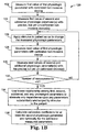
- FIG 1A A list of steps of an embodiment of a protocol 100 to calibrate a non-invasive medical device is depicted in FIG 1A.
- This particular embodiment is directed to a protocol for calibrating a device used to measure one physiologic parameter, P1, using calibrated measurements of a second parameter, P2.
- a known relationship, of the type listed as expression (1), exists between the first and second parameters via a third parameter, P3.
- the third parameter remains unchanged when a stimulus is applied to the patient.
- the protocol 100 may yield quantitative results when a known chemical equilibrium/stoichiometry expression is known between the physiologic parameters of interest.
- the first physiologic parameter is measured with a non-invasive device that has been pre-calibrated for the first physiologic parameter.
- the first non-invasive device may measure the first physiologic parameter using any type of non-invasive modality, such as optical, electrochemical, acoustic, magnetic resonance, biochemical or osmotic assist.
- Measurement step 104 determines the first value of the second physiologic parameter, the measurement 104 being taken with a non-invasive device which reports precise, but yet un-calibrated results.
- the device used for measuring the second physiologic parameter may use any suitable non-invasive modality, such as optical, electrochemical, acoustic, magnetic resonance, biochemical or osmotic assist.
- the two measurement devices may be housed within the same mechanical structure. The measurements of the first and second physiologic parameters may take place at substantially the same time and at substantially the same physical location.
- protocol step 106 a stimulus is applied to the patient, the stimulus changing both the first and second physiologic parameters.
- the stimulus in protocol step 106 is applied to the patient non-invasively.
- the stimulus may be applied at the same physical location that measurements 102 and 104 were taken.
- any suitable stimulus that affects the desired physiologic parameters may be used. It is important to understand that the stimulus applied to the patient may be a systemic change or a local change.
- a systemic change is one that is applied to a large part, or substantially all, of the patient's body, as might be expected, for example, with the application of one or more drugs.
- a local stimulus is one that is applied to the patient's body in substantially only the location of the assay.
- the application of localized pressure, localized induced temperature changes (heating and/or cooling), directed ultrasonic energy or the like, may result in local changes to physiologic parameters.
- protocol step 108 a second measurement of the first physiologic parameter is recorded with the calibrated non-invasive device.
- protocol step 110 a second value of the second physiologic parameter is measured with the precise, but yet un-calibrated, non-invasive device.
- the measurement steps 108 and 110 may be made at substantially the same time after application of the stimulus in step 106, or may be made while the stimulus is still being applied. In addition, the measurement steps 108 and 110 may be made at substantially at the same physical location as the measurements taken at steps 104 and 106.
- the known relationship between the first and second physiologic parameters is used to eliminate the dependence upon any other physiologic parameter which may participate in the chemical equilibrium between the first and second physiologic parameters.
- the third physiologic parameter is substantially unchanged by the stimulus applied to the patient in step 106 .
- the third physiologic parameter may be eliminated using the first and second measurements of the first and second physiologic parameters. If the known relationship involves additional physiologic parameters, then additional measurements of the first and second physiologic parameters may be made at different levels of applied stimulus, and the additional measurements used to eliminate the additional physiologic parameters.
- the calibration coefficients for the second physiologic parameter are calculated at protocol step 114.
- the two calibrated data points may be used to calculate calibration coefficients for the non-invasive device for the second physiologic parameter.
- the first and/or second parameters may be relatively time-independent, varying slowly with long-term physiological changes in the body over minutes or longer.
- the first and second parameters may also be time-variant.
- blood gas and pH level may have pulsatile characteristics that depend on the pulsatile nature of the blood flow.
- physiologic parameters of the tissue bed that are isolated from the pressure variations of pulsing arterial blood, are less likely to have pulsatile characteristics and are more likely to take on characteristics that drift over time.
- FIG. 1B Another embodiment of a protocol 120 for calibrating a sensor for measuring a physiologic parameter P1 that is related to other parameters through a complex expression of the type described with reference to expression (2) is illustrated in FIG. 1B.
- the signal S' is related to n parameters, although not all n parameters necessarily change with application of the stimulus to the patient. Those parameters that do not change, or whose change is insignificant, under application of the stimulus to the patient may be treated in the analysis as constants. Accordingly, N sets of measurements need to be made, where N is the number of parameters that change with application of the stimulus.
- the protocol 120 commences with a measurement of the first physiologic parameter using a calibrated sensor, at step 102.
- first values of the second physiologic parameter, and additional physiologic parameters are measured, at step 124.
- the second physiologic parameter is the parameter whose sensor is being calibrated in the protocol, and is measured using a sensor that is uncalibrated but precise.
- the additional parameters may be measured using uncalibrated but precise sensors, or using calibrated sensors, or a combination of un-calibrated but precise, and calibrated sensors.
- the stimulus is applied to the patient at step 126, so that the values of the measured physiologic parameters change.
- the next value of the first physiologic parameter is measured using the calibrated sensor.
- the next values of the second parameter and the additional parameters are measured.
- the number of measurements made for each parameter should be at least equal to the number parameters being measured, N.
- Each measurement for a parameter is made at a different level of applied stimulus, so that the parameter takes on a different value. If it is determined that an insufficient number of measurements has been taken, then the protocol returns to step 126 and the stimulus is applied at a new level so that the measured parameters adopt new values. The protocol continues to accumulate measurements of the different parameters at different levels of stimulus until the number of measurements reaches N.
- the known relationship among the different measurements is used, at step 134, to eliminate any dependence on any parameters that do not significantly change under the stimulus. This permits calibration coefficients to be calculated for those physiologic parameters that were measured using precise, but yet un-calibrated sensors.
- FIG. 2 depicts steps of an embodiment of a protocol 200 to calibrate a non-invasive optical pH sensor.
- the change in bicarbonate level is insignificant because the normal bicarbonate level is quite large, typically in the range of 23 to 25 milli-equivalents per liter.
- the protocol 200 utilizes this physiologic situation to simplify the calibration procedure of the non-invasive optical pH sensor as is outlined below. Accordingly, the change in bicarbonate level is taken to be substantially constant under localized changes of pH. Therefore, only two physiologic parameters, CO 2 and pH are measured, and so only two measurements for each parameter are required.
- the first value of the CO 2 concentration is measured with a calibrated non-invasive device.
- the CO 2 concentration may be measured indirectly by measuring the partial pressure of CO 2 of the tissue (commonly designated as pCO 2 ) generated in its gaseous form.
- the relationship between dissolved CO 2 and the CO 2 partial pressure, pCO 2 is given by equation 4, below:
- Total Carbon Dioxide dissolved Solubility coefficient * pCO 2
- Approaches for non-invasively measuring the CO 2 concentration of tissue include optical methods, such as near-infrared absorbance measurements, in particular, a method for measuring the CO 2 content of tissue using an optical approach is discussed in U.S. Patent Application Serial No. 10/162,028 titled, "Noninvasive Detection of A Physiologic Parameter Within A Body Tissue of A Patient", by inventors Edward J. Anderson et al.
- step 204 the first value of physiologic parameter pH is measured, the measurement being taken with a non-invasive pH measuring device which reports precise but yet un-calibrated results.
- a non-invasive pH measuring device which reports precise but yet un-calibrated results.
- One approach to non-invasively measure pH is based around a measurement of the fluorescence of NADH, whose fluorescence efficiency is dependent on the local pH. This approach is discussed further in U.S. Patent Application Serial No. 10/195004 titled, "Non-invasive Measurement of pH", by inventors Victor Kimball, Steven Furlong, and Irvin Pierskalla, Altera Law Group Docket # 1535.2US01, filed on even date herewith.
- the CO 2 and pH measurement devices may be housed within the same mechanical structure and the measurements may take place at substantially the same time and at substantially the same physical location.
- protocol step 206 a stimulus is applied to the patient, the stimulus changing both the CO 2 and pH physiologic parameters.
- the stimulus in protocol step 206 to the patient is applied non-invasively and the stimulus is applied at the same physical location that measurements 202 and 204 were taken.
- Any suitable stimulus that affects the desired physiologic parameters may be used.
- the stimulus may affect the parameter directly or indirectly.
- Indirect affects may result, for example, by stopping blood flow to the tissue being assayed or by changing the metabolism of the tissue being assayed.
- An example of a direct effect is injecting a quantity of one of the physiologic parameters being measured, for example, glucose or electrolytes.
- a systemic change in the physiologic parameters may be expected, for example, after the administration of a drug that affects metabolism: the induced change in the body's metabolism results in a systemic change in the CO 2 and/or pH values of the patient, which may be measured at the measurement site.
- a local stimulus such as the application of localized pressure, localized induced temperature changes (heating and/or cooling), directed ultrasonic energy or the like, may also produce a change in the physiologic parameters.
- a drug may be administered so that it has a local effect, for example by being applied topically via a DMSO solution absorbed into the tissue.
- a second measurement of the physiologic parameter CO 2 is recorded with the calibrated non-invasive device, at step 208.
- a measurement is also made, at step 210, to record a second value of the physiologic parameter pH with the precise but yet uncalibrated non-invasive device.
- the measurement steps 208 and 210 may be made at substantially the same time after the stimulus in protocol step 206, and substantially at the same physical location as measurement steps 204 and 206. The measurements in steps 208 and 210 may be made while the stimulus is being applied or after the application of the stimulus has ceased.
- step 212 the known relationship between the physiologic parameters CO 2 and pH, as given by equation (3) is used to eliminate the dependence upon the bicarbonate ion concentration, [HCO3 - ], the bicarbonate ion concentration being substantially unchanged by the stimulus in protocol step 206 to the patient. This is described further below.
- the two, or more, calibrated data points are used to calculate calibration coefficients for the non-invasive pH device.
- Equation (8) highlights an important aspect of the present invention.
- the protocols described above with reference to FIGs. 1A, 1 B and 2 may be used also for quality assurance following calibration.
- the pH sensor Once the pH sensor has been calibrated, it may be used over a prolonged period, for example for many minutes, or some hours, taking measurements of pH at regular intervals. It is useful to perform periodic quality assurance checks to ensure that the sensor has not drifted out of calibration. This may be done, after taking a measurement of pH and CO 2 , by applying the stimulus to the patient and re-measuring the levels of pH and CO 2 under the stimulus. The calibration coefficients for the pH sensor may be recalculated and compared to those currently in use.
- the coefficients are within an acceptable error range of the current coefficients, then use of the current coefficients may continue, or the coefficients may be automatically updated with the new coefficients. If the newly calculated calibration coefficients are outside the acceptable error range, then the coefficients are typically updated with the new values. The user may also be notified that the coefficients have been changed.
- a processor/controller module 302 may contain various sub-systems and a central processing unit to control the timing, delivery, routing and post processing of signals for the monitoring device 300.
- An interface 304 connects the controller module 302 to a first non-invasive physiologic sensor 312, which may be housed in a patient interface module 310.
- the first non-invasive sensor may be based on any type of non-invasive sensor including, but not restricted to, optical, electro-chemical, acoustic, magnetic resonance, biochemical or osmotic assist.
- the interface 304 is an optical interface, and may include a fiber optic waveguide or a fiber optic bundle, or discrete bulk optical components such as a condensing lens or a series of condensing lenses.
- the patient interface module 310 may provide protection from such unwanted outside influences as stray light, fluid spills, and the like.
- the first non-invasive physiologic sensor 312 may be in direct physical contact with the patient's tissue surface 318.
- the tissue may be organ tissue, epithelial tissue, skin or any other type of tissue that is being assayed non-invasively.
- An interconnect device 306 connects the controller module 302 with an optional stimulus transducer 314, which may also be housed in the patient interface module 310.
- the stimulus transducer 314 is advantageously included in the patient interface module 310 when the stimulus applied to the patient is a local stimulus, although it may also be included when the applied stimulus is systemic.
- An interface 308 connects the controller module 302 with a second non-invasive physiologic sensor 316, which may also be housed in the patient interface module 310.
- the stimulus transducer 314 and the non-invasive sensors 312 and 316 may be mounted sufficiently close so that the same location of the tissue 318 is both stimulated and measured.
- the interface 308 is typically an optical interface.
- FIG. 3B An example of an end-on view of the interface module 310 is schematically represented in FIG. 3B, showing the non-invasive physiologic sensors 312b and 316b and the stimulus transducer 314b.
- FIG. 3C Another example of an end-on view of the interface module 310 is schematically represented in FIG. 3C, showing the relative locations of the non-invasive physiologic sensors 312c and 316c relative to the stimulus transducer 314c.
- the spacing between the first physiologic sensor and the stimulus transducer may be the same as the distance between the second physiologic sensor and the stimulus transducer. In such a case, the tissue assayed by the first sensor is advantageously subject to the same magnitude of stimulus as the tissue assayed by second sensor.
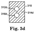
- FIG. 3D Another embodiment of an interface module 310 is schematically illustrated in FIG. 3D, in which a large portion 314d of the face represents the stimulus transducer.
- the physiologic sensors 312d and 316d are surrounded by the stimulus transducer 314d.
- Such an embodiment advantageously ensures that the level of stimulus applied to the site assayed for the first physiologic parameter is similar to that applied to the site assayed for the second physiologic parameter.
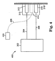
- FIG. 4 schematically depicts another embodiment of the invention. Elements that are the same as shown in FIG. 3A are labeled with the same reference number.
- the stimulus transducer 414 is not integrated within the patient interface module 310, and external controller unit 420 is a stand-alone unit separate from the main controller/processor unit 302.
- the stimulus transducer 414 may be removed from the patient when the device is not being calibrated, or may be left in place.
- the stimulus transducer may also be used for occasional quality assurance checks while the device is in use.
- the stimulus transducer 414 may also be replaced by alternative devices which stimulate the patient's tissue 318 by pressure, temperature, acoustic or other such appropriate stimulating techniques.
- FIG. 5 A particular embodiment of an optically based, non-invasive physiologic monitoring device 500 is depicted in FIG. 5. This embodiment may be particularly useful for conducting assays in a lumen, such as the esophagus.
- a processor/controller module 502 may contain the electro-optic sub-systems and a central processing unit to control the timing, delivery, routing and post processing of signals for the monitoring device 500.
- An optical interface 504 connects the controller module 502 to a first non-invasive optical physiologic sensor 512, which may be housed in a patient interface module 510.
- the optical interface 504 may be a fiber optic waveguide or a fiber optic bundle, or discrete bulk optical components such as a condensing lens or a series of condensing lenses.
- the patient interface module 510 may provide protection from such unwanted outside influences as stray light, fluid spills, and the like.
- the first non-invasive optical physiologic sensor 512 may be in direct physical contact with the patient's tissue surface 518.
- the interconnect device 506 connects the controller module 502 with the stimulus transducer 514, which may also be housed in the patient interface module 510.
- the optical interface 508 connects the controller module 502 with a second non-invasive optical physiologic sensor 516, which may also be housed in the patient interface module 510.
- the stimulus transducer 514 and the non-invasive optical sensors 512 and 516 may be mounted sufficiently close so as to stimulate and measure the tissue response at substantially the same physical location.
- An Inflatable bladder 520 may be incorporated into the patient interface module 510 for those applications where the sensor is inserted into a body cavity or orifice.
- This embodiment is advantageous in applications where it is desirable to apply pressure from the back surface 518b of the patient's epithelial tissue surface 518b to either mechanically secure the sensor against slippage during measurement or to apply additional pressure stimulus to aid in the calibration process.
- Other patient interface geometries and alternative sensor configurations are described in U.S. Patent Application Serial No. 10/162,028 titled, "Noninvasive Detection of A Physiologic Parameter Within A Body Tissue of A Patient", by inventors Edward J. Anderson et al.
- a patient monitor that includes sensors for measuring pH and CO 2 may be combined with other sensors for measuring other physiologic parameters, such as O 2 sat, O 2 , hematocrit, and the like.
Landscapes
- Health & Medical Sciences (AREA)
- Life Sciences & Earth Sciences (AREA)
- Physics & Mathematics (AREA)
- Molecular Biology (AREA)
- Animal Behavior & Ethology (AREA)
- Pathology (AREA)
- Engineering & Computer Science (AREA)
- Biomedical Technology (AREA)
- Heart & Thoracic Surgery (AREA)
- Medical Informatics (AREA)
- Veterinary Medicine (AREA)
- Surgery (AREA)
- Biophysics (AREA)
- General Health & Medical Sciences (AREA)
- Public Health (AREA)
- Optics & Photonics (AREA)
- Cardiology (AREA)
- Vascular Medicine (AREA)
- Physiology (AREA)
- Measurement Of The Respiration, Hearing Ability, Form, And Blood Characteristics Of Living Organisms (AREA)
- Investigating, Analyzing Materials By Fluorescence Or Luminescence (AREA)
- Investigating Or Analysing Materials By Optical Means (AREA)
Abstract
Description
- The present invention is directed generally to medical devices and more particularly to techniques to calibrate non-invasive devices.
- Optical spectroscopy techniques have been developed for a wide variety of uses within the medical community. For example, pulse oximetry and capnography instruments are in widespread use at hospitals, both in the surgery suites and the post-op ICU's. These technologies have historically been based on absorption-based spectroscopy techniques and have typically been used as trend monitors in critical care environments where it is necessary to quickly determine if a patient's vital parameters are undergoing large physiologic changes. Given this operating environment, it has been acceptable for these devices to have somewhat relaxed precision and accuracy requirements, given the clinical need for real-time point-of-care data for patients in critical care situations.
- Both pulse oximeters and capnography instruments can be labeled as non-invasive in that neither require penetrating the outer skin or tissue to make a measurement, nor do they require a blood or serum sample from the patient to custom calibrate the instrument to each individual patient. These instruments typically have pre-selected global calibration coefficients that have been determined from clinical trial results over a large patient population, and the results represent statistical averages over such variables as patient age, sex, race, and the like.
- There is, however, a growing desire within the medical community for non-invasive instruments for use in such areas as the emergency room, critical care ICU's, and trauma centers where fast and accurate data are needed for patients in potentially life threatening situations. One such measurement needed in these environments is the blood and/or tissue pH level, which is a measure of the free hydrogen ion concentration. This is an important measure of intracellular metabolism. Biological processes within the human body require a narrow range of pH for normal function, and significant changes of pH from this range may be life threatening.
- In addition to pH, it is also typical for the blood gases (O2 and CO2), blood electrolytes, and other blood chemistry parameters such as glucose, to be measured and monitored during critical care treatment. Technologies for making these measurements have been in place for nearly fifty years in hospital laboratories. These measurements are made from blood samples drawn from the patient which are then sent to a laboratory for analysis. These laboratory measurements are typically made with electro-chemical sensors.
- Recent developments in non-invasive optical technology hold the potential that some of these measurements may be made at the point of care with sufficient precision and accuracy to carry out critical care monitoring and treatment. For ease of use, and for meeting accuracy requirements, it is desirable that these non-invasive optical devices be custom calibrated to each Individual patient at the point of care. The calibration technique should compensate for each Individual's body chemistry and tissue make-up, including such things as collagen, elastin, and skin pigmentation, all of which affect skin and tissue optical properties. Ideally, the calibration technique for these optical sensors is quick, accurate, and easy to perform.
- In view of the above discussion, there is a need to calibrate medical devices at the point of care, where the calibration is performed without the removal of blood or bodily fluids. Such a technique may be applicable to a wide variety of commonly monitored physiologic parameters during critical care patient management.
- The invention provides a method of calibrating a non-invasive sensor for a first physiologic parameter In tissue and an apparatus for determining a value of a first physiologic parameter within tissue as defined by the appended independent claims. Preferred or advantageous features of the invention are set out in dependent sub-claims.
- Generally, the present invention relates to an approach to calibrating a first non-invasive sensor in which the tissue being measured is modulated in some way so as to alter the value of the parameter being measured by the first sensor. A second sensor detects another parameter that also changes with the modulation. The second sensor is absolutely calibrated. Where there is a known relationship between the first and second parameters, a calibration may be derived for the first sensor.
- One embodiment of the invention is directed to a method of calibrating a non-invasive sensor for determining a value of a first physiologic parameter within living tissue. The method includes non-invasively measuring first values of the first physiologic parameter and of a second physiologic parameter in the tissue. A known relationship exists between the first and second physiologic parameters. The tissue is acted on so as to change the first and second physiologic parameters. Second values of the first physiologic parameter and of the second physiologic parameter in the tissue are then measured. The measurements of the first and second values of the second physiologic parameter are absolutely calibrated. A calibrated value is then determined for at least one of the first and second values of the first physiologic parameter from the first and second values of both the first and second physiologic parameters.
- Another embodiment of the invention is directed to an apparatus for determining a value of a first physiologic parameter within tissue. The apparatus includes an uncalibrated, first non-invasive sensor for measuring the first physiologic parameter of the tissue, and a calibrated, second non-invasive sensor for measuring a second physiologic parameter of the tissue. A processor is coupled to receive first and second signals from each of the first and second non-invasive sensors. The processor calculates a point-slope calibration for the first physiologic parameter from the first and second signals from each of the first and second non-invasive sensors when a characteristic of the tissue is modulated between the first and second signals.
- The above summary of the present invention is not intended to describe each illustrated embodiment or every implementation of the present Invention. The figures and the detailed description which follow more particularly exemplify these embodiments.
- The invention may be more completely understood in consideration of the following detailed description of various embodiments of the invention in connection with the accompanying drawings, in which:
- FIGs. 1A and 1B illustrate steps of different embodiments of a protocol to non-invasively calibrate a physiologic sensor, according to the present invention;
- FIG. 2 illustrates steps of one embodiment of a protocol to non-Invasively calibrate an optical pH physiologic sensor, according to the present invention;
- FIG. 3A-3D schematically illustrate embodiments of a physiologic sensor module to non-invasively calibrate a physiologic sensor, according to the present invention;
- FIG. 4 schematically illustrates another embodiment of a physiologic sensor module to non-invasively calibrate a sensor, according to the present invention; and
- FIG. 5 schematically illustrates an embodiment of a physiologic sensor module to non-invasively calibrate an optical physiologic sensor placed in-situ in a body cavity or orifice, according to the present invention.
- The present invention Is applicable to medical devices and is believed to be particularly useful for calibrating non-invasive medical devices at the point of care.
- According to one embodiment of the invention, a set of calibration coefficients is determined using the known chemical relationship between at least a first physiologic parameter (P1) and a second physiologic parameter (P2), where the physiologic parameters P1 and P2 can be measured non-invasively. For example, P1 and P2 may be related to optical spectra that be measured directly. Also, the technique can accommodate a third physiologic parameter (P3), which may participate in the equilibrium process between physiologic parameters P1 and P2. The measurable signal, S, can be characterized by equation (1), as follows:

- In one embodiment, the invention Is directed to a technique to calibrate a medical device which has a measurable signal, S, whose dependence is governed by equation 1, in only two steps, by using a known relationship among the physiologic parameters P1, P2, and P3. This technique may be expanded to include more complicated physiologic systems having a measurable signal, S' of the form:

where, Pn is the nth physiologic parameter and use may be made of the relationship between the physiologic parameters to reduce the number of measurements needed. In the above case n measurements may be required if no relationship among the parameters is identified. - A list of steps of an embodiment of a
protocol 100 to calibrate a non-invasive medical device is depicted in FIG 1A. This particular embodiment is directed to a protocol for calibrating a device used to measure one physiologic parameter, P1, using calibrated measurements of a second parameter, P2. A known relationship, of the type listed as expression (1), exists between the first and second parameters via a third parameter, P3. In this particular case, the third parameter remains unchanged when a stimulus is applied to the patient. - The
protocol 100 may yield quantitative results when a known chemical equilibrium/stoichiometry expression is known between the physiologic parameters of interest. Inmeasurement step 102, the first physiologic parameter is measured with a non-invasive device that has been pre-calibrated for the first physiologic parameter. The first non-invasive device may measure the first physiologic parameter using any type of non-invasive modality, such as optical, electrochemical, acoustic, magnetic resonance, biochemical or osmotic assist. -
Measurement step 104 determines the first value of the second physiologic parameter, themeasurement 104 being taken with a non-invasive device which reports precise, but yet un-calibrated results. Like the first device, the device used for measuring the second physiologic parameter may use any suitable non-invasive modality, such as optical, electrochemical, acoustic, magnetic resonance, biochemical or osmotic assist. In one embodiment of the invention, the two measurement devices may be housed within the same mechanical structure. The measurements of the first and second physiologic parameters may take place at substantially the same time and at substantially the same physical location. - In protocol step 106, a stimulus is applied to the patient, the stimulus changing both the first and second physiologic parameters. In one embodiment of the invention, the stimulus in protocol step 106 is applied to the patient non-invasively. The stimulus may be applied at the same physical location that
measurements - Any suitable stimulus that affects the desired physiologic parameters may be used. It is important to understand that the stimulus applied to the patient may be a systemic change or a local change. A systemic change is one that is applied to a large part, or substantially all, of the patient's body, as might be expected, for example, with the application of one or more drugs.
- A local stimulus is one that is applied to the patient's body in substantially only the location of the assay. For example, the application of localized pressure, localized induced temperature changes (heating and/or cooling), directed ultrasonic energy or the like, may result in local changes to physiologic parameters.
- In
protocol step 108, a second measurement of the first physiologic parameter is recorded with the calibrated non-invasive device. Inprotocol step 110, a second value of the second physiologic parameter is measured with the precise, but yet un-calibrated, non-invasive device. - The measurement steps 108 and 110 may be made at substantially the same time after application of the stimulus in step 106, or may be made while the stimulus is still being applied. In addition, the measurement steps 108 and 110 may be made at substantially at the same physical location as the measurements taken at
steps 104 and 106. - At
protocol step 112, the known relationship between the first and second physiologic parameters is used to eliminate the dependence upon any other physiologic parameter which may participate in the chemical equilibrium between the first and second physiologic parameters. The third physiologic parameter is substantially unchanged by the stimulus applied to the patient in step 106 . - Where the known relationship between the first and second physiologic parameters involves one other physiologic parameter, a third physiologic parameter, then the third physiologic parameter may be eliminated using the first and second measurements of the first and second physiologic parameters. If the known relationship involves additional physiologic parameters, then additional measurements of the first and second physiologic parameters may be made at different levels of applied stimulus, and the additional measurements used to eliminate the additional physiologic parameters.
- The calibration coefficients for the second physiologic parameter are calculated at
protocol step 114. In the example case where the relationship between the first and second physiologic parameters includes only the third physiologic parameter, then the two calibrated data points may be used to calculate calibration coefficients for the non-invasive device for the second physiologic parameter. - The first and/or second parameters may be relatively time-independent, varying slowly with long-term physiological changes in the body over minutes or longer. The first and second parameters may also be time-variant. For example, blood gas and pH level may have pulsatile characteristics that depend on the pulsatile nature of the blood flow. On the other hand, physiologic parameters of the tissue bed, that are isolated from the pressure variations of pulsing arterial blood, are less likely to have pulsatile characteristics and are more likely to take on characteristics that drift over time.
- Another embodiment of a
protocol 120 for calibrating a sensor for measuring a physiologic parameter P1 that is related to other parameters through a complex expression of the type described with reference to expression (2) is illustrated in FIG. 1B. The signal S' is related to n parameters, although not all n parameters necessarily change with application of the stimulus to the patient. Those parameters that do not change, or whose change is insignificant, under application of the stimulus to the patient may be treated in the analysis as constants. Accordingly, N sets of measurements need to be made, where N is the number of parameters that change with application of the stimulus. - The
protocol 120 commences with a measurement of the first physiologic parameter using a calibrated sensor, atstep 102. Next, first values of the second physiologic parameter, and additional physiologic parameters are measured, atstep 124. The second physiologic parameter is the parameter whose sensor is being calibrated in the protocol, and is measured using a sensor that is uncalibrated but precise. The additional parameters may be measured using uncalibrated but precise sensors, or using calibrated sensors, or a combination of un-calibrated but precise, and calibrated sensors. - The stimulus is applied to the patient at
step 126, so that the values of the measured physiologic parameters change. Atstep 128, the next value of the first physiologic parameter is measured using the calibrated sensor. Atstep 130, the next values of the second parameter and the additional parameters are measured. - At
step 132, a determination is made as to whether a sufficient number of measurements have been made. The number of measurements made for each parameter should be at least equal to the number parameters being measured, N. Each measurement for a parameter is made at a different level of applied stimulus, so that the parameter takes on a different value. If it is determined that an insufficient number of measurements has been taken, then the protocol returns to step 126 and the stimulus is applied at a new level so that the measured parameters adopt new values. The protocol continues to accumulate measurements of the different parameters at different levels of stimulus until the number of measurements reaches N. - Once it has been determined that a sufficient number of measurements has been made, the known relationship among the different measurements is used, at
step 134, to eliminate any dependence on any parameters that do not significantly change under the stimulus. This permits calibration coefficients to be calculated for those physiologic parameters that were measured using precise, but yet un-calibrated sensors. - FIG. 2 depicts steps of an embodiment of a
protocol 200 to calibrate a non-invasive optical pH sensor. Theprotocol 200 may yield quantitative results when utilizing the relationship between the physiologic parameters given in equation (3), below:
where pH is the negative logarithm of the hydrogen ion concentration [H+], pK is the logarithm of the equilibrium constant, [CO2] is the carbon dioxide concentration, and [HCO3 -] is the bicarbonate ion concentration. - Under normal physiologic conditions the pH of human blood is maintained near 7.4 pH units. If, however, the pH drops below 6.8 (acidic) or rises above 7.8 (alkaline) the condition can become life threatening. Fortunately, the pH of human blood is buffered against such large deviations. For example, when moderate amounts of hydrogen ions, H+, are added to blood during normal metabolic processes, the pH of blood is "buffered" against large swings. The reason is that the pH is dependent only on the ratio of CO2 to bicarbonate ion (pK remains constant, unless the temperature changes), and under these conditions the CO2 level is slightly elevated with an almost insignificant change in the bicarbonate level. The change in bicarbonate level is insignificant because the normal bicarbonate level is quite large, typically in the range of 23 to 25 milli-equivalents per liter. The
protocol 200 utilizes this physiologic situation to simplify the calibration procedure of the non-invasive optical pH sensor as is outlined below. Accordingly, the change in bicarbonate level is taken to be substantially constant under localized changes of pH. Therefore, only two physiologic parameters, CO2 and pH are measured, and so only two measurements for each parameter are required. - In
measurement step 202 , the first value of the CO2 concentration is measured with a calibrated non-invasive device. In one embodiment of the invention, the CO2 concentration may be measured indirectly by measuring the partial pressure of CO2 of the tissue (commonly designated as pCO2) generated in its gaseous form. The relationship between dissolved CO2 and the CO2 partial pressure, pCO2, is given by equation 4, below:
U.S. Patent Application Serial No. 10/162,028 titled, "Noninvasive Detection of A Physiologic Parameter Within A Body Tissue of A Patient", by inventors Edward J. Anderson et al. - In
step 204, the first value of physiologic parameter pH is measured, the measurement being taken with a non-invasive pH measuring device which reports precise but yet un-calibrated results. One approach to non-invasively measure pH is based around a measurement of the fluorescence of NADH, whose fluorescence efficiency is dependent on the local pH. This approach is discussed further inU.S. Patent Application Serial No. 10/195004 titled, "Non-invasive Measurement of pH", by inventors Victor Kimball, Steven Furlong, and Irvin Pierskalla, Altera Law Group Docket # 1535.2US01, filed on even date herewith. - In one embodiment of the invention, the CO2 and pH measurement devices may be housed within the same mechanical structure and the measurements may take place at substantially the same time and at substantially the same physical location.
- In
protocol step 206, a stimulus is applied to the patient, the stimulus changing both the CO2 and pH physiologic parameters. In one embodiment of the invention, the stimulus inprotocol step 206 to the patient is applied non-invasively and the stimulus is applied at the same physical location thatmeasurements - Any suitable stimulus that affects the desired physiologic parameters may be used. The stimulus may affect the parameter directly or indirectly. Indirect affects may result, for example, by stopping blood flow to the tissue being assayed or by changing the metabolism of the tissue being assayed. An example of a direct effect is injecting a quantity of one of the physiologic parameters being measured, for example, glucose or electrolytes. A systemic change in the physiologic parameters may be expected, for example, after the administration of a drug that affects metabolism: the induced change in the body's metabolism results in a systemic change in the CO2 and/or pH values of the patient, which may be measured at the measurement site.
- A local stimulus, such as the application of localized pressure, localized induced temperature changes (heating and/or cooling), directed ultrasonic energy or the like, may also produce a change in the physiologic parameters. In addition, a drug may be administered so that it has a local effect, for example by being applied topically via a DMSO solution absorbed into the tissue.
- Following application of the stimulus in
step 206, a second measurement of the physiologic parameter CO2 is recorded with the calibrated non-invasive device, atstep 208. A measurement is also made, atstep 210, to record a second value of the physiologic parameter pH with the precise but yet uncalibrated non-invasive device. In one embodiment of the invention, the measurement steps 208 and 210 may be made at substantially the same time after the stimulus inprotocol step 206, and substantially at the same physical location as measurement steps 204 and 206. The measurements insteps - At
step 212, the known relationship between the physiologic parameters CO2 and pH, as given by equation (3) is used to eliminate the dependence upon the bicarbonate ion concentration, [HCO3-], the bicarbonate ion concentration being substantially unchanged by the stimulus inprotocol step 206 to the patient. This is described further below. - At
protocol step 214, the two, or more, calibrated data points are used to calculate calibration coefficients for the non-invasive pH device. - One algebraic approach useful for calibrating the non-invasive pH device, according to the
protocol 200, is outlined below. A short hand notation is employed to ease in following the calculations. Recall that the known relationship between pH and CO2 concentration is given by the following expression:

- Eliminating the logarithmic dependence, yields the expression:

- Here we employ the following shorthand notation:
- Hn = the nth measurement of the Hydrogen ion concentration, [H+]
- Cn = the nth measurement of the Carbon dioxide concentration, [CO2]
- Bn = the nth measurement of the Bicarbonate ion concentration, [HCO3]
- We can write the result of correlating the results of
measurement steps 
- Similarly, after applying the stimulus to the patient in
protocol step 206, the result of correlating the measurements insteps 
- Where we have utilized the fact that the pK remains constant, i.e., K1 = K2, and the bicarbonate level remains substantially unchanged by the stimulus, B1 = B2. Given this, we can solve for the bicarbonate concentration by subtracting equation 6 from equation 7, which yields

- Equation (8) highlights an important aspect of the present invention. The precise, but yet uncalibrated non-invasive pH measuring device utilized in
measurement steps 

- Therefore, all terms on the right-hand side of equations (6) and (7) are now accurately known and a two-point calibration of the pH measurement device can be calculated directly from these results.
- It will be appreciated that the protocols described above with reference to FIGs. 1A, 1 B and 2 may be used also for quality assurance following calibration. To illustrate, consider the specific example discussed above with reference to FIG. 2 concerning a pH sensor. Once the pH sensor has been calibrated, it may be used over a prolonged period, for example for many minutes, or some hours, taking measurements of pH at regular intervals. It is useful to perform periodic quality assurance checks to ensure that the sensor has not drifted out of calibration. This may be done, after taking a measurement of pH and CO2, by applying the stimulus to the patient and re-measuring the levels of pH and CO2 under the stimulus. The calibration coefficients for the pH sensor may be recalculated and compared to those currently in use. If the newly calculated calibration coefficients are within an acceptable error range of the current coefficients, then use of the current coefficients may continue, or the coefficients may be automatically updated with the new coefficients. If the newly calculated calibration coefficients are outside the acceptable error range, then the coefficients are typically updated with the new values. The user may also be notified that the coefficients have been changed.
- One particular embodiment of a non-invasive
physiologic monitoring device 300 is schematically depicted in FIG. 3A. A processor/controller module 302 may contain various sub-systems and a central processing unit to control the timing, delivery, routing and post processing of signals for themonitoring device 300. Aninterface 304 connects thecontroller module 302 to a first non-invasivephysiologic sensor 312, which may be housed in apatient interface module 310. The first non-invasive sensor may be based on any type of non-invasive sensor including, but not restricted to, optical, electro-chemical, acoustic, magnetic resonance, biochemical or osmotic assist. In the case of an optical sensor, theinterface 304 is an optical interface, and may include a fiber optic waveguide or a fiber optic bundle, or discrete bulk optical components such as a condensing lens or a series of condensing lenses. Thepatient interface module 310 may provide protection from such unwanted outside influences as stray light, fluid spills, and the like. The first non-invasivephysiologic sensor 312 may be in direct physical contact with the patient'stissue surface 318. The tissue may be organ tissue, epithelial tissue, skin or any other type of tissue that is being assayed non-invasively. - An
interconnect device 306 connects thecontroller module 302 with anoptional stimulus transducer 314, which may also be housed in thepatient interface module 310. Thestimulus transducer 314 is advantageously included in thepatient interface module 310 when the stimulus applied to the patient is a local stimulus, although it may also be included when the applied stimulus is systemic. - An
interface 308 connects thecontroller module 302 with a second non-invasivephysiologic sensor 316, which may also be housed in thepatient interface module 310. In this configuration, thestimulus transducer 314 and thenon-invasive sensors tissue 318 is both stimulated and measured. Where the secondnon-invasive sensor 316 is an optical sensor, theinterface 308 is typically an optical interface. - An example of an end-on view of the
interface module 310 is schematically represented in FIG. 3B, showing the non-invasivephysiologic sensors stimulus transducer 314b. Another example of an end-on view of theinterface module 310 is schematically represented in FIG. 3C, showing the relative locations of the non-invasivephysiologic sensors stimulus transducer 314c. It will be appreciated that other configurations may be used in addition to those illustrated in FIGs. 3A-3C. The spacing between the first physiologic sensor and the stimulus transducer may be the same as the distance between the second physiologic sensor and the stimulus transducer. In such a case, the tissue assayed by the first sensor is advantageously subject to the same magnitude of stimulus as the tissue assayed by second sensor. - Another embodiment of an
interface module 310 is schematically illustrated in FIG. 3D, in which alarge portion 314d of the face represents the stimulus transducer. Thephysiologic sensors stimulus transducer 314d. Such an embodiment advantageously ensures that the level of stimulus applied to the site assayed for the first physiologic parameter is similar to that applied to the site assayed for the second physiologic parameter. - FIG. 4 schematically depicts another embodiment of the invention. Elements that are the same as shown in FIG. 3A are labeled with the same reference number. In this embodiment, the
stimulus transducer 414 is not integrated within thepatient interface module 310, andexternal controller unit 420 is a stand-alone unit separate from the main controller/processor unit 302. In this embodiment, thestimulus transducer 414 may be removed from the patient when the device is not being calibrated, or may be left in place. The stimulus transducer may also be used for occasional quality assurance checks while the device is in use. Thestimulus transducer 414 may also be replaced by alternative devices which stimulate the patient'stissue 318 by pressure, temperature, acoustic or other such appropriate stimulating techniques. - A particular embodiment of an optically based, non-invasive
physiologic monitoring device 500 is depicted in FIG. 5. This embodiment may be particularly useful for conducting assays in a lumen, such as the esophagus. A processor/controller module 502 may contain the electro-optic sub-systems and a central processing unit to control the timing, delivery, routing and post processing of signals for themonitoring device 500. Anoptical interface 504 connects thecontroller module 502 to a first non-invasive opticalphysiologic sensor 512, which may be housed in apatient interface module 510. Theoptical interface 504 may be a fiber optic waveguide or a fiber optic bundle, or discrete bulk optical components such as a condensing lens or a series of condensing lenses. Thepatient interface module 510 may provide protection from such unwanted outside influences as stray light, fluid spills, and the like. The first non-invasive opticalphysiologic sensor 512 may be in direct physical contact with the patient'stissue surface 518. Theinterconnect device 506 connects thecontroller module 502 with thestimulus transducer 514, which may also be housed in thepatient interface module 510. Theoptical interface 508 connects thecontroller module 502 with a second non-invasive opticalphysiologic sensor 516, which may also be housed in thepatient interface module 510. In this configuration, thestimulus transducer 514 and the non-invasiveoptical sensors - An
Inflatable bladder 520 may be incorporated into thepatient interface module 510 for those applications where the sensor is inserted into a body cavity or orifice. This embodiment is advantageous in applications where it is desirable to apply pressure from theback surface 518b of the patient'sepithelial tissue surface 518b to either mechanically secure the sensor against slippage during measurement or to apply additional pressure stimulus to aid in the calibration process. Other patient interface geometries and alternative sensor configurations are described inU.S. Patent Application Serial No. 10/162,028 titled, "Noninvasive Detection of A Physiologic Parameter Within A Body Tissue of A Patient", by inventors Edward J. Anderson et al. - The present invention should not be considered limited to the particular examples described above, but rather should be understood to cover all aspects of the invention as fairly set out in the attached claims. Various modifications, equivalent processes, as well as numerous structures to which the present invention may be applicable will be readily apparent to those of skill in the art to which the present invention is directed upon review of the present specification. The claims are intended to cover such modifications and devices.
- For example, a patient monitor that includes sensors for measuring pH and CO2 may be combined with other sensors for measuring other physiologic parameters, such as O2 sat, O2, hematocrit, and the like.
Claims (43)
- A method of calibrating a non-invasive sensor for a first physiologic parameter in tissue, comprising:(a) non-invasively measuring first values of the first physiologic parameter and of a second physiologic parameter in the tissue, a known relationship existing between the first and second physiologic parameters;(b) non-invasively measuring second values of the first physiologic parameter and of the second physiologic parameter in the tissue after the tissue has been acted upon so as to change the first and second physiological parameters, the measurements of the first and second values of the second physiologic parameter being absolutely calibrated; and(c) determining a calibrated value for at least one of the first and second values of the first physiologic parameter from the first and second values of both the first and second physiologic parameters.
- A method as recited in claim 1, wherein non-invasively measuring the first and second values of the first parameter include measuring light received from the tissue.
- A method as recited in claim 1, wherein non-invasively measuring the first and second values of the second parameter include measuring light received from the tissue.
- A method as recited in claim 1, wherein the values for the first and second physiologic parameters are measured in the same region of the tissue.
- A method as recited in claim 1, wherein non-invasively measuring the first values of the first physiologic parameter includes exciting the tissue with excitation light and detecting a resulting fluorescent signal.
- A method as recited in claim 1, wherein acting on the tissue includes applying a systemic stimulus to the tissue.
- A method as recited in claim 1, wherein acting on the tissue includes applying a stimulus to the tissue localized to a region of the tissue where the first and second physiologic parameters are non-invasively measured.
- A method as recited in claim 1, wherein acting on the tissue includes administering a drug to the patient whose tissue is being examined.
- A method as recited in claim 1, wherein applying a systemic stimulus to the tissue includes changing a metabolic rate of the tissue.
- A method as recited in claim 1, wherein acting on the tissue includes applying pressure to a region of the tissue where the first and second physiologic parameters are measured.
- A method as recited in claim 1, wherein acting on the body includes changing temperature of a region of the body where the first and second physiologic parameters are measured.
- A method as recited in claim 1, wherein non-invasively measuring the second values of the first physiologic parameter and of the second physiologic parameter includes measuring the second values of the first physiologic parameter and of the second physiologic parameter simultaneously.
- A method as recited in claim 1, wherein the first physiologic parameter is pH.
- A method as recited in claim 13, wherein measuring the pH includes making a fluorescence measurement of NADH in the region of the tissue being measured.
- A method as recited in claim 13, wherein the second physiologic parameter is concentration of CO2.
- A method as recited in claim 1, wherein the known relationship between the first and second physiologic parameters includes a third physiologic parameter.
- A method as recited in claim 16, wherein the third physiologic parameter is substantially unaffected by the acting on the body.
- A method as recited in claim 16, wherein determining the calibrated value for at least one of the first and second values of the first physiologic parameter includes determining a value of the third physiologic parameter from a ratio of i) the difference between the first and second values of the second physiologic parameter over ii) the difference between the first and second values of the first physiologic parameter.
- A method as recited in claim 18, wherein determining the calibrated value for at least one of the first and second values of the first physiologic parameter further includes calculating the calibrated value using the value of the third physiologic parameter and at least the respective calibrated first or second value of the second physiologic parameter.
- A method as recited in claim 1, wherein the known relationship between the first and second physiologic parameters is pH = pK - log([C02]/[HC03-]) where pH is the first physiologic parameter and [CO:J is the second physiologic parameter.
- A method as recited in claim 1, wherein the known relationship relates N physiologic parameters that change upon acting on the tissue so as to change the first and second physiologic parameters, and acting on the tissue N-1 times so as to take N measurements of each of the N physiologic parameters at different levels of stimulus arising from the acting on the tissue.
- A method as recited in claim 1, further comprising non-invasively measuring the second values of the first physiologic parameter and of the second physiologic parameter in the tissue while acting on the tissue.
- A method as recited in claim 1, wherein the tissue is epithelial tissue.
- A method as recited in claim 1, wherein the tissue is selected from skin tissue, muscle tissue and organ tissue.
- A method as recited in claim 1, further comprising performing successive non-invasive measurements of the first physiologic parameter of the tissue and determining respective absolute calibrated values of the first physiologic parameter from the first and second values of both the first and second physiologic parameters.
- A method as recited in claim 25, further comprising repeating steps (a) - (c) after performing the successive non-invasive measurements and determining whether a calibration of an instrument for non-invasively measuring the first physiologic parameter has changed.
- An apparatus (300) for determining a value of a first physiologic parameter within tissue (318), comprising:an uncalibrated, first non-invasive sensor (312) for measuring the first physiologic parameter of the tissue (318);a calibrated, second non-invasive sensor (316) for measuring a second physiologic parameter of the tissue (318); anda processor (302) coupled to receive first and second signals from each of the first and second non-invasive sensors (312, 316), and to calculate calibrated values for the first physiologic parameter from the first and second signals from each of the first and second non-invasive sensors (312, 316) when a characteristic of the tissue (318) is modulated between the first and second signals.
- An apparatus (300) as recited in claim 27, wherein the first non-invasive sensor (312) is an optically based sensor.
- An apparatus (300) as recited in claim 27, wherein the second non-invasive sensor (316) is an optically based sensor.
- An apparatus (300) as recited in claim 27, wherein the first non-invasive sensor (312) is a pH sensor.
- An apparatus (300) as recited in claim 27, wherein the second non-invasive sensor (316) is a calibrated, non-invasive CO2 sensor.
- An apparatus (300) as recited in claim 30, wherein the pH sensor emits excitation light in the wavelength range 300 nm - 400 nm.
- An apparatus (300) as recited in claim 30, wherein the pH sensor detects light emitted for the tissue (318) in a wavelength range of 400 - 600 nm.
- An apparatus (300) as recited in claim 30, wherein the pH sensor detects fluorescence from NADH in the tissue (318).
- An apparatus (300) as recited in claim 27, further comprising a patient interface module (310) to house the first and second non-invasive sensors (312, 316).
- An apparatus (300) as recited in claim 27, further comprising a modulator (314) to modulate the characteristic of the tissue (318) at the region of the tissue (318) where the first and second physiologic parameters are measured.
- An apparatus (300) as recited in claim 36, wherein the modulator (314) includes an extensible member (520) to apply pressure of the region of the tissue (318) where the first and second physiologic parameters are measured.
- An apparatus (300) as recited in claim 33, wherein the modulator (314) includes a thermally responsive member to change temperature of the region of the tissue (318) where the first and second physiologic parameters are measured.
- An apparatus (300) as recited in claim 36, further comprising a patient interface module (310), the first and second non-invasive sensors (312, 316) and the modulator (314) being housed within the patient interface module (310).
- An apparatus (300) as recited in claim 39, wherein the modulator (314) is removable from the patient interface module (310), the first and second non-invasive sensors (312, 316) being enabled to perform measurements on the first and second physiologic parameters respectively after the modulator (314) is removed from the patient interface (310).
- An apparatus (300) as recited in claim 27, wherein the processor (302) calculates calibration constants for the first physiologic parameter from the first and second signals from each of the first and second non-invasive sensors (312, 316).
- An apparatus (300) as recited in claim 41, wherein the processor (302) produces calibrated values for the first physiologic parameter from measurement signals received from the first non-invasive sensor (312) and the calibration constants.
- An apparatus (300) as recited in claim 42, wherein the processor (302) performs a check on the calibration constants by measuring the first and second physiologic parameters before and after application of a stimulus to the tissue (318).
Applications Claiming Priority (3)
| Application Number | Priority Date | Filing Date | Title |
|---|---|---|---|
| US195120 | 2002-07-11 | ||
| US10/195,120 US6865407B2 (en) | 2002-07-11 | 2002-07-11 | Calibration technique for non-invasive medical devices |
| PCT/US2003/021604 WO2004006760A1 (en) | 2002-07-11 | 2003-07-09 | Calibration technique for non-invasive medical devices |
Publications (2)
| Publication Number | Publication Date |
|---|---|
| EP1545296A1 EP1545296A1 (en) | 2005-06-29 |
| EP1545296B1 true EP1545296B1 (en) | 2007-12-26 |
Family
ID=30114906
Family Applications (1)
| Application Number | Title | Priority Date | Filing Date |
|---|---|---|---|
| EP03764446A Expired - Lifetime EP1545296B1 (en) | 2002-07-11 | 2003-07-09 | Calibration technique for non-invasive medical devices |
Country Status (6)
| Country | Link |
|---|---|
| US (1) | US6865407B2 (en) |
| EP (1) | EP1545296B1 (en) |
| JP (1) | JP2005532867A (en) |
| AU (1) | AU2003251847A1 (en) |
| DE (1) | DE60318323T2 (en) |
| WO (1) | WO2004006760A1 (en) |
Families Citing this family (169)
| Publication number | Priority date | Publication date | Assignee | Title |
|---|---|---|---|---|
| US6675031B1 (en) | 1999-04-14 | 2004-01-06 | Mallinckrodt Inc. | Method and circuit for indicating quality and accuracy of physiological measurements |
| US6748254B2 (en) | 2001-10-12 | 2004-06-08 | Nellcor Puritan Bennett Incorporated | Stacked adhesive optical sensor |
| US7190986B1 (en) | 2002-10-18 | 2007-03-13 | Nellcor Puritan Bennett Inc. | Non-adhesive oximeter sensor for sensitive skin |
| US20190357827A1 (en) | 2003-08-01 | 2019-11-28 | Dexcom, Inc. | Analyte sensor |
| US7299082B2 (en) * | 2003-10-31 | 2007-11-20 | Abbott Diabetes Care, Inc. | Method of calibrating an analyte-measurement device, and associated methods, devices and systems |
| US7162288B2 (en) | 2004-02-25 | 2007-01-09 | Nellcor Purtain Bennett Incorporated | Techniques for detecting heart pulses and reducing power consumption in sensors |
| EA200601463A1 (en) * | 2004-03-06 | 2007-02-27 | Калисто Медикал, Инк. | METHODS AND DEVICES FOR NON-INVASIVE OBTAINING QUANTITATIVE INFORMATION ON SUBSTANCES IN LIVING ORGANISMS |
| US20100331646A1 (en) * | 2009-06-30 | 2010-12-30 | Abbott Diabetes Care Inc. | Health Management Devices and Methods |
| US9636450B2 (en) | 2007-02-19 | 2017-05-02 | Udo Hoss | Pump system modular components for delivering medication and analyte sensing at seperate insertion sites |
| US7657295B2 (en) | 2005-08-08 | 2010-02-02 | Nellcor Puritan Bennett Llc | Medical sensor and technique for using the same |
| US7590439B2 (en) | 2005-08-08 | 2009-09-15 | Nellcor Puritan Bennett Llc | Bi-stable medical sensor and technique for using the same |
| US7657294B2 (en) | 2005-08-08 | 2010-02-02 | Nellcor Puritan Bennett Llc | Compliant diaphragm medical sensor and technique for using the same |
| JP2009507224A (en) | 2005-08-31 | 2009-02-19 | ユニヴァーシティー オブ ヴァージニア パテント ファンデーション | Improving the accuracy of continuous glucose sensors |
| US20070060808A1 (en) | 2005-09-12 | 2007-03-15 | Carine Hoarau | Medical sensor for reducing motion artifacts and technique for using the same |
| US8092379B2 (en) | 2005-09-29 | 2012-01-10 | Nellcor Puritan Bennett Llc | Method and system for determining when to reposition a physiological sensor |
| US7899510B2 (en) | 2005-09-29 | 2011-03-01 | Nellcor Puritan Bennett Llc | Medical sensor and technique for using the same |
| US7869850B2 (en) | 2005-09-29 | 2011-01-11 | Nellcor Puritan Bennett Llc | Medical sensor for reducing motion artifacts and technique for using the same |
| US7904130B2 (en) | 2005-09-29 | 2011-03-08 | Nellcor Puritan Bennett Llc | Medical sensor and technique for using the same |
| US7881762B2 (en) | 2005-09-30 | 2011-02-01 | Nellcor Puritan Bennett Llc | Clip-style medical sensor and technique for using the same |
| US8880138B2 (en) | 2005-09-30 | 2014-11-04 | Abbott Diabetes Care Inc. | Device for channeling fluid and methods of use |
| US7486979B2 (en) | 2005-09-30 | 2009-02-03 | Nellcor Puritan Bennett Llc | Optically aligned pulse oximetry sensor and technique for using the same |
| US8062221B2 (en) | 2005-09-30 | 2011-11-22 | Nellcor Puritan Bennett Llc | Sensor for tissue gas detection and technique for using the same |
| US7555327B2 (en) | 2005-09-30 | 2009-06-30 | Nellcor Puritan Bennett Llc | Folding medical sensor and technique for using the same |
| US8233954B2 (en) | 2005-09-30 | 2012-07-31 | Nellcor Puritan Bennett Llc | Mucosal sensor for the assessment of tissue and blood constituents and technique for using the same |
| US7483731B2 (en) | 2005-09-30 | 2009-01-27 | Nellcor Puritan Bennett Llc | Medical sensor and technique for using the same |
| US7826879B2 (en) | 2006-02-28 | 2010-11-02 | Abbott Diabetes Care Inc. | Analyte sensors and methods of use |
| US7885698B2 (en) * | 2006-02-28 | 2011-02-08 | Abbott Diabetes Care Inc. | Method and system for providing continuous calibration of implantable analyte sensors |
| US9675290B2 (en) | 2012-10-30 | 2017-06-13 | Abbott Diabetes Care Inc. | Sensitivity calibration of in vivo sensors used to measure analyte concentration |
| US7653425B2 (en) | 2006-08-09 | 2010-01-26 | Abbott Diabetes Care Inc. | Method and system for providing calibration of an analyte sensor in an analyte monitoring system |
| US8473022B2 (en) * | 2008-01-31 | 2013-06-25 | Abbott Diabetes Care Inc. | Analyte sensor with time lag compensation |
| US8374668B1 (en) | 2007-10-23 | 2013-02-12 | Abbott Diabetes Care Inc. | Analyte sensor with lag compensation |
| US9326709B2 (en) | 2010-03-10 | 2016-05-03 | Abbott Diabetes Care Inc. | Systems, devices and methods for managing glucose levels |
| US8583205B2 (en) | 2008-03-28 | 2013-11-12 | Abbott Diabetes Care Inc. | Analyte sensor calibration management |
| US8346335B2 (en) | 2008-03-28 | 2013-01-01 | Abbott Diabetes Care Inc. | Analyte sensor calibration management |
| US7630748B2 (en) | 2006-10-25 | 2009-12-08 | Abbott Diabetes Care Inc. | Method and system for providing analyte monitoring |
| US7618369B2 (en) | 2006-10-02 | 2009-11-17 | Abbott Diabetes Care Inc. | Method and system for dynamically updating calibration parameters for an analyte sensor |
| US8224415B2 (en) | 2009-01-29 | 2012-07-17 | Abbott Diabetes Care Inc. | Method and device for providing offset model based calibration for analyte sensor |
| US8219173B2 (en) | 2008-09-30 | 2012-07-10 | Abbott Diabetes Care Inc. | Optimizing analyte sensor calibration |
| US8140312B2 (en) | 2007-05-14 | 2012-03-20 | Abbott Diabetes Care Inc. | Method and system for determining analyte levels |
| US9392969B2 (en) | 2008-08-31 | 2016-07-19 | Abbott Diabetes Care Inc. | Closed loop control and signal attenuation detection |
| US7522948B2 (en) | 2006-05-02 | 2009-04-21 | Nellcor Puritan Bennett Llc | Medical sensor and technique for using the same |
| US7477924B2 (en) | 2006-05-02 | 2009-01-13 | Nellcor Puritan Bennett Llc | Medical sensor and technique for using the same |
| US8073518B2 (en) | 2006-05-02 | 2011-12-06 | Nellcor Puritan Bennett Llc | Clip-style medical sensor and technique for using the same |
| US7507019B2 (en) | 2006-05-19 | 2009-03-24 | Covidien Ag | Thermometer calibration |
| US20070268954A1 (en) * | 2006-05-19 | 2007-11-22 | Sherwood Services Ag | Portable test apparatus for radiation-sensing thermometer |
| US8145288B2 (en) | 2006-08-22 | 2012-03-27 | Nellcor Puritan Bennett Llc | Medical sensor for reducing signal artifacts and technique for using the same |
| US8219170B2 (en) | 2006-09-20 | 2012-07-10 | Nellcor Puritan Bennett Llc | System and method for practicing spectrophotometry using light emitting nanostructure devices |
| US8195264B2 (en) | 2006-09-22 | 2012-06-05 | Nellcor Puritan Bennett Llc | Medical sensor for reducing signal artifacts and technique for using the same |
| US8396527B2 (en) | 2006-09-22 | 2013-03-12 | Covidien Lp | Medical sensor for reducing signal artifacts and technique for using the same |
| US8175671B2 (en) | 2006-09-22 | 2012-05-08 | Nellcor Puritan Bennett Llc | Medical sensor for reducing signal artifacts and technique for using the same |
| US7869849B2 (en) | 2006-09-26 | 2011-01-11 | Nellcor Puritan Bennett Llc | Opaque, electrically nonconductive region on a medical sensor |
| US7574245B2 (en) | 2006-09-27 | 2009-08-11 | Nellcor Puritan Bennett Llc | Flexible medical sensor enclosure |
| US7796403B2 (en) | 2006-09-28 | 2010-09-14 | Nellcor Puritan Bennett Llc | Means for mechanical registration and mechanical-electrical coupling of a faraday shield to a photodetector and an electrical circuit |
| US7890153B2 (en) | 2006-09-28 | 2011-02-15 | Nellcor Puritan Bennett Llc | System and method for mitigating interference in pulse oximetry |
| US7684842B2 (en) | 2006-09-29 | 2010-03-23 | Nellcor Puritan Bennett Llc | System and method for preventing sensor misuse |
| US7476131B2 (en) | 2006-09-29 | 2009-01-13 | Nellcor Puritan Bennett Llc | Device for reducing crosstalk |
| US8068891B2 (en) | 2006-09-29 | 2011-11-29 | Nellcor Puritan Bennett Llc | Symmetric LED array for pulse oximetry |
| US7680522B2 (en) | 2006-09-29 | 2010-03-16 | Nellcor Puritan Bennett Llc | Method and apparatus for detecting misapplied sensors |
| US8175667B2 (en) | 2006-09-29 | 2012-05-08 | Nellcor Puritan Bennett Llc | Symmetric LED array for pulse oximetry |
| US7549792B2 (en) * | 2006-10-06 | 2009-06-23 | Covidien Ag | Electronic thermometer with selectable modes |
| EP2106238A4 (en) | 2006-10-26 | 2011-03-09 | Abbott Diabetes Care Inc | METHOD, SYSTEM AND COMPUTER PROGRAM PRODUCT FOR REAL-TIME DETECTION OF SENSITIVITY FAILURE IN ANALYTICAL SENSORS |
| US20080161666A1 (en) * | 2006-12-29 | 2008-07-03 | Abbott Diabetes Care, Inc. | Analyte devices and methods |
| US8930203B2 (en) | 2007-02-18 | 2015-01-06 | Abbott Diabetes Care Inc. | Multi-function analyte test device and methods therefor |
| US8732188B2 (en) * | 2007-02-18 | 2014-05-20 | Abbott Diabetes Care Inc. | Method and system for providing contextual based medication dosage determination |
| US7894869B2 (en) | 2007-03-09 | 2011-02-22 | Nellcor Puritan Bennett Llc | Multiple configuration medical sensor and technique for using the same |
| US8280469B2 (en) | 2007-03-09 | 2012-10-02 | Nellcor Puritan Bennett Llc | Method for detection of aberrant tissue spectra |
| US8265724B2 (en) | 2007-03-09 | 2012-09-11 | Nellcor Puritan Bennett Llc | Cancellation of light shunting |
| CA2683959C (en) * | 2007-04-14 | 2017-08-29 | Abbott Diabetes Care Inc. | Method and apparatus for providing data processing and control in medical communication system |
| EP3741291A1 (en) | 2007-04-14 | 2020-11-25 | Abbott Diabetes Care, Inc. | Method and apparatus for providing data processing and control in medical communication system |
| EP2146623B1 (en) * | 2007-04-14 | 2014-01-08 | Abbott Diabetes Care Inc. | Method and apparatus for providing data processing and control in medical communication system |
| WO2008130898A1 (en) | 2007-04-14 | 2008-10-30 | Abbott Diabetes Care, Inc. | Method and apparatus for providing data processing and control in medical communication system |
| US8239166B2 (en) | 2007-05-14 | 2012-08-07 | Abbott Diabetes Care Inc. | Method and apparatus for providing data processing and control in a medical communication system |
| US8444560B2 (en) | 2007-05-14 | 2013-05-21 | Abbott Diabetes Care Inc. | Method and apparatus for providing data processing and control in a medical communication system |
| US9125548B2 (en) | 2007-05-14 | 2015-09-08 | Abbott Diabetes Care Inc. | Method and apparatus for providing data processing and control in a medical communication system |
| US10002233B2 (en) * | 2007-05-14 | 2018-06-19 | Abbott Diabetes Care Inc. | Method and apparatus for providing data processing and control in a medical communication system |
| US8103471B2 (en) | 2007-05-14 | 2012-01-24 | Abbott Diabetes Care Inc. | Method and apparatus for providing data processing and control in a medical communication system |
| US20080312845A1 (en) * | 2007-05-14 | 2008-12-18 | Abbott Diabetes Care, Inc. | Method and apparatus for providing data processing and control in a medical communication system |
| US8260558B2 (en) | 2007-05-14 | 2012-09-04 | Abbott Diabetes Care Inc. | Method and apparatus for providing data processing and control in a medical communication system |
| US8560038B2 (en) | 2007-05-14 | 2013-10-15 | Abbott Diabetes Care Inc. | Method and apparatus for providing data processing and control in a medical communication system |
| US8600681B2 (en) | 2007-05-14 | 2013-12-03 | Abbott Diabetes Care Inc. | Method and apparatus for providing data processing and control in a medical communication system |
| WO2008144616A1 (en) * | 2007-05-18 | 2008-11-27 | Heidi Kay | Lipid raft, caveolin protein, and caveolar function modulation compounds and associated synthetic and therapeutic methods |
| US8160900B2 (en) | 2007-06-29 | 2012-04-17 | Abbott Diabetes Care Inc. | Analyte monitoring and management device and method to analyze the frequency of user interaction with the device |
| US20090036760A1 (en) * | 2007-07-31 | 2009-02-05 | Abbott Diabetes Care, Inc. | Method and apparatus for providing data processing and control in a medical communication system |
| US8834366B2 (en) * | 2007-07-31 | 2014-09-16 | Abbott Diabetes Care Inc. | Method and apparatus for providing analyte sensor calibration |
| US8216138B1 (en) | 2007-10-23 | 2012-07-10 | Abbott Diabetes Care Inc. | Correlation of alternative site blood and interstitial fluid glucose concentrations to venous glucose concentration |
| US8409093B2 (en) | 2007-10-23 | 2013-04-02 | Abbott Diabetes Care Inc. | Assessing measures of glycemic variability |
| US8377031B2 (en) | 2007-10-23 | 2013-02-19 | Abbott Diabetes Care Inc. | Closed loop control system with safety parameters and methods |
| US20090164239A1 (en) | 2007-12-19 | 2009-06-25 | Abbott Diabetes Care, Inc. | Dynamic Display Of Glucose Information |
| US20090164190A1 (en) * | 2007-12-19 | 2009-06-25 | Abbott Diabetes Care, Inc. | Physiological condition simulation device and method |
| US8352004B2 (en) | 2007-12-21 | 2013-01-08 | Covidien Lp | Medical sensor and technique for using the same |
| US8346328B2 (en) | 2007-12-21 | 2013-01-01 | Covidien Lp | Medical sensor and technique for using the same |
| US8366613B2 (en) | 2007-12-26 | 2013-02-05 | Covidien Lp | LED drive circuit for pulse oximetry and method for using same |
| US8577434B2 (en) | 2007-12-27 | 2013-11-05 | Covidien Lp | Coaxial LED light sources |
| US8442608B2 (en) | 2007-12-28 | 2013-05-14 | Covidien Lp | System and method for estimating physiological parameters by deconvolving artifacts |
| US8452364B2 (en) | 2007-12-28 | 2013-05-28 | Covidien LLP | System and method for attaching a sensor to a patient's skin |
| US8897850B2 (en) | 2007-12-31 | 2014-11-25 | Covidien Lp | Sensor with integrated living hinge and spring |
| US8199007B2 (en) | 2007-12-31 | 2012-06-12 | Nellcor Puritan Bennett Llc | Flex circuit snap track for a biometric sensor |
| US8092993B2 (en) | 2007-12-31 | 2012-01-10 | Nellcor Puritan Bennett Llc | Hydrogel thin film for use as a biosensor |
| US8070508B2 (en) | 2007-12-31 | 2011-12-06 | Nellcor Puritan Bennett Llc | Method and apparatus for aligning and securing a cable strain relief |
| US20090171173A1 (en) * | 2007-12-31 | 2009-07-02 | Nellcor Puritan Bennett Llc | System and method for reducing motion artifacts in a sensor |
| US8437822B2 (en) | 2008-03-28 | 2013-05-07 | Covidien Lp | System and method for estimating blood analyte concentration |
| US8112375B2 (en) | 2008-03-31 | 2012-02-07 | Nellcor Puritan Bennett Llc | Wavelength selection and outlier detection in reduced rank linear models |
| EP3501384B1 (en) | 2008-05-20 | 2024-07-17 | University Health Network | Method for fluorescence-based imaging and monitoring |
| US8924159B2 (en) | 2008-05-30 | 2014-12-30 | Abbott Diabetes Care Inc. | Method and apparatus for providing glycemic control |
| US8591410B2 (en) | 2008-05-30 | 2013-11-26 | Abbott Diabetes Care Inc. | Method and apparatus for providing glycemic control |
| US7887345B2 (en) | 2008-06-30 | 2011-02-15 | Nellcor Puritan Bennett Llc | Single use connector for pulse oximetry sensors |
| US8071935B2 (en) | 2008-06-30 | 2011-12-06 | Nellcor Puritan Bennett Llc | Optical detector with an overmolded faraday shield |
| US7880884B2 (en) | 2008-06-30 | 2011-02-01 | Nellcor Puritan Bennett Llc | System and method for coating and shielding electronic sensor components |
| US8876755B2 (en) | 2008-07-14 | 2014-11-04 | Abbott Diabetes Care Inc. | Closed loop control system interface and methods |
| US8734422B2 (en) | 2008-08-31 | 2014-05-27 | Abbott Diabetes Care Inc. | Closed loop control with improved alarm functions |
| US20100057040A1 (en) | 2008-08-31 | 2010-03-04 | Abbott Diabetes Care, Inc. | Robust Closed Loop Control And Methods |
| US8622988B2 (en) * | 2008-08-31 | 2014-01-07 | Abbott Diabetes Care Inc. | Variable rate closed loop control and methods |
| US9943644B2 (en) * | 2008-08-31 | 2018-04-17 | Abbott Diabetes Care Inc. | Closed loop control with reference measurement and methods thereof |
| US8364220B2 (en) | 2008-09-25 | 2013-01-29 | Covidien Lp | Medical sensor and technique for using the same |
| US8417309B2 (en) | 2008-09-30 | 2013-04-09 | Covidien Lp | Medical sensor |
| US8914088B2 (en) | 2008-09-30 | 2014-12-16 | Covidien Lp | Medical sensor and technique for using the same |
| US8423112B2 (en) | 2008-09-30 | 2013-04-16 | Covidien Lp | Medical sensor and technique for using the same |
| US8986208B2 (en) | 2008-09-30 | 2015-03-24 | Abbott Diabetes Care Inc. | Analyte sensor sensitivity attenuation mitigation |
| US9326707B2 (en) | 2008-11-10 | 2016-05-03 | Abbott Diabetes Care Inc. | Alarm characterization for analyte monitoring devices and systems |
| US8103456B2 (en) | 2009-01-29 | 2012-01-24 | Abbott Diabetes Care Inc. | Method and device for early signal attenuation detection using blood glucose measurements |
| US8560082B2 (en) * | 2009-01-30 | 2013-10-15 | Abbott Diabetes Care Inc. | Computerized determination of insulin pump therapy parameters using real time and retrospective data processing |
| US20100198196A1 (en) * | 2009-01-30 | 2010-08-05 | Abbott Diabetes Care, Inc. | Therapy Delivery Device Programming Tool |
| EP2400886A4 (en) * | 2009-02-26 | 2014-07-02 | Abbott Diabetes Care Inc | Improved analyte sensors and methods of making and using the same |
| US8452366B2 (en) | 2009-03-16 | 2013-05-28 | Covidien Lp | Medical monitoring device with flexible circuitry |
| US8221319B2 (en) | 2009-03-25 | 2012-07-17 | Nellcor Puritan Bennett Llc | Medical device for assessing intravascular blood volume and technique for using the same |
| WO2010121084A1 (en) * | 2009-04-15 | 2010-10-21 | Abbott Diabetes Care Inc. | Analyte monitoring system having an alert |
| US20100274515A1 (en) * | 2009-04-28 | 2010-10-28 | Abbott Diabetes Care Inc. | Dynamic Analyte Sensor Calibration Based On Sensor Stability Profile |
| US8483967B2 (en) | 2009-04-29 | 2013-07-09 | Abbott Diabetes Care Inc. | Method and system for providing real time analyte sensor calibration with retrospective backfill |
| US8509869B2 (en) | 2009-05-15 | 2013-08-13 | Covidien Lp | Method and apparatus for detecting and analyzing variations in a physiologic parameter |
| US8634891B2 (en) | 2009-05-20 | 2014-01-21 | Covidien Lp | Method and system for self regulation of sensor component contact pressure |
| US8597274B2 (en) | 2009-05-22 | 2013-12-03 | Abbott Diabetes Care Inc. | Usability features for integrated insulin delivery system |
| US8311601B2 (en) | 2009-06-30 | 2012-11-13 | Nellcor Puritan Bennett Llc | Reflectance and/or transmissive pulse oximeter |
| US8505821B2 (en) | 2009-06-30 | 2013-08-13 | Covidien Lp | System and method for providing sensor quality assurance |
| US9010634B2 (en) | 2009-06-30 | 2015-04-21 | Covidien Lp | System and method for linking patient data to a patient and providing sensor quality assurance |
| US8391941B2 (en) | 2009-07-17 | 2013-03-05 | Covidien Lp | System and method for memory switching for multiple configuration medical sensor |
| FI3936032T3 (en) | 2009-07-23 | 2024-08-14 | Abbott Diabetes Care Inc | Real-time management of data related to the physiological regulation of glucose levels |
| EP3932309A1 (en) * | 2009-07-23 | 2022-01-05 | Abbott Diabetes Care, Inc. | Continuous analyte measurement system |
| WO2011014851A1 (en) | 2009-07-31 | 2011-02-03 | Abbott Diabetes Care Inc. | Method and apparatus for providing analyte monitoring system calibration accuracy |
| US8417310B2 (en) | 2009-08-10 | 2013-04-09 | Covidien Lp | Digital switching in multi-site sensor |
| US8428675B2 (en) | 2009-08-19 | 2013-04-23 | Covidien Lp | Nanofiber adhesives used in medical devices |
| ES2950160T3 (en) | 2009-08-31 | 2023-10-05 | Abbott Diabetes Care Inc | Displays for a medical device |
| US9066660B2 (en) | 2009-09-29 | 2015-06-30 | Nellcor Puritan Bennett Ireland | Systems and methods for high-pass filtering a photoplethysmograph signal |
| WO2011041469A1 (en) * | 2009-09-29 | 2011-04-07 | Abbott Diabetes Care Inc. | Method and apparatus for providing notification function in analyte monitoring systems |
| WO2011053881A1 (en) | 2009-10-30 | 2011-05-05 | Abbott Diabetes Care Inc. | Method and apparatus for detecting false hypoglycemic conditions |
| US20110184265A1 (en) * | 2010-01-22 | 2011-07-28 | Abbott Diabetes Care Inc. | Method and Apparatus for Providing Notification in Analyte Monitoring Systems |
| US8635046B2 (en) | 2010-06-23 | 2014-01-21 | Abbott Diabetes Care Inc. | Method and system for evaluating analyte sensor response characteristics |
| US10092229B2 (en) | 2010-06-29 | 2018-10-09 | Abbott Diabetes Care Inc. | Calibration of analyte measurement system |
| EP2624745A4 (en) | 2010-10-07 | 2018-05-23 | Abbott Diabetes Care, Inc. | Analyte monitoring devices and methods |
| ES2847578T3 (en) | 2011-04-15 | 2021-08-03 | Dexcom Inc | Advanced analyte sensor calibration and error detection |
| US9974472B2 (en) | 2011-06-16 | 2018-05-22 | Abbott Diabetes Care Inc. | Temperature-compensated analyte monitoring devices, systems, and methods thereof |
| WO2013066849A1 (en) | 2011-10-31 | 2013-05-10 | Abbott Diabetes Care Inc. | Model based variable risk false glucose threshold alarm prevention mechanism |
| US9317656B2 (en) | 2011-11-23 | 2016-04-19 | Abbott Diabetes Care Inc. | Compatibility mechanisms for devices in a continuous analyte monitoring system and methods thereof |
| US8710993B2 (en) | 2011-11-23 | 2014-04-29 | Abbott Diabetes Care Inc. | Mitigating single point failure of devices in an analyte monitoring system and methods thereof |
| WO2013078426A2 (en) | 2011-11-25 | 2013-05-30 | Abbott Diabetes Care Inc. | Analyte monitoring system and methods of use |
| EP2825096B1 (en) | 2012-03-16 | 2023-04-26 | Dexcom, Inc. | Systems and methods for processing analyte sensor data |
| US10132793B2 (en) | 2012-08-30 | 2018-11-20 | Abbott Diabetes Care Inc. | Dropout detection in continuous analyte monitoring data during data excursions |
| US9907492B2 (en) | 2012-09-26 | 2018-03-06 | Abbott Diabetes Care Inc. | Method and apparatus for improving lag correction during in vivo measurement of analyte concentration with analyte concentration variability and range data |
| EP2946195B1 (en) * | 2013-01-17 | 2018-11-21 | Koninklijke Philips N.V. | Method and apparatus for monitoring a level of a gaseous species of interest |
| US10076285B2 (en) | 2013-03-15 | 2018-09-18 | Abbott Diabetes Care Inc. | Sensor fault detection using analyte sensor data pattern comparison |
| US9474475B1 (en) | 2013-03-15 | 2016-10-25 | Abbott Diabetes Care Inc. | Multi-rate analyte sensor data collection with sample rate configurable signal processing |
| US10433773B1 (en) | 2013-03-15 | 2019-10-08 | Abbott Diabetes Care Inc. | Noise rejection methods and apparatus for sparsely sampled analyte sensor data |
| CN105899132B (en) | 2013-12-31 | 2020-02-18 | 雅培糖尿病护理公司 | Self-powered analyte sensors and devices using the same |
| US20170185748A1 (en) | 2014-03-30 | 2017-06-29 | Abbott Diabetes Care Inc. | Method and Apparatus for Determining Meal Start and Peak Events in Analyte Monitoring Systems |
| PL3171765T3 (en) | 2014-07-24 | 2022-01-03 | University Health Network | Collection and analysis of data for diagnostic purposes |
| US11553883B2 (en) | 2015-07-10 | 2023-01-17 | Abbott Diabetes Care Inc. | System, device and method of dynamic glucose profile response to physiological parameters |
| WO2017072346A1 (en) | 2015-10-30 | 2017-05-04 | F. Hoffmann-La Roche Ag | Identification of calibration deviations of ph-measuring devices |
| EP3600014B1 (en) | 2017-03-21 | 2025-07-30 | Abbott Diabetes Care Inc. | Devices and system for providing diabetic condition diagnosis |
| WO2019083939A1 (en) | 2017-10-24 | 2019-05-02 | Dexcom, Inc. | Pre-connected analyte sensors |
| US11331022B2 (en) | 2017-10-24 | 2022-05-17 | Dexcom, Inc. | Pre-connected analyte sensors |
Family Cites Families (58)
| Publication number | Priority date | Publication date | Assignee | Title |
|---|---|---|---|---|
| US3918033A (en) * | 1974-11-11 | 1975-11-04 | Ibm | SCR memory cell |
| US3978577A (en) * | 1975-06-30 | 1976-09-07 | International Business Machines Corporation | Fixed and variable threshold N-channel MNOSFET integration technique |
| US3964085A (en) * | 1975-08-18 | 1976-06-15 | Bell Telephone Laboratories, Incorporated | Method for fabricating multilayer insulator-semiconductor memory apparatus |
| DE3175263D1 (en) * | 1981-06-25 | 1986-10-09 | Ibm | Electrically programmable read-only memory |
| US4791604A (en) * | 1984-02-15 | 1988-12-13 | Joseph J. Bednarz | Sheet random access memory |
| US4829482A (en) * | 1985-10-18 | 1989-05-09 | Xicor, Inc. | Current metering apparatus for optimally inducing field emission of electrons in tunneling devices and the like |
| US4870470A (en) * | 1987-10-16 | 1989-09-26 | International Business Machines Corporation | Non-volatile memory cell having Si rich silicon nitride charge trapping layer |
| US5075888A (en) * | 1988-01-09 | 1991-12-24 | Sharp Kabushiki Kaisha | Semiconductor memory device having a volatile memory device and a non-volatile memory device |
| GB9100351D0 (en) * | 1991-01-08 | 1991-02-20 | Secr Defence | Semiconductor heterostructure device |
| WO1992020273A2 (en) | 1991-05-16 | 1992-11-26 | Nim Incorporated | Measuring metabolic conditions with hemoglobinometers |
| US5792050A (en) * | 1992-07-06 | 1998-08-11 | Alam; Mary K. | Near-infrared noninvasive spectroscopic determination of pH |
| US5355880A (en) * | 1992-07-06 | 1994-10-18 | Sandia Corporation | Reliable noninvasive measurement of blood gases |
| JP3321899B2 (en) * | 1992-12-04 | 2002-09-09 | 株式会社デンソー | Semiconductor device |
| WO1995005120A1 (en) * | 1993-08-12 | 1995-02-23 | Kurashiki Boseki Kabushiki Kaisha | Blood sugar level non-invasion measuring method and measuring instrument therefor |
| US5396454A (en) * | 1993-09-24 | 1995-03-07 | Vlsi Technology, Inc. | Static random access memory cell utilizing a gated diode load element |
| US5557569A (en) * | 1993-10-12 | 1996-09-17 | Texas Instruments Incorporated | Low voltage flash EEPROM C-cell using fowler-nordheim tunneling |
| KR100193101B1 (en) * | 1994-07-22 | 1999-06-15 | 모리시다 요이치 | Nonvolatile Semiconductor Memory and Driving Method thereof |
| GB9424598D0 (en) * | 1994-12-06 | 1995-01-25 | Philips Electronics Uk Ltd | Semiconductor memory with non-volatile memory transistor |
| US5697366A (en) * | 1995-01-27 | 1997-12-16 | Optical Sensors Incorporated | In situ calibration system for sensors located in a physiologic line |
| US5814853A (en) * | 1996-01-22 | 1998-09-29 | Advanced Micro Devices, Inc. | Sourceless floating gate memory device and method of storing data |
| WO1998010471A1 (en) * | 1996-09-05 | 1998-03-12 | Macronix International Co., Ltd. | Triple well floating gate memory and operating method with isolated channel program, preprogram and erase processes |
| US5914904A (en) * | 1996-10-01 | 1999-06-22 | Altera Corporation | Compact electrically erasable memory cells and arrays |
| KR100232190B1 (en) * | 1996-10-01 | 1999-12-01 | 김영환 | Nonvolatile semiconductor memory device |
| US5981335A (en) * | 1997-11-20 | 1999-11-09 | Vanguard International Semiconductor Corporation | Method of making stacked gate memory cell structure |
| KR100472736B1 (en) | 1998-02-05 | 2005-03-08 | 인-라인다이아그노스틱스코포레이션 | Apparatus for non-invasive blood constituent monitoring |
| JP3528575B2 (en) * | 1998-02-17 | 2004-05-17 | セイコーエプソン株式会社 | Nonvolatile semiconductor memory device and method of manufacturing the same |
| US6445032B1 (en) * | 1998-05-04 | 2002-09-03 | International Business Machines Corporation | Floating back gate electrically erasable programmable read-only memory(EEPROM) |
| US6104045A (en) * | 1998-05-13 | 2000-08-15 | Micron Technology, Inc. | High density planar SRAM cell using bipolar latch-up and gated diode breakdown |
| US6545297B1 (en) * | 1998-05-13 | 2003-04-08 | Micron Technology, Inc. | High density vertical SRAM cell using bipolar latchup induced by gated diode breakdown |
| JP3391436B2 (en) * | 1998-05-15 | 2003-03-31 | 理研計器株式会社 | Carbon dioxide concentration measurement device |
| US6662031B1 (en) * | 1998-05-18 | 2003-12-09 | Abbott Laboratoies | Method and device for the noninvasive determination of hemoglobin and hematocrit |
| US6526298B1 (en) | 1998-05-18 | 2003-02-25 | Abbott Laboratories | Method for the non-invasive determination of analytes in a selected volume of tissue |
| US6229161B1 (en) * | 1998-06-05 | 2001-05-08 | Stanford University | Semiconductor capacitively-coupled NDR device and its applications in high-density high-speed memories and in power switches |
| US6201734B1 (en) * | 1998-09-25 | 2001-03-13 | Sandisk Corporation | Programmable impedance device |
| US6587704B1 (en) * | 1999-06-16 | 2003-07-01 | Orsense Ltd. | Method for non-invasive optical measurements of blood parameters |
| EP1107317B1 (en) * | 1999-12-09 | 2007-07-25 | Hitachi Europe Limited | Memory device |
| US6554774B1 (en) * | 2000-03-23 | 2003-04-29 | Tensys Medical, Inc. | Method and apparatus for assessing hemodynamic properties within the circulatory system of a living subject |
| EP1259791B1 (en) * | 2000-05-02 | 2013-11-13 | Cas Medical Systems, Inc. | Method for non-invasive spectrophotometric blood oxygenation monitoring |
| US6754104B2 (en) * | 2000-06-22 | 2004-06-22 | Progressant Technologies, Inc. | Insulated-gate field-effect transistor integrated with negative differential resistance (NDR) FET |
| IL138683A0 (en) | 2000-09-25 | 2001-10-31 | Vital Medical Ltd | Apparatus and method for monitoring tissue vitality parameters |
| AU2002251690A1 (en) * | 2000-12-13 | 2002-08-12 | Rochester Institute Of Technology | A method and system for electrostatic bonding |
| US6660616B2 (en) * | 2001-01-31 | 2003-12-09 | Texas Instruments Incorporated | P-i-n transit time silicon-on-insulator device |
| TW476144B (en) * | 2001-02-02 | 2002-02-11 | Macronix Int Co Ltd | Non-volatile memory |
| US6462359B1 (en) * | 2001-03-22 | 2002-10-08 | T-Ram, Inc. | Stability in thyristor-based memory device |
| US6600188B1 (en) * | 2001-06-25 | 2003-07-29 | Lattice Semiconductor Corporation | EEPROM with a neutralized doping at tunnel window edge |
| JP2003031693A (en) * | 2001-07-19 | 2003-01-31 | Toshiba Corp | Semiconductor memory |
| US6700771B2 (en) * | 2001-08-30 | 2004-03-02 | Micron Technology, Inc. | Decoupling capacitor for high frequency noise immunity |
| US7012297B2 (en) * | 2001-08-30 | 2006-03-14 | Micron Technology, Inc. | Scalable flash/NV structures and devices with extended endurance |
| US6743681B2 (en) * | 2001-11-09 | 2004-06-01 | Micron Technology, Inc. | Methods of Fabricating Gate and Storage Dielectric Stacks having Silicon-Rich-Nitride |
| US6784480B2 (en) * | 2002-02-12 | 2004-08-31 | Micron Technology, Inc. | Asymmetric band-gap engineered nonvolatile memory device |
| US6661042B2 (en) * | 2002-03-11 | 2003-12-09 | Monolithic System Technology, Inc. | One-transistor floating-body DRAM cell in bulk CMOS process with electrically isolated charge storage region |
| US6611452B1 (en) * | 2002-04-05 | 2003-08-26 | T-Ram, Inc. | Reference cells for TCCT based memory cells |
| US7195594B2 (en) * | 2002-05-14 | 2007-03-27 | Pacesetter, Inc. | Method for minimally invasive calibration of implanted pressure transducers |
| JP2004039965A (en) * | 2002-07-05 | 2004-02-05 | Renesas Technology Corp | Nonvolatile semiconductor memory device |
| US6828632B2 (en) * | 2002-07-18 | 2004-12-07 | Micron Technology, Inc. | Stable PD-SOI devices and methods |
| US6888200B2 (en) * | 2002-08-30 | 2005-05-03 | Micron Technology Inc. | One transistor SOI non-volatile random access memory cell |
| US6917078B2 (en) * | 2002-08-30 | 2005-07-12 | Micron Technology Inc. | One transistor SOI non-volatile random access memory cell |
| US8125003B2 (en) * | 2003-07-02 | 2012-02-28 | Micron Technology, Inc. | High-performance one-transistor memory cell |
-
2002
- 2002-07-11 US US10/195,120 patent/US6865407B2/en not_active Expired - Fee Related
-
2003
- 2003-07-09 WO PCT/US2003/021604 patent/WO2004006760A1/en not_active Ceased
- 2003-07-09 EP EP03764446A patent/EP1545296B1/en not_active Expired - Lifetime
- 2003-07-09 DE DE60318323T patent/DE60318323T2/en not_active Expired - Fee Related
- 2003-07-09 JP JP2004521626A patent/JP2005532867A/en active Pending
- 2003-07-09 AU AU2003251847A patent/AU2003251847A1/en not_active Abandoned
Also Published As
| Publication number | Publication date |
|---|---|
| WO2004006760A1 (en) | 2004-01-22 |
| DE60318323T2 (en) | 2009-03-05 |
| DE60318323D1 (en) | 2008-02-07 |
| EP1545296A1 (en) | 2005-06-29 |
| AU2003251847A1 (en) | 2004-02-02 |
| JP2005532867A (en) | 2005-11-04 |
| US6865407B2 (en) | 2005-03-08 |
| US20040010186A1 (en) | 2004-01-15 |
Similar Documents
| Publication | Publication Date | Title |
|---|---|---|
| EP1545296B1 (en) | Calibration technique for non-invasive medical devices | |
| JP3621699B2 (en) | Highly reliable non-invasive blood gas measurement method | |
| US7510849B2 (en) | OCT based method for diagnosis and therapy | |
| US8326390B2 (en) | Optical non-invasive blood monitoring system and method | |
| US6353226B1 (en) | Non-invasive sensor capable of determining optical parameters in a sample having multiple layers | |
| US8972196B2 (en) | Algorithms for calibrating an analyte sensor | |
| US6662031B1 (en) | Method and device for the noninvasive determination of hemoglobin and hematocrit | |
| EP0404562A2 (en) | Method of and apparatus for determining the similarity of a biological analyte from a model constructed from known biological fluids | |
| EP3733065A1 (en) | Method and system for calibrating orthogonally redundant sensor devices | |
| US20020026106A1 (en) | Non-invasive sensor having controllable temperature feature | |
| JP2005530137A (en) | Optical sampling interface system for in vivo measurement of tissue | |
| KR20070052781A (en) | sensor | |
| JP2007175514A (en) | Implantable sensor and system for in vivo measurement and control of body fluid constituent concentration | |
| JP2011062335A (en) | Blood sugar level monitoring apparatus | |
| Fogt | Continuous ex vivo and in vivo monitoring with chemical sensors | |
| AU2021308774B2 (en) | Device for obtaining an indicator of a microcirculatory condition | |
| US20040010185A1 (en) | Method for measuring a physiologic parameter using a preferred site | |
| US7486976B1 (en) | Optical non-invasive blood monitoring system and method | |
| Gilbert et al. | Arterial blood gas monitoring | |
| WO2010033104A1 (en) | Optical non-invasive blood monitoring system and method | |
| Talukdar | Non-Invasive Measurement of Glucose Content in Human Body: A Comparative Study | |
| Burritt | Noninvasive and invasive sensors for patient monitoring | |
| Harsanyi | Chemical Sensors for Biomedical Applications | |
| JP2001120520A (en) | Blood glucose measurement cartridge | |
| KR102854976B1 (en) | Glucose concentration measurement device using reflection spectrum |
Legal Events
| Date | Code | Title | Description |
|---|---|---|---|
| PUAI | Public reference made under article 153(3) epc to a published international application that has entered the european phase |
Free format text: ORIGINAL CODE: 0009012 |
|
| 17P | Request for examination filed |
Effective date: 20050111 |
|
| AK | Designated contracting states |
Kind code of ref document: A1 Designated state(s): AT BE BG CH CY CZ DE DK EE ES FI FR GB GR HU IE IT LI LU MC NL PT RO SE SI SK TR |
|
| AX | Request for extension of the european patent |
Extension state: AL LT LV MK |
|
| DAX | Request for extension of the european patent (deleted) | ||
| RBV | Designated contracting states (corrected) |
Designated state(s): DE FR GB IT |
|
| 17Q | First examination report despatched |
Effective date: 20070129 |
|
| GRAP | Despatch of communication of intention to grant a patent |
Free format text: ORIGINAL CODE: EPIDOSNIGR1 |
|
| GRAS | Grant fee paid |
Free format text: ORIGINAL CODE: EPIDOSNIGR3 |
|
| GRAA | (expected) grant |
Free format text: ORIGINAL CODE: 0009210 |
|
| AK | Designated contracting states |
Kind code of ref document: B1 Designated state(s): DE FR GB IT |
|
| REG | Reference to a national code |
Ref country code: GB Ref legal event code: FG4D |
|
| REF | Corresponds to: |
Ref document number: 60318323 Country of ref document: DE Date of ref document: 20080207 Kind code of ref document: P |
|
| ET | Fr: translation filed | ||
| PGFP | Annual fee paid to national office [announced via postgrant information from national office to epo] |
Ref country code: DE Payment date: 20080829 Year of fee payment: 6 |
|
| PLBE | No opposition filed within time limit |
Free format text: ORIGINAL CODE: 0009261 |
|
| STAA | Information on the status of an ep patent application or granted ep patent |
Free format text: STATUS: NO OPPOSITION FILED WITHIN TIME LIMIT |
|
| PGFP | Annual fee paid to national office [announced via postgrant information from national office to epo] |
Ref country code: FR Payment date: 20080729 Year of fee payment: 6 |
|
| 26N | No opposition filed |
Effective date: 20080929 |
|
| PGFP | Annual fee paid to national office [announced via postgrant information from national office to epo] |
Ref country code: GB Payment date: 20080729 Year of fee payment: 6 |
|
| GBPC | Gb: european patent ceased through non-payment of renewal fee |
Effective date: 20090709 |
|
| REG | Reference to a national code |
Ref country code: FR Ref legal event code: ST Effective date: 20100331 |
|
| PG25 | Lapsed in a contracting state [announced via postgrant information from national office to epo] |
Ref country code: FR Free format text: LAPSE BECAUSE OF NON-PAYMENT OF DUE FEES Effective date: 20090731 |
|
| PG25 | Lapsed in a contracting state [announced via postgrant information from national office to epo] |
Ref country code: GB Free format text: LAPSE BECAUSE OF NON-PAYMENT OF DUE FEES Effective date: 20090709 |
|
| PG25 | Lapsed in a contracting state [announced via postgrant information from national office to epo] |
Ref country code: DE Free format text: LAPSE BECAUSE OF NON-PAYMENT OF DUE FEES Effective date: 20100202 |
|
| PG25 | Lapsed in a contracting state [announced via postgrant information from national office to epo] |
Ref country code: IT Free format text: LAPSE BECAUSE OF NON-PAYMENT OF DUE FEES Effective date: 20080731 |