EP1544299A1 - Modified chimeric polypeptides with improved pharmacokinetic properties - Google Patents
Modified chimeric polypeptides with improved pharmacokinetic properties Download PDFInfo
- Publication number
- EP1544299A1 EP1544299A1 EP05001328A EP05001328A EP1544299A1 EP 1544299 A1 EP1544299 A1 EP 1544299A1 EP 05001328 A EP05001328 A EP 05001328A EP 05001328 A EP05001328 A EP 05001328A EP 1544299 A1 EP1544299 A1 EP 1544299A1
- Authority
- EP
- European Patent Office
- Prior art keywords
- flt1
- fcδc1
- vegf
- domain
- binding
- Prior art date
- Legal status (The legal status is an assumption and is not a legal conclusion. Google has not performed a legal analysis and makes no representation as to the accuracy of the status listed.)
- Granted
Links
Images


Classifications
-
- C—CHEMISTRY; METALLURGY
- C07—ORGANIC CHEMISTRY
- C07K—PEPTIDES
- C07K14/00—Peptides having more than 20 amino acids; Gastrins; Somatostatins; Melanotropins; Derivatives thereof
- C07K14/435—Peptides having more than 20 amino acids; Gastrins; Somatostatins; Melanotropins; Derivatives thereof from animals; from humans
- C07K14/705—Receptors; Cell surface antigens; Cell surface determinants
- C07K14/71—Receptors; Cell surface antigens; Cell surface determinants for growth factors; for growth regulators
-
- C—CHEMISTRY; METALLURGY
- C12—BIOCHEMISTRY; BEER; SPIRITS; WINE; VINEGAR; MICROBIOLOGY; ENZYMOLOGY; MUTATION OR GENETIC ENGINEERING
- C12N—MICROORGANISMS OR ENZYMES; COMPOSITIONS THEREOF; PROPAGATING, PRESERVING, OR MAINTAINING MICROORGANISMS; MUTATION OR GENETIC ENGINEERING; CULTURE MEDIA
- C12N15/00—Mutation or genetic engineering; DNA or RNA concerning genetic engineering, vectors, e.g. plasmids, or their isolation, preparation or purification; Use of hosts therefor
- C12N15/09—Recombinant DNA-technology
- C12N15/11—DNA or RNA fragments; Modified forms thereof; Non-coding nucleic acids having a biological activity
-
- A—HUMAN NECESSITIES
- A61—MEDICAL OR VETERINARY SCIENCE; HYGIENE
- A61P—SPECIFIC THERAPEUTIC ACTIVITY OF CHEMICAL COMPOUNDS OR MEDICINAL PREPARATIONS
- A61P11/00—Drugs for disorders of the respiratory system
- A61P11/06—Antiasthmatics
-
- A—HUMAN NECESSITIES
- A61—MEDICAL OR VETERINARY SCIENCE; HYGIENE
- A61P—SPECIFIC THERAPEUTIC ACTIVITY OF CHEMICAL COMPOUNDS OR MEDICINAL PREPARATIONS
- A61P13/00—Drugs for disorders of the urinary system
- A61P13/12—Drugs for disorders of the urinary system of the kidneys
-
- A—HUMAN NECESSITIES
- A61—MEDICAL OR VETERINARY SCIENCE; HYGIENE
- A61P—SPECIFIC THERAPEUTIC ACTIVITY OF CHEMICAL COMPOUNDS OR MEDICINAL PREPARATIONS
- A61P17/00—Drugs for dermatological disorders
-
- A—HUMAN NECESSITIES
- A61—MEDICAL OR VETERINARY SCIENCE; HYGIENE
- A61P—SPECIFIC THERAPEUTIC ACTIVITY OF CHEMICAL COMPOUNDS OR MEDICINAL PREPARATIONS
- A61P17/00—Drugs for dermatological disorders
- A61P17/02—Drugs for dermatological disorders for treating wounds, ulcers, burns, scars, keloids, or the like
-
- A—HUMAN NECESSITIES
- A61—MEDICAL OR VETERINARY SCIENCE; HYGIENE
- A61P—SPECIFIC THERAPEUTIC ACTIVITY OF CHEMICAL COMPOUNDS OR MEDICINAL PREPARATIONS
- A61P19/00—Drugs for skeletal disorders
- A61P19/02—Drugs for skeletal disorders for joint disorders, e.g. arthritis, arthrosis
-
- A—HUMAN NECESSITIES
- A61—MEDICAL OR VETERINARY SCIENCE; HYGIENE
- A61P—SPECIFIC THERAPEUTIC ACTIVITY OF CHEMICAL COMPOUNDS OR MEDICINAL PREPARATIONS
- A61P27/00—Drugs for disorders of the senses
-
- A—HUMAN NECESSITIES
- A61—MEDICAL OR VETERINARY SCIENCE; HYGIENE
- A61P—SPECIFIC THERAPEUTIC ACTIVITY OF CHEMICAL COMPOUNDS OR MEDICINAL PREPARATIONS
- A61P27/00—Drugs for disorders of the senses
- A61P27/02—Ophthalmic agents
-
- A—HUMAN NECESSITIES
- A61—MEDICAL OR VETERINARY SCIENCE; HYGIENE
- A61P—SPECIFIC THERAPEUTIC ACTIVITY OF CHEMICAL COMPOUNDS OR MEDICINAL PREPARATIONS
- A61P29/00—Non-central analgesic, antipyretic or antiinflammatory agents, e.g. antirheumatic agents; Non-steroidal antiinflammatory drugs [NSAID]
-
- A—HUMAN NECESSITIES
- A61—MEDICAL OR VETERINARY SCIENCE; HYGIENE
- A61P—SPECIFIC THERAPEUTIC ACTIVITY OF CHEMICAL COMPOUNDS OR MEDICINAL PREPARATIONS
- A61P31/00—Antiinfectives, i.e. antibiotics, antiseptics, chemotherapeutics
-
- A—HUMAN NECESSITIES
- A61—MEDICAL OR VETERINARY SCIENCE; HYGIENE
- A61P—SPECIFIC THERAPEUTIC ACTIVITY OF CHEMICAL COMPOUNDS OR MEDICINAL PREPARATIONS
- A61P35/00—Antineoplastic agents
-
- A—HUMAN NECESSITIES
- A61—MEDICAL OR VETERINARY SCIENCE; HYGIENE
- A61P—SPECIFIC THERAPEUTIC ACTIVITY OF CHEMICAL COMPOUNDS OR MEDICINAL PREPARATIONS
- A61P43/00—Drugs for specific purposes, not provided for in groups A61P1/00-A61P41/00
-
- A—HUMAN NECESSITIES
- A61—MEDICAL OR VETERINARY SCIENCE; HYGIENE
- A61P—SPECIFIC THERAPEUTIC ACTIVITY OF CHEMICAL COMPOUNDS OR MEDICINAL PREPARATIONS
- A61P9/00—Drugs for disorders of the cardiovascular system
-
- A—HUMAN NECESSITIES
- A61—MEDICAL OR VETERINARY SCIENCE; HYGIENE
- A61K—PREPARATIONS FOR MEDICAL, DENTAL OR TOILETRY PURPOSES
- A61K38/00—Medicinal preparations containing peptides
-
- C—CHEMISTRY; METALLURGY
- C07—ORGANIC CHEMISTRY
- C07K—PEPTIDES
- C07K2319/00—Fusion polypeptide
-
- C—CHEMISTRY; METALLURGY
- C07—ORGANIC CHEMISTRY
- C07K—PEPTIDES
- C07K2319/00—Fusion polypeptide
- C07K2319/30—Non-immunoglobulin-derived peptide or protein having an immunoglobulin constant or Fc region, or a fragment thereof, attached thereto
Landscapes
- Health & Medical Sciences (AREA)
- Life Sciences & Earth Sciences (AREA)
- Chemical & Material Sciences (AREA)
- Organic Chemistry (AREA)
- General Health & Medical Sciences (AREA)
- Medicinal Chemistry (AREA)
- Engineering & Computer Science (AREA)
- Public Health (AREA)
- Nuclear Medicine, Radiotherapy & Molecular Imaging (AREA)
- Pharmacology & Pharmacy (AREA)
- Animal Behavior & Ethology (AREA)
- General Chemical & Material Sciences (AREA)
- Chemical Kinetics & Catalysis (AREA)
- Veterinary Medicine (AREA)
- Bioinformatics & Cheminformatics (AREA)
- Genetics & Genomics (AREA)
- Zoology (AREA)
- Biomedical Technology (AREA)
- Molecular Biology (AREA)
- General Engineering & Computer Science (AREA)
- Biochemistry (AREA)
- Rheumatology (AREA)
- Pulmonology (AREA)
- Immunology (AREA)
- Wood Science & Technology (AREA)
- Biotechnology (AREA)
- Dermatology (AREA)
- Biophysics (AREA)
- Proteomics, Peptides & Aminoacids (AREA)
- Physical Education & Sports Medicine (AREA)
- Ophthalmology & Optometry (AREA)
- Gastroenterology & Hepatology (AREA)
- Cell Biology (AREA)
- Urology & Nephrology (AREA)
- Oncology (AREA)
- Communicable Diseases (AREA)
- Pain & Pain Management (AREA)
- Heart & Thoracic Surgery (AREA)
- Physics & Mathematics (AREA)
- Toxicology (AREA)
Abstract
Description
- The application claims priority of U.S. Provisional Application No. 60/138,133, filed on June 8, 1999. Throughout this application various publications are referenced. The disclosures of these publications in their entireties are hereby incorporated by reference into this application.
- The field of this invention is modified polypeptides with improved pharmacokinetics. Specifically, the field of this invention relates to Flt1 receptor polypeptides that have been modified in such a way as to improve their pharmacokinetic profile. The field of this invention also relates to methods of making and using the modified polypeptides including but not limited to using the modified polypeptides to decrease or inhibit plasma leakage and/or vascular permeability in a mammal.
- The ability of polypeptide ligands to bind to cells and thereby elicit a phenotypic response such as cell growth, survival, cell product secretion, or differentiation is often mediated through transmembrane receptors on the cells. The extracellular domain of such receptors (i.e. that portion of the receptor that is displayed on the surface of the cell) is generally the most distinctive portion of the molecule, as it provides the protein with its ligand binding characteristic. Binding of a ligand to the extracellular domain generally results in signal transduction which transmits a biological signal to intracellular targets. Often, this signal transduction acts via a catalytic intracellular domain. The particular array of sequence motifs of this catalytic intracellular domain determines its access to potential kinase substrates (Mohammadi, et al.,1990, Mol. Cell. Biol. 11:5068-5078; Fantl, et al., 1992, Cell 69:413-413). Examples of receptors that transduce signals via catalytic intracellular domains include the receptor tyrosine kinases (RTKs) such as the Trk family of receptors which are generally limited to cells of the nervous system, the cytokine family of receptors including the tripartate CNTF receptor complex (Stahl & Yancopoulos, 1994, J. Neurobio. 25:1454-1466) which is also generally limited to the cells of the nervous system, G-protein coupled receptors such as the β2-adrenergic receptor found on, for instance, cardiac muscle cells, and the multimeric IgE high affinity receptor FcεRI which is localized, for the most part, on mast cells and basophils (Sutton & Gould, 1993, Nature 366:421-428).
- All receptors identified so far appear to undergo dimerization, multimerization, or some related conformational change following ligand binding (Schlessinger, J., 1988, Trend Biochem. Sci. 13:443-447; Ullrich & Schlessinger, 1990, Cell 61:203-212; Schlessinger & Ullrich, 1992, Neuron 9:383-391) and molecular interactions between dimerizing intracellular domains lead to activation of catalytic function. In some instances, such as platelet-derived growth factor (PDGF), the ligand is a dimer that binds two receptor molecules (Hart, et al., 1988, Science, 240:1529-1531; Heldin, 1989, J. Biol. Chem. 264:8905-8912) while, for example, in the case of epidermal growth factor (EGF), the ligand is a monomer (Weber, et al., 1984, J. Biol. Chem. 259:14631-14636). In the case of the FcεRI receptor, the ligand, IgE, exists bound to FcεRI in a monomeric fashion and only becomes activated when antigen binds to the IgE/FcεRI complex and cross-links adjacent IgE molecules (Sutton & Gould, 1993, Nature 366:421-428).
- Often, the tissue distribution of a particular receptor within higher organisms provides insight into the biological function of the receptor. The RTKs for some growth and differentiation factors, such as fibroblast growth factor (FGF), are widely expressed and therefore appear to play some general role in tissue growth and maintenance. Members of the Trk RTK family (Glass & Yancopoulos, 1993, Trends in Cell Biol. 3:262-268) of receptors are more generally limited to cells of the nervous system, and the Nerve Growth Factor family consisting of nerve growth factor (NGF), brain-derived neurotrophic factor (BDNF), neurotrophin-3 (NT-3) and neurotrophin-4/5 (NT-4/5), which bind the Trk RTK family receptors, promote the differentiation of diverse groups of neurons in the brain and periphery (Lindsay, R. M, 1993, in Neurotrophic Factors, S.E. Loughlin & J.H. Fallon, eds., pp. 257-284, San Diego, CA, Academic Press). FcεRI is localized to a very limited number of types of cells such as mast cells and basophils. Mast cells derive from bone marrow pluripotent hematopoietic stem cell lineage, but complete their maturation in the tissue following migration from the blood stream (See Janeway & Travers, 1996, in Immunobiology, 2d. Edition, M. Robertson & E. Lawrence, eds., pp. 1:3-1:4, Current Biology Ltd., London, UK, Publisher) and are involved in the allergic response.
- Many studies have demonstrated that the extracellular domain of a receptor provides the specific ligand binding characteristic. Furthermore, the cellular environment in which a receptor is expressed may influence the biological response exhibited upon binding of a ligand to the receptor. For example, when a neuronal cell expressing a Trk receptor is exposed to a neurotrophin which binds to that receptor, neuronal survival and differentiation results. When the same receptor is expressed by a fibroblast, exposure to the neurotrophin results in proliferation of the fibroblast (Glass, et al., 1991, Cell 66:405-413).
- A class of cell-derived dimeric mitogens with selectivity for vascular endothelial cells has been identified and designated vascular endothelial cell growth factor (VEGF). VEGF has been purified from conditioned growth media of rat glioma cells [Conn et al., (1990), Proc. Natl. Acad. Sci. U.S.A., 87. pp 2628-2632]; and conditioned growth media of bovine pituitary follicle stellate cells [Ferrara and Henzel, (1989), Biochem. Biophys. Res. Comm., 161, pp. 851-858; Gozpadorowicz et al., (1989), Proc. Natl. Acad. Sci. U.S.A., 86, pp. 7311-7315] and conditioned growth medium from human U937 cells [Connolly, D. T. et al. (1989), Science, 246, pp. 1309-1312]. VEGF is a dimer with an apparent molecular mass of about 46 kDa with each subunit having an apparent molecular mass of about 23 kDa. VEGF has some structural similarities to platelet derived growth factor (PDGF), which is a mitogen for connective tissue cells but not mitogenic for vascular endothelial cells from large vessels.
- The membrane-bound tyrosine kinase receptor, known as Flt, was shown to be a VEGF receptor [DeVries, C. et al., (1992), Science, 255, pp.989-991]. The Flt receptor specifically binds VEGF which induces mitogenesis. Another form of the VEGF receptor, designated KDR, is also known to bind VEGF and induce mitogenesis. The partial cDNA sequence and nearly full length protein sequence of KDR is known as well [Terman, B. I. et al., (1991) Oncogene 6, pp. 1677-1683; Terman, B. I. et al., (1992) Biochem. Biophys. Res. Comm. 187, pp. 1579-1586].
- Persistent angiogenesis may cause or exacerbate certain diseases such as psoriasis, rheumatoid arthritis, hemangiomas, angiofibromas, diabetic retinopathy and neovascular glaucoma. An inhibitor of VEGF activity would be useful as a treatment for such diseases and other VEGF-induced pathological angiogenesis and vascular permeability conditions, such as tumor vascularization. The present invention relates to a VEGF inhibitor that is based on the VEGF receptor Flt1.
- Plasma leakage, a key component of inflammation, occurs in a distinct subset of microvessels. In particular, in most organs plasma leakage occurs specifically in the venules. Unlike arterioles and capillaries, venules become leaky in response to numerous inflammatory mediators including histamine, bradykinin, and serotonin. One characteristic of inflammation is the plasma leakage that results from intercellular gaps that form in the endothelium of venules. Most experimental models of inflammation indicate that these intercellular gaps occur between the endothelial cells of postcapillary and collecting venules (Baluk, P., et al., Am. J. Pathol. 1998 152:1463-76). It has been shown that certain lectins may be used to reveal features of focal sites of plasma leakage, endothelial gaps, and finger-like processes at endothelial cell borders in inflamed venules (Thurston, G., et al., Am. J.
- Physiol, 1996, 271: H2547-62). In particular, plant lectins have been used to visualize morphological changes at endothelial cell borders in inflamed venules of, for example, the rat trachea. Lectins, such as conconavalin A and ricin, that bind focally to inflamed venules reveal regions of the subendothelial vessel wall exposed by gaps that correspond to sites of plasma leakage (Thurston, G., et al., Am J Physiol, 1996, 271: H2547-62).
- The properties of the microvessels are dynamic. Chronic inflammatory diseases, for example, are associated with microvascular remodeling, including angiogenesis and microvessel enlargement. Microvessels can also remodel by acquiring abnormal phenotypic properties. In a murine model of chronic airway inflammation, airway capillaries acquire properties of venules, including widened vessel diameter, increased immunoreactivity for von Willebrand factor, and increased immunoreactivity for P-selectin. In addition, these remodeled vessels leak in response to inflammatory mediators, whereas vessels in the same position in the airways of normal mice do not.
- Certain substances have been shown to decrease or inhibit vascular permeability and/or plasma leakage. For example, mystixins are synthetic polypeptides that have been reported to inhibit plasma leakage without blocking endothelial gap formation (Baluk, P., et al., J. Pharmacol. Exp. Ther., 1998, 284: 693-9). Also, the beta 2-adrenergic receptor agonist formoterol reduces microvascular leakage by inhibiting endothelial gap formation (Baluk, P. and McDonald, D.M., Am. J. Physiol., 1994, 266:L461-8).
- The angiopoietins and members of the vascular endothelial growth factor (VEGF) family are the only growth factors thought to be largely specific for vascular endothelial cells. Targeted gene inactivation studies in mice have shown that VEGF is necessary for the early stages of vascular development and that Ang-1 is required for later stages of vascular remodeling.
- US Patent No. 6,011,003, issued January 4, 2000, in the name of Metris Therapeutics Limited, discloses an altered, soluble form of FLT polypeptide being capable of binding to VEGF and thereby exerting an inhibitory effect thereon, the polypeptide comprising five or fewer complete immunoglobulin domains.
- US Patent No. 5,712,380, issued January 27, 1998 and assigned to Merck & Co., discloses vascular endothelial cell growth factor (VEGF) inhibitors that are naturally occurring or recombinantly engineered soluble forms with or without a C-terminal transmembrane region of the receptor for VEGF.
- Also assigned to Merck & Co. is PCT Publication No. WO 98/13071, published April 2, 1998, which discloses gene therapy methodology for inhibition of primary tumor growth and metastasis by gene transfer of a nucleotide sequence encoding a soluble receptor protein which binds to VEGF.
- PCT Publication No. WO 97/44453, published November 27, 1997, in the name of Genentech, Inc., discloses novel chimeric VEGF receptor proteins comprising amino acid sequences derived from the vascular endothelial growth factor (VEGF) receptors Flt1 and KDR, including the murine homologue to the human KDR receptor FLK1, wherein said chimeric VEGF receptor proteins bind to VEGF and antagonize the endothelial cell proliferative and angiogenic activity thereof.
- PCT Publication No. WO 97/13787, published April 17, 1997, in the name of Toa Gosei Co., LTD., discloses a low molecular weight VEGF inhibitor usable in the treatment of diseases accompanied by neovascularization such as solid tumors. A polypeptide containing the first immunoglobulin-like domain and the second immunoglobulin-like domain in the extracellular region of a VEGF receptor FLT but not containing the sixth immunoglobulin-like domain and the seventh immunoglobulin-like domain thereof shows a VEGF inhibitory activity.
- Sharifi, J. et al., 1998, The Quarterly Jour. of Nucl. Med. 42:242-249, disclose that because monoclonal antibodies (MAbs) are basic, positively charged proteins, and mammalian cells are negatively charged, the electrostatic interactions between the two can create higher levels of background binding resulting in low tumor to normal organ ratios. To overcome this effect, the investigators attempted to improve MAb clearance by using various methods such as secondary agents as well as chemical and charge modifications of the MAb itself.
- Jensen-Pippo, et al., 1996, Pharmaceutical Research 13:102-107, disclose that pegylation of a therapeutic protein, recombinant human granulocyte colony stimulating factor (PEG-G-CSF), results in an increase in stability and in retention of in vivo bioactivity when administered by the intraduodenal route.
- Tsutsumi, et al., 1997, Thromb Haemost. 77:168-73, disclose experiments wherein the in vivo thrombopoietic activity of polyethylene glycol-modified interleukin-6 (MPEG-IL-6), in which 54% of the 14 lysine amino groups of IL-6 were coupled with PEG, was compared to that of native IL-6.
- Yang, et al., 1995, Cancer 76:687-94, disclose that conjugation of polyethylene glycol to recombinant human interleukin-2 (IL-2) results in a compound, polyethylene glycol-modified IL-2 (PEG-IL-2) that retains the in vitro and in vivo activity of IL-2, but exhibits a markedly prolonged circulating half-life.
- R. Duncan and F. Spreafico, Clin. Pharmacokinet. 27: 290-306, 296 (1994) review efforts to improve the plasma half-life of asparaginase by conjugating polyethylene glycol.
- PCT International Publication No. WO 99/03996 published January 28, 1999 in the name of Regeneron Pharmaceuticals, Inc. and The Regents of The University of California describes modified human noggin polypeptides having deletions of regions of basic amino acids. The modified human noggin polypeptides are described as retaining biological activity while having reduced affinity for heparin and superior pharmacokinetics in animal sera as compared to the unmodified human noggin.
- The present invention is directed to VEGF antagonists with improved pharmacokinetic properties. A preferred embodiment is an isolated nucleic acid molecule encoding a fusion polypeptide capable of binding a VEGF polypeptide comprising (a) a nucleotide sequence encoding a VEGF receptor component operatively linked to (b) a nucleotide sequence encoding a multimerizing component, wherein the VEGF receptor component is the only VEGF receptor component of the fusion polypeptide and wherein the nucleotide sequence of (a) consists essentially of a nucleotide sequence encoding the amino acid sequence of
Ig domain 2 of the extracellular domain of a first VEGF receptor and a nucleotide sequence encoding the amino acid sequence ofIg domain 3 of the extracellular domain of a second VEGF receptor. - In a further embodiment, the isolated nucleic acid of the first VEGF receptor is Flt1.
- In a further embodiment, the isolated nucleic acid of the second VEGF receptor is Flk1.
- In yet another embodiment, the isolated nucleic acid of the second VEGF receptor is Flt4.
- In another preferred embodiment, the nucleotide sequence encoding
Ig domain 2 of the extracellular domain of the first VEGF receptor is upstream of the nucleotide sequence encodingIg domain 3 of the extracellular domain of the second VEGF receptor. - In still another preferred embodiment, the nucleotide sequence encoding
Ig domain 2 of the extracellular domain of the first VEGF receptor is downstream of the nucleotide sequence encodingIg domain 3 of the extracellular domain of the second VEGF receptor. - In a preferred embodiment of the invention, the multimerizing component comprises an immunoglobulin domain.
- In another embodiment, the immunoglobulin domain is selected from the group consisting of the Fc domain of IgG, the heavy chain of IgG, and the light chain of IgG.
- Preferred embodiments include an isolated nucleic acid molecule comprising a nucleotide sequence encoding a modified Flt1 receptor fusion polypeptide, wherein the coding region of the nucleic acid molecule consists of a nucleotide sequence selected from the group consisting of
- (a) the nucleotide sequence set forth in Figure 13A-13D;
- (b) the nucleotide sequence set forth in Figure 14A-14C;
- (c) the nucleotide sequence set forth in Figure 15A-15C;
- (d) the nucleotide sequence set forth in Figure 16A-16D;
- (e) the nucleotide sequence set forth in Figure 21 A-21 C;
- (f) the nucleotide sequence set forth in Figure 22A-22C;
- (g) the nucleotide sequence set forth in Figure 24A-24C; and
- (h) a nucleotide sequence which, as a result of the degeneracy of the genetic code, differs from the nucleotide sequence of (a), (b), (c ), (d),
- (e), (f), or (g) and which encodes a fusion polypeptide molecule having the biological activity of the modified Flt1 receptor fusion polypeptide.
-
- In a further embodiment of the invention, a fusion polypeptide is encoded by the isolated nucleic acid molecules described above.
- A preferred embodiment is a composition capable of binding a VEGF molecule to form a nonfunctional complex comprising a multimer of the fusion polypeptide.
- Also preferred is a composition wherein the multimer is a dimer.
- In yet another embodiment, the composition is in a carrier.
- Another embodiment is a vector which comprises the nucleic acid molecules described above, including an expression vector comprising a the nucleic acid molecules described wherein the nucleic acid molecule is operatively linked to an expression control sequence.
- Other included embodiments are a host-vector system for the production of a fusion polypeptide which comprises the expression vector, in a suitable host cell; the host-vector system wherein the suitable host cell is a bacterial cell, yeast cell, insect cell, or mammalian cell; the host-vector system wherein the suitable host cell is E. Coli; the host-vector system wherein the suitable host cell is a COS cell; the host-vector system wherein the suitable host cell is a CHO cell.
- Another embodiment of the invention is a method of producing a fusion polypeptide which comprises growing cells of the host-vector system under conditions permitting production of the fusion polypeptide and recovering the fusion polypeptide so produced.
- Additional embodiments include a fusion polypeptide encoded by the nucleic acid sequence set forth in Figure 10A-10D or Figure 24A-24C, which has been modified by acetylation or pegylation wherein the acetylation is accomplished with at least about a 100 fold molar excess of acetylation reagent or wherein acetylation is accomplished with a molar excess of acetylation reagent ranging from at least about a 10 fold molar excess to about a 100 fold molar excess or wherein the pegylation is 10K or 20K PEG.
- A preferred embodiment includes a method of decreasing or inhibiting plasma leakage in a mammal comprising administering to the mammal the fusion polypeptide described above, including embodiments wherein the mammal is a human, the fusion polypeptide is acetylated or the fusion polypeptide is pegylated.
- A further embodiments is a fusion polypeptide which specifically binds the VEGF receptor ligand VEGF.
- A preferred embodiment of the invention is a method of blocking blood vessel growth in a human comprising administering an effective amount of the fusion polypeptide described above.
- Also preferred is a method of inhibiting VEGF receptor ligand activity in a mammal comprising administering to the mammal an effective amount of the fusion polypeptide described above.
- Preferred embodiments of these methods are wherein the mammal is a human.
- Further embodiments of the methods of the invention include attenuation or prevention of tumor growth in a human; attenuation or prevention of edema in a human, especially wherein the edema is brain edema; attenuation or prevention of ascites formation in a human, especially wherein the ascites is ovarian cancer-associated ascites.
- Preferred embodiments of the invention include a fusion polypeptide capable of binding a VEGF polypeptide comprising (a) a VEGF receptor component operatively linked to (b) a multimerizing component, wherein the VEGF receptor component is the only VEGF receptor component in the fusion polypeptide and consists essentially of the amino acid sequence of
Ig domain 2 of the extracellular domain of a first VEGF receptor and the amino acid sequence ofIg domain 3 of the extracellular domain of a second VEGF receptor. - In a further embodiment of the fusion polypeptide, the first VEGF receptor is Flt1.
- In yet a further embodiment of the fusion polypeptide, the second VEGF receptor is Flk1.
- Still another embodiment of the fusion polypeptide is one in which the second VEGF receptor is Flt4.
- Preferred embodiments include a fusion polypeptide wherein amino acid sequence of
Ig domain 2 of the extracellular domain of the first VEGF receptor is upstream of the amino acid sequence ofIg domain 3 of the extracellular domain of the second VEGF receptor and a fusion polypeptide wherein the amino acid sequence ofIg domain 2 of the extracellular domain of the first VEGF receptor is downstream of the amino acid sequence ofIg domain 3 of the extracellular domain of the second VEGF receptor. - In yet another embodiment, the fusion polypeptide multimerizing component comprises an immunoglobulin domain including an embodiment wherein the immunoglobulin domain is selected from the group consisting of the Fc domain of IgG, the heavy chain of IgG, and the light chain of IgG.
- Preferred embodiments include a fusion polypeptide comprising an amino acid sequence of a modified Flt1 receptor, wherein the amino acid sequence selected from the group consisting of (a) the amino acid sequence set forth in Figure 13A-13D; (b) the amino acid sequence set forth in Figure 14A-14C; (c) the amino acid sequence set forth in Figure 15A-15C; (d) the amino acid sequence set forth in Figure 16A-16D; (e) the amino acid sequence set forth in Figure 21A-21C; (f) the amino acid sequence set forth in Figure 22A-22C; and (g) the amino acid sequence set forth in Figure 24A-24C.
- Another preferred embodiment is a method of decreasing or inhibiting plasma leakage in a mammal comprising administering to the mammal the fusion polypeptide described above.
- An alternative preferred embodiment is a method of inhibiting VEGF receptor ligand activity in a mammal comprising administering to the mammal an effective amount of the fusion polypeptide described above.
-
- Figure 1. IEF gel analysis of unmodified and acetylated Flt1(1-3)-Fc proteins. Unmodified Flt1(1-3)-Fc protein is unable to enter the gel due to its >9.3 pl, whereas acetylated Flt1(1-3)-Fc is able to enter the gel and equilibrate at pl 5.2.
- Figure 2. Binding of unmodified Flt1(1-3)-Fc and acetylated Flt1(1-3)-Fc proteins to Matrigel® coated plates. Unmodified Flt1(1-3)-Fc proteins binds extensive to extracellular matrix components in Matrigel®, whereas acetylated Flt1(1-3)-Fc does not bind.
- Figure 3. Binding of unmodified Flt1(1-3)-Fc, acetylated Flt1(1-3)-Fc, and pegylated Flt1(1-3)-Fc in a Biacore-based assay. Acetylated (columns 13-16), pegylated (columns 17-20), and heparin-treated Flt1(1-3)-Fc (columns 21-24) are each able to completely compete with the Biacore chip-bound Flt1(1-3)-Fc for VEGF binding as compared to control (columns 1-4) and irrelevant protein (columns 5-8). Unmodified Flt1(1-3)-Fc (columns 5-6) appears to only partially compete with Biacore chip-bound Flt1(1-3)-Fc for VEGF binding. However, washing the bound samples with 0.5M NaCl (columns 7-8) results in a binding profile similar to the modified forms of Flt1(1-3)-Fc, indicating that the unmodified protein is exhibiting non-specific binding to the chip that can be eliminated by the salt wash.
- Figure 4. Binding of unmodified Flt1(1-3)-Fc, acetylated Flt1(1-3)-Fc, and pegylated Flt1(1-3)-Fc to VEGF in an ELISA-based assay. Both pegylated and acetylated Flt1(1-3)-Fc proteins bind to VEGF with affinities approaching that of unmodified Flt1(1-3)-Fc.
- Figure 5. Pharmacokinetic profiles of unmodified Flt1(1-3)-Fc, acetylated Flt1(1-3)-Fc, and pegylated Flt1(1-3)-Fc. Balb/c mice (23-28g) were injected subcutaneously with 4mg/kg of unmodified, acetylated, or pegylated Flt1(1-3)-Fc. The mice were tail bled at 1, 2, 4, 6, 24 hours, 2 days, and 3 days after injection of protein and the sera were assayed in a standard ELISA-based assay designed to detect Flt1(1-3)-Fc protein. The Tmax for all of the Flt1(1-3)-Fc proteins was between the 6 hour and 24 hour time points. The Cmax for the different proteins was as follows: Unmodified: 0.06 µg/ml - 0.15 µg/ml; acetylated: 1.5 µg/ml - 4.0 µg/ml; and pegylated: approximately 5 µg/ml.
- Figure 6A-6B. IEF gel analysis of unmodified and step-acetylated Flt1(1-3)-Fc proteins. Unmodified Flt1(1-3)-Fc protein is unable to enter the gel due to its >9.3 pl, whereas most of the step-acetylated Flt1(1-3)-Fc samples (30-100 fold excess samples) were able to migrate into the gel and equilibrate at pls ranging between 4.55 - 8.43, depending on the degree of acetylation.
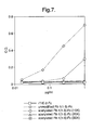
- Figure 7. Binding of unmodified Flt1(1-3)-Fc and step-acetylated Flt1(1-3)-Fc proteins to Matrigel® coated plates. As with the irrelevant control protein, rTie2-Fc, step-acetylated Flt1(1-3)-Fc (20 and 30 fold excess samples) does not exhibit any binding to the Matrigel coated plate, whereas the non-acetylated Flt1(1-3)-Fc protein exhibits significant binding. The 10 fold excess sample shows reduced binding, but the degree of acetylation is not enough to completely block binding to extracellular matrix components.
- Figure 8. Binding of unmodified Flt1(1-3)-Fc and step-acetylated Flt1(1-3)-Fc in a Biacore-based assay. At a sub-stoichiometric ratio (0.5 µg/ml of either unmodified Flt1(1-3) or step-acetylated Flt1(1-3)-Fc vs. 0.2 µg/ml VEGF), there is not enough Flt1(1-3)-Fc (either unmodified or step-acetylated) in the solution to completely bind the VEGF. At 1.0 µg/ml, which approximates a 1:1 stoichiometric ratio, the both unmodified and step-acetylated Flt1(1-3)-Fc are better able to compete for VEGF binding, but there is still insufficient Flt1(1-3)-Fc protein (either unmodified or step-acetylated) to completely saturate the available VEGF. However, at 5.0 µg/ml, which is several times greater than a 1:1 stoichiometric ratio, both the Flt1(1-3)-Fc and the step-acetylated Flt1(1-3)-Fc proteins are able to saturate the VEGF, regardless of the degree of acetylation.
- Figure 9. Pharmacokinetic profiles of unmodified Flt1(1-3)-Fc and step-acetylated Flt1(1-3)-Fc. Balb/c mice (23-28g) were injected subcutaneously with 4mg/kg of unmodified or 10, 20, 40, 60 and 100 fold excess samples of step-acetylated Flt1(1-3)-Fc (3 mice for unmodified, 10, 20 and 40 fold excess samples and 2 mice for 60 and 100 fold excess samples). The mice were tail bled at 1, 2, 4, 6, 24 hours, 2 days and 3 days after injection. The sera were assayed in an ELISA-based assay designed to detect Flt1(1-3)-Fc. The Tmax for all of the Flt1(1-3)-Fc proteins tested was at the 6 hour time point but the Cmax was as follows: Unmodified Flt1(1-3)-Fc: 0.06µg/ml; 10 fold excess sample: - 0.7µg/ml, 20 fold excess sample - 2µg/ml, 40 fold excess sample - 4µg/ml, 60 fold excess sample - 2µg/ml, 100 fold excess sample - 1µg/ml.
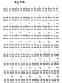
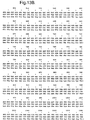
- Figure 10A-10D. Nucleic acid and deduced amino acid sequence of Flt1(1-3)-Fc.
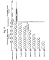
- Figure 11. Schematic diagram of the structure of Flt1.
- Figure 12A and 12B. Hydrophilicity analysis of the amino acid
sequences of
Ig domain 2 andIg domain 3 of Flt1. - Figure 13A-13D. Nucleic acid and deduced amino acid sequence of Mut1: Flt1(1-3ΔB)-Fc.
- Figure 14A-14 C. Nucleic acid and deduced amino acid sequence of Mut2: Flt1(2-3ΔB)-Fc.
- Figure 15A-15C. Nucleic acid and deduced amino acid sequence of Mut3: Flt1(2-3)-Fc.
- Figure 16A-16D. Nucleic acid and deduced amino acid sequence of Mut4: Flt1(1-3R->N)-Fc.
- Figure 17. Binding of unmodified Flt1(1-3)-Fc, basic region deletion mutant Flt1(1-3)-Fc, and Flt1(1-3)R->N mutant proteins in a Biacore-based assay. At the sub-stoichiometric ratio (0.25 µg/ml Flt1(1-3)-Fc of unmodified, acetylated or genetically modified samples vs. 01. µg/ml VEGF), there is insufficient Flt1(1-3)-Fc protein to block binding of VEGF to the Flt1(1-3)-Fc immobilized on the Biacore chip. At 0.5 µg/ml of unmodified, acetylated or genetically modified Flt1(1-3)-Fc proteins, the stoichiometric ratio approximates 1:1 and there is an increased ability to block VEGF binding to the Biacore chip. At 1.0 µg/ml of unmodified, acetylated or genetically modified Flt1(1-3)-Fc proteins, which is approximately a 10:1 stoichiometric ratio, the Flt1(1-3)-Fc proteins are able to block binding of VEGF to the Biacore chip, but they are not equivalent. Unmodified, acetylated, and Mut1: Flt1(1-3ΔB)-Fc are essentially equal in their ability to block VEGF binding, whereas Mut4: Flt1(1-3R->N)-Fc is somewhat less efficient at blocking binding
- Figure 18. Binding of unmodified Flt1(1-3)-Fc, Mut1: Flt1(1-3ΔB)-Fc, Mut2: Flt1(2-3ΔB)-Fc, and Flt1(2-3) mutant proteins to Matrigel® coated plates. Unmodified Flt1(1-3)-Fc protein binds avidly to these wells, the Mut3: Flt1(2-3)-Fc protein binds somewhat more weakly, the Mut1: Flt1(1-3ΔB)-Fc protein binds more weakly still, and the Mut2: Flt1(2-3ΔB)-Fc protein shows the best profile, binding more weakly than any of the other mutant proteins. The Mut4: Flt1(1-3R->N)-Fc glycosylation mutant protein shows only marginal benefit on the Matrigel assay.
- Figure 19. Binding of unmodified Flt1(1-3)-Fc, Mut1: Flt1(1-3ΔB)-Fc, Mut2: Flt1(2-3ΔB)-Fc, and Flt1(2-3) mutant proteins in an ELISA-based assay. At the concentrations tested, unmodified Flt1(1-3)-Fc, Mut1: Flt1(1-3ΔB)-Fc, Mut2: Flt1(2-3ΔB)-Fc, and Flt1(2-3) mutant proteins bind VEGF similarly.
- Figure 20. Pharmacokinetic profiles of unmodified Flt1(1-3)-Fc, Mut1: Flt1(1-3ΔB)-Fc, Mut2: Flt1(2-3ΔB)-Fc, and Flt1(2-3) mutant proteins. the Cmax for these reagents was as follows: Unmodified Flt1(1-3)-Fc - 0.15µg/ml; 40 fold molar excess acetylated Flt1(1-3)-Fc - 1.5µg/ml; and Mut1: Flt1(1-3ΔB)-Fc - 0.7µg/ml.
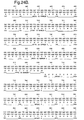
- Figure 21A-21C. Nucleotide and deduced amino acid sequence of the modified Flt1 receptor termed Flt1D2.Flk1D3.FcΔC1(a).
- Figure 22A-22C. Nucleotide and deduced amino acid sequence of the modified Flt1 receptor termed Flt1D2.VEGFR3D3.FcΔC1(a).
- Figure 23. Extracellular Matrix (ECM) Assay. The results of this assay demonstrate that the Flt1D2.Flk1D3.FcΔC1(a) and Flt1D2.VEGFR3D3.FcΔC1(a) proteins are considerably less sticky to the ECM as compared to the Flt1(1-3)-Fc protein.
- Figure 24A-24C. Nucleotide and deduced amino acid sequence of the modified Flt1 receptor termed VEGFR1R2-FcΔC1(a).
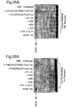
- Figure 25A-25C. Phosphorylation assay. At a 1.5 molar excess of either Flt1(1-3)-Fc , Flt1(1-3)-Fc (A40) or transient Flt1D2Flk1D3.FcΔC1(a) there is complete blockage of receptor stimulation by these three modified Flt1 receptors as compared to control media challenge. In contrast, transient Flt1D2VEGFR3D3.FcΔC1(a) does not show significant blockage at this molar excess, as compared with VEGF positive control challenge. Similar results are seen in Figure 25B, where the modified Flt receptors are in a 3-fold molar excess to VEGF165 ligand. In Figure 25C, where the modified Flt1 receptors are in a 6-fold molar excess to VEGF165 ligand, transient Flt1D2VEGFR3D3.FcΔC1(a) can now be shown to be partially blocking VEGF165-induced stimulation of cell-surface receptors.
- Figure 26A-26B. Phosphorylation assay. Detection by Western blot of tyrosine phosphorylated VEGFR2(Flk1) by VEGF165 ligand stimulation shows that cell-surface receptors are not phosphorylated by challenge samples which have VEGF165 preincubated with 1 and 2 fold molar excess (Figure 26A) or 3 and 4 fold molar excess (Figure 26B) of either transient Flt1D2Flk1D3.FcΔC1(a), stable Flt1D2Flk1D3.FcΔC1(a), or transient VEGFR1R2-FcΔC1(a). At all modified Flt1 receptor concentrations tested there is complete binding of VEGF165 ligand during the preincubation, resulting in no detectable stimulation of cell-surface receptors by unbound VEGF165 as compared to control media challenge.
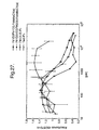
- Figure 27. MG/R2 Cell proliferation assay. The following modified Flt receptors Flt1(1-3)-Fc, Flt1D2.Flk1D3.FcΔC1(a) and Flt1D2.VEGFR3D3.FcΔC1(a), plus an irrelevant receptor termed Tie2-Fc as a negative control, were titrated from 40nM to 20pM and incubated on the cells for 1hr at 37°C. Human recombinant VEGF165 in defined media was then added to all the wells at a concentration of 1.56nM. The negative control receptor Tie2-Fc does not block VEGF165-induced cell proliferation at any concentration whereas Flt1D2.Flk1D3.FcΔC1(a) blocks 1.56nM VEGF165 with a half maximal dose of 0.8nM. Flt1(1-3)-Fc and Flt1D2.VEGFR3D3.FcΔC1(a) are less effective in blocking VEGF165 in this assay with a half maximal dose of - 2nM. VEGF165 alone gives a reading of 1.2 absorbance units and the background is 0.38 absorbance units.
- Figure 28. Biacore analysis of Binding Stoichiometry. Binding stoichiometry was calculated as a molar ratio of bound VEGF165 to the immobilized Flt1D2Flk1D3.FcΔC1(a) or VEGFR1R2-FcΔC1(a), using the conversion factor of 1000 RU equivalent to 1 ng/ml. The results indicated binding stoichiometry of one VEGF165 dimeric molecule per one Flt1D2Flk1D3.FcΔC1(a) or VEGFR1R2-FcΔC1(a) molecule.
- Figure 29 and Figure 30. Size Exclusion Chromatography Stoichiometry. Flt1D2Flk1D3.FcΔC1(a) or VEGFR1R2-FcΔC1(a) at a concentration of 1 nM (estimated to be 1000 times higher than the KD of the Flt1D2Flk1D3.FcΔC1(a) or VEGFR1R2-FcΔC1(a)/VEGF165 interaction) were mixed with varied concentrations of VEGF165. After incubation, concentrations of the free Flt1D2Flk1D3.FcΔC1(a) in solution were measured. The data shows that the addition of 1 nM VEGF165 into the Flt1D2Flk1D3.FcΔC1(a) solution completely blocks Flt1D2Flk1D3.FcΔC1(a) binding to the VEGF165 surface. This result suggested the binding stoichiometry of one VEGF165 molecule per one Flt1D2Flk1D3.FcΔC1(a) molecule.
- Figure 31. Size Exclusion Chromatography (SEC) under native
conditions.
Peak # 1 represents the Flt1D2Flk1D3.FcΔC1(a)/ VEGF165 complex andpeak # 2 represents unbound VEGF165. Fractions eluted between 1.1 and 1.2 ml were combined and guanidinium hydrochloride (GuHCl)was added to a final concentration 4.5M to dissociate the complex. - Figure 32. Size Exclusion Chromatography (SEC) under dissociative
conditions. To separate the components of the receptor-ligand complex
and to determine their molar ratio, 50µl of dissociated complex was
loaded onto a Superose 12 PC 3.2/30 equilibrated in 6M GuHCl and
eluted.
Peak # 1 represents Flt1D2Flk1D3.FcΔC1(a) andpeak # 2 represents VEGF165. - Figure 33, Figure 34 and Figure 35. Size Exclusion
Chromatography (SEC) with On-Line Light Scattering. Size exclusion
chromatography column with a MiniDawn on-line light scattering
detector (Wyatt Technology, Santa Barbara, California) and refractive
index (RI) detectors (Shimadzu, Kyoto, Japan) was used to determine
the molecular weight (MW) of the receptor-ligand complex. As shown in
Figure 33, the elution profile shows two peaks.
Peak # 1 represents the receptor-ligand complex andpeak # 2 represents the unbound VEGF165. MW was calculated from LS and RI signals. The same procedure was used to determine MW of the individual components of the receptor-ligand complex. The results of these determinations are as follows: MW of the Flt1D2Flk1D3.FcΔC1(a)/VEGF165 complex at the peak position is 157 300 (Figure 33), the MW of VEGF165 at the peak position is 44 390 (Figure 34) and the MW of R1 R2 at the peak is 113 300 (Figure 35). - Figure 36. Peptide mapping and glycosylation analysis. The disulfide structures and glycosylation sites in Flt1D2.Flk1D3.FcΔC1(a) were determined by a peptide mapping method. There are a total of ten cysteines in Flt1D2.Flk1D3.FcΔC1(a); six of them belong to the Fc region. Cys27 is disulfide bonded to Cys76. Cys121 is disulfide bonded to Cys 182. The first two cysteines in the Fc region (Cys211 and Cys214) form an intermolecular disulfide bond with the same two cysteines in another Fc chain. However, it can not be determined whether disulfide bonding is occurring between same cysteines (Cys211 to Cys211, for example) or between Cys211 and Cys214. Cys216 is disulfide bonded to Cys306. Cys 352 is disulfide bonded to Cys410. There are five possible N-linked glycosylation sites in Flt1D2.Flk1D3.FcΔC1(a) and are found to be glycosylated to varying degrees. Complete glycosylation is observed at Asn33, Asn193, and Asn282. Partial glycosylation is observed on Asn65 and Asn120. Sites of glycosylation are highlighted by underline in the Figure.
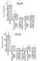
- Figure 37. Pharmacokinetics of Flt1(1-3)-Fc (A40), Flt1D2.Flk1D3.FcΔC1(a) and VEGFR1R2-FcΔC1(a). Balb/c mice were injected subcutaneously with 4mg/kg of Flt1(1-3)-Fc (A40), CHO transiently expressed Flt1D2.Flk1D3.FcΔC1(a), CHO stably expressed Flt1D2.Flk1D3.FcΔC1(a), and CHO transiently expressed VEGFR1 R2-FcΔC1(a). The mice were tail bled at 1, 2, 4, 6, 24 hrs, 2 days, 3 days and 6 days after injection. The sera were assayed in an ELISA designed to detect Flt1(1-3)-Fc (A40), Flt1D2.Flk1D3.FcΔC1(a) or VEGFR1 R2-FcΔC1(a). The Tmax for Flt1(1-3)-Fc (A40) was at 6 hrs while the Tmax for the transient and stable Flt1D2.Flk1D3.FcΔC1(a) and the transient VEGFR1R2-FcΔC1(a) was 24hrs. The Cmax for Flt1(1-3)-Fc (A40) was 8µg/ml, For both transients (Flt1D2.Flk1D3.FcΔC1(a) and VEGFR1R2-FcΔC1(a)) the Cmax was 18µg/ml and the Cmax for the stable VEGFR1R2-FcΔC1(a) was 30µg/ml.
- Figure 38. Pharmacokinetics of Flt1(1-3)-Fc (A40),
Flt1 D2.Flk1D3.FcΔC1(a) and Flt1D2.VEGFR3D3.FcΔC1(a). Balb/c mice
were injected subcutaneously with 4mg/kg of Flt1(1-3)-Fc (A40), CHO
transiently expressed Flt1D2.Flk1D3.FcΔC1(a) and CHO transiently
expressed Flt1D2.VEGFR3D3.FcΔC1(a). The mice were tail bled at 1, 2,
5, 6, 7, 8, 12, 15 and 20 days after injection. The sera were assayed in
an ELISA designed to detect Flt1(1-3)-Fc, Flt1D2.Flk1D3.FcΔC1(a) and
Flt1D2.VEGFR3D3.FcΔC1(a). Flt1(1-3)-Fc (A40) could no longer be
detected in the serum after
day 5 whereas Flt1D2.Flk1D3.FcΔC1(a) and Flt1 D2.VEGFR3D3.FcΔC1(a) were detectable for 15 days or more. - Figure 39. The Ability of Flt1D2.Flk1D3.FcΔC1(a) to Inhibit HT-1080 Fibrosarcoma Tumor Growth In Vivo. Every other day or 2 times per week treatment of SCID mice with Flt1D2.Flk1D3.FcΔC1(a) at 25mg/Kg significantly decreases the growth of subcutaneous HT-1080 fibrosarcoma tumors.
- Figure 40. The Ability of Flt1D2.Flk1D3.FcΔC1(a) to Inhibit C6 Glioma Tumor Growth In Vivo. Every other day or 2 times a week treatment of SCID mice with Flt1D2.Flk1D3.FcΔC1(a) significantly decreases the growth of subcutaneous C6 glioma tumors at doses as low as 2.5mg/Kg.
- Figure 41. VEGF-Induced Uterine Hyperpermeability. PMSG injected subcutaneously (5 IU) to induce ovulation in prepubertal female rats results in a surge of estradiol after 2 days which in turn causes an induction of VEGF in the uterus. This induction results in hyperpermeability of the uterus and an increase in uterine wet. Subcutaneous injection of Flt1(1-3)-Fc (A40), Flt1D2.Flk1D3.FcΔC1(a) and Flt1D2.VEGFR3D3.FcΔC1(a) at 25mg/kg at 1 hr after PMSG injection results in about a 50% inhibition of the increase in uterine wet weight.
- Figure 42A-42B. Assessment of Corpus Luteum Angiogenesis Using
Progesterone as a Readout. PMSG was injected subcutaneously (5 IU) to
induce ovulation in prepubertal female rats, resulting in a fully
functioning corpus luteum containing a dense network of blood vessels
that secretes progesterone into the blood stream to prepare the uterus
for implantation. The induction of angiogenesis in the corpus luteum
requires VEGF. Resting levels of progesterone are about 5ng/ml and can
be induced to 25-40ng/ml after PMSG. Subcutaneous injection of
Flt1(1-3)-Fc (A40) or Flt1D2.Flk1D3.FcΔC1(a) at 25mg/kg or 5mg/kg at
1 hr. after PMSG injection resulted in a complete inhibition of the
progesterone induction on
day 4. -
- It has been a long standing problem in the art to produce a receptor based VEGF antagonist that has a pharmacokinetic profile that is appropriate for consideration of the antagonist as a therapeutic candidate. Applicants describe herein, for the first time, a chimeric polypeptide molecule, capable of antagonizing VEGF activity, that exhibits improved pharmacokinetic properties as compared to other known receptor-based VEGF antagonists. The chimeric polypeptide molecules described herein thus provide for the first time appropriate molecules for use in therapies in which antagonism of VEGF is a desired result.
- The present invention provides for novel chimeric polypeptide molecules formed by fusing a modified extracellular ligand binding domain of the Flt1 receptor to the Fc region of IgG.
- The extracellular ligand binding domain is defined as the portion of a receptor that, in its native conformation in the cell membrane, is oriented extracellularly where it can contact with its cognate ligand. The extracellular ligand binding domain does not include the hydrophobic amino acids associated with the receptor's transmembrane domain or any amino acids associated with the receptor's intracellular domain. Generally, the intracellular or cytoplasmic domain of a receptor is usually composed of positively charged or polar amino acids (i.e. lysine, arginine, histidine, glutamic acid, aspartic acid). The preceding 15-30, predominantly hydrophobic or apolar amino acids (i.e. leucine, valine, isoleucine, and phenylalanine) comprise the transmembrane domain. The extracellular domain comprises the amino acids that precede the hydrophobic transmembrane stretch of amino acids. Usually the transmembrane domain is flanked by positively charged or polar amino acids such as lysine or arginine. von Heijne has published detailed rules that are commonly referred to by skilled artisans when determining which amino acids of a given receptor belong to the extracellular, transmembrane, or intracellular domains (See von Heijne, 1995, BioEssays 17:25-30). Alternatively, websites on the Internet, such as
http://ulrec3.unil.ch/software/TMPRED_form.html. have become available to provide protein chemists with information about making predictions about protein domains. - The present invention provides for the construction of nucleic acid molecules encoding chimeric polypeptide molecules that are inserted into a vector that is able to express the chimeric polypeptide molecules when introduced into an appropriate host cell. Appropriate host cells include, but are not limited to, bacterial cells, yeast cells, insect cells, and mammalian cells. Any of the methods known to one skilled in the art for the insertion of DNA fragments into a vector may be used to construct expression vectors encoding the chimeric polypeptide molecules under control of transcriptional/translational control signals. These methods may include in vitro recombinant DNA and synthetic techniques and in vivo recombinations (genetic recombination) (See Sambrook, et al., Molecular Cloning, A Laboratory Manual, Cold Spring Harbor Laboratory; Current Protocols in Molecular Biology, Eds. Ausubel, et al., Greene Publ. Assoc., Wiley-Interscience, NY).
- Expression of nucleic acid molecules encoding the chimeric polypeptide molecules may be regulated by a second nucleic acid sequence so that the chimeric polypeptide molecule is expressed in a host transformed with the recombinant DNA molecule. For example, expression of the chimeric polypeptide molecules described herein may be controlled by any promoter/enhancer element known in the art. Promoters which may be used to control expression of the chimeric polypeptide molecules include, but are not limited to, the long terminal repeat as described in Squinto et al., (1991, Cell 65:1-20); the SV40 early promoter region (Bernoist and Chambon, 1981, Nature 290:304-310), the CMV promoter, the M-MuLV 5' terminal repeat the promoter contained in the 3' long terminal repeat of Rous sarcoma virus (Yamamoto, et al., 1980, Cell 22:787-797), the herpes thymidine kinase promoter (Wagner et al., 1981, Proc. Natl. Acad. Sci. U.S.A. 78:144-1445), the regulatory sequences of the metallothionine gene (Brinster et al., 1982, Nature 296:39-42); prokaryotic expression vectors such as the β-lactamase promoter (Villa-Kamaroff, et al., 1978, Proc. Natl. Acad. Sci. U.S.A. 75:3727-3731), or the tac promoter (DeBoer, et al., 1983, Proc. Natl. Acad. Sci. U.S.A. 80:21-25, see also "Useful proteins from recombinant bacteria" in Scientific American, 1980, 242:74-94); promoter elements from yeast or other fungi such as the Gal 4 promoter, the ADH (alcohol dehydrogenase) promoter, PGK (phosphoglycerol kinase) promoter, alkaline phosphatase promoter, and the following animal transcriptional control regions, which exhibit tissue specificity and have been utilized in transgenic animals: elastase I gene control region which is active in pancreatic acinar cells (Swift et al., 1984, Cell 38:639-646; Ornitz et al., 1986, Cold Spring Harbor Symp. Quant. Biol. 50:399-409; MacDonald, 1987, Hepatology 7:425-515); insulin gene control region which is active in pancreatic beta cells (Hanahan, 1985, Nature 315:115-122), immunoglobulin gene control region which is active in lymphoid cells (Grosschedl et al., 1984, Cell 38:647-658; Adames et al., 1985, Nature 318:533-538; Alexander et al., 1987, Mol. Cell. Biol. 7:1436-1444), mouse mammary tumor virus control region which is active in testicular, breast, lymphoid and mast cells (Leder et al., 1986, Cell 45:485-495), albumin gene control region which is active in liver (Pinkert et al., 1987, Genes and Devel. 1:268-276), alpha-fetoprotein gene control region which is active in liver (Krumlauf et al., 1985, Mol. Cell. Biol. 5:1639-1648; Hammer et al., 1987, Science 235:53-58); alpha 1-antitrypsin gene control region which is active in the liver (Kelsey et al, 1987, Genes and Devel. 1:161-171), beta-globin gene control region which is active in myeloid cells (Mogram et al., 1985, Nature 315:338-340; Kollias et al., 1986, Cell 46:89-94); myelin basic protein gene control region which is active in oligodendrocyte cells in the brain (Readhead et al., 1987, Cell 48:703-712); myosin light chain-2 gene control region which is active in skeletal muscle (Shani, 1985, Nature 314:283-286), and gonadotropic releasing hormone gene control region which is active in the hypothalamus (Mason et al., 1986, Science 234:1372-1378).
- Thus, according to the invention, expression vectors capable of being replicated in a bacterial or eukaryotic host comprising chimeric polypeptide molecule-encoding nucleic acid as described herein, are used to transfect the host and thereby direct expression of such nucleic acids to produce the chimeric polypeptide molecules, which may then be recovered in a biologically active form. As used herein, a biologically active form includes a form capable of binding to VEGF.
- Expression vectors containing the chimeric nucleic acid molecules described herein can be identified by three general approaches: (a) DNA-DNA hybridization, (b) presence or absence of "marker" gene functions, and (c) expression of inserted sequences. In the first approach, the presence of a foreign gene inserted in an expression vector can be detected by DNA-DNA hybridization using probes comprising sequences that are homologous to the inserted chimeric polypeptide molecule sequences. In the second approach, the recombinant vector/host system can be identified and selected based upon the presence or absence of certain "marker" gene functions (e.g., thymidine kinase activity, resistance to antibiotics, transformation phenotype, occlusion body formation in baculovirus, etc.) caused by the insertion of foreign genes in the vector. For example, if the chimeric polypeptide molecule DNA sequence is inserted within the marker gene sequence of the vector, recombinants containing the insert can be identified by the absence of the marker gene function. In the third approach, recombinant expression vectors can be identified by assaying the foreign gene product expressed by the recombinant. Such assays can be based, for example, on the physical or functional properties of the chimeric polypeptide molecules.
- Cells of the present invention may transiently or, preferably, constitutively and permanently express the chimeric polypeptide molecules.
- The chimeric polypeptide molecules may be purified by any technique which allows for the subsequent formation of a stable, biologically active chimeric polypeptide molecule. For example, and not by way of limitation, the factors may be recovered from cells either as soluble proteins or as inclusion bodies, from which they may be extracted quantitatively by 8M guanidinium hydrochloride and dialysis (see, for example, Builder, et al., US Patent No. 5,663,304). In order to further purify the factors, conventional ion exchange chromatography, hydrophobic interaction chromatography, reverse phase chromatography or gel filtration may be used.
- In one embodiment of the invention, the nucleotide sequence encoding the first component is upstream of the nucleotide sequence encoding the second component. In another embodiment of the invention, the nucleotide sequence encoding the first component is downstream of the nucleotide sequence encoding the second component. Further embodiments of the invention may be prepared in which the order of the first, second and third fusion polypeptide components are rearranged. For example, if the nucleotide sequence encoding the first component is designated 1, the nucleotide sequence encoding the second component is designated 2, and the nucleotide sequence of the third component is designated 3, then the order of the components in the isolated nucleic acid of the invention as read from 5' to 3' may be any of the following six combinations: 1,2,3; 1,3,2; 2,1,3; 2,3,1; 3,1,2; or 3,2,1.
- The present invention also has diagnostic and therapeutic utilities. In particular embodiments of the invention, methods of detecting aberrancies in the function or expression of the chimeric polypeptide molecules described herein may be used in the diagnosis of disorders. In other embodiments, manipulation of the chimeric polypeptide molecules or agonists or antagonists which bind the chimeric polypeptide molecules may be used in the treatment of diseases. In further embodiments, the chimeric polypeptide molecule is utilized as an agent to block the binding of a binding agent to its target.
- By way of example, but not limitation, the method of the invention may be useful in treating clinical conditions that are characterized by vascular permeability, edema or inflammation such as brain edema associated with injury, stroke or tumor; edema associated with inflammatory disorders such as psoriasis or arthritis, including rheumatoid arthritis; asthma; generalized edema associated with burns; ascites and pleural effusion associated with tumors, inflammation or trauma; chronic airway inflammation; capillary leak syndrome; sepsis; kidney disease associated with increased leakage of protein; and eye disorders such as age related macular degeneration and diabetic retinopathy.
- An amino acid sequence analysis of Flt1(1-3)-Fc revealed the presence of an unusually high number (46) of the basic amino acid residue lysine. An IEF analysis of Flt1(1-3)-Fc showed that this protein has pl greater than 9.3, confirming the prediction that the protein is very basic. It was hypothesized that the basic nature of Flt1(1-3)-Fc protein was causing it to bind to extracellular matrix components and that this interaction might be the cause of the extremely short detectable circulating serum half-life exhibited by Flt1(1-3)-Fc when injected into mice. In order to test this hypothesis, Flt1(1-3)-Fc protein was acetylated at the lysine residues to reduce the basic charge. Acetylated Flt1(1-3)-Fc was then tested in the assays described infra.
- The following examples are offered by way of illustration and not by way of limitation.
- Using standard molecular biology techniques (see e.g., Molecular Cloning, A Laboratory Manual (Sambrook, et al., Cold Spring Harbor Laboratory), Current Protocols in Molecular Biology (Eds. Ausubel, et al., Greene Publ. Assoc., Wiley-Interscience, NY), the gene encoding Flt1(1-3)-Fc was inserted into the expression vector pEE14.1 (Lonza Biologics, plc) at a multiple cloning site downstream of the CMV promoter. CHO K1 cells were transfected with the pEE14.1/Flt1(1-3)-Fc DNA construct using lipofectamine (Gaithersburg, MD). The transfected CHO K1 cells were grown in glutamine-free DMEM (JRH, Kansas City, MO) containing 25µM methionine sulfoximine (MSX) from Sigma Inc., St. Louis, MO, and high recombinant protein expressors were obtained by screening the CHO K1 cell supernatants from over 100 hand-picked colony isolates using a standard immunoassay which captures and detects human Fc. The selected hand-picked clone was amplified in the presence of 100 µM MSX followed by a second round of screening of the amplified clones. The highest producing clone had a specific productivity of recombinant Flt1(1-3)-Fc protein of 55 pg/cell/day.
- The selected clone was expanded in 225cm2 T-flasks (Corning, Acton, MA) and then into 8.5L roller bottles (Corning, Acton, MA) using the cell culture media described supra. Cells were removed from the roller bottles by standard trypsinization and put into 3.5L of suspension medium. The suspension medium is comprised of glutamine-free ISCHO medium (Irvine Scientific, Santa Ana, CA) containing 5% fetal bovine serum (FBS from Hyclone Labs, Logan, UT), 100µM MSX and GS supplement (JRH Scientific, Kansas City, MO) in a 5L Celligen bioreactor (New Brunswick Scientific, New Brunswick, NJ) at a density of 0.3 x 10⁶ cells/mL. After the cells reached a density of 3.6 x 10⁶/mL and were adapted to suspension they were transferred to a 60L bioreactor (ABEC, Allentown, PA) at a density of 0.5 x 10⁶ cells/mL in 20L of ISCHO medium with 5% fetal bovine serum. After two days an additional 20L of ISCHO + 5% fetal bovine serum was added to the bioreactor. The cells were allowed to grow for an additional two days reaching a final density of 3.1 x 10⁶ cells/mL, and a final Flt1(1-3)-Fc concentration at harvest was 95 mg/L. At harvest the cells were removed by tangential flow filtration using 0.45µm Prostak Filters (Millipore, Inc., Bedford, MA).
- Flt1(1-3)-Fc protein was initially purified by affinity chromatography. A Protein A column was used to bind, with high specificity, the Fc portion of the molecule. This affinity-purified protein was then concentrated and passed over a SEC column. The protein was then
eluted into the formulation buffer. The following describes these procedures in detail. - All chemicals were obtained from J.T. Baker, Phillipsburg, NJ with the exception of PBS; which was obtained as a 10X concentrate from Life Technologies, Gaithersburg, MD. Protein A Fast Flow and
Superdex 200 preparation grade resins were obtained from Pharmacia, Piscataway, NJ. Equipment and membranes for protein concentration were obtained from Millipore, Bedford, MA. - Approximately 40L of 0.45µm-filtered CHO conditioned media containing Flt1(1-3)-Fc protein was applied to a 290mL Protein A Fast Flow column (10cm diameter) that had been equilibrated with PBS. The column was washed with PBS containing 350mM NaCl and 0.02% CHAPS and the bound protein was eluted with 20mM Citric Acid containing 10mM Na2HPO4. The single peak in the elution was collected and its pH was raised to neutrality with 1 M NaOH. The eluate fractions was concentrated to approximately 9 mg/mL using 10K regenerated cellulose membranes by both tangential flow filtration and by stirred cell concentration. To remove aggregates and other contaminants, the concentrated protein was applied to a column packed with
Superdex 200 preparation grade resin (10cm x 55cm) and run in PBS containing 5 % glycerol. The main peak fractions were pooled, sterile filtered, aliquoted and stored at -80°C. - Two milligrams of Flt1(1-3)-Fc protein were acetylated as described in the instruction manual provided with the sulfo-NHS-acetate modification kit (Pierce Chemical Co., Rockford, IL, Cat.#26777).
- Flt1(1-3)-Fc and acetylated Flt1(1-3)-Fc were analyzed by standard IEF analysis. As shown in Figure 1, Flt1(1-3)-Fc protein is not able to migrate into the gel and therefore must have a pl greater than 9.3, the highest pl in the standard. However, acetylated Flt1(1-3)-Fc is able to migrate into the gel and equilibrate at a pl of approximately 5.2. This result demonstrates that acetylation reduces the net positive charge of the protein and therefore its pl considerably.
- To test for binding to extracellular matrix components, Flt1(1-3)-Fc and acetylated Flt1(1-3)-Fc where tested in an assay designed to mimic the interaction with extracellular matrix components. In this assay, 96-well tissue culture plates are coated with Matrigel (Biocoat MATRIGEL® matrix thin layer 96 well plate, Catalog #40607, Becton Dickinson Labware, Bedford, MA). The plates are incubated with varying concentrations of either Flt1(1-3)-Fc, acetylated Flt1(1-3)-Fc, or rTie2-Fc (an irrelevant control) protein are added to the wells. The plates are incubated for 1-2 hours at either room temperature or 37°C degrees and then detection of bound proteins is accomplished by adding a secondary alkaline phosphatase-conjugated anti-human Fc antibody to the wells. Finally, alkaline phosphatase substrate is added to the wells and optical density is measured. Figure 2 shows the results of this assay. Like the irrelevant control protein rTie2-Fc, acetylated Flt1(1-3)-Fc does not exhibit any binding to the Matrigel coated plate, whereas the non-acetylated Flt1(1-3)-Fc protein exhibits significant binding. This result indicates that acetylation of basic amino acid residues is an effective way to interfere with the charge interactions that exist between positively charged proteins and the negatively charged extracellular matrix components they are exposed to in vivo.
- Although pegylation (polyethylene glycol - PEG) of proteins has been shown to increase their in vivo potency by enhancing stability and bioavailability while minimizing immunogenicity (see references cited supra), it is counter-intuitive that pegylating molecules that are too large to be filtered by the kidney glomeruli would improve their pharmacokinetic properties. Without being bound by theory, Applicants postulated that pegylation of the Flt1(1-3)-Fc molecules could improve the pharmacokinetic properties, possibly not by altering the positive charge or by decreasing the pl of Flt1(1-3)-Fc, but rather by physically shielding the positive charges from interacting with the extracellular matrix. Applicants decided to attempt to improve the pharmacokinetic properties of Flt1(1-3)-Fc molecules by attaching strands of 20K PEGs as described infra.
- Purified Flt1(1-3)-Fc derived from CHO cells (see supra) was used in the following pegylation experiments. Functionalized PEGs were obtained from Shearwater Polymers, Huntsville, AL; Bicine from Sigma, St Louis, MO;
Superose 6 column from Pharmacia, Piscataway, NJ; PBS as a 10X concentrate from Life Technologies, Gaithersburg, MD; Glycerol from J.T. Baker, Phillipsburg, NJ; and Bis-Tris precast gels from Novex, CA. - 20K PEG strands functionalized with amine-specific terminal moieties were used in small-scale reaction studies that were set-up to evaluate different reaction conditions in which the PEG:protein stoichiometry was varied. Based on these reactions and the analyses of samples on standard SDS-PAGE, Flt1(1-3)-Fc at a concentration of 1.5 mg/mL was reacted at pH 8.1 with 20K SPA-PEG (PEG succinimidyl propionate) molecules at a PEG-to-Flt1(1-3)-Fc monomer molar ratio of 1:6. The reaction was allowed to proceed at 8°C overnight. For initial purification, the reaction products were applied to a 10mm
x 30cm Superose 6 column equilibrated with PBS containing 5% Glycerol. The column appeared to separate pegylated Flt1(1-3)-Fc molecules based on the extent of pegylation. Fractions corresponding to what appeared to be primarily mono-pegylated and di-pegylated dimeric Flt1(1-3)-Fc, as judged by banding patterns on reducing and non-reducing SDS-PAGE gels were pooled. The protein concentration was determined by measuring absorbance at 280 nm. The pegylated Flt1(1-3)-Fc protein was sterile filtered, aliquoted and stored at -40°C. - Unmodified, acetylated, and pegylated Flt1(1-3)-Fc proteins were tested in a Biacore-based assay to evaluate their ability to bind to the Flt1 ligand, VEGF. In this assay, unmodified Flt1(1-3)-Fc protein was immobilized on the surface of a Biacore chip (see Biacore Instruction Manual, Pharmacia, Inc., Piscataway, NJ, for standard procedures) and a sample containing 0.2 µg/ml VEGF and either unmodified Flt1(1-3)-Fc, acetylated Flt1(1-3)-Fc or pegylated Flt1(1-3)-Fc (each at 25 µg/ml) was passed over the Flt1(1-3)-Fc-coated chip. To minimize the effects of non-specific binding, the bound samples were washed with a 0.5M NaCl wash. In one sample, unmodified Flt1(1-3)-Fc was mixed with heparin. Heparin is a negatively charged molecule and the Flt1(1-3)-Fc protein is a positively charged molecule, so when the two molecules are mixed together, they should interact through their respective charges. This essentially neutralizes Flt1(1-3)-Fc's inherent positive charge making the molecule behave as if it has been chemically or genetically modified so as to reduce its charge and its tendency to bind via charge interactions. As shown in Figure 3, acetylated (columns 13-16), pegylated (columns 17-20), and heparin-treated Flt1(1-3)-Fc (columns 21-24) are each able to completely compete with the Biacore chip-bound Flt1(1-3)-Fc for VEGF binding as compared to control (columns 1-4) and irrelevant protein (columns 5-8). Unmodified Flt1(1-3)-Fc (columns 5-6) appeared to only partially compete with Biacore chip-bound Flt1(1-3)-Fc for VEGF binding. However, washing the bound samples with 0.5M NaCl (columns 7-8) resulted in a binding profile similar to the modified forms of Flt1(1-3)-Fc, indicating that the unmodified protein was exhibiting non-specific binding to the chip that could be eliminated by the salt wash.
- Unmodified, acetylated, and pegylated Flt1(1-3)-Fc proteins were tested in a standard ELISA-based assay to evaluate their ability to bind the Flt1 receptor ligand VEGF. As shown in Figure 4, both pegylated and acetylated Flt1(1-3)-Fc proteins are capable of binding to VEGF, demonstrating that modifying the protein either by pegylation or acetylation does not destroy its ability to bind its ligand.
- In vivo experiments were designed to assess the pharmacokinetic profiles of unmodified Flt1(1-3)-Fc, acetylated Flt1(1-3)-Fc, and pegylated Flt1(1-3)-Fc protein. Balb/c mice (23-28g; 3 mice/group) were injected subcutaneously with 4mg/kg of unmodified, acetylated, or pegylated Flt1(1-3)-Fc. The mice were tail bled at 1, 2, 4, 6, 24 hours, 2 days, and 3 days after injection of protein. The sera were assayed in a standard ELISA-based assay designed to detect Flt1(1-3)-Fc protein. Briefly, the assay involves coating an ELISA plate with VEGF, binding the unmodified, acetylated, or pegylated Flt1(1-3)-Fc-containing sera, and reporting with an anti-Fc antibody linked to alkaline phosphatase. As shown in Figure 5, the Tmax for all of the Flt1(1-3)-Fc proteins was between the 6 hour and 24 hour time points. The Cmax for the different proteins was as follows: Unmodified: 0.06 µ/ml - 0.15 µg/ml; acetylated: 1.5 µg/ml - 4.0 µg/ml; and pegylated: approximately 5 µg/ml.
- To determine what minimal amount of acetylation is necessary to eliminate binding to extracellular matrix components, an experiment was designed that acetylated the Flt1(1-3)-Fc protein in a step-wise fashion by using increasing amounts of molar excess of acetylation reagent in the acetylation reaction mixture. The range of molar excess was as follows: 0, 10, 20, 30, 40, 50, 60, 70, 80 , 90, and 100 moles of acetylation reagent per 1 mole of Flt1(1-3)-Fc monomer. The reactions were performed as detailed in the instruction manual provided with the sulfo-NHS-Acetate modification kit (Pierce Chemical Co., Rockford, IL, Cat.# 26777).
- Unmodified Flt1(1-3)-Fc and step-acetylated Flt1(1-3)-Fc proteins were analyzed by standard IEF analysis. As shown in Figure 6A-6B, unmodified Flt1(1-3)-Fc protein was not able to migrate into the gel due to its extremely high pl (greater than 9.3). However, most of the step-acetylated Flt1(1-3)-Fc samples (30-100 fold molar excess samples) were able to migrate into the gel and equilibrate at pls ranging between 4.55 - 8.43, depending on the degree of acetylation of the protein. This result demonstrates that acetylation can change the positive charge of the protein in a dose-dependent manner and that reduction of the pl can be controlled by controlling the degree of acetylation.
- To test for binding to extracellular matrix components, Flt1(1-3)-Fc and step-acetylated Flt1(1-3)-Fc where tested in the above-described assay designed to mimic the interaction with extracellular matrix components. Varying concentrations of either unmodified Flt1(1-3)-Fc, step-acetylated Flt1(1-3)-Fc (10, 20, and 30 fold molar excess samples), or rTie2-Fc (an irrelevant control) protein were added to the wells. The plates were incubated for 1-2 hours at room temperature or 37°C and then detection of bound proteins was accomplished by adding a secondary alkaline phosphatase-conjugated anti-human Fc antibody to the wells. Alkaline phosphatase substrate was subsequently added to the wells and optical density measured. Figure 7 shows the results of this assay. Like the irrelevant control protein rTie2-Fc, step-acetylated Flt1(1-3)-Fc (20 and 30 fold molar excess samples) did not exhibit any significant binding to the Matrigel coated plate, whereas the non-acetylated Flt1(1-3)-Fc protein exhibited significant binding. The binding is saturable, indicating that the Flt1(1-3)-Fc protein may be binding to specific sites, rather than a more general charge-mediated interaction that might not be saturable. The 10 fold molar excess sample showed reduced binding, but the degree of acetylation was not enough to completely block binding to extracellular matrix components. The 20 fold molar excess and higher samples displayed no detectable binding, despite the fact that by IEF analysis (Figure 6A and 6B) the lower molar excess samples still had a large net positive charge. This result demonstrates that it is not necessary to completely acetylate all available basic amino acids in order to eliminate binding to extracellular matrix components.
- Unmodified and step-acetylated Flt1(1-3)-Fc proteins where tested in a Biacore-based assay to evaluate their ability to bind to the Flt1 ligand, VEGF. In this assay, unmodified Flt1(1-3)-Fc protein (0.5, 1.0, or 5.0 µg/ml) was immobilized on the surface of a Biacore chip (see Biacore Instruction Manual, Pharmacia, Inc., Piscataway, NJ, for standard procedures) and a solution containing 0.2 µg/ml VEGF and either unmodified Flt1(1-3)-Fc (at either 0.5, 1.0, or 5.0 µg/ml) or 10 different step-acetylated Flt1(1-3)-Fc samples (at 0.5, 1.0, or 5.0 µg/ml each) were passed over the Flt1(1-3)-Fc-coated chip. As shown in Figure 8, at a sub-stoichiometric ratio (0.5 µg/ml of either unmodified Flt1(1-3) or step-acetylated Flt1(1-3)-Fc vs. 0.2 µg/ml VEGF), there is not enough Flt1(1-3)-Fc (either unmodified or step-acetylated) in the solution to completely bind the VEGF. At 1.0 µg/ml, which approximates a 1:1 stoichiometric ratio, both unmodified and step-acetylated Flt1(1-3)-Fc are better able to compete for VEGF binding, but there is still insufficient Flt1(1-3)-Fc protein (either unmodified or step-acetylated) to completely bind the available VEGF. However, at 5.0 µg/ml, which is several times greater than a 1:1 stoichiometric ratio, both the Flt1(1-3)-Fc and the step-acetylated Flt1(1-3)-Fc proteins are able to bind the VEGF, regardless of the degree of acetylation. This clearly demonstrates that acetylation does not alter Flt1(1-3)-Fc's ability to bind VEGF.
- In vivo experiments were designed to assess the pharmacokinetic profiles of unmodified Flt1(1-3)-Fc and step-acetylated Flt1(1-3)-Fc protein. Balb/c mice (23-28g) were injected subcutaneously with 4mg/kg of unmodified or 10, 20, 40, 60 and 100 fold molar excess samples of step-acetylated Flt1(1-3)-Fc (3 mice for unmodified, 10, 20 and 40 fold molar excess samples and 2 mice for 60 and 100 fold molar excess samples). The mice were tail bled at 1, 2, 4, 6, 24 hours, 2 days and 3 days after injection. The sera were assayed in an ELISA-based assay designed to detect Flt1(1-3)-Fc (described supra). Figure 9 details the results of this study. The Tmax for all of the Flt1(1-3)-Fc proteins tested was at the 6 hour time point but the Cmax was as follows: Unmodified Flt1(1-3)-Fc: 0.06µg/ml; 10 fold molar excess sample: - 0.7µg/ml, 20 fold molar excess sample - 2µg/ml, 40 fold molar excess sample - 4µg/ml, 60 fold molar excess sample - 2µg/ml, 100 fold molar excess sample - 1µg/ml. This results demonstrates that acetylation or pegylation of Flt1(1-3)-Fc significantly improves its pharmacokinetic profile.
- Based on the observation that acetylated Flt1(1-3)-Fc, which has a pl below 6, has much better pharmacokinetics than the highly positive unmodified Flt1(1-3)-Fc (pl > 9.3), it was asked whether the difference in pharmacokinetics could be attributed to the net charge of the protein, which made it stick to negatively charged extracellular matrix components, or whether there were perhaps specific locations on the surface of the Flt1(1-3)-Fc protein that constituted specific binding sites for extracellular matrix components. For example, many proteins are known to have heparin binding sites, often consisting of a cluster of basic residues. Sometimes these residues are found in a cluster on the primary sequence of the protein; some of the literature has identified "consensus sequences" for such heparin binding sites (see for example Hileman, et al., 1998, Bioessays 20(2):156-67). In other cases, the known crystal structure of a protein reveals a cluster of positively charged residues on the surface of a protein, but the residues come from different regions of the primary sequence and are only brought together when the protein folds into its tertiary structure. Thus it is difficult to deduce whether an isolated amino acid residue forms part of a cluster of basic residues on the surface of the protein. However, if there is a cluster of positively charged amino acid residues in the primary sequence, it is not unreasonable to surmise that the residues are spatially close to one another and might therefore be part of an extracellular matrix component binding site. Flt1 receptor has been studied extensively and various domains have been described (see for example Tanaka et al., 1997, Jpn. J. Cancer Res 88:867-876). Referring to the nucleic acid and amino acid sequence set forth in Figure 10A-10D of this application, one can identify the signal sequence for secretion which is located at the beginning of the sequence and extends to the glycine coded for by nucleotides 76-78. The mature protein begins with Ser-Lys-Leu-Lys, starting at nucleotide 79 of the nucleic acid sequence.
Flt1 Ig domain 1 extends from nucleotide 79 to 393, ending with the amino acids Ser-Asp-Thr.Flt1 Ig domain 2 extends from nucleotide 394 to 687 (encoding Gly-Arg-Pro to Asn-Thr-Ile), andFlt1 Ig domain 3 extends from nucleotides 688 to 996 (encoding Ile-Asp-Val to Asp-Lys-Ala). There is a bridging amino acid sequence, Gly-Pro-Gly, encoded by nucleotides 997-1005, followed by the nucleotide sequence encoding human Fc (nucleotides 1006-1701 or amino acids Glu-Pro-Lys to Pro-Gly-Lys-stop). - A more detailed analysis of the Flt1 amino acid sequence reveals that there is a cluster, namely, amino acid residues 272-281 (KNKRASVRR) of Figure 10A-10D, in which 6 out of 10 amino acid residues are basic. This sequence is located in
Flt1 Ig domain 3 of the receptor (see Figure 11), which is not itself essential for binding of VEGF ligand, but which confers a higher affinity binding to ligand. An alignment of the sequence ofIg domain 3 with that ofIg domain 2 reveals that in this region, there is very poor alignment between the two Ig domains, and that there are about 10 additional amino acids inIg domain 3. An analysis of the hydrophilicity profiles (MacVector computer software) of these two domains clearly indicates the presence of a hydrophilic region in the protein (Figure 12A-12B). These observations raised the possibility that the actual three dimensional conformation ofFlt1 Ig domain 3 allowed for some type of protrusion that is not inFlt1 Ig domain 2. To test this hypothesis, the 10 additional amino acids were deleted and the resulting protein was tested to see whether the deletion would affect the pharmacokinetics favorably without seriously compromising the affinity of the receptor for VEGF. This DNA construct, which was constructed using standard molecular biology techniques (see e.g., Molecular Cloning, A Laboratory Manual (Sambrook, et al., Cold Spring Harbor Laboratory), Current Protocols in Molecular Biology (Eds. Ausubel, et al., Greene Publ. Assoc., Wiley-Interscience, NY) in the mammalian expression vector pMT21 (Genetics Institute, Inc., Cambridge, MA), is referred to as Mut1: Flt1(1-3ΔB)-Fc. The Mut1: Flt1(1-3ΔB)-Fc construct was derived from Flt1(1-3)-Fc by deletion of nucleotides 814-843 (set forth in Figure 10A-10D), which deletes the highly basic 10-amino acid residue sequence Lys-Asn-Lys-Arg-Ala-Ser-Val-Arg-Arg-Arg fromFlt1 Ig domain 3. - The final DNA construct was sequence-verified using an ABI 373A DNA sequencer and Taq Dideoxy Terminator Cycle Sequencing Kit (Applied Biosystems, Inc., Foster City, CA). The sequence of Mut1: Flt1(1-3ΔB)-Fc is set forth in Figure 13A-13D.
- A second deletion mutant construct, designated Mut2: Flt1(2-3ΔB)-Fc, was derived from the Mut1: Flt1(1-3ΔB)-Fc construct by deletion of
Flt1 Ig domain 1 encoded by nucleotides 79-393 (see Figure 10A-10D); for convenience, nucleotides 73-78 (TCA GGT) were changed to TCC GGA. This introduced a restriction site (BspE1) without altering the associated amino acid sequence, Ser-Gly. This DNA construct, which was constructed using standard molecular biology techniques (see e.g., Molecular Cloning, A Laboratory Manual (Sambrook, et al., Cold Spring Harbor Laboratory), Current Protocols in Molecular Biology (Eds. Ausubel, et al., Greene Publ. Assoc., Wiley-Interscience, NY) in the mammalian expression vector pMT21 (Genetics Institute, Inc., Cambridge, MA), was also sequence-verified using an ABI 373A DNA sequencer and Taq Dideoxy Terminator Cycle Sequencing Kit (Applied Biosystems, Inc., Foster City, CA). The sequence of Mut2: Flt1(2-3ΔB)-Fc is set forth in Figure 14A-14C. - A third deletion mutate construct, designated Mut3: Flt1(2-3)-Fc, was constructed the same way as the Mut2: Flt1(2-3ΔB)-Fc construct, except that
Flt1 Ig domain 3 was left intact (the basic region amino acids were not deleted). The construct was constructed using standard molecular biology techniques and the final construct was sequence-verified as described supra. The sequence of Mut3: Flt1(2-3)-Fc is set forth in Figure 15A-15C. - A final construct was made in which a N-glycosylation site was introduced into the middle of the basic region of
Flt1 Ig domain 3. This construct was designated Mut4: Flt1(1-3R->N)-Fc and was made by changing nucleotides 824-825 from GA to AC, consequently changing the coded Arg residue (AGA) into an Asn residue (AAC) (see Figure 10A-10D). The resulting amino acid sequence is therefore changed from Arg-Ala-Ser to Asn-Ala-Ser, which matches the canonical signal (Asn-Xxx-Ser/Thr) for the addition of a N-glycosylation site at the Asn residue. The sequence of Mut4: Flt1(1-3R->N)-Fc is set forth in Figure 16A-16D. - To determine whether the three modified proteins were more or less likely to have improved pharmacokinetic properties, Matrigel coated 96-well dishes (as described supra) were incubated with varying concentrations of the mutant proteins and detected with anti-human Fc/alkaline-phosphatase conjugated antibodies. As shown in Figure 18, this experiment showed that while the unmodified Flt1(1-3)-Fc protein could bind avidly to these wells, the Mut3: Flt1(2-3)-Fc protein bound somewhat more weakly, the Mut1: Flt1(1-3ΔB)-Fc protein bound more weakly still, and the Mut2: Flt1(2-3ΔB)-Fc protein showed the best profile, binding more weakly than any of the other mutant proteins. The Mut4: Flt1(1-3R->N)-Fc glycosylation mutant protein showed only marginal benefit on the Matrigel assay. These results confirm the hypothesis that a linear sequence of positive amino acids can be deleted from the primary sequence resulting in a decrease in charge interaction with extracellular matrix components.
- Unmodified and acetylated Flt1(1-3)-Fc and genetically modified Mut1: Flt1(1-3ΔB)-Fc and Mut4: Flt1(1-3R->N)-Fc proteins where tested in a Biacore-based assay to evaluate their ability to bind to the Flt1 ligand, VEGF. In this assay, unmodified Flt1(1-3)-Fc protein (0.25, 0.5, or 1.0 µg/ml) was immobilized on the surface of a Biacore chip (see Biacore Instruction Manual, Pharmacia, Inc., Piscataway, NJ, for standard procedures) and a solution containing 0.1 µg/ml VEGF and either purified or COS cell supernatant containing unmodified Flt1(1-3)-Fc (at approximately (0.25, 0.5, or 1.0 µg/ml), purified acetylated Flt1(1-3)-Fc (at (0.25, 0.5, or 1.0 µg/ml), COS cell supernatant containing Mut1: Flt1(1-3ΔB)-Fc (at approximately (0.25, 0.5, or 1.0 µg/ml), or COS cell supernatant containing Mut4: Flt1(1-3R->N)-Fc (at approximately (0.25, 0.5, or 1.0 µg/ml) were passed over the Flt1(1-3)-Fc-coated chip. As shown in Figure 17, at the sub-stoichiometric ratio (0.25 µg/ml Flt1(1-3)-Fc of unmodified, acetylated or genetically modified samples vs. 01. µg/ml VEGF), there is insufficient Flt1(1-3)-Fc protein to block binding of VEGF to the Flt1(1-3)-Fc immobilized on the Biacore chip. At 0.5 µg/ml of unmodified, acetylated or genetically modified Flt1(1-3)-Fc proteins, the stoichiometric ratio approximates 1:1 and there is an increased ability to block VEGF binding to the Biacore chip. At 1.0 µg/ml of unmodified, acetylated or genetically modified Flt1(1-3)-Fc proteins, which is approximately a 10:1 stoichiometric ratio, the Flt1(1-3)-Fc proteins are able to block binding of VEGF to the Biacore chip, but they are not equivalent. Unmodified, acetylated, and Mut1: Flt1(1-3ΔB)-Fc are essentially equal in their ability to block VEGF binding, whereas Mut4: Flt1(1-3R->N)-Fc is somewhat less efficient at blocking binding. These results confirm the hypothesis that it is possible to reduce the non-specific binding of a positively charged molecule by genetically removing a linear sequence of predominantly negatively charged amino acids.
- To determine whether the three mutant proteins could bind the Flt1 ligand VEGF, binding experiments were done in which 96-well plates coated with VEGF were incubated with varying concentrations of the respective mutant protein, and after washing, the amount bound was detected by incubating with an alkaline phosphatase conjugated anti-human Fc antibody and quantitated colorimetrically by the addition of an appropriate alkaline phosphatase substrate. As shown in Figure 19, this experiment showed that all the mutant proteins could bind VEGF similarly, at the concentrations tested.
- In vivo experiments were designed to assess the pharmacokinetic profiles of unmodified Flt1(1-3)-Fc, Mut1: Flt1(1-3ΔB)-Fc, and 40 fold molar excess acetylated Flt1(1-3)-Fc protein. Balb/c mice (25-30g) were injected subcutaneously with 4mg/kg of unmodified Flt1(1-3)-Fc, 40 fold molar excess acetylated Flt1(1-3)-Fc, and Mut1: Flt1(1-3ΔB)-Fc proteins (4 mice each). These mice were tail bled at 1, 2, 4, 6, 24 hours, 2 days, 3 days, and 5 days after injection. The sera were assayed in an ELISA designed to detect Flt1(1-3)-Fc protein which involves coating an ELISA plate with VEGF, binding the Flt1(1-3)-Fc and reporting with an anti-Fc antibody linked to alkaline phosphatase. As shown in Figure 20, the Cmax for these reagents was as follows: Unmodified Flt1(1-3)-Fc - 0.15µg/ml; 40 fold molar excess acetylated Flt1(1-3)-Fc - 1.5µg/ml; and Mut1: Flt1(1-3ΔB)-Fc - 0.7µg/ml.
- The rationale for constructing modified versions of the Flt1 receptor (also known as VEGFR1) was based on the observation that the protein sequence of Flt1 was highly basic, and was therefore likely to stick to extracellular matrix (ECM). The highly basic nature of Flt1 probably explains why unmodified Flt1(1-3)-Fc (described supra) has poor pharmacokinetics that make it difficult to use as a therapeutic agent. As described supra, the chemically modified form of 40 fold molar excess acetylated Flt1(1-3)-Fc, hereinafter termed A40, exhibited a greatly improved pharmacokinetic (PK) profile over the non-acetylated Flt1(1-3)-Fc. Therefore, attempts were made to engineer DNA molecules that could be used to recombinantly express modified forms of a Flt1 receptor molecule that would possess the improved PK profile exhibited by A40 and still maintain the ability to bind tightly to VEGF.
- It is known in the literature that the first Ig domain of Flt1 (which has a net charge of +5 at neutral pH) is not essential for tight binding to VEGF, so this domain was deleted. The third Ig domain (having a net charge of +11) is not essential for binding, but confers higher affinity for VEGF than the second Ig domain, so instead of deleting it entirely, it was replaced with the equivalent domains of the Flt1 receptor relatives Flk1 (also known as VEGFR2) and Flt4 (also known as VEGFR3). These chimeric molecules (denoted R1 R2 (Flt1.D2.Flk1D3.FcΔC1(a) and VEGFR1R2-FcΔC1(a) and R1 R3 (Flt1D2.VEGFR3D3-FcΔC1(a) and VEGFR1R3-FcΔC1(a) respectively, wherein R1 and Flt1D2 =
Ig domain 2 of Flt1 (VEGFR1); R2 and Flk1D3 =Ig domain 3 of Flk1 (VEGFR2); and R3 and VEGFR3D3 =Ig domain 3 of Flt4 (VEGFR3)) were much less sticky to ECM, as judged by an in vitro ECM binding assay as described infra , had greatly improved PK as described infra. In addition, these molecules were able to bind VEGF tightly as described infra and block phosphorylation of the native Flk1 receptor expressed in endothelial cells as described infra. - Expression plasmids pMT21.Flt1(1-3).Fc (6519bp) and pMT21.Flk-1(1-3).Fc (5230bp) are plasmids that encode ampicillin resistance and Fc-tagged versions of Ig domains 1-3 of human Flt1 and human Flk1, respectively. These plasmids were used to construct a DNA fragment consisting of a fusion of
Ig domain 2 of Flt1 withIg domain 3 of Flk1, using PCR amplification of the respective Ig domains followed by further rounds of PCR to achieve fusion of the two domains into a single fragment. ForIg domain 2 of Flt1, the 5' and 3' amplification primers were as follows:

- The 5' amplification primer encodes a BspE1 restriction enzyme site upstream of
Ig domain 2 of Flt1, defined by the amino acid sequence GRPFVEM (corresponding to amino acids 27-33 of Figure 21A-21C). The 3' primer encodes the reverse complement of the 3' end ofFIt1 Ig domain 2 fused directly to the 5' beginning ofFlk1 Ig domain 3, with the fusion point defined as TIID of FIt1 (corresponding to amino acids 123-126 of Figure 21A-21C) and continuing into VVLS (corresponding to amino acids 127-130 of Figure 21A-21C) of Flk1. - For
Ig domain 3 of Flk1, the 5' and 3' amplification primers were as follows:

- The 5' amplification primer encodes the end of
Flt1 Ig domain 2 fused directly to the beginning ofFlk1 Ig domain 3, as described above. The 3' amplification primer encodes the end ofFlk1 Ig domain 3, defined by the amino acids VRVHEK (corresponding to amino acids 223-228 of Figure 21A-21C), followed by a bridging sequence that includes a recognition sequence for the restriction enzyme Srf1, and encodes the amino acids GPG. The bridging sequence corresponds to amino acids 229-231 of Figure 21A-21C. - After a round of PCR amplification to produce the individual domains, the products were combined in a tube and subjected to a further round of PCR with the primers bsp/flt1D2 and Flk1D3/apa/srf.as (described supra) to produce the fusion product. This PCR product was subsequently digested with the restriction enzymes BspEI and Smal and the resulting 614bp fragment was subcloned into the BspEI to Srfl restriction sites of the vector pMT21/ΔB2.Fc, to create the plasmid pMT21/Flt1D2.Flk1D3.Fc. The nucleotide sequence of the Flt1D2-Flk1D3 gene fusion insert was verified by standard sequence analysis. This plasmid was then digested with the restriction enzymes EcoRI and Srfl and the resulting 702bp fragment was transferred into the EcoRI to Srfl restriction sites of the plasmid pFlt1(1-3)B2-FcΔC1(a) to produce the plasmid pFlt1D2.Flk1D3.FcΔC1(a). The complete DNA and deduced amino acid sequences of the Flt1D2.Flk1D3.FcΔC1(a) chimeric molecule is set forth in Figure 21A-21C.
- The expression plasmid pMT21.Flt1(1-3).Fc (6519bp) encodes ampicillin resistance and an Fc-tagged version of Ig domains 1-3 of human Flt1 receptor. This plasmid was used to produce a DNA fragment containing
Ig domain 2 of Flt1 by PCR. RNA from the cell line HEL921.7 was used to produceIg domain 3 of Flk1, using standard RT-PCR methodology. A further round of PCR amplification was used to achieve fusion of the two Ig domains into a single fused fragment. ForIg domain 2 of Flt1, the 5' and 3' amplification primers were as follows:

- The 5' amplification primer encodes a BspE1 restriction site upstream of
Ig domain 2 of Flt1, defined by the amino acid sequence GRPFVEM (corresponding to amino acids 27-33 of Figure 22A-22C). The 3' amplification primer encodes the reverse complement of the end ofFlt1 Ig domain 2 fused directly to the beginning ofVEGFR3 Ig domain 3, with the fusion point defined as TIID of Flt1 (corresponding to amino acids 123-126 of Figure 22A-22C) and continuing into IQLL of VEGFR3 (corresponding to amino acids 127-130 of Figure 22A-22C). - For
Ig domain 3 of VEGFR3, the 5' and 3' primers used for RT-PCR were as follows:

- Both the 5' and 3' amplification primers match the sequence of VEGFR3. The 296bp amplification product of this RT-PCR reaction was isolated by standard techniques and subjected to a second round of PCR to add suitable sequences to allow for fusion of the Flt1D2 with the Flk1D3 domains and fusion of the Flk1D3 and Fc domains via a GPG bridge (see below). The amplification primers were as follows:


- The 5' amplification primer encodes the 3' end of
Flt1 Ig domain 2 fused directly to the beginning (5' end) ofVEGFR3 lg domain 3, as described above. The 3' amplification primer encodes the 3' end ofVEGFR3 Ig domain 3, defined by the amino acids VIVHEN (corresponding to amino acids 221-226 of Figure 22A-22C), followed by a bridging sequence that includes a recognition sequence for Srf1, and encodes the amino acids GPG. The bridging sequence corresponds to amino acids 227-229 of Figure 22A-22C. - After one round (for Flt1 Ig domain 2) or two rounds (for Flt4 Ig domain 3) of PCR to produce the individual Ig domains, the PCR products were combined in a tube and subjected to a further round of PCR amplification with the amplification primers bsp/flt1D2 and VEGFR3D3/srf.as described supra, to produce the fusion product. This PCR product was subsequently digested with the restriction enzymes BspEI and Smal and the resulting 625bp fragment was subcloned into the BspEI to Srfl restriction sites of the vector pMT21/Flt1ΔB2.Fc (described supra), to create the plasmid pMT21/Flt1D2.VEGFR3D3.Fc. The sequence of the Flt1D2-VEGFR3D3 gene fusion insert was verified by standard sequence analysis. This plasmid was then digested with the restriction enzymes EcoRI and Srfl and the resulting 693bp fragment was subcloned into the EcoRI to Srfl restriction sites of the plasmid pFlt1(1-3)ΔB2-FcΔC1(a) to produce the plasmid designated pFlt1D2.VEGFR3D3.FcΔC1(a). The complete DNA deduced amino acid sequence of the Flt1D2.VEGFR3D3.FcΔC1(a) chimeric molecule is set forth in Figure 22A-22C.
- ECM-coated plates (Becton Dickinson catalog # 35-4607) were rehydrated with warm DME supplemented with glutamine (2mM), 100U penicillin, 100U streptomycin, and 10% BCS for at least 1 hr. before adding samples. The plates were then incubated for 1 hr. at room temperature with varying concentrations of Flt1D2.Flk1D3.FcΔC1(a) and Flt1D2.VEGFR3D3.FcΔC1(a) starting at 10nM with subsequent 2-fold dilutions in PBS plus 10% BCS. The plates were then washed 3 times with PBS plus 0.1% Triton-X and incubated with alkaline phosphatase-conjugated anti-human Fc antibody (Promega, 1:4000 in PBS plus 10% BCS) for 1 hr. at room temperature. The plates were then washed 4 times with PBS 0.1% Triton-X and alkaline phosphatase buffer/pNPP solution (Sigma) was added for color development. Plates were read at I = 405-570nm. The results of this experiment are shown in Figure 23 and demonstrate that the Flt1D2.Flk1D3.FcΔC1(a) and Flt1D2.VEGFR3D3.FcΔC1(a) proteins are considerably less sticky to the ECM as compared to the Flt1(1-3)-Fc protein.
- A large scale (2L) culture of E. coli DH10B cells carrying the pFlt1D2.Flk1D3.FcΔC1(a) plasmid described supra in Example 17(a) was grown overnight in Terrific Broth (TB) plus 100µg/ml ampicillin. The next day, the plasmid DNA was extracted using a QIAgen Endofree Megaprep kit following the manufacturer's protocol. The concentration of the purified plasmid DNA was determined by standard techniques using a UV spectrophotometer and fluorometer. The plasmid DNA was verified by standard restriction enzyme digestion of aliquots using the restriction enzymes EcoRI plus Notl and Asel. All restriction enzyme digest fragments corresponded to the predicted sizes when analyzed on a 1% agarose gel.
- Forty 15 cm petri plates were seeded with CHO-K1/E1A cells at a density of 4 x 10⁶ cells/plate. Plating media was Gibco Ham's F-12 supplemented with 10% Hyclone Fetal Bovine Serum (FBS), 100U penicillin/100U streptomycin and glutamine (2mM). The following day each plate of cells was transfected with 6 µg of the pFlt1D2.Flk1D3.FcΔC1(a) plasmid DNA using Gibco Optimem and Gibco Lipofectamine in 12 ml volume, following the manufacturer's protocol. Four hours after adding the transfection mix to the cells, 12 ml/plate of Optimem supplemented with 10% FBS was added. Plates were incubated at 37°C in a 5% CO2 incubator overnight. The following day the media was removed from each plate and 25 ml expression media (Gibco CHO-S-SFM II supplemented with glutamine (2mM) and 1 mM sodium butyrate) was added. The plates were incubated at 37°C for 3 days. After 3 days of incubation, the media was aspirated from each plate and centrifuged at 400 rpm in a swinging bucket rotor to pellet cells. The supernatant was decanted into sterile 1 L bottles and purification of the expressed protein was performed as described infra.
- The pVEGFR1R2.FcΔC1(a) expression plasmid was constructed by insertion of DNA encoding amino acids SDT (corresponding to amino acids 27-29 of Figure 24A-24C) between Flt1d2-Flk1d3-FcΔC1(a) amino acids 26 and 27 of Figure 21A-21C (GG) and removal of DNA encoding amino acids GPG corresponding to amino acids 229-231 of Figure. The SDT amino acid sequence is native to the Flt1 receptor and was added back in to decrease the likelihood of heterogeneous N-terminal processing. The GPG (bridging sequence) was removed so that the Flt1 and Flk1 Ig domains were fused directly to one another. The complete DNA and deduced amino acid sequences of the pVEGFR1R2.FcΔC1(a) chimeric molecule is set forth in Figure 24A-24C.
- The process for production of Flt1D2.Flk1D3.FcΔC1(a) protein using the expression plasmid pFlt1D2.Flk1D3.FcΔC1(a) described supra in Example 1 involves suspension culture of recombinant Chinese hamster ovary (CHO K1/E1A) cells which constitutively express the protein product. The cells are grown in bioreactors and the protein product is isolated and purified by affinity and size exclusion chromatography. The process is provided in greater detail below.
- Two confluent T-225 cm2 flasks containing the Flt1D2.Flk1D3.FcΔC1(a) expressing cell line were expanded by passaging cells into eight T-225 cm2 flasks in medium (GMEM + 10% serum, GIBCO) and incubated at 37°C and 5% CO2. When the flasks approached confluence (approximately 3 to 4 days) the cells were detached using trypsin. Fresh medium was added to protect the cells from further exposure to the trypsin. The cells were centrifuged and resuspended in fresh medium then transferred to eight 850 cm2 roller bottles and incubated at 37°C and 5% CO2 until confluent.
- Cells grown in roller bottles were trypsinized to detach them from the surface and washed with suspension culture medium. The cells are aseptically transferred to a 5L bioreactor (New Brunswick Celligen Plus) where the cells are grown in 3.5L of suspension culture. The suspension culture medium was a glutamine-free low glucose modification of IS-CHO (Irvine Scientific) to which 5% fetal bovine serum (Hyclone), GS supplement (Life Technologies) and 25 µM methionine sulfoximine (Sigma) was added. The pH was controlled at 7.2 by addition of carbon dioxide to the inlet gas or by addition of a liquid solution of sodium carbonate to the bioreactor. Dissolved oxygen level was maintained at 30% of saturation by addition of oxygen or nitrogen to the inlet gas and temperature controlled at 37°C. When a density of 4 x10⁶ cells/mL was reached the cells were transferred to a 40L bioreactor containing the same medium and setpoints for controlling the bioreactor. The temperature setpoint was reduced to 34°C to slow cell growth and increase the relative rate of protein expression.
- The same methodologies as described supra for Flt1D2.Flk1D3.FcΔC1(a) were used to produce Flt1D2.VEGFR3D3.FcΔC1(a).
- The product protein was aseptically harvested from the bioreactor while retaining cells using Millipore Prostak tangential-flow filtration modules and a low-shear mechanical pump (Fristam). Fresh medium was added to the bioreactor to replace that removed during the harvest filtration. Approximately 40L of harvest filtrate was then loaded onto a 400 mL column containing Protein A Sepharose resin (Amersham Pharmacia). After loading the resin was washed with buffer containing 10 mM sodium phosphate, 500 mM sodium chloride, pH 7.2 to remove any unbound contaminating proteins. Flt1D2.Flk1D3.FcΔC1(a) protein was eluted with a pH 3.0 citrate buffer. The eluted protein was neutralized by addition of Tris base and frozen at -20°C.
- Several frozen lots of Flt1D2.Flk1D3.FcΔC1(a) protein from the Protein A step above were thawed, pooled and concentrated using a Millipore 30kD nominal molecular weight cutoff (NMWCO) tangential flow filtration membrane. The protein was transferred to a stirred cell concentrator (Millipore) and further concentrated to 30 mg/mL using a 30kD NMWCO membrane. The concentrated protein was loaded onto a size exclusion column packed with
Superdex 200 resin (Amersham Pharmacia) that was equilibrated with phosphate buffered saline plus 5% glycerol. The same buffer was used to run the column. The fractions corresponding to Flt1D2.Flk1D3.FcΔC1(a) dimer were pooled, sterile filtered through a 0.22 micron filter, aliquoted and frozen. - The same methodologies as described supra for Flt1D2.Flk1D3.FcΔC1(a) were used to harvest and purify Flt1D2.VEGFR3D3.FcΔC1(a).
- Primary human umbilical vein endothelial cells (HUVECs), passage 4-6, were starved for 2 hrs in serum-free DME high glucose media. Samples containing 40 ng/ml (1 nM) human VEGF165, which is a ligand for the VEGF receptors Flt1, Flk1 and Flt4(VEGFR3) were prepared and were preincubated for 1 hr. at room temperature with varying amounts of the modified Flt1 receptors Flt1(1-3)-Fc, Flt1(1-3)-Fc (A40), Flt1D2Flk1D3.FcΔC1(a) and Flt1D2VEGFR3D3.FcΔC1(a) in serum-free DME-high glucose media containing 0.1% BSA. Cells were challenged for 5 minutes with the samples prepared above +/- VEGF165, followed by whole cell lysis using complete lysis buffer. Cell lysates were immunoprecipitated with an antibody directed against the C-terminus of VEGFR2 receptor. The immunoprecipitated lysates were loaded onto 4-12% SDS-PAGE Novex gel and then transferred to PVDF membrane using standard transfer methodologies. Detection of phosphorylated VEGFR2 was done by immunoblotting with the anti-phospho Tyrosine mAb called 4G10 (UBI) and developed using ECL-reagent (Amersham).
- Figures 25A-25C and 26A-26B show the results of this experiment. Figure 25A-25C reveals that detection by Western blot of tyrosine phosphorylated VEGFR2(Flk1) by VEGF165 ligand stimulation shows that cell-surface receptors are phosphorylated to varying levels depending on which modified Flt1 receptor is used during the preincubations with VEGF. As is seen in Figure 25A, at a 1.5 molar excess of either Flt1(1-3)-Fc , Flt1(1-3)-Fc (A40) or transient Flt1D2Flk1D3.FcΔC1(a) there is complete blockage of receptor stimulation by these three modified Flt1 receptors as compared to control media challenge. In contrast, transient Flt1D2VEGFR3D3.FcΔC1(a) does not show significant blockage at this molar excess, as compared with VEGF positive control challenge. Similar results are seen in Figure 25B, where the modified Flt receptors are in a 3-fold molar excess to VEGF165 ligand. In Figure 25C, where the modified Flt1 receptors are in a 6-fold molar excess to VEGF165 ligand, transient Flt1D2VEGFR3D3.FcΔC1(a) can now be shown to be partially blocking VEGF165-induced stimulation of cell-surface receptors.
- In Figure 26A-26B, detection by Western blot of tyrosine phosphorylated VEGFR2(Flk1) by VEGF165 ligand stimulation shows that cell-surface receptors are not phosphorylated by challenge samples which have VEGF165 preincubated with 1 and 2 fold molar excess (Figure 26A) or 3 and 4 fold molar excess (Figure 26B) of either transient Flt1D2Flk1D3.FcΔC1(a), stable Flt1D2Flk1D3.FcΔC1(a), or transient VEGFR1R2-FcΔC1(a). At all modified Flt1 receptor concentrations tested there is complete binding of VEGF165 ligand during the preincubation, resulting in no detectable stimulation of cell-surface receptors by unbound VEGF165 as compared to control media challenge.
- The test cell population is MG87 cells that have been stably transfected with a expression plasmid that contains a DNA insert encoding the VEGFR2(Flk1) extracellular domain fused to the TrkB intracellular kinase domain, thus producing a chimeric molecule. The reason the TrkB intracellular kinase domain was used rather than the native VEGFR2(Flk1) intracellular kinase domain is that the intracellular kinase domain of VEGFR2(Flk1) does not cause a strong proliferative response when stimulated by VEGF165 in these cells. It is known that MG87 cells containing full length TrkB receptor give a robust proliferative response when stimulated with BDNF, so the TrkB intracellular kinase domain was engineered to replace the intracellular kinase domain of VEGFR2(Flk1) to take advantage of this proliferative response capability.
- 5 x 10³ cells/well were plated in a 96 well plate and allowed to settle for 2 hrs at 37°C. The following modified Flt receptors Flt1(1-3)-Fc, Flt1 D2.Flk1D3.FcΔC1(a) and Flt1D2.VEGFR3D3.FcΔC1(a), plus an irrelevant receptor termed Tie2-Fc as a negative control, were titrated from 40nM to 20pM and incubated on the cells for 1 hr at 37°C. Human recombinant VEGF165 in defined media was then added to all the wells at a concentration of 1.56nM. The plates were incubated for 72 hrs at 37°C and then MTS (Owen's reagent, Promega) added and the plates were incubated for an additional for 4 hrs. Finally, the plates were read on a spectrophotometer at 450/570nm. The results of this experiment are shown in Figure 27. The control receptor Tie2-Fc does not block VEGF165-induced cell proliferation at any concentration whereas Flt1D2.Flk1D3.FcΔC1(a) blocks 1.56nM VEGF165 with a half maximal dose of 0.8nM. Flt1(1-3)-Fc and Flt1D2.VEGFR3D3.FcΔC1(a) are less effective in blocking VEGF165 in this assay with a half maximal dose of ∼ 2nM. VEGF165 alone gives a reading of 1.2 absorbance units and the background is 0.38 absorbance units.
- The stoichiometry of Flt1D2Flk1D3.FcΔC1(a) and VEGFR1R2-FcΔC1(a) interaction with human VEGF165 was determined by measuring either the level of VEGF saturation binding to the Flt1D2Flk1D3.FcΔC1(a) or VEGFR1R2-FcΔC1(a) surfaces or measuring concentration of VEGF165 needed to completely prevent binding of Flt1D2Flk1D3.FcΔC1(a) or VEGFR1R2-FcΔC1(a) to VEGF BIAcore chip surface.
- Modified Flt receptors Flt1D2Flk1D3.FcΔC1(a) and VEGFR1R2-FcΔC1(a), were captured with an anti-Fc specific antibody that was first immobilized on a Biacore chip (BIACORE) using amine-coupling chemistry. A blank antibody surface was used as a negative control. VEGF165 was injected at a concentration of 1 nM, 10 nM, and 50 nM over the Flt1D2Flk1D3.FcΔC1(a) and VEGFR1R2-FcΔC1(a) surfaces at 10 µl/min for one hour. A real-time binding signal was recorded and saturation binding was achieved at the end of each injection. Binding stoichiometry was calculated as a molar ratio of bound VEGF165 to the immobilized Flt1D2Flk1D3.FcΔC1(a) or VEGFR1R2-FcΔC1(a), using the conversion factor of 1000 RU equivalent to 1 ng/ml. The results indicated binding stoichiometry of one VEGF165 dimeric molecule per one Flt1D2Flk1D3.FcΔC1(a) or VEGFR1R2-FcΔC1(a) molecule (Figure 28).
- In solution, Flt1D2Flk1D3.FcΔC1(a) or VEGFR1R2-FcΔC1(a) at a concentration of 1 nM (estimated to be 1000 times higher than the KD of the Flt1D2Flk1D3.FcΔC1(a) or VEGFR1R2-FcΔC1(a)/VEGF165 interaction) were mixed with varied concentrations of VEGF165. After one hour incubation, concentrations of the free Flt1D2Flk1D3.FcΔC1(a) in solution were measured as a binding signal to an amine-coupled VEGF165 surface. A calibration curve was used to convert the Flt1D2Flk1D3.FcΔC1(a) BIAcore binding signal to its molar concentration. The data showed that the addition of 1 nM VEGF165 into the Flt1D2Flk1D3.FcΔC1(a) solution completely blocked Flt1D2Flk1D3.FcΔC1(a) binding to the VEGF165 surface. This result suggested the binding stoichiometry of one VEGF165 molecule per one Flt1D2Flk1D3.FcΔC1(a) molecule (Figure 29 and Figure 30). When the concentration of Flt1D2Flk1D3.FcΔC1(a) was plotted as a function of added concentration of VEGF165, the slope of the linear portion was -1.06 for Flt1D2Flk1D3.FcΔC1(a) and -1,07 for VEGFR1R2-FcΔC1(a). The magnitude of the slope, very close to negative one, was indicative that one molecule of VEGF165 bound to one molecule of either Flt1D2Flk1D3.FcΔC1(a) or VEGFR1R2-FcΔC1(a).
- Flt1D2Flk1D3.FcΔC1(a) was mixed with a 3-fold excess of VEGF165 and the receptor-ligand complex was purified using a
Pharmacia Superose 6 size exclusion chromatography column. The receptor-ligand complex was then incubated in a buffer containing 6M guanidine hydrochloride in order to dissociate it into its component proteins. Flt1D2Flk1D3.FcΔC1(a) was separated fromVEGF165 using Superose 6 size exclusion chromatography column run in 6M guanidium chloride. In order to determine complex stoichiometry, several injections of Flt1D2Flk1D3.FcΔC1(a) and VEGF165 were made and peak height or peak integrated intensity was plotted as a function of the concentration of injected protein. The calibration was done under condition identical to one used in separating components of Flt1D2Flk1D3.FcΔC1(a)/VEGF complex. Quantification of the Flt1D2Flk1D3.FcΔC1(a)/VEGF complex composition was based on the calibration curves. The results of this experiment are set forth in Figure 28, which shows the ratio of VEGF165 to Flt1D2Flk1D3.FcΔC1(a) in a complex to be 1:1. - VEGF165 (concentration = 3.61 mg/ml) was mixed with CHO cell transiently expressed Flt1D2.Flk1D3.FcΔC1(a) (concentration = 0.9 mg/ml) in molar ratio of 3:1 (VEGF165:Flt1D2.Flk1D3.FcΔC1(a)) and incubated overnight at 4°C.
- To separate the complex from excess of unbound VEGF165, 50 µl of the complex was loaded on a
Pharmacia Superose 12 PC 3.2/30 which was equilibrated in PBS buffer. The sample was eluted with the same buffer at flow rate 40µl/min. at room temperature. The results of this SEC are shown in Figure 31.Peak # 1 represents the complex andpeak # 2 represents unbound VEGF165. Fractions eluted between 1.1 and 1.2 ml were combined and guanidinium hydrochloride (GuHCl)was added to a final concentration 4.5M to dissociate the complex. - To separate the components of the receptor-ligand complex and to determine their molar ratio, 50µl of dissociated complex as described supra was loaded onto a Superose 12 PC 3.2/30 equilibrated in 6M GuHCl and eluted with the same solution at a flow rate 40µl/min. at room temperature. The results of this SEC are shown in Figure 32.
Peak # 1 represents Flt1D2Flk1D3.FcΔC1(a) andpeak # 2 represents VEGF165. - The stoichiometry of the receptor-ligand complex was determined from the peak area or the peak height of the components. Concentrations of VEGF165 and Flt1D2Flk1D3.FcΔC1(a) corresponding to the peak height or peak area, respectively, were obtained from the standard curves for VEGF165 and Flt1D2Flk1D3.FcΔC1(a). To obtain a standard curve, four different concentrations (0.04 mg/ml -0.3mg/ml) of either component were injected onto a
Pharmacia Superose 12 PC 3.2/30 column equilibrated in 6M guanidinium chloride and eluted with the same solution at flow rate 40µl/min. at room temperature. The standard curve was obtained by plotting peak area or peak height vs protein concentration. The molar ratio of VEGF165:Flt1D2Flk1D3.FcΔC1(a) determined from the peak area of the components was 1.16. The molar ratio of VEGF165:Flt1D2Flk1D3.FcΔC1(a) determined from the peak height of the components was 1.10. - VEGF165 was mixed with CHO transiently expressed Flt1D2.Flk1D3.FcΔC1(a) protein in molar ratio of 3:1 (VEGF165:Flt1D2Flk1D3.FcΔC1(a)) and incubated overnight at 4°C.
- Size exclusion chromatography column with a MiniDawn on-line light scattering detector (Wyatt Technology, Santa Barbara, California) and refractive index (RI) detectors (Shimadzu, Kyoto, Japan) was used to determine the molecular weight (MW) of the receptor-ligand complex. Samples were injected onto a Superose 12
HR 10/30 column (Pharmacia) equilibrated in PBS buffer and eluted with the same buffer at flow rate 0.5 ml/min. at room temperature. As shown in Figure 33, the elution profile shows two peaks.Peak # 1 represents the receptor-ligand complex andpeak # 2 represents the unbound VEGF165. MW was calculated from LS and RI signals. The same procedure was used to determine MW of the individual components of the receptor-ligand complex. The results of these determinations are as follows: MW of the Flt1D2Flk1D3.FcΔC1(a)/VEGF165 complex at the peak position is 157 300 (Figure 33), the MW of VEGF165 at the peak position is 44 390 (Figure 34) and the MW of R1 R2 at the peak is 113 300 (Figure 35). - These data indicated that the stoichiometry of the Flt1D2Flk1D3.FcΔC1(a)/VEGF complex is 1:1 as its corresponds to the sum of molecular weights for Flt1D2Flk1D3.FcΔC1(a) and VEGF165. Importantly, this method conclusively proved that the Flt1D2Flk1D3.FcΔC1(a)/VEGF165 complex was indeed composed of only one molecule of VEGF165 ligand and only one molecule of the Flt1D2Flk1D3.FcΔC1(a).
- The disulfide structures and glycosylation sites in Flt1D2.Flk1D3.FcΔC1(a) were determined by a peptide mapping method. In this method, the protein was first cleaved with trypsin. Tryptic fragments were analyzed and identified by HPLC coupled with mass spectrometry, in addition to an N-terminal sequencing technique. Reduction of the tryptic digest was employed to help identify disulfide-bond-containing fragments. Treatment of the tryptic digest with PNGase F (Glyko, Novato, CA) was employed to help identify fragments with N-linked glycosylation sites. The results are summarized in the accompanying Figure 36.
- There are a total of ten cysteines in Flt1 D2.Flk1D3.FcΔC1(a); six of them belong to the Fc region. Cys27 has been confirmed to be disulfide bonded to Cys76. Cys121 is confirmed to be disulfide bonded to Cys 182. The first two cysteines in the Fc region (Cys211 and Cys214) form an intermolecular disulfide bond with the same two cysteines in another Fc chain. However, because these two cysteines can not be separated enzymatically from each other, it can not be determined whether disulfide bonding is occurring between same cysteines (Cys211 to Cys211, for example) or between Cys211 and Cys214. Cys216 is confirmed to be disulfide bonded to Cys306. Cys 352 is confirmed to be disulfide bonded to Cys410.
- There are five possible N-linked glycosylation sites in Flt1D2.Flk1D3.FcΔC1(a). All five of them are found to be glycosylated to varying degrees. Complete glycosylation was observed at Asn33 (amino acid sequence NIT), Asn193 (amino acid sequence NST), and Asn282 (amino acid sequence NST). In addition, partial glycosylation is observed on Asn65 and Asn120. Sites of glycosylation are highlighted by underline in the Figure 36.
- Balb/c mice (25-30g) were injected subcutaneously with 4mg/kg of Flt1(1-3)-Fc (A40), CHO transiently expressed Flt1D2.Flk1D3.FcΔC1(a), CHO stably expressed Flt1D2.Flk1D3.FcΔC1(a), and CHO transiently expressed VEGFR1R2-FcΔC1(a). The mice were tail bled at 1, 2, 4, 6, 24hrs, 2 days, 3 days and 6 days after injection. The sera were assayed in an ELISA designed to detect Flt1(1-3)-Fc (A40), Flt1 D2.Flk1D3.FcΔC1(a) or VEGFR1R2-FcΔC1(a). The ELISA involves coating an ELISA plate with VEGF165, binding the detect Flt1(1-3)-Fc (A40), Flt1D2.Flk1D3.FcΔC1(a) or VEGFR1R2-FcΔC1(a) and reporting with an anti-Fc antibody linked to horse radish peroxidase. The results of this experiments are shown in Figure 37. The Tmax for Flt1(1-3)-Fc (A40) was at 6 hrs while the Tmax for the transient and stable Flt1D2.Flk1D3.FcΔC1(a) and the transient VEGFR1R2-FcΔC1(a) was 24hrs. The Cmax for Flt1(1-3)-Fc (A40) was 8µg/ml. For both transients (Flt1D2.Flk1D3.FcΔC1(a) and VEGFR1R2-FcΔC1(a)) the Cmax was 18µg/ml and the Cmax for the stable VEGFR1R2-FcΔC1(a) was 30µg/ml.
- Balb/c mice (25-30g) were injected subcutaneously with 4mg/kg of Flt1(1-3)-Fc (A40), CHO transiently expressed Flt1D2.Flk1D3.FcΔC1(a) and CHO transiently expressed Flt1D2.VEGFR3D3.FcΔC1(a). The mice were tail bled at 1, 2, 5, 6, 7, 8, 12, 15 and 20 days after injection. The sera were assayed in an ELISA designed to detect Flt1(1-3)-Fc, Flt1 D2.Flk1 D3.FcΔC1(a) and Flt1 D2.VEGFR3D3.FcΔC1(a). The ELISA involves coating an ELISA plate with 165, binding the Flt1(1-3)-Fc, Flt1 D2.Flk1 D3.FcΔC1(a) or Flt1D2.VEGFR3D3.FcΔC1(a) and reporting with an anti-Fc antibody linked to horse radish peroxidase. Flt1(1-3)-Fc (A40) could no longer be detected in the serum after
day 5 whereas , Flt1 D2.Flk1D3.FcΔC1(a) and Flt1 D2.VEGFR3D3.FcΔC1(a) were detectable for 15 days or more. The results of this experiment are shown in Figure 38. - To evaluate the ability of Flt1D2.Flk1D3.FcΔC1(a) to inhibit tumor growth in vivo a model in which tumor cell suspensions are implanted subcutaneously on the right flank of male severe combined immunodeficiency (SCID) mice was employed. Two cell lines, the human HT-1080 fibrosarcoma cell line (ATCC accession no. CCL-121) and the rat C6 glioma cell line (ATCC accession no. CCL-107), each of which exhibit distinctly different morphologies and growth characteristics, were used in the assay. The first dose of Flt1D2.Flk1D3.FcΔC1(a) (at 25mg/Kg or as indicated in Figures 39 and 40) was given on the day of tumor implantation. Animals subsequently received subcutaneous injections of Flt1(1-3)-Fc (A40), Flt1 D2.Flk1D3.FcΔC1(a) or vehicle either every other day (EOD) or two times per week (2X/wk) for a period of 2 weeks. After 2 weeks, animals were perfused with fixative, tumors were removed and samples were blinded. Tumor volume was determined by measuring the length and width of visible subcutaneous tumors. Both of Flt1(1-3)-Fc (A40) and Flt1D2.Flk1D3.FcΔC1(a) significantly reduced the growth of tumors formed by HT-1080 and C6 cells. The results of these experiments are shown in Figure 39 and Figure 40.
- The stereotypic pattern of vascular remodeling which occur in the uterus and ovary over the course of the reproductive cycle has been well characterized, making these tissues particularly well suited to the study of mechanisms which regulate angiogenesis, vascular remodeling and vascular regression. Indeed, in situ hybridization studies in the reproductive tissues provided the first clear evidence that VEGF acts as a mediator of physiological angiogenesis in mature rodents, as well as humans and non-human primates (Phillips et al, 1990; Ravindranath et al, 1992; Shweiki et al, 1993; Kamat et al, 1995). As cyclic angiogenesis and vascular remodeling are prominent features of the normal ovary and uterus, it is not surprising that abnormal blood vessel growth and/or vascular dysfunction have been found to characterize many pathological conditions which affect these organs. Furthermore, these pathogenic vascular abnormalities are thought to be caused or perpetuated by the dysregulated expression of one or more angiogenic or anti-angiogenic factors, most prominently VEGF.
- For example, abnormal angiogenesis is characteristic of polycystic ovary disease, endometriosis and endometrial carcinoma, and in each case VEGF is over expressed in the affected tissue (Kamat et al, 1995; Shifren et al, 1996; Guidi et al, 1996; Donnez et al, 1998). Overexpression of VEGF is also thought to play a pathogenic role in the establishment of systemic vascular hyperpermeability in ovarian hyperstimulation syndrome (McClure et al, 1994; Levin et al, 1998) and preeclampsia (Baker et al, 1995; Sharkey et al, 1996). In addition, VEGF has been implicated as the permeability factor responsible for the production of ascites associated with ovarian carcinoma and other tumors (Senger et al, 1983; Boocock et al, 1995). Agents which effectively neutralize the biological actions of VEGF can reasonably be anticipated to be of therapeutic benefit in the above and related conditions.
- Angiogenesis and vascular remodeling are also hallmarks of blastocyst implantation and placental development (Findlay, 1986). VEGF is strongly expressed both in the maternal decidua and in embryonic trophoblasts, where it is thought to first stimulate expansion and hyperpermeability of the uterine vasculature during the peri-implantation period and subsequently mediate formation of both the maternal and embryonic components of the placental vasculature (Shweiki et al, 1993; Cullinan-Bove and Koos, 1993; Chakraborty et al, 1995; Das et al, 1997). VEGF is also required for luteal angiogenesis and associated progesterone secretion necessary to prepare the uterus for implantation (Ferrara et al, 1998). Thus, agents which inhibit the biological actions of VEGF may prove to be useful as contraceptive agents (by preventing implantation), or as an abortifacients in the early stages of gestation. The latter application might find particular use as a non-surgical intervention for the termination of ectopic pregnancies.
- While the expression of VEGF receptors is largely confined to the vascular endothelium in normal reproductive tissues, Flt1 is also expressed by trophoblasts in the placenta in both humans and animals (Clark et al, 1996; He et al, 1999) where it has been proposed to play a role in trophoblast invasion. Interestingly, both Flt1 and KDR (Flk1) are expressed by choriocarcinoma cell line BeWo (Charnock-Jones et al, 1994), and VEGF has been shown to promote DNA synthesis and tyrosine phosphorylation of MAP kinase in these cells. Furthermore, primary and metastatic ovarian carcinomas not only to express high levels of VEGF, but - in addition to the vascular endothelium - the tumor cells themselves express KDR and/ or Flt1 (Boocock et al, 1995). These findings suggest that VEGF may not only be critically involved in the generation and maintenance of tumor vasculature, but that at least in some tumors of reproductive origin VEGF may subserve an autocrine role, directly supporting the survival and proliferation of the tumor cells. Thus agents which block the actions of VEGF may have particularly beneficial applications to the treatment of tumors of reproductive origin.
- Pregnant mare's serum gonadotrophin (PMSG) was injected subcutaneously (5 IU) to induce ovulation in prepubertal female rats. This results in a surge of estradiol after 2 days which in turn causes an induction of VEGF in the uterus. It is reported that this induction results in hyperpermeability of the uterus and an increase in uterine
wet weight 6 hrs. later and, therefore, could potentially be blocked by the modified Flt receptors Flt1(1-3)-Fc (A40), Flt1D2.Flk1D3.FcΔC1(a) and Flt1D2.VEGFR3D3.FcΔC1(a). In this in vivo model, the normal weight of the rat uterus is about 50 mg and this can be induced to 300-350 mg by PMSG. Desiccation of the tissue reveals that this is all water weight. Subcutaneous injection of Flt1(1-3)-Fc (A40), Flt1D2.Flk1D3.FcΔC1(a) and Flt1D2.VEGFR3D3.FcΔC1(a) at 25mg/kg at 1 hr. after PMSG injection results in about a 50% inhibition of the increase in uterine wet weight. Increasing the dose of modified Flt receptor does not further reduce the increase in wet weight suggesting that there is a VEGF-independent component to this model. The results of this experiment are shown in Figure 41. - Pregnant mare's serum gonadotrophin (PMSG) is injected subcutaneously (5 IU) to induce ovulation in prepubertal female rats. This results in a fully functioning corpus luteum containing a dense network of blood vessels after 4 days that allows for the secretion of progesterone into the blood stream in order to prepare the uterus for implantation. The induction of angiogenesis in the corpus luteum requires VEGF; therefore, blocking VEGF would result in a lack of new blood vessels and thus a lack of progesterone secreted into the blood stream. In this in vivo model, resting levels of progesterone are about 5ng/ml and this can be induced to a level of 25-40ng/ml after PMSG. Subcutaneous injection of Flt1(1-3)-Fc (A40) or Flt1D2.Flk1D3.FcΔC1(a) at 25mg/kg or 5mg/kg at 1 hr. after PMSG injection results in a complete inhibition of the progesterone induction on
day 4. The results of this experiment are shown in Figure 42A-42B. - Flt1(1-3)-Fc was PEGylated with either 10kD PEG or 20kD PEG and tested in balb/c mice for their pharmacokinetic profile. Both PEGylated forms of Flt1(1-3)-Fc were found to have much better PK profiles than Flt1(1-3)-Fc (A40), with the Tmax occurring at 24 hrs. for the PEGylated molecules as opposed to 6 hrs. for Flt1(1-3)-Fc (A40).
- 10pM of VEGF165 was incubated overnight at room temperature with modified Flt1 receptor variants ranging from 160pM to 0.1 pM. The modified Flt1 receptor variants used in this experiment were Flt1(1-3)-Fc, Flt1(1-3)-Fc (A40), transiently expressed Flt1D2Flk1D3.FcΔC1(a), transiently expressed Flt1D2VEFGFR3D3-FcΔC1(a), Flt1-(1-3NAS)-Fc, Flt1(1-3R->C)-Fc and Tie2-Fc. Flt1(1-3 NAS)-Fc is a modified version of Flt1(1-3)-Fc in which the highly basic amino acid sequence KNKRASVRRR is replaced by NASVNGSR, resulting in the incorporation of two new glycosylation sites and a net reduction of five positive charges, both with the purpose of reducing the unfavorable effects of this sequence on PK. Flt1(1-3 R->C)-Fc is a modification in which a single arginine (R) residue within the same basic amino acid sequence is changed to a cysteine (C) (KNKRASVRRR -> KNKCASVRRR) to allow for pegylation at that residue, which could then shield the basic region from exerting its unfavorable effects on PK. After incubation the solution was transferred to a plate containing a capture antibody for VEGF165 (R&D). The amount of free VEGF165 was then determined using an antibody to report free VEGF165. This showed that the modified Flt1 receptor variant with the highest affinity for VEGF165 (determined as the lowest amount of free VEGF165) was Flt1D2Flk1D3.FcΔC1(a), followed by Flt1(1-3)-Fc and Flt1(1-3)-Fc (A40) and then by Flt1(1-3R->C)-Fc, Flt1(1-3NAS)-Fc and Flt1D2VEFGFR3D3-FcΔC1(a). Tie2Fc has no affinity for VEGF165.


Claims (13)
- Use of a fusion polypeptide capable of binding vascular endothelial growth factor (VEGF) in the manufacture of a medicament for treating an eye disorder or arthritis, wherein the fusion polypeptide consists essentially of an immunoglobulin-like (Ig) domain 2 of a first VEGF receptor and Ig domain 3 of a second VEGF receptor and a multimerizing component.
- Use according to claim 1, wherein the first VEGF receptor is Flt1, the second VEGF receptor is Flk1 or Flt4, and the first and second VEGF receptor components are the only VEGF receptor components of the fusion polypeptide.
- Use according to claim 2, wherein the fusion polypeptide is arranged as 1,2,3; 1,3,2; 2,1,3; 2,3,1; 3,1,2; or 3,2,1, wherein 1 is the first VEGF receptor component, 2 is the second VEGF receptor component, and 3 is the multimerizing component.
- Use according to claim 1,2 or 3 wherein the multimerizing component comprises an immunoglobulin domain which is the Fc domain of IgG or the heavy chain of IgG.
- Use according to any one of the preceding claims, wherein the fusion polypeptide is encoded by a nucleic acid sequence as set forth in (a) Figures 21A-21C, (b) Figures 22A-22C, (c) Figures 24A-24C or a nucleotide sequence which, as a result of the degeneracy of the genetic code, differs from the nucleotide sequence of (a), (b) or (c).
- Use according to any one of the preceding claims wherein the fusion polypeptide comprises the amino acid sequence set forth in Figures 21A-21 C, Figures 22A-22C or Figures 24A-24C.
- Use according to claim 6, wherein the fusion polypeptide comprises the amino acid sequence set forth in Figures 24A-24C.
- Use according to any one of the preceding claims, wherein the eye disorder is age related macular degeneration or diabetic retinopathy.
- Use according to any one of claims 1-7 wherein the arthritis is rheumatoid arthritis.
- Use of a fusion polypeptide capable of binding vascular endothelial growth factor (VEGF) in the manufacture of a medicament for use in treating an eye disorder, wherein the fusion polypeptide consists essentially of an immunoglobulin-like (Ig) domain 2 of a Flt1, an Ig domain 3 of a Flk1 or Flk4 and a multimerizing component, wherein the multimerizing component is an immunoglobulin domain.
- Use of a fusion polypeptide capable of binding vascular endothelial growth factor (VEGF) in the manufacture of a medicament for use in treating arthritis, wherein the fusion polypeptide consists essentially of an immunoglobulin-like (Ig) domain 2 of a Flt1, an Ig domain 3 of a Flk1 or Flk4 and a multimerizing component, wherein the multimerizing component is an immunoglobulin domain.
- Use according to claim 10 or 11, wherein the immunoglobulin domain is the Fc domain of IgG or the heavy chain of IgG
- Use according to claim 12, wherein the fusion polypeptide comprises the amino acid sequence set forth in Figures 21A-21C, Figures 22A-22C or Figures 24A-24C.
Priority Applications (1)
| Application Number | Priority Date | Filing Date | Title |
|---|---|---|---|
| CY20091100288T CY1108883T1 (en) | 1999-06-08 | 2009-03-16 | VEGF CHEMICALS FOR THE THERAPEUTIC ADMINISTRATION OF EYE DISORDERS CHARACTERIZED BY VASCULAR TRANSFER |
Applications Claiming Priority (3)
| Application Number | Priority Date | Filing Date | Title |
|---|---|---|---|
| US13813399P | 1999-06-08 | 1999-06-08 | |
| US138133P | 1999-06-08 | ||
| EP00932721A EP1183353B1 (en) | 1999-06-08 | 2000-05-23 | Modified chimeric polypeptides with improved pharmacokinetic properties |
Related Parent Applications (1)
| Application Number | Title | Priority Date | Filing Date |
|---|---|---|---|
| EP00932721A Division EP1183353B1 (en) | 1999-06-08 | 2000-05-23 | Modified chimeric polypeptides with improved pharmacokinetic properties |
Publications (2)
| Publication Number | Publication Date |
|---|---|
| EP1544299A1 true EP1544299A1 (en) | 2005-06-22 |
| EP1544299B1 EP1544299B1 (en) | 2008-12-17 |
Family
ID=22480568
Family Applications (2)
| Application Number | Title | Priority Date | Filing Date |
|---|---|---|---|
| EP00932721A Expired - Lifetime EP1183353B1 (en) | 1999-06-08 | 2000-05-23 | Modified chimeric polypeptides with improved pharmacokinetic properties |
| EP05001328A Expired - Lifetime EP1544299B1 (en) | 1999-06-08 | 2000-05-23 | VEGF receptor chimeras for the treatment of eye disorders characterized by vascular permeability |
Family Applications Before (1)
| Application Number | Title | Priority Date | Filing Date |
|---|---|---|---|
| EP00932721A Expired - Lifetime EP1183353B1 (en) | 1999-06-08 | 2000-05-23 | Modified chimeric polypeptides with improved pharmacokinetic properties |
Country Status (33)
| Country | Link |
|---|---|
| EP (2) | EP1183353B1 (en) |
| JP (2) | JP4723140B2 (en) |
| KR (1) | KR100659477B1 (en) |
| CN (3) | CN100523187C (en) |
| AT (2) | ATE293164T1 (en) |
| AU (2) | AU779303B2 (en) |
| BE (1) | BE2013C029I2 (en) |
| BR (2) | BRPI0011407B8 (en) |
| CA (1) | CA2376379C (en) |
| CY (2) | CY1108883T1 (en) |
| CZ (2) | CZ303656B6 (en) |
| DE (2) | DE60019415T2 (en) |
| DK (2) | DK1183353T3 (en) |
| ES (2) | ES2237429T3 (en) |
| FR (1) | FR13C0028I2 (en) |
| HK (3) | HK1043388A1 (en) |
| HR (1) | HRP20010908B1 (en) |
| HU (3) | HU230159B1 (en) |
| IL (3) | IL146890A0 (en) |
| LT (1) | LTC1183353I2 (en) |
| LU (1) | LU92195I2 (en) |
| ME (2) | ME00024B (en) |
| MX (1) | MXPA01012630A (en) |
| NO (3) | NO330775B1 (en) |
| NZ (1) | NZ515913A (en) |
| PL (1) | PL208247B1 (en) |
| PT (2) | PT1544299E (en) |
| RS (1) | RS50073B (en) |
| RU (1) | RU2265661C2 (en) |
| SK (1) | SK287332B6 (en) |
| UA (1) | UA74146C2 (en) |
| WO (1) | WO2000075319A1 (en) |
| ZA (1) | ZA200110068B (en) |
Families Citing this family (98)
| Publication number | Priority date | Publication date | Assignee | Title |
|---|---|---|---|---|
| US6710174B2 (en) | 2001-09-13 | 2004-03-23 | Isis Pharmaceuticals, Inc. | Antisense inhibition of vascular endothelial growth factor receptor-1 expression |
| US6100071A (en) | 1996-05-07 | 2000-08-08 | Genentech, Inc. | Receptors as novel inhibitors of vascular endothelial growth factor activity and processes for their production |
| US7070959B1 (en) | 1999-06-08 | 2006-07-04 | Regeneron Pharmaceuticals, Inc. | Modified chimeric polypeptides with improved pharmacokinetic properties |
| US6833349B2 (en) * | 1999-06-08 | 2004-12-21 | Regeneron Pharmaceuticals, Inc. | Methods of treating inflammatory skin diseases |
| US7396664B2 (en) | 1999-06-08 | 2008-07-08 | Regeneron Pharmaceuticals, Inc. | VEGF-binding fusion proteins and nucleic acids encoding the same |
| US7087411B2 (en) * | 1999-06-08 | 2006-08-08 | Regeneron Pharmaceuticals, Inc. | Fusion protein capable of binding VEGF |
| ATE359300T1 (en) | 2001-01-19 | 2007-05-15 | Ludwig Inst Cancer Res | FLT4 (VEGFR-3) AS A TARGET FOR CANCER DETECTION AND ANTI-CANCER TREATMENT |
| US6734017B2 (en) | 2001-09-28 | 2004-05-11 | Isis Pharmaceuticals, Inc. | Antisense modulation of vascular endothelial growth factor receptor-2 expression |
| EP1575548B1 (en) | 2002-05-04 | 2011-03-09 | Acorda Therapeutics, Inc. | Compositions and methods for promoting neuronal outgrowth |
| WO2004087206A2 (en) * | 2003-03-28 | 2004-10-14 | Regeneron Pharmaceuticals, Inc. | Methods of treating diabetes by blocking vegf-mediated activity |
| JP5399612B2 (en) * | 2003-05-16 | 2014-01-29 | アコーダ セラピューティクス、インク. | Fusion protein for treating CNS |
| US7959914B2 (en) | 2003-05-16 | 2011-06-14 | Acorda Therapeutics, Inc. | Methods of reducing extravasation of inflammatory cells |
| MXPA05012307A (en) | 2003-05-16 | 2006-07-03 | Acorda Therapeutics Inc | Proteoglycan degrading mutants for treatment of cns. |
| WO2004106378A2 (en) * | 2003-05-28 | 2004-12-09 | Regeneron Pharmaceuticals, Inc. | Method of treating corneal transplant rejection by using vegf antagonists |
| US7186699B2 (en) | 2003-06-03 | 2007-03-06 | Cell Genesys, Inc. | Method for treating cancer by vector-mediated delivery of one or more anti-angiogenic or pro-apoptotic genes |
| WO2004110490A2 (en) * | 2003-06-06 | 2004-12-23 | Regeneron Pharmaceuticals, Inc. | Use of vegf inhibitors for tumor regression |
| US7399612B2 (en) | 2003-06-30 | 2008-07-15 | Regeneron Pharmaceuticals, Inc. | VEGF-binding fusion proteins and nucleic acids encoding the same |
| AR046510A1 (en) * | 2003-07-25 | 2005-12-14 | Regeneron Pharma | COMPOSITION OF A VEGF ANTAGONIST AND AN ANTI-PROLIFERATIVE AGENT |
| CA2534197A1 (en) * | 2003-08-06 | 2005-02-24 | Thomas Jefferson University | Use of a vegf antagonist in combination with radiation therapy |
| AU2005244933B2 (en) | 2004-05-18 | 2011-08-04 | Acorda Therapeutics, Inc. | Methods of purifying chondroitinase and stable formulations thereof |
| WO2005121176A1 (en) * | 2004-06-08 | 2005-12-22 | Chengdu Kanghong Biotechnologies Co. Ltd | Angiogenesis-inhibiting chimeric protein and the use |
| WO2005123104A2 (en) | 2004-06-10 | 2005-12-29 | Regeneron Pharmaceuticals, Inc. | Use of vegf inhibitors for the treatment of human cancer |
| CN1968709A (en) * | 2004-06-18 | 2007-05-23 | 瑞泽恩制药公司 | VEGF inhibitors for the treatment of malignant pleural effusion |
| JP4807802B2 (en) * | 2004-07-30 | 2011-11-02 | リジェネロン・ファーマシューティカルズ・インコーポレイテッド | Methods of treating type I diabetes by blocking VEGF-mediated activity |
| ES2347340T3 (en) | 2004-09-13 | 2010-10-28 | Genzyme Corporation | MULTIMERICAL CONSTRUCTIONS. |
| FR2878749B1 (en) * | 2004-12-03 | 2007-12-21 | Aventis Pharma Sa | ANTITUMOR COMBINATIONS CONTAINING IN VEGT INHIBITOR AGENT AND 5FU OR ONE OF ITS DERIVATIVES |
| WO2006088650A2 (en) * | 2005-02-02 | 2006-08-24 | Regeneron Pharmaceuticals, Inc. | Method of treating eye injury with local administration of a vegf inhibitor |
| LT2586459T (en) | 2005-03-25 | 2017-09-25 | Regeneron Pharmaceuticals, Inc. | Vegf antagonist formulations |
| EP1928490B8 (en) | 2005-09-26 | 2012-10-03 | Acorda Therapeutics, Inc. | Compositions and methods of using chondroitinase abci mutants |
| CA2630839C (en) † | 2005-12-16 | 2017-01-17 | Regeneron Pharmaceuticals, Inc. | Therapeutic methods for inhibiting tumor growth with dll4 antagonists |
| FR2895258B1 (en) | 2005-12-22 | 2008-03-21 | Aventis Pharma Sa | COMBINATION COMPRISING COMBRETASTATIN AND ANTICANCER AGENTS |
| CN100502945C (en) * | 2006-03-31 | 2009-06-24 | 成都康弘生物科技有限公司 | Application of fusion protein of VEGF receptor for treating disease of eye |
| US8216575B2 (en) | 2006-03-31 | 2012-07-10 | Chengdu Kanghong Biotechnologies Co., Ltd. | Inhibition of neovascularization with a soluble chimeric protein comprising VEGF FLT-1 and KDR domains |
| LT2944306T (en) | 2006-06-16 | 2021-02-25 | Regeneron Pharmaceuticals, Inc. | Vegf antagonist formulations suitable for intravitreal administration |
| PL2066694T3 (en) | 2006-09-29 | 2016-04-29 | Oncomed Pharm Inc | Compositions and methods for diagnosing and treating cancer |
| US7722864B2 (en) | 2006-10-10 | 2010-05-25 | Acorda Therapeutics, Inc. | Compositions and methods of using chondroitinase ABCI mutants |
| FR2918279B1 (en) * | 2007-07-05 | 2010-10-22 | Aventis Pharma Sa | ANTITUMOR COMBINATIONS CONTAINING A VEGF INHIBITOR AGENT AND IRINOTECAN |
| CN101575379B (en) * | 2008-05-09 | 2012-05-30 | 上海抗体药物国家工程研究中心有限公司 | Soluble VEGFR difunctional fusion receptors, preparation method and use thereof |
| EP2358746B1 (en) | 2008-11-03 | 2020-09-16 | Molecular Partners AG | Binding proteins inhibiting the vegf-a receptor interaction |
| CN101838329A (en) * | 2009-03-18 | 2010-09-22 | 嘉和生物药业有限公司 | Anti-angiogenesis fusion protein |
| PL3072526T3 (en) | 2009-10-16 | 2019-04-30 | Oncomed Pharm Inc | Therapeutic combination and use of dll4 antagonist antibodies and anti-hypertensive agents |
| TWI510246B (en) | 2010-04-30 | 2015-12-01 | Molecular Partners Ag | Modified binding proteins inhibiting the vegf-a receptor interaction |
| SI2601214T1 (en) | 2010-08-06 | 2018-03-30 | Genzyme Corporation | Vegf antagonist compositions and uses thereof |
| EP3539991A1 (en) * | 2011-01-07 | 2019-09-18 | Chugai Seiyaku Kabushiki Kaisha | Method for improving physical properties of antibody |
| KR20200077622A (en) | 2011-01-13 | 2020-06-30 | 리제너론 파아마슈티컬스, 인크. | Use of a vegf antagonist to treat angiogenic eye disorders |
| JO3283B1 (en) | 2011-04-26 | 2018-09-16 | Sanofi Sa | Composition comprising aflibercept, folinic acid, 5-fluorouracil (5-fu) and irinocetan (folfiri) |
| KR101397088B1 (en) | 2011-06-10 | 2014-05-19 | 강원대학교산학협력단 | A fusion protein for inhibiting both angiogenesis and tumor cell proliferation, and composition comprising the same |
| JO3370B1 (en) * | 2011-11-10 | 2019-03-13 | Regeneron Pharma | Methods of inhibiting tumor growth by antagonizing il-6 receptor |
| BR112014013205A2 (en) * | 2011-12-01 | 2020-10-27 | Protevobio, Inc. | fusion protein, its use and production method, pharmaceutical composition, nucleic acid, and kit |
| CN103304668B (en) * | 2012-03-12 | 2015-10-28 | 江苏健德生物药业有限公司 | Ultra-VEGF-trap immune fusion protein, its preparation method and application thereof |
| HUE046134T2 (en) | 2012-05-07 | 2020-02-28 | Allergan Inc | Method of treating amd in patients refractory to anti-vegf therapy |
| WO2014006113A1 (en) | 2012-07-03 | 2014-01-09 | Sanofi | Method of treating cancer by effective amounts of aflibercept |
| TW201438736A (en) | 2012-11-14 | 2014-10-16 | Regeneron Pharma | Methods of treating ovarian cancer with Dll4 antagonists |
| US9745558B2 (en) | 2013-02-18 | 2017-08-29 | Vegenics Pty Limited | VEGFR-3 ligand binding molecules and uses thereof |
| AR095196A1 (en) * | 2013-03-15 | 2015-09-30 | Regeneron Pharma | SERUM FREE CELL CULTIVATION MEDIA |
| CN104193828B (en) * | 2013-09-12 | 2017-04-05 | 北京韩美药品有限公司 | The recombination fusion protein of HER2 and VEGFR signal paths is blocked simultaneously |
| ES2773317T3 (en) | 2013-11-05 | 2020-07-10 | Allergan Inc | Method of treating eye conditions with an anti-VEGF DARPin |
| EP3492495A1 (en) | 2014-05-12 | 2019-06-05 | Formycon AG | Pre-filled plastic syringe containing a vegf antagonist |
| KR102486321B1 (en) | 2014-07-18 | 2023-01-09 | 사노피 | Method for predicting the outcome of a treatment with aflibercept of a patient suspected to suffer from a cancer |
| JP2016036322A (en) * | 2014-08-11 | 2016-03-22 | 日本化薬株式会社 | Chimeric protein binding to angiogenic factor |
| CN112826934A (en) | 2014-12-11 | 2021-05-25 | 拜耳医药保健有限责任公司 | Treatment of age-related macular degeneration with small active choroidal neovascularisation |
| GB201503453D0 (en) | 2015-03-01 | 2015-04-15 | Jain Arjun | Endothelin-1"sponge" |
| TWI797060B (en) | 2015-08-04 | 2023-04-01 | 美商再生元醫藥公司 | Taurine supplemented cell culture medium and methods of use |
| WO2017046140A1 (en) | 2015-09-18 | 2017-03-23 | Bayer Pharma Aktiengesellschaft | Treatment regimens for dr and rvo in dependence of the extent of retinal ischemia |
| WO2017065559A1 (en) | 2015-10-15 | 2017-04-20 | (주)알테오젠 | Method for producing fusion protein having igg fc domain |
| KR101936049B1 (en) * | 2015-10-15 | 2019-01-08 | (주)알테오젠 | Method for manufacturing fusion proteins with IgG Fc domain |
| US20180326126A1 (en) | 2015-11-18 | 2018-11-15 | Formycon Ag | Pre-filled plastic syringe containing a vegf antagonist |
| EP3377100A1 (en) | 2015-11-18 | 2018-09-26 | Formycon AG | Pre-filled pharmaceutical package comprising a liquid formulation of a vegf-antagonist |
| RU2751510C2 (en) | 2015-11-18 | 2021-07-14 | Сио2 Медикал Продактс, Инк. | Pharmaceutical packaging for ophthalmic formulations |
| CN108697792A (en) * | 2015-11-19 | 2018-10-23 | 珠海泰瑞尚生物医药科技有限公司 | Method and composition for combining VEGF |
| FI3384049T3 (en) | 2015-12-03 | 2023-09-25 | Regeneron Pharma | Methods of associating genetic variants with a clinical outcome in patients suffering from age-related macular degeneration treated with anti-vegf |
| EP3407868A1 (en) | 2016-01-26 | 2018-12-05 | Formycon AG | Liquid formulation of a vegf antagonist |
| JPWO2018070390A1 (en) | 2016-10-12 | 2019-08-22 | 第一三共株式会社 | Composition comprising anti-ROBO4 antibody and other agent |
| WO2018094316A1 (en) | 2016-11-21 | 2018-05-24 | Just Biotherapeutics, Inc. | Aflibercept formulations and uses thereof |
| EP3630062A2 (en) | 2017-05-24 | 2020-04-08 | SiO2 Medical Products, Inc. | Sterilizable pharmaceutical package for ophthalmic formulations |
| WO2018217995A1 (en) | 2017-05-24 | 2018-11-29 | Formycon Ag | Sterilizable pre-filled pharmaceutical packages comprising a liquid formulation of a vegf-antagonist |
| WO2018215580A1 (en) | 2017-05-24 | 2018-11-29 | Formycon Ag | Method for sterilizing prefilled plastic syringes containing a vegf antagonist |
| TW201904610A (en) | 2017-06-14 | 2019-02-01 | 德商拜耳製藥公司 | Non-antibody vegf antagonists for the treatment of neovascular glaucoma |
| WO2019020777A1 (en) | 2017-07-26 | 2019-01-31 | Formycon Ag | Liquid formulation of a vegf antagonist |
| DK3668985T3 (en) | 2017-08-17 | 2021-09-20 | Just Evotec Biologics Inc | PROCEDURE FOR PURIFICATION OF GLYCOSYLED PROTEIN FROM HOST CELL GALECTINS AND OTHER IMPURITIES |
| WO2019099965A1 (en) | 2017-11-20 | 2019-05-23 | Just Biotherapeutics, Inc. | Aflibercept formulations containing a lysine salt as tonicifying agent and uses thereof |
| FI3717636T3 (en) | 2017-11-27 | 2023-06-01 | 4D Molecular Therapeutics Inc | Adeno-associated virus variant capsids and use for inhibiting angiogenesis |
| MX2020005166A (en) | 2017-11-30 | 2020-09-03 | Regeneron Pharma | Use of a vegf antagonist to treat angiogenic eye disorders. |
| IL275125B2 (en) | 2017-12-13 | 2023-12-01 | Regeneron Pharma | Devices and systems for chromatography column bed support management and related methods |
| SG11202007130RA (en) * | 2018-01-26 | 2020-08-28 | Univ California | Methods and compositions for treatment of angiogenic disorders using anti-vegf agents |
| JP7235770B2 (en) | 2018-05-10 | 2023-03-08 | リジェネロン・ファーマシューティカルズ・インコーポレイテッド | Formulations Containing High Concentrations of VEGF Receptor Fusion Proteins |
| US11519020B2 (en) | 2018-05-25 | 2022-12-06 | Regeneron Pharmaceuticals, Inc. | Methods of associating genetic variants with a clinical outcome in patients suffering from age-related macular degeneration treated with anti-VEGF |
| CN111378044B (en) | 2018-12-28 | 2022-07-15 | 长春金赛药业有限责任公司 | Antibody fusion protein, preparation method and application thereof |
| US20220204920A1 (en) | 2019-05-16 | 2022-06-30 | Formycon Ag | Method for reducing methionine oxidation in recombinant proteins |
| AU2020343242A1 (en) | 2019-09-03 | 2022-03-10 | Amgen Inc. | Injection device for drug delivery and packaging for the injection device |
| WO2021056020A1 (en) | 2019-09-16 | 2021-03-25 | Amgen Inc. | Method for external sterilization of drug delivery device |
| CA3157509A1 (en) | 2019-10-10 | 2021-04-15 | Kodiak Sciences Inc. | Methods of treating an eye disorder |
| WO2021108255A1 (en) | 2019-11-25 | 2021-06-03 | The Regents Of The University Of California | Long-acting vegf inhibitors for intraocular neovascularization |
| BR112021025769A2 (en) | 2019-12-06 | 2022-04-12 | Regeneron Pharma | Anti-vegf protein compositions and methods for their production |
| BR112022010686A2 (en) | 2019-12-06 | 2022-08-23 | Regeneron Pharma | VEGF MINI-TRAPS AND METHODS OF USING THEM |
| CA3167020A1 (en) | 2020-02-24 | 2021-09-02 | Amgen Inc. | Containers and systems for use during external sterilization of drug delivery devices |
| EP4145456A1 (en) | 2021-09-06 | 2023-03-08 | Bayer AG | Prediction of a change related to a macular fluid |
| KR20240019034A (en) * | 2022-08-02 | 2024-02-14 | 주식회사 파노로스바이오사이언스 | Modified fusion protein and the use thereof |
Citations (1)
| Publication number | Priority date | Publication date | Assignee | Title |
|---|---|---|---|---|
| WO1997044453A1 (en) * | 1996-05-07 | 1997-11-27 | Genentech, Inc. | Novel inhibitors of vascular endothelial growth factor activity, their uses and processes for their production |
Family Cites Families (1)
| Publication number | Priority date | Publication date | Assignee | Title |
|---|---|---|---|---|
| GB9506466D0 (en) * | 1994-08-26 | 1995-05-17 | Prolifix Ltd | Cell cycle regulated repressor and dna element |
-
2000
- 2000-05-23 IL IL14689000A patent/IL146890A0/en unknown
- 2000-05-23 CZ CZ20100386A patent/CZ303656B6/en not_active IP Right Cessation
- 2000-05-23 RS YUP-869/01A patent/RS50073B/en unknown
- 2000-05-23 ME MEP-2008-32A patent/ME00024B/en unknown
- 2000-05-23 JP JP2001502582A patent/JP4723140B2/en not_active Expired - Lifetime
- 2000-05-23 AU AU50404/00A patent/AU779303B2/en active Active
- 2000-05-23 CN CNB008115443A patent/CN100523187C/en not_active Expired - Lifetime
- 2000-05-23 EP EP00932721A patent/EP1183353B1/en not_active Expired - Lifetime
- 2000-05-23 DE DE60019415T patent/DE60019415T2/en not_active Expired - Lifetime
- 2000-05-23 BR BRPI0011407A patent/BRPI0011407B8/en unknown
- 2000-05-23 DK DK00932721T patent/DK1183353T3/en active
- 2000-05-23 HU HU1300086A patent/HU230159B1/en unknown
- 2000-05-23 ES ES00932721T patent/ES2237429T3/en not_active Expired - Lifetime
- 2000-05-23 BR BR0011407-3A patent/BR0011407A/en not_active IP Right Cessation
- 2000-05-23 CA CA002376379A patent/CA2376379C/en not_active Expired - Lifetime
- 2000-05-23 PT PT05001328T patent/PT1544299E/en unknown
- 2000-05-23 NZ NZ515913A patent/NZ515913A/en not_active IP Right Cessation
- 2000-05-23 DK DK05001328T patent/DK1544299T3/en active
- 2000-05-23 AT AT00932721T patent/ATE293164T1/en active
- 2000-05-23 AT AT05001328T patent/ATE417928T1/en active
- 2000-05-23 EP EP05001328A patent/EP1544299B1/en not_active Expired - Lifetime
- 2000-05-23 KR KR1020017015839A patent/KR100659477B1/en active Protection Beyond IP Right Term
- 2000-05-23 ES ES05001328T patent/ES2319305T3/en not_active Expired - Lifetime
- 2000-05-23 RU RU2002100071/13A patent/RU2265661C2/en active Protection Beyond IP Right Term
- 2000-05-23 WO PCT/US2000/014142 patent/WO2000075319A1/en active Application Filing
- 2000-05-23 SK SK1752-2001A patent/SK287332B6/en not_active IP Right Cessation
- 2000-05-23 CN CN2008101093559A patent/CN101433715B/en not_active Expired - Lifetime
- 2000-05-23 CZ CZ20014387A patent/CZ302689B6/en unknown
- 2000-05-23 PT PT00932721T patent/PT1183353E/en unknown
- 2000-05-23 CN CN201310118971.1A patent/CN103349781B/en not_active Expired - Lifetime
- 2000-05-23 DE DE60041159T patent/DE60041159D1/en not_active Expired - Lifetime
- 2000-05-23 PL PL352246A patent/PL208247B1/en active Protection Beyond IP Right Term
- 2000-05-23 MX MXPA01012630A patent/MXPA01012630A/en active IP Right Grant
- 2000-05-23 HU HU0201515A patent/HU229156B1/en active Protection Beyond IP Right Term
- 2000-05-23 UA UA2001128378A patent/UA74146C2/en unknown
- 2000-05-23 ME MEP-32/08A patent/MEP3208A/en unknown
-
2001
- 2001-12-03 IL IL146890A patent/IL146890A/en active Protection Beyond IP Right Term
- 2001-12-06 ZA ZA200110068A patent/ZA200110068B/en unknown
- 2001-12-07 HR HR20010908A patent/HRP20010908B1/en not_active IP Right Cessation
- 2001-12-10 NO NO20016036A patent/NO330775B1/en active Protection Beyond IP Right Term
-
2002
- 2002-07-04 HK HK02105009A patent/HK1043388A1/en not_active IP Right Cessation
-
2005
- 2005-03-31 AU AU2005201365A patent/AU2005201365B2/en not_active Expired
-
2008
- 2008-03-17 IL IL190234A patent/IL190234A0/en not_active IP Right Cessation
-
2009
- 2009-03-16 CY CY20091100288T patent/CY1108883T1/en unknown
- 2009-11-05 HK HK09110323.8A patent/HK1132653A1/en unknown
-
2010
- 2010-05-06 NO NO20100656A patent/NO332559B1/en not_active IP Right Cessation
- 2010-10-06 JP JP2010226970A patent/JP5273746B2/en not_active Expired - Lifetime
-
2013
- 2013-05-13 BE BE2013C029C patent/BE2013C029I2/fr unknown
- 2013-05-13 CY CY2013019C patent/CY2013019I1/en unknown
- 2013-05-14 LU LU92195C patent/LU92195I2/en unknown
- 2013-05-14 LT LTPA2013009C patent/LTC1183353I2/en unknown
- 2013-05-14 FR FR13C0028C patent/FR13C0028I2/en active Active
- 2013-05-15 NO NO2013010C patent/NO2013010I1/en unknown
- 2013-09-17 HU HUS1300052C patent/HUS1300052I1/en unknown
- 2013-11-26 HK HK13113191.5A patent/HK1185798A1/en unknown
Patent Citations (1)
| Publication number | Priority date | Publication date | Assignee | Title |
|---|---|---|---|---|
| WO1997044453A1 (en) * | 1996-05-07 | 1997-11-27 | Genentech, Inc. | Novel inhibitors of vascular endothelial growth factor activity, their uses and processes for their production |
Also Published As
Similar Documents
| Publication | Publication Date | Title |
|---|---|---|
| EP1183353B1 (en) | Modified chimeric polypeptides with improved pharmacokinetic properties | |
| US10392430B2 (en) | Methods of making nucleic acid molecules encoding modified chimeric polypeptides with improved pharmacokinetic properties | |
| CA2588221C (en) | Modified chimeric polypeptides with improved pharmacokinetic properties |
Legal Events
| Date | Code | Title | Description |
|---|---|---|---|
| PUAI | Public reference made under article 153(3) epc to a published international application that has entered the european phase |
Free format text: ORIGINAL CODE: 0009012 |
|
| AC | Divisional application: reference to earlier application |
Ref document number: 1183353 Country of ref document: EP Kind code of ref document: P |
|
| AK | Designated contracting states |
Kind code of ref document: A1 Designated state(s): AT BE CH CY DE DK ES FI FR GB GR IE IT LI LU MC NL PT SE |
|
| AX | Request for extension of the european patent |
Extension state: LT |
|
| 17P | Request for examination filed |
Effective date: 20050714 |
|
| AKX | Designation fees paid |
Designated state(s): AT BE CH CY DE DK ES FI FR GB GR IE IT LI LU MC NL PT SE |
|
| AXX | Extension fees paid |
Extension state: LT Payment date: 20050714 |
|
| 17Q | First examination report despatched |
Effective date: 20060105 |
|
| GRAP | Despatch of communication of intention to grant a patent |
Free format text: ORIGINAL CODE: EPIDOSNIGR1 |
|
| RTI1 | Title (correction) |
Free format text: VEGF RECEPTOR CHIMERAS FOR THE TREATMENT OF EYE DISORDERS CHARACTERIZED BY VASCULAR PERMEABILITY |
|
| GRAS | Grant fee paid |
Free format text: ORIGINAL CODE: EPIDOSNIGR3 |
|
| GRAA | (expected) grant |
Free format text: ORIGINAL CODE: 0009210 |
|
| AC | Divisional application: reference to earlier application |
Ref document number: 1183353 Country of ref document: EP Kind code of ref document: P |
|
| AK | Designated contracting states |
Kind code of ref document: B1 Designated state(s): AT BE CH CY DE DK ES FI FR GB GR IE IT LI LU MC NL PT SE |
|
| AX | Request for extension of the european patent |
Extension state: LT |
|
| REG | Reference to a national code |
Ref country code: GB Ref legal event code: FG4D |
|
| REG | Reference to a national code |
Ref country code: CH Ref legal event code: EP |
|
| REG | Reference to a national code |
Ref country code: IE Ref legal event code: FG4D |
|
| REF | Corresponds to: |
Ref document number: 60041159 Country of ref document: DE Date of ref document: 20090129 Kind code of ref document: P |
|
| REG | Reference to a national code |
Ref country code: CH Ref legal event code: NV Representative=s name: PATENTANWAELTE SCHAAD, BALASS, MENZL & PARTNER AG |
|
| REG | Reference to a national code |
Ref country code: SE Ref legal event code: TRGR Ref country code: GR Ref legal event code: EP Ref document number: 20090400439 Country of ref document: GR |
|
| REG | Reference to a national code |
Ref country code: PT Ref legal event code: SC4A Free format text: AVAILABILITY OF NATIONAL TRANSLATION Effective date: 20090310 |
|
| REG | Reference to a national code |
Ref country code: DK Ref legal event code: T3 |
|
| REG | Reference to a national code |
Ref country code: ES Ref legal event code: FG2A Ref document number: 2319305 Country of ref document: ES Kind code of ref document: T3 |
|
| PLBE | No opposition filed within time limit |
Free format text: ORIGINAL CODE: 0009261 |
|
| STAA | Information on the status of an ep patent application or granted ep patent |
Free format text: STATUS: NO OPPOSITION FILED WITHIN TIME LIMIT |
|
| 26N | No opposition filed |
Effective date: 20090918 |
|
| REG | Reference to a national code |
Ref country code: CH Ref legal event code: NV Representative=s name: R. A. EGLI AND CO. PATENTANWAELTE, CH |
|
| REG | Reference to a national code |
Ref country code: FR Ref legal event code: PLFP Year of fee payment: 17 |
|
| REG | Reference to a national code |
Ref country code: FR Ref legal event code: PLFP Year of fee payment: 18 |
|
| REG | Reference to a national code |
Ref country code: FR Ref legal event code: PLFP Year of fee payment: 19 |
|
| PGFP | Annual fee paid to national office [announced via postgrant information from national office to epo] |
Ref country code: LU Payment date: 20190424 Year of fee payment: 20 Ref country code: NL Payment date: 20190426 Year of fee payment: 20 |
|
| PGFP | Annual fee paid to national office [announced via postgrant information from national office to epo] |
Ref country code: FI Payment date: 20190423 Year of fee payment: 20 Ref country code: ES Payment date: 20190603 Year of fee payment: 20 Ref country code: MC Payment date: 20190424 Year of fee payment: 20 Ref country code: DK Payment date: 20190425 Year of fee payment: 20 Ref country code: IT Payment date: 20190418 Year of fee payment: 20 Ref country code: CY Payment date: 20190419 Year of fee payment: 20 Ref country code: IE Payment date: 20190423 Year of fee payment: 20 Ref country code: DE Payment date: 20190418 Year of fee payment: 20 Ref country code: PT Payment date: 20190502 Year of fee payment: 20 |
|
| PGFP | Annual fee paid to national office [announced via postgrant information from national office to epo] |
Ref country code: FR Payment date: 20190419 Year of fee payment: 20 Ref country code: BE Payment date: 20190424 Year of fee payment: 20 Ref country code: SE Payment date: 20190425 Year of fee payment: 20 Ref country code: GR Payment date: 20190424 Year of fee payment: 20 |
|
| PGFP | Annual fee paid to national office [announced via postgrant information from national office to epo] |
Ref country code: CH Payment date: 20190419 Year of fee payment: 20 |
|
| PGFP | Annual fee paid to national office [announced via postgrant information from national office to epo] |
Ref country code: AT Payment date: 20190423 Year of fee payment: 20 Ref country code: GB Payment date: 20190423 Year of fee payment: 20 |
|
| REG | Reference to a national code |
Ref country code: DE Ref legal event code: R071 Ref document number: 60041159 Country of ref document: DE |
|
| REG | Reference to a national code |
Ref country code: DK Ref legal event code: EUP Expiry date: 20200523 |
|
| REG | Reference to a national code |
Ref country code: NL Ref legal event code: MK Effective date: 20200522 |
|
| REG | Reference to a national code |
Ref country code: CH Ref legal event code: PL |
|
| REG | Reference to a national code |
Ref country code: FI Ref legal event code: MAE |
|
| REG | Reference to a national code |
Ref country code: GB Ref legal event code: PE20 Expiry date: 20200522 |
|
| REG | Reference to a national code |
Ref country code: SE Ref legal event code: EUG |
|
| PG25 | Lapsed in a contracting state [announced via postgrant information from national office to epo] |
Ref country code: PT Free format text: LAPSE BECAUSE OF EXPIRATION OF PROTECTION Effective date: 20200603 Ref country code: IE Free format text: LAPSE BECAUSE OF EXPIRATION OF PROTECTION Effective date: 20200523 |
|
| REG | Reference to a national code |
Ref country code: BE Ref legal event code: MK Effective date: 20200523 |
|
| REG | Reference to a national code |
Ref country code: AT Ref legal event code: MK07 Ref document number: 417928 Country of ref document: AT Kind code of ref document: T Effective date: 20200523 |
|
| PG25 | Lapsed in a contracting state [announced via postgrant information from national office to epo] |
Ref country code: GB Free format text: LAPSE BECAUSE OF EXPIRATION OF PROTECTION Effective date: 20200522 |
|
| REG | Reference to a national code |
Ref country code: ES Ref legal event code: FD2A Effective date: 20220126 |
|
| PG25 | Lapsed in a contracting state [announced via postgrant information from national office to epo] |
Ref country code: ES Free format text: LAPSE BECAUSE OF EXPIRATION OF PROTECTION Effective date: 20200524 |