EP1536003A1 - Method of detecting gene polymorphism - Google Patents
Method of detecting gene polymorphism Download PDFInfo
- Publication number
- EP1536003A1 EP1536003A1 EP03733147A EP03733147A EP1536003A1 EP 1536003 A1 EP1536003 A1 EP 1536003A1 EP 03733147 A EP03733147 A EP 03733147A EP 03733147 A EP03733147 A EP 03733147A EP 1536003 A1 EP1536003 A1 EP 1536003A1
- Authority
- EP
- European Patent Office
- Prior art keywords
- nucleotide
- oligonucleotide
- sequence
- polymorphism
- dna
- Prior art date
- Legal status (The legal status is an assumption and is not a legal conclusion. Google has not performed a legal analysis and makes no representation as to the accuracy of the status listed.)
- Withdrawn
Links
Images







Classifications
-
- C—CHEMISTRY; METALLURGY
- C12—BIOCHEMISTRY; BEER; SPIRITS; WINE; VINEGAR; MICROBIOLOGY; ENZYMOLOGY; MUTATION OR GENETIC ENGINEERING
- C12Q—MEASURING OR TESTING PROCESSES INVOLVING ENZYMES, NUCLEIC ACIDS OR MICROORGANISMS; COMPOSITIONS OR TEST PAPERS THEREFOR; PROCESSES OF PREPARING SUCH COMPOSITIONS; CONDITION-RESPONSIVE CONTROL IN MICROBIOLOGICAL OR ENZYMOLOGICAL PROCESSES
- C12Q1/00—Measuring or testing processes involving enzymes, nucleic acids or microorganisms; Compositions therefor; Processes of preparing such compositions
- C12Q1/68—Measuring or testing processes involving enzymes, nucleic acids or microorganisms; Compositions therefor; Processes of preparing such compositions involving nucleic acids
- C12Q1/6813—Hybridisation assays
- C12Q1/6816—Hybridisation assays characterised by the detection means
- C12Q1/6818—Hybridisation assays characterised by the detection means involving interaction of two or more labels, e.g. resonant energy transfer
-
- C—CHEMISTRY; METALLURGY
- C12—BIOCHEMISTRY; BEER; SPIRITS; WINE; VINEGAR; MICROBIOLOGY; ENZYMOLOGY; MUTATION OR GENETIC ENGINEERING
- C12Q—MEASURING OR TESTING PROCESSES INVOLVING ENZYMES, NUCLEIC ACIDS OR MICROORGANISMS; COMPOSITIONS OR TEST PAPERS THEREFOR; PROCESSES OF PREPARING SUCH COMPOSITIONS; CONDITION-RESPONSIVE CONTROL IN MICROBIOLOGICAL OR ENZYMOLOGICAL PROCESSES
- C12Q1/00—Measuring or testing processes involving enzymes, nucleic acids or microorganisms; Compositions therefor; Processes of preparing such compositions
- C12Q1/68—Measuring or testing processes involving enzymes, nucleic acids or microorganisms; Compositions therefor; Processes of preparing such compositions involving nucleic acids
- C12Q1/6813—Hybridisation assays
- C12Q1/6827—Hybridisation assays for detection of mutation or polymorphism
-
- C—CHEMISTRY; METALLURGY
- C12—BIOCHEMISTRY; BEER; SPIRITS; WINE; VINEGAR; MICROBIOLOGY; ENZYMOLOGY; MUTATION OR GENETIC ENGINEERING
- C12Q—MEASURING OR TESTING PROCESSES INVOLVING ENZYMES, NUCLEIC ACIDS OR MICROORGANISMS; COMPOSITIONS OR TEST PAPERS THEREFOR; PROCESSES OF PREPARING SUCH COMPOSITIONS; CONDITION-RESPONSIVE CONTROL IN MICROBIOLOGICAL OR ENZYMOLOGICAL PROCESSES
- C12Q1/00—Measuring or testing processes involving enzymes, nucleic acids or microorganisms; Compositions therefor; Processes of preparing such compositions
- C12Q1/68—Measuring or testing processes involving enzymes, nucleic acids or microorganisms; Compositions therefor; Processes of preparing such compositions involving nucleic acids
- C12Q1/6876—Nucleic acid products used in the analysis of nucleic acids, e.g. primers or probes
- C12Q1/6883—Nucleic acid products used in the analysis of nucleic acids, e.g. primers or probes for diseases caused by alterations of genetic material
-
- C—CHEMISTRY; METALLURGY
- C12—BIOCHEMISTRY; BEER; SPIRITS; WINE; VINEGAR; MICROBIOLOGY; ENZYMOLOGY; MUTATION OR GENETIC ENGINEERING
- C12Q—MEASURING OR TESTING PROCESSES INVOLVING ENZYMES, NUCLEIC ACIDS OR MICROORGANISMS; COMPOSITIONS OR TEST PAPERS THEREFOR; PROCESSES OF PREPARING SUCH COMPOSITIONS; CONDITION-RESPONSIVE CONTROL IN MICROBIOLOGICAL OR ENZYMOLOGICAL PROCESSES
- C12Q1/00—Measuring or testing processes involving enzymes, nucleic acids or microorganisms; Compositions therefor; Processes of preparing such compositions
- C12Q1/68—Measuring or testing processes involving enzymes, nucleic acids or microorganisms; Compositions therefor; Processes of preparing such compositions involving nucleic acids
- C12Q1/6813—Hybridisation assays
- C12Q1/6834—Enzymatic or biochemical coupling of nucleic acids to a solid phase
- C12Q1/6837—Enzymatic or biochemical coupling of nucleic acids to a solid phase using probe arrays or probe chips
-
- C—CHEMISTRY; METALLURGY
- C12—BIOCHEMISTRY; BEER; SPIRITS; WINE; VINEGAR; MICROBIOLOGY; ENZYMOLOGY; MUTATION OR GENETIC ENGINEERING
- C12Q—MEASURING OR TESTING PROCESSES INVOLVING ENZYMES, NUCLEIC ACIDS OR MICROORGANISMS; COMPOSITIONS OR TEST PAPERS THEREFOR; PROCESSES OF PREPARING SUCH COMPOSITIONS; CONDITION-RESPONSIVE CONTROL IN MICROBIOLOGICAL OR ENZYMOLOGICAL PROCESSES
- C12Q2600/00—Oligonucleotides characterized by their use
- C12Q2600/156—Polymorphic or mutational markers
Definitions
- the present invention relates to information on genetic polymorphisms; a method for detecting information on genetic polymorphisms; a method for evaluating drugs using genetic polymorphisms; and a method for screening for drugs.
- the human genetic code consisting of three billion (3,000,000,000) base pairs vary at a considerably large number of sites when compared among individuals. These differences in the genetic code are called genetic polymorphisms, and single nucleotide polymorphism is known as a representative polymorphism.
- Single nucleotide polymorphism means a difference in one DNA letter among individuals.
- nucleotide sequences i.e. genetic code
- SNPs are classified into cSNP (coding SNP) and gSNP (genome SNP) depending on their locations; cSNP is further classified into sSNP (silent SNP), rSNP (regulatory SNP) and iSNP (intron SNP).
- SNPs are useful as polymorphic markers in searching for those genes which are associated in the development or worsening of diseases; finally, these SNPs directly relates to risk diagnosis of diseases or selection and use of therapeutic drugs in the clinical field.
- drug development on the basis of evidence obtained using causative substances as target molecules has become the trend of the world.
- responsiveness to a drug varies greatly depending on the patient. Even if the conditions of patients are the same and diagnosed as the same disease, the routes which have caused that disease may be different; or the rate of metabolizing drugs may vary greatly among patients. Therefore, it is desired to select an appropriate drug and develop an appropriate therapeutic method against a target disease based on genetic polymorphisms such as SNPs (i.e. the so-called tailored medicine is desired).
- the present inventors have succeeded in establishing a method which comprises detecting genetic polymorphisms in a gene encoding a drug metabolizing enzyme and evaluating with the resultant information the cause and effect relationship between the drug and the disease.
- the present invention has been achieved.
- the present invention is as described below.
- the present invention relates to a method for detecting genetic polymorphisms in a subject using information on genetic polymorphism in a gene encoding drug metabolizing enzyme, especially, a gene encoding cytochrome P450 and/or a sequence complementary thereto.
- the present invention is also characterized by analyzing the absence/presence or intensity of the efficacy and safety of the drug; based on the analysis result, the relation between the disease and the drug is evaluated. Even when a plurality of patients suffer from the same disease, it is quite often that genetic polymorphism information on drug metabolizing enzyme gene varies by individual patient. Therefore, difference in drug response is drawn out from genetic polymorphism information different among individuals; then, the efficacy or safety of the drug (i.e.
- the efficacy of the specific drug is recognized or not recognized when a patient has such and such genetic polymorphism information, or side effect occurs frequently or seldom when a patient has such and such genetic polymorphism information) is evaluated. From these results, it becomes possible to determine what drug should be used for a specific disease, and to administer a drug suitable for an individual patient (tailored medicine) based on his/her genetic polymorphism information.
- drug metabolizing enzyme is a general term for enzymes catalyzing in-vivo structural conversion of an exogenous substance including a medicament. Enzymes that are involved in metabolism of some endogenous substances are also included in the drug metabolizing enzyme as long as they are administered for a therapeutic purpose.
- efficacy refers to a property of a drug that has a beneficial effect (efficacy) on a subject when the drug is administered to the subject.
- safety is a property of a drug that has an adverse effect or a side effect on a subject when the drug is administered to the subject.
- genetic polymorphism information includes both polymorphism information in a gene and polymorphism information in its complementary sequence.
- the term "therapy” means that at least one symptom of a disorder or a disease is cured or mitigated.
- screening means a step of selecting a drug candidate and refers to a step of narrowing down a drug candidate by screening numerous test substances.
- RNA having no coding function e.g., ribosome RNA or transfer RNA
- a regulatory sequence required for producing a polypeptide or a precursor e.g., a promoter for RNA sequence
- DNA sequence having a coding sequence e.g., a sequence having a coding sequence.
- the RNA or polypeptide may be encoded by a full-length coding sequence or a part of the coding sequence as long as it can be provided with a desired activity or function.
- wild type means a gene or gene product isolated from a naturally occurring source and having a function inherent to the gene or gene product.
- the wild type gene is one that is most frequently observed in a population, and more specifically, means a "normal type” or “wild type” of gene.
- the “mutation type” means a gene or gene product having a change in sequence and/or functional characteristics in comparison with a wild-type gene or gene product, and sometimes called as a "modification type” or a "polymorphism type". Examples of such a mutation type include (1) a gene having a modified characteristic compared to a wild-type gene or gene product and (2) a gene having the same function although it differs in gene sequence.
- the wild type structural gene and its mutation type, these two type of structural genes have sequence variation caused by substitution and/or deletion of a single nucleotide, or insertion of not less than one nucleotide.
- oligonucleotide is defined as a molecule having not less than two deoxyribonucleotides or ribonucleotides, preferably at least 5 nucleotides, more preferably at least about 10 to 15 nucleotides, and further preferably, at least about 15 to 30 nucleotides.
- the accurate size of the oligonucleotide may be changed depending upon its function and use.
- the oligonucleotide can be produced by a known method in the art, for example, chemical synthesis, DNA replication, reverse transcription, PCR, or a combination thereof.
- the oligonucleotide is prepared by binding the 5' phosphate group of its pentose ring to the 3' oxygen of the pentose ring of an adjacent mononucleotide via a phosphodiester bond in a single orientation.
- the end of the oligonucleotide is called the "5'-end”
- the 3' oxygen is not bound to the 5' phosphate group of a mononucleotide pentose
- the end of the oligonucletode is called the "3'-end”.
- a polynucleotide has the 5'-end and the 3'-end.
- a polynucleotide chain has a first region and a second region and the 3'-end of the first region lies anterior to the 5'-end of the second region, the first region is present upstream of the second region.
- polynucleotide sequence means (1) an oligonucleotide, nucleotide, or polynucleotide, (2) a fragment or a part of them, or (3) a genome or synthesized DNA or RNA.
- the polynucleotide sequence may be a single strand chain or a double strand chain, or a sense chain or an antisense chain.
- a nucleotide that is rarely found in a naturally occurring polynucleotide may be included in a polynucleotide used in the present invention. Examples of such a nucleotide include inosine and 7-deazaguanine.
- nucleotide analogue refers to a modified nucleotide or a non-naturally occurring nucleotide, for example, 7-deazapurine (i.e., 7-deaza-dATP and 7-deaza-dGTP).
- 7-deazapurine i.e., 7-deaza-dATP and 7-deaza-dGTP.
- nucleotide analogue include a base analogue as well as a modified deoxyribonucleotide and a modified ribonucleotide.
- PNA peptide nucleic acid
- PNA includes a base or a base analogue as found in a naturally occurring polynucleotide and refers to a nucleic acid molecule bound not to the sugarphosphate skeleton of a typical polynucleotide but to a peptide skeleton. Since a nucleotide is bound to a peptide, the nucleotide can be paired with a complementary nucleotide of a polynucleotide similarly to the case of an oligonucleotide.
- the aforementioned small molecule is also called as an antigene agent, which binds to a complementary polynucleotide chain, thereby stopping the transcriptional extension (Nielsen, et al., Anticancer Drug Des. 8:53 63 (1993)).
- the nucleotide analogue and a peptide nucleic acid mentioned above may be used.
- protein protein
- polypeptide and peptide are defined to have the same meaning when they are used regarding a gene product.
- a polypeptide produced by recombinant DNA technology is particularly referred to as a "recombinant protein”.
- a recombinant protein can be obtained through recombinant DNA technology by inserting DNA encoding a polypeptide into an appropriate vector, thereby transforming a host cell, and expressing a heterogeneous protein.
- purified means that a polynucleotide molecule, peptide, polypeptide or protein molecule is taken out from a natural environment, or isolated or separated by recombinant DNA technology, and that it contains, in a natural state, other components in an amount of 40% or less, preferably 25% or less, and most preferably 10% or less. Therefore, the term “isolated polynucleotide” or “isolated oligonucleotide” is a substantially purified polynucleotide.
- polymorphism refers to the fact that one or more types of morphology simultaneously exist with respect to a gene or a part of the gene.
- the portion of a gene presenting at least two different morphologies, that is, having at least different nucleotide sequences, is called a "polymorphic region of a gene".
- the polymorphic region includes, for example, a genetic polymorphism such as a single nucleotide polymorphism as described later.
- the polymorphic region is different depending upon various alleles. There are some polymorphic regions having a several-nucleotide length.
- polymorphic gene refers to a gene having at least one polymorphic region.
- polymorphic gene locus is a locus present in a population but varies between members of the population. The most common allele is present with a frequency of less than 0.95.
- mono morphic gene locus is a locus that is rarely different between members of a population or completely the same between them. In general, the most common allele is considered to be a gene locus whose frequency does not exceed 0.95 in a gene pool of a population.
- the term "information” refers to accumulation of fact or data.
- the information stored or processed by use of a computer system means data stored in accordance with an arbitrary format. Examples of such a computer system include, but not particularly limited to, the Internet and the like. Examples of such a format include analog, digital and optical formats.
- the term "information on a subject” means that fact or data regarding the subject.
- genomic information refers to information regarding a genome and include, but not limited to, a nucleotide sequence, gene, allele frequency, RNA expression level, protein expression, and the correlation between a genotype and a phenotype.
- allele frequency information refers to fact or data regarding an allele frequency and include, but not limited to, allele identification, statistical correlation between the presence of an allele and a trait of a subject, the presence or absence of an allele in an individual or a population, a probability (%) of the presence of an allele in an individual showing at least one specific trait.
- complementary or “complementarity” is a concept used in mentioning the relationship between polynucleotides having nucleotides that can be paired with each other (i.e., nucleotide sequences of an oligonucleotide or a target polynucleotide).
- sequence "5'-A-G-T-3'” is complementary to the sequence "3'-T-C-A-5"'.
- the “complementarity” may be established “partly”, that is, a certain number of nucleotides of a polynucleotide complementarily can make a pair of nucleotides or established completely, that is, all nucleotides can make a pair.
- a nucleotide analogue to be used to make a pair with another nucleotide analogue e.g., IsoC/IsoG nucleotide pair
- a naturally occurring nucleotide is also included in the definition range of the "complementary” or “complementarity” and may be said that they are complementary to the partner to be paired.
- nucleotide sequences are laid side by side such that the 5'-end of one of the sequences faces the 3'-end of the other, oligonucleotide sequences are in "inverse-parallel relationship".
- sequences may not be necessary to be completely complementary.
- the double helix thus is stably formed even if it includes a mismatched nucleotide pair or an uncoupled nucleotide.
- hybridization is the concept used in pairing of complementary polynucleotides with each other.
- the hybridization and hybridization intensity (in other words, polynucleotide-polynucleotide binding strength) is influenced by the complementarity between polynucleotides, the stringency of the conditions to be employed, and the T m of the hybrid to be formed.
- a polynucleotide is annealed with a complementary polynucleotide (polynucleotide having a complementary nucleotide sequence).
- T m means a "melting temperature", which refers to a temperature at which a double stranded polynucleotide is denatured with heat to convert into single strands.
- T m a melting temperature
- T m 81.5 + 0.41 (% G + C)
- stringency is a concept used in mentioning conditions of hybridization including temperature, ionic strength and the presence of other compounds.
- high stringency conditions nucleotide paring (base pairing) takes place only between polynucleotide fragments constituted of sequences having complementary nucleotides with a high frequency. Therefore, “poor” or “low” stringency conditions are used when hybridization is performed between a polynucleotide that is not completely complementary to a polynucleotide to be coupled by annealing.
- high stringency conditions in the case where a probe having about 500 nucleotide length is used, mention may be made of the conditions in which hybridization is performed in a solution containing 5 ⁇ SSPE (43.8 g/l NaCl, 6.9 g/l NaH 2 PO 4 H 2 O and 1.85 g/l EDTA, pH is adjusted to 7.4 with Na-OH), 0.5% SDS, 5 ⁇ Denhalt agent, and 100 ⁇ g/ml denatured sermon sperm DNA, at 42°C, followed by washing in a solution containing 0.1 ⁇ SSPE and 1.0% SDS at 42°C.
- 500 ml of the 50 ⁇ Denhalt solution contains 5 g Ficoll (Type 400, Pharamcia), and 5 g BSA (fraction V; Sigma).
- the “medium stringency conditions” when a probe having about 500 nucleotide length is used , mention may be made of the conditions in which hybridization is performed in a solution containing 5 ⁇ SSPE (43.8 g/l NaCl, 6.9 g/l NaH 2 PO 4 H 2 O and 1.85 g/l EDTA, pH is adjusted to 7.4 with NaOH), 0.5% SDS, 5 ⁇ Denhalt agent, and 100 ⁇ g/ml denatured sermon sperm DNA, at 42°C, followed by washing in a solution containing 1.0 ⁇ SSPE and 1.0% SDS at 42°C.
- 5 ⁇ SSPE 43.8 g/l NaCl, 6.9 g/l NaH 2 PO 4 H 2 O and 1.85 g/l EDTA, pH is adjusted to 7.4 with NaOH
- 0.5% SDS 5 ⁇ Denhalt agent
- 100 ⁇ g/ml denatured sermon sperm DNA at 42°C, followed by washing in a solution containing 1.0 ⁇
- low stringency conditions when a probe having about 500 nucleotide length is used, mention may be made of the conditions in which hybridization is performed in a solution containing 5 ⁇ SSPE (43.8 g/l NaCl, 6.9 g/l NaH 2 PO 4 H 2 O and 1.85 g/l EDTA, pH is adjusted to 7.4 with NaOH), 0.1% SDS, 5 ⁇ Denhalt agent and 100 ⁇ g/ml denatured sermon sperm DNA, at 42°C , followed by washing in a solution containing 5 ⁇ SSPE and 0.1% SDS at 42°C.
- complementarity it is important to determine whether hybridization is completely performed or partially performed, depending upon its usage. For example, for the purpose of simply detecting the presence or absence of an exogenous DNA sequence, it is important to choose conditions under which hybridization takes place without fail as long as a detection-target, DNA sequence is present, by either a partially complementary probe or a completely complementary probe. On the other hand, there are some occasions requiring a hybridization method that can distinguish partially complementarity from the complete complementarity.
- primer means an oligonucleotide capable of starting synthesis when it is placed under conditions capable of starting primer extension.
- An oligonucleotide primer may be naturally occurring one, purified one through restriction digestion, or synthetically produced.
- the primer is designed so as to be complementary to a specific sequence of a template.
- a primer has to be sufficiently complementary to a template sequence such that it hybridizes with a template so as to cause primer extension, thereby forming a template-primer complex to obtain a primer extension product. Note that a primer sequence may not be always completely complementary to a template as long as it can couple with the template due to the presence of a partially complementary sequence.
- label refers to any atom or molecule as long as it can be used in detection (preferably quantification) and bind to a polynucleotide or a protein.
- a label include, but not limited to, a dye such as a radioactive label (e.g., 32P), a binding group (e.g., biotin), a hapten (e.g., digoxigenin), a luminescence group emitting phosphorous light or fluorescent light, and a fluorescent dye.
- a dye such as a radioactive label (e.g., 32P), a binding group (e.g., biotin), a hapten (e.g., digoxigenin), a luminescence group emitting phosphorous light or fluorescent light, and a fluorescent dye.
- FRET fluorescent resonance energy transfer
- the label is specified as one capable of generating a detectable signal based on fluorescence, radioactivity, colorimetric determination, weight determination, X-ray diffraction or absorption, magnetic or enzymatic activity.
- the label used herein may be a charged group (positively or negatively charged group) or a group electrically neutral.
- the label may contain or be composed of such a charged sequence as mentioned above.
- signal means a physicochemically detectable signal generated by a label or an assay reaction.
- the "detector” refers to a system or a structural element of a system, more specifically, a device or a reactive medium of informing the presence of a signal to a user or another structural element of the system (e.g., computer or controller).
- a device include, but not limited to, a camera, fluorescence detector, a charge coupled device, and scintillation counter.
- a reactive medium include, but not limited to, X-ray, photographic film, and pH indicator.
- Examples of such a detector include a light analysis system capable of detecting ultraviolet rays, visible rays or infrared rays (e.g., fluorescent or chemical luminescence), a spectrophotometric system, a radiation detection system, spectrometry (such as magnetic nuclear resonance spectrometry, mass spectrometry, surface-induced Raman spectrophotometry), a system such as gel electrophoresis, capillary electrophoresis, or gel exclusion chromatography, other detection systems known in the art, and a combination of these systems.
- a light analysis system capable of detecting ultraviolet rays, visible rays or infrared rays (e.g., fluorescent or chemical luminescence)
- a spectrophotometric system e.g., a spectrophotometric system
- a radiation detection system e.g., spectrometry (such as magnetic nuclear resonance spectrometry, mass spectrometry, surface-induced Raman spectrophotometry),
- sample refers to a biological specimen and a synthetic specimen.
- a biological specimen include an animal specimen (e.g., human) such as a liquid, solid (fecal matter) or tissue sample, food in a liquid or solid form, feed and feed staff (daily product, vegetable, meat, meat products), and other miscellaneous wastes.
- the biological sample may be taken from a domestic animal or wild animal, which includes, but not limited to, an ungulate animal, bear, fish, and rodent.
- a detection-target, a polynucleotide is supplied from a sample containing or conceivably containing a polynucleotide (RNA or DNA).
- RNA or DNA a polynucleotide
- a particularly preferable source of such a target polynucleotide is a biological sample, which includes, but not limited to, blood, saliva, cerebrospinal fluid, pleural fluid, milk, lymph, sputum and semen.
- a "cleaved structure" of a nucleotide refers to a structure formed by interacting at least one probe oligonucleotide with a target polynucleotide to form a double stranded structure and cleaving the double stranded structure by a cleaving agent.
- a cleaving agent include, but not limited to, enzymes (such as a restriction enzyme).
- the structure to be cleaved is a substrate for specific cleavage by a cleaving means and thus differs from a polynucleotide molecule, which is a substrate for non-specific cleavage.
- Examples of such a cleavage means include an agent such as phosphodiesterase cleaving a polynucleotide molecule regardless of its secondary structure.
- kit refers to a supply system for supplying a substance.
- a system for storing, transporting or supplying a reaction reagent and/or auxiliary substance is included in such a supply system.
- a reaction reagent include, but not limited to, an oligonucleotide and an enzyme contained in a container.
- an auxiliary substance include, but not limited to, a buffer and an instruction leaflet.
- Examples of such a kit include at least one type of accommodation unit (e.g., box) containing a relevant reaction reagent and/or auxiliary substance, and the like.
- partial kit refers to a supply system containing a part of all components of the kit, that is, no smaller than two discrete containers.
- the containers are supplied together or separately to a user.
- a first container contains an enzyme to be used for assay and a second container contains an oligonucleotide.
- the kit may be supplied as a "composite kit”.
- the composite kit refers to a supply system containing all ingredients for a reaction assay in a single container.
- a composite kit may contain necessary ingredients in a single accommodation case. Both the partial kit and the composite kit as mentioned above are included in the "kit" according to the present invention.
- subject means a target living organism from which genetic polymorphism information is obtained by the present invention and in which a drug is evaluated and screened.
- a subject include, but not limited to, mammals (such as a rodent, a primate including a human, sheep, goat, horse, dog, and cow), birds, amphibians, and reptiles.
- a subject is preferably selected from primates including human and rodents including rats and mice, most preferably, a human.
- Genetically manipulated animals, namely, transgenic animals, and knockout animals are also useful for evaluating and screening of a drug having an effect upon various processes (embryogenesis and histogenesis).
- Genetic polymorphism includes single nucleotide polymorphism, insertion/deletion type polymorphism, and polymorphism caused by difference in the number of repetition of a nucleotide sequence.
- single nucleotide polymorphism means a polymorphism caused by substitution of one specific nucleotide with other nucleotide in a gene or its complementary strand (complementary sequence) region.
- SNP also includes a polymorphism caused by deletion of the nucleotide and a polymorphism caused by addition of one more nucleotide to the nucleotide.
- Insertion/deletion type polymorphism means a polymorphism caused by deletion or insertion of a plurality of nucleotides (e.g. two to several ten nucleotides). Sometimes, several hundred to several thousand nucleotides may be deleted or inserted.
- the polymorphism caused by difference in the number of repetition of a nucleotide sequence has repetition of a sequence of two to several ten nucleotides, and the number of this repetition varies among individuals.
- VNTR variable number of tandem repeat
- microsatellite polymorphism those polymorphisms where the repeat unit consists of about two to four nucleotides.
- VNTR or microsatellite polymorphisms the number of such repetition is different among individuals' alleles, which results in acquisition of variation.
- the drug metabolizing enzyme is involved in absorption, metabolism, and excretion of drugs.
- a polymorphism of the enzyme changes an expression (transcription and translation) amount and the activity of the enzyme, with the result that blood concentrations of an unchanged drug or its metabolites are changed.
- examples of a drug metabolizing enzyme expressed by a target gene of gene polymorphism analysis include cytochrome P450 (CYP) and the like.
- CYP cytochrome P450
- CYP cytochrome P450
- Examples of CYP include CYP 2A6, CYP 2A13, CYP 2B6, CYP 2E, CYP 2S1, CYP 5A1, CYP 7A1 and CYP 7B1.
- a single CYP or a combination of them is appropriately selected.
- Information on genetic polymorphisms may be obtained by conventional methods for detecting genetic polymorphisms.
- the sequencing method, the PCR method, Fragment length polymorphism assay, hybridization methods using an allele-specific oligonucleotide as a template e.g. TaqMan PCR method, the invader method, the DNA chip method
- methods using primer extension reaction e.g. MALDI-TOF/MS method, the DNA chip method, and the like
- the PCR method and the direct sequencing method may be used for detecting any type of genetic polymorphism.
- the other methods may be used mainly for detecting SNPs.
- a gene polymorphism sequence can be detected by use of a direct sequencing method.
- a DNA sample is first taken from a subject by an appropriate method.
- a target detection region is cloned into an appropriate vector and amplified through proliferation of a host cell (e.g., bacterial cell).
- the target detection region refers to a region containing, for example, an SNP or a mutation.
- DNA within the target detection region may be amplified by use of PCR.
- DNA within the target detection region is subjected to sequencing by an appropriate method. Examples of such a sequencing method include, but not limited to, a manual sequencing method or an automatic sequencing method.
- Examples of such a manual sequencing method include a method of using a radioactive marker nucleotide and the like.
- Examples of such an automatic sequencing method include a method using a Dye Terminator and the like. The sequencing results are shown by an appropriate display method. Thereafter, the presence or absence of a predetermined SNP or mutation is determined.
- a gene polymorphism sequence can be detected by use of an assay based on PCR.
- the PCR assay uses an oligonucleotide primer forming a hybrid only within a mutation type or wild type allele (e.g., polymorphic or mutation region).
- a DNA sample is amplified by use of a primer set consisting of primers for a mutation type and a wild type.
- a primer set consisting of primers for a mutation type and a wild type.
- a gene polymorphism sequence can be detected by fragment length polymorphism assay.
- fragment length polymorphism assay a specific DNA band pattern is obtained based on a series of enzymatic DNA cleavage sites.
- examples of such an enzyme include restriction enzymes and Cleavase I (Third Wave Technologies, Inc.).
- the DNA fragment derived from a sample and having a SNP or a mutation exhibits a different band pattern from that of a wild type.
- Restriction Fragment Length Polymorphism (RFLP) assay is also used to detect a gene polymorphism sequence.
- a target detection region is amplified by PCR.
- the resultant PCR product is cleaved with a restriction enzyme(s) known to produce a fragment of a specific length inherent to a predetermined polymorphism.
- the PCR product digested into pieces with the restriction enzyme(s) is generally separated by gel electrophoresis and visualized by staining with ethidium bromide. The lengths of fragments are compared to those of a molecular marker and wild type and mutation-type controls.
- Cleavase Fragment Length Polymorphism can be also used to detect a gene polymorphism sequence (see Third Wave Technologies Inc., USP Nos. 5,843,654, 5,843,669, 5,719,208 and 5,888,780).
- Cleavase I enzyme is a structure-specific heat resistant nuclease which recognizes and cleaves the junction between a single stranded region and a double-stranded region of DNA.
- the DNA of a target detection region is isolated, for example, by PCR.
- one or two stands are preferably labeled.
- the DNA strands are separated by heating.
- the resultant product is annealed to form a secondary structure within a strand.
- the PCR product is treated with Cleavase I enzyme to generate a series of fragments inherent to a predetermined SNP or mutation.
- the PCR product treated with Cleavase enzyme is separated and detected by denatured gel electrophoresis, and subsequently visualized by autoradiography, fluorescent imaging or staining. The length of fragments is compared to those of a molecular maker and wild type and mutation-type controls.
- a gene polymorphism sequence can be detected by hybridization assay.
- the hybridization assay is a method of determining the presence or absence of a predetermined SNP or mutation based on the ability of the DNA derived from a sample to hybridize with a complementary DNA molecule (e.g., oligonucleotide probe).
- Hybridization assay is performed by various hybridization techniques and detection techniques. Which assay should be selected from them will be explained below.
- a probe hybridizes with a target detection sequence can be directly detected by visualizing a hybridized probe.
- This method is known as Northern or Southern assay (see. e.g., Ausabel et al.(edited), Current Protocols in Molecular Biology, John Wiley & Sons, NY (1991)).
- Southern assay genomic DNA is isolated from a subject and in Northern assay, RNA is isolated from a subject. Subsequently, isolated DNA or RNA is cleaved with a series of restriction enzymes that infrequently cleave sites except for the region near a marker in the genome. Thereafter, DNA or RNA is separated and transferred onto a film.
- the separation operation can be carried out, for example, on an agarose gel.
- the transferring onto the film is performed by transferring a labeled probe or a probe specific to a detection target, an SNP or mutation, onto the film under low, medium or high stringency conditions. Labeling is performed by introducing, for example, a radioactive nucleotide. After an unbound probe is removed, a labeled probe is visualized, thereby detecting the presence of the binding.
- a gene polymorphism sequence can be detected also by DNA chip hybridization assay.
- a series of oligonucleotide probes are adhered onto a solid support.
- the oligonucleotide probes are designed so as to have a specificity to a predetermined SNP or mutation.
- a test DNA sample is brought into contact with the DNA chip thus formed to detect hybridization.
- hybridization can be also detected by using the cleavage of a specific structure with an enzyme (Invader assay; Third Wave Technologies Inc., see, e.g., USP Nos. 5,846,717, 6,090,543, 6,001,567, 5,985,557 and 5,994,069).
- Invader assay detects specific DNA and RNA sequences by forming an overlapped portion by an oligonucleotide probe and cleaving the overlapped portion with a structure-specific enzyme.
- hybridized structure with a probe can be detected by TAQMAN assay (PE Biosystems; see, e.g., USP Nos. 5,962,233 and 5,538,848).
- TAQMAN assay uses the 5'-3' exonuclease activity of a DNA polymerase (e.g., AMPLITAQ DNA polymerase).
- a probe specific to a predetermined allele or mutation is introduced in PCR.
- the probe is formed of an oligonucleotide having a 5' reporter dye (e.g., fluorescent dye) and 3' quencher dye.
- a probe when a probe is bound to a target sequence, a probe is cleaved at a site between the reporter dye and the quencher dye by the 5'-3' polynucleotide decomposition activity of AMPLITAQ polymerase.
- the intensity of fluorescence increases by separating the reporter dye from the quencher dye.
- the resultant signal is accumulated in each cycle of PCR, so that the signal can be detected by a fluorescence detector.
- a polymorphism can be detected by SNP-IT primer extension assay (Orchid Biosciences Inc.; see, e.g., USP Nos. 5,952,174 and 5,919,626).
- SNP-IT primer extension assay an SNP is specified by selectively extending DNA from a nucleotide positioned at a putative SNP site by use of a DNA primer specifically synthesized and a DNA polymerase.
- DNA within a target detection region is amplified and denatured.
- a polymerase reaction is performed by a micro system called a microfluidics. Detection is performed by attaching a label to a nucleotide probably positioned at an SNP or a mutation site.
- the label introduced into DNA can be detected by an appropriate method arbitrarily chosen. For example, when a nucleotide contains a biotin label, it can be detected by a fluorescence-labeled antibody specific to biotin.
- a gene polymorphism sequence can be detected also by use of a primer extension reaction.
- a SniPer method may be employed as a method of using a primer extension reaction.
- the SniPer method which is fundamentally based on a procedure called rolling circle amplification (RCA), is one in which a DNA polymerase continuously synthesizes a complementary DNA while moving on a single stranded circular DNA serving as a template (see, e.g., USP Nos. 6,210,884 and 6,183,960).
- RCA rolling circle amplification
- an SNP can be determined by determining the presence or absence of a color reaction generated when DNA amplification takes place (Lizardi, P. M. et al., Nature Genet., 19, 225-232 (1998); Piated, A.S. et al., Nature Biotech., 16, 359-363 (1998)).
- a gene polymorphism sequence can be detected by using MassARRAY system (Sequenom Inc.; see, e.g., USP Nos. 6,043,031, 5,777,324 and 5,605,798).
- MassARRAY system Sequenom Inc.; see, e.g., USP Nos. 6,043,031, 5,777,324 and 5,605,798).
- DNA is isolated from a sample by a standard procedure.
- a specific DNA region containing a detection target, a mutation or an SNP is amplified by PCR.
- the region may have a length of 200-nucleotides.
- one of the strands thus amplified is bound and fixed to a surface of a solid phase, and then, the other chain unbound thereto is removed by denaturing generally performed or washing.
- a product to be detected having specificity to genotype is produced.
- the MassARRAY system uses MALDI-TOF(Matrix Assisted Laser Desorption Ionization-Time of Fight) mass spectrometry.
- the MALDI-TOF method is one performing SNP genotyping based on fundamental difference of single nucleotides in mass by using a mass spectrometer.
- Examples of such a method include a method of using PCR amplification and a method of using a multiplex (Haff, L.A., Smirnov, I. P., Genome Res.,7, 378-(1997); Little, D.P. et al., Eur. J. Clinica. Chem, Clin. Biochem., 35, 545- (1997); Ross, P., et al., Nat Biotechnol., 16, 1347- (1998)).
- a matrix collides against a pulse derived from a laser beam in a process known as an elimination process.
- the energy is transferred from the laser beam to the matrix to vaporize the matrix, with the result that a small amount of detection product is excluded into a flight tube.
- an electric field pulse is applied to the flight tube, the detection product is electrically charged. Because of the presence of the detection product, the flight tube proceeds toward a detector.
- the time between the application of the electric field pulse and collision of the detection product onto the detector is expressed as flight time. Since the mass of a molecule is directly correlated to the flight time in the method, the molecular weight of the detection product can be quite accurately determined. More specifically, the smaller the molecule of the detection product, the shorter the flight time.
- a sample can be analyzed within 3-5 seconds in total including time for obtaining repetitive data. Subsequently, a genotype can be computationally obtained, recorded, compared and reported at a rate of 3 seconds per sample in accordance with SpectroTYPER soft ware.
- Examples of other detection assays available in the method of the present invention include, but not limited to, an enzyme-mismatch cleavage method (see, e.g., Variagenics Inc.; USP Nos. 6,110,684, 5,958,692, and 5,851,770), polymerase chain reaction, branched chain DNA hybridization method (see, e.g., Chiron Corporation; USP Nos. 5,849,481, 5,710,264, 5,124, 246, and 5, 624,802), NASBA method (see, e.g., USP No.5,409,818), molecular beacon technique (see, e.g., USP No. 6,150,097), E-sensor technique (see Motorola; USP Nos.
- an enzyme-mismatch cleavage method see, e.g., Variagenics Inc.; USP Nos. 6,110,684, 5,958,692, and 5,851,770
- polymerase chain reaction see, e.g., Chiron Corporation
- the "Designation of Gene” column shows the designations of the genes encoding drug metabolizing enzymes (CYP).
- the nucleotides expressed with capital letters in the "Sequence” column are the polymorphic information.
- the two nucleotides on both sides of the mark "/” represent a homozygous or heterozygous SNP.
- “A/G” means that the allele is A/A or G/G homozygote or A/G heterozygote.
- the sequences in this Table basically represent 20 nucleotides each before and after the SNP. However, the nucleotide in parentheses [e.g. (C) in No.
- CYP5A1 SEQ ID NO: 119
- the mark "?" e.g. see No. 21 of CYP5A1: SEQ ID NO: 100
- the nucleotide in parentheses provided with a number means that the nucleotide is repeated that number of times.
- "(T) 9-11" appearing in SEQ ID NO: 66 (Table 1, No. 16 of CYP2E) means a sequence where T is repeated from 9 to 11 times.
- the term "21st (position 21)" used in explaining locational relations in the nucleotide sequence described in the "Sequence” column in Table 1 means the location of a genetic polymorphism site. Therefore, in the case of deletion SNP (?), the deleted, imaginary nucleotide is the "21st". In the case where a plurality of nucleotides are present at the polymorphic site, a group of those nucleotides is the "21st”. For example, in the case of No. 15 of CYP2E (SEQ ID NO: 65), the polymorphic site "TGT” is the 21st; in the case of No. 16 of CYP2E (SEQ ID NO: 66), the polymorphic site "(T) 9-11" is the 21st.
- the "Location" in the Table shows the location of SNP in the genome.
- the locations of SNPs in 5' flanking region, intron regions and 3' flanking region are expressed taking the nucleotide located 1 bp upstream of the 5' utmost end nucleotide of exon 1 as -1 position. Then, nucleotides are numbered -2, -3, -4, ... toward the 5' end of the gene.
- An intron region means a region spanning from a nucleotide positioned 1 bp downstream of the 3' end of an exon to the subsequent exon. The number of an intron is the same as the number of the exon located 5' to the relevant intron.
- an intron which exists between exon 3 and exon 4 is called intron 3.
- the locations of SNPs in an intron region are counted taking the first nucleotide located immediately 3' to the exon/intron junction located 5' to the relevant intron as position 1 of the nucleotide sequence of the intron.
- Numbers with "+” mark or without any mark in the "Location” column mean that they are counted toward the 3' end of the gene; numbers with "-" mark mean that they are counted toward the 5' end of the gene.
- the location is expressed as "intron 3, +3".
- the location is expressed as "intron 3, -5".
- the region located 3' to the final exon is designated 3' flanking region. The locations of SNPs in this region are counted taking the nucleotide located 1 bp downstream of the 3' end nucleotide of the final exon as position 1.
- Examples of the above-mentioned genome databases include, but are not limited to, the list of services provided by IMS-JST JSNP database website (Laboratory for Genotyping, SNP Research Center, RIKEN, Japan).
- Oligonucleotides which are used in the detection method of the present invention as primers and/or probes may be prepared based on the nucleotide sequences described in Table 1 (SEQ ID NOS: 1-215). For example, when SNPs are to be detected, these sequences per se may be synthesized, or primers and/or probes may be designed and synthesized so that they contain a part of these sequences. However, it should be noted here that the nucleotide sequences of such primers or probes must contain an genetic polymorphism (the portion indicated in capital letters in the "Sequence" column in Table 1) The present invention also includes complementary strands to such sequences.
- a primer or probe is designed so that an SNP site is located at the 3' or 5' end of the nucleotide sequence of the primer or probe; or a primer or probe is designed so that an SNP site is located at the 3' or 5' end of the sequence complementary to its nucleotide sequence; or a primer or probe is designed so that an SNP site is located within four nucleotides, preferably two nucleotides, from the 3' or 5' end of its nucleotide sequence or the sequence complementary thereto.
- a primer or probe is designed so that an SNP site is located at the center of the full-length sequence of the oligonucleotide.
- the “center” refers to a central region where the number of nucleotides counted from there toward the 3' end and the number of nucleotides counted from there toward the 5' end are almost equal. If the number of nucleotides of the oligonucleotide is an odd number, the “center” is the central five nucleotides, preferably the central three nucleotides, more preferably the single nucleotide at the very center. For example, if the oligonucleotide consists of 41 nucleotides, the "center” is from position 19 to position 23 nucleotides, preferably from position 20 to position 22 nucleotides, more preferably the nucleotide at position 21.
- the "center” refers to the central four nucleotides, preferably the central two nucleotides.
- the "Sequence" column in Table 1 if the polymorphism is deletion polymorphism, the actual length of such sequences is 40, an even number. Therefore, if an oligonucleotide consisting of 40 nucleotides is designed based on such sequences, the "center” is from position 19 to position 22 nucleotides, preferably the nucleotide at position 20.
- a probe or primer is designed and prepared so that the entire or a partial sequence of the polymorphic site or a sequence complementary thereto is contained in the nucleotide sequence of the probe or primer.
- the thus prepared oligonucleotide is used as a probe, it is possible to determine alleles using the presence or absence of hybridization or difference of hybridization.
- those nucleotides in a probe or primer DNA which form a complementary strand with the polymorphic site or its peripheral site are called "corresponding nucleotides”.
- a probe or primer may be designed so that corresponding nucleotides are located on any nucleotide(s) of the sequence constituting a polymorphism.
- the "peripheral site” means a region one to three nucleotides outside (5' side) of the 5' utmost end of the sequence constituting a polymorphism, or a region one to three nucleotides outside (3' side) of the 3' utmost end of the sequence constituting a polymorphism.
- the corresponding nucleotides in a probe or primer can be designed so that the 5' or 3' end nucleotide when forming a complementary strand is located on the 5' terminal side, 3' terminal side, or the center of the sequence constituting a polymorphism.
- the above-mentioned 5' or 3' end nucleotide is located on the center of the sequence constituting a polymorphism. It is also possible to design corresponding nucleotides so that they are located on a peripheral region of the sequence constituting a polymorphism.
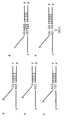
- allele probes are designed so that nucleotides complementary to the polymorphic site TGT (i.e. A, C or A in the 5' to 3' direction) are the corresponding nucleotides of the allele probes and hybridize (see panels a to c in Fig. 4B). For example, in panel (a) of Fig.
- the 5' utmost, corresponding nucleotide "C” forming a complementary strand is located on the center of the nucleotides (TGT) constituting a polymorphism; in panel (b) of Fig. 4B, the 5' utmost, corresponding nucleotide "A” forming a complementary strand is located on "T” of the nucleotides (TGT) constituting a polymorphism.
- an allele probe is shown which is designed so that its corresponding nucleotides are located on the peripheral region (three nucleotides) of the polymorphic site (the underlined portion).
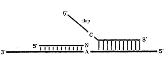
- An invader probe is designed so that the position of its 3' end nucleotide "N" (any one of A, T, C or G) corresponds to the position of any of the nucleotides of the polymorphic site (TGT) when hybridized.
- N any one of A, T, C or G
- the invader probe and allele probe may be designed so that a nucleotide located one to three nucleotides upstream or downstream of the deletion site will be the overlapping nucleotide taking into consideration of the deletion of TGT (i.e.
- Panel (e) of Fig. 4B shows an allele probe which is designed so that the two nucleotides of the both sides of the deletion site hybridize thereto.
- TaqMan probes they may be designed so that any of these nucleotides TGT or the deletion site is located at the center of the probe.
- the length of the nucleotide sequence of the oligonucleotide is at least 13 nucleotides, preferably 13 to 60 nucleotides, more preferably 15 to 40 nucleotides, and most preferably 18-30 nucleotides.
- This oligonucleotide sequence may be used as a probe for detecting a target gene, and it may be used as either a forward (sense) primer or a reverse (antisense) primer.
- the oligonucleotide used in the invention may be an oligonucleotide composed of two regions connected in tandem, one region being hybridizable to the genomic DNA and the other region being not hybridizable thereto.
- the order of connection is not particularly limited; either region may be located upstream or downstream.
- the hybridizable region of this oligonucleotide is designed based on the information on SNP-containing sequences described in Table 1.
- the oligonucleotide is prepared so that the sequence located at the 5' or 3' utmost end of the region hybridizable to the genomic DNA includes the SNP.
- the region of the above oligonucleotide not hybridizable to the genomic DNA is designed at random so that it does not hybridize to the SNP-containing sequence described in Table 1.
- This oligonucleotide may be used as a probe mainly for detecting SNPs in the invader method.
- the primer used in the present invention is designed so that a nucleotide sequence given in Table 1 contains an SNP when amplified by PCR for the purposes of examining functional changes resulted from the SNP, judging the efficiency or non-efficiency of drugs, and examining the occurrence of side effect.
- the length of the primer is designed so that at least 15 nucleotides, preferably 15 to 30 nucleotides, more preferably 18 to 24 nucleotides are contained in the primer.
- the primer sequence is appropriately selected from the template DNA so that the amplified fragment has a length of 1000 bp or less, preferably within 500 bp (e.g. 120 to 500 bp), more preferably within 200 bp (120 to 200 bp).
- primers are designed so that at least one of the sense or the antisense primer is contained in a region within 1000 bp, preferably 200 bp, more preferably 100 bp counted from the nucleotide immediately adjacent to 5' or 3' to the SNP toward the 5' or 3' end of the gene, respectively.
- oligonucleotide primers or probes may be synthesized chemically according to known techniques. Usually, such primers or probes are synthesized with a commercial chemical synthesizer.
- fluorescent substances e.g. FAM, VIC, Redmond Dye, etc.
- the above-described oligonucleotide may be included in a genetic polymorphism detection kit.
- the genetic polymorphism detection kit of the present invention comprises one or more components necessary for practicing the present invention.
- the detection kit of the present invention comprises components to preserve or supply enzymes and/or reaction components necessary for performing cleavage assay (e.g. invader assay).
- the detection kit of the present invention comprises any and all components enzymes or components necessary (suitable) for an intended assay. Examples of such components include, but are not limited to, oligonucleotides, polymerases (e.g. Taq polymerase), buffers (e.g. Tris buffer), dNTPs, control reagents (e.g.
- the detection kit of the present invention may be a partial kit which comprises only a part of the necessary components. In this case, users may provide the remaining components.
- the detection kit of the present invention may comprise two or more separate containers, each containing a part of the components to be used.
- the kit may comprise a first container containing an enzyme and a second container containing an oligonucleotide.
- the enzyme include a structure-specific cleaving enzyme contained in an appropriate storage buffer or a container.
- the oligonucleotide include invader oligonucleotides, probe oligonucleotides, target oligonucleotides for use as control, and the like.
- ore or more reaction components may be provided in such a manner that they are pre-divided into portions of a specific amount. Since such a kit contains components which have already been quantitatively determined for use in one step of the method of the present invention, it is not necessary to remeasure or re-divide. Selected reaction components may also be mixed and divided into portions of a specific amount.
- reaction components should be pre-divided into portions and contained in a reactor.
- the reactor include, but are not limited to, reaction tubes or wells, or microtiter plates. It is especially preferable that the pre-divided reaction component should be kept dry in a reactor by means of, for example, dehydration or freeze drying.
- a gene encoding a drug metabolizing enzyme using DNA polymerase is amplified using the oligonucleotide prepared as above as a primer.
- the probe prepared as above is hybridized with a template DNA to detect a target DNA with a gene polymorphism.
- the template DNA can be prepared by a known technique, for example, cesium chloride density-gradient ultracentrifugation, SDS dissolution, or phenol-chloroform extraction.
- Amplification may be performed by polymerase chain reaction (PCR).
- PCR polymerase chain reaction
- useful DNA polymerase include LA Taq DNA polymerase (Takara), Ex Taq polymerase (Takara), Gold Taq polymerase (Perkin Elmer), AmpliTaq (Perkin Elmer), Pfu DNA polymerase (Stratagene) and the like.
- Amplification conditions are as follows. Denaturation step at 85-105°C for 10-40 seconds, preferably at 94°C for 20-30 seconds; annealing step at 50-72°C for 20 seconds to 1 minute, preferably at 60°C for 20 seconds to 1 minutes; and extension step at 65-75°C for 1-4 minutes, preferably at 72°C for 2-3 minutes constitute one cycle, and 30 to 40 cycles are performed. However, in order to denature the template DNA and the primers sufficiently, a denaturation step of at 95°C for 1-5 minutes [if Gold Taq polymerase (Perkin Elmer) is used, at least 8-15 minutes, preferably 10-12 minutes] may be added before the start of the above-described amplification cycles.
- Gold Taq polymerase Perkin Elmer
- an extension step of at 72°C for 1-5 minutes for 1-10 minutes may be added after the above amplification cycles. If the detection of the amplified product is not performed immediately, it is desirable to add a step of storing the amplified product at 4°C to avoid unspecific amplification.
- a gene encoding drug metabolizing enzyme (CYP) can be amplified.
- the amplified product is subjected to agarose gel electrophoresis, followed by staining with ethidium bromide, SYBR Green solution or the like to thereby detect the amplified product as a band or two to three bands (DNA fragments).
- a part of a gene encoding drug metabolizing enzyme, containing a genetic polymorphism can be detected as a DNA fragment.
- polyacrylamide gel electrophoresis or capillary electrophoresis may be performed. It is also possible to perform PCR using primers labeled with a substance such as fluorescent dye and to detect the amplified product.
- a detection method which does not require electrophoresis may also be employed; in such a method, the amplified product is bound to a solid support such as a microplate, and a DNA fragment of interest is detected by means of fluorescence, enzyme reaction, or the like.
- TaqMan PCR is a method using PCR reaction with fluorescently labeled allele-specific oligos and Taq DNA polymerase.
- the allele-specific oligo used in TaqMan PCR (called "TaqMan probe") may be designed based on the SNP information described above.
- the 5' end of TaqMan probe is labeled with fluorescence reporter dye R (e.g. FAM or VIC), and at the same time, the 3' end thereof is labeled with quencher Q (quenching substance) (Fig. 1). Under these conditions, fluorescence is not detectable since the quencher absorbs fluorescence energy. Since the 3' end of TaqMan probe is phosphorylated, no extension reaction occurs from TaqMan probe during PCR Fig. 1). However, when PCR is performed using this TaqMan probe together with Taq DNA polymerase and primers designed so that an SNP-containing region is amplified, the reaction described below occurs.
- R fluorescence reporter dye
- quencher Q quen
- a TaqMan probe hybridizes to a specific sequence in the template DNA (Fig. 2a), and at the same time, an extension reaction occurs from a PCR primer (Fig. 2b). At this time, Taq DNA polymerase having 5' nuclease activity cleaves the hybridized TaqMan probe as the extension reaction proceeds. When the TaqMan probe has been cleaved, the fluorescent dye becomes free from the influence of the quencher. Then, fluorescence can be detected (Fig. 2c).
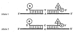
- Fig. 3 For example, as shown in Fig. 3, two alleles are supposed: one allele has A at the SNP site (allele 1) and the other allele has G at the SNP site (allele 2).
- a TaqMan probe specific to allele 1 is labeled with FAM and another TaqMan probe specific to allele 2 is labeled with VIC (Fig. 3).
- the probe hybridizes to the allele; and Taq polymerase cleaves the fluorescent dye of the probe, which becomes free form the influence of the quencher. As a result, fluorescence intensity is detected.
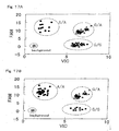
- the template is a homozygote of allele 1, strong fluorescence intensity of FAM is recognized but the fluorescence of VIC is hardly recognized. If the template is a heterozygote of allele 1 and allele 2, fluorescence of both FAM and VIC can be detected.
- the invader method is a method for detecting SNPs by hybridizing allele-specific oligos to the template.
- two unlabeled oligos and one fluorescently labeled oligo are used.
- One of the two unlabeled oligos is called an "allele probe".
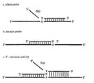
- the allele probe is composed of a region which hybridizes to the genomic DNA (template DNA) to form a complementary double strand, and a region (called "flap") which has a sequence entirely unrelated to the sequence of the template DNA and thus does not hybridize to the genomic DNA.
- a nucleotide located at the 5' or 3' utmost end of the hybridizable region corresponds to the SNP (panel (a) in Fig. 4A).
- the above-described flap sequence is an oligonucleotide having a sequence complementary to a FRET probe described later.
- the other unlabeled oligo is called an "invader probe".
- This oligo is designed so that it hybridizes complementarily to the template DNA from the SNP site toward the 3' end of the genomic DNA (panel (b) in Fig. 4A).
- the nucleotide corresponding to the SNP (“N" in panel (b) in Fig. 4A) may be any nucleotide.
- the fluorescently labeled oligo has a sequence completely unrelated to the allele. This sequence is common regardless of the types of SNPs.
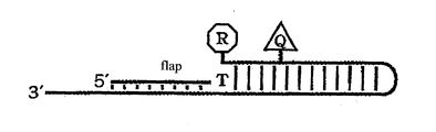
- This probe is called a "FRET" probe (fluorescence resonance energy transfer probe) (Fig. 5).
- FRET probe fluorescence resonance energy transfer probe
- the nucleotide at the 5' end of FRET probe (reporter) is labeled with fluorescent dye R, while quencher Q is linked upstream of the reporter. Therefore, under these conditions, the quencher absorbs the fluorescent dye and no fluorescence is detectable.
- region 1 A certain region of the FRET probe starting from the 5' end reporter nucleotide (designated "region 1") is designed so that it is complementary to a certain region of the probe located 3' to region 1 (designated “region 2") when region 1 and region 2 are faced with each other. Therefore, region 1 and region 2 form a complementary strand within the FRET probe (Fig. 5). Also, the region located downstream of this complementary strand forming region is designed so that it hybridizes to the flap of the allele probe to thereby form a complementary strand_(Fig. 5).
- cleavase is one of 5' nucleotidases having a unique endonuclease activity of cleaving upon recognition of a special structure of DNA.
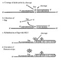
- Cleavase is an enzyme which cleaves the allele probe at a point immediately 3' to the SNP site when the genomic DNA, the allele probe and the invader probe form a triple strand at the SNP site. Therefore, when three nucleotides form a triple strand as shown in panel (c) in Fig. 4A, cleavase recognizes the 5' flap and cuts off this flap. As a result, the structure of this SNP site is recognized by cleavage (panel (a) in Fig.
- the allele probe is cut at the site of its flap to liberate the flap (panel (b) in Fig. 6).
- the flap liberated from the allele probe complementarily binds to the FRET probe since it has a sequence complementary to the FRET probe (panel (c) in Fig. 6).
- the SNP site of the flap invades into the portion of the FRET probe which has already formed a double strand. Cleavase again recognizes this structure and cuts off the nucleotide labeled with the fluorescent dye. The thus cleaved fluorescent dye becomes free from the influence of the quencher and emits fluorescence (panel (d) in Fig. 6).
- an invader probe and an allele probe for T are prepared.
- a FRET probe with a FAM-linked reporter corresponding to the SNP are prepared.
- an invader probe and an allele probe for C, and a FRET probe with a VIC-linked reporter corresponding to the SNP are prepared are also prepared. Then, all of them are mixed to carry out SNP detection.
- the genomic DNA to be used as a template is linearized.
- a probe is hybridized to this genomic DNA.
- the genomic DNA can be converted into a circular DNA through ligation reaction.
- RCA of the circular DNA proceeds.
- the ends of the probe do not match with the genomic DNA, the DNA is not ligated to become a circular DNA. Thus, RCA reaction does not proceed.
- a single-stranded probe which anneals with the genomic DNA and is circularizable is designed.
- This single-stranded probe is called a padlock probe.
- the sequences of the two ends of this padlock probe are designed so that they correspond to the SNP to be detected. Then, this padlock probe and the genomic DNA are mixed for ligation. If the two ends of the padlock probe and the SNP site of the genomic DNA are complementary to each other, the two ends of the padlock probe are joined by ligation, yielding a circular probe. If the two ends of the padlock probe and the SNP site of the genomic DNA are not complementary to each other, the probe does not become circular.
- padlock probes which are complementary to the SNP to be detected become circular and are amplified by DNA polymerase. By detecting the presence or absence of this amplification, SNP may be detected.
- synthetic oligonucleotides which have a fluorescent dye and a quencher at their respective ends and also have a hairpin structure are used.
- MALDI-TOF/MS (Matrix Assisted Laser Desorption-Time of Flight/Mass Spectrometry) is a method using a mass spectrometer in SNP typing. This method is composed of the following steps.
- polymorphisms may be detected by using single nucleotide extension reactions. Briefly, four types of dideoxynucleotides labeled with different fluorescent compounds are added to a reaction system containing a gene of interest. Then, single nucleotide extension reactions are performed. In this case, the nucleotide to be extended is the polymorphic site. Also, two reactions of DNA synthesis termination and the fluorescent labeling of the 3' end of DNA molecules are operated. Four types of reaction solutions are subjected to electrophoresis on the same lane of a sequencing gel or on capillary. Difference in the fluorescent dyes used for labeling is detected with a fluorescence detector to thereby sequence the DNA band.
- the one-nucleotide extended oligonucleotide is examined with a fluorescence detection system or a mass spectrometry system to thereby determine which nucleotide was extended using the difference in the fluorescent dyes.
- primers may be fluorescently labeled and used with unlabeled dideoxynucleotides.
- DNA microarrays are solid supports onto which nucleotide probes are immobilized, and they include DNA chips, gene chips, microchips, beads arrays, and the like.
- DNA microarray e.g. DNA chip
- GeneChip assay Affymetrix; US Patent Nos. 6,045,996; 5,925,525; and 5,858,659
- GeneChip technology uses small sized, high density microarrays of oligonucleotide probes affixed to chips. Probe arrays are manufactured, for example, by the light irradiation chemical synthesis method (Affymetrix) which is a combination of solid chemical synthesis and photolithography production technology used in the semiconductor industry.
- Affymetrix the light irradiation chemical synthesis method
- High density arrays can be constructed by using photolithography masks in order to make the boundary of the chemical reaction site of chips definite and by performing a specific chemical synthesis step.
- Multi-probe arrays are synthesized simultaneously on a large glass baseboard. Subsequently, this baseboard is dried, and individual probe arrays are packed in injection-molded plastic cartridges. This cartridge protects the array from the outer environment and also serves as a hybridization chamber.
- a polynucleotide to be analyzed is isolated, amplified by PCR, and labeled with a fluorescent reporter group. Then, the labeled DNA is incubated with an array using a fluid station. This array is inserted into a scanner to detect a hybridization pattern. Hybridization data are collected as luminescence from fluorescent reporter group bound to the probe array (i.e. taken into the target sequence). Generally, probes which completely matched with the target sequence generate stronger signal than those probes which have portions not matching with the target sequence. Since the sequences and locations of individual probes on the array are known, it is possible to determine the sequence of the target polynucleotide reacted with the probe array on the basis of complementation.
- Nanogen see, for example, US Patents Nos. 6,017,696; 6,068,818; and 6,051,380.
- the technology of Nanogen is capable of transferring charged molecules to and from specific test sites on semidonductor microchips and concentrating them.
- DNA capturing type probes specific to certain SNPs or variations are arranged on specific sites on microchips or assigned addresses. Since DNA is strongly negatively charged, it is capable of moving electronically to a positively charged area.
- test site or a row of the test site on a microchip is electronically activated with positive charge. Then, a solution containing DNA probes is introduced onto the microchip. Since negatively charged probes quickly moves to a positively charged site, probes are concentrated at this site on the microchip and chemically bind thereto. This microchip is washed, and another DNA probe solution is added to the microchip to allow specific binding of DNA probes to the chip.
- a test sample a gene of interest which has been amplified by PCR may be given.
- target molecules may be moved to one or more test sites on the microchip and concentrated.
- the hybridization between the sample DNA and a capturing probe complementary thereto is performed quickly. For example, as a result of these operations, hybridization occurs in several minutes.
- the polarity or electric charge of the site is converted to negative charge to thereby return the unbound DNA or non-specifically bound DNA into the solution.
- specific binding can be detected, for example, with a fluorescence scanner utilizing laser.
- ProtoGene an array technology utilizing fluid separation phenomenon on a plane surface (chip) because of difference in surface tensions
- the technology of ProtoGene is based on a fact that fluids are separated from each other on a plane surface because of difference in surface tensions given by chemical coating. Since oligonucleotide probes may be separated based on the above-mentioned principle, it is possible to synthesize probes directly on a chip by the ink-jet printing of a reagent containing probes.
- An array having reaction sites defined by surface tension is mounted on X/Y movable stage located under one set (4) of piezoelectric nozzles. Each piezoelectric nozzle contains four standard nucleotides.
- This movable stage moves along each row of the array to supply an appropriate reagent (e.g. amidite) to each reaction site. The entire surface of the array is soaked in a reagent common to the test sites in the array and then in a washing solution. Subsequently, the array is rotated to remove these solutions.
- reagent e.g. amidite
- DNA probes specific to the SNPs or variations to be detected are affixed to a chip using the technology of Protogene. Then, the chip is contacted with PCR-amplified gene of interest. After hybridization, unbound DNA is removed, followed by detection of hybridization using an appropriate method.
- Illumina Inc. utilizes Bead Array technology which uses a combination of optical fiber bundles and beads that undergo self-association with the array. Each optical fiber bundle has several millions of fibers depending on the diameter of the bundle. Beads are coated with oligonucleotides specific for the detection of certain SNPs or variations. Various types of beads are mixed in specific amounts to allow the formation of an array-specific pool. For assay, a bead array is contacted with a sample prepared from a subject. Then, hybridization is detected by an appropriate method.
- Evaluation of drugs may be performed by typing system. Briefly, according to any one of the detection methods described above, allele frequencies between toxicity (side effect) occurrence group and non-occurrence group are compared. A polymorphism which brings about difference in allele frequencies between the two groups is selected as a marker for recognizing the occurrence of toxicity. As a statistical test, usually ⁇ 2 test is carried out, but other statistical processing such as Fisher test may also be used. Further, it is possible to reflect this result on the concentration of active body of drugs (or unchanged body or metabolite of drugs) in blood or tissue. With respect to all genetic polymorphisms, the relation of cause and effect with the toxicity is examined. Then, only those genetic polymorphism sites that show correlation with the toxicity are selected.
- Allele pattern can be examined by preparing in advance all probes or primers for analyzing the genetic polymorphisms and reagents necessary for each technique in reaction plates, cards, glass baseboards or the like, and adding thereto the genomic DNA of a human subject for reaction.
- the subject has a genetic polymorphism which has correlation with the toxicity, it is possible to predict whether the drug exhibits effect or toxicity in that subject.
- the efficacy of a drug may be evaluated in a similar manner. Genetic polymorphisms which correlate with side effect or efficacy vary depending on drugs. Therefore, by conducting typing using the genetic polymorphisms, it becomes possible to predict the efficacy or side effect of the drug.
- the frequency of the genetic polymorphism is compared with efficacy/non-efficacy or presence/absence of side effect.
- a judgment on the drug can be made.
- the genetic polymorphism information obtained as described above in the present invention may be compared with genetic polymorphism information in a gene of a subject encoding corresponding drug metabolizing enzyme. By doing so, the above information may be used as an indicator for analyzing the efficacy and safety of a drug metabolized by the drug metabolizing enzyme. Therefore, the genetic polymorphism information obtained in the present invention serves as an information source for selecting most effective drug to treat a disease.
- the evaluation method described in "8. Evaluation of Drugs” may be used. Briefly, it can be said that the genetic polymorphisms which were recognized to be correlating with side effect or efficacy in the preceding sub-section give influence upon the activity, the transcription, and the translation of the enzyme. Also, it can be said that such genetic polymorphisms have some indirect relation with a mechanism of the occurrence of side effect or efficacy. The metabolism of a drug is examined and confirmed by a pharmaceutical manufacturer or the like in pre-clinical test or clinical test.
- the drug is an enzyme and a polymorphism which correlates with severe side effect exists among the polymorphisms located in the gene encoding the enzyme, it is possible to delete the relevant polymorphism or to use the drug conditionally. With respect to efficacy, a similar evaluation can be made. From such information on side effect and efficacy, drug screening becomes possible.
- DNA Stringy white precipitate
- TE solution 10 mM Tris-HCl (pH 7.4) and 1 mM EDTA (pH 7.4)
- the genomic sequence was obtained from GenBank DNA database. The repeat sequence was removed using RepMask computer program, and PCR primers were designed so that PCR products would be approximately 1 kb.
- the genomic DNA used was DNA from 48 unrelated individuals prepared to be at the same concentration. Equal amounts of each DNA from 3 individuals were placed in one tube and mixed, and 60 ng out of the mixture was used for PCR. PCR was performed using Ex-Taq (2.5 U; TaKaRa) on GeneAmp PCR System 9700 (PE Applied Biosystems). One cycle was composed of reaction at 94°C for 2 minutes followed by denaturation at 94°C for 30 seconds, annealing at 60°C or 55°C for 30 seconds, and extension at 72°C for 1 minute, and 35 cycles were completed.
- PCR products were purified using Arraylt (Telechem), and underwent sequence reaction with BigDye Terminator RR Mix (PE Applied Biosystems). One cycle was composed of reaction at 96°C for 2 minutes, followed by denaturation at 96°C for 20 seconds, annealing at 50°C for 30 seconds, and extension at 60°C for 4 minutes using GeneAmp PCR System 9700 (PE Applied Biosystems), and 25 cycles were completed. After the sequence reafction, sequence analysis was carried out on ABI PRISM 3700 DNA Analyzer.
- Table 1 The results as shown in Table 1 were obtained for SNP.
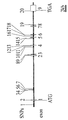
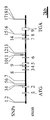
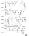
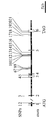
- the names and abbreviations of drug metabolizing enzymes analyzed, ACCESSION number from database (GenBank), and genetic structure and positions of SNPs of drug metabolizing enzyme are shown in Figures 9 to 16.
- exons are shown as white boxes or black lines across genes which are shown as horizontal lines.
- Positions of SNPs are shown with solid lines above the genes and numbered.
- methods for analyzing SNPs are provided. According to the methods of the invention, it becomes possible to select appropriate drugs for target diseases. Thus, the methods of the invention are extremely useful.
Landscapes
- Chemical & Material Sciences (AREA)
- Life Sciences & Earth Sciences (AREA)
- Organic Chemistry (AREA)
- Health & Medical Sciences (AREA)
- Proteomics, Peptides & Aminoacids (AREA)
- Wood Science & Technology (AREA)
- Zoology (AREA)
- Engineering & Computer Science (AREA)
- Genetics & Genomics (AREA)
- Analytical Chemistry (AREA)
- Biochemistry (AREA)
- General Engineering & Computer Science (AREA)
- Microbiology (AREA)
- Biotechnology (AREA)
- Physics & Mathematics (AREA)
- Immunology (AREA)
- Bioinformatics & Cheminformatics (AREA)
- Molecular Biology (AREA)
- General Health & Medical Sciences (AREA)
- Biophysics (AREA)
- Pathology (AREA)
- Measuring Or Testing Involving Enzymes Or Micro-Organisms (AREA)
- Investigating Or Analysing Biological Materials (AREA)
- Apparatus Associated With Microorganisms And Enzymes (AREA)
Abstract
A method for detecting a genetic polymorphism(s) in a gene
encoding cytochrome P450 using as oligonucleotide probes
and/or oligonucleotide primers at least one sequence selected
from the group consisting of an at least 13 nucleotide
sequence within any of the nucleotide sequences as shown in
SEQ ID NOS: 1 through 215, said at least 13 nucleotide sequence
containing the 21st nucleotide, or a sequence
complementary to said at least 13 nucleotide sequence.
Description
The present invention relates to information on genetic
polymorphisms; a method for detecting information on genetic
polymorphisms; a method for evaluating drugs using genetic polymorphisms;
and a method for screening for drugs.
As physical appearances of human individuals vary infinitely,
the human genetic code consisting of three billion
(3,000,000,000) base pairs vary at a considerably large number
of sites when compared among individuals. These differences
in the genetic code are called genetic polymorphisms, and single
nucleotide polymorphism is known as a representative polymorphism.
Single nucleotide polymorphism (SNP) means a difference in
one DNA letter among individuals. As faces and shapes of human
individuals vary infinitely, nucleotide sequences (i.e.
genetic code) of individuals vary at a considerably large number
of sites. SNPs are classified into cSNP (coding SNP) and
gSNP (genome SNP) depending on their locations; cSNP is
further classified into sSNP (silent SNP), rSNP (regulatory
SNP) and iSNP (intron SNP).
These SNPs are useful as polymorphic markers in searching
for those genes which are associated in the development or
worsening of diseases; finally, these SNPs directly relates to
risk diagnosis of diseases or selection and use of therapeutic
drugs in the clinical field. Also, drug development on the
basis of evidence obtained using causative substances as target
molecules has become the trend of the world. When a drug
is administered to patients with the same disease, their
responsiveness is diverse. Some patients show remarkable effect;
some patients show low effect; and some patients show no
effect. Thus, responsiveness to a drug varies greatly depending
on the patient. Even if the conditions of patients are
the same and diagnosed as the same disease, the routes which
have caused that disease may be different; or the rate of metabolizing
drugs may vary greatly among patients. Therefore,
it is desired to select an appropriate drug and develop an appropriate
therapeutic method against a target disease based on
genetic polymorphisms such as SNPs (i.e. the so-called tailored
medicine is desired).
In addition to responsiveness to drugs, the problem of
strong side effect which sometimes might be lethal is also one
of the major problems that medical staffs should address. Even
if there is no excessive administration caused by prescription
error or the like, unexpected, lethal side effect
might occur. Therefore, with respect to responsiveness to a
drug, it is desired that individual difference in the metabolism
of the drugs, delivery of the drug, and the strength of
responsiveness or side effects by the drug's receptor should
be determined taking into account genetic polymorphisms such
as SNPs.
It is an object of the present invention to provide a method
for detecting information on genetic polymorphism; a method
for evaluating the efficacy and safety of drugs based on
the information; and a method for screening for drugs.
As a result of extensive and intensive researches toward
the solution of the above problem, the present inventors have
succeeded in establishing a method which comprises detecting
genetic polymorphisms in a gene encoding a drug metabolizing
enzyme and evaluating with the resultant information the cause
and effect relationship between the drug and the disease.
Thus, the present invention has been achieved.
The present invention is as described below.
Accession No.: NG_000008.2
Accession No.: AC008962.9
Accession No.: NG_000010.1
Accession No.: AL161645.14
Accession No.: NG_000011.1
Accession No.: AC006021.2, AC004914.1, and AC004961.2
Accession No.: AC020782.10
Accession No.: AF127089.1, AF176800.1, and AF215845.2
The present invention is explained in detail as follows.
The present invention relates to a method for detecting
genetic polymorphisms in a subject using information on genetic
polymorphism in a gene encoding drug metabolizing enzyme,
especially, a gene encoding cytochrome P450 and/or a sequence
complementary thereto. The present invention is also characterized
by analyzing the absence/presence or intensity of the
efficacy and safety of the drug; based on the analysis result,
the relation between the disease and the drug is evaluated.
Even when a plurality of patients suffer from the same disease,
it is quite often that genetic polymorphism information on
drug metabolizing enzyme gene varies by individual patient.
Therefore, difference in drug response is drawn out from genetic
polymorphism information different among individuals;
then, the efficacy or safety of the drug (i.e. the efficacy of
the specific drug is recognized or not recognized when a patient
has such and such genetic polymorphism information, or side
effect occurs frequently or seldom when a patient has such
and such genetic polymorphism information) is evaluated. From
these results, it becomes possible to determine what drug
should be used for a specific disease, and to administer a
drug suitable for an individual patient (tailored medicine)
based on his/her genetic polymorphism information.
Now, the terms used in this specification will be explained
below.
The term "drug metabolizing enzyme" is a general term for
enzymes catalyzing in-vivo structural conversion of an exogenous
substance including a medicament. Enzymes that are involved
in metabolism of some endogenous substances are also
included in the drug metabolizing enzyme as long as they are
administered for a therapeutic purpose.
The term "efficacy" refers to a property of a drug that
has a beneficial effect (efficacy) on a subject when the drug
is administered to the subject. The term "safety" is a property
of a drug that has an adverse effect or a side effect on
a subject when the drug is administered to the subject.
The term "genetic polymorphism information" includes both
polymorphism information in a gene and polymorphism information
in its complementary sequence.
The term "therapy" means that at least one symptom of a
disorder or a disease is cured or mitigated.
The term "screening" means a step of selecting a drug candidate
and refers to a step of narrowing down a drug candidate
by screening numerous test substances.
The term "gene" refers to RNA having no coding function
(e.g., ribosome RNA or transfer RNA), a regulatory sequence
required for producing a polypeptide or a precursor, and a DNA
sequence having a coding sequence. The RNA or polypeptide may
be encoded by a full-length coding sequence or a part of the
coding sequence as long as it can be provided with a desired
activity or function.
The term "wild type" means a gene or gene product isolated
from a naturally occurring source and having a function inherent
to the gene or gene product. The wild type gene is one
that is most frequently observed in a population, and more
specifically, means a "normal type" or "wild type" of gene.
On the other hand, the "mutation type" means a gene or gene
product having a change in sequence and/or functional characteristics
in comparison with a wild-type gene or gene product,
and sometimes called as a "modification type" or a
"polymorphism type". Examples of such a mutation type include
(1) a gene having a modified characteristic compared to a
wild-type gene or gene product and (2) a gene having the same
function although it differs in gene sequence. The wild type
structural gene and its mutation type, these two type of
structural genes, have sequence variation caused by substitution
and/or deletion of a single nucleotide, or insertion of
not less than one nucleotide.
The term "oligonucleotide" is defined as a molecule having
not less than two deoxyribonucleotides or ribonucleotides,
preferably at least 5 nucleotides, more preferably at least about
10 to 15 nucleotides, and further preferably, at least about
15 to 30 nucleotides. The accurate size of the oligonucleotide
may be changed depending upon its function and use.
The oligonucleotide can be produced by a known method in the
art, for example, chemical synthesis, DNA replication, reverse
transcription, PCR, or a combination thereof.
The oligonucleotide is prepared by binding the 5' phosphate
group of its pentose ring to the 3' oxygen of the pentose
ring of an adjacent mononucleotide via a phosphodiester bond
in a single orientation. When the 5' phosphate group is not
bound to the 3' oxygen of a mononucleotide pentose ring, the
end of the oligonucleotide is called the "5'-end", whereas the
3' oxygen is not bound to the 5' phosphate group of a mononucleotide
pentose, the end of the oligonucletode is called the
"3'-end". Thus, a polynucleotide has the 5'-end and the 3'-end.
In the case where a polynucleotide chain has a first region
and a second region and the 3'-end of the first region
lies anterior to the 5'-end of the second region, the first
region is present upstream of the second region.
The term "polynucleotide sequence" means (1) an oligonucleotide,
nucleotide, or polynucleotide, (2) a fragment or a
part of them, or (3) a genome or synthesized DNA or RNA. The
polynucleotide sequence may be a single strand chain or a
double strand chain, or a sense chain or an antisense chain.
Note that a nucleotide that is rarely found in a naturally occurring
polynucleotide may be included in a polynucleotide used
in the present invention. Examples of such a nucleotide
include inosine and 7-deazaguanine.
The term "nucleotide analogue" refers to a modified nucleotide
or a non-naturally occurring nucleotide, for example, 7-deazapurine
(i.e., 7-deaza-dATP and 7-deaza-dGTP). Examples
of such a nucleotide analogue include a base analogue as well
as a modified deoxyribonucleotide and a modified ribonucleotide.
The term "peptide nucleic acid" (PNA) includes a base or a
base analogue as found in a naturally occurring polynucleotide
and refers to a nucleic acid molecule bound not to the sugarphosphate
skeleton of a typical polynucleotide but to a peptide
skeleton. Since a nucleotide is bound to a peptide, the
nucleotide can be paired with a complementary nucleotide of a
polynucleotide similarly to the case of an oligonucleotide.
The aforementioned small molecule is also called as an antigene
agent, which binds to a complementary polynucleotide
chain, thereby stopping the transcriptional extension
(Nielsen, et al., Anticancer Drug Des. 8:53 63 (1993)). In
the present invention, in place of a polynucleotide, the nucleotide
analogue and a peptide nucleic acid mentioned above
may be used.
The terms "protein", "polypeptide" and "peptide" are defined
to have the same meaning when they are used regarding a
gene product. Note that a polypeptide produced by recombinant
DNA technology is particularly referred to as a "recombinant
protein". Such a recombinant protein can be obtained through
recombinant DNA technology by inserting DNA encoding a polypeptide
into an appropriate vector, thereby transforming a
host cell, and expressing a heterogeneous protein.
The term "purified" means that a polynucleotide molecule,
peptide, polypeptide or protein molecule is taken out from a
natural environment, or isolated or separated by recombinant
DNA technology, and that it contains, in a natural state, other
components in an amount of 40% or less, preferably 25% or
less, and most preferably 10% or less. Therefore, the term
"isolated polynucleotide" or "isolated oligonucleotide" is a
substantially purified polynucleotide.
The term "polymorphism" refers to the fact that one or more
types of morphology simultaneously exist with respect to a
gene or a part of the gene. The portion of a gene presenting
at least two different morphologies, that is, having at least
different nucleotide sequences, is called a "polymorphic region
of a gene". The polymorphic region includes, for example,
a genetic polymorphism such as a single nucleotide polymorphism
as described later. The polymorphic region is different
depending upon various alleles. There are some polymorphic
regions having a several-nucleotide length.
The term "polymorphic gene" refers to a gene having at least
one polymorphic region. Note that the term "polymorphic
gene locus" is a locus present in a population but varies between
members of the population. The most common allele is
present with a frequency of less than 0.95. On the other
hand, the term "mono morphic gene locus" is a locus that is
rarely different between members of a population or completely
the same between them. In general, the most common allele is
considered to be a gene locus whose frequency does not exceed
0.95 in a gene pool of a population.
The term "information" refers to accumulation of fact or
data. The information stored or processed by use of a computer
system means data stored in accordance with an arbitrary
format. Examples of such a computer system include, but not
particularly limited to, the Internet and the like. Examples
of such a format include analog, digital and optical formats.
The term "information on a subject" means that fact or data
regarding the subject. The term "genomic information" refers
to information regarding a genome and include, but not limited
to, a nucleotide sequence, gene, allele frequency, RNA expression
level, protein expression, and the correlation between a
genotype and a phenotype. The term "allele frequency information"
refers to fact or data regarding an allele frequency and
include, but not limited to, allele identification, statistical
correlation between the presence of an allele and a trait
of a subject, the presence or absence of an allele in an individual
or a population, a probability (%) of the presence of
an allele in an individual showing at least one specific
trait.
The term "complementary" or "complementarity" is a concept
used in mentioning the relationship between polynucleotides
having nucleotides that can be paired with each other (i.e.,
nucleotide sequences of an oligonucleotide or a target polynucleotide).
To explain more specifically, the sequence "5'-A-G-T-3'"
is complementary to the sequence "3'-T-C-A-5"'. The
"complementarity" may be established "partly", that is, a certain
number of nucleotides of a polynucleotide complementarily
can make a pair of nucleotides or established completely, that
is, all nucleotides can make a pair. The extent of the
complementarity has a significant effect upon the efficiency
and intensity of hybridization. This is particularly important
in a detection method by use of an amplification reaction
and coupling between polynucleotides. A nucleotide analogue
to be used to make a pair with another nucleotide analogue
(e.g., IsoC/IsoG nucleotide pair) or a naturally occurring
nucleotide is also included in the definition range of the
"complementary" or "complementarity" and may be said that they
are complementary to the partner to be paired.
The complementarity of a nucleotide sequence means that
when nucleotide sequences are laid side by side such that the
5'-end of one of the sequences faces the 3'-end of the other,
oligonucleotide sequences are in "inverse-parallel relationship".
The sequences may not be necessary to be completely
complementary. The double helix thus is stably formed even if
it includes a mismatched nucleotide pair or an uncoupled nucleotide.
It can be easy for one skilled in the art to empirically
determine the stability of a double helix in consideration
of length of the oligonucleotide, the nucleotide composition,
nucleotide sequence of the oligonucleotide, ionic
strength and the presence of a mismatch nucleotide pair of oligonucleotides.
The term "hybridization" is the concept used in pairing of
complementary polynucleotides with each other. The hybridization
and hybridization intensity (in other words, polynucleotide-polynucleotide
binding strength) is influenced by the
complementarity between polynucleotides, the stringency of the
conditions to be employed, and the Tm of the hybrid to be formed.
In the hybridization method, a polynucleotide is annealed
with a complementary polynucleotide (polynucleotide having
a complementary nucleotide sequence).
The symbol "Tm" means a "melting temperature", which refers
to a temperature at which a double stranded polynucleotide
is denatured with heat to convert into single strands. To
cause a priming reaction during which a template DNA and a
primer form into a double strand, it is necessary to set the
annealing temperature to be equal to or lower than the Tm of
the primer. Some calculation formulas for obtaining Tm of a
polynucleotide are known in the art. As is described in a general
reference, when a polynucleotide is dissolved in a 1 M
aqueous NaCl solution, the Tm value can be simply estimated,
based on the following formula:
Tm = 81.5 + 0.41 (% G + C)
(See, for example, Anderson and Young, Quantitative Filter
Hybridization, Nucleic Acid hybridization (1985)). Also, another
reference describes a method of calculating Tm more precisely
in consideration of structural, environmental characteristics
and sequential characteristics (see, for example, Allawi,
H.T. & SantaLucia, J., Jr. Thermodynamics and NMR of internal
G. T mismatches in DNA. Biochemistry 36, 10581-94
(1997)).
The term "stringency" is a concept used in mentioning conditions
of hybridization including temperature, ionic strength
and the presence of other compounds. Under "high stringency
conditions", nucleotide paring (base pairing) takes place only
between polynucleotide fragments constituted of sequences having
complementary nucleotides with a high frequency. Therefore,
"poor" or "low" stringency conditions are used when
hybridization is performed between a polynucleotide that is
not completely complementary to a polynucleotide to be coupled
by annealing.
As the "high stringency conditions", in the case where a
probe having about 500 nucleotide length is used, mention may
be made of the conditions in which hybridization is performed
in a solution containing 5 × SSPE (43.8 g/l NaCl, 6.9 g/l
NaH2PO4 H2O and 1.85 g/l EDTA, pH is adjusted to 7.4 with Na-OH),
0.5% SDS, 5 × Denhalt agent, and 100 µg/ml denatured sermon
sperm DNA, at 42°C, followed by washing in a solution containing
0.1 × SSPE and 1.0% SDS at 42°C. 500 ml of the 50 ×
Denhalt solution contains 5 g Ficoll (Type 400, Pharamcia),
and 5 g BSA (fraction V; Sigma).
As the "medium stringency conditions", when a probe having
about 500 nucleotide length is used , mention may be made of
the conditions in which hybridization is performed in a solution
containing 5 × SSPE (43.8 g/l NaCl, 6.9 g/l NaH2PO4 H2O and
1.85 g/l EDTA, pH is adjusted to 7.4 with NaOH), 0.5% SDS, 5 ×
Denhalt agent, and 100 µg/ml denatured sermon sperm DNA, at
42°C, followed by washing in a solution containing 1.0 × SSPE
and 1.0% SDS at 42°C.
As the "low stringency conditions", when a probe having
about 500 nucleotide length is used, mention may be made of
the conditions in which hybridization is performed in a solution
containing 5 × SSPE (43.8 g/l NaCl, 6.9 g/l NaH2PO4 H2O and
1.85 g/l EDTA, pH is adjusted to 7.4 with NaOH), 0.1% SDS, 5 ×
Denhalt agent and 100 µg/ml denatured sermon sperm DNA, at 42°C
, followed by washing in a solution containing 5 × SSPE and
0.1% SDS at 42°C.
As to the complementarity, it is important to determine
whether hybridization is completely performed or partially
performed, depending upon its usage. For example, for the
purpose of simply detecting the presence or absence of an exogenous
DNA sequence, it is important to choose conditions under
which hybridization takes place without fail as long as a
detection-target, DNA sequence is present, by either a partially
complementary probe or a completely complementary probe.
On the other hand, there are some occasions requiring a hybridization
method that can distinguish partially complementarity
from the complete complementarity.
The term "primer" means an oligonucleotide capable of
starting synthesis when it is placed under conditions capable
of starting primer extension. An oligonucleotide primer may
be naturally occurring one, purified one through restriction
digestion, or synthetically produced. The primer is designed
so as to be complementary to a specific sequence of a template.
A primer has to be sufficiently complementary to a
template sequence such that it hybridizes with a template so
as to cause primer extension, thereby forming a template-primer
complex to obtain a primer extension product. Note
that a primer sequence may not be always completely complementary
to a template as long as it can couple with the template
due to the presence of a partially complementary sequence.
The "label" refers to any atom or molecule as long as it
can be used in detection (preferably quantification) and bind
to a polynucleotide or a protein. Examples of such a label
include, but not limited to, a dye such as a radioactive label
(e.g., 32P), a binding group (e.g., biotin), a hapten (e.g.,
digoxigenin), a luminescence group emitting phosphorous light
or fluorescent light, and a fluorescent dye. These labels may
be used singly or in combination with a group capable of
suppressing or shifting an emission spectrum by fluorescent
resonance energy transfer (FRET). The label is specified as
one capable of generating a detectable signal based on fluorescence,
radioactivity, colorimetric determination, weight
determination, X-ray diffraction or absorption, magnetic or
enzymatic activity. The label used herein may be a charged
group (positively or negatively charged group) or a group electrically
neutral. When a polynucleotide or a protein sequence
contains a detectable label, the label may contain or
be composed of such a charged sequence as mentioned above.
The term "signal" means a physicochemically detectable
signal generated by a label or an assay reaction.
The "detector" refers to a system or a structural element
of a system, more specifically, a device or a reactive medium
of informing the presence of a signal to a user or another
structural element of the system (e.g., computer or controller).
Examples of such a device include, but not limited to,
a camera, fluorescence detector, a charge coupled device, and
scintillation counter. Examples of such a reactive medium
include, but not limited to, X-ray, photographic film, and pH
indicator.
Examples of such a detector include a light analysis system
capable of detecting ultraviolet rays, visible rays or
infrared rays (e.g., fluorescent or chemical luminescence), a
spectrophotometric system, a radiation detection system,
spectrometry (such as magnetic nuclear resonance spectrometry,
mass spectrometry, surface-induced Raman spectrophotometry), a
system such as gel electrophoresis, capillary electrophoresis,
or gel exclusion chromatography, other detection systems known
in the art, and a combination of these systems.
The "sample" refers to a biological specimen and a synthetic
specimen. Examples of such a biological specimen include
an animal specimen (e.g., human) such as a liquid, solid
(fecal matter) or tissue sample, food in a liquid or solid
form, feed and feed staff (daily product, vegetable, meat,
meat products), and other miscellaneous wastes. The biological
sample may be taken from a domestic animal or wild animal,
which includes, but not limited to, an ungulate animal, bear,
fish, and rodent.
A detection-target, a polynucleotide, is supplied from a
sample containing or conceivably containing a polynucleotide
(RNA or DNA). A particularly preferable source of such a target
polynucleotide is a biological sample, which includes, but
not limited to, blood, saliva, cerebrospinal fluid, pleural
fluid, milk, lymph, sputum and semen.
The term of a "cleaved structure" of a nucleotide refers
to a structure formed by interacting at least one probe oligonucleotide
with a target polynucleotide to form a double
stranded structure and cleaving the double stranded structure
by a cleaving agent. Examples of such a cleaving agent include,
but not limited to, enzymes (such as a restriction enzyme).
The structure to be cleaved is a substrate for specific
cleavage by a cleaving means and thus differs from a polynucleotide
molecule, which is a substrate for non-specific cleavage.
Examples of such a cleavage means include an agent such
as phosphodiesterase cleaving a polynucleotide molecule regardless
of its secondary structure.
The term "kit" refers to a supply system for supplying a
substance. When it is used in a reaction assay, a system for
storing, transporting or supplying a reaction reagent and/or
auxiliary substance is included in such a supply system. Examples
of such a reaction reagent include, but not limited
to, an oligonucleotide and an enzyme contained in a container.
Examples of such an auxiliary substance include, but not limited
to, a buffer and an instruction leaflet. Examples of such
a kit include at least one type of accommodation unit (e.g.,
box) containing a relevant reaction reagent and/or auxiliary
substance, and the like. The term "partial kit" refers to a
supply system containing a part of all components of the kit,
that is, no smaller than two discrete containers. The containers
are supplied together or separately to a user. For example,
a first container contains an enzyme to be used for
assay and a second container contains an oligonucleotide. Alternatively,
the kit may be supplied as a "composite kit".
The composite kit refers to a supply system containing all ingredients
for a reaction assay in a single container. For example,
a composite kit may contain necessary ingredients in a
single accommodation case. Both the partial kit and the composite
kit as mentioned above are included in the "kit" according
to the present invention.
The term "subject" means a target living organism from
which genetic polymorphism information is obtained by the present
invention and in which a drug is evaluated and screened.
Examples of such a subject include, but not limited to, mammals
(such as a rodent, a primate including a human, sheep,
goat, horse, dog, and cow), birds, amphibians, and reptiles.
A subject is preferably selected from primates including human
and rodents including rats and mice, most preferably, a human.
Genetically manipulated animals, namely, transgenic animals,
and knockout animals are also useful for evaluating and screening
of a drug having an effect upon various processes
(embryogenesis and histogenesis).
Genetic polymorphism includes single nucleotide polymorphism,
insertion/deletion type polymorphism, and polymorphism
caused by difference in the number of repetition of a
nucleotide sequence. Generally, single nucleotide polymorphism
(SNP) means a polymorphism caused by substitution of
one specific nucleotide with other nucleotide in a gene or its
complementary strand (complementary sequence) region. In the
present invention, however, the term SNP also includes a polymorphism
caused by deletion of the nucleotide and a polymorphism
caused by addition of one more nucleotide to the nucleotide.
Insertion/deletion type polymorphism means a polymorphism
caused by deletion or insertion of a plurality of
nucleotides (e.g. two to several ten nucleotides). Sometimes,
several hundred to several thousand nucleotides may be deleted
or inserted. The polymorphism caused by difference in the
number of repetition of a nucleotide sequence has repetition
of a sequence of two to several ten nucleotides, and the number
of this repetition varies among individuals. Those polymorphisms
where the repeat unit consists of several to several
ten nucleotides are called VNTR (variable number of tandem repeat)
polymorphism, and those polymorphisms where the repeat
unit consists of about two to four nucleotides are called microsatellite
polymorphism. In VNTR or microsatellite polymorphisms,
the number of such repetition is different among
individuals' alleles, which results in acquisition of variation.
The drug metabolizing enzyme is involved in absorption,
metabolism, and excretion of drugs. A polymorphism of the enzyme
changes an expression (transcription and translation) amount
and the activity of the enzyme, with the result that
blood concentrations of an unchanged drug or its metabolites
are changed.
In the present invention, examples of a drug metabolizing
enzyme expressed by a target gene of gene polymorphism analysis
include cytochrome P450 (CYP) and the like.
CYP (cytochrome P450), which is responsible for a first-phase
drug metabolism, introduces an oxygen atom into a drug.
Examples of CYP include CYP 2A6, CYP 2A13, CYP 2B6, CYP 2E,
CYP 2S1, CYP 5A1, CYP 7A1 and CYP 7B1. A single CYP or a combination
of them is appropriately selected.
Information on genetic polymorphisms may be obtained by
conventional methods for detecting genetic polymorphisms. For
example, the sequencing method, the PCR method, Fragment
length polymorphism assay, hybridization methods using an allele-specific
oligonucleotide as a template (e.g. TaqMan PCR
method, the invader method, the DNA chip method), methods using
primer extension reaction, MALDI-TOF/MS method, the DNA
chip method, and the like may be employed. The PCR method and
the direct sequencing method may be used for detecting any type
of genetic polymorphism. The other methods may be used
mainly for detecting SNPs.
Hereinafter, the outline of each method for detecting genetic
polymorphism sequence is explained.
A gene polymorphism sequence can be detected by use of a
direct sequencing method. In this assay, a DNA sample is
first taken from a subject by an appropriate method. A target
detection region is cloned into an appropriate vector and
amplified through proliferation of a host cell (e.g., bacterial
cell). The target detection region refers to a region containing,
for example, an SNP or a mutation. Alternatively,
DNA within the target detection region may be amplified by use
of PCR. After the amplification, DNA within the target detection
region is subjected to sequencing by an appropriate method.
Examples of such a sequencing method include, but not
limited to, a manual sequencing method or an automatic sequencing
method. Examples of such a manual sequencing method include
a method of using a radioactive marker nucleotide and the
like. Examples of such an automatic sequencing method include
a method using a Dye Terminator and the like. The sequencing
results are shown by an appropriate display method. Thereafter,
the presence or absence of a predetermined SNP or mutation
is determined.
In the present invention, a gene polymorphism sequence can
be detected by use of an assay based on PCR. The PCR assay uses
an oligonucleotide primer forming a hybrid only within a
mutation type or wild type allele (e.g., polymorphic or mutation
region). A DNA sample is amplified by use of a primer
set consisting of primers for a mutation type and a wild type.
When only the mutation-type primer generates a PCR product, it
is demonstrated that the subject has a mutation allele. When
only the wild-type primer generates a PCR product, it is demonstrated
that the subject has a wild-type allele.
In the present invention, a gene polymorphism sequence can
be detected by fragment length polymorphism assay. In the
fragment length polymorphism assay, a specific DNA band pattern
is obtained based on a series of enzymatic DNA cleavage
sites. Examples of such an enzyme include restriction enzymes
and Cleavase I (Third Wave Technologies, Inc.). The DNA fragment
derived from a sample and having a SNP or a mutation exhibits
a different band pattern from that of a wild type.
In the present invention, Restriction Fragment Length Polymorphism
(RFLP) assay is also used to detect a gene polymorphism
sequence. First, a target detection region is amplified
by PCR. Subsequently, the resultant PCR product is cleaved
with a restriction enzyme(s) known to produce a fragment
of a specific length inherent to a predetermined polymorphism.
The PCR product digested into pieces with the restriction enzyme(s)
is generally separated by gel electrophoresis and visualized
by staining with ethidium bromide. The lengths of
fragments are compared to those of a molecular marker and wild
type and mutation-type controls.
In the present invention, Cleavase Fragment Length Polymorphism
(CFLP) can be also used to detect a gene polymorphism
sequence (see Third Wave Technologies Inc., USP Nos.
5,843,654, 5,843,669, 5,719,208 and 5,888,780). Cleavase I
enzyme is a structure-specific heat resistant nuclease which
recognizes and cleaves the junction between a single stranded
region and a double-stranded region of DNA.
In the first place, the DNA of a target detection region
is isolated, for example, by PCR. In this step, one or two
stands are preferably labeled. Subsequently, the DNA strands
are separated by heating. The resultant product is annealed
to form a secondary structure within a strand. Subsequently,
the PCR product is treated with Cleavase I enzyme to generate
a series of fragments inherent to a predetermined SNP or mutation.
The PCR product treated with Cleavase enzyme is separated
and detected by denatured gel electrophoresis, and subsequently
visualized by autoradiography, fluorescent imaging or
staining. The length of fragments is compared to those of a
molecular maker and wild type and mutation-type controls.
In the present invention, a gene polymorphism sequence can
be detected by hybridization assay. The hybridization assay
is a method of determining the presence or absence of a predetermined
SNP or mutation based on the ability of the DNA derived
from a sample to hybridize with a complementary DNA molecule
(e.g., oligonucleotide probe). Hybridization assay is
performed by various hybridization techniques and detection
techniques. Which assay should be selected from them will be
explained below.
Whether or not a probe hybridizes with a target detection
sequence (e.g., SNP or mutation) can be directly detected by
visualizing a hybridized probe. This method is known as Northern
or Southern assay (see. e.g., Ausabel et al.(edited),
Current Protocols in Molecular Biology, John Wiley & Sons, NY
(1991)). In Southern assay, genomic DNA is isolated from a
subject and in Northern assay, RNA is isolated from a subject.
Subsequently, isolated DNA or RNA is cleaved with a series of
restriction enzymes that infrequently cleave sites except for
the region near a marker in the genome. Thereafter, DNA or
RNA is separated and transferred onto a film. The separation
operation can be carried out, for example, on an agarose gel.
The transferring onto the film is performed by transferring a
labeled probe or a probe specific to a detection target, an
SNP or mutation, onto the film under low, medium or high
stringency conditions. Labeling is performed by introducing,
for example, a radioactive nucleotide. After an unbound probe
is removed, a labeled probe is visualized, thereby detecting
the presence of the binding.
In the present invention, a gene polymorphism sequence can
be detected also by DNA chip hybridization assay. In this assay,
a series of oligonucleotide probes are adhered onto a solid
support. The oligonucleotide probes are designed so as to
have a specificity to a predetermined SNP or mutation. A test
DNA sample is brought into contact with the DNA chip thus formed
to detect hybridization.
In the present invention, hybridization can be also detected
by using the cleavage of a specific structure with an enzyme
(Invader assay; Third Wave Technologies Inc., see, e.g.,
USP Nos. 5,846,717, 6,090,543, 6,001,567, 5,985,557 and
5,994,069). Invader assay detects specific DNA and RNA sequences
by forming an overlapped portion by an oligonucleotide
probe and cleaving the overlapped portion with a structure-specific
enzyme.
Alternatively, hybridized structure with a probe can be
detected by TAQMAN assay (PE Biosystems; see, e.g., USP Nos.
5,962,233 and 5,538,848). This assay is usually performed during
the process of PCR. TAQMAN assay uses the 5'-3' exonuclease
activity of a DNA polymerase (e.g., AMPLITAQ DNA polymerase).
A probe specific to a predetermined allele or mutation
is introduced in PCR. The probe is formed of an oligonucleotide
having a 5' reporter dye (e.g., fluorescent dye) and 3'
quencher dye. During the PCR process, when a probe is bound
to a target sequence, a probe is cleaved at a site between the
reporter dye and the quencher dye by the 5'-3' polynucleotide
decomposition activity of AMPLITAQ polymerase. The intensity
of fluorescence increases by separating the reporter dye from
the quencher dye. The resultant signal is accumulated in each
cycle of PCR, so that the signal can be detected by a fluorescence
detector.
Furthermore, in the present invention, a polymorphism can
be detected by SNP-IT primer extension assay (Orchid Biosciences
Inc.; see, e.g., USP Nos. 5,952,174 and 5,919,626). In
this assay, an SNP is specified by selectively extending DNA
from a nucleotide positioned at a putative SNP site by use of
a DNA primer specifically synthesized and a DNA polymerase.
First, DNA within a target detection region is amplified and
denatured. Then, a polymerase reaction is performed by a micro
system called a microfluidics. Detection is performed by
attaching a label to a nucleotide probably positioned at an
SNP or a mutation site. The label introduced into DNA can be
detected by an appropriate method arbitrarily chosen. For example,
when a nucleotide contains a biotin label, it can be
detected by a fluorescence-labeled antibody specific to biotin.
In the present invention, a gene polymorphism sequence can
be detected also by use of a primer extension reaction. As a
method of using a primer extension reaction, for example, a
SniPer method may be employed. The SniPer method, which is
fundamentally based on a procedure called rolling circle
amplification (RCA), is one in which a DNA polymerase continuously
synthesizes a complementary DNA while moving on a single
stranded circular DNA serving as a template (see, e.g., USP
Nos. 6,210,884 and 6,183,960). According to this method, an
SNP can be determined by determining the presence or absence
of a color reaction generated when DNA amplification takes
place (Lizardi, P. M. et al., Nature Genet., 19, 225-232
(1998); Piated, A.S. et al., Nature Biotech., 16, 359-363
(1998)).
In the present invention, a gene polymorphism sequence can
be detected by using MassARRAY system (Sequenom Inc.; see,
e.g., USP Nos. 6,043,031, 5,777,324 and 5,605,798). First,
DNA is isolated from a sample by a standard procedure. Subsequently,
a specific DNA region containing a detection target,
a mutation or an SNP, is amplified by PCR. For example, the
region may have a length of 200-nucleotides. Subsequently,
one of the strands thus amplified is bound and fixed to a surface
of a solid phase, and then, the other chain unbound thereto
is removed by denaturing generally performed or washing.
Using the remaining and fixed single strand as a template in
an automatic enzyme reaction, a product to be detected
(detection product) having specificity to genotype is produced.
An extremely small amount of enzyme product (generally, 5-10
nano-liter) is transferred on SpectroCHIP array and then
subjected to automatic analysis using an SpectroREADER mass
spectrometer. To each spot, the detection product and a light
absorbing crystal forming a matrix, are introduced in advance.
The MassARRAY system uses MALDI-TOF(Matrix Assisted Laser Desorption
Ionization-Time of Fight) mass spectrometry. The
MALDI-TOF method is one performing SNP genotyping based on
fundamental difference of single nucleotides in mass by using
a mass spectrometer. Examples of such a method include a method
of using PCR amplification and a method of using a multiplex
(Haff, L.A., Smirnov, I. P., Genome Res.,7, 378-(1997);
Little, D.P. et al., Eur. J. Clinica. Chem, Clin. Biochem.,
35, 545- (1997); Ross, P., et al., Nat Biotechnol., 16,
1347- (1998)).
In the MALDI-TOF, a matrix collides against a pulse derived
from a laser beam in a process known as an elimination
process. The energy is transferred from the laser beam to the
matrix to vaporize the matrix, with the result that a small amount
of detection product is excluded into a flight tube.
When an electric field pulse is applied to the flight tube,
the detection product is electrically charged. Because of the
presence of the detection product, the flight tube proceeds
toward a detector. The time between the application of the electric
field pulse and collision of the detection product onto
the detector is expressed as flight time. Since the mass
of a molecule is directly correlated to the flight time in the
method, the molecular weight of the detection product can be
quite accurately determined. More specifically, the smaller
the molecule of the detection product, the shorter the flight
time. The larger the molecule of the product, the longer the
flight time. Since the assay is completed within 1/1000 seconds,
a sample can be analyzed within 3-5 seconds in total
including time for obtaining repetitive data. Subsequently, a
genotype can be computationally obtained, recorded, compared
and reported at a rate of 3 seconds per sample in accordance
with SpectroTYPER soft ware.
Examples of other detection assays available in the method
of the present invention include, but not limited to, an enzyme-mismatch
cleavage method (see, e.g., Variagenics Inc.; USP
Nos. 6,110,684, 5,958,692, and 5,851,770), polymerase chain
reaction, branched chain DNA hybridization method (see, e.g.,
Chiron Corporation; USP Nos. 5,849,481, 5,710,264, 5,124, 246,
and 5, 624,802), NASBA method (see, e.g., USP No.5,409,818),
molecular beacon technique (see, e.g., USP No. 6,150,097), E-sensor
technique (see Motorola; USP Nos. 6,248,229, 6,221,583,
6,013,170, and 6,063,573), cycling probe technique (see, e.g.,
USP Nos. 5,403,711, 5,011,769, and 5,660,988), Dade Behring
signal amplification method (see, e.g., USP Nos. 6,121,001,
6,110,677, 5,914,230, 5,882,867, and 5,792,614), ligase chain
reaction (Barnay Proc., Natl. Acad. Sci USA 88, 189-93
(1991)), and sandwich hybridization method (see, e.g., USP No.
5,288,609).
Gene polymorphism information of a CYP gene obtained by
the detection methods mentioned above is listed in Table 1.
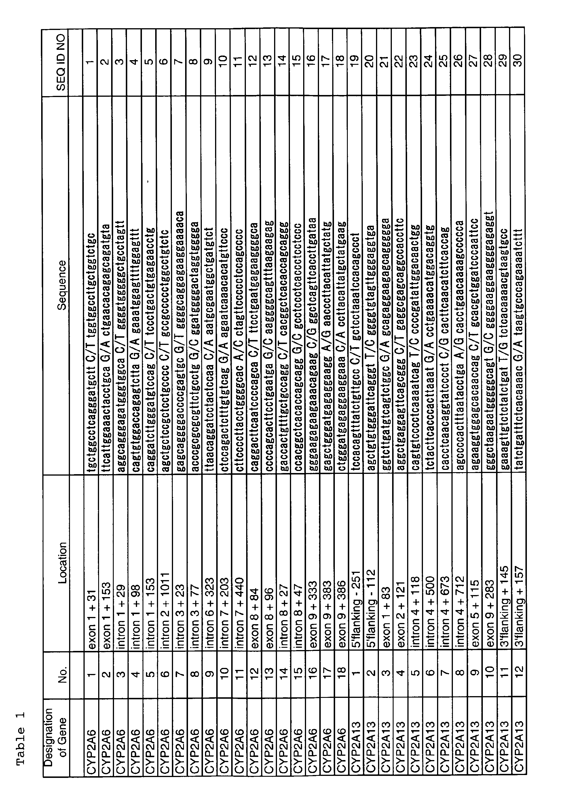
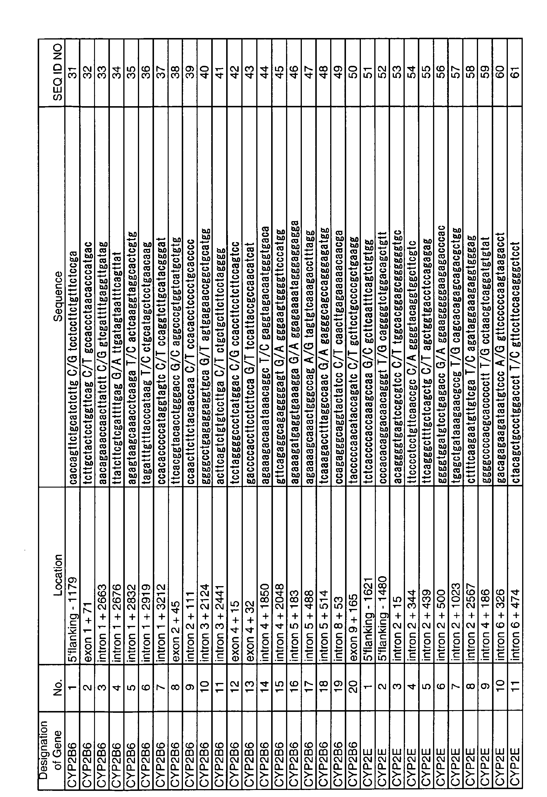


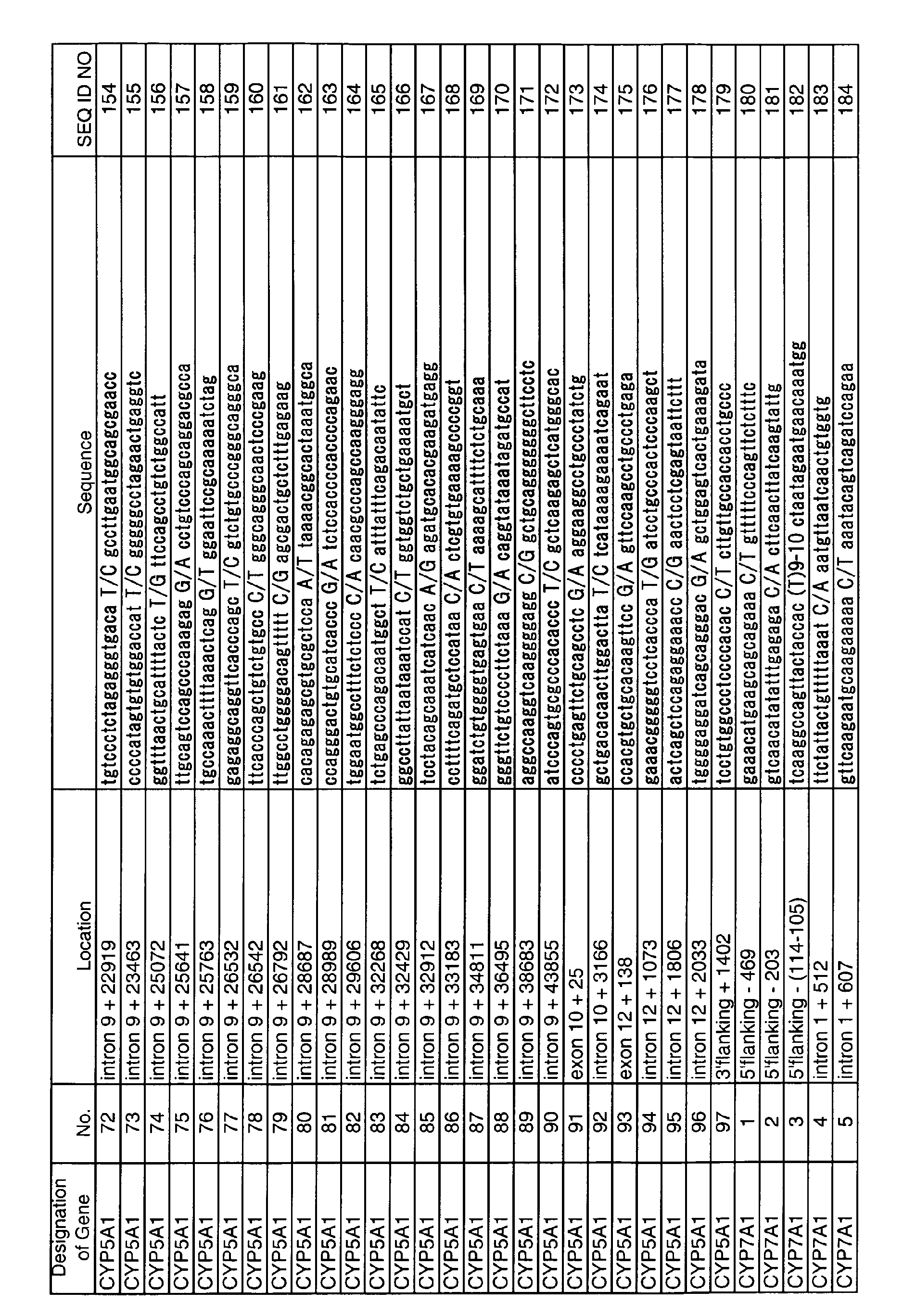

In Table 1, the "Designation of Gene" column shows the
designations of the genes encoding drug metabolizing enzymes
(CYP). The nucleotides expressed with capital letters
in the "Sequence" column are the polymorphic information.
The two nucleotides on both sides of the mark "/" represent
a homozygous or heterozygous SNP. For example, "A/G" means
that the allele is A/A or G/G homozygote or A/G heterozygote.
The sequences in this Table basically represent 20
nucleotides each before and after the SNP. However, the
nucleotide in parentheses [e.g. (C) in No. 40 of CYP5A1:
SEQ ID NO: 119] represents a polymorphism caused by insertion.
The mark "?" (e.g. see No. 21 of CYP5A1: SEQ ID NO:
100) means a polymorphism caused by deletion of one or more
nucleotides. The nucleotide in parentheses provided with a
number means that the nucleotide is repeated that number of
times. For example, "(T) 9-11" appearing in SEQ ID NO: 66
(Table 1, No. 16 of CYP2E) means a sequence where T is repeated
from 9 to 11 times. It should be noted that the
term "21st (position 21)" used in explaining locational relations
in the nucleotide sequence described in the
"Sequence" column in Table 1 means the location of a genetic
polymorphism site. Therefore, in the case of deletion
SNP (?), the deleted, imaginary nucleotide is the "21st".
In the case where a plurality of nucleotides are present at
the polymorphic site, a group of those nucleotides is the
"21st". For example, in the case of No. 15 of CYP2E (SEQ
ID NO: 65), the polymorphic site "TGT" is the 21st; in the
case of No. 16 of CYP2E (SEQ ID NO: 66), the polymorphic
site "(T) 9-11" is the 21st.
The "Location" in the Table shows the location of SNP
in the genome. The locations of SNPs in 5' flanking region,
intron regions and 3' flanking region are expressed taking
the nucleotide located 1 bp upstream of the 5' utmost
end nucleotide of exon 1 as -1 position. Then, nucleotides
are numbered -2, -3, -4, ... toward the 5' end of the gene.
An intron region means a region spanning from a nucleotide
positioned 1 bp downstream of the 3' end of an exon to the
subsequent exon. The number of an intron is the same as
the number of the exon located 5' to the relevant intron.
For example, an intron which exists between exon 3 and exon
4 (i.e. the intron located 3' to exon 3) is called intron
3. The locations of SNPs in an intron region are counted
taking the first nucleotide located immediately 3' to the
exon/intron junction located 5' to the relevant intron as
position 1 of the nucleotide sequence of the intron. Numbers
with "+" mark or without any mark in the "Location"
column mean that they are counted toward the 3' end of the
gene; numbers with "-" mark mean that they are counted toward
the 5' end of the gene. For example, if the third
nucleotide counted from exon 3/intron 3 junction toward the
3' end of the gene is polymorphic, the location is expressed
as "intron 3, +3". Similarly, if the fifth nucleotide
counted from intron 3/exon 4 junction toward the 5' end of
the gene is polymorphic, the location is expressed as
"intron 3, -5". The region located 3' to the final exon is
designated 3' flanking region. The locations of SNPs in
this region are counted taking the nucleotide located 1 bp
downstream of the 3' end nucleotide of the final exon as
position 1.
The numbers appearing in the "No." column in Table 1
correspond to the numbers appearing in respective gene maps
(Figs. 9-16) which show the locations of SNPs. In Figs. 9-16,
accession numbers of polymorphisms in public genome databases
are also provided. By using information given in
Table 1, Figures and databases, sequences adjacent to polymorphisms
can be specified. Such information is useful,
for example, in preparing PCR primers adjacent to a polymorphism.
Examples of the above-mentioned genome databases include,
but are not limited to, the list of services provided
by IMS-JST JSNP database website (Laboratory for Genotyping,
SNP Research Center, RIKEN, Japan).
Oligonucleotides which are used in the detection method
of the present invention as primers and/or probes may
be prepared based on the nucleotide sequences described in
Table 1 (SEQ ID NOS: 1-215). For example, when SNPs are to
be detected, these sequences per se may be synthesized, or
primers and/or probes may be designed and synthesized so
that they contain a part of these sequences. However, it
should be noted here that the nucleotide sequences of such
primers or probes must contain an genetic polymorphism (the
portion indicated in capital letters in the "Sequence" column
in Table 1) The present invention also includes
complementary strands to such sequences.
Taking SNP as an example for the purpose of illustration,
a primer or probe is designed so that an SNP site is
located at the 3' or 5' end of the nucleotide sequence of
the primer or probe; or a primer or probe is designed so
that an SNP site is located at the 3' or 5' end of the sequence
complementary to its nucleotide sequence; or a primer
or probe is designed so that an SNP site is located
within four nucleotides, preferably two nucleotides, from
the 3' or 5' end of its nucleotide sequence or the sequence
complementary thereto. Alternatively, a primer or probe is
designed so that an SNP site is located at the center of
the full-length sequence of the oligonucleotide. The
"center" refers to a central region where the number of
nucleotides counted from there toward the 3' end and the
number of nucleotides counted from there toward the 5' end
are almost equal. If the number of nucleotides of the oligonucleotide
is an odd number, the "center" is the central
five nucleotides, preferably the central three nucleotides,
more preferably the single nucleotide at the very center.
For example, if the oligonucleotide consists of 41 nucleotides,
the "center" is from position 19 to position 23 nucleotides,
preferably from position 20 to position 22 nucleotides,
more preferably the nucleotide at position 21. If
the number of nucleotides of the oligonucleotide is an even
number, the "center" refers to the central four nucleotides,
preferably the central two nucleotides. In the nucleotide
sequences shown in the "Sequence" column in Table 1,
if the polymorphism is deletion polymorphism, the actual
length of such sequences is 40, an even number. Therefore,
if an oligonucleotide consisting of 40 nucleotides is designed
based on such sequences, the "center" is from position
19 to position 22 nucleotides, preferably the nucleotide
at position 20.
When a polymorphic site is composed of a plurality of
nucleotides, a probe or primer is designed and prepared so
that the entire or a partial sequence of the polymorphic
site or a sequence complementary thereto is contained in
the nucleotide sequence of the probe or primer. When the
thus prepared oligonucleotide is used as a probe, it is
possible to determine alleles using the presence or absence
of hybridization or difference of hybridization. Hereinbelow,
those nucleotides in a probe or primer DNA which form
a complementary strand with the polymorphic site or its peripheral
site are called "corresponding nucleotides". A
probe or primer may be designed so that corresponding nucleotides
are located on any nucleotide(s) of the sequence
constituting a polymorphism. The "peripheral site" means a
region one to three nucleotides outside (5' side) of the 5'
utmost end of the sequence constituting a polymorphism, or
a region one to three nucleotides outside (3' side) of the
3' utmost end of the sequence constituting a polymorphism.
In particular, the corresponding nucleotides in a probe or
primer can be designed so that the 5' or 3' end nucleotide
when forming a complementary strand is located on the 5'
terminal side, 3' terminal side, or the center of the sequence
constituting a polymorphism. In the present invention,
it is preferred that the above-mentioned 5' or 3' end
nucleotide is located on the center of the sequence constituting
a polymorphism. It is also possible to design corresponding
nucleotides so that they are located on a peripheral
region of the sequence constituting a polymorphism.
For example, when an invader probe and allele probes
are prepared to detect the genetic polymorphism (TGT) as
shown in No. 15 of CYP2E (SEQ ID NO: 65) in Table 1 by the
invader method described later, allele probes are designed
so that nucleotides complementary to the polymorphic site
TGT (i.e. A, C or A in the 5' to 3' direction) are the corresponding
nucleotides of the allele probes and hybridize
(see panels a to c in Fig. 4B). For example, in panel (a)
of Fig. 4B, the 5' utmost, corresponding nucleotide "C"
forming a complementary strand is located on the center of
the nucleotides (TGT) constituting a polymorphism; in panel
(b) of Fig. 4B, the 5' utmost, corresponding nucleotide "A"
forming a complementary strand is located on "T" of the
nucleotides (TGT) constituting a polymorphism. In panel
(d) of Fig. 4B, an allele probe is shown which is designed
so that its corresponding nucleotides are located on the
peripheral region (three nucleotides) of the polymorphic
site (the underlined portion).
An invader probe is designed so that the position of
its 3' end nucleotide "N" (any one of A, T, C or G) corresponds
to the position of any of the nucleotides of the
polymorphic site (TGT) when hybridized. However, it is
most effective to design an invader probe so that there is
an overlap of one nucleotide between the invader probe and
the allele probe (Fig. 4C). On the other hand, in order to
detect another polymorphism (i.e. deletion of TGT), the invader
probe and allele probe may be designed so that a nucleotide
located one to three nucleotides upstream or
downstream of the deletion site will be the overlapping
nucleotide taking into consideration of the deletion of TGT
(i.e. deleting from the sequence). Panel (e) of Fig. 4B
shows an allele probe which is designed so that the two
nucleotides of the both sides of the deletion site hybridize
thereto. With respect to TaqMan probes, they may be designed
so that any of these nucleotides TGT or the deletion
site is located at the center of the probe.
The length of the nucleotide sequence of the oligonucleotide
is at least 13 nucleotides, preferably 13 to 60
nucleotides, more preferably 15 to 40 nucleotides, and most
preferably 18-30 nucleotides. This oligonucleotide sequence
may be used as a probe for detecting a target gene, and
it may be used as either a forward (sense) primer or a reverse
(antisense) primer.
The oligonucleotide used in the invention may be an
oligonucleotide composed of two regions connected in tandem,
one region being hybridizable to the genomic DNA and
the other region being not hybridizable thereto. The order
of connection is not particularly limited; either region
may be located upstream or downstream. The hybridizable
region of this oligonucleotide is designed based on the information
on SNP-containing sequences described in Table 1.
The oligonucleotide is prepared so that the sequence located
at the 5' or 3' utmost end of the region hybridizable
to the genomic DNA includes the SNP. The region of the above
oligonucleotide not hybridizable to the genomic DNA is
designed at random so that it does not hybridize to the
SNP-containing sequence described in Table 1. This oligonucleotide
may be used as a probe mainly for detecting SNPs
in the invader method.
Further, the primer used in the present invention is
designed so that a nucleotide sequence given in Table 1
contains an SNP when amplified by PCR for the purposes of
examining functional changes resulted from the SNP, judging
the efficiency or non-efficiency of drugs, and examining
the occurrence of side effect. The length of the primer is
designed so that at least 15 nucleotides, preferably 15 to
30 nucleotides, more preferably 18 to 24 nucleotides are
contained in the primer. The primer sequence is appropriately
selected from the template DNA so that the amplified
fragment has a length of 1000 bp or less, preferably within
500 bp (e.g. 120 to 500 bp), more preferably within 200 bp
(120 to 200 bp).
For example, primers are designed so that at least one
of the sense or the antisense primer is contained in a region
within 1000 bp, preferably 200 bp, more preferably 100
bp counted from the nucleotide immediately adjacent to 5'
or 3' to the SNP toward the 5' or 3' end of the gene,
respectively.
The thus designed oligonucleotide primers or probes
may be synthesized chemically according to known techniques.
Usually, such primers or probes are synthesized with
a commercial chemical synthesizer.
It is also possible to label probes with fluorescent
substances (e.g. FAM, VIC, Redmond Dye, etc.) in advance to
thereby automate detection procedures.
The above-described oligonucleotide may be included in
a genetic polymorphism detection kit. The genetic polymorphism
detection kit of the present invention comprises
one or more components necessary for practicing the present
invention. For example, the detection kit of the present
invention comprises components to preserve or supply enzymes
and/or reaction components necessary for performing
cleavage assay (e.g. invader assay). The detection kit of
the present invention comprises any and all components enzymes
or components necessary (suitable) for an intended
assay. Examples of such components include, but are not
limited to, oligonucleotides, polymerases (e.g. Taq polymerase),
buffers (e.g. Tris buffer), dNTPs, control reagents
(e.g. tissue samples, target oligonucleotides for positive
and negative controls, etc.), labeling and/or detection
reagents (fluorescent dyes such as VIC, FAM), solid supports,
manual, illustrative diagrams and/or product information,
inhibitors, and packing environment adjusting agents
(e.g. ice, desiccating agents). The detection kit of
the present invention may be a partial kit which comprises
only a part of the necessary components. In this case, users
may provide the remaining components. The detection
kit of the present invention may comprise two or more separate
containers, each containing a part of the components
to be used. For example, the kit may comprise a first container
containing an enzyme and a second container containing
an oligonucleotide. Specific examples of the enzyme
include a structure-specific cleaving enzyme contained in
an appropriate storage buffer or a container. Specific examples
of the oligonucleotide include invader oligonucleotides,
probe oligonucleotides, target oligonucleotides for
use as control, and the like. Alternatively, ore or more
reaction components may be provided in such a manner that
they are pre-divided into portions of a specific amount.
Since such a kit contains components which have already
been quantitatively determined for use in one step of the
method of the present invention, it is not necessary to remeasure
or re-divide. Selected reaction components may also
be mixed and divided into portions of a specific amount.
It is preferred that reaction components should be pre-divided
into portions and contained in a reactor. Specific
examples of the reactor include, but are not limited to,
reaction tubes or wells, or microtiter plates. It is especially
preferable that the pre-divided reaction component
should be kept dry in a reactor by means of, for example,
dehydration or freeze drying.
A gene encoding a drug metabolizing enzyme using DNA
polymerase (template DNA) is amplified using the oligonucleotide
prepared as above as a primer. Alternatively, the
probe prepared as above is hybridized with a template DNA
to detect a target DNA with a gene polymorphism. The
template DNA can be prepared by a known technique, for example,
cesium chloride density-gradient ultracentrifugation,
SDS dissolution, or phenol-chloroform extraction.
Amplification may be performed by polymerase chain
reaction (PCR). Specific examples of useful DNA polymerase
include LA Taq DNA polymerase (Takara), Ex Taq polymerase
(Takara), Gold Taq polymerase (Perkin Elmer), AmpliTaq
(Perkin Elmer), Pfu DNA polymerase (Stratagene) and the like.
Amplification conditions are as follows. Denaturation
step at 85-105°C for 10-40 seconds, preferably at 94°C for
20-30 seconds; annealing step at 50-72°C for 20 seconds to
1 minute, preferably at 60°C for 20 seconds to 1 minutes;
and extension step at 65-75°C for 1-4 minutes, preferably
at 72°C for 2-3 minutes constitute one cycle, and 30 to 40
cycles are performed. However, in order to denature the
template DNA and the primers sufficiently, a denaturation
step of at 95°C for 1-5 minutes [if Gold Taq polymerase
(Perkin Elmer) is used, at least 8-15 minutes, preferably
10-12 minutes] may be added before the start of the above-described
amplification cycles. Also, in order to extend
the amplified DNA completely, an extension step of at 72°C
for 1-5 minutes for 1-10 minutes may be added after the above
amplification cycles. If the detection of the amplified
product is not performed immediately, it is desirable
to add a step of storing the amplified product at 4°C to
avoid unspecific amplification. Thus, a gene encoding drug
metabolizing enzyme (CYP) can be amplified.
Subsequently, the amplified product is subjected to
agarose gel electrophoresis, followed by staining with ethidium
bromide, SYBR Green solution or the like to thereby
detect the amplified product as a band or two to three
bands (DNA fragments). Thus, a part of a gene encoding
drug metabolizing enzyme, containing a genetic polymorphism
can be detected as a DNA fragment. Instead of agarose gel
electrophoresis, polyacrylamide gel electrophoresis or capillary
electrophoresis may be performed. It is also possible
to perform PCR using primers labeled with a substance
such as fluorescent dye and to detect the amplified product.
A detection method which does not require electrophoresis
may also be employed; in such a method, the
amplified product is bound to a solid support such as a
microplate, and a DNA fragment of interest is detected by
means of fluorescence, enzyme reaction, or the like.
TaqMan PCR is a method using PCR reaction with fluorescently
labeled allele-specific oligos and Taq DNA polymerase.
The allele-specific oligo used in TaqMan PCR
(called "TaqMan probe") may be designed based on the SNP
information described above. The 5' end of TaqMan probe is
labeled with fluorescence reporter dye R (e.g. FAM or VIC),
and at the same time, the 3' end thereof is labeled with
quencher Q (quenching substance) (Fig. 1). Under these
conditions, fluorescence is not detectable since the quencher
absorbs fluorescence energy. Since the 3' end of TaqMan
probe is phosphorylated, no extension reaction occurs
from TaqMan probe during PCR Fig. 1). However, when PCR is
performed using this TaqMan probe together with Taq DNA polymerase
and primers designed so that an SNP-containing region
is amplified, the reaction described below occurs.
First, a TaqMan probe hybridizes to a specific sequence
in the template DNA (Fig. 2a), and at the same time, an
extension reaction occurs from a PCR primer (Fig. 2b). At
this time, Taq DNA polymerase having 5' nuclease activity
cleaves the hybridized TaqMan probe as the extension reaction
proceeds. When the TaqMan probe has been cleaved, the
fluorescent dye becomes free from the influence of the
quencher. Then, fluorescence can be detected (Fig. 2c).
For example, as shown in Fig. 3, two alleles are supposed:
one allele has A at the SNP site (allele 1) and the
other allele has G at the SNP site (allele 2). A TaqMan
probe specific to allele 1 is labeled with FAM and another
TaqMan probe specific to allele 2 is labeled with VIC (Fig.
3). These two allele specific oligos are added to PCR reagents,
and then TaqMan PCR is performed with a template DNA
whose SNP is to be detected. Subsequently, fluorescence
intensities of FAM and VIC are determined with a fluorescence
detector. When the SNP site of the allele is complementary
to the site within TaqMan probe corresponding to
the SNP, the probe hybridizes to the allele; and Taq polymerase
cleaves the fluorescent dye of the probe, which becomes
free form the influence of the quencher. As a result,
fluorescence intensity is detected.
If the template is a homozygote of allele 1, strong
fluorescence intensity of FAM is recognized but the fluorescence
of VIC is hardly recognized. If the template is a
heterozygote of allele 1 and allele 2, fluorescence of both
FAM and VIC can be detected.
The invader method is a method for detecting SNPs by
hybridizing allele-specific oligos to the template. In the
invader method, two unlabeled oligos and one fluorescently
labeled oligo are used. One of the two unlabeled oligos is
called an "allele probe". The allele probe is composed of
a region which hybridizes to the genomic DNA (template DNA)
to form a complementary double strand, and a region (called
"flap") which has a sequence entirely unrelated to the sequence
of the template DNA and thus does not hybridize to
the genomic DNA. A nucleotide located at the 5' or 3' utmost
end of the hybridizable region corresponds to the SNP
(panel (a) in Fig. 4A). The above-described flap sequence
is an oligonucleotide having a sequence complementary to a
FRET probe described later. The other unlabeled oligo is
called an "invader probe". This oligo is designed so that
it hybridizes complementarily to the template DNA from the
SNP site toward the 3' end of the genomic DNA (panel (b) in
Fig. 4A). However, the nucleotide corresponding to the SNP
("N" in panel (b) in Fig. 4A) may be any nucleotide. Thus,
when the genomic DNA (the template) is hybridized to the
above-described two probes, one nucleotide (N) of the invader
probe invades into the SNP site (panel (c) in Fig. 4A).
As a result, a triple strand is formed at the SNP site.
On the other hand, the fluorescently labeled oligo has
a sequence completely unrelated to the allele. This sequence
is common regardless of the types of SNPs. This
probe is called a "FRET" probe (fluorescence resonance energy
transfer probe) (Fig. 5). The nucleotide at the 5'
end of FRET probe (reporter) is labeled with fluorescent
dye R, while quencher Q is linked upstream of the reporter.
Therefore, under these conditions, the quencher absorbs the
fluorescent dye and no fluorescence is detectable. A certain
region of the FRET probe starting from the 5' end reporter
nucleotide (designated "region 1") is designed so
that it is complementary to a certain region of the probe
located 3' to region 1 (designated "region 2") when region
1 and region 2 are faced with each other. Therefore, region
1 and region 2 form a complementary strand within the
FRET probe (Fig. 5). Also, the region located downstream
of this complementary strand forming region is designed so
that it hybridizes to the flap of the allele probe to thereby
form a complementary strand_(Fig. 5).
In the invader method, an enzyme called cleavase is
used which is one of 5' nucleotidases having a unique endonuclease
activity of cleaving upon recognition of a special
structure of DNA. Cleavase is an enzyme which cleaves the
allele probe at a point immediately 3' to the SNP site when
the genomic DNA, the allele probe and the invader probe
form a triple strand at the SNP site. Therefore, when
three nucleotides form a triple strand as shown in panel
(c) in Fig. 4A, cleavase recognizes the 5' flap and cuts
off this flap. As a result, the structure of this SNP site
is recognized by cleavage (panel (a) in Fig. 6), and the
allele probe is cut at the site of its flap to liberate the
flap (panel (b) in Fig. 6). Subsequently, the flap liberated
from the allele probe complementarily binds to the FRET
probe since it has a sequence complementary to the FRET
probe (panel (c) in Fig. 6). At this time, the SNP site of
the flap invades into the portion of the FRET probe which
has already formed a double strand. Cleavase again recognizes
this structure and cuts off the nucleotide labeled
with the fluorescent dye. The thus cleaved fluorescent dye
becomes free from the influence of the quencher and emits
fluorescence (panel (d) in Fig. 6). When the SNP in the
genomic DNA does not match the nucleotide corresponding to
the SNP in the allele probe, a specific DNA structure recognizable
by cleavase is not formed as seen in Fig. 7.
Thus, the allele probe is not cleaved and no fluorescence
is detected.
For example, when an SNP to be detected is T/C, an invader
probe and an allele probe for T, and a FRET probe
with a FAM-linked reporter corresponding to the SNP are
prepared. Separately, an invader probe and an allele probe
for C, and a FRET probe with a VIC-linked reporter corresponding
to the SNP are prepared are also prepared.
Then, all of them are mixed to carry out SNP detection. As
a result, if the SNP of a subject is T/T homozygous, the
fluorescence of FAM is emitted; if the SNP of a subject is
C/C homozygous, the fluorescence of VIC is emitted; and if
the SNP of a subject is T/C heterozygous, the fluorescence
of both FAM and VIC is emitted. Since FAM and VIC have
different fluorescent wavelengths, they can be discriminated.
Products labeled with fluorescent dyes can be detected
with a fluorescent plate reader or an apparatus for
collecting fluorescence data generated during reaction
(real-time fluorescence detector). Specific examples of
real-time fluorescence detector include ABI 7700 sequence
detection system (Applied Biosystems).
In order to detect SNPs by SniPer method, it is possible
to discriminate alleles by examining the presence or
absence of amplification by RCA. Briefly, the genomic DNA
to be used as a template is linearized. Then, a probe is
hybridized to this genomic DNA. When the probe sequence
and the sequence of the genomic DNA are complementary to
each other and form a double strand, the genomic DNA can be
converted into a circular DNA through ligation reaction.
As a result, RCA of the circular DNA proceeds. On the other
hand, when the ends of the probe do not match with the
genomic DNA, the DNA is not ligated to become a circular
DNA. Thus, RCA reaction does not proceed. Therefore, in
Sniper method, a single-stranded probe which anneals with
the genomic DNA and is circularizable is designed. This
single-stranded probe is called a padlock probe. The sequences
of the two ends of this padlock probe are designed
so that they correspond to the SNP to be detected. Then,
this padlock probe and the genomic DNA are mixed for ligation.
If the two ends of the padlock probe and the SNP site
of the genomic DNA are complementary to each other, the
two ends of the padlock probe are joined by ligation, yielding
a circular probe. If the two ends of the padlock probe
and the SNP site of the genomic DNA are not complementary
to each other, the probe does not become circular. Therefore,
only those padlock probes which are complementary
to the SNP to be detected become circular and are amplified
by DNA polymerase. By detecting the presence or absence of
this amplification, SNP may be detected. For the detection,
synthetic oligonucleotides which have a fluorescent dye
and a quencher at their respective ends and also have a
hairpin structure are used.
MALDI-TOF/MS (Matrix Assisted Laser Desorption-Time of
Flight/Mass Spectrometry) is a method using a mass spectrometer
in SNP typing. This method is composed of the following
steps.
In the present invention, polymorphisms may be detected
by using single nucleotide extension reactions.
Briefly, four types of dideoxynucleotides labeled with different
fluorescent compounds are added to a reaction system
containing a gene of interest. Then, single nucleotide extension
reactions are performed. In this case, the nucleotide
to be extended is the polymorphic site. Also, two
reactions of DNA synthesis termination and the fluorescent
labeling of the 3' end of DNA molecules are operated. Four
types of reaction solutions are subjected to electrophoresis
on the same lane of a sequencing gel or on capillary.
Difference in the fluorescent dyes used for labeling is detected
with a fluorescence detector to thereby sequence the
DNA band. Alternatively, the one-nucleotide extended oligonucleotide
is examined with a fluorescence detection system
or a mass spectrometry system to thereby determine
which nucleotide was extended using the difference in the
fluorescent dyes. Instead of fluorescently labeled dideoxynucleotides,
primers may be fluorescently labeled and used
with unlabeled dideoxynucleotides.
DNA microarrays are solid supports onto which nucleotide
probes are immobilized, and they include DNA chips,
gene chips, microchips, beads arrays, and the like. As a
specific example of DNA microarray (e.g. DNA chip) assay,
GeneChip assay (Affymetrix; US Patent Nos. 6,045,996;
5,925,525; and 5,858,659) may be given. GeneChip technology
uses small sized, high density microarrays of oligonucleotide
probes affixed to chips. Probe arrays are manufactured,
for example, by the light irradiation chemical synthesis
method (Affymetrix) which is a combination of solid
chemical synthesis and photolithography production technology
used in the semiconductor industry. High density arrays
can be constructed by using photolithography masks in
order to make the boundary of the chemical reaction site of
chips definite and by performing a specific chemical synthesis
step. Multi-probe arrays are synthesized simultaneously
on a large glass baseboard. Subsequently, this baseboard
is dried, and individual probe arrays are packed in
injection-molded plastic cartridges. This cartridge protects
the array from the outer environment and also serves
as a hybridization chamber.
First, a polynucleotide to be analyzed is isolated,
amplified by PCR, and labeled with a fluorescent reporter
group. Then, the labeled DNA is incubated with an array
using a fluid station. This array is inserted into a scanner
to detect a hybridization pattern. Hybridization data
are collected as luminescence from fluorescent reporter
group bound to the probe array (i.e. taken into the target
sequence). Generally, probes which completely matched with
the target sequence generate stronger signal than those
probes which have portions not matching with the target sequence.
Since the sequences and locations of individual
probes on the array are known, it is possible to determine
the sequence of the target polynucleotide reacted with the
probe array on the basis of complementation.
In the present invention, it is also possible to use
DNA microchips with electrically captured probes (Nanogen;
see, for example, US Patents Nos. 6,017,696; 6,068,818; and
6,051,380). By using microelectronics, the technology of
Nanogen is capable of transferring charged molecules to and
from specific test sites on semidonductor microchips and
concentrating them. DNA capturing type probes specific to
certain SNPs or variations are arranged on specific sites
on microchips or assigned addresses. Since DNA is strongly
negatively charged, it is capable of moving electronically
to a positively charged area.
First, the test site or a row of the test site on a
microchip is electronically activated with positive charge.
Then, a solution containing DNA probes is introduced onto
the microchip. Since negatively charged probes quickly moves
to a positively charged site, probes are concentrated
at this site on the microchip and chemically bind thereto.
This microchip is washed, and another DNA probe solution is
added to the microchip to allow specific binding of DNA
probes to the chip.
Subsequently, whether the target DNA molecule is present
in a test sample or not is analyzed. This analysis is
performed by judging the type of the DNA capturing probe
which has hybridized to a complementary DNA in a test sample.
As a test sample, a gene of interest which has been
amplified by PCR may be given. By using electric charge,
target molecules may be moved to one or more test sites on
the microchip and concentrated. As a result of electronic
concentration of the sample DNA at each test site, the
hybridization between the sample DNA and a capturing probe
complementary thereto is performed quickly. For example,
as a result of these operations, hybridization occurs in
several minutes. In order to remove unbound DNA or non-specifically
bound DNA from each test site, the polarity or
electric charge of the site is converted to negative charge
to thereby return the unbound DNA or non-specifically bound
DNA into the solution. In this method, specific binding
can be detected, for example, with a fluorescence scanner
utilizing laser.
Further, in the present invention, it is also possible
to use an array technology utilizing fluid separation phenomenon
on a plane surface (chip) because of difference in
surface tensions (ProtoGene; see, for example. US Patents
Nos. 6,001,311; 5,985,551; and 5,474,796). The technology
of ProtoGene is based on a fact that fluids are separated
from each other on a plane surface because of difference in
surface tensions given by chemical coating. Since oligonucleotide
probes may be separated based on the above-mentioned
principle, it is possible to synthesize probes
directly on a chip by the ink-jet printing of a reagent
containing probes. An array having reaction sites defined
by surface tension is mounted on X/Y movable stage located
under one set (4) of piezoelectric nozzles. Each piezoelectric
nozzle contains four standard nucleotides. This
movable stage moves along each row of the array to supply
an appropriate reagent (e.g. amidite) to each reaction site.
The entire surface of the array is soaked in a reagent
common to the test sites in the array and then in a washing
solution. Subsequently, the array is rotated to remove
these solutions.
DNA probes specific to the SNPs or variations to be
detected are affixed to a chip using the technology of Protogene.
Then, the chip is contacted with PCR-amplified gene
of interest. After hybridization, unbound DNA is removed,
followed by detection of hybridization using an appropriate
method.
Further, it is possible to detect polymorphisms using
"bead arrays" (Illumina Inc.; see, for example, PCT International
Publication Nos. WO 99/67641 and WO 00/39587).
Illumina Inc. utilizes Bead Array technology which uses a
combination of optical fiber bundles and beads that undergo
self-association with the array. Each optical fiber bundle
has several millions of fibers depending on the diameter of
the bundle. Beads are coated with oligonucleotides specific
for the detection of certain SNPs or variations. Various
types of beads are mixed in specific amounts to allow
the formation of an array-specific pool. For assay, a bead
array is contacted with a sample prepared from a subject.
Then, hybridization is detected by an appropriate method.
In the present invention, it is possible to evaluate
the efficacy and safety of a drug metabolized by the drug
metabolizing enzyme, from the results of detection of genetic
polymorphisms such as SNP obtained as described above.
Evaluation of drugs may be performed by typing system.
Briefly, according to any one of the detection methods
described above, allele frequencies between toxicity (side
effect) occurrence group and non-occurrence group are compared.
A polymorphism which brings about difference in allele
frequencies between the two groups is selected as a
marker for recognizing the occurrence of toxicity. As a
statistical test, usually ÷2 test is carried out, but other
statistical processing such as Fisher test may also be used.
Further, it is possible to reflect this result on the
concentration of active body of drugs (or unchanged body or
metabolite of drugs) in blood or tissue. With respect to
all genetic polymorphisms, the relation of cause and effect
with the toxicity is examined. Then, only those genetic
polymorphism sites that show correlation with the toxicity
are selected. Allele pattern can be examined by preparing
in advance all probes or primers for analyzing the genetic
polymorphisms and reagents necessary for each technique in
reaction plates, cards, glass baseboards or the like, and
adding thereto the genomic DNA of a human subject for reaction.
When the subject has a genetic polymorphism which
has correlation with the toxicity, it is possible to predict
whether the drug exhibits effect or toxicity in that
subject. The efficacy of a drug may be evaluated in a similar
manner. Genetic polymorphisms which correlate with
side effect or efficacy vary depending on drugs. Therefore,
by conducting typing using the genetic polymorphisms,
it becomes possible to predict the efficacy or side effect
of the drug.
Using this, the frequency of the genetic polymorphism
is compared with efficacy/non-efficacy or presence/absence
of side effect. When there is difference in allele frequency,
a judgment on the drug can be made.
For example, if the results of analysis of an SNP in
persons who showed toxicity (side effect) upon administration
of drug A have revealed statistically that 90% of those
persons have T/T (e.g. fluorescence intensity of FAM was
detected), and if the results of analysis of the SNP in
persons who did not show toxicity (side effect) have revealed
that only 10% of those persons have T/T and 90% of them
have C/C, drug A can be evaluated that it should not be administered
to persons with T/T.
The genetic polymorphism information obtained as described
above in the present invention may be compared with
genetic polymorphism information in a gene of a subject encoding
corresponding drug metabolizing enzyme. By doing
so, the above information may be used as an indicator for
analyzing the efficacy and safety of a drug metabolized by
the drug metabolizing enzyme. Therefore, the genetic polymorphism
information obtained in the present invention serves
as an information source for selecting most effective
drug to treat a disease.
As a method, the evaluation method described in "8.
Evaluation of Drugs" may be used. Briefly, it can be said
that the genetic polymorphisms which were recognized to be
correlating with side effect or efficacy in the preceding
sub-section give influence upon the activity, the transcription,
and the translation of the enzyme. Also, it can
be said that such genetic polymorphisms have some indirect
relation with a mechanism of the occurrence of side effect
or efficacy. The metabolism of a drug is examined and confirmed
by a pharmaceutical manufacturer or the like in pre-clinical
test or clinical test. Therefore, if the drug is
an enzyme and a polymorphism which correlates with severe
side effect exists among the polymorphisms located in the
gene encoding the enzyme, it is possible to delete the relevant
polymorphism or to use the drug conditionally. With
respect to efficacy, a similar evaluation can be made.
From such information on side effect and efficacy, drug
screening becomes possible.
Further, by conducting genetic polymorphism frequency
analysis on cases with side effect occurrence and cases
without side effect occurrence in clinical tests (from phase
I to phase III tests), it becomes possible to detect new
genetic polymorphisms other than the above-mentioned polymorphism
which correlate with side effect or efficacy. By
examining such polymorphisms in the same manner as described
above, drug screening becomes possible.
Hereinbelow, the present invention will be described
more specifically with reference to the following Example.
However, the technical scope of the present invention is
not limited to the Example.
Blood was collected in the presence of EDTA from 48
unrelated individuals. Extraction of DNA was carried out
as described below according to the method described in
"Genomic Analysis Laboratory Manual (Genomu Kaiseki Rabo
Manyuaru)" (Yusuke Nakamura ed., Springer-Verlag Tokyo).
Ten ml of blood was placed in a 50-ml Falcon tube and
centrifuged at 3000 rpm at room temperature for 5 minutes.
The supernatant (serum) was collected with a pipette, followed
by addition of 30 ml of RBC lysing buffer (10 mM
NH4HCO3, 144 mM NH3Cl). After mixing until the sediment
was loosened, the mixture was left at room temperature for
20 minutes. This was then centrifuged at 3000 rpm at room
temperature for 5 minutes, and the supernatant (serum) was
discarded using a pipette to obtain leukocyte pellets.
Thirty ml of RBC lysing buffer was added to the pellets and
the same procedures were further repeated 2 times. To the
leukocyte pellets 4 ml of Proteinase K buffer (50 mM Tris-HCl
(pH 7.4), 100 mM NaCl, 1 mM EDTA (pH 8.0)), 200 µl of
10% SDS, 200 µl of 10 mg/ml Proteinase K were added, and
the mixture was mixed with inversion, and left overnight at
37°C. Four ml of phenol was added and slowly mixed with
inversion using a rotator (Rotator T-50, Taitec). This
mixture was centrifuged at 3000 rpm at room temperature for
10 minutes, and the top layer was collected into a new tube.
Four ml of phenol-chloroform-isoamyl alcohol (ratio by
volume 25:24:1) was added, mixed with inversion for 2 hours
as above and centrifuged. The top layer was collected into
a new tube, and 4 ml of chloroform-isoamyl alcohol (ratio
by volume 24:1) was added, and mixed with inversion for 30
minutes as above and centrifuged. The top layer was collected
into a new tube, and 400 µl of 8 M ammonium acetate
and 4 ml of isopropanol were added, and then mixed with inversion.
Stringy white precipitate (DNA) was collected into
a 2-ml tube, and 1 ml of 70% ethanol was added and mixed
with inversion. After DNA was collected into a new 2-ml
tube and air-dried, 500 µl of TE solution (10 mM Tris-HCl
(pH 7.4) and 1 mM EDTA (pH 7.4)) were added to dissolve the
DNA to obtain genomic DNA sample.
The genomic sequence was obtained from GenBank DNA database.
The repeat sequence was removed using RepMask computer
program, and PCR primers were designed so that PCR
products would be approximately 1 kb. The genomic DNA used
was DNA from 48 unrelated individuals prepared to be at the
same concentration. Equal amounts of each DNA from 3 individuals
were placed in one tube and mixed, and 60 ng out of
the mixture was used for PCR. PCR was performed using Ex-Taq
(2.5 U; TaKaRa) on GeneAmp PCR System 9700 (PE Applied
Biosystems). One cycle was composed of reaction at 94°C
for 2 minutes followed by denaturation at 94°C for 30 seconds,
annealing at 60°C or 55°C for 30 seconds, and extension
at 72°C for 1 minute, and 35 cycles were completed.
PCR products were purified using Arraylt (Telechem),
and underwent sequence reaction with BigDye Terminator RR
Mix (PE Applied Biosystems). One cycle was composed of
reaction at 96°C for 2 minutes, followed by denaturation at
96°C for 20 seconds, annealing at 50°C for 30 seconds, and
extension at 60°C for 4 minutes using GeneAmp PCR System
9700 (PE Applied Biosystems), and 25 cycles were completed.
After the sequence reafction, sequence analysis was carried
out on ABI PRISM 3700 DNA Analyzer.
PolyPhred computer program (Nickerson et al., 1997,
Nucleic Acids Res., 25, 2745-2751) was used to carry out
analysis for SNP detection.
The results as shown in Table 1 were obtained for SNP. In
addition, the names and abbreviations of drug metabolizing
enzymes analyzed, ACCESSION number from database (GenBank),
and genetic structure and positions of SNPs of drug metabolizing
enzyme are shown in Figures 9 to 16. In Figures 9
to 16, exons are shown as white boxes or black lines across genes
which are shown as horizontal lines. Positions of SNPs are
shown with solid lines above the genes and numbered.
Polymorphism of CYP genes obtained from 2 different groups
of subjects was determined with typing by the Invader method.
The results are shown in Figure 17. In Figure 17, the horizontal
(Allele 1) and vertical (Allele 2) axes represent fluorescence
intensity of VIC corresponding to G and FAM corresponding to
A, respectively. Each area circled in with broken lines shows
SNP patterns for dark rectangles (▪) , dark circles (), and dark
triangles (▴) are G/G, A/A, and G/A, respectively. White
rectangles (□) mean background values. It can be seen that the
subject group shown in the graph of panel A (top) frequently
has a SNP pattern of G/G, whereas the subject group shown in
the graph of panel B (bottom) frequently has a SNP pattern of
A/A.
Using as samples genomic DNA collected from 5 unrelated
individuals as described in Example 1, detection of SNP in 2
types of CYP genes (CYP2E, CYP2S) was performed by the Invader
method. Invader probes and allele probes designed based on the
respective sequences of Nos. 6 and 13 (SEQ ID Nos. 56 and 63)
for CYP2E and Nos. 3 and 10 (SEQ ID Nos. 72 and 79) for CYP2S
were used. Locations of each SNP are shown in Table 1.
Results are shown in Table 2.
| Drug metabolizing enzyme gene | CYP2E | CYP2S | ||
| No. 6 | No. 13 | No. 3 | No. 10 | |
| SEQ ID No. 56 | SEQ ID No. 63 | SEQ ID No. 72 | SEQ ID No. 79 | |
| SNP | (G/A) | (T/C) | (G/C) | (C/A) |
| Subject I | G/G | T/C | C/C | C/C |
| Subject II | G/A | T/C | G/C | A/A |
| Subject III | G/G | T/T | G/C | C/C |
| Subject IV | G/A | T/T | G/C | C/A |
| Subject V | G/A | T/C | G/G | C/A |
| Subject VI | A/A | C/C | G/C | C/A |
It can be seen from the result shown in Table 2 that
the method of the present invention can detect SNP and determine
their patterns in drug metabolizing enzyme genes of
each subject.
All publications, patents, and patent applications cited
herein are incorporated herein by reference in their
entirety.
According to the present invention, methods for analyzing
SNPs are provided. According to the methods of the
invention, it becomes possible to select appropriate drugs
for target diseases. Thus, the methods of the invention
are extremely useful.
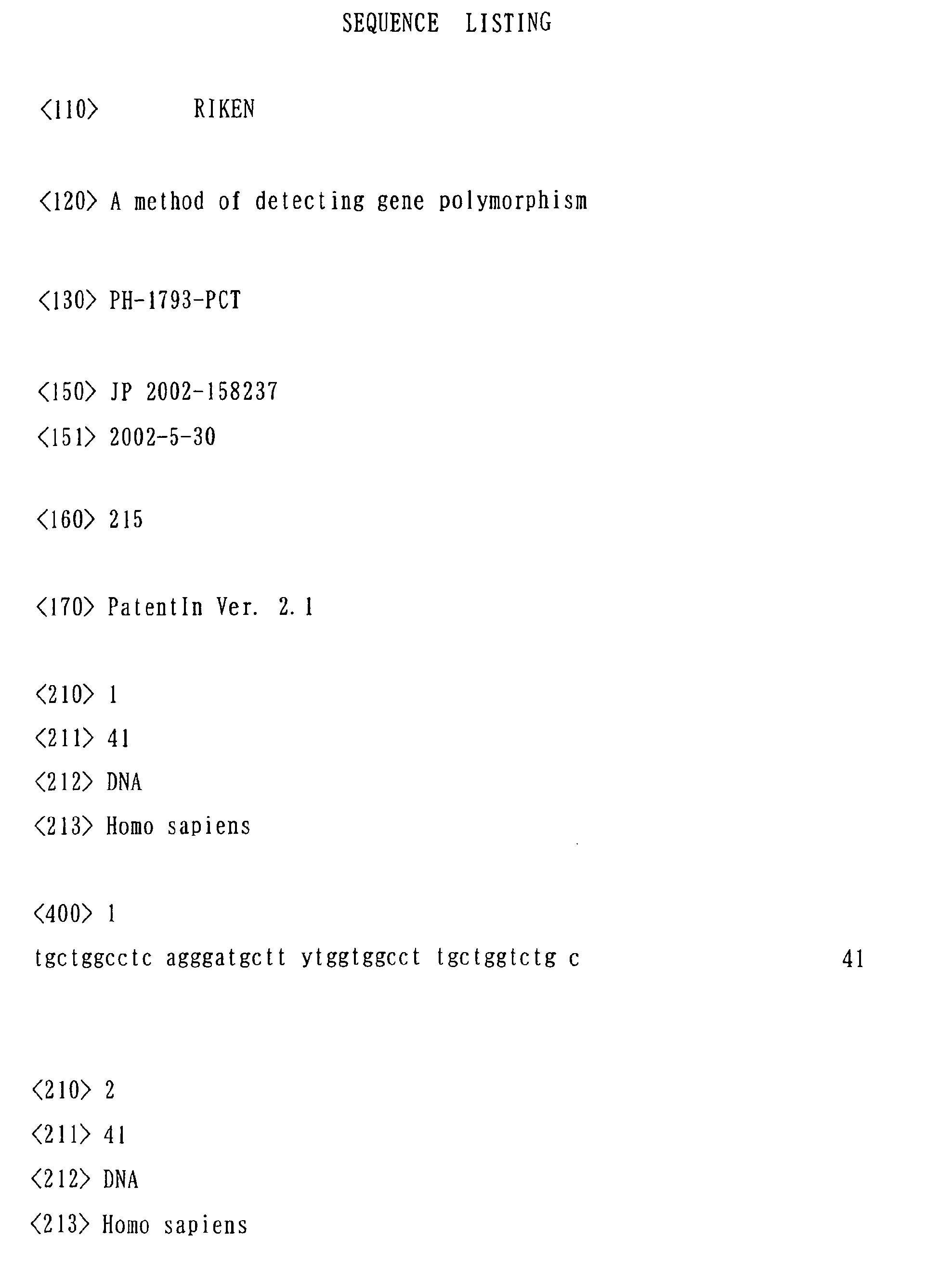


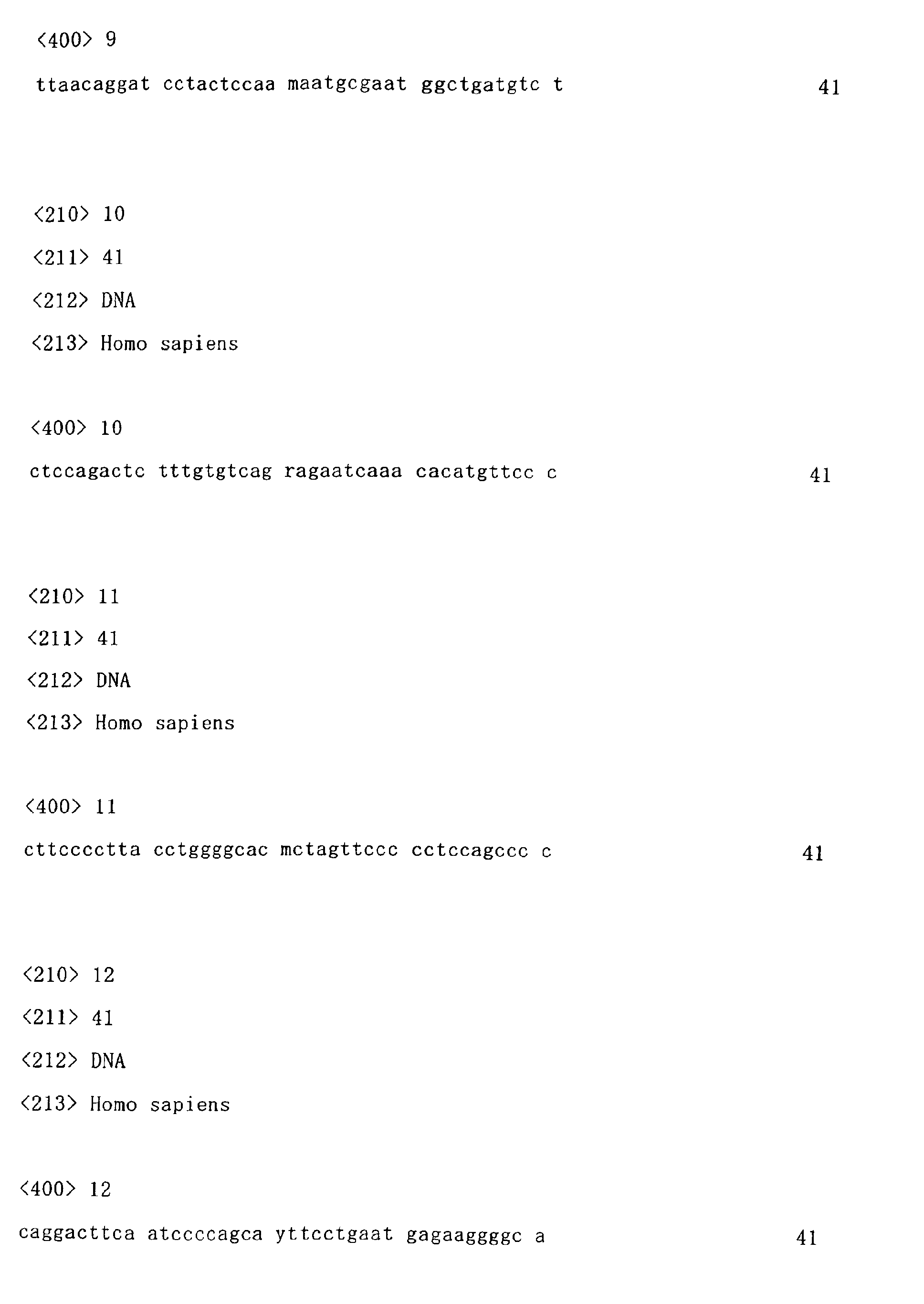

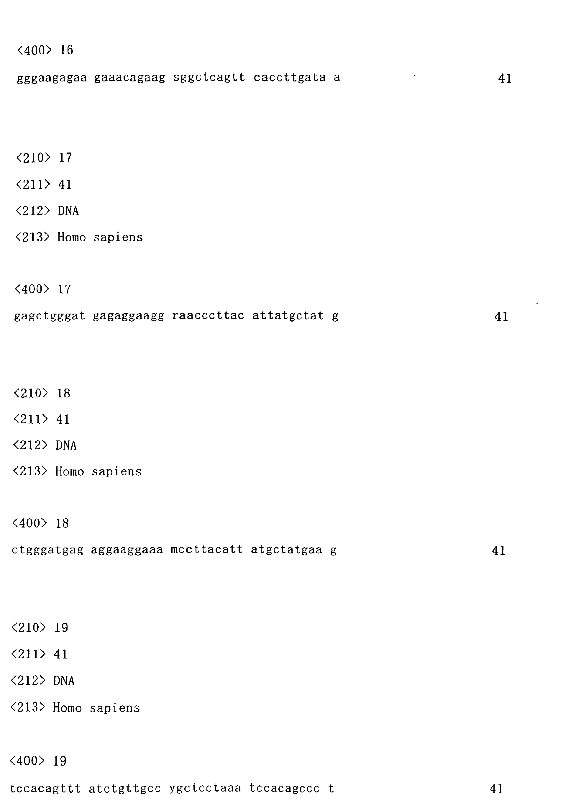

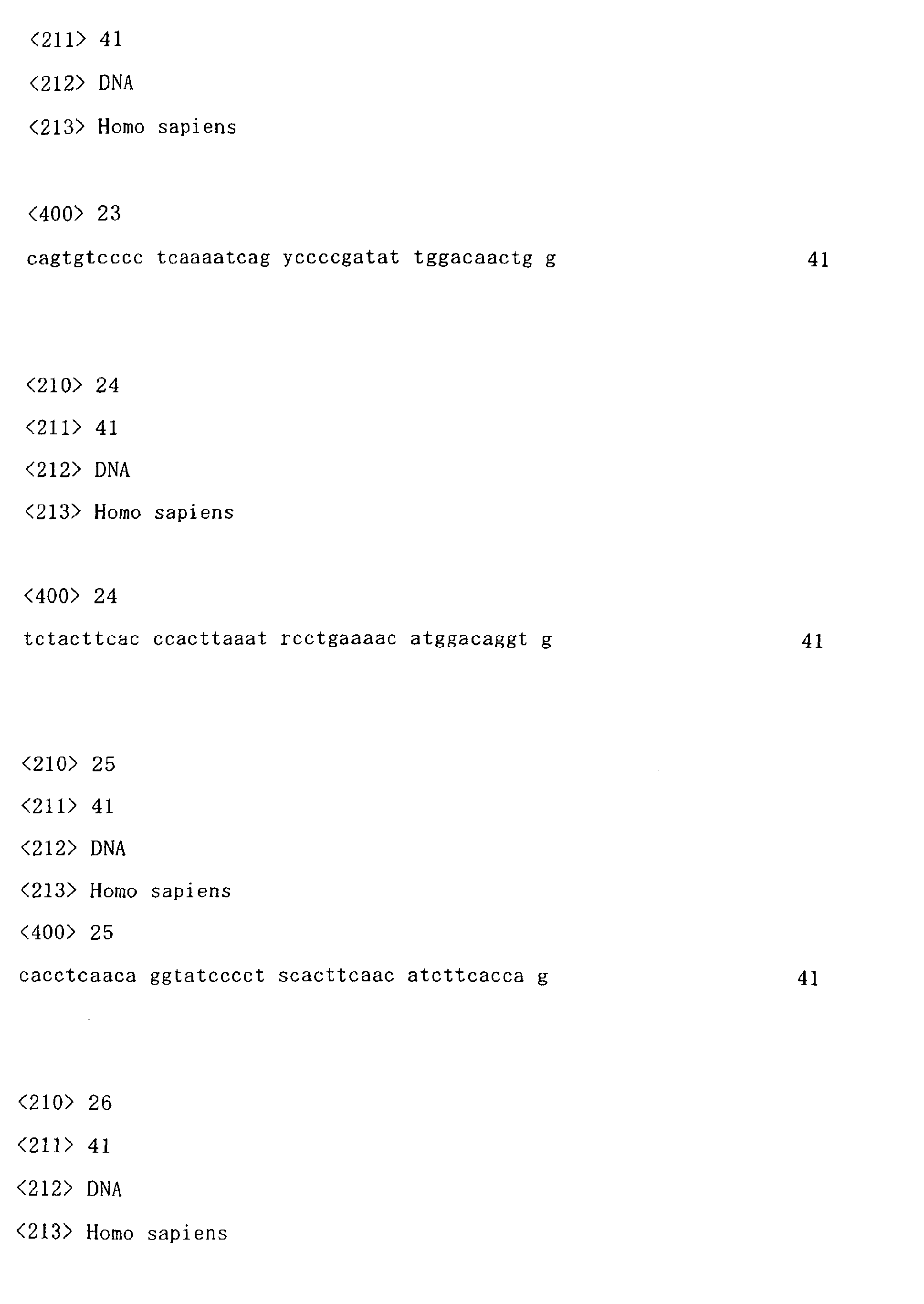


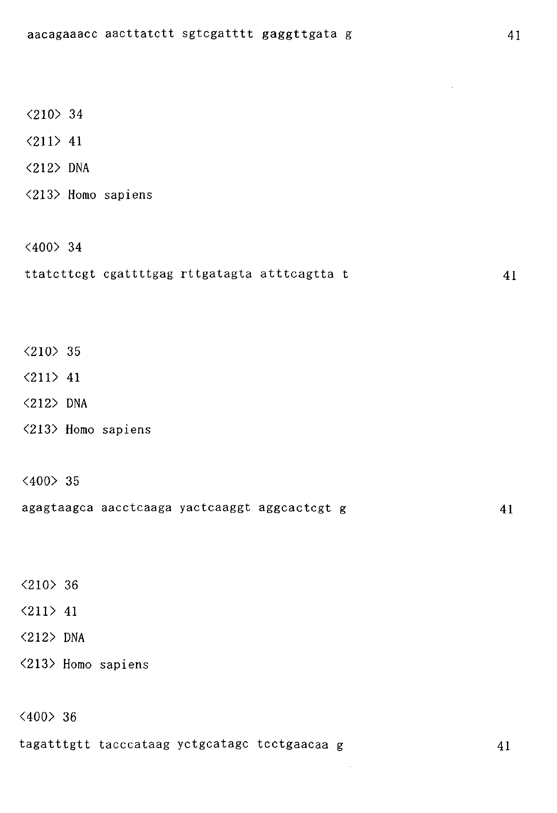


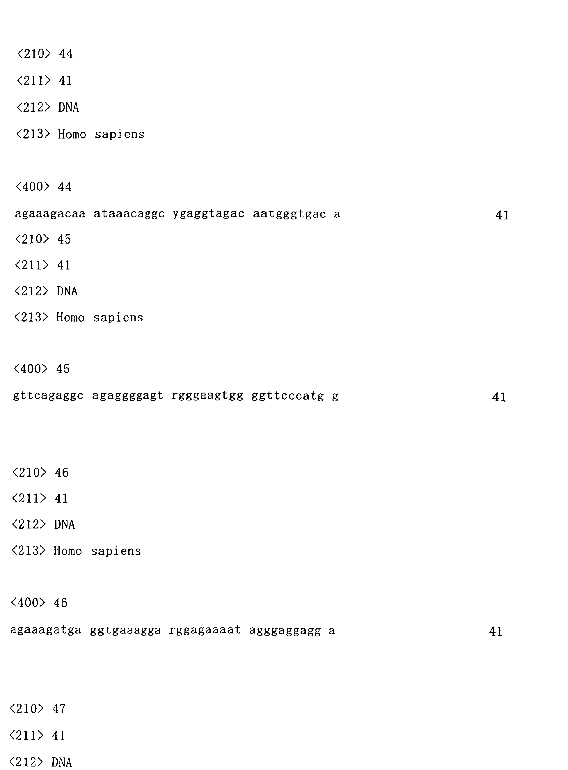

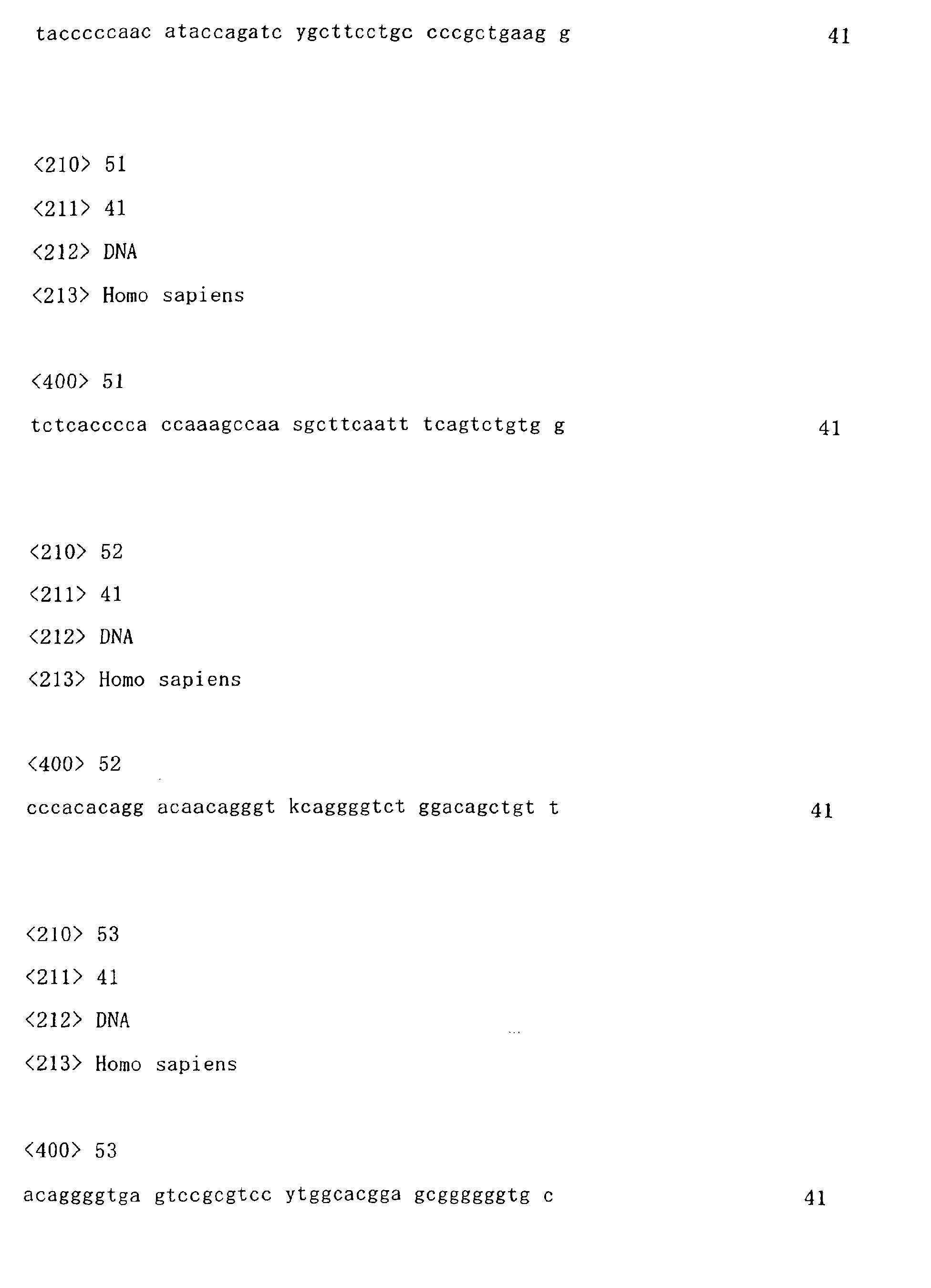


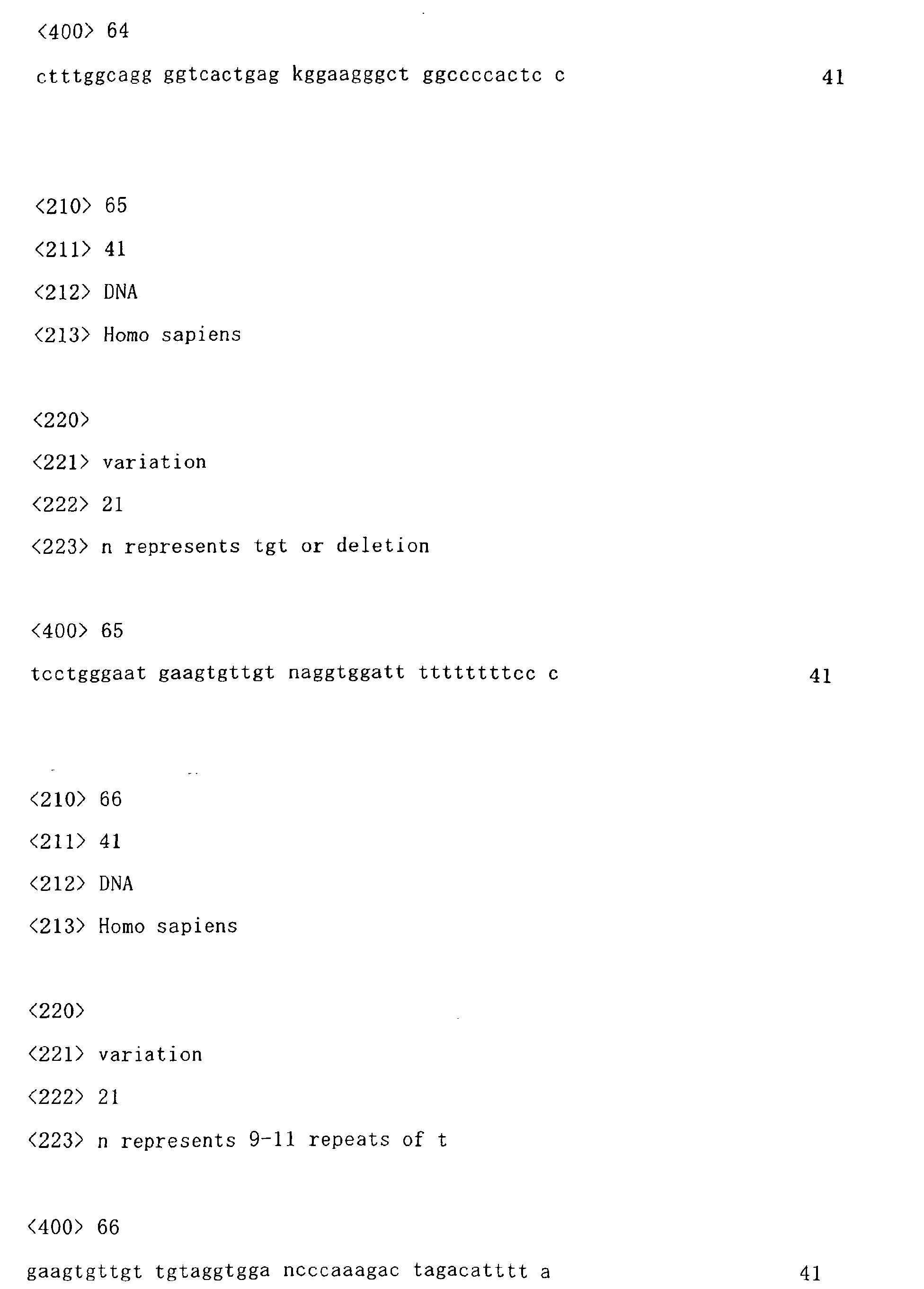

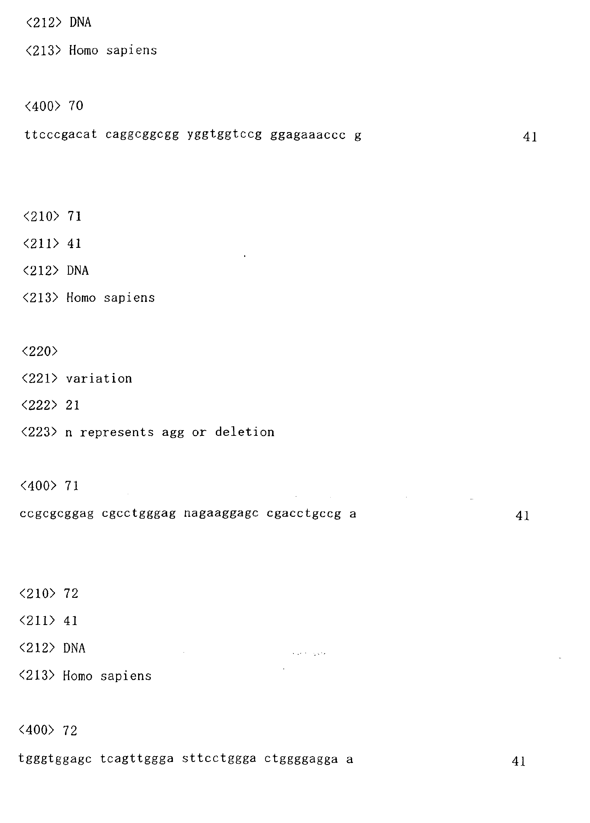


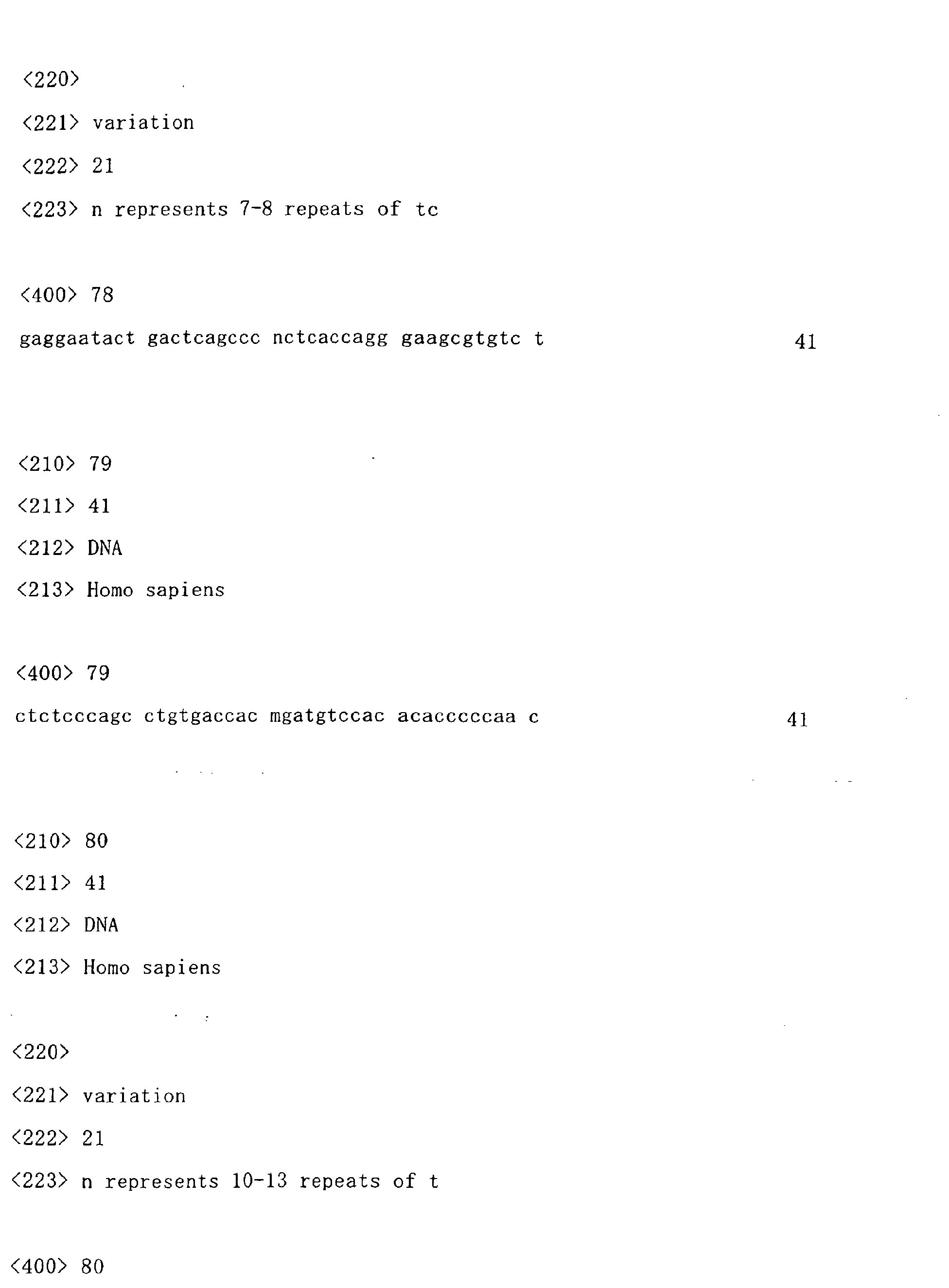


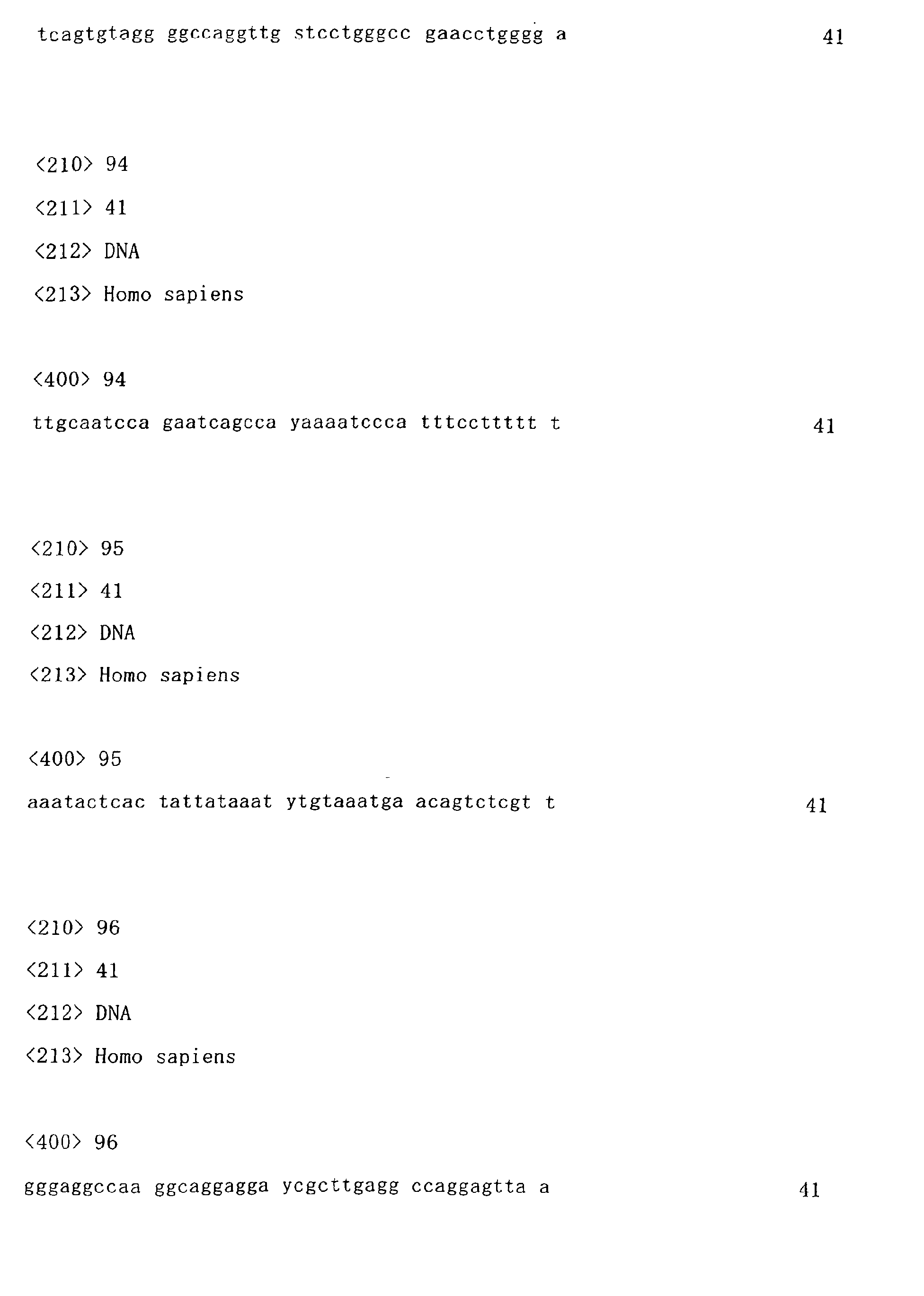
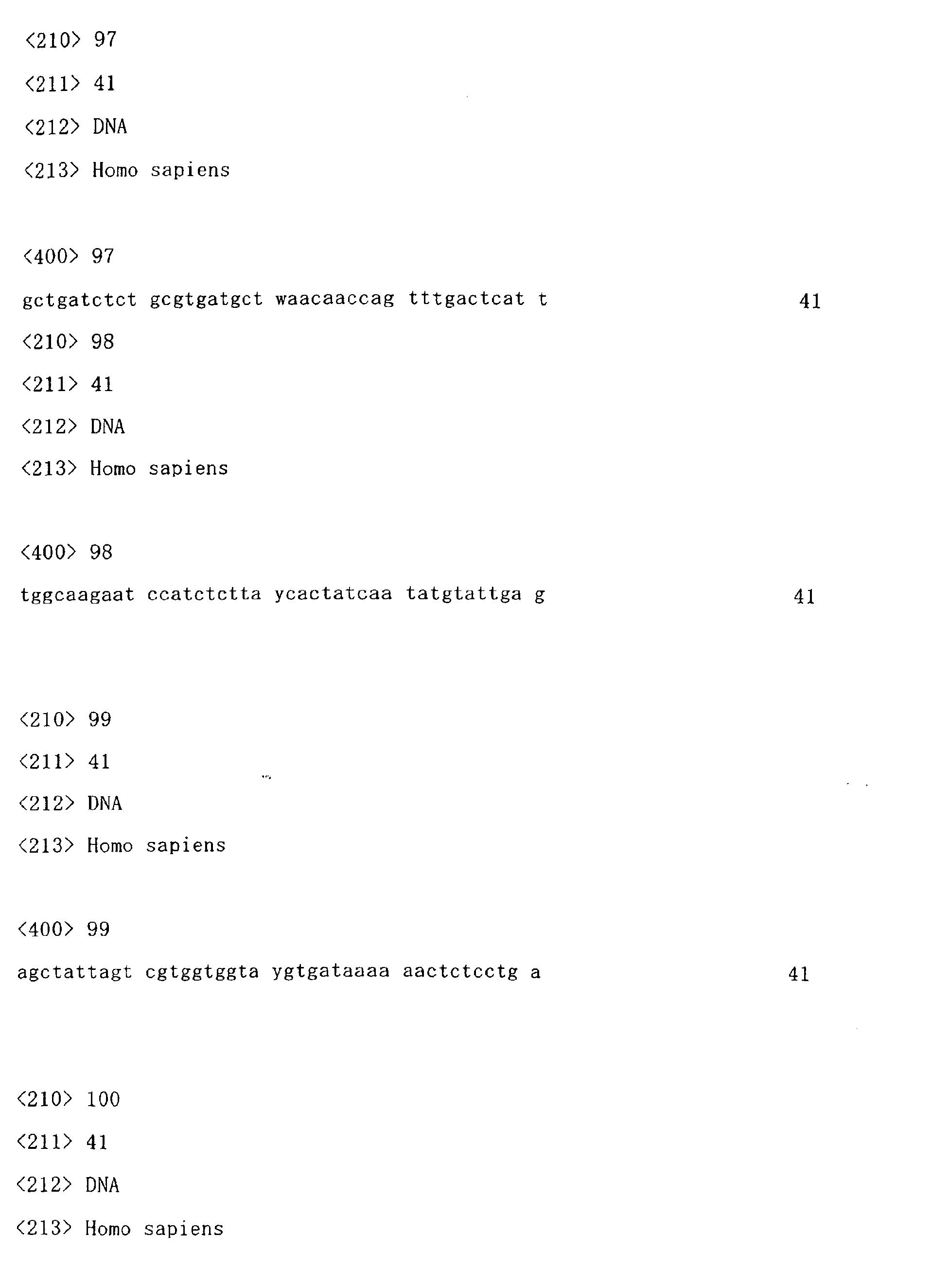


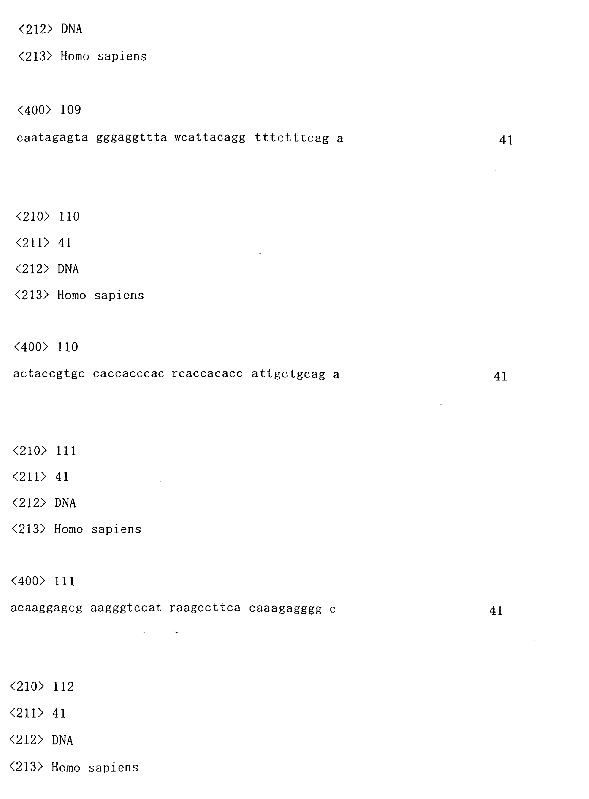


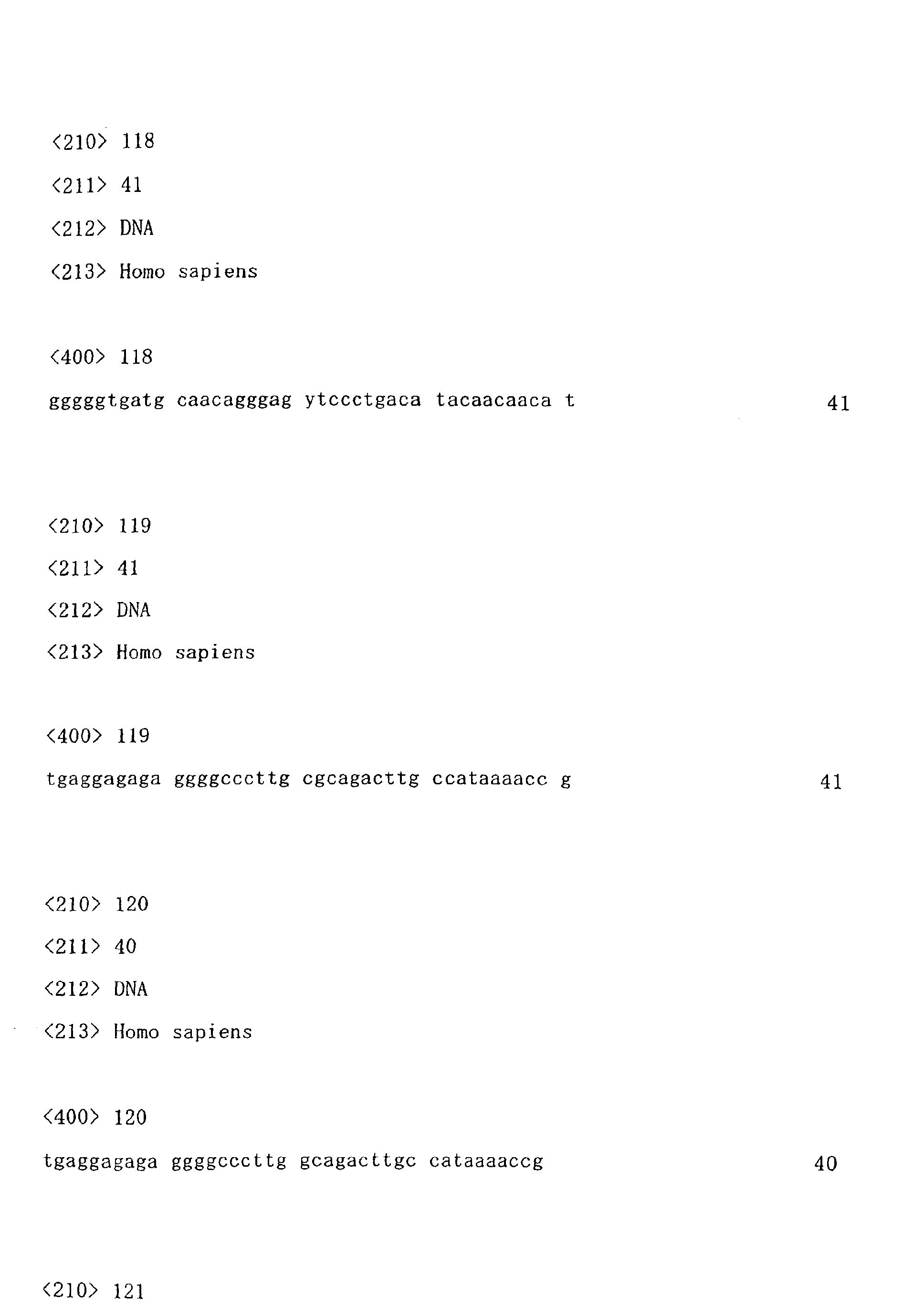


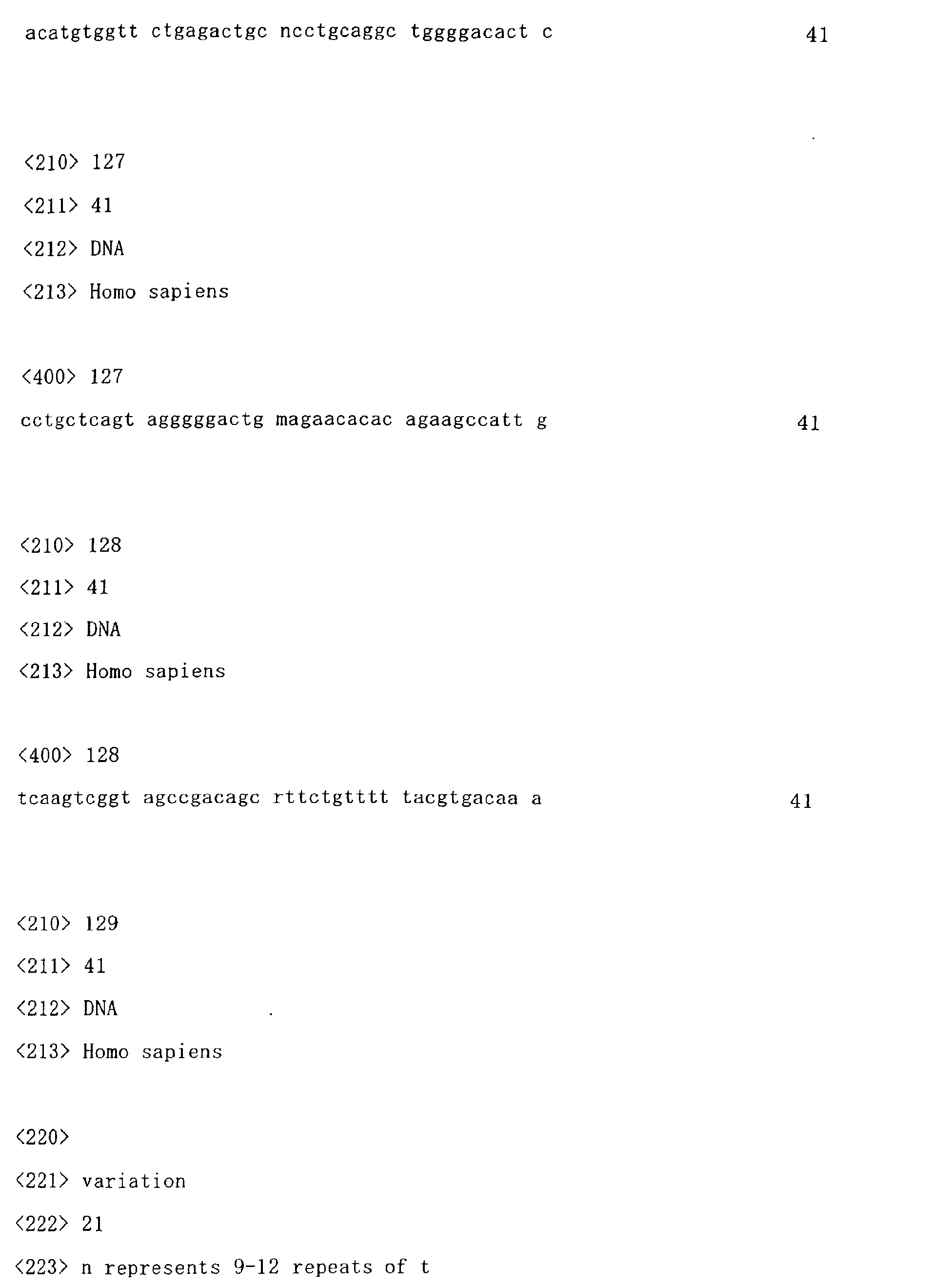


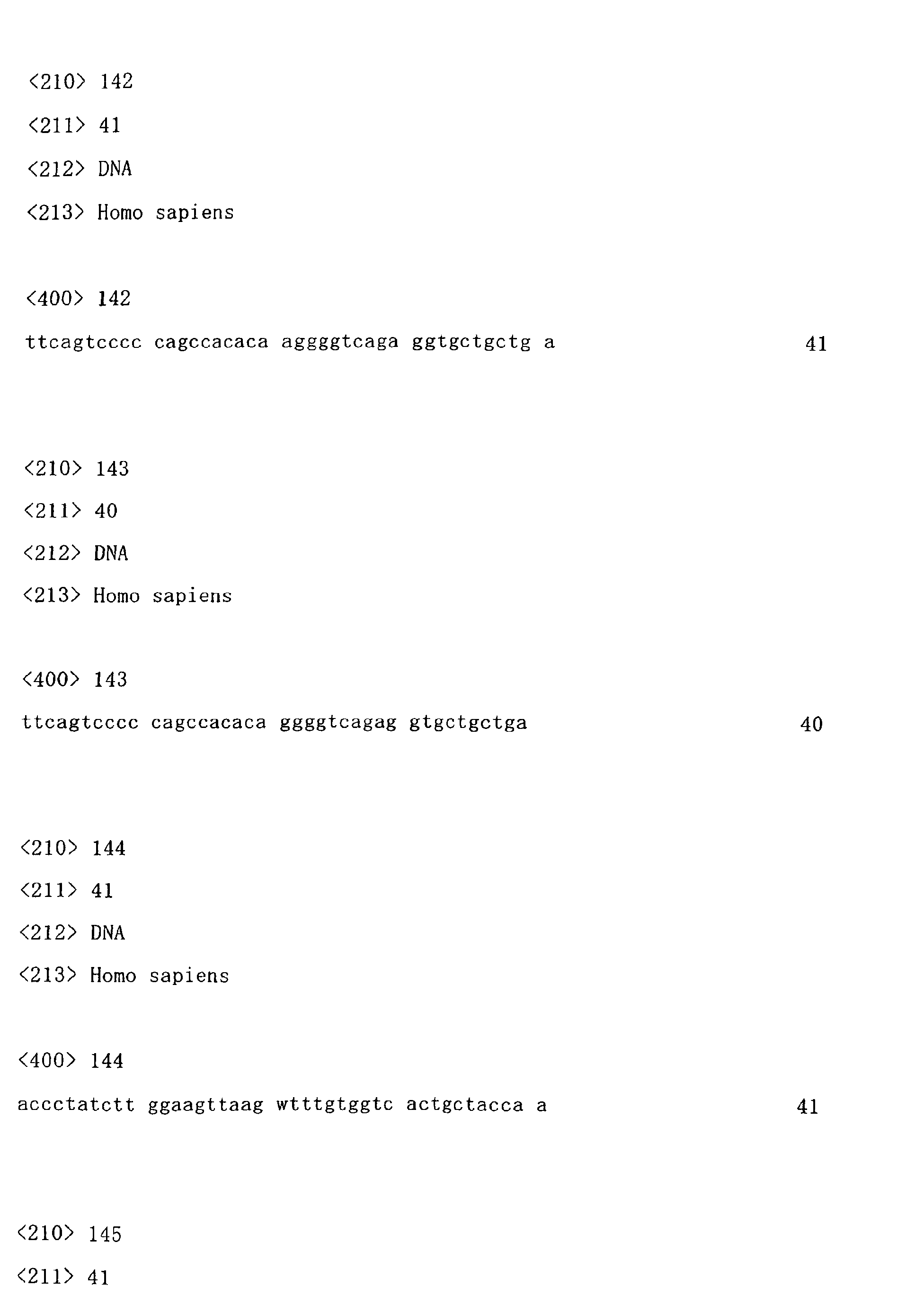


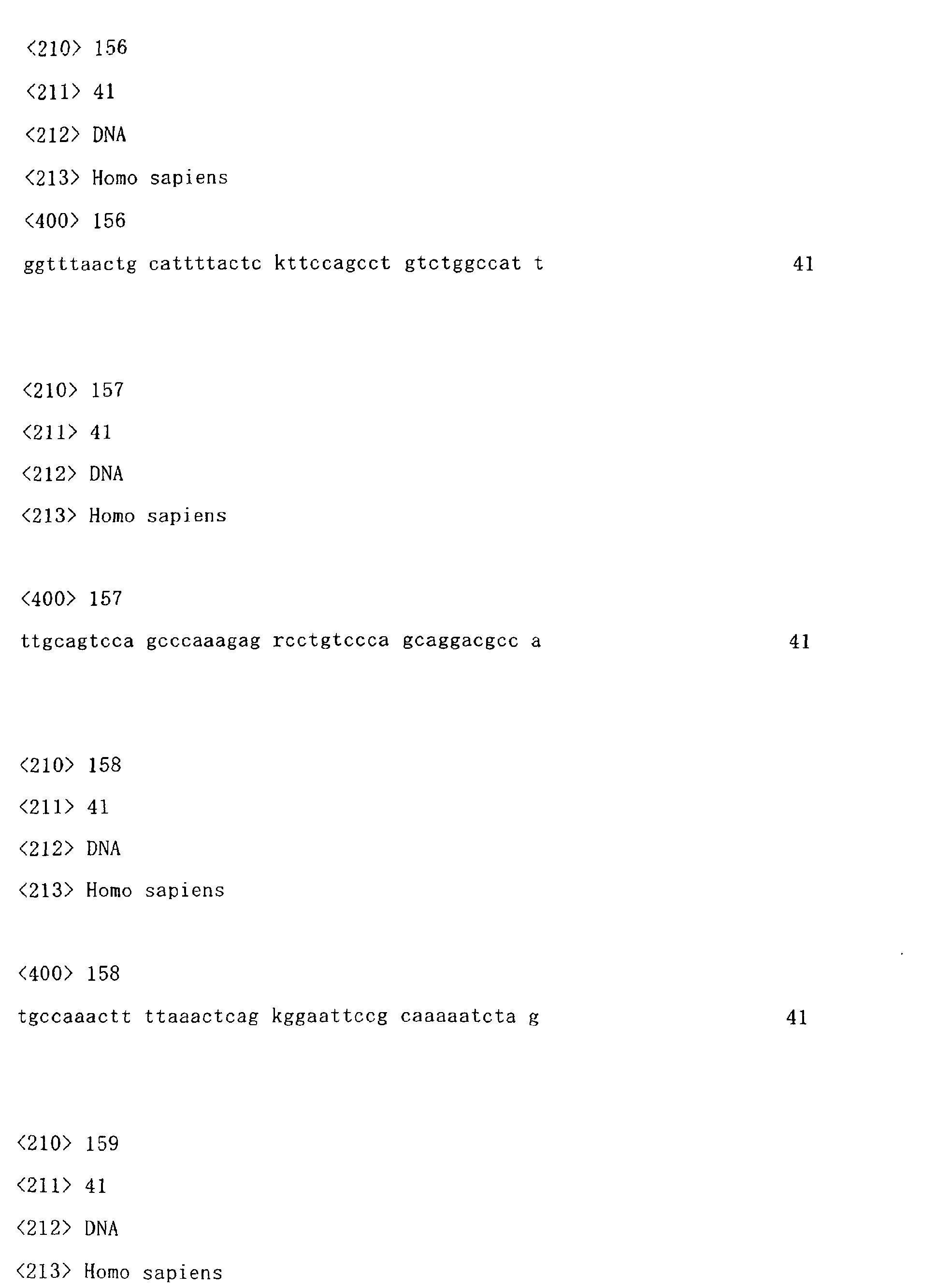


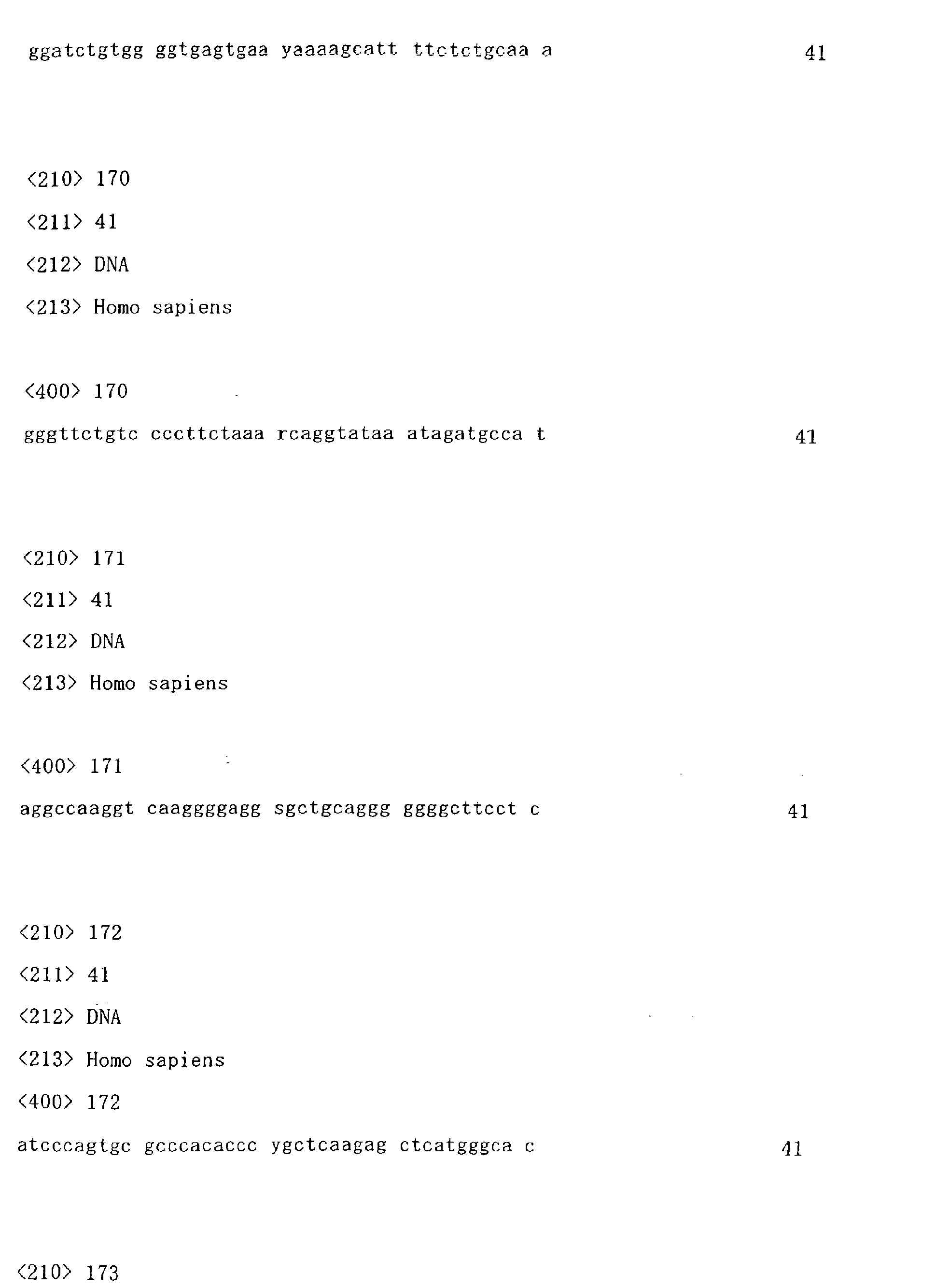


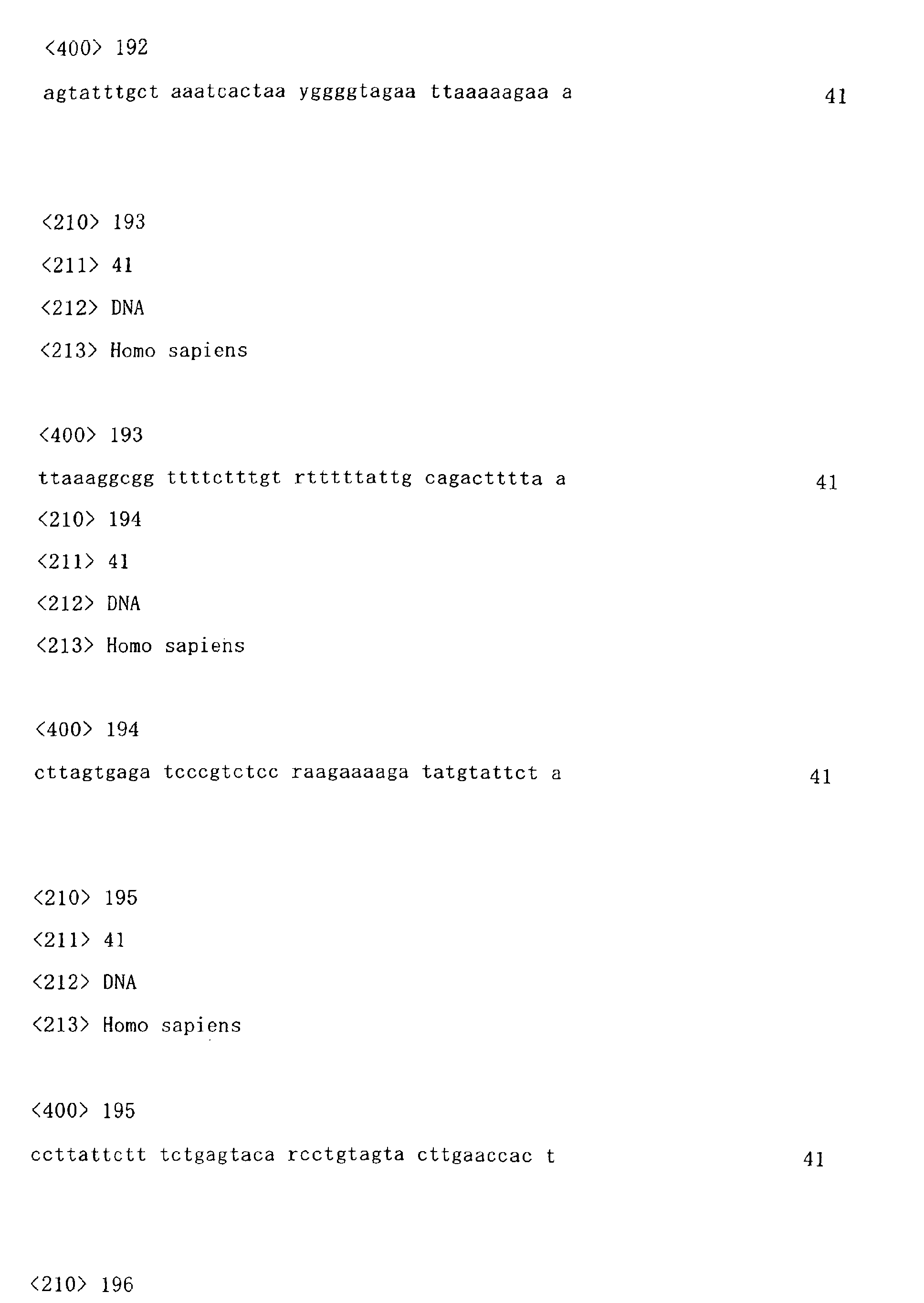
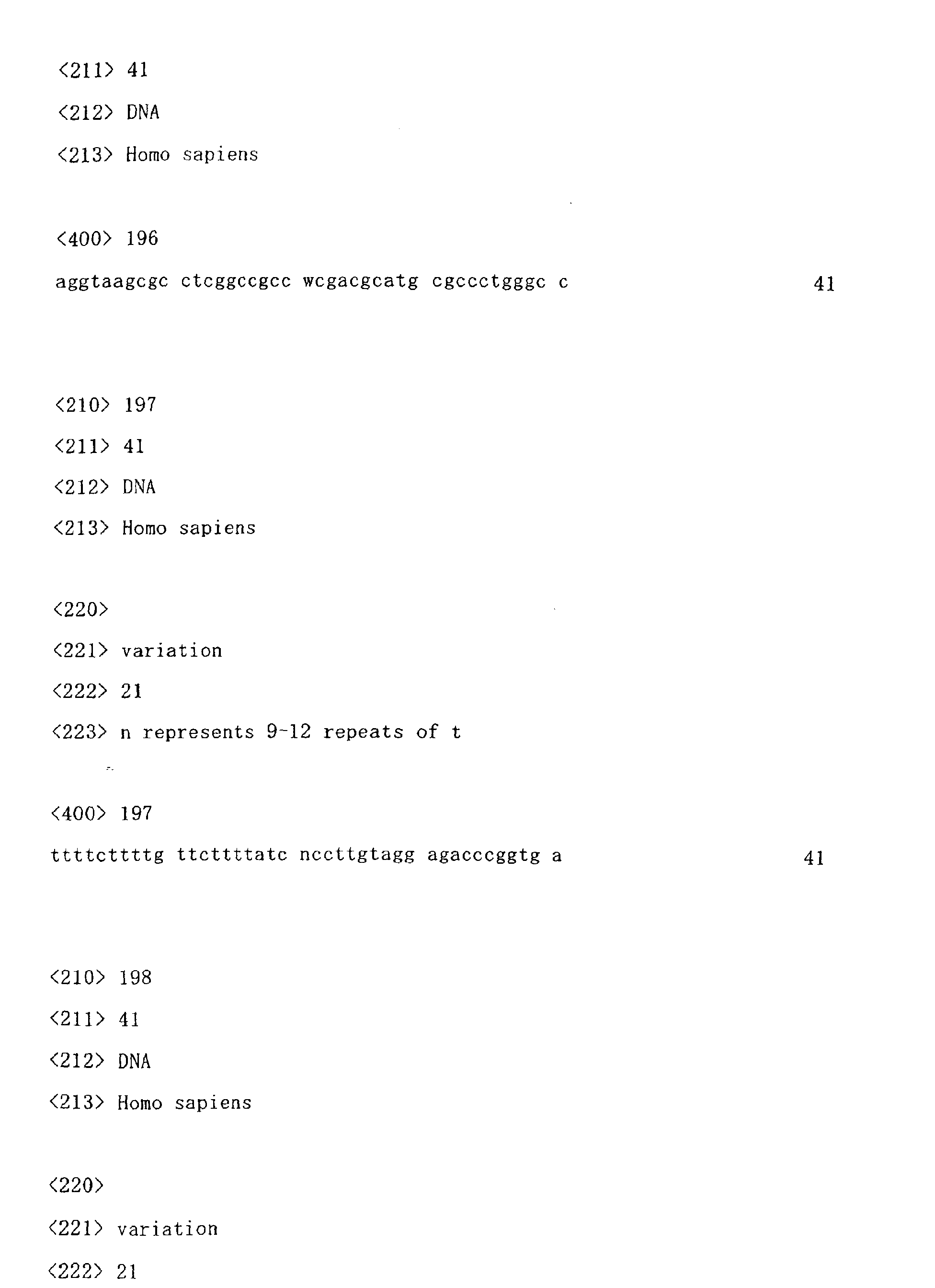
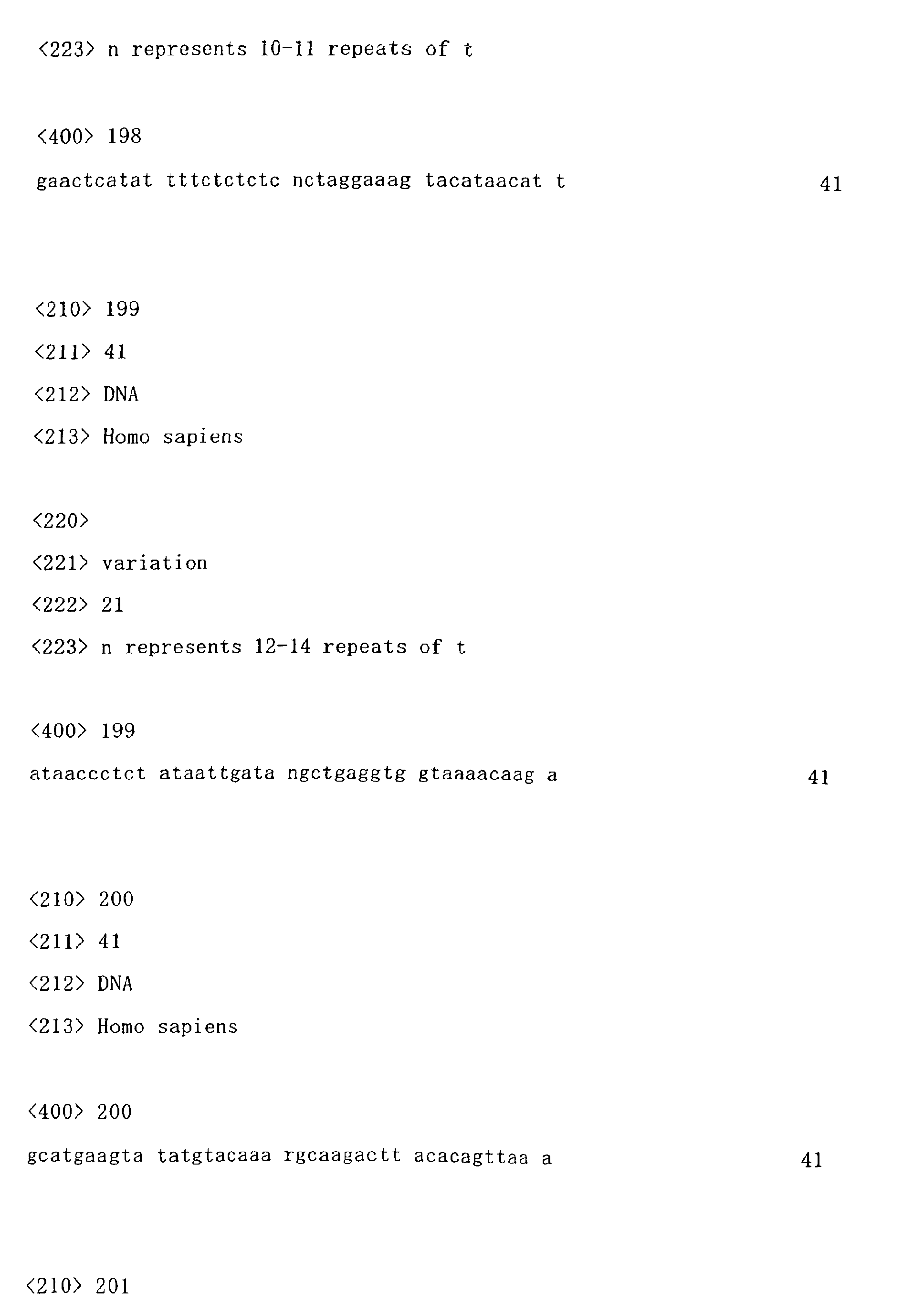
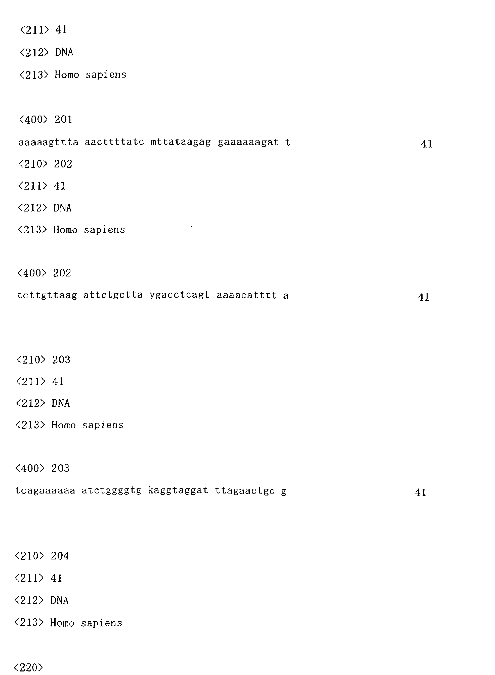

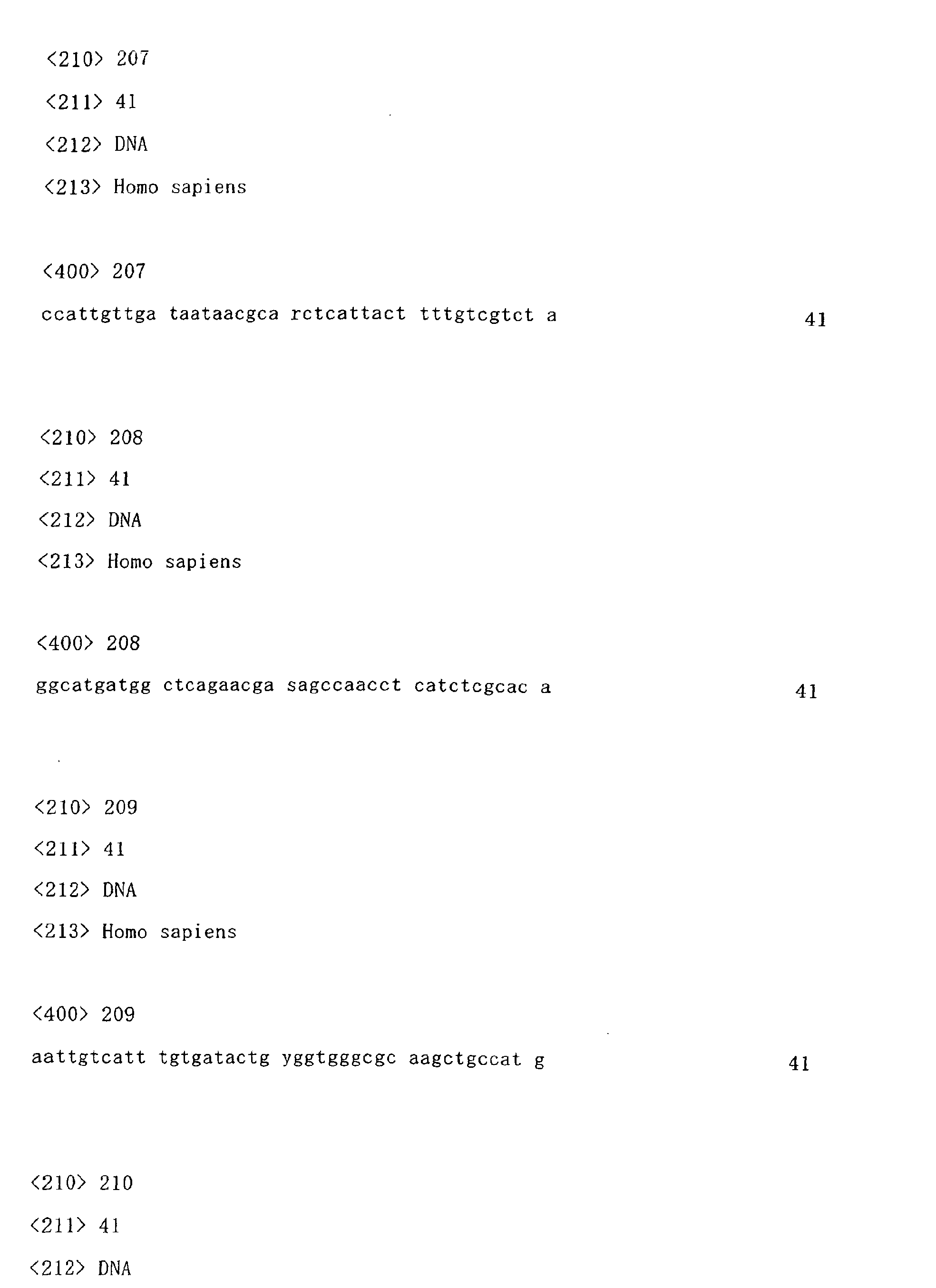

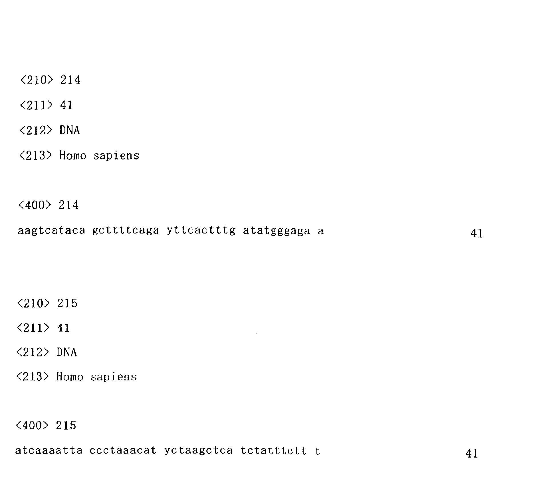
Claims (14)
- A method for detecting a genetic polymorphism(s) in a gene encoding cytochrome P450 using as oligonucleotide probes and/or oligonucleotide primers at least one sequence selected from the group consisting of an at least 13 nucleotide sequence within any of the nucleotide sequences as shown in SEQ ID NOS: 1 through 215, said at least 13 nucleotide sequence containing the 21st nucleotide, or a sequence complementary to said at least 13 nucleotide sequence.
- The method according to claim 1, wherein the polymorphism is a single-nucleotide polymorphism, a polymorphism caused by deletion, substitution or insertion of a plurality of nucleotides, or a VNTR or microsatellite polymorphism.
- A method for evaluating a drug, comprising evaluating the efficacy and safety of the drug metabolized by cytochrome P450 from the detection results obtained by the method according to claim 1 or 2.
- A method for screening a drug, comprising selecting a drug to be used with reference to the evaluation obtained by the method according to claim 3.
- A method for screening a drug, comprising comparing information about a polymorphism(s) in a gene encoding cytochrome P450 and/or a sequence complementary thereto with information about a polymorphism(s) in a gene encoding cytochrome P450 and/or a sequence complementary thereto obtained from a subject; analyzing the efficacy and/or safety of drugs metabolized by cytochrome P450; and selecting a drug to be used from the analysis results obtained.
- The method according to any one of claims 1 to 5, wherein the cytochrome P450 is at least one selected from the group consisting of 2A6, 2Al3, 2B6, 2E, 2S1, 5A1, 7A1 and 7B1.
- An oligonucleotide selected from the group consisting of the nucleotide sequences as shown in SEQ ID NOS: 1 through 215 and sequences complementary thereto.
- An oligonucleotide prepared so as to include a gene polymorphic site present in a gene encoding cytochrome P450 and/or a sequence complementary thereto based on the information about a polymorphism(s) in the gene and/or the complementary sequence thereto.
- The oligonucleotide according to claim 8, wherein the oligonucleotide is designed such that the nucleotide at the 5'-end, the 3'-end or the center of the oligonucleotide corresponds to the gene polymorphic site.
- The oligonucleotide according to claim 9, wherein the oligonucleotide containing a gene polymorphic site is composed of two fragments being linked to each other, one fragment being hybridizable to the gene encoding a cytochrome P450 or the sequence complementary thereto and the other fragment being not hybridizable thereto, and said polymorphic site is positioned at the 5'-end or the 3'-end of the hybridizable fragment.
- The oligonucleotide according to claim 9 or 10, wherein the oligonucleotide consists of an at least 13 nucleotide sequence within any of the nucleotide sequences as shown in SEQ ID NOS: 1 through 215, said at least 13 nucleotide sequence containing the 21st nucleotide, or a sequence complementary to said at least 13 nucleotide sequence.
- An oligonucleotide which is designed in a genomic DNA region containing a gene polymorphic site in any of the nucleotide sequences as shown in SEQ ID NOS: 1 through 215 so that it is located within 1000 bp of the polymorphic site toward the 5' -end and/or the 3' -end of the genomic DNA region, and which has a length of 13-60 nucleotides.
- A microarray wherein the oligonucleotide according to any one of claims 7 to 12 is immobilized on a support.
- A genetic polymorphism detection kit comprising the oligonucleotide according to any one of claims 7 to 12 and/or the microarray according to claim 13.
Applications Claiming Priority (3)
| Application Number | Priority Date | Filing Date | Title |
|---|---|---|---|
| JP2002158237 | 2002-05-30 | ||
| JP2002158237A JP2004000039A (en) | 2002-05-30 | 2002-05-30 | Gene polymorphism detection method |
| PCT/JP2003/006750 WO2003102181A1 (en) | 2002-05-30 | 2003-05-29 | Method of detecting gene polymorphism |
Publications (2)
| Publication Number | Publication Date |
|---|---|
| EP1536003A1 true EP1536003A1 (en) | 2005-06-01 |
| EP1536003A4 EP1536003A4 (en) | 2007-01-17 |
Family
ID=29706482
Family Applications (1)
| Application Number | Title | Priority Date | Filing Date |
|---|---|---|---|
| EP03733147A Withdrawn EP1536003A4 (en) | 2002-05-30 | 2003-05-29 | PROCESS FOR DETECTING GENE POLYMORPHISM |
Country Status (7)
| Country | Link |
|---|---|
| US (1) | US20060234230A1 (en) |
| EP (1) | EP1536003A4 (en) |
| JP (1) | JP2004000039A (en) |
| CN (1) | CN1678741A (en) |
| AU (1) | AU2003241899A1 (en) |
| CA (1) | CA2510406A1 (en) |
| WO (1) | WO2003102181A1 (en) |
Families Citing this family (8)
| Publication number | Priority date | Publication date | Assignee | Title |
|---|---|---|---|---|
| FI20040723A0 (en) * | 2004-05-26 | 2004-05-26 | Orpana Aarne | Method for a quantitative and / or relative measurement of mRNA expression levels in small biological samples |
| JP5067830B2 (en) * | 2006-06-06 | 2012-11-07 | 株式会社島津製作所 | Polymorphic allele determining program, computer-readable recording medium recording the polymorphic allele determining program, and polymorphic allele determining method |
| JP5017947B2 (en) * | 2006-07-12 | 2012-09-05 | 東洋紡績株式会社 | Identification method of multiple nucleotide polymorphisms |
| US20090298839A1 (en) * | 2008-05-29 | 2009-12-03 | Mccarty Catherine Anne | Adrb2 gene polymorphism associated with intraocular pressure response to topical beta-blockers |
| AU2018271846A1 (en) * | 2017-05-22 | 2019-11-21 | Gen9, Inc. | Device and method for nucleic acid manipulation |
| CN108774640B (en) * | 2018-06-20 | 2021-06-11 | 深圳海普洛斯医学检验实验室 | Single-molecule label and application thereof |
| WO2020145351A1 (en) * | 2019-01-09 | 2020-07-16 | 国立研究開発法人理化学研究所 | Comprehensive sequence analysis method of pharmacokinetic-related genes and primer sets to be used therein |
| CN109903816A (en) * | 2019-01-29 | 2019-06-18 | 郑州金域临床检验中心有限公司 | A pharmacogenomics analysis system |
Family Cites Families (4)
| Publication number | Priority date | Publication date | Assignee | Title |
|---|---|---|---|---|
| US5891633A (en) * | 1994-06-16 | 1999-04-06 | The United States Of America As Represented By The Department Of Health And Human Services | Defects in drug metabolism |
| US5834293A (en) * | 1994-09-28 | 1998-11-10 | Vanderbilt University | Cytochrome P450 arachidonic acid epoxygenase genetic mutation associated with hypertension |
| GB9828619D0 (en) * | 1998-12-23 | 1999-02-17 | Janssen Pharmaceutica Nv | An assay for genotyping cytochrome expression |
| ATE348170T1 (en) * | 2000-06-01 | 2007-01-15 | Otsuka Pharma Co Ltd | METHOD FOR DETECTING AND QUANTIFYING HUMAN P450 MOLECULES AND PROBE AND KIT FOR SUCH METHOD |
-
2002
- 2002-05-30 JP JP2002158237A patent/JP2004000039A/en active Pending
-
2003
- 2003-05-29 AU AU2003241899A patent/AU2003241899A1/en not_active Abandoned
- 2003-05-29 CA CA002510406A patent/CA2510406A1/en not_active Abandoned
- 2003-05-29 CN CN03817304.2A patent/CN1678741A/en active Pending
- 2003-05-29 EP EP03733147A patent/EP1536003A4/en not_active Withdrawn
- 2003-05-29 US US10/516,038 patent/US20060234230A1/en not_active Abandoned
- 2003-05-29 WO PCT/JP2003/006750 patent/WO2003102181A1/en not_active Ceased
Also Published As
| Publication number | Publication date |
|---|---|
| AU2003241899A1 (en) | 2003-12-19 |
| WO2003102181A1 (en) | 2003-12-11 |
| CN1678741A (en) | 2005-10-05 |
| CA2510406A1 (en) | 2003-12-11 |
| JP2004000039A (en) | 2004-01-08 |
| EP1536003A4 (en) | 2007-01-17 |
| US20060234230A1 (en) | 2006-10-19 |
Similar Documents
| Publication | Publication Date | Title |
|---|---|---|
| KR102018122B1 (en) | Biomarkers for Individual confirmation of Hanwoo Beef and uses thereof | |
| US20020182622A1 (en) | Method for SNP (single nucleotide polymorphism) typing | |
| KR101157526B1 (en) | Snp for diagnosing adhd, microarray and kit comprising the same, and method of diagnosing adhd using thereof | |
| Twyman et al. | Techniques patents for SNP genotyping | |
| US20040072156A1 (en) | Detection of genetic polymorphisms | |
| US20060234230A1 (en) | Method of detecting gene polymorphism | |
| EP1514946A1 (en) | Method of detecting gene polymorphism | |
| CA2324866A1 (en) | Biallelic markers for use in constructing a high density disequilibrium map of the human genome | |
| JP2007526764A (en) | APOE gene marker related to age of onset of Alzheimer's disease | |
| US20070003938A1 (en) | Hybridization of genomic nucleic acid without complexity reduction | |
| WO2000058519A2 (en) | Charaterization of single nucleotide polymorphisms in coding regions of human genes | |
| JP2007516719A (en) | Polynucleotide involved in type 2 diabetes including single nucleotide polymorphism, microarray and diagnostic kit containing the same, and method for analyzing polynucleotide using the same | |
| JP2008079604A (en) | Primer set, probe set, method and kit for predicting alcohol resolution and drunkenness tolerance | |
| US20050255498A1 (en) | APOC1 genetic markers associated with age of onset of Alzheimer's Disease | |
| US7794982B2 (en) | Method for identifying gene with varying expression levels | |
| JP2007510404A (en) | NTRK1 gene marker associated with age of onset of Alzheimer's disease | |
| KR101187317B1 (en) | Polymorphic markers predicting susceptibility to diffuse-type gastric cancer and the prediction method thereof using the same | |
| JP2982304B2 (en) | Method for identifying nucleic acid and test set for identifying nucleic acid | |
| WO2013030786A1 (en) | Method for diagnosing or predicting hepatocellular carcinoma outcome | |
| RU2600874C2 (en) | Set of oligonucleotide primers and probes for genetic typing of polymorphous dna loci associated with a risk of progression of sporadic form of alzheimer's disease in russian populations | |
| KR101145178B1 (en) | Genetic marker combinations for prognosis on tramadol-induced nausea and vomiting | |
| WO2005072150A2 (en) | Ldlr genetic markers associated with age of onset of alzheimer's disease | |
| JP2006141301A (en) | Gene polymorphism detection method | |
| US8268562B2 (en) | Biomarkers for predicting response of esophageal cancer patient to chemoradiotherapy | |
| US20030148288A1 (en) | Colorimetric genetic test for clinically significant TNF polymorphism and methods of use thereof |
Legal Events
| Date | Code | Title | Description |
|---|---|---|---|
| PUAI | Public reference made under article 153(3) epc to a published international application that has entered the european phase |
Free format text: ORIGINAL CODE: 0009012 |
|
| 17P | Request for examination filed |
Effective date: 20041230 |
|
| AK | Designated contracting states |
Kind code of ref document: A1 Designated state(s): AT BE BG CH CY CZ DE DK EE ES FI FR GB GR HU IE IT LI LU MC NL PT RO SE SI SK TR |
|
| AX | Request for extension of the european patent |
Extension state: AL LT LV MK |
|
| A4 | Supplementary search report drawn up and despatched |
Effective date: 20061218 |
|
| STAA | Information on the status of an ep patent application or granted ep patent |
Free format text: STATUS: THE APPLICATION IS DEEMED TO BE WITHDRAWN |
|
| 18D | Application deemed to be withdrawn |
Effective date: 20091201 |