EP1525478B1 - Shiga-toxin-untereinheit b als vektor für die tumordiagnose und zur arzneimttelverabreichung an gb3-exprimierenden tumoren - Google Patents
Shiga-toxin-untereinheit b als vektor für die tumordiagnose und zur arzneimttelverabreichung an gb3-exprimierenden tumoren Download PDFInfo
- Publication number
- EP1525478B1 EP1525478B1 EP03775133A EP03775133A EP1525478B1 EP 1525478 B1 EP1525478 B1 EP 1525478B1 EP 03775133 A EP03775133 A EP 03775133A EP 03775133 A EP03775133 A EP 03775133A EP 1525478 B1 EP1525478 B1 EP 1525478B1
- Authority
- EP
- European Patent Office
- Prior art keywords
- stxb
- hybrid compound
- cells
- cys
- cleavable
- Prior art date
- Legal status (The legal status is an assumption and is not a legal conclusion. Google has not performed a legal analysis and makes no representation as to the accuracy of the status listed.)
- Expired - Lifetime
Links
- 108010079723 Shiga Toxin Proteins 0.000 title claims abstract description 19
- 238000003745 diagnosis Methods 0.000 title claims abstract description 15
- 206010028980 Neoplasm Diseases 0.000 title description 109
- 238000012377 drug delivery Methods 0.000 title description 2
- 150000001875 compounds Chemical class 0.000 claims abstract description 65
- 239000003814 drug Substances 0.000 claims abstract description 33
- 229940079593 drug Drugs 0.000 claims abstract description 31
- 108090000765 processed proteins & peptides Proteins 0.000 claims abstract description 11
- XUJNEKJLAYXESH-UHFFFAOYSA-N cysteine Natural products SCC(N)C(O)=O XUJNEKJLAYXESH-UHFFFAOYSA-N 0.000 claims abstract description 6
- 235000018417 cysteine Nutrition 0.000 claims abstract description 6
- 102000004196 processed proteins & peptides Human genes 0.000 claims abstract description 5
- 229920001184 polypeptide Polymers 0.000 claims abstract description 4
- 210000004027 cell Anatomy 0.000 claims description 120
- QZGIWPZCWHMVQL-UIYAJPBUSA-N neocarzinostatin chromophore Chemical group O1[C@H](C)[C@H](O)[C@H](O)[C@@H](NC)[C@H]1O[C@@H]1C/2=C/C#C[C@H]3O[C@@]3([C@@H]3OC(=O)OC3)C#CC\2=C[C@H]1OC(=O)C1=C(O)C=CC2=C(C)C=C(OC)C=C12 QZGIWPZCWHMVQL-UIYAJPBUSA-N 0.000 claims description 56
- 239000000651 prodrug Substances 0.000 claims description 40
- 229940002612 prodrug Drugs 0.000 claims description 40
- 229950009268 zinostatin Drugs 0.000 claims description 40
- 102000004190 Enzymes Human genes 0.000 claims description 25
- 108090000790 Enzymes Proteins 0.000 claims description 25
- 208000005016 Intestinal Neoplasms Diseases 0.000 claims description 19
- 239000002872 contrast media Substances 0.000 claims description 19
- 210000004881 tumor cell Anatomy 0.000 claims description 19
- -1 porphyrin-gadolinium Chemical class 0.000 claims description 13
- 238000002560 therapeutic procedure Methods 0.000 claims description 11
- IJGRMHOSHXDMSA-UHFFFAOYSA-N Atomic nitrogen Chemical compound N#N IJGRMHOSHXDMSA-UHFFFAOYSA-N 0.000 claims description 10
- 238000006243 chemical reaction Methods 0.000 claims description 10
- 208000001333 Colorectal Neoplasms Diseases 0.000 claims description 9
- 238000001727 in vivo Methods 0.000 claims description 9
- 150000004032 porphyrins Chemical class 0.000 claims description 9
- 230000002829 reductive effect Effects 0.000 claims description 9
- 125000003396 thiol group Chemical group [H]S* 0.000 claims description 9
- AOJJSUZBOXZQNB-TZSSRYMLSA-N Doxorubicin Chemical compound O([C@H]1C[C@@](O)(CC=2C(O)=C3C(=O)C=4C=CC=C(C=4C(=O)C3=C(O)C=21)OC)C(=O)CO)[C@H]1C[C@H](N)[C@H](O)[C@H](C)O1 AOJJSUZBOXZQNB-TZSSRYMLSA-N 0.000 claims description 8
- 230000001472 cytotoxic effect Effects 0.000 claims description 8
- 239000000203 mixture Substances 0.000 claims description 8
- 229940127089 cytotoxic agent Drugs 0.000 claims description 7
- 239000002254 cytotoxic agent Substances 0.000 claims description 7
- 229910052688 Gadolinium Inorganic materials 0.000 claims description 6
- 239000002253 acid Substances 0.000 claims description 6
- 230000004913 activation Effects 0.000 claims description 6
- 239000003795 chemical substances by application Substances 0.000 claims description 6
- 231100000433 cytotoxic Toxicity 0.000 claims description 6
- 101001122938 Homo sapiens Lysosomal protective protein Proteins 0.000 claims description 5
- 102100028524 Lysosomal protective protein Human genes 0.000 claims description 5
- 102000001696 Mannosidases Human genes 0.000 claims description 5
- 108010054377 Mannosidases Proteins 0.000 claims description 5
- 231100000599 cytotoxic agent Toxicity 0.000 claims description 5
- 229960002963 ganciclovir Drugs 0.000 claims description 5
- 150000002678 macrocyclic compounds Chemical class 0.000 claims description 5
- 229910052757 nitrogen Inorganic materials 0.000 claims description 5
- STQGQHZAVUOBTE-UHFFFAOYSA-N 7-Cyan-hept-2t-en-4,6-diinsaeure Natural products C1=2C(O)=C3C(=O)C=4C(OC)=CC=CC=4C(=O)C3=C(O)C=2CC(O)(C(C)=O)CC1OC1CC(N)C(O)C(C)O1 STQGQHZAVUOBTE-UHFFFAOYSA-N 0.000 claims description 4
- NWIBSHFKIJFRCO-WUDYKRTCSA-N Mytomycin Chemical compound C1N2C(C(C(C)=C(N)C3=O)=O)=C3[C@@H](COC(N)=O)[C@@]2(OC)[C@@H]2[C@H]1N2 NWIBSHFKIJFRCO-WUDYKRTCSA-N 0.000 claims description 4
- 229960004150 aciclovir Drugs 0.000 claims description 4
- MKUXAQIIEYXACX-UHFFFAOYSA-N aciclovir Chemical compound N1C(N)=NC(=O)C2=C1N(COCCO)C=N2 MKUXAQIIEYXACX-UHFFFAOYSA-N 0.000 claims description 4
- OPTASPLRGRRNAP-UHFFFAOYSA-N cytosine Chemical compound NC=1C=CNC(=O)N=1 OPTASPLRGRRNAP-UHFFFAOYSA-N 0.000 claims description 4
- STQGQHZAVUOBTE-VGBVRHCVSA-N daunorubicin Chemical compound O([C@H]1C[C@@](O)(CC=2C(O)=C3C(=O)C=4C=CC=C(C=4C(=O)C3=C(O)C=21)OC)C(C)=O)[C@H]1C[C@H](N)[C@H](O)[C@H](C)O1 STQGQHZAVUOBTE-VGBVRHCVSA-N 0.000 claims description 4
- 229960004679 doxorubicin Drugs 0.000 claims description 4
- IRSCQMHQWWYFCW-UHFFFAOYSA-N ganciclovir Chemical compound O=C1NC(N)=NC2=C1N=CN2COC(CO)CO IRSCQMHQWWYFCW-UHFFFAOYSA-N 0.000 claims description 4
- 230000007062 hydrolysis Effects 0.000 claims description 4
- 238000006460 hydrolysis reaction Methods 0.000 claims description 4
- 238000004519 manufacturing process Methods 0.000 claims description 4
- 230000005298 paramagnetic effect Effects 0.000 claims description 4
- 239000003504 photosensitizing agent Substances 0.000 claims description 4
- 231100000331 toxic Toxicity 0.000 claims description 4
- 230000002588 toxic effect Effects 0.000 claims description 4
- 230000001131 transforming effect Effects 0.000 claims description 4
- 102000002260 Alkaline Phosphatase Human genes 0.000 claims description 3
- 108020004774 Alkaline Phosphatase Proteins 0.000 claims description 3
- 229960003330 pentetic acid Drugs 0.000 claims description 3
- 230000002165 photosensitisation Effects 0.000 claims description 3
- 238000001126 phototherapy Methods 0.000 claims description 3
- 108010080937 Carboxypeptidases A Proteins 0.000 claims description 2
- 102000000496 Carboxypeptidases A Human genes 0.000 claims description 2
- WQZGKKKJIJFFOK-QTVWNMPRSA-N D-mannopyranose Chemical compound OC[C@H]1OC(O)[C@@H](O)[C@@H](O)[C@@H]1O WQZGKKKJIJFFOK-QTVWNMPRSA-N 0.000 claims description 2
- WEAHRLBPCANXCN-UHFFFAOYSA-N Daunomycin Natural products CCC1(O)CC(OC2CC(N)C(O)C(C)O2)c3cc4C(=O)c5c(OC)cccc5C(=O)c4c(O)c3C1 WEAHRLBPCANXCN-UHFFFAOYSA-N 0.000 claims description 2
- 108090000204 Dipeptidase 1 Proteins 0.000 claims description 2
- GHASVSINZRGABV-UHFFFAOYSA-N Fluorouracil Chemical compound FC1=CNC(=O)NC1=O GHASVSINZRGABV-UHFFFAOYSA-N 0.000 claims description 2
- 102000002464 Galactosidases Human genes 0.000 claims description 2
- 108010093031 Galactosidases Proteins 0.000 claims description 2
- XDXDZDZNSLXDNA-TZNDIEGXSA-N Idarubicin Chemical compound C1[C@H](N)[C@H](O)[C@H](C)O[C@H]1O[C@@H]1C2=C(O)C(C(=O)C3=CC=CC=C3C3=O)=C3C(O)=C2C[C@@](O)(C(C)=O)C1 XDXDZDZNSLXDNA-TZNDIEGXSA-N 0.000 claims description 2
- XDXDZDZNSLXDNA-UHFFFAOYSA-N Idarubicin Natural products C1C(N)C(O)C(C)OC1OC1C2=C(O)C(C(=O)C3=CC=CC=C3C3=O)=C3C(O)=C2CC(O)(C(C)=O)C1 XDXDZDZNSLXDNA-UHFFFAOYSA-N 0.000 claims description 2
- FBOZXECLQNJBKD-ZDUSSCGKSA-N L-methotrexate Chemical compound C=1N=C2N=C(N)N=C(N)C2=NC=1CN(C)C1=CC=C(C(=O)N[C@@H](CCC(O)=O)C(O)=O)C=C1 FBOZXECLQNJBKD-ZDUSSCGKSA-N 0.000 claims description 2
- 102000004459 Nitroreductase Human genes 0.000 claims description 2
- 108010073038 Penicillin Amidase Proteins 0.000 claims description 2
- 102000006601 Thymidine Kinase Human genes 0.000 claims description 2
- 108020004440 Thymidine kinase Proteins 0.000 claims description 2
- 150000001408 amides Chemical class 0.000 claims description 2
- 102000006635 beta-lactamase Human genes 0.000 claims description 2
- 229930195731 calicheamicin Natural products 0.000 claims description 2
- HXCHCVDVKSCDHU-LULTVBGHSA-N calicheamicin Chemical compound C1[C@H](OC)[C@@H](NCC)CO[C@H]1O[C@H]1[C@H](O[C@@H]2C\3=C(NC(=O)OC)C(=O)C[C@](C/3=C/CSSSC)(O)C#C\C=C/C#C2)O[C@H](C)[C@@H](NO[C@@H]2O[C@H](C)[C@@H](SC(=O)C=3C(=C(OC)C(O[C@H]4[C@@H]([C@H](OC)[C@@H](O)[C@H](C)O4)O)=C(I)C=3C)OC)[C@@H](O)C2)[C@@H]1O HXCHCVDVKSCDHU-LULTVBGHSA-N 0.000 claims description 2
- 229940104302 cytosine Drugs 0.000 claims description 2
- 229960000975 daunorubicin Drugs 0.000 claims description 2
- NDMPLJNOPCLANR-PETVRERISA-N deacetylvinblastine Chemical compound C([C@@H](C[C@]1(C(=O)OC)C=2C(=CC3=C([C@]45[C@H]([C@@]([C@H](O)[C@]6(CC)C=CCN([C@H]56)CC4)(O)C(=O)OC)N3C)C=2)OC)C[C@@](C2)(O)CC)N2CCC2=C1NC1=CC=CC=C21 NDMPLJNOPCLANR-PETVRERISA-N 0.000 claims description 2
- 229960002949 fluorouracil Drugs 0.000 claims description 2
- 235000019152 folic acid Nutrition 0.000 claims description 2
- 150000002224 folic acids Chemical class 0.000 claims description 2
- 229930182830 galactose Natural products 0.000 claims description 2
- 108010062699 gamma-Glutamyl Hydrolase Proteins 0.000 claims description 2
- 230000003301 hydrolyzing effect Effects 0.000 claims description 2
- 229960000908 idarubicin Drugs 0.000 claims description 2
- 150000003951 lactams Chemical group 0.000 claims description 2
- 229960000485 methotrexate Drugs 0.000 claims description 2
- 229960004857 mitomycin Drugs 0.000 claims description 2
- 108020001162 nitroreductase Proteins 0.000 claims description 2
- 125000002467 phosphate group Chemical group [H]OP(=O)(O[H])O[*] 0.000 claims description 2
- 125000000539 amino acid group Chemical group 0.000 claims 2
- 230000004543 DNA replication Effects 0.000 claims 1
- 108010031186 Glycoside Hydrolases Proteins 0.000 claims 1
- 102000005744 Glycoside Hydrolases Human genes 0.000 claims 1
- 108090001060 Lipase Proteins 0.000 claims 1
- 102000004882 Lipase Human genes 0.000 claims 1
- 239000004367 Lipase Substances 0.000 claims 1
- 108091005804 Peptidases Proteins 0.000 claims 1
- 102000035195 Peptidases Human genes 0.000 claims 1
- UIWYJDYFSGRHKR-UHFFFAOYSA-N gadolinium atom Chemical compound [Gd] UIWYJDYFSGRHKR-UHFFFAOYSA-N 0.000 claims 1
- 235000019421 lipase Nutrition 0.000 claims 1
- 235000019833 protease Nutrition 0.000 claims 1
- 229920001059 synthetic polymer Polymers 0.000 claims 1
- 150000001413 amino acids Chemical class 0.000 abstract description 6
- 235000001014 amino acid Nutrition 0.000 abstract description 5
- 231100000252 nontoxic Toxicity 0.000 abstract description 5
- 230000003000 nontoxic effect Effects 0.000 abstract description 5
- 210000003370 receptor cell Anatomy 0.000 abstract description 4
- 238000011275 oncology therapy Methods 0.000 abstract description 3
- 239000012502 diagnostic product Substances 0.000 abstract description 2
- 210000001519 tissue Anatomy 0.000 description 41
- 201000011510 cancer Diseases 0.000 description 31
- 238000010168 coupling process Methods 0.000 description 27
- 241001465754 Metazoa Species 0.000 description 26
- 238000000034 method Methods 0.000 description 26
- 230000008878 coupling Effects 0.000 description 25
- 238000005859 coupling reaction Methods 0.000 description 25
- 238000003384 imaging method Methods 0.000 description 22
- 108020003175 receptors Proteins 0.000 description 22
- 102000005962 receptors Human genes 0.000 description 22
- OKKJLVBELUTLKV-UHFFFAOYSA-N Methanol Chemical compound OC OKKJLVBELUTLKV-UHFFFAOYSA-N 0.000 description 21
- 230000000694 effects Effects 0.000 description 19
- 230000014509 gene expression Effects 0.000 description 19
- IAZDPXIOMUYVGZ-UHFFFAOYSA-N Dimethylsulphoxide Chemical compound CS(C)=O IAZDPXIOMUYVGZ-UHFFFAOYSA-N 0.000 description 18
- 238000010186 staining Methods 0.000 description 18
- 230000008685 targeting Effects 0.000 description 16
- 238000011282 treatment Methods 0.000 description 16
- XLYOFNOQVPJJNP-UHFFFAOYSA-N water Chemical compound O XLYOFNOQVPJJNP-UHFFFAOYSA-N 0.000 description 16
- 230000027455 binding Effects 0.000 description 15
- LOKCTEFSRHRXRJ-UHFFFAOYSA-I dipotassium trisodium dihydrogen phosphate hydrogen phosphate dichloride Chemical compound P(=O)(O)(O)[O-].[K+].P(=O)(O)([O-])[O-].[Na+].[Na+].[Cl-].[K+].[Cl-].[Na+] LOKCTEFSRHRXRJ-UHFFFAOYSA-I 0.000 description 14
- 238000002474 experimental method Methods 0.000 description 14
- 238000011534 incubation Methods 0.000 description 14
- 230000000968 intestinal effect Effects 0.000 description 14
- 239000002953 phosphate buffered saline Substances 0.000 description 14
- 108090000623 proteins and genes Proteins 0.000 description 14
- 101710204212 Neocarzinostatin Proteins 0.000 description 13
- 201000009019 intestinal benign neoplasm Diseases 0.000 description 13
- YBJHBAHKTGYVGT-ZKWXMUAHSA-N (+)-Biotin Chemical compound N1C(=O)N[C@@H]2[C@H](CCCCC(=O)O)SC[C@@H]21 YBJHBAHKTGYVGT-ZKWXMUAHSA-N 0.000 description 12
- 235000018102 proteins Nutrition 0.000 description 12
- 102000004169 proteins and genes Human genes 0.000 description 12
- 108010053857 aponeocarzinostatin Proteins 0.000 description 11
- 125000005647 linker group Chemical group 0.000 description 11
- 238000002595 magnetic resonance imaging Methods 0.000 description 11
- 239000002105 nanoparticle Substances 0.000 description 11
- 239000000047 product Substances 0.000 description 11
- HEDRZPFGACZZDS-UHFFFAOYSA-N Chloroform Chemical compound ClC(Cl)Cl HEDRZPFGACZZDS-UHFFFAOYSA-N 0.000 description 10
- KAESVJOAVNADME-UHFFFAOYSA-N Pyrrole Chemical compound C=1C=CNC=1 KAESVJOAVNADME-UHFFFAOYSA-N 0.000 description 10
- 238000013459 approach Methods 0.000 description 10
- 210000002288 golgi apparatus Anatomy 0.000 description 10
- 239000008194 pharmaceutical composition Substances 0.000 description 10
- 102000053187 Glucuronidase Human genes 0.000 description 9
- 108010060309 Glucuronidase Proteins 0.000 description 9
- 210000004185 liver Anatomy 0.000 description 9
- 230000001225 therapeutic effect Effects 0.000 description 9
- 231100000765 toxin Toxicity 0.000 description 9
- 241000699666 Mus <mouse, genus> Species 0.000 description 8
- 241000699670 Mus sp. Species 0.000 description 8
- 230000001413 cellular effect Effects 0.000 description 8
- 210000001198 duodenum Anatomy 0.000 description 8
- 210000004940 nucleus Anatomy 0.000 description 8
- 239000003053 toxin Substances 0.000 description 8
- 108700012359 toxins Proteins 0.000 description 8
- 108020004414 DNA Proteins 0.000 description 7
- 238000009826 distribution Methods 0.000 description 7
- 239000000975 dye Substances 0.000 description 7
- 238000002523 gelfiltration Methods 0.000 description 7
- 230000002018 overexpression Effects 0.000 description 7
- 239000000523 sample Substances 0.000 description 7
- 239000000243 solution Substances 0.000 description 7
- QTBSBXVTEAMEQO-UHFFFAOYSA-N Acetic acid Chemical compound CC(O)=O QTBSBXVTEAMEQO-UHFFFAOYSA-N 0.000 description 6
- 206010009944 Colon cancer Diseases 0.000 description 6
- YMWUJEATGCHHMB-UHFFFAOYSA-N Dichloromethane Chemical compound ClCCl YMWUJEATGCHHMB-UHFFFAOYSA-N 0.000 description 6
- 229960002685 biotin Drugs 0.000 description 6
- 235000020958 biotin Nutrition 0.000 description 6
- 239000011616 biotin Substances 0.000 description 6
- 230000003013 cytotoxicity Effects 0.000 description 6
- 231100000135 cytotoxicity Toxicity 0.000 description 6
- 210000002919 epithelial cell Anatomy 0.000 description 6
- 150000002632 lipids Chemical class 0.000 description 6
- 238000010172 mouse model Methods 0.000 description 6
- 125000003729 nucleotide group Chemical group 0.000 description 6
- 230000037361 pathway Effects 0.000 description 6
- 230000007441 retrograde transport Effects 0.000 description 6
- 206010010144 Completed suicide Diseases 0.000 description 5
- 108010017898 Shiga Toxins Proteins 0.000 description 5
- 230000006907 apoptotic process Effects 0.000 description 5
- 238000012412 chemical coupling Methods 0.000 description 5
- 238000001514 detection method Methods 0.000 description 5
- 210000002472 endoplasmic reticulum Anatomy 0.000 description 5
- 238000002372 labelling Methods 0.000 description 5
- 239000002773 nucleotide Substances 0.000 description 5
- 238000002360 preparation method Methods 0.000 description 5
- 238000000746 purification Methods 0.000 description 5
- 230000004083 survival effect Effects 0.000 description 5
- 230000032258 transport Effects 0.000 description 5
- 230000001173 tumoral effect Effects 0.000 description 5
- 239000004971 Cross linker Substances 0.000 description 4
- KCXVZYZYPLLWCC-UHFFFAOYSA-N EDTA Chemical compound OC(=O)CN(CC(O)=O)CCN(CC(O)=O)CC(O)=O KCXVZYZYPLLWCC-UHFFFAOYSA-N 0.000 description 4
- 230000009471 action Effects 0.000 description 4
- 150000001720 carbohydrates Chemical group 0.000 description 4
- 125000003178 carboxy group Chemical group [H]OC(*)=O 0.000 description 4
- 238000005119 centrifugation Methods 0.000 description 4
- 238000002784 cytotoxicity assay Methods 0.000 description 4
- 231100000263 cytotoxicity test Toxicity 0.000 description 4
- 210000004443 dendritic cell Anatomy 0.000 description 4
- 230000001419 dependent effect Effects 0.000 description 4
- 238000000605 extraction Methods 0.000 description 4
- 150000002339 glycosphingolipids Chemical class 0.000 description 4
- 229930186900 holotoxin Natural products 0.000 description 4
- 238000002347 injection Methods 0.000 description 4
- 239000007924 injection Substances 0.000 description 4
- 230000003834 intracellular effect Effects 0.000 description 4
- 230000010189 intracellular transport Effects 0.000 description 4
- 239000003550 marker Substances 0.000 description 4
- 239000002609 medium Substances 0.000 description 4
- 230000007935 neutral effect Effects 0.000 description 4
- 108091033319 polynucleotide Proteins 0.000 description 4
- 102000040430 polynucleotide Human genes 0.000 description 4
- 239000002157 polynucleotide Substances 0.000 description 4
- 230000000717 retained effect Effects 0.000 description 4
- JWDFQMWEFLOOED-UHFFFAOYSA-N (2,5-dioxopyrrolidin-1-yl) 3-(pyridin-2-yldisulfanyl)propanoate Chemical compound O=C1CCC(=O)N1OC(=O)CCSSC1=CC=CC=N1 JWDFQMWEFLOOED-UHFFFAOYSA-N 0.000 description 3
- HTSGKJQDMSTCGS-UHFFFAOYSA-N 1,4-bis(4-chlorophenyl)-2-(4-methylphenyl)sulfonylbutane-1,4-dione Chemical compound C1=CC(C)=CC=C1S(=O)(=O)C(C(=O)C=1C=CC(Cl)=CC=1)CC(=O)C1=CC=C(Cl)C=C1 HTSGKJQDMSTCGS-UHFFFAOYSA-N 0.000 description 3
- JKMHFZQWWAIEOD-UHFFFAOYSA-N 2-[4-(2-hydroxyethyl)piperazin-1-yl]ethanesulfonic acid Chemical compound OCC[NH+]1CCN(CCS([O-])(=O)=O)CC1 JKMHFZQWWAIEOD-UHFFFAOYSA-N 0.000 description 3
- CSCPPACGZOOCGX-UHFFFAOYSA-N Acetone Chemical compound CC(C)=O CSCPPACGZOOCGX-UHFFFAOYSA-N 0.000 description 3
- 108020004705 Codon Proteins 0.000 description 3
- 241000588724 Escherichia coli Species 0.000 description 3
- LFQSCWFLJHTTHZ-UHFFFAOYSA-N Ethanol Chemical compound CCO LFQSCWFLJHTTHZ-UHFFFAOYSA-N 0.000 description 3
- 239000007995 HEPES buffer Substances 0.000 description 3
- 241000282412 Homo Species 0.000 description 3
- 206010020751 Hypersensitivity Diseases 0.000 description 3
- 241001529936 Murinae Species 0.000 description 3
- 241000699660 Mus musculus Species 0.000 description 3
- 108090000854 Oxidoreductases Proteins 0.000 description 3
- 102000004316 Oxidoreductases Human genes 0.000 description 3
- 238000010521 absorption reaction Methods 0.000 description 3
- 125000002777 acetyl group Chemical group [H]C([H])([H])C(*)=O 0.000 description 3
- 238000004458 analytical method Methods 0.000 description 3
- 230000015572 biosynthetic process Effects 0.000 description 3
- 210000004369 blood Anatomy 0.000 description 3
- 239000008280 blood Substances 0.000 description 3
- AOJDZKCUAATBGE-UHFFFAOYSA-N bromomethane Chemical compound Br[CH2] AOJDZKCUAATBGE-UHFFFAOYSA-N 0.000 description 3
- 235000014633 carbohydrates Nutrition 0.000 description 3
- 210000001072 colon Anatomy 0.000 description 3
- 230000000295 complement effect Effects 0.000 description 3
- 238000011161 development Methods 0.000 description 3
- 230000018109 developmental process Effects 0.000 description 3
- 210000003158 enteroendocrine cell Anatomy 0.000 description 3
- 239000012634 fragment Substances 0.000 description 3
- 210000001035 gastrointestinal tract Anatomy 0.000 description 3
- 238000010348 incorporation Methods 0.000 description 3
- 230000001965 increasing effect Effects 0.000 description 3
- 239000003446 ligand Substances 0.000 description 3
- 230000007774 longterm Effects 0.000 description 3
- 210000004379 membrane Anatomy 0.000 description 3
- 239000012528 membrane Substances 0.000 description 3
- 210000002284 membrane microdomain Anatomy 0.000 description 3
- 238000000386 microscopy Methods 0.000 description 3
- 230000004048 modification Effects 0.000 description 3
- 238000012986 modification Methods 0.000 description 3
- VLKZOEOYAKHREP-UHFFFAOYSA-N n-Hexane Chemical compound CCCCCC VLKZOEOYAKHREP-UHFFFAOYSA-N 0.000 description 3
- 230000001575 pathological effect Effects 0.000 description 3
- 239000008188 pellet Substances 0.000 description 3
- 125000000951 phenoxy group Chemical group [H]C1=C([H])C([H])=C(O*)C([H])=C1[H] 0.000 description 3
- 125000001997 phenyl group Chemical group [H]C1=C([H])C([H])=C(*)C([H])=C1[H] 0.000 description 3
- 238000010926 purge Methods 0.000 description 3
- 230000035945 sensitivity Effects 0.000 description 3
- 239000012134 supernatant fraction Substances 0.000 description 3
- 238000003786 synthesis reaction Methods 0.000 description 3
- 230000009466 transformation Effects 0.000 description 3
- 230000009261 transgenic effect Effects 0.000 description 3
- 238000005406 washing Methods 0.000 description 3
- 238000001262 western blot Methods 0.000 description 3
- 208000035657 Abasia Diseases 0.000 description 2
- NLXLAEXVIDQMFP-UHFFFAOYSA-N Ammonia chloride Chemical compound [NH4+].[Cl-] NLXLAEXVIDQMFP-UHFFFAOYSA-N 0.000 description 2
- 206010003571 Astrocytoma Diseases 0.000 description 2
- 206010003594 Ataxia telangiectasia Diseases 0.000 description 2
- 208000003950 B-cell lymphoma Diseases 0.000 description 2
- 241000894006 Bacteria Species 0.000 description 2
- 108010077805 Bacterial Proteins Proteins 0.000 description 2
- 108091003079 Bovine Serum Albumin Proteins 0.000 description 2
- 208000026310 Breast neoplasm Diseases 0.000 description 2
- 241000283707 Capra Species 0.000 description 2
- 102000010871 Chromogranin A/B Human genes 0.000 description 2
- 108050001055 Chromogranin A/B Proteins 0.000 description 2
- 102000005768 DNA-Activated Protein Kinase Human genes 0.000 description 2
- 108010006124 DNA-Activated Protein Kinase Proteins 0.000 description 2
- 239000006144 Dulbecco’s modified Eagle's medium Substances 0.000 description 2
- WSFSSNUMVMOOMR-UHFFFAOYSA-N Formaldehyde Chemical compound O=C WSFSSNUMVMOOMR-UHFFFAOYSA-N 0.000 description 2
- 229930186217 Glycolipid Natural products 0.000 description 2
- 239000012981 Hank's balanced salt solution Substances 0.000 description 2
- UQSXHKLRYXJYBZ-UHFFFAOYSA-N Iron oxide Chemical compound [Fe]=O UQSXHKLRYXJYBZ-UHFFFAOYSA-N 0.000 description 2
- 208000031422 Lymphocytic Chronic B-Cell Leukemia Diseases 0.000 description 2
- 206010025323 Lymphomas Diseases 0.000 description 2
- 108091000080 Phosphotransferase Proteins 0.000 description 2
- 206010035226 Plasma cell myeloma Diseases 0.000 description 2
- 108091007187 Reductases Proteins 0.000 description 2
- 240000004808 Saccharomyces cerevisiae Species 0.000 description 2
- 229920005654 Sephadex Polymers 0.000 description 2
- 239000012507 Sephadex™ Substances 0.000 description 2
- FAPWRFPIFSIZLT-UHFFFAOYSA-M Sodium chloride Chemical compound [Na+].[Cl-] FAPWRFPIFSIZLT-UHFFFAOYSA-M 0.000 description 2
- HEDRZPFGACZZDS-MICDWDOJSA-N Trichloro(2H)methane Chemical compound [2H]C(Cl)(Cl)Cl HEDRZPFGACZZDS-MICDWDOJSA-N 0.000 description 2
- 229920004890 Triton X-100 Polymers 0.000 description 2
- 239000013504 Triton X-100 Substances 0.000 description 2
- GLNADSQYFUSGOU-GPTZEZBUSA-J Trypan blue Chemical compound [Na+].[Na+].[Na+].[Na+].C1=C(S([O-])(=O)=O)C=C2C=C(S([O-])(=O)=O)C(/N=N/C3=CC=C(C=C3C)C=3C=C(C(=CC=3)\N=N\C=3C(=CC4=CC(=CC(N)=C4C=3O)S([O-])(=O)=O)S([O-])(=O)=O)C)=C(O)C2=C1N GLNADSQYFUSGOU-GPTZEZBUSA-J 0.000 description 2
- 238000002835 absorbance Methods 0.000 description 2
- 230000035508 accumulation Effects 0.000 description 2
- 238000009825 accumulation Methods 0.000 description 2
- 208000009956 adenocarcinoma Diseases 0.000 description 2
- 208000026935 allergic disease Diseases 0.000 description 2
- 125000003277 amino group Chemical group 0.000 description 2
- 229940045799 anthracyclines and related substance Drugs 0.000 description 2
- 239000000427 antigen Substances 0.000 description 2
- 108091007433 antigens Proteins 0.000 description 2
- 102000036639 antigens Human genes 0.000 description 2
- 239000002246 antineoplastic agent Substances 0.000 description 2
- 210000003719 b-lymphocyte Anatomy 0.000 description 2
- 230000008901 benefit Effects 0.000 description 2
- 230000003115 biocidal effect Effects 0.000 description 2
- 238000001574 biopsy Methods 0.000 description 2
- 239000000872 buffer Substances 0.000 description 2
- 210000004899 c-terminal region Anatomy 0.000 description 2
- 150000001718 carbodiimides Chemical class 0.000 description 2
- 239000006285 cell suspension Substances 0.000 description 2
- 201000006662 cervical adenocarcinoma Diseases 0.000 description 2
- 238000002512 chemotherapy Methods 0.000 description 2
- HVYWMOMLDIMFJA-DPAQBDIFSA-N cholesterol Chemical compound C1C=C2C[C@@H](O)CC[C@]2(C)[C@@H]2[C@@H]1[C@@H]1CC[C@H]([C@H](C)CCCC(C)C)[C@@]1(C)CC2 HVYWMOMLDIMFJA-DPAQBDIFSA-N 0.000 description 2
- 208000029742 colonic neoplasm Diseases 0.000 description 2
- 238000004624 confocal microscopy Methods 0.000 description 2
- 230000034994 death Effects 0.000 description 2
- 230000003247 decreasing effect Effects 0.000 description 2
- 239000003937 drug carrier Substances 0.000 description 2
- 238000003379 elimination reaction Methods 0.000 description 2
- 230000012202 endocytosis Effects 0.000 description 2
- 210000001163 endosome Anatomy 0.000 description 2
- 230000002255 enzymatic effect Effects 0.000 description 2
- 239000011554 ferrofluid Substances 0.000 description 2
- 239000012894 fetal calf serum Substances 0.000 description 2
- 238000001914 filtration Methods 0.000 description 2
- 239000012737 fresh medium Substances 0.000 description 2
- 230000006870 function Effects 0.000 description 2
- 230000000762 glandular Effects 0.000 description 2
- 230000013595 glycosylation Effects 0.000 description 2
- 238000006206 glycosylation reaction Methods 0.000 description 2
- 239000001963 growth medium Substances 0.000 description 2
- 230000036541 health Effects 0.000 description 2
- 125000002887 hydroxy group Chemical group [H]O* 0.000 description 2
- 206010020718 hyperplasia Diseases 0.000 description 2
- 230000009610 hypersensitivity Effects 0.000 description 2
- 210000003405 ileum Anatomy 0.000 description 2
- 238000000338 in vitro Methods 0.000 description 2
- 230000005764 inhibitory process Effects 0.000 description 2
- 230000003993 interaction Effects 0.000 description 2
- 210000004347 intestinal mucosa Anatomy 0.000 description 2
- 210000000936 intestine Anatomy 0.000 description 2
- 210000001630 jejunum Anatomy 0.000 description 2
- 230000003902 lesion Effects 0.000 description 2
- 239000007788 liquid Substances 0.000 description 2
- 230000001926 lymphatic effect Effects 0.000 description 2
- 125000005439 maleimidyl group Chemical group C1(C=CC(N1*)=O)=O 0.000 description 2
- 230000003211 malignant effect Effects 0.000 description 2
- 238000001840 matrix-assisted laser desorption--ionisation time-of-flight mass spectrometry Methods 0.000 description 2
- 238000005259 measurement Methods 0.000 description 2
- 239000002207 metabolite Substances 0.000 description 2
- 210000003470 mitochondria Anatomy 0.000 description 2
- 238000002156 mixing Methods 0.000 description 2
- 230000000877 morphologic effect Effects 0.000 description 2
- 230000035772 mutation Effects 0.000 description 2
- 238000012758 nuclear staining Methods 0.000 description 2
- 238000011580 nude mouse model Methods 0.000 description 2
- 231100000590 oncogenic Toxicity 0.000 description 2
- 230000002246 oncogenic effect Effects 0.000 description 2
- 238000010397 one-hybrid screening Methods 0.000 description 2
- 230000003287 optical effect Effects 0.000 description 2
- 230000002611 ovarian Effects 0.000 description 2
- 239000008363 phosphate buffer Substances 0.000 description 2
- 102000020233 phosphotransferase Human genes 0.000 description 2
- 150000003141 primary amines Chemical class 0.000 description 2
- 238000000425 proton nuclear magnetic resonance spectrum Methods 0.000 description 2
- 230000000241 respiratory effect Effects 0.000 description 2
- 230000004044 response Effects 0.000 description 2
- 238000012552 review Methods 0.000 description 2
- 210000002966 serum Anatomy 0.000 description 2
- 210000000813 small intestine Anatomy 0.000 description 2
- 238000001228 spectrum Methods 0.000 description 2
- 235000000891 standard diet Nutrition 0.000 description 2
- 238000003860 storage Methods 0.000 description 2
- 239000000126 substance Substances 0.000 description 2
- 238000011830 transgenic mouse model Methods 0.000 description 2
- 239000001226 triphosphate Substances 0.000 description 2
- 238000000870 ultraviolet spectroscopy Methods 0.000 description 2
- 238000002371 ultraviolet--visible spectrum Methods 0.000 description 2
- 238000003260 vortexing Methods 0.000 description 2
- LLXVXPPXELIDGQ-UHFFFAOYSA-N (2,5-dioxopyrrolidin-1-yl) 3-(2,5-dioxopyrrol-1-yl)benzoate Chemical compound C=1C=CC(N2C(C=CC2=O)=O)=CC=1C(=O)ON1C(=O)CCC1=O LLXVXPPXELIDGQ-UHFFFAOYSA-N 0.000 description 1
- FUKOTTQGWQVMQB-UHFFFAOYSA-N (2-bromoacetyl) 2-bromoacetate Chemical compound BrCC(=O)OC(=O)CBr FUKOTTQGWQVMQB-UHFFFAOYSA-N 0.000 description 1
- OXEVOBRQLSFMMV-MQGWWLBPSA-N (2r,3s,4r,5r)-4,5,6-trihydroxy-2-[(2s,3r,4s,5r,6r)-3,4,5-trihydroxy-6-(hydroxymethyl)oxan-2-yl]oxy-3-[(2r,3r,4s,5r,6r)-3,4,5-trihydroxy-6-(hydroxymethyl)oxan-2-yl]oxyhexanal Chemical compound O([C@@H]([C@H](O)[C@H](O)CO)[C@@H](O[C@H]1[C@@H]([C@@H](O)[C@@H](O)[C@@H](CO)O1)O)C=O)[C@H]1O[C@H](CO)[C@H](O)[C@H](O)[C@H]1O OXEVOBRQLSFMMV-MQGWWLBPSA-N 0.000 description 1
- AALXZHPCKJILAZ-UHFFFAOYSA-N (4-propan-2-ylphenyl)methyl 2-hydroxybenzoate Chemical compound C1=CC(C(C)C)=CC=C1COC(=O)C1=CC=CC=C1O AALXZHPCKJILAZ-UHFFFAOYSA-N 0.000 description 1
- RTBFRGCFXZNCOE-UHFFFAOYSA-N 1-methylsulfonylpiperidin-4-one Chemical compound CS(=O)(=O)N1CCC(=O)CC1 RTBFRGCFXZNCOE-UHFFFAOYSA-N 0.000 description 1
- 150000003923 2,5-pyrrolediones Chemical class 0.000 description 1
- ASJSAQIRZKANQN-CRCLSJGQSA-N 2-deoxy-D-ribose Chemical compound OC[C@@H](O)[C@@H](O)CC=O ASJSAQIRZKANQN-CRCLSJGQSA-N 0.000 description 1
- FMJUDUJLTNVWCH-UHFFFAOYSA-N 2-ethoxy-3-(4-hydroxyphenyl)propanoic acid Chemical compound CCOC(C(O)=O)CC1=CC=C(O)C=C1 FMJUDUJLTNVWCH-UHFFFAOYSA-N 0.000 description 1
- 239000001431 2-methylbenzaldehyde Substances 0.000 description 1
- AZKSAVLVSZKNRD-UHFFFAOYSA-M 3-(4,5-dimethylthiazol-2-yl)-2,5-diphenyltetrazolium bromide Chemical compound [Br-].S1C(C)=C(C)N=C1[N+]1=NC(C=2C=CC=CC=2)=NN1C1=CC=CC=C1 AZKSAVLVSZKNRD-UHFFFAOYSA-M 0.000 description 1
- HVCOBJNICQPDBP-UHFFFAOYSA-N 3-[3-[3,5-dihydroxy-6-methyl-4-(3,4,5-trihydroxy-6-methyloxan-2-yl)oxyoxan-2-yl]oxydecanoyloxy]decanoic acid;hydrate Chemical compound O.OC1C(OC(CC(=O)OC(CCCCCCC)CC(O)=O)CCCCCCC)OC(C)C(O)C1OC1C(O)C(O)C(O)C(C)O1 HVCOBJNICQPDBP-UHFFFAOYSA-N 0.000 description 1
- HSHNITRMYYLLCV-UHFFFAOYSA-N 4-methylumbelliferone Chemical compound C1=C(O)C=CC2=C1OC(=O)C=C2C HSHNITRMYYLLCV-UHFFFAOYSA-N 0.000 description 1
- ARQXEQLMMNGFDU-JHZZJYKESA-N 4-methylumbelliferone beta-D-glucuronide Chemical compound C1=CC=2C(C)=CC(=O)OC=2C=C1O[C@@H]1O[C@H](C(O)=O)[C@@H](O)[C@H](O)[C@H]1O ARQXEQLMMNGFDU-JHZZJYKESA-N 0.000 description 1
- 108010085238 Actins Proteins 0.000 description 1
- 102000007469 Actins Human genes 0.000 description 1
- HJCMDXDYPOUFDY-WHFBIAKZSA-N Ala-Gln Chemical compound C[C@H](N)C(=O)N[C@H](C(O)=O)CCC(N)=O HJCMDXDYPOUFDY-WHFBIAKZSA-N 0.000 description 1
- 108700028369 Alleles Proteins 0.000 description 1
- 235000006576 Althaea officinalis Nutrition 0.000 description 1
- 101001023095 Anemonia sulcata Delta-actitoxin-Avd1a Proteins 0.000 description 1
- 101150071279 Apc gene Proteins 0.000 description 1
- 101000641989 Araneus ventricosus Kunitz-type U1-aranetoxin-Av1a Proteins 0.000 description 1
- 240000003291 Armoracia rusticana Species 0.000 description 1
- 235000011330 Armoracia rusticana Nutrition 0.000 description 1
- 208000012526 B-cell neoplasm Diseases 0.000 description 1
- 102000051485 Bcl-2 family Human genes 0.000 description 1
- 108700038897 Bcl-2 family Proteins 0.000 description 1
- 108010006654 Bleomycin Proteins 0.000 description 1
- BTBUEUYNUDRHOZ-UHFFFAOYSA-N Borate Chemical compound [O-]B([O-])[O-] BTBUEUYNUDRHOZ-UHFFFAOYSA-N 0.000 description 1
- 206010006187 Breast cancer Diseases 0.000 description 1
- 208000011691 Burkitt lymphomas Diseases 0.000 description 1
- YDNKGFDKKRUKPY-JHOUSYSJSA-N C16 ceramide Natural products CCCCCCCCCCCCCCCC(=O)N[C@@H](CO)[C@H](O)C=CCCCCCCCCCCCCC YDNKGFDKKRUKPY-JHOUSYSJSA-N 0.000 description 1
- 241000252983 Caecum Species 0.000 description 1
- 108010078791 Carrier Proteins Proteins 0.000 description 1
- 102000014914 Carrier Proteins Human genes 0.000 description 1
- 101001028691 Carybdea rastonii Toxin CrTX-A Proteins 0.000 description 1
- 101000685083 Centruroides infamatus Beta-toxin Cii1 Proteins 0.000 description 1
- 101000685085 Centruroides noxius Toxin Cn1 Proteins 0.000 description 1
- 101001028688 Chironex fleckeri Toxin CfTX-1 Proteins 0.000 description 1
- 102000007345 Chromogranins Human genes 0.000 description 1
- 108010007718 Chromogranins Proteins 0.000 description 1
- 101000644407 Cyriopagopus schmidti U6-theraphotoxin-Hs1a Proteins 0.000 description 1
- 102000007605 Cytochromes b5 Human genes 0.000 description 1
- 108010007167 Cytochromes b5 Proteins 0.000 description 1
- 102000053602 DNA Human genes 0.000 description 1
- 230000005778 DNA damage Effects 0.000 description 1
- 231100000277 DNA damage Toxicity 0.000 description 1
- 230000006820 DNA synthesis Effects 0.000 description 1
- BWGNESOTFCXPMA-UHFFFAOYSA-N Dihydrogen disulfide Chemical compound SS BWGNESOTFCXPMA-UHFFFAOYSA-N 0.000 description 1
- 239000006145 Eagle's minimal essential medium Substances 0.000 description 1
- 101710081048 Endonuclease III Proteins 0.000 description 1
- WQZGKKKJIJFFOK-GASJEMHNSA-N Glucose Natural products OC[C@H]1OC(O)[C@H](O)[C@@H](O)[C@@H]1O WQZGKKKJIJFFOK-GASJEMHNSA-N 0.000 description 1
- 102100031573 Hematopoietic progenitor cell antigen CD34 Human genes 0.000 description 1
- 101000777663 Homo sapiens Hematopoietic progenitor cell antigen CD34 Proteins 0.000 description 1
- 206010061598 Immunodeficiency Diseases 0.000 description 1
- 108060003951 Immunoglobulin Proteins 0.000 description 1
- 206010061218 Inflammation Diseases 0.000 description 1
- 108010044467 Isoenzymes Proteins 0.000 description 1
- PIWKPBJCKXDKJR-UHFFFAOYSA-N Isoflurane Chemical compound FC(F)OC(Cl)C(F)(F)F PIWKPBJCKXDKJR-UHFFFAOYSA-N 0.000 description 1
- 241000235058 Komagataella pastoris Species 0.000 description 1
- 108090001030 Lipoproteins Proteins 0.000 description 1
- 102000004895 Lipoproteins Human genes 0.000 description 1
- 238000012307 MRI technique Methods 0.000 description 1
- 231100000002 MTT assay Toxicity 0.000 description 1
- 238000000134 MTT assay Methods 0.000 description 1
- 208000034578 Multiple myelomas Diseases 0.000 description 1
- 101100435060 Mus musculus Apc gene Proteins 0.000 description 1
- QPCDCPDFJACHGM-UHFFFAOYSA-N N,N-bis{2-[bis(carboxymethyl)amino]ethyl}glycine Chemical compound OC(=O)CN(CC(O)=O)CCN(CC(=O)O)CCN(CC(O)=O)CC(O)=O QPCDCPDFJACHGM-UHFFFAOYSA-N 0.000 description 1
- NQTADLQHYWFPDB-UHFFFAOYSA-N N-Hydroxysuccinimide Chemical class ON1C(=O)CCC1=O NQTADLQHYWFPDB-UHFFFAOYSA-N 0.000 description 1
- CRJGESKKUOMBCT-VQTJNVASSA-N N-acetylsphinganine Chemical compound CCCCCCCCCCCCCCC[C@@H](O)[C@H](CO)NC(C)=O CRJGESKKUOMBCT-VQTJNVASSA-N 0.000 description 1
- LYPFDBRUNKHDGX-SOGSVHMOSA-N N1C2=CC=C1\C(=C1\C=CC(=N1)\C(=C1\C=C/C(/N1)=C(/C1=N/C(/CC1)=C2/C1=CC(O)=CC=C1)C1=CC(O)=CC=C1)\C1=CC(O)=CC=C1)C1=CC(O)=CC=C1 Chemical compound N1C2=CC=C1\C(=C1\C=CC(=N1)\C(=C1\C=C/C(/N1)=C(/C1=N/C(/CC1)=C2/C1=CC(O)=CC=C1)C1=CC(O)=CC=C1)\C1=CC(O)=CC=C1)C1=CC(O)=CC=C1 LYPFDBRUNKHDGX-SOGSVHMOSA-N 0.000 description 1
- 108700020796 Oncogene Proteins 0.000 description 1
- 229930040373 Paraformaldehyde Natural products 0.000 description 1
- 101000679608 Phaeosphaeria nodorum (strain SN15 / ATCC MYA-4574 / FGSC 10173) Cysteine rich necrotrophic effector Tox1 Proteins 0.000 description 1
- BELBBZDIHDAJOR-UHFFFAOYSA-N Phenolsulfonephthalein Chemical compound C1=CC(O)=CC=C1C1(C=2C=CC(O)=CC=2)C2=CC=CC=C2S(=O)(=O)O1 BELBBZDIHDAJOR-UHFFFAOYSA-N 0.000 description 1
- 206010034972 Photosensitivity reaction Diseases 0.000 description 1
- WDVSHHCDHLJJJR-UHFFFAOYSA-N Proflavine Chemical compound C1=CC(N)=CC2=NC3=CC(N)=CC=C3C=C21 WDVSHHCDHLJJJR-UHFFFAOYSA-N 0.000 description 1
- LCTONWCANYUPML-UHFFFAOYSA-M Pyruvate Chemical compound CC(=O)C([O-])=O LCTONWCANYUPML-UHFFFAOYSA-M 0.000 description 1
- 239000012083 RIPA buffer Substances 0.000 description 1
- 230000010799 Receptor Interactions Effects 0.000 description 1
- 108010039491 Ricin Proteins 0.000 description 1
- 239000002262 Schiff base Substances 0.000 description 1
- 150000004753 Schiff bases Chemical class 0.000 description 1
- 241000607764 Shigella dysenteriae Species 0.000 description 1
- VYPSYNLAJGMNEJ-UHFFFAOYSA-N Silicium dioxide Chemical compound O=[Si]=O VYPSYNLAJGMNEJ-UHFFFAOYSA-N 0.000 description 1
- 241000700584 Simplexvirus Species 0.000 description 1
- WQDUMFSSJAZKTM-UHFFFAOYSA-N Sodium methoxide Chemical compound [Na+].[O-]C WQDUMFSSJAZKTM-UHFFFAOYSA-N 0.000 description 1
- 108010090804 Streptavidin Proteins 0.000 description 1
- 102000002932 Thiolase Human genes 0.000 description 1
- 108060008225 Thiolase Proteins 0.000 description 1
- 229910009372 YVO4 Inorganic materials 0.000 description 1
- 210000001015 abdomen Anatomy 0.000 description 1
- 230000003187 abdominal effect Effects 0.000 description 1
- 238000004847 absorption spectroscopy Methods 0.000 description 1
- 238000000862 absorption spectrum Methods 0.000 description 1
- GTDPSWPPOUPBNX-UHFFFAOYSA-N ac1mqpva Chemical compound CC12C(=O)OC(=O)C1(C)C1(C)C2(C)C(=O)OC1=O GTDPSWPPOUPBNX-UHFFFAOYSA-N 0.000 description 1
- 239000000370 acceptor Substances 0.000 description 1
- 239000012190 activator Substances 0.000 description 1
- 230000002776 aggregation Effects 0.000 description 1
- 238000004220 aggregation Methods 0.000 description 1
- 150000001409 amidines Chemical class 0.000 description 1
- 229940126575 aminoglycoside Drugs 0.000 description 1
- 230000003444 anaesthetic effect Effects 0.000 description 1
- JFCQEDHGNNZCLN-UHFFFAOYSA-N anhydrous glutaric acid Natural products OC(=O)CCCC(O)=O JFCQEDHGNNZCLN-UHFFFAOYSA-N 0.000 description 1
- 238000010171 animal model Methods 0.000 description 1
- 238000005349 anion exchange Methods 0.000 description 1
- 230000000259 anti-tumor effect Effects 0.000 description 1
- 230000005875 antibody response Effects 0.000 description 1
- 229940041181 antineoplastic drug Drugs 0.000 description 1
- 239000012062 aqueous buffer Substances 0.000 description 1
- 239000012736 aqueous medium Substances 0.000 description 1
- 239000012298 atmosphere Substances 0.000 description 1
- 125000000852 azido group Chemical group *N=[N+]=[N-] 0.000 description 1
- 230000009286 beneficial effect Effects 0.000 description 1
- HUMNYLRZRPPJDN-UHFFFAOYSA-N benzenecarboxaldehyde Natural products O=CC1=CC=CC=C1 HUMNYLRZRPPJDN-UHFFFAOYSA-N 0.000 description 1
- IYNDLOXRXUOGIU-LQDWTQKMSA-M benzylpenicillin potassium Chemical compound [K+].N([C@H]1[C@H]2SC([C@@H](N2C1=O)C([O-])=O)(C)C)C(=O)CC1=CC=CC=C1 IYNDLOXRXUOGIU-LQDWTQKMSA-M 0.000 description 1
- 230000004071 biological effect Effects 0.000 description 1
- 230000008033 biological extinction Effects 0.000 description 1
- 230000033228 biological regulation Effects 0.000 description 1
- 239000003131 biological toxin Substances 0.000 description 1
- 230000036983 biotransformation Effects 0.000 description 1
- 229960001561 bleomycin Drugs 0.000 description 1
- OYVAGSVQBOHSSS-UAPAGMARSA-O bleomycin A2 Chemical compound N([C@H](C(=O)N[C@H](C)[C@@H](O)[C@H](C)C(=O)N[C@@H]([C@H](O)C)C(=O)NCCC=1SC=C(N=1)C=1SC=C(N=1)C(=O)NCCC[S+](C)C)[C@@H](O[C@H]1[C@H]([C@@H](O)[C@H](O)[C@H](CO)O1)O[C@@H]1[C@H]([C@@H](OC(N)=O)[C@H](O)[C@@H](CO)O1)O)C=1N=CNC=1)C(=O)C1=NC([C@H](CC(N)=O)NC[C@H](N)C(N)=O)=NC(N)=C1C OYVAGSVQBOHSSS-UAPAGMARSA-O 0.000 description 1
- 230000000903 blocking effect Effects 0.000 description 1
- 230000036760 body temperature Effects 0.000 description 1
- 210000001185 bone marrow Anatomy 0.000 description 1
- 239000012888 bovine serum Substances 0.000 description 1
- 210000004556 brain Anatomy 0.000 description 1
- 201000009613 breast lymphoma Diseases 0.000 description 1
- 125000006278 bromobenzyl group Chemical group 0.000 description 1
- 244000309466 calf Species 0.000 description 1
- 238000001460 carbon-13 nuclear magnetic resonance spectrum Methods 0.000 description 1
- 150000007942 carboxylates Chemical class 0.000 description 1
- 239000000969 carrier Substances 0.000 description 1
- 230000015556 catabolic process Effects 0.000 description 1
- 210000004534 cecum Anatomy 0.000 description 1
- 230000021164 cell adhesion Effects 0.000 description 1
- 230000030833 cell death Effects 0.000 description 1
- 230000010261 cell growth Effects 0.000 description 1
- 239000013592 cell lysate Substances 0.000 description 1
- 210000000170 cell membrane Anatomy 0.000 description 1
- 238000001516 cell proliferation assay Methods 0.000 description 1
- 230000010307 cell transformation Effects 0.000 description 1
- 230000005754 cellular signaling Effects 0.000 description 1
- 230000006364 cellular survival Effects 0.000 description 1
- 229940106189 ceramide Drugs 0.000 description 1
- ZVEQCJWYRWKARO-UHFFFAOYSA-N ceramide Natural products CCCCCCCCCCCCCCC(O)C(=O)NC(CO)C(O)C=CCCC=C(C)CCCCCCCCC ZVEQCJWYRWKARO-UHFFFAOYSA-N 0.000 description 1
- 238000012512 characterization method Methods 0.000 description 1
- 238000007385 chemical modification Methods 0.000 description 1
- 235000012000 cholesterol Nutrition 0.000 description 1
- 208000029664 classic familial adenomatous polyposis Diseases 0.000 description 1
- 230000003021 clonogenic effect Effects 0.000 description 1
- 239000011248 coating agent Substances 0.000 description 1
- 238000000576 coating method Methods 0.000 description 1
- 238000007398 colorimetric assay Methods 0.000 description 1
- 238000002591 computed tomography Methods 0.000 description 1
- 238000009833 condensation Methods 0.000 description 1
- 230000005494 condensation Effects 0.000 description 1
- 238000012790 confirmation Methods 0.000 description 1
- 230000001268 conjugating effect Effects 0.000 description 1
- 238000011109 contamination Methods 0.000 description 1
- 238000002607 contrast-enhanced ultrasound Methods 0.000 description 1
- 230000002596 correlated effect Effects 0.000 description 1
- 239000013078 crystal Substances 0.000 description 1
- 125000000151 cysteine group Chemical group N[C@@H](CS)C(=O)* 0.000 description 1
- 230000016396 cytokine production Effects 0.000 description 1
- 210000000172 cytosol Anatomy 0.000 description 1
- 230000006378 damage Effects 0.000 description 1
- 230000007547 defect Effects 0.000 description 1
- 230000002950 deficient Effects 0.000 description 1
- 230000004069 differentiation Effects 0.000 description 1
- 239000001177 diphosphate Substances 0.000 description 1
- XPPKVPWEQAFLFU-UHFFFAOYSA-J diphosphate(4-) Chemical compound [O-]P([O-])(=O)OP([O-])([O-])=O XPPKVPWEQAFLFU-UHFFFAOYSA-J 0.000 description 1
- 235000011180 diphosphates Nutrition 0.000 description 1
- 201000010099 disease Diseases 0.000 description 1
- 208000037265 diseases, disorders, signs and symptoms Diseases 0.000 description 1
- 239000012153 distilled water Substances 0.000 description 1
- PBMXMECNZYCOBN-UHFFFAOYSA-N dodeca-1,3-diyne Chemical compound CCCCCCCCC#CC#C PBMXMECNZYCOBN-UHFFFAOYSA-N 0.000 description 1
- 230000005782 double-strand break Effects 0.000 description 1
- 230000012361 double-strand break repair Effects 0.000 description 1
- 238000002592 echocardiography Methods 0.000 description 1
- 239000012636 effector Substances 0.000 description 1
- 238000001378 electrochemiluminescence detection Methods 0.000 description 1
- 230000008030 elimination Effects 0.000 description 1
- 238000010828 elution Methods 0.000 description 1
- 238000001952 enzyme assay Methods 0.000 description 1
- 210000005081 epithelial layer Anatomy 0.000 description 1
- 210000000981 epithelium Anatomy 0.000 description 1
- 238000011156 evaluation Methods 0.000 description 1
- 230000017188 evasion or tolerance of host immune response Effects 0.000 description 1
- 230000005284 excitation Effects 0.000 description 1
- 229920002457 flexible plastic Polymers 0.000 description 1
- 239000012530 fluid Substances 0.000 description 1
- 238000009472 formulation Methods 0.000 description 1
- 150000008195 galaktosides Chemical class 0.000 description 1
- 238000003304 gavage Methods 0.000 description 1
- 239000000499 gel Substances 0.000 description 1
- 230000002068 genetic effect Effects 0.000 description 1
- 210000001102 germinal center b cell Anatomy 0.000 description 1
- 239000011521 glass Substances 0.000 description 1
- 239000008103 glucose Substances 0.000 description 1
- 125000005179 haloacetyl group Chemical group 0.000 description 1
- 201000005787 hematologic cancer Diseases 0.000 description 1
- 208000024200 hematopoietic and lymphoid system neoplasm Diseases 0.000 description 1
- 210000003958 hematopoietic stem cell Anatomy 0.000 description 1
- 238000007490 hematoxylin and eosin (H&E) staining Methods 0.000 description 1
- 239000008241 heterogeneous mixture Substances 0.000 description 1
- 150000002429 hydrazines Chemical class 0.000 description 1
- 238000010191 image analysis Methods 0.000 description 1
- 150000002463 imidates Chemical class 0.000 description 1
- 230000028993 immune response Effects 0.000 description 1
- 230000002163 immunogen Effects 0.000 description 1
- 102000018358 immunoglobulin Human genes 0.000 description 1
- 230000008676 import Effects 0.000 description 1
- 230000006872 improvement Effects 0.000 description 1
- 230000000415 inactivating effect Effects 0.000 description 1
- 230000001939 inductive effect Effects 0.000 description 1
- 230000004054 inflammatory process Effects 0.000 description 1
- 230000000977 initiatory effect Effects 0.000 description 1
- 230000010354 integration Effects 0.000 description 1
- 210000002490 intestinal epithelial cell Anatomy 0.000 description 1
- 238000001155 isoelectric focusing Methods 0.000 description 1
- 229960002725 isoflurane Drugs 0.000 description 1
- 210000003734 kidney Anatomy 0.000 description 1
- 210000002429 large intestine Anatomy 0.000 description 1
- 150000002605 large molecules Chemical class 0.000 description 1
- 231100000636 lethal dose Toxicity 0.000 description 1
- 230000000670 limiting effect Effects 0.000 description 1
- 239000002502 liposome Substances 0.000 description 1
- 230000004807 localization Effects 0.000 description 1
- 230000033001 locomotion Effects 0.000 description 1
- 210000004072 lung Anatomy 0.000 description 1
- 229920002521 macromolecule Polymers 0.000 description 1
- 210000002540 macrophage Anatomy 0.000 description 1
- 239000002122 magnetic nanoparticle Substances 0.000 description 1
- 210000004962 mammalian cell Anatomy 0.000 description 1
- 239000000463 material Substances 0.000 description 1
- 230000035800 maturation Effects 0.000 description 1
- 238000013208 measuring procedure Methods 0.000 description 1
- 230000007246 mechanism Effects 0.000 description 1
- 238000004452 microanalysis Methods 0.000 description 1
- 230000003228 microsomal effect Effects 0.000 description 1
- 239000007758 minimum essential medium Substances 0.000 description 1
- 238000012544 monitoring process Methods 0.000 description 1
- 210000001616 monocyte Anatomy 0.000 description 1
- 210000004877 mucosa Anatomy 0.000 description 1
- 231100000350 mutagenesis Toxicity 0.000 description 1
- 238000002703 mutagenesis Methods 0.000 description 1
- 201000000050 myeloid neoplasm Diseases 0.000 description 1
- VMGAPWLDMVPYIA-HIDZBRGKSA-N n'-amino-n-iminomethanimidamide Chemical compound N\N=C\N=N VMGAPWLDMVPYIA-HIDZBRGKSA-N 0.000 description 1
- 229940031182 nanoparticles iron oxide Drugs 0.000 description 1
- 150000002812 neutral glycosphingolipids Chemical class 0.000 description 1
- VVGIYYKRAMHVLU-UHFFFAOYSA-N newbouldiamide Natural products CCCCCCCCCCCCCCCCCCCC(O)C(O)C(O)C(CO)NC(=O)CCCCCCCCCCCCCCCCC VVGIYYKRAMHVLU-UHFFFAOYSA-N 0.000 description 1
- 230000009871 nonspecific binding Effects 0.000 description 1
- 231100000956 nontoxicity Toxicity 0.000 description 1
- 239000002777 nucleoside Substances 0.000 description 1
- 210000000056 organ Anatomy 0.000 description 1
- 230000008520 organization Effects 0.000 description 1
- QNGNSVIICDLXHT-UHFFFAOYSA-N para-ethylbenzaldehyde Natural products CCC1=CC=C(C=O)C=C1 QNGNSVIICDLXHT-UHFFFAOYSA-N 0.000 description 1
- 229920002866 paraformaldehyde Polymers 0.000 description 1
- 230000001717 pathogenic effect Effects 0.000 description 1
- 230000035515 penetration Effects 0.000 description 1
- 210000001986 peyer's patch Anatomy 0.000 description 1
- 229960003531 phenolsulfonphthalein Drugs 0.000 description 1
- 230000002186 photoactivation Effects 0.000 description 1
- 229940109328 photofrin Drugs 0.000 description 1
- 238000006303 photolysis reaction Methods 0.000 description 1
- 208000007578 phototoxic dermatitis Diseases 0.000 description 1
- 231100000018 phototoxicity Toxicity 0.000 description 1
- 238000000053 physical method Methods 0.000 description 1
- INAAIJLSXJJHOZ-UHFFFAOYSA-N pibenzimol Chemical compound C1CN(C)CCN1C1=CC=C(N=C(N2)C=3C=C4NC(=NC4=CC=3)C=3C=CC(O)=CC=3)C2=C1 INAAIJLSXJJHOZ-UHFFFAOYSA-N 0.000 description 1
- 239000013612 plasmid Substances 0.000 description 1
- 229920002401 polyacrylamide Polymers 0.000 description 1
- 239000011148 porous material Substances 0.000 description 1
- 239000000843 powder Substances 0.000 description 1
- 239000002244 precipitate Substances 0.000 description 1
- 239000002243 precursor Substances 0.000 description 1
- 230000002028 premature Effects 0.000 description 1
- 238000012746 preparative thin layer chromatography Methods 0.000 description 1
- 230000008569 process Effects 0.000 description 1
- 238000012545 processing Methods 0.000 description 1
- 230000002062 proliferating effect Effects 0.000 description 1
- 230000035755 proliferation Effects 0.000 description 1
- 230000002035 prolonged effect Effects 0.000 description 1
- 230000001737 promoting effect Effects 0.000 description 1
- 231100000654 protein toxin Toxicity 0.000 description 1
- 238000011002 quantification Methods 0.000 description 1
- 239000001397 quillaja saponaria molina bark Substances 0.000 description 1
- 150000003254 radicals Chemical class 0.000 description 1
- 230000002285 radioactive effect Effects 0.000 description 1
- 230000000113 radiomimetic effect Effects 0.000 description 1
- 238000001959 radiotherapy Methods 0.000 description 1
- 108010014186 ras Proteins Proteins 0.000 description 1
- 230000009257 reactivity Effects 0.000 description 1
- 238000011084 recovery Methods 0.000 description 1
- 238000004064 recycling Methods 0.000 description 1
- 230000009467 reduction Effects 0.000 description 1
- 238000006722 reduction reaction Methods 0.000 description 1
- 230000001105 regulatory effect Effects 0.000 description 1
- BOLDJAUMGUJJKM-LSDHHAIUSA-N renifolin D Natural products CC(=C)[C@@H]1Cc2c(O)c(O)ccc2[C@H]1CC(=O)c3ccc(O)cc3O BOLDJAUMGUJJKM-LSDHHAIUSA-N 0.000 description 1
- 230000010076 replication Effects 0.000 description 1
- 230000003362 replicative effect Effects 0.000 description 1
- 238000011160 research Methods 0.000 description 1
- 108020004418 ribosomal RNA Proteins 0.000 description 1
- 102200029029 rs149082963 Human genes 0.000 description 1
- 238000007127 saponification reaction Methods 0.000 description 1
- 229930182490 saponin Natural products 0.000 description 1
- 150000007949 saponins Chemical class 0.000 description 1
- 238000012216 screening Methods 0.000 description 1
- 230000028327 secretion Effects 0.000 description 1
- 229940007046 shigella dysenteriae Drugs 0.000 description 1
- 230000011664 signaling Effects 0.000 description 1
- 239000000741 silica gel Substances 0.000 description 1
- 229910002027 silica gel Inorganic materials 0.000 description 1
- 210000001626 skin fibroblast Anatomy 0.000 description 1
- 239000011780 sodium chloride Substances 0.000 description 1
- 239000007787 solid Substances 0.000 description 1
- 230000003595 spectral effect Effects 0.000 description 1
- 230000007480 spreading Effects 0.000 description 1
- 238000003892 spreading Methods 0.000 description 1
- 238000011476 stem cell transplantation Methods 0.000 description 1
- 239000011550 stock solution Substances 0.000 description 1
- 210000002784 stomach Anatomy 0.000 description 1
- UCSJYZPVAKXKNQ-HZYVHMACSA-N streptomycin Chemical compound CN[C@H]1[C@H](O)[C@@H](O)[C@H](CO)O[C@H]1O[C@@H]1[C@](C=O)(O)[C@H](C)O[C@H]1O[C@@H]1[C@@H](NC(N)=N)[C@H](O)[C@@H](NC(N)=N)[C@H](O)[C@H]1O UCSJYZPVAKXKNQ-HZYVHMACSA-N 0.000 description 1
- 239000000758 substrate Substances 0.000 description 1
- 230000002195 synergetic effect Effects 0.000 description 1
- 230000009885 systemic effect Effects 0.000 description 1
- 210000004876 tela submucosa Anatomy 0.000 description 1
- 229960002197 temoporfin Drugs 0.000 description 1
- 238000012360 testing method Methods 0.000 description 1
- 108700004921 tetramethylrhodaminylphalloidine Proteins 0.000 description 1
- 229940124597 therapeutic agent Drugs 0.000 description 1
- 238000001931 thermography Methods 0.000 description 1
- 238000004809 thin layer chromatography Methods 0.000 description 1
- 230000001052 transient effect Effects 0.000 description 1
- 230000014616 translation Effects 0.000 description 1
- UNXRWKVEANCORM-UHFFFAOYSA-I triphosphate(5-) Chemical class [O-]P([O-])(=O)OP([O-])(=O)OP([O-])([O-])=O UNXRWKVEANCORM-UHFFFAOYSA-I 0.000 description 1
- 230000005748 tumor development Effects 0.000 description 1
- 230000005740 tumor formation Effects 0.000 description 1
- 238000010200 validation analysis Methods 0.000 description 1
- 108090000195 villin Proteins 0.000 description 1
- 238000010792 warming Methods 0.000 description 1
- 210000005253 yeast cell Anatomy 0.000 description 1
- HFQKBOPMAOTAIR-TZSVBWBLSA-N α-d-galactosyl-(1->4)-β-d-galactosyl-(1->4)-β-d-glucosylceramide Chemical compound O[C@@H]1[C@@H](O)[C@H](OC[C@@H]([C@H](O)/C=C/CCCCCCCCCCCCC)NC(C)=O)O[C@H](CO)[C@H]1O[C@H]1[C@H](O)[C@@H](O)[C@@H](O[C@@H]2[C@@H]([C@@H](O)[C@@H](O)[C@@H](CO)O2)O)[C@@H](CO)O1 HFQKBOPMAOTAIR-TZSVBWBLSA-N 0.000 description 1
Images


Classifications
-
- A—HUMAN NECESSITIES
- A61—MEDICAL OR VETERINARY SCIENCE; HYGIENE
- A61K—PREPARATIONS FOR MEDICAL, DENTAL OR TOILETRY PURPOSES
- A61K49/00—Preparations for testing in vivo
- A61K49/06—Nuclear magnetic resonance [NMR] contrast preparations; Magnetic resonance imaging [MRI] contrast preparations
- A61K49/08—Nuclear magnetic resonance [NMR] contrast preparations; Magnetic resonance imaging [MRI] contrast preparations characterised by the carrier
- A61K49/085—Nuclear magnetic resonance [NMR] contrast preparations; Magnetic resonance imaging [MRI] contrast preparations characterised by the carrier conjugated systems
-
- A—HUMAN NECESSITIES
- A61—MEDICAL OR VETERINARY SCIENCE; HYGIENE
- A61K—PREPARATIONS FOR MEDICAL, DENTAL OR TOILETRY PURPOSES
- A61K41/00—Medicinal preparations obtained by treating materials with wave energy or particle radiation ; Therapies using these preparations
- A61K41/0057—Photodynamic therapy with a photosensitizer, i.e. agent able to produce reactive oxygen species upon exposure to light or radiation, e.g. UV or visible light; photocleavage of nucleic acids with an agent
- A61K41/0071—PDT with porphyrins having exactly 20 ring atoms, i.e. based on the non-expanded tetrapyrrolic ring system, e.g. bacteriochlorin, chlorin-e6, or phthalocyanines
-
- A—HUMAN NECESSITIES
- A61—MEDICAL OR VETERINARY SCIENCE; HYGIENE
- A61K—PREPARATIONS FOR MEDICAL, DENTAL OR TOILETRY PURPOSES
- A61K47/00—Medicinal preparations characterised by the non-active ingredients used, e.g. carriers or inert additives; Targeting or modifying agents chemically bound to the active ingredient
- A61K47/50—Medicinal preparations characterised by the non-active ingredients used, e.g. carriers or inert additives; Targeting or modifying agents chemically bound to the active ingredient the non-active ingredient being chemically bound to the active ingredient, e.g. polymer-drug conjugates
- A61K47/51—Medicinal preparations characterised by the non-active ingredients used, e.g. carriers or inert additives; Targeting or modifying agents chemically bound to the active ingredient the non-active ingredient being chemically bound to the active ingredient, e.g. polymer-drug conjugates the non-active ingredient being a modifying agent
- A61K47/62—Medicinal preparations characterised by the non-active ingredients used, e.g. carriers or inert additives; Targeting or modifying agents chemically bound to the active ingredient the non-active ingredient being chemically bound to the active ingredient, e.g. polymer-drug conjugates the non-active ingredient being a modifying agent the modifying agent being a protein, peptide or polyamino acid
- A61K47/64—Drug-peptide, drug-protein or drug-polyamino acid conjugates, i.e. the modifying agent being a peptide, protein or polyamino acid which is covalently bonded or complexed to a therapeutically active agent
- A61K47/6415—Toxins or lectins, e.g. clostridial toxins or Pseudomonas exotoxins
-
- A—HUMAN NECESSITIES
- A61—MEDICAL OR VETERINARY SCIENCE; HYGIENE
- A61K—PREPARATIONS FOR MEDICAL, DENTAL OR TOILETRY PURPOSES
- A61K47/00—Medicinal preparations characterised by the non-active ingredients used, e.g. carriers or inert additives; Targeting or modifying agents chemically bound to the active ingredient
- A61K47/50—Medicinal preparations characterised by the non-active ingredients used, e.g. carriers or inert additives; Targeting or modifying agents chemically bound to the active ingredient the non-active ingredient being chemically bound to the active ingredient, e.g. polymer-drug conjugates
- A61K47/51—Medicinal preparations characterised by the non-active ingredients used, e.g. carriers or inert additives; Targeting or modifying agents chemically bound to the active ingredient the non-active ingredient being chemically bound to the active ingredient, e.g. polymer-drug conjugates the non-active ingredient being a modifying agent
- A61K47/62—Medicinal preparations characterised by the non-active ingredients used, e.g. carriers or inert additives; Targeting or modifying agents chemically bound to the active ingredient the non-active ingredient being chemically bound to the active ingredient, e.g. polymer-drug conjugates the non-active ingredient being a modifying agent the modifying agent being a protein, peptide or polyamino acid
- A61K47/66—Medicinal preparations characterised by the non-active ingredients used, e.g. carriers or inert additives; Targeting or modifying agents chemically bound to the active ingredient the non-active ingredient being chemically bound to the active ingredient, e.g. polymer-drug conjugates the non-active ingredient being a modifying agent the modifying agent being a protein, peptide or polyamino acid the modifying agent being a pre-targeting system involving a peptide or protein for targeting specific cells
- A61K47/67—Enzyme prodrug therapy, e.g. gene directed enzyme drug therapy [GDEPT] or VDEPT
-
- A—HUMAN NECESSITIES
- A61—MEDICAL OR VETERINARY SCIENCE; HYGIENE
- A61K—PREPARATIONS FOR MEDICAL, DENTAL OR TOILETRY PURPOSES
- A61K49/00—Preparations for testing in vivo
- A61K49/0004—Screening or testing of compounds for diagnosis of disorders, assessment of conditions, e.g. renal clearance, gastric emptying, testing for diabetes, allergy, rheuma, pancreas functions
- A61K49/0008—Screening agents using (non-human) animal models or transgenic animal models or chimeric hosts, e.g. Alzheimer disease animal model, transgenic model for heart failure
-
- A—HUMAN NECESSITIES
- A61—MEDICAL OR VETERINARY SCIENCE; HYGIENE
- A61K—PREPARATIONS FOR MEDICAL, DENTAL OR TOILETRY PURPOSES
- A61K49/00—Preparations for testing in vivo
- A61K49/06—Nuclear magnetic resonance [NMR] contrast preparations; Magnetic resonance imaging [MRI] contrast preparations
- A61K49/08—Nuclear magnetic resonance [NMR] contrast preparations; Magnetic resonance imaging [MRI] contrast preparations characterised by the carrier
- A61K49/10—Organic compounds
- A61K49/14—Peptides, e.g. proteins
-
- A—HUMAN NECESSITIES
- A61—MEDICAL OR VETERINARY SCIENCE; HYGIENE
- A61P—SPECIFIC THERAPEUTIC ACTIVITY OF CHEMICAL COMPOUNDS OR MEDICINAL PREPARATIONS
- A61P35/00—Antineoplastic agents
-
- A—HUMAN NECESSITIES
- A61—MEDICAL OR VETERINARY SCIENCE; HYGIENE
- A61P—SPECIFIC THERAPEUTIC ACTIVITY OF CHEMICAL COMPOUNDS OR MEDICINAL PREPARATIONS
- A61P43/00—Drugs for specific purposes, not provided for in groups A61P1/00-A61P41/00
-
- B—PERFORMING OPERATIONS; TRANSPORTING
- B82—NANOTECHNOLOGY
- B82Y—SPECIFIC USES OR APPLICATIONS OF NANOSTRUCTURES; MEASUREMENT OR ANALYSIS OF NANOSTRUCTURES; MANUFACTURE OR TREATMENT OF NANOSTRUCTURES
- B82Y5/00—Nanobiotechnology or nanomedicine, e.g. protein engineering or drug delivery
-
- C—CHEMISTRY; METALLURGY
- C07—ORGANIC CHEMISTRY
- C07K—PEPTIDES
- C07K14/00—Peptides having more than 20 amino acids; Gastrins; Somatostatins; Melanotropins; Derivatives thereof
- C07K14/195—Peptides having more than 20 amino acids; Gastrins; Somatostatins; Melanotropins; Derivatives thereof from bacteria
- C07K14/24—Peptides having more than 20 amino acids; Gastrins; Somatostatins; Melanotropins; Derivatives thereof from bacteria from Enterobacteriaceae (F), e.g. Citrobacter, Serratia, Proteus, Providencia, Morganella, Yersinia
- C07K14/25—Shigella (G)
-
- C—CHEMISTRY; METALLURGY
- C07—ORGANIC CHEMISTRY
- C07K—PEPTIDES
- C07K14/00—Peptides having more than 20 amino acids; Gastrins; Somatostatins; Melanotropins; Derivatives thereof
- C07K14/195—Peptides having more than 20 amino acids; Gastrins; Somatostatins; Melanotropins; Derivatives thereof from bacteria
- C07K14/36—Peptides having more than 20 amino acids; Gastrins; Somatostatins; Melanotropins; Derivatives thereof from bacteria from Actinomyces; from Streptomyces (G)
-
- A—HUMAN NECESSITIES
- A61—MEDICAL OR VETERINARY SCIENCE; HYGIENE
- A61K—PREPARATIONS FOR MEDICAL, DENTAL OR TOILETRY PURPOSES
- A61K38/00—Medicinal preparations containing peptides
Definitions
- the invention relates to new compounds for cancer therapy or diagnosis and more specifically to the use of a non-toxic Shiga toxin B subunit mutant as a vector for diagnostic products or drugs in Gb 3 over-expressing receptor cells.
- glycosphingolipids are major components of membrane microdomains (rafts) that play a central role in receptor aggregation and receptor interaction with signaling molecules, such as kinases of the Src family.
- rafts membrane microdomains
- signaling molecules such as kinases of the Src family.
- a role for glycosphingolipids and membrane microdomains in intracellular sorting is currently evaluated. According to the so-called "raft hypothesis" proposed by Simons and coworkers, asymmetry in the lipid and protein distribution in the lateral plane of membranes contributes to membrane sorting to distinct intracellular destinations.
- GSL globotriaosyl ceramide (Gb 3 or CD77) is expressed on a narrow range of committed B cells and associated B cell lymphomas (Gordon et al., 1983; Kalisiak et al., 1991; Mangeney et al., 1991; Murray et al., 1985; Oosterwijk et al., 1991). Indeed, it was recently reported that binding sites for Gb 3 -specific ligands could be detected on all grades of follicle centre cell lymphomas, with more than 70% of patient tumor samples being positive (LaCasse et al., 1999).
- Gb 3 In the light of the described Gb 3 expression on human cancer cells it is plausible to propose the use of the lipid for vectorization purposes.
- Natural ligands of Gb 3 have been described, encompassing the bacterial protein toxins Shiga toxin from Shigella dysenteriae and the verotoxins from Escherichia coli (Lingwood, 1996; Sandvig and van Deurs, 1996). These toxins are composed of two subunits.
- the enzymatic A-subunit modifies ribosomal RNA thus leading to an inhibition of protein biosynthesis.
- the A-subunit has to interact with the non-toxic B-subunit, a homopentamer of 5 B-fragments.
- the B-subunit binds, under certain conditions in a cooperative manner, to 10-15 Gb 3 molecules. This clustering leads to the association of the toxin with membrane microdomains, an important event for the intracellular trafficking of the toxin (Falguaires et al., 2001).
- Shiga toxin and its non-toxic B-subunit are targeted by retrograde transport from the plasma membrane to the endoplasmic reticulum, via the early endosome and the Golgi apparatus (for a review, see (Johannes, 2002)).
- the A-subunit passes via retrotranslocation across the membrane into the cytosol.
- WO 99/03881 discloses the targeting of Gb 3 receptor expressing cells using the Shiga toxin B subunit as carrier bound directly to a polypeptidic or polynucleotidic agent of therapeutic interest.
- Shiga Holotoxin has been described as an anti-tumor agent in xenograft transplants in mice (Arab et al., 1999). Furthermore, it eliminates clonogenic tumor cells in purging applications (LaGasse et al., 1996).
- the use of the holotoxin as a therapeutic agent has important limitations.
- First, the action of the A-subunit of the toxin is not tumor cell specific.
- Second, the holotoxin is a large protein whose capacity to infiltrate solid tumors is limited.
- a large bacterial protein as the holotoxin leads to an efficient immune response. Forth, the necessity to maintain simultaneous Gb 3 and A-subunit binding limits the possibility to introduce mutations that favor immune evasion or intracellular targeting.
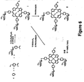
- the B-subunit mutant, or derivative named STxB-Z(n)-Cys, wherein n is 0 or 1, has been designed.
- a Cysteine is added at the C-terminus of mature STxB.
- the protein when purified from bacteria, carries the internal disulfide bond, as wild type STxB, while the sulfhydryl group at the C-terminal Cys is free. Due to their nucleophilicity, free sulfhydryl groups are excellent acceptors for directed coupling approaches (Philippe Schelté et al., 1999).
- mutants can be used as universal carriers for targeting molecules to Gb 3 receptor expressing cells.
- the present invention relates to a the use of hybrid compound for the manufacture of a compositiion for the diagnosis or therapy of intestinal turmor cells over-expressing the receptor Gb 3 , having the following formula: STxB-Z(n)-Cys-Y(m)-T, wherein:
- T is operably linked to Cys directly throught a covalent binding or indirectly through a linker, Y, allowing or not said releasing of T moiety.
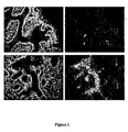
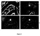
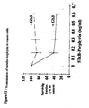
- the invention results from the constantation that cancer cells, and more particularly tumors, and more particularly intestinal and colorectal tumors, over-express Gb 3 receptor, as can be seen in table 1, figure 1 and figure 2 hereinafter.
- Gb 3 is not present in normal intestinal epithelia in
- the invention results from the observation that cancer cells, and more particularly tumors, and more particularly intestinal and colorectal tumors, over-express Gb 3 receptor, as can be seen in, figure 1 , figure 2 and figure 10 hereinafter.
- Gb 3 is not present in normal intestinal epithelia in humans (Jones et al., 2000). Accordingly, it has been shown that Gb 3 is present in low to undetectable levels in mouse. On the contrary, Gb 3 , is over-expressed in the human colon cancer cell line CaC02 (Jones et al., 2000). Thus, it constitutes an excellent marker to distinguish between tumor cells and normal intestinal cells both in humans as well as in murine models.
- This differential pattern of Gb 3 expression provides the basis for the new therapeutic and diagnostic developments of the present invention in colorectal cancer, and more generally in any Gb 3 over-expressing tumor or cancer cell.
- Over expression of Gb3 receptor on cells can be assessed by carrying out a method such as that disclosed in Example 1 when assessing the mouse model, especially starting from biopsy material. Another method encompasses performing an MRI for determination of Gb3 distribution. In some situations however, confirmation that the tumor to be treated is a Gb3 over-expressing tumor is not specifically sought prior to initiating the treatment.
- the expression "therapeutic treatment” encompasses the action of the hybrid compound of the invention which results in a beneficial effect for the patient undergoing the treatment, said effect being either obtained at a cellular level or at a clinical level, including encompassing as a result an improvement of the condition of the patient or a remission state or a recovery of a health state.
- said therapeutic treatment is provided to a patient having a tumour and especially suffering from a cancer.
- the expression "diagnosis” encompasses the detection of a pathological state or the detection of one or several parameters which may be correlated either directly or indirectly, possibly in combination with other parameters, to a pathological state and which may provide information useful in a diagnostic protocol.
- the expression also encompasses the possible quantitative detection of parameters related to such a pathological state.
- the hybrid compounds of the invention can bear a T moiety which is a contrast agent for the detection of Gb 3 -expressing cancer cells by life-imaging techniques such as Magnetic Resonance Imaging (MRI).
- life-imaging techniques such as Magnetic Resonance Imaging (MRI).
- Other non-invasive life imaging techniques include two-photon microscopy, contrast enhanced ultrasound, contrast enhanced X-ray, computed tomography, isotope scanning, contrast enhanced thermography.
- said contrast agents can be selected in a group comprising paramagnetic compounds, such as porphyrin-gadolinium, porphyrin-manganese, synthetic polymer-gadolinium, gadolinium-ethoxybenzyl-diethylenetriaminepentaacetic acid, DOPTA-gadolinium, ferrofluide and nanoparticules, which are then administrated to humans or animals.
- paramagnetic compounds such as porphyrin-gadolinium, porphyrin-manganese, synthetic polymer-gadolinium, gadolinium-ethoxybenzyl-diethylenetriaminepentaacetic acid, DOPTA-gadolinium, ferrofluide and nanoparticules, which are then administrated to humans or animals.
- the present invention also dicloses the use of such compounds for in vivo diagnosis of tumors, more specifically for MRI diagnosis.
- the use is advantageous for intestinal and colorectal cancers as far as it has been shown that Gb 3 receptors are expressed specifically in cancer cells but not in normal cells.
- the hybrid compounds of the invention can bear as a T moiety a tumor specific drug or pro-drug which is vectorized to tumor-specific transport pathways in Gb 3 -positive cancer cells allowing the increase of the efficiency and/or specificity of these treatments.
- T moiety might also be a pro-drug activator while the pro-drug alone is administered directly by any known drug delivery system, i.e. by systemic, transdermic, oral, rectal administration.
- the use of the B-subunit as a cancer targeting means has the following advantages. First, due to its small size, tissue penetration with the B-subunit is efficient. Second, the antibody response to the B-subunit is inefficient. Third, tumor-selective compounds can be coupled to the B-subunit. Forth, tumor-specific transport pathways can be exploited to increase the efficiency of the treatment. Fifth, when modifications of the B-subunit are done, only Gb 3 binding ability needs to be preserved.
- the drug is a photosensitizing drug suitable for Dynamic Phototherapy (DPT).
- DPT is a recently developed technique for the treatment of solid human tumors. It is based on the targeting to and photoactivation of dyes such as porphyrins or related system within tumor tissue. The molecular events are beginning to be understood, such as cellular death through apoptosis and other mechanisms, implicating mitochondria, nuclei, ...
- Some photosensitizing drugs are already used in the clinics (Photofrin®, Foscan®, ). However, these substances have a number of drawbacks, most notably the absence of tumor-specific targeting.
- Several strategies have been proposed to improve the tumor selectivity of photosensitizers.
- Suitable delivery systems such as liposomes, lipoproteins, monoclonal antibodies, nanoparticules to modify biodistribution of dyes.
- Another approach developed at the Institute Curie is to modulate the amphiphilicity of the macrocycle. Structural modifications induced by glycoconjugation of the tetrapyrrolic system is an effective means to create a balance between hydrophilicity and hydrophobicity. Following this approach, neutral tri- and tetra-glycoconjugated tetrapyrrolic macrocycles were prepared and evaluated in vitro for their photocytotoxicity (Momenteau et al., 1999).
- a glycoconjugated, relatively hydrophilic tetrapyrrolic macrocycle (porphyrin) has been synthesized, which carries a bromo-benzyl group that allows coupling to STxB/Cys. Synthesis, coupling to STxB/Cys, and purification of the resulting compound are described hereunder.
- the therapeutic compound composed of STxB/Cys and the glycoconjugated porphyrin accumulates stably in the endoplasmic reticulum and the Golgi apparatus.
- the phototoxicity of the porphyrin can then be activated locally by irradiation with visible light after the clearance of the cytotoxic compound from cells that need to be preserved, such as dendritic cells in which STxB does not target the retrograde route (Falguieres et al., 2001).
- T is a cytotoxic agent.
- Said cytotoxic agent might be toxic for the cell after internalization either directly, or indirectly through the action of a second component, said second component acting as a mean for transforming a pro-drug into a cytotoxic drug.
- cytotoxic agent is Neocarzinostatin (NCS).
- NCS Neocarzinostatin
- Holo-neocarzinostatin is the prototype of the protein antibiotic family. It is a 11.3 kDa complex consisting of a dodecadiyne antibiotic (NCS Chrom ) which contains the cytotoxic activity, reversibly bound to a carrier protein known as apo-neocarzinostatin (apo-NCS) (for a review, see (Favaudon, 1982)).
- NCS Chrom dodecadiyne antibiotic
- apo-NCS apo-neocarzinostatin
- Holo-NCS is active in the nanomolar range, and NCS Chrom cleaves DNA in the course of a suicide reaction leaving no residual active drug after a few minutes incubation. No resistance to NCS Chrom associated with reduced drug import (MDR) has been reported.
- the major DNA lesions induced by NCS Chrom in DNA result from radical attack and consist of a blunt end break bearing a thymidine-5'-aldehyde residue on one strand, with an abasic site at two nucleotide interval on the complementary strand. This abasic site is substrate for endonuclease III in such a way that NCS Chrom -induced damage is very rapidly converted into DNA double-strand breaks in living cells.
- Mutants of E. coli , yeast or mammalian cells defective in any pathway of double-strand break repair, most notably through a defect in DNA-dependent protein kinase are consistently hypersensitive to induced cell kill by NCS.
- Prodrugs are defined as therapeutics agents which are inactive but are transformed in active metabolites by biotransformation.
- the pro-drug is then transformed by a second component inside the cell after internalization of the hybrid compound.
- Said second component might be a cell metabolite such as an enzyme.
- T is a cytotoxic drug such as anthracyclins (daunomycin, doxorubicin, daunorubicin%), idarubicin, cis-platinium, mitomycin C, desacetylvinblastine, methotrexate, N-acetylmelphan, 5-fluorouracil, nitrogen mustards, calicheamicin, maytansinodids, and Y is a linker sensitive to an endogeneous enzyme such as a mannosidase.
- Another embodiment includes the approach described by (Saxon and Bertozzi, 2000).
- a sugar precursor containing an azido group is targeted to cancer cells via STxB/Cys. After liberation of the azido-carrying sugar in the Golgi apparatus, the later is integrated into carbohydrate chains of the cancer cell. In interaction with a phosphin-carrying prodrug, a therapeutic compound is released that specifically acts in the tumor environment
- Another embodiment includes the use of amidoximes (N-Hydroxyamidines) as pro-drugs, cleavable by endogeneous reductases.
- reductases are responsible for the reduction of amidoximes to amidines.
- a microsomal enzyme system has been purified from pig and human liver consisting of cytochrome b5, its reductase and a P450 isoenzyme (Clement B et al., 1997).
- a similar enzyme system is present in mitochondria. Reductive activities are located in several organs such as liver, kidney, lung and even brain.
- prodrugs are made incorporating linkages that are sensitive to mannosidase.
- the prodrugs are coupled to STxB/Cys and targeted to Gb 3 expressing cancer cells.
- the activation of the prodrug occurs in the Golgi apparatus of the cancer cell using endogenous mannosidase without prior vectorization of the enzyme.
- prodrugs that contain glucoronic acid conjugated through a linker moiety to the aminoglycoside of doxorubicin.
- prodrugs are synthesized as described in (Bakina and Farquhar, 1999).
- the anthracycline prodrug can be converted to doxorubicin by ⁇ -glucuronidase.
- the T moiety of the second coumpound is the ⁇ -glucuronidase.
- ⁇ -glucuronidase is coupled to STxB/Cys as described hereunder for BSA and NCS.
- the coupling product is targeted to the Golgi apparatus ( Figure 13 ) and the endoplasmic reticulum of Gb 3 expressing tumors cells and retained in this compartment. In other cells, such as dendritic cells, the coupling product is rapidly degraded (Falguaires et al., 2001).
- the prodrug is coupled to STxB/Cys.
- the product can be activated in cells that have retained the STxB/Cys- ⁇ -glucuronidase coupling product (cancer cells), but not in cells that have lost this product (dendritic cells).
- the prodrug is a nucleotide analog which, after enzymatic transformation, can be incorporated into the replicating DNA and stop said replication.
- Such prodrugs for gene suicide cancer therapy are reviewed in Singhal S. et al. (1999).
- One example is an hybrid compound wherein T is Ganciclovir (GCV) or acyclovir (ACV), and Y is a linker cleavable by an endogeneous enzyme such as mannonidase.
- the second component is a second hybrid compound wherein T is the Thymidine kinase of Herpes Simplex Virus (HSV 1 -TK). This enzyme can convert GCV or ACV to GCV monophosphate or ACV-monophosphate.
- GCV-triphosphate lacks the 3' OH on the deoxyribose as well as the bond between the 2' and 3' carbons which are necessary for DNA chain elongation. A a result, GCV-triphosphate integration causes premature DNA chain termination and leads to apoptosis.
- the present invention encompasses also:
- the skilled person can easily adapt this strategy of pro-drug conversion using the Hybrid compounds according to the invention to any known pro-drug principle, and more particularly to the multiple and complementary suicide therapies.
- the synergistic effect of the multiple suicide strategy enables lower doses or individual drugs for maximum sensitivity and reduces cytotoxicity in nontransduced cells. Furthermore, development of resistance to the suicide strategy is greatly reduced when two (or more) separate pathways are targeted.
- the present invention discloses the use of an hybrid compounds of the formula STxB-Z(n)-Cys-Y(m)-T for the therapies of Gb 3 expressing cells.
- Gb 3 expressing cells include intestinal, and particularly colorectal cells which express Gb 3 receptor only when they are tumor cells.
- STxB-Z(n)Cys-Y(m)-T as defined in the present invention provides means for therapy of pathogenic states including tumors or cancer associated with over-expression of Gb3 receptor on tumor cells.
- the present invention also pertains to pharmaceutical compositions containing at least one hybrid compound of the formula STxB-Z(n)-Cys-Y(m)-T for the diagnosis or therapy of cancers having over expressing Gb 3 receptor cells, in combination with acceptable pharmaceutical carriers.
- compositions apply either for in vivo diagnosis or for therapy of cancers cells or tumors and might contain any of the hybrid compounds described herein.
- a pharmaceutical composition according to the invention advantageously comprises one or more components to be administered in one step or sequentially in time.
- a first component of the pharmaceutical composition contains as an active component an hybrid compound wherein T is an enzyme, and for example ⁇ -glucoronidase which is first administrated to a patient bearing a tumor having over-expressing Gb 3 receptor cells; this first component is administered once to such patients;
- the second component of the pharmaceutical composition contains an hybrid compound according to the invention wherein T is a prodrug and Y is glucoronic acid; this second component of the pharmaceutical composition might be administered to the patient sequentially in the time with repetited administrations to obtain a long term effect of the therapy.
- a pharmaceutical composition according to the invention is particularly interesting for the treatment of intestinal tumors and more particularly for colorectal tumors as far as, it has been demonstrated herein, first that these tumors express specifically Gb 3 receptor and second that the pharmaceutical compositions bearing pharmaceutical carriers for oral or rectal administration are effective after an oral or rectal administration of the pharmaceutical composition.
- the present invention also discloses a method for inducing the death of cancer cells bearing over expressing Gb 3 receptors, the method comprising administering an effective amount of at least one hybrid compound described herein above, such that cancer cells death occurs.
- the invention relates in particular to means suitable for in vivo diagnosis of tumor or in vivo diagnosis of cancer.
- the invention also discloses a method for in vivo diagnosis of cancer or tumor cells over-expressing Gb 3 receptor, this method comprising administering an effective amount of an hybrid compound having a T moiety which is a contrast agent.
- the coupling approaches for covalent binding of -Y(m)-T moiety to STxB-Z(n)-Cys, m being 0 or 1, can be any method or processes described or carried out by a skilled person.
- a first method that can be embodied is the use of SPDP hetero-bi-functional cross-linker described par Carlsson et al.
- SPDP is capable of being cleavable by serum thiolases that is a cause of decreasing the yield of the reaction.
- a second method for covalent coupling of STxB -Z(n)-Cys peptides with another peptide of interest is to produce bromoacetyl or maleimide functions on the latter as described by P. Schelte et al. Briefly, the peptide of interest is chemically activated with bromoacetate anhydride or by a maleimide group respectively. In appropriate reaction conditions (pH, temperature, incubation times), these groups are eliminated by cis-elimination, yielding respectively to -S-S, -S-CH 2 -, to -S-CO- or to -S-NH-covalents linkages.
- the polypeptide or the peptide to be coupled to the -SH moiety the C-terminal Cysteine of the universal carrier has its N-terminus activated with bromoacetic anhydride following the reaction scheme:
- the Bromoacetyl function has high chemoselectivity for peptide thiol groups and the activated peptide can be reacted with STxB-Cys as follows:
- the resulting thioether-linkage is stable to hydrolysis.
- Another method for coupling a molecule to the universal carrier of the invention is to use MBS (m-Maleimidobenzoyl-N-hydroxysuccinimide ester) as shown in figure 9 .
- MBS m-Maleimidobenzoyl-N-hydroxysuccinimide ester
- EXAMPLE 1 VALIDATION OF THE MOUSE MODEL FOR IN VIVO DIAGNOSTIC AND THERAPEUTIC ADMINISTRATION OF HYBRID COMPOUNDS
- the murine model bears a colorectal cancer.
- mice Three genetically modified mouse models for colorectal cancer are used, and they show essentially the same results in this study.
- a double transgenic line RasAPC is created by crossing the two transgenic lines.
- the animals used have an age of >6 months and a weight of 25-35 grams at the time of injection.
- the mice are maintained under a 12 hour light-dark cycle and fed with standard diet and water ad libitum .
- Intestinal specimens are processed immediately after sacrificing the animal.
- Small and large intestines are opened longitudinally, tumorous regions are dissected together with surrounding normal tissue and either embedded for cryosections (see below), or fixed in AFA (75% ethanol, 20% formalin, and 5% acetic acid) for 24 hours.
- AFA 75% ethanol, 20% formalin, and 5% acetic acid
- After embedding, three ⁇ m thick sections are cut from the tissue blocks, dewaxed, rehydrated and processed by routine H&E staining. Tumors are classified and graded according to the World Health Organization classification of tumors (Hamilton and Aaltonen, 2000).
- Adenocarcinomas are considered invasive if malignant epithelial cells, arranged in glandular and/or trabecular structures, are found invading at least the submucosa.
- Lipid extraction is done according to the method of Bligh and Dyer (Bligh and Dyer, 1959). Human and mouse tumor tissue and adjacent non-tumoral specimen are weighted, mechanically homogenized in 1 ml of aqueous buffer and injeceted into 3.75 ml of chlmoroform:methanol (1:2).
- Dried plates are soaked in 0.1% polyisobutylmetacrylate in hexane, floated for 1 hour in blocking solution, followed by incubation with STxB (20 nM), primary polyclonal anti-STxB and secondary horse radish peroxidase- or alkaline phosphatase-coupled anti-rabbit antibodies. Reactive bands are revealed using enhanced chemiluminescence or chemifluorescence (Amersham Pharmacia, Little Chalfont, UK) and Phosphorlmager.
- Gb 3 expression levels are compared between normal and tumor tissue.
- FIG. 10 shows overexpression of Gb 3 by murine and human tumors
- STxB-Cy3 labeling of cryosections is carried out to detect endogenous Gb 3 in normal intestinal and tumor tissue of otherwise untreated animals.
- a stock solution of STxB-Cy3 (0.22 mg/ml) is diluted 22 fold in PBS+0.2 % BSA (final concentration 10 ⁇ g/ml), and is incubated on sections for 30 min either before or after PFA fixation at room temperature for 20 min. Subsequently, the paraformaldehyde-fixed sections are treated with 50 mM NH 4 Cl in PBS for 20 min, and solubilized with 0.1% Triton X-100 for 5 min. Counterstaining with FITC-phalloidin and Hoechst dye as described above.
- Normal tissue is overall negative, except for a faint staining that was sometimes observable in the crypts, and occasional staining of single cells within the normal tissue. These cells may constitute enteroendocrine / lymphatic cells, based on morphological criteria. In contrast, tumors are strongly stained.
- a pilot experiment is undertaken with a color marker to follow the distribution of injected fluid in the murine intestine: 0.5 ml of trypan blue is injected. The animal is sacrificed after 45 min, and the intestinal tract is removed and analyzed for distribution of trypan blue. The blue staining has clearly progressed through the largest part of the small intestine.
- Two animals are then injected with STxB, using in one animal a dose of 0.5 ml of a 1 mg/ml solution (animal A), or a smaller dose of 0.5 ml of a 0.1 mg/ml solution (animal B).
- a flexible plastic needle with a length of 40 mm and a diameter of 0.4 mm is used (Marquat Genie Biomedical, Boissy St Leger, Reference V010440).
- STxB is purified from bacteria (Mallard and Johannes, 2002) and dialyzed against IPBS before injection. Animals are injected in the oesophagus with a single dose of 0.5 ml of a solution of varying concentrations STxB in PBS without anaesthesis. After the force-feeding, animals are kept for various time points and are allowed to feed with standard diet and water ad libitum .
- mice are sacrificed by cervical dislocation and tissues are removed for subsequent analyses. Tissue samples are taken along the intestinal tract, as well as tumors from each animal. The resected normal and tumoral tissue are prepared for cryosections, or processed for lipid extraction and subsequent overlay experiments, as described above. Animals are analyzed on serial cryosections with the monoclonal as well as the polyclonal antibodies for STxB. Mouse tissues embedded in Tissue-tek OCT (Sakura) are cut in serial sections at 5 ⁇ m thickness, air dried, and fixed with 3% paraformaldehyde at room temperature for 20 min.
- the paraformaldehyde-fixed sections are treated with 50 mM NH 4 Cl in PBS for 20 min, and solubilized with 0.1% Triton X-100 for 5 min.
- Antibodies used are: monoclonal and polyclonal antibodies specific for STxB, diluted 1/100 (Falguaires et al., 2001), mAb anti-Villin ID2C3 (Dudouet et al., 1987), anti-Ki67 pAb (Novocastra), polyclonal antiserum anti-chromogranin A/B (ProGen, Heidelberg), secondary antibodies were goat anti-mouse IgG and goat anti-rabbit IgG coupled to Alexa488 or Cy3 (Jackson Immunoresearch), TRITC-phalloidin (Sigma) to visualize actin, and the dye Hoechst 33258 (Sigma) to stain nuclei.
- Tumoral regions are identified by standard histological criteria, and they are positive for the proliferative marker Ki67.
- Epithelial cells are identified with a monoclonal anti-villin antibody. No STxB staining is observable in normal intestinal epithelial cells ( Figure 1 ), but strong staining occurrs in some occasional single cells that are interspersed in the epithelial layer, and present morphological characteristics of enteroendocrine cells, and are found to be positive for chromogranin A/B staining, a marker of enteroendocrine cells ( Figure 2 ). Furthermore, a few cells are labeled that might be of lymphatic origin (macrophages or dendritic cells).
- FIG. 1 A periampullar adenocarcinoma from animal A is very strongly labeled ( Figure 1 ).
- the labeling comprises about 50% of the whole tumor surface area, and is found in epithelial cells lining trabecular or glandular structures.
- stromal regions with signs of inflammation sometimes show staining, labeled cells are of putative lymphoid origin. The stroma is otherwise negative. However, two different lesions from the same animal are negative, apart from the staining in cells of putative lymphoid origin in the stroma.
- the animal B receives the weaker dose of STxB and shows essentially the same results, normal tissue is negative with the exception of a few single cells.
- a periampullar tumor is not marked, but a second tumor from the duodenum is labeled in tumor cells of epithelial origin (as evidenced by anti-villin staining). However, the overall intensity of the staining is markedly decreased as compared to animal A (dosis of 1.0 mg/ml).
- Two animals are injected with 0.35 ml of a 1 mg/ml solution of STxB in PBS.
- the mice are sacrificed after 24 hours.
- Tissue samples are taken from control tissue (liver), and the intestinal tract: duodenum, jejunum, ileum, proximal colon.
- Tumors are isolated from both animals, and prepared for cryosections, and stained with polyclonal Ab anti-STxB and anti-chromogranin A/B. Even after 24 hours, STxB is still detectable in occasional cells in the otherwise negative epithelium, and is still very strongly present in tumors ( Figure 3 ). No staining is observable in liver sections from both animals ( Figure 4 ).
- a contrast agent that is commonly used for RMI studies are paramagnetic metalloporphyrins.
- the porphyrin I ( Figure 6 ) is prepared by condensation of pyrrole, para -2,3,4,6-tetraacetyl glucosyloxy benzaldehyde (Halazy et al., 1990) and -bromo- para-tolualdehyde (Wen et al., 1997) in high yield (31%) by Lindsey's method (Lindsey et al., 1987).
- Compound I is purified by silica gel preparative thin layer chromatography eluted by a mixture of methylene chloride/acetone (10/1, v/v) and characterized by physical methods.
- Glycoconjugated compound II ( Figure 6 ) is obtained quantitatively from I by treatment with MeONa/MeOH (Zemplén, 1927).
- BSA bovine serum albumine
- HEPES bovine serum albumine
- pH 7.4 20 mg/ml of purified bovine serum albumine (BSA) in 100 mM HEPES, pH 7.4, is incubated with 1 mM of the heterobifunctional cross linker MBS for 30 min at room temperature.
- the reaction is passed through a PBS/EDTA 10 mM equilibrated gel filtration column.
- Eluted BSA is concentrated to 20 mg/ml.
- 1 volume of STxB/Cys at 3.5 mg/ml in PBS/EDTA is mixed with 1 volume of activated BSA and incubated over night at room temperature.
- the coupling product is purified by passage over an anti-STxB immunopurification column and a MonoQ anion exchange column. According to Western analysis, the product is essentially pure. Binding of STxB/Cys-BSA to nanoparticles is done as described (Wilhelm et al., 2002).
- Gb 3 distribution by MRI techniques requires administration of a compound composed of the receptor specific molecule, STxB, linked to an appropriate paramagnetic contrast agent (ferrofluid or porphyrin). Accumulation of this targeted contrast agent at the fixation site locally modifies the water relaxation rates R1,R2,R2* leading to MRI signal modification using T1 and/or T2 and/or T2* weighted imaging sequences. Significant signal difference in images acquired before and after administration of the contrast agent depicts the regions of Gb 3 over-expression when non-specific accumulation can be excluded.
- the protocol can be applied to different tumor types over-expressing Gb 3 .
- the typical measuring procedure described hereafter is optimized for imaging intestinal tumors developed by our transgenic mouse model.
- Tumors are first detected by MRI in a screening session without contrast agent administration. Imaging is performed on a high field mini-imaging system, in our case a Bruker Biospec 47/30 system equipped with a 4.7 Tesla horizontal magnet.
- the mouse is anaesthetized (preferred anaesthetic: isoflurane) and placed in a cradle in supine position. Tubes containing circulating warm water are placed close to the animal in order to maintain body temperature.
- the animal is introduced in an MRI probe adapted to the size of the animal.
- a tube containing a water solution with adjusted R1/R2 relaxation rates is also introduced into the MRI probe and serves as an external signal intensity reference.
- Respiratory triggering has to be performed in order to obtain images of the abdominal region with minimal motion artifacts.
- Our preferred high sensitivity triggering device is home-made and is based on an inflatable chamber placed on the mouse abdomen and connected to a pressure transducer which delivers the trigger signal via appropriate electronics to the MRI system.
- Tumor detection is performed by means of a 3D T2 weighted fast spin echo imaging sequence with a field of view covering typically the liver and the intestinal region. Under these conditions tumors appear as hypersignal regions.
- the image resolution is typically 0.1x0.1x0.5 mm 3 .
- Tumors are localized with respect to anatomical markers, for example the stomach - intestine junction. Tumors are then imaged with T1 and T2* weighted fast gradient echo sequences. This terminates the first imaging session.
- the second imaging session is performed on the same mouse after administration of the contrast agent (preferred administration: oral).
- the imaging protocol matches exactly that of the first imaging session.
- Regions of interest (ROIs) covering the tumors are defined on co-localized slices from the two imaging sessions. Mean intensities of the ROIs are measured and normalized with respect to intensity of the external reference. Significantly different signal intensity detected in tumors after contrast agent administration demonstrates therefore Gb 3 over-expression.
- MRI is performed on pellets of HeLa cells (about 50 x 10 6 cells/pellet) obtained after incubation with iron oxide nanoparticle based contrast agents (nanoparticles, NP) and subsequent centrifugation in Eppendorf tubes. More precisely, cell sample preparation comprised the following main steps : 1. Incubation of cells and NP at 4°C for 30 min. (binding); 2. washing (3 times); 3. incubation at 37°C for 2 hours (endocytosis); 4. washing (3 times); 6. centrifugation. Four different conditions were used: 1. Incubation with untreated iron oxide nanoparticles (NP); 2. incubation with BSA coated nanoparticles (NP-BSA); 3. incubation with the targeted contrast agent, i.e. NP-BSA linked to STxB (STxB-Cys-MBS-BSA-NP); 4. same condition as (3.) with PPMP treated cells to reduce Gb 3 expression.
- NP iron oxide nanoparticle based contrast agents
- Resected tumoral tissu samples from villin-RasV12 mice are placed directly into an imaging chamber containing Dulbecco's modified Eagle's medium (DMEM) without phenol red.
- DMEM Dulbecco's modified Eagle's medium
- a tunable pulsed Ti:Sapphire laser (Tsunami; Spectra Physics) pumped by a Nd-YVO4 laser (Milennia, Spectra Physics) provides 70 fs pulses at 750 nm with a 80 MHz repetition. Fluorescence is detected with the built-in Fluoview photomultiplier (R928, Hamamatsu Photonics) in the descanned configuration.
- Functionalized STxB can be used to target and identify intestinal tumors by non-invasive in-depth imaging approaches.
- the fluorophore Cy3 when coupled to STxB, is strongly accumulated in intestinal tumors, demonstrating that STxB delivers contrast agents for in vivo diagnostics to Gb3-expressing tumors.
- villi of the duodenum of resected normal or cancerous tissue are observed without prior fixation or staining, using nonlinear autofluorescence ( Figure 11 A-C ).
- the regular alignment of nuclei (dark zones) in the epithelial cells is clearly visible ( Figure 11 B) .
- glycoporphyrin H2TPP(p-O- ⁇ -D-GluOAc)3( p -CH2Br) is dissolved in DMSO at 0.7 mM and mixed with an equal volume of STxB-Cys at 5.3 mg/ml. The mixture is incubated for 2 hours at room temperature and then passed through an G25 gel filtration column. The coupling product is snap frozen in liquid nitrogen and kept at -80°C for storage.
- Human tumor cells are cultivated in Dulbecco's MEM supplemented with 10% fetal calf serum (FCS).
- FCS fetal calf serum
- Cells from log-phase culture are seeded in 96-microwell plates (0.2 mL-3x10 4 cells/well) and kept at 37°C in a water-jacketed incubator for 3 hour under an air/CO 2 atmosphere (5% CO 2 ).
- Tested compounds are added under the minimum volume. Plates are incubated 3 hours, then medium is removed and cells are washed twice with phosphate buffered saline (PBS) before addition of fresh medium free of drug.
- PBS phosphate buffered saline
- Irradiation with visible light (2J/cm 2 ) is performed through the bottom of the plates using a home made "light box” fitted with an orange filter (0% T at 520 nm and 80%T at 590 nm and above) leading to a fluence of 2 mW/ cm 2 .
- Plates are incubated for three days before evaluation of the cell survival using the MTT assay (Mosmann, 1983) using 30 min incubation with 10 ⁇ g/well of 3-[4,5-dimethylthiazol-2-yl]-2,5-diphenyl tetrazolium bromide (MTT, Sigma). After removal of the medium, formazan crystals are taken up with 100 ⁇ L of DMSO and absorbance at 540 nm are measured with a Bio-Rad microplate reader (Model 450). Survival is expressed as % of untreated controls.
- Holo-NCS is purchased from Nippon Kayaku Co. (Tokyo, Japan) and purified as described (Favaudon, 1983). Purified holo-NCS is dialyzed against distilled water acidified with 1 mM acetic acid, lyophilized to dryness and stored in the dark at -80°C.
- NCS Chrom is mostly (> 90%) in "A" form according to the nomenclature of Napier et al . (Napier et al., 1981).
- apo-NCS/STxB 20 mg/ml of purified apo-NCS in 100 mM HEPES, pH 7.4, is incubated with 1 mM of the heterobifunctional cross linker MBS for 30 min at room temperature.
- the reaction is passed through a gel filtration column equilibrated with PBS containing 10 mM EDTA. Eluted activated apo-NCS is concentrated to 20 mg/ml.
- One volume of STxB/Cys at 3.5 mg/ml in PBS/EDTA is mixed with 1 volume of activated apo-NCS and incubated over night at room temperature.
- the coupling product is purified by passage over an anti-STxB immunoprufication column and a gel filtration column. According to Western analysis, the coupling product, termed apo-NCS/STxB is essentially pure.
- the protein fractions are pooled, concentrated by centrifugation over a Centricon ® centrifugal filter unit (3.000 Da cutoff), sterilized by filtration over a Millex ® unit (0.2 ⁇ m pore size) and stored in the dark at liquid nitrogen temperature (for prolonged storage it is recommended to lower the pH down to pH 5.0).
- the final concentration of the reconstituted holo-NCS/STxB is 9.8 ⁇ M relative to the absorption of protein-bound NCS Chrom at 340 nm.
- HeLa cells are maintained as exponentially growing monolayers in Dulbecco modified Eagle's minimum essential medium with 4.5 g/l glucose, 0.1 g/l pyruvate, 10 5 UI/I penicilline, 0.1 g/l streptomycine, 0.86 g/l Glutamax I and 10% v/v foetal calf serum (37°C, 5% CO 2 ), without or with 5 ⁇ M DL-threo-1-phenyl-2-hexadecanoyl-amino-3-morpholino-1-propanol (PPMP) to inhibit Gb 3 expression.
- PPMP DL-threo-1-phenyl-2-hexadecanoyl-amino-3-morpholino-1-propanol
- PPMP is introduced into pre-cultures for at least 6 days prior to cytotoxicity assays and is present for up to drug wash.
- Cytotoxicity assays are performed in parallel using HeLa cells subcultured with or without PPMP. Briefly, cells are seeded at a density of 800 cells (25 cm2 flasks) and incubated 4 hours prior to treatment to permit adhesion and spreading. Care is taken to avoid contamination of seed cell suspensions by clusters, as their presence introduces major errors in the measurement of clonogenicity.
- sterile aliquots of drug are thawed immediately prior to use, adjusted to the suitable concentration in pH 6.0 PBS and immediately introduced into culture flasks. All experiments are carried out in dim light to prevent photodecomposition of drugs.
- the flasks are washed twice with Hank's balanced salt solution, supplied with fresh medium, and returned to the incubator for 8 days. Colonies are then fixed with methanol, stained and scored.
- cytotoxicity of holo-NCS remains unchanged after its coupling to STxB/Cys.
- concentration of drug that kills 50% of the cell population is 0.71 ⁇ 0.05 nM.
- the cytotoxicity of holo-NCS and holo-NCS/STxB is then investigated at 0°C without and with PPMP, using a lethal concentration of NCS (4 nM).
- holo-NCS is known to be inactive at low temperature (Kappen et al., 1980) while STxB is still able to bind and saturate its receptor under these conditions (Johannes et al., 1997), and is expected to be internalized upon warming up.
- Seeded cells in flasks are cooled in ice for 15 min prior to exposure to holo-NCS or holo-NCS/STxB.
- the drugs (4 nM) are then introduced and incubated for a further 15 min in ice.
- the medium is removed by aspiration, and the flasks were carefully rinsed twice with ice-cold Hank's balanced salt solution to remove drugs.
- the flasks are finally supplied with ice-cold, drug-free medium and allowed to re-equilibrate at room temperature, then incubated at 37°C for 8 days. Blanks are made to check that no toxicity is introduced by chilling the cells for the duration of the experiment. The results are shown in Figure 8 .
- holo-NCS survival to holo-NCS is in the range 90% or more. Therefore, a low temperature effectively abolishes the cytotoxicity of holo-NCS. In contrast, the holo-NCS/STxB is still active at 0°C, in such a way that cell survival is ca. 30% only. PPMP induced marked resistance to holo-NCS/STxB.
- compositions are made using STxB as a carrier that allow for targeting of therapeutic compounds to Gb3 expressing tumors.
- the effect of these compositions on tumor cells in vitro, on tumors in the described animal models, and on human tumors has been demonstrated.
- the formed conjugate STxB-Cys- ⁇ -GUS is purified by passage on a gel filtration column and an anti-STxB immunoaffinity column. Purified coupling product is highly pure, as tested by Western blotting, and the activity of the enzyme is not altered by the chemical modifications.
- the cells are then washed with ice-cold culture medium, shifted to 37°C for 40 min (internalization step), fixed with 4% PFA for 10 min, permeablized with saponin, stained with the indicated primary and secondary antibodies and analyzed by confocal microscopy.
- the obtained results are illustrated in figure 13 .
- Hela cells (10 6 ) are incubated for 30 min at 4°C in presence or absence of 0.5 ⁇ M STxB-Cys- ⁇ -GUS (binding step). After washing with culture medium, cells are shifted to 37°C for 60 min (internalization step). Cell lysates are prepared in RIPA buffer (PBS 1X, NP40 1%, Doc 0.5%, SDS 0.5%).
- RIPA buffer PBS 1X, NP40 1%, Doc 0.5%, SDS 0.5%).
- the basal cellular ⁇ -GUS activity and ⁇ -GUS activity associated with ⁇ -GUS-STxB treated, Gb3 expressing and non-expressing Hela cells are summarized in figure 14 .
- Gb3 expressing HeLa cells show a significant increase of ⁇ -GUS activity comparatively to non-expressing cells. These latters possess the same level of ⁇ -Glucuronidase activity as control cells (basal activity), indicating that cellular targeting of ⁇ -GUS activity is dependent on STx
- Alternatives to coupling via the sulfhydryl groups (-SH) are coupling via amino groups (-NH2), carbohydrates, carboxyls (-COOH), or hydroxyls (-OH).
- Examples of reactive groups on to-be-vectorized compounds are imidoesters (react on primary amines), N-hydroxysuccinimide esters (react on primary amines), maleimides (react on sulfhydryls), haloacetyls (react on sulfhydryls), hydrazines (react on oxidized carbohydrates), carbodiimides (react on carboxyls).
- Wild type STxB is not glycosylated.
- Glycosylated STxB for chemical coupling can be obtained by expressing a glycosylation site-carrying variant of STxB in glycosylation competent cells, such as the yeast Pichia pastoris. Amino groups, carboxylates, and hydroxyls are present in wild-type STxB, and chemical coupling results in heterogenous mixtures containing inactivated protein.
- amino acids with appropriate side chains are fused to the carboxyl terminus of STxB.
- the amino acid sequence EDEKKK Glu-Asp-Glu-Lys-Lys-Lys
Landscapes
- Health & Medical Sciences (AREA)
- Life Sciences & Earth Sciences (AREA)
- Chemical & Material Sciences (AREA)
- General Health & Medical Sciences (AREA)
- Engineering & Computer Science (AREA)
- Medicinal Chemistry (AREA)
- Animal Behavior & Ethology (AREA)
- Veterinary Medicine (AREA)
- Molecular Biology (AREA)
- Public Health (AREA)
- Epidemiology (AREA)
- Bioinformatics & Cheminformatics (AREA)
- Proteomics, Peptides & Aminoacids (AREA)
- Pharmacology & Pharmacy (AREA)
- Organic Chemistry (AREA)
- Nuclear Medicine, Radiotherapy & Molecular Imaging (AREA)
- Genetics & Genomics (AREA)
- Biophysics (AREA)
- Biochemistry (AREA)
- Gastroenterology & Hepatology (AREA)
- Toxicology (AREA)
- Nanotechnology (AREA)
- Radiology & Medical Imaging (AREA)
- Diabetes (AREA)
- Biotechnology (AREA)
- Zoology (AREA)
- Biomedical Technology (AREA)
- General Chemical & Material Sciences (AREA)
- Endocrinology (AREA)
- Pathology (AREA)
- Rheumatology (AREA)
- Urology & Nephrology (AREA)
- Cell Biology (AREA)
- General Engineering & Computer Science (AREA)
- Medical Informatics (AREA)
- Crystallography & Structural Chemistry (AREA)
- Chemical Kinetics & Catalysis (AREA)
- Medicines That Contain Protein Lipid Enzymes And Other Medicines (AREA)
- Pharmaceuticals Containing Other Organic And Inorganic Compounds (AREA)
- Medicinal Preparation (AREA)
Claims (16)
- Verwendung einer Hybridverbindung in der Herstellung einer Zusammensetzung, nutzbar für die Diagnose oder Therapie von intestinalen Tumorzellen, die den Rezeptor Gb3 überexprimieren, worin die Hybridverbindung die folgende Formel aufweist: STxB-Z(n)-Cys-Y(m)-T, worin- STxB die Shiga Toxin B-Untereinheit ist,- Z(n), worin n 0 oder 1 ist, und wenn n 1 ist, Z eine Aminosäure, befreit von einer Sulfydrylgruppe, oder ein Polypeptid ist,- Cys die Aminosäure Cystein ist,- T ein Molekül ist, gebunden durch eine kovalente Bindung an den S-Teil von Cys, gewählt aus einer Gruppe, umfassend:• Mittel für in vivo-Diagnostik,• zytotoxische Mittel,• Prodrugs,• Enzyme für die Umwandlung eines Prodrugs in ein Arzneimittel,- Y(m), worin m 0 oder 1 ist, und wenn m 1 ist, Y ein Linker zwischen T und Cys ist, wobei der Linker für die Freigabe von T nach Internalisierung der Hybridverbindung in die Zellen spaltbar oder nicht ist.
- Verwendung einer Hybridverbindung gemäss Anspruch 1, worin n = 0.
- Verwendung einer Hybridverbindung gemäss Anspruch 1 oder 2, wobei T ein Kontrastmittel für MRI ist, gewählt aus einer Gruppe, umfassend paramagnetische Verbindungen, wie z.B. Porphyrin-Gadolinium, Porphyrin-Mangan, synthetisches Polymer-Gadolinium, Gadolinium-Ethoxybenzyl-Diethylentriaminpentaessigsäure, DOPTA-Gadolinium, Ferrofluid und Nanopartikel.
- Verwendung einer Hybridverbindung gemäss Anspruch 1 oder 2, worin m = 1 und T gewählt ist aus einer Gruppe, umfassend cytotoxische Arzneimittel, wie z.B. Anthracycline (Daunomycin, Doxorubicin, Daunorubicin), Idarubicin, cis-Platin, Mitomycin C, Desacetylvinblastin, Methotrexat, N-Acetylmelphalan, 5-Fluoruracil, Stickstoffsenf (mustards), Calicheamicin, Maytansinoide, konjugiert mit einem Enzym-empfindlichen Linker;- Nucleotidanaloge, die die DNA-Replikation beenden können, wie z.B. Ganciclovir oder Aciclovir,- Amidoxime.
- Verwendung einer Hybridverbindung gemäss Anspruch 1, wobei der Linker Y ein Enzym-spaltbarer Linker ist, gewählt aus einer Gruppe, umfassend reduzierte und nicht-reduzierte Folate, spaltbar durch Carboxypeptidase G, Phosphatgruppen von phosphorylierten Prodrugs, spaltbar durch alkalische Phosphatase, durch Carboxypeptidase A hydrolytisch spaltbare Verbindungen, Nitroreduktase für die Prodrug-Aktivierung, Hydrolyse des Lactamrings, spaltbar durch β-Lactamase, Amid, spaltbar durch Penicillinamidase, Cytosindesamidase für die Prodrug-Aktivierung, Glucuronsäure, spaltbar durch β-Glucuronidase, Galactose, spaltbar durch Galactosidase, Mannose, spaltbar durch Mannosidase.
- Verwendung einer Hybridverbindung gemäss Anspruch 4, wobei das Enzym in den überexprimierenden Gb3-Zellen endogen ist oder mit einer zweiten Hybridverbindung gemäss Anspruch 1, worin T das Enzym ist, internalisiert wird.
- Verwendung einer Hybridverbindung gemäss Anspruch 1, wobei T ein Enzym ist, gewählt aus einer Gruppe, enthaltend Peptidasen, Thymidinkinase von HSV1, Lipasen, Glycosidasen.
- Verwendung einer Hybridverbindung gemäss Anspruch 1, wobei T ein photosensibilisierendes Arzneimittel für dynamische Phototherapien ist.
- Verwendung einer Hybridverbindung gemäss Anspruch 8, wobei das Arzneimittel gewählt ist aus der Gruppe, umfassend Photoform®, Forean®, glycokonjugierten Tetrapyrrolmakrozyklus, wie z.B. Porphyrin.
- Verwendung einer Hybridverbindung gemäss Anspruch 1, wobei T Holo-Neocarzinostatin ist.
- Verwendung einer ersten wie in Anspruch 1 definierten Hybridverbindung, worin T ein Prodrug ist, und eines zweiten Bestandteils zur Transformation des Prodrugs in ein toxisches Arzneimittel in der Herstellung einer Zusammensetzung, nutzbar für die Therapie von intestinalen Tumorzellen, die den Rezeptor Gb3 überexprimieren.
- Verwendung gemäss Anspruch 11, worin der zweite Bestandteil eine wie in Anspruch 1 definierte Hybridverbindung ist, worin T ein Enzym zur Transformation des Prodrugs in ein toxisches Arzneimittel ist.
- Verwendung gemäss Anspruch 11 oder 12, wobei die zwei Verbindungen simultan oder sequenziell verabreicht werden.
- Verwendung gemäss irgendeinem der Ansprüche 1 bis 13, worin die intestinalen Tumorzellen kolorektale Tumorzellen sind.
- Hybridverbindung, wie in irgendeinem der Ansprüche 1 bis 10 und 14 definiert, zur Verwendung in der Diagnose oder Therapie von intestinalen Tumorzellen, die den Rezeptor Gb3 überexprimieren.
- Zusammensetzung, wie in irgendeinem der Ansprüche 11 bis 14 definiert, umfassend eine erste wie in Anspruch 1 definierte Hybridverbindung, worin T ein Prodrug ist, und einen zweiten Bestandteil zur Transformation des Prodrugs in ein toxisches Arzneimittel, zur Verwendung in der Therapie von intestinalen Tumorzellen, die den Rezeptor Gb3 überexprimieren.
Priority Applications (1)
| Application Number | Priority Date | Filing Date | Title |
|---|---|---|---|
| EP03775133A EP1525478B1 (de) | 2002-08-02 | 2003-07-31 | Shiga-toxin-untereinheit b als vektor für die tumordiagnose und zur arzneimttelverabreichung an gb3-exprimierenden tumoren |
Applications Claiming Priority (4)
| Application Number | Priority Date | Filing Date | Title |
|---|---|---|---|
| EP02291962A EP1386927B1 (de) | 2002-08-02 | 2002-08-02 | Shiga-Toxin-Untereinheit B als Vektor zur Diagnose von Tumoren und zur Arzneimittelverabreichung an GB3-exprimierenden Tumoren |
| EP02291962 | 2002-08-02 | ||
| PCT/EP2003/009308 WO2004016148A2 (en) | 2002-08-02 | 2003-07-31 | Shiga toxin b-subunit as a vector for tumor diagnosis and drug delivery to gb3 expressing tumors |
| EP03775133A EP1525478B1 (de) | 2002-08-02 | 2003-07-31 | Shiga-toxin-untereinheit b als vektor für die tumordiagnose und zur arzneimttelverabreichung an gb3-exprimierenden tumoren |
Publications (2)
| Publication Number | Publication Date |
|---|---|
| EP1525478A2 EP1525478A2 (de) | 2005-04-27 |
| EP1525478B1 true EP1525478B1 (de) | 2009-08-12 |
Family
ID=30011281
Family Applications (2)
| Application Number | Title | Priority Date | Filing Date |
|---|---|---|---|
| EP02291962A Expired - Lifetime EP1386927B1 (de) | 2002-08-02 | 2002-08-02 | Shiga-Toxin-Untereinheit B als Vektor zur Diagnose von Tumoren und zur Arzneimittelverabreichung an GB3-exprimierenden Tumoren |
| EP03775133A Expired - Lifetime EP1525478B1 (de) | 2002-08-02 | 2003-07-31 | Shiga-toxin-untereinheit b als vektor für die tumordiagnose und zur arzneimttelverabreichung an gb3-exprimierenden tumoren |
Family Applications Before (1)
| Application Number | Title | Priority Date | Filing Date |
|---|---|---|---|
| EP02291962A Expired - Lifetime EP1386927B1 (de) | 2002-08-02 | 2002-08-02 | Shiga-Toxin-Untereinheit B als Vektor zur Diagnose von Tumoren und zur Arzneimittelverabreichung an GB3-exprimierenden Tumoren |
Country Status (9)
| Country | Link |
|---|---|
| US (3) | US7718601B2 (de) |
| EP (2) | EP1386927B1 (de) |
| JP (1) | JP4339251B2 (de) |
| AT (2) | ATE292144T1 (de) |
| AU (1) | AU2003283228A1 (de) |
| CA (1) | CA2494541C (de) |
| DE (2) | DE60203491D1 (de) |
| ES (1) | ES2330429T3 (de) |
| WO (1) | WO2004016148A2 (de) |
Families Citing this family (13)
| Publication number | Priority date | Publication date | Assignee | Title |
|---|---|---|---|---|
| EP1229045A1 (de) | 2001-02-01 | 2002-08-07 | Institut Curie | Universeller Träger zum Zielrichten von Molekülen zu Rezeptor Gb3 - exprimierenden Zellen |
| EP1386927B1 (de) * | 2002-08-02 | 2005-03-30 | Institut Curie | Shiga-Toxin-Untereinheit B als Vektor zur Diagnose von Tumoren und zur Arzneimittelverabreichung an GB3-exprimierenden Tumoren |
| AR047692A1 (es) * | 2003-07-10 | 2006-02-08 | Epix Medical Inc | Imagenes de blancos estacionarios |
| KR20080050503A (ko) * | 2005-10-12 | 2008-06-05 | 시바 홀딩 인코포레이티드 | 캡슐화된 발광 안료 |
| US20110152252A1 (en) * | 2007-12-18 | 2011-06-23 | Institut Curie | Shiga toxin b-subunit/chemotherapeutics conjugates |
| EP2072060A1 (de) | 2007-12-18 | 2009-06-24 | Institut Curie | Verfahren und Zusammensetzungen zur Herstellung und Verwendung von Toxinkonjugaten |
| AU2010234459A1 (en) * | 2009-04-08 | 2011-11-03 | The Regents Of The University Of California | Human protein scaffold with controlled serum pharmacokinetics |
| EP2740491A1 (de) * | 2012-12-05 | 2014-06-11 | Institut Curie | Konjugate der B-Untereinheit von Shiga-Toxin zur Verwendung als Kontrastmittel zur Bildgebung und Therapie |
| CN112851769A (zh) | 2014-01-27 | 2021-05-28 | 分子模板公司 | 用于哺乳动物中的去免疫化志贺毒素a亚基效应子多肽 |
| AU2015274647C1 (en) * | 2014-06-11 | 2020-01-30 | Molecular Templates, Inc. | Protease-cleavage resistant, Shiga toxin a subunit effector polypeptides and cell-targeted molecules comprising the same |
| AU2017283653C1 (en) | 2016-06-16 | 2022-05-05 | Oncoselect Therapeutics, Llc | Porphyrin compounds and compositions useful for treating cancer |
| JP7123921B2 (ja) * | 2016-11-09 | 2022-08-23 | グリフィス・ユニバーシティ | サブチラーゼサイトトキシンbサブユニット変異体 |
| JP2020519244A (ja) * | 2017-04-20 | 2020-07-02 | アンスティトゥート・ナシオナル・ドゥ・ラ・サンテ・エ・ドゥ・ラ・ルシャルシュ・メディカル・(インセルム) | 研究及び生物医学応用のためのペプチド、特にポリペプチド、ファージディスプレイスクリーニング法及び関連する手段、並びにその使用 |
Citations (1)
| Publication number | Priority date | Publication date | Assignee | Title |
|---|---|---|---|---|
| WO1999003881A2 (fr) * | 1997-07-18 | 1999-01-28 | Institut Curie | Polypeptide chimerique comprenant le fragment b de la toxine shiga et des peptides d'interet therapeutique |
Family Cites Families (19)
| Publication number | Priority date | Publication date | Assignee | Title |
|---|---|---|---|---|
| JPS6075432A (ja) * | 1983-08-08 | 1985-04-27 | Kuraray Co Ltd | ネオカルチノスタチン誘導体及びその製造方法 |
| US4827945A (en) * | 1986-07-03 | 1989-05-09 | Advanced Magnetics, Incorporated | Biologically degradable superparamagnetic materials for use in clinical applications |
| US5250565A (en) * | 1987-02-10 | 1993-10-05 | Abbott Laboratories | Indole-,benzofuran-,and benzothiophene-containing lipoxygenase-inhibiting compounds |
| US5753627A (en) * | 1988-12-05 | 1998-05-19 | Novartis Ag | Use of certain complexed somatostatin peptides for the invivo imaging of somatostatin receptor-positive tumors and metastasis |
| US5053423A (en) * | 1990-03-22 | 1991-10-01 | Quadra Logic Technologies Inc. | Compositions for photodynamic therapy |
| US5326778A (en) * | 1992-03-03 | 1994-07-05 | Research Corporation Technologies, Inc. | Conjugates of biotin and deferoxamine for radioimmunoimaging and radioimmunotherapy |
| US5965132A (en) * | 1992-03-05 | 1999-10-12 | Board Of Regents, The University Of Texas System | Methods and compositions for targeting the vasculature of solid tumors |
| US5840485A (en) * | 1993-05-27 | 1998-11-24 | Selectide Corporation | Topologically segregated, encoded solid phase libraries |
| CA2175579A1 (en) * | 1993-10-26 | 1995-05-04 | Chang Yi Wang | Structured synthetic antigen libraries as diagnostics, vaccines and therapeutics |
| DE4430601A1 (de) * | 1994-08-22 | 1996-02-29 | Beiersdorf Ag | Zelladhäsions-Peptide zur Modifikation des Haftungsverhaltens eukaryontischer Zellen untereinander |
| GB9600272D0 (en) * | 1996-01-06 | 1996-03-06 | Univ Nottingham | Polymers |
| US5801145A (en) * | 1996-02-09 | 1998-09-01 | Ontario Cancer Institute | Method for selectively purging CD77+ cells from bone marrow |
| US6368598B1 (en) * | 1996-09-16 | 2002-04-09 | Jcrt Radiation Oncology Support Services, Inc. | Drug complex for treatment of metastatic prostate cancer |
| WO2000061183A2 (en) * | 1999-04-09 | 2000-10-19 | Hsc Research And Development Limited Partnership | Verotoxin treatment of lymphomas |
| EP1297002A2 (de) * | 2000-06-30 | 2003-04-02 | Board Of Regents, The University Of Texas System | Isolierung eines zell-spezifisch internalisierenden peptids, das tumor gewebe zur gezielten medikamenten freisetzung infiltriert |
| EP1229045A1 (de) * | 2001-02-01 | 2002-08-07 | Institut Curie | Universeller Träger zum Zielrichten von Molekülen zu Rezeptor Gb3 - exprimierenden Zellen |
| WO2002096910A1 (en) * | 2001-05-31 | 2002-12-05 | Medarex, Inc. | Cytotoxins, prodrugs, linkers and stabilizers useful therefor |
| EP1386927B1 (de) * | 2002-08-02 | 2005-03-30 | Institut Curie | Shiga-Toxin-Untereinheit B als Vektor zur Diagnose von Tumoren und zur Arzneimittelverabreichung an GB3-exprimierenden Tumoren |
| US20110152252A1 (en) * | 2007-12-18 | 2011-06-23 | Institut Curie | Shiga toxin b-subunit/chemotherapeutics conjugates |
-
2002
- 2002-08-02 EP EP02291962A patent/EP1386927B1/de not_active Expired - Lifetime
- 2002-08-02 DE DE60203491T patent/DE60203491D1/de not_active Expired - Lifetime
- 2002-08-02 AT AT02291962T patent/ATE292144T1/de not_active IP Right Cessation
-
2003
- 2003-07-31 EP EP03775133A patent/EP1525478B1/de not_active Expired - Lifetime
- 2003-07-31 DE DE60328789T patent/DE60328789D1/de not_active Expired - Lifetime
- 2003-07-31 WO PCT/EP2003/009308 patent/WO2004016148A2/en not_active Ceased
- 2003-07-31 AT AT03775133T patent/ATE439597T1/de not_active IP Right Cessation
- 2003-07-31 JP JP2004528507A patent/JP4339251B2/ja not_active Expired - Fee Related
- 2003-07-31 AU AU2003283228A patent/AU2003283228A1/en not_active Abandoned
- 2003-07-31 CA CA2494541A patent/CA2494541C/en not_active Expired - Fee Related
- 2003-07-31 ES ES03775133T patent/ES2330429T3/es not_active Expired - Lifetime
-
2005
- 2005-02-01 US US11/046,786 patent/US7718601B2/en not_active Expired - Fee Related
-
2010
- 2010-03-12 US US12/723,361 patent/US7981400B2/en not_active Expired - Fee Related
-
2011
- 2011-06-13 US US13/159,124 patent/US8313731B2/en not_active Expired - Fee Related
Patent Citations (1)
| Publication number | Priority date | Publication date | Assignee | Title |
|---|---|---|---|---|
| WO1999003881A2 (fr) * | 1997-07-18 | 1999-01-28 | Institut Curie | Polypeptide chimerique comprenant le fragment b de la toxine shiga et des peptides d'interet therapeutique |
Also Published As
| Publication number | Publication date |
|---|---|
| EP1386927B1 (de) | 2005-03-30 |
| JP4339251B2 (ja) | 2009-10-07 |
| US7718601B2 (en) | 2010-05-18 |
| CA2494541C (en) | 2011-10-18 |
| WO2004016148A3 (en) | 2004-10-28 |
| US20110243914A1 (en) | 2011-10-06 |
| EP1525478A2 (de) | 2005-04-27 |
| CA2494541A1 (en) | 2004-02-26 |
| AU2003283228A8 (en) | 2004-03-03 |
| US8313731B2 (en) | 2012-11-20 |
| ATE439597T1 (de) | 2009-08-15 |
| ES2330429T3 (es) | 2009-12-10 |
| ATE292144T1 (de) | 2005-04-15 |
| DE60328789D1 (de) | 2009-09-24 |
| US20060008475A1 (en) | 2006-01-12 |
| DE60203491D1 (de) | 2005-05-04 |
| AU2003283228A1 (en) | 2004-03-03 |
| EP1386927A1 (de) | 2004-02-04 |
| WO2004016148A2 (en) | 2004-02-26 |
| US20100329992A1 (en) | 2010-12-30 |
| JP2006509727A (ja) | 2006-03-23 |
| US7981400B2 (en) | 2011-07-19 |
Similar Documents
| Publication | Publication Date | Title |
|---|---|---|
| US8313731B2 (en) | Shiga toxin B-subunit as a vector for tumor diagnosis and drug delivery to GB3 expressing tumors | |
| US11229710B2 (en) | Environmentally sensitive compositions comprising a pH-triggered membrane protein and methods of use thereof in the treatment and diagnosis of tumors | |
| CA2406650C (en) | Cationic diagnostic, imaging and therapeutic agents associated with activated vascular sites | |
| US5518888A (en) | ST receptor binding compounds and methods of using the same | |
| US6843980B2 (en) | Methods for using annexin for detecting cell death in vivo and treating associated conditions | |
| US5919764A (en) | Compounds that bind to p185 and methods of using the same | |
| Pietersz | The linkage of cytotoxic drugs to monoclonal antibodies for the treatment of cancer | |
| AU2001266272A1 (en) | Cationic diagnostic, imaging and therapeutic agents associated with activated vascular sites | |
| JP2914737B2 (ja) | 免疫試薬 | |
| US9526795B2 (en) | N-BOC-dendrimers and their conjugates | |
| US7097839B1 (en) | ST receptor binding compounds and methods of using the same | |
| Naffouje et al. | A method of tumor in vivo imaging with a new peptide-based fluorescent probe | |
| US10059950B2 (en) | Compositions comprising an internalizing nucleic acid molecule, and their methods of use |
Legal Events
| Date | Code | Title | Description |
|---|---|---|---|
| PUAI | Public reference made under article 153(3) epc to a published international application that has entered the european phase |
Free format text: ORIGINAL CODE: 0009012 |
|
| 17P | Request for examination filed |
Effective date: 20050210 |
|
| AK | Designated contracting states |
Kind code of ref document: A2 Designated state(s): AT BE BG CH CY CZ DE DK EE ES FI FR GB GR HU IE IT LI LU MC NL PT RO SE SI SK TR |
|
| AX | Request for extension of the european patent |
Extension state: AL LT LV MK |
|
| DAX | Request for extension of the european patent (deleted) | ||
| 17Q | First examination report despatched |
Effective date: 20071018 |
|
| RIN1 | Information on inventor provided before grant (corrected) |
Inventor name: ROBINE, SYLVIE Inventor name: FLORENT, JEAN-CLAUDE Inventor name: MAILLARD, PHILIPPE Inventor name: JOHANNES, LUDGER Inventor name: GRIERSON, DAVID Inventor name: ROGER, JACKY |
|
| GRAP | Despatch of communication of intention to grant a patent |
Free format text: ORIGINAL CODE: EPIDOSNIGR1 |
|
| RTI1 | Title (correction) |
Free format text: SHIGA TOXIN B-SUBUNIT AS A VECTOR FOR TUMOR DIAGNOSIS AND DRUG DELIVERY TO GB3-EXPRESSING TUMORS |
|
| GRAS | Grant fee paid |
Free format text: ORIGINAL CODE: EPIDOSNIGR3 |
|
| GRAA | (expected) grant |
Free format text: ORIGINAL CODE: 0009210 |
|
| AK | Designated contracting states |
Kind code of ref document: B1 Designated state(s): AT BE BG CH CY CZ DE DK EE ES FI FR GB GR HU IE IT LI LU MC NL PT RO SE SI SK TR |
|
| REG | Reference to a national code |
Ref country code: GB Ref legal event code: FG4D |
|
| REG | Reference to a national code |
Ref country code: CH Ref legal event code: EP |
|
| REG | Reference to a national code |
Ref country code: IE Ref legal event code: FG4D |
|
| REF | Corresponds to: |
Ref document number: 60328789 Country of ref document: DE Date of ref document: 20090924 Kind code of ref document: P |
|
| REG | Reference to a national code |
Ref country code: CH Ref legal event code: NV Representative=s name: PATENTANWAELTE SCHAAD, BALASS, MENZL & PARTNER AG |
|
| REG | Reference to a national code |
Ref country code: SE Ref legal event code: TRGR |
|
| REG | Reference to a national code |
Ref country code: ES Ref legal event code: FG2A Ref document number: 2330429 Country of ref document: ES Kind code of ref document: T3 |
|
| PG25 | Lapsed in a contracting state [announced via postgrant information from national office to epo] |
Ref country code: AT Free format text: LAPSE BECAUSE OF FAILURE TO SUBMIT A TRANSLATION OF THE DESCRIPTION OR TO PAY THE FEE WITHIN THE PRESCRIBED TIME-LIMIT Effective date: 20090812 Ref country code: FI Free format text: LAPSE BECAUSE OF FAILURE TO SUBMIT A TRANSLATION OF THE DESCRIPTION OR TO PAY THE FEE WITHIN THE PRESCRIBED TIME-LIMIT Effective date: 20090812 |
|
| PG25 | Lapsed in a contracting state [announced via postgrant information from national office to epo] |
Ref country code: SI Free format text: LAPSE BECAUSE OF FAILURE TO SUBMIT A TRANSLATION OF THE DESCRIPTION OR TO PAY THE FEE WITHIN THE PRESCRIBED TIME-LIMIT Effective date: 20090812 |
|
| PG25 | Lapsed in a contracting state [announced via postgrant information from national office to epo] |
Ref country code: PT Free format text: LAPSE BECAUSE OF FAILURE TO SUBMIT A TRANSLATION OF THE DESCRIPTION OR TO PAY THE FEE WITHIN THE PRESCRIBED TIME-LIMIT Effective date: 20091212 Ref country code: BG Free format text: LAPSE BECAUSE OF FAILURE TO SUBMIT A TRANSLATION OF THE DESCRIPTION OR TO PAY THE FEE WITHIN THE PRESCRIBED TIME-LIMIT Effective date: 20091112 |
|
| PG25 | Lapsed in a contracting state [announced via postgrant information from national office to epo] |
Ref country code: CZ Free format text: LAPSE BECAUSE OF FAILURE TO SUBMIT A TRANSLATION OF THE DESCRIPTION OR TO PAY THE FEE WITHIN THE PRESCRIBED TIME-LIMIT Effective date: 20090812 Ref country code: RO Free format text: LAPSE BECAUSE OF FAILURE TO SUBMIT A TRANSLATION OF THE DESCRIPTION OR TO PAY THE FEE WITHIN THE PRESCRIBED TIME-LIMIT Effective date: 20090812 Ref country code: EE Free format text: LAPSE BECAUSE OF FAILURE TO SUBMIT A TRANSLATION OF THE DESCRIPTION OR TO PAY THE FEE WITHIN THE PRESCRIBED TIME-LIMIT Effective date: 20090812 Ref country code: DK Free format text: LAPSE BECAUSE OF FAILURE TO SUBMIT A TRANSLATION OF THE DESCRIPTION OR TO PAY THE FEE WITHIN THE PRESCRIBED TIME-LIMIT Effective date: 20090812 |
|
| PG25 | Lapsed in a contracting state [announced via postgrant information from national office to epo] |
Ref country code: SK Free format text: LAPSE BECAUSE OF FAILURE TO SUBMIT A TRANSLATION OF THE DESCRIPTION OR TO PAY THE FEE WITHIN THE PRESCRIBED TIME-LIMIT Effective date: 20090812 |
|
| PLBE | No opposition filed within time limit |
Free format text: ORIGINAL CODE: 0009261 |
|
| STAA | Information on the status of an ep patent application or granted ep patent |
Free format text: STATUS: NO OPPOSITION FILED WITHIN TIME LIMIT |
|
| PG25 | Lapsed in a contracting state [announced via postgrant information from national office to epo] |
Ref country code: BE Free format text: LAPSE BECAUSE OF FAILURE TO SUBMIT A TRANSLATION OF THE DESCRIPTION OR TO PAY THE FEE WITHIN THE PRESCRIBED TIME-LIMIT Effective date: 20090812 |
|
| 26N | No opposition filed |
Effective date: 20100517 |
|
| PG25 | Lapsed in a contracting state [announced via postgrant information from national office to epo] |
Ref country code: GR Free format text: LAPSE BECAUSE OF FAILURE TO SUBMIT A TRANSLATION OF THE DESCRIPTION OR TO PAY THE FEE WITHIN THE PRESCRIBED TIME-LIMIT Effective date: 20091113 |
|
| PG25 | Lapsed in a contracting state [announced via postgrant information from national office to epo] |
Ref country code: MC Free format text: LAPSE BECAUSE OF NON-PAYMENT OF DUE FEES Effective date: 20100731 |
|
| PG25 | Lapsed in a contracting state [announced via postgrant information from national office to epo] |
Ref country code: IE Free format text: LAPSE BECAUSE OF NON-PAYMENT OF DUE FEES Effective date: 20100731 |
|
| PG25 | Lapsed in a contracting state [announced via postgrant information from national office to epo] |
Ref country code: CY Free format text: LAPSE BECAUSE OF FAILURE TO SUBMIT A TRANSLATION OF THE DESCRIPTION OR TO PAY THE FEE WITHIN THE PRESCRIBED TIME-LIMIT Effective date: 20090812 |
|
| PG25 | Lapsed in a contracting state [announced via postgrant information from national office to epo] |
Ref country code: HU Free format text: LAPSE BECAUSE OF FAILURE TO SUBMIT A TRANSLATION OF THE DESCRIPTION OR TO PAY THE FEE WITHIN THE PRESCRIBED TIME-LIMIT Effective date: 20100213 Ref country code: LU Free format text: LAPSE BECAUSE OF NON-PAYMENT OF DUE FEES Effective date: 20100731 |
|
| PG25 | Lapsed in a contracting state [announced via postgrant information from national office to epo] |
Ref country code: TR Free format text: LAPSE BECAUSE OF FAILURE TO SUBMIT A TRANSLATION OF THE DESCRIPTION OR TO PAY THE FEE WITHIN THE PRESCRIBED TIME-LIMIT Effective date: 20090812 |
|
| PGFP | Annual fee paid to national office [announced via postgrant information from national office to epo] |
Ref country code: NL Payment date: 20140716 Year of fee payment: 12 Ref country code: CH Payment date: 20140715 Year of fee payment: 12 |
|
| PGFP | Annual fee paid to national office [announced via postgrant information from national office to epo] |
Ref country code: ES Payment date: 20140725 Year of fee payment: 12 Ref country code: SE Payment date: 20140717 Year of fee payment: 12 |
|
| PGFP | Annual fee paid to national office [announced via postgrant information from national office to epo] |
Ref country code: IT Payment date: 20140717 Year of fee payment: 12 |
|
| REG | Reference to a national code |
Ref country code: CH Ref legal event code: PL |
|
| REG | Reference to a national code |
Ref country code: SE Ref legal event code: EUG |
|
| REG | Reference to a national code |
Ref country code: NL Ref legal event code: MM Effective date: 20150801 |
|
| PG25 | Lapsed in a contracting state [announced via postgrant information from national office to epo] |
Ref country code: CH Free format text: LAPSE BECAUSE OF NON-PAYMENT OF DUE FEES Effective date: 20150731 Ref country code: IT Free format text: LAPSE BECAUSE OF NON-PAYMENT OF DUE FEES Effective date: 20150731 Ref country code: LI Free format text: LAPSE BECAUSE OF NON-PAYMENT OF DUE FEES Effective date: 20150731 |
|
| PG25 | Lapsed in a contracting state [announced via postgrant information from national office to epo] |
Ref country code: SE Free format text: LAPSE BECAUSE OF NON-PAYMENT OF DUE FEES Effective date: 20150801 Ref country code: NL Free format text: LAPSE BECAUSE OF NON-PAYMENT OF DUE FEES Effective date: 20150801 |
|
| REG | Reference to a national code |
Ref country code: FR Ref legal event code: PLFP Year of fee payment: 14 |
|
| REG | Reference to a national code |
Ref country code: ES Ref legal event code: FD2A Effective date: 20160826 |
|
| PG25 | Lapsed in a contracting state [announced via postgrant information from national office to epo] |
Ref country code: ES Free format text: LAPSE BECAUSE OF NON-PAYMENT OF DUE FEES Effective date: 20150801 |
|
| REG | Reference to a national code |
Ref country code: FR Ref legal event code: PLFP Year of fee payment: 15 |
|
| REG | Reference to a national code |
Ref country code: FR Ref legal event code: PLFP Year of fee payment: 16 |
|
| PGFP | Annual fee paid to national office [announced via postgrant information from national office to epo] |
Ref country code: FR Payment date: 20180711 Year of fee payment: 16 Ref country code: DE Payment date: 20180717 Year of fee payment: 16 |
|
| PGFP | Annual fee paid to national office [announced via postgrant information from national office to epo] |
Ref country code: GB Payment date: 20180725 Year of fee payment: 16 |
|
| REG | Reference to a national code |
Ref country code: DE Ref legal event code: R119 Ref document number: 60328789 Country of ref document: DE |
|
| GBPC | Gb: european patent ceased through non-payment of renewal fee |
Effective date: 20190731 |
|
| PG25 | Lapsed in a contracting state [announced via postgrant information from national office to epo] |
Ref country code: DE Free format text: LAPSE BECAUSE OF NON-PAYMENT OF DUE FEES Effective date: 20200201 Ref country code: GB Free format text: LAPSE BECAUSE OF NON-PAYMENT OF DUE FEES Effective date: 20190731 |
|
| PG25 | Lapsed in a contracting state [announced via postgrant information from national office to epo] |
Ref country code: FR Free format text: LAPSE BECAUSE OF NON-PAYMENT OF DUE FEES Effective date: 20190731 |