EP1513445B1 - Pneumatic device for measuring skin elasticity - Google Patents
Pneumatic device for measuring skin elasticity Download PDFInfo
- Publication number
- EP1513445B1 EP1513445B1 EP03735582A EP03735582A EP1513445B1 EP 1513445 B1 EP1513445 B1 EP 1513445B1 EP 03735582 A EP03735582 A EP 03735582A EP 03735582 A EP03735582 A EP 03735582A EP 1513445 B1 EP1513445 B1 EP 1513445B1
- Authority
- EP
- European Patent Office
- Prior art keywords
- skin
- hemg
- measuring
- der
- suction
- Prior art date
- Legal status (The legal status is an assumption and is not a legal conclusion. Google has not performed a legal analysis and makes no representation as to the accuracy of the status listed.)
- Expired - Lifetime
Links
- 230000037394 skin elasticity Effects 0.000 title claims description 13
- 238000000034 method Methods 0.000 claims abstract description 23
- 239000000463 material Substances 0.000 claims abstract description 10
- 238000005259 measurement Methods 0.000 claims description 23
- 238000006073 displacement reaction Methods 0.000 claims description 15
- 239000013598 vector Substances 0.000 claims description 14
- 241000282414 Homo sapiens Species 0.000 claims description 12
- 238000011156 evaluation Methods 0.000 claims description 10
- 238000001727 in vivo Methods 0.000 claims description 8
- 238000012545 processing Methods 0.000 claims description 7
- 239000002537 cosmetic Substances 0.000 claims description 6
- 238000002360 preparation method Methods 0.000 claims description 6
- 230000001419 dependent effect Effects 0.000 claims description 4
- 239000002390 adhesive tape Substances 0.000 claims description 3
- 230000000750 progressive effect Effects 0.000 claims description 3
- 239000013013 elastic material Substances 0.000 claims description 2
- 239000004033 plastic Substances 0.000 claims description 2
- 229920003023 plastic Polymers 0.000 claims description 2
- 238000007781 pre-processing Methods 0.000 claims description 2
- 230000005855 radiation Effects 0.000 claims description 2
- 238000011144 upstream manufacturing Methods 0.000 claims description 2
- 230000001131 transforming effect Effects 0.000 claims 2
- 239000003292 glue Substances 0.000 claims 1
- 230000002040 relaxant effect Effects 0.000 claims 1
- 210000003491 skin Anatomy 0.000 description 92
- 210000002615 epidermis Anatomy 0.000 description 20
- 210000004207 dermis Anatomy 0.000 description 17
- 210000003128 head Anatomy 0.000 description 12
- 206010033675 panniculitis Diseases 0.000 description 7
- XUMBMVFBXHLACL-UHFFFAOYSA-N Melanin Chemical compound O=C1C(=O)C(C2=CNC3=C(C(C(=O)C4=C32)=O)C)=C2C4=CNC2=C1C XUMBMVFBXHLACL-UHFFFAOYSA-N 0.000 description 6
- 206010040954 Skin wrinkling Diseases 0.000 description 5
- 230000032683 aging Effects 0.000 description 5
- 210000002808 connective tissue Anatomy 0.000 description 5
- 230000006870 function Effects 0.000 description 5
- 239000000523 sample Substances 0.000 description 5
- 238000012360 testing method Methods 0.000 description 5
- 102000008186 Collagen Human genes 0.000 description 4
- 108010035532 Collagen Proteins 0.000 description 4
- 238000004364 calculation method Methods 0.000 description 4
- 210000004027 cell Anatomy 0.000 description 4
- 230000008859 change Effects 0.000 description 4
- 229920001436 collagen Polymers 0.000 description 4
- 230000000694 effects Effects 0.000 description 4
- 230000008569 process Effects 0.000 description 4
- 210000001732 sebaceous gland Anatomy 0.000 description 4
- 210000004304 subcutaneous tissue Anatomy 0.000 description 4
- 210000001519 tissue Anatomy 0.000 description 4
- 206010020649 Hyperkeratosis Diseases 0.000 description 3
- 230000015572 biosynthetic process Effects 0.000 description 3
- 210000004369 blood Anatomy 0.000 description 3
- 239000008280 blood Substances 0.000 description 3
- 238000001514 detection method Methods 0.000 description 3
- 239000003814 drug Substances 0.000 description 3
- 238000005755 formation reaction Methods 0.000 description 3
- 238000005286 illumination Methods 0.000 description 3
- 208000014674 injury Diseases 0.000 description 3
- 238000004519 manufacturing process Methods 0.000 description 3
- 210000005036 nerve Anatomy 0.000 description 3
- 230000003287 optical effect Effects 0.000 description 3
- 201000000849 skin cancer Diseases 0.000 description 3
- 230000035882 stress Effects 0.000 description 3
- 210000000106 sweat gland Anatomy 0.000 description 3
- 102000016942 Elastin Human genes 0.000 description 2
- 108010014258 Elastin Proteins 0.000 description 2
- 208000001126 Keratosis Diseases 0.000 description 2
- 208000002193 Pain Diseases 0.000 description 2
- 206010037549 Purpura Diseases 0.000 description 2
- 241001672981 Purpura Species 0.000 description 2
- 208000000453 Skin Neoplasms Diseases 0.000 description 2
- 206010040860 Skin haemorrhages Diseases 0.000 description 2
- 208000027418 Wounds and injury Diseases 0.000 description 2
- 206010048222 Xerosis Diseases 0.000 description 2
- 239000000853 adhesive Substances 0.000 description 2
- 230000001070 adhesive effect Effects 0.000 description 2
- 210000000577 adipose tissue Anatomy 0.000 description 2
- 230000003139 buffering effect Effects 0.000 description 2
- 238000010276 construction Methods 0.000 description 2
- 230000006378 damage Effects 0.000 description 2
- 238000013016 damping Methods 0.000 description 2
- 230000007423 decrease Effects 0.000 description 2
- 230000003247 decreasing effect Effects 0.000 description 2
- 229920002549 elastin Polymers 0.000 description 2
- 210000000981 epithelium Anatomy 0.000 description 2
- 239000000835 fiber Substances 0.000 description 2
- 230000003810 hyperpigmentation Effects 0.000 description 2
- 208000000069 hyperpigmentation Diseases 0.000 description 2
- 210000001821 langerhans cell Anatomy 0.000 description 2
- 210000002751 lymph Anatomy 0.000 description 2
- 210000002752 melanocyte Anatomy 0.000 description 2
- 210000004761 scalp Anatomy 0.000 description 2
- 231100000241 scar Toxicity 0.000 description 2
- 230000001953 sensory effect Effects 0.000 description 2
- 210000000697 sensory organ Anatomy 0.000 description 2
- 210000002023 somite Anatomy 0.000 description 2
- 238000003860 storage Methods 0.000 description 2
- 239000000758 substrate Substances 0.000 description 2
- 238000010998 test method Methods 0.000 description 2
- 230000008719 thickening Effects 0.000 description 2
- 238000011282 treatment Methods 0.000 description 2
- 206010003694 Atrophy Diseases 0.000 description 1
- 241000894006 Bacteria Species 0.000 description 1
- 208000035484 Cellulite Diseases 0.000 description 1
- 229920001651 Cyanoacrylate Polymers 0.000 description 1
- 208000001840 Dandruff Diseases 0.000 description 1
- 241001295925 Gegenes Species 0.000 description 1
- 206010049752 Peau d'orange Diseases 0.000 description 1
- 239000004809 Teflon Substances 0.000 description 1
- 229920006362 Teflon® Polymers 0.000 description 1
- 206010043189 Telangiectasia Diseases 0.000 description 1
- 208000010011 Vitamin A Deficiency Diseases 0.000 description 1
- 210000003815 abdominal wall Anatomy 0.000 description 1
- 206010000269 abscess Diseases 0.000 description 1
- 238000009825 accumulation Methods 0.000 description 1
- 150000001252 acrylic acid derivatives Chemical class 0.000 description 1
- 230000009471 action Effects 0.000 description 1
- 238000004026 adhesive bonding Methods 0.000 description 1
- 238000013459 approach Methods 0.000 description 1
- 230000037444 atrophy Effects 0.000 description 1
- 230000036760 body temperature Effects 0.000 description 1
- 230000037396 body weight Effects 0.000 description 1
- 230000032823 cell division Effects 0.000 description 1
- 230000036232 cellulite Effects 0.000 description 1
- 238000006243 chemical reaction Methods 0.000 description 1
- 230000001684 chronic effect Effects 0.000 description 1
- 230000003750 conditioning effect Effects 0.000 description 1
- 230000008878 coupling Effects 0.000 description 1
- 238000010168 coupling process Methods 0.000 description 1
- 238000005859 coupling reaction Methods 0.000 description 1
- 125000004122 cyclic group Chemical group 0.000 description 1
- 230000006735 deficit Effects 0.000 description 1
- 238000013461 design Methods 0.000 description 1
- 230000001066 destructive effect Effects 0.000 description 1
- 238000011161 development Methods 0.000 description 1
- 230000018109 developmental process Effects 0.000 description 1
- 238000010586 diagram Methods 0.000 description 1
- 238000010790 dilution Methods 0.000 description 1
- 239000012895 dilution Substances 0.000 description 1
- 229940079593 drug Drugs 0.000 description 1
- 230000002500 effect on skin Effects 0.000 description 1
- 238000004146 energy storage Methods 0.000 description 1
- 210000003743 erythrocyte Anatomy 0.000 description 1
- 235000020774 essential nutrients Nutrition 0.000 description 1
- 210000000744 eyelid Anatomy 0.000 description 1
- 210000003195 fascia Anatomy 0.000 description 1
- 210000001654 germ layer Anatomy 0.000 description 1
- 230000012010 growth Effects 0.000 description 1
- 230000009931 harmful effect Effects 0.000 description 1
- 230000036541 health Effects 0.000 description 1
- 229920001903 high density polyethylene Polymers 0.000 description 1
- 239000004700 high-density polyethylene Substances 0.000 description 1
- 230000007124 immune defense Effects 0.000 description 1
- 238000012623 in vivo measurement Methods 0.000 description 1
- 230000003993 interaction Effects 0.000 description 1
- 230000002452 interceptive effect Effects 0.000 description 1
- 230000007794 irritation Effects 0.000 description 1
- 230000003780 keratinization Effects 0.000 description 1
- 239000002655 kraft paper Substances 0.000 description 1
- 230000004807 localization Effects 0.000 description 1
- 238000000691 measurement method Methods 0.000 description 1
- 239000000203 mixture Substances 0.000 description 1
- 210000003205 muscle Anatomy 0.000 description 1
- 230000005693 optoelectronics Effects 0.000 description 1
- 210000000056 organ Anatomy 0.000 description 1
- 230000035515 penetration Effects 0.000 description 1
- 239000000049 pigment Substances 0.000 description 1
- 210000004694 pigment cell Anatomy 0.000 description 1
- 229920006255 plastic film Polymers 0.000 description 1
- 239000002985 plastic film Substances 0.000 description 1
- 229920006254 polymer film Polymers 0.000 description 1
- 229920001296 polysiloxane Polymers 0.000 description 1
- 239000004800 polyvinyl chloride Substances 0.000 description 1
- 229920000915 polyvinyl chloride Polymers 0.000 description 1
- 102000004169 proteins and genes Human genes 0.000 description 1
- 108090000623 proteins and genes Proteins 0.000 description 1
- 238000011002 quantification Methods 0.000 description 1
- 230000009467 reduction Effects 0.000 description 1
- 230000001105 regulatory effect Effects 0.000 description 1
- 230000003252 repetitive effect Effects 0.000 description 1
- 238000011160 research Methods 0.000 description 1
- 238000005070 sampling Methods 0.000 description 1
- 238000007789 sealing Methods 0.000 description 1
- 210000002374 sebum Anatomy 0.000 description 1
- 230000028327 secretion Effects 0.000 description 1
- 230000035807 sensation Effects 0.000 description 1
- 231100001068 severe skin irritation Toxicity 0.000 description 1
- 238000010008 shearing Methods 0.000 description 1
- 210000004927 skin cell Anatomy 0.000 description 1
- 239000007787 solid Substances 0.000 description 1
- 230000000638 stimulation Effects 0.000 description 1
- 210000004003 subcutaneous fat Anatomy 0.000 description 1
- 230000036561 sun exposure Effects 0.000 description 1
- 230000008961 swelling Effects 0.000 description 1
- 208000009056 telangiectasis Diseases 0.000 description 1
- 238000009864 tensile test Methods 0.000 description 1
- 238000004154 testing of material Methods 0.000 description 1
- 230000001225 therapeutic effect Effects 0.000 description 1
- 238000012876 topography Methods 0.000 description 1
- 230000008733 trauma Effects 0.000 description 1
- 230000001960 triggered effect Effects 0.000 description 1
- 238000002604 ultrasonography Methods 0.000 description 1
- 210000003462 vein Anatomy 0.000 description 1
- 210000001835 viscera Anatomy 0.000 description 1
- 230000000007 visual effect Effects 0.000 description 1
- 238000012800 visualization Methods 0.000 description 1
- XLYOFNOQVPJJNP-UHFFFAOYSA-N water Substances O XLYOFNOQVPJJNP-UHFFFAOYSA-N 0.000 description 1
- 238000003466 welding Methods 0.000 description 1
Images


Classifications
-
- A—HUMAN NECESSITIES
- A61—MEDICAL OR VETERINARY SCIENCE; HYGIENE
- A61B—DIAGNOSIS; SURGERY; IDENTIFICATION
- A61B5/00—Measuring for diagnostic purposes; Identification of persons
- A61B5/0048—Detecting, measuring or recording by applying mechanical forces or stimuli
-
- A—HUMAN NECESSITIES
- A61—MEDICAL OR VETERINARY SCIENCE; HYGIENE
- A61B—DIAGNOSIS; SURGERY; IDENTIFICATION
- A61B5/00—Measuring for diagnostic purposes; Identification of persons
- A61B5/0048—Detecting, measuring or recording by applying mechanical forces or stimuli
- A61B5/0055—Detecting, measuring or recording by applying mechanical forces or stimuli by applying suction
-
- A—HUMAN NECESSITIES
- A61—MEDICAL OR VETERINARY SCIENCE; HYGIENE
- A61B—DIAGNOSIS; SURGERY; IDENTIFICATION
- A61B5/00—Measuring for diagnostic purposes; Identification of persons
- A61B5/44—Detecting, measuring or recording for evaluating the integumentary system, e.g. skin, hair or nails
- A61B5/441—Skin evaluation, e.g. for skin disorder diagnosis
- A61B5/442—Evaluating skin mechanical properties, e.g. elasticity, hardness, texture, wrinkle assessment
Definitions
- the invention relates to a measuring device and a method for determining certain mechanical properties of an elastic and / or stretchable material, in particular the skin, wherein a selected area of the material is tensioned or relaxed by means of negative pressure.
- the skin is not only the largest, but also one of the most amazing organs of our body. It protects the internal organs against injury and against the ingress of bacteria, regulates the body temperature and gives us as sensory organs sensory impressions and experiences.
- the skin consists of the epidermis, a multi-layered keratinized squamous epithelium, and the dermis (dermis, corium) with a tight network of fibers firmly entangled with the epidermis. This is followed by the subcutaneous tissue (Tela subcutanea).
- the epidermis is the skin layer that closes outward with a hom layer. In their germ layer - where by the way also the skin pigments sit - by cell division constantly arise new skin cells that migrate to the surface of the skin in 28 days, where they die and are rejected as dandruff. So the epidermis renews every 28 days.
- the dermis As a source of essential nutrients for the epidermis, the dermis is interspersed with nerves, blood and lymph vessels and consists to a large extent of fibrous connective tissue. The texture of this connective tissue determines the elasticity and structure of the skin. Hair roots, sweat and sebaceous glands are located in the dermis.
- the subcutaneous tissue is the continuation of the dermis, but differs from it in its structure.
- the dermis is firm and fibrous, while the subcutis is coarser and more spongy. It consists mainly of connective and fatty tissue, the u. a. serves as cold protection and energy storage. In these cells, larger amounts of fat can be stored, which can lead to cellulite (especially women).
- the pigment cells (melanocytes) occurring in the layers of the keratinized squamous epithelium are responsible for the skin color. Melanin formation can be intensified by the sun's rays - suntan. Vitamin A deficiency leads to increased keratinization (hyperkeratosis). Only through the dermis, the skin reaches its strength and elasticity. The upper parts extend peg-shaped into the epidermis - the papillary layer (papillary body, stratum papillare). In addition to the collagen fibers, it contains the blood and lymph capillaries as well as the nerve branches and special sensory organs (M dire Tastkönperchen).
- the braid layer (stratum reticulare) underneath ensures the elasticity of the skin.
- the subcutis (subcutis) is filled with chambered connective and fatty tissue and connects to the superficial body fascia.
- the hair (pili) are mostly used for tactile sensation and as heat protection.
- the nails (Ungues) are special horn formations of the skin.
- One of the essential features of the skin is sebum secretion per unit area and various measuring devices have already been proposed for its determination. Another important property is their elasticity, which can be determined by determining the elastic modulus (Young's modulus) and other parameters.
- the invention now provides a measuring method whereby it is possible to determine the elasticity of human skin in vivo before and after the application of certain preparations, taking into account and detecting the preferred direction, thereby providing both an objective test of the efficacy of such preparations , as well as a test for the effect on the person treated with it, is obtained.
- a skin-like substrate is to be understood as the skin whose elastic properties can be determined by the measuring device according to the invention.
- Film-like substrates are, for example, thin polymer films which can undergo a certain amount of stretching. They too can be influenced by the production process have anisotropic stretching and stretching behavior, which can be determined by the measuring device described below.
- DE 29 09 092 a method for measuring the skin elasticity is described, in which - based on the usual test methods of material testing - a button is applied with a certain pressure on the skin and depending on the penetration of the skin elasticity is determined.
- None of the devices listed is capable of measuring in vivo the preferred directions of skin elasticity, whose existence has long been known to medicine and human biological research, and to take into account in the collection of measurement parameters.
- the multi-axial extensometer is a further development of the extensometer, in which the elongation behavior is detected simultaneously in several directions (DE 197 19 336).
- the adhesive needed to couple the skin to the meter also has a partly unknown influence on the measured deformations.
- a further disadvantage is that acrylates (superglues) or double-sided adhesive tapes are used as the adhesive, which are very difficult to remove and tend to cause severe skin irritation.
- the object of the invention described below is to provide a device and a method with which precise and reproducible measurement results on the skin elasticity can be achieved. Furthermore, it is an object of the invention to allow a multi-axial, defined load on the skin with simultaneous sensory detection of the respective strains occurring (retardation attempt), and moreover allow exact discrimination and localization of the elastic preferred directions of the skin (Langer's lines) ,
- the device according to the invention contains a mechanical device for stretching the skin and preferably has an optoelectronic evaluation device.
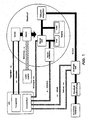
- FIGS 1 to 3 show a structure according to the invention and Figures 4 to 5 illustrate the function of the skin elasticity measuring device (HEMG). The illustrations are explained below.
- HEMG skin elasticity measuring device
- HEMG skin elasticity measuring device
- the measuring and control unit takes over the test control, the recording and storage of the measured values (data), the evaluation of the data as well as the presentation of results.
- the measuring and control unit is equipped with a frame grabber and a controller board with digital and analog inputs and outputs in addition to the conventional components. Preference is given to the use of a microprocessor-controlled data processing system. Particularly suitable are programmable digital data processing systems.
- the vacuum supply is effected by compressed air, wherein an electrically controlled proportional pressure control valve regulates the pressure of the air flowing through the suction nozzle.
- an electrically controlled proportional pressure control valve regulates the pressure of the air flowing through the suction nozzle.
- Upstream of the pressure regulator is a compressed air preparation, which may contain a manual override valve, a particulate filter and a manual pressure control valve for adjusting the regulator input pressure.
- vacuum pumps in particular regulated vacuum pumps.
- the light source provides the light for the telecentric incident illumination of the measurement object.
- Inventive light sources provide light in the visible range. For certain series of measurements it is possible to use light sources that emit polarized light or light of a specific wavelength. The use of radiation outside the visible range can also be advantageous. Particularly suitable are cold light sources that allow a precisely positionable illumination via flexible light guides.
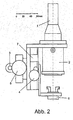
- FIG. 2 and 3 An embodiment of a measuring head according to the invention is shown in Figure 2 and 3 as a construction drawing, whereby the invention should not be limited.
- the measuring head is used to actually carry out the measurement. It is fastened to the attachment point (3) on a flexible stand (not shown in Figs. 2 and 3) and is thus positioned over the area to be examined. It only contains the components that are needed in the immediate vicinity of the DUT. So that the volume to be evacuated during a pressure jump remains small, this also includes the suction nozzle (8), solenoid valve (9) and pressure sensor (7).
- the supply lines (cables, light guides, compressed air hoses) are not shown for the sake of clarity in Figs. 2 and 3.
- the measuring head of the HEMG with the suction ring (6) is placed on the skin surface of the subject, the alignment is possible by the fine adjustment (5), whereby the contact pressure on the area to be examined is to be minimized.
- the focusing of the camera image is effected by the adjusting device (4), through which the distance between the suction ring and the measuring lens can be adjusted.
- the adjusting devices (4) and (5) can be designed as a drive via a rack by means of a side knurling wheel, as used in particular in optical devices such as microscopes and cameras.
- a side knurling wheel as used in particular in optical devices such as microscopes and cameras.
- the structure of the measuring head can be derived from the structure of a reflected-light microscope, whereby the eyepiece in the microscope can be compared with the digital camera and the object table with the suction ring. Again, there are options for positioning and focusing.
- the pressure or the force with which the suction ring rests on the area to be examined can be measured.
- pressure sensors may be used to measure the vacuum applied to the suction ring.
- pressure sensors which are designed for a pressure range from 0 to -1000 mbar (vacuum pressure sensors) or for a pressure range from 0 to +1300 mbar (absolute pressure sensors).
- a pressure sensor which determines the pressure in the inner suction channel, that is arranged between the control valve and suction ring.
- the measurement of the pressure prevailing in the outer suction channel pressure is possible by a second pressure sensor.
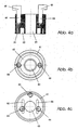
- Figures 4a to 4b show a suction ring for measuring multiaxial displacements in section and in plan view. It consists of a narrow outer suction channel (61) and a wide inner suction channel (62). The channels are each connected by a bore with a threaded blind hole (64) into which a hose connection (68) is screwed. The central, continuous bore (65) is used for lighting and image acquisition.
- hose connection in another way, e.g. by gluing, welding, plugging or hooking (bayonet fitting).
- the outer suction channel (61) is used for fixing the measuring object. He is very narrow so that he does not cause major deformations on the examined skin area. Preferably, the outer suction channel has a width of 0.2 to 5 mm.
- a fixing of the measurement object can also take place via bonding, in particular with double-sided adhesive tape.
- the inner suction channel (62) causes a multiaxial stretching of the measurement object in the observation region of the camera when a negative pressure is created by the suction of skin into the suction channel (62).
- Figure 4c shows a suction ring for the measurement of uniaxial displacements.
- the outer suction channel (61) which serves for fixing, no second (inner) suction channel available.
- the skin is stretched by two equally sized parallel suction chambers (66) symmetrically disposed about the inner bore (65).
- the inner suction channel has a width of 0.5 to 10 mm
- the suction ring can be made of all materials which are not subject to any change in shape when used according to the invention since the reproducibility of the measurement results can be influenced by deformations.
- the surface of a suction ring according to the invention should preferably consist of a solid, inert, vacuum-resistant material, in particular a plastic material, especially Teflon, HDPE or PVC.
- the method for measuring the elastic properties of the skin is as follows. On the skin in vivo, a groove provided with a groove is set, the groove (suction channel (62) in Fig. 4) is evacuated to measure the elasticity / stretch behavior. The skin is pulled into the evacuated area. The stretch of the skin can be captured visually indoors.
- the presented HEMG has application possibilities which make it possible, for example, to carry out and analyze further optical recordings of the skin surface structure or to measure the depth effect of the deflection by means of conventional ultrasonic ultrasound diagnostics.
- the flexible use of the HEMG the use of other, the geometry of the meter customizable measurement methods possible, such as the change in the microcapillary erythrocyte by means of laser Doppler flowmetry.
- a preferred embodiment of the measuring device centrally has an opening into which said other measuring devices (for example visual documentation by means of an endoscope, topography change kinetics using laser triangulation sensors) are introduced in order to determine further parameters of the one to be examined Determine skin areas.
- FIG. 5a schematically shows the suction ring (6) of the HEMG placed on the skin (10), which was fixed on the skin by applying a negative pressure to the outer suction channel (61) in the region (611). This condition corresponds to the period of time indicated by t 0 in Figure 6.
- the outer and the inner suction ring to act simultaneously with a negative pressure. This makes it possible to minimize the pre-stretching, the error that occurs when the skin is sucked into the outer suction channel.
- Figure 5b shows schematically the load condition of the skin, which is triggered by applying a negative pressure in the inner suction channel (62) and results in the skin being sucked into the suction channel (area 621) and an extension of the skin in the area of the observation field (651). , which lies in the center of the through hole (65), triggers.
- the illustrated arrows illustrate the shifts of material points of the skin surface occurring as a result of the stress. This state corresponds to the period of time denoted by t 1 in FIG.
- Figure 5c shows schematically the state after the vacuum load. Lack of elasticity of the skin does not result in complete withdrawal and residual deflection remains. This condition corresponds to the period of time denoted t 2 in Figure 6.
- the skin of the subject is stretched.
- individual photographs are recorded by the CCD camera and stored digitally in the measuring and control unit.
- the synchronization of negative pressure on the inner suction ring and the image acquisition is done by trigger pulses generated by the measuring and evaluation system (see Fig. 6).
- Figure 7 shows a displacement vector field created by comparing two images acquired before and during a multiaxial loading of the DUT.
- the arrows indicate the scaled directions of the displacements.
- the actual size of the shift is given by the amounts in Figure 9.
- Figure 8 shows a displacement vector field in uniaxial plane tensile test.
- Preference is also the cyclic loading and unloading of skin areas.
- An ideally interactive user interface allows at least the entry of exposure levels and durations. These parameters are buffered by an evaluation computer, logged and transmitted as suitable command sequences to a control computer. This can be carried out, for example, as a plug-in card for the evaluation computer.
- control computer generates, independently of the evaluation computer, the electrical voltages required to control the valves (9).
- Suitable algorithms evaluate the pressure in the inner suction channel and adjust the pressure so that the tightening of the skin corresponds to the load diagram required by the user.
- the video signals of the inside of the housing (1) mounted CCD camera are provided at predetermined sampling times, but at least at the beginning and at the end of Retardationsterrorism by the vacuum application of the skin by a FrameGrabber card the evaluation computer digitally.
- the preferential stretching directions of the skin i. the directions of the largest or smallest strain, recognized in the image sequences and determines their respective angular position.
- the device according to the invention is outstandingly suitable for carrying out the method according to the invention.
- In the foreground is the determination of the skin elasticity in vivo of a predefined skin area.
- the preferred directions of the skin can be determined from the results. These play a role in particular in operations in which the direction of the cut to be performed is widely selectable. For example, operations that involve opening the abdominal wall may be mentioned here. Often, the surgeon can freely decide how he wants to set the cut. If he places it along the preferential lines previously determined by the meter, it is to be expected that the resulting scar will be of relatively inconspicuous format. In some cases, especially in dermatological interventions by skillful choice of the cut can be achieved that virtually no visible scar remains.
- the meter allows to determine the skin elasticity even in difficult applications.
- the determination of the prevailing elasticity conditions in the skin is actually unavoidable.
- the known test methods fail here many times, be it that a removal of a skin sample should be omitted due to the extremely sensitive area, either because the structurally predetermined version of the available device prevents application in this area. Then, with appropriate design of the meter and the measurement of the elasticity of the scalp is possible, the scalp must first be freed by a shave of hair.
- the measuring device for the non-destructive measurement of the properties of an elastic material, in particular of plastic films.
Landscapes
- Health & Medical Sciences (AREA)
- Life Sciences & Earth Sciences (AREA)
- Heart & Thoracic Surgery (AREA)
- Medical Informatics (AREA)
- Biophysics (AREA)
- Pathology (AREA)
- Engineering & Computer Science (AREA)
- Biomedical Technology (AREA)
- Veterinary Medicine (AREA)
- Physics & Mathematics (AREA)
- Molecular Biology (AREA)
- Surgery (AREA)
- Animal Behavior & Ethology (AREA)
- General Health & Medical Sciences (AREA)
- Public Health (AREA)
- Dermatology (AREA)
- Measuring And Recording Apparatus For Diagnosis (AREA)
- Investigating Strength Of Materials By Application Of Mechanical Stress (AREA)
Abstract
Description
Die Erfindung bezieht sich auf ein Messgerät und ein Verfahren zur Bestimmung bestimmter mechanischer Eigenschaften eines elastischen und/oder dehnbaren Materials, insbesondere der Haut, wobei mittels Unterdruck ein ausgewählter Bereich des Materials gespannt bzw. entspannt wird.The invention relates to a measuring device and a method for determining certain mechanical properties of an elastic and / or stretchable material, in particular the skin, wherein a selected area of the material is tensioned or relaxed by means of negative pressure.
Mit fast zwei Quadratmetern Fläche und ca. 15 % des Körpergewichts ist die Haut nicht nur das größte, sondern auch eines der erstaunlichsten Organe unseres Körpers. Sie schützt die inneren Organe gegen Verletzung und gegen das Eindringen von Bakterien, reguliert die Körpertemperatur und vermittelt uns als Tastorgan sinnliche Eindrücke und Erfahrungen.With almost two square meters of surface and about 15% of the body weight, the skin is not only the largest, but also one of the most amazing organs of our body. It protects the internal organs against injury and against the ingress of bacteria, regulates the body temperature and gives us as sensory organs sensory impressions and experiences.
Jeder Quadratzentimeter unserer Haut besteht aus ungefähr
- 600.000 Zellen,
- 5.000 Rezeptoren zum Tasten und Fühlen von Temperatur, Druck, Schmerz, Berührungen,
- 100 Schweißdrüsen,
- 20 Talgdrüsen,
- 4 Metern Nervensträngen und
- einem Meter Adern.
- 600,000 cells,
- 5,000 receptors for touching and feeling temperature, pressure, pain, touch,
- 100 sweat glands,
- 20 sebaceous glands,
- 4 meters nerve strands and
- one meter of veins.
Allgemein besteht die Haut (Cutis) aus der Oberhaut (Epidermis), einem mehrschichtigen verhornten Plattenepithel, und der Lederhaut (Dermis, Corium) mit einem engen Geflecht von Fasern, die fest mit der Oberhaut verflochten sind. Darunter schließt sich die Unterhaut (Tela subcutanea) an.Generally, the skin (cutis) consists of the epidermis, a multi-layered keratinized squamous epithelium, and the dermis (dermis, corium) with a tight network of fibers firmly entangled with the epidermis. This is followed by the subcutaneous tissue (Tela subcutanea).
Die Oberhaut ist die Hautschicht, die mit einer Homschicht nach außen hin abschließt. In ihrer Keimschicht - wo übrigens auch die Hautpigmente sitzen - entstehen durch Zellteilung ständig neue Hautzellen, die in 28 Tagen an die Hautoberfläche wandern, wo sie absterben und als Schuppen abgestoßen werden. So erneuert sich die Epidermis alle 28 Tage.The epidermis is the skin layer that closes outward with a hom layer. In their germ layer - where by the way also the skin pigments sit - by cell division constantly arise new skin cells that migrate to the surface of the skin in 28 days, where they die and are rejected as dandruff. So the epidermis renews every 28 days.
Erheblich dicker als die Oberhaut ist die darunter liegende Lederhaut. Als Lieferant wichtiger Nährstoffe für die Oberhaut ist die Lederhaut mit Nerven, Blut- und Lymphgefäßen durchsetzt und besteht zu einem großen Teil aus faserigem Bindegewebe. Die Beschaffenheit dieses Bindegewebes bestimmt die Elastizität und Struktur der Haut. Haarwurzeln, Schweiß- und Talgdrüsen sitzen in der Lederhaut.Significantly thicker than the epidermis is the underlying dermis. As a source of essential nutrients for the epidermis, the dermis is interspersed with nerves, blood and lymph vessels and consists to a large extent of fibrous connective tissue. The texture of this connective tissue determines the elasticity and structure of the skin. Hair roots, sweat and sebaceous glands are located in the dermis.
Die Unterhaut ist die Fortsetzung der Lederhaut, unterscheidet sich von dieser jedoch in ihrem Aufbau. Wie der Name vermuten lässt, ist die Lederhaut fest und faserig, während die Unterhaut gröber und eher schwammig ist. Sie besteht vorwiegend aus Binde- und Fettgewebe, das dem Körper u. a. als Kälteschutz und Energiespeicher dient. In diesen Zellen können größere Mengen Fett eingelagert werden, was zu Cellulite (besonders betroffen sind Frauen) führen kann.The subcutaneous tissue is the continuation of the dermis, but differs from it in its structure. As the name suggests, the dermis is firm and fibrous, while the subcutis is coarser and more spongy. It consists mainly of connective and fatty tissue, the u. a. serves as cold protection and energy storage. In these cells, larger amounts of fat can be stored, which can lead to cellulite (especially women).
Die in den Schichten des verhornten Plattenepithels vorkommenden Pigmentzellen (Melanozyten) sind für die Hautfarbe verantwortlich. Die Melaninbildung kann durch Sonnenstrahlen verstärkt werden ― Sonnenbräune. Vitamin A-Mangel führt zu vermehrter Verhornung (Hyperkeratose). Erst durch die Lederhaut erreicht die Haut ihre Festigkeit und Elastizität. Die oberen Anteile reichen zapfenförmig in die Oberhaut ― die Papillarschicht (Papillarkörper, Stratum papillare). In ihr befinden sich neben den Kollagenfasern die Blut- und Lymphkapillare sowie die Nervenaufzweigungen und spezielle Sinnesorgane (Meißner-Tastkönperchen). Die Geflechtsschicht (Stratum reticulare) darunter sorgt für die Elastizität der Haut. In ihr befinden sich die Haarwurzeln mit dem Haarmuskel (M. arrector pili), den Knäueln der Talgdrüsen sowie Vater-Pacini-Lamellenkörperchen. Die Unterhaut (Subcutis) ist mit gekammertem Binde- und Fettgewebe ausgefüllt und stellt die Verbindung zur oberflächlichen Körperfaszie her. In der Unterhaut befinden sich die größeren Blut- und Lyphgefäße. Die Haare (Pili) dienen meist der Tastempfindung und als Wärmeschutz. Die Nägel (Ungues) sind besondere Hornbildungen der Haut.The pigment cells (melanocytes) occurring in the layers of the keratinized squamous epithelium are responsible for the skin color. Melanin formation can be intensified by the sun's rays - suntan. Vitamin A deficiency leads to increased keratinization (hyperkeratosis). Only through the dermis, the skin reaches its strength and elasticity. The upper parts extend peg-shaped into the epidermis - the papillary layer (papillary body, stratum papillare). In addition to the collagen fibers, it contains the blood and lymph capillaries as well as the nerve branches and special sensory organs (Meißner Tastkönperchen). The braid layer (stratum reticulare) underneath ensures the elasticity of the skin. In it are the hair roots with the hair muscle (M. arrector pili), the balls of the sebaceous glands and father Pacini lamellar bodies. The subcutis (subcutis) is filled with chambered connective and fatty tissue and connects to the superficial body fascia. In the subcutaneous tissue are the larger blood and Lyphgefäße. The hair (pili) are mostly used for tactile sensation and as heat protection. The nails (Ungues) are special horn formations of the skin.
Die Oberhaut und Lederhaut zusammen messen an ihren dicksten Stellen - Rücken, Fußsohlen und Handflächen - 3 bis 5 mm, an der dünnsten Stelle, den Augenlidern, hingegen weniger als 1 Millimeter.The epidermis and dermis together measure at their thickest points - back, soles and palms - 3 to 5 mm, at the thinnest point, the eyelids, however, less than 1 millimeter.
Die Elastizität der Gewebe verändert sich mit dem Alter. Sie wird durch verschiedene andere Parameter, beispielsweise Gesundheit, Ermüdungszustände, Einfluss äußerer Faktoren und dergleichen, beeinflusst.
Bereits ab dem 25. Lebensjahr verlangsamt sich die Zellerneuerung der Haut. Das Kollagen (quellende Eiweißkörper, wichtiger Bestandteil des Bindegewebes) kann weniger Wasser einlagern, da sich die Zusammensetzung im Laufe der Hautalterung verändert, und die Anzahl der Talg- und Schweißdrüsen nimmt ab. Dadurch wird die Oberhaut dünner und verliert zunehmend an Elastizität. Schlechtere Hautdurchblutung führt zu Feuchtigkeitsverlusten, das Hautgewebe erscheint nicht mehr straff.
Im Vergleich zum gesunden jungen Menschen treten an der Haut des alternden Menschen folgende Veränderungen auf :
- Es kann zu einer zunehmenden Verdünnung der obersten Hautschicht, der Oberhaut oder Epidermis, kommen.
- Epidermis und Dermis bilden immer weniger eine kompakte Einheit. Dadurch wird die alternde Haut anfälliger auch schon für kleinere Traumen und Scherbeanspruchungen.
- Die Dermis wird immer dünner und weniger elastisch.
- Die Dermis weist immer weniger Flbroblasten auf. Dadurch kommt es zu einer sinkenden Produktion von :
- Elastin, das für die Elastizität der Haut verantwortlich ist und
- Kollagen, das der Haut ihre Widerstandsfähigkeit, verleiht. Somit nehmen beim alten Menschen die Anfälligkeit für Scherbeanspruchungen und die Faltenbildung zu.
- Die Altershaut ist trocken (Xerosis).
- Geschädigte Hautpartien werden viel langsamer erneuert.
- Die Zahl der für die Immunabwehr wichtigen spezialisierten "Wächterzellen" (der sogenannten "Langerhans-Zellen") nimmt ab.
- An den Handrücken können sich homartige Wucherungen bilden (Keratosis senilis).
- Infolge der zunehmenden Beeinträchtigung der Melanozytenfunktion bräunt der alte Mensch nicht mehr so leicht. Was dabei aber schwerer wiegt, ist die Tatsache, daß dadurch die Haut alter Menschen weniger gut als die jüngerer gegen den schädlichen Wirkungen der Sonnenstrahlen geschützt ist und somit das Hautkrebsrisiko steigt.
- Es entwickeln sich Bereiche mit Hyperpigmentation Infolge von Melaninanhäufungen, etwa im Gesicht (Teleangiektasien).
- Bei vielen alten Menschen finden sich Hautblutungen bzw. braune Flecken an Händen und Armen (Purpura senilis). Dies ist vermutlich darauf zurückzuführen, dass die mangelnde Elastizität der Altershaut die Hautkapillaren verletzungsanfälliger macht und dass es zu einer Verdickung und Versteifung der Gefäßwände kommt.
- vermehrte Melaninbildung in der Epidermis. Es kommt zunächst zu einer Verdickung der Epidermis, sodann zu einer Atrophierung,
- Faltenbildung,
- deutliche Abnahme der Zahl der Langerhans-Zellen,
- signifikante Verminderung des Kollagens und
- chronische Sonnenexposition ab der Jugend bewirkt, dass im Alter ein erhöhtes Hautkrebs-Risiko gegeben ist.
Already from the age of 25, the cell renewal of the skin slows down. The collagen (swelling protein, important component of the connective tissue) can store less water, as the composition changes in the course of aging, and the number of sebaceous and sweat glands decreases. As a result, the epidermis becomes thinner and loses more and more elasticity. Poor skin circulation leads to loss of moisture, the skin tissue no longer appears taut.
Compared to healthy young people, the following changes occur on the skin of aging people:
- It may come to an increasing dilution of the uppermost skin layer, the epidermis or epidermis.
- Epidermis and dermis are less and less a compact unit. This makes the aging skin more susceptible even to minor trauma and shear stress.
- The dermis is getting thinner and less elastic.
- The dermis has fewer and fewer flbrobasts. This leads to a decreasing production of:
- Elastin, which is responsible for the elasticity of the skin and
- Collagen, which gives the skin its resistance. Thus, susceptibility to shearing and wrinkling increases in the elderly.
- The aging skin is dry (xerosis).
- Damaged skin is renewed much slower.
- The number of specialized "guardian cells" (the so-called "Langerhans cells") that are important for the immune defense is decreasing.
- Homogeneous growths can form on the back of the hand (keratosis senilis).
- As a result of the increasing impairment of melanocyte function, the elderly no longer tans so easily. What weighs heavier, however, is the fact that the skin of older people is less well protected than the younger ones against the harmful effects of the sun's rays and thus the risk of skin cancer increases.
- Hyperpigmentation develops as a result of melanin accumulation, such as in the face (telangiectasia).
- Many old people have skin bleeding or brown spots on their hands and arms (purpura senilis). This is probably due to the fact that the Lack of elasticity of the aging skin makes the skin capillaries vulnerable to injury and that it comes to a thickening and stiffening of the vessel walls.
- increased melanin formation in the epidermis. It comes first to a thickening of the epidermis, then to an atrophy,
- Wrinkling,
- significant decrease in the number of Langerhans cells,
- significant reduction of collagen and
- chronic sun exposure from adolescence causes an increased skin cancer risk in old age.
Die Erhöhung der Elastizität und der Feuchtigkeit der Haut und somit die Wiederherstellung des jugendlichen Aussehens und die Verhinderung der Faltenbildung ist daher eines der Hauptziele der therapeutischen Kosmetik.Increasing the elasticity and moisture of the skin and thus restoring the youthful appearance and preventing wrinkling is therefore one of the major goals of therapeutic cosmetics.
Es ist bekannt, zur Bestimmung der Eigenschaften oder Wirkungen bestimmter dermatoiogischer Behandlungen, bestimmte spezifische Eigenschaften der Haut, ihre Entwicklung im Laufe der Zeit und gegebenenfalls die eventuell bestehenden Beziehungen der Eigenschaften zueinander zu untersuchen.It is known, in order to determine the properties or effects of certain dermatological treatments, to investigate certain specific properties of the skin, its evolution over time, and possibly the relationships of the properties that may exist with each other.
Eines der wesentlichen Merkmale der Haut ist die Talgabsonderung pro Flächeneinheit, und zu ihrer Bestimmung sind bereits verschiedene Messvorrichtungen vorgeschlagen worden. Eine weitere wesentliche Eigenschaft ist ihre Elastizität, die durch Bestimmung des Elastizitätsmodul (Youngscher Modul) und anderer Parameter festgestellt werden kann.One of the essential features of the skin is sebum secretion per unit area and various measuring devices have already been proposed for its determination. Another important property is their elasticity, which can be determined by determining the elastic modulus (Young's modulus) and other parameters.
Durch die Erfindung wird nun eine Meßmethode zur Verfügung gestellt, wodurch es möglich ist, die Elastizität der menschlichen Haut in vivo vor und nach der Aufbringung von bestimmten Zubereitungen unter Berücksichtigung und Erfassung der Vorzugsrichtung zu bestimmen, wodurch sowohl ein objektiver Test für die Wirksamkeit solcher Zubereitungen, als auch ein Test hinsichtlich der Wirkung auf die damit behandelte Person, erhalten wird.The invention now provides a measuring method whereby it is possible to determine the elasticity of human skin in vivo before and after the application of certain preparations, taking into account and detecting the preferred direction, thereby providing both an objective test of the efficacy of such preparations , as well as a test for the effect on the person treated with it, is obtained.
Im Sinne der Erfindung ist als Haut auch ein folienartiges Substrat zu verstehen, dessen elastische Eigenschaften durch das erfindungsgemäße Messgerät bestimmbar sind. Folienartige Substrate sind zum Beispiel dünne Polymerfolien, die eine gewisse Dehnung erfahren können. Auch sie können durch Einflüsse, die durch das Produktionsverfahren gegeben sind, ein anisotropes Dehnungs- und Streckverhalten aufweisen, weiches sich durch das unten beschriebene Messgerät bestimmen lässt.For the purposes of the invention, a skin-like substrate is to be understood as the skin whose elastic properties can be determined by the measuring device according to the invention. Film-like substrates are, for example, thin polymer films which can undergo a certain amount of stretching. They too can be influenced by the production process have anisotropic stretching and stretching behavior, which can be determined by the measuring device described below.
Den derzeit in der kosmetischen Industrie am weitesten verbreiteten Ansatz, um die Hautoberfläche einer messtechnischen Erfassung zugänglich zu machen, stellt die Anfertigung von Silikon-Negativabdrücken - sogenannten Replikaten - dar. Diese werden dann stellvertretend taktil oder optisch vermessen. Die Anfertigung der Replikate erweist sich jedoch insbesondere bei hohen Probandenzahlen als sehr zeitaufwendig und ist immer mit einem in aller Regel unbekannten informationsverlust verbunden.The most widespread approach currently used in the cosmetic industry to make the surface of the skin accessible to metrological detection is the preparation of silicone negative impressions - so-called replicas. These are then measured tactually or optically. However, the production of the replicas proves to be very time-consuming, especially with high test subjects, and is always associated with a generally unknown loss of information.
Die Bestimmung von mechanischen Eigenschaften der Haut in zwei Dimensionen wird in dem Artikel "Two-dimensional elastic properties of human skin in terms of an incremental model at the in vivo configuration" von Reishner, Balogh und Menzel (Med.Eng.Phys., 1995, Vol. 17, 304-313) dargestellt. Die Messungen finden hier an Hautproben, die einem Probanden entnommen wurden, statt. Eine in vivo Messung ist nicht vorgesehen, bzw. ist nach der In diesem Artikel offenbarten Meßmethode nicht möglich.The determination of skin mechanical properties in two dimensions is described in the article "Two-dimensional elastic properties of human skin in terms of an incremental model at the in vivo configuration" by Reishner, Balogh and Menzel (Med. Eng. Phys., 1995 , Vol. 17, 304-313). The measurements are taken here on skin samples taken from a subject. An in vivo measurement is not intended or is not possible according to the method of measurement disclosed in this article.
Aus Ullstein Lexikon der Medizin (Ullsteinverlag 1970, Seiten 590 und 674) ist es in der Medizin bekannt, eine Vakuumquelle zu verwenden, um auf das menschliche Gewebe eine Saugwirkung zu erzielen, wobei es sich hier um eine Saugglocke zum Absaugen von Abszessen und einen Vakuumextraktor zur Verwendung in der Geburtshilfe handelt.From Ullstein Lexicon of Medicine (Ullsteinverlag 1970, pages 590 and 674), it is known in medicine to use a vacuum source to achieve a suction effect on the human tissue, which is a suction cup for sucking abscesses and a vacuum extractor for use in obstetrics.
In DE 29 09 092 wird eine Methode zur Messung der Hautelastizität beschrieben, bei der - angelehnt an die üblichen Prüfmethoden der Werkstoffprüfung - ein Taster mit einem bestimmten Druck auf die Haut aufgebracht wird und abhängig von der Eindringtiefe die Hautelastizität bestimmt wird.In DE 29 09 092 a method for measuring the skin elasticity is described, in which - based on the usual test methods of material testing - a button is applied with a certain pressure on the skin and depending on the penetration of the skin elasticity is determined.
Die Messung der mechanischen Eigenschaften der Haut in vivo wird gegenwärtig auf dem Gebiet der kosmetischen wie der medizinischen Dermatologie vorwiegend mittels vier verschiedener Geräte bzw. Verfahren durchgeführt.The measurement of the mechanical properties of the skin in vivo is currently carried out in the field of cosmetic as well as medical dermatology predominantly by means of four different devices or methods.
Einige Verfahren am Markt sind:
- das Extensometer: Dehnung der Haut mittels zwei auf der Haut aufgesetzter Messfühler, wobei die Dehnung (Änderung des Abstandes zwischen den Messfühlern) in Abhängigkeit der angelegten Kraft gemessen wird.
- das Torquemeter (Fa. Diastron Ltd., UK): Auf die Hautoberfläche wird hierbei eine innere Scheibe und ein äußerer Haltering verklebt. Mittels der Scheibe wird ein Drehmoment angelegt und der Winkel der dann erfolgten Auslenkung gegenüber dem Halteringgemessen. Danach stoppt die Krafteinleitung und die Scheibe kehrt infolge der Hautelastizität nahezu in die Ausgangslage zurück. Aufgrund der Komplexität der Auslenkung, namentlich durch die Verknüpfung von Normal- und Torsionsspannungen, ist eine Interpretation der erhaltenen Meßdaten hinsichtlich ihrer Relevanz zur Beurteilung von einzelnen Hautkomponenten ausgesprochen problematisch.
- das Cutometer (Fa. Courage Khazaka, Köln, Deutschland): Die Meßsonde wird bei diesem Gerät senkrecht auf die Haut gesetzt, die Meßsonde hatan der Unterseite eine Öffnung. Ein Unterdruck wird angelegt und die Haut wird in die Öffnung hineingezogen. In der Meßsonde wird das Maß der Auslenkung optoelektronisch gemessen. Auch in diesem Verfahren erfolgt keine tangentiale Auslenkung der Haut, so dass die dämpfenden Elemente des dermalen Gewebes sowie des subkutanen Fettgewebes vornehmlich in die Messung eingehen.
- das Ballistömeter: Bestimmung der Hautelastizität durch Messung des Dämpfungs- bzw. Schwingverhaltens der Haut bei Anregung mit einem Gewicht (DE 27 32 836), wobei ein oberflächenparallel aufgehängtes Pendel auf die Hautoberfläche fallengelassen und die Dämpfung der Schwingung aufgezeichnet wird. Da Schwingungsdämpfungen stets eine Funktion viskoser Elemente ist, muß vermutet werden, gerade hinsichtlich der Krafteinwirkung in Richtung des Normalenvektors, dass vor allem die Eigenschaften der Subkutis und weniger der Dermis und Epidermis in die Meßergebnisse eingehen.
- the extensometer: Stretching of the skin by means of two sensors placed on the skin, with the strain (change in the distance between the probes) being measured as a function of the applied force.
- the Torquemeter (Diastron Ltd., UK): In this case, an inner disc and an outer retaining ring is glued to the skin surface. By means of the disc, a torque is applied and measured the angle of the then made deflection relative to the retaining ring. Thereafter, the force application stops and the disc returns due to the elasticity of the skin almost to the starting position. Due to the complexity of the deflection, in particular by the combination of normal and torsional stresses, an interpretation of the obtained measurement data in terms of their relevance to the assessment of individual skin components is very problematic.
- the Cutometer (Courage Khazaka, Cologne, Germany): The probe is placed perpendicular to the skin in this device, the probe has an opening at the bottom. A vacuum is applied and the skin is drawn into the opening. In the probe, the degree of deflection is measured optoelectronically. Also in this method, there is no tangential deflection of the skin, so that the attenuating elements of the dermal tissue as well as the subcutaneous fat tissue are mainly included in the measurement.
- the Ballistömeter: Determination of skin elasticity by measuring the damping or vibration behavior of the skin upon stimulation with a weight (DE 27 32 836), wherein a surface parallel suspended pendulum dropped onto the skin surface and the attenuation of the vibration is recorded. Since vibration damping is always a function of viscous elements, it must be assumed, especially with regard to the action of force in the direction of the normal vector, that above all the properties of the subcutis and less of the dermis and epidermis are included in the measuring results.
Keines der aufgeführten Geräte ist zur Messung in vivo in der Lage, die Vorzugsrichtungen der Hautelastizität, deren Existenz der Medizin und der humanblologischen Forschung lange bekannt sind, zu detektieren und bei der Erhebung der Meßparameter zu berücksichtigen.None of the devices listed is capable of measuring in vivo the preferred directions of skin elasticity, whose existence has long been known to medicine and human biological research, and to take into account in the collection of measurement parameters.
Das Multiaxiale-Extensometer ist eine Weiterentwicklung des Extensometers, bei der das Dehnungsverhalten in mehreren Richtungen simultan erfasst wird (DE 197 19 336). Leider hat auch hier der Kleber, der zur Kopplung der Haut an das Messgerät benötigt wird, einen zum Teil unbekannten Einfluss auf die gemessenen Deformationen.
Ein weiterer Nachteil ist, dass als Kleber Acrylate (Sekundenkleber) oder doppelseitige Klebebänder verwendet werden, die sehr schwer entfernbar sind und zu starken Hautreizungen neigen.The multi-axial extensometer is a further development of the extensometer, in which the elongation behavior is detected simultaneously in several directions (DE 197 19 336). Unfortunately, the adhesive needed to couple the skin to the meter also has a partly unknown influence on the measured deformations.
A further disadvantage is that acrylates (superglues) or double-sided adhesive tapes are used as the adhesive, which are very difficult to remove and tend to cause severe skin irritation.
Aufgabe der im folgenden beschriebenen Erfindung ist es, eine Vorrichtung und ein Verfahren zur Verfügung zu stellen, mit dem präzise und reproduzierbare Messergebnisse über die Hautelastizität erzielbar sind. Des weiteren ist es ein Ziel der Erfindung, eine multiaxiale, definierte Belastung der Haut bei gleichzeitiger sensorischer Erfassung der jeweils auftretenden Dehnungen zu ermöglichen (Retardationsversuch), und darüber hinaus eine exakte Diskriminierung und Lokalisiervng der elastischen Vorzugsrichtungen der Haut (Langer'sche Linien) zuzulassen.The object of the invention described below is to provide a device and a method with which precise and reproducible measurement results on the skin elasticity can be achieved. Furthermore, it is an object of the invention to allow a multi-axial, defined load on the skin with simultaneous sensory detection of the respective strains occurring (retardation attempt), and moreover allow exact discrimination and localization of the elastic preferred directions of the skin (Langer's lines) ,
Gelöst wird diese Aufgabe durch ein Hautelastizitätsmessgerät wie es in Anspruch 1 gekennzeichnet ist. Das Messverfahren ist dabei Gegenstand der Unteransprüche.This object is achieved by a skin elasticity measuring device as characterized in
Die erfindungsgemäße Vorrichtung enthält eine mechanische Einrichtung zur Streckung der Haut und hat vorzugsweise eine optoelektronische Auswertungseinrichtung.The device according to the invention contains a mechanical device for stretching the skin and preferably has an optoelectronic evaluation device.
Die Bilder 1 bis 3 zeigen einen erfindungsgemäßen Aufbau und die Bilder 4 bis 5 verdeutlichen die Funktion des Hautelastizitätsmessgeräts (HEMG). Die Darstellungen werden im folgenden erläutert.Figures 1 to 3 show a structure according to the invention and Figures 4 to 5 illustrate the function of the skin elasticity measuring device (HEMG). The illustrations are explained below.
Es zeigen
Abbildung 1- den prinzipiellen Aufbau des Messgeräts in Verbindung mit den Prozess- und Signalleitwegen, durch die die Einzelkomponenten in Verbindung stehen;
Abbildung 2- einen nicht maßstabsgetreue schematische Seitenansicht einer Ausführungsform eines erfindungsgemäßen Messkopfes;
Abbildung 3- einen nicht maßstabsgetreue schematische Vorderansicht einer Ausführungsform eines erfindungsgemäßen Messkopfes;
Abbildungen 4a bis- einen nicht maßstabsgetreuen schematischen Querschnitt einer Ausführungsform eines erfindungsgemäßen Saugringes;
Abbildungen 5a bis- die schematische Darstellung der Prozessschritte vor (5a), bei (5b) und nach (5c) Hautdehnung durch Anlegen eines Unterdrucks;
Abbildung 6- die Gegenüberstellung der Größen Druck, Triggersignal und Dehnung, wie sie In Abhängigkeit der Messzeit gewonnen werden;
Abbildung 7- die Schematische Darstellung der Hautdehnung als Verschiebungsvektorfeld bei uniaxialer Dehnung;
Abbildung 8- die Schematische Darstellung der Hautdehnung als Verschiebungsvektorfeld bei multiaxialer Dehnung und
- Abbildung 9
- die Darstellung der Beträge der Hautdehnung als Graustufenbild bei multiaxialer Dehnung.
-
illustration 1 - the basic structure of the measuring device in conjunction with the process and signal paths through which the individual components are connected;
- Figure 2
- a schematic side view, not to scale, of an embodiment of a measuring head according to the invention;
- Figure 3
- a schematic not to scale front view of an embodiment of a measuring head according to the invention;
- Figures 4a to 4c
- a not to scale schematic cross section of an embodiment of a suction ring according to the invention;
- Figures 5a to 5c
- the schematic representation of the process steps before (5a), at (5b) and after (5c) skin elongation by applying a negative pressure;
- Figure 6
- the comparison of the quantities pressure, trigger signal and strain, as they are obtained as a function of the measuring time;
- Figure 7
- the Schematic representation of the skin strain as a displacement vector field in uniaxial strain;
- Figure 8
- Schematic representation of the skin strain as a displacement vector field in multiaxial stretching and
- Figure 9
- the representation of the amounts of the skin stretch as grayscale image with multiaxial stretching.
Im folgenden wird der Aufbau des Hautelastizitätsmessgerätes - im weiteren Text mit HEMG bezeichnet - anhand der Abbildungen 1 bis 4 näher erläutert, wobei hierdurch die Erfindung nicht beschränkt werden soll.In the following, the structure of the skin elasticity measuring device - hereinafter referred to as HEMG - explained in more detail with reference to Figures 1 to 4, whereby the invention should not be limited.
Abbildung 1 gibt eine Übersicht über die im HEMG enthaltenen Komponenten und deren Zusammenwirken. Aus Gründen der Übersichtlichkeit ist die Spannungsversorgung der elektrisch betriebenen Elemente nicht dargestellt. Das HEMG besteht aus vier wesentlichen Gruppen:
- einer Mess- und Steuereinheit (PC),
- einer Vakuumbereitstellung,
- einer Lichtquelle und
- einem Messkopf.
- a measuring and control unit (PC),
- a vacuum deployment,
- a light source and
- a measuring head.
Die Mess- und Steuereinheit übernimmt die Versuchssteuerung, die Aufnahme und Speicherung der Messwerte (Daten), die Auswertung der Daten sowie die Darstellung von Ergebnissen.
Hierzu ist die Mess- und Steuereinheit neben den herkömmlichen Komponenten mit einem Framegrabber und einem Controller Board mit digitalen und analogen Ein- und Ausgängen ausgestattet.
Bevorzugt ist die Verwendung einer mikroprozessorgesteuerten Datenverarbeitungsanlage. Besonders geeignet sind programmierbare digitale Datenverarbeitungsanlagen.The measuring and control unit takes over the test control, the recording and storage of the measured values (data), the evaluation of the data as well as the presentation of results.
For this purpose, the measuring and control unit is equipped with a frame grabber and a controller board with digital and analog inputs and outputs in addition to the conventional components.
Preference is given to the use of a microprocessor-controlled data processing system. Particularly suitable are programmable digital data processing systems.
Die Vakuumbereitstellung erfolgt durch Druckluft, wobei ein elektrisch angesteuertes Proportional-Druckregelventil den Druck, der die Saugdüse durchströmenden Luft, regelt. Dem Druckregler vorgeschaltet ist eine Druckluftaufbereitung, die ein Handeinschaltventil, einen Partikelfilter und ein manuelles Druckregelventil zum Einstellen des Reglereingangsdrucks enthalten kann.The vacuum supply is effected by compressed air, wherein an electrically controlled proportional pressure control valve regulates the pressure of the air flowing through the suction nozzle. Upstream of the pressure regulator is a compressed air preparation, which may contain a manual override valve, a particulate filter and a manual pressure control valve for adjusting the regulator input pressure.
Bevorzugt ist ebenfalls der Einsatz von Vakuumpumpen, insbesondere geregelten Vakuumpumpen.Also preferred is the use of vacuum pumps, in particular regulated vacuum pumps.
Die Lichtquelle liefert das Licht zur telezentrischen Auflichtbeleuchtung des Messobjekts. Erfindungsgemäße Lichtquellen stellen Licht im sichtbaren Bereich zur Verfügung. Für bestimmte Messreihen ist der Einsatz von Lichtquellen möglich, die polarisiertes Licht oder Licht bestimmter Wellenlänge aussenden. Auch die Verwendung von Strahlung außerhalb des sichtbaren Bereiches kann von Vorteil sein.
Besonders geeignet sind Kaltlichtquellen, die über flexible Lichtleiter eine genau positionierbare Ausleuchtung ermöglichen.The light source provides the light for the telecentric incident illumination of the measurement object. Inventive light sources provide light in the visible range. For certain series of measurements it is possible to use light sources that emit polarized light or light of a specific wavelength. The use of radiation outside the visible range can also be advantageous.
Particularly suitable are cold light sources that allow a precisely positionable illumination via flexible light guides.
Eine Ausführungsform eines erfindungsgemäßen Messkopfes ist in Bild 2 und 3 als Konstruktionszeichnung dargestellt, wobei hierdurch die Erfindung nicht beschränkt werden soll.An embodiment of a measuring head according to the invention is shown in Figure 2 and 3 as a construction drawing, whereby the invention should not be limited.
Der Messkopf dient der eigentlichen Durchführung der Messung. Er ist am Befestigungspunkt (3) an einem flexiblen Stativ (in Abb. 2 und 3 nicht wiedergegeben) befestigt und wird damit über dem zu untersuchenden Areal positioniert. Er enthält nur die Komponenten, die in der unmittelbaren Nähe des Messobjekts benötigt werden. Damit das während eines Drucksprungs zu evakuierende Volumen klein bleibt, zählen hierzu auch Saugdüse (8), Magnetventil (9) und Drucksensor (7). Die Versorgungsleitungen (Kabel, Lichtleiter, Druckluftschläuche) sind der besseren Übersichtlichkeit wegen in den Abb. 2 und 3 nicht dargestellt.The measuring head is used to actually carry out the measurement. It is fastened to the attachment point (3) on a flexible stand (not shown in Figs. 2 and 3) and is thus positioned over the area to be examined. It only contains the components that are needed in the immediate vicinity of the DUT. So that the volume to be evacuated during a pressure jump remains small, this also includes the suction nozzle (8), solenoid valve (9) and pressure sensor (7). The supply lines (cables, light guides, compressed air hoses) are not shown for the sake of clarity in Figs. 2 and 3.
Der Messkopf beinhaltet neben weiteren Teilen folgende Elemente (Abbildung 2 und 3):
- (1) Digitalkamera (Progressive Scan Kamera,)
- (2) Telezentrisches Messobjektiv mit integrierter Einrichtung für telezentrische Auflichtbeleuchtung zur optischen Erfassung des Messobjekts im innenloch des Saugrings,
- (3) Befestigungspunkt zur Ankopplung an das Stativ,
- (4) Verstelleinrichtung zum positionieren und arretieren des Saugrings im Telezentriebereich des Objektivs,
- (5) Feinverstellung zur Positionierung auf dem Messobjekt,
- (6) Saugring (siehe auch Abbildung 4),
- (7) Vakuum-Drucksensor zum Messen des Drucks im inneren Saugkanal,
- (8) Vakuumsaugdüse zum Erzeugen eines Vakuums durch das Ejektor-Prinzip. Die Höhe des Unterdrucks ist abhängig vom Druck der durchströmenden Luft.
- (9) Magnetventil zum Abdichten bzw. Belüften des inneren Saugkanals, Das Ventil ist hängend in der Schlauchleitung angebracht.
- (1) Digital Camera (Progressive Scan Camera,)
- (2) telecentric measuring objective with integrated device for telecentric epi-illumination for the optical detection of the test object in the inner hole of the suction ring,
- (3) attachment point for coupling to the tripod,
- (4) adjusting device for positioning and locking the suction ring in the telecentric region of the objective,
- (5) fine adjustment for positioning on the measurement object,
- (6) Suction ring (see also Figure 4),
- (7) Vacuum pressure sensor for measuring the pressure in the inner suction port,
- (8) Vacuum suction nozzle for generating a vacuum by the ejector principle. The amount of negative pressure depends on the pressure of the air flowing through.
- (9) Solenoid Valve for Sealing the Inner Suction Channel. The valve is suspended in the hose line.
Die Ausrichtung des Messkopfes Ober dem zu vermessenden Hautareal erfolgt grob über ein Stativ, an dem der Messkopf befestigt ist. Für den Messvorgang wird der Messkopf des HEMG mit dem Saugring (6) auf die Hautoberfläche des Probanden gesetzt, die Ausrichtung ist durch die Feinverstellung (5) möglich, wobei der Anpressdruck auf dem zu untersuchenden Bereich zu minimieren ist.The alignment of the measuring head Above the area of the skin to be measured, roughly takes place via a stand to which the measuring head is attached. For the measuring process, the measuring head of the HEMG with the suction ring (6) is placed on the skin surface of the subject, the alignment is possible by the fine adjustment (5), whereby the contact pressure on the area to be examined is to be minimized.
Die Scharfstellung des Kamerabildes erfolgt durch die Verstelleinrichtung (4), durch die der Abstand zwischen Saugring und Messobjektiv einsteilbar ist.The focusing of the camera image is effected by the adjusting device (4), through which the distance between the suction ring and the measuring lens can be adjusted.
Die Verstelleinrichtungen (4) und (5) können als Trieb über Zahnstange mittels seitlichem Rändelrad, wie sie insbesondere in optischen Geräten wie Mikroskopen, Kameras eingesetzt werden, ausgeführt werden. Auch der Einsatz von Linearachsen und Spindeltrieben, wie sie im Maschinen- und Instrumentenbau verbreitet sind, ist möglich.The adjusting devices (4) and (5) can be designed as a drive via a rack by means of a side knurling wheel, as used in particular in optical devices such as microscopes and cameras. The use of linear axes and spindle drives, as they are common in mechanical and instrument engineering, is possible.
In seinen Grundzügen ist der Aufbau des Messkopfes aus dem Aufbau eines Auflichtmikroskops ableitbar, wobei das Okular im Mikroskop mit der Digitalkamera und der Objekttisch mit dem Saugring vergleichbar sind. Auch hier gibt es Möglichkeiten zur Positionierung und Scharfstellung.In its basic features, the structure of the measuring head can be derived from the structure of a reflected-light microscope, whereby the eyepiece in the microscope can be compared with the digital camera and the object table with the suction ring. Again, there are options for positioning and focusing.
In einer ganz besonders vorteilhaften Ausführung des Messkopfes kann der Druck bzw. die Kraft, mit der der Saugring auf dem zu untersuchenden Areal aufliegt, gemessen werden.In a particularly advantageous embodiment of the measuring head, the pressure or the force with which the suction ring rests on the area to be examined can be measured.
Zur Messung des am Saugring anliegenden Vakuums können kommerziell erhältliche Drucksensoren verwendet werden. Besonders eignen sich Drucksensoren, die für einen Druckbereich von 0 bis -1000 mbar (Vakuum-Drucksensoren) bzw. für einen Druckbereich von 0 bis +1300 mbar (Absolut-Drucksensoren) ausgelegt sind.Commercially available pressure sensors may be used to measure the vacuum applied to the suction ring. Especially suitable are pressure sensors, which are designed for a pressure range from 0 to -1000 mbar (vacuum pressure sensors) or for a pressure range from 0 to +1300 mbar (absolute pressure sensors).
Vorteilhaft ist die Verwendung eines Drucksensors, der den Druck im inneren Saugkanal ermittelt, also zwischen Steuerventil und Saugring angeordnet ist.Advantageously, the use of a pressure sensor which determines the pressure in the inner suction channel, that is arranged between the control valve and suction ring.
In einer ganz besonders vorteilhaften Ausführungsform ist durch einen zweiten Drucksensor die Messung des im äußeren Saugkanal herrschenden Drucks möglich.In a particularly advantageous embodiment, the measurement of the pressure prevailing in the outer suction channel pressure is possible by a second pressure sensor.
Abbildungen 4a bis 4b zeigen einen Saugring zur Messung multiaxialer Verschiebungen im Schnitt und in der Draufsicht. Er besteht aus einem schmalen äußeren Saugkanal (61) und einem breiten inneren Saugkanal (62). Die Kanäle sind jeweils durch eine Bohrung mit einem Gewindesackloch (64) verbunden, in das ein Schlauchanschluss (68) geschraubt wird. Die mittlere, durchgehende Bohrung (65) dient der Beleuchtung und der Bildaufnahme.Figures 4a to 4b show a suction ring for measuring multiaxial displacements in section and in plan view. It consists of a narrow outer suction channel (61) and a wide inner suction channel (62). The channels are each connected by a bore with a threaded blind hole (64) into which a hose connection (68) is screwed. The central, continuous bore (65) is used for lighting and image acquisition.
Wie aus dem Stand der Technik bekannt, ist es möglich, den Schlauchanschlusses auch in anderer Weise auszubilden, z.B. durch Verkleben, Verschweißen, Zusammenstecken oder Verhaken (Bajonettverschraubung).As is known from the prior art, it is possible to form the hose connection in another way, e.g. by gluing, welding, plugging or hooking (bayonet fitting).
Der äußere Saugkanal (61) wird zum Fixieren des Messobjekts verwendet. Damit er keine größeren Deformationen am zu untersuchenden Hautareal verursacht, ist er sehr schmal ausgeführt.
Vorzugsweise hat der äußere Saugkanal eine Breite von 0,2 bis 5 mm.The outer suction channel (61) is used for fixing the measuring object. He is very narrow so that he does not cause major deformations on the examined skin area.
Preferably, the outer suction channel has a width of 0.2 to 5 mm.
Vorteilhaft kann ein Fixieren des Messobjekts auch über ein Verkleben, insbesondere mit doppelseitigem Klebeband, erfolgen.Advantageously, a fixing of the measurement object can also take place via bonding, in particular with double-sided adhesive tape.
Der innere Saugkanal (62) verursacht beim Anlegen eines Unterdrucks durch das Einsaugen von Haut in den Saugkanal (62) eine multiaxiale Dehnung des Messobjekts im Beobachtungsbereich der Kamera.The inner suction channel (62) causes a multiaxial stretching of the measurement object in the observation region of the camera when a negative pressure is created by the suction of skin into the suction channel (62).
Abbildung 4c zeigt einen Saugring für die Messung uniaxialer Verschiebungen. Hier ist neben dem äußeren Saugkanal (61), der zur Fixierung dient, kein zweiter (innerer) Saugkanal vorhanden. Die Streckung der Haut erfolgt durch zwei gleichgroße, parallele Saugkammem (66), die symmetrisch um die innere Bohrung (65) angeordnet sind.Figure 4c shows a suction ring for the measurement of uniaxial displacements. Here, in addition to the outer suction channel (61), which serves for fixing, no second (inner) suction channel available. The skin is stretched by two equally sized parallel suction chambers (66) symmetrically disposed about the inner bore (65).
Vorzugsweise hat der innere Saugkanal eine Breite von 0,5 bis 10 mmPreferably, the inner suction channel has a width of 0.5 to 10 mm
Der Saugring kann aus allen Materialien gefertigt sein, die bei erfindungsgemäßer Verwendung keiner Formänderung unterliegen, da durch Deformationen die Reproduzierbarkeit der Messergebnisse beeinflusst werden kann.The suction ring can be made of all materials which are not subject to any change in shape when used according to the invention since the reproducibility of the measurement results can be influenced by deformations.
Die Oberfläche eines erfindungsgemäßen Saugringes sollte vorzugsweise aus einem festen, inerten, vakuumresistenten Material, insbesondere einem Kunststoffmaterial, ganz besonders Teflon, HDPE oder PVC, bestehen.The surface of a suction ring according to the invention should preferably consist of a solid, inert, vacuum-resistant material, in particular a plastic material, especially Teflon, HDPE or PVC.
Ganz: besonders vorteilhaft sind Materialien mit niedriger Wärmekapazität, da sich durch den Temperaturunterschied zwischen Saugring und zu untersuchender Haut Irritationen einstellen können. Ebenso ist eine Temperaturkonditionierung des Saugringes, an einer Stelle die nicht untersucht werden soll, vorteilhaft.Whole: materials with low heat capacity are particularly advantageous, since irritations can occur due to the temperature difference between the suction ring and the skin to be examined. Likewise, a temperature conditioning of the suction ring, at a point which should not be investigated, advantageous.
Im folgenden sollen das Verfahren sowie das zur Durchführung des Verfahrens vorteilhaft gestaltete Messgerät näher dargelegt werden, ohne die Erfindung damit unnötig einschränken zu wollen.In the following, the method as well as the advantageously designed for carrying out the method measuring device will be set forth in more detail without wishing to unnecessarily limit the invention.
Das Verfahren zur Messung der elastischen Eigenschaften der Haut, insbesondere der menschlichen Haut, stellt sich wie folgt dar.
Auf die Haut in vivo wird ein mit einer Nut versehener Ring gesetzt, dessen Nut (Saugkanal (62) in Abb. 4) zur Messung der Elastizität/Streckverhaltens evakuiert wird. Dabei wird die Haut in den evakuierten Bereich gezogen. Die Streckung der Haut kann im Innenbereich bildmäßig erfasst werden.The method for measuring the elastic properties of the skin, in particular the human skin, is as follows.
On the skin in vivo, a groove provided with a groove is set, the groove (suction channel (62) in Fig. 4) is evacuated to measure the elasticity / stretch behavior. The skin is pulled into the evacuated area. The stretch of the skin can be captured visually indoors.
In einer vorteilhaften Ausführung verfügt das vorgestellte HEMG über Applikationsmöglichkeiten, die es gestatten, beispielsweise weitere optische Aufzeichnungen der Hautoberflächenstruktur durchzuführen und zu analysieren oder mittels im Medizinbereich üblicher Ultraschalldiagnostik die Tiefenauswirkung der Auslenkung zu messen. Selbstverständlich ist durch die flexible Einsatzmöglichkeit des HEMG auch die Verwendung anderer, der Geometrie des Messgerätes anpassbarer Meßmethoden möglich, wie beispielsweise die Veränderung des mikrokapillaren Erythrozytenstroms mittels Laser Doppler Flowmetrie.
So weist eine bevorzugte Ausführungsform des Messgeräts, um den Messvorgang so umfassend wie möglich analysieren zu können, zentral eine Öffnung vor, in die die genannten anderen Messgeräte (beispielsweise visuelle Dokumentation mittels Endoskop; Topographieänderungskinetik mittels Lasertriangulationssensoren) eingeführt werden, um weitere Parameter des zu untersuchenden Hautareals zu bestimmen.In an advantageous embodiment, the presented HEMG has application possibilities which make it possible, for example, to carry out and analyze further optical recordings of the skin surface structure or to measure the depth effect of the deflection by means of conventional ultrasonic ultrasound diagnostics. Of course, the flexible use of the HEMG, the use of other, the geometry of the meter customizable measurement methods possible, such as the change in the microcapillary erythrocyte by means of laser Doppler flowmetry.
Thus, in order to be able to analyze the measuring process as comprehensively as possible, a preferred embodiment of the measuring device centrally has an opening into which said other measuring devices (for example visual documentation by means of an endoscope, topography change kinetics using laser triangulation sensors) are introduced in order to determine further parameters of the one to be examined Determine skin areas.
Abbildung 5a zeigt schematisch den auf die Haut (10) aufgesetzten Saugring (6) des HEMG, der durch Anlegen eines Unterdrucks an den äußeren Saugkanal (61) im Bereich (611) auf der Haut fixiert wurde. Dieser Zustand entspricht dem in Abbildung 6 mit t0 bezeichneten Zeitraum.FIG. 5a schematically shows the suction ring (6) of the HEMG placed on the skin (10), which was fixed on the skin by applying a negative pressure to the outer suction channel (61) in the region (611). This condition corresponds to the period of time indicated by t 0 in Figure 6.
Vorzugsweise ist es auch den äußeren und des inneren Saugringes gleichzeitig mit einem Unterdruck zu beaufschlagen. Dadurch ist eine Minimierung der Vorstreckung, der Fehler der durch das Hineinsaugen der Haut in den äußeren Saugkanal auftritt, möglich.Preferably, it is also the outer and the inner suction ring to act simultaneously with a negative pressure. This makes it possible to minimize the pre-stretching, the error that occurs when the skin is sucked into the outer suction channel.
Abbildung 5b zeigt schematisch den Belastungszustand der Haut, der durch Anlegen eines Unterdrucks im inneren Saugkanal (62) ausgelöst wird und zur Folge hat, das Haut in den Saugkanal hineingesaugt wird (Bereich 621) und eine Streckung der Haut im Bereich des Beobachtungsfeldes (651), welches im Zentrum der durchgehenden Bohrung (65) liegt, auslöst. Die dargestellten Pfeile veranschaulichen die infolge der Belastung auftretenden Verschiebungen materieller Punkte der Hautoberfläche. Dieser Zustand entspricht dem in Abbildung 6 mit t1 bezeichneten Zeitabschnitt.Figure 5b shows schematically the load condition of the skin, which is triggered by applying a negative pressure in the inner suction channel (62) and results in the skin being sucked into the suction channel (area 621) and an extension of the skin in the area of the observation field (651). , which lies in the center of the through hole (65), triggers. The illustrated arrows illustrate the shifts of material points of the skin surface occurring as a result of the stress. This state corresponds to the period of time denoted by t 1 in FIG.
Abbildung 5c zeigt schematisch den Zustand nach der Vakuumbelastung. Durch mangelnde Hautelastizität erfolgt kein vollständiger Rückzug und eine Restauslenkung verbleibt. Dieser Zustand entspricht dem In Abbildung 6 mit t2 bezeichneten Zeitabschnitt.Figure 5c shows schematically the state after the vacuum load. Lack of elasticity of the skin does not result in complete withdrawal and residual deflection remains. This condition corresponds to the period of time denoted t 2 in Figure 6.
Durch Anlegen eines Vakuums an den inneren Saugkanal des Saugringes wird die Haut des Probanden gedehnt. Während des Dehnungsprozesses werden zeitlich aufgelöst einzelne Photographien durch die CCD Kamera aufgenommen und in der Mess- und Steuereinheit digital gespeichert.By applying a vacuum to the inner suction channel of the suction ring, the skin of the subject is stretched. During the stretching process, individual photographs are recorded by the CCD camera and stored digitally in the measuring and control unit.
Die Synchronisation von Unterdruckbeaufschlagung des inneren Saugringes und der Bildaufnahme geschieht durch vom Mess- und Auswertesystem generierte Triggerimpulse (s. Abb. 6).The synchronization of negative pressure on the inner suction ring and the image acquisition is done by trigger pulses generated by the measuring and evaluation system (see Fig. 6).
Wird der Unterdruck im inneren Saugring abgebaut, kommt es zu einer Retardationsphase, in der sich die Haut in den Ausgangszustand vor der Dehnung begibt. Schematisch ist die Be- und Entlastung in Abbildung 6 wiedergegeben. Die nach der Entlastung zunächst verbleibende Restauslenkung wird ebenso wie die Auslenkung während der Belastung bildmäßig erfasst.
Aus den so gewonnen Aufnahmen können nach bekannten Methoden der Bildverarbeitung, wie in dem Artikel "Quantification of mechanical properties of human skin in vivo" von Heinrich und Lunderstädt (Proc. of SPIE, Applications of Digital Image Processing XXIV, 4472, pp. 11-20, San Diego, USA, 2001) gezeigt wird, die Verschiebungsvektoren einzelner Hautareale berechnet werden. Dabei werden unter Berücksichtigung eines Ähnlichkeitskriteriums kleine Gebiete aus dem Referenzbild In dem Folgebild wiedergefunden (Template Matching, Mustervergleich) und der Vektor vom Zentrum des Referenz-Musters zum Punkt der besten Übereinstimmung im Folgebild als Verschiebungsvektor definiert. Alle Verschiebungsvektoren zusammen bilden das Verschiebungsvektorfeld. Der Vergleich der Verschiebungsvektoren untereinander liefert Aussagen über Längenänderungen und damit über Dehnungen.
Durch grafische Darstellung der Ergebnisse als winkelabhängige Dehnungskurven (Abbildung 6c) Verschiebungsvektorfeld (Abbildung 7 und 8) oder Graubild (Abbildung 9) sowie durch aus der Mechanik bekannte Berechnungsmethoden (ebener Verzerrungszustand) können Aussagen über Vorzugsrichtungen und Dehnbarkeit der Haut gewonnen werden.If the negative pressure in the inner suction ring degrades, it comes to a retardation phase, in which the skin goes to the initial state before stretching. Schematically, the loading and unloading is shown in Figure 6. The remaining after the discharge first residual deflection is recorded as the deflection during the load imagewise.
The images obtained in this way can be analyzed by known methods of image processing, as described in the article "Quantification of mechanical properties of human skin in vivo" by Heinrich and Lunderstädt (Proc. Of SPIE, Applications of Digital Image Processing XXIV, 4472, pp. 11-13). 20 San Diego, USA, 2001), the displacement vectors of individual skin areas are calculated. In this case, taking into account a similarity criterion, small regions from the reference image are found in the subsequent image (template matching, pattern definition) and the vector from the center of the reference pattern to the point of best match in the subsequent image is defined as a displacement vector. All displacement vectors together form the displacement vector field. The comparison of the displacement vectors with each other provides statements about changes in length and thus about strains.
Graphical representation of the results as angle-dependent strain curves (Figure 6c) Displacement vector field (Figures 7 and 8) or gray image (Figure 9) and calculation methods known from mechanics (plane distortion state) can be used to obtain information about preferred directions and extensibility of the skin.
Die Abbildung 7 zeigt ein Verschiebungsvektorfeld, das durch Vergleich zweier Bilder, die vor und während einer multiaxialen Belastung des Messobjekts erfasst wurden, entstanden ist. Die Pfeile geben die skalierten Richtungen der Verschiebungen an. Die tatsächliche Größe der Verschiebung ist durch die Beträge in Abbildung 9 gegeben.Figure 7 shows a displacement vector field created by comparing two images acquired before and during a multiaxial loading of the DUT. The arrows indicate the scaled directions of the displacements. The actual size of the shift is given by the amounts in Figure 9.
Die Abbildung 8 zeigt ein Verschiebungsvektorfeld im uniaxialen ebenen Zugversuch.Figure 8 shows a displacement vector field in uniaxial plane tensile test.
Bevorzugt ist auch die zyklische Be- und Entlastung von Hautarealen.Preference is also the cyclic loading and unloading of skin areas.
Die Durchführung der Elastizitätsmessung erfolgt, nachdem der Messkopf auf dem zu untersuchenden Areal positioniert wurde, vorzugsweise nach nachfolgend dargestelltem Ablaufplan:
- a) Eingabe der Messparameter,
- b) Zwischenspeichern und gegebenenfalls Protokollieren der Messparameter,
- c) entsprechendes Umarbeiten der Messparameter in Befehlssequenzen für den Steuerrechner,
- d) Abgabe der Steuerbefehle vom Steuerrechner an die Regelventile, bei gleichzeitigem Empfang der Rückmeldung über den angelegten Unterdruck, welcher die Fixierung und Belastung (Dehnung) der Haut auslöst,
- e) Abgabe der Triggersignale vom Steuerrechner an die Bilderfassungseinheit,
- f) Umwandlung der erfassten Bilder in geeignete Bildformate und Zwischenspeichern der Bilder,
- g) Bildverarbeitung:
- Bildvorverarbeitung,
- Berechnung der Verschiebungsvektorfelder
- h) Auswertung:
- Berechnung der zeit- und richtungsabhängigen Dehnungsverläufe,
- Ermittlung der Vorzugsrichtungen (Richtung maximaler/ minimaler Dehnung),
- Berechnung weiterer Kenngrößen der Dehnungsverläufe
- i) Speicherung und Visualisierung der Ergebnisse,
- a) input of the measurement parameters,
- b) buffering and optionally logging the measurement parameters,
- c) corresponding processing of the measurement parameters in command sequences for the control computer,
- d) delivery of the control commands from the control computer to the control valves, while receiving the feedback on the applied negative pressure, which causes the fixation and strain (strain) of the skin,
- e) delivery of the trigger signals from the control computer to the image acquisition unit,
- f) conversion of the acquired images into suitable image formats and buffering of the images,
- g) image processing:
- image preprocessing,
- Calculation of the displacement vector fields
- h) Evaluation:
- Calculation of time- and direction-dependent strain curves,
- Determination of the preferred directions (direction of maximum / minimum strain),
- Calculation of further characteristics of the strain curves
- i) storage and visualization of the results,
Eine idealer weise interaktive Benutzeroberfläche ermöglicht mindestens die Eingabe von Belastungshöhe und -dauer. Diese Parameter werden von einem Auswerterechner zwischengespeichert, protokolliert und als geeignete Befehlssequenzen an einen Steuerrechner übertragen. Dieser kann beispielsweise als Einschubkarte für den Auswerterechner ausgeführt sein.An ideally interactive user interface allows at least the entry of exposure levels and durations. These parameters are buffered by an evaluation computer, logged and transmitted as suitable command sequences to a control computer. This can be carried out, for example, as a plug-in card for the evaluation computer.
Der Steuerrechner erzeugt im weiteren unabhängig vom Auswerterechner die zur Ansteuerung der Ventile (9) benötigten elektrischen Spannungen. Geeignete Algorithmen werten dabei den Druck im inneren Saugkanal aus und führen den Druck so nach, dass die Straffung der Haut dem vom Benutzer geforderten Belastungsdiagramm entspricht.In addition, the control computer generates, independently of the evaluation computer, the electrical voltages required to control the valves (9). Suitable algorithms evaluate the pressure in the inner suction channel and adjust the pressure so that the tightening of the skin corresponds to the load diagram required by the user.
Die Videosignale der im Gehäuseinnenteil (1) angebrachten CCD-Kamera werden zu vorgegebenen Abtastzeitpunkten, mindestens jedoch zu Beginn und am Ende der Retardationsbewegung durch die Vakuumbeaufschlagung der Haut, durch eine FrameGrabber-Karte dem Auswerterechner digital zur Verfügung gestellt. Durch geeignete Algorithmen werden dann die Vorzugsdehnrichtungen der Haut, d.h. die Richtungen der größten bzw. kleinsten Dehnung, in den Bildsequenzen erkannt und ihre jeweilige Winkellage bestimmt.The video signals of the inside of the housing (1) mounted CCD camera are provided at predetermined sampling times, but at least at the beginning and at the end of Retardationsbewegung by the vacuum application of the skin by a FrameGrabber card the evaluation computer digitally. By suitable algorithms, the preferential stretching directions of the skin, i. the directions of the largest or smallest strain, recognized in the image sequences and determines their respective angular position.
Das erfindungsgemäße Gerät ist zur Durchführung des erfindungsgemäßen Verfahrens hervorragend geeignet.
Im Vordergrund steht dabei die Bestimmung der Hautelastizität in vivo eines vorher festgelegten Hautareals.The device according to the invention is outstandingly suitable for carrying out the method according to the invention.
In the foreground is the determination of the skin elasticity in vivo of a predefined skin area.
Ist die Hautelastizität des vorgegebenen Hautareals vermessen, lassen sich aus den Ergebnissen die Vorzugsrichtungen der Haut ermitteln. Diese spielen insbesondere bei Operationen eine Rolle, bei denen die Richtung des durchzuführenden Schnittes in weitem Rahmen wählbar ist. Beispielsweise seien hier Operationen genannt, die eine Öffnung der Bauchdecke beinhalten. Oftmals kann hier der Chirurg frei bestimmen, wie er den Schnitt setzen möchte. Setzt er ihn entlang der mittels des Messgerätes zuvor bestimmten Vorzugslinien, ist zu erwarten, dass die sich ergebende Narbe von relativ unscheinbarem Format ist. In einigen Fällen kann insbesondere bei dermatologischen Eingriffen durch geschickte Wahl des Schnitts erreicht werden, dass quasi keine sichtbare Narbe verbleibt.If the skin elasticity of the given skin area is measured, the preferred directions of the skin can be determined from the results. These play a role in particular in operations in which the direction of the cut to be performed is widely selectable. For example, operations that involve opening the abdominal wall may be mentioned here. Often, the surgeon can freely decide how he wants to set the cut. If he places it along the preferential lines previously determined by the meter, it is to be expected that the resulting scar will be of relatively inconspicuous format. In some cases, especially in dermatological interventions by skillful choice of the cut can be achieved that virtually no visible scar remains.
Weiterhin ermöglicht das Messgerät, auch in schwierigen Anwendungsfällen die Hautelastizität zu bestimmen. So ist für kosmetische Operationen an der Haut in der Nähe des Auges die Bestimmung der herrschenden Elastizitätsverhältnisse in der Haut eigentlich unumgänglich. Die bekannten Testmethoden versagen hier aber vielmals, sei es, dass eine Entnahme einer Hautprobe aufgrund des äußerst empfindlichen Bereiches unterbleiben sollte, sei es, weil die baulich vorgegebene Ausführung des zur Verfügung stehenden Gerätes eine Anwendung in diesem Bereich verhindert.
Dann ist bei entsprechender Ausgestaltung des Messgeräts auch die Vermessung der Elastizität der Kopfhaut möglich, wobei die Kopfhaut zuvor durch eine Rasur von Haaren befreit werden muss.Furthermore, the meter allows to determine the skin elasticity even in difficult applications. Thus, for cosmetic operations on the skin near the eye, the determination of the prevailing elasticity conditions in the skin is actually unavoidable. However, the known test methods fail here many times, be it that a removal of a skin sample should be omitted due to the extremely sensitive area, either because the structurally predetermined version of the available device prevents application in this area.
Then, with appropriate design of the meter and the measurement of the elasticity of the scalp is possible, the scalp must first be freed by a shave of hair.
Vergleicht man die Ergebnisse vor und nach Behandlung mit kosmetischen und/oder dermatologischen Produkten, lassen sich Erkenntnisse über die Wirksamkeit der applizierten Zubereitungen treffen.Comparing the results before and after treatment with cosmetic and / or dermatological products, findings on the efficacy of the applied preparations can be made.
Schließlich bietet sich die Verwendung des Messgeräts, an zur zerstörungsfreien Vermessung der Eigenschaften eines elastischen Materials, .insbesondere von Kunststofffolien.Finally, the use of the measuring device, for the non-destructive measurement of the properties of an elastic material, in particular of plastic films.
Claims (15)
- Device for measuring skin elasticity (HEMG) comprising:- one or a plurality of pressure sensors for measuring the vacuum applied,- one or a plurality of electronic control and measuring units,- one or a plurality of recording devices for capturing a picture of the skin,characterized in that it comprises a measuring head with a suction ring (6) having one or a plurality of suction grooves (61, 62) for loading and relaxing the skin using a vacuum and a middle central bore for observing the skin area to be examined.
- HEMG of claim 1, characterized in that the measuring head is fixed on the skin by means of a vacuum.
- HEMG of claim 1, characterized in that the measuring head is fixed on the skin by means of glue or adhesive tape.
- HEMG of at least one of the preceding claims, characterized in that the suction groove is annular in shape.
- HEMG of at least one of the preceding claims, characterized in that the measuring head comprises at least two suction grooves symmetrically arranged about the middle central bore.
- HEMG of at least one of the preceding claims, characterized in that the recording device is a digital camera, especially a progressive scan camera.
- HEMG of at least one of the preceding claims, characterized in that the electronic control and evaluating unit is a suitable personal computer.
- HEMG of at least one of the preceding claims, characterized in that the measuring field is irradiated by means a radiation source, especially a cold-light lamp.
- HEMG of at least one of the preceding claims, characterized in that an optic is disposed upstream of the recording device.
- HEMG of at least one of the preceding claims, characterized in that a central opening is provided in the suction ring, into which measuring devices may be inserted to determine further parameters of the skin area to be examined.
- HEMG of at least one of the preceding claims, characterized in that the control unit and the evaluating unit are located at different places and/or form separate units adapted to be replaced individually.
- Method for performing an in-vivo elasticity measurement using a HEMG of at least one of claims 1 to 11, characterized in that a ring with a suction channel is applied to the skin, the ring being evacuated to measure the elasticity/stretching behaviour of the skin, wherein the skin is drawn into the evacuated area so that the stretching of the skin can be captured as a picture on the inside thereof, the following steps being performed:a) inputting the measuring parameters and applying the measuring head to the area to be examined,b) latching and possibly recording the measuring parameters,c) correspondingly transforming the measuring parameters into instruction sequences for the controlling computer,d) outputting the control instructions from the controlling computer to the control valves with a simultaneous reception of a feedback on the vacuum applied,e) outputting trigger signals from the control computer to the recording device,f) transforming the captured pictures into suitable picture formats and latching the pictures,g) picture processing:- picture pre-processing,- calculating the displacement vector field,h) evaluation:- calculating the time- and direction-dependent stretching characteristics,- determining the preferred directions (direction of maximum/minimum stretching),- calculating further parameters of the stretching characteristics,i) storing and visualizing the results,wherein the steps d) to f) may be performed in parallel and/or repeatedly.
- Use of a HEMG of at least one of the preceding claims 1 to 11 for a nondestructive measuring of the properties of skin, especially of human skin.
- Use of a HEMG of at least one of the preceding claims 1 to 11 for a nondestructive determination of the changes in the properties of skin, especially of human skin, before and after the application of dermatologic and/or cosmetic preparations.
- Use of a HEMG of at least one of the preceding claims 1 to 11 for a nondestructive measuring of the properties of an elastic material, especially of plastic materials.
Applications Claiming Priority (3)
| Application Number | Priority Date | Filing Date | Title |
|---|---|---|---|
| DE10226708 | 2002-06-14 | ||
| DE10226708A DE10226708A1 (en) | 2002-06-14 | 2002-06-14 | Pneumatic skin elastomer |
| PCT/EP2003/006017 WO2003105689A1 (en) | 2002-06-14 | 2003-06-07 | Pneumatic device for measuring skin elasticity |
Publications (2)
| Publication Number | Publication Date |
|---|---|
| EP1513445A1 EP1513445A1 (en) | 2005-03-16 |
| EP1513445B1 true EP1513445B1 (en) | 2006-09-06 |
Family
ID=29723184
Family Applications (1)
| Application Number | Title | Priority Date | Filing Date |
|---|---|---|---|
| EP03735582A Expired - Lifetime EP1513445B1 (en) | 2002-06-14 | 2003-06-07 | Pneumatic device for measuring skin elasticity |
Country Status (4)
| Country | Link |
|---|---|
| EP (1) | EP1513445B1 (en) |
| AT (1) | ATE338511T1 (en) |
| DE (2) | DE10226708A1 (en) |
| WO (1) | WO2003105689A1 (en) |
Cited By (2)
| Publication number | Priority date | Publication date | Assignee | Title |
|---|---|---|---|---|
| WO2020227809A1 (en) | 2019-05-13 | 2020-11-19 | Carruthers Jean M D | Skin torsionometer |
| US12484837B2 (en) | 2018-05-11 | 2025-12-02 | Carruthers Instruments Inc. | Skin torsionometer |
Families Citing this family (11)
| Publication number | Priority date | Publication date | Assignee | Title |
|---|---|---|---|---|
| DE102007048514B4 (en) * | 2006-10-11 | 2016-07-21 | Access Business Group International Llc | Method for determining the elastic and viscoelastic properties of the skin |
| EP2301436A1 (en) * | 2009-09-23 | 2011-03-30 | Courage + Khazaka electronic GmbH | Device and method for measuring the elastic and visco-elastic deformability of skin |
| US7955278B1 (en) | 2009-11-30 | 2011-06-07 | Artann Laboratories Inc. | Aspiration methods and devices for assessment of viscoelastic properties of soft tissues |
| DE102010037578A1 (en) | 2010-09-16 | 2012-03-22 | Technische Universität Dresden | Device for measuring mechanical properties of viscoelastic sample i.e. vital soft tissue, of internal organ, has measuring head securely held at sample by fixator even when maximum permissible pressure is applied to measuring chamber |
| FR2967886B1 (en) * | 2010-11-29 | 2013-11-08 | Oreal | DEVICE FOR EVALUATING THE FERMETE OF A FLEXIBLE MATERIAL. |
| EP3195854A1 (en) | 2016-01-22 | 2017-07-26 | Tomorrowlabs GmbH | Cosmetic treatment of healthy skin, in particular aged skin |
| WO2018210693A1 (en) | 2017-05-15 | 2018-11-22 | Smith & Nephew Plc | Negative pressure wound therapy system using eulerian video magnification |
| US11791030B2 (en) * | 2017-05-15 | 2023-10-17 | Smith & Nephew Plc | Wound analysis device and method |
| EP3979936A4 (en) * | 2019-06-09 | 2023-07-12 | APYX Medical Corporation | DEVICES, SYSTEMS AND METHODS FOR MEASURING SKIN ELASTICITY AND FOR PERFORMING SUBDERMAL COAGULATION TO INCREASE TISSUE STRENGTH |
| WO2023056395A1 (en) * | 2021-09-29 | 2023-04-06 | The Trustees Of The Stevens Institute Of Technology | Non-destructive pressure-assisted tissue stiffness measurement apparatus |
| CN118533653A (en) * | 2024-07-23 | 2024-08-23 | 柏意慧心(杭州)网络科技有限公司 | Young modulus testing device |
Family Cites Families (6)
| Publication number | Priority date | Publication date | Assignee | Title |
|---|---|---|---|---|
| FR2359411A1 (en) | 1976-07-20 | 1978-02-17 | Oreal | APPARATUS FOR MEASURING THE MECHANICAL CHARACTERISTICS OF A MATERIAL SUCH AS SKIN |
| IL54277A0 (en) | 1978-03-14 | 1978-06-15 | Yissum Res Dev Co | Measurement of elasticity |
| FR2629595B1 (en) * | 1988-03-31 | 1991-06-21 | Oreal | METHOD AND DEVICE FOR MEASURING THE ELASTICITY OF A SURFACE LAYER, PARTICULARLY OF THE SKIN |
| DE3832690C1 (en) * | 1988-09-26 | 1990-04-12 | Courage + Khazaka Electronic Gmbh, 5000 Koeln, De | |
| EP0515091B1 (en) * | 1991-05-21 | 1997-01-02 | Jack Fisher | System and method for the measurement of mechanical properties of elastic materials |
| DE19719336A1 (en) * | 1997-05-07 | 1998-11-12 | Beiersdorf Ag | Human skin elastic property measuring apparatus |
-
2002
- 2002-06-14 DE DE10226708A patent/DE10226708A1/en not_active Withdrawn
-
2003
- 2003-06-07 AT AT03735582T patent/ATE338511T1/en not_active IP Right Cessation
- 2003-06-07 DE DE50304961T patent/DE50304961D1/en not_active Expired - Lifetime
- 2003-06-07 EP EP03735582A patent/EP1513445B1/en not_active Expired - Lifetime
- 2003-06-07 WO PCT/EP2003/006017 patent/WO2003105689A1/en not_active Ceased
Cited By (3)
| Publication number | Priority date | Publication date | Assignee | Title |
|---|---|---|---|---|
| US12484837B2 (en) | 2018-05-11 | 2025-12-02 | Carruthers Instruments Inc. | Skin torsionometer |
| WO2020227809A1 (en) | 2019-05-13 | 2020-11-19 | Carruthers Jean M D | Skin torsionometer |
| EP3809965A4 (en) * | 2019-05-13 | 2022-06-08 | Jean Carruthers M.D. | SKIN TORSIOMETER |
Also Published As
| Publication number | Publication date |
|---|---|
| DE50304961D1 (en) | 2006-10-19 |
| DE10226708A1 (en) | 2004-01-29 |
| EP1513445A1 (en) | 2005-03-16 |
| ATE338511T1 (en) | 2006-09-15 |
| WO2003105689A1 (en) | 2003-12-24 |
Similar Documents
| Publication | Publication Date | Title |
|---|---|---|
| EP1513445B1 (en) | Pneumatic device for measuring skin elasticity | |
| DE69227463T2 (en) | METHOD AND DEVICE FOR DIAGNOSIS OF LIVING ORGANISMS | |
| Corcuff et al. | Morphometry of human epidermis in vivo by real-time confocal microscopy | |
| DE102007018641B4 (en) | Navigation system for brain surgery | |
| DE69216292T2 (en) | System and method for measuring mechanical properties of elastic materials | |
| DE69032618T2 (en) | DETERMINATION OF BLOOD FLOW | |
| DE2909092C2 (en) | ||
| WO2014032773A1 (en) | System and method for optical coherence tomography and positioning element | |
| DE202014010558U1 (en) | Device for receiving a hyperspectral image | |
| EP0772768A1 (en) | Apparatus and process for optical characterisation of structure and composition of a scattering sample | |
| EP2557991A2 (en) | Device and method for determining a biological, chemical and/or physical parameter in a living biological tissue | |
| DE60035985T2 (en) | METHOD AND DEVICE FOR HISTOLOGICAL STUDY OF SKIN | |
| WO1994024936A1 (en) | Process and device for the technically measuring the texture of the human skin surface by the recording, reproduction and analysis of image data | |
| Chambert et al. | Multimodal investigation of a keloid scar by combining mechanical tests in vivo with diverse imaging techniques | |
| DE102017117053A1 (en) | FACE EDITION | |
| EP3795731B1 (en) | Method for determining the orientation of fibers of a nonwoven fabric and fiber orientation measuring device | |
| DE19719336A1 (en) | Human skin elastic property measuring apparatus | |
| EP3773150B1 (en) | Device for examining metabolic autoregulation | |
| DE3838396C2 (en) | ||
| EP2382916B1 (en) | Method and device for determining the fat content of the human body | |
| WO2015018900A1 (en) | Device and method for measuring the elasticity of a macroscopic sample | |
| DE112024002390T5 (en) | Method and apparatus for measuring the surface viscoelasticity of a biological tissue | |
| DE102008039643A1 (en) | Method for identifying person by depth measurement for e.g. administrative purposes, involves detecting electromagnetic radiation emerging from skin, and identifying person based on signals for determining biometric characteristics | |
| DE102008013859A1 (en) | Catheter for use in medical examining and treating device, for treating tachycardia of heart during minimally invasive surgery, has flexible sleeve surrounding catheter cavity, and sensor arranged in area of catheter tip | |
| DE10013682A1 (en) | Medical device for determining the extent of a patient's field of vision, especially for determining the extent of a scotoma, in which reaction time corrections are made to an initial field determination |
Legal Events
| Date | Code | Title | Description |
|---|---|---|---|
| PUAI | Public reference made under article 153(3) epc to a published international application that has entered the european phase |
Free format text: ORIGINAL CODE: 0009012 |
|
| 17P | Request for examination filed |
Effective date: 20050106 |
|
| AK | Designated contracting states |
Kind code of ref document: A1 Designated state(s): AT BE BG CH CY CZ DE DK EE ES FI FR GB GR HU IE IT LI LU MC NL PT RO SE SI SK TR |
|
| GRAP | Despatch of communication of intention to grant a patent |
Free format text: ORIGINAL CODE: EPIDOSNIGR1 |
|
| GRAS | Grant fee paid |
Free format text: ORIGINAL CODE: EPIDOSNIGR3 |
|
| RAP1 | Party data changed (applicant data changed or rights of an application transferred) |
Owner name: COURAGE + KHAZAKA ELECTRONIC GMBH |
|
| RAP1 | Party data changed (applicant data changed or rights of an application transferred) |
Owner name: COURAGE + KHAZAKA ELECTRONIC GMBH |
|
| RIN1 | Information on inventor provided before grant (corrected) |
Inventor name: KOOP, URTE Inventor name: LUNDERSTAEDT, REINHART Inventor name: HEINRICH, THORSTEN Inventor name: ENNEN, JOACHIM Inventor name: SCHREINER, VOLKER Inventor name: JASPERS, SOEREN |
|
| GRAA | (expected) grant |
Free format text: ORIGINAL CODE: 0009210 |
|
| AK | Designated contracting states |
Kind code of ref document: B1 Designated state(s): AT BE BG CH CY CZ DE DK EE ES FI FR GB GR HU IE IT LI LU MC NL PT RO SE SI SK TR |
|
| PG25 | Lapsed in a contracting state [announced via postgrant information from national office to epo] |
Ref country code: IT Free format text: LAPSE BECAUSE OF FAILURE TO SUBMIT A TRANSLATION OF THE DESCRIPTION OR TO PAY THE FEE WITHIN THE PRESCRIBED TIME-LIMIT;WARNING: LAPSES OF ITALIAN PATENTS WITH EFFECTIVE DATE BEFORE 2007 MAY HAVE OCCURRED AT ANY TIME BEFORE 2007. THE CORRECT EFFECTIVE DATE MAY BE DIFFERENT FROM THE ONE RECORDED. Effective date: 20060906 Ref country code: SK Free format text: LAPSE BECAUSE OF FAILURE TO SUBMIT A TRANSLATION OF THE DESCRIPTION OR TO PAY THE FEE WITHIN THE PRESCRIBED TIME-LIMIT Effective date: 20060906 Ref country code: NL Free format text: LAPSE BECAUSE OF FAILURE TO SUBMIT A TRANSLATION OF THE DESCRIPTION OR TO PAY THE FEE WITHIN THE PRESCRIBED TIME-LIMIT Effective date: 20060906 Ref country code: CZ Free format text: LAPSE BECAUSE OF FAILURE TO SUBMIT A TRANSLATION OF THE DESCRIPTION OR TO PAY THE FEE WITHIN THE PRESCRIBED TIME-LIMIT Effective date: 20060906 Ref country code: SI Free format text: LAPSE BECAUSE OF FAILURE TO SUBMIT A TRANSLATION OF THE DESCRIPTION OR TO PAY THE FEE WITHIN THE PRESCRIBED TIME-LIMIT Effective date: 20060906 Ref country code: RO Free format text: LAPSE BECAUSE OF FAILURE TO SUBMIT A TRANSLATION OF THE DESCRIPTION OR TO PAY THE FEE WITHIN THE PRESCRIBED TIME-LIMIT Effective date: 20060906 Ref country code: IE Free format text: LAPSE BECAUSE OF FAILURE TO SUBMIT A TRANSLATION OF THE DESCRIPTION OR TO PAY THE FEE WITHIN THE PRESCRIBED TIME-LIMIT Effective date: 20060906 Ref country code: FI Free format text: LAPSE BECAUSE OF FAILURE TO SUBMIT A TRANSLATION OF THE DESCRIPTION OR TO PAY THE FEE WITHIN THE PRESCRIBED TIME-LIMIT Effective date: 20060906 |
|
| REG | Reference to a national code |
Ref country code: GB Ref legal event code: FG4D Free format text: NOT ENGLISH |
|
| REG | Reference to a national code |
Ref country code: CH Ref legal event code: EP |
|
| REG | Reference to a national code |
Ref country code: IE Ref legal event code: FG4D Free format text: LANGUAGE OF EP DOCUMENT: GERMAN |
|
| REF | Corresponds to: |
Ref document number: 50304961 Country of ref document: DE Date of ref document: 20061019 Kind code of ref document: P |
|
| PG25 | Lapsed in a contracting state [announced via postgrant information from national office to epo] |
Ref country code: DK Free format text: LAPSE BECAUSE OF FAILURE TO SUBMIT A TRANSLATION OF THE DESCRIPTION OR TO PAY THE FEE WITHIN THE PRESCRIBED TIME-LIMIT Effective date: 20061206 Ref country code: BG Free format text: LAPSE BECAUSE OF FAILURE TO SUBMIT A TRANSLATION OF THE DESCRIPTION OR TO PAY THE FEE WITHIN THE PRESCRIBED TIME-LIMIT Effective date: 20061206 Ref country code: SE Free format text: LAPSE BECAUSE OF FAILURE TO SUBMIT A TRANSLATION OF THE DESCRIPTION OR TO PAY THE FEE WITHIN THE PRESCRIBED TIME-LIMIT Effective date: 20061206 |
|
| PG25 | Lapsed in a contracting state [announced via postgrant information from national office to epo] |
Ref country code: ES Free format text: LAPSE BECAUSE OF FAILURE TO SUBMIT A TRANSLATION OF THE DESCRIPTION OR TO PAY THE FEE WITHIN THE PRESCRIBED TIME-LIMIT Effective date: 20061217 |
|
| PG25 | Lapsed in a contracting state [announced via postgrant information from national office to epo] |
Ref country code: PT Free format text: LAPSE BECAUSE OF FAILURE TO SUBMIT A TRANSLATION OF THE DESCRIPTION OR TO PAY THE FEE WITHIN THE PRESCRIBED TIME-LIMIT Effective date: 20070219 |
|
| NLV1 | Nl: lapsed or annulled due to failure to fulfill the requirements of art. 29p and 29m of the patents act | ||
| ET | Fr: translation filed | ||
| REG | Reference to a national code |
Ref country code: IE Ref legal event code: FD4D |
|
| PLBE | No opposition filed within time limit |
Free format text: ORIGINAL CODE: 0009261 |
|
| STAA | Information on the status of an ep patent application or granted ep patent |
Free format text: STATUS: NO OPPOSITION FILED WITHIN TIME LIMIT |
|
| 26N | No opposition filed |
Effective date: 20070607 |
|
| BERE | Be: lapsed |
Owner name: COURAGE + KHAZAKA ELECTRONIC G.M.B.H. Effective date: 20070630 |
|
| PG25 | Lapsed in a contracting state [announced via postgrant information from national office to epo] |
Ref country code: MC Free format text: LAPSE BECAUSE OF NON-PAYMENT OF DUE FEES Effective date: 20070630 |
|
| REG | Reference to a national code |
Ref country code: CH Ref legal event code: PL |
|
| PG25 | Lapsed in a contracting state [announced via postgrant information from national office to epo] |
Ref country code: BE Free format text: LAPSE BECAUSE OF NON-PAYMENT OF DUE FEES Effective date: 20070630 |
|
| PG25 | Lapsed in a contracting state [announced via postgrant information from national office to epo] |
Ref country code: CH Free format text: LAPSE BECAUSE OF NON-PAYMENT OF DUE FEES Effective date: 20070630 Ref country code: GR Free format text: LAPSE BECAUSE OF FAILURE TO SUBMIT A TRANSLATION OF THE DESCRIPTION OR TO PAY THE FEE WITHIN THE PRESCRIBED TIME-LIMIT Effective date: 20061207 Ref country code: LI Free format text: LAPSE BECAUSE OF NON-PAYMENT OF DUE FEES Effective date: 20070630 |
|
| PG25 | Lapsed in a contracting state [announced via postgrant information from national office to epo] |
Ref country code: EE Free format text: LAPSE BECAUSE OF FAILURE TO SUBMIT A TRANSLATION OF THE DESCRIPTION OR TO PAY THE FEE WITHIN THE PRESCRIBED TIME-LIMIT Effective date: 20060906 |
|
| PG25 | Lapsed in a contracting state [announced via postgrant information from national office to epo] |
Ref country code: AT Free format text: LAPSE BECAUSE OF NON-PAYMENT OF DUE FEES Effective date: 20070607 |
|
| PG25 | Lapsed in a contracting state [announced via postgrant information from national office to epo] |
Ref country code: LU Free format text: LAPSE BECAUSE OF NON-PAYMENT OF DUE FEES Effective date: 20070607 Ref country code: CY Free format text: LAPSE BECAUSE OF FAILURE TO SUBMIT A TRANSLATION OF THE DESCRIPTION OR TO PAY THE FEE WITHIN THE PRESCRIBED TIME-LIMIT Effective date: 20060906 |
|
| PG25 | Lapsed in a contracting state [announced via postgrant information from national office to epo] |
Ref country code: TR Free format text: LAPSE BECAUSE OF FAILURE TO SUBMIT A TRANSLATION OF THE DESCRIPTION OR TO PAY THE FEE WITHIN THE PRESCRIBED TIME-LIMIT Effective date: 20060906 Ref country code: HU Free format text: LAPSE BECAUSE OF FAILURE TO SUBMIT A TRANSLATION OF THE DESCRIPTION OR TO PAY THE FEE WITHIN THE PRESCRIBED TIME-LIMIT Effective date: 20070307 |
|
| PGFP | Annual fee paid to national office [announced via postgrant information from national office to epo] |
Ref country code: GB Payment date: 20130620 Year of fee payment: 11 |
|
| PGFP | Annual fee paid to national office [announced via postgrant information from national office to epo] |
Ref country code: IT Payment date: 20130624 Year of fee payment: 11 |
|
| GBPC | Gb: european patent ceased through non-payment of renewal fee |
Effective date: 20140607 |
|
| PG25 | Lapsed in a contracting state [announced via postgrant information from national office to epo] |
Ref country code: IT Free format text: LAPSE BECAUSE OF NON-PAYMENT OF DUE FEES Effective date: 20140607 |
|
| PG25 | Lapsed in a contracting state [announced via postgrant information from national office to epo] |
Ref country code: GB Free format text: LAPSE BECAUSE OF NON-PAYMENT OF DUE FEES Effective date: 20140607 |
|
| REG | Reference to a national code |
Ref country code: FR Ref legal event code: PLFP Year of fee payment: 13 |
|
| REG | Reference to a national code |
Ref country code: FR Ref legal event code: PLFP Year of fee payment: 14 |
|
| REG | Reference to a national code |
Ref country code: FR Ref legal event code: PLFP Year of fee payment: 15 |
|
| REG | Reference to a national code |
Ref country code: FR Ref legal event code: PLFP Year of fee payment: 16 |
|
| PGFP | Annual fee paid to national office [announced via postgrant information from national office to epo] |
Ref country code: DE Payment date: 20180628 Year of fee payment: 16 |
|
| REG | Reference to a national code |
Ref country code: DE Ref legal event code: R119 Ref document number: 50304961 Country of ref document: DE |
|
| PG25 | Lapsed in a contracting state [announced via postgrant information from national office to epo] |
Ref country code: DE Free format text: LAPSE BECAUSE OF NON-PAYMENT OF DUE FEES Effective date: 20200101 |
|
| PGFP | Annual fee paid to national office [announced via postgrant information from national office to epo] |
Ref country code: FR Payment date: 20200623 Year of fee payment: 18 |
|
| PG25 | Lapsed in a contracting state [announced via postgrant information from national office to epo] |
Ref country code: FR Free format text: LAPSE BECAUSE OF NON-PAYMENT OF DUE FEES Effective date: 20210630 |