EP1508043B1 - Antigenziele der autoimmunbedingten schallempfindungsschwerhörigkeit (aisnhl) und entwicklung von tests zur diagnose und behandlung von aisnhl - Google Patents
Antigenziele der autoimmunbedingten schallempfindungsschwerhörigkeit (aisnhl) und entwicklung von tests zur diagnose und behandlung von aisnhl Download PDFInfo
- Publication number
- EP1508043B1 EP1508043B1 EP03799763A EP03799763A EP1508043B1 EP 1508043 B1 EP1508043 B1 EP 1508043B1 EP 03799763 A EP03799763 A EP 03799763A EP 03799763 A EP03799763 A EP 03799763A EP 1508043 B1 EP1508043 B1 EP 1508043B1
- Authority
- EP
- European Patent Office
- Prior art keywords
- antibody
- iesca
- sequence
- protein
- ctl2
- Prior art date
- Legal status (The legal status is an assumption and is not a legal conclusion. Google has not performed a legal analysis and makes no representation as to the accuracy of the status listed.)
- Expired - Lifetime
Links
Images


Classifications
-
- C—CHEMISTRY; METALLURGY
- C07—ORGANIC CHEMISTRY
- C07K—PEPTIDES
- C07K16/00—Immunoglobulins [IGs], e.g. monoclonal or polyclonal antibodies
- C07K16/18—Immunoglobulins [IGs], e.g. monoclonal or polyclonal antibodies against material from animals or humans
- C07K16/28—Immunoglobulins [IGs], e.g. monoclonal or polyclonal antibodies against material from animals or humans against receptors, cell surface antigens or cell surface determinants
-
- A—HUMAN NECESSITIES
- A61—MEDICAL OR VETERINARY SCIENCE; HYGIENE
- A61K—PREPARATIONS FOR MEDICAL, DENTAL OR TOILETRY PURPOSES
- A61K31/00—Medicinal preparations containing organic active ingredients
-
- C—CHEMISTRY; METALLURGY
- C07—ORGANIC CHEMISTRY
- C07K—PEPTIDES
- C07K14/00—Peptides having more than 20 amino acids; Gastrins; Somatostatins; Melanotropins; Derivatives thereof
- C07K14/435—Peptides having more than 20 amino acids; Gastrins; Somatostatins; Melanotropins; Derivatives thereof from animals; from humans
- C07K14/46—Peptides having more than 20 amino acids; Gastrins; Somatostatins; Melanotropins; Derivatives thereof from animals; from humans from vertebrates
- C07K14/47—Peptides having more than 20 amino acids; Gastrins; Somatostatins; Melanotropins; Derivatives thereof from animals; from humans from vertebrates from mammals
- C07K14/4701—Peptides having more than 20 amino acids; Gastrins; Somatostatins; Melanotropins; Derivatives thereof from animals; from humans from vertebrates from mammals not used
- C07K14/4713—Autoimmune diseases, e.g. Insulin-dependent diabetes mellitus, multiple sclerosis, rheumathoid arthritis, systemic lupus erythematosus; Autoantigens
-
- C—CHEMISTRY; METALLURGY
- C07—ORGANIC CHEMISTRY
- C07K—PEPTIDES
- C07K16/00—Immunoglobulins [IGs], e.g. monoclonal or polyclonal antibodies
- C07K16/40—Immunoglobulins [IGs], e.g. monoclonal or polyclonal antibodies against enzymes
-
- G—PHYSICS
- G01—MEASURING; TESTING
- G01N—INVESTIGATING OR ANALYSING MATERIALS BY DETERMINING THEIR CHEMICAL OR PHYSICAL PROPERTIES
- G01N33/00—Investigating or analysing materials by specific methods not covered by groups G01N1/00 - G01N31/00
- G01N33/48—Biological material, e.g. blood, urine; Haemocytometers
- G01N33/50—Chemical analysis of biological material, e.g. blood, urine; Testing involving biospecific ligand binding methods; Immunological testing
- G01N33/53—Immunoassay; Biospecific binding assay; Materials therefor
- G01N33/564—Immunoassay; Biospecific binding assay; Materials therefor for pre-existing immune complex or autoimmune disease, i.e. systemic lupus erythematosus, rheumatoid arthritis, multiple sclerosis, rheumatoid factors or complement components C1-C9
-
- A—HUMAN NECESSITIES
- A61—MEDICAL OR VETERINARY SCIENCE; HYGIENE
- A61K—PREPARATIONS FOR MEDICAL, DENTAL OR TOILETRY PURPOSES
- A61K38/00—Medicinal preparations containing peptides
-
- C—CHEMISTRY; METALLURGY
- C07—ORGANIC CHEMISTRY
- C07K—PEPTIDES
- C07K2317/00—Immunoglobulins specific features
- C07K2317/30—Immunoglobulins specific features characterized by aspects of specificity or valency
- C07K2317/34—Identification of a linear epitope shorter than 20 amino acid residues or of a conformational epitope defined by amino acid residues
-
- G—PHYSICS
- G01—MEASURING; TESTING
- G01N—INVESTIGATING OR ANALYSING MATERIALS BY DETERMINING THEIR CHEMICAL OR PHYSICAL PROPERTIES
- G01N2800/00—Detection or diagnosis of diseases
- G01N2800/52—Predicting or monitoring the response to treatment, e.g. for selection of therapy based on assay results in personalised medicine; Prognosis
Definitions
- the present invention also provides a method for predicting the response of an AISNHL patient to therapeutic treatment with steroids, comprising providing the glycoprotein as described above and serum from a patient suspected of having AISNHL, contacting the glycoprotein with the serum, and detecting binding of an antibody in the serum to the glycoprotein under conditions sufficient to effect binding of the antibody to the glycoprotein, where the response of an AISNHL patient to therapeutic treatment with steroids is correlated to the presence of an antibody in the patient serum which binds to the glycoprotein.
- the present invention also relates to a method for screening for a compound that binds to IESCA, comprising providing any of the glycoproteins described above, contacting the glycoprotein with the compound under conditions sufficient to effect binding of the compound to the glycoprotein, and detecting binding to the glycoprotein.
- the present invention also relates to methods using the above-named antigen and compositions in screening assays for AISNHL.
- the present invention relates to a variety of methods of testing patients suspected of having AISNHL, and of those patients who will most likely respond to steroid treatment. For example guinea pig (or other suitable animal) organ of Corti (or other suitable tissue) is exposed to a patient's sera followed by immunofluorescent staining by methods known to those in the field. A positive result comprises a distinctive staining pattern of "wine glass" shapes on the organ of Corti (or other distinguishable pattern depending on the tissue used). Staining with MAb KHRI-3, or other suitable antibody, serves as a positive control.
- immunoprecipitate refers to the use of an antibody to separate its antigen or a portion thereof from a mixture of other molecules.
- protein and polypeptide refer to compounds comprising amino acids joined via peptide bonds and are used interchangeably.
- a “protein” or “polypeptide” encoded by a gene is not limited to the amino acid sequence encoded by the gene, but includes post-translational modifications of the protein.
- the term "substantial identity” means that two peptide sequences, when optimally aligned, such as by the programs GAP or BESTFIT using default gap weights, share at least 80 percent sequence identity, preferably at least 90 percent sequence identity, more preferably at least 95 percent sequence identity or more ( e.g ., 99 percent sequence identity). Preferably, residue positions which are not identical differ by conservative amino acid substitutions.
- heterologous when used in reference to a gene refers to a gene encoding a factor that is not in its natural environment (i.e ., has been altered by the hand of man).
- a heterologous gene includes a gene from one species introduced into another species.
- a heterologous gene also includes a gene native to an organism that has been altered in some way (e.g ., mutated, added in multiple copies, linked to a non-native promoter or enhancer sequence, etc.).
- low stringency conditions are such that non-specific binding is permitted; low stringency conditions require that the binding of two sequences to one another be a specific ( i.e ., selective) interaction.
- the absence of non-specific binding may be tested by the use of a second target which lacks even a partial degree of complementarity (e.g ., less than about 30% identity); in the absence of non-specific binding the probe will not hybridize to the second non-complementary target.
- reference sequence is a defined sequence used as a basis for a sequence comparison; a reference sequence may be a subset of a larger sequence, for example, as a segment of a full-length cDNA sequence given in a sequence listing or may comprise a complete gene sequence. Generally, a reference sequence is at least 20 nucleotides in length, frequently at least 25 nucleotides in length, and often at least 50 nucleotides in length.
- a “comparison window”, as used herein, refers to a conceptual segment of at least 20 contiguous nucleotide positions wherein a polynucleotide sequence may be compared to a reference sequence of at least 20 contiguous nucleotides and wherein the portion of the polynucleotide sequence in the comparison window may comprise additions or deletions ( i.e ., gaps) of 20 percent or less as compared to the reference sequence (which does not comprise additions or deletions) for optimal alignment of the two sequences.
- Optimal alignment of sequences for aligning a comparison window may be conducted by the local homology algorithm of Smith and Waterman [ Smith and Waterman, Adv. Appl. Math.
- sequence identity means that two polynucleotide sequences are identical (i.e. , on a nucleotide-by-nucleotide basis) over the window of comparison.
- percentage of sequence identity is calculated by comparing two optimally aligned sequences over the window of comparison, determining the number of positions at which the identical nucleic acid base (e.g ., A, T, C, G, U, or I) occurs in both sequences to yield the number of matched positions, dividing the number of matched positions by the total number of positions in the window of comparison (i.e., the window size), and multiplying the result by 100 to yield the percentage of sequence identity.
- substantially identical denotes a characteristic of a polynucleotide sequence, wherein the polynucleotide comprises a sequence that has at least 85 percent sequence identity, preferably at least 90 to 95 percent sequence identity, more usually at least 99 percent sequence identity as compared to a reference sequence over a comparison window of at least 20 nucleotide positions, frequently over a window of at least 25-50 nucleotides, wherein the percentage of sequence identity is calculated by comparing the reference sequence to the polynucleotide sequence which may include deletions or additions which total 20 percent or less of the reference sequence over the window of comparison.
- the reference sequence may be a subset of a larger sequence, for example, as a segment of the full-length sequences of the compositions claimed in the present invention.
- substantially homologous when used in reference to a single-stranded nucleic acid sequence refers to any probe that can hybridize (i.e ., it is the complement of) the single-stranded nucleic acid sequence under conditions of low to high stringency as described above.
- T m refers to the "melting temperature" of a nucleic acid.
- the melting temperature is the temperature at which a population of double-stranded nucleic acid molecules becomes half dissociated into single strands.
- Low stringency conditions when used in reference to nucleic acid hybridization comprise conditions equivalent to binding or hybridization, for example, at 42°C in a solution consisting of 5X SSPE (43.8 g/l NaCl, 6.9 g/l NaH 2 PO 4 •H 2 O and 1.85 g/l EDTA, pH adjusted to 7.4 with NaOH), 0.1% SDS, 5X Denhardt's reagent [50X Denhardt's contains per 500 ml: 5 g Ficoll (Type 400, Pharmacia), 5 g BSA (Fraction V; Sigma)] and 100 ⁇ g/ml denatured salmon sperm DNA followed by washing in a solution comprising 5X SSPE, 0.1% SDS at 42°C when a probe of about 500 nucleotides in length is employed.
- 5X SSPE 43.8 g/l NaCl, 6.9 g/l NaH 2 PO 4 •H 2 O and 1.85 g/l EDTA, pH adjusted to 7.4 with NaOH
- “Medium stringency conditions” when used in reference to nucleic acid hybridization comprise conditions equivalent to binding or hybridization, for example, at 42°C in a solution consisting of 5X SSPE (43.8 g/l NaCl, 6.9 g/l NaH 2 PO 4 •H 2 O and 1.85 g/l EDTA, pH adjusted to 7.4 with NaOH), 0.5% SDS, 5X Denhardt's reagent and 100 ⁇ g/ml denatured salmon sperm DNA followed by washing in a solution comprising 1.0X SSPE, 1.0% SDS at 42°C when a probe of about 500 nucleotides in length is employed.
- 5X SSPE 43.8 g/l NaCl, 6.9 g/l NaH 2 PO 4 •H 2 O and 1.85 g/l EDTA, pH adjusted to 7.4 with NaOH
- SDS 5X Denhardt's reagent
- 100 ⁇ g/ml denatured salmon sperm DNA followed by washing in a solution comprising
- antisense RNA refers to a RNA transcript that is complementary to all or part of a target primary transcript or mRNA and that blocks the expression of a target gene by interfering with the processing, transport and/or translation of its primary transcript or mRNA.
- the complementarity of an antisense RNA may be with any part of the specific gene transcript, i.e ., at the 5' non-coding sequence, 3' non-coding sequence, introns, or the coding sequence.
- antisense RNA may contain regions of ribozyme sequences that increase the efficacy of antisense RNA to block gene expression.
- Ribozyme refers to a catalytic RNA and includes sequence-specific endoribonucleases.
- Antisense inhibition refers to the production of antisense RNA transcripts capable of preventing the expression of the target protein.
- Amplification is a special case of nucleic acid replication involving template specificity. It is to be contrasted with non-specific template replication (i.e ., replication that is template-dependent but not dependent on a specific template). Template specificity is here distinguished from fidelity of replication (i.e ., synthesis of the proper polynucleotide sequence) and nucleotide (ribo- or deoxyribo-) specificity. Template specificity is frequently described in terms of “target” specificity. Target sequences are “targets” in the sense that they are sought to be sorted out from other nucleic acid. Amplification techniques have been designed primarily for this sorting out.
- T4 DNA ligase the enzyme will not ligate the two oligonucleotides or polynucleotides, where there is a mismatch between the oligonucleotide or polynucleotide substrate and the template at the ligation junction ( Wu and Wallace, Genomics, 4:560 [1989 ]).
- Taq and Pfu polymerases by virtue of their ability to function at high temperature, are found to display high specificity for the sequences bounded and thus defined by the primers; the high temperature results in thermodynamic conditions that favor primer hybridization with the target sequences and not hybridization with non-target sequences ( H.A. Erlich (ed.), PCR Technology, Stockton Press [1989 ]).
- amplifiable nucleic acid refers to nucleic acids that may be amplified by any amplification method. It is contemplated that "amplifiable nucleic acid” will usually comprise "sample template.”
- sample template refers to nucleic acid originating from a sample that is analyzed for the presence of "target” (defined below).
- background template is used in reference to nucleic acid other than sample template that may or may not be present in a sample. Background template is most often inadvertent. It may be the result of carryover, or it may be due to the presence of nucleic acid contaminants sought to be purified away from the sample. For example, nucleic acids from organisms other than those to be detected may be present as background in a test sample.
- the primer is an oligodeoxyribonucleotide.
- the primer must be sufficiently long to prime the synthesis of extension products in the presence of the inducing agent. The exact lengths of the primers will depend on many factors, including temperature, source of primer and the use of the method.
- target when used in reference to the polymerase chain reaction, refers to the region of nucleic acid bounded by the primers used for polymerase chain reaction. Thus, the “target” is sought to be sorted out from other nucleic acid sequences.
- a “segment” is defined as a region of nucleic acid within the target sequence.
- PCR polymerase chain reaction
- PCR product refers to the resultant mixture of compounds after two or more cycles of the PCR steps of denaturation, annealing and extension are complete. These terms encompass the case where there has been amplification of one or more segments of one or more target sequences.
- RNA expression refers to the process of converting genetic information encoded in a gene into RNA (e.g ., mRNA, rRNA, tRNA, or snRNA) through "transcription" of the gene (i.e ., via the enzymatic action of an RNA polymerase), and into protein, through “translation” of mRNA.
- Gene expression can be regulated at many stages in the process.
- Up-regulation” or “activation” refers to regulation that increases the production of gene expression products (i.e ., RNA or protein), while “down-regulation” or “repression” refers to regulation that decrease production.
- Molecules e.g ., transcription factors
- activators e.g ., transcription factors
- Some eukaryotic promoters and enhancers have a broad host range while others are functional in a limited subset of cell types (for review, see Voss, et al., Trends Biochem. Sci., 11:287, 1986 ; and Maniatis, et al., supra 1987).
- the poly(A) signal utilized in an expression vector may be "heterologous” or "endogenous.”
- An endogenous poly(A) signal is one that is found naturally at the 3' end of the coding region of a given gene in the genome.
- a heterologous poly(A) signal is one which has been isolated from one gene and positioned 3' to another gene.
- a commonly used heterologous poly(A) signal is the SV40 poly(A) signal.
- the SV40 poly(A) signal is contained on a 237 bp Bam HI/ Bcl I restriction fragment and directs both termination and polyadenylation (Sambrook, supra, at 16.6-16.7).
- transient transfection or “transiently transfected” refers to the introduction of foreign DNA into a cell where the foreign DNA fails to integrate into the genome of the transfected cell.
- the foreign DNA persists in the nucleus of the transfected cell for several days. During this time the foreign DNA is subject to the regulatory controls that govern the expression of endogenous genes in the chromosomes.
- transient transfectant refers to cells that have taken up foreign DNA but have failed to integrate this DNA.
- calcium phosphate co-precipitation refers to a technique for the introduction of nucleic acids into a cell.
- the uptake of nucleic acids by cells is enhanced when the nucleic acid is presented as a calcium phosphate-nucleic acid co-precipitate.
- Graham and van der Eb Graham and van der Eb, Virol., 52:456 [1973 ]
- the original technique of Graham and van der Eb has been modified by several groups to optimize conditions for particular types of cells. The art is well aware of these numerous modifications.
- transformants or transformed cells include the primary transformed cell and cultures derived from that cell without regard to the number of transfers. All progeny may not be precisely identical in DNA content, due to deliberate or inadvertent mutations. Mutant progeny that have the same functionality as screened for in the originally transformed cell are included in the definition of transformants.
- Negative selectable markers encode an enzymatic activity whose expression is cytotoxic to the cell when grown in an appropriate selective medium.
- the HSV- tk gene is commonly used as a negative selectable marker. Expression of the HSV- tk gene in cells grown in the presence of gancyclovir or acyclovir is cytotoxic; thus, growth of cells in selective medium containing gancyclovir or acyclovir selects against cells capable of expressing a functional HSV TK enzyme.
- reporter gene refers to a gene encoding a protein that may be assayed.
- reporter genes include, but are not limited to, luciferase ( See, e.g., deWet et al., Mol. Cell. Biol. 7:725 [1987 ] and U.S. Pat Nos.,6,074,859 ; 5,976,796 ; 5,674,713 ; and 5,618,682 ,) green fluorescent protein (e.g.
- antigenic determinant refers to that portion of an antigen that makes contact with a particular antibody (i.e. , an epitope).
- a protein or fragment of a protein is used to immunize a host animal, numerous regions of the protein may induce the production of antibodies that bind specifically to a given region or three-dimensional structure on the protein; these regions or structures are referred to as antigenic determinants.
- An antigenic determinant may compete with the intact antigen (i.e ., the "immunogen" used to elicit the immune response) for binding to an antibody.
- non-isolated nucleic acids include: a given DNA sequence (e.g ., a gene) found on the host cell chromosome in proximity to neighboring genes; RNA sequences, such as a specific mRNA sequence encoding a specific protein, found in the cell as a mixture with numerous other mRNAs which encode a multitude of proteins.
- isolated nucleic acid encoding a particular protein includes, by way of example, such nucleic acid in cells ordinarily expressing the protein, where the nucleic acid is in a chromosomal location different from that of natural cells, or is otherwise flanked by a different nucleic acid sequence than that found in nature.
- the isolated nucleic acid or oligonucleotide may be present in single-stranded or double-stranded form.
- the oligonucleotide will contain at a minimum the sense or coding strand (i.e ., the oligonucleotide may single-stranded), but may contain both the sense and anti-sense strands ( i.e ., the oligonucleotide may be double-stranded).
- the CTL2 encoding polynucleotide sequences are typically employed in an aqueous solution containing salts (e.g., NaCl), detergents (e.g ., SDS), and other components (e.g ., Denhardt's solution, dry milk, salmon sperm DNA, etc.).
- salts e.g., NaCl
- detergents e.g ., SDS
- other components e.g ., Denhardt's solution, dry milk, salmon sperm DNA, etc.
- response when used in reference to an assay, refers to the generation of a detectable signal (e.g. , accumulation of reporter protein, increase in ion concentration, accumulation of a detectable chemical product).
- a detectable signal e.g. , accumulation of reporter protein, increase in ion concentration, accumulation of a detectable chemical product.
- Harris Harris (1997) Laryngoscope 97:63-76 ) demonstrated hearing loss and inner-ear lesions in guinea pigs immunized with bovine inner-ear extract. Subsequently, Orosco et al. ( Orozco et al. (1990) Laryngoscope 100:941-947 ) immunized mice and guinea pigs with chick and guinea pig inner-ear tissues and found that the animals developed transient hearing loss and serum antibodies to hair cell stereocilia.
- the present inventors followed these experiments with the development of monoclonal antibodies to inner-ear antigens by immunizing mice with chick and guinea pig inner-ear tissue ( Zajic et al. (1991) Hear. Res. 52:59-72 ; Ptok et al. (1991) Hear. Res. 57:79-90 ).
- Two classes of monoclonal antibodies (MAb) to inner-ear antigens were characterized.
- One class KHRI-5 and KHRI-6) stains stereocilia ( Ptok et al. (1991) Hear. Res. 57:79-90 ).
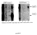
- the other class is represented by MAb KHRI-3, which binds to inner ear supporting cells in a characteristic punctate "stacked wine glass" staining pattern ( Zajic et al. (1991) Hear. Res. 52:59-72 ).
- MaB KHRI-3 also identifies a protein in Western blots of inner-ear extracts ( Nair et al. (1995) Hear. Res. 83:101-113 ).
- the protein to which KHRI-3 binds was termed Inner Ear Supporting Cell Antigen, or IESCA.
- HSP-70 heat shock protein 70
- HSP-70 is a ubiquitous protein that is not specific for the inner-ear. However, reactivity with stress proteins of various classes has been reported in a number of autoimmune diseases as well as non-autoimmune disease states. When cells or organisms are placed under stress, synthesis of HSPs increases presumably to protect critical proteins from denaturation. Thus, antibodies to HSPs may be produced as a result of release of cellular contents of damaged cells under the same stress that leads to formation of antibodies to autoimmune specific antigens. In fact, surveys of patients with other autoimmune diseases and patients with some types of infection also frequently have antibodies to heat shock proteins. In spite of the lack of specificity, HSP-70 is being used in some studies, including a national clinical trial sponsored by the NIH, as a target antigen for assessing AISNHL.
- IESCA is Associated with AISNHL In Humans
- IESCA is a target antigen in human AINSL .
- the present inventors used guinea pig inner ear tissue as the substrate for detection of inner ear reactive antibodies in sera from patients with AISNHL. This selection was made based upon extensive experience with this model ( Zajic et al. (1991) Hearing Res. 52:59-72 ; Ptok et al. (1991) Hearing Res.
- the inventors had prepared a monoclonal antibody, KHRI-3, which bound to an inner ear supporting cell antigen (IESCA) both in vitro and in vivo, and which when present in animals, resulted in the development of hearing loss in the animals.
- IESCA inner ear supporting cell antigen
- IESCA inner ear supporting cell antigen
- human inner ear tissue but not blood cells from the same donor, absorbs the human serum antibody reactivity to guinea pig inner ear substrate, indicating that the antibodies define a phylogenetically conserved protein expressed in human and guinea pig ears.
- IESCA and HSP-70 are different proteins . The present inventors have demonstrated that IESCA and HSP-70 are different proteins, even though they appear to have similar molecular weights.
- the inventors have now discovered that the presence of antibodies to IESCA in AISNHL patients is predictive of the response of patients to treatment with immunosuppressive therapy; those patients with antibodies are more likely to respond to treatment than those without. This discovery is based upon the results of a prospective study, which demonstrated that patients with antibody to IESCA were statistically significantly more likely to have improvement in hearing after steroid therapy than those without such antibody.
- the presence of antibody to IESCA was determined by one of two methods, Western blotting (where the separated proteins from the patient sera were stained with Mab KHRI-3), and immunofluroescence (where guinea pig organs of Corti were stained by antibody present in the sera).
- Western blotting where the separated proteins from the patient sera were stained with Mab KHRI-3
- immunofluroescence where guinea pig organs of Corti were stained by antibody present in the sera.
- IESCA antibodies in patients sera has been shown to have a high correlation with the probable response of a patient to corticosteroid treatment for AISNHL, as described above. Therefore, a diagnostic screen based on the detection of anti-IESCA antibodies would identify these patients while protecting those that do not express anti-IESCA from unnecessary treatment and any associated side effects. Additionally, the identification of compounds that are agonistic and/or antagonistic to IESCA binding and/or anti-IESCA binding would be advantageous in determining methodologies for the treatment of AISNHL.
- Characterization and identification of IESCA would facilitate the implementation of diagnostic screens based upon the detection of anti-IESCA antibodies, as well as the identification of compounds that are agonistic and/or antagonistic to IESCA binding and/or anti-IESCA binding, as described above. Therefore, the inventors set out to identify this inner ear supporting cell antigen.
- CTL1 may, like CTL1 serve as a choline transporter. Choline is required for biosynthesis of acetylcholine, which is a primary neurotransmitter in cholinergic nerve terminals. Choline is also required for biosynthesis of phosphatidylcholine, a major component of membranes. Human CTL2 is predicted to contain ten membrane-spanning regions, with both the N and C terminus residing on the cytoplasmic side of the cell membrane (as shown in Figure 5 ). Such a structure is consistent with a transporter function.
- RT-PCR using RNA from human inner ear tissue and guinea pig inner ear demonstrated that full length CTL2 mRNA is expressed in guinea pig and human inner ear tissue, as well as in a number of human cell lines.
- the guinea pig CTL2 is 72% homologous to the protein encoded by human cDNA.
- the pattern of CTL2 expression in the human ampulla is the same as that previously observed in the guinea pig vestibular system using the KHRI-3 antibody ( Ptok, M. et al. (1993a) Hear. Res. 66, 245-252 ).
- the CTL2 protein is predicted to be a membrane protein, and the immunofluorescence studies are consistent with this prediction.
- the antibody blocks the function of the antigenic protein, perhaps by sterically hindering transport of choline or some other molecule transported by CTL2.
- CTL2 is part of a multimolecular complex, as is described in more detail below, and that antibodies to other members of this complex might also result in similar in vivo staining patterns and damage to the inner ear and to hearing.
- the predicted CTL2 protein of 706 amino acids is expected to be 80 kDa.
- the actual mass of the protein doublet routinely observed by the inventors is 68 kDa and70 kDa (in the older sizing) or 68 kDa and 72 kDA (in the newer sizing), and this doublet contains carbohydrate modifications.
- the size discrepancy and the presence of glycosylation suggests that there are posttranslational modifications of newly synthesized CTL2 to become the mature protein that is inserted into the membrane.
- an 80 kDa protein band was immunoprecipitated by the CTL2 antiserum, but not by KHRI-3. It is postulated that the 80 kDa protein is the nascent protein, which is then cleaved and glycosylated to form the final membrane embedded form.
- Another method contemplates providing purified IESCA from source tissue and determining if sera from a patient suspected of having AISNHL will react with the antigen.
- the present invention is not limited by the particular method of screening, or by the particular source of IESCA.
- One embodiment comprises providing purified IESCA, and Western blotting precipitated IESCA with the patient's sera.
- Another embodiment comprises making tissue extracts from guinea pig organ of Corti, followed by immunoprecipitation of IESCA with monoclonal antibody KHRI-3, or other suitable antibody, and Western blotting precipitated IESCA with patients' sera.
- a positive result in either embodiment comprises recognition of the precipitated IESCA with the patient's sera.
- the present invention relates to methods and compositions for using IESCA and anti-IESCA antibodies ("anti-IESCA”) as a target for screening drugs that can alter, for example, IESCA and anti-IESCA binding (e.g ., and thus alleviate AISNHL symptoms).
- anti-IESCA anti-IESCA antibodies
- drugs that induce or inhibit anti-IESCA binding to IESCA can be identified by screening for compounds that bind to either IESCA or anti-IESCA, or that affect the binding of the antigen to the antibody.
- the present invention relates to the use of cell lines transfected with CTL2 and variants thereof for screening compounds for activity, and in particular to high throughput screening of compounds from combinatorial libraries (e.g ., libraries containing greater than 10 4 compounds).
- the cell lines of the present invention can be used in a variety of screening methods.
- variants e.g ., mutant or polymorphic nucleic acid sequences.
- Assays for detections variants e.g ., polymorphisms or mutations
- fall into several categories including, but not limited to direct sequencing assays, fragment polymorphism assays, hybridization assays, and computer based data analysis. Protocols and commercially available kits or services for performing multiple variations of these assays are available.
- assays are performed in combination or in hybrid ( e.g ., different reagents or technologies from several assays are combined to yield one assay). The following assays are useful in the present invention.
- variant sequences are detected using a direct sequencing technique.
- DNA samples are first isolated from a subject using any suitable method.
- the region of interest is cloned into a suitable vector and amplified by growth in a host cell (e.g., a bacteria) or DNA in the region of interest is amplified using PCR.
- a host cell e.g., a bacteria
- DNA in the region of interest (e.g ., the region containing the mutation of interest) is sequenced using any suitable method, including but not limited to manual sequencing using radioactive marker nucleotides, or automated sequencing. The results of the sequencing are displayed using any suitable method. The sequence is examined and the presence or absence of a given mutation is determined.
- variant sequences are detected using a PCR-based assay.
- the PCR assay comprises the use of oligonucleotide primers that hybridize only to the variant or wild type allele of CTL2 (e.g ., to the region of polymorphism or mutation). Both sets of primers are used to amplify a sample of DNA. If only the mutant primers result in a PCR product, then the patient has the mutant CTL2 allele. If only the wild-type primers result in a PCR product, then the patient has the wild type allele of CTL2.
- a variant sequence is detected using a fragment length polymorphism assay.
- a fragment length polymorphism assay a unique DNA banding pattern based on cleaving the DNA at a series of positions is generated using an enzyme. DNA fragments from a sample containing a mutation will have a different banding pattern than wild type.
- variant sequences are detected using a restriction fragment length polymorphism assay (RFLP).
- RFLP restriction fragment length polymorphism assay
- the region of interest is first isolated using PCR.
- the PCR products are then cleaved with restriction enzymes known to give a unique length fragment for a given polymorphism.
- the restriction-enzyme digested PCR products are separated by agarose gel electrophoresis and visualized by ethidium bromide staining. The length of the fragments is compared to molecular weight markers and fragments generated from wild-type and mutant controls.
- variant sequences are detected using a CLEAVASE fragment length polymorphism assay (CFLP; Third Wave Technologies, Madison, WI; See e.g., U.S. Patent Nos. 5,843,654 ; 5,843,669 ; 5,719,208 ; and 5,888,780 ).
- CFLP CLEAVASE fragment length polymorphism assay
- This assay is based on the observation that when single strands of DNA fold on themselves, they assume higher order structures that are highly individual to the precise sequence of the DNA molecule. These secondary structures involve partially duplexed regions of DNA such that single stranded regions are juxtaposed with double stranded DNA hairpins.
- the CLEAVASE I enzyme is a structure-specific, thermostable nuclease that recognizes and cleaves the junctions between these single-stranded and double-stranded regions.
- the region of interest is first isolated, for example, using PCR. Then DNA strands are separated by heating. Next, the reactions are cooled to allow intrastrand secondary structure to form. The PCR products are then treated with the CLEAVASE I enzyme to generate a series of fragments that are unique to a given mutation. The CLEAVASE enzyme treated PCR products are separated and detected ( e.g ., by agarose gel electrophoresis) and visualized ( e.g ., by ethidium bromide staining). The length of the fragments is compared to molecular weight markers and fragments generated from wild-type and mutant controls.
- variant sequences are detected a hybridization assay.
- a hybridization assay the presence or absence of a given mutation is determined based on the ability of the DNA from the sample to hybridize to a complementary DNA molecule (e.g ., a oligonucleotide probe).
- a complementary DNA molecule e.g ., a oligonucleotide probe.
- hybridization of a probe to the sequence of interest is detected directly by visualizing a bound probe (e.g ., a Northern or Southern assay; See e.g., Ausabel et al. (eds.), Current Protocols in Molecular Biology, John Wiley & Sons, NY [1991 ]).
- a Northern or Southern assay See e.g., Ausabel et al. (eds.), Current Protocols in Molecular Biology, John Wiley & Sons, NY [1991 ]).
- genomic DNA Southern or RNA (Northern) is isolated from a subject. The DNA or RNA is then cleaved with a series of restriction enzymes that cleave infrequently in the genome and not near any of the markers being assayed.
- variant sequences are detected using a DNA chip hybridization assay.
- a DNA chip hybridization assay In this assay, a series of oligonucleotide probes are affixed to a solid support. The oligonucleotide probes are designed to be unique to a given mutation. The DNA sample of interest is contacted with the DNA "chip” and hybridization is detected.
- the DNA chip assay is a GeneChip (Affymetrix, Santa Clara, CA; See e.g., U.S. Patent Nos. 6,045,996 ; 5,925,525 ; and 5,858,659 ) assay.
- the GeneChip technology uses miniaturized, high-density arrays of oligonucleotide probes affixed to a "chip.” Probe arrays are manufactured by Affymetrix's light-directed chemical synthesis process, which combines solid-phase chemical synthesis with photolithographic fabrication techniques employed in the semiconductor industry.
- the process constructs high-density arrays of oligonucleotides, with each probe in a predefined position in the array. Multiple probe arrays are synthesized simultaneously on a large glass wafer. The wafers are then diced, and individual probe arrays are packaged in injection-molded plastic cartridges, which protect them from the environment and serve as chambers for hybridization.
- the nucleic acid to be analyzed is isolated, amplified by PCR, and labeled with a fluorescent reporter group.
- the labeled DNA is then incubated with the array using a fluidics station.
- the array is then inserted into the scanner, where patterns of hybridization are detected.
- the hybridization data are collected as light emitted from the fluorescent reporter groups already incorporated into the target, which is bound to the probe array. Probes that perfectly match the target generally produce stronger signals than those that have mismatches. Since the sequence and position of each probe on the array are known, by complementarity, the identity of the target nucleic acid applied to the probe array can be determined.
- a test site or a row of test sites on the microchip is electronically activated with a positive charge.
- a solution containing the DNA probes is introduced onto the microchip.
- the negatively charged probes rapidly move to the positively charged sites, where they concentrate and are chemically bound to a site on the microchip.
- the microchip is then washed and another solution of distinct DNA probes is added until the array of specifically bound DNA probes is complete.
- a test sample is then analyzed for the presence of target DNA molecules by determining which of the DNA capture probes hybridize, with complementary DNA in the test sample (e.g ., a PCR amplified gene of interest).
- An electronic charge is also used to move and concentrate target molecules to one or more test sites on the microchip.
- the electronic concentration of sample DNA at each test site promotes rapid hybridization of sample DNA with complementary capture probes (hybridization may occur in minutes).
- the polarity or charge of the site is reversed to negative, thereby forcing any unbound or nonspecifically bound DNA back into solution away from the capture probes.
- a laser-based fluorescence scanner is used to detect binding.
- an array technology based upon the segregation of fluids on a flat surface (chip) by differences in surface tension is utilized ( See e.g., U.S. Patent Nos. 6,001,311 ; 5,985,551 ; and 5,474,796 ; ).
- Protogene's technology is based on the fact that fluids can be segregated on a flat surface by differences in surface tension that have been imparted by chemical coatings. Once so segregated, oligonucleotide probes are synthesized directly on the chip by ink-jet printing of reagents.
- the array with its reaction sites defined by surface tension is mounted on a X/Y translation stage under a set of four piezoelectric nozzles, one for each of the four standard DNA bases.
- the translation stage moves along each of the rows of the array and the appropriate reagent is delivered to each of the reaction site.
- the A amidite is delivered only to the sites where amidite A is to be coupled during that synthesis step and so on.
- Common reagents and washes are delivered by flooding the entire surface and then removing them by spinning.
- DNA probes unique for the mutation of interest are affixed to the chip using Protogene's technology.
- the chip is then contacted with the PCR-amplified genes of interest.
- unbound DNA is removed and hybridization is detected using any suitable method (e.g., by fluorescence de-quenching of an incorporated fluorescent group).
- genomic profiles are generated using a assay that detects hybridization by enzymatic cleavage of specific structures (INVADER assay, Third Wave Technologies; See e.g., U.S. Patent Nos. 5,846,717 ; 6,090,543 ; 6,001,567 ; 5,985,557 ; and 5,994,069 ).
- the INVADER assay detects specific DNA and RNA sequences by using structure-specific enzymes to cleave a complex formed by the hybridization of overlapping oligonucleotide probes. Elevated temperature and an excess of one of the probes enable multiple probes to be cleaved for each target sequence present without temperature cycling.
- the INVADER assay detects specific mutations in unamplified genomic DNA.
- the isolated DNA sample is contacted with the first probe specific either for a mutation or wild type sequence and allowed to hybridize. Then a secondary probe, specific to the first probe, and containing the fluorescein label, is hybridized and the enzyme is added. Binding is detected by using a fluorescent plate reader and comparing the signal of the test sample to known positive and negative controls.
- hybridization of a bound probe is detected using a TaqMan assay (PE Biosystems, Foster City, CA; See e.g., U.S. Patent Nos. 5,962,233 and 5,538,848 ).
- the assay is performed during a PCR reaction.
- the TaqMan assay exploits the 5'-3' exonuclease activity of the AMPLITAQ GOLD DNA polymerase.
- a probe, specific for a given allele or mutation, is included in the PCR reaction.
- the probe consists of an oligonucleotide with a 5'-reporter dye (e.g., a fluorescent dye) and a 3'-quencher dye.
- the 5'-3' nucleolytic activity of the AMPLITAQ GOLD polymerase cleaves the probe between the reporter and the quencher dye.
- the separation of the reporter dye from the quencher dye results in an increase of fluorescence.
- the signal accumulates with each cycle of PCR and can be monitored with a fluorimeter.
- polymorphisms are detected using the SNP-IT primer extension assay (Orchid Biosciences, Princeton, NJ; See e.g., U.S. Patent Nos. 5,952,174 and 5,919,626 ).
- SNPs are identified by using a specially synthesized DNA primer and a DNA polymerase to selectively extend the DNA chain by one base at the suspected SNP location. DNA in the region of interest is amplified and denatured. Polymerase reactions are then performed using miniaturized systems called microfluidics. Detection is accomplished by adding a label to the nucleotide suspected of being at the SNP or mutation location. Incorporation of the label into the DNA can be detected by any suitable method (e.g ., if the nucleotide contains a biotin label, detection is via a fluorescently labelled antibody specific for biotin).
- a MassARRAY system (Sequenom, San Diego, CA.) is used to detect variant sequences (see e.g., U.S. Patent Nos. 6,043,031 ; 5,777,324 ; and 5,605,798 ).
- DNA is isolated from blood samples using standard procedures.
- specific DNA regions containing the mutation of interest about 200 base pairs in length, are amplified by PCR.
- the amplified fragments are then attached by one strand to a solid surface and the non-immobilized strands are removed by standard denaturation and washing. The remaining immobilized single strand then serves as a template for automated enzymatic reactions that produce genotype specific diagnostic products.
- the diagnostic product As the diagnostic product is charged when an electrical field pulse is subsequently applied to the tube they are launched down the flight tube towards a detector.
- the time between application of the electrical field pulse and collision of the diagnostic product with the detector is referred to as the time of flight.
- This is a very precise measure of the product's molecular weight, as a molecule's mass correlates directly with time of flight with smaller molecules flying faster than larger molecules.
- the entire assay is completed in less than one thousandth of a second, enabling samples to be analyzed in a total of 3-5 second including repetitive data collection.
- the SpectroTYPER software then calculates, records, compares and reports the genotypes at the rate of three seconds per sample.
- antibodies are used to determine if an individual contains an allele encoding a variant CTL2 gene. In some methods, antibodies are utilized that discriminate between mutant (e.g. , non-glycosylated proteins, or incorrectly processed proteins) and wild-type proteins.
- the reagent is a nucleic acid that hybridizes to nucleic acids containing the mutation and that does not bind to nucleic acids that do not contain the mutation or the reagents are primers for amplifying the region of DNA containing the mutation or the reagents are antibodies which preferentially bind either the wild-type or truncated CTL2 proteins.
- the kit contains instructions for determining whether the subject is at risk for developing hearing loss or damage.
- the kits include ancillary reagents such as buffering agents, nucleic acid stabilizing reagents, protein stabilizing reagents, and signal producing systems (e.g ., florescence generating systems as Fret systems).
- the test kit may be packaged in any suitable manner, typically with the elements in a single container or various containers as necessary along with a sheet of instructions for carrying out the test.
- the kits also preferably include a positive control sample.
- yeast two-hybrid system to identify binding partners
- a protein fused to a DNA-binding domain (the bait) and a protein fused to an activation domain (the prey) are expressed in two different haploid yeast strains of opposite mating type (MATa and MAT ⁇ ), and the strains are mated to determine if the two proteins interact. Mating occurs when haploid yeast strains of opposite mating type come into contact, and results in fusion of the two haploids to form a diploid yeast strain.
- the present invention relates to methods of treating AISNHL, where the first step is determining whether a patient experiencing hearing damage or loss has serum antibodies to IESCA.
- the presence of antibodies indicates a generally favorable response to treatment with steroids; thus, patients with anti-IESCA antibodies in their serum are treated with steroids.
- the present invention also relates to methods of treating, hearing damage or loss due to mutations present in CTL2 genes.
- the invention relates to methods and compositions suitable for gene therapy to alter CTL2 expression, production, or function.
- the present invention provides CTL2 genes.
- the methods described below are generally applicable.
- the gene therapy is performed by providing a subject with a wild-type allele of CTL2 (i.e ., an allele that does not contain a mutation which results in hearing damage or loss). Subjects in need of such therapy are identified by the methods described above.
- Viral vectors commonly used for in vivo or ex vivo targeting and therapy procedures are DNA-based vectors and retroviral vectors. Methods for constructing and using viral vectors are known in the art (See e.g. ( 1992) Miller and Rosman, BioTech., 7:980-990 ).
- the viral vectors are replication defective, that is, they are unable to replicate autonomously in the target cell.
- the genome of the replication defective viral vectors that are used within the scope of the present invention lack at least one region that is necessary for the replication of the virus in the infected cell. These regions can either be eliminated (in whole or in part), or be rendered non-functional by any technique known to a person skilled in the art.
- These techniques include the total removal, substitution (by other sequences, in particular by the inserted nucleic acid), partial deletion or addition of one or more bases to an essential (for replication) region.
- Such techniques may be performed in vitro ( i.e ., on the isolated DNA) or in situ, using the techniques of genetic manipulation or by treatment with mutagenic agents.
- DNA viral vectors include an attenuated or defective DNA viruses, including, but not limited to, herpes simplex virus (HSV), papillomavirus, Epstein Barr virus (EBV), adenovirus, adeno-associated virus (AAV), and the like.
- HSV herpes simplex virus
- EBV Epstein Barr virus
- AAV adeno-associated virus
- Defective viruses that entirely or almost entirely lack viral genes, are preferred, as defective virus is not infective after introduction into a cell.
- Use of defective viral vectors allows for administration to cells in a specific, localized area, without concern that the vector can infect other cells. Thus, a specific tissue can be specifically targeted.
- Examples of particular vectors include, but are not limited to, a defective herpes virus 1 (HSV1) vector ( Kaplitt et al. (1991) Mol. Cell. Neurosci., 2:320-330 ), defective herpes virus vector lacking a glycoprotein L gene (See e.g., Patent Publication RD 371005 A), or other defective herpes virus vectors (See e.g., WO 94/21807 ; and WO 92/05263 ); an attenuated adenovirus vector, such as the vector described by Stratford-Perricaudet et al. (J. Clin. Invest., 90:626-630 (1992 ); See also, La Salle et al.
- HSV1 herpes virus 1
- Kaplitt et al. (1991) Mol. Cell. Neurosci., 2:320-330 defective herpes virus vector lacking a glycoprotein L gene
- other defective herpes virus vectors See e.g., WO
- an appropriate immunosuppressive treatment is employed in conjunction with the viral vector (e.g ., adenovirus vector), to avoid immuno-deactivation of the viral vector and transfected cells.
- the viral vector e.g ., adenovirus vector
- immunosuppressive cytokines such as interleukin-12 (IL-12), interferon-gamma (IFN- ⁇ ), or anti-CD4 antibody
- IL-12 interleukin-12
- IFN- ⁇ interferon-gamma
- anti-CD4 antibody can be administered to block humoral or cellular immune responses to the viral vectors.
- a viral vector that is engineered to express a minimal number of antigens.
- the replication defective adenoviral vectors comprise the ITRs, an encapsidation sequence and the nucleic acid of interest.
- at least the E1 region of the adenoviral vector is non-functional.
- the deletion in the E1 region preferably extends from nucleotides 455 to 3329 in the sequence of the Ad5 adenovirus ( Pvu II- Bgl II fragment) or 382 to 3446 (Hinf II- Sau 3A fragment).
- Other regions may also be modified, in particular the E3 region (e.g ., WO95/02697 ), the E2 region ( e . g ., WO94/28938 ), the E4 region ( e.g ., WO94/28152 , WO94/12649 and WO95/02697 ), or in any of the late genes L1-L5.
- the adenoviral vector may have a deletion in the E1 region (Ad 1.0). Examples of E1-deleted adenoviruses are disclosed in EP 185,573 or the adenoviral vector has a deletion in the E1 and E4 regions (Ad 3.0). Examples of E1/E4-deleted adenoviruses are disclosed in WO95/02697 and WO96/22378 .
- the adenoviral vector may have a deletion in the E1 region into which the E4 region and the nucleic acid sequence are inserted.
- the cell line that is employed should preferably (i) be transformable by the elements to be used, and (ii) contain the sequences that are able to complement the part of the genome of the replication defective adenovirus, preferably in integrated form in order to avoid the risks of recombination.
- Examples of cell lines that may be used are the human embryonic kidney cell line 293 ( Graham et al. (1977) J. Gen. Virol., 36:59 ), which contains the left-hand portion of the genome of an Ad5 adenovirus (12%) integrated into its genome, and cell lines that are able to complement the E1 and E4 functions, as described in applications WO94/2691 and WO95/02697 .
- Recombinant adenoviruses are recovered and purified using standard molecular biological techniques that are well known to one of ordinary skill in the art.
- the adeno-associated viruses are DNA viruses of relatively small size that can integrate, in a stable and site-specific manner, into the genome of the cells that they infect. They are able to infect a wide spectrum of cells without inducing any effects on cellular growth, morphology or differentiation, and they do not appear to be involved in human pathologies.
- the AAV genome has been cloned, sequenced and characterized. It encompasses approximately 4700 bases and contains an inverted terminal repeat (ITR) region of approximately 145 bases at each end, which serves as an origin of replication for the virus.
- ITR inverted terminal repeat
- the remainder of the genome is divided into two essential regions that carry the encapsidation functions: the left-hand part of the genome, that contains the rep gene involved in viral replication and expression of the viral genes; and the right-hand part of the genome, that contains the cap gene encoding the capsid proteins of the virus.
- the retroviruses are integrating viruses that infect dividing cells.
- the retrovirus genome includes two LTRs, an encapsidation sequence and three coding regions ( gag, pol and env ) .
- gag, pol and env coding regions
- the gag, pol and env genes are generally deleted, in whole or in part, and replaced with a heterologous nucleic acid sequence of interest.
- the recombinant retroviral vectors can contain modifications within the LTRs for suppressing transcriptional activity as well as extensive encapsidation sequences that may include a part of the gag gene ( Bender et al. (1987) Virol., 61:1639 ). Recombinant retroviral vectors are purified by standard techniques known to those having ordinary skill in the art.
- a nucleic acid in vivo such as a cationic oligopeptide (e.g ., WO95/21931 ), peptides derived from DNA binding proteins (e.g .. WO96/25508 ), or a cationic polymer (e.g., WO95/21931 ). It is also possible to introduce the vector in vivo as a naked DNAplasmid. Methods for formulating and administering naked DNA to mammalian muscle tissue are disclosed in U.S. Pat. Nos. 5,580,859 and 5,589,466 .
- DNA vectors for gene therapy can be introduced into the desired host cells by methods known in the art, including but not limited to transfection, electroporation, microinjection, transduction, cell fusion, DEAE dextran, calcium phosphate precipitation, use of a gene gun, or use of a DNA vector transporter ( See e.g ., Wu et al. (1992) J. Biol. Chem., 267:963 ; Wu and Wu (1988) J. Biol. Chem., 263:14621 ; and Williams et al. (1991) Proc. Natl. Acad. Sci. USA 88:2726 ). Receptor-mediated DNA delivery approaches can also be used ( Curiel et al. (1992) Hum. Gene Ther., 3:147 ; and Wu and Wu (1987) J. Biol. Chem., 262:4429 ).
- N normal
- M molar
- mM millimolar
- ⁇ M micromolar
- mol molecular weight
- mol molecular weight
- mmol millimoles
- ⁇ mol micromol
- nmol nanomoles
- pmol picomoles
- g grams); mg (milligrams); ⁇ g (micrograms); ng (nanograms); 1 or L (liters); ml (milliliters); ⁇ l (microliters); cm (centimeters); mm (millimeters); ⁇ m (micrometers); nm (nanometers); °C (degrees Centigrade); SNHL (Sensorineural Hearing Loss); AISNHL, (Auto Immune Sensorineural Hearing Loss); IESCA (Inner Ear Supporting Cell Antigen); Mab (Monoclonal Antibody); HSP-70 (Heat Shock Protein); IF (Immunofluor
- Hybridoma cells were cultured in a CELLMAX (CELLMAX ® Artificial Capillary System, Cellco, Inc., Germantown, MD) bioreactor. The bioreactor was inoculated with KHRI-3 hybridoma cells. The serum-free, antibiotic-free medium was changed to lactic acid production to maintain optimal cell growth and antibody production. Once the system was operating at optimal conditions, 45 ml of fluid containing antibody and excess cells was harvested every second day from the extra-capillary space. The IgG1 antibody binding capacity of a protein G affinity column was determined.
- HSP-70 heat shock protein
- KHRI-3 can precipitate an antigen from inner ear extracts that reacts strongly with AISNHL patients' sera, but purified KHRI-3 does not precipitate a protein from these extracts that binds antibodies to HSP-70.
- KHRI-3 and Western blot positive AISNHL human sera stain inner ear tissues with a similar pattern, but highly purified, high titered KHRI-3 antibody concentrated from hybridoma supernatant or produced in a bioreactor does not stain HSP-70 protein on Western blots.
- HSP-70 is present in the inner ear, as demonstrated by the observations that it can be immunoprecipitated from the inner ear and detected on Western blots using anti-HSP antibody.
- proteins immunoprecipitated from guinea pig inner ear by anti HSP-70 and highly purified KHRI-3 monoclonal antibodies were Western blotted, KHRI-3 stained only the protein it precipitated, and not HSP-70.
- anti-HSP-70 stained only the 70 kD protein it precipitated, and not the protein precipitated by KHRI-3 (Nair et al ., Association for Research in Otolaryngology Feb 15-19, 1998).
- This example describes the characterisation and role in hearing loss of antibodies to IESCA in AISNHL patients.
- Hearing loss was defined as greater than 30 dB at any frequency or less than 85% speech discrimination in either one (unilateral) or both ears (bilateral). If the hearing loss developed within a 24 hour period and did not progress subsequently, the patient was considered to have sudden hearing loss. Either unilateral or bilateral hearing loss with greater than 10 dB progression within three months was classified as rapidly progressive ( Moscicki et al. (1994) JAMA 272:611-616 ). An initial analysis of 91 patients has been reported ( Disher et al. (1997) Ann. NY Acad. Sci. 830:253-265 ). Of the 91 patients, seventeen did not fit in these categories and were not included in the analysis.
- sample buffer containing 0.0625 M tris-HCL pH 6.8, 2% SDS, 10% glycerol, and 0.005% bromophenol blue.
- sample buffer containing 0.0625 M tris-HCL pH 6.8, 2% SDS, 10% glycerol, and 0.005% bromophenol blue.
- sample buffer containing 0.0625 M tris-HCL pH 6.8, 2% SDS, 10% glycerol, and 0.005% bromophenol blue.
- the sample contained 5% (v/v) 2-mercaptoethanol.
- Strips were incubated for 2 hours at room temperature with human serum diluted at 1:50, or KHRI-3 diluted to 10 ug/mL, washed 3 times for 10 minutes each, and incubated for 2 hours at room temperature with secondary antibody (goat anti-human IgG/IgM or anti-mouse IfG heavy and light chain-specific conjugated with horseradish peroxidase (Jackson ImmunoResearch Laboratories, Inc., West Grove, Pennsylvania), diluted at 1:500).
- the blots were developed in 4-chloro-1-napthol (0.5 mg/mL) in methanol-PBS, pH 7.6 (1:5) containing 0.05 % hydrogen peroxide.
- Guinea pigs weighing 250-300 g were anesthetized, decapitated, and the bullas were fixed locally with 4% paraformaldehyde for 2 hours. After the cochleae were dissected free of the surrounding bone, the spiral ligament and tectorial membrane were removed. The central bony cochlae containing the organ of Corti were incubated in 3% normal goat serum for 1 hour.
- the specimens were washed 3 times for 5 minutes in PBS, and incubated in primary antibody (serum diluted at 1:50 or KHRI-3 10 ug/mL) overnight at 4 °C, washed three times for 5 minutes in PBS, and incubated for 45 min at room temperature in secondary antibody (Lissamine Rhodamine LRSC anti-Human IgG/IgM heavy and light chain-specific (Jackson ImmunoResearch Laboratories Inc, West Grove) or rhodamine (TRITC) conjugated to goat antimouse anti-IgG (Accurate Chemicals, Old Westbury, NY), diluted 1:200 in PBS (pH 7.4).
- the cochleae were washed in PBS, and then the organ of Corti was carefully peeled off the modiolus and mounted in GVA mounting media (Zymed Labs, San Francisco, CA).
- the KHRI-3 antigen was immunoprecipitated from guinea pig cochlea and vestibular tissue extracts using KHRI-3 antibody.
- cochlear and vestibular tissue from both ears of a guinea pig were homogenized in 25 uL lysis buffer with protease inhibitors, as described above.
- the lysate (25 ul) was mixed with 50 ul of BSA and 150 ul of wash buffer containing 1% Nonidet P-40, 50 mM tris-HCl (pH 8), 150 mM NaCl, 0.1% sodium deoxycholate, 0.1% SDS, and 1 mM phenylmethylsulfonyl fluoride per sample.
- the samples were precleared twice with protein G-agarose for 30 minutes and incubated overnight at 4 °C with 200 ul of the KHRI-3 antibody from hybridoma supernatant.
- Antibody-antigen complexes were precipitated by incubation with protein G-agarose for 2 hours at 4 °C.
- the supernatant was removed after centrifugation at 5000 x g for 5 minutes.
- the precipitates were washed four times with wash buffer, centrifuged, resuspended in running buffer under reducing or nonreducing conditions, and separated by SDS-PAGE using a 7% gel.
- the gel was electrophoresed at 25 mA of constant current.
- the proteins were transferred as described above, and Western blotted and human sera or KHRI-3 antibody as described above.
- This example describes the characterization and identification of IESCA as CTL2.
- Serum free KHRI-3 was produced in the CELLMAX ® Artificial Capillary System according to the manufacturer's instruction ( Nair TS et al. (1999) Hearing Res 129:50-60 ).
- the hybridoma was inoculated into the extra capillary space and fresh chemically defined, serum-free medium designed for hybridoma cultures (GIBCO, Grand Island, NY) was circulated through the capillary network. Excess cells and fluid containing antibody were removed by flushing the extra capillary space at regular intervals. The cells were removed by centrifugation and the supernatants containing the antibody were pooled.
- the pooled KHRI-3 antibody was affinity purified using protein G columns (Pierce, Rockford, IL) ( Nair, T.S et al. (1997) Hear. Res. 107:93-101 ) following the kit protocol. Purified antibody was tested for purity by gel electrophoresis and antibody activity was tested by immunofluorescence (IF) as previously described ( Nair, T.S. et al. (1995) Hear. Res. 83:101-113 ).
- IF immunofluorescence
- a custom 19 amino acid peptide corresponding to an antigenic region of human CTL2 that fell within one of the regions identified in guinea pig IESCA by MS/MS amino acid sequencing was synthesized and used to raise rabbit antisera (Princeton Biomolecules, Langome, PA).
- Two rabbits (R228 and R229) were immunized with 500ug of the CTL2 peptide, boosted two times and bled 10 days later, boosted again and bled 10 days after immunization.
- the antisera were identified as bleeds 1, 2, and 3.
- Inner ear lysates were analyzed using sodium dodecylsulfate-polyacrylamide gel electrophoresis (SDS-PAGE).
- Cochlear extracts, immunoprecipitated antigen, and molecular weight standards were prepared using sample buffer containing 0.0625M Tris-HCl pH 6.8, 2% SDS, 10% glycerol, 0.005% bromophenol blue.
- the sample buffer contained 5% (v/v) 2-mercaptoethanol.
- the samples were boiled for 5 minutes and separated on 3% stacking and 8 or 10% separating gels.
- Samples for MS-MS sequencing were isolated using 1mm thick gels. All the reagents used for sequencing studies were prepared in HPLC grade water.
- the KHRI-3 antigen and CTL2 antigen were immunoprecipitated from guinea pig cochlea and vestibular tissue extracts using KHRI-3 and anti CTL2 antibodies.
- cochlear and vestibular tissue from one ear were homogenized in 50ul lysis buffer with protease inhibitors as described earlier.
- the lysate was mixed with either 20ul of KHRI-3-CNBr beads or CNBr coupled CTL2 antibody (20ul/ear), and incubated overnight at 4°C; the precipitated proteins were recovered by centrifugation of the beads.
- CTL2 antigen was incubated overnight with anti CTL2 antibody, and the immune complexes then precipitated by addition of 30ul of protein A agarose (Sigma) for 2 hours at 4°C. The supernatants were removed after centrifugation at 5000 x g for 5 min. The precipitates were washed three times, resuspended in sample buffer for either reducing or non-reducing conditions, boiled, and subjected to electrophoresis.
- Proteins from electrophoresis gels were transferred to nitrocellulose membranes at constant voltage of 25 V in transfer buffer (20% methanol, 150 mM glycine, 20 mM tris buffer).
- transfer buffer (20% methanol, 150 mM glycine, 20 mM tris buffer).
- the membranes were incubated in 5% dry milk in phosphate buffered saline containing 0.1% Tween (PBS - T) on a rocker for one hour at room temperature to block non-specific binding. All subsequent incubations were done in this buffer.
- nitrocellulose strips were incubated for 2 hours at room temperature with primary antibody, either KHRI-3 monoclonal antibody diluted to 1ug/ml or anti CTL2 peptide antibody (rabbit antiserum) diluted 1:1000, and washed 3 times with PBS-T for 10 mins each time. After washing, the strips were incubated for 2 hr in affinity purified secondary antibodies (either rat-anti-mouse IgG heavy and light chain specific, or mouse-anti-rabbit) conjugated to horseradish peroxidase (Jackson ImmunoResearch Laboratories Inc., West Grove, PA.) diluted 1:5000 and 1:10000 respectively.
- primary antibody either KHRI-3 monoclonal antibody diluted to 1ug/ml or anti CTL2 peptide antibody (rabbit antiserum) diluted 1:1000
- the blots were developed with enhanced chemiluminescence (ECL) by applying a 1:1 mix of ECL reagents 1 and 2 for 1 minute, after which the blots were drained, covered with plastic wrap and exposed to X-ray film. Exposure time varied from 10 seconds to a few minutes, depending on the reaction intensity.
- ECL enhanced chemiluminescence
- the IESCA was purified by immunoaffinity column purification. Protein extracts from six animals (12 ears) were divided into two aliquots and each was mixed with 1 ml of beads coupled to 1.25mg of purified KHRI-3 monoclonal antibody. The antibody-coupled beads were washed with binding buffer (lysis buffer, described above) and incubated with guinea pig cochlear lysate overnight at 4°C, with gentle rocking. The beads were then transferred to a 10 ml Poly-Prep Chromatography Column (Bio-Rad Richmond, CA).
- the beads were washed with 10 bed volumes of binding buffer (Dulbecco's phosphate buffered saline (GIBCO, Grand Island, NY) containing 0.5% NP-40 and a cocktail of protease inhibitors (Roche Diagnostics, Mannheim, Germany).
- binding buffer Dulbecco's phosphate buffered saline (GIBCO, Grand Island, NY) containing 0.5% NP-40 and a cocktail of protease inhibitors (Roche Diagnostics, Mannheim, Germany).
- the antigen was eluted with 100mM glycine/HCl pH 2.8, containing 0.5% NP-40 and collected as 1 ml fractions.
- the IESCA was immunoprecipitated. Extracts from 12 ears were incubated overnight with 240ul of KHRI-3-CNBr beads ( ⁇ 300ug of purified antibody) overnight at 4°C and then washed with 5 times with 1ml of binding buffer each time. After the last wash, 180 ul of the beads corresponding to 9 ears were resuspended in 120ul of reducing buffer (1%SDS, glycerol, 2 mercaptoethanol) and boiled. After brief centrifugation to pellet the beads, the supernatant was removed and loaded in one lane of an 8% 1mm thick SDS-PAGE gel for electrophoresis and colloidal Coomassie blue staining.
- reducing buffer 1%SDS, glycerol, 2 mercaptoethanol
- Antisera were tested for approximate antibody titer by ELISA ( Zajic, G. et al. (1991) Hear. Res. 52: 59-72 ), using 96 well plastic plates coated with 1ug CTL2 peptide. After coating for overnight at 4°C, the plates were washed, blocked with 5%goat serum, then incubated sequentially for 1 hour each with serial dilutions (1/10-1/320) of the rabbit anti-CTL2 sera, washed, and incubated with alkaline phosphatase-conjugated goat anti-rabbit IgG diluted 1/5000 as described for western blots. After a final wash with PBS, para-nitrophenylphosphate was used as the substrate and the absorbance at 415 nm was determined after 30 min.
- Guinea pigs were decapitated and the bullas with exposed otic capsule were fixed locally with 2% paraformaldehyde for 2 hours.
- the cochlea were dissected free of spiral ligament and tectorial membrane, permeabilized with 0.3% triton X-100, and incubated in 3% normal goat serum for one hour.
- Fresh normal human inner ear vestibular tissue was obtained from patients undergoing translabyrinthine procedures for removal of acoustic tumors. Patients all gave prior informed written consent that was approved by the University of Michigan Institutional Review Committee for the Protection of Human Research Subjects.
- the human tissues were prepared by fixation in 2% paraformaldehyde in phosphate buffer pH 7.2 for 2 hr.
- the soft tissue was homogenized in the tissue lysis buffer provided in RNeasy Mini kit, (Qiagen, Chatworth, CA).
- the tissue was sheared by passing through an 18-gauge needle 5 times and then centrifuged through a QIAShredder, (Qiagen, Chatworth, CA), for efficient homogenization.
- the supernatant was collected and processed further using the RNeasy Mini kit according to the manufacturer's protocol.
- RNA was used to synthesize sscDNA.
- Both oligo dT and random primers were used with M-MLV reverse transcriptase (Invitrogen Life Technologies (Carlsbad, CA)) according to the manufacturer's instructions.
- M-MLV reverse transcriptase Invitrogen Life Technologies (Carlsbad, CA)
- To amplify the CTL2 cDNA seven sets of sense and antisense primers were designed using the Primer 3 Input software (Howard Hughes Medical Institute, NIH, National Human Genome Research Institute) based on the human CTL2 gene sequence.
- a back translation algorithm (ExPasY, Swiss Institute of Bioinformatics Web Site) was used to deduce the guinea pig coding region from the amino acid sequence. This information was then was used to design initial primer sets, after which a walking primer strategy was used so that the next set of primers was designed based on the sequence of the product from a previous successful PCR reaction. Long-range guinea pig primers based on these known sequences were then designed. All eight primer sets are shown in Table 6 (below).
- PCR was performed using 2 ⁇ L of cDNA, 0.5 mM of dNTP, 0.2 ⁇ M of each primer, in 10X buffer with MgCl and 0.625 ⁇ L of Taq DNA Polymerase (Promega, Madison, WI]. After denaturation at 94°C for one minute, the reaction was carried out for 30 cycles at 94°C for 1 min, 58-62°C (depending on the primer set) for 1 min and 72°C for two minutes. For amplification of long sequences (i.e. greater than 1kb), the Clontech Long Distance cDNA amplification kit was used as described in the manufacturer's instructions. The elongation step at 72°C was increased to 3 minutes in each cycle.
- PCR products were visualized on a 1% agarose gel using ethidium bromide fluorescence.
- ⁇ -actin cDNA was amplified from both human and guinea pig using primers based on the human sequence.
- the PCR products were purified using the QIAquick pPCR purification kit (Qiagen, , Chatworth, CA), and submitted for sequencing to the University of Michigan DNA Sequencing Core.
- Immunoaffinity purification of the KHRI-3 defined IESCA was carried out as described in the methods under section A.
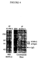
- the affinity-purified proteins were subjected to SDS-PAGE, and the 68 and 72 kDa bands were identified by western blot and colloidal Coomassie blue staining, as shown in Figure 4 .
- the 68 and 72 kDa bands identified by colloidal Coomassie blue staining were cut from the gel and combined in a single tube for sequencing. A control slice from an empty lane of the same gel was used as a background control for contaminating proteins.
- the 68 kDa and 72 kDa bands were cut out as before but sent as separate samples for independent sequencing.
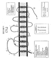
- FIG. 5 A diagrammatic representation of the predicted structure of the CTL2 protein in the cell membrane is shown in Figure 5 .
- This structure was predicted using Swiss Prot Expert Protein Analysis System (ExPASy) based on the various domains within the protein and the conformation with the lowest predicted entropy.
- the lowest entropy calculation of CTL2 is predicted to have ten helical transmembrane domains with both the N-terminal and C-terminal portions residing in the cytoplasmic domain.
- the human CTL2 open reading frame includes 22 exons mapped to human chromosome 19p13.1 (accession number AJ245621 O'Regan et al .) and encodes a protein of 706 amino acids.
- Each of the 10 guinea pig peptides identified by MS/MS sequencing (Table 5) are identical in humans and guinea pig. Colored regions that are labeled with circled numerals from 1 to 10 indicate the location of each peptide in the model.
- the fine black lines in the figure that cross the backbone of the protein show the relative locations of the boundaries of the 22 exons. Red and green circles in the backbone indicate the approximate locations of half-cysteine and arginine residues, respectively.
- Predicted N-glycosylation sites in the extracellular loops were determined using PROSITE (7) and are shown with black arrowheads.
- CTL2 specific antibodies were prepared by immunizing rabbits with a CTL2 peptide.
- a hydrophobicity and antigenicity plot of the entire protein was constructed using Lasergene-DNA Star software. From this plot, an antigenic segment in the N-terminal domain of CTL2 that includes peptide 1 (Green bar in figure 5 , to the right of the N terminal sequence line) isolated from the guinea pig inner ear (Table 5) was selected.
- the full sequence of the synthesized peptide sequence is HGTPQKYDPTFKGPIYNR (SEQ ID NO:17), which corresponds to all of the amino acids encoded by exon 2 with overlap into exon 1 and 3.
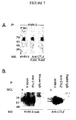
- a second immunoprecipitation of guinea pig inner ear extracts was carried out using anti-CTL2 serum (R229) coupled to CNBr beads, KHRI-3 antibody coupled to CNBr beads, and rabbit and mouse IgG coupled to CNBr beads as a controls.
- the immunoprecipitates were electrophoresed along with whole cochlear lysates (WCL) and then the blots were probed with either KHRI-3 antibody (left panel) or with the R229 anti-CTL2 antiserum (right panel).
- both KHRI-3 and the rabbit anti CTL2 antibodies bind to 68 and 72kDa bands in the whole cell lysate and also in the immunoprecipitates, indicating that both reagents identify the same proteins.
- the anti-CTL2 antibody stains a faint 80kD band in the lanes loaded with whole cell lysate.
- the first primers were designed using Primer3 Output from the Whitehead Institute MIT web site. These primers, SPF and SPR (screen primers forward and screen primers reverse) (Table 6 and Figure 8 ), were used to screen a series of human cell lines for CTL2 expression.
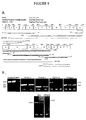
- cDNAs from normal human keratinocytes (NHK), a glioblastoma (Glio54s10), a neuroblastoma, (SJNB20) and a squamous cell carcinoma (UM-SCC-11A), all resulted in a 619 bp PCR product ( Figure 8B left panel) with sequence identity to the predicted CTL2 sequence.
- this primer set did not work with RNA from guinea pig inner ear.
- the knowledge that CTL2 is expressed in some human cells allowed the use of RNA from UM-SCC-11A (shown as 11A in the figure) for testing additional primers based on the peptide sequences obtained from MS/MS sequencing from the guinea pig protein.
- primer set 1 the forward primer corresponds to sequences in peptide 1
- the reverse primer to peptide 4 Tables 5, 6, and Figure 8A
- primer set 2 the forward primer is located in peptide 6
- the reverse primer is in peptide 7.
- Primer set 3 begins in peptide 7 and ends in peptide 9
- primer set 4 begins in peptide 8 and ends in peptide 10.
- Figure 8B Results of representative RT-PCR reactions with human inner ear RNA (Hu) and guinea pig inner ear RNA (GP) are shown in Figure 8B . All four sets of primers amplified products from RNA from human inner ear tissue and from UM-SCC-11A. Sequencing of the PCR products from all four primer sets revealed that the products are identical to the expected CTL2 sequence in both human tissues.
- primer sets 1 and 4 were designed within the regions amplified by primer sets 1 and 4 ( Figure 8A ). These primers amplified cDNA from both human inner ear and UM-SCC-11A, giving the correct human CTL2 sequence for both samples. Primer sets 3 and 4 also produced PCR products from guinea pig inner ear RNA ( Figure 8B ), yielding sequences that are homologous to human CTL2, 89% and 86% respectively.
- This primer set successfully amplified a 1737 bp segment from guinea pig inner ear cDNA (last panel, Figure 8B ).
- the guinea pig PCR product was sequenced from 3' to 5' (86% homology to human CTL2) and 5' to 3' (85% homology to human CTL2).
Landscapes
- Health & Medical Sciences (AREA)
- Chemical & Material Sciences (AREA)
- Life Sciences & Earth Sciences (AREA)
- Immunology (AREA)
- Organic Chemistry (AREA)
- Medicinal Chemistry (AREA)
- General Health & Medical Sciences (AREA)
- Molecular Biology (AREA)
- Hematology (AREA)
- Biochemistry (AREA)
- Proteomics, Peptides & Aminoacids (AREA)
- Biophysics (AREA)
- Engineering & Computer Science (AREA)
- Genetics & Genomics (AREA)
- Urology & Nephrology (AREA)
- Biomedical Technology (AREA)
- Rheumatology (AREA)
- Rehabilitation Therapy (AREA)
- Gastroenterology & Hepatology (AREA)
- Pharmacology & Pharmacy (AREA)
- Veterinary Medicine (AREA)
- Toxicology (AREA)
- Zoology (AREA)
- Public Health (AREA)
- Animal Behavior & Ethology (AREA)
- Epidemiology (AREA)
- Biotechnology (AREA)
- Cell Biology (AREA)
- Microbiology (AREA)
- Diabetes (AREA)
- Food Science & Technology (AREA)
- Physics & Mathematics (AREA)
- Analytical Chemistry (AREA)
- General Physics & Mathematics (AREA)
- Pathology (AREA)
- Peptides Or Proteins (AREA)
- Investigating Or Analysing Biological Materials (AREA)
- Measuring Or Testing Involving Enzymes Or Micro-Organisms (AREA)
- Measurement Of The Respiration, Hearing Ability, Form, And Blood Characteristics Of Living Organisms (AREA)
Claims (4)
- Ein Verfahren zur Prognose der Reaktion eines an autoimmunen sensorineuralen Hörverlust leidenden Patienten auf eine immunsuppresive therapeutische Behandlung mit Steroiden, umfassend:a. die Bereitstellung eines Glykoproteins aus dem inneren Corti-Organ, welches mit einem Antikörper reagiert, der mit sensorineuralen Hörverlust assoziiert ist, wobei das Glykoprotein durch die Nukleinsäuresequenz SEQ ID Nr.: 1 oder die Aminosäuresequenz SEQ ID Nr.: 2 kodiert wird; und Serum von einem Patienten, bei dem der Verdacht auf autoimmunen sensorineuralen Hörverlust besteht;b. in Kontakt bringen des Glykoproteins mit dem Serum in vitro oder ex vivo; undc. die Detektion der Bindung eines Antikörpers aus dem Serum an das Glykoprotein unter Bedingungen, die hinreichend sind um eine Bindung des Antikörpers an das Glykoprotein herbeizuführen, wobei die Reaktion eines an autoimmunen sensorineuralen Hörverlust leidenden Patienten auf eine immunsuppresive therapeutische Behandlung mit Steroiden korreliert ist mit der Anwesenheit eines Antikörpers in dem Patientenserum, der an das Glykoprotein bindet.
- Ein Kit zur Untersuchung der Anwesenheit eines mit autoimmunen sensorineuralen Hörverlust assoziierten Antikörpers bei einem Patienten, wobei der Kit ein immungereinigtes Glykoprotein aus dem inneren Corti-Organ, welches mit einem Antikörper reagiert, der mit sensorineuralen Hörverlust assoziiert ist, wobei das Glykoprotein durch die Nukleinsäuresequenz SEQ ID Nr.: 1 oder die Aminosäuresequenz SEQ ID Nr.: 2 kodiert wird; und einem an das Glykoprotein bindenden Antikörper umfasst.
- Das Verfahren nach Anspruch 1, wobei der Patient mit dem an das Glykoprotein bindenden Antikörper eine höhere Erfolgsaussicht auf besseres Hören nach der Stereoidbehandlung hat, als ein Patient ohne den an das Glykoprotein bindenden Antikörper.
- Der Kit nach Anspruch 2, wobei der Antikörper KHRI-3 ist.
Applications Claiming Priority (3)
| Application Number | Priority Date | Filing Date | Title |
|---|---|---|---|
| US10/139,496 US20030082646A1 (en) | 1998-12-29 | 2002-05-06 | Antigenic targets of autoimmune sensorineural hearing loss (AISNHL) and development of tests for diagnosis and management of AISNHL |
| US139496 | 2002-05-06 | ||
| PCT/US2003/014248 WO2004041152A2 (en) | 2002-05-06 | 2003-05-06 | Antigenic targets of autoimmune sensorineural hearing loss (aisnhl) and development of tests fro diagnosis and management of aisnhl |
Publications (3)
| Publication Number | Publication Date |
|---|---|
| EP1508043A2 EP1508043A2 (de) | 2005-02-23 |
| EP1508043A4 EP1508043A4 (de) | 2006-09-06 |
| EP1508043B1 true EP1508043B1 (de) | 2011-11-02 |
Family
ID=32311697
Family Applications (1)
| Application Number | Title | Priority Date | Filing Date |
|---|---|---|---|
| EP03799763A Expired - Lifetime EP1508043B1 (de) | 2002-05-06 | 2003-05-06 | Antigenziele der autoimmunbedingten schallempfindungsschwerhörigkeit (aisnhl) und entwicklung von tests zur diagnose und behandlung von aisnhl |
Country Status (6)
| Country | Link |
|---|---|
| US (2) | US20030082646A1 (de) |
| EP (1) | EP1508043B1 (de) |
| AT (1) | ATE532065T1 (de) |
| AU (1) | AU2003299473A1 (de) |
| CA (1) | CA2489275C (de) |
| WO (1) | WO2004041152A2 (de) |
Cited By (1)
| Publication number | Priority date | Publication date | Assignee | Title |
|---|---|---|---|---|
| WO2020188573A1 (en) * | 2019-03-21 | 2020-09-24 | Gamida-Cell Ltd. | Expanded nk cell fractions for transplantation in combination therapy |
Families Citing this family (6)
| Publication number | Priority date | Publication date | Assignee | Title |
|---|---|---|---|---|
| US20030082646A1 (en) * | 1998-12-29 | 2003-05-01 | The Regents Of The University Of Michigan | Antigenic targets of autoimmune sensorineural hearing loss (AISNHL) and development of tests for diagnosis and management of AISNHL |
| US7829292B2 (en) * | 2002-05-06 | 2010-11-09 | The Regents Of The University Of Michigan | Compositions and methods for diagnosing, predicting therapeutic response of, and monitoring autoimmune disease |
| EP1533319B1 (de) * | 2002-06-27 | 2011-01-12 | Nippon Medical School Foundation | Verfahren zum nachweis von perilymphatischen fisteln |
| WO2006113723A2 (en) | 2005-04-18 | 2006-10-26 | The Regents Of The University Of Michigan | Compositions and methods for diagnosing, predicting therapeutic response of, and monitoring autoimmune disease |
| WO2008070236A2 (en) * | 2006-09-07 | 2008-06-12 | The Cleveland Clinic Foundation | Diagnostic assay for detecting and monitoring hearing loss |
| DE102008045696A1 (de) | 2008-09-04 | 2010-03-11 | Drk Blutspendedienst West Ggmbh | Granulozyten HNA-3a/b-Antigen |
Family Cites Families (6)
| Publication number | Priority date | Publication date | Assignee | Title |
|---|---|---|---|---|
| US4208479A (en) * | 1977-07-14 | 1980-06-17 | Syva Company | Label modified immunoassays |
| US5422282A (en) * | 1991-04-29 | 1995-06-06 | The Regents Of The University Of California | Methods for detecting autoimmune sensorineural hearing loss |
| US20010006788A1 (en) * | 1998-12-29 | 2001-07-05 | Thomas E. Carey | Identifying the antigenic target of autoimmune sensorineural hearing loss (aisnhl) and developement of specific tests for diagnosis and management of aisnhl |
| US20030082646A1 (en) * | 1998-12-29 | 2003-05-01 | The Regents Of The University Of Michigan | Antigenic targets of autoimmune sensorineural hearing loss (AISNHL) and development of tests for diagnosis and management of AISNHL |
| FR2800750B1 (fr) * | 1999-11-05 | 2003-01-31 | Centre Nat Rech Scient | Proteines membranaires ctl (choline transporter like) impliquees dans le transport de la choline |
| US20030022195A1 (en) * | 2001-02-01 | 2003-01-30 | Millennium Pharmaceuticals, Inc. | 59914 and 59921, choline transporters and uses therefor |
-
2002
- 2002-05-06 US US10/139,496 patent/US20030082646A1/en not_active Abandoned
-
2003
- 2003-05-06 AT AT03799763T patent/ATE532065T1/de active
- 2003-05-06 AU AU2003299473A patent/AU2003299473A1/en not_active Abandoned
- 2003-05-06 CA CA2489275A patent/CA2489275C/en not_active Expired - Fee Related
- 2003-05-06 EP EP03799763A patent/EP1508043B1/de not_active Expired - Lifetime
- 2003-05-06 WO PCT/US2003/014248 patent/WO2004041152A2/en not_active Ceased
-
2008
- 2008-03-25 US US12/055,173 patent/US20090075268A1/en not_active Abandoned
Cited By (1)
| Publication number | Priority date | Publication date | Assignee | Title |
|---|---|---|---|---|
| WO2020188573A1 (en) * | 2019-03-21 | 2020-09-24 | Gamida-Cell Ltd. | Expanded nk cell fractions for transplantation in combination therapy |
Also Published As
| Publication number | Publication date |
|---|---|
| WO2004041152A2 (en) | 2004-05-21 |
| CA2489275A1 (en) | 2004-05-21 |
| WO2004041152A3 (en) | 2004-10-14 |
| EP1508043A2 (de) | 2005-02-23 |
| US20090075268A1 (en) | 2009-03-19 |
| CA2489275C (en) | 2012-12-18 |
| EP1508043A4 (de) | 2006-09-06 |
| US20030082646A1 (en) | 2003-05-01 |
| AU2003299473A1 (en) | 2004-06-07 |
| ATE532065T1 (de) | 2011-11-15 |
Similar Documents
| Publication | Publication Date | Title |
|---|---|---|
| Koster et al. | Role of binding of plectin to the integrin β4 subunit in the assembly of hemidesmosomes | |
| US20090075268A1 (en) | Antigenic Targets Of Autoimmune Sensorineural Hearing Loss (AISNHL) And Development Of Tests For Diagnosis And Management Of AISNHL | |
| CA2427471C (en) | Nod2 nucleic acids and proteins | |
| US20050260623A1 (en) | Oct-4 and GJIC expression as markers for adult human stem cells and cancer cell precursors | |
| US6586577B2 (en) | Telomere repeat binding factors and diagnostic and therapeutic use thereof | |
| US20150045307A1 (en) | Identification of a novel bhd gene | |
| EP1330525B1 (de) | Nod2 nukleinsäuren und proteine | |
| AU2008201559B2 (en) | Antigenic targets of autoimmune sensorineural hearing loss (AISNHL) and development of tests for diagnosis and management of AISNHL | |
| AU2002227350A1 (en) | Nod2 nucleic acids and proteins | |
| AU2006236406B2 (en) | Compositions and methods for diagnosing, predicting therapeutic response of, and monitoring autoimmune disease | |
| US7108975B2 (en) | Atlastin | |
| US7829292B2 (en) | Compositions and methods for diagnosing, predicting therapeutic response of, and monitoring autoimmune disease | |
| US20090098556A1 (en) | Genetic Test For PSE-Susceptible Turkeys | |
| WO2008104575A2 (en) | Immune system mediator | |
| WO2004039979A1 (en) | Means and methods for diagnosing and treating idiopathic generalized epilepsy (ige) | |
| WO2004072305A2 (en) | Atlastin | |
| WO2003061583A9 (en) | Digenic mutations associated with severe insulin resistance and type 2 diabetes and their use in the diagnosis and treatment of diabetes |
Legal Events
| Date | Code | Title | Description |
|---|---|---|---|
| PUAI | Public reference made under article 153(3) epc to a published international application that has entered the european phase |
Free format text: ORIGINAL CODE: 0009012 |
|
| 17P | Request for examination filed |
Effective date: 20041206 |
|
| AK | Designated contracting states |
Kind code of ref document: A2 Designated state(s): AT BE BG CH CY CZ DE DK EE ES FI FR GB GR HU IE IT LI LU MC NL PT RO SE SI SK TR |
|
| AX | Request for extension of the european patent |
Extension state: AL LT LV MK |
|
| A4 | Supplementary search report drawn up and despatched |
Effective date: 20060803 |
|
| 17Q | First examination report despatched |
Effective date: 20071128 |
|
| GRAP | Despatch of communication of intention to grant a patent |
Free format text: ORIGINAL CODE: EPIDOSNIGR1 |
|
| GRAS | Grant fee paid |
Free format text: ORIGINAL CODE: EPIDOSNIGR3 |
|
| GRAA | (expected) grant |
Free format text: ORIGINAL CODE: 0009210 |
|
| AK | Designated contracting states |
Kind code of ref document: B1 Designated state(s): AT BE BG CH CY CZ DE DK EE ES FI FR GB GR HU IE IT LI LU MC NL PT RO SE SI SK TR |
|
| AX | Request for extension of the european patent |
Extension state: AL LT LV MK |
|
| REG | Reference to a national code |
Ref country code: GB Ref legal event code: FG4D |
|
| REG | Reference to a national code |
Ref country code: CH Ref legal event code: EP |
|
| REG | Reference to a national code |
Ref country code: IE Ref legal event code: FG4D |
|
| REG | Reference to a national code |
Ref country code: DE Ref legal event code: R096 Ref document number: 60339013 Country of ref document: DE Effective date: 20120112 |
|
| REG | Reference to a national code |
Ref country code: NL Ref legal event code: VDEP Effective date: 20111102 |
|
| LTLA | Lt: lapse of european patent or patent extension | ||
| PG25 | Lapsed in a contracting state [announced via postgrant information from national office to epo] |
Ref country code: SE Free format text: LAPSE BECAUSE OF FAILURE TO SUBMIT A TRANSLATION OF THE DESCRIPTION OR TO PAY THE FEE WITHIN THE PRESCRIBED TIME-LIMIT Effective date: 20111102 Ref country code: SI Free format text: LAPSE BECAUSE OF FAILURE TO SUBMIT A TRANSLATION OF THE DESCRIPTION OR TO PAY THE FEE WITHIN THE PRESCRIBED TIME-LIMIT Effective date: 20111102 Ref country code: PT Free format text: LAPSE BECAUSE OF FAILURE TO SUBMIT A TRANSLATION OF THE DESCRIPTION OR TO PAY THE FEE WITHIN THE PRESCRIBED TIME-LIMIT Effective date: 20120302 Ref country code: BE Free format text: LAPSE BECAUSE OF FAILURE TO SUBMIT A TRANSLATION OF THE DESCRIPTION OR TO PAY THE FEE WITHIN THE PRESCRIBED TIME-LIMIT Effective date: 20111102 Ref country code: GR Free format text: LAPSE BECAUSE OF FAILURE TO SUBMIT A TRANSLATION OF THE DESCRIPTION OR TO PAY THE FEE WITHIN THE PRESCRIBED TIME-LIMIT Effective date: 20120203 Ref country code: NL Free format text: LAPSE BECAUSE OF FAILURE TO SUBMIT A TRANSLATION OF THE DESCRIPTION OR TO PAY THE FEE WITHIN THE PRESCRIBED TIME-LIMIT Effective date: 20111102 |
|
| PG25 | Lapsed in a contracting state [announced via postgrant information from national office to epo] |
Ref country code: CY Free format text: LAPSE BECAUSE OF FAILURE TO SUBMIT A TRANSLATION OF THE DESCRIPTION OR TO PAY THE FEE WITHIN THE PRESCRIBED TIME-LIMIT Effective date: 20111102 |
|
| PG25 | Lapsed in a contracting state [announced via postgrant information from national office to epo] |
Ref country code: SK Free format text: LAPSE BECAUSE OF FAILURE TO SUBMIT A TRANSLATION OF THE DESCRIPTION OR TO PAY THE FEE WITHIN THE PRESCRIBED TIME-LIMIT Effective date: 20111102 Ref country code: CZ Free format text: LAPSE BECAUSE OF FAILURE TO SUBMIT A TRANSLATION OF THE DESCRIPTION OR TO PAY THE FEE WITHIN THE PRESCRIBED TIME-LIMIT Effective date: 20111102 Ref country code: DK Free format text: LAPSE BECAUSE OF FAILURE TO SUBMIT A TRANSLATION OF THE DESCRIPTION OR TO PAY THE FEE WITHIN THE PRESCRIBED TIME-LIMIT Effective date: 20111102 Ref country code: EE Free format text: LAPSE BECAUSE OF FAILURE TO SUBMIT A TRANSLATION OF THE DESCRIPTION OR TO PAY THE FEE WITHIN THE PRESCRIBED TIME-LIMIT Effective date: 20111102 Ref country code: BG Free format text: LAPSE BECAUSE OF FAILURE TO SUBMIT A TRANSLATION OF THE DESCRIPTION OR TO PAY THE FEE WITHIN THE PRESCRIBED TIME-LIMIT Effective date: 20120202 |
|
| PGFP | Annual fee paid to national office [announced via postgrant information from national office to epo] |
Ref country code: DE Payment date: 20120529 Year of fee payment: 10 |
|
| PG25 | Lapsed in a contracting state [announced via postgrant information from national office to epo] |
Ref country code: IT Free format text: LAPSE BECAUSE OF FAILURE TO SUBMIT A TRANSLATION OF THE DESCRIPTION OR TO PAY THE FEE WITHIN THE PRESCRIBED TIME-LIMIT Effective date: 20111102 Ref country code: RO Free format text: LAPSE BECAUSE OF FAILURE TO SUBMIT A TRANSLATION OF THE DESCRIPTION OR TO PAY THE FEE WITHIN THE PRESCRIBED TIME-LIMIT Effective date: 20111102 |
|
| PGFP | Annual fee paid to national office [announced via postgrant information from national office to epo] |
Ref country code: FR Payment date: 20120607 Year of fee payment: 10 Ref country code: GB Payment date: 20120525 Year of fee payment: 10 |
|
| PLBE | No opposition filed within time limit |
Free format text: ORIGINAL CODE: 0009261 |
|
| STAA | Information on the status of an ep patent application or granted ep patent |
Free format text: STATUS: NO OPPOSITION FILED WITHIN TIME LIMIT |
|
| REG | Reference to a national code |
Ref country code: AT Ref legal event code: MK05 Ref document number: 532065 Country of ref document: AT Kind code of ref document: T Effective date: 20111102 |
|
| 26N | No opposition filed |
Effective date: 20120803 |
|
| REG | Reference to a national code |
Ref country code: DE Ref legal event code: R097 Ref document number: 60339013 Country of ref document: DE Effective date: 20120803 |
|
| PG25 | Lapsed in a contracting state [announced via postgrant information from national office to epo] |
Ref country code: MC Free format text: LAPSE BECAUSE OF NON-PAYMENT OF DUE FEES Effective date: 20120531 |
|
| REG | Reference to a national code |
Ref country code: CH Ref legal event code: PL |
|
| PG25 | Lapsed in a contracting state [announced via postgrant information from national office to epo] |
Ref country code: LI Free format text: LAPSE BECAUSE OF NON-PAYMENT OF DUE FEES Effective date: 20120531 Ref country code: CH Free format text: LAPSE BECAUSE OF NON-PAYMENT OF DUE FEES Effective date: 20120531 Ref country code: AT Free format text: LAPSE BECAUSE OF FAILURE TO SUBMIT A TRANSLATION OF THE DESCRIPTION OR TO PAY THE FEE WITHIN THE PRESCRIBED TIME-LIMIT Effective date: 20111102 |
|
| REG | Reference to a national code |
Ref country code: IE Ref legal event code: MM4A |
|
| PG25 | Lapsed in a contracting state [announced via postgrant information from national office to epo] |
Ref country code: ES Free format text: LAPSE BECAUSE OF FAILURE TO SUBMIT A TRANSLATION OF THE DESCRIPTION OR TO PAY THE FEE WITHIN THE PRESCRIBED TIME-LIMIT Effective date: 20120213 Ref country code: IE Free format text: LAPSE BECAUSE OF NON-PAYMENT OF DUE FEES Effective date: 20120506 |
|
| PG25 | Lapsed in a contracting state [announced via postgrant information from national office to epo] |
Ref country code: FI Free format text: LAPSE BECAUSE OF FAILURE TO SUBMIT A TRANSLATION OF THE DESCRIPTION OR TO PAY THE FEE WITHIN THE PRESCRIBED TIME-LIMIT Effective date: 20111102 |
|
| GBPC | Gb: european patent ceased through non-payment of renewal fee |
Effective date: 20130506 |
|
| PG25 | Lapsed in a contracting state [announced via postgrant information from national office to epo] |
Ref country code: DE Free format text: LAPSE BECAUSE OF NON-PAYMENT OF DUE FEES Effective date: 20131203 |
|
| REG | Reference to a national code |
Ref country code: DE Ref legal event code: R119 Ref document number: 60339013 Country of ref document: DE Effective date: 20131203 |
|
| REG | Reference to a national code |
Ref country code: FR Ref legal event code: ST Effective date: 20140131 |
|
| PG25 | Lapsed in a contracting state [announced via postgrant information from national office to epo] |
Ref country code: GB Free format text: LAPSE BECAUSE OF NON-PAYMENT OF DUE FEES Effective date: 20130506 Ref country code: TR Free format text: LAPSE BECAUSE OF FAILURE TO SUBMIT A TRANSLATION OF THE DESCRIPTION OR TO PAY THE FEE WITHIN THE PRESCRIBED TIME-LIMIT Effective date: 20111102 |
|
| PG25 | Lapsed in a contracting state [announced via postgrant information from national office to epo] |
Ref country code: LU Free format text: LAPSE BECAUSE OF NON-PAYMENT OF DUE FEES Effective date: 20120506 Ref country code: FR Free format text: LAPSE BECAUSE OF NON-PAYMENT OF DUE FEES Effective date: 20130531 |
|
| PG25 | Lapsed in a contracting state [announced via postgrant information from national office to epo] |
Ref country code: HU Free format text: LAPSE BECAUSE OF FAILURE TO SUBMIT A TRANSLATION OF THE DESCRIPTION OR TO PAY THE FEE WITHIN THE PRESCRIBED TIME-LIMIT Effective date: 20030506 |