EP1496360A1 - Method of judging leukemia, pre-leukemia or aleukemic malignant blood disease and diagnostic therefor - Google Patents
Method of judging leukemia, pre-leukemia or aleukemic malignant blood disease and diagnostic therefor Download PDFInfo
- Publication number
- EP1496360A1 EP1496360A1 EP03745988A EP03745988A EP1496360A1 EP 1496360 A1 EP1496360 A1 EP 1496360A1 EP 03745988 A EP03745988 A EP 03745988A EP 03745988 A EP03745988 A EP 03745988A EP 1496360 A1 EP1496360 A1 EP 1496360A1
- Authority
- EP
- European Patent Office
- Prior art keywords
- scgf
- leukemia
- antibody
- amino acid
- acid sequence
- Prior art date
- Legal status (The legal status is an assumption and is not a legal conclusion. Google has not performed a legal analysis and makes no representation as to the accuracy of the status listed.)
- Withdrawn
Links
Images




Classifications
-
- G—PHYSICS
- G01—MEASURING; TESTING
- G01N—INVESTIGATING OR ANALYSING MATERIALS BY DETERMINING THEIR CHEMICAL OR PHYSICAL PROPERTIES
- G01N33/00—Investigating or analysing materials by specific methods not covered by groups G01N1/00 - G01N31/00
- G01N33/48—Biological material, e.g. blood, urine; Haemocytometers
- G01N33/50—Chemical analysis of biological material, e.g. blood, urine; Testing involving biospecific ligand binding methods; Immunological testing
- G01N33/53—Immunoassay; Biospecific binding assay; Materials therefor
-
- C—CHEMISTRY; METALLURGY
- C07—ORGANIC CHEMISTRY
- C07K—PEPTIDES
- C07K16/00—Immunoglobulins [IG], e.g. monoclonal or polyclonal antibodies
- C07K16/18—Immunoglobulins [IG], e.g. monoclonal or polyclonal antibodies against material from animals or humans
- C07K16/22—Immunoglobulins [IG], e.g. monoclonal or polyclonal antibodies against material from animals or humans against growth factors ; against growth regulators
-
- C—CHEMISTRY; METALLURGY
- C12—BIOCHEMISTRY; BEER; SPIRITS; WINE; VINEGAR; MICROBIOLOGY; ENZYMOLOGY; MUTATION OR GENETIC ENGINEERING
- C12N—MICROORGANISMS OR ENZYMES; COMPOSITIONS THEREOF; PROPAGATING, PRESERVING, OR MAINTAINING MICROORGANISMS; MUTATION OR GENETIC ENGINEERING; CULTURE MEDIA
- C12N5/00—Undifferentiated human, animal or plant cells, e.g. cell lines; Tissues; Cultivation or maintenance thereof; Culture media therefor
- C12N5/10—Cells modified by introduction of foreign genetic material
- C12N5/12—Fused cells, e.g. hybridomas
- C12N5/16—Animal cells
-
- G—PHYSICS
- G01—MEASURING; TESTING
- G01N—INVESTIGATING OR ANALYSING MATERIALS BY DETERMINING THEIR CHEMICAL OR PHYSICAL PROPERTIES
- G01N33/00—Investigating or analysing materials by specific methods not covered by groups G01N1/00 - G01N31/00
- G01N33/48—Biological material, e.g. blood, urine; Haemocytometers
- G01N33/50—Chemical analysis of biological material, e.g. blood, urine; Testing involving biospecific ligand binding methods; Immunological testing
- G01N33/53—Immunoassay; Biospecific binding assay; Materials therefor
- G01N33/575—Immunoassay; Biospecific binding assay; Materials therefor for cancer
- G01N33/57505—Immunoassay; Biospecific binding assay; Materials therefor for cancer of the blood, e.g. leukaemia
-
- G—PHYSICS
- G01—MEASURING; TESTING
- G01N—INVESTIGATING OR ANALYSING MATERIALS BY DETERMINING THEIR CHEMICAL OR PHYSICAL PROPERTIES
- G01N2333/00—Assays involving biological materials from specific organisms or of a specific nature
- G01N2333/435—Assays involving biological materials from specific organisms or of a specific nature from animals; from humans
- G01N2333/475—Assays involving growth factors
Definitions
- the present invention relates to a method of diagnosing leukemia, pre-leukemia or aleukemic malignant blood diseases, a method of discriminating leukemia from pre-leukemia or aleukemic malignant blood diseases, a method of discriminating aplastic anemia from myelodysplastic syndrome, a method of diagnosing the engraftment state of the hematopoietic stem cells after transplantation of the hematopoietic stem cells, or a method of diagnosing graft versus host disease (GVHD), wherein stem cell growth factor (SCGF) is quantified.
- GVHD graft versus host disease
- the present invention also relates to an agent and a kit for diagnosing leukemia, pre-leukemia or aleukemic malignant blood diseases, for diagnosing engraftment state of the hematopoietic stem cells after transplantation of the hematopoietic stem cells, or for diagnosing graft versus host disease (GVHD), each comprising as an active ingredient an antibody reacting with stem cell growth factor (SCGF).
- SCGF stem cell growth factor
- pre-leukemia or aleukemic malignant blood diseases are important for determining the strategy for treating these diseases.
- the white blood cell count in the peripheral blood of a patient is determined and when the white blood cell count is beyond the normal level, the occurrence of leukemia is suspected.
- the white blood cell counts also increase due to the enhancement of immune-response within the body in the case of diseases other than leukemia such as cold, hence determination of the white blood cell counts alone may allow false-positive results.
- normal white blood cell counts in the peripheral blood range as broad as 4,000-8,000 cell/ ⁇ L, so that false-negative cases are possibly raised. Therefore, a method for diagnosing leukemia with higher accuracy is in need.
- Drawbacks associated with the hematopoietic stem cell transplantation therapy include the HLA-type incompatibility between the hematopoietic stem cells of a donor and a patient, insufficient effect of the therapy owing to the physical condition of a patient and to infection, etc., such as non-engraftment of the transplanted hematopoietic stem cells, delayed engraftment of the hematopoietic stem cells, occurrence of graft versus host disease (hereinafter referred to as GVHD) . When things turn to the worst, it may proceed to death.
- GVHD graft versus host disease
- Delayed engraftment of the hematopoietic stem cells can be coped with in vivo administration of G-CSF to promote the engraftment of the hematopoietic stem cells.
- in-vivo administration of immunosuppressants also suppresses rejection of the transplanted hematopoietic stem cells.
- side effects for either kind of agents, if administered in an excess amount. Therefore, it is crucial for determining the treatment strategy to diagnose or predict the engraftment of the hematopoietic stem cells or the occurrence of GVHD.
- a method of confirming the engraftment of hematopoietic stem cells after transplantation of the hematopoietic stem cells there is a method in which white blood cell counts or platelet counts in the peripheral blood are determined.
- Hematopoietic stem cells can be diagnosed as being engrafted if these levels are increased. However, it sometimes requires from 10 days to a month or longer after transplantation for the hematopoietic stem cells to engraft, so that the engraftment of hematopoietic stem cells cannot be diagnosed at an early stage by only determining white blood cell counts or platelet counts in the peripheral blood.
- a method for diagnosing the occurrence of GVHD includes observing skin rush or the like emerging at a recovery phase after transplantation of the hematopoietic stem cells.
- easy and accurate method for diagnosing the occurrence of GVHD remains unknown.
- any method to predict the occurrence of GVHD prior to its occurrence is unknown either.
- SCGF Human stem cell growth factor
- antibodies which recognize SCGF known are a polyclonal antibody which is prepared by using SCGF obtained by genetic recombination and a partial peptide of SCGF which consists of 6-25 amino acid residues from the N-terminal, as an immunogen and monoclonal antibodies which are prepared by using SCGF purified partially from the cell culture supernatant and SCGF obtained by genetic recombination, as an immunogen [WO98/08869].
- this monoclonal antibody has a neutralizing activity
- a polyclonal antibody which is prepared by using SCGF obtained by genetic recombination as an immunogen, reacts with SCGF obtained by genetic recombination when subjected to ELISA, and that SCGF obtained by genetic recombination can be detected by western blotting using a polyclonal antibody prepared by using a partial peptide of SCGF consisting of 6-25 amino acid residues from N-terminal as an immunogen.
- anti-SCGF monoclonal KM2142 antibody which is prepared by using a partial peptide of SCGF consisting of 6-25 amino acid residues from the N-terminal as an immunogen [The Hematology Journal, 2, 307 (2001)].
- SCGF gene as revealed by northern blotting for human normal tissues are high in the kidney, low in the heart, and nil in the brain, placenta, lung, liver, skeletal muscles and pancreas [Proc. Natl. Acad. Sci. USA, 94, 7577 (1997)], high in the spleen, thymus, cecum, bone marrows, fetal liver and low in peripheral blood [Biochem. Biophys. Res. Comm., 249, 124 (1998)].
- SCGF is expressed in the bone marrows, proliferating cartilage, and in the proximal of periosteum [The Hematology Journal, 2 , 307 (2001)].
- SCGF protein in the body fluid such as serum and plasma and in the tissues from animals including humans are left unrevealed.
- An object of the present invention is to provide a method for diagnosing leukemia, pre-leukemia or aleukemic malignant blood diseases, a method for discriminating leukemia from pre-leukemia or aleukemic malignant blood diseases, a method for discriminating aplastic anemia from myelodysplastic syndrome, and a method for diagnosing the engraftment of hematopoietic stem cells and GVHD after transplantation of the hematopoietic stem cells, and to provide an agent and a kit for diagnosing leukemia, pre-leukemia or aleukemic malignant blood diseases, and an agent and a kit for diagnosing the engraftment of hematopoietic stem cells and GVHD after transplantation of the hematopoietic stem cells.
- the present invention relates to the following (1) to (20).
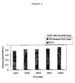
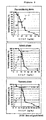
- Fig. 1 shows the reactivity of monoclonal antibodies to a partial peptide of human SCGF (Compound 1) (Binding ELISA).
- Fig. 2 shows the results of SDS-PAGE and western blotting for the purified human SCGF protein.
- Lanes 1 and 2 shows SDS-PAGE patterns of a molecular weight marker and the purified human SCGF protein.
- Lanes 3, 4 and 5 indicate the results of western blotting for the purified human SCGF protein using KM2142, KM2804 and KM2945, respectively.
- Fig. 3 shows reactivity of monoclonal antibodies to the CHO cell-expressing human SCGF protein (Binding ELISA)
- Fig. 4 shows reactivity of monoclonal antibodies to the SDS-degenerated human SCGF protein (expressing CHO cells) (Binding ELISA).
- Fig. 5 shows reactivity of monoclonal antibodies to human and mouse SCGF proteins (expressing CHO cells) (Binding ELISA).
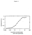
- Fig. 6 shows a quantification curve for the human SCGF protein examined by sandwich ELISA using a monoclonal antibody.
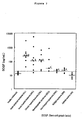
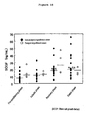
- Fig. 7 shows SCGF concentrations in the sera of patients suffering various blood diseases.
- the horizontal full lines show the medians of various blood disease groups and the horizontal dotted line shows the cut-off value calculated from the healthy individual group (18.2 ng/mL).
- * significant difference with either normal (healthy individual group) or AA (aplastic anemia group)
- # significant difference with NHL (non-Hodgkin' s lymphoma)
- p ⁇ 0.5 $ significant difference with MM (multiple myeloma) p ⁇ 0.05
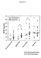
- Fig. 8 shows the difference between the cases with and without the occurrence of GVHD depending on the serum SCGF concentration in the patients who underwent transplantation of hematopoietic stem cells.
- the horizontal full lines show the median of each group. * : significant difference with the cases without the occurrence of GVHD p ⁇ 0.05 # : significant difference with pre-condition phase p ⁇ 0.05 $ : significant difference with aplastic phase p ⁇ 0.05 & : significant difference with recovery phase p ⁇ 0.05
- Fig. 9 shows relationships between detection sensitivity of the GVHD-occurred patients/specificity of the non-occurred patients and the serum SCGF concentration of the patients who underwent transplantation of the hematopoietic stem cells.
- ⁇ represents sensitivity
- ⁇ represents specificity
- vertical dotted lines represent temporary cut-off values.
- Fig. 10 shows differences between the cases for delayed and non-delayed engraftment depending on the serum SCGF concentration of the patients who underwent transplantation of the hematopoietic stem cells.
- the horizontal full line shows the median of each group. # : significant difference with pre-condition phase p ⁇ 0.05, $ : significant difference with aplastic phase p ⁇ 0.05 & : significant difference with recovery phase p ⁇ 0.05
- Fig. 11 shows relationships between the serum SCGF concentration of the patients who underwent transplantation of the hematopoietic stem cells and the detection sensitivity of the delayed engraftment/specificity of the non-delayed engraftment of hematopoietic stem cells.
- ⁇ represents sensitivity
- ⁇ represents specificity
- vertical dotted lines represent temporary cut-off values.
- the present invention relates to a method for diagnosing leukemia, pre-leukemia or aleukemic malignant blood diseases.
- leukemia any type of leukemia is encompassed in the present invention as long as immature cells such as hematopoietic cells among the cells of hematopoietic system have turned into tumors, and the examples include acute lymphocytic leukemia (hereinafter referred to as ALL), acute myeloid leukemia ( hereinafter referred to as AML), chronic myeloid leukemia (hereinafter referred to as CML).
- ALL acute lymphocytic leukemia
- AML acute myeloid leukemia
- CML chronic myeloid leukemia
- MDS myelodysplastic syndrome
- aleukemic malignant blood diseases are lymphoma, myeloma and the like.
- lymphoma examples include Hodgkin's lymphoma, non-Hodgkin's lymphoma (hereinafter referred to as NHL) and the like.
- myeloma examples include multiple myeloma (hereinafter referred to as MM) and the like.
- SCGF concentrations of in-vivo samples of patients with leukemia, pre-leukemia and aleukemic malignant blood diseases are significantly increased compared to those of healthy individuals. Cut-off value is therefore applied to SCGF concentration in quantifying SCGF contained in the in-vivo samples collected, and it can be diagonsed as being leukemia, pre-leukemia or aleukemic malignant blood diseases when a SCGF concentration exceeds the cut-off value.
- a cut-off value means a value set to diagnose a disease group of the interest and the non-disease group by laying focus on a substance. In diagnosing disease of the interest and the non-disease cases, diagnosis can be made for the disease of the interest as negative when the value is equal to or below the cut-off value and as positive when equal to or above the cut-off value, or likewise it can be diagnosed as positive when the value is equal to or below the cut-off value and as negative when equal to or above the cut-off value (Outline for Laboratory Tests (Rinsho kensahou teiyo), edited by Masamitsu Kanai, Kanehara & Co., Ltd.).
- Sensitivity and specificity are noted as indexes used for the purpose of evaluating clinical availability of cut-off values.
- a group is diagnosedwith a cut-off value. Those diagnosed as positive among the disease patients are presented as "a” (true positive), those diagnosed as negative in spite of being disease patients are presented as "b” (false negative), those diagnosed as positive in spite of not being disease patients are presented as "c” ( false positive) , and those who are not the disease patients and diagnosed as negative are presented as "d” (true negative). According to the above diagnoses, sensitivity (a true positive rate) can be represented by a level calculated as a/(a + b) and specificity (a true negative rate) can be represented by a value calculated as d/(c + d).
- sensitivity and specificity will vary by shifting a cut-off value up and down. Shifting a cut-off value downwards leads to higher sensitivity while specificity lowers. Shifting a cut-off value upwards leads to lower sensitivity while specificity mounts.
- a diagnosis method in which values of both sensitivity and specificity are high. In addition, diagnosis methods in which sensitivity and specificity values do not exceed 50% are not considered as being available.
- Examples of the methods for setting a cut-off value are a method in which a level at either end from the median where 95% of the non-disease group distribution is inclusive, is set as a cut-off value, and a method in which "average + doubled standard deviation (SD)" or “average - 2SD” is set as a cut-off value when the non-disease group distribution exhibits a regular distribution, and so on.
- SD standard deviation
- diagnosis can be given at a sensitivity of 89.5% and a specificity of 70% when a cut-off value is set at 15.0 ng/mL, and at a sensitivity of 100% and a specificity of 60% when a cut-off value is set at 13.0 ng/mL.
- a cut-off value is set at 18.2 ng/mL which is "mean + 2SD" from the SCGF concentration of healthy individuals, diagnosis can be given at a sensitivity of 89.5% and a specificity of 100%.
- this cut-off value whether being leukemia or not can be diagnosed at a sensitivity of 95% and a specificity of 100%, whether being an aleukemic malignant blood disease or not at a sensitivity of 76.9% and a specificity of 100%, and whether being pre-leukemia or not at a sensitivity of 100% and a specificity of 100%.
- blood examples include, whole blood, plasma, serum, hemocytic laked blood, the blood cell's inner fluid and the like, among which serum or plasma are preferred.
- the present invention relates to a method for discriminating leukemia from pre-leukemia or aleukemic malignant blood diseases.
- SCGF concentrations of in-vivo samples of leukemia patients have been significantly increased compared to those of patients with pre-leukemia or an aleukemic malignant blood disease. Therefore, after diagnosing a sample as being leukemia, pre-leukemia or an aleukemic malignant blood disease according to a method described above, a cut-off value is further set for leukemia to be diagnosed, then it can be diagnosed as being leukemia when the SCGF concentration of the in-vivo sample collected is higher than the cut-off value and as being pre-leukemia or an aleukemic malignant blood disease when such concentration is lower than the cut-off value.
- diagnosis can be given at a sensitivity of 85% and a specificity of 69.2%. Further, with a cut-off value set at 32.8 ng/mL from "average of the aleukemic malignant blood disease patients + 2SD", diagnosis can be given at a sensitivity of 80% and a specificity of 100%.
- the present invention relates to a method for discriminating aplastic anemia from myelodysplastic syndrome.
- Aplastic anemia and myelodysplastic syndrome have pathologies characterized in that abnormalities are raised in the counts and morphology of white blood cells in the bone marrows and peripheral blood: Discrimination of the two diseases have been considered to be difficult.
- SCGF concentration of a myelodysplastic syndrome patient has been significantly increased compared to that in the blood of a healthy individual, while the blood SCGF concentration of an aplastic anemia patient is comparable to that of a healthy individual.
- the blood SCGF concentration of a myelodysplastic syndrome patient is significantly higher than that of an aplastic anemia patient, so that measuring the blood SCGF concentrations of patients of the two diseases enables to discriminate between aplastic anemia and myelodysplastic syndrome.
- the present invention further relates to a method for diagnosing delayed engraftment of the hematopoietic stem cells after transplantation of the hematopoietic stem cells.
- transplantation of the hematopoietic stem cells can be applied as transplantation of the hematopoietic stem cells, and the examples include transplantation of bone marrows, cord blood, peripheral blood stem cells or the like.
- the period from transplantation of the hematopoietic stem cells to the engraftment of hematopoietic stem cells are divided into four phases as follows based on the blood cell counts in the peripheral blood of patients. That is, pre-conditioning phase when anticancer agents are administered at a high dose prior to transplantation, aplastic phase when the blood cell counts have been decreased following transplantation, recovery phase when the blood cell counts have been recovered after transplantation, and stable phase when the hematopoietic stem cells have engrafted after transplantation.
- the concentrations of in-vivo samples of patients with delayed engraftment of the hematopoietic stem cells are higher than those of patients without delayed engraftment of the hematopoietic stem cells.
- SCGF concentration in each phase is measured, then the SCGF concentration which is considered as possibly resulting in delayed engraftment of the hematopoietic stem cells is specified as a cut-off value, and when a SCGF concentration is lower than the cut-off value, it can be diagnosed as free of delayed engraftment and when a SCGF concentration is higher than the cut-off value it can be diagnosed that delayed engraftment should occur.
- diagnosis can be given at a sensitivity of 75% and a specificity of 67% by affording a cut-off value of , for instance, 9.5 ng/mL for pre-conditioning phase, and at a sensitivity of 75% and a specificity of 63% by affording a cut-off value of 12 ng/mL for aplastic phase.
- the present invention further relates to a method for diagnosing the occurrence of GVHD.
- SCGF concentrations of in-vivo samples at aplastic and recovery phases of patients who underwent transplantation of the hematopoietic stem cells are higher in patients occurring GVHD than those in patients not occurring GVHD. Accordingly, SCGF concentration in each phase is measured, the SCGF concentration which is considered as possibly occurring GVHD is specified as a cut-off value in each phase, and it can be diagnosed GVHD is not occurred when a SCGF concentration is lower than the cut-off value and that GVHD is possibly occurring when a SCGF concentration is higher than the cut-off value.
- GVHD-occurring and non-occurring patients can be diagnosed at a sensitivity of 87% and a specificity of 57% by affording a cut-off value of, for instance, 5 ng/mL for pre-conditioning phase, and at a sensitivity of 87% and a specificity of 63% by affording a cut-off value of, for instance, 10 ng/mL for aplastic phase.
- SCGF stem cell growth factor
- immunological assays Any method is encompassed by such immunological assays as long as it is a method using antigen-antibody reaction such as immunoassays, immunoblotting methods, agglutination test, complement fixation test, hemolysis test, precipitation test, colloidal gold method, chromatography methods or immunostaining methods, and immunoassays are preferred.
- antigen-antibody reaction such as immunoassays, immunoblotting methods, agglutination test, complement fixation test, hemolysis test, precipitation test, colloidal gold method, chromatography methods or immunostaining methods, and immunoassays are preferred.
- molecular-biological assay examples include RT-PCR method, northern blotting method, in situ hybridization method and the like.
- An immunoassay is a method to detect or to quantify antibodies or antigens by using antigens or antibodies that are labeled in various ways, and the examples are given on the basis of labeling means for antigens or antibodies, which include radioimmuno assay (RIA) , enzyme-linked immunosorbent assay (EIA or ELISA), fluorescent immunoassay (FIA), luminescent immunoassay, physicochemical assays (TIA, LAPIA, PCIA), flow cytometry, among which enzyme-linked immunosorbent assay is preferred.
- RIA radioimmuno assay
- EIA or ELISA enzyme-linked immunosorbent assay
- FIA fluorescent immunoassay
- TIA physicochemical assays
- LAPIA LAPIA
- PCIA flow cytometry
- radioisotope Any known (Enzyme-linked Immunosorbent Assay, edited by Eiji Ishikawa et al. , Igaku-Shoin Ltd.) radioisotope may be used as a radioactive label in radioimmunoassay. For instance, 32 P, 125 I, 131 I and the like may be used.
- Enzyme-linked Immunosorbent Assay edited by Eiji Ishikawa et al., Igaku-Shoin Ltd.
- enzyme may be used as an enzyme label in an enzyme-linked immunosorbent assay.
- alkaline phosphatase, peroxidase, luciferase and the like may be used.
- measurement/detection is carried out by measuring substances produced through enzymatic action, and various measuring methods can be employed including a method for measuring absorbance of a substance having absorption maximum at the ultraviolet range or visible range, a method for measuring fluorescence intensity of the fluorescent material produced, a method for measuring luminescence intensity of the substance produced.
- alkaline phosphatase is used as an enzyme label, e.g. 4-nitrophenyl phosphate and the like are given as a substrate for alkaline phosphatase which produces, through the action of alkaline phosphatase, a substance having absorption maximum at the ultraviolet range or visible range.
- 4-nitrophenyl phosphate is converted to 4-nitrophenol by alkaline phosphatase.
- alkaline phosphatase As a substrate for alkaline phosphatase which give rise to luminescence through the action of alkaline phosphatase, the followings are exemplified: 3-(2'-spiroadamantane)-4-methoxy-4-(3'-phosphoryloxine)phen yl-1,2-dioxetane disodium salt(AMPPD), 2-chloro-5- ⁇ 4-methoxyspiro[1,2-dioxetane-3,2'-(5'chloro)tri cyclo[3. 3.
- luminescent label used in the luminescent immunodetection methods, any known [Biological luminescence and Chemiluminescence, edited by Kazuhiro Imai, Hirokawa Shoten; Clinical Tests 42 (1998) ] luminescent material may be used.
- acridinium ester, rofin and the like may be used.
- any known [Fluorescence-Antibody method, Akira Kawaoi, Soft Science, Inc.] fluorescence may be used.
- FITC, RITC and the like may be used.
- Procedure of sandwich assay is described as follows.
- a second antibody (secondary antibody) is simultaneously or independently reacted with the object substance in a sample together with the primary antibody that were bound through the antigen-antibody reaction, then the object substance in the sample is detected or quantified with the use of the same or different antibodies.
- this method comprises in the course of measuring operation a step to wash away unreacted sample components or components of the measuring system in the sample. For example, after the first antibody (primary antibody) is fixed to the solid phase, a sample to be measured is brought into contact with the first antibody.
- solid phases used in the sandwich assay include, a polyvinylchloride microtiter plate, a polystyrene microtiter plate and the like.
- Either of polyclonal antibodies and monoclonal antibodies may be used as antibodies for sandwich assay, and antibody fragments such as Fab, Fab', F(ab) 2 may be used as well.
- any combination of antibodies may be accepted as long as the antibodies for combination recognize different epitopes, yet it is preferred that at least one of the antibodies is a monoclonal antibody.
- Examples of monoclonal antibodies for use in the sandwich assay of the present invention include a monoclonal antibody which recognizes the region shown by the amino acid sequence of 6-28 amino acids in SEQ. ID No. 1, a monoclonal antibody which recognizes the region shown by the amino acid sequence of 29-59 amino acids in SEQ. ID No. 1, and a monoclonal antibody which recognizes the region shown by the amino acid sequence of 60-302 amino acids in SEQ. ID No. 1.
- monoclonal antibody recognizing the region shown by the amino acid sequence of 6-28 amino acids in SEQ. ID No. 1 monoclonal KM2142 antibody produced by KM2142 hybridoma [The Hematology Journal, 2 , 307 (2001) ] is mentioned.
- monoclonal antibody recognizing the region shown by the amino acid sequence of 29-59 amino acids in SEQ. ID No. 1 monoclonal KM2804 antibody produced by KM2804 hybridoma is mentioned.
- monoclonal antibody recognizing the region shown by the amino acid sequence of 60-302 amino acids in SEQ. ID No. 1 monoclonal KM2945 antibody produced by KM2945 hybridoma is mentioned.
- KM2142 hybridoma producing monoclonal KM2142 antibody KM2804 hybridoma producing monoclonal KM2804 antibody and KM2945 hybridoma producing monoclonal KM2945 antibody were deposited at International Patent Organism Depositary, National Institute of Advanced Industrial Science and Technology (Central 6, 1-1 Higashi, Tsukuba, Ibaraki) on February 26, 2002 as FERM BP-7922, FERM BP-7923 and FERM BP-7924, respectively.
- the monoclonal antibodies mentioned above recognize SCGF at different sites so that these can be used in combination for sandwich assay.
- a preferable combination of antibodies is the combination of amonoclonal antibody recognizing the region shown by the amino acid sequence of 6-28 amino acids in SEQ. ID No. 1, i.e. monoclonal KM2142 antibody produced by KM2142 hybridoma [The Hematology Journal, 2 , 307 (2001)] and a monoclonal antibody recognizing the region shown by the amino acid sequence of 29-59 amino acids in SEQ. ID No. 1, i.e. monoclonal KM2804 antibody produced by KM2804 hybridoma (FERM BP-7923).
- an anti-SCGF antibody (primary antibody) mentioned above is absorbed and immobilized on the surface of an appropriate carrier for immobilization.
- a primary antibody is diluted with an appropriate buffer, e.g. phosphate buffer, borate buffer, carbonate buffer and the like, then brought into contact with the solid carrier surface and allowed to react for 30 min or longer at 4-37° C, and thus the primary antibody can be immobilized.
- an appropriate buffer e.g. phosphate buffer, borate buffer, carbonate buffer and the like
- protein-binding ability of the immobilized carrier surface is blocked. For instance, any free binding group on the immobilized carrier surface is brought into contact with a blocking buffer.
- blocking buffer examples include a buffer containing 1-10% bovine serum albumin or 10-30% Block Ace (Snow Brand Milk Products Co. Ltd.), e.g. phosphate buffer, borate buffer, carbonate buffer and the like.
- Blocking treatment may be carried out by raising reaction for 30 min or longer at 4-37°C.
- In-vivo samples may be diluted as necessary with a buffer e.g. phosphate buffer, borate buffer, carbonate buffer, etc. containing for example 0.01-1% bovine serum albumin.
- a buffer e.g. phosphate buffer, borate buffer, carbonate buffer, etc. containing for example 0.01-1% bovine serum albumin.
- the primary antibody and in-vivo samples can be brought into contact by raising reaction for 30 min or longer at 4-37° C.
- the samples are washed several times as necessary with a buffer e. g. phosphate buffer, borate buffer, carbonate buffer, etc. containing a surfactant such as Tween 20.
- a buffer e. g. phosphate buffer, borate buffer, carbonate buffer, etc. containing a surfactant such as Tween 20.
- SCGF in in-vivo samples will be immobilized to the immobilized carrier via anti-SCGF antibody due to its specific binding with the anti-SCGF antibody that has been immobilized in advance.
- the aforementioned carrier to which SCGF is immobilized is then brought into contact with a solution containing a secondary antibody.
- any anti-SCGF antibody having a different epitope from that of the primary antibody may be used.
- a secondary antibody may be labeled in advance as necessary with any of the labels mentioned above.
- the carrier is washed several times with a buffer e.g. phosphate buffer, borate buffer, carbonate buffer and the like containing as necessary a surfactant such as Tween 20.
- a buffer e.g. phosphate buffer, borate buffer, carbonate buffer and the like containing as necessary a surfactant such as Tween 20.
- the secondary antibody thus immobilized can be determined on the basis of labels of the secondary antibody.
- a third antibody specific to the secondary antibody may be used, which is labeled in various ways to detect or determine labeling for the third antibody.
- the amount of the bound secondary antibody is determined, a calibration curve is constructed using a standard substance, and the SCGF amount in in-vivo samples can be determined.
- a calibration curve can be obtained by providing as a standard substance a solution serially diluted to several folds which contains a human SCGF protein with known concentration, and by carrying out a sandwich assay described above using in-vivo samples.
- any antibody react with SCGF such as polyclonal antibodies, monoclonal antibodies or antibody fragments may be used, and monoclonal antibodies are preferred.
- monoclonal antibodies are a monoclonal antibody which recognizes the region shown by the amino acid sequence of 6-28 amino acids in SEQ . ID No. 1, amonoclonal antibody which recognizes the region shown by the amino acid sequence of 29-59 amino acids in SEQ. ID No. 1, and a monoclonal antibody which recognizes the region shown by the amino acid sequence of 60-302 amino acids in SEQ. ID No. 1.
- monoclonal antibody recognizing the region shown by the amino acid sequence of 6-28 amino acids in SEQ. ID No. 1 monoclonal KM2142 antibody produced by KM2142 hybridoma (FERM BP-7922) is mentioned.
- monoclonal antibody recognizing the region shown by the amino acid sequence of 29-59 amino acids in SEQ. ID No. 1 monoclonal KM2804 antibody produced by KM2804 hybridoma (FERMBP-7923) is mentioned.
- monoclonal antibody recognizing the region shown by the amino acid sequence of 60-302 amino acids in SEQ. ID No. 1 monoclonal KM2945 antibody produced by KM2945 hybridoma (FERM BP-7924) is mentioned.
- kits of the present invention consists of a combination of devices or reagents, however, a kit with different constitution or conformation will be encompassed by the kit of the present invention in so far as the kit contains a substance that is essentially the same as each component described below or essentially the same as part of the component.
- Reagents contain antibody reacting with SCGF and further contain, where appropriate, dilution for in-vivo samples, solid phase for immmobilizing antibodies, reaction buffer, washing solution, a labeled secondary antibody or an antibody fragment thereof, detection reagent for a label, standard substance such as SCGF.
- dilutions for in-vivo samples are aqueous solutions containing proteins such as BSA or casein in addition to surfactants or buffer.
- various high-polymer materials are formed into a shape to meet the use, on which an antibody or the antibody fragment of the present invention is applied to give a solid phase.
- the solid phase for immobilizing antibodies are in the form of tubes, beads, plates, microparticles such as latex, sticks, etc..
- materials are high-polymer materials such as polystyrene, polycarbonate, polyvinyltoluene, polypropylene, polyethylene, polyvinyl chloride, nylon, polymethacrylate, gelatin, agarose, cellulose and polyethylene terephthalate, glass, ceramics, metals, and the like.
- Antibodies can be immobilized on solid phase by the known physical and chemical methods or by the combination of these methods. For instance, antibodies and the like are hydrophobically immobilized on a polystylene 96-well microtiter plate for immunoassays.
- reaction buffer which provides a solvent environment at the binding reaction of an antibody in the solid phase for immobilizing antibodies and an antigen in the in-vivo samples:
- the examples include surfactants, buffers, proteins such as BSA and casein, antiseptics, stabilizers and reaction promoters.
- a solution is exemplified which contains buffer agents such as phosphate, Tris (tris-hydroxy-methyl-amino-methane) or Good's buffers such as HEPES and MOPS, and further contains at least one kind selected from the group consisting of surfactants such as Tween20, Tween40, Tween60, Tween80 and TritonTM X-705; salts such as NaCl, KCl and ammonium sulphate; proteins such as BSA and casein; antiseptics such as sodium azide; denaturants such as guanidine hydrochloride, urea, sodium dodecyl sulphate; and stabilizers such as polyethylene glycol, carboxymethylcellulose, dextran sulphate and chondroitin sulphate.
- buffer agents such as phosphate, Tris (tris-hydroxy-methyl-amino-methane) or Good's buffers such as HEPES and MOPS
- surfactants such as Tween20
- Tween/PBS consisting of 0.15 mol/L sodium chloride, 0.05% Tween 20 and 10 mmol/L phosphated buffer (pH 7.4)
- Tween/TBS consisting of 0.15 mol/L sodium chloride, 0.05% Tween20 and 10 mmol/L Tris-HCl buffer (pH 7.4) and the like.
- labeling enzymes such as horseradish peroxidase (HRP), bovine intestine alkaline phosphatase, ⁇ -galactosidase, and those admixed with buffers, proteins such as BSA and casein, antiseptics, etc. are used.
- HRP horseradish peroxidase
- bovine intestine alkaline phosphatase bovine intestine alkaline phosphatase
- ⁇ -galactosidase ⁇ -galactosidase
- buffers proteins such as BSA and casein, antiseptics, etc.
- the detection reagents for the labels vary depending on the aforementioned labeling enzymes.
- substrates for measuring absorbance such as tetramethylbenzidine and ortophenylenediamine
- fluorescent substrates such as hydroxyphenyl propionic acid and hydroxyphenyl acetic acid
- luminescent substrates such as luminor
- alkaline phosphatase substrates for measuring absorbance such as 4-nitrophenylphosphate
- fluorescent substrates such as 4-methylumbelliferylphosphate are exemplified.
- Standard substances include SCGF prepared by the method described in WO98/08869, and a peptide containing epitopes for two kinds of antibodies used in a kit.
- the present invention relates to a monoclonal antibody recognizing a region shown by the amino acid sequence of 29-59 amino acids in SEQ. IDNo. 1, and a monoclonal antibody recognizing a region shown by the amino acid sequence of 60-302 amino acids in SEQ. ID No. 1.
- Monoclonal KM2804 antibody produced by KM2804 hybridoma (FERM BP-7923) is mentioned as a monoclonal antibody recognizing a region shown by the amino acid sequence of 29-59 amino acids in SEQ. ID No. 1.
- Monoclonal KM2945 antibody produced by KM2945 hybridoma (FERM BP-7924) is mentioned as a monoclonal antibody recognizing a region shown by the amino acid sequence of 60-302 amino acids in SEQ. ID No. 1.
- Monoclonal antibodies for use in the present invention can be produced by a known method for producing monoclonal antibodies.
- antigens are a human SCGF protein obtained by introducing an expression vector, which contains cDNA encoding human SCGF, into E. coli, yeast, insect cells, animal cells, etc. , and a synthetic peptide having a partial sequence of human SCGF obtained by peptide synthesis.
- a partial antigenic peptide As a partial antigenic peptide, a partial sequence of a protein in a stretch of approximately 5-30 residues is selected. In order to obtain an antibody recognizing the protein having a native conformation without any denaturation, a partial sequence present on the protein surface in a three-dimensional structure needs tobe selected as an antigenic peptide. Aportion present on the protein surface in a three-dimensional structure can be speculated by predicting a partial sequence with high hydrophilicity according to a method of Kyte and Doolittle [Journal of Molecular Biology, 157, 105-132 (1982)] and the like. This is because, in general, low hydrophilic portions are often present inside a protein in a three-dimensional structure, and high hydrophilic portions are often present on the protein surface.
- N- and C-terminals of a protein are often present on the protein surface.
- Information about the protein secondary structure can also be referred to. Portions with a turn structure or random-coil structure can be considered to be suitable as an antigenic peptide in the protein secondary structure predicted on the basis of an amino acid sequence according to the method of Chou-Fasman [Advances in Enzymology, 47, 45-147 (1978)] and the like. However, a partial peptide thus selected is not always the antigen that establishes the antibody of the interest.
- Cystein is added to a partial peptide on its terminal for the cross-linking with the protein.
- N-terminal of the peptide is acetylated and the C-terminal is amidated, if necessary.
- Partial peptides can be synthesized by common methods such as liquid-phase peptide synthesis method and solid-phase peptide synthesis method, methods combining these methods, or methods similar to these [International Journal of Peptide Protein Research, 35, 161-214 (1990); Solid-Phase Peptide Synthesis, Methods in Enzymology, vol. 289, edited by Gregg B. Fields, Academic Press (1997); Peptide Synthesis Protocols, Methods in Molecular Biology, vol. 35, edited by Michael W. Pennington & Ben M. Dunn, Humana Press (1994)].
- peptide synthesizers may also be used. Peptide synthesis on a peptide synthesizer can be carried out on a commercial peptide synthesizer such as a peptide synthesizer manufactured by Shimadzu Corp., a peptide synthesizer manufactured by Advanced ChemTech Inc., USA (hereinafter referred to as ACT) according to their synthesis program by using N ⁇ -Fmoc- or N ⁇ -Boc-amino acids and the like whose side-chains are suitably protected. Protected amino acids and carrier resins as raw materials are available from ABI, Shimadzu Corp., Kokusan Chemical Co.,Ltd., NovaBiochem Corp.,Watanabe Chemical Ind. Ltd., ACT, AnaSpec Inc., Peptide Institute, Inc., and so on.
- ACT Advanced ChemTech Inc.
- mice rats or hamsters of 3 to 20 weeks of age are immunized with antigens prepared in (1), and the antibody-producing cells in the spleen, lymph nodes and peripheral blood of the animals are collected.
- the animals are immunized with antigens subcutaneously, intravenously or intraperitoneally together with suitable adjuvants [e.g. complete freund's adjuvant, or alminium hydroxide gel and pertussis vaccine].
- suitable adjuvants e.g. complete freund's adjuvant, or alminium hydroxide gel and pertussis vaccine.
- an antigen is a partial peptide
- the antigen is conjugatedwith a carrier protein such as BSA (bovine serum albumin) or KLH (Keyhole Limpet Hemocyanin), and the conjugate is then used as an immunogen.
- BSA bovine serum albumin
- KLH Keyhole Limpet Hemocyanin
- Antigen is administered 3-10 times at 1-2 week-intervals after the first administration. Blood is collected from the venous plexus in ocular fundus 3 to 7 days after each administration. The collected blood is examined whether the sera react with the antigen or not by an enzyme-linked immunosorbent assay [Antibodies - A Laboratory Manual, Cold Spring Harbor Laboratory, 1988], etc.. Mice, rats or hamsters whose sera exhibited sufficient antibody titers against the immunized antigen are provided as source for antibody-producing cells.
- the spleens are excised from the immunized mice, rats or hamsters, and splenocytes for fusion of antibody-producing cells and myeloma cells are collected therefrom.
- the spleens are chopped in a MEM medium (Nissui Pharmaceutical Co., Ltd.), loosened with a forcep and subjected to centrifugation (1,200 rpm, 5 min).
- the supernatants are then discarded, and the spleens are treated with a Tris-ammonium chloride buffer (pH 7.65) for 1-2 min to remove erythrocytes and washed three times in a MEM medium, which are then provided as splenocytes for fusion.
- a Tris-ammonium chloride buffer pH 7.65
- mice are used as myeloma cells.
- the myeloma cell lines from the 8-azaguanine-resistance mice such as P3-X63Ag8-U1(P3-U1) [Current Topics in Microbiology and Immunology, 18: 1-7 (1978)], P3-NS1/1-Ag41 (NS-1) [European J. Immunology, 6: 511-519 (1976)], SP2/O-Agl4 (SP-2) [Nature, 276: 269-270 (1978)], P3-X63-Ag8653 (653) [J.
- BALB/c-derived such as P3-X63Ag8-U1(P3-U1) [Current Topics in Microbiology and Immunology, 18: 1-7 (1978)], P3-NS1/1-Ag41 (NS-1) [European J. Immunology, 6: 511-519 (1976)], SP2/O-Agl4 (SP-2) [Nature, 276
- Antibody-producing cells immunized in (2) and myeloma cells obtained in (3) are well washed in MEM medium or PBS (1.83 g disodium phosphate, 0.21 g monopotassium phosphate, 7.65 g salt, 1 L distilled water, pH 7.2), and the antibody-producing cells and myeloma cells are mixed in the ratio of 5-10:1.
- the cells are then drawn in a measuring pipette and blown out into a 100 mL of HAT medium [a normal medium supplemented with hypoxanthine (10 -4 mol/L), thymidine (1.5 ⁇ 10 -5 mol/L) and aminopterin (4 ⁇ 10 -7 mol/L)] to allow gentle suspension of the cells.
- HAT medium a normal medium supplemented with hypoxanthine (10 -4 mol/L), thymidine (1.5 ⁇ 10 -5 mol/L) and aminopterin (4 ⁇ 10 -7 mol/L)] to allow gentle suspension of the cells.
- the suspension is pipetted into a 96-well culture plate at 100 ⁇ L/well and cultured in a 5% CO 2 incubator for 7 to 14 days at 37°C.
- a part of the culture supernatant is collected after the cultivation and subjected to, for example, Enzyme-linked immunosorbent assay described below to select cells which react to human SCGF but does not react to antigens free from human SCGF. Subsequently, cloning is repeated twice using a limiting dilution method [a HT medium (HAT medium removed of aminopterin) is used for the first time and a normal medium is used for the second time], and those observed for stable and high antibody titers are selected as the human SCGF monoclonal antibody-producing hybridoma lines.
- a HT medium HAT medium removed of aminopterin
- Antigens or antigen-expressing cells, etc. are coated onto a 96-well plate, and the culture supernatant of the hybridoma or purified antibody obtained by the method described above are allowed to react as a primary antibody.
- the plate After the reaction with a primary antibody, the plate is washed, and a secondary antibody is added thereto.
- a secondary antibody is an antibody which can recognize the immunoglobulin of the primary antibody and which is labeled with biotin, enzymes, chemiluminescent substances or radioactive compounds, etc. Specifically, when a mouse is used for preparing a hybridoma, an antibody capable of recognizing the mouse immunoglobulin is used as a second antibody.
- a reaction is raised depending on the substance used for labeling the second antibody, then a hybridoma which produces a monoclonal antibody reacting specifically to the antigen is selected.
- the anti-human SCGF monoclonal antibody-producing hybridoma cells obtained in (4) are intraperitoneally injected to the 8-10 week-old mice or nude mice that have been Pristane-treated [breeding for 2 weeks after intraperitoneal injection of 0.5 mL of 2, 6, 10, 14-tetramethylpentadecane (Pristane)] at 2 ⁇ 10 6 - 5 ⁇ 10 7 cells/mouse.
- the hybridomas turned into ascites tumor 10 to 21 days after the injection.
- Ascitic fluid is collected from the mice, and the solid is removed by centrifugation (3,000 rpm, 5 min), which is subjected to salting out with 40-50% ammonium sulfate. Then, the ascitic fluid is purified either by caprylic acid precipitation or with DEAE- sepharose column, protein-A column or gel filtration column to collect IgG or IgM fractions to give purified monoclonal antibodies.
- Subclasses of the antibodies are determined by an enzyme-linked immunosorbent assay using a subclass typing kit. Proteins are quantified by Lawry method and from the absorbance at 280 nm.
- Example 1 Production of the anti-human SCGF monoclonal antibody by using a partial peptide of human SCGF
- SCGF-1 Compound 1
- Mass spectroscopy was carried out by either FAB-MS method using JMS-HX110A from JEOL Ltd. or MALDI-TOFMS method using REFLEX, a mass spectrometer from Bruker.
- Amino acid analysis was carried out by the method of Cohen, S. A. et al [Analytical Biochemistry, 222, 19 (1994)].
- Hydrolysis was carried out for 20 h at 110° C under a hydrochloric steam, and the amino acid composition of the hydrolytic substance was analyzed by using a Waters AccQ-Tag amino acid analyzer (Waters Corp.).
- the resin was sequentially washed with methanol and butylethanol and dried for 12 h under reduced pressure to afford a carrier resin to which a side-chain protecting peptide with the acetylated N-terminal was bound.
- 1 mL of a mixed solution consisting of TFA containing 2-methylindol at a concentration of 5 mg/mL (82.5%), thioanisole (5%), water (5%), ethylmethylsulfide (3%), 1,2-ethanedithiol (2.5%) and thiophenol (2%) was added to this carrier resin and left for 6 h at room temperature, then a peptide was excised from the resin as well as the side-chain protecting group being removed.
- the partial peptide of human SCGF obtained in Example 1 (1) was conjugated with KLH (Calbiochem) according to the following method in order to enhance immunogenicity, and the conjugate was used as an immunogen. More specifically, KLH is dissolved in PBS such that the solution was in a final concentration of 10 mg/mL , and 1/10 volume of 25 mg/mL MBS (Nakalai Tesque, Inc.) was added thereto dropwise. Then, the mixture was stirred for 30 min for reaction.
- KLH Calbiochem
- Free MBS was removed by subjecting the reaction mixture to a gel filtration column such as a Sephadex G-25 column that had been counterbalanced with PBS beforehand, and 2.5 mg of KLH-MB obtained as a result was mixed with 1 mg of a peptide dissolved in a 0.1 mol/L sodium phosphate buffer (pH 7.0), which was stirred for 3 h at room temperature for reaction. After the reaction, the mixture was dialyzed with PBS.
- a gel filtration column such as a Sephadex G-25 column that had been counterbalanced with PBS beforehand
- the spleens were chopped in a MEM medium (Nissui Pharmaceutical Co., Ltd.), loosened with a forcep and subjected to centrifugation (1,200 rpm, 5 min). Upon removing the supernatants, the spleens were treated with a Tris-ammonium chloride buffer (pH 7.65) for 1-2 min to remove erythrocytes and washed three times in a MEM medium and then provided as splenocytes for use in the cell fusion.
- MEM medium Nasui Pharmaceutical Co., Ltd.
- a conjugate of the partial peptide of human SCGF obtained in Example 1 (1) and thyroglobulin (hereinafter abbreviated as THY) was used as an assay antigen.
- Method for preparing the conjugate is the same as described in Example 1 (2) except that SMCC (Sigma Corp.) was used instead of MBS as a cross-linking agent.
- the conjugate prepared as above was pipetted to a 96-well EIA plate (Greiner Bio-One) at 10 [ ⁇ g/mL and 50 ⁇ L/well and placed overnight at 4° C to allow the conjugate to be absorbed.
- 1% BSA-PBS was added at 100 ⁇ L/well, and the reaction was raised for 1 h at room temperature to block the remaining active group.
- 1% BSA-PBS was discarded, then anti-serum of the immunized mice, culture supernatant of the anti-human SCGF monoclonal antibody, or a purified monoclonal antibody was pipetted at 50 ⁇ L/well, and reaction was raised for 2 h.
- the peroxidase-labeled rabbit anti-rat immunoglobulin (DAKO Corp.) was added at 50 ⁇ L/well, and reaction was raised for 1 h at room temperature.
- an ABTS substrate solution [2.2-azinobis(3-ethylbenzothiazol-6-sulfonate)ammonium] was used for color development, then the absorbance at OD415 nm was measured with a plate reader (E-max; Molecular Devices).
- P3-U1 the 8-azaguanine-resistance mouse myeloma cell line, was cultured in a normal medium, and 2 ⁇ 10 7 or more cells are reserved at the time of cell fusion and provided as a parent line for fusing the cells.
- Rat splenocytes obtained in Example 1 (3) and myeloma cells obtained in (5) were mixed at the ratio of 10:1 and subjected to centrifugation (1,200 rpm, 5 min). The supernatant was then discarded, and the precipitated cells were well loosened, then a mixed solution composed of polyethylene glycol-1000 (PEG-1,000) (2 g), MEM medium (2 mL) and dimethyl sulphoxide (0.7 mL) was added at 0.2-1 mL per 10 8 rat splenocytes under stirring at 37°C. MEM medium was added by 1-2 mL for several times every 1-2 min, then MEM medium was further added to make the total amount 50 mL.
- PEG-1,000 polyethylene glycol-1000
- MEM medium dimethyl sulphoxide
- This suspension was pipetted to a 96-well culture plate at 100 ⁇ L/well and cultured in a 5% CO 2 incubator for 10 to 14 days at 37°C.
- the culture supernatant was examined by an enzyme-linked immunosorbent assay described in Example 1 (4) to select wells that react to a partial peptide of human SCGF (Compound 1) but that do not react to another partial peptide of SCGF, i.e. a peptide consisting of 140-156 amino acids within SEQ. ID No. 1.
- the medium was replaced by a HT medium and a normal medium, and cloning was repeated twice to establish the anti-human SCGF monoclonal antibody- producing hybridomas; KM2141, KM2142, KM2143, KM2144 and KM2145.
- Example 1 (6) The hybridoma lines obtained in Example 1 (6) were intraperitoneally injected to 8-week-old nude female mice (Balb/c) that had been treated with Pristane at 5-20 ⁇ 10 6 cells per mouse. The hybridomas turned into ascites tumor 10 to 21 days after the injection. Ascitic fluid is collected from the mice accumulating ascitic fluid (1-8 mL/mouse), and the solid was removed by centrifugation (3,000 rpm, 5 min).
- IgM When IgM was adopted as a monoclonal antibody, salting out was carried out with 50% ammonium sulfate and dialysis was carried out with PBS which had been added 0.5 M sodium chloride, IgM fraction was then collected by passing through a Cellurofine GSL2000 column (SEIKAGAKU CORPORATION) (bed volume of 750 mL) at a flow rate of 15 mL/h to give a purified monoclonal antibody. When IgG was adopted as a monoclonal antibody, purification was carried out by the caprylic acid precipitation [Antibodies - A Laboratory Manual, Cold Spring Harbor Laboratory (1988)] to give a purified monoclonal antibody.
- Subclasses for the antibodies were determined by an enzyme-linked immunosorbent assay using a subclass typing kit (Table 1).
- the reactivity of the anti-human SCGF monoclonal antibodies selected in Example 1 (6) to the partial peptide of human SCGF (Compound 1) was examined by an enzyme-linked immunosorbent assay described in (4).
- the anti-human SCGF-monoclonal antibodies (KM2141-2145) specifically reacted to Compound 1 but not to a control peptide.
- a fragment of pAGE210 (W096/34016), an expression vector for animal cells, which is treated with HindIII/KpnI, was bound to DNA encoding SCGF protein [Mio et. al., BBRC 249, 124-130 (1998)] to construct pAGE-SCGF ⁇ , a human SCGF- expressing vector.
- the plasmid was introduced into animal cells by electroporation method according to the method of Miyaji et al. [Miyaji et al., Cytotechnology, 3, 133-140 (1990)]. 4 ⁇ g of pAGE-SCGF-a was introduced into 4 x 10⁶ cells of the dhfr gene-deficient CHO cell line [Urlaub and Chasin, Proc. Natl. Acad. Sci. USA, 77, 4216-4220 (1980)].
- MEMa2000-dFCS(5) medium MEMa2000 medium (GIBCO/BRL) containing 5% dFCS, 1/40 volume of 7.5% NaHCO 3 , 200 mL of 3% glutamine solution (GIBCO/BRL), and 0.5% penicillin/streptomycin solution (GIBCO/BRL, containing 5,000 units/mL penicillin and 5,000 mg/mL streptomycin)] , then placed in a 10-cm plate (IWAKI & Co . Ltd.), and cultured in a CO 2 incubator under 37°C for 24 h.
- MEMa2000-dFCS(5) medium MEMa2000 medium (GIBCO/BRL) containing 5% dFCS, 1/40 volume of 7.5% NaHCO 3 , 200 mL of 3% glutamine solution (GIBCO/BRL), and 0.5% penicillin/streptomycin solution (GIBCO/BRL, containing 5,000 units/mL penicillin and 5,000 mg/mL streptomycin)
- Hygromycin (GIBCO/BRL) was added thereto to the final concentration of 0.3 mg/mL, and the cells were further cultured for 1 to 2 weeks. The cells were collected when the transformant cells become confluent, suspended to a MEMa2000-dFCS(5) medium containing 0.3 mg/mL of hygromycin and 50 nmol/L of methotrexate (MTX) at 1-2 ⁇ 10 6 cells/mL, and then pipetted to F75 flasks (Greiner) by 2 mL.
- MEMa2000-dFCS(5) medium containing 0.3 mg/mL of hygromycin and 50 nmol/L of methotrexate (MTX) at 1-2 ⁇ 10 6 cells/mL
- the cells were subjected to centrifugation, and the culture supernatant samples were obtained.
- the transferred membrane was shaken for 30 min in a blocking solution [PBS buffer (137 mmol/L NaCl, 2.7 mmol/L KCl, 9.6 mmol/L Na 2 HPO 4/ KH 2 PO 4 (pH 7.2)) containing 1% BSA], followed by shaking for 60 min at room temperature in a solution containing an anti-SCGF monoclonal antibody diluted to 1 mg/mL with the blocking solution.
- a blocking solution [PBS buffer (137 mmol/L NaCl, 2.7 mmol/L KCl, 9.6 mmol/L Na 2 HPO 4/ KH 2 PO 4 (pH 7.2)) containing 1% BSA]
- This transcribed membrane was further washed twice for 5 min with a PBS buffer containing 0.05% tween20, washed once for 5 min with PBS buffer, then shaken for 60 min at room temperature in a solution containing the peroxidase-labeled anti-rat IgG antibody (anti-rat immunoglobulin 1.3 g/L, DAKO Corp.) diluted to 1/1000 with PBS. Washing was carried out twice each for 5 min in PBS buffer containing 0.05% tween20, and further once for 5 min in PBS buffer, then the detection was carried out by a luminescent method (ECL Western blotting detection reagents, Amersham Pharmacia Biotech).
- the purified human SCGF protein was obtained by the following two-step chromatography from the culture supernatant of CHO cells obtained under the culture conditions of the medium 1) described in the above-mentioned (1).
- the Chelating Sepharose Fast Flow carrier saturated with Zn 2+ ion was filled in a 2.5 cm ⁇ x 20 cm column (BioRad) to the height of 11 cm and equilibrated with 20 mmol/L of the sodium phosphate buffer containing 0.5 mol/L sodium chloride (pH 7.1).
- 2.4 L of the culture supernatant of CHO cells obtained in the above-described (1) was added to the column, which was washed well with the above-mentioned buffer, and eluted by the linear concentration gradient with 0-100 mmol/L histidine.
- SDS-PAGE was carried out about a part of the eluted fraction, and the fraction including a crossed band of approximately 45 kDa was collected by western blotting using KM2142 shown in the above-described (2).
- Ammonium sulfate was added at a final concentration of 65% to the fraction that had been crudely purified by the above-mentioned zinc chelate chromatography. The mixture was stirred and left for 2 h at 4°C. The precipitate obtained by the centrifugation at 18,800 x g for 30 min was dissolved in a 10 mmol/L tris-HCl buffer (pH 7.0), and added to a MonoQ HR 5/5 column (Amersham Pharmacia Biotech) equilibrated with the tris-HCl buffer. The column was then well washed with the above-mentioned tris-HCl buffer, and eluted by the linear concentration gradient with 0-1 mol/L sodium chloride.
- the culture supernatant of CHO cells obtained under the culture conditions of the medium 2) described in the above-mentioned (1) was subjected to purification by the following three-step chromatography to obtain a standard substance for the human SCGF protein for use in a quantification system for the human SCGF protein as described in Example 6.
- the Chelating Sepharose Fast Flow carrier saturated with Zn 2+ ion was filled in a 5.0 cm ⁇ x 20 cm column (BioRad) to the height of 14.5 cm and equilibrated with 20 mmol/L of the sodium phosphate buffer containing 0.5 mol/L sodium chloride (pH 7.1). 12 L of the culture supernatant of CHO cells obtained in the above-described (1) was added to the column, which was washed well with the above-mentioned buffer, and eluted by the linear concentration gradient with 0-100 mmol/L histidine. SDS-PAGE was carried out about a part of the eluted fraction, and the fraction including a crossed band of approximately 45 kDa was obtained by western blotting using KM2142 shown in the above-described (2).
- Step 3 S-400 gel filtration chromatography
- XK50/60 column (Amersham Pharmacia Biotech) filled with a Sephacryl S-400 HR carrier (Amersham Pharmacia Biotech) to the height of 51.5 cm was serially connected with XK50/100 column (Amersham Pharmacia Biotech) filled with the same carrier to the height of 93 cm.
- the columns were well equilibrated with PBS buffer, and 28 mL of the fraction that had been purified by the above-described MonoQ anion-exchange chromatography was added thereto, which was subjected to elution with PBS buffer at a flow rate of 6 mL/min. SDS-PAGE was carried out about a part of the eluted fraction, and the fraction including a crossed band of approximately 45 kDa was collected by western blotting using the monoclonal KM2142 antibody shown in the above- described (2).
- the N-terminal amino acid sequence in the purified human SCGF protein obtained in Example 2 (3) was determined according to a common protocol in protein chemistry.
- a fraction including the purified human SCGF protein was subjected to SDS-PAGE and silver-stained (Fig. 2, Lane 2) or electrically transferred onto a PVDF membrane (ProBlott, Applied Biosystems) by the method of P. Matsudaira.
- the transferred membrane was stained with Coomassie blue, and the bands with apparent molecular weights of 45 kDa (Fig. 2, Lane 2, Band A), 41 kDa (Fig. 2, Lane 2, Band B) and 34 kDa (Fig.
- Example 2 (3) 100 ⁇ g of the CHO cell-expressing human SCGF protein obtained in Example 2 (3) (a SCGF mixture consisting of SCGF, ⁇ 28 and ⁇ 59) was administered to the 6-week-old female mice (Balb/c) together with 2 mg of aluminum gel and 1 ⁇ 10 9 cells of pertussis vaccine (Chiba Serum Institute). Commencing 2 weeks after this administration, 100 ⁇ g of the human SCGF protein was administered once a week for the total of three times.
- Blood was collected from the venous plexus in ocular fundus, and the serum antibody titers were measured by an enzyme-linked immunosorbent assay (the CHO cell-expressing human SCGF protein was used as an assay antigen and 1% BSA-PBS was used as a control antigen) and by the sandwich ELISA method shown below.
- the spleens that exhibited sufficient antibody titers were excised from the mice 3 days after the final immunization.
- Antibody-producing cells were prepared by a similar manner as in Example 1 (3).
- the anti-human SCGF monoclonal antibody, KM2142, obtained in Example 1 was pipetted to a 96-well EIA plate (Greiner Bio-One) at 10 ⁇ g/mL and 50 ⁇ L/well, and left overnight at 4°C to allow the antibody to be absorbed. After washing the plate, 1% BSA-PBS was added at 100 ⁇ L/well, which was subjected to reaction for 1 h at room temperature to block any remaining active group. The 1% BSA-PBS was discarded, and the CHO cell-expressing human SCGF protein which was diluted with 1% BSA-PBS to 5 ⁇ g/mL was pipetted at 50 ⁇ L/well, which was subjected to reaction for 2 h at room temperature.
- 1% BSA-PBS was pipetted at 50 ⁇ L/well as a control, which was subjected to reaction in a similar manner.
- the culture supernatant of the immunizedmouse anti-serum obtained in the above (1) was pipetted at 50 ⁇ L/well, which was subjected to reaction for 2 h.
- the peroxidase-labeled anti-mouse immunoglobulin (absorbing the rat serum protein in advance; CALTAG) was added at 50 ⁇ L/well, which was subjected to reaction for 1 h at room temperature.
- an ABTS substrate solution [2.2-azinobis (3-ethylbenzothiazol-6-sulfonate) ammonium] was added for color development, and the absorbance at OD415 nm was measured with a plate reader (Emax; Molecular Devices).
- the cells were prepared similarly as in Example 1 (5).
- Example 3 Mouse spleen cells obtained in Example 3 (1) and myeloma cells obtained in (3) were subjected to cell fusion in a similar manner as in Example 1 (6).
- the cell suspension obtained was pipetted to a 96-well culture plate at 100 ⁇ L/well and cultured in a 5% CO 2 incubator for 10 to 14 days at 37° C.
- This culture supernatant was examined by the sandwich ELISA method described in Example 3 (2), and the wells responsive to the human SCGF protein but unresponsive to the control 1% BSA-PBS were selected. Further, the medium was replaced by a HT medium and normal medium, cloning was repeated twice, and the anti-human SCGF monoclonal antibody-producing hybridomas, KM2801, KM2802, KM2803 and KM2804 were established.
- Hybridoma lines obtained in Example 3 (4) were intraperitoneally administered to nude female mice in a similar manner as in Example 1 (7), and the purifiedmonoclonal antibodies were obtained from the ascitic fluid.
- Example 3 (4) The reactivity of the anti-human SCGF monoclonal antibodies obtained in Example 3 (4) to the CHO cell-expressing human SCGF protein was examined by an enzyme-linked immunosorbent assay described in Example 1 (4). As shown in Fig. 3, the anti-human SCGF monoclonal antibodies (KM2801, KM2802, KM2803 and KM2804) specifically reacted to the CHO cell-expressinghuman SCGF protein but did not to the control 1% BSA-PBS.
- Example 4 Production of the anti-human SCGF monoclonal antibodies by using the SDS-denatured human SCGF protein (expressed by CHO cells)
- any monoclonal antibody reacting with ⁇ 59 was not obtained. Accordingly, in order to produce a monoclonal antibody reacting with ⁇ 59, a hybridoma was prepared using the SDS-denatured SCGF as an antigen.
- An immunogen was prepared by denaturing the CHO cell-expressing human SCGF protein obtained in Example 2 (3) by the addition of SDS (sodium dodecyl sulfate; Nacalai Tesque, Inc.) . More specifically, 5% SDS-PBS was prepared, and the 1/9 volume was added to the CHO cell-expressing human SCGF protein, which was then boiled at 100° C for 5 min, and thus the SDS-denatured human SCGF protein was provided.
- SDS sodium dodecyl sulfate
- mice Six-week-old female mice (Balb/c) were administered 100 ⁇ g of the SDS-denatured human SCGF protein prepared in Example 4 (1) together with aluminum gel (2 mg) and pertussis vaccine (1 ⁇ 10 9 cells ) (Chiba Serum Institute). Commencing 2 weeks after this administration, 100 ⁇ g of the SDS-denatured human SCGF protein was administered to the mice once a week for the total of three times. Blood was collected from the venous plexus in ocular fundus, and the serum antibody titers were measured by an enzyme-linked immunosorbent assay as shown in Example 1 (4) (SDS-denatured human SCGF protein was used as an assay antigen and 1% BSA-PBS was used as a control antigen). Then, the spleens were excised from the mice which exhibited sufficient antibody titers 3 days after the final immunization.
- Antibody-producing cells were prepared in a similar manner as in Example 1 (3).
- the cells were prepared in a similar manner as described in Example 1 (5).
- Example 4 (2) The mouse myeloma cells obtained in Example 4 (2) and the myeloma cells obtained in (3) were subjected to cell fusion in a similar manner as in Example 1 (6).
- the cell suspension obtained was pipetted to a 96-well culture plate by 100 ⁇ L/well and cultured in a 5% CO 2 incubator for 10 to 14 days at 37° C.
- This culture supernatant was examined by the enzyme-linked immunosorbent assay described in Example 1 (4), and the wells responsive to the SDS-denatured human SCGF protein but unresponsive to the control 1% BSA-PBS were selected. Further, the medium was replaced by a HT medium and normal medium, cloning was repeated twice, and the anti-human SCGF monoclonal antibody-producing hybridomas, KM2941, KM2942, KM2943, KM2944 and KM2945 were established.
- Example 4 (4) The hybridoma lines obtained in Example 4 (4) were intraperitoneally administered to nude female mice in a similar manner as in Example 1 ( 7 ) , and the purified monoclonal antibodies were obtained from the ascitic fluid.
- Example 4 Reactivity of the anti-human SCGF monoclonal antibodies obtained in Example 4 (4) to the SDS-denatured human SCGF protein was examined by the enzyme-linked immunosorbent assay described in Example 1 (4). As shown in Fig. 4, the anti-human SCGF monoclonal antibodies (KM2941, KM2942, KM2943, KM2944 and KM2945) specifically reacted to the SDS-denatured human SCGF protein but did not react to the control 1% BSA-PBS.
- Example 5 Study of reactivity of the anti-human SCGF monoclonal antibodies
- CHO cell-expressing human and mouse SCGF proteins were used as assay antigens, and the assay was conducted according to the method described in Example 1 (4). The results are shown in Fig. 5.
- An anti-SCGF monoclonal KM2142 antibody is a hybridoma-derived antibody prepared by using a partial peptide corresponding to the 6-25 residues on the N-terminal of the SCGF amino acid sequence shown by SEQ. ID No. 1 (Compound 1) as an antigen.
- the anti-SCGF monoclonal KM2142 antibody has been shown to possess reactivity to SCGF protein as well.
- the anti-SCGF monoclonal KM2142 antibody has been further shown to possess reactivity to both human and mouse SCGF proteins.
- An anti-SCGF monoclonal KM2804 antibody is a hybridoma-derived antibody prepared by using the CHO cell-expressing human SCGF protein as an antigen.
- the anti-SCGF monoclonal KM2804 antibody exclusively reacts to human SCGF and did not exhibit cross-reactivity to mouse SCGF.
- An anti-SCGF monoclonal KM2945 antibody is a hybridoma-derived antibody produced by using the SDS-denatured SCGF protein (expressing CHO cells) as an antigen.
- the anti-SCGF monoclonal KM2945 antibody has been shown to possess reactivity to undenatured SCGF protein as well.
- the anti-SCGF monoclonal KM2945 antibody did not exhibit cross-reactivity to mouse SCGF.
- the anti-human SCGF monoclonal antibodies prepared in Examples 3 and 4 were studied for their reactivity in western blotting, using the CHO cell-expressing human SCGF protein obtained in Example 2 (3).
- a sample transferred onto a PVDF membrane in a similar manner as in Example 2 (2) was shaken in a blocking solution for 30 min at room temperature, and subsequently shaken for 60 min at room temperature in a solution of anti-SCGF monoclonal antibody diluted with a blocking solution to 1 mg/mL.
- the transferred membrane was further washed twice for 5 min with a PBS buffer containing 0.05% Tween20, once for 5 min with a PBS buffer, and was further shaken for 60 min at room temperature in a solution of peroxidase-labeled anti-mouse IgG antibody (Amersham Pharmacia Biotech) diluted with PBS to 1/1000. Washing was carried out twice for 5 min with a PBS buffer containing 0.05% Tween20, once for 5 min in a PBS buffer, then the detection was carried out with the ECL luminescence method as above described.
- Lanes 3, 4 and 5 in Fig. 2 represent the results of western blotting for the purified human SCGF proteins using KM2142, KM2804 and KM2945, respectively.
- KM2804 did not possess reactivity to ⁇ 59 which is a SCGF protein lacking the 59th residue on the N-terminal, it possessed reactivity to ⁇ 28 which is a SCGF protein lacking the 28th residue on the N-terminal.
- KM2945 possessed reactivity to both the full-length and deletion-type SCGFs.
- An anti-human SCGF monoclonal KM2142 antibody obtained in Example 1 was biotin-labeled by the following procedure.
- the purified KM2142 antibody obtained in Example 1 was diluted with PBS to 1 mg/mL, and 1/4 volume of 0.5 mol/L carbonate buffer (pH 9.2) was added thereto.
- NHS-Lc-Biotin dissolved to 1 mg/mL with dimethylformamide; Pierce Biotechnology, Inc.
- Example 3 An anti-human SCGF monoclonal KM2804 antibody obtained in Example 3 was pipetted to a 96-well EIA plate (Greiner) at 5 ⁇ g/mL and 50 ⁇ L/well, and the plate was left overnight at 4°C to allow the antibody to be adsorbed. After washing the plate, 1% BSA-PBS was added at 100 ⁇ L/well, which was subjected to reaction for 1 h at room temperature to block any remaining active group. The 1% BSA-PBS was discarded, then the CHO cell-expressing human SCGF protein obtained in Example 2 (4), which was diluted with a serum dilution solution (Kyowa Medex Co. , Ltd.
- Example 7 Serum SCGF concentration of the patients of leukemia, pre-leukemia and aleukemic malignant blood diseases
- the mean value and the standard deviation (SD) were calculated for this group, and the value of "mean value + 2SD" was set as a standard value to distinguish between normal and abnormal.
- the values of patients suffering leukemia, pre-leukemia or an aleukemic malignant blood disease were sorted based on this standard value, and whether patients of leukemia, pre-leukemia and aleukemic malignant blood diseases are detectable or not from the measured SCGF values was examined. The results are shown in Table.4.
- AML acute myeloid leukemia
- ALL acute lymphoid leukemia
- CML chronic myeloid leukemia
- MDS myelodysplastic syndrome
- NHL non-Hodgkin's lymphoma
- MM multiple myeloma
- non-Hodgkin's lymphoma NHL
- multiple myeloma MM
- myelodysplastic syndrome MDS
- acute myeloid leukemia AML
- acute lymphoid leukemia ALL
- chronic myeloid leukemia CML
- the blood SCGF concentrations of leukemia patients suffering such as acute myeloid leukemia (AML), acute lymphocytic leukemia (ALL) and chronic myeloid leukemia (CML) were significantly higher than those of patients suffering other pre-leukemia or aleukemic malignant blood diseases such as non-Hodgkin's lymphoma (NHL), multiple myeloma (MM) and myelodysplastic syndrome (MDS).
- the blood SCGF concentration was available for discriminating leukemia, pre-leukemia or aleukemic malignant blood diseases. Further, comparison between AA and MDS patients that are difficult to discriminate and diagnose revealed that blood SCGF concentrations of MDS patients were significantly higher than those of AA patients so that it was enabled to discriminate and diagnose the patients of both diseases.
- Example 8 Occurrence of graft versus host disease (GVHD) after transplantation of the hematopoietic stem cells and SCGF concentration
- SCGF concentrations of the patients who underwent transplantation of the hematopoietic stem cells exhibited significantly higher levels at recovery and stable phases compared to those at pre-conditioning and aplastic phases.
- the serum SCGF concentrations of 4 cases with delayed engraftment and 19 cases without delayed engraftment were measured in each phase by using the method of Example 6. The results are shown in Fig. 10.
- Peripheral blood cells of various leukemia patients with informed consent were subjected to treatment with Rneasy Mini Kit (Qiagen) according to the protocol to extract total RNA. Then 1 ⁇ g of the total RNA was treated with DNaseI (GIBCO), reversely transcribed using Superscript First-Strand Synthesis SystemforRT-PCR (GIBCO), and thus First -Strand DNA was prepared.
- the First-Strand DNA thus prepared was subjected to detection of the human G3PDH and SCGF genes using Taq Polymerase (TaKaRa), the prepared First-Strand DNA as a template, and oligo DNA having base sequences of SEQ. ID Nos. 8 and 9, and SEP ID Nos. 10 and 11 as primers.
- the SCGF expression was not detected for a single healthy individual, the SCGF expression was detected in 1 out of 2 cases for acute lymphocytic leukemia (ALL) and 2 out of 2 cases for acute myeloid leukemia (AML) under the conditions where detected levels of G3PDH was almost equal.
- ALL acute lymphocytic leukemia
- AML acute myeloid leukemia
- the present invention provides a method of diagnosing leukemia, pre-leukemia or aleukemic malignant blood diseases, a method of diagnosing delayed engraftment of the hematopoietic stem cells after transplantation of the hematopoietic stem cells and graft versus host disease, and a therapeutic agent and a diagnostic kit for these, with the use of antibodies reacting with SCGF.
Landscapes
- Health & Medical Sciences (AREA)
- Life Sciences & Earth Sciences (AREA)
- Chemical & Material Sciences (AREA)
- Engineering & Computer Science (AREA)
- Immunology (AREA)
- Biomedical Technology (AREA)
- Molecular Biology (AREA)
- Biochemistry (AREA)
- Organic Chemistry (AREA)
- Hematology (AREA)
- General Health & Medical Sciences (AREA)
- Urology & Nephrology (AREA)
- Genetics & Genomics (AREA)
- Medicinal Chemistry (AREA)
- Biotechnology (AREA)
- Cell Biology (AREA)
- Microbiology (AREA)
- Food Science & Technology (AREA)
- General Physics & Mathematics (AREA)
- Pathology (AREA)
- Physics & Mathematics (AREA)
- Analytical Chemistry (AREA)
- Proteomics, Peptides & Aminoacids (AREA)
- Biophysics (AREA)
- Bioinformatics & Cheminformatics (AREA)
- Wood Science & Technology (AREA)
- Zoology (AREA)
- General Engineering & Computer Science (AREA)
- Peptides Or Proteins (AREA)
- Preparation Of Compounds By Using Micro-Organisms (AREA)
- Investigating Or Analysing Biological Materials (AREA)
Abstract
The present invention relates to a method for diagnosing
leukemia, pre-leukemia or aleukemic malignant blood diseases,
a method of discriminating leukemia from pre-leukemia or
aleukemic malignant blood diseases, a method of discriminating
aplastic anemia from myelodysplastic syndrome, a method of
diagnosing delayed engraftment of the hematopoietic stem cells
after transplantation of the hematopoietic stem cells, and a
method of diagnosing the graft versus host disease, each of said
methods comprising quantifying stem cell growth factor (SCGF).
The present invention also makes it possible to provide an agent
for diagnosing leukemia, pre-leukemia or aleukemic malignant
blood diseases and an agent for diagnosing delayed engraftment
of the hematopoietic stem cells after transplantation of the
hematopoietic stem cells or an agent for diagnosing graft versus
host disease (GVHD), each containing as an active ingredient
an antibody reacting with stem cell growth factor (SCGF).
Description
The present invention relates to a method of diagnosing
leukemia, pre-leukemia or aleukemic malignant blood diseases,
a method of discriminating leukemia from pre-leukemia or
aleukemic malignant blood diseases, a method of discriminating
aplastic anemia from myelodysplastic syndrome, a method of
diagnosing the engraftment state of the hematopoietic stem cells
after transplantation of the hematopoietic stem cells, or a
method of diagnosing graft versus host disease (GVHD), wherein
stem cell growth factor (SCGF) is quantified. The present
invention also relates to an agent and a kit for diagnosing
leukemia, pre-leukemia or aleukemic malignant blood diseases,
for diagnosing engraftment state of the hematopoietic stem cells
after transplantation of the hematopoietic stem cells, or for
diagnosing graft versus host disease (GVHD), each comprising
as an active ingredient an antibody reacting with stem cell growth
factor (SCGF).
To make a diagnosis or a post-treatment diagnosis for
leukemia, pre-leukemia or aleukemic malignant blood diseases
are important for determining the strategy for treating these
diseases.
For diagnosing leukemia at its first occurrence, there
is a method in which the white blood cell count in the peripheral
blood of a patient is determined and when the white blood cell
count is beyond the normal level, the occurrence of leukemia
is suspected. However, the white blood cell counts also increase
due to the enhancement of immune-response within the body in
the case of diseases other than leukemia such as cold, hence
determination of the white blood cell counts alone may allow
false-positive results. Besides, normal white blood cell
counts in the peripheral blood range as broad as 4,000-8,000
cell/µL, so that false-negative cases are possibly raised.
Therefore, a method for diagnosing leukemia with higher accuracy
is in need.
As a method for diagnosing recurrence of leukemia,
detection by RT-PCR for WT-1 gene is mentioned [Clinical
Pathology, 48, 155 (2000), Blood, 84, 3071 (1994), Japanese
Patent No. 3122771]. This diagnostic method is complicated in
handling, and requires a special device. As therapies for
leukemia, pre-leukemia or aleukemic malignant blood diseases
mentioned above, congenital metabolic diseases, solid cancers
and the like, the hematopoietic stem cell transplantation therapy
is mentioned. Drawbacks associated with the hematopoietic stem
cell transplantation therapy include the HLA-type
incompatibility between the hematopoietic stem cells of a donor
and a patient, insufficient effect of the therapy owing to the
physical condition of a patient and to infection, etc., such
as non-engraftment of the transplanted hematopoietic stem cells,
delayed engraftment of the hematopoietic stem cells, occurrence
of graft versus host disease (hereinafter referred to as GVHD) .
When things turn to the worst, it may proceed to death.
Delayed engraftment of the hematopoietic stem cells can
be coped with in vivo administration of G-CSF to promote
the engraftment of the hematopoietic stem cells. To cope
with GVHD, in-vivo administration of immunosuppressants
also suppresses rejection of the transplanted hematopoietic
stem cells. However, there is a fear of side effects for
either kind of agents, if administered in an excess amount.
Therefore, it is crucial for determining the treatment
strategy to diagnose or predict the engraftment of the
hematopoietic stem cells or the occurrence of GVHD.
As a method of confirming the engraftment of
hematopoietic stem cells after transplantation of the
hematopoietic stem cells, there is a method in which white
blood cell counts or platelet counts in the peripheral
blood are determined. Hematopoietic stem cells can be
diagnosed as being engrafted if these levels are increased.
However, it sometimes requires from 10 days to a month or
longer after transplantation for the hematopoietic stem
cells to engraft, so that the engraftment of hematopoietic
stem cells cannot be diagnosed at an early stage by only
determining white blood cell counts or platelet counts in
the peripheral blood.
A method for diagnosing the occurrence of GVHD
includes observing skin rush or the like emerging at a
recovery phase after transplantation of the hematopoietic
stem cells. However, easy and accurate method for
diagnosing the occurrence of GVHD remains unknown.
Moreover, any method to predict the occurrence of GVHD
prior to its occurrence is unknown either.
Human stem cell growth factor (hereinafter
abbreviated as SCGF) is a protein comprising an amino acid
sequence of SEQ. ID No. 1 or 2 [W098/08869, Proc. Natl.
Acad. Sci. USA, 94, 7577 (1997), Biochem. Biophys. Res.
Comm., 249, 124 (1998)].
As antibodies which recognize SCGF, known are a
polyclonal antibody which is prepared by using SCGF
obtained by genetic recombination and a partial peptide of
SCGF which consists of 6-25 amino acid residues from the N-terminal,
as an immunogen and monoclonal antibodies which
are prepared by using SCGF purified partially from the cell
culture supernatant and SCGF obtained by genetic
recombination, as an immunogen [WO98/08869]. The reference
reports that this monoclonal antibody has a neutralizing
activity, that a polyclonal antibody, which is prepared by
using SCGF obtained by genetic recombination as an
immunogen, reacts with SCGF obtained by genetic
recombination when subjected to ELISA, and that SCGF
obtained by genetic recombination can be detected by
western blotting using a polyclonal antibody prepared by
using a partial peptide of SCGF consisting of 6-25 amino
acid residues from N-terminal as an immunogen.
Further, there is a report as to anti-SCGF monoclonal
KM2142 antibody which is prepared by using a partial
peptide of SCGF consisting of 6-25 amino acid residues from
the N-terminal as an immunogen [The Hematology Journal, 2,
307 (2001)].
It is known that the expression levels of SCGF gene
as revealed by northern blotting for human normal tissues
are high in the kidney, low in the heart, and nil in the
brain, placenta, lung, liver, skeletal muscles and pancreas
[Proc. Natl. Acad. Sci. USA, 94, 7577 (1997)], high in the
spleen, thymus, cecum, bone marrows, fetal liver and low in
peripheral blood [Biochem. Biophys. Res. Comm., 249, 124
(1998)]. Also, it is reported that, as a result of in-situ
hybridization with normal new-born mice, SCGF is expressed
in the bone marrows, proliferating cartilage, and in the
proximal of periosteum [The Hematology
Journal, 2, 307 (2001)].
It is further reported that while the SCGF gene expression
is observed in bone marrow cell lines (HT60, KPB-M15), monocyte
cell lines (THP-1, U-937), an erythroblast cell line (HEL) and
a fibroblast cell line (NHF), expression of the gene is not
observed in B-cell lines (U266B1, IM-9), a T-cell line (MOLT-4),
an erythroblast cell line (K562), epithelial cancer cell lines
(HeLaS3, A431), a melanoma cell line (Bowes), an adenovirally
transformed fetal kidney cell line (293) and a fibroblast cell
line (CCD-8Lu) [Proc. Natl. Acad. Sci. USA, 94, 7577 (1997)].
There has been, however, no reports with regard to
differences in mRNA amounts of SCGF in the peripheral blood and
in the bone-marrow blood cells from animals including humans
that are either normal or suffering blood diseases, or from those
that underwent transplantation of the hametopoietic stem cells.
Since the mRNA levels of tissues and cells have low
correlation with the encoded proteins (correlation coefficient
= 0.48) [Electrophoresis, 18, 533 (1997)], it is a hard task
to estimate the level of SCGF protein from that of SCGF mRNA.
Thus, the existence, function and association with
diseases of SCGF protein in the body fluid such as serum and
plasma and in the tissues from animals including humans are left
unrevealed.
An object of the present invention is to provide a method
for diagnosing leukemia, pre-leukemia or aleukemic malignant
blood diseases, a method for discriminating leukemia from
pre-leukemia or aleukemic malignant blood diseases, a method
for discriminating aplastic anemia from myelodysplastic
syndrome, and a method for diagnosing the engraftment of
hematopoietic stem cells and GVHD after transplantation of the
hematopoietic stem cells, and to provide an agent and a kit for
diagnosing leukemia, pre-leukemia or aleukemic malignant blood
diseases, and an agent and a kit for diagnosing the engraftment
of hematopoietic stem cells and GVHD after transplantation of
the hematopoietic stem cells.
The present invention relates to the following (1) to (20).
Fig. 1 shows the reactivity of monoclonal antibodies to
a partial peptide of human SCGF (Compound 1) (Binding ELISA).
Fig. 2 shows the results of SDS-PAGE and western blotting
for the purified human SCGF protein. Lanes 1 and 2 shows SDS-PAGE
patterns of a molecular weight marker and the purified human
SCGF protein. Lanes 3, 4 and 5 indicate the results of western
blotting for the purified human SCGF protein using KM2142, KM2804
and KM2945, respectively.
Fig. 3 shows reactivity of monoclonal antibodies to the
CHO cell-expressing human SCGF protein (Binding ELISA)
Fig. 4 shows reactivity of monoclonal antibodies to the
SDS-degenerated human SCGF protein (expressing CHO cells)
(Binding ELISA).
Fig. 5 shows reactivity of monoclonal antibodies to human
and mouse SCGF proteins (expressing CHO cells) (Binding ELISA).
Fig. 6 shows a quantification curve for the human SCGF
protein examined by sandwich ELISA using a monoclonal antibody.
Fig. 7 shows SCGF concentrations in the sera of patients
suffering various blood diseases. The horizontal full lines
show the medians of various blood disease groups and the
horizontal dotted line shows the cut-off value calculated from
the healthy individual group (18.2 ng/mL).
* : significant difference with either normal (healthy individual group) or AA (aplastic anemia group) p < 0.05
# : significant difference with NHL (non-Hodgkin' s lymphoma) p < 0.5
$ : significant difference with MM (multiple myeloma) p < 0.05
* : significant difference with either normal (healthy individual group) or AA (aplastic anemia group) p < 0.05
# : significant difference with NHL (non-Hodgkin' s lymphoma) p < 0.5
$ : significant difference with MM (multiple myeloma) p < 0.05
Fig. 8 shows the difference between the cases with and
without the occurrence of GVHD depending on the serum SCGF
concentration in the patients who underwent transplantation of
hematopoietic stem cells. The horizontal full lines show the
median of each group.
* : significant difference with the cases without the occurrence of GVHD p < 0.05
# : significant difference with pre-condition phase p < 0.05
$ : significant difference with aplastic phase p < 0.05
& : significant difference with recovery phase p < 0.05
* : significant difference with the cases without the occurrence of GVHD p < 0.05
# : significant difference with pre-condition phase p < 0.05
$ : significant difference with aplastic phase p < 0.05
& : significant difference with recovery phase p < 0.05
Fig. 9 shows relationships between detection sensitivity
of the GVHD-occurred patients/specificity of the non-occurred
patients and the serum SCGF concentration of the patients who
underwent transplantation of the hematopoietic stem cells.
represents sensitivity, ○ represents specificity, vertical
dotted lines represent temporary cut-off values.
Fig. 10 shows differences between the cases for delayed
and non-delayed engraftment depending on the serum SCGF
concentration of the patients who underwent transplantation of
the hematopoietic stem cells. The horizontal full line shows
the median of each group.
# : significant difference with pre-condition phase p < 0.05,
$ : significant difference with aplastic phase p < 0.05
& : significant difference with recovery phase p < 0.05
# : significant difference with pre-condition phase p < 0.05,
$ : significant difference with aplastic phase p < 0.05
& : significant difference with recovery phase p < 0.05
Fig. 11 shows relationships between the serum SCGF
concentration of the patients who underwent transplantation of
the hematopoietic stem cells and the detection sensitivity of
the delayed engraftment/specificity of the non-delayed
engraftment of hematopoietic stem cells. represents
sensitivity, ○ represents specificity, vertical dotted lines
represent temporary cut-off values.
The present invention relates to a method for diagnosing
leukemia, pre-leukemia or aleukemic malignant blood diseases.
Any type of leukemia is encompassed in the present
invention as long as immature cells such as hematopoietic cells
among the cells of hematopoietic system have turned into tumors,
and the examples include acute lymphocytic leukemia (hereinafter
referred to as ALL), acute myeloid leukemia ( hereinafter referred
to as AML), chronic myeloid leukemia (hereinafter referred to
as CML).
Any type of pre-leukemia is emcompassed in the present
invention as long as mature cells such as lymphocytes among the
cells of hematopoietic system have turned into tumors, and the
examples include myelodysplastic syndrome (hereinafter
referred to as MDS).
Examples of aleukemic malignant blood diseases are
lymphoma, myeloma and the like.
Examples of lymphoma include Hodgkin's lymphoma,
non-Hodgkin's lymphoma (hereinafter referred to as NHL) and the
like.
Examples of myeloma include multiple myeloma (hereinafter
referred to as MM) and the like.
SCGF concentrations of in-vivo samples of patients with
leukemia, pre-leukemia and aleukemic malignant blood diseases
are significantly increased compared to those of healthy
individuals. Cut-off value is therefore applied to SCGF
concentration in quantifying SCGF contained in the in-vivo
samples collected, and it can be diagonsed as being leukemia,
pre-leukemia or aleukemic malignant blood diseases when a SCGF
concentration exceeds the cut-off value.
"A cut-off value" means a value set to diagnose a disease
group of the interest and the non-disease group by laying focus
on a substance. In diagnosing disease of the interest and the
non-disease cases, diagnosis can be made for the disease of the
interest as negative when the value is equal to or below the
cut-off value and as positive when equal to or above the cut-off
value, or likewise it can be diagnosed as positive when the value
is equal to or below the cut-off value and as negative when equal
to or above the cut-off value (Outline for Laboratory Tests
(Rinsho kensahou teiyo), edited by Masamitsu Kanai, Kanehara
& Co., Ltd.).
Sensitivity and specificity are noted as indexes used for
the purpose of evaluating clinical availability of cut-off
values.
A group is diagnosedwith a cut-off value. Those diagnosed
as positive among the disease patients are presented as "a" (true
positive), those diagnosed as negative in spite of being disease
patients are presented as "b" (false negative), those diagnosed
as positive in spite of not being disease patients are presented
as "c" ( false positive) , and those who are not the disease patients
and diagnosed as negative are presented as "d" (true negative).
According to the above diagnoses, sensitivity (a true positive
rate) can be represented by a level calculated as a/(a + b) and
specificity (a true negative rate) can be represented by a value
calculated as d/(c + d).
In general, distribution of the measured values of a
disease group of the interest partially overlaps those of the
non-disease group. Therefore, sensitivity and specificity will
vary by shifting a cut-off value up and down. Shifting a cut-off
value downwards leads to higher sensitivity while specificity
lowers. Shifting a cut-off value upwards leads to lower
sensitivity while specificity mounts. Preferred is a diagnosis
method in which values of both sensitivity and specificity are
high. In addition, diagnosis methods in which sensitivity and
specificity values do not exceed 50% are not considered as being
available.
Examples of the methods for setting a cut-off value are
a method in which a level at either end from the median where
95% of the non-disease group distribution is inclusive, is set
as a cut-off value, and a method in which "average + doubled
standard deviation (SD)" or "average - 2SD" is set as a cut-off
value when the non-disease group distribution exhibits a regular
distribution, and so on.
In diagnosing whether being leukemia, pre-leukemia or
aleukemic malignant blood diseases or not, diagnosis can be given
at a sensitivity of 89.5% and a specificity of 70% when a cut-off
value is set at 15.0 ng/mL, and at a sensitivity of 100% and
a specificity of 60% when a cut-off value is set at 13.0 ng/mL.
When a cut-off value is set at 18.2 ng/mL which is "mean + 2SD"
from the SCGF concentration of healthy individuals, diagnosis
can be given at a sensitivity of 89.5% and a specificity of 100%.
Moreover, by the use of this cut-off value, whether being leukemia
or not can be diagnosed at a sensitivity of 95% and a specificity
of 100%, whether being an aleukemic malignant blood disease or
not at a sensitivity of 76.9% and a specificity of 100%, and
whether being pre-leukemia or not at a sensitivity of 100% and
a specificity of 100%.
Though any in-vivo sample may be used such as blood, urine,
spinal fluid or puncture fluid, blood is preferred. Examples
of blood include, whole blood, plasma, serum, hemocytic laked
blood, the blood cell's inner fluid and the like, among which
serum or plasma are preferred.
The present invention relates to a method for
discriminating leukemia from pre-leukemia or aleukemic
malignant blood diseases.
SCGF concentrations of in-vivo samples of leukemia
patients have been significantly increased compared to those
of patients with pre-leukemia or an aleukemic malignant blood
disease. Therefore, after diagnosing a sample as being leukemia,
pre-leukemia or an aleukemic malignant blood disease according
to a method described above, a cut-off value is further set for
leukemia to be diagnosed, then it can be diagnosed as being
leukemia when the SCGF concentration of the in-vivo sample
collected is higher than the cut-off value and as being
pre-leukemia or an aleukemic malignant blood disease when such
concentration is lower than the cut-off value.
In discriminating leukemia from pre-leukemia or aleukemic
malignant blood diseases, with a cut-off value set at 23. 8 ng/mL,
diagnosis can be given at a sensitivity of 85% and a specificity
of 69.2%. Further, with a cut-off value set at 32.8 ng/mL from
"average of the aleukemic malignant blood disease patients +
2SD", diagnosis can be given at a sensitivity of 80% and a
specificity of 100%.
The present invention relates to a method for
discriminating aplastic anemia from myelodysplastic syndrome.
Aplastic anemia and myelodysplastic syndrome have
pathologies characterized in that abnormalities are raised in
the counts and morphology of white blood cells in the bone marrows
and peripheral blood: Discrimination of the two diseases have
been considered to be difficult.
SCGF concentration of a myelodysplastic syndrome patient
has been significantly increased compared to that in the blood
of a healthy individual, while the blood SCGF concentration of
an aplastic anemia patient is comparable to that of a healthy
individual. The blood SCGF concentration of a myelodysplastic
syndrome patient is significantly higher than that of an aplastic
anemia patient, so that measuring the blood SCGF concentrations
of patients of the two diseases enables to discriminate between
aplastic anemia and myelodysplastic syndrome.
For discriminating between patients of aplastic anemia
and of myelodysplastic syndrome among the patients accompanying
abnormality of white blood cells, patients of the two diseases
of interest can be discriminated at a sensitivity of 100% and
a specificity of 100% by setting a cut-off value (average + 2SD
= 16.6 ng/mL) from SCGF concentrations of the aplastic anemia
patients and diagnosing on the basis of the cut-off value.
Further, by setting the standard value between 15.6 ng/mL and
18.6 ng/mL, patients of aplastic anemia and of myelodysplastic
syndrome can be discriminated at a sensitivity of 100% and a
specificity of 100%.
The present invention further relates to a method for
diagnosing delayed engraftment of the hematopoietic stem cells
after transplantation of the hematopoietic stem cells.
Any method for transplantation of the hematopoietic stem
cells can be applied as transplantation of the hematopoietic
stem cells, and the examples include transplantation of bone
marrows, cord blood, peripheral blood stem cells or the like.
The period from transplantation of the hematopoietic stem
cells to the engraftment of hematopoietic stem cells are divided
into four phases as follows based on the blood cell counts in
the peripheral blood of patients. That is, pre-conditioning
phase when anticancer agents are administered at a high dose
prior to transplantation, aplastic phase when the blood cell
counts have been decreased following transplantation, recovery
phase when the blood cell counts have been recovered after
transplantation, and stable phase when the hematopoietic stem
cells have engrafted after transplantation.
With regard to SCGF concentrations at pre-conditioning
and aplastic phases of in-vivo samples of the patients subjected
to transplantation of the hematopoietic stem cells, the
concentrations of in-vivo samples of patients with delayed
engraftment of the hematopoietic stem cells are higher than those
of patients without delayed engraftment of the hematopoietic
stem cells. Therefore, SCGF concentration in each phase is
measured, then the SCGF concentration which is considered as
possibly resulting in delayed engraftment of the hematopoietic
stem cells is specified as a cut-off value, and when a SCGF
concentration is lower than the cut-off value, it can be diagnosed
as free of delayed engraftment and when a SCGF concentration
is higher than the cut-off value it can be diagnosed that delayed
engraftment should occur.
In diagnosing delayed engraftment of the hematopoietic
stem cells, diagnosis can be given at a sensitivity of 75% and
a specificity of 67% by affording a cut-off value of , for instance,
9.5 ng/mL for pre-conditioning phase, and at a sensitivity of
75% and a specificity of 63% by affording a cut-off value of
12 ng/mL for aplastic phase.
The present invention further relates to a method for
diagnosing the occurrence of GVHD.
SCGF concentrations of in-vivo samples at aplastic and
recovery phases of patients who underwent transplantation of
the hematopoietic stem cells are higher in patients occurring
GVHD than those in patients not occurring GVHD. Accordingly,
SCGF concentration in each phase is measured, the SCGF
concentration which is considered as possibly occurring GVHD
is specified as a cut-off value in each phase, and it can be
diagnosed GVHD is not occurred when a SCGF concentration is lower
than the cut-off value and that GVHD is possibly occurring when
a SCGF concentration is higher than the cut-off value.
In diagnosing occurrence of GVHD after transplantation
of the hematopoietic stem cells, GVHD-occurring and
non-occurring patients can be diagnosed at a sensitivity of 87%
and a specificity of 57% by affording a cut-off value of, for
instance, 5 ng/mL for pre-conditioning phase, and at a
sensitivity of 87% and a specificity of 63% by affording a cut-off
value of, for instance, 10 ng/mL for aplastic phase.
Any method such as immunological assays and
molecular-biological assays can be employed as a method for
measuring stem cell growth factor (hereinafter referred to as
SCGF) in in-vivo samples, and immunological assays are preferred.
Any method is encompassed by such immunological assays
as long as it is a method using antigen-antibody reaction such
as immunoassays, immunoblotting methods, agglutination test,
complement fixation test, hemolysis test, precipitation test,
colloidal gold method, chromatography methods or immunostaining
methods, and immunoassays are preferred.
Examples of molecular-biological assay include RT-PCR
method, northern blotting method, in situ hybridization method
and the like.
An immunoassay is a method to detect or to quantify
antibodies or antigens by using antigens or antibodies that are
labeled in various ways, and the examples are given on the basis
of labeling means for antigens or antibodies, which include
radioimmuno assay (RIA) , enzyme-linked immunosorbent assay (EIA
or ELISA), fluorescent immunoassay (FIA), luminescent
immunoassay, physicochemical assays (TIA, LAPIA, PCIA), flow
cytometry, among which enzyme-linked immunosorbent assay is
preferred.
Any known (Enzyme-linked Immunosorbent Assay, edited by
Eiji Ishikawa et al. , Igaku-Shoin Ltd.) radioisotope may be used
as a radioactive label in radioimmunoassay. For instance, 32P,
125I, 131I and the like may be used.
Any known (Enzyme-linked Immunosorbent Assay, edited by
Eiji Ishikawa et al., Igaku-Shoin Ltd.) enzyme may be used as
an enzyme label in an enzyme-linked immunosorbent assay. For
instance, alkaline phosphatase, peroxidase, luciferase and the
like may be used.
In the enzyme-linked immunosorbent assay,
measurement/detection is carried out by measuring substances
produced through enzymatic action, and various measuring methods
can be employed including a method for measuring absorbance of
a substance having absorption maximum at the ultraviolet range
or visible range, a method for measuring fluorescence intensity
of the fluorescent material produced, a method for measuring
luminescence intensity of the substance produced. For instance,
when alkaline phosphatase is used as an enzyme label, e.g.
4-nitrophenyl phosphate and the like are given as a substrate
for alkaline phosphatase which produces, through the action of
alkaline phosphatase, a substance having absorption maximum at
the ultraviolet range or visible range. 4-nitrophenyl
phosphate is converted to 4-nitrophenol by alkaline phosphatase.
As a substrate for alkaline phosphatase which give rise to
luminescence through the action of alkaline phosphatase, the
followings are exemplified:
3-(2'-spiroadamantane)-4-methoxy-4-(3'-phosphoryloxine)phen
yl-1,2-dioxetane disodium salt(AMPPD),
2-chloro-5-{4-methoxyspiro[1,2-dioxetane-3,2'-(5'chloro)tri
cyclo[3. 3. 13,7 ]decane]-4-yl}phenylphosphate disodium salt
(CDP-Star™ ),
3-{4-methoxyspiro[1,2-dioxetane-3,2'-(5'-chrolo)tricyclo[3.
3. 13,7]decane]-4-yl}phenylphosphate disodium (CSPD™ ),
[10-methyl-9(10H)-acridinyl iden]phenoxymethyl phosphate
disodium salt (Lumigen™ APS-5), etc. Also, as a reagent which
forms dye through the action of alkaline phosphatase, AmpliQ
(DAKO Corp.), an enzyme cycling reaction reagent containing NADPH,
a substrate of alkaline phosphatase, is exemplified.
As a luminescent label used in the luminescent
immunodetection methods, any known [Biological luminescence and
Chemiluminescence, edited by Kazuhiro Imai, Hirokawa Shoten;
Clinical Tests 42 (1998) ] luminescent material may be used. For
example, acridinium ester, rofin and the like may be used.
As a fluorescent label used in the fluorescent
immunodetection methods, any known [Fluorescence-Antibody
method, Akira Kawaoi, Soft Science, Inc.] fluorescence may be
used. For example, FITC, RITC and the like may be used.
As an assay in immunoassays, competitive method and
sandwich method [Immunology illustrated, 5th Edition (Nankodo)]
are exemplified, and sandwich method is preferred.
Procedure of sandwich assay is described as follows. A
second antibody (secondary antibody) is simultaneously or
independently reacted with the object substance in a sample
together with the primary antibody that were bound through the
antigen-antibody reaction, then the object substance in the
sample is detected or quantified with the use of the same or
different antibodies. In many cases this method comprises in
the course of measuring operation a step to wash away
unreacted sample components or components of the measuring
system in the sample. For example, after the first antibody
(primary antibody) is fixed to the solid phase, a sample to be
measured is brought into contact with the first antibody.
Unreacted sample components in the sample are washed and
removed from the reaction system, then the second antibody
(secondary antibody) is reacted with the complex of the object
substance in the sample and the first antibody that were bound
through the antigen-antibody reaction. After components, such
as a secondary antibody, that were not involved in the
reaction occurred within the measuring system are washed and
removed, the object substance in the sample in the reaction
system is detected or quantified.
Examples of solid phases used in the sandwich assay
include, a polyvinylchloride microtiter plate, a polystyrene
microtiter plate and the like.
Either of polyclonal antibodies and monoclonal antibodies
may be used as antibodies for sandwich assay, and antibody
fragments such as Fab, Fab', F(ab)2 may be used as well.
As for combinations of primary and secondary antibodies
for use in sandwich assay, any combination of antibodies may
be accepted as long as the antibodies for combination recognize
different epitopes, yet it is preferred that at least one of
the antibodies is a monoclonal antibody.
Examples of monoclonal antibodies for use in the sandwich
assay of the present invention include a monoclonal antibody
which recognizes the region shown by the amino acid sequence
of 6-28 amino acids in SEQ. ID No. 1, a monoclonal antibody which
recognizes the region shown by the amino acid sequence of 29-59
amino acids in SEQ. ID No. 1, and a monoclonal antibody which
recognizes the region shown by the amino acid sequence of 60-302
amino acids in SEQ. ID No. 1.
As a monoclonal antibody recognizing the region shown by
the amino acid sequence of 6-28 amino acids in SEQ. ID No. 1,
monoclonal KM2142 antibody produced by KM2142 hybridoma [The
Hematology Journal, 2, 307 (2001) ] is mentioned. As a monoclonal
antibody recognizing the region shown by the amino acid sequence
of 29-59 amino acids in SEQ. ID No. 1, monoclonal KM2804 antibody
produced by KM2804 hybridoma is mentioned. As a monoclonal
antibody recognizing the region shown by the amino acid sequence
of 60-302 amino acids in SEQ. ID No. 1, monoclonal KM2945 antibody
produced by KM2945 hybridoma is mentioned.
KM2142 hybridoma producing monoclonal KM2142 antibody,
KM2804 hybridoma producing monoclonal KM2804 antibody and KM2945
hybridoma producing monoclonal KM2945 antibody were deposited
at International Patent Organism Depositary, National Institute
of Advanced Industrial Science and Technology (Central 6, 1-1
Higashi, Tsukuba, Ibaraki) on February 26, 2002 as FERM BP-7922,
FERM BP-7923 and FERM BP-7924, respectively.
The monoclonal antibodies mentioned above recognize SCGF
at different sites so that these can be used in combination for
sandwich assay. A preferable combination of antibodies is the
combination of amonoclonal antibody recognizing the region shown
by the amino acid sequence of 6-28 amino acids in SEQ. ID No.
1, i.e. monoclonal KM2142 antibody produced by KM2142 hybridoma
[The Hematology Journal, 2, 307 (2001)] and a monoclonal antibody
recognizing the region shown by the amino acid sequence of 29-59
amino acids in SEQ. ID No. 1, i.e. monoclonal KM2804 antibody
produced by KM2804 hybridoma (FERM BP-7923).
A specific example of a method for detecting or quantifying
SCGF by the sandwich assay of the present invention is described
below.
First, an anti-SCGF antibody (primary antibody) mentioned
above is absorbed and immobilized on the surface of an appropriate
carrier for immobilization. For instance, a primary antibody
is diluted with an appropriate buffer, e.g. phosphate buffer,
borate buffer, carbonate buffer and the like, then brought into
contact with the solid carrier surface and allowed to react for
30 min or longer at 4-37° C, and thus the primary antibody can
be immobilized.
Next, protein-binding ability of the immobilized carrier
surface is blocked. For instance, any free binding group on
the immobilized carrier surface is brought into contact with
a blocking buffer.
Examples of blocking buffer include a buffer containing
1-10% bovine serum albumin or 10-30% Block Ace (Snow Brand Milk
Products Co. Ltd.), e.g. phosphate buffer, borate buffer,
carbonate buffer and the like.
Blocking treatment may be carried out by raising reaction
for 30 min or longer at 4-37°C.
Next, the primary antibody is brought into contact with
in-vivo samples. In-vivo samples may be diluted as necessary
with a buffer e.g. phosphate buffer, borate buffer, carbonate
buffer, etc. containing for example 0.01-1% bovine serum albumin.
The primary antibody and in-vivo samples can be brought
into contact by raising reaction for 30 min or longer at 4-37° C.
After the contact, the samples are washed several times
as necessary with a buffer e. g. phosphate buffer, borate buffer,
carbonate buffer, etc. containing a surfactant such as Tween
20.
In the course of this process, SCGF in in-vivo samples
will be immobilized to the immobilized carrier via anti-SCGF
antibody due to its specific binding with the anti-SCGF antibody
that has been immobilized in advance.
The aforementioned carrier to which SCGF is immobilized
is then brought into contact with a solution containing a
secondary antibody.
As a secondary antibody, any anti-SCGF antibody having
a different epitope from that of the primary antibody may be
used. Also, a secondary antibody may be labeled in advance as
necessary with any of the labels mentioned above.
For removing unabsorbed secondary antibody, the carrier
is washed several times with a buffer e.g. phosphate buffer,
borate buffer, carbonate buffer and the like containing as
necessary a surfactant such as Tween 20. This process leads
the secondary antibody to bind to the immobilized carrier via
the primary antibody and SCGF having been bound to the carrier
in advance, and the amount of the secondary antibody bound will
reflect the SCGF amount in in-vivo samples.
The secondary antibody thus immobilized can be determined
on the basis of labels of the secondary antibody. Besides, a
third antibody specific to the secondary antibody may be used,
which is labeled in various ways to detect or determine labeling
for the third antibody.
As described above, the amount of the bound secondary
antibody is determined, a calibration curve is constructed using
a standard substance, and the SCGF amount in in-vivo samples
can be determined.
A calibration curve can be obtained by providing as a
standard substance a solution serially diluted to several folds
which contains a human SCGF protein with known concentration,
and by carrying out a sandwich assay described above using in-vivo
samples.
As an antibody to SCGF contained in a diagnostic agent
for leukemia,pre-leukemia or aleukemic malignant blood diseases,
a diagnostic agent for delayed engraftment of the hematopoietic
stem cells after transplantation of the hematopoietic stem cells,
and a diagnostic agent for GVHD occurrence, of the present
invention, any antibody react with SCGF such as polyclonal
antibodies, monoclonal antibodies or antibody fragments may be
used, and monoclonal antibodies are preferred.
Examples of monoclonal antibodies are a monoclonal
antibody which recognizes the region shown by the amino acid
sequence of 6-28 amino acids in SEQ . ID No. 1, amonoclonal antibody
which recognizes the region shown by the amino acid sequence
of 29-59 amino acids in SEQ. ID No. 1, and a monoclonal antibody
which recognizes the region shown by the amino acid sequence
of 60-302 amino acids in SEQ. ID No. 1.
As a monoclonal antibody recognizing the region shown by
the amino acid sequence of 6-28 amino acids in SEQ. ID No. 1,
monoclonal KM2142 antibody produced by KM2142 hybridoma (FERM
BP-7922) is mentioned. As a monoclonal antibody recognizing
the region shown by the amino acid sequence of 29-59 amino acids
in SEQ. ID No. 1, monoclonal KM2804 antibody produced by KM2804
hybridoma (FERMBP-7923) is mentioned. As a monoclonal antibody
recognizing the region shown by the amino acid sequence of 60-302
amino acids in SEQ. ID No. 1, monoclonal KM2945 antibody produced
by KM2945 hybridoma (FERM BP-7924) is mentioned.
A kit of the present invention consists of a combination
of devices or reagents, however, a kit with different
constitution or conformation will be encompassed by the kit of
the present invention in so far as the kit contains a substance
that is essentially the same as each component described below
or essentially the same as part of the component.
Reagents contain antibody reacting with SCGF and further
contain, where appropriate, dilution for in-vivo samples, solid
phase for immmobilizing antibodies, reaction buffer, washing
solution, a labeled secondary antibody or an antibody fragment
thereof, detection reagent for a label, standard substance such
as SCGF.
Examples of dilutions for in-vivo samples are aqueous
solutions containing proteins such as BSA or casein in addition
to surfactants or buffer.
For preparing a solid phase for immobilizing antibody,
various high-polymer materials are formed into a shape to meet
the use, on which an antibody or the antibody fragment of the
present invention is applied to give a solid phase. The solid
phase for immobilizing antibodies are in the form of tubes, beads,
plates, microparticles such as latex, sticks, etc.. Examples
of materials are high-polymer materials such as polystyrene,
polycarbonate, polyvinyltoluene, polypropylene, polyethylene,
polyvinyl chloride, nylon, polymethacrylate, gelatin, agarose,
cellulose and polyethylene terephthalate, glass, ceramics,
metals, and the like. Antibodies can be immobilized on solid
phase by the known physical and chemical methods or by the
combination of these methods. For instance, antibodies and the
like are hydrophobically immobilized on a polystylene 96-well
microtiter plate for immunoassays.
Any reaction buffer may be used which provides a solvent
environment at the binding reaction of an antibody in the solid
phase for immobilizing antibodies and an antigen in the in-vivo
samples: The examples include surfactants, buffers, proteins
such as BSA and casein, antiseptics, stabilizers and reaction
promoters.
As for washing solutions, a solution is exemplified which
contains buffer agents such as phosphate, Tris
(tris-hydroxy-methyl-amino-methane) or Good's buffers such as
HEPES and MOPS, and further contains at least one kind selected
from the group consisting of surfactants such as Tween20, Tween40,
Tween60, Tween80 and Triton™ X-705; salts such as NaCl, KCl
and ammonium sulphate; proteins such as BSA and casein;
antiseptics such as sodium azide; denaturants such as guanidine
hydrochloride, urea, sodium dodecyl sulphate; and stabilizers
such as polyethylene glycol, carboxymethylcellulose, dextran
sulphate and chondroitin sulphate. Specific examples are
Tween/PBS consisting of 0.15 mol/L sodium chloride, 0.05 % Tween
20 and 10 mmol/L phosphated buffer (pH 7.4), Tween/TBS consisting
of 0.15 mol/L sodium chloride, 0.05% Tween20 and 10 mmol/L
Tris-HCl buffer (pH 7.4) and the like.
As the labeled secondary antibodies and antibody fragments
thereof, antibodies or antibody fragments thereof of the present
invention labeled with labeling enzymes such as horseradish
peroxidase (HRP), bovine intestine alkaline phosphatase,
β-galactosidase, and those admixed with buffers, proteins such
as BSA and casein, antiseptics, etc. are used.
The detection reagents for the labels vary depending on
the aforementioned labeling enzymes. For horseradish
peroxidase, substrates for measuring absorbance such as
tetramethylbenzidine and ortophenylenediamine, fluorescent
substrates such as hydroxyphenyl propionic acid and
hydroxyphenyl acetic acid, and luminescent substrates such as
luminor are exemplified. For alkaline phosphatase, substrates
for measuring absorbance such as 4-nitrophenylphosphate, and
fluorescent substrates such as 4-methylumbelliferylphosphate
are exemplified.
Standard substances include SCGF prepared by the method
described in WO98/08869, and a peptide containing epitopes for
two kinds of antibodies used in a kit.
The present invention relates to a monoclonal antibody
recognizing a region shown by the amino acid sequence of 29-59
amino acids in SEQ. IDNo. 1, and a monoclonal antibody recognizing
a region shown by the amino acid sequence of 60-302 amino acids
in SEQ. ID No. 1.
Monoclonal KM2804 antibody produced by KM2804 hybridoma
(FERM BP-7923) is mentioned as a monoclonal antibody recognizing
a region shown by the amino acid sequence of 29-59 amino acids
in SEQ. ID No. 1. Monoclonal KM2945 antibody produced by KM2945
hybridoma (FERM BP-7924) is mentioned as a monoclonal antibody
recognizing a region shown by the amino acid sequence of 60-302
amino acids in SEQ. ID No. 1.
Monoclonal antibodies for use in the present invention
can be produced by a known method for producing monoclonal
antibodies.
A method for producing a monoclonal antibody for use in
the present invention will be described below in detail.
Examples of antigens are a human SCGF protein obtained
by introducing an expression vector, which contains cDNA encoding
human SCGF, into E. coli, yeast, insect cells, animal cells,
etc. , and a synthetic peptide having a partial sequence of human
SCGF obtained by peptide synthesis.
As a partial antigenic peptide, a partial sequence of a
protein in a stretch of approximately 5-30 residues is selected.
In order to obtain an antibody recognizing the protein having
a native conformation without any denaturation, a partial
sequence present on the protein surface in a three-dimensional
structure needs tobe selected as an antigenic peptide. Aportion
present on the protein surface in a three-dimensional structure
can be speculated by predicting a partial sequence with high
hydrophilicity according to a method of Kyte and Doolittle
[Journal of Molecular Biology, 157, 105-132 (1982)] and the like.
This is because, in general, low hydrophilic portions are often
present inside a protein in a three-dimensional structure, and
high hydrophilic portions are often present on the protein
surface. In addition, N- and C-terminals of a protein are often
present on the protein surface. Information about the protein
secondary structure can also be referred to. Portions with a
turn structure or random-coil structure can be considered to
be suitable as an antigenic peptide in the protein secondary
structure predicted on the basis of an amino acid sequence
according to the method of Chou-Fasman [Advances in Enzymology,
47, 45-147 (1978)] and the like. However, a partial peptide
thus selected is not always the antigen that establishes the
antibody of the interest.
Cystein is added to a partial peptide on its terminal for
the cross-linking with the protein. When an internal sequence
of the protein is selected, N-terminal of the peptide is
acetylated and the C-terminal is amidated, if necessary.
Partial peptides can be synthesized by common methods such
as liquid-phase peptide synthesis method and solid-phase peptide
synthesis method, methods combining these methods, or methods
similar to these [International Journal of Peptide Protein
Research, 35, 161-214 (1990); Solid-Phase Peptide Synthesis,
Methods in Enzymology, vol. 289, edited by Gregg B. Fields,
Academic Press (1997); Peptide Synthesis Protocols, Methods in
Molecular Biology, vol. 35, edited by Michael W. Pennington &
Ben M. Dunn, Humana Press (1994)].
Automatic peptide synthesizers may also be used. Peptide
synthesis on a peptide synthesizer can be carried out on a
commercial peptide synthesizer such as a peptide synthesizer
manufactured by Shimadzu Corp., a peptide synthesizer
manufactured by Advanced ChemTech Inc., USA (hereinafter
referred to as ACT) according to their synthesis program by using
Nα-Fmoc- or Nα-Boc-amino acids and the like whose side-chains
are suitably protected. Protected amino acids and carrier
resins as raw materials are available from ABI, Shimadzu Corp.,
Kokusan Chemical Co.,Ltd., NovaBiochem Corp.,Watanabe Chemical
Ind. Ltd., ACT, AnaSpec Inc., Peptide Institute, Inc., and so
on.
Mice, rats or hamsters of 3 to 20 weeks of age are immunized
with antigens prepared in (1), and the antibody-producing cells
in the spleen, lymph nodes and peripheral blood of the animals
are collected.
The animals are immunized with antigens subcutaneously,
intravenously or intraperitoneally together with suitable
adjuvants [e.g. complete freund's adjuvant, or alminium
hydroxide gel and pertussis vaccine]. When an antigen is a
partial peptide, the antigen is conjugatedwith a carrier protein
such as BSA (bovine serum albumin) or KLH (Keyhole Limpet
Hemocyanin), and the conjugate is then used as an immunogen.
Antigen is administered 3-10 times at 1-2 week-intervals
after the first administration. Blood is collected from the
venous plexus in ocular fundus 3 to 7 days after each
administration. The collected blood is examined whether the
sera react with the antigen or not by an enzyme-linked
immunosorbent assay [Antibodies - A Laboratory Manual, Cold
Spring Harbor Laboratory, 1988], etc.. Mice, rats or hamsters
whose sera exhibited sufficient antibody titers against the
immunized antigen are provided as source for antibody-producing
cells.
Three to seven days after the final administration of
antigenic substance, the spleens are excised from the immunized
mice, rats or hamsters, and splenocytes for fusion of
antibody-producing cells and myeloma cells are collected
therefrom. The spleens are chopped in a MEM medium (Nissui
Pharmaceutical Co., Ltd.), loosened with a forcep and subjected
to centrifugation (1,200 rpm, 5 min). The supernatants are then
discarded, and the spleens are treated with a Tris-ammonium
chloride buffer (pH 7.65) for 1-2 min to remove erythrocytes
and washed three times in a MEM medium, which are then provided
as splenocytes for fusion.
Cell lines obtained from mice are used as myeloma cells.
For instance, the myeloma cell lines from the
8-azaguanine-resistance mice (BALB/c-derived) such as
P3-X63Ag8-U1(P3-U1) [Current Topics in Microbiology and
Immunology, 18: 1-7 (1978)], P3-NS1/1-Ag41 (NS-1) [European J.
Immunology, 6: 511-519 (1976)], SP2/O-Agl4 (SP-2) [Nature, 276:
269-270 (1978)], P3-X63-Ag8653 (653) [J. Immunology, 123:
1548-1550 (1979)], and P3-X63-Ag8 (X63) [Nature, 256: 495-497
(1975)] are used. These cell lines are subcultured in a
8-azaguanine medium [a RPMI-1640 medium supplemented with
glutamine (1.5 mmol/L), 2-mercaptoethanol (5 × 10⁻⁵ mol/L),
Gentamysin (10 µg/mL) and fetal calf serum (FCS) (hereinafter
referred to as a normal medium), to which 8-azaguanine (15 µg/mL)
is further added thereto], but they are subcultured in a normal
medium 3 to 4 days prior to the cell fusion so that 2 × 10⁷ or
more cells will be reserved on the day of fusion.
Antibody-producing cells immunized in (2) and myeloma
cells obtained in (3) are well washed in MEM medium or PBS (1.83
g disodium phosphate, 0.21 g monopotassium phosphate, 7.65 g
salt, 1 L distilled water, pH 7.2), and the antibody-producing
cells and myeloma cells are mixed in the ratio of 5-10:1. After
the cell mixture is centrifuged (1,200 rpm, 5 min), the
supernatant is discarded and the precipitated cells are well
loosened, to which a mixed solution of 2 g of polyethylene
glycol-1, 000 (PEG-1, 000), 2 mL MEM and 0.7 mL dimethyl sulfoxide
is added at 0.2-1 mL per 108 antibody-producing cells under
stirring at 37°C. To this mixture, 1-2 mL of MEM medium is added
every 1-2 min for several times, and MEM medium is further added
thereto to give the total volume of 50 mL, which was subjected
to centrifugation (900 rpm, 5min). The supernatant is discarded,
and the cells are gently loosened. The cells are then drawn
in a measuring pipette and blown out into a 100 mL of HAT medium
[a normal medium supplemented with hypoxanthine (10-4 mol/L),
thymidine (1.5 × 10⁻⁵ mol/L) and aminopterin (4 × 10⁻⁷ mol/L)]
to allow gentle suspension of the cells. The suspension is
pipetted into a 96-well culture plate at 100 µL/well and cultured
in a 5% CO2 incubator for 7 to 14 days at 37°C.
A part of the culture supernatant is collected after the
cultivation and subjected to, for example, Enzyme-linked
immunosorbent assay described below to select cells which react
to human SCGF but does not react to antigens free from human
SCGF. Subsequently, cloning is repeated twice using a limiting
dilution method [a HT medium (HAT medium removed of aminopterin)
is used for the first time and a normal medium is used for the
second time], and those observed for stable and high antibody
titers are selected as the human SCGF monoclonal antibody-producing
hybridoma lines.
Antigens or antigen-expressing cells, etc. are coated onto
a 96-well plate, and the culture supernatant of the hybridoma
or purified antibody obtained by the method described above are
allowed to react as a primary antibody.
After the reaction with a primary antibody, the plate is
washed, and a secondary antibody is added thereto.
A secondary antibody is an antibody which can recognize
the immunoglobulin of the primary antibody and which is labeled
with biotin, enzymes, chemiluminescent substances or
radioactive compounds, etc. Specifically, when a mouse is used
for preparing a hybridoma, an antibody capable of recognizing
the mouse immunoglobulin is used as a second antibody.
After the reaction, a reaction is raised depending on the
substance used for labeling the second antibody, then a hybridoma
which produces a monoclonal antibody reacting specifically to
the antigen is selected.
The anti-human SCGF monoclonal antibody-producing
hybridoma cells obtained in (4) are intraperitoneally injected
to the 8-10 week-old mice or nude mice that have been
Pristane-treated [breeding for 2 weeks after intraperitoneal
injection of 0.5 mL of 2, 6, 10, 14-tetramethylpentadecane
(Pristane)] at 2 × 10⁶ - 5 × 10⁷ cells/mouse. The hybridomas
turned into ascites tumor 10 to 21 days after the injection.
Ascitic fluid is collected from the mice, and the solid is removed
by centrifugation (3,000 rpm, 5 min), which is subjected to
salting out with 40-50% ammonium sulfate. Then, the ascitic
fluid is purified either by caprylic acid precipitation or with
DEAE- sepharose column, protein-A column or gel filtration column
to collect IgG or IgM fractions to give purified monoclonal
antibodies.
Subclasses of the antibodies are determined by an
enzyme-linked immunosorbent assay using a subclass typing kit.
Proteins are quantified by Lawry method and from the absorbance
at 280 nm.
The sequence of human SCGF protein is analyzed, and
Compound 1 (SCGF-1) was selected as a partial sequence which
is presumably suitable as an antigen from among high-hydrophilic
portions, N-terminal, C-terminal, portions with a turn structure
and random-coil structure in the secondary structure.
The brevity codes for amino acids and their protecting
groups used in the present invention comply with the
recommendation of IUPAC-IUB Joint Commission on Biochemical
Nomenclature concerning biochemical nomenclatures [European
Journal of Biochemistry, vol.138, p.9 (1984)].
The following brevity codes represent the corresponding
amino acids as listed below unless otherwise stated.
The following brevity codes represent the corresponding
protecting groups of the amino acids and the side-chain
protecting amino acids as listed below.
The following brevity codes represent the corresponding
reaction solvents, reaction reagents, etc. as listed below.
PyBOP: benzotriazole-1-yloxy tripyrrolidino phosphonium
hexafluoro phosphate
Physicochemical properties of the compound was determined
by the following methods in the following Examples.
Mass spectroscopy was carried out by either FAB-MS method
using JMS-HX110A from JEOL Ltd. or MALDI-TOFMS method using
REFLEX, a mass spectrometer from Bruker. Amino acid analysis
was carried out by the method of Cohen, S. A. et al [Analytical
Biochemistry, 222, 19 (1994)]. Hydrolysis was carried out for
20 h at 110° C under a hydrochloric steam, and the amino acid
composition of the hydrolytic substance was analyzed by using
a Waters AccQ-Tag amino acid analyzer (Waters Corp.).
Thirty mg of carrier resin (H-Cys (Trt)-2-ClTrt resin,
Novabiochem), to which 14.1 µmol of Fmoc-Cys (Trt) was bound,
was put in areactionvessel of an automatic synthesizer (Shimadzu
Corp. ) . 600 µL of DMF was added to the vessel followed by stirring
for 3 min, then the solution was discharged, and the following
operation was carried out according to the synthesis program
of Shimadzu Corp.
The resin was sequentially washed with methanol and
butylethanol and dried for 12 h under reduced pressure to afford
a carrier resin to which a side-chain protecting peptide with
the acetylated N-terminal was bound. 1 mL of a mixed solution
consisting of TFA containing 2-methylindol at a concentration
of 5 mg/mL (82.5%), thioanisole (5%), water (5%),
ethylmethylsulfide (3%), 1,2-ethanedithiol (2.5%) and
thiophenol (2%) was added to this carrier resin and left for
6 h at room temperature, then a peptide was excised from the
resin as well as the side-chain protecting group being removed.
After the filtration of the resin, approximately 10 mL of ether
was added to the obtained solution, and the precipitation yielded
was collected by centrifugation and decantation to afford 44.6
mg of a crude peptide. The total amount of the crude product
was dissolved in a mixed solution consisting of dithiothreitol
and DMF, then purified by HPLC using a reversed-phase column
(Shiseido Co. Ltd. , CAPCELL PAK C18 30 mmI.D. X 250 mm). Elution
was carried out by a linear concentration gradient method in
which a 90% acetonitrile aqueous solution containing 0.1% TFA
was added to a 0.1% TFA aqueous solution, then the detection
at 220 nm was performed, and the fraction containing Compound
1 was obtained. This fraction was freeze-dried to afford 1.6
mg of Compound 1.
Mass spectroscopy [TOFMS]; m/z = 2520.7 (M + H+)
Amino acid analysis; Glx 7.6 (8), Gly 4.0 (4), Arg 2.9 (3), Ala 2.2 (2), Leu 1.2 (1), Cys 1.7 (1)
Mass spectroscopy [TOFMS]; m/z = 2520.7 (M + H+)
Amino acid analysis; Glx 7.6 (8), Gly 4.0 (4), Arg 2.9 (3), Ala 2.2 (2), Leu 1.2 (1), Cys 1.7 (1)
The partial peptide of human SCGF obtained in Example 1
(1) was conjugated with KLH (Calbiochem) according to the
following method in order to enhance immunogenicity, and the
conjugate was used as an immunogen. More specifically, KLH is
dissolved in PBS such that the solution was in a final
concentration of 10 mg/mL , and 1/10 volume of 25 mg/mL MBS (Nakalai
Tesque, Inc.) was added thereto dropwise. Then, the mixture
was stirred for 30 min for reaction. Free MBS was removed by
subjecting the reaction mixture to a gel filtration column such
as a Sephadex G-25 column that had been counterbalanced with
PBS beforehand, and 2.5 mg of KLH-MB obtained as a result was
mixed with 1 mg of a peptide dissolved in a 0.1 mol/L sodium
phosphate buffer (pH 7.0), which was stirred for 3 h at room
temperature for reaction. After the reaction, the mixture was
dialyzed with PBS.
Five-week-old female rats (SD) were administered 100 µg
of the peptide-KLH conjugate prepared in Example 1 (2) together
with 2 mg aluminum gel and 1 × 10⁹ cells of pertussis vaccine
(Chiba Serum Institute). From 2 weeks after the administration,
100 µg of the conjugate was administered to the rats once a week
for the total of four times. Blood was collected from the venous
plexus in ocular fundus, and the serum antibody titers were
examined by an enzyme-linked immunosorbent assay as shown in
the following section (4). Then the spleens were excised from
rats that exhibited sufficient antibody titers 3 days after the
final immunization.
. The spleens were chopped in a MEM medium (Nissui
Pharmaceutical Co., Ltd.), loosened with a forcep and subjected
to centrifugation (1,200 rpm, 5 min). Upon removing the
supernatants, the spleens were treated with a Tris-ammonium
chloride buffer (pH 7.65) for 1-2 min to remove erythrocytes
and washed three times in a MEM medium and then provided as
splenocytes for use in the cell fusion.
A conjugate of the partial peptide of human SCGF obtained
in Example 1 (1) and thyroglobulin (hereinafter abbreviated as
THY) was used as an assay antigen. Method for preparing the
conjugate is the same as described in Example 1 (2) except that
SMCC (Sigma Corp.) was used instead of MBS as a cross-linking
agent. The conjugate prepared as above was pipetted to a 96-well
EIA plate (Greiner Bio-One) at 10 [µg/mL and 50 µL/well and placed
overnight at 4° C to allow the conjugate to be absorbed. After
the plate being washed, 1% BSA-PBS was added at 100 µL/well,
and the reaction was raised for 1 h at room temperature to block
the remaining active group. 1% BSA-PBS was discarded, then
anti-serum of the immunized mice, culture supernatant of the
anti-human SCGF monoclonal antibody, or a purified monoclonal
antibody was pipetted at 50 µL/well, and reaction was raised
for 2 h. After washing with tween-PBS, the peroxidase-labeled
rabbit anti-rat immunoglobulin (DAKO Corp.) was added at 50
µL/well, and reaction was raised for 1 h at room temperature.
After washing with tween-PBS, an ABTS substrate solution
[2.2-azinobis(3-ethylbenzothiazol-6-sulfonate)ammonium]was
used for color development, then the absorbance at OD415 nm was
measured with a plate reader (E-max; Molecular Devices).
P3-U1, the 8-azaguanine-resistance mouse myeloma cell
line, was cultured in a normal medium, and 2 × 10⁷ or more cells
are reserved at the time of cell fusion and provided as a parent
line for fusing the cells.
Rat splenocytes obtained in Example 1 (3) and myeloma cells
obtained in (5) were mixed at the ratio of 10:1 and subjected
to centrifugation (1,200 rpm, 5 min). The supernatant was then
discarded, and the precipitated cells were well loosened, then
a mixed solution composed of polyethylene glycol-1000
(PEG-1,000) (2 g), MEM medium (2 mL) and dimethyl sulphoxide
(0.7 mL) was added at 0.2-1 mL per 108 rat splenocytes under
stirring at 37°C. MEM medium was added by 1-2 mL for several
times every 1-2 min, then MEM medium was further added to make
the total amount 50 mL. After the centrifugation (900 rpm, 5
min), the supernatant was discarded, and the cells were gently
loosened. These cells were then drawn in a measuring pipette
and blown out in a 100 mL HAT medium to allow gentle suspension.
This suspension was pipetted to a 96-well culture plate
at 100 µL/well and cultured in a 5% CO2 incubator for 10 to 14
days at 37°C. The culture supernatant was examined by an
enzyme-linked immunosorbent assay described in Example 1 (4)
to select wells that react to a partial peptide of human SCGF
(Compound 1) but that do not react to another partial peptide
of SCGF, i.e. a peptide consisting of 140-156 amino acids within
SEQ. ID No. 1. Further, the medium was replaced by a HT medium
and a normal medium, and cloning was repeated twice to establish
the anti-human SCGF monoclonal antibody- producing hybridomas;
KM2141, KM2142, KM2143, KM2144 and KM2145.
The hybridoma lines obtained in Example 1 (6) were
intraperitoneally injected to 8-week-old nude female mice
(Balb/c) that had been treated with Pristane at 5-20 × 106 cells
per mouse. The hybridomas turned into ascites tumor 10 to 21
days after the injection. Ascitic fluid is collected from the
mice accumulating ascitic fluid (1-8 mL/mouse), and the solid
was removed by centrifugation (3,000 rpm, 5 min). When IgM was
adopted as a monoclonal antibody, salting out was carried out
with 50% ammonium sulfate and dialysis was carried out with PBS
which had been added 0.5 M sodium chloride, IgM fraction was
then collected by passing through a Cellurofine GSL2000 column
(SEIKAGAKU CORPORATION) (bed volume of 750 mL) at a flow rate
of 15 mL/h to give a purified monoclonal antibody. When IgG
was adopted as a monoclonal antibody, purification was carried
out by the caprylic acid precipitation [Antibodies - A Laboratory
Manual, Cold Spring Harbor Laboratory (1988)] to give a purified
monoclonal antibody.
Subclasses for the antibodies were determined by an
enzyme-linked immunosorbent assay using a subclass typing kit
(Table 1).
| Antibody | Antibody class |
| KM2141 | G2a |
| KM2142 | G2a |
| KM2143 | G2a |
| KM2144 | G2a |
| KM2145 | G1 |
The reactivity of the anti-human SCGF monoclonal
antibodies selected in Example 1 (6) to the partial peptide of
human SCGF (Compound 1) was examined by an enzyme-linked
immunosorbent assay described in (4). A peptide comprising an
amino acid sequence of 140-156 amino acids in SEQ. ID No. 1,
being a partial peptide of SCGF different from Compound 1, was
used as a control peptide. As shown in Fig. 1, the anti-human
SCGF-monoclonal antibodies (KM2141-2145) specifically reacted
to Compound 1 but not to a control peptide.
A fragment of pAGE210 (W096/34016), an expression vector
for animal cells, which is treated with HindIII/KpnI, was bound
to DNA encoding SCGF protein [Mio et. al., BBRC 249, 124-130
(1998)] to construct pAGE-SCGFα, a human SCGF- expressing vector.
The plasmid was introduced into animal cells by
electroporation method according to the method of Miyaji et al.
[Miyaji et al., Cytotechnology, 3, 133-140 (1990)]. 4 µg of
pAGE-SCGF-a was introduced into 4 x 10⁶ cells of the dhfr
gene-deficient CHO cell line [Urlaub and Chasin, Proc. Natl.
Acad. Sci. USA, 77, 4216-4220 (1980)]. These cells were
suspended in a 10 mL MEMa2000-dFCS(5) medium [MEMa2000 medium
(GIBCO/BRL) containing 5% dFCS, 1/40 volume of 7.5% NaHCO3, 200
mL of 3% glutamine solution (GIBCO/BRL), and 0.5%
penicillin/streptomycin solution (GIBCO/BRL, containing 5,000
units/mL penicillin and 5,000 mg/mL streptomycin)] , then placed
in a 10-cm plate (IWAKI & Co . Ltd.), and cultured in a CO2 incubator
under 37°C for 24 h. Hygromycin (GIBCO/BRL) was added thereto
to the final concentration of 0.3 mg/mL, and the cells were further
cultured for 1 to 2 weeks. The cells were collected when the
transformant cells become confluent, suspended to a
MEMa2000-dFCS(5) medium containing 0.3 mg/mL of hygromycin and
50 nmol/L of methotrexate (MTX) at 1-2 × 10⁶ cells/mL, and then
pipetted to F75 flasks (Greiner) by 2 mL. After the cultivation
for 1 to 2 weeks, those cells resistant to 50 nmol/L MTX were
suspended to a MEMa2000-dFCS(5) medium containing 0.3 mg/mL
hygromycin and 200 nmol/L MTX at 1-2 × 10⁵ cells/mL, and pipetted
to F75 flasks (Greiner) by 2 mL. After the cultivation for 1
to 2 weeks, the cells resistant to 200 nmol/L MTX were obtained.
These cells resistant to 200 nmol/L MTX were cultured in a 2
L-roller bottle (Greiner) at 37°C and 80 rounds/min by using
the medium 1) and medium 2) shown below.
Medium 1) Ex-cell 301 serum-free medium (JRH Biosciences)
Medium 2) Ex-cell 301 serum-free medium containing 10 mg/L aprotinin (Sigma)
Medium 1) Ex-cell 301 serum-free medium (JRH Biosciences)
Medium 2) Ex-cell 301 serum-free medium containing 10 mg/L aprotinin (Sigma)
After the cultivation for about 5 days, the cells were
subjected to centrifugation, and the culture supernatant samples
were obtained.
The presence of SCGF protein in the culture supernatant
obtained in the above-described (1) was examined by western
blotting using the anti-human SCGF monoclonal KM2142 antibody
obtained in Example 1, by the method described below.
After the SDS-PAGE separation of each purified fraction
that had been obtained by purifying SCGF protein through
chromatography as described (3) and (4) below, the fractions
were electronically transferred onto a PVDF membrane (Immobilon
Tansfer Membranes, Millipore) according to the method of P.
Matsudaira [J. B. C. 262, 10035-10038 (1987)]. The transferred
membrane was shaken for 30 min in a blocking solution [PBS buffer
(137 mmol/L NaCl, 2.7 mmol/L KCl, 9.6 mmol/L Na2HPO4/KH2PO4 (pH
7.2)) containing 1% BSA], followed by shaking for 60 min at room
temperature in a solution containing an anti-SCGF monoclonal
antibody diluted to 1 mg/mL with the blocking solution. This
transcribed membrane was further washed twice for 5 min with
a PBS buffer containing 0.05% tween20, washed once for 5 min
with PBS buffer, then shaken for 60 min at room temperature in
a solution containing the peroxidase-labeled anti-rat IgG
antibody (anti-rat immunoglobulin 1.3 g/L, DAKO Corp.) diluted
to 1/1000 with PBS. Washing was carried out twice each for 5
min in PBS buffer containing 0.05% tween20, and further once
for 5 min in PBS buffer, then the detection was carried out by
a luminescent method (ECL Western blotting detection reagents,
Amersham Pharmacia Biotech).
For producing the anti-human SCGF monoclonal antibodies
described in Example 3, the purified human SCGF protein was
obtained by the following two-step chromatography from the
culture supernatant of CHO cells obtained under the culture
conditions of the medium 1) described in the above-mentioned
(1).
The Chelating Sepharose Fast Flow carrier saturated with
Zn2+ ion (Amersham Pharmacia Biotech) was filled in a 2.5 cm
⊘ x 20 cm column (BioRad) to the height of 11 cm and equilibrated
with 20 mmol/L of the sodium phosphate buffer containing 0.5
mol/L sodium chloride (pH 7.1). 2.4 L of the culture supernatant
of CHO cells obtained in the above-described (1) was added to
the column, which was washed well with the above-mentioned buffer,
and eluted by the linear concentration gradient with 0-100 mmol/L
histidine. SDS-PAGE was carried out about a part of the eluted
fraction, and the fraction including a crossed band of
approximately 45 kDa was collected by western blotting using
KM2142 shown in the above-described (2).
Ammonium sulfate was added at a final concentration of
65% to the fraction that had been crudely purified by the
above-mentioned zinc chelate chromatography. The mixture was
stirred and left for 2 h at 4°C. The precipitate obtained by
the centrifugation at 18,800 x g for 30 min was dissolved in
a 10 mmol/L tris-HCl buffer (pH 7.0), and added to a MonoQ HR
5/5 column (Amersham Pharmacia Biotech) equilibrated with the
tris-HCl buffer. The column was then well washed with the
above-mentioned tris-HCl buffer, and eluted by the linear
concentration gradient with 0-1 mol/L sodium chloride.
SDS-PAGE was carried out about a part of the eluted fraction
and the fraction including a crossed band of approximately 45
kDa was collected by western blotting using monoclonal KM2142
antibody shown in the above-described (2) (Fig. 2, Lane 2).
The culture supernatant of CHO cells obtained under the
culture conditions of the medium 2) described in the
above-mentioned (1) was subjected to purification by the
following three-step chromatography to obtain a standard
substance for the human SCGF protein for use in a quantification
system for the human SCGF protein as described in Example 6.
The Chelating Sepharose Fast Flow carrier saturated with
Zn2+ ion (Amersham Pharmacia Biotech) was filled in a 5.0 cm
⊘ x 20 cm column (BioRad) to the height of 14.5 cm and equilibrated
with 20 mmol/L of the sodium phosphate buffer containing 0.5
mol/L sodium chloride (pH 7.1). 12 L of the culture supernatant
of CHO cells obtained in the above-described (1) was added to
the column, which was washed well with the above-mentioned buffer,
and eluted by the linear concentration gradient with 0-100 mmol/L
histidine. SDS-PAGE was carried out about a part of the eluted
fraction, and the fraction including a crossed band of
approximately 45 kDa was obtained by western blotting using
KM2142 shown in the above-described (2).
Ammonium sulfate was added to the final concentration of
50% to the fraction which had been crudely purified by the
above-mentioned zinc chelate chromatography. The mixture was
stirred and left for 2 h at 4° C. The precipitate obtained by
the centrifugation at 18,800 g for 30 min was dissolved in a
10 mmol/L tris-HCl buffer (pH 7.0), which was then added to a
MonoQ HR 10/10 column (Amersham Pharmacia Biotech) equilibrated
with the tris-HCl buffer. The column was then washed well with
the above-mentioned tris-HCl buffer, and eluted by the linear
concentration gradient with 0-1 mol/L sodium chloride.
SDS-PAGE was carried out about a part of the eluted fraction,
and the fraction including a crossed band of approximately 45
kDa was collected by western blotting using KM2142 shown in the
above-described (2).
XK50/60 column (Amersham Pharmacia Biotech) filled with
a Sephacryl S-400 HR carrier (Amersham Pharmacia Biotech) to
the height of 51.5 cm was serially connected with XK50/100 column
(Amersham Pharmacia Biotech) filled with the same carrier to
the height of 93 cm. The columns were well equilibrated with
PBS buffer, and 28 mL of the fraction that had been purified
by the above-described MonoQ anion-exchange chromatography was
added thereto, which was subjected to elution with PBS buffer
at a flow rate of 6 mL/min. SDS-PAGE was carried out about a
part of the eluted fraction, and the fraction including a crossed
band of approximately 45 kDa was collected by western blotting
using the monoclonal KM2142 antibody shown in the above- described
(2).
The N-terminal amino acid sequence in the purified human
SCGF protein obtained in Example 2 (3) was determined according
to a common protocol in protein chemistry. A fraction including
the purified human SCGF protein was subjected to SDS-PAGE and
silver-stained (Fig. 2, Lane 2) or electrically transferred onto
a PVDF membrane (ProBlott, Applied Biosystems) by the method
of P. Matsudaira. The transferred membrane was stained with
Coomassie blue, and the bands with apparent molecular weights
of 45 kDa (Fig. 2, Lane 2, Band A), 41 kDa (Fig. 2, Lane 2, Band
B) and 34 kDa (Fig. 2, Lane 2, Band C) were excised, and the
amino acid sequence for each was determined with a gas-phase
protein sequencer (PPSQ-10, Shimadzu Corp.) according to the
method recommended by the manufacturer. The obtained amino acid
sequences were the sequences shown by SEQ ID Nos. 5, 6 and 7,
respectively, which were identical with the amino acid sequences
each starting from the first amino acid residue, the 29th amino
acid residue and 60th amino acid residue from the N-terminal
in the SCGF amino acid sequence shown by SEQ. ID No. 1. The
SCGF protein with the approximate apparent molecular weight of
41 kDa shown in Lane 2 in Fig. 2 with the deletion of its 28th
residue on the N-terminal is named Δ28, and that with the
approximate apparent molecularweight of 34 kDa with the deletion
of its 59th residue on the N-terminal is named Δ59, hereinafter.
100 µg of the CHO cell-expressing human SCGF protein
obtained in Example 2 (3) (a SCGF mixture consisting of SCGF,
Δ28 and Δ59) was administered to the 6-week-old female mice
(Balb/c) together with 2 mg of aluminum gel and 1 × 10⁹ cells
of pertussis vaccine (Chiba Serum Institute). Commencing 2
weeks after this administration, 100 µg of the human SCGF protein
was administered once a week for the total of three times. Blood
was collected from the venous plexus in ocular fundus, and the
serum antibody titers were measured by an enzyme-linked
immunosorbent assay (the CHO cell-expressing human SCGF protein
was used as an assay antigen and 1% BSA-PBS was used as a control
antigen) and by the sandwich ELISA method shown below. The
spleens that exhibited sufficient antibody titers were excised
from the mice 3 days after the final immunization.
Antibody-producing cells were prepared by a similar manner
as in Example 1 (3).
The anti-human SCGF monoclonal antibody, KM2142, obtained
in Example 1 was pipetted to a 96-well EIA plate (Greiner Bio-One)
at 10 µg/mL and 50 µL/well, and left overnight at 4°C to allow
the antibody to be absorbed. After washing the plate, 1% BSA-PBS
was added at 100 µL/well, which was subjected to reaction for
1 h at room temperature to block any remaining active group.
The 1% BSA-PBS was discarded, and the CHO cell-expressing human
SCGF protein which was diluted with 1% BSA-PBS to 5 µg/mL was
pipetted at 50 µL/well, which was subjected to reaction for 2
h at room temperature. 1% BSA-PBS was pipetted at 50 µL/well
as a control, which was subjected to reaction in a similar manner.
After washing with Tween-PBS, the culture supernatant of the
immunizedmouse anti-serum obtained in the above (1) was pipetted
at 50 µL/well, which was subjected to reaction for 2 h. After
washing with Tween-PBS, the peroxidase-labeled anti-mouse
immunoglobulin (absorbing the rat serum protein in advance;
CALTAG) was added at 50 µL/well, which was subjected to reaction
for 1 h at room temperature. After washing with Tween-PBS, an
ABTS substrate solution [2.2-azinobis
(3-ethylbenzothiazol-6-sulfonate) ammonium] was added for
color development, and the absorbance at OD415 nm was measured
with a plate reader (Emax; Molecular Devices).
The cells were prepared similarly as in Example 1 (5).
Mouse spleen cells obtained in Example 3 (1) and myeloma
cells obtained in (3) were subjected to cell fusion in a similar
manner as in Example 1 (6).
The cell suspension obtained was pipetted to a 96-well
culture plate at 100 µL/well and cultured in a 5% CO2 incubator
for 10 to 14 days at 37° C. This culture supernatant was examined
by the sandwich ELISA method described in Example 3 (2), and
the wells responsive to the human SCGF protein but unresponsive
to the control 1% BSA-PBS were selected. Further, the medium
was replaced by a HT medium and normal medium, cloning was repeated
twice, and the anti-human SCGF monoclonal antibody-producing
hybridomas, KM2801, KM2802, KM2803 and KM2804 were established.
Hybridoma lines obtained in Example 3 (4) were
intraperitoneally administered to nude female mice in a similar
manner as in Example 1 (7), and the purifiedmonoclonal antibodies
were obtained from the ascitic fluid.
The subclasses of the antibodies were determined by an
enzyme-linked immunosorbent assay using a subclass typing kit.
The results are shown in Table 2.
The results are shown in Table 2.
| Antibody | Antibody class |
| KM2801 | G1 |
| KM2802 | G1 |
| KM2803 | G1 |
| KM2804 | G1 |
The reactivity of the anti-human SCGF monoclonal
antibodies obtained in Example 3 (4) to the CHO cell-expressing
human SCGF protein was examined by an enzyme-linked immunosorbent
assay described in Example 1 (4). As shown in Fig. 3, the
anti-human SCGF monoclonal antibodies (KM2801, KM2802, KM2803
and KM2804) specifically reacted to the CHO cell-expressinghuman
SCGF protein but did not to the control 1% BSA-PBS.
When the undenatured human SCGF protein described in
Example 3 was used as an immunogen, any monoclonal antibody
reacting with Δ59 was not obtained. Accordingly, in order to
produce a monoclonal antibody reacting with Δ59, a hybridoma
was prepared using the SDS-denatured SCGF as an antigen.
An immunogen was prepared by denaturing the CHO
cell-expressing human SCGF protein obtained in Example 2 (3)
by the addition of SDS (sodium dodecyl sulfate; Nacalai Tesque,
Inc.) . More specifically, 5% SDS-PBS was prepared, and the 1/9
volume was added to the CHO cell-expressing human SCGF protein,
which was then boiled at 100° C for 5 min, and thus the SDS-denatured
human SCGF protein was provided.
Six-week-old female mice (Balb/c) were administered 100
µg of the SDS-denatured human SCGF protein prepared in Example
4 (1) together with aluminum gel (2 mg) and pertussis vaccine
(1 × 10⁹ cells ) (Chiba Serum Institute). Commencing 2 weeks
after this administration, 100 µg of the SDS-denatured human
SCGF protein was administered to the mice once a week for the
total of three times. Blood was collected from the venous plexus
in ocular fundus, and the serum antibody titers were measured
by an enzyme-linked immunosorbent assay as shown in Example 1
(4) (SDS-denatured human SCGF protein was used as an assay antigen
and 1% BSA-PBS was used as a control antigen). Then, the spleens
were excised from the mice which exhibited sufficient antibody
titers 3 days after the final immunization.
Antibody-producing cells were prepared in a similar manner
as in Example 1 (3).
The cells were prepared in a similar manner as described
in Example 1 (5).
The mouse myeloma cells obtained in Example 4 (2) and the
myeloma cells obtained in (3) were subjected to cell fusion in
a similar manner as in Example 1 (6).
The cell suspension obtained was pipetted to a 96-well
culture plate by 100 µL/well and cultured in a 5% CO2 incubator
for 10 to 14 days at 37° C. This culture supernatant was examined
by the enzyme-linked immunosorbent assay described in Example
1 (4), and the wells responsive to the SDS-denatured human SCGF
protein but unresponsive to the control 1% BSA-PBS were selected.
Further, the medium was replaced by a HT medium and normal medium,
cloning was repeated twice, and the anti-human SCGF monoclonal
antibody-producing hybridomas, KM2941, KM2942, KM2943, KM2944
and KM2945 were established.
The hybridoma lines obtained in Example 4 (4) were
intraperitoneally administered to nude female mice in a similar
manner as in Example 1 ( 7 ) , and the purified monoclonal antibodies
were obtained from the ascitic fluid.
The subclasses of the antibodies were determined by an
enzyme-linked immunosorbent assay using a subclass typing kit.
The results are shown in Table 3.
| Antibody | Antibody class |
| KM2941 | G1 |
| KM2942 | G1 |
| KM2943 | G1 |
| KM2944 | G1 |
| KM2945 | G1 |
Reactivity of the anti-human SCGF monoclonal antibodies
obtained in Example 4 (4) to the SDS-denatured human SCGF protein
was examined by the enzyme-linked immunosorbent assay described
in Example 1 (4). As shown in Fig. 4, the anti-human SCGF
monoclonal antibodies (KM2941, KM2942, KM2943, KM2944 and
KM2945) specifically reacted to the SDS-denatured human SCGF
protein but did not react to the control 1% BSA-PBS.
Reactivity of the anti-human SCGF monoclonal antibodies
produced in Examples 1, 3 and 4 to the human andmouse SCGF proteins
were examined by an enzyme-linked immunosorbent assay (binding
ELISA). The mouse SCGF protein was produced in accordance with
the method described in Example 2.
The CHO cell-expressing human and mouse SCGF proteins were
used as assay antigens, and the assay was conducted according
to the method described in Example 1 (4). The results are shown
in Fig. 5.
An anti-SCGF monoclonal KM2142 antibody is a
hybridoma-derived antibody prepared by using a partial peptide
corresponding to the 6-25 residues on the N-terminal of the SCGF
amino acid sequence shown by SEQ. ID No. 1 (Compound 1) as an
antigen. The anti-SCGF monoclonal KM2142 antibody has been
shown to possess reactivity to SCGF protein as well. The
anti-SCGF monoclonal KM2142 antibody has been further shown to
possess reactivity to both human and mouse SCGF proteins.
An anti-SCGF monoclonal KM2804 antibody is a
hybridoma-derived antibody prepared by using the CHO
cell-expressing human SCGF protein as an antigen. The anti-SCGF
monoclonal KM2804 antibody exclusively reacts to human SCGF and
did not exhibit cross-reactivity to mouse SCGF.
An anti-SCGF monoclonal KM2945 antibody is a
hybridoma-derived antibody produced by using the SDS-denatured
SCGF protein (expressing CHO cells) as an antigen. The anti-SCGF
monoclonal KM2945 antibody has been shown to possess reactivity
to undenatured SCGF protein as well. The anti-SCGF monoclonal
KM2945 antibody did not exhibit cross-reactivity to mouse SCGF.
KM2804 and KM2945, the anti-human SCGF monoclonal
antibodies prepared in Examples 3 and 4 were studied for their
reactivity in western blotting, using the CHO cell-expressing
human SCGF protein obtained in Example 2 (3).
A sample transferred onto a PVDF membrane in a similar manner
as in Example 2 (2) was shaken in a blocking solution for 30
min at room temperature, and subsequently shaken for 60 min at
room temperature in a solution of anti-SCGF monoclonal antibody
diluted with a blocking solution to 1 mg/mL. The transferred
membrane was further washed twice for 5 min with a PBS buffer
containing 0.05% Tween20, once for 5 min with a PBS buffer, and
was further shaken for 60 min at room temperature in a solution
of peroxidase-labeled anti-mouse IgG antibody (Amersham
Pharmacia Biotech) diluted with PBS to 1/1000. Washing was
carried out twice for 5 min with a PBS buffer containing 0.05%
Tween20, once for 5 min in a PBS buffer, then the detection was
carried out with the ECL luminescence method as above described.
An anti-human SCGF monoclonal KM2142 antibody obtained
in Example 1 was biotin-labeled by the following procedure. The
purified KM2142 antibody obtained in Example 1 was diluted with
PBS to 1 mg/mL, and 1/4 volume of 0.5 mol/L carbonate buffer
(pH 9.2) was added thereto. Then NHS-Lc-Biotin (dissolved to
1 mg/mL with dimethylformamide; Pierce Biotechnology, Inc. ) was
added dropwise under stirring in the amount equal to the buffer.
The mixture was stirred for 3 h at room temperature for reaction
followed by dialysis with PBS overnight, and the resultant was
used as a biotin-labeled KM2142.
An anti-human SCGF monoclonal KM2804 antibody obtained
in Example 3 was pipetted to a 96-well EIA plate (Greiner) at
5 µg/mL and 50 µL/well, and the plate was left overnight at 4°C
to allow the antibody to be adsorbed. After washing the plate,
1% BSA-PBS was added at 100 µL/well, which was subjected to
reaction for 1 h at room temperature to block any remaining active
group. The 1% BSA-PBS was discarded, then the CHO
cell-expressing human SCGF protein obtained in Example 2 (4),
which was diluted with a serum dilution solution (Kyowa Medex
Co. , Ltd. ) 14 times in two-fold dilution line starting from 17.5
ng/mL, was pipetted at 50 µL/well and allowed to react for 2
h at room temperature. After washing with Tween-PBS, the
biotin-labeled KM2142 obtained in the above (diluted to 0. 2 µg/mL
with BSA-PBS) was added at 50 µL/well, which was subjected to
reaction for 2 h at room temperature. After washing with
Tween-PBS, alkaline phosphatase-labeled avidin (Zymed
Laboratories, Inc.) was further added at x32,000 dilution at
50 µL/well, which was subjected to reaction for 1 h at room
temperature. After washing with Tween-PBS, AmpliQ (DAKO Corp.)
was added thereto for color development, and the absorbance at
OD490 nm was measured with a plate reader (E-max; Molecular
Devices). The results revealed that the determination system
of the present invention enabled to quantify the human SCGF
protein in a range of 0.04-2.0 ng/mL as shown in Fig. 6.
The SCGF concentrations of sera from patients with informed
consent who suffer leukemia, pre-leukemia or aleukemic malignant
blood diseases were determined by the method of Example 6. Ten
healthy individuals, both men and women, who exhibit normal test
levels in the blood cell test were served as control examples,
and their serum SCGF concentrations were also determined. The
results are shown in Fig. 7.
Upon confirming that the levels of normal individuals fall
within regular distribution, the mean value and the standard
deviation (SD) were calculated for this group, and the value
of "mean value + 2SD" was set as a standard value to distinguish
between normal and abnormal. The values of patients suffering
leukemia, pre-leukemia or an aleukemic malignant blood disease
were sorted based on this standard value, and whether patients
of leukemia, pre-leukemia and aleukemic malignant blood diseases
are detectable or not from the measured SCGF values was examined.
The results are shown in Table.4.
| Patients | Positive patients | Positive rate (%) | |
| ALL | 7 | 7 | 100.0 |
| | 7 | 6 | 85.7 |
| CML | 6 | 6 | 100.0 |
| | 5 | 5 | 100.0 |
| | 7 | 6 | 85.7 |
| MM | 6 | 4 | 66.7 |
| | 7 | 0 | 0.0 |
| Cut-off value was set as; the mean value of normal individuals + 2SD = 18.2 ng/mL. |
The medians of the patients suffering acute myeloid
leukemia (AML), acute lymphoid leukemia (ALL), chronic myeloid
leukemia (CML), myelodysplastic syndrome (MDS), non-Hodgkin's
lymphoma (NHL) and multiple myeloma (MM) were significantly
higher than those of healthy individual group, demonstrating
that measured SCGF values in these diseases had been
significantly elevated (Fig. 7).
It was also demonstrated that patients of leukemia,
pre-leukemia or aleukemic malignant blood diseases can be
detected at a high senseitivity with the use of a cut-off value
set down from the values for healthy individuals (Table 4). On
the other hand, in spite of being a blood disease as well, no
significant difference was observed between the values for
patients of aplastic anemia (AA) and healthy individuals, and
this disease was not detectable even with the use of a cut-off
value.
Comparison among non-Hodgkin's lymphoma (NHL), multiple
myeloma (MM), myelodysplastic syndrome (MDS), acute myeloid
leukemia (AML), acute lymphoid leukemia (ALL) and chronic myeloid
leukemia (CML) that accompany abnormality in the blood cell
counts revealed that the blood SCGF concentrations of leukemia
patients suffering such as acute myeloid leukemia (AML), acute
lymphocytic leukemia (ALL) and chronic myeloid leukemia (CML)
were significantly higher than those of patients suffering other
pre-leukemia or aleukemic malignant blood diseases such as
non-Hodgkin's lymphoma (NHL), multiple myeloma (MM) and
myelodysplastic syndrome (MDS). Therefore, the blood SCGF
concentration was available for discriminating leukemia,
pre-leukemia or aleukemic malignant blood diseases. Further,
comparison between AA and MDS patients that are difficult to
discriminate and diagnose revealed that blood SCGF
concentrations of MDS patients were significantly higher than
those of AA patients so that it was enabled to discriminate and
diagnose the patients of both diseases.
Among 23 cases for leukemia and pre-leukemia patients with
informed consent who underwent transplantation of the
hematopoietic stem cells, 15 cases that occured GVHD and 8 cases
that did not were measured for the serum SCGF concentration in
each phase according to the method of Example 6. The results
are shown in Fig. 8.
SCGF concentrations of the patients who underwent
transplantation of the hematopoietic stem cells exhibited
significantly higher levels at recovery and stable phases
compared to those at pre-conditioning and aplastic phases.
Among the patients who underwent transplantation of the
hematopoietic stem cells, cases with the occurrence of GVHD
exhibited significantly higher serum SCGF concentrations in
aplastic and recovery phases compared to those in the cases
without occurrence of the disease. Thus, the occurrence of GVHD
was able to diagnose by measuring the serum SCGF concentration.
Further, study was made as to the possibility of diagnosing
the cases with and without the occurrence of GVHD by setting
the cut-off value. The results are shown in Fig. 9. In
pre-conditioning phase, by setting the cut-off value, for
instance, at 5 ng/mL, SCGF concentration was measured with
sensitivity of 87% and specificity of 57%; in aplastic phase,
with sensitivity of 87% and specificity of 63% by setting the
cut-off value at 10 ng/mL; and in recovery phase, with sensitivity
of 87% and specificity of 63% by setting the cut-off value at
15 ng/mL. Consequently, it was possible to significantly (p
< 0.05) discriminate the cases with or without the occurrence
of GVHD compared to the diagnosis without the use of the cut-off
values.
Among 23 cases of patients suffering blood diseases with
informed consent who underwent transplantation of the
hematopoietic stem cells, the serum SCGF concentrations of 4
cases with delayed engraftment and 19 cases without delayed
engraftment were measured in each phase by using the method of
Example 6. The results are shown in Fig. 10.
In the cases without delayed engraftment, the SCGF
concentrations at recovery and stable phases rose significantly
compared to those in pre-conditioning phase. While in the cases
with delayed engraftment, any significant rise in the
concentrations was not observed even in these phases.
Therefore, it was examined whether the cases with and
without delayed engraftment of the hematopoietic stem cells can
be diagnosed by measuring SCGF concentrations of the patients
who underwent transplantation of the hematopoietic stem cells
and by setting down a cut-off value and comparing the value of
each patient with the cut-off value. The results are shown in
Fig. 11. In pre-conditioning phase, by setting the cut-off
value, for instance, at 9.5 ng/mL, cases with and without delayed
engraftment of the hematopoietic stem cells were diagnosed with
sensitivity of 75% and specificity of 67% and in aplastic phase,
with sensitivity of 75% and specificity of 63% by setting the
cut-off value at 12 ng/mL. Consequently, it was possible to
significantly diagnose the cases with and without delayed
engraftment of the hematopoietic stem cells.
Peripheral blood cells of various leukemia patients with
informed consent were subjected to treatment with Rneasy Mini
Kit (Qiagen) according to the protocol to extract total RNA.
Then 1 µg of the total RNA was treated with DNaseI (GIBCO),
reversely transcribed using Superscript First-Strand Synthesis
SystemforRT-PCR (GIBCO), and thus First -Strand DNA was prepared.
The First-Strand DNA thus prepared was subjected to detection
of the human G3PDH and SCGF genes using Taq Polymerase (TaKaRa),
the prepared First-Strand DNA as a template, and oligo DNA having
base sequences of SEQ. ID Nos. 8 and 9, and SEP ID Nos. 10 and
11 as primers. As a result, although the SCGF expression was
not detected for a single healthy individual, the SCGF expression
was detected in 1 out of 2 cases for acute lymphocytic leukemia
(ALL) and 2 out of 2 cases for acute myeloid leukemia (AML) under
the conditions where detected levels of G3PDH was almost equal.
The present invention provides a method of diagnosing
leukemia, pre-leukemia or aleukemic malignant blood diseases,
a method of diagnosing delayed engraftment of the hematopoietic
stem cells after transplantation of the hematopoietic stem cells
and graft versus host disease, and a therapeutic agent and a
diagnostic kit for these, with the use of antibodies reacting
with SCGF.

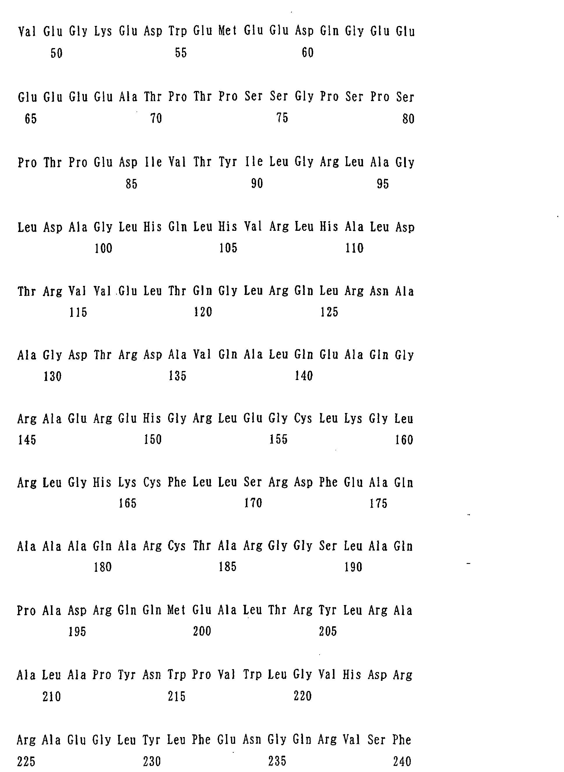


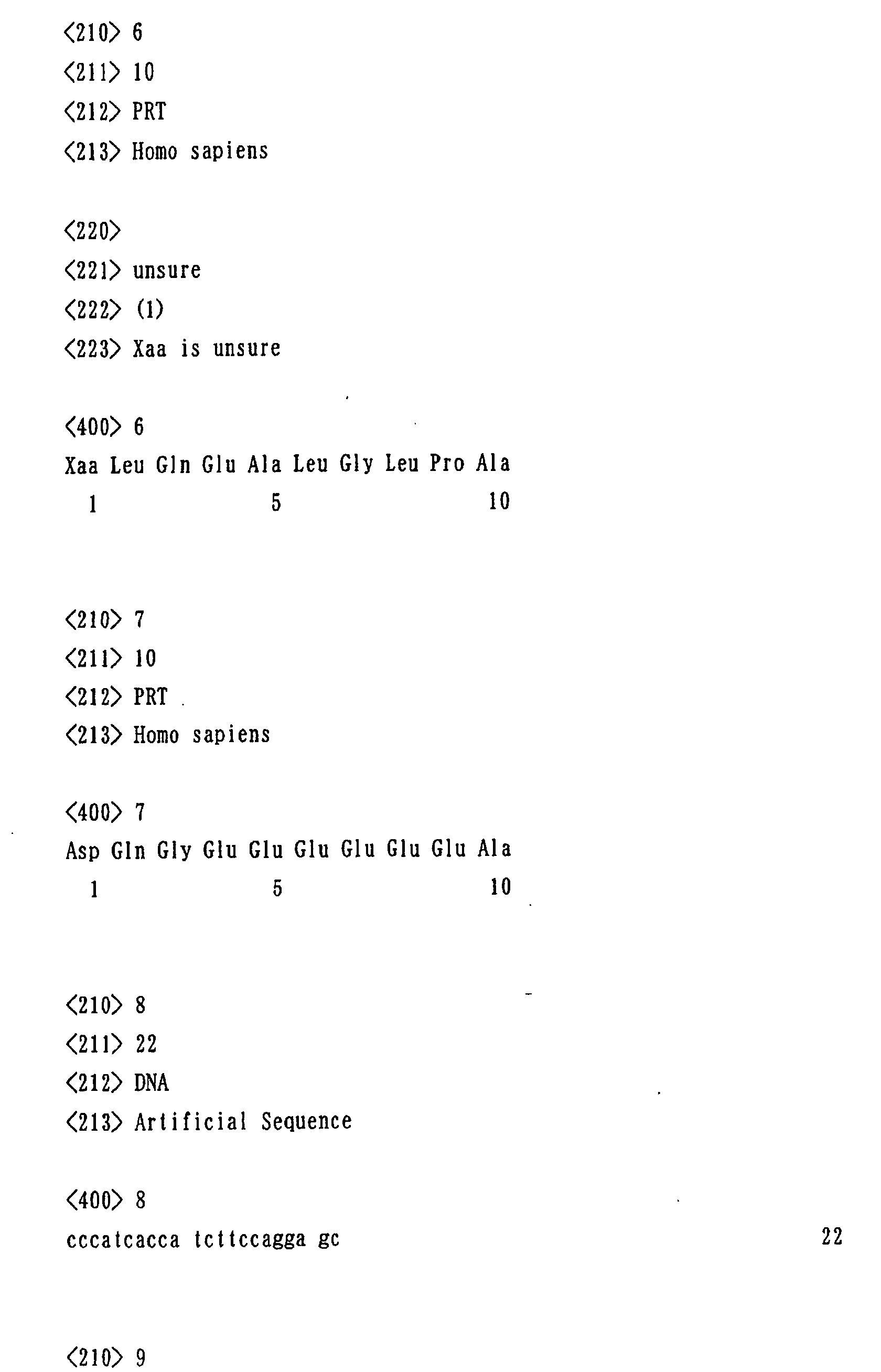

Claims (20)
- A method for diagnosing leukemia, pre-leukemia or aleukemic malignant blood diseases wherein stem cell growth factor (SCGF) in an in-vivo sample is quantified.
- A method for discriminating leukemia from pre-leukemia or aleukemic malignant blood diseases wherein stem cell growth factor (SCGF) in an in-vivo sample is quantified.
- A method for discriminating aplastic anemia from myelodysplastic syndrome wherein stem cell growth factor (SCGF) in an in-vivo sample is quantified.
- A method for diagnosing the engraftment state of the hematopoietic. stem cells after transplantation of the hematopoietic stem cells wherein stem cell growth factor (SCGF) in an in-vivo sample is quantified.
- A method for diagnosing graft versus host disease (GVHD) wherein stem cell growth factor (SCGF) in an in-vivo sample is quantified.
- The method according to any one of claims 1 to 5 wherein a method for diagnosing or discriminating is an immunological assay.
- The method according to claim 6, wherein the immunological assay is a sandwich assay.
- The method according to claim 7, wherein two kinds of antibodies reacting with different epitopes of stem cell growth factor (SCGF) are used in the sandwich assay.
- The method according to claim 8, wherein the antibodies are selected from polyclonal and monoclonal antibodies.
- The method according to claim 9, wherein the monoclonal antibodies are selected from the group consisting of a monoclonal antibody recognizing the region shown by the amino acid sequence of 6 - 2 8 amino acids, a monoclonal antibody recognizing the region shown by the amino acid sequence of 29-59 amino acids, and a monoclonal antibody recognizing the region shown by the amino acid sequence of 60-302 amino acids , all in the amino acid sequence of SEQ. ID No. 1.
- A diagnostic agent for leukemia, pre-leukemia or aleukemic malignant blood diseases comprising an antibody reacting with stem cell growth factor (SCGF) as an active ingredient.
- A diagnostic agent for the engraftment state of the hematopoietic stem cells after transplantation of the hematopoietic stem cells comprising an antibody reacting with stem cell growth factor (SCGF) as an active ingredient.
- A diagnostic agent for graft versus host disease (GVHD) comprising an antibody reacting with stem cell growth factor (SCGF) as an active ingredient.
- The diagnostic agent according to any one of claims 11 to 13, wherein the antibody is selected from polyclonal and monoclonal antibodies.
- The diagnostic agent according to claim 14, wherein the monoclonal antibody is selected from the group consisting of a monoclonal antibody recognizing the region shown by the amino acid sequence of 6-28 amino acids, a monoclonal antibody recognizing the region shown by the amino acid sequence of 29-59 amino acids, and a monoclonal antibody recognizing the region shown by the amino acid sequence of 60-302 amino acids, all in the amino acid sequence of SEQ. ID No. 1.
- A diagnostic kit containing an antibody reacting with stem cell growth factor (SCGF), which is for leukemia, pre-leukemia or aleukemic malignant blood diseases, for engraftment state of the hematopoietic stem cells after transplantation of the hematopoietic stem cells, and for graft versus host disease (GVHD).
- The diagnostic kit according to claim 16, which contains stem cell growth factor (SCGF).
- A monoclonal antibody which recognizes the region shown by the amino acid sequence of 29-59 amino acids in SEQ. ID No. 1.
- A monoclonal antibody which recognizes the region shown by the amino acid sequence shown by 60-302 amino acids in SEQ. ID No. 1.
- A hybridoma which produces the monoclonal antibody according to claim 18 or 19.
Applications Claiming Priority (3)
| Application Number | Priority Date | Filing Date | Title |
|---|---|---|---|
| JP2002106786 | 2002-04-09 | ||
| JP2002106786 | 2002-04-09 | ||
| PCT/JP2003/004531 WO2003085399A1 (en) | 2002-04-09 | 2003-04-09 | Method of judging leukemia, pre-leukemia or aleukemic malignant blood disease and diagnostic therefor |
Publications (2)
| Publication Number | Publication Date |
|---|---|
| EP1496360A1 true EP1496360A1 (en) | 2005-01-12 |
| EP1496360A4 EP1496360A4 (en) | 2007-06-27 |
Family
ID=28786442
Family Applications (1)
| Application Number | Title | Priority Date | Filing Date |
|---|---|---|---|
| EP03745988A Withdrawn EP1496360A4 (en) | 2002-04-09 | 2003-04-09 | METHOD OF DETERMINING LEUKEMIA, PRELEUGEEMY OR ALEUCEMIC MALIGNANT BLOOD DISEASE, AND DIAGNOSTIC METHOD |
Country Status (8)
| Country | Link |
|---|---|
| US (1) | US7479371B2 (en) |
| EP (1) | EP1496360A4 (en) |
| JP (1) | JPWO2003085399A1 (en) |
| KR (1) | KR20050027211A (en) |
| CN (1) | CN1646910A (en) |
| AU (1) | AU2003236028A1 (en) |
| CA (1) | CA2481710A1 (en) |
| WO (1) | WO2003085399A1 (en) |
Families Citing this family (42)
| Publication number | Priority date | Publication date | Assignee | Title |
|---|---|---|---|---|
| US20060078893A1 (en) | 2004-10-12 | 2006-04-13 | Medical Research Council | Compartmentalised combinatorial chemistry by microfluidic control |
| GB0307428D0 (en) * | 2003-03-31 | 2003-05-07 | Medical Res Council | Compartmentalised combinatorial chemistry |
| GB0307403D0 (en) | 2003-03-31 | 2003-05-07 | Medical Res Council | Selection by compartmentalised screening |
| US20050221339A1 (en) * | 2004-03-31 | 2005-10-06 | Medical Research Council Harvard University | Compartmentalised screening by microfluidic control |
| US7968287B2 (en) | 2004-10-08 | 2011-06-28 | Medical Research Council Harvard University | In vitro evolution in microfluidic systems |
| WO2007081385A2 (en) | 2006-01-11 | 2007-07-19 | Raindance Technologies, Inc. | Microfluidic devices and methods of use in the formation and control of nanoreactors |
| WO2007133710A2 (en) | 2006-05-11 | 2007-11-22 | Raindance Technologies, Inc. | Microfluidic devices and methods of use thereof |
| US9562837B2 (en) | 2006-05-11 | 2017-02-07 | Raindance Technologies, Inc. | Systems for handling microfludic droplets |
| US9012390B2 (en) | 2006-08-07 | 2015-04-21 | Raindance Technologies, Inc. | Fluorocarbon emulsion stabilizing surfactants |
| WO2008097559A2 (en) | 2007-02-06 | 2008-08-14 | Brandeis University | Manipulation of fluids and reactions in microfluidic systems |
| WO2008130623A1 (en) | 2007-04-19 | 2008-10-30 | Brandeis University | Manipulation of fluids, fluid components and reactions in microfluidic systems |
| CN101250526B (en) * | 2007-12-26 | 2010-06-02 | 暨南大学 | The Y gene sequence of benzene-induced aplastic anemia-associated antigen-specific TCR Vα19 subfamily |
| EP2241892B1 (en) * | 2007-12-28 | 2012-08-08 | National University Corporation Hokkaido University | Method of detecting leukemic cell |
| US12038438B2 (en) | 2008-07-18 | 2024-07-16 | Bio-Rad Laboratories, Inc. | Enzyme quantification |
| EP4047367A1 (en) | 2008-07-18 | 2022-08-24 | Bio-Rad Laboratories, Inc. | Method for detecting target analytes with droplet libraries |
| WO2010111231A1 (en) | 2009-03-23 | 2010-09-30 | Raindance Technologies, Inc. | Manipulation of microfluidic droplets |
| US10520500B2 (en) | 2009-10-09 | 2019-12-31 | Abdeslam El Harrak | Labelled silica-based nanomaterial with enhanced properties and uses thereof |
| EP2517025B1 (en) | 2009-12-23 | 2019-11-27 | Bio-Rad Laboratories, Inc. | Methods for reducing the exchange of molecules between droplets |
| EP4435111A1 (en) | 2010-02-12 | 2024-09-25 | Bio-Rad Laboratories, Inc. | Digital analyte analysis |
| US10351905B2 (en) | 2010-02-12 | 2019-07-16 | Bio-Rad Laboratories, Inc. | Digital analyte analysis |
| US9399797B2 (en) | 2010-02-12 | 2016-07-26 | Raindance Technologies, Inc. | Digital analyte analysis |
| US9366632B2 (en) | 2010-02-12 | 2016-06-14 | Raindance Technologies, Inc. | Digital analyte analysis |
| WO2012045012A2 (en) | 2010-09-30 | 2012-04-05 | Raindance Technologies, Inc. | Sandwich assays in droplets |
| EP3859011A1 (en) | 2011-02-11 | 2021-08-04 | Bio-Rad Laboratories, Inc. | Methods for forming mixed droplets |
| WO2012112804A1 (en) | 2011-02-18 | 2012-08-23 | Raindance Technoligies, Inc. | Compositions and methods for molecular labeling |
| US8841071B2 (en) | 2011-06-02 | 2014-09-23 | Raindance Technologies, Inc. | Sample multiplexing |
| EP2714970B1 (en) | 2011-06-02 | 2017-04-19 | Raindance Technologies, Inc. | Enzyme quantification |
| US8658430B2 (en) | 2011-07-20 | 2014-02-25 | Raindance Technologies, Inc. | Manipulating droplet size |
| US20130210659A1 (en) | 2012-02-10 | 2013-08-15 | Andrew Watson | Molecular diagnostic screening assay |
| WO2013165748A1 (en) | 2012-04-30 | 2013-11-07 | Raindance Technologies, Inc | Digital analyte analysis |
| AU2013274965B2 (en) * | 2012-06-14 | 2018-07-19 | Erasmus University Medical Center Rotterdam | Methods, reagents and kits for detecting minimal residual disease |
| EP2986762B1 (en) | 2013-04-19 | 2019-11-06 | Bio-Rad Laboratories, Inc. | Digital analyte analysis |
| US11901041B2 (en) | 2013-10-04 | 2024-02-13 | Bio-Rad Laboratories, Inc. | Digital analysis of nucleic acid modification |
| US9944977B2 (en) | 2013-12-12 | 2018-04-17 | Raindance Technologies, Inc. | Distinguishing rare variations in a nucleic acid sequence from a sample |
| WO2015103367A1 (en) | 2013-12-31 | 2015-07-09 | Raindance Technologies, Inc. | System and method for detection of rna species |
| JP6170865B2 (en) | 2014-03-31 | 2017-07-26 | シスメックス株式会社 | Blood analyzer |
| US10647981B1 (en) | 2015-09-08 | 2020-05-12 | Bio-Rad Laboratories, Inc. | Nucleic acid library generation methods and compositions |
| CN105424929B (en) * | 2015-11-24 | 2017-08-08 | 四川夹金山逢春养殖科技有限公司 | A kind of pseudomonas aeruginosa antibody assay kit and detection method |
| CN106872686B (en) * | 2017-02-16 | 2019-04-05 | 广东顺德工业设计研究院(广东顺德创新设计研究院) | Preservation solution for time-resolved fluorescent microsphere-labeled myoglobin antibody |
| US10998178B2 (en) | 2017-08-28 | 2021-05-04 | Purdue Research Foundation | Systems and methods for sample analysis using swabs |
| CN111198271B (en) * | 2018-11-16 | 2023-04-28 | 山东泽济生物科技有限公司 | Chemiluminescent enzyme-linked immunosorbent assay kit for detecting glyceraldehyde-3-phosphate dehydrogenase |
| CN111476754B (en) * | 2020-02-28 | 2022-12-09 | 中国人民解放军陆军军医大学第二附属医院 | Bone marrow cell image artificial intelligence auxiliary grading diagnosis system and method |
Family Cites Families (4)
| Publication number | Priority date | Publication date | Assignee | Title |
|---|---|---|---|---|
| US6541217B2 (en) * | 1996-08-27 | 2003-04-01 | Kyowa Hakko Kogyo Co., Ltd. | Hematopoietic stem cell growth factor (SCGF) |
| DE19648596A1 (en) | 1996-11-23 | 1998-05-28 | Teves Gmbh Alfred | Method for operating an anti-lock motor vehicle brake system |
| FR2771750A1 (en) | 1997-12-03 | 1999-06-04 | Univ Nice Sophia Antipolis | Lymphocytic virus-binding lectin LVBL |
| EP1248848B1 (en) | 1999-12-23 | 2007-02-28 | Kirin Beer Kabushiki Kaisha | Methods and materials relating to stem cell growth factor-like poypeptides and polynucleotides |
-
2003
- 2003-04-09 US US10/510,627 patent/US7479371B2/en not_active Expired - Fee Related
- 2003-04-09 WO PCT/JP2003/004531 patent/WO2003085399A1/en not_active Ceased
- 2003-04-09 CN CNA038080362A patent/CN1646910A/en active Pending
- 2003-04-09 KR KR1020047016144A patent/KR20050027211A/en not_active Withdrawn
- 2003-04-09 CA CA002481710A patent/CA2481710A1/en not_active Abandoned
- 2003-04-09 JP JP2003582535A patent/JPWO2003085399A1/en active Pending
- 2003-04-09 EP EP03745988A patent/EP1496360A4/en not_active Withdrawn
- 2003-04-09 AU AU2003236028A patent/AU2003236028A1/en not_active Abandoned
Also Published As
| Publication number | Publication date |
|---|---|
| US20060084122A1 (en) | 2006-04-20 |
| WO2003085399A1 (en) | 2003-10-16 |
| US7479371B2 (en) | 2009-01-20 |
| KR20050027211A (en) | 2005-03-18 |
| AU2003236028A1 (en) | 2003-10-20 |
| CN1646910A (en) | 2005-07-27 |
| JPWO2003085399A1 (en) | 2005-08-11 |
| CA2481710A1 (en) | 2003-10-16 |
| EP1496360A4 (en) | 2007-06-27 |
Similar Documents
| Publication | Publication Date | Title |
|---|---|---|
| US7479371B2 (en) | Method of judging leukemia, pre-leukemia or aleukemic malignant blood disease and diagnostic therefor | |
| JP5424331B2 (en) | Biomarker for liver disease diagnosis | |
| KR102404285B1 (en) | Assays for igfbp7 having improved performance in biological samples | |
| EP4060338B1 (en) | Kit and method for mixed detection of pct and presepsin, and use thereof | |
| CN108473552B (en) | Antigenic composition for detecting specific autoantibodies of exosomal protein EIF3A and method for diagnosing liver cancer using the same | |
| EP4059517A1 (en) | New anti-soluble cd14 subtype antibody and use thereof | |
| US20230314433A1 (en) | Method of determining viral infection | |
| JPWO2009044561A1 (en) | Anti-proNT / NMN monoclonal antibody | |
| CN104066749A (en) | Monoclonal antibody for specific recognition of human liver carboxylesterase 1, hybridoma cell line producing monoclonal antibody and use thereof | |
| EP2541249A1 (en) | EpCAM detection | |
| EP1213302B1 (en) | Specific antibody directed to active but not inactive hepatocyte growth factor activator (HGFA) | |
| EP0411018B1 (en) | Detection, quantitation and classification of ras proteins in body fluids and tissues | |
| JP6537095B2 (en) | Markers for predictive diagnosis of cancer recurrence and / or metastasis | |
| US8361706B2 (en) | Monoclonal antibodies and uses thereof | |
| CN115698718A (en) | Pancreatic cancer detection method and detection reagent | |
| JP6407990B2 (en) | Augrin immunoassay | |
| US7087391B2 (en) | Antibody to hepatocyte growth factor activator inhibitor-1 and use thereof | |
| JP4838436B2 (en) | Anti-human hepatic triglyceride lipase antibody | |
| US9017957B2 (en) | Prostasin partial peptide and anti-prostasin antibody | |
| JP2012018119A (en) | Colon cancer detection marker and colon cancer detection method using the same | |
| JP2007119390A (en) | How to predict the prognosis of colorectal cancer patients | |
| JP2001215227A (en) | RCAS1 immunoassay method and assay kit, anti-RCAS1 antibody immunoassay method and assay kit, and RCAS1 receptor immunoassay method | |
| HK1112965A (en) | Drug for monitoring worsening of hepatitis |
Legal Events
| Date | Code | Title | Description |
|---|---|---|---|
| PUAI | Public reference made under article 153(3) epc to a published international application that has entered the european phase |
Free format text: ORIGINAL CODE: 0009012 |
|
| 17P | Request for examination filed |
Effective date: 20041029 |
|
| AK | Designated contracting states |
Kind code of ref document: A1 Designated state(s): AT BE BG CH CY CZ DE DK EE ES FI FR GB GR HU IE IT LI LU MC NL PT RO SE SI SK TR |
|
| AX | Request for extension of the european patent |
Extension state: AL LT LV MK |
|
| A4 | Supplementary search report drawn up and despatched |
Effective date: 20070530 |
|
| 17Q | First examination report despatched |
Effective date: 20070910 |
|
| STAA | Information on the status of an ep patent application or granted ep patent |
Free format text: STATUS: THE APPLICATION IS DEEMED TO BE WITHDRAWN |
|
| 18D | Application deemed to be withdrawn |
Effective date: 20100622 |