EP1492869B9 - Transplant acceptance inducing cells of monocytic origin and their preparation and use - Google Patents
Transplant acceptance inducing cells of monocytic origin and their preparation and use Download PDFInfo
- Publication number
- EP1492869B9 EP1492869B9 EP03763823A EP03763823A EP1492869B9 EP 1492869 B9 EP1492869 B9 EP 1492869B9 EP 03763823 A EP03763823 A EP 03763823A EP 03763823 A EP03763823 A EP 03763823A EP 1492869 B9 EP1492869 B9 EP 1492869B9
- Authority
- EP
- European Patent Office
- Prior art keywords
- cells
- taic
- monocytes
- lymphocytes
- transplant
- Prior art date
- Legal status (The legal status is an assumption and is not a legal conclusion. Google has not performed a legal analysis and makes no representation as to the accuracy of the status listed.)
- Expired - Lifetime
Links
- 230000001939 inductive effect Effects 0.000 title claims abstract description 46
- 238000002360 preparation method Methods 0.000 title claims description 29
- 241000282414 Homo sapiens Species 0.000 claims abstract description 25
- 238000001514 detection method Methods 0.000 claims abstract description 14
- 238000004519 manufacturing process Methods 0.000 claims abstract description 6
- 210000004027 cell Anatomy 0.000 claims description 269
- 210000001616 monocyte Anatomy 0.000 claims description 99
- 210000004698 lymphocyte Anatomy 0.000 claims description 74
- 238000000034 method Methods 0.000 claims description 72
- 101000946889 Homo sapiens Monocyte differentiation antigen CD14 Proteins 0.000 claims description 54
- 102100035877 Monocyte differentiation antigen CD14 Human genes 0.000 claims description 54
- 230000008569 process Effects 0.000 claims description 40
- 239000002609 medium Substances 0.000 claims description 38
- 210000004369 blood Anatomy 0.000 claims description 32
- 239000008280 blood Substances 0.000 claims description 32
- 210000003289 regulatory T cell Anatomy 0.000 claims description 31
- 108010046938 Macrophage Colony-Stimulating Factor Proteins 0.000 claims description 27
- 239000000427 antigen Substances 0.000 claims description 25
- 108091007433 antigens Proteins 0.000 claims description 25
- 102000036639 antigens Human genes 0.000 claims description 25
- 101001057504 Homo sapiens Interferon-stimulated gene 20 kDa protein Proteins 0.000 claims description 24
- 101001055144 Homo sapiens Interleukin-2 receptor subunit alpha Proteins 0.000 claims description 24
- 102100026878 Interleukin-2 receptor subunit alpha Human genes 0.000 claims description 24
- 210000001744 T-lymphocyte Anatomy 0.000 claims description 22
- 239000001963 growth medium Substances 0.000 claims description 21
- 238000000338 in vitro Methods 0.000 claims description 17
- 108091003079 Bovine Serum Albumin Proteins 0.000 claims description 15
- IAZDPXIOMUYVGZ-UHFFFAOYSA-N Dimethylsulphoxide Chemical compound CS(C)=O IAZDPXIOMUYVGZ-UHFFFAOYSA-N 0.000 claims description 15
- 239000012894 fetal calf serum Substances 0.000 claims description 15
- FAPWRFPIFSIZLT-UHFFFAOYSA-M Sodium chloride Chemical compound [Na+].[Cl-] FAPWRFPIFSIZLT-UHFFFAOYSA-M 0.000 claims description 12
- 238000009739 binding Methods 0.000 claims description 12
- 210000004408 hybridoma Anatomy 0.000 claims description 9
- 206010052779 Transplant rejections Diseases 0.000 claims description 7
- 238000006243 chemical reaction Methods 0.000 claims description 7
- 230000001629 suppression Effects 0.000 claims description 7
- 239000003102 growth factor Substances 0.000 claims description 6
- 239000008194 pharmaceutical composition Substances 0.000 claims description 6
- 239000011780 sodium chloride Substances 0.000 claims description 6
- 239000012595 freezing medium Substances 0.000 claims description 4
- 210000002966 serum Anatomy 0.000 claims description 4
- 102100028123 Macrophage colony-stimulating factor 1 Human genes 0.000 claims description 3
- 239000006143 cell culture medium Substances 0.000 claims description 3
- 230000001902 propagating effect Effects 0.000 claims description 2
- 230000010261 cell growth Effects 0.000 claims 1
- 238000001943 fluorescence-activated cell sorting Methods 0.000 claims 1
- 210000002216 heart Anatomy 0.000 description 56
- 210000000056 organ Anatomy 0.000 description 56
- 238000002054 transplantation Methods 0.000 description 55
- 241000700159 Rattus Species 0.000 description 46
- 241001465754 Metazoa Species 0.000 description 44
- 230000037396 body weight Effects 0.000 description 31
- 230000007774 longterm Effects 0.000 description 30
- 206010068051 Chimerism Diseases 0.000 description 27
- 102000007651 Macrophage Colony-Stimulating Factor Human genes 0.000 description 25
- 230000000638 stimulation Effects 0.000 description 25
- 229930105110 Cyclosporin A Natural products 0.000 description 22
- PMATZTZNYRCHOR-CGLBZJNRSA-N Cyclosporin A Chemical compound CC[C@@H]1NC(=O)[C@H]([C@H](O)[C@H](C)C\C=C\C)N(C)C(=O)[C@H](C(C)C)N(C)C(=O)[C@H](CC(C)C)N(C)C(=O)[C@H](CC(C)C)N(C)C(=O)[C@@H](C)NC(=O)[C@H](C)NC(=O)[C@H](CC(C)C)N(C)C(=O)[C@H](C(C)C)NC(=O)[C@H](CC(C)C)N(C)C(=O)CN(C)C1=O PMATZTZNYRCHOR-CGLBZJNRSA-N 0.000 description 22
- 108010036949 Cyclosporine Proteins 0.000 description 22
- 229960001265 ciclosporin Drugs 0.000 description 22
- 230000002980 postoperative effect Effects 0.000 description 21
- 102000006495 integrins Human genes 0.000 description 18
- 108010044426 integrins Proteins 0.000 description 18
- 230000000735 allogeneic effect Effects 0.000 description 17
- 210000001519 tissue Anatomy 0.000 description 17
- 230000000694 effects Effects 0.000 description 15
- 230000004083 survival effect Effects 0.000 description 15
- 241000282898 Sus scrofa Species 0.000 description 14
- 210000004072 lung Anatomy 0.000 description 14
- 230000001506 immunosuppresive effect Effects 0.000 description 13
- 238000011282 treatment Methods 0.000 description 13
- 210000004185 liver Anatomy 0.000 description 12
- 230000015572 biosynthetic process Effects 0.000 description 11
- 210000003734 kidney Anatomy 0.000 description 11
- 210000005259 peripheral blood Anatomy 0.000 description 11
- 239000011886 peripheral blood Substances 0.000 description 11
- 108090000623 proteins and genes Proteins 0.000 description 11
- 239000000243 solution Substances 0.000 description 11
- 230000001154 acute effect Effects 0.000 description 10
- 235000015097 nutrients Nutrition 0.000 description 10
- 108091032973 (ribonucleotides)n+m Proteins 0.000 description 9
- 238000003501 co-culture Methods 0.000 description 9
- LOKCTEFSRHRXRJ-UHFFFAOYSA-I dipotassium trisodium dihydrogen phosphate hydrogen phosphate dichloride Chemical compound P(=O)(O)(O)[O-].[K+].P(=O)(O)([O-])[O-].[Na+].[Na+].[Cl-].[K+].[Cl-].[Na+] LOKCTEFSRHRXRJ-UHFFFAOYSA-I 0.000 description 9
- 238000001727 in vivo Methods 0.000 description 9
- 230000006698 induction Effects 0.000 description 9
- 238000001990 intravenous administration Methods 0.000 description 9
- 239000002953 phosphate buffered saline Substances 0.000 description 9
- 238000000926 separation method Methods 0.000 description 9
- 206010062016 Immunosuppression Diseases 0.000 description 8
- QIVBCDIJIAJPQS-VIFPVBQESA-N L-tryptophane Chemical compound C1=CC=C2C(C[C@H](N)C(O)=O)=CNC2=C1 QIVBCDIJIAJPQS-VIFPVBQESA-N 0.000 description 8
- 238000012258 culturing Methods 0.000 description 8
- 238000000684 flow cytometry Methods 0.000 description 8
- 229960003444 immunosuppressant agent Drugs 0.000 description 8
- 239000003018 immunosuppressive agent Substances 0.000 description 8
- 238000002347 injection Methods 0.000 description 8
- 239000007924 injection Substances 0.000 description 8
- 239000000825 pharmaceutical preparation Substances 0.000 description 8
- 239000006228 supernatant Substances 0.000 description 8
- 108010021064 CTLA-4 Antigen Proteins 0.000 description 7
- 102000008203 CTLA-4 Antigen Human genes 0.000 description 7
- 229940045513 CTLA4 antagonist Drugs 0.000 description 7
- 102000008070 Interferon-gamma Human genes 0.000 description 7
- 108010074328 Interferon-gamma Proteins 0.000 description 7
- 238000002474 experimental method Methods 0.000 description 7
- 229940044627 gamma-interferon Drugs 0.000 description 7
- 238000012986 modification Methods 0.000 description 7
- 230000004048 modification Effects 0.000 description 7
- 230000035755 proliferation Effects 0.000 description 7
- 208000009329 Graft vs Host Disease Diseases 0.000 description 6
- 102000004142 Trypsin Human genes 0.000 description 6
- 108090000631 Trypsin Proteins 0.000 description 6
- 229960002170 azathioprine Drugs 0.000 description 6
- LMEKQMALGUDUQG-UHFFFAOYSA-N azathioprine Chemical compound CN1C=NC([N+]([O-])=O)=C1SC1=NC=NC2=C1NC=N2 LMEKQMALGUDUQG-UHFFFAOYSA-N 0.000 description 6
- 210000001185 bone marrow Anatomy 0.000 description 6
- 230000003750 conditioning effect Effects 0.000 description 6
- 208000037265 diseases, disorders, signs and symptoms Diseases 0.000 description 6
- 230000006870 function Effects 0.000 description 6
- 208000024908 graft versus host disease Diseases 0.000 description 6
- 210000000987 immune system Anatomy 0.000 description 6
- 230000001861 immunosuppressant effect Effects 0.000 description 6
- 238000011835 investigation Methods 0.000 description 6
- 239000007788 liquid Substances 0.000 description 6
- 239000003550 marker Substances 0.000 description 6
- 230000020382 suppression by virus of host antigen processing and presentation of peptide antigen via MHC class I Effects 0.000 description 6
- 238000012360 testing method Methods 0.000 description 6
- 239000012588 trypsin Substances 0.000 description 6
- 229960004799 tryptophan Drugs 0.000 description 6
- 102100039498 Cytotoxic T-lymphocyte protein 4 Human genes 0.000 description 5
- 101000889276 Homo sapiens Cytotoxic T-lymphocyte protein 4 Proteins 0.000 description 5
- JGSARLDLIJGVTE-MBNYWOFBSA-N Penicillin G Chemical compound N([C@H]1[C@H]2SC([C@@H](N2C1=O)C(O)=O)(C)C)C(=O)CC1=CC=CC=C1 JGSARLDLIJGVTE-MBNYWOFBSA-N 0.000 description 5
- WCUXLLCKKVVCTQ-UHFFFAOYSA-M Potassium chloride Chemical compound [Cl-].[K+] WCUXLLCKKVVCTQ-UHFFFAOYSA-M 0.000 description 5
- QIVBCDIJIAJPQS-UHFFFAOYSA-N Tryptophan Natural products C1=CC=C2C(CC(N)C(O)=O)=CNC2=C1 QIVBCDIJIAJPQS-UHFFFAOYSA-N 0.000 description 5
- 201000010099 disease Diseases 0.000 description 5
- 210000003743 erythrocyte Anatomy 0.000 description 5
- 230000028993 immune response Effects 0.000 description 5
- 150000003431 steroids Chemical class 0.000 description 5
- 238000012546 transfer Methods 0.000 description 5
- ZADWXFSZEAPBJS-JTQLQIEISA-N 1-methyl-L-tryptophan Chemical compound C1=CC=C2N(C)C=C(C[C@H](N)C(O)=O)C2=C1 ZADWXFSZEAPBJS-JTQLQIEISA-N 0.000 description 4
- ALYNCZNDIQEVRV-UHFFFAOYSA-N 4-aminobenzoic acid Chemical compound NC1=CC=C(C(O)=O)C=C1 ALYNCZNDIQEVRV-UHFFFAOYSA-N 0.000 description 4
- 102000004190 Enzymes Human genes 0.000 description 4
- 108090000790 Enzymes Proteins 0.000 description 4
- 108091006905 Human Serum Albumin Proteins 0.000 description 4
- 102000008100 Human Serum Albumin Human genes 0.000 description 4
- CSNNHWWHGAXBCP-UHFFFAOYSA-L Magnesium sulfate Chemical compound [Mg+2].[O-][S+2]([O-])([O-])[O-] CSNNHWWHGAXBCP-UHFFFAOYSA-L 0.000 description 4
- UIIMBOGNXHQVGW-UHFFFAOYSA-M Sodium bicarbonate Chemical compound [Na+].OC([O-])=O UIIMBOGNXHQVGW-UHFFFAOYSA-M 0.000 description 4
- IQFYYKKMVGJFEH-XLPZGREQSA-N Thymidine Chemical compound O=C1NC(=O)C(C)=CN1[C@@H]1O[C@H](CO)[C@@H](O)C1 IQFYYKKMVGJFEH-XLPZGREQSA-N 0.000 description 4
- 238000010322 bone marrow transplantation Methods 0.000 description 4
- ZCCIPPOKBCJFDN-UHFFFAOYSA-N calcium nitrate Chemical compound [Ca+2].[O-][N+]([O-])=O.[O-][N+]([O-])=O ZCCIPPOKBCJFDN-UHFFFAOYSA-N 0.000 description 4
- 238000004113 cell culture Methods 0.000 description 4
- 230000001413 cellular effect Effects 0.000 description 4
- 210000000038 chest Anatomy 0.000 description 4
- 238000011161 development Methods 0.000 description 4
- 230000018109 developmental process Effects 0.000 description 4
- MHMNJMPURVTYEJ-UHFFFAOYSA-N fluorescein-5-isothiocyanate Chemical compound O1C(=O)C2=CC(N=C=S)=CC=C2C21C1=CC=C(O)C=C1OC1=CC(O)=CC=C21 MHMNJMPURVTYEJ-UHFFFAOYSA-N 0.000 description 4
- 210000003714 granulocyte Anatomy 0.000 description 4
- 230000001965 increasing effect Effects 0.000 description 4
- 230000016507 interphase Effects 0.000 description 4
- 238000010253 intravenous injection Methods 0.000 description 4
- 210000000265 leukocyte Anatomy 0.000 description 4
- 210000002540 macrophage Anatomy 0.000 description 4
- 239000000203 mixture Substances 0.000 description 4
- 238000002203 pretreatment Methods 0.000 description 4
- 238000011552 rat model Methods 0.000 description 4
- 241000894007 species Species 0.000 description 4
- 210000001541 thymus gland Anatomy 0.000 description 4
- 230000024664 tolerance induction Effects 0.000 description 4
- 229920001917 Ficoll Polymers 0.000 description 3
- 241000714260 Human T-lymphotropic virus 1 Species 0.000 description 3
- ZDXPYRJPNDTMRX-VKHMYHEASA-N L-glutamine Chemical compound OC(=O)[C@@H](N)CCC(N)=O ZDXPYRJPNDTMRX-VKHMYHEASA-N 0.000 description 3
- 229930182816 L-glutamine Natural products 0.000 description 3
- 241000699670 Mus sp. Species 0.000 description 3
- 229930182555 Penicillin Natural products 0.000 description 3
- 241000282887 Suidae Species 0.000 description 3
- 108091023040 Transcription factor Proteins 0.000 description 3
- 102000040945 Transcription factor Human genes 0.000 description 3
- 210000001015 abdomen Anatomy 0.000 description 3
- 239000006285 cell suspension Substances 0.000 description 3
- 230000001684 chronic effect Effects 0.000 description 3
- KRKNYBCHXYNGOX-UHFFFAOYSA-N citric acid Chemical compound OC(=O)CC(O)(C(O)=O)CC(O)=O KRKNYBCHXYNGOX-UHFFFAOYSA-N 0.000 description 3
- 230000007423 decrease Effects 0.000 description 3
- 239000003814 drug Substances 0.000 description 3
- 239000003112 inhibitor Substances 0.000 description 3
- 238000002372 labelling Methods 0.000 description 3
- 230000001404 mediated effect Effects 0.000 description 3
- 108020004999 messenger RNA Proteins 0.000 description 3
- 244000309715 mini pig Species 0.000 description 3
- 239000008188 pellet Substances 0.000 description 3
- 229940049954 penicillin Drugs 0.000 description 3
- 230000001105 regulatory effect Effects 0.000 description 3
- 230000004044 response Effects 0.000 description 3
- 238000003860 storage Methods 0.000 description 3
- 229940104230 thymidine Drugs 0.000 description 3
- YBJHBAHKTGYVGT-ZKWXMUAHSA-N (+)-Biotin Chemical compound N1C(=O)N[C@@H]2[C@H](CCCCC(=O)O)SC[C@@H]21 YBJHBAHKTGYVGT-ZKWXMUAHSA-N 0.000 description 2
- MTCFGRXMJLQNBG-REOHCLBHSA-N (2S)-2-Amino-3-hydroxypropansäure Chemical compound OC[C@H](N)C(O)=O MTCFGRXMJLQNBG-REOHCLBHSA-N 0.000 description 2
- DWRXFEITVBNRMK-UHFFFAOYSA-N Beta-D-1-Arabinofuranosylthymine Natural products O=C1NC(=O)C(C)=CN1C1C(O)C(O)C(CO)O1 DWRXFEITVBNRMK-UHFFFAOYSA-N 0.000 description 2
- 241000282693 Cercopithecidae Species 0.000 description 2
- 241000557626 Corvus corax Species 0.000 description 2
- 241000588724 Escherichia coli Species 0.000 description 2
- 238000012413 Fluorescence activated cell sorting analysis Methods 0.000 description 2
- WQZGKKKJIJFFOK-GASJEMHNSA-N Glucose Natural products OC[C@H]1OC(O)[C@H](O)[C@@H](O)[C@@H]1O WQZGKKKJIJFFOK-GASJEMHNSA-N 0.000 description 2
- DHMQDGOQFOQNFH-UHFFFAOYSA-N Glycine Chemical compound NCC(O)=O DHMQDGOQFOQNFH-UHFFFAOYSA-N 0.000 description 2
- DCXYFEDJOCDNAF-REOHCLBHSA-N L-asparagine Chemical compound OC(=O)[C@@H](N)CC(N)=O DCXYFEDJOCDNAF-REOHCLBHSA-N 0.000 description 2
- HNDVDQJCIGZPNO-YFKPBYRVSA-N L-histidine Chemical compound OC(=O)[C@@H](N)CC1=CN=CN1 HNDVDQJCIGZPNO-YFKPBYRVSA-N 0.000 description 2
- ROHFNLRQFUQHCH-YFKPBYRVSA-N L-leucine Chemical compound CC(C)C[C@H](N)C(O)=O ROHFNLRQFUQHCH-YFKPBYRVSA-N 0.000 description 2
- COLNVLDHVKWLRT-QMMMGPOBSA-N L-phenylalanine Chemical compound OC(=O)[C@@H](N)CC1=CC=CC=C1 COLNVLDHVKWLRT-QMMMGPOBSA-N 0.000 description 2
- KZSNJWFQEVHDMF-BYPYZUCNSA-N L-valine Chemical compound CC(C)[C@H](N)C(O)=O KZSNJWFQEVHDMF-BYPYZUCNSA-N 0.000 description 2
- 239000012980 RPMI-1640 medium Substances 0.000 description 2
- AUNGANRZJHBGPY-SCRDCRAPSA-N Riboflavin Chemical compound OC[C@@H](O)[C@@H](O)[C@@H](O)CN1C=2C=C(C)C(C)=CC=2N=C2C1=NC(=O)NC2=O AUNGANRZJHBGPY-SCRDCRAPSA-N 0.000 description 2
- QTENRWWVYAAPBI-YZTFXSNBSA-N Streptomycin sulfate Chemical compound OS(O)(=O)=O.OS(O)(=O)=O.OS(O)(=O)=O.CN[C@H]1[C@H](O)[C@@H](O)[C@H](CO)O[C@H]1O[C@@H]1[C@](C=O)(O)[C@H](C)O[C@H]1O[C@H]1[C@H](N=C(N)N)[C@@H](O)[C@H](N=C(N)N)[C@@H](O)[C@@H]1O.CN[C@H]1[C@H](O)[C@@H](O)[C@H](CO)O[C@H]1O[C@@H]1[C@](C=O)(O)[C@H](C)O[C@H]1O[C@H]1[C@H](N=C(N)N)[C@@H](O)[C@H](N=C(N)N)[C@@H](O)[C@@H]1O QTENRWWVYAAPBI-YZTFXSNBSA-N 0.000 description 2
- 229920006329 Styropor Polymers 0.000 description 2
- 125000000539 amino acid group Chemical group 0.000 description 2
- 229960004050 aminobenzoic acid Drugs 0.000 description 2
- 239000012298 atmosphere Substances 0.000 description 2
- 210000003719 b-lymphocyte Anatomy 0.000 description 2
- 229960004669 basiliximab Drugs 0.000 description 2
- 239000011324 bead Substances 0.000 description 2
- IQFYYKKMVGJFEH-UHFFFAOYSA-N beta-L-thymidine Natural products O=C1NC(=O)C(C)=CN1C1OC(CO)C(O)C1 IQFYYKKMVGJFEH-UHFFFAOYSA-N 0.000 description 2
- 210000000601 blood cell Anatomy 0.000 description 2
- 210000002798 bone marrow cell Anatomy 0.000 description 2
- 238000005119 centrifugation Methods 0.000 description 2
- 239000003795 chemical substances by application Substances 0.000 description 2
- 230000006378 damage Effects 0.000 description 2
- 230000003247 decreasing effect Effects 0.000 description 2
- 239000000975 dye Substances 0.000 description 2
- 230000008030 elimination Effects 0.000 description 2
- 238000003379 elimination reaction Methods 0.000 description 2
- 238000011156 evaluation Methods 0.000 description 2
- OVBPIULPVIDEAO-LBPRGKRZSA-N folic acid Chemical compound C=1N=C2NC(N)=NC(=O)C2=NC=1CNC1=CC=C(C(=O)N[C@@H](CCC(O)=O)C(O)=O)C=C1 OVBPIULPVIDEAO-LBPRGKRZSA-N 0.000 description 2
- 238000007710 freezing Methods 0.000 description 2
- 230000008014 freezing Effects 0.000 description 2
- 102000054766 genetic haplotypes Human genes 0.000 description 2
- 230000001900 immune effect Effects 0.000 description 2
- 230000004957 immunoregulator effect Effects 0.000 description 2
- 108020004201 indoleamine 2,3-dioxygenase Proteins 0.000 description 2
- 102000006639 indoleamine 2,3-dioxygenase Human genes 0.000 description 2
- 230000008595 infiltration Effects 0.000 description 2
- 238000001764 infiltration Methods 0.000 description 2
- 210000004153 islets of langerhan Anatomy 0.000 description 2
- 229910052943 magnesium sulfate Inorganic materials 0.000 description 2
- 238000002826 magnetic-activated cell sorting Methods 0.000 description 2
- 229960004452 methionine Drugs 0.000 description 2
- 210000005087 mononuclear cell Anatomy 0.000 description 2
- 239000001103 potassium chloride Substances 0.000 description 2
- 235000011164 potassium chloride Nutrition 0.000 description 2
- 230000003389 potentiating effect Effects 0.000 description 2
- 238000000746 purification Methods 0.000 description 2
- 238000003753 real-time PCR Methods 0.000 description 2
- 230000009467 reduction Effects 0.000 description 2
- 230000009711 regulatory function Effects 0.000 description 2
- 238000012552 review Methods 0.000 description 2
- 239000011734 sodium Substances 0.000 description 2
- 229910000030 sodium bicarbonate Inorganic materials 0.000 description 2
- UCSJYZPVAKXKNQ-HZYVHMACSA-N streptomycin Chemical compound CN[C@H]1[C@H](O)[C@@H](O)[C@H](CO)O[C@H]1O[C@@H]1[C@](C=O)(O)[C@H](C)O[C@H]1O[C@@H]1[C@@H](NC(N)=N)[C@H](O)[C@@H](NC(N)=N)[C@H](O)[C@H]1O UCSJYZPVAKXKNQ-HZYVHMACSA-N 0.000 description 2
- 239000000725 suspension Substances 0.000 description 2
- 230000001225 therapeutic effect Effects 0.000 description 2
- JZRWCGZRTZMZEH-UHFFFAOYSA-N thiamine Chemical compound CC1=C(CCO)SC=[N+]1CC1=CN=C(C)N=C1N JZRWCGZRTZMZEH-UHFFFAOYSA-N 0.000 description 2
- 230000001052 transient effect Effects 0.000 description 2
- 239000006163 transport media Substances 0.000 description 2
- FUOUNKGLDGJSME-JIZZDEOASA-N (2r)-2-amino-3-sulfanylpropanoic acid;dihydrochloride Chemical compound Cl.Cl.SC[C@H](N)C(O)=O FUOUNKGLDGJSME-JIZZDEOASA-N 0.000 description 1
- IJSMFQNTEUNRPY-UHFFFAOYSA-N 2-[3-(dimethylamino)-6-dimethylazaniumylidenexanthen-9-yl]-5-isothiocyanatobenzoate Chemical compound C=12C=CC(=[N+](C)C)C=C2OC2=CC(N(C)C)=CC=C2C=1C1=CC=C(N=C=S)C=C1C([O-])=O IJSMFQNTEUNRPY-UHFFFAOYSA-N 0.000 description 1
- 239000001763 2-hydroxyethyl(trimethyl)azanium Substances 0.000 description 1
- 102000007469 Actins Human genes 0.000 description 1
- 108010085238 Actins Proteins 0.000 description 1
- IJGRMHOSHXDMSA-UHFFFAOYSA-N Atomic nitrogen Chemical compound N#N IJGRMHOSHXDMSA-UHFFFAOYSA-N 0.000 description 1
- 206010051779 Bone marrow toxicity Diseases 0.000 description 1
- 102000000905 Cadherin Human genes 0.000 description 1
- 108050007957 Cadherin Proteins 0.000 description 1
- 208000024172 Cardiovascular disease Diseases 0.000 description 1
- 235000019743 Choline chloride Nutrition 0.000 description 1
- AUNGANRZJHBGPY-UHFFFAOYSA-N D-Lyxoflavin Natural products OCC(O)C(O)C(O)CN1C=2C=C(C)C(C)=CC=2N=C2C1=NC(=O)NC2=O AUNGANRZJHBGPY-UHFFFAOYSA-N 0.000 description 1
- 108020004414 DNA Proteins 0.000 description 1
- 206010052804 Drug tolerance Diseases 0.000 description 1
- 239000012591 Dulbecco’s Phosphate Buffered Saline Substances 0.000 description 1
- KCXVZYZYPLLWCC-UHFFFAOYSA-N EDTA Chemical compound OC(=O)CN(CC(O)=O)CCN(CC(O)=O)CC(O)=O KCXVZYZYPLLWCC-UHFFFAOYSA-N 0.000 description 1
- 108700039887 Essential Genes Proteins 0.000 description 1
- 102100027581 Forkhead box protein P3 Human genes 0.000 description 1
- 101710088098 Forkhead box protein P3 Proteins 0.000 description 1
- 108010024636 Glutathione Proteins 0.000 description 1
- 102100031181 Glyceraldehyde-3-phosphate dehydrogenase Human genes 0.000 description 1
- 239000004471 Glycine Substances 0.000 description 1
- 102100036242 HLA class II histocompatibility antigen, DQ alpha 2 chain Human genes 0.000 description 1
- 102000008949 Histocompatibility Antigens Class I Human genes 0.000 description 1
- 108010088652 Histocompatibility Antigens Class I Proteins 0.000 description 1
- 241000282412 Homo Species 0.000 description 1
- 101000930801 Homo sapiens HLA class II histocompatibility antigen, DQ alpha 2 chain Proteins 0.000 description 1
- 101000801234 Homo sapiens Tumor necrosis factor receptor superfamily member 18 Proteins 0.000 description 1
- PMMYEEVYMWASQN-DMTCNVIQSA-N Hydroxyproline Chemical compound O[C@H]1CN[C@H](C(O)=O)C1 PMMYEEVYMWASQN-DMTCNVIQSA-N 0.000 description 1
- 108060003951 Immunoglobulin Proteins 0.000 description 1
- 239000007836 KH2PO4 Substances 0.000 description 1
- FFEARJCKVFRZRR-UHFFFAOYSA-N L-Methionine Natural products CSCCC(N)C(O)=O FFEARJCKVFRZRR-UHFFFAOYSA-N 0.000 description 1
- ONIBWKKTOPOVIA-BYPYZUCNSA-N L-Proline Chemical compound OC(=O)[C@@H]1CCCN1 ONIBWKKTOPOVIA-BYPYZUCNSA-N 0.000 description 1
- ODKSFYDXXFIFQN-BYPYZUCNSA-N L-arginine Chemical compound OC(=O)[C@@H](N)CCCN=C(N)N ODKSFYDXXFIFQN-BYPYZUCNSA-N 0.000 description 1
- 229930064664 L-arginine Natural products 0.000 description 1
- 235000014852 L-arginine Nutrition 0.000 description 1
- CKLJMWTZIZZHCS-REOHCLBHSA-N L-aspartic acid Chemical compound OC(=O)[C@@H](N)CC(O)=O CKLJMWTZIZZHCS-REOHCLBHSA-N 0.000 description 1
- AGPKZVBTJJNPAG-WHFBIAKZSA-N L-isoleucine Chemical compound CC[C@H](C)[C@H](N)C(O)=O AGPKZVBTJJNPAG-WHFBIAKZSA-N 0.000 description 1
- 229930182844 L-isoleucine Natural products 0.000 description 1
- 239000004395 L-leucine Substances 0.000 description 1
- 235000019454 L-leucine Nutrition 0.000 description 1
- BVHLGVCQOALMSV-JEDNCBNOSA-N L-lysine hydrochloride Chemical compound Cl.NCCCC[C@H](N)C(O)=O BVHLGVCQOALMSV-JEDNCBNOSA-N 0.000 description 1
- FFEARJCKVFRZRR-BYPYZUCNSA-N L-methionine Chemical compound CSCC[C@H](N)C(O)=O FFEARJCKVFRZRR-BYPYZUCNSA-N 0.000 description 1
- 229930195722 L-methionine Natural products 0.000 description 1
- 229930182821 L-proline Natural products 0.000 description 1
- 206010025323 Lymphomas Diseases 0.000 description 1
- 102000043129 MHC class I family Human genes 0.000 description 1
- 108091054437 MHC class I family Proteins 0.000 description 1
- 101710127797 Macrophage colony-stimulating factor 1 Proteins 0.000 description 1
- 241000124008 Mammalia Species 0.000 description 1
- FQISKWAFAHGMGT-SGJOWKDISA-M Methylprednisolone sodium succinate Chemical compound [Na+].C([C@@]12C)=CC(=O)C=C1[C@@H](C)C[C@@H]1[C@@H]2[C@@H](O)C[C@]2(C)[C@@](O)(C(=O)COC(=O)CCC([O-])=O)CC[C@H]21 FQISKWAFAHGMGT-SGJOWKDISA-M 0.000 description 1
- 241000699666 Mus <mouse, genus> Species 0.000 description 1
- OVBPIULPVIDEAO-UHFFFAOYSA-N N-Pteroyl-L-glutaminsaeure Natural products C=1N=C2NC(N)=NC(=O)C2=NC=1CNC1=CC=C(C(=O)NC(CCC(O)=O)C(O)=O)C=C1 OVBPIULPVIDEAO-UHFFFAOYSA-N 0.000 description 1
- 206010028980 Neoplasm Diseases 0.000 description 1
- DFPAKSUCGFBDDF-UHFFFAOYSA-N Nicotinamide Chemical compound NC(=O)C1=CC=CN=C1 DFPAKSUCGFBDDF-UHFFFAOYSA-N 0.000 description 1
- 206010053159 Organ failure Diseases 0.000 description 1
- 108700020962 Peroxidase Proteins 0.000 description 1
- 102000003992 Peroxidases Human genes 0.000 description 1
- BELBBZDIHDAJOR-UHFFFAOYSA-N Phenolsulfonephthalein Chemical compound C1=CC(O)=CC=C1C1(C=2C=CC(O)=CC=2)C2=CC=CC=C2S(=O)(=O)O1 BELBBZDIHDAJOR-UHFFFAOYSA-N 0.000 description 1
- 206010035226 Plasma cell myeloma Diseases 0.000 description 1
- 241000288906 Primates Species 0.000 description 1
- 239000012979 RPMI medium Substances 0.000 description 1
- 239000012891 Ringer solution Substances 0.000 description 1
- 229930006000 Sucrose Natural products 0.000 description 1
- CZMRCDWAGMRECN-UGDNZRGBSA-N Sucrose Chemical compound O[C@H]1[C@H](O)[C@@H](CO)O[C@@]1(CO)O[C@@H]1[C@H](O)[C@@H](O)[C@H](O)[C@@H](CO)O1 CZMRCDWAGMRECN-UGDNZRGBSA-N 0.000 description 1
- 230000006052 T cell proliferation Effects 0.000 description 1
- 108091008874 T cell receptors Proteins 0.000 description 1
- 230000005867 T cell response Effects 0.000 description 1
- 102000016266 T-Cell Antigen Receptors Human genes 0.000 description 1
- QJJXYPPXXYFBGM-LFZNUXCKSA-N Tacrolimus Chemical compound C1C[C@@H](O)[C@H](OC)C[C@@H]1\C=C(/C)[C@@H]1[C@H](C)[C@@H](O)CC(=O)[C@H](CC=C)/C=C(C)/C[C@H](C)C[C@H](OC)[C@H]([C@H](C[C@H]2C)OC)O[C@@]2(O)C(=O)C(=O)N2CCCC[C@H]2C(=O)O1 QJJXYPPXXYFBGM-LFZNUXCKSA-N 0.000 description 1
- 102100033728 Tumor necrosis factor receptor superfamily member 18 Human genes 0.000 description 1
- 241000251539 Vertebrata <Metazoa> Species 0.000 description 1
- 229930003779 Vitamin B12 Natural products 0.000 description 1
- 210000000683 abdominal cavity Anatomy 0.000 description 1
- 239000002253 acid Substances 0.000 description 1
- 230000001464 adherent effect Effects 0.000 description 1
- 230000001070 adhesive effect Effects 0.000 description 1
- 210000004504 adult stem cell Anatomy 0.000 description 1
- 238000011316 allogeneic transplantation Methods 0.000 description 1
- 229940024606 amino acid Drugs 0.000 description 1
- 235000001014 amino acid Nutrition 0.000 description 1
- 150000001413 amino acids Chemical class 0.000 description 1
- 230000003321 amplification Effects 0.000 description 1
- 238000004458 analytical method Methods 0.000 description 1
- 238000010171 animal model Methods 0.000 description 1
- 239000003146 anticoagulant agent Substances 0.000 description 1
- 229940127219 anticoagulant drug Drugs 0.000 description 1
- 239000002246 antineoplastic agent Substances 0.000 description 1
- 238000013459 approach Methods 0.000 description 1
- 210000001367 artery Anatomy 0.000 description 1
- 229960001230 asparagine Drugs 0.000 description 1
- 230000001580 bacterial effect Effects 0.000 description 1
- 229960002685 biotin Drugs 0.000 description 1
- 235000020958 biotin Nutrition 0.000 description 1
- 239000011616 biotin Substances 0.000 description 1
- 231100000366 bone marrow toxicity Toxicity 0.000 description 1
- 229940046731 calcineurin inhibitors Drugs 0.000 description 1
- FAPWYRCQGJNNSJ-UBKPKTQASA-L calcium D-pantothenic acid Chemical compound [Ca+2].OCC(C)(C)[C@@H](O)C(=O)NCCC([O-])=O.OCC(C)(C)[C@@H](O)C(=O)NCCC([O-])=O FAPWYRCQGJNNSJ-UBKPKTQASA-L 0.000 description 1
- 201000011510 cancer Diseases 0.000 description 1
- 210000000748 cardiovascular system Anatomy 0.000 description 1
- 239000012876 carrier material Substances 0.000 description 1
- 230000015556 catabolic process Effects 0.000 description 1
- 238000000423 cell based assay Methods 0.000 description 1
- 210000000170 cell membrane Anatomy 0.000 description 1
- 230000008614 cellular interaction Effects 0.000 description 1
- 210000003850 cellular structure Anatomy 0.000 description 1
- 230000008859 change Effects 0.000 description 1
- 238000012512 characterization method Methods 0.000 description 1
- 229960003178 choline chloride Drugs 0.000 description 1
- SGMZJAMFUVOLNK-UHFFFAOYSA-M choline chloride Chemical compound [Cl-].C[N+](C)(C)CCO SGMZJAMFUVOLNK-UHFFFAOYSA-M 0.000 description 1
- 238000005345 coagulation Methods 0.000 description 1
- 230000015271 coagulation Effects 0.000 description 1
- 239000011248 coating agent Substances 0.000 description 1
- 238000000576 coating method Methods 0.000 description 1
- AGVAZMGAQJOSFJ-WZHZPDAFSA-M cobalt(2+);[(2r,3s,4r,5s)-5-(5,6-dimethylbenzimidazol-1-yl)-4-hydroxy-2-(hydroxymethyl)oxolan-3-yl] [(2r)-1-[3-[(1r,2r,3r,4z,7s,9z,12s,13s,14z,17s,18s,19r)-2,13,18-tris(2-amino-2-oxoethyl)-7,12,17-tris(3-amino-3-oxopropyl)-3,5,8,8,13,15,18,19-octamethyl-2 Chemical compound [Co+2].N#[C-].[N-]([C@@H]1[C@H](CC(N)=O)[C@@]2(C)CCC(=O)NC[C@@H](C)OP(O)(=O)O[C@H]3[C@H]([C@H](O[C@@H]3CO)N3C4=CC(C)=C(C)C=C4N=C3)O)\C2=C(C)/C([C@H](C\2(C)C)CCC(N)=O)=N/C/2=C\C([C@H]([C@@]/2(CC(N)=O)C)CCC(N)=O)=N\C\2=C(C)/C2=N[C@]1(C)[C@@](C)(CC(N)=O)[C@@H]2CCC(N)=O AGVAZMGAQJOSFJ-WZHZPDAFSA-M 0.000 description 1
- 230000002860 competitive effect Effects 0.000 description 1
- 238000007906 compression Methods 0.000 description 1
- 230000006835 compression Effects 0.000 description 1
- 239000012141 concentrate Substances 0.000 description 1
- 210000004087 cornea Anatomy 0.000 description 1
- 230000002435 cytoreductive effect Effects 0.000 description 1
- 210000001151 cytotoxic T lymphocyte Anatomy 0.000 description 1
- 229940127089 cytotoxic agent Drugs 0.000 description 1
- 230000000593 degrading effect Effects 0.000 description 1
- 230000001419 dependent effect Effects 0.000 description 1
- 238000013461 design Methods 0.000 description 1
- 239000008121 dextrose Substances 0.000 description 1
- 235000013681 dietary sucrose Nutrition 0.000 description 1
- 150000004683 dihydrates Chemical class 0.000 description 1
- ASIYFCYUCMQNGK-JZGIKJSDSA-L disodium L-tyrosinate Chemical compound [Na+].[Na+].[O-]C(=O)[C@@H](N)CC1=CC=C([O-])C=C1 ASIYFCYUCMQNGK-JZGIKJSDSA-L 0.000 description 1
- BNIILDVGGAEEIG-UHFFFAOYSA-L disodium hydrogen phosphate Chemical compound [Na+].[Na+].OP([O-])([O-])=O BNIILDVGGAEEIG-UHFFFAOYSA-L 0.000 description 1
- 229910000397 disodium phosphate Inorganic materials 0.000 description 1
- 238000006073 displacement reaction Methods 0.000 description 1
- 229940079593 drug Drugs 0.000 description 1
- 239000003937 drug carrier Substances 0.000 description 1
- 210000002889 endothelial cell Anatomy 0.000 description 1
- 230000003511 endothelial effect Effects 0.000 description 1
- 210000002919 epithelial cell Anatomy 0.000 description 1
- 238000011066 ex-situ storage Methods 0.000 description 1
- 230000001747 exhibiting effect Effects 0.000 description 1
- 210000002950 fibroblast Anatomy 0.000 description 1
- 238000000799 fluorescence microscopy Methods 0.000 description 1
- 239000007850 fluorescent dye Substances 0.000 description 1
- 229960000304 folic acid Drugs 0.000 description 1
- 235000019152 folic acid Nutrition 0.000 description 1
- 239000011724 folic acid Substances 0.000 description 1
- 238000009472 formulation Methods 0.000 description 1
- 230000004927 fusion Effects 0.000 description 1
- 239000008103 glucose Substances 0.000 description 1
- RWSXRVCMGQZWBV-WDSKDSINSA-N glutathione Chemical compound OC(=O)[C@@H](N)CCC(=O)N[C@@H](CS)C(=O)NCC(O)=O RWSXRVCMGQZWBV-WDSKDSINSA-N 0.000 description 1
- 108020004445 glyceraldehyde-3-phosphate dehydrogenase Proteins 0.000 description 1
- 238000005534 hematocrit Methods 0.000 description 1
- 210000003958 hematopoietic stem cell Anatomy 0.000 description 1
- 210000003494 hepatocyte Anatomy 0.000 description 1
- 229960002885 histidine Drugs 0.000 description 1
- 239000000710 homodimer Substances 0.000 description 1
- 210000005260 human cell Anatomy 0.000 description 1
- 238000002649 immunization Methods 0.000 description 1
- 102000018358 immunoglobulin Human genes 0.000 description 1
- 238000013115 immunohistochemical detection Methods 0.000 description 1
- 230000036046 immunoreaction Effects 0.000 description 1
- 238000002650 immunosuppressive therapy Methods 0.000 description 1
- 230000001976 improved effect Effects 0.000 description 1
- 230000000415 inactivating effect Effects 0.000 description 1
- 238000010348 incorporation Methods 0.000 description 1
- 208000015181 infectious disease Diseases 0.000 description 1
- 230000002401 inhibitory effect Effects 0.000 description 1
- 230000005764 inhibitory process Effects 0.000 description 1
- CDAISMWEOUEBRE-GPIVLXJGSA-N inositol Chemical compound O[C@H]1[C@H](O)[C@@H](O)[C@H](O)[C@H](O)[C@@H]1O CDAISMWEOUEBRE-GPIVLXJGSA-N 0.000 description 1
- 230000003993 interaction Effects 0.000 description 1
- 210000000936 intestine Anatomy 0.000 description 1
- 238000002955 isolation Methods 0.000 description 1
- 229960000310 isoleucine Drugs 0.000 description 1
- 230000002045 lasting effect Effects 0.000 description 1
- 231100000518 lethal Toxicity 0.000 description 1
- 230000001665 lethal effect Effects 0.000 description 1
- 229960003136 leucine Drugs 0.000 description 1
- 208000032839 leukemia Diseases 0.000 description 1
- 239000011777 magnesium Substances 0.000 description 1
- 235000019341 magnesium sulphate Nutrition 0.000 description 1
- 238000012423 maintenance Methods 0.000 description 1
- 210000004962 mammalian cell Anatomy 0.000 description 1
- 239000011159 matrix material Substances 0.000 description 1
- 239000012528 membrane Substances 0.000 description 1
- 229930182817 methionine Natural products 0.000 description 1
- 125000001360 methionine group Chemical group N[C@@H](CCSC)C(=O)* 0.000 description 1
- 229960004584 methylprednisolone Drugs 0.000 description 1
- 230000007193 modulation by symbiont of host erythrocyte aggregation Effects 0.000 description 1
- 238000012544 monitoring process Methods 0.000 description 1
- 239000000178 monomer Substances 0.000 description 1
- 229910000402 monopotassium phosphate Inorganic materials 0.000 description 1
- 238000010172 mouse model Methods 0.000 description 1
- RTGDFNSFWBGLEC-SYZQJQIISA-N mycophenolate mofetil Chemical compound COC1=C(C)C=2COC(=O)C=2C(O)=C1C\C=C(/C)CCC(=O)OCCN1CCOCC1 RTGDFNSFWBGLEC-SYZQJQIISA-N 0.000 description 1
- 229960004866 mycophenolate mofetil Drugs 0.000 description 1
- 230000001400 myeloablative effect Effects 0.000 description 1
- 201000000050 myeloid neoplasm Diseases 0.000 description 1
- 208000010125 myocardial infarction Diseases 0.000 description 1
- 210000004165 myocardium Anatomy 0.000 description 1
- 230000025020 negative regulation of T cell proliferation Effects 0.000 description 1
- 229960003966 nicotinamide Drugs 0.000 description 1
- 235000005152 nicotinamide Nutrition 0.000 description 1
- 239000011570 nicotinamide Substances 0.000 description 1
- 238000003199 nucleic acid amplification method Methods 0.000 description 1
- 230000003287 optical effect Effects 0.000 description 1
- 239000000082 organ preservation Substances 0.000 description 1
- 210000000496 pancreas Anatomy 0.000 description 1
- 229940056360 penicillin g Drugs 0.000 description 1
- 230000010412 perfusion Effects 0.000 description 1
- 230000002093 peripheral effect Effects 0.000 description 1
- 230000002085 persistent effect Effects 0.000 description 1
- 229960003531 phenolsulfonphthalein Drugs 0.000 description 1
- 229960005190 phenylalanine Drugs 0.000 description 1
- 230000004962 physiological condition Effects 0.000 description 1
- 239000002504 physiological saline solution Substances 0.000 description 1
- 239000011148 porous material Substances 0.000 description 1
- GNSKLFRGEWLPPA-UHFFFAOYSA-M potassium dihydrogen phosphate Chemical compound [K+].OP(O)([O-])=O GNSKLFRGEWLPPA-UHFFFAOYSA-M 0.000 description 1
- 239000002244 precipitate Substances 0.000 description 1
- 238000004321 preservation Methods 0.000 description 1
- 229960002429 proline Drugs 0.000 description 1
- 230000002035 prolonged effect Effects 0.000 description 1
- 230000001737 promoting effect Effects 0.000 description 1
- 230000000644 propagated effect Effects 0.000 description 1
- 230000001681 protective effect Effects 0.000 description 1
- 230000009993 protective function Effects 0.000 description 1
- 235000018102 proteins Nutrition 0.000 description 1
- 102000004169 proteins and genes Human genes 0.000 description 1
- LXNHXLLTXMVWPM-UHFFFAOYSA-N pyridoxine Chemical compound CC1=NC=C(CO)C(CO)=C1O LXNHXLLTXMVWPM-UHFFFAOYSA-N 0.000 description 1
- 235000019171 pyridoxine hydrochloride Nutrition 0.000 description 1
- 239000011764 pyridoxine hydrochloride Substances 0.000 description 1
- 238000011002 quantification Methods 0.000 description 1
- 230000005855 radiation Effects 0.000 description 1
- ZAHRKKWIAAJSAO-UHFFFAOYSA-N rapamycin Natural products COCC(O)C(=C/C(C)C(=O)CC(OC(=O)C1CCCCN1C(=O)C(=O)C2(O)OC(CC(OC)C(=CC=CC=CC(C)CC(C)C(=O)C)C)CCC2C)C(C)CC3CCC(O)C(C3)OC)C ZAHRKKWIAAJSAO-UHFFFAOYSA-N 0.000 description 1
- 230000001846 repelling effect Effects 0.000 description 1
- 229960002477 riboflavin Drugs 0.000 description 1
- 235000019192 riboflavin Nutrition 0.000 description 1
- 239000002151 riboflavin Substances 0.000 description 1
- 239000012266 salt solution Substances 0.000 description 1
- 150000003839 salts Chemical class 0.000 description 1
- CDAISMWEOUEBRE-UHFFFAOYSA-N scyllo-inosotol Natural products OC1C(O)C(O)C(O)C(O)C1O CDAISMWEOUEBRE-UHFFFAOYSA-N 0.000 description 1
- 230000003248 secreting effect Effects 0.000 description 1
- 229960001153 serine Drugs 0.000 description 1
- 229960002930 sirolimus Drugs 0.000 description 1
- QFJCIRLUMZQUOT-HPLJOQBZSA-N sirolimus Chemical compound C1C[C@@H](O)[C@H](OC)C[C@@H]1C[C@@H](C)[C@H]1OC(=O)[C@@H]2CCCCN2C(=O)C(=O)[C@](O)(O2)[C@H](C)CC[C@H]2C[C@H](OC)/C(C)=C/C=C/C=C/[C@@H](C)C[C@@H](C)C(=O)[C@H](OC)[C@H](O)/C(C)=C/[C@@H](C)C(=O)C1 QFJCIRLUMZQUOT-HPLJOQBZSA-N 0.000 description 1
- 210000000813 small intestine Anatomy 0.000 description 1
- ZEYOIOAKZLALAP-UHFFFAOYSA-M sodium amidotrizoate Chemical compound [Na+].CC(=O)NC1=C(I)C(NC(C)=O)=C(I)C(C([O-])=O)=C1I ZEYOIOAKZLALAP-UHFFFAOYSA-M 0.000 description 1
- 235000017557 sodium bicarbonate Nutrition 0.000 description 1
- 239000001509 sodium citrate Substances 0.000 description 1
- 239000001488 sodium phosphate Substances 0.000 description 1
- 229910000162 sodium phosphate Inorganic materials 0.000 description 1
- 159000000000 sodium salts Chemical class 0.000 description 1
- JWYQQWMDIXSRTH-UHFFFAOYSA-M sodium;hydroxy oxido hydrogen phosphate Chemical compound [Na+].OOP([O-])(=O)OO JWYQQWMDIXSRTH-UHFFFAOYSA-M 0.000 description 1
- 239000007787 solid Substances 0.000 description 1
- 210000000952 spleen Anatomy 0.000 description 1
- 210000004989 spleen cell Anatomy 0.000 description 1
- 239000003381 stabilizer Substances 0.000 description 1
- 238000010561 standard procedure Methods 0.000 description 1
- 210000000130 stem cell Anatomy 0.000 description 1
- 238000011476 stem cell transplantation Methods 0.000 description 1
- 230000004936 stimulating effect Effects 0.000 description 1
- 229960005322 streptomycin Drugs 0.000 description 1
- QTENRWWVYAAPBI-YCRXJPFRSA-N streptomycin sulfate Chemical compound OS(O)(=O)=O.OS(O)(=O)=O.OS(O)(=O)=O.CN[C@H]1[C@H](O)[C@@H](O)[C@H](CO)O[C@H]1O[C@@H]1[C@](C=O)(O)[C@H](C)O[C@H]1O[C@@H]1[C@@H](N=C(N)N)[C@H](O)[C@@H](N=C(N)N)[C@H](O)[C@H]1O.CN[C@H]1[C@H](O)[C@@H](O)[C@H](CO)O[C@H]1O[C@@H]1[C@](C=O)(O)[C@H](C)O[C@H]1O[C@@H]1[C@@H](N=C(N)N)[C@H](O)[C@@H](N=C(N)N)[C@H](O)[C@H]1O QTENRWWVYAAPBI-YCRXJPFRSA-N 0.000 description 1
- 239000000126 substance Substances 0.000 description 1
- 229960004793 sucrose Drugs 0.000 description 1
- 238000011477 surgical intervention Methods 0.000 description 1
- 229960001967 tacrolimus Drugs 0.000 description 1
- QJJXYPPXXYFBGM-SHYZHZOCSA-N tacrolimus Natural products CO[C@H]1C[C@H](CC[C@@H]1O)C=C(C)[C@H]2OC(=O)[C@H]3CCCCN3C(=O)C(=O)[C@@]4(O)O[C@@H]([C@H](C[C@H]4C)OC)[C@@H](C[C@H](C)CC(=C[C@@H](CC=C)C(=O)C[C@H](O)[C@H]2C)C)OC QJJXYPPXXYFBGM-SHYZHZOCSA-N 0.000 description 1
- 229940124597 therapeutic agent Drugs 0.000 description 1
- 235000019157 thiamine Nutrition 0.000 description 1
- 239000011721 thiamine Substances 0.000 description 1
- 210000000115 thoracic cavity Anatomy 0.000 description 1
- FGMPLJWBKKVCDB-UHFFFAOYSA-N trans-L-hydroxy-proline Natural products ON1CCCC1C(O)=O FGMPLJWBKKVCDB-UHFFFAOYSA-N 0.000 description 1
- 230000009466 transformation Effects 0.000 description 1
- HRXKRNGNAMMEHJ-UHFFFAOYSA-K trisodium citrate Chemical compound [Na+].[Na+].[Na+].[O-]C(=O)CC(O)(CC([O-])=O)C([O-])=O HRXKRNGNAMMEHJ-UHFFFAOYSA-K 0.000 description 1
- 229940038773 trisodium citrate Drugs 0.000 description 1
- RYFMWSXOAZQYPI-UHFFFAOYSA-K trisodium phosphate Chemical compound [Na+].[Na+].[Na+].[O-]P([O-])([O-])=O RYFMWSXOAZQYPI-UHFFFAOYSA-K 0.000 description 1
- 238000003211 trypan blue cell staining Methods 0.000 description 1
- 230000003827 upregulation Effects 0.000 description 1
- 229960004295 valine Drugs 0.000 description 1
- 230000002792 vascular Effects 0.000 description 1
- 239000003981 vehicle Substances 0.000 description 1
- 210000003462 vein Anatomy 0.000 description 1
- 230000003612 virological effect Effects 0.000 description 1
- 229940088594 vitamin Drugs 0.000 description 1
- 235000013343 vitamin Nutrition 0.000 description 1
- 239000011782 vitamin Substances 0.000 description 1
- 229930003231 vitamin Natural products 0.000 description 1
- 239000011715 vitamin B12 Substances 0.000 description 1
- 235000019163 vitamin B12 Nutrition 0.000 description 1
- 229940011671 vitamin b6 Drugs 0.000 description 1
- 238000005406 washing Methods 0.000 description 1
- 239000002023 wood Substances 0.000 description 1
Images


Classifications
-
- A—HUMAN NECESSITIES
- A61—MEDICAL OR VETERINARY SCIENCE; HYGIENE
- A61K—PREPARATIONS FOR MEDICAL, DENTAL OR TOILETRY PURPOSES
- A61K39/00—Medicinal preparations containing antigens or antibodies
- A61K39/0005—Vertebrate antigens
- A61K39/001—Preparations to induce tolerance to non-self, e.g. prior to transplantation
-
- A—HUMAN NECESSITIES
- A61—MEDICAL OR VETERINARY SCIENCE; HYGIENE
- A61K—PREPARATIONS FOR MEDICAL, DENTAL OR TOILETRY PURPOSES
- A61K39/00—Medicinal preparations containing antigens or antibodies
- A61K39/46—Cellular immunotherapy
- A61K39/461—Cellular immunotherapy characterised by the cell type used
- A61K39/4614—Monocytes; Macrophages
-
- A—HUMAN NECESSITIES
- A61—MEDICAL OR VETERINARY SCIENCE; HYGIENE
- A61K—PREPARATIONS FOR MEDICAL, DENTAL OR TOILETRY PURPOSES
- A61K39/00—Medicinal preparations containing antigens or antibodies
- A61K39/46—Cellular immunotherapy
- A61K39/462—Cellular immunotherapy characterized by the effect or the function of the cells
- A61K39/4621—Cellular immunotherapy characterized by the effect or the function of the cells immunosuppressive or immunotolerising
-
- A—HUMAN NECESSITIES
- A61—MEDICAL OR VETERINARY SCIENCE; HYGIENE
- A61K—PREPARATIONS FOR MEDICAL, DENTAL OR TOILETRY PURPOSES
- A61K39/00—Medicinal preparations containing antigens or antibodies
- A61K39/46—Cellular immunotherapy
- A61K39/462—Cellular immunotherapy characterized by the effect or the function of the cells
- A61K39/4622—Antigen presenting cells
-
- A—HUMAN NECESSITIES
- A61—MEDICAL OR VETERINARY SCIENCE; HYGIENE
- A61K—PREPARATIONS FOR MEDICAL, DENTAL OR TOILETRY PURPOSES
- A61K39/00—Medicinal preparations containing antigens or antibodies
- A61K39/46—Cellular immunotherapy
- A61K39/464—Cellular immunotherapy characterised by the antigen targeted or presented
- A61K39/4643—Vertebrate antigens
- A61K39/46434—Antigens related to induction of tolerance to non-self
-
- A—HUMAN NECESSITIES
- A61—MEDICAL OR VETERINARY SCIENCE; HYGIENE
- A61P—SPECIFIC THERAPEUTIC ACTIVITY OF CHEMICAL COMPOUNDS OR MEDICINAL PREPARATIONS
- A61P37/00—Drugs for immunological or allergic disorders
-
- A—HUMAN NECESSITIES
- A61—MEDICAL OR VETERINARY SCIENCE; HYGIENE
- A61P—SPECIFIC THERAPEUTIC ACTIVITY OF CHEMICAL COMPOUNDS OR MEDICINAL PREPARATIONS
- A61P37/00—Drugs for immunological or allergic disorders
- A61P37/02—Immunomodulators
- A61P37/06—Immunosuppressants, e.g. drugs for graft rejection
-
- C—CHEMISTRY; METALLURGY
- C07—ORGANIC CHEMISTRY
- C07K—PEPTIDES
- C07K16/00—Immunoglobulins [IGs], e.g. monoclonal or polyclonal antibodies
- C07K16/18—Immunoglobulins [IGs], e.g. monoclonal or polyclonal antibodies against material from animals or humans
- C07K16/28—Immunoglobulins [IGs], e.g. monoclonal or polyclonal antibodies against material from animals or humans against receptors, cell surface antigens or cell surface determinants
- C07K16/2803—Immunoglobulins [IGs], e.g. monoclonal or polyclonal antibodies against material from animals or humans against receptors, cell surface antigens or cell surface determinants against the immunoglobulin superfamily
- C07K16/2833—Immunoglobulins [IGs], e.g. monoclonal or polyclonal antibodies against material from animals or humans against receptors, cell surface antigens or cell surface determinants against the immunoglobulin superfamily against MHC-molecules, e.g. HLA-molecules
-
- C—CHEMISTRY; METALLURGY
- C12—BIOCHEMISTRY; BEER; SPIRITS; WINE; VINEGAR; MICROBIOLOGY; ENZYMOLOGY; MUTATION OR GENETIC ENGINEERING
- C12N—MICROORGANISMS OR ENZYMES; COMPOSITIONS THEREOF; PROPAGATING, PRESERVING, OR MAINTAINING MICROORGANISMS; MUTATION OR GENETIC ENGINEERING; CULTURE MEDIA
- C12N5/00—Undifferentiated human, animal or plant cells, e.g. cell lines; Tissues; Cultivation or maintenance thereof; Culture media therefor
- C12N5/06—Animal cells or tissues; Human cells or tissues
- C12N5/0602—Vertebrate cells
- C12N5/0634—Cells from the blood or the immune system
- C12N5/0636—T lymphocytes
-
- C—CHEMISTRY; METALLURGY
- C12—BIOCHEMISTRY; BEER; SPIRITS; WINE; VINEGAR; MICROBIOLOGY; ENZYMOLOGY; MUTATION OR GENETIC ENGINEERING
- C12N—MICROORGANISMS OR ENZYMES; COMPOSITIONS THEREOF; PROPAGATING, PRESERVING, OR MAINTAINING MICROORGANISMS; MUTATION OR GENETIC ENGINEERING; CULTURE MEDIA
- C12N5/00—Undifferentiated human, animal or plant cells, e.g. cell lines; Tissues; Cultivation or maintenance thereof; Culture media therefor
- C12N5/06—Animal cells or tissues; Human cells or tissues
- C12N5/0602—Vertebrate cells
- C12N5/0634—Cells from the blood or the immune system
- C12N5/0645—Macrophages, e.g. Kuepfer cells in the liver; Monocytes
-
- A—HUMAN NECESSITIES
- A61—MEDICAL OR VETERINARY SCIENCE; HYGIENE
- A61K—PREPARATIONS FOR MEDICAL, DENTAL OR TOILETRY PURPOSES
- A61K35/00—Medicinal preparations containing materials or reaction products thereof with undetermined constitution
- A61K35/12—Materials from mammals; Compositions comprising non-specified tissues or cells; Compositions comprising non-embryonic stem cells; Genetically modified cells
- A61K2035/122—Materials from mammals; Compositions comprising non-specified tissues or cells; Compositions comprising non-embryonic stem cells; Genetically modified cells for inducing tolerance or supression of immune responses
-
- A—HUMAN NECESSITIES
- A61—MEDICAL OR VETERINARY SCIENCE; HYGIENE
- A61K—PREPARATIONS FOR MEDICAL, DENTAL OR TOILETRY PURPOSES
- A61K35/00—Medicinal preparations containing materials or reaction products thereof with undetermined constitution
- A61K35/12—Materials from mammals; Compositions comprising non-specified tissues or cells; Compositions comprising non-embryonic stem cells; Genetically modified cells
- A61K2035/124—Materials from mammals; Compositions comprising non-specified tissues or cells; Compositions comprising non-embryonic stem cells; Genetically modified cells the cells being hematopoietic, bone marrow derived or blood cells
-
- C—CHEMISTRY; METALLURGY
- C12—BIOCHEMISTRY; BEER; SPIRITS; WINE; VINEGAR; MICROBIOLOGY; ENZYMOLOGY; MUTATION OR GENETIC ENGINEERING
- C12N—MICROORGANISMS OR ENZYMES; COMPOSITIONS THEREOF; PROPAGATING, PRESERVING, OR MAINTAINING MICROORGANISMS; MUTATION OR GENETIC ENGINEERING; CULTURE MEDIA
- C12N2501/00—Active agents used in cell culture processes, e.g. differentation
- C12N2501/20—Cytokines; Chemokines
- C12N2501/22—Colony stimulating factors (G-CSF, GM-CSF)
-
- C—CHEMISTRY; METALLURGY
- C12—BIOCHEMISTRY; BEER; SPIRITS; WINE; VINEGAR; MICROBIOLOGY; ENZYMOLOGY; MUTATION OR GENETIC ENGINEERING
- C12N—MICROORGANISMS OR ENZYMES; COMPOSITIONS THEREOF; PROPAGATING, PRESERVING, OR MAINTAINING MICROORGANISMS; MUTATION OR GENETIC ENGINEERING; CULTURE MEDIA
- C12N2501/00—Active agents used in cell culture processes, e.g. differentation
- C12N2501/20—Cytokines; Chemokines
- C12N2501/24—Interferons [IFN]
-
- C—CHEMISTRY; METALLURGY
- C12—BIOCHEMISTRY; BEER; SPIRITS; WINE; VINEGAR; MICROBIOLOGY; ENZYMOLOGY; MUTATION OR GENETIC ENGINEERING
- C12N—MICROORGANISMS OR ENZYMES; COMPOSITIONS THEREOF; PROPAGATING, PRESERVING, OR MAINTAINING MICROORGANISMS; MUTATION OR GENETIC ENGINEERING; CULTURE MEDIA
- C12N2502/00—Coculture with; Conditioned medium produced by
- C12N2502/11—Coculture with; Conditioned medium produced by blood or immune system cells
Definitions
- the invention relates to transplant acceptance inducing cells of monocytic origin and their preparation as well as their use for generating transplant acceptance.
- the invention relates to transplant acceptance inducing cells (in the following also called TAIC) which are derived from human monocytes.
- TAIC transplant acceptance inducing cells
- the invention moreover relates to the monoclonal antibody GM-7, which specifically recognises transplant acceptance inducing cells according to the invention which are derived from man.
- the invention relates to the use of the antibody GM-7 for detecting and/or selecting transplant acceptance inducing cells.
- transplantation is used in the field of immunology for the transfer of cells, tissue or organs from one body to another.
- the need for transplantation arises from the finding that numerous diseases can be cured by transferring (transplanting) healthy organs, tissue or cells from one individual who is healthy in this respect - the donor - to an individual suffering from the disease concerned - the recipient or host.
- transplant Depending on the relationship between the donor and the recipient, the following types of transplant can be distinguished:
- T-lymphocytes T-lymphocytes
- MHC major histocompatibility complexes
- Micro-chimerism refers to a state in which cells derived from the donor, e.g. after a blood or bone marrow transfusion or after transplanting lymphocyte-rich organs such as the small intestine or the liver, remain persistent in the MHC-different host but are detectable only in a few isolated cases.
- macro-chimerism is used when more than 5% of the cells detected in the recipient originate from the donor, a distinction needing to be made in both cases between the blood and the organ chimerism with the corresponding detection of donor cells in the recipient's blood or organs.
- Chimerism can, to a certain extent, lead to tolerance vis-à-vis transplants.
- a "donor chimerism" was thus observed after corresponding myelo-ablative conditioning (destruction of the individual's own bone marrow and the blood cells derived therefrom) and subsequent stem cell transplantation.
- myelo-ablative conditioning detruction of the individual's own bone marrow and the blood cells derived therefrom
- stem cell transplantation In the case of these patients, more than 99% of the cells detectable in the blood originated from the donor's stem cells and this chimerism formed the basis for the tolerance vis-à-vis all organs transplanted from this donor.
- This disease occurs whenever the recipient's immune system has been weakened by the prior conditioning to such an extent that the T-cells transferred with the bone marrow are capable of causing a lethal rejection in the recipient.
- the transplant acceptance inducing cells of monocytic origin (TAIC) from vertebrates, in particular form mammals, and more preferred from humans are provided.
- monocytes from the blood of the donor (organ donor), modified according to the invention are capable of protecting the recipient organism, in the case of preoperative and/or postoperative administration, against its own body T-cells activated by the foreign MHC-complex thereby preventing the rejection of the transplant.
- the modified cells When used as "cellular therapeutic agents" for inducing transplant tolerance, the modified cells cause no or no noteworthy side effects in the sense of cellular rejection, induction of tumours, in particular of malignant tumours and graft versus host disease in the patient concerned.
- the process according to the invention leads to the in vitro modification of monocytes in such a way that cells are obtained which, after injection into a not immune suppressed allogeneic recipient, are capable of preventing the naturally occurring immune response against cells or tissue of the donor and which are thus capable of circulating in the peripheral blood for at least three weeks.
- BW body weight
- the immunosuppressive effect of the cells of the invention are not associated with the macrophage-induced T-cell suppression by expression of the tryptophan degrading enzyme Indolamine-2,3-Diogenase (IDO), as observed by Munn et al., (see above). Rather, the TAIC of the invention induce a specific tolerance versus the donor in the recipient, by inactivating alloreactive T-cells on the one hand and by inducing the formation of regulatory T-cells in the recipient on the other hand, see Examples 12 and 13.
- IDO Indolamine-2,3-Diogenase
- Macrophages derived from monocytes which inhibit the proliferation of T-cells by an IDO-mediated elimination of Tryptophan in the environment surrounding the cells, are therefore not included in the invention.
- TAIC transplant acceptance inducing cells of monocytic origin
- This cell population comprises next to the cells derived from monocytes, which are effective according to the invention, also lymphocytes, see Example 11, as well as optionally further cells derived from the buffy-coat, as for instance granulocytes.
- the amount of cells derived from monocytes within the TAIC-population is preferably 50 to 90 %, more preferably 60 to 70 %, referring to the total cell number.
- total cell number refers to the amount of vital cells in the cell population under consideration. This amount can be determined by the "trypan blue dye exclusion technique", since this dye allows to distinguish vital cells from non-vital cells by optical means.
- the TAIC according to the invention may usually be used in a quantity of 10 4 -10 6 cells per kilogram body weight, preferably 10 5 cells per kilogram body weight, to induce transplant acceptance.
- TAIC administration may be carried out repeatedly, in the case of a large MHC difference preferably three times at intervals of approximately 10 days where the administration of the cells may take place prior to or after transplantation (see below).
- the total number of cells necessary for this purpose can be provided within 6 to 8 days after the blood was taken.
- the cells according to the invention have proved to be risk-free regarding the formation of malignoma, both in the animal test and in culture; this is a result which could not have been expected in any other way because of the nature of the original monocytic cell from which the cells according to the invention are derived.
- the monoclonal antibody GM-7 is an antibody of the immunoglobulin isotype IgG 2a , the light chain of which exhibits the kappa-isotype.
- the characteristic property of this antibody is its stringent capacity to bind to the monocytes modified by the culture conditions according to the invention since original monocytic cells are not recognised, i.e. binding of the antibody to the original cells does not take place, (see Example 9).
- the antibody was prepared by immunising mice with TAIC derived from human monocytes using methods known to the person skilled in the art (Davis, W.C. "Methods in Molecular Biology: Monoclonal Antibody Protocols", New York: Humana Press Inc. Totowa, 1995).
- a hybridoma cell line was then produced by fusion of a B cell generating the antibody and a myeloma cell from the mouse. Methods used for the preparation of such cell lines are known in the state of the art (Davis, W.C. "Methods in Molecular Biology: Monoclonal Antibody Protocols", New York: Humana Press Inc. Totowa, 1995; Kohler, G., Milstein, C.
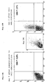
- Figure 1 shows the binding capacity, determined by flow cytometry, of GM-7 to monocytic cells after in vitro modification according to the invention. It can be seen that the CD14-positive monocytes obtained directly from buffy-coat do not bind the antibody GM-7 (the cloud shaded grey is congruent with the non-shaded antibody control). In contrast, following cultivation in the presence of M-CSF and stimulation with ⁇ -IFN, part of the monocytes express an antigen which is recognised by the monoclonal antibody GM-7. The monoclonal antibody GM-7 was characterised as isotype ⁇ -IgG 2a . The process according to the invention consequently leads to a change in the phenotypic pattern of the antigen expression on the cell membrane of the modified monocytes ( Figure 1).
- the monoclonal antibody GM-7 binds specifically to that cell population which, among those cells produced by the process according to the invention, induce the most effective transplant acceptance (see Figure 9).
- a preferred embodiment of the invention relates to such TAIC, which are capable of binding the antibody GM-7. These cells are subsequently designated as TAIC GM7 .
- the antibody GM-7 according to the invention therefore represents an extraordinarily effective and easy to handle agent for selecting and purifying the cells inducing transplant acceptance (TAIC).
- TAIC transplant acceptance
- the transplant acceptance inducing cells formed in step c) of the above described process of the invention, which express the antigen binding to the antibody GM-7 may either be selected directly from the culture medium after step c), or they may be selected from the cell population obtained after separating the cells from the culture medium according to step d) of the above-mentioned process of the invention by binding to the antibody GM-7 produced by the hybridoma cell line DSM ACC2542.
- the antibody is contacted with the sample under conditions which permit binding of the antibody to the transplant acceptance inducing cells present in the sample.
- the reaction complexes resulting from the binding reaction are subsequently separated from the sample.
- the antibody can be immobilised on a carrier material before contact with the sample; for example, it can be bound to a matrix suitable for chromatographic purposes or to so-called "magnetic beads". This procedure allows to select and concentrate transplant acceptance inducing cells from large volumes of sample.
- the bond between the antibody and the transplant acceptance inducing cells is separated after the isolation of the reaction complex from the sample.
- This can be effected by methods known in the state of the art such as e.g. by competitive displacement or by washing with salt solutions.
- Corresponding methods are for instance described by Utz U. et al. ("Analysis of the T-cell Receptor repertoire of human T-cell leukemia virus type-1 (HTLV-1) Tax-specific CD8+ Cytotoxic T Lymphocytes from patients with HTLV-1 associated disease: Evidence for the oligoclonal expansion" J. of Virology Feb. 1996, 843-851).
- the monoclonal antibody GM-7 permits the qualitative and quantitative detection of the transplant acceptance inducing cells of monocytic origin according to the invention in blood and/or tissue samples of the patient in vitro.
- This patient may, for example, be the recipient of an organ yet to be transplanted or already transplanted.
- the formation of reaction complexes in the sample which indicate the presence and, if applicable, the quantity of the transplant acceptance inducing cells is detected by known methods.
- detectable molecules are described in large numbers in the field of molecular diagnostics and includé, among others, fluorescent dyes such as fluorescein isothiocyanate or tetramethyl rhodamine-5-isothiocyanate, luminescent dyes, radioactively labelled molecules and enzymes such as peroxidases (compare Lottspeich, F., Zorbas, H. "Bioanalytik", Spektrum Akademischer Verlag GmbH, Heidelberg-Berlin, 1998).
- the detection of the antibody takes place dependent of the molecule selected for labelling of the former.
- the antibody GM-7 was coupled with the fluorescent molecule fluorescein isothiocyanate (FITC) so that the detection of the antibody could be carried out by means of flow cytometry and/or fluorescence microscopy.
- FITC fluorescent molecule fluorescein isothiocyanate
- the reaction complex can also be detected in a two-stage process using secondary antibodies.
- the unlabelled antibody GM-7 can be detected in the reaction complex with a further labelled antibody (compare Lottspeich, F., Zorbas, H. "Bioanalytik", Spektrum Akademischer Verlag GmbH, Heidelberg-Berlin, 1998).
- This two-stage method of detection is considerably more sensitive than the direct detection of binding of the antibody according to the invention since several labelled secondary antibodies can bind to one GM-7 antibody (signal amplification).
- the antibody GM-7 consequently allows the detection of TAIC in the peripheral blood of the patient treated with TAIC, for example in the form of "monitoring", during which the number of cells in the peripheral blood is determined at specific time intervals.
- TAIC prepared from monocytes of the donor correlates, in the animal test in the peripheral blood of the transplant recipient, with the tolerance of the transplanted organ. This finding consequently permits the clinician to wean off or to reduce the dosage of the immuno-suppressants that are optionally administered stepwise.
- a particularly preferred embodiment of the invention relates to a sub-population of the TAIC of the invention, which co-express the antigens CD3 and CD14 on their cell surface. These cells are subsequently indicated as TAIC CD3+/CD14+ . Such cells have to date not been reported in the state of the art. Monocytes and known cells derived from monocytes do carry the surface marker CD14, however, they do not additionally carry the surface marker CD3 at the same time.
- TAIC which co-express the surface antigens CD3 and CD14 may either be directly selected from the transplant acceptance inducing cells formed in step c) of the above described process of the invention, or they may be selected from the cell population obtained after separating the cells from the culture medium according to step d) of the above-mentioned process of the invention, or they may alternatively be selected from the TAIC GM7 population.
- the TAIC CD3+/CD14+ express the genes Foxp3, CTLA4 and Integrin ⁇ E ⁇ 7 strongly (see Example 12). In contrast, these genes are not or only to a small extent expressed by the original monocytes. The upregulation of the expression of the genes Foxp3, CTLA4 and Integrin ⁇ E ⁇ 7 is therefore a characteristic of TAIC CD3+/CD14+ -cells.
- T-lymphocytes which co-express the surface antigens CD4 and CD25 are a sub-population of regulatory T-lymphocytes, which are also indicated as "suppressor cells”. It is their function to suppress the immune response of the body.
- Foxp3 is seen as a specific transcription factor, which serves as a control gene for the development of regulatory T-cells, and which is specifically expressed by these cells.
- the TAIC CD3+/CD14+ -cells express at least 1 ⁇ 10 -9 , more preferably at least 5 ⁇ 10 -9 , and in particularly preferred manner at least 1 ⁇ 10 -8 ⁇ g Foxp3-RNA per ⁇ g total RNA.
- CTLA4 is similarly viewed as a marker for the detection of the regulatory function of T-lymphocytes, in particular of CD4/CD25 positive T-lymphocytes (see the literature cited in Example 12) .
- the TAIC CD3+/CD14+ -cells should preferably express at least 5 ⁇ 10 -7 , more preferably at least 3 ⁇ 10 -6 and in a particularly preferred manner at least 5 ⁇ 10 -6 ⁇ g CTLA4-RNA per ⁇ g total RNA.
- Integrin ⁇ E ⁇ 7 which recognizes epithelial Cadherin was recently described by Lehmann et al. in PNAS 99 , pages 13031-13036 (2002) as a new marker for a sub-population of highly potent regulatory T-lymphocytes, which interact with the epithelial environment.
- the expression of the Integrin ⁇ E ⁇ 7 -RNA should according to the invention amount in TAIC CD3+/CD14+ -cells to preferably at least 1 ⁇ 10 -12 , more preferably to at least 1 ⁇ 10 -11 , and in a particularly preferred manner to at least 1 ⁇ 10 -10 , and most preferably to at least 1 ⁇ 10 -9 ⁇ g per 1 ⁇ g total RNA.
- the direct co-culturing of the TAIC of the invention with lymphocytes leads to a significant increase in the number of regulatory T-lymphocytes, in particular of CD4/CD25 double-positive cells in the lymphocyte population with strongly up-regulated expression of the genes Foxp3, CTLA4 and Integrin ⁇ E ⁇ 7 .
- the Example further demonstrates that this effect is not observed if TAIC are indirectly co-cultured with lymphocytes.
- Example 13 confirms this hypothesis.
- lymphocytes from the recipient animals of Examples 3, 4, 5, 6 and 7 were incubated with TAIC from the respective donor animals in vitro.
- TAIC pre-incubated with the lymphocytes from the recipients were injected to the animals instead of TAIC.
- Donor specific tolerance could be induced also in this manner, while animals, to which recipient lymphocytes not co-cultivated with donor derived TAIC were administered, did not develop tolerance.
- the TAIC of the invention can be used as such as pharmaceutical preparation.
- the cells obtained from step d) of the method of the invention as described above can be used directly. About 10 - 50 % of the total cells of the populations so obtained is formed by lymphocytes and granulocytes, which stem from the initial monocyte isolate (buffy-coat). These cells support the formation of the TAIC of the invention derived from the monocytes in the culturing step (see Example 11); they do not interfere with the tolerance induction if the TAIC of the invention are used as a pharmaceutical preparation.
- the sub-populations TAIC GM7 and/or TAIC CD3+/CD14+ may be isolated from the totality of the TAIC population obtained from process of the invention (see above) and may be used for tolerance induction.
- the TAIC or the TAIC GM7 and/or TAIC CD3+/CD14+ may be kept for at least 48 hours without their tolerance inducing effect becoming lost.
- the TAIC or the sub-populations TAIC GM-7 and/or TAIC CD3+/CD14+ suspended in e.g. human AB serum can be administered intravenously as short transfusion.
- the TAIC generated from the monocytes of the donor or the sub-populations TAIC GM7 and/or TAIC CD3+/CD14+ can be injected into the MHC-different recipient either pre-operatively or postoperatively.
- the TAIC should be injected once to three times approximately 1 week before the operation.
- the period between the operation and the single administration of the cells should not be longer than 7 days.
- the TAIC according to the invention or the sub-populations TAIC GM7 and/or TAIC CD3+/CD14+ are then capable of repelling the T-cell response of the recipient's immune system against the transplant and to persist in the recipient blood for a sufficiently long period of time to guarantee long-term transplant acceptance.
- Preoperative intravenous injection can be considered in connection with donations from living individuals; however, if a corpse donation (blood and organ from a dead body) is at issue, postoperative administration of the TAIC according to the invention or the sub-populations TAIC GM7 and/or TAIC CD3+/CD14+ may be preferred.
- a corpse donation the body of the donor is flushed with a perfusion medium by switch of the principal artery for purposes of organ preservation.
- the venous blood is normally sucked out via the vena cava and discarded.
- the venous blood can be collected and processed as described in Example 1.
- TAIC may also be obtained from cells (lymphocytes and monocytes) from the donor spleen.
- the interval between the transplantation and the application of the cells can be overcome by combination with immuno-suppressants, in order to prevent an acute rejection of the organ in the interval between the transplantation and the provision of the TAIC obtained from the donor blood.
- immuno-suppressants such as, for example, calcineurin inhibitors such as cyclosporin A (CSA) or tacrolimus or with azathioprine (AZA), mycophenolate mofetil, rapamycin, monoclonal antibodies (ATG, ALG, but neither with Dicliziumab or Basiliximab) or steroids (STE) can be considered.
- immuno-suppressants except for known IL-2-receptor- ⁇ monoclonal antibodies such as Dicliziumab and Basiliximab
- TAIC TAIC
- the cells of monocytic origin which have been modified according to the invention can be used as "vehicle for tolerance transfer" for any cellular transplant (such as islet cells, hepatocytes, adult stem cells and for any other programmed cell type or tissue type) and organ (such as e.g. kidney, liver, heart) insofar as they are genetically identical to the cells to be transplanted (organs), i.e. they must originate from the donor him/herself or from identical twins thereof.
- the TAIC fulfil their protective function by allowing the transplanted cells / organs to become adherent in the new environment, thus saving the recipient from suffering the side effects of a longterm immunosuppressive therapy.
- the starting cells for the process according to the invention are blood monocytes. These are preferably monocytes from human blood. For purposes of inducing transplant acceptance, the cells must originate from the donor of the transplant (or his/her identical twin). In the case of xenogeneic transplantations of e.g. monkey or pig organs into man, the TAIC according to the invention must consequently be derived from the monocytes of the donor animal concerned.
- the blood can first be separated, after the usual treatment with an anticoagulant, into plasma and into white and red blood cells using methods known in the art, preferably by centrifugation. After centrifugation, the plasma will be present in the supernatant; below it, there is a layer which contains the white blood cells in their entirety. This layer is also referred to as buffy-coat. Below this is the phase containing the red blood cells (hematocrit).
- the buffy-coat layer is first isolated and separated to obtain the monocytes e.g. by centrifuging according to known methods.
- the buffy-coat layer is applied onto a lymphocyte separation medium (Ficoll-Hypaque) and centrifuged (see Example 1).
- Example 1 describes the preferred embodiment of the invention, wherein the erythrocytes and dead cells still contained in the buffy-coat are separated by centrifuging, and the white blood cells including the monocytes are present as an isolate on the separation medium. Thereafter, the white phase of monocytes may be carefully pipetted off, and, for enrichment of the monocytes within the isolate, are repeatedly centrifuged and washed. In the course of this process, the monocytes will assemble at the bottom of the centrifuge vessel together with a part of the lymphocytes.
- a lymphocyte separation medium Ficoll-Hypaque
- the conditions for obtaining the monocyte containing isolate are controlled such that the isolate contains about 10 - 50 % lymphocytes next to the monocytes, by reference to the total number of cells.
- the isolate contains about 50 - 90 %, in a particularly preferred manner 60 - 70 % monocytes and about 10 - 50 %, in a particularly preferred manner 20 - 50 % lymphocytes, each by reference to the total cell count, wherein the difference will optionally be provided by granolocytes.
- lymphocytes in a magnitude of 20 - 30 % referred to the total cell count
- M-CSF macrophage-colony-stimulating-factor
- the concentration of M-CSF in the culture medium may preferably amount from 2 to 20 ⁇ g/l medium, more preferably 4 to 6 ⁇ g/l and in particularly preferred manner 5 ⁇ g/l.
- the cells must be stimulated with ⁇ -IFN, i.e. cultured in the presence of ⁇ -IFN.
- ⁇ -IFN i.e. cultured in the presence of ⁇ -IFN.
- the stimulation of the monocytes with ⁇ -IFN takes place after an initial propagation phase lasting 3 to 6 days in the culture medium containing the growth factor.
- ⁇ -IFN stimulation is carried out and this stimulation is extended over a period of preferably 24 to 72 hours, more preferably 48 hours under incubator conditions i.e. at 37°C and in a 5% CO 2 atmosphere.
- the concentration of ⁇ -IFN in the medium may be 0.1 to 20 ng/ml, preferably 1 to 10 ng/ml and particularly preferably 5 ng/ml.
- the stimulation with ⁇ -IFN may begin simultaneously with the propagation of the monocytes in the medium containing the growth factor. However, stimulation after a 3 to 6 day long initial propagation phase, as indicated above, is preferred.
- the propagation of the cells and stimulation with ⁇ -IFN should, overall, preferably not take more than 8 days. In any case, treatment with ⁇ -IFN should be carried out such that after the propagation phase it lasts for at least 24 hours, at maximum 72 hours, preferably 48 hours. The period for propagation and stimulation of the cells should consequently last for a total of preferably 4 to 8 days.
- the propagation and stimulation with ⁇ -interferon is carried out as indicated in Example 2 in such a way that the monocytes are first propagated in a culture medium containing the growth factor and that ⁇ -IFN is added to the culture medium after 3 to 6 days in such an amount that a concentration of 0.1 to 20 ng/ml, preferably 1 to 10 ng/ml and particularly preferably 5 ng/ml is obtained in the medium.
- the process according to the invention is carried out in culture vessels the surface of which has previously been coated with fetal calf serum (FCS) or alternatively with human AB serum (see Example 2).
- FCS fetal calf serum
- Coating with FCS can take place by covering the surface of the culture vessels with FCS before use and, following a period of interaction of a few hours, in particular 4 to 72 hours, preferably 12-48 hours and in particular 24 hours, removing the FCS not adhering to the surface in a suitable manner.
- the cells will settle at the bottom of the culturing vessel after about 24 hours. Due to their adhesive properties the monocytes and the TAIC, derived from the monocytes during the process, will then adhere to the bottom of the respective culture vessel. If, as described in Example 2, the culture medium is changed during cultivation, the supernatant is initially carefully removed, for instance by pipetting off or decanting, and subsequently fresh culture medium is filled in. Preferably however, the cells adhering to the bottom are not or only carefully washed, so that any lymphocytes present are not removed.
- Removing the adhering cells can take place mechanically, e.g. by means of a fine cell scraper or spatula.
- the complete removal of the cell takes place by treatment with a suitable enzyme, e.g. with trypsin (see Example 2).
- a suitable enzyme e.g. with trypsin (see Example 2).
- the trypsin solution (0.1 to 0.025 g/l, preferably 0,05 g/l) can be allowed to act onto the cells for 2 to 10 minutes at 35°C to 39°C, preferably at 37°C, in the presence of 5% CO 2 .
- the enzyme activity is then blocked in the usual manner and the now freely floating TAIC can be obtained in the usual way by centrifuging. They are then available for immediate use, optionally in suspension in a suitable medium, e.g. in PBS. However, they can also be kept for several days, in particular for approximately 2 to 3 days, in a nutrient medium (see Example 2); wherein this preservation medium should contain neither the growth factor nor ⁇ -IFN.
- the cells can be kept in such a nutrient medium as TAIC for at least 48 hours.
- the cells can be deep frozen. Protocols for deep freezing of living cells are known in the state of the art, compare Griffith M. et al., ("Epithelial Cell Culture, Cornea", in Methods of tissue engineering, Atala A. and Lanza R.P., Academic Press 2002, chapter 4, pages 131-140).
- a preferred suspension medium for deep freezing of the cells according to the invention is FCS containing DMSO.
- the cell suspension containing transplant acceptance inducing cells obtained from steps c) or d) may be further purified with respect to those cells, which bind the antibody GM-7, so as to obtain a sub-population TAIC GM7 .
- Methods for such purification are described above in detail.
- such cells are selected from the TAIC population, which co-express the antigens CD3 and CD14 on their cell surfaces.
- Methods for selecting such cells are known in the art. Examples for such methods are the “Fluorescent-Activating Cell Sorting” (FACS), the “Immuno Magnetic Bead Sorting” and the “Magnetic Activated Cell Sorting” (MACS), or the so-called “Rosetting Method” [see Gmelig-Meyling F. et al. "Simplified procedure for the separation of human T- and non-T-cells", Vox Sang. 33, 5-8 (1977)].
- the selection of the TAIC sub-population TAIC CD3+/CD14 + can take place directly from the TAIC population obtained from steps c) or d) of the above-described process of the invention, or from the TAIC GM7 -sub-population.
- the latter way to proceed means that a stepwise enrichment of TAIC CD3+/CD14+ will take place.
- the TAIC according to the invention are used per se for the production of a pharmaceutical composition for the in vivo suppression of transplant rejection.
- Such pharmaceutical preparation may contain vital TAIC according to the invention, which are obtained from step d) of the process of the invention, suspended in a pharmaceutically acceptable carrier preferably in a quantity of about 1 ⁇ 10 5 to 1 ⁇ 10 7 cells/ml and more preferably of about 1 ⁇ 10 6 cells/ml of the preparation.
- the cells of the TAIC GM7 sub-population according to the invention are used per se for the production of a pharmaceutical composition for the in vivo suppression of transplant rejection.
- Such pharmaceutical preparation may contain vital TAIC GM7 cells according to the invention, which bind to the antibody GM-7, suspended in a pharmacologically acceptable liquid carrier, preferably in a quantity of about 1 ⁇ 10 6 to 1 ⁇ 10 8 cells/ml and more preferably of about 1 ⁇ 10 6 cells/ml of the preparation.
- the cells of the TAIC CD3+/CD14+ sub-population according to the invention are used per se for the production of a pharmaceutical composition for the in vivo suppression of transplant rejection.
- Such pharmaceutical preparation may contain vital TAIC CD3+/CD14+ cells according to the invention, which co-express the antigens CD3 and CD14, in a quantity of preferably about 5 ⁇ 10 5 to 5 ⁇ 10 7 cells/ml and more preferably of about 5 ⁇ 10 6 cells/ml of the preparation.
- the above described pharmaceutical preparations may contain the cells according to the invention suspended in a physiologically well-tolerated medium.
- Suitable media are for example Ringer solution, physiological saline or 5 to 20 % human albumin solution and the like.
- Cell preparations according to the invention may contain vital TAIC, which are obtained from step d) of the process of the invention.
- the preparations may contain cells belonging to the sub-populations of TAIC GM7 cells, which bind to the antibody GM-7, or of TAIC CD3+/CD14+ cells, which co-express the antigens CD3 and CD14 on their cell surface.
- the preparations may contain the respective cells in a quantity of preferably at least 1 ⁇ 10 5 , more preferably at least 5 ⁇ 10 5 and most preferably at least 1 ⁇ 10 6 cells per ml suspended in a liquid carrier medium.
- the medium may be a cell culture or transport medium well-tolerated by cells, such as 5 to 20% human albumin solution.
- the cells within the preparation may be deep-frozen and contained in a suitable storage medium, such as e.g. RPMI with 50% human albumin solution and 10% DMSO.
- the invention also relates to a method wherein the transplant-acceptance inducing cells of the invention (TAIC, TAIC GM7 or TAIC CD3+/CD14+ ) are used for generating or expanding regulatory T-lymphocytes in vitro.
- the direct in vitro cocultivation of TAIC with lymphocytes leads to a significant proliferation of regulatory T-lymphocytes, in particular of CD4 + /CD25 + lymphocytes, see Wood and Sakaguchi: "Regulatory Cells in Transplantation Tolerance", Nature Review Immunology 3, 199-210 (2003). It is therefore possible to generate and/or expand regulatory T-lymphocytes in particular CD4 + /CD25 + lymphocytes by directly coculturing TAIC with lymphocytes as described in Example 12.
- Direct in vitro culturing means that TAIC and lymphocytes are cocultured in direct physical contact within the same medium, as a.o. exemplified in Example 12.
- the medium preferably contains the respective cells i.e. TAIC and lymphocytes in about equal cell numbers and each in a quantity of preferably at least 1 ⁇ 10 5 , more preferably at least 5 ⁇ 10 5 and most preferably at least 1 ⁇ 10 6 cells per ml suspended in a liquid carrier medium; the medium may be a cell culture or transport medium well-tolerated by cells, such as 5-20% human albumin solution.
- the co-cultivation preferably should take place under physiological conditions at about 37°C, e.g. in an incubator, for preferably about 3 to 5, more preferably 4 days.
- transplant acceptance can not only be induced by administering TAIC generated from donor monocytes to the recipient, but also by readministering recipient lymphocytes to the recipient, which have previously been directly cocultured in vitro with TAIC prepared from donor monocytes as described above.
- regulatory T-lymphocytes can therefore be prepared in vitro from lymphocytes originating form the recipient of a transplant, by direct coculturing of lymphocytes from the recipient with TAIC originating from the donor of that transplant. Readministering the cocultivated lymphocytes to the recipient will lead to transplant acceptance by the recipient, as shown in Example 13.
- the recipient derived regulatory T-lymphocytes so prepared may be isolated by FACS as described herein (see above) and may be used in a pharmaceutical preparation for preventing transplant rejection in the recipient, wherein the cells are suspended in a pharmacologically acceptable carrier as described above.
- composition of the media and substances used are as follows: 1. Penicillin/streptomycin solution: 10,000 units of penicillin as sodium salt of penicillin G and 1000 ⁇ g streptomycin as streptomycin sulphate per ml physiological sodium chloride solution (NaCl 0,85 %) (Gibco Catalogue No. 15140122). 2. Trypsin-EDTA 0.5 g trypsin and 0.2 g EDTA (4 Na)/l 3. RPMI 1640 (1x, liquid (11875)) contains L-Glutamine RPMI (Roswell Park Memorial Institute) Media 1640 are enriched formulations, which can be used extensively for mammalian cells. Components Mol-weight Conc.
- 450 ml human whole blood was mixed in a triple chamber bag set with 63 ml of a stabiliser solution which contained, per litre H 2 O, 3.27 g of citric acid, 26.3 of g trisodium citrate, 25.5 g of dextrose and 22.22 g of sodium dihydroxy phosphate.
- the pH of the solution was 5.6-5.8.
- the quantity of 50 ml of freshly obtained buffy-coat was then divided into 2 portions of 25 ml each and layered onto 25 ml of Ficoll-Hypaque separation medium respectively which had been introduced previously into two 50 ml Falcon tubes.
- This preparation was centrifuged for 30 minutes at 2500 rpm without braking. Thereafter, any erythrocytes and dead cells still present in the buffy-coat were below the Ficoll phase, whereas the white blood cells, including the monocytes, were separated off on the Ficoll as white interphase.
- the white interphase including the monocytes was carefully removed by pipetting and mixed with 10 ml of phosphate-buffered saline (PBS).
- PBS phosphate-buffered saline
- This preparation was then centrifuged three times for 10 minutes at 1800 rpm with braking, the supernatant being removed by pipetting after each centrifuging process and fresh PBS being introduced.
- the cell pellets assembled at the bottom of the centrifuging vessel contained the mononuclear cell fraction, i.e. the monocytes.
- the cultivation and propagation of the monocytes was carried out in nutrient medium with the following composition: RPMI 1640 medium 440 ml Fetal calf serum (FCS), alternatively, AB serum 50 ml Penicillin / Streptomycin Solution 5 ml Total volume 500 ml
- the nutrient medium contained 2,5 ⁇ g / 500 ml M-CSF.
- the monocytes isolated in Example 1 were suspended in a total quantity of 10 6 cells in 10 ml of the nutrient medium and transferred onto a Petri dish (100 mm in diameter).
- the Petri dish had previously been filled with pure inactivated FCS and the FCS had been poured off after 24 hours in order to obtain, in this way, a dish coated with FCS.
- the Petri dish was covered with the appropriate cover and kept for 3 days in an incubator at 37°C.
- the cells settled at the bottom of the Petri dish after 24 hours.
- the supernatant was pipetted off and the Petri dish again filled with 10 ml of fresh nutrient medium.
- the determination of the cell count for the determination of the exact dosage took place according to known methods, compare Hay R.J., "Cell Quantification and Characterisation", in Methods of Tissue Engineering, Academic Press 2002, Chapter 4, S. 55-84.
- This cell suspension was centrifuged (1800 rpm, 10 minutes, see above) and the cell pellet were incorporated either into PBS or for application in man into NaCl (physiol.). Subsequently, the intravenous administration can take place directly or within 48 hours.
- FCS/DMSO was added as freezing medium to the cells and these were deep frozen in a volume of 10 ml.
- the freezing medium contained 95% FCS and 5% DMSO. In each case, approximately 10 6 cells were incorporated into 1 ml of the medium and cooled in the following steps:
- Figure 1 shows the phenotypic changes in the antigen expression, determined by flow cytometry, on the monocytes used after cultivation and ⁇ -IFN stimulation of the original monocytic cells.
- TIC Transplant Acceptance Inducing Cells
- TAIC for treating a potential recipient before transplantation offers itself, for example, in the clinical case of a live donation, where it has been clarified in advance of the organ transplantation who the donor will be and who will receive the donor organ.
- the heterotopic heart transplantation was carried out in the rat model on male inbred rats of the strain combination LEW[RT1. 1 ] (in connection with the present invention referred to as "LEW") -> DA [RT1. avl ] (in connection with the present invention referred to as "DA") using the technique described by Ono and Lindsey [Ref. Ono, K. and Lindsey, E.S. "Improved technique of heart transplantation in rats". J. Thorac. Cardiovasc. Surg. 57, 225-229 (1969)].
- the DA recipient rats received 10 6 TAIC derived from LEW monocytes intravenously in 1 ml of PBS.
- the transplantation took place seven days later of a heart taken from the LEW rat strain, which was heterotopically implanted into the abdomen (abdominal cavity) of the DA recipient animal. Since the rat strains used were inbred strains, the TAIC according to the invention expressed identical tissue antigens as the transplanted LEW heart.
- TAIC 10 6
- CAP CAP rat inbred strain
- CAP rats express the haplotype RT1. c , they are therefore fully MHC-discordant (MHC-different) to the LEW donor animals.
- LEW -> DA 10 6 LEW derived TAIC i.v., day -7 before LEW heart transplantation; 3. CAP -> DA; 10 6 LEW derived TAIC i.v., day -7 before LEW heart transplantation; 4. CAP -> DA; untreated; 5. LEW -> LEW, untreated; 6. LEW -> LEW, 10 6 LEW derived TAIC i.v., day -7 before LEW heart transplantation.
- Figure 2B shows the histological evaluation of a LEW allogeneic transplant on postoperative day (POD) 150, following heterotopic transplantation into the abdomen of a DA recipient rat.
- the recipient rat had been pre-treated on day -7 before transplantation with 10 6 LEW-derived TAIC.
- the transplant was functioning satisfactorily (strong heart beat) up to the time of its removal from the recipient rat (POD) 150.
- the histological preparation (x40) showed a healthy heart muscle morphology, normal vascular endothelial tissue and only minimal infiltration with mononuclear cells without any indication of acute and chronic rejection processes.
- FIG. 2C shows a histological preparation of a thymus, removed on POD 150, of a DA rat pre-treated with 10 6 LEW-derived TAIC. The preparation was labelled with the monoclonal antibody I1.69 specific for LEW-MHC class I.
- administering TAIC leads to the colonisation of the thymus gland where the presentation of donor-derived MHC antigens to the maturing T-cells takes place; small nests of cells can be clearly recognised which can be labelled with the antibody.
- the induction of a mixed chimerism by the intravenous injection of the LEW-derived TAIC in the DA animal concerned can be considered as a possible cause of the tolerance induced according to the invention.
- a test was therefore carried out to check whether, following the injection of LEW-derived TAIC, more than 5% of cells can be detected in the blood of the recipient animal by flow cytometry in the long term (>45 days).
- the flow cytometry was carried out as described in the state of the art [Ref. Preffer F.I., "Flow cytometry" In : Diagnostic Immunopathology, Colvin RB, Bhan AK, McCluskey, RT ( eds. ), Raven Press New York, pp. 725-449 (1994)].
- the proportion of donor cells in the peripheral blood of the DA rat can be highly accurately determined with I1.69.
- I1.69 the donor antigen labelled by I1.69 within the first 6 weeks following the intravenous injection of the TAIC.
- this proportion of the chimerism in the peripheral blood of these DA rats decreases substantially and disappears completely on day 100.
- this transient chimerism correlates closely to the transplant survival of LEW hearts transplanted after double time (i.e.
- LEW -> DA untreated 2.
- LEW -> DA; CSA 5 mg / kg ⁇ BW, i.v., day 0, 1, 2 and 3 3.
- LEW -> DA; CSA 5 mg / kg ⁇ BW, i.v., day 0, 1, 2 and 3 plus 10 6 LEW-derived TAIC, i.v., day 7 4.
- CAP -> DA; CSA 5 mg / kg ⁇ BW, i.v. day 0, 1, 2 and 3 plus 10 6 LEW-derived TAIC, i.v., day 7
- the postoperative intravenous treatment of the DA recipient rats with 10 6 LEW-derived TAIC on POD (postoperative day) 7 leads to long-term tolerance if an initial administration of 4 cycles of CSA (5mg/kg ⁇ BW, i.v.) has been effected from day 0-3.
- the initial administration of CSA is necessary since the sole treatment with a single administration of 10 6 TAIC on POD 7 (postoperative day) prevents acute rejection only in 1 out of 6 cases.
- the CSA dose chosen in this case has only a sub-therapeutic effect since no long-term acceptance can be induced by the exclusive CSA administration even though the survival of the transplant is prolonged.
- the induced transplant acceptance is donor-specific since a corresponding treatment of DA rats with LEW-derived TAIC does not induce any tolerance for CAP-third strain heart transplants.
- the hearts of experimental group 1 were rejected acutely after 7.0 ⁇ 0.8 days.
- the hearts of group 2 were rejected with a delay after 14.4 ⁇ 7.0 days.
- the hearts of group 3 were accepted for longer than 150 days in 5 out of 6 cases.
- the sole postoperative administration of TAIC (10 6 cells) was able to prevent acute rejection only in 1 out of 6 cases since the heart organs ceased to beat after 22.6 ⁇ 31.6 days (group 4).
- the third strain control with CAP hearts showed the specificity of induced transplant acceptance since third strain hearts were again rejected acutely after 7.4 ⁇ 2.2 days.
- DA -> LEW rat inbred system 1. DA (kidney) -> LEW; untreated 2. DA (kidney) -> LEW; 10 6 DA-derived TAIC, i.v., day -7 and -1 before transplantation 3. LEW (kidney) -> LEW; untreated 4. DA (liver) -> LEW; untreated 5.
- FIG. 4D shows that as a result of the intravenous injection, given twice, of 10 6 TAIC per kg body weight on days -7 and -1 before transplantation, the morphology of the transplanted kidney can be shown to be intact (compare control kidney and syngeneic transplant LEW -> LEW), whereas, in the untreated allogeneic control group DA -> LEW, a clear infiltration of the transplant in the interstice can be recognised (on postoperative day 14).
- the allogeneic liver transplant ( Figure 4E), which had been fully rejected on POD 12 without TAIC pre-treatment (DA -> LEW without TAIC), behaves in an analogous manner whereas the prior administration of TAIC allows the parenchyma of liver transplants to appear as being completely homogeneous and intact in comparison with the syngeneic transplant.
- the allogeneic skin transplant (DA) also heals without complication (compare comparison with the syngeneic LEW transplant) whereas the CAP third strain graft transplanted simultaneously was rejected acutely as early as on postoperative day 12.
- the inbred strain BN[RT1. n ] (referred to as "BN" in connection with the present invention) was also used, among others. 1. DA -> LEW; untreated 2. DA -> LEW; CSA, 5 mg / kg ⁇ BW, i.v., day 0, 1, 2, 3 plus 10 6 DA-derived TAIC / kg ⁇ BW, i.v., day 7 and 10 postoperative 3. CAP -> LEW; untreated 4.
- LEW -> DA 10 6 / kg ⁇ BW LEW-derived blood monocytes (stimulated with ⁇ -IFN), i.v., day -7 before heart transplantation 4.
- LEW -> DA 10 6 / kg ⁇ BW LEW-derived blood monocytes (treated with M-CSF plus stimulated with ⁇ -IFN), i.v., day -7 before heart transplantation
- the left lung of a donor pig is removed and transplanted orthotopically into a genetically different recipient pig (namely into the left thoracic cavity following the removal of the left lung from the recipient pig).
- 500 ml of donor blood was removed simultaneously which served to prepare the TAIC from the monocytes of this pig.
- the donor animals all had a weight of 30-35 kg.
- Figure 6B shows the lung transplants of a pig of experimental group 1 (POD 41) and a pig treated with TAIC from experimental group 3 (55 days after transplantation; B).
- a visible reduction in transparency shading
- the thorax x-ray picture of the animal treated with TAIC shows an x-ray unobtrusive lung and transplant finding respectively, with a good delimitation with respect to the heart silhouette.
- the monoclonal antibody GM-7 was generated by immunising mice with human TAIC which had been prepared as described in Example 2.
- the hybridoma cells producing the antibody were deposited at the "Deutsche Sammlung für Mikroorganismen" under the accession no. DSM ACC2542.
- the results reported below demonstrate that the antibody binds specifically to an antigen which is expressed only on CD14 + cells which had been subjected to the 6 day ex situ modification with M-CSF and 2 day ⁇ -IFN stimulation according to the invention.
- Figure 1 shows the binding capacity, determined by flow cytometry, of GM-7 to monocytic cells after in vitro modification, i.e. after transformation into TAIC. It can be seen that the CD14-positive monocytes obtained directly from buffy-coat do not bind the antibody GM-7 (the grey shaded cloud corresponds to the antibody control). In contrast, part of the monocytes express an antigen after cultivation in M-CSF and stimulation with ⁇ -IFN, which antigen is recognised by the monoclonal antibody GM-7. After cultivation as described in Example 2, about 80% of the transformed monocytes are able to bind the monoclonal antibody GM-7.
- the suppressor activity of the CD14 + /GM-7 + cells was compared with that of CD14 + /GM-7 - cells in a mixed lymphocyte culture (MLC).
- MLC mixed lymphocyte culture
- the TAIC stem from an individual B.
- lymphocytes with 2 ⁇ g PHA were transferred into the wells of a 96-well-plate, followed by a proliferation period over 144 hours (indicated as "PhaLy”). Lymphocytes only cultivated in medium without addition of PHA were run as a further control (indicated as "Ly”).
- lymphocytes not stimulated with PHA do not significantly proliferate, the mean radioactivity observed being 367 cpm (see Figure 8).
- the stimulation with 2 ⁇ g PHA leads to a significant increase in the proliferation rate of the lymphocytes (“PhaLy”), the highest incorporation being measured at a mean value of 18459 cpm in these samples.
- lymphocytes present in the monocyte fraction were determined by comparison of two different set ups.
- the monocyte fraction was initially taken from the interphase of the buffy-coat as described in Example 1. As described in Example 2, the cells were subsequently transferred to the cultivation step with M-CSF. Within one hour after the starting point of the cultivation, the monocytes adhering to the bottom of the tissue culture flask were washed five times with 10 ml PBS each, and the amount of the lymphocytes present in the culture was thereby decreased to ⁇ 5 % (4,8 ⁇ 2,4 %), while the amount of enriched monocytes (CD14 + ) so obtained was above 90 % (92 ⁇ 5.6 %). Additional cell components within the set up were B-lymphocytes and granulocytes.
- the cells in the second set up were also taken from the interphase of the "buffy-coat” as monocyte fraction as described in Example 1.
- the cells adhering to the bottom of the tissue culture flask were only washed once after 24 hours starting from the beginning of the cultivation phase, as described in Example 2.
- a cell population was obtained, which was composed of 45 ⁇ 5,3 % CD14 + -monocytes and 23,5 ⁇ 8,9 % CD2 + -lymphocytes.
- lymphocytes and granulocytes were also present in this set up.
- the lymphocytes proliferate quicker than the monocytes; the relative amount of lymphocytes increased in set up "Mo” from 4,8 % to 69,8 % and in set up "Mo+Ly” from 23,5 % to 50,6 %.
- CD14 + /CD3 + -cells effective as TAIC are formed in both cultures. It is important in this context that a significantly higher increase in CD14 + /CD3 + -cells is observed in the set up "Mo+Ly" with an amount of 32,0 ⁇ 5,3 % as opposed to set up "Mo" with only 7,2 ⁇ 3,2 %.
- TAIC of donor A were directly or indirectly co-cultivated in vitro together with lymphocytes of a donor B.
- the direct cell-to-cell-contact between the TAIC (of donor A) and the lymphocytes (of donor B) was made possible, while in the indirect co-culture a membrane ("cell culture insert", 0,4 ⁇ m pore size, Falcon, order no. 353090) allowed exchange of the medium, but inhibited the physical contact of the two cell populations.
- the direct or indirect co-culture is carried out for preferably 3 to 5 days, more preferably for 4 days under incubator conditions i.e. at 37°C and in a 5% CO 2 atmosphere.
- the respective numbers of regulatory T-cells (CD4 + /CD25 + ) were determined in both set ups as well as in the controls, wherein TAIC or lymphocytes respectively were cultivated alone. Further, the number of CD3 + /CD14 + -cells, which represent the component of the TAIC-cell population having the most significant suppressor function in the mixed lymphocyte culture, was determined in the control set up, wherein the TAIC were cultured alone.
- the surface antigens of the cells were determined per FACS analysis and the amount of the respective cell populations in the total amount of the cells was analysed.
- Foxp3 is a specific transcription factor, which is viewed as a control gene for the development of regulatory T-cells, and which is specifically expressed by these cells [see Hori, S. et al., "Control of Regulatory T-cell Development by the Transcription Factor Foxp3", Science 299 , 1057 - 1061 (2003)].
- CTLA-4 is a further factor, which is used as a marker for the determination of the regulatory function of CD4 + /CD25 + -T-cells (see Khattri, R. et al., "An essential role for Scurfin in CD4 + /CD25 + T regulatory cells", Nature Immunology, online publication, doi:10.1038/ni909 (2003); Shimizu, J. et al. "Stimulation of CD25 + /CD4 + regulatory T cells through GITR breaks immunological self-tolerance", Nature Immunology, online publication, doi:10.1038/ni759 (2003); Cobbold, S.P. et al. "Regulatory T cells in the induction and maintenance of peripheral transplantation tolerance", Transpl. Int. 16(2) , 66-75 (2003)].
- Integrin ⁇ E ⁇ 7 is an integrin, which binds to epithelial Catherine and which may be used as a marker for the most potent sub-population of regulatory CD25 + -T-cells [see Lehmann, J. et al. "Expression of the integrin ⁇ E ⁇ 7 identifies unique subsets of CD25 + as well as CD25 - regulatory T cells" PNAS 99(20) , 13031-13036 (2002)].
- the Table shows that within the population of lymphocytes obtained from the direct co-culture, the percentage of double positive cells CD4 + /CD25 + increases manifold to a value of 8,7 % in comparison to the amount of CD4 + /CD25 + lymphocytes obtained from the indirect co-culture or control, which is nearly identical with 2,38 % and 2,65 % respectively.
- CD4 + /CD25 + cells expresses the highest relative amounts of mRNA from all tested mastergenes as compared to the expression in CD4 + /CD25 - cells (see Table). While the expression of Foxp3 is increased by about a factor 10 (37 versus 3,75), CTLA-4 expression reaches an even higher maximum expression (4699 versus 0,376).
- the increase of expression of the third mastergene integrin ⁇ E ⁇ 7 in the CD4 + /CD25 + cells during co-culturing is nearly as high as for CTLA-4 since the absolute amount of integrin ⁇ E ⁇ 7 mRNA is raised to 1,4 ⁇ 10 -9 in CD4 + /CD25 + cells as compared to 3,4 ⁇ 10 -12 ⁇ g mRNA/ ⁇ g total RNA in CD4 + /CD25 - cells.
- the relative amount of Foxp3-mRNA in the CD4 + /CD25 + sub-population amounts to only 10 and is nearly as low as the relative amount expressed by the lymphocytes of the control, which expresses a relative amount of Foxp3-mRNA of 15.
- the relative amount of CTLA-4-mRNA expressed by the lymphocytes of the control is as low as 0,375 in the CD4 + /CD25 + -population and 0,1 in the CD4 + /CD25 - population.
- CTLA-4 in the CD14 + /CD3 + sub-population of the TAIC cells was similar to the expression of Foxp3 and was also significantly higher than in all other cell populations.
- CTLA-4 expression (relative value of 12.500) was manifold stronger in the CD14 + /CD3 + -sub-population than the Foxp3-expression, which exhibited a relative value of 50.
- Example 12 The results and conclusions presented in Example 12 were confirmed in vivo in an animal experiment. Different from the procedure described in Examples 3, 4, 5, 6 and 7, the animals in the selected LEW->DA Inbred combinations were not injected with LEW-derived TAIC for induction of tolerance, but DA-lymphocytes from the recipient which, over a period of 5 days, were previously directly co-cultivated (LEW)-derived TAIC from the donor, or which were cultivated alone in medium as controls, were injected to the animals in this example.
- LEW directly co-cultivated
Landscapes
- Health & Medical Sciences (AREA)
- Life Sciences & Earth Sciences (AREA)
- Immunology (AREA)
- Chemical & Material Sciences (AREA)
- General Health & Medical Sciences (AREA)
- Engineering & Computer Science (AREA)
- Cell Biology (AREA)
- Organic Chemistry (AREA)
- Medicinal Chemistry (AREA)
- Microbiology (AREA)
- Veterinary Medicine (AREA)
- Pharmacology & Pharmacy (AREA)
- Animal Behavior & Ethology (AREA)
- Public Health (AREA)
- Biomedical Technology (AREA)
- Genetics & Genomics (AREA)
- Bioinformatics & Cheminformatics (AREA)
- Mycology (AREA)
- Epidemiology (AREA)
- Biotechnology (AREA)
- Biochemistry (AREA)
- Wood Science & Technology (AREA)
- Zoology (AREA)
- Biophysics (AREA)
- Proteomics, Peptides & Aminoacids (AREA)
- Molecular Biology (AREA)
- General Engineering & Computer Science (AREA)
- Hematology (AREA)
- Transplantation (AREA)
- General Chemical & Material Sciences (AREA)
- Nuclear Medicine, Radiotherapy & Molecular Imaging (AREA)
- Chemical Kinetics & Catalysis (AREA)
- Gastroenterology & Hepatology (AREA)
- Micro-Organisms Or Cultivation Processes Thereof (AREA)
- Medicines Containing Material From Animals Or Micro-Organisms (AREA)
- Preparation Of Compounds By Using Micro-Organisms (AREA)
- Peptides Or Proteins (AREA)
- Measuring Or Testing Involving Enzymes Or Micro-Organisms (AREA)
Abstract
Description
- The invention relates to transplant acceptance inducing cells of monocytic origin and their preparation as well as their use for generating transplant acceptance.
- Preferably, the invention relates to transplant acceptance inducing cells (in the following also called TAIC) which are derived from human monocytes.
- The invention moreover relates to the monoclonal antibody GM-7, which specifically recognises transplant acceptance inducing cells according to the invention which are derived from man.
- In addition, the invention relates to the use of the antibody GM-7 for detecting and/or selecting transplant acceptance inducing cells.
- The term "transplantation" is used in the field of immunology for the transfer of cells, tissue or organs from one body to another. The need for transplantation arises from the finding that numerous diseases can be cured by transferring (transplanting) healthy organs, tissue or cells from one individual who is healthy in this respect - the donor - to an individual suffering from the disease concerned - the recipient or host.
- Depending on the relationship between the donor and the recipient, the following types of transplant can be distinguished:
- 1. Autologous transplants: These are tissues or cells which are transplanted within one and the same individual from one area of the body to another.
- 2. Isologous transplants: This involves the transfer between genetically identical individuals. Examples of these are inbred strains of rats or mice. For man, isologous (syngeneic) transplants can be considered between genetically identical (monocygotic) twins.
- 3. Allogeneic transplants: This term designates transplants between genetically different members of the same species such as, for example, from one human individual to another or, in the case of experimental animals, from one inbred strain to another.
- 4. Xenogeneic transplants: This involves the transfer between different species such as, for example, the transfer of a monkey heart into a human being.
- As a rule, autologous and isologous transplants cause no immunological problems in the sense of a rejection response. However, this is not true in the case of allogeneic and xenologous transplants. Such transplants are recognised as foreign by the recipient's immune system and rejected after a relatively short time. In the case of the allogeneic/ xenologous rejection response, T-lymphocytes (hereinafter called T-cells) play a central part. These cells recognise the foreign cells by way of their major histocompatibility complexes (MHC). The foreign MHC-complex sensitises the antigen-reactive T-cells of the recipient and, subsequently, the T-cells thus activated lead to the destruction of the donor cells or the donor tissue respectively. Cells with different MHC-complexes are also called "MHC-different".
- Transplanting allogeneic or xenogeneic cells, tissues or organs necessarily leads to a situation where cells of two different individuals, i.e. of MHC-different cells, are present in the blood of the recipient at the same time. This phenomenon is referred to as "chimerism". A distinction is made between micro-chimerism and macro-chimerism. Micro-chimerism refers to a state in which cells derived from the donor, e.g. after a blood or bone marrow transfusion or after transplanting lymphocyte-rich organs such as the small intestine or the liver, remain persistent in the MHC-different host but are detectable only in a few isolated cases. The term macro-chimerism is used when more than 5% of the cells detected in the recipient originate from the donor, a distinction needing to be made in both cases between the blood and the organ chimerism with the corresponding detection of donor cells in the recipient's blood or organs.
- Chimerism can, to a certain extent, lead to tolerance vis-à-vis transplants. In the case of persons who, as a result of leukaemia or a lymphoma disease, for example, had to undergo an allogeneic bone marrow transplantation with a largely corresponding MHC pattern between the donor and the recipient, a "donor chimerism" was thus observed after corresponding myelo-ablative conditioning (destruction of the individual's own bone marrow and the blood cells derived therefrom) and subsequent stem cell transplantation. In the case of these patients, more than 99% of the cells detectable in the blood originated from the donor's stem cells and this chimerism formed the basis for the tolerance vis-à-vis all organs transplanted from this donor. This observation was confirmed in a series of clinical examples of subsequent kidney, liver and lung segment donations [Dey B., et al., "Outcomes of Recipients of both bone marrow and solid organ transplants: A review." Medicine (Baltimore) 77, 355-369 (1989)]. For a tolerance induction to occur, it is not decisive in this connection that a complete donor chimerism - defined as the presence of more than 90% donor cells in the peripheral blood of an allogeneic recipient - exists; instead, it has been found that a fraction of 5% donor cells - referred to as "mixed" chimerism - is sufficient to induce tolerance [compare survey by Acholonu I.N. and Ildstad S.T., "The role of bone marrow transplantation and tolerance: Organ-specific and cellular grafts", Curr. Opin. Organ.
Transplant 4, 189-196 (1999)]. - Although, basically, proof has thus been provided that the donor chimerism which accompanies a bone marrow transplantation (BMTx) induces tolerance, it has nevertheless not been possible so far to carry out a successful allogeneic BMTx in the case of completely MHC-different donor and recipient constellations. This is due to the fact that the preconditioning required for this, i.e. conditioning of the recipient (see above) holds three substantial types of risk for the recipient. Firstly, the bone marrow toxicity of the chemotherapeutic agents used, which are administered together with or without additional radiation treatment, leads to a significant morbidity; secondly, there is the risk of the transplanted bone marrow being rejected in spite of prior conditioning; and, finally, there is the risk of a graft versus host disease (GvHD) in the case of which the T-cells transferred into the host together with the transplant turn against the host's organism [compare Wolff S.N., "Hematopoietic cell transplant from volunteer unrelated or partially matched related donors: Recent developments." Curr. Opin. Organ.
Transplant 5, 372-381 (2000)]. This disease occurs whenever the recipient's immune system has been weakened by the prior conditioning to such an extent that the T-cells transferred with the bone marrow are capable of causing a lethal rejection in the recipient. - To avoid the problems outlined above, efforts are under way at present to design the conditioning of the recipient to be as weak as possible. However, the greater the MHC difference between the donor and the recipient, the stronger a conditioning treatment must be chosen in order to prevent the rejection of the transplanted bone marrow; this again leads to an increase of the risk of a graft versus host disease (GvHD).
- So far, no detailed investigation has been carried out regarding the time for which the induced chimerism must remain detectable in the recipient's blood in order to guarantee a stable tolerance situation for a donor organ. Reliable data from the mouse model, however, indicate that a macrochimerism detectable for two weeks is already sufficient for tolerance to arise [Weckerle, T. et al. "Allogeneic bone marrow transplantation with co-stimulatory blockade induces macro-chimerism and tolerance without cytoreductive host treatment." Nat. Med. 6, 464-469 (2000)].
- The need to develop alternatives to the immunosuppressants available at present to generate tolerance arises from the angle of two major points of view. On the one hand, the immuno-suppressants clinically available so far have not been capable of preventing chronic transplant rejection and, on the other hand, taking immunosuppressants permanently is accompanied by considerable side effects which make the patient prone to viral, bacterial and mycotic infections and which generate substantial risks through the formation of malignoma and diseases of the cardiovascular system, in particular by myocardial infarction and by cardial insufficiency [see Wheeler, D. C. and Steiger, J. "Evolution and Etiology of Cardiovascular Diseases in Renal Transplant Recipients" Transplantation 70 , 41-45 (2000)].
- Munn et al. have described a macrophage-induced T-cell suppression, which is based on the selective elimination of Tryptophan and/or increase in one or more Tryptophan metabolites in the environment surrounding the cells [see Munn et al. "Inhibition of T Cell Proliferation by Macrophage Tryptophan Catabolism" J. Exp. Med. 189, 1363-1372 (1999)]. Starting from this observation, the authors suggest to modify T-cell mediated immuno-reactions by the modification of the local extracellular tryptophan concentration, in particular by inhibiting or increasing the IDO-mediated Tryptophan-metabolism, see for instance the US-patens 6,482,416 and 6,451,840. Thus, this approach only provides a further antigen independent and unspecific immunosuppression directed against T-cells.
- What is urgently needed, however, is a clinically applicable concept for inducing donor-specific tolerance, i.e. a concept according to which the host's immune response vis-à-vis the tissue antigens present on the organ transferred from the donor is specifically suppressed but in the case of which the host's immunocompetence is otherwise completely preserved:
- By way of such a concept, a substantial reduction in the discrepancy between organ demand and organ supply would arise in the medium term since, in this way, far fewer organs would be lost as a result of acute and chronic rejection processes. The lack of suitable organs can be explained by way of statistics from the USA dating from February 2001:
Table 1: Up-to-date data from "UNOS (United Nations Organ Share Registry) National Patient Waiting List for Organ Transplant (February 2001)" Type of transplant Number of patients on the waiting list Kidney 47,996 Liver 17,151 Pancreas 1,060 Pancreatic islet cell 185 Kidney-pancreas 2,442 Intestine 151 Heart 4,222 Heart-lung 210 Lung 3,721 Total number of patients 74,800 - This explains the immense national and international efforts being made to develop concepts for inducing donor-specific tolerance.
- It is consequently the problem underlying the invention to provide means for influencing the T-cells of the recipient organism in such a way that they tolerate and/or accept the foreign cells, tissues or organs concerned in the long term without rejection. The constitution of these means and their preparation or use should not cause any ethical and/or legal problems and it must be possible to prepare them rapidly for the intended therapeutic use in the quantities required for the purpose and at acceptable manufacturing costs.
- For the solution of the problem, the transplant acceptance inducing cells of monocytic origin (TAIC) according to the invention from vertebrates, in particular form mammals, and more preferred from humans are provided. It has surprisingly been found that monocytes from the blood of the donor (organ donor), modified according to the invention, are capable of protecting the recipient organism, in the case of preoperative and/or postoperative administration, against its own body T-cells activated by the foreign MHC-complex thereby preventing the rejection of the transplant. When used as "cellular therapeutic agents" for inducing transplant tolerance, the modified cells cause no or no noteworthy side effects in the sense of cellular rejection, induction of tumours, in particular of malignant tumours and graft versus host disease in the patient concerned.
- It has surprisingly been found that the process according to the invention leads to the in vitro modification of monocytes in such a way that cells are obtained which, after injection into a not immune suppressed allogeneic recipient, are capable of preventing the naturally occurring immune response against cells or tissue of the donor and which are thus capable of circulating in the peripheral blood for at least three weeks. Following the administration of approximately 105 cells / kg body weight (BW), the resulting chimerism of GM-7 positive cells, see below, is in the region of 5-20%. This transient chimerism induces
- a) long-term acceptance for subsequent organ transplantations from the same donor, preferably within 10 days following the intravenous administration of the cells and
- b) long-term acceptance in combination with short-term immunosuppressive treatment when the cells are used subsequent to organ transplantation.
- As shown in Example 10, the immunosuppressive effect of the cells of the invention are not associated with the macrophage-induced T-cell suppression by expression of the tryptophan degrading enzyme Indolamine-2,3-Diogenase (IDO), as observed by Munn et al., (see above). Rather, the TAIC of the invention induce a specific tolerance versus the donor in the recipient, by inactivating alloreactive T-cells on the one hand and by inducing the formation of regulatory T-cells in the recipient on the other hand, see Examples 12 and 13.
- Macrophages derived from monocytes, which inhibit the proliferation of T-cells by an IDO-mediated elimination of Tryptophan in the environment surrounding the cells, are therefore not included in the invention.
-
- Figure 1:
- Flow cytometric determination of the binding capacity of GM-7 to original monocytic cells before (graph on the left hand side) and after (graph on the right hand side) modification of the cells according to the invention. The x axis indicates the number of bound cells.
- Figure 2A:
- Kaplan-Meier survival curves of heart transplants following heterotopic heart transplantation with and_ without preoperative administration of donor-derived TAIC in the rat model (n = 10 animals per group).
- Figure 2B:
- Histological preparation of an LEW allogeneic transplant on POD (postoperative day) 150 following heterotopic transplantation into the abdomen of a DA recipient rat that had been pre-treated on day -7 with 106 LEW-derived TAIC. Magnification factor: 40.
- Figure 2C:
- Histological preparation from the thymus of a DA rat pre-treated with 106 LEW-derived TAIC following labelling of the preparation with the monoclonal antibody I1.69 specific for LEW-MHC class I. Magnification factor: 40.
- Figure 2D:
- Flow cytometric detection of donor-derived cells in four DA recipient rats not subjected to immunosuppression following the injection of LEW-derived TAIC (rats 1-3: 106 cells / kg × BW; rat 4: 104 cells / kg × BW).
- Figure 3:
- Kaplan-Meier survival curves of heart transplants following heterotopic heart transplantation with and without postoperative administration of donor-derived TAIC in combination with an initial administration of 4 cycles of Cyclosporin A (CSA; 5 mg / kg × BW) in the rat model (n = 6-10 animals per group).
- Figure 4A-C:
- Kaplan-Meier survival curve of heart (A), liver (B) and kidney (C) transplants following heterotopic (A and C) or orthotopic (B) transplantation respectively with and without preoperative administration of donor-derived TAIC (n = 6 animals per group).
- Figure 4D-F:
- Histological preparations of DA transplants of the kidney (D), the liver (E) and the skin (F) following heterotopic transplantation into a LEW recipient rat.
- Figure 5A:
- Kaplan-Meier survival curves of heart transplants following heterotopic heart transplantations of different combinations of inbred strains with and without postoperative administration of donor-derived TAIC in combination with an initial administration of 4 cycles of CSA (5mg/kg × BW) in the rat model (n = 6 animals per group).
- Figure 5B:
- Kaplan-Meier survival curves of heart transplants following
heterotopic heart transplantations 7 days after the administration of differently treated blood monocytes from LEW donor animals (n = 6 animals per group). - Figure 6A:
- Kaplan-Meier survival curves of lung transplants following orthotopic left sided lung transplantations in pigs ("outbred minipigs") using a triple immunosuppression of CSA, Azathioprin (AZA) and steroids (STE). (n = 4 animals per group).
- Figure 6B:
- X-ray picture of the thorax (posterior-anterior technique) of two single-side left lung transplanted pigs ("outbred minipigs") on POD (postoperative day) 41 and 55. The triple immunosuppression of CSA, AZA and steroids was discontinued on day 28 after transplantation.
- Figure 7:
- Mixed lymphocytes culture of CD14+ monocytes from an individual B (GM-7-: grey column; GM-7+: black column), responder cells from an MHC-discordant donor A and irradiated cells from a donor B to compare the suppressor activity of CD14+/GM-7+ and CD14+/GM-7- cells.
- Figure 8:
- Mixed lymphocyte culture of PHA-stimulated lymphocytes (PhaLy) and TAIC ("Mo+Ly" or "Mo"), preincubated in two experiments with an inhibitor (1-MT) of indolamine-2,3-dioxygenase (IDO) for determining the influence of 1-MT on the suppressor activity of the TAIC.
- Figure 9:
- Flow cytometric determination of the amount of CD14+-monocytes and of CD2+-lymphocytes in the monocyte fraction as well as the amount of the CD-14+/CD3+-cells effective as TAIC, for determining the influence of purifying the cells for enriching monocytes at the beginning of the culture on the formation of immunosuppressive CD14+/CD3+-cells.
- Figure 10:
- Flow cytometric determination of GM-7-expression in the blood of patients postoperatively prior (left Figure) and after (right Figure) injection of TAIC, for determining the influence of TAIC on the in vivo-GM-7-expression in blood cells.
- The main steps of the process according to the invention for the preparation of TAIC of monocytic origin comprise:
- (a) Isolating monocytes from blood, preferably from human blood;
- (b) Propagating the monocytes in a suitable culture medium which contains the macrophage colony stimulating factor (hereinafter referred to as M-CSF) as growth promoting agent;
- (c) Stimulating the monocytes with γ-interferon (hereinafter called γ-IFN); and
- (d) Obtaining the transplant acceptance inducing cells formed in stage c) by separating the cells from the culture medium.
- According to the invention, it has been shown that the stimulation with γ-IFN represents a decisive step in the preparation of TAIC (see Example 7).
- With respect to the present invention the term "TAIC" (transplant acceptance inducing cells of monocytic origin) designates the cell population, which is obtained from step (d) of the process described above. This cell population comprises next to the cells derived from monocytes, which are effective according to the invention, also lymphocytes, see Example 11, as well as optionally further cells derived from the buffy-coat, as for instance granulocytes. The amount of cells derived from monocytes within the TAIC-population is preferably 50 to 90 %, more preferably 60 to 70 %, referring to the total cell number.
- With respect to the present invention the term "total cell number" refers to the amount of vital cells in the cell population under consideration. This amount can be determined by the "trypan blue dye exclusion technique", since this dye allows to distinguish vital cells from non-vital cells by optical means.
- The TAIC according to the invention may usually be used in a quantity of 104-106 cells per kilogram body weight, preferably 105 cells per kilogram body weight, to induce transplant acceptance. TAIC administration may be carried out repeatedly, in the case of a large MHC difference preferably three times at intervals of approximately 10 days where the administration of the cells may take place prior to or after transplantation (see below). The total number of cells necessary for this purpose can be provided within 6 to 8 days after the blood was taken.
- The cells according to the invention (TAIC) have proved to be risk-free regarding the formation of malignoma, both in the animal test and in culture; this is a result which could not have been expected in any other way because of the nature of the original monocytic cell from which the cells according to the invention are derived.
- As explained below, further sub-populations of cells having optimized effectivity according to the invention, can be isolated from the fraction of cells present in TAIC, which are derived from monocytes.
- Following in vitro cultivation and stimulation of the original cells (monocytes) with γ-interferon TAIC are formed, which comprise a sub-population of cells, see Example 9, which binds the monoclonal antibody GM-7, which is expressed by the hybridoma cell line DSM ACC2542. The monoclonal antibody GM-7 is an antibody of the immunoglobulin isotype IgG2a, the light chain of which exhibits the kappa-isotype. The characteristic property of this antibody is its stringent capacity to bind to the monocytes modified by the culture conditions according to the invention since original monocytic cells are not recognised, i.e. binding of the antibody to the original cells does not take place, (see Example 9). In addition, it was demonstrated with 20 volunteers that GM-7 does not bind to human cells in the peripheral blood, see Figure 10.
- The antibody was prepared by immunising mice with TAIC derived from human monocytes using methods known to the person skilled in the art (Davis, W.C. "Methods in Molecular Biology: Monoclonal Antibody Protocols", New York: Humana Press Inc. Totowa, 1995). A hybridoma cell line was then produced by fusion of a B cell generating the antibody and a myeloma cell from the mouse. Methods used for the preparation of such cell lines are known in the state of the art (Davis, W.C. "Methods in Molecular Biology: Monoclonal Antibody Protocols", New York: Humana Press Inc. Totowa, 1995; Kohler, G., Milstein, C. "Continuous cultures of fused cells secreting antibody of predefined specificity", Nature 256, 495-497 (1975)). The hybridoma cell line producing the antibody GM-7 was deposited according to the rules of the Budapest Convention at DSMZ (Deutsche Sammlung von Mikroorganismen und Zellkultur GmbH, Braunschweig, Germany) under the accession no. DSM ACC2542 (date of deposit: May 13, 2002).
- Figure 1 shows the binding capacity, determined by flow cytometry, of GM-7 to monocytic cells after in vitro modification according to the invention. It can be seen that the CD14-positive monocytes obtained directly from buffy-coat do not bind the antibody GM-7 (the cloud shaded grey is congruent with the non-shaded antibody control). In contrast, following cultivation in the presence of M-CSF and stimulation with γ-IFN, part of the monocytes express an antigen which is recognised by the monoclonal antibody GM-7. The monoclonal antibody GM-7 was characterised as isotype κ-IgG2a. The process according to the invention consequently leads to a change in the phenotypic pattern of the antigen expression on the cell membrane of the modified monocytes (Figure 1).
- The monoclonal antibody GM-7 binds specifically to that cell population which, among those cells produced by the process according to the invention, induce the most effective transplant acceptance (see Figure 9).
- Therefore, a preferred embodiment of the invention relates to such TAIC, which are capable of binding the antibody GM-7. These cells are subsequently designated as TAICGM7.
- The antibody GM-7 according to the invention therefore represents an extraordinarily effective and easy to handle agent for selecting and purifying the cells inducing transplant acceptance (TAIC). By means of the antibody, it is possible according to the invention to generate a homogeneous and highly effective TAIC population.
- According to a preferred embodiment of the invention, the transplant acceptance inducing cells formed in step c) of the above described process of the invention, which express the antigen binding to the antibody GM-7, may either be selected directly from the culture medium after step c), or they may be selected from the cell population obtained after separating the cells from the culture medium according to step d) of the above-mentioned process of the invention by binding to the antibody GM-7 produced by the hybridoma cell line DSM ACC2542.
- To select the TAIC according to the invention, the antibody is contacted with the sample under conditions which permit binding of the antibody to the transplant acceptance inducing cells present in the sample. The reaction complexes resulting from the binding reaction are subsequently separated from the sample. For this purpose, the antibody can be immobilised on a carrier material before contact with the sample; for example, it can be bound to a matrix suitable for chromatographic purposes or to so-called "magnetic beads". This procedure allows to select and concentrate transplant acceptance inducing cells from large volumes of sample.
- To obtain the transplant acceptance inducing cells, the bond between the antibody and the transplant acceptance inducing cells is separated after the isolation of the reaction complex from the sample. This can be effected by methods known in the state of the art such as e.g. by competitive displacement or by washing with salt solutions. Corresponding methods are for instance described by Utz U. et al. ("Analysis of the T-cell Receptor repertoire of human T-cell leukemia virus type-1 (HTLV-1) Tax-specific CD8+ Cytotoxic T Lymphocytes from patients with HTLV-1 associated disease: Evidence for the oligoclonal expansion" J. of Virology Feb. 1996, 843-851).
- Moreover, the monoclonal antibody GM-7 permits the qualitative and quantitative detection of the transplant acceptance inducing cells of monocytic origin according to the invention in blood and/or tissue samples of the patient in vitro. This patient may, for example, be the recipient of an organ yet to be transplanted or already transplanted. The formation of reaction complexes in the sample which indicate the presence and, if applicable, the quantity of the transplant acceptance inducing cells is detected by known methods.
- To detect the reaction complexes, it is possible in this case to couple ("label") the antibody GM-7, for example, directly with a detectable molecule which is e.g, covalently bound to the antibody. Suitable detectable molecules are described in large numbers in the field of molecular diagnostics and includé, among others, fluorescent dyes such as fluorescein isothiocyanate or tetramethyl rhodamine-5-isothiocyanate, luminescent dyes, radioactively labelled molecules and enzymes such as peroxidases (compare Lottspeich, F., Zorbas, H. "Bioanalytik", Spektrum Akademischer Verlag GmbH, Heidelberg-Berlin, 1998).
- The detection of the antibody takes place dependent of the molecule selected for labelling of the former. In connection with the present invention, the antibody GM-7 was coupled with the fluorescent molecule fluorescein isothiocyanate (FITC) so that the detection of the antibody could be carried out by means of flow cytometry and/or fluorescence microscopy. Methods for labelling antibodies with FITC are known to the person skilled in the art who operates in the field.
- Alternatively, the reaction complex can also be detected in a two-stage process using secondary antibodies. In this connection, the unlabelled antibody GM-7 can be detected in the reaction complex with a further labelled antibody (compare Lottspeich, F., Zorbas, H. "Bioanalytik", Spektrum Akademischer Verlag GmbH, Heidelberg-Berlin, 1998). This two-stage method of detection is considerably more sensitive than the direct detection of binding of the antibody according to the invention since several labelled secondary antibodies can bind to one GM-7 antibody (signal amplification).
- The antibody GM-7 consequently allows the detection of TAIC in the peripheral blood of the patient treated with TAIC, for example in the form of "monitoring", during which the number of cells in the peripheral blood is determined at specific time intervals. According to the invention, it has been found that the presence of TAIC prepared from monocytes of the donor correlates, in the animal test in the peripheral blood of the transplant recipient, with the tolerance of the transplanted organ. This finding consequently permits the clinician to wean off or to reduce the dosage of the immuno-suppressants that are optionally administered stepwise. Previously, it had not been possible from the clinical point of view to provide accurate evidence as to whether the immune system of a patient exhibited tolerance at a given point in time following transplantation.
- As is obvious to the person skilled in the art, it is possible to prepare monoclonal antibodies against TAIC from monocytes also of non-human vertebrates, in particular from monocytes of primates and pigs modified according to the invention. In this respect, the immunisation of the corresponding host animals and the generation of the corresponding hybridoma cell line are carried out as described above for the TAIC of human origin. The generation of effective TAIC populations from other species provides an important contribution in the field of xenogeneic transplantation medicine.
- A particularly preferred embodiment of the invention relates to a sub-population of the TAIC of the invention, which co-express the antigens CD3 and CD14 on their cell surface. These cells are subsequently indicated as TAICCD3+/CD14+. Such cells have to date not been reported in the state of the art. Monocytes and known cells derived from monocytes do carry the surface marker CD14, however, they do not additionally carry the surface marker CD3 at the same time.
- TAIC which co-express the surface antigens CD3 and CD14 may either be directly selected from the transplant acceptance inducing cells formed in step c) of the above described process of the invention, or they may be selected from the cell population obtained after separating the cells from the culture medium according to step d) of the above-mentioned process of the invention, or they may alternatively be selected from the TAICGM7 population.
- Further, it has been shown according to the invention that the TAICCD3+/CD14+ express the genes Foxp3, CTLA4 and Integrin αEβ7 strongly (see Example 12). In contrast, these genes are not or only to a small extent expressed by the original monocytes. The upregulation of the expression of the genes Foxp3, CTLA4 and Integrin αEβ7 is therefore a characteristic of TAICCD3+/CD14+-cells.
- As discussed in Example 12, the expression of the markers FoxP3, CTLA4 and Integrin αEβ7 was previously only described for regulatory T-lymphocytes. T-lymphocytes, which co-express the surface antigens CD4 and CD25 are a sub-population of regulatory T-lymphocytes, which are also indicated as "suppressor cells". It is their function to suppress the immune response of the body. In particular, Foxp3 is seen as a specific transcription factor, which serves as a control gene for the development of regulatory T-cells, and which is specifically expressed by these cells. According to the invention, it is preferred that the TAICCD3+/CD14+-cells express at least 1 × 10⁻⁹, more preferably at least 5 × 10⁻⁹, and in particularly preferred manner at least 1 × 10⁻⁸ µg Foxp3-RNA per µg total RNA.
- CTLA4 is similarly viewed as a marker for the detection of the regulatory function of T-lymphocytes, in particular of CD4/CD25 positive T-lymphocytes (see the literature cited in Example 12) . According to the invention, the TAICCD3+/CD14+-cells should preferably express at least 5 × 10⁻⁷, more preferably at least 3 × 10⁻⁶ and in a particularly preferred manner at least 5 × 10⁻⁶ µg CTLA4-RNA per µg total RNA.
- Integrin αEβ7, which recognizes epithelial Cadherin was recently described by Lehmann et al. in PNAS 99, pages 13031-13036 (2002) as a new marker for a sub-population of highly potent regulatory T-lymphocytes, which interact with the epithelial environment. The expression of the Integrin αEβ7-RNA should according to the invention amount in TAICCD3+/CD14+-cells to preferably at least 1 × 10-12, more preferably to at least 1 × 10-11, and in a particularly preferred manner to at least 1 × 10⁻¹⁰, and most preferably to at least 1 × 10⁻⁹ µg per 1 µg total RNA.
- As shown in the Table in Example 12, the direct co-culturing of the TAIC of the invention with lymphocytes leads to a significant increase in the number of regulatory T-lymphocytes, in particular of CD4/CD25 double-positive cells in the lymphocyte population with strongly up-regulated expression of the genes Foxp3, CTLA4 and Integrin αEβ7. The Example further demonstrates that this effect is not observed if TAIC are indirectly co-cultured with lymphocytes.
- These results indicate that stimulation of the formation and/or propagation of regulatory T-lymphocytes by TAIC is involved in the induction of transplant acceptance by the TAIC of the invention.
- Example 13 confirms this hypothesis. In this Example, lymphocytes from the recipient animals of Examples 3, 4, 5, 6 and 7 were incubated with TAIC from the respective donor animals in vitro. For the induction of tolerance, TAIC pre-incubated with the lymphocytes from the recipients were injected to the animals instead of TAIC. Donor specific tolerance could be induced also in this manner, while animals, to which recipient lymphocytes not co-cultivated with donor derived TAIC were administered, did not develop tolerance.
- The TAIC of the invention can be used as such as pharmaceutical preparation. The cells obtained from step d) of the method of the invention as described above can be used directly. About 10 - 50 % of the total cells of the populations so obtained is formed by lymphocytes and granulocytes, which stem from the initial monocyte isolate (buffy-coat). These cells support the formation of the TAIC of the invention derived from the monocytes in the culturing step (see Example 11); they do not interfere with the tolerance induction if the TAIC of the invention are used as a pharmaceutical preparation.
- However, according to further preferred embodiments of the invention, the sub-populations TAICGM7 and/or TAICCD3+/CD14+ may be isolated from the totality of the TAIC population obtained from process of the invention (see above) and may be used for tolerance induction.
- In culture media (see Example 2), the TAIC or the TAICGM7 and/or TAICCD3+/CD14+ may be kept for at least 48 hours without their tolerance inducing effect becoming lost.
- For use as pharmaceutical preparation, the TAIC or the sub-populations TAICGM-7 and/or TAICCD3+/CD14+ suspended in e.g. human AB serum (universally suitable for use) can be administered intravenously as short transfusion. For the induction of transplant acceptance in the case of allogeneic transplantation, the TAIC generated from the monocytes of the donor or the sub-populations TAICGM7 and/or TAICCD3+/CD14+ can be injected into the MHC-different recipient either pre-operatively or postoperatively. In the case of preoperative administration, the TAIC should be injected once to three times approximately 1 week before the operation. In the case of postoperative administration, the period between the operation and the single administration of the cells should not be longer than 7 days. The TAIC according to the invention or the sub-populations TAICGM7 and/or TAICCD3+/CD14+ are then capable of repelling the T-cell response of the recipient's immune system against the transplant and to persist in the recipient blood for a sufficiently long period of time to guarantee long-term transplant acceptance.
- Preoperative intravenous injection can be considered in connection with donations from living individuals; however, if a corpse donation (blood and organ from a dead body) is at issue, postoperative administration of the TAIC according to the invention or the sub-populations TAICGM7 and/or TAICCD3+/CD14+ may be preferred. In the case of a corpse donation, the body of the donor is flushed with a perfusion medium by canalisation of the principal artery for purposes of organ preservation. In this case, the venous blood is normally sucked out via the vena cava and discarded. To obtain the TAIC to be used according to the invention, the venous blood can be collected and processed as described in Example 1. In the alternative, in the case of corpse donation, TAIC may also be obtained from cells (lymphocytes and monocytes) from the donor spleen.
- In the case of postoperative application of the TAIC according to the invention in connection with corpse donation, the interval between the transplantation and the application of the cells can be overcome by combination with immuno-suppressants, in order to prevent an acute rejection of the organ in the interval between the transplantation and the provision of the TAIC obtained from the donor blood. In this connection, therapeutic combinations with conventional immuno-suppressants such as, for example, calcineurin inhibitors such as cyclosporin A (CSA) or tacrolimus or with azathioprine (AZA), mycophenolate mofetil, rapamycin, monoclonal antibodies (ATG, ALG, but neither with Dicliziumab or Basiliximab) or steroids (STE) can be considered. The presence of immuno-suppressants (except for known IL-2-receptor-α monoclonal antibodies such as Dicliziumab and Basiliximab) in the recipient blood has no negative effect on the effectiveness of the immuno-suppressants, on the one hand, or the TAIC, on the other hand.
- In this sense, the cells of monocytic origin which have been modified according to the invention can be used as "vehicle for tolerance transfer" for any cellular transplant (such as islet cells, hepatocytes, adult stem cells and for any other programmed cell type or tissue type) and organ (such as e.g. kidney, liver, heart) insofar as they are genetically identical to the cells to be transplanted (organs), i.e. they must originate from the donor him/herself or from identical twins thereof. The TAIC fulfil their protective function by allowing the transplanted cells / organs to become adherent in the new environment, thus saving the recipient from suffering the side effects of a longterm immunosuppressive therapy.
- The starting cells for the process according to the invention are blood monocytes. These are preferably monocytes from human blood. For purposes of inducing transplant acceptance, the cells must originate from the donor of the transplant (or his/her identical twin). In the case of xenogeneic transplantations of e.g. monkey or pig organs into man, the TAIC according to the invention must consequently be derived from the monocytes of the donor animal concerned.
- To obtain the monocytes, the blood can first be separated, after the usual treatment with an anticoagulant, into plasma and into white and red blood cells using methods known in the art, preferably by centrifugation. After centrifugation, the plasma will be present in the supernatant; below it, there is a layer which contains the white blood cells in their entirety. This layer is also referred to as buffy-coat. Below this is the phase containing the red blood cells (hematocrit).
- In connection with the process according to the invention, the buffy-coat layer is first isolated and separated to obtain the monocytes e.g. by centrifuging according to known methods. According to a preferred process embodiment, the buffy-coat layer is applied onto a lymphocyte separation medium (Ficoll-Hypaque) and centrifuged (see Example 1). Example 1 describes the preferred embodiment of the invention, wherein the erythrocytes and dead cells still contained in the buffy-coat are separated by centrifuging, and the white blood cells including the monocytes are present as an isolate on the separation medium. Thereafter, the white phase of monocytes may be carefully pipetted off, and, for enrichment of the monocytes within the isolate, are repeatedly centrifuged and washed. In the course of this process, the monocytes will assemble at the bottom of the centrifuge vessel together with a part of the lymphocytes.
- According to a particularly preferred embodiment of the method of the invention, the conditions for obtaining the monocyte containing isolate are controlled such that the isolate contains about 10 - 50 % lymphocytes next to the monocytes, by reference to the total number of cells. Preferably, the isolate contains about 50 - 90 %, in a particularly preferred manner 60 - 70 % monocytes and about 10 - 50 %, in a particularly preferred manner 20 - 50 % lymphocytes, each by reference to the total cell count, wherein the difference will optionally be provided by granolocytes.
- As shown in Example 11, the presence of lymphocytes in a magnitude of 20 - 30 %, referred to the total cell count, during the culturing of the original monocytes with MCSF and γ-interferon will lead to the generation of a significantly higher amount of CD3/CD14-double positive TAIC, as will be the case if only a few lymphocytes (about 5 %) are present.
- To prepare a sufficient quantity of TAIC, it is first necessary to allow propagation of the monocytes. For this purpose, known growth media suitable for monocytes can be used; however, the medium must contain the growth factor M-CSF (macrophage-colony-stimulating-factor). M-CSF (also-called CSF-1) is produced by monocytes, fibroblasts, lymphocytes and endothelial cells. The concentration of M-CSF in the culture medium may preferably amount from 2 to 20 µg/l medium, more preferably 4 to 6 µg/l and in particularly
preferred manner 5 µg/l. - Subsequently or simultaneously, the cells must be stimulated with γ-IFN, i.e. cultured in the presence of γ-IFN. The stimulation of the monocytes with γ-IFN takes place after an initial propagation phase lasting 3 to 6 days in the culture medium containing the growth factor. Preferably on
day 4 after the beginning of cultivation, γ-IFN stimulation is carried out and this stimulation is extended over a period of preferably 24 to 72 hours, more preferably 48 hours under incubator conditions i.e. at 37°C and in a 5% CO2 atmosphere. - The concentration of γ-IFN in the medium may be 0.1 to 20 ng/ml, preferably 1 to 10 ng/ml and particularly preferably 5 ng/ml.
- The stimulation with γ-IFN may begin simultaneously with the propagation of the monocytes in the medium containing the growth factor. However, stimulation after a 3 to 6 day long initial propagation phase, as indicated above, is preferred. The propagation of the cells and stimulation with γ-IFN should, overall, preferably not take more than 8 days. In any case, treatment with γ-IFN should be carried out such that after the propagation phase it lasts for at least 24 hours, at maximum 72 hours, preferably 48 hours. The period for propagation and stimulation of the cells should consequently last for a total of preferably 4 to 8 days.
- According to a preferred embodiment of the invention, the propagation and stimulation with γ-interferon is carried out as indicated in Example 2 in such a way that the monocytes are first propagated in a culture medium containing the growth factor and that γ-IFN is added to the culture medium after 3 to 6 days in such an amount that a concentration of 0.1 to 20 ng/ml, preferably 1 to 10 ng/ml and particularly preferably 5 ng/ml is obtained in the medium.
- Preferably, the process according to the invention is carried out in culture vessels the surface of which has previously been coated with fetal calf serum (FCS) or alternatively with human AB serum (see Example 2). Coating with FCS can take place by covering the surface of the culture vessels with FCS before use and, following a period of interaction of a few hours, in particular 4 to 72 hours, preferably 12-48 hours and in particular 24 hours, removing the FCS not adhering to the surface in a suitable manner.
- During the culturing step the cells will settle at the bottom of the culturing vessel after about 24 hours. Due to their adhesive properties the monocytes and the TAIC, derived from the monocytes during the process, will then adhere to the bottom of the respective culture vessel. If, as described in Example 2, the culture medium is changed during cultivation, the supernatant is initially carefully removed, for instance by pipetting off or decanting, and subsequently fresh culture medium is filled in. Preferably however, the cells adhering to the bottom are not or only carefully washed, so that any lymphocytes present are not removed.
- Removing the adhering cells can take place mechanically, e.g. by means of a fine cell scraper or spatula.
- According to a preferred embodiment of the process according to the invention, however, the complete removal of the cell takes place by treatment with a suitable enzyme, e.g. with trypsin (see Example 2). The trypsin solution (0.1 to 0.025 g/l, preferably 0,05 g/l) can be allowed to act onto the cells for 2 to 10 minutes at 35°C to 39°C, preferably at 37°C, in the presence of 5% CO2.
- The enzyme activity is then blocked in the usual manner and the now freely floating TAIC can be obtained in the usual way by centrifuging. They are then available for immediate use, optionally in suspension in a suitable medium, e.g. in PBS. However, they can also be kept for several days, in particular for approximately 2 to 3 days, in a nutrient medium (see Example 2); wherein this preservation medium should contain neither the growth factor nor γ-IFN. The cells can be kept in such a nutrient medium as TAIC for at least 48 hours.
- For storage over longer periods, the cells can be deep frozen. Protocols for deep freezing of living cells are known in the state of the art, compare Griffith M. et al., ("Epithelial Cell Culture, Cornea", in Methods of tissue engineering, Atala A. and Lanza R.P., Academic Press 2002,
chapter 4, pages 131-140). A preferred suspension medium for deep freezing of the cells according to the invention is FCS containing DMSO. - According to one embodiment of the invention the cell suspension containing transplant acceptance inducing cells obtained from steps c) or d) may be further purified with respect to those cells, which bind the antibody GM-7, so as to obtain a sub-population TAICGM7. Methods for such purification are described above in detail.
- According to a further, preferred embodiment of the invention such cells are selected from the TAIC population, which co-express the antigens CD3 and CD14 on their cell surfaces. Methods for selecting such cells are known in the art. Examples for such methods are the "Fluorescent-Activating Cell Sorting" (FACS), the "Immuno Magnetic Bead Sorting" and the "Magnetic Activated Cell Sorting" (MACS), or the so-called "Rosetting Method" [see Gmelig-Meyling F. et al. "Simplified procedure for the separation of human T- and non-T-cells", Vox Sang. 33, 5-8 (1977)].
- The selection of the TAIC sub-population TAICCD3+/CD14+ can take place directly from the TAIC population obtained from steps c) or d) of the above-described process of the invention, or from the TAICGM7-sub-population. The latter way to proceed means that a stepwise enrichment of TAICCD3+/CD14+ will take place.
- In a preferred embodiment of the invention the TAIC according to the invention are used per se for the production of a pharmaceutical composition for the in vivo suppression of transplant rejection.
- Such pharmaceutical preparation may contain vital TAIC according to the invention, which are obtained from step d) of the process of the invention, suspended in a pharmaceutically acceptable carrier preferably in a quantity of about 1 × 10⁵ to 1 × 10⁷ cells/ml and more preferably of about 1 × 10⁶ cells/ml of the preparation.
- In a further preferred embodiment of the invention the cells of the TAICGM7 sub-population according to the invention are used per se for the production of a pharmaceutical composition for the in vivo suppression of transplant rejection.
- Such pharmaceutical preparation may contain vital TAICGM7 cells according to the invention, which bind to the antibody GM-7, suspended in a pharmacologically acceptable liquid carrier, preferably in a quantity of about 1 × 10⁶ to 1 × 10⁸ cells/ml and more preferably of about 1 × 10⁶ cells/ml of the preparation.
- In a particularly preferred embodiment of the invention the cells of the TAICCD3+/CD14+ sub-population according to the invention are used per se for the production of a pharmaceutical composition for the in vivo suppression of transplant rejection.
- Such pharmaceutical preparation may contain vital TAICCD3+/CD14+ cells according to the invention, which co-express the antigens CD3 and CD14, in a quantity of preferably about 5 × 10⁵ to 5 × 10⁷ cells/ml and more preferably of about 5 × 10⁶ cells/ml of the preparation.
- The above described pharmaceutical preparations may contain the cells according to the invention suspended in a physiologically well-tolerated medium. Suitable media are for example Ringer solution, physiological saline or 5 to 20 % human albumin solution and the like.
- Cell preparations according to the invention may contain vital TAIC, which are obtained from step d) of the process of the invention. Alternatively, the preparations may contain cells belonging to the sub-populations of TAICGM7 cells, which bind to the antibody GM-7, or of TAICCD3+/CD14+ cells, which co-express the antigens CD3 and CD14 on their cell surface. The preparations may contain the respective cells in a quantity of preferably at least 1 × 10⁵, more preferably at least 5 × 10⁵ and most preferably at least 1 × 10⁶ cells per ml suspended in a liquid carrier medium. The medium may be a cell culture or transport medium well-tolerated by cells, such as 5 to 20% human albumin solution. Alternatively, the cells within the preparation may be deep-frozen and contained in a suitable storage medium, such as e.g. RPMI with 50% human albumin solution and 10% DMSO.
- Finally, the invention also relates to a method wherein the transplant-acceptance inducing cells of the invention (TAIC, TAICGM7 or TAICCD3+/CD14+) are used for generating or expanding regulatory T-lymphocytes in vitro. As shown in Example 12, the direct in vitro cocultivation of TAIC with lymphocytes leads to a significant proliferation of regulatory T-lymphocytes, in particular of CD4+/CD25+ lymphocytes, see Wood and Sakaguchi: "Regulatory Cells in Transplantation Tolerance",
Nature Review Immunology 3, 199-210 (2003). It is therefore possible to generate and/or expand regulatory T-lymphocytes in particular CD4+/CD25+ lymphocytes by directly coculturing TAIC with lymphocytes as described in Example 12. - Direct in vitro culturing means according to the invention that TAIC and lymphocytes are cocultured in direct physical contact within the same medium, as a.o. exemplified in Example 12.
- In this method the medium preferably contains the respective cells i.e. TAIC and lymphocytes in about equal cell numbers and each in a quantity of preferably at least 1 × 10⁵, more preferably at least 5 × 10⁵ and most preferably at least 1 × 10⁶ cells per ml suspended in a liquid carrier medium; the medium may be a cell culture or transport medium well-tolerated by cells, such as 5-20% human albumin solution. The co-cultivation preferably should take place under physiological conditions at about 37°C, e.g. in an incubator, for preferably about 3 to 5, more preferably 4 days.
- As shown in Example 13, transplant acceptance can not only be induced by administering TAIC generated from donor monocytes to the recipient, but also by readministering recipient lymphocytes to the recipient, which have previously been directly cocultured in vitro with TAIC prepared from donor monocytes as described above.
- According to a further embodiment of the invention regulatory T-lymphocytes can therefore be prepared in vitro from lymphocytes originating form the recipient of a transplant, by direct coculturing of lymphocytes from the recipient with TAIC originating from the donor of that transplant. Readministering the cocultivated lymphocytes to the recipient will lead to transplant acceptance by the recipient, as shown in Example 13.
- The recipient derived regulatory T-lymphocytes so prepared may be isolated by FACS as described herein (see above) and may be used in a pharmaceutical preparation for preventing transplant rejection in the recipient, wherein the cells are suspended in a pharmacologically acceptable carrier as described above.
- The invention is illustrated in further detail by way of examples.
- If not defined within the examples, the composition of the media and substances used are as follows:
1. Penicillin/streptomycin solution:
10,000 units of penicillin as sodium salt of penicillin G and 1000 µg streptomycin as streptomycin sulphate per ml physiological sodium chloride solution (NaCl 0,85 %) (Gibco Catalogue No. 15140122).
2. Trypsin-EDTA
0.5 g trypsin and 0.2 g EDTA (4 Na)/l
3. RPMI 1640 (1x, liquid (11875))
contains L-Glutamine
RPMI (Roswell Park Memorial Institute) Media 1640 are enriched formulations, which can be used extensively for mammalian cells.Components Mol-weight Conc. (mg/l) Molarity (nM) Anorganic salts Calcium nitrate (Ca(NO3)2 4H2O) 236 100.00 0.424 Potassium chloride (KCl) 75 400.00 5.30 Magnesium sulphate (MgSO4) 120 48.84 0.407 Sodium chloride (NaCI) 58 6000.00 103.44 Sodium bicarbonate (NaHCO3) 84 2000.00 23.800 Sodium phosphate (Na2HPO4) 142 800.00 5.63 Further components Glucose 180 2000.00 11.10 Glutathione, reduced 307 1.50 0.0032 Phenol red 398 5.00 0.0125 Amino acids L-Arginine 174 200.00 1.10 L-Asparagine 132 50.00 0.379 L-Asparaginic acid 133 20.00 0.150 L-Cysteine dihydrochloride 313 65.00 0.206 L-Glutamininc acid 147 20.00 0.136 L-Glutamine 146 300.00 2.05 Glycine 75 10.00 0.133 L-Histidine 155 15.00 0.0967 L-Hydroxyproline 131 20.00 0.153 L-Isoleucine 131 50.00 0.382 L-Leucine 131 50.00 0.382 L-Lysine hydrochloride 146 40.00 0.219 L-Methionine 149 15.00 0.101 L-Phenylalanine 165 15.00 0.0909 L-Proline 115 20.00 0.174 L-Serine 105 30.00 0.286 LThreonine 119 20.00 0.168 L-Tryptophan 204 5.00 0.0245 L-Tyrosine disodium, dihydrate 261 29.00 0.110 L-Valine 117 20.00 0.171 Vitamins Biotin 244 0.20 0.008 D-calcium pantothenate 477 0.25 0.0005 Choline chloride 140 3.00 0.0214 Folic acid 441 1.00 0.0022 i-Inositol 180 35.00 0.194 Niacinamide 122 1.00 0.0081 p-aminobenzoic acid (PABA) 137 1.00 0.0072 Pyridoxine HCl 206 1.00 0.0048 Riboflavin 376 0.20 0.0005 Thiamin HCl 337 1.00 0.0029 Vitamin B12 1355 0.005 0.00000369 Reference: Moore G.E., et al., J.A.M.A. 199: 519 (1967) Components g/l KCl 0.2 KH2PO4 0.2 NaCl 8.00 Na2PHO4 1.15 - Lymphocyte separation medium (saccharose/epichlorohydrin-copolymerisate Mg 400,000; Density 1.077, adjusted with Sodium diatrizoate).
- To avoid the coagulation of the blood and to feed the cells, 450 ml human whole blood was mixed in a triple chamber bag set with 63 ml of a stabiliser solution which contained, per litre H2O, 3.27 g of citric acid, 26.3 of g trisodium citrate, 25.5 g of dextrose and 22.22 g of sodium dihydroxy phosphate. The pH of the solution was 5.6-5.8.
- To separate the components of the blood, "sharp centrifuging" of this mixture was subsequently carried out at 4000 rpm for 7 minutes at 20°C. This resulted in the layering of the corpuscular and non-corpuscular components within three layers. By using the bag set in a compression machine provided for this purpose, the erythrocytes were then pressed into the lower bag, the plasma was pressed into the upper bag and the so-called buffy-coat remained in the middle bag and contained a volume of approximately 50 ml.
- The quantity of 50 ml of freshly obtained buffy-coat was then divided into 2 portions of 25 ml each and layered onto 25 ml of Ficoll-Hypaque separation medium respectively which had been introduced previously into two 50 ml Falcon tubes.
- This preparation was centrifuged for 30 minutes at 2500 rpm without braking. Thereafter, any erythrocytes and dead cells still present in the buffy-coat were below the Ficoll phase, whereas the white blood cells, including the monocytes, were separated off on the Ficoll as white interphase.
- Subsequently, the white interphase including the monocytes was carefully removed by pipetting and mixed with 10 ml of phosphate-buffered saline (PBS).
- This preparation was then centrifuged three times for 10 minutes at 1800 rpm with braking, the supernatant being removed by pipetting after each centrifuging process and fresh PBS being introduced.
- The cell pellets assembled at the bottom of the centrifuging vessel (Falcon tubes) contained the mononuclear cell fraction, i.e. the monocytes.
- To prepare the monocytes required for the rat experiments, 25 ml of whole blood were isolated from four genetically identical donor rats respectively and each was layered onto a 25 ml Ficoll-Hypaque separation medium. The procedure was then continued in the same way as described above. Alternatively, analogous separation steps on Ficoll may also be applied to spleen cell suspensions.
- The cultivation and propagation of the monocytes was carried out in nutrient medium with the following composition:
RPMI 1640 medium 440 ml Fetal calf serum (FCS), alternatively, AB serum 50 ml Penicillin / Streptomycin Solution 5 ml Total volume 500 ml - The nutrient medium contained 2,5 µg / 500 ml M-CSF.
- The monocytes isolated in Example 1 were suspended in a total quantity of 106 cells in 10 ml of the nutrient medium and transferred onto a Petri dish (100 mm in diameter). The Petri dish had previously been filled with pure inactivated FCS and the FCS had been poured off after 24 hours in order to obtain, in this way, a dish coated with FCS.
- The Petri dish was covered with the appropriate cover and kept for 3 days in an incubator at 37°C. The cells settled at the bottom of the Petri dish after 24 hours. On day two, the supernatant was pipetted off and the Petri dish again filled with 10 ml of fresh nutrient medium.
- On
day - Subsequently, 10 ml of trypsin solution each diluted 1:10 with PBS were pipetted into the Petri dish. The closed Petri dish was kept for 10 minutes in the incubator at 37°C.
- Thereafter, the separation of the cells adhering to the bottom of the Petri dish was carried out with a cell scraper in such a way that the major part (>90%) of the cells floated in the supernatant.
- The total supernatant (10 ml of trypsin solution + 10 ml of medium) was pipetted off, combined in a 50 ml Falcon tube and centrifuged for 10 minutes at 1800 rpm. The supernatant was then discarded and fresh nutrient medium (see above) was added to the precipitate (the remaining cell pellet), 1 ml of nutrient being added per 106 cells. The determination of the cell count for the determination of the exact dosage took place according to known methods, compare Hay R.J., "Cell Quantification and Characterisation", in Methods of Tissue Engineering, Academic Press 2002,
Chapter 4, S. 55-84. - This cell suspension was centrifuged (1800 rpm, 10 minutes, see above) and the cell pellet were incorporated either into PBS or for application in man into NaCl (physiol.). Subsequently, the intravenous administration can take place directly or within 48 hours.
- Alternatively, after centrifuging and discarding the supernatant containing trypsin, FCS/DMSO was added as freezing medium to the cells and these were deep frozen in a volume of 10 ml.
- The freezing medium contained 95% FCS and 5% DMSO. In each case, approximately 106 cells were incorporated into 1 ml of the medium and cooled in the following steps:
- 30 minutes on ice;
- 2 hours at -20°C in the pre-cooled Styropor box;
- 24 hours at -80°C in Styropor;
- Storage in small tubes in liquid nitrogen (N2) at -180°C.
- Figure 1 shows the phenotypic changes in the antigen expression, determined by flow cytometry, on the monocytes used after cultivation and γ-IFN stimulation of the original monocytic cells.
- The use of TAIC for treating a potential recipient before transplantation offers itself, for example, in the clinical case of a live donation, where it has been clarified in advance of the organ transplantation who the donor will be and who will receive the donor organ.
- For this case, the heterotopic heart transplantation was carried out in the rat model on male inbred rats of the strain combination LEW[RT1.1] (in connection with the present invention referred to as "LEW") -> DA [RT1.avl] (in connection with the present invention referred to as "DA") using the technique described by Ono and Lindsey [Ref. Ono, K. and Lindsey, E.S. "Improved technique of heart transplantation in rats". J. Thorac. Cardiovasc. Surg. 57, 225-229 (1969)]. On day -7 before the transplantation, the DA recipient rats received 106 TAIC derived from LEW monocytes intravenously in 1 ml of PBS. Subsequently, the transplantation took place seven days later of a heart taken from the LEW rat strain, which was heterotopically implanted into the abdomen (abdominal cavity) of the DA recipient animal. Since the rat strains used were inbred strains, the TAIC according to the invention expressed identical tissue antigens as the transplanted LEW heart.
- For control purposes, a so-called third strain transplantation was carried out. For this purpose, TAIC (106) originating from the same LEW donor animal were injected intravenously into DA animals again on day -7 (i.e. 7 days before the surgical intervention). 7 days afterwards, the transplantation of a heart removed from a CAP[RT1.c] (in connection with the present invention referred to as "CAP") rat inbred strain was carried out. CAP rats express the haplotype RT1.c, they are therefore fully MHC-discordant (MHC-different) to the LEW donor animals. Below, the individual test groups in which a heterotopic heart transplant took place (n=10) are detailed:
1. LEW -> DA; untreated; 2. LEW -> DA; 106 LEW derived TAIC i.v., day -7 before LEW heart transplantation; 3. CAP -> DA; 106 LEW derived TAIC i.v., day -7 before LEW heart transplantation; 4. CAP -> DA; untreated; 5. LEW -> LEW, untreated; 6. LEW -> LEW, 106 LEW derived TAIC i.v., day -7 before LEW heart transplantation. - As can be seen from the Kaplan-Meier survival curves of the heart organs (Figure 2A), 9 of 10 LEW hearts were accepted long-term (>150 days) by DA recipient animals whereas CAP third strain hearts were rejected acutely after 6.7 ± 0.8 days. Where no prior administration of TAIC took place, the LEW hearts were also rejected acutely after 7.2 ± 1.0 days. The rejection of CAP hearts by the immune response stimulated in the DA animal after 6.7 ± 0.8 days took place within the same time span as in the untreated control group in which the CAP hearts of DA rats were rejected after 6.9 ± 1.0 days, compare
group 3 andgroup 4, respectively. All hearts transplanted syngeneically in the LEW -> LEW scheme survived long-term (>150 days) irrespective of whether or not the TAIC pretreatments had taken place. From this, the conclusion can be drawn that TAIC do not put the recipient organism at risk in the sense of a graft versus host disease. Moreover, it is possible to draw the conclusion from this result that the i.v. administration of 106TAIC 1 week before transplantation opens up the possibility of inducing long term organ acceptance. The induced tolerance is donor-specific since a corresponding pre-treatment of DA rats with LEW-derived TAIC does not induce tolerance for CAP third strain heart transplants. In the syngeneic LEW > LEW scheme, the additional administration of TAIC influences neither the survival of the recipient animals nor that of the transplants. - Figure 2B shows the histological evaluation of a LEW allogeneic transplant on postoperative day (POD) 150, following heterotopic transplantation into the abdomen of a DA recipient rat. The recipient rat had been pre-treated on day -7 before transplantation with 106 LEW-derived TAIC. The transplant was functioning satisfactorily (strong heart beat) up to the time of its removal from the recipient rat (POD) 150. The histological preparation (x40) showed a healthy heart muscle morphology, normal vascular endothelial tissue and only minimal infiltration with mononuclear cells without any indication of acute and chronic rejection processes.
- Remarkable in this connection is also the fact that donor-derived cells (detected by immunohistochemical detection using LEW-MHC class I-specific antibodies I1.69) could be detected long-term in the course of a year in the thymus of the DA recipient rats. Figure 2C shows a histological preparation of a thymus, removed on
POD 150, of a DA rat pre-treated with 106 LEW-derived TAIC. The preparation was labelled with the monoclonal antibody I1.69 specific for LEW-MHC class I. As shown in the Figure, administering TAIC leads to the colonisation of the thymus gland where the presentation of donor-derived MHC antigens to the maturing T-cells takes place; small nests of cells can be clearly recognised which can be labelled with the antibody. - The induction of a mixed chimerism by the intravenous injection of the LEW-derived TAIC in the DA animal concerned can be considered as a possible cause of the tolerance induced according to the invention. A test was therefore carried out to check whether, following the injection of LEW-derived TAIC, more than 5% of cells can be detected in the blood of the recipient animal by flow cytometry in the long term (>45 days). The flow cytometry was carried out as described in the state of the art [Ref. Preffer F.I., "Flow cytometry" In: Diagnostic Immunopathology, Colvin RB, Bhan AK, McCluskey, RT (eds.), Raven Press New York, pp. 725-449 (1994)]. The detection of the LEW donor cells in the DA recipient blood took place using the monoclonal antibody I1.69 which specifically detects the LEW-MHC class I antigens [see Fändrich F., et al. Nature Med. 8, 171-178, (2002)]. The chimerism data which were determined for 4 of the LEW -> DA transplantations described in Example 3, following the administration of 106 or 104 TAIC / kg × BW respectively, in the peripheral blood of the recipient animals are shown in Figure 2D.
- Following the injection of LEW-derived TAIC into four DA recipient rats not subjected to immunosuppression (106 cells / kg × BW into rats 1-3 and 104 cells / kg × BW into rat 4), rats 1-3 showed a longer term chimerism (60-80 days) in the DA recipient blood, which could be attributed to donor-derived cells. The detection of donor-derived cells took place by flow cytometric determination using the LEW-MHC class I- specific antibody I1.69 (a monoclonal antibody which binds only to MHC class I positive cells of the LEW strain of rat and does not cross-react with cells of DA strains of rat). Since quasi all cells of the peripheral blood express MHC class I antigen on the cell surface, the proportion of donor cells in the peripheral blood of the DA rat can be highly accurately determined with I1.69. As shown in the graphs in Figure 20, in 3 of 4 animals, more than 10% of the cells express the donor antigen labelled by I1.69 within the first 6 weeks following the intravenous injection of the TAIC. After 60 days following injection, this proportion of the chimerism in the peripheral blood of these DA rats decreases substantially and disappears completely on
day 100. However, this transient chimerism correlates closely to the transplant survival of LEW hearts transplanted after double time (i.e. 7 days after TAIC injection) which are no longer rejected in spite of a lack of donor chimerism (after day 100) (rats 1-3 in the example). On the other hand, the induction of short-term chimerism following the administration of 104 cells / kg × BW (Rat 4; <20 days) does not guarantee the long-term transplant acceptance of the transferred heart. - An evaluation was then to be carried out for the clinical case of corpse (deadbody) donation to determine to what extent the postoperative administration of TAIC was capable of inducing the corresponding long-term acceptance of the transplanted organ in the recipient. For this purpose, heterotopic heart transplantations were carried out in the LEW -> DA rat inbred strain model. The DA recipient rats received 4 cycles of cyclosporin A (CSA) intravenously in the dosages of 5 mg CSA / kg × BW on
days day 7, the intravenous administration of 106 LEW-derived TAIC intravenously into the tail vein of the DA recipient rats took place. The following experimental groups served as controls (n=6-10) .1. LEW -> DA; untreated 2. LEW -> DA; CSA, 5 mg / kg × BW, i.v., day 3. LEW -> DA; CSA, 5 mg / kg × BW, i.v., day day 74. LEW -> DA; 106 LEW-derived TAIC, i.v., day 75. CAP -> DA; CSA, 5 mg / kg × BW, i.v. day day 7 - The postoperative intravenous treatment of the DA recipient rats with 106 LEW-derived TAIC on POD (postoperative day) 7 leads to long-term tolerance if an initial administration of 4 cycles of CSA (5mg/kg × BW, i.v.) has been effected from day 0-3. The initial administration of CSA is necessary since the sole treatment with a single administration of 106 TAIC on POD 7 (postoperative day) prevents acute rejection only in 1 out of 6 cases. The CSA dose chosen in this case has only a sub-therapeutic effect since no long-term acceptance can be induced by the exclusive CSA administration even though the survival of the transplant is prolonged. The induced transplant acceptance is donor-specific since a corresponding treatment of DA rats with LEW-derived TAIC does not induce any tolerance for CAP-third strain heart transplants.
- As shown in Figure 3, the hearts of
experimental group 1 were rejected acutely after 7.0 ± 0.8 days. The hearts ofgroup 2 were rejected with a delay after 14.4 ± 7.0 days. The hearts ofgroup 3 were accepted for longer than 150 days in 5 out of 6 cases. In the rat system, the acceptance of organs for more than 100 days is considered to be long-term acceptance since the average life expectancy of an inbred rat is only approximately 2 years. The sole postoperative administration of TAIC (106 cells) was able to prevent acute rejection only in 1 out of 6 cases since the heart organs ceased to beat after 22.6 ± 31.6 days (group 4). The third strain control with CAP hearts showed the specificity of induced transplant acceptance since third strain hearts were again rejected acutely after 7.4 ± 2.2 days. These results also prove that the additional application of TAIC cannot generate any general immunosuppression which did not prevent the acute rejection to the CAP hearts. - To exclude the possibility of TAIC being able to provide a protective effect against the allogeneically specific immune response directed by the recipient against the donor antigen only for heart transplants, the following organs were transplanted in the following experimental groups (n=6) in the strongly rejecting DA -> LEW rat inbred system:
1. DA (kidney) -> LEW; untreated 2. DA (kidney) -> LEW; 106 DA-derived TAIC, i.v., day -7 and -1 before transplantation 3. LEW (kidney) -> LEW; untreated 4. DA (liver) -> LEW; untreated 5. DA (liver) -> LEW; 106 DA- derived TAIC, i.v., day -7 and -1 before transplantation 6. LEW (liver) -> LEW; untreated 7. DA (skin) -> LEW; untreated 8. DA (skin) -> LEW; 106 DA- derived TAIC, i.v., day -7 and -1 transplantation 9. LEW (skin) -> LEW; untreated - As shown by the Kaplan-Meier plots illustrated in Figure 4A-C, 6 out of 6 kidney organs, 5 out of 6 liver organs and 5 out of 6 skin grafts were accepted long term (>150 days) by LEW recipient rats. All syngeneic organ transplantations served as controls of the technical execution without complications and were accepted by the donor animal without restriction (not shown in the survival curves). Consequently, these test schemes also show that the intravenous prior injection of 2 × 10⁶ TAIC induces long-term organ acceptance for more than 80% of the transplanted organs.
- The kidney-, liver- and skin transplants listed above which were transplanted with and without the prior administration of TAIC were then examined histologically. Figure 4D shows that as a result of the intravenous injection, given twice, of 106 TAIC per kg body weight on days -7 and -1 before transplantation, the morphology of the transplanted kidney can be shown to be intact (compare control kidney and syngeneic transplant LEW -> LEW), whereas, in the untreated allogeneic control group DA -> LEW, a clear infiltration of the transplant in the interstice can be recognised (on postoperative day 14). The allogeneic liver transplant (Figure 4E), which had been fully rejected on
POD 12 without TAIC pre-treatment (DA -> LEW without TAIC), behaves in an analogous manner whereas the prior administration of TAIC allows the parenchyma of liver transplants to appear as being completely homogeneous and intact in comparison with the syngeneic transplant. The allogeneic skin transplant (DA) also heals without complication (compare comparison with the syngeneic LEW transplant) whereas the CAP third strain graft transplanted simultaneously was rejected acutely as early as onpostoperative day 12. This example proves the specificity of the tolerance induced by TAIC since the pre-treatment of the LEW recipient animal with DA-derived TAIC on days-7 and -1 leads to the acceptance of the DA transplant whereas the CAP skin graft transplanted simultaneously onday 0 is acutely rejected (Figure 4F). - In order to exclude the possibility of TAIC inducing long-term organ acceptance only in the selected rat inbred strain combination LEW -> DA, the following further inbred strain combinations were examined in the heterotopic heart transplant model (n=6). For this purpose, the inbred strain BN[RT1.n] (referred to as "BN" in connection with the present invention) was also used, among others.
1. DA -> LEW; untreated 2. DA -> LEW; CSA, 5 mg / kg × BW, i.v., day day 3. CAP -> LEW; untreated 4. CAP -> LEW; CSA, 5 mg / kg × BW, i.v., day day 5. BN -> LEW; untreated 6. BN -> LEW; CSA, 5 mg / kg × BW, i.v., day day - It had already been known from preliminary experiments that in the so-called "high-responder" combination (DA -> LEW), which is associated with the strongest rejection response known in the rat inbred model, no long-term acceptance can be achieved with the single postoperative i.v. administration of 106 TAIC. An attempt was therefore made to increase the presence of TAIC in the recipient animal by administering it twice. As shown by the corresponding survival curves illustrated in Figure 5A, the administration of TAIC given twice on
postoperative days group 2 and in 5 out of 6 cases inexperimental groups 4. In the BN -> LEW strain combination, all transplanted organs survived long-term without any indication of an acute rejection. In all cases in which the transplanted hearts had been accepted long term (>150 days), a mixed chimerism could be detected in the recipient animals during the first 20-45 days following the TAIC administration. An early organ failure, however, was always accompanied by a lack of donor cells in the peripheral blood of the recipient. From these data, the conclusion can be drawn that the i.v. administration of TAIC leads to long-term organ acceptance only if the cells induce a mixed donor chimerism which can be detected for at least 21 days. This also confirms the experimental results shown in Figure 2D. - By means of the postoperative administration of 106 TAIC per kg body weight given to the recipient animals intravenously on
days - In order to be able to characterise the efficiency of the additional stimulation of M-CSF-treated blood monocytes with γ-interferon on the tolerance development in vivo in further detail, the following test groups were examined in the LEW -> DA rat inbred model for heterotopic heart transplantation (n = 6 animals per group):
1. LEW -> DA; 106 / kg × BW LEW-derived blood monocytes (uncultivated monocytes), i.v., day -7 before heart transplantation 2. LEW -> DA; 106 / kg × BW LEW-derived blood monocytes (treated with M-CSF), i.v., day -7 before heart transplantation 3. LEW -> DA; 106 / kg × BW LEW-derived blood monocytes (stimulated with γ-IFN), i.v., day -7 before heart transplantation 4. LEW -> DA; 106 / kg × BW LEW-derived blood monocytes (treated with M-CSF plus stimulated with γ-IFN), i.v., day -7 before heart transplantation - As shown by the Kaplan-Meier survival curves of Figure 5B, the intravenous administration of fresh non-cultivated LEW-derived blood monocytes, 7 days before heart transplantation, could not prevent the acute allogeneic transplant rejection of the LEW hearts in the DA donor animal. The transplants of this experimental group were rejected on average (± SD) after 7.8 ± 0.8 days. The prior administration of monocytes cultivated over 6 days with M-CSF, however, resulted in a significant extension of the organ survival time since, in this experimental group, a rejection was observed on average only after 35.0 ± 51.5 days. The sole stimulation of fresh blood monocytes with γ-IFN over 48 hours in RPMI medium containing no M-CSF could not prevent the acute rejection of the heart transplants which ceased to beat on average after 7.8 ± 1.2 days. Only the M-CSF treatment of fresh blood monocytes over 6 days and the additional stimulation of these cells with γ-IFN over 48 hours (on
day 5 and 6) led to long-term organ tolerance (> 150 days) in 4 out of 6 transplants. The results show the decisive importance of the additional stimulation of the M-CSF treated monocytes with γ-IFN for the generation of in vivo tolerance. - In order to exclude the possibility of TAIC effectively inducing a long-term organ acceptance only in the selected animal species of rat, cells of monocytic origin were isolated from the donor pig in a manner analogous to the method detailed in Example 2, whereby the stimulation with γ-IFN was effected by adding 1000 U/10 ml to the nutrient medium over 24 hours. Subsequently, the investigation for the effectiveness of TAIC to suppress the acute rejection was carried out in the model of the left side lung transplantation on "SLA-mismatched minipigs" which have a potency to reject their organs comparable to that of man. In this case, the left lung of a donor pig is removed and transplanted orthotopically into a genetically different recipient pig (namely into the left thoracic cavity following the removal of the left lung from the recipient pig). At the time of the lung removal, 500 ml of donor blood was removed simultaneously which served to prepare the TAIC from the monocytes of this pig.
- The donor animals all had a weight of 30-35 kg.
The following experimental groups were established in the pig (n=4) : - 1. CSA, 5 g / day, 50 mg azathioprin / day, 25 mg methyl prednisolone / day, for a total of 28 days
- 2. As in
group 1, plus 108 bone marrow cells (BM) of the donor pig / kg × BW recipient pig plus 4.5 Gy whole body irradiation (WBI) of the recipient pig - 3. As in
group 1, plus 106 TAIC derived from the donor pig / kg × BW onpostoperative days - As shown in Figure 6A, the rejection of the lung transplants in
group 1 took place on average 50.3 ± 9.9 days following the termination of immunosuppression. The animals ingroup 2, on the other hand, rejected their organs after 102.3 ± 81.0 days. Triple immunosuppression in combination with TAIC treatment repeated twice prolongs the organ survival to 292 ± 135.5 days, 3 out of 4 animals exhibiting a long-term organ acceptance (>350 days) of their organs. In all cases ofgroup 3, a mixed donor chimerism could be observed in the recipient blood for >21 days, whereas animals ofgroup experimental group 1, the thorax x-ray picture of the animal treated with TAIC shows an x-ray unobtrusive lung and transplant finding respectively, with a good delimitation with respect to the heart silhouette. - The monoclonal antibody GM-7 was generated by immunising mice with human TAIC which had been prepared as described in Example 2. The hybridoma cells producing the antibody were deposited at the "Deutsche Sammlung für Mikroorganismen" under the accession no. DSM ACC2542. The results reported below demonstrate that the antibody binds specifically to an antigen which is expressed only on CD14+ cells which had been subjected to the 6 day ex situ modification with M-CSF and 2 day γ-IFN stimulation according to the invention.
- Figure 1 shows the binding capacity, determined by flow cytometry, of GM-7 to monocytic cells after in vitro modification, i.e. after transformation into TAIC. It can be seen that the CD14-positive monocytes obtained directly from buffy-coat do not bind the antibody GM-7 (the grey shaded cloud corresponds to the antibody control). In contrast, part of the monocytes express an antigen after cultivation in M-CSF and stimulation with γ-IFN, which antigen is recognised by the monoclonal antibody GM-7. After cultivation as described in Example 2, about 80% of the transformed monocytes are able to bind the monoclonal antibody GM-7.
- In a further experiment, the suppressor activity of the CD14+/GM-7+ cells was compared with that of CD14+/GM-7- cells in a mixed lymphocyte culture (MLC). The MLC was carried out as described by Kurnick, J.T. "Cellular Assays" in: Diagnostic Immunopathology, [Colvin R.B., Bhan A.K., McCluskey, R.U. (ed.), Raven Press, New York, pages 751-771 (1994)].
- In this example, the TAIC stem from an individual B. As shown in Figure 7, a significant difference exist between the suppressor activity of the GM-7 positive and GM-7 negative TAIC. Only the GM-7 positive fraction the TAIC population obtained after 6 days treatment with M-CSF and 2 days stimulation with γ-IFN exhibits a significant inhibition effect on the T-cell proliferation activity of responder cells of the individual A after stimulation with cells of B.
- These data indicate that, for clinical purposes, a cell population with optimised suppressor function can be isolated from the TAIC population obtained after cultivation of original monocytes with M-CSF and γ-IFN-stimulation through the availability of the monoclonal antibody GM-7.
- In order to clarify, whether the inhibitor of the enzyme indolamine-2,3-dioxygenase (IDO) 1-methyltryptophane (1-MT) influences the suppressor function of the TAIC generated in the "Mo" and "Mo+Ly" set ups (see Example 11), a variety of mixed lymphocyte cultures (MLC) with PHA (phytohaemagglutinine) stimulated T-cells in the presence and in the absence of 1-MT were established.
- In these mixed lymphocyte cultures 50.000 lymphocytes with 2 µg PHA were transferred into the wells of a 96-well-plate, followed by a proliferation period over 144 hours (indicated as "PhaLy"). Lymphocytes only cultivated in medium without addition of PHA were run as a further control (indicated as "Ly").
- In order to determine the proliferation of PHA-stimulated lymphocytes in the different co-cultures, the following 4 set ups were run and examined:
- PhaLy + "Mo+Ly"
- PHA-stimulated lymphocytes and TAIC [105 cells from the set up "Mo+Ly" according to Example 11].
- PhaLy+"Mo+Ly"+1-MT
- PHA-stimulated lymphocytes and TAIC in the presence of 2 µmol 1-MT [105 cells from the set up "Mo+Ly" according to Example 11].
- PhaLy + "Mo"
- PHA-stimulated lymphocytes and TAIC [105 cells from the set up "Mo" according to Example 11].
- PhaLy+"Mo"+1-MT
- PHA-stimulated lymphocytes and TAIC in the presence of 2 µmol 1-MT (105 cells from the set up "Mo" according to Example 11] .
- After 144 hours cultivation, all controls or co-cultures were further cultivated for 24 hours in the presence of 3[H]-thymidine ("pulsed") and thereafter the amount of radioactively labeled thymidine incorporated was determined as counts per minute (cpm), see Figure 8. In this arrangement, the amount of the radio activity determined is a measure for the amount of labeled thymidine incorporated into the DNA, and therefore a measure for the proliferation rate of the lymphocytes. The values reported in Figure 8 correspond to mean values from triple determinations of 3 experiments each with indication of the standard deviation.
- The results demonstrated that lymphocytes not stimulated with PHA ("Ly") do not significantly proliferate, the mean radioactivity observed being 367 cpm (see Figure 8). In contrast, the stimulation with 2 µg PHA leads to a significant increase in the proliferation rate of the lymphocytes ("PhaLy"), the highest incorporation being measured at a mean value of 18459 cpm in these samples.
- The addition of TAIC to the lymphocyte cultures clearly decreased the proliferation rate strongly if cells from the set up "Mo+Ly" from Example 11 were added (PhaLy + "Mo+Ly", determined value 1498 cpm), and less strongly when cells from the set up "Mo" from Example 11 were added (PhaLy + "Mo", measured value 3369 cpm).
- The results obtained after addition of 2 µmol 1-methyltryptophane (1-MT) to the set ups containing "Mo+Ly" and "Mo" with stimulated lymphocytes demonstrated that 1-methyltryptophane (1-MT) increases the suppressor function of the TAIC synergistically, whereby the decrease was stronger with the TAIC from the set up "Mo+Ly" (measured value 267cpm) than with the TAIC from the set up "Mo" (measured value 390 cpm).
- The influence of lymphocytes present in the monocyte fraction on the generation of CD14+/CD3+-cells effective as TAIC was determined by comparison of two different set ups.
- For the first set up (subsequently indicated as "Mo") the monocyte fraction was initially taken from the interphase of the buffy-coat as described in Example 1. As described in Example 2, the cells were subsequently transferred to the cultivation step with M-CSF. Within one hour after the starting point of the cultivation, the monocytes adhering to the bottom of the tissue culture flask were washed five times with 10 ml PBS each, and the amount of the lymphocytes present in the culture was thereby decreased to < 5 % (4,8 ± 2,4 %), while the amount of enriched monocytes (CD14+) so obtained was above 90 % (92 ± 5.6 %). Additional cell components within the set up were B-lymphocytes and granulocytes.
- The cells in the second set up (subsequently indicated as "Mo+Ly") were also taken from the interphase of the "buffy-coat" as monocyte fraction as described in Example 1. However, different from set up "Mo" the cells adhering to the bottom of the tissue culture flask were only washed once after 24 hours starting from the beginning of the cultivation phase, as described in Example 2. As a result, a cell population was obtained, which was composed of 45 ± 5,3 % CD14+-monocytes and 23,5 ± 8,9 % CD2+-lymphocytes. As in set up "Mo" lymphocytes and granulocytes were also present in this set up.
- The determination of the amounts of the respective cell types in the total cell population per set up was carried out through flow cytometry of three experimental set ups each (see Figure 9). The results are reported as mean values including the standard deviation.
- CD14+/CD3+-cells can neither be determined in the set up "Mo" nor in the set up "Mo+Ly" at the beginning of the culture (
d 0 = day 0). After a cultivation period of five days, the experiment was terminated and the cells were characterised by FACS-analysis after detaching the cells from the bottom of the tissue culture flask as described in Example 2. It was found that the relative amount of CD14+-cells decreases in these set ups, from 92 % to 42 % in set up "Mo" and from 45 % to 28 % in set up "Mo+Ly". Seemingly, the lymphocytes proliferate quicker than the monocytes; the relative amount of lymphocytes increased in set up "Mo" from 4,8 % to 69,8 % and in set up "Mo+Ly" from 23,5 % to 50,6 %. During the cultivation, CD14+/CD3+-cells effective as TAIC are formed in both cultures. It is important in this context that a significantly higher increase in CD14+/CD3+-cells is observed in the set up "Mo+Ly" with an amount of 32,0 ± 5,3 % as opposed to set up "Mo" with only 7,2 ± 3,2 %. - These results demonstrate that the purification of the cells for enrichment of monocytes to a relative amount of more than 90 % at the beginning of the cultivation has a negative influence on the generation of the immuno suppressive CD14+/CD3+-cells in the TAIC population, while the method described in Examples 1 and 2 leads to a significantly higher yield of CD14+/CD3+-cells.
- To examine whether TAIC induce the formation of regulatory T-cells, i.e. CD4+/CD25+-lymphocytes, in the recipient, several different in vitro cultures of TAIC with lymphocytes, were run and the formation of regulatory T-cells resulting therefrom was analyzed.
- For this purpose, TAIC of donor A were directly or indirectly co-cultivated in vitro together with lymphocytes of a donor B. In the direct co-culture, the direct cell-to-cell-contact between the TAIC (of donor A) and the lymphocytes (of donor B) was made possible, while in the indirect co-culture a membrane ("cell culture insert", 0,4 µm pore size, Falcon, order no. 353090) allowed exchange of the medium, but inhibited the physical contact of the two cell populations. The direct or indirect co-culture is carried out for preferably 3 to 5 days, more preferably for 4 days under incubator conditions i.e. at 37°C and in a 5% CO2 atmosphere.
- After culturing, the respective numbers of regulatory T-cells (CD4+/CD25+) were determined in both set ups as well as in the controls, wherein TAIC or lymphocytes respectively were cultivated alone. Further, the number of CD3+/CD14+-cells, which represent the component of the TAIC-cell population having the most significant suppressor function in the mixed lymphocyte culture, was determined in the control set up, wherein the TAIC were cultured alone.
- In all set ups, the surface antigens of the cells were determined per FACS analysis and the amount of the respective cell populations in the total amount of the cells was analysed.
- Further, the relative expression of three new master genes specifically expressed by regulatory T-cells (Foxp3, CTLA-4 and Integrin αEβ7) was determined by PCR in the respective cell population (see Table). Foxp3 is a specific transcription factor, which is viewed as a control gene for the development of regulatory T-cells, and which is specifically expressed by these cells [see Hori, S. et al., "Control of Regulatory T-cell Development by the Transcription Factor Foxp3", Science 299, 1057 - 1061 (2003)].
- CTLA-4 is a further factor, which is used as a marker for the determination of the regulatory function of CD4+/CD25+-T-cells (see Khattri, R. et al., "An essential role for Scurfin in CD4+/CD25+ T regulatory cells", Nature Immunology, online publication, doi:10.1038/ni909 (2003); Shimizu, J. et al. "Stimulation of CD25+/CD4+ regulatory T cells through GITR breaks immunological self-tolerance", Nature Immunology, online publication, doi:10.1038/ni759 (2003); Cobbold, S.P. et al. "Regulatory T cells in the induction and maintenance of peripheral transplantation tolerance", Transpl. Int. 16(2), 66-75 (2003)].
- Integrin αEβ7 is an integrin, which binds to epithelial Catherine and which may be used as a marker for the most potent sub-population of regulatory CD25+-T-cells [see Lehmann, J. et al. "Expression of the integrin αEβ7 identifies unique subsets of CD25+ as well as CD25- regulatory T cells" PNAS 99(20), 13031-13036 (2002)].
- The determination of Foxp3-, CTLA-4- and integrin αEβ7-expession was carried out by means of quantitative PCR using GAPDH and β-actin, two "housekeeping genes", as controls; the values determined were, on the one hand, placed in relationship to each other, the value obtained for the CD14+/CD3- cells being set at 1; on the other hand the absolute RNA amounts were indicated in µg per µg total RNA, in order to correlate the measured suppression effected by the TAIC in vitro and the formation of the CD4+/CD25+ double positive cells with the expression rate of the respective genes. Standard methods were used for the quantitative PCR, which are known to the skilled person (see Lottspeich, F., Zorbas, H. "Bioanalytik", Spektrum Akademischer Verlag GmbH, Heidelberg-Berlin, 1998).
- The Table shows that within the population of lymphocytes obtained from the direct co-culture, the percentage of double positive cells CD4+/CD25+ increases manifold to a value of 8,7 % in comparison to the amount of CD4+/CD25+ lymphocytes obtained from the indirect co-culture or control, which is nearly identical with 2,38 % and 2,65 % respectively.
- The sub-population of CD4+/CD25+ cells expresses the highest relative amounts of mRNA from all tested mastergenes as compared to the expression in CD4+/CD25- cells (see Table). While the expression of Foxp3 is increased by about a factor 10 (37 versus 3,75), CTLA-4 expression reaches an even higher maximum expression (4699 versus 0,376). The increase of expression of the third mastergene integrin αEβ7 in the CD4+/CD25+ cells during co-culturing is nearly as high as for CTLA-4 since the absolute amount of integrin αEβ7 mRNA is raised to 1,4 × 10⁻⁹ in CD4+/CD25+ cells as compared to 3,4 × 10⁻¹² µg mRNA/µg total RNA in CD4+/CD25- cells.
- After indirect co-culture, the relative amount of Foxp3-mRNA in the CD4+/CD25+ sub-population amounts to only 10 and is nearly as low as the relative amount expressed by the lymphocytes of the control, which expresses a relative amount of Foxp3-mRNA of 15.
- The relative amount of CTLA-4-mRNA expressed by the lymphocytes of the control is as low as 0,375 in the CD4+/CD25+-population and 0,1 in the CD4+/CD25-population.

- The expression of CTLA-4 in the CD14+/CD3+ sub-population of the TAIC cells was similar to the expression of Foxp3 and was also significantly higher than in all other cell populations.
- In this regard, it is noteworthy that the CTLA-4 expression (relative value of 12.500) was manifold stronger in the CD14+/CD3+-sub-population than the Foxp3-expression, which exhibited a relative value of 50.
- No expression of the integrin αEβ7 was detectable in the CD14+/CD3- sub-population of the TAIC, while similar to the behavior of the expression of the other two analyzed genes, the expression of the integrin in the CD14+/CD3+-sub-population of the TAIC, measured as absolute value in µg RNA per µg total RNA, reached the highest value (3,4 × 10⁹ µg / µg total RNA).
- The significantly increased expression of the three lymphocyte markers Foxp3, CTLA-4 and integrin αEβ7 on the TAIC derived from monocytes as compared to normal lymphocytes is totally unexpected and has to date never been observed on monocytes or on cells derived from monocytes.
- In conclusion, the results of this Example as shown above demonstrate the following: If one starts from the assumption that the level of Foxp3-, CTLA-4-, and integrin αEβ7- expression correlates with the immunoregulatory properties of the respective cells, it was demonstrated here, that the suppressor activity of the CD3+/CD14+ sub-population of the TAIC is associated with a high expression of these three genes. This is even more surprising when it is considered that these markers have to date only been described for lymphocytes, but never in cells of monocytic origin.
- It was further shown that the direct co-culture of TAIC with lymphocytes leads to a significantly higher amount of CD4+/CD25+ lymphocytes as compared to indirect co-cultivation and the cultivation of only lymphocytes. This supports the assumption that also in vivo (i.e. in a patient) regulatory T-cells are formed after administration of TAIC. These results are in conformity with the known immunoregulatory function of CD4+/CD25+ lymphocytes and with the fact, that the contents of Foxp3-, CTLA-4-, and integrin αEβ7-mRNA in these cells are significantly higher than in indirect co-cultivated lymphocytes or in control lymphocytes.
- The results and conclusions presented in Example 12 were confirmed in vivo in an animal experiment. Different from the procedure described in Examples 3, 4, 5, 6 and 7, the animals in the selected LEW->DA Inbred combinations were not injected with LEW-derived TAIC for induction of tolerance, but DA-lymphocytes from the recipient which, over a period of 5 days, were previously directly co-cultivated (LEW)-derived TAIC from the donor, or which were cultivated alone in medium as controls, were injected to the animals in this example.
- Animals, which received autologous co-cultivated DA-lymphocytes prior to an allogeneic heart transplantation, developed donor specific tolerance, whereas control animals, which received naive non-cocultivated control DA-lymphocytes, acutely rejected the transplanted heart within 10 to 14 days.
- These results indicate that the physical cell to cell interaction between donor-TAIC and recipient-lymphocytes induces the generation of regulatory T-cells, which, per se, are capable of modifying the syngeneic immune system of the recipients such, that potentially alloreactive T-cells are suppressed and the organ rejection is thereby prevented.


Liquid: 29.2 mg/ml
7. Macrophage-colony stimulating factor (M-CSF)
Recombinant human M-CSF from E. coli; contains as monomer (18.5 kD) 135 amino acid residues including the N-terminal methionine; is present as a homodimer with a molar mass of 37 kD; (SIGMA Catalogue No. M 6518)
8. γ-Interferon (γ-IFN)
Recombinant human γ-IFN from E. coli; 16.7 kD protein containing 143 amino acid residues (CHEMICON Catalogue No. IF002)
Claims (26)
- A process for the preparation of transplant acceptance inducing cells of monocytic origin characterised in thata) monocytes are isolated from blood;b) the monocytes are multiplied in a suitable culture medium which contains the cellular growth factor M-CSF;c) the monocytes are cultivated simultaneously with or following step b) in a culture medium containing γ-IFN; andd) the transplant acceptance inducing cells formed in step c) are obtained by separating the cells from the culture medium.
- A process according to claim 1 characterised in that the monocytes are of human origin.
- A process according to claims 1 or 2 characterised in that the monocytes are isolated from the blood in such a manner that next to the monocytes also lymphocytes are present in an amount of at least 10% by reference to the total cell number in the isolate.
- A process according to claims 1 to 3, characterised in that the transplant acceptance inducing cells formed in step c) or obtained in step d) are selected by binding to the antibody produced by the hybridoma cell line DSM ACC2542.
- A process according to claims 1 to 4, characterised in that among the transplant acceptance inducing cells formed in step c) or obtained in step d) of claim 1 or obtained in the selection step according to claim 4 those cells are selected which co-express the antigens CD3 and CD14 on their cell surface.
- A process according to claims 1 to 5, characterised in that the M-CSF concentration in the culture medium is 1 to 20 µg/l.
- A process according to claims 1 to 6, characterised in that, subsequent to step b) the monocytes are cultivated for 24 to 72 hours in a culture medium containing γ-IFN, the cultivation in the presence of γ-IFN beginning 3 to 6 days after the beginning of cultivation step b).
- A process according to claim 7, characterised in chat the γ-IFN concentration in the culture medium is 0.1 to 20 ng/ml.
- A process according to claims 1 to 8 characterised in that the total cultivation period in steps b) and c) is 4 to 8 days.
- A process according to claims 1 to 8 characterised in that subsequent to step d) of claim 1, or subsequent to the selection steps according to claims 4 and 5, the cells are suspended in a suitable cell culture medium or in a PBS or NaCl solution.
- A process according to claims 1 to 10 characterised in that the cells are suspended in a freezing medium and are subsequently deep frozen.
- A process according to claim 11 characterised in that the freezing medium comprises fetal calf serum (FC5) or human AB serum and DMSO.
- Transplant acceptance inducing cells of monocytic origin obtainable by any of the processes according to claims 1 to 12 characterised in that they co-express the antigens CD3 and CD14 on their cell surface.
- Transplant acceptance inducing cells according to claim 13 characterised in that they are of human origin.
- Cell preparation containing the transplant acceptance inducing cells according to claims 13 or 14 in a suitable medium.
- Pharmaceutical composition containing transplant acceptance inducing cells of monocytic origin characterised in that they co-express the antigens CD3 and CD14 on their cell surface.
- Pharmaceutical composition containing the transplant acceptance inducing cells according to claims 13 or 14 or the cell preparation according to claim 15.
- Use of the transplant acceptance inducing cells according' to claims 13. or 14 or the cell preparation according to claim 15 for manufacturing a pharmaceutical composition for the suppression of transplant rejection reactions.
- The use of transplant acceptance inducing cells according to claims 13 or 14 or the cell preparation of claim 15 for in vitro generating and/or propagating regulatory T-lymphocytes.
- The use according to claim 19, wherein the regulatory T-lymphocytes co-express the antigens CD4 and CD25 on their cell surface.
- A process for the generation and/or propagation of regulatory T-lymphocytes', characterised in thata) transplant acceptance inducing cells according to claims 13 or 14 or a cell preparation according to claim 15 are co-cultivated with a T-lymphocyte preparation, andb) the regulatory T-lymphocytes are optionally obtained from the culture medium.
- A process according to claim 21, characterised in that the regulatory T-lymphocytes co-express the antigens CD4 and CD25 on their cell surface.
- A process according to claims 21 or 22, characterised in that the regulatory T-lymphocytes are obtained from the culture medium by FACS sorting.
- Hybridoma cell line DSM ACC2542.
- Antibodies produced by the hybridoma cell line DSM ACC2542.
- The use of the antibody according to claim 25 for the detection and/or selection of transplant acceptance inducing cells.
Priority Applications (2)
| Application Number | Priority Date | Filing Date | Title |
|---|---|---|---|
| EP05009317A EP1557462B1 (en) | 2002-07-12 | 2003-07-11 | Transplant acceptance inducing cells of monocytic origin and their preparation and use |
| ES05009317T ES2304647T3 (en) | 2002-07-12 | 2003-07-11 | INDUCTION CELLS OF ACCEPTANCE OF TRANSPLANTS OF MONOCITIC ORIGIN AND ITS PREPARATION AND USE. |
Applications Claiming Priority (3)
| Application Number | Priority Date | Filing Date | Title |
|---|---|---|---|
| DE10231655A DE10231655A1 (en) | 2002-07-12 | 2002-07-12 | Graft acceptance-inducing cells of monocytic origin, as well as their production and use |
| DE10231655 | 2002-07-12 | ||
| PCT/EP2003/007551 WO2004007701A1 (en) | 2002-07-12 | 2003-07-11 | Transplant acceptance inducing cells of monocytic origin and their preparation and use |
Related Child Applications (1)
| Application Number | Title | Priority Date | Filing Date |
|---|---|---|---|
| EP05009317A Division EP1557462B1 (en) | 2002-07-12 | 2003-07-11 | Transplant acceptance inducing cells of monocytic origin and their preparation and use |
Publications (3)
| Publication Number | Publication Date |
|---|---|
| EP1492869A1 EP1492869A1 (en) | 2005-01-05 |
| EP1492869B1 EP1492869B1 (en) | 2005-11-09 |
| EP1492869B9 true EP1492869B9 (en) | 2006-05-24 |
Family
ID=30009939
Family Applications (2)
| Application Number | Title | Priority Date | Filing Date |
|---|---|---|---|
| EP05009317A Expired - Lifetime EP1557462B1 (en) | 2002-07-12 | 2003-07-11 | Transplant acceptance inducing cells of monocytic origin and their preparation and use |
| EP03763823A Expired - Lifetime EP1492869B9 (en) | 2002-07-12 | 2003-07-11 | Transplant acceptance inducing cells of monocytic origin and their preparation and use |
Family Applications Before (1)
| Application Number | Title | Priority Date | Filing Date |
|---|---|---|---|
| EP05009317A Expired - Lifetime EP1557462B1 (en) | 2002-07-12 | 2003-07-11 | Transplant acceptance inducing cells of monocytic origin and their preparation and use |
Country Status (19)
| Country | Link |
|---|---|
| US (1) | US20060286670A1 (en) |
| EP (2) | EP1557462B1 (en) |
| JP (1) | JP2005532803A (en) |
| CN (1) | CN100523176C (en) |
| AT (2) | ATE309331T1 (en) |
| AU (1) | AU2003250939B2 (en) |
| BR (1) | BR0312693A (en) |
| CA (1) | CA2492383A1 (en) |
| DE (3) | DE10231655A1 (en) |
| DK (2) | DK1492869T3 (en) |
| ES (3) | ES2304647T3 (en) |
| HK (1) | HK1071586A1 (en) |
| IL (3) | IL166021A0 (en) |
| NO (1) | NO20050719L (en) |
| PT (2) | PT1557462E (en) |
| RU (1) | RU2370535C2 (en) |
| SI (3) | SI1492869T1 (en) |
| WO (1) | WO2004007701A1 (en) |
| ZA (2) | ZA200501054B (en) |
Families Citing this family (27)
| Publication number | Priority date | Publication date | Assignee | Title |
|---|---|---|---|---|
| EP1644484B1 (en) * | 2003-07-11 | 2008-09-17 | Blasticon Biotechnologische Forschung GmbH | Autologous self-tolerance inducing cells of monocytic origin and their use in pharmaceutical proparations |
| DE10362002B4 (en) | 2003-06-23 | 2006-10-12 | Fraunhofer-Gesellschaft zur Förderung der angewandten Forschung e.V. | Adult pluripotent stem cells |
| EP2514315A3 (en) * | 2005-08-24 | 2012-11-07 | Yeda Research and Development Co. Ltd. | Universal donor-derived tolerogenic cells for inducing non-syngeneic transplantation tolerance |
| JP5014143B2 (en) | 2005-10-14 | 2012-08-29 | 学校法人福岡大学 | Inhibitor of transplanted islet injury in islet transplantation |
| AR058135A1 (en) | 2005-10-21 | 2008-01-23 | Chugai Pharmaceutical Co Ltd | AGENTS FOR THE TREATMENT OF CARDIOPATIAS |
| AR057582A1 (en) | 2005-11-15 | 2007-12-05 | Nat Hospital Organization | AGENTS TO DELETE INDUCTION OF CYTOTOXIC T LYMPHOCYTES |
| JP5754875B2 (en) | 2006-04-07 | 2015-07-29 | 国立大学法人大阪大学 | Muscle regeneration promoter |
| TWI438208B (en) | 2007-01-23 | 2014-05-21 | Chugai Pharmaceutical Co Ltd | Agents for suppressing chronic rejection reaction |
| CN104906581A (en) | 2008-06-05 | 2015-09-16 | 国立研究开发法人国立癌症研究中心 | Neuroinvasion inhibitor |
| EP2365823B1 (en) * | 2008-10-30 | 2016-11-30 | Yeda Research And Development Company Ltd. | Anti third party central memory t cells, methods of producing same and use of same in transplantation and disease treatment |
| JP5436905B2 (en) * | 2009-03-26 | 2014-03-05 | テルモ株式会社 | Method for producing sheet cell culture |
| JP5667357B2 (en) * | 2009-11-30 | 2015-02-12 | テルモ株式会社 | Method for producing sheet cell culture |
| CN101812428A (en) * | 2010-04-26 | 2010-08-25 | 西北农林科技大学 | Method for culturing tissue blocks of mammary epithelial cells |
| JP6051049B2 (en) | 2010-05-28 | 2016-12-21 | 中外製薬株式会社 | Anti-tumor T cell response enhancer |
| DE102011004335A1 (en) | 2011-02-17 | 2012-08-23 | Thomas Grammel | Process for the preparation of a vaccine |
| RU2508924C1 (en) * | 2013-02-21 | 2014-03-10 | Государственное бюджетное учреждение здравоохранения Московской области "Московский областной научно-исследовательский клинический институт им. М.Ф. Владимирского" | Method for prevention and treatment of renal graft rejection |
| JP5727665B2 (en) * | 2014-12-11 | 2015-06-03 | テルモ株式会社 | Method for producing sheet cell culture |
| JP5893786B2 (en) * | 2015-04-02 | 2016-03-23 | テルモ株式会社 | Method for producing sheet cell culture |
| JP6916746B2 (en) * | 2015-07-03 | 2021-08-11 | インセルム(インスティチュート ナショナル デ ラ サンテ エ デ ラ リシェルシェ メディカル) | How to Obtain Regulatory T Cells and Their Use |
| JP6903866B2 (en) * | 2016-01-27 | 2021-07-14 | 国立大学法人 熊本大学 | Method for inducing proliferation of blood-derived monocytes |
| JP6140322B2 (en) * | 2016-02-24 | 2017-05-31 | テルモ株式会社 | Method for producing sheet cell culture |
| JP6490738B2 (en) * | 2017-04-28 | 2019-03-27 | テルモ株式会社 | Method for producing sheet cell culture |
| US11851486B2 (en) | 2017-05-02 | 2023-12-26 | National Center Of Neurology And Psychiatry | Method for predicting and evaluating therapeutic effect in diseases related to IL-6 and neutrophils |
| JP7235249B2 (en) | 2017-10-20 | 2023-03-08 | 学校法人兵庫医科大学 | Pharmaceutical composition for suppressing postoperative adhesion containing anti-IL-6 receptor antibody |
| JP6657452B2 (en) * | 2019-02-27 | 2020-03-04 | テルモ株式会社 | Method for producing sheet-shaped cell culture |
| JP7002497B2 (en) * | 2019-06-20 | 2022-01-20 | テルモ株式会社 | Method for producing sheet-shaped cell culture |
| JP6872051B2 (en) * | 2020-02-05 | 2021-05-19 | テルモ株式会社 | Method for producing sheet-shaped cell culture |
Family Cites Families (10)
| Publication number | Priority date | Publication date | Assignee | Title |
|---|---|---|---|---|
| US5766920A (en) * | 1982-08-11 | 1998-06-16 | Cellcor, Inc. | Ex vivo activation of immune cells |
| US7112440B2 (en) * | 1995-02-02 | 2006-09-26 | Ghazi Jaswinder Dhoot | Method of increasing the relative number of CD45 low cells in a cell population |
| US7410773B2 (en) * | 1995-02-02 | 2008-08-12 | Ghazi Jaswinder Dhoot | Method of preparing an undifferentiated cell |
| US9068164B1 (en) * | 1995-02-02 | 2015-06-30 | Tristem Trading (Cyprus) Limited | Method of preparing an undifferentiated cell |
| GB9502022D0 (en) * | 1995-02-02 | 1995-03-22 | Abuljadayel Ilham M S | A method for preparing lymphohaematopoietic progenitor cells |
| JP3926842B2 (en) * | 1995-03-08 | 2007-06-06 | ザ・スクリプス・リサーチ・インステイチユート | ANTIGEN PRESENTATION SYSTEM AND T-CELL ACTIVATION METHOD |
| AU7499498A (en) * | 1997-05-21 | 1998-12-11 | Government Of The United States Of America, As Represented By The Secretary Of The Department Of Health And Human Services, The | Methods and compositions for making dendritic cells from expanded populations ofmonocytes and for activating t cells |
| NL1017973C2 (en) * | 2000-05-10 | 2002-11-08 | Tristem Trading Cyprus Ltd | Design. |
| ES2234928T3 (en) * | 2000-11-14 | 2005-07-01 | Universite Libre De Bruxelles | GENERATION AND USE OF DENDRITIC CELLS. |
| TWI288779B (en) * | 2002-03-28 | 2007-10-21 | Blasticon Biotech Forschung | Dedifferentiated, programmable stem cells of monocytic origin, and their production and use |
-
2002
- 2002-07-12 DE DE10231655A patent/DE10231655A1/en not_active Ceased
-
2003
- 2003-07-11 IL IL16602103A patent/IL166021A0/en unknown
- 2003-07-11 AT AT03763823T patent/ATE309331T1/en not_active IP Right Cessation
- 2003-07-11 CA CA002492383A patent/CA2492383A1/en not_active Abandoned
- 2003-07-11 PT PT05009317T patent/PT1557462E/en unknown
- 2003-07-11 DK DK03763823T patent/DK1492869T3/en active
- 2003-07-11 DE DE60320009T patent/DE60320009T2/en not_active Expired - Fee Related
- 2003-07-11 ES ES05009317T patent/ES2304647T3/en not_active Expired - Lifetime
- 2003-07-11 DK DK05009317T patent/DK1557462T3/en active
- 2003-07-11 WO PCT/EP2003/007551 patent/WO2004007701A1/en active Application Filing
- 2003-07-11 ES ES03763823T patent/ES2252696T3/en not_active Expired - Lifetime
- 2003-07-11 US US10/520,931 patent/US20060286670A1/en not_active Abandoned
- 2003-07-11 BR BR0312693-5A patent/BR0312693A/en not_active IP Right Cessation
- 2003-07-11 AT AT05009317T patent/ATE390480T1/en not_active IP Right Cessation
- 2003-07-11 AU AU2003250939A patent/AU2003250939B2/en not_active Ceased
- 2003-07-11 SI SI200330146T patent/SI1492869T1/en unknown
- 2003-07-11 JP JP2004520603A patent/JP2005532803A/en active Pending
- 2003-07-11 DE DE60302231T patent/DE60302231T2/en not_active Expired - Fee Related
- 2003-07-11 CN CNB038215748A patent/CN100523176C/en not_active Expired - Fee Related
- 2003-07-11 RU RU2005103627/13A patent/RU2370535C2/en not_active IP Right Cessation
- 2003-07-11 EP EP05009317A patent/EP1557462B1/en not_active Expired - Lifetime
- 2003-07-11 SI SI200331240T patent/SI1557462T1/en unknown
- 2003-07-11 EP EP03763823A patent/EP1492869B9/en not_active Expired - Lifetime
-
2004
- 2004-01-09 ES ES04701001T patent/ES2310711T3/en not_active Expired - Lifetime
- 2004-01-09 PT PT04701001T patent/PT1644484E/en unknown
- 2004-01-09 SI SI200430965T patent/SI1644484T1/en unknown
- 2004-12-28 IL IL166021A patent/IL166021A/en not_active IP Right Cessation
-
2005
- 2005-02-04 ZA ZA200501054A patent/ZA200501054B/en unknown
- 2005-02-10 NO NO20050719A patent/NO20050719L/en not_active Application Discontinuation
- 2005-05-24 HK HK05104363A patent/HK1071586A1/en not_active IP Right Cessation
- 2005-12-27 IL IL172854A patent/IL172854A0/en unknown
-
2006
- 2006-02-07 ZA ZA200601074A patent/ZA200601074B/en unknown
Also Published As
Similar Documents
| Publication | Publication Date | Title |
|---|---|---|
| EP1492869B9 (en) | Transplant acceptance inducing cells of monocytic origin and their preparation and use | |
| Bordignon et al. | Cell therapy: achievements and perspectives | |
| KR102073901B1 (en) | Anti third party central memory t cells, methods of producing same and use of same in transplantation and disease treatment | |
| EP0726941B1 (en) | Methods for (ex vivo) therapy using peptide-loaded antigen presenting cells for the activation of ctl | |
| US8075921B2 (en) | Rapamycin-resistant T cells and therapeutic uses thereof | |
| EP3091991B1 (en) | Immunomodulatory compositions | |
| JP2002506831A (en) | Method and composition for utilizing mesenchymal stem cells for prevention and treatment of immune response in transplantation | |
| EP2613801B1 (en) | Use of anti third party central memory t cells for anti-leukemia/lymphoma treatment | |
| US20120082687A1 (en) | Use of cell adhesion inhibitor for the mobilization of antigen presenting cells and immune cells in a cell mixture (AIM) from the peripheral blood and methods of use | |
| US10821134B2 (en) | BK virus specific T cells | |
| EP1644484B1 (en) | Autologous self-tolerance inducing cells of monocytic origin and their use in pharmaceutical proparations | |
| CA2634909A1 (en) | Method of using hepatic progenitors in treating liver dysfunction | |
| KR20050018998A (en) | Transplant acceptance inducing cells of monocytic origin and their preparation and use | |
| WO2005003335A9 (en) | Rapamycin resistant t cells and therapeutic uses thereof | |
| MX2008008333A (en) | Method of using hepatic progenitors in treating liver dysfunction |
Legal Events
| Date | Code | Title | Description |
|---|---|---|---|
| PUAI | Public reference made under article 153(3) epc to a published international application that has entered the european phase |
Free format text: ORIGINAL CODE: 0009012 |
|
| 17P | Request for examination filed |
Effective date: 20041027 |
|
| AK | Designated contracting states |
Kind code of ref document: A1 Designated state(s): AT BE BG CH CY CZ DE DK EE ES FI FR GB GR HU IE IT LI LU MC NL PT RO SE SI SK TR |
|
| AX | Request for extension of the european patent |
Extension state: AL LT LV MK |
|
| GRAP | Despatch of communication of intention to grant a patent |
Free format text: ORIGINAL CODE: EPIDOSNIGR1 |
|
| GRAS | Grant fee paid |
Free format text: ORIGINAL CODE: EPIDOSNIGR3 |
|
| RIN1 | Information on inventor provided before grant (corrected) |
Inventor name: RUHNKE, MAREN Inventor name: FAENDRICH, FRED Inventor name: KREMER, BERND, KARL, FRIEDRICH |
|
| REG | Reference to a national code |
Ref country code: HK Ref legal event code: DE Ref document number: 1071586 Country of ref document: HK |
|
| GRAA | (expected) grant |
Free format text: ORIGINAL CODE: 0009210 |
|
| AK | Designated contracting states |
Kind code of ref document: B1 Designated state(s): AT BE BG CH CY CZ DE DK EE ES FI FR GB GR HU IE IT LI LU MC NL PT RO SE SI SK TR |
|
| AX | Request for extension of the european patent |
Extension state: AL LT LV MK |
|
| PG25 | Lapsed in a contracting state [announced via postgrant information from national office to epo] |
Ref country code: RO Free format text: LAPSE BECAUSE OF FAILURE TO SUBMIT A TRANSLATION OF THE DESCRIPTION OR TO PAY THE FEE WITHIN THE PRESCRIBED TIME-LIMIT Effective date: 20051109 |
|
| REG | Reference to a national code |
Ref country code: GB Ref legal event code: FG4D |
|
| REG | Reference to a national code |
Ref country code: CH Ref legal event code: EP |
|
| REG | Reference to a national code |
Ref country code: IE Ref legal event code: FG4D |
|
| REF | Corresponds to: |
Ref document number: 60302231 Country of ref document: DE Date of ref document: 20051215 Kind code of ref document: P |
|
| REG | Reference to a national code |
Ref country code: SE Ref legal event code: TRGR |
|
| REG | Reference to a national code |
Ref country code: HU Ref legal event code: AG4A Ref document number: E000165 Country of ref document: HU Ref country code: CH Ref legal event code: NV Representative=s name: E. BLUM & CO. PATENTANWAELTE |
|
| REG | Reference to a national code |
Ref country code: DK Ref legal event code: T3 |
|
| REG | Reference to a national code |
Ref country code: GR Ref legal event code: EP Ref document number: 20060400317 Country of ref document: GR |
|
| LTIE | Lt: invalidation of european patent or patent extension |
Effective date: 20051109 |
|
| REG | Reference to a national code |
Ref country code: HK Ref legal event code: GR Ref document number: 1071586 Country of ref document: HK |
|
| REG | Reference to a national code |
Ref country code: ES Ref legal event code: FG2A Ref document number: 2252696 Country of ref document: ES Kind code of ref document: T3 |
|
| ET | Fr: translation filed | ||
| PLBE | No opposition filed within time limit |
Free format text: ORIGINAL CODE: 0009261 |
|
| STAA | Information on the status of an ep patent application or granted ep patent |
Free format text: STATUS: NO OPPOSITION FILED WITHIN TIME LIMIT |
|
| 26N | No opposition filed |
Effective date: 20060810 |
|
| REG | Reference to a national code |
Ref country code: CH Ref legal event code: PFA Owner name: BLASTICON BIOTECHNOLOGISCHE FORSCHUNG GMBH Free format text: BLASTICON BIOTECHNOLOGISCHE FORSCHUNG GMBH#POSTFACH 5113#24063 KIEL (DE) -TRANSFER TO- BLASTICON BIOTECHNOLOGISCHE FORSCHUNG GMBH#POSTFACH 5113#24063 KIEL (DE) |
|
| PG25 | Lapsed in a contracting state [announced via postgrant information from national office to epo] |
Ref country code: EE Free format text: LAPSE BECAUSE OF FAILURE TO SUBMIT A TRANSLATION OF THE DESCRIPTION OR TO PAY THE FEE WITHIN THE PRESCRIBED TIME-LIMIT Effective date: 20051109 |
|
| PGFP | Annual fee paid to national office [announced via postgrant information from national office to epo] |
Ref country code: CZ Payment date: 20080626 Year of fee payment: 6 |
|
| PGFP | Annual fee paid to national office [announced via postgrant information from national office to epo] |
Ref country code: TR Payment date: 20080618 Year of fee payment: 6 Ref country code: PT Payment date: 20080627 Year of fee payment: 6 Ref country code: MC Payment date: 20080627 Year of fee payment: 6 Ref country code: BG Payment date: 20080622 Year of fee payment: 6 |
|
| PGFP | Annual fee paid to national office [announced via postgrant information from national office to epo] |
Ref country code: CH Payment date: 20080912 Year of fee payment: 6 Ref country code: DE Payment date: 20080717 Year of fee payment: 6 Ref country code: DK Payment date: 20080721 Year of fee payment: 6 Ref country code: ES Payment date: 20080821 Year of fee payment: 6 Ref country code: LU Payment date: 20080721 Year of fee payment: 6 Ref country code: SI Payment date: 20080701 Year of fee payment: 6 |
|
| PG25 | Lapsed in a contracting state [announced via postgrant information from national office to epo] |
Ref country code: CY Free format text: LAPSE BECAUSE OF FAILURE TO SUBMIT A TRANSLATION OF THE DESCRIPTION OR TO PAY THE FEE WITHIN THE PRESCRIBED TIME-LIMIT Effective date: 20051109 |
|
| PGFP | Annual fee paid to national office [announced via postgrant information from national office to epo] |
Ref country code: AT Payment date: 20080711 Year of fee payment: 6 Ref country code: FI Payment date: 20080714 Year of fee payment: 6 Ref country code: FR Payment date: 20080718 Year of fee payment: 6 Ref country code: IT Payment date: 20080729 Year of fee payment: 6 Ref country code: NL Payment date: 20080703 Year of fee payment: 6 Ref country code: SK Payment date: 20080703 Year of fee payment: 6 Ref country code: IE Payment date: 20080714 Year of fee payment: 6 |
|
| PGFP | Annual fee paid to national office [announced via postgrant information from national office to epo] |
Ref country code: GB Payment date: 20080716 Year of fee payment: 6 |
|
| PGFP | Annual fee paid to national office [announced via postgrant information from national office to epo] |
Ref country code: SE Payment date: 20080709 Year of fee payment: 6 |
|
| PGFP | Annual fee paid to national office [announced via postgrant information from national office to epo] |
Ref country code: GR Payment date: 20080619 Year of fee payment: 6 |
|
| PGFP | Annual fee paid to national office [announced via postgrant information from national office to epo] |
Ref country code: BE Payment date: 20090126 Year of fee payment: 6 |
|
| REG | Reference to a national code |
Ref country code: PT Ref legal event code: MM4A Free format text: LAPSE DUE TO NON-PAYMENT OF FEES Effective date: 20100111 |
|
| BERE | Be: lapsed |
Owner name: *BLASTICON BIOTECHNOLOGISCHE FORSCHUNG G.M.B.H. Effective date: 20090731 |
|
| PG25 | Lapsed in a contracting state [announced via postgrant information from national office to epo] |
Ref country code: MC Free format text: LAPSE BECAUSE OF NON-PAYMENT OF DUE FEES Effective date: 20090731 |
|
| REG | Reference to a national code |
Ref country code: CH Ref legal event code: PL |
|
| REG | Reference to a national code |
Ref country code: DK Ref legal event code: EBP |
|
| EUG | Se: european patent has lapsed | ||
| GBPC | Gb: european patent ceased through non-payment of renewal fee |
Effective date: 20090711 |
|
| PGFP | Annual fee paid to national office [announced via postgrant information from national office to epo] |
Ref country code: HU Payment date: 20081001 Year of fee payment: 6 |
|
| REG | Reference to a national code |
Ref country code: SI Ref legal event code: KO00 Effective date: 20100225 |
|
| NLV4 | Nl: lapsed or anulled due to non-payment of the annual fee |
Effective date: 20100201 |
|
| REG | Reference to a national code |
Ref country code: SK Ref legal event code: MM4A Ref document number: E 411 Country of ref document: SK Effective date: 20090711 |
|
| REG | Reference to a national code |
Ref country code: FR Ref legal event code: ST Effective date: 20100331 |
|
| REG | Reference to a national code |
Ref country code: IE Ref legal event code: MM4A |
|
| PG25 | Lapsed in a contracting state [announced via postgrant information from national office to epo] |
Ref country code: PT Free format text: LAPSE BECAUSE OF NON-PAYMENT OF DUE FEES Effective date: 20100111 Ref country code: LI Free format text: LAPSE BECAUSE OF NON-PAYMENT OF DUE FEES Effective date: 20090731 Ref country code: CZ Free format text: LAPSE BECAUSE OF NON-PAYMENT OF DUE FEES Effective date: 20090711 Ref country code: FR Free format text: LAPSE BECAUSE OF NON-PAYMENT OF DUE FEES Effective date: 20090731 Ref country code: CH Free format text: LAPSE BECAUSE OF NON-PAYMENT OF DUE FEES Effective date: 20090731 Ref country code: HU Free format text: LAPSE BECAUSE OF NON-PAYMENT OF DUE FEES Effective date: 20090712 Ref country code: FI Free format text: LAPSE BECAUSE OF NON-PAYMENT OF DUE FEES Effective date: 20090711 |
|
| PG25 | Lapsed in a contracting state [announced via postgrant information from national office to epo] |
Ref country code: SK Free format text: LAPSE BECAUSE OF NON-PAYMENT OF DUE FEES Effective date: 20090711 Ref country code: GB Free format text: LAPSE BECAUSE OF NON-PAYMENT OF DUE FEES Effective date: 20090711 Ref country code: SI Free format text: LAPSE BECAUSE OF NON-PAYMENT OF DUE FEES Effective date: 20090712 |
|
| PG25 | Lapsed in a contracting state [announced via postgrant information from national office to epo] |
Ref country code: AT Free format text: LAPSE BECAUSE OF NON-PAYMENT OF DUE FEES Effective date: 20090711 Ref country code: BE Free format text: LAPSE BECAUSE OF NON-PAYMENT OF DUE FEES Effective date: 20090731 Ref country code: DE Free format text: LAPSE BECAUSE OF NON-PAYMENT OF DUE FEES Effective date: 20100202 |
|
| PG25 | Lapsed in a contracting state [announced via postgrant information from national office to epo] |
Ref country code: IE Free format text: LAPSE BECAUSE OF NON-PAYMENT OF DUE FEES Effective date: 20090713 Ref country code: DK Free format text: LAPSE BECAUSE OF NON-PAYMENT OF DUE FEES Effective date: 20090731 Ref country code: BG Free format text: FAILURE TO ELECT DOMICILE IN THE NATIONAL COUNTRY Effective date: 20100131 |
|
| REG | Reference to a national code |
Ref country code: ES Ref legal event code: FD2A Effective date: 20090713 |
|
| PG25 | Lapsed in a contracting state [announced via postgrant information from national office to epo] |
Ref country code: GR Free format text: LAPSE BECAUSE OF NON-PAYMENT OF DUE FEES Effective date: 20100204 Ref country code: ES Free format text: LAPSE BECAUSE OF NON-PAYMENT OF DUE FEES Effective date: 20090713 |
|
| PG25 | Lapsed in a contracting state [announced via postgrant information from national office to epo] |
Ref country code: IT Free format text: LAPSE BECAUSE OF NON-PAYMENT OF DUE FEES Effective date: 20090711 |
|
| PG25 | Lapsed in a contracting state [announced via postgrant information from national office to epo] |
Ref country code: LU Free format text: LAPSE BECAUSE OF NON-PAYMENT OF DUE FEES Effective date: 20090711 |
|
| PG25 | Lapsed in a contracting state [announced via postgrant information from national office to epo] |
Ref country code: SE Free format text: LAPSE BECAUSE OF NON-PAYMENT OF DUE FEES Effective date: 20090712 |
|
| PG25 | Lapsed in a contracting state [announced via postgrant information from national office to epo] |
Ref country code: NL Free format text: LAPSE BECAUSE OF NON-PAYMENT OF DUE FEES Effective date: 20100201 |
|
| PG25 | Lapsed in a contracting state [announced via postgrant information from national office to epo] |
Ref country code: TR Free format text: LAPSE BECAUSE OF NON-PAYMENT OF DUE FEES Effective date: 20090711 |