EP1481680A1 - Use of S1P - Google Patents
Use of S1P Download PDFInfo
- Publication number
- EP1481680A1 EP1481680A1 EP20030012389 EP03012389A EP1481680A1 EP 1481680 A1 EP1481680 A1 EP 1481680A1 EP 20030012389 EP20030012389 EP 20030012389 EP 03012389 A EP03012389 A EP 03012389A EP 1481680 A1 EP1481680 A1 EP 1481680A1
- Authority
- EP
- European Patent Office
- Prior art keywords
- pam
- activity
- pain
- compound
- derivative
- Prior art date
- Legal status (The legal status is an assumption and is not a legal conclusion. Google has not performed a legal analysis and makes no representation as to the accuracy of the status listed.)
- Withdrawn
Links
- 239000012634 fragment Substances 0.000 claims abstract description 59
- 150000001875 compounds Chemical class 0.000 claims abstract description 49
- 238000002360 preparation method Methods 0.000 claims abstract description 15
- 102100035102 E3 ubiquitin-protein ligase MYCBP2 Human genes 0.000 claims description 229
- 102000030621 adenylate cyclase Human genes 0.000 claims description 140
- 108060000200 adenylate cyclase Proteins 0.000 claims description 140
- DUYSYHSSBDVJSM-KRWOKUGFSA-N sphingosine 1-phosphate Chemical compound CCCCCCCCCCCCC\C=C\[C@@H](O)[C@@H](N)COP(O)(O)=O DUYSYHSSBDVJSM-KRWOKUGFSA-N 0.000 claims description 132
- 230000000694 effects Effects 0.000 claims description 116
- 208000002193 Pain Diseases 0.000 claims description 56
- 230000036407 pain Effects 0.000 claims description 50
- 102000011011 Sphingosine 1-phosphate receptors Human genes 0.000 claims description 47
- 108050001083 Sphingosine 1-phosphate receptors Proteins 0.000 claims description 47
- 230000005945 translocation Effects 0.000 claims description 39
- 238000000034 method Methods 0.000 claims description 37
- 238000011282 treatment Methods 0.000 claims description 33
- 102000005962 receptors Human genes 0.000 claims description 22
- 108020003175 receptors Proteins 0.000 claims description 22
- 238000012360 testing method Methods 0.000 claims description 19
- 239000003814 drug Substances 0.000 claims description 18
- 230000002401 inhibitory effect Effects 0.000 claims description 15
- 230000003993 interaction Effects 0.000 claims description 10
- KKGQTZUTZRNORY-UHFFFAOYSA-N fingolimod Chemical compound CCCCCCCCC1=CC=C(CCC(N)(CO)CO)C=C1 KKGQTZUTZRNORY-UHFFFAOYSA-N 0.000 claims description 8
- 238000012216 screening Methods 0.000 claims description 8
- 229960000556 fingolimod Drugs 0.000 claims description 7
- 230000003278 mimic effect Effects 0.000 claims description 6
- 150000003839 salts Chemical class 0.000 claims description 6
- 230000002265 prevention Effects 0.000 claims description 5
- 206010061218 Inflammation Diseases 0.000 claims description 4
- 230000004054 inflammatory process Effects 0.000 claims description 4
- 108010024144 adenylyl cyclase 1 Proteins 0.000 claims 1
- 108010078651 adenylyl cyclase type V Proteins 0.000 claims 1
- 101710105221 E3 ubiquitin-protein ligase MYCBP2 Proteins 0.000 description 218
- 210000004027 cell Anatomy 0.000 description 109
- 210000000278 spinal cord Anatomy 0.000 description 68
- WSFSSNUMVMOOMR-UHFFFAOYSA-N Formaldehyde Chemical compound O=C WSFSSNUMVMOOMR-UHFFFAOYSA-N 0.000 description 64
- 230000005764 inhibitory process Effects 0.000 description 44
- 241000700159 Rattus Species 0.000 description 41
- FAPWRFPIFSIZLT-UHFFFAOYSA-M Sodium chloride Chemical compound [Na+].[Cl-] FAPWRFPIFSIZLT-UHFFFAOYSA-M 0.000 description 35
- 108090000623 proteins and genes Proteins 0.000 description 35
- 102000004169 proteins and genes Human genes 0.000 description 34
- 230000000692 anti-sense effect Effects 0.000 description 30
- 238000003556 assay Methods 0.000 description 29
- 239000002299 complementary DNA Substances 0.000 description 29
- 210000002966 serum Anatomy 0.000 description 27
- 241001465754 Metazoa Species 0.000 description 26
- 108020004999 messenger RNA Proteins 0.000 description 26
- IVOMOUWHDPKRLL-KQYNXXCUSA-N Cyclic adenosine monophosphate Chemical compound C([C@H]1O2)OP(O)(=O)O[C@H]1[C@@H](O)[C@@H]2N1C(N=CN=C2N)=C2N=C1 IVOMOUWHDPKRLL-KQYNXXCUSA-N 0.000 description 24
- IVOMOUWHDPKRLL-UHFFFAOYSA-N UNPD107823 Natural products O1C2COP(O)(=O)OC2C(O)C1N1C(N=CN=C2N)=C2N=C1 IVOMOUWHDPKRLL-UHFFFAOYSA-N 0.000 description 24
- 229940095074 cyclic amp Drugs 0.000 description 24
- 229940046166 oligodeoxynucleotide Drugs 0.000 description 24
- 230000003834 intracellular effect Effects 0.000 description 23
- 108010029485 Protein Isoforms Proteins 0.000 description 22
- 102000001708 Protein Isoforms Human genes 0.000 description 22
- 102000040430 polynucleotide Human genes 0.000 description 22
- 108091033319 polynucleotide Proteins 0.000 description 22
- 239000002157 polynucleotide Substances 0.000 description 22
- 210000003594 spinal ganglia Anatomy 0.000 description 22
- OHCQJHSOBUTRHG-KGGHGJDLSA-N FORSKOLIN Chemical compound O=C([C@@]12O)C[C@](C)(C=C)O[C@]1(C)[C@@H](OC(=O)C)[C@@H](O)[C@@H]1[C@]2(C)[C@@H](O)CCC1(C)C OHCQJHSOBUTRHG-KGGHGJDLSA-N 0.000 description 20
- 229920000392 Zymosan Polymers 0.000 description 20
- 238000002474 experimental method Methods 0.000 description 19
- 108090000765 processed proteins & peptides Proteins 0.000 description 19
- 238000002347 injection Methods 0.000 description 18
- 239000007924 injection Substances 0.000 description 18
- 239000006166 lysate Substances 0.000 description 18
- 230000003040 nociceptive effect Effects 0.000 description 18
- 239000011780 sodium chloride Substances 0.000 description 17
- 239000002953 phosphate buffered saline Substances 0.000 description 16
- 230000004913 activation Effects 0.000 description 15
- 210000000170 cell membrane Anatomy 0.000 description 15
- 239000002773 nucleotide Substances 0.000 description 15
- 125000003729 nucleotide group Chemical group 0.000 description 15
- 102000004196 processed proteins & peptides Human genes 0.000 description 15
- 101000959111 Homo sapiens Peptidyl-alpha-hydroxyglycine alpha-amidating lyase Proteins 0.000 description 13
- 230000007423 decrease Effects 0.000 description 13
- 102000055412 human PAM Human genes 0.000 description 13
- 238000011534 incubation Methods 0.000 description 13
- 210000002569 neuron Anatomy 0.000 description 13
- 230000001105 regulatory effect Effects 0.000 description 13
- 238000004458 analytical method Methods 0.000 description 12
- 239000003112 inhibitor Substances 0.000 description 12
- 229920001184 polypeptide Polymers 0.000 description 12
- 239000000523 sample Substances 0.000 description 12
- 102000003688 G-Protein-Coupled Receptors Human genes 0.000 description 11
- 108090000045 G-Protein-Coupled Receptors Proteins 0.000 description 11
- 102000014384 Type C Phospholipases Human genes 0.000 description 11
- 108010079194 Type C Phospholipases Proteins 0.000 description 11
- 230000000202 analgesic effect Effects 0.000 description 11
- 230000003247 decreasing effect Effects 0.000 description 11
- 230000006870 function Effects 0.000 description 11
- 239000007789 gas Substances 0.000 description 11
- 238000009396 hybridization Methods 0.000 description 11
- 108091032973 (ribonucleotides)n+m Proteins 0.000 description 10
- SUZLHDUTVMZSEV-UHFFFAOYSA-N Deoxycoleonol Natural products C12C(=O)CC(C)(C=C)OC2(C)C(OC(=O)C)C(O)C2C1(C)C(O)CCC2(C)C SUZLHDUTVMZSEV-UHFFFAOYSA-N 0.000 description 10
- 108091034117 Oligonucleotide Proteins 0.000 description 10
- OHCQJHSOBUTRHG-UHFFFAOYSA-N colforsin Natural products OC12C(=O)CC(C)(C=C)OC1(C)C(OC(=O)C)C(O)C1C2(C)C(O)CCC1(C)C OHCQJHSOBUTRHG-UHFFFAOYSA-N 0.000 description 10
- 238000005259 measurement Methods 0.000 description 10
- 239000012528 membrane Substances 0.000 description 10
- 230000019491 signal transduction Effects 0.000 description 10
- 108091026890 Coding region Proteins 0.000 description 9
- IAZDPXIOMUYVGZ-UHFFFAOYSA-N Dimethylsulphoxide Chemical compound CS(C)=O IAZDPXIOMUYVGZ-UHFFFAOYSA-N 0.000 description 9
- 241000282414 Homo sapiens Species 0.000 description 9
- 108010081690 Pertussis Toxin Proteins 0.000 description 9
- 239000000872 buffer Substances 0.000 description 9
- 238000007913 intrathecal administration Methods 0.000 description 9
- 150000002500 ions Chemical class 0.000 description 9
- 238000012545 processing Methods 0.000 description 9
- 238000003757 reverse transcription PCR Methods 0.000 description 9
- 102000004190 Enzymes Human genes 0.000 description 8
- 108090000790 Enzymes Proteins 0.000 description 8
- 108091006027 G proteins Proteins 0.000 description 8
- 102000030782 GTP binding Human genes 0.000 description 8
- 108091000058 GTP-Binding Proteins 0.000 description 8
- 102000019149 MAP kinase activity proteins Human genes 0.000 description 8
- 108040008097 MAP kinase activity proteins Proteins 0.000 description 8
- 102000003923 Protein Kinase C Human genes 0.000 description 8
- 108090000315 Protein Kinase C Proteins 0.000 description 8
- 101000959116 Rattus norvegicus Peptidyl-alpha-hydroxyglycine alpha-amidating lyase Proteins 0.000 description 8
- 230000009471 action Effects 0.000 description 8
- 239000011575 calcium Substances 0.000 description 8
- 229910052791 calcium Inorganic materials 0.000 description 8
- 239000012091 fetal bovine serum Substances 0.000 description 8
- 230000001965 increasing effect Effects 0.000 description 8
- 239000000463 material Substances 0.000 description 8
- 230000004048 modification Effects 0.000 description 8
- 238000012986 modification Methods 0.000 description 8
- 108020000948 Antisense Oligonucleotides Proteins 0.000 description 7
- OYPRJOBELJOOCE-UHFFFAOYSA-N Calcium Chemical compound [Ca] OYPRJOBELJOOCE-UHFFFAOYSA-N 0.000 description 7
- JLCPHMBAVCMARE-UHFFFAOYSA-N [3-[[3-[[3-[[3-[[3-[[3-[[3-[[3-[[3-[[3-[[3-[[5-(2-amino-6-oxo-1H-purin-9-yl)-3-[[3-[[3-[[3-[[3-[[3-[[5-(2-amino-6-oxo-1H-purin-9-yl)-3-[[5-(2-amino-6-oxo-1H-purin-9-yl)-3-hydroxyoxolan-2-yl]methoxy-hydroxyphosphoryl]oxyoxolan-2-yl]methoxy-hydroxyphosphoryl]oxy-5-(5-methyl-2,4-dioxopyrimidin-1-yl)oxolan-2-yl]methoxy-hydroxyphosphoryl]oxy-5-(6-aminopurin-9-yl)oxolan-2-yl]methoxy-hydroxyphosphoryl]oxy-5-(6-aminopurin-9-yl)oxolan-2-yl]methoxy-hydroxyphosphoryl]oxy-5-(6-aminopurin-9-yl)oxolan-2-yl]methoxy-hydroxyphosphoryl]oxy-5-(6-aminopurin-9-yl)oxolan-2-yl]methoxy-hydroxyphosphoryl]oxyoxolan-2-yl]methoxy-hydroxyphosphoryl]oxy-5-(5-methyl-2,4-dioxopyrimidin-1-yl)oxolan-2-yl]methoxy-hydroxyphosphoryl]oxy-5-(4-amino-2-oxopyrimidin-1-yl)oxolan-2-yl]methoxy-hydroxyphosphoryl]oxy-5-(5-methyl-2,4-dioxopyrimidin-1-yl)oxolan-2-yl]methoxy-hydroxyphosphoryl]oxy-5-(5-methyl-2,4-dioxopyrimidin-1-yl)oxolan-2-yl]methoxy-hydroxyphosphoryl]oxy-5-(6-aminopurin-9-yl)oxolan-2-yl]methoxy-hydroxyphosphoryl]oxy-5-(6-aminopurin-9-yl)oxolan-2-yl]methoxy-hydroxyphosphoryl]oxy-5-(4-amino-2-oxopyrimidin-1-yl)oxolan-2-yl]methoxy-hydroxyphosphoryl]oxy-5-(4-amino-2-oxopyrimidin-1-yl)oxolan-2-yl]methoxy-hydroxyphosphoryl]oxy-5-(4-amino-2-oxopyrimidin-1-yl)oxolan-2-yl]methoxy-hydroxyphosphoryl]oxy-5-(6-aminopurin-9-yl)oxolan-2-yl]methoxy-hydroxyphosphoryl]oxy-5-(4-amino-2-oxopyrimidin-1-yl)oxolan-2-yl]methyl [5-(6-aminopurin-9-yl)-2-(hydroxymethyl)oxolan-3-yl] hydrogen phosphate Polymers Cc1cn(C2CC(OP(O)(=O)OCC3OC(CC3OP(O)(=O)OCC3OC(CC3O)n3cnc4c3nc(N)[nH]c4=O)n3cnc4c3nc(N)[nH]c4=O)C(COP(O)(=O)OC3CC(OC3COP(O)(=O)OC3CC(OC3COP(O)(=O)OC3CC(OC3COP(O)(=O)OC3CC(OC3COP(O)(=O)OC3CC(OC3COP(O)(=O)OC3CC(OC3COP(O)(=O)OC3CC(OC3COP(O)(=O)OC3CC(OC3COP(O)(=O)OC3CC(OC3COP(O)(=O)OC3CC(OC3COP(O)(=O)OC3CC(OC3COP(O)(=O)OC3CC(OC3COP(O)(=O)OC3CC(OC3COP(O)(=O)OC3CC(OC3COP(O)(=O)OC3CC(OC3COP(O)(=O)OC3CC(OC3COP(O)(=O)OC3CC(OC3CO)n3cnc4c(N)ncnc34)n3ccc(N)nc3=O)n3cnc4c(N)ncnc34)n3ccc(N)nc3=O)n3ccc(N)nc3=O)n3ccc(N)nc3=O)n3cnc4c(N)ncnc34)n3cnc4c(N)ncnc34)n3cc(C)c(=O)[nH]c3=O)n3cc(C)c(=O)[nH]c3=O)n3ccc(N)nc3=O)n3cc(C)c(=O)[nH]c3=O)n3cnc4c3nc(N)[nH]c4=O)n3cnc4c(N)ncnc34)n3cnc4c(N)ncnc34)n3cnc4c(N)ncnc34)n3cnc4c(N)ncnc34)O2)c(=O)[nH]c1=O JLCPHMBAVCMARE-UHFFFAOYSA-N 0.000 description 7
- 125000003275 alpha amino acid group Chemical group 0.000 description 7
- 239000000074 antisense oligonucleotide Substances 0.000 description 7
- 238000012230 antisense oligonucleotides Methods 0.000 description 7
- 238000006243 chemical reaction Methods 0.000 description 7
- 239000000975 dye Substances 0.000 description 7
- 230000007246 mechanism Effects 0.000 description 7
- 238000000746 purification Methods 0.000 description 7
- IJGRMHOSHXDMSA-UHFFFAOYSA-N Atomic nitrogen Chemical compound N#N IJGRMHOSHXDMSA-UHFFFAOYSA-N 0.000 description 6
- 239000006144 Dulbecco’s modified Eagle's medium Substances 0.000 description 6
- KCXVZYZYPLLWCC-UHFFFAOYSA-N EDTA Chemical compound OC(=O)CN(CC(O)=O)CCN(CC(O)=O)CC(O)=O KCXVZYZYPLLWCC-UHFFFAOYSA-N 0.000 description 6
- 238000000137 annealing Methods 0.000 description 6
- 230000006399 behavior Effects 0.000 description 6
- 230000001593 cAMP accumulation Effects 0.000 description 6
- 230000001413 cellular effect Effects 0.000 description 6
- 230000001419 dependent effect Effects 0.000 description 6
- 230000009762 endothelial cell differentiation Effects 0.000 description 6
- 230000001404 mediated effect Effects 0.000 description 6
- 230000004044 response Effects 0.000 description 6
- 150000003408 sphingolipids Chemical class 0.000 description 6
- 230000006641 stabilisation Effects 0.000 description 6
- UCSJYZPVAKXKNQ-HZYVHMACSA-N streptomycin Chemical compound CN[C@H]1[C@H](O)[C@@H](O)[C@H](CO)O[C@H]1O[C@@H]1[C@](C=O)(O)[C@H](C)O[C@H]1O[C@@H]1[C@@H](NC(N)=N)[C@H](O)[C@@H](NC(N)=N)[C@H](O)[C@H]1O UCSJYZPVAKXKNQ-HZYVHMACSA-N 0.000 description 6
- 230000008685 targeting Effects 0.000 description 6
- 238000001262 western blot Methods 0.000 description 6
- YJIYWYAMZFVECX-UHFFFAOYSA-N 2-[N-[2-(acetyloxymethoxy)-2-oxoethyl]-2-[2-[2-[bis[2-(acetyloxymethoxy)-2-oxoethyl]amino]phenoxy]ethoxy]anilino]acetic acid acetyloxymethyl ester Chemical compound CC(=O)OCOC(=O)CN(CC(=O)OCOC(C)=O)C1=CC=CC=C1OCCOC1=CC=CC=C1N(CC(=O)OCOC(C)=O)CC(=O)OCOC(C)=O YJIYWYAMZFVECX-UHFFFAOYSA-N 0.000 description 5
- QKNYBSVHEMOAJP-UHFFFAOYSA-N 2-amino-2-(hydroxymethyl)propane-1,3-diol;hydron;chloride Chemical compound Cl.OCC(N)(CO)CO QKNYBSVHEMOAJP-UHFFFAOYSA-N 0.000 description 5
- 108020004414 DNA Proteins 0.000 description 5
- 208000004454 Hyperalgesia Diseases 0.000 description 5
- 240000004808 Saccharomyces cerevisiae Species 0.000 description 5
- 238000000692 Student's t-test Methods 0.000 description 5
- 238000013459 approach Methods 0.000 description 5
- 230000033228 biological regulation Effects 0.000 description 5
- 238000009826 distribution Methods 0.000 description 5
- 238000001802 infusion Methods 0.000 description 5
- 230000035772 mutation Effects 0.000 description 5
- 230000003389 potentiating effect Effects 0.000 description 5
- 238000012552 review Methods 0.000 description 5
- 230000011664 signaling Effects 0.000 description 5
- 239000000243 solution Substances 0.000 description 5
- 230000000946 synaptic effect Effects 0.000 description 5
- 210000001519 tissue Anatomy 0.000 description 5
- ZHNUHDYFZUAESO-UHFFFAOYSA-N Formamide Chemical compound NC=O ZHNUHDYFZUAESO-UHFFFAOYSA-N 0.000 description 4
- 108091006101 Gi proteins Proteins 0.000 description 4
- 102000034354 Gi proteins Human genes 0.000 description 4
- TWRXJAOTZQYOKJ-UHFFFAOYSA-L Magnesium chloride Chemical compound [Mg+2].[Cl-].[Cl-] TWRXJAOTZQYOKJ-UHFFFAOYSA-L 0.000 description 4
- 238000010240 RT-PCR analysis Methods 0.000 description 4
- 206010070834 Sensitisation Diseases 0.000 description 4
- 125000000539 amino acid group Chemical group 0.000 description 4
- 150000001413 amino acids Chemical class 0.000 description 4
- 230000003502 anti-nociceptive effect Effects 0.000 description 4
- 210000004556 brain Anatomy 0.000 description 4
- 230000015556 catabolic process Effects 0.000 description 4
- 238000006731 degradation reaction Methods 0.000 description 4
- 238000011161 development Methods 0.000 description 4
- 230000018109 developmental process Effects 0.000 description 4
- 229940079593 drug Drugs 0.000 description 4
- 238000007901 in situ hybridization Methods 0.000 description 4
- 238000012544 monitoring process Methods 0.000 description 4
- 229940021182 non-steroidal anti-inflammatory drug Drugs 0.000 description 4
- 102000039446 nucleic acids Human genes 0.000 description 4
- 108020004707 nucleic acids Proteins 0.000 description 4
- 150000007523 nucleic acids Chemical class 0.000 description 4
- 229940005483 opioid analgesics Drugs 0.000 description 4
- 230000037324 pain perception Effects 0.000 description 4
- 230000008569 process Effects 0.000 description 4
- 230000002035 prolonged effect Effects 0.000 description 4
- 239000000018 receptor agonist Substances 0.000 description 4
- 229940044601 receptor agonist Drugs 0.000 description 4
- 230000002829 reductive effect Effects 0.000 description 4
- 230000000638 stimulation Effects 0.000 description 4
- 230000004960 subcellular localization Effects 0.000 description 4
- 239000000126 substance Substances 0.000 description 4
- -1 zinc or sodium Chemical class 0.000 description 4
- QMGUOJYZJKLOLH-UHFFFAOYSA-N 3-[1-[3-(dimethylamino)propyl]indol-3-yl]-4-(1h-indol-3-yl)pyrrole-2,5-dione Chemical compound C12=CC=CC=C2N(CCCN(C)C)C=C1C1=C(C=2C3=CC=CC=C3NC=2)C(=O)NC1=O QMGUOJYZJKLOLH-UHFFFAOYSA-N 0.000 description 3
- WFDIJRYMOXRFFG-UHFFFAOYSA-N Acetic anhydride Chemical compound CC(=O)OC(C)=O WFDIJRYMOXRFFG-UHFFFAOYSA-N 0.000 description 3
- 241000972773 Aulopiformes Species 0.000 description 3
- 108091003079 Bovine Serum Albumin Proteins 0.000 description 3
- 208000000094 Chronic Pain Diseases 0.000 description 3
- 101000693265 Homo sapiens Sphingosine 1-phosphate receptor 1 Proteins 0.000 description 3
- 101000653757 Homo sapiens Sphingosine 1-phosphate receptor 4 Proteins 0.000 description 3
- DGAQECJNVWCQMB-PUAWFVPOSA-M Ilexoside XXIX Chemical compound C[C@@H]1CC[C@@]2(CC[C@@]3(C(=CC[C@H]4[C@]3(CC[C@@H]5[C@@]4(CC[C@@H](C5(C)C)OS(=O)(=O)[O-])C)C)[C@@H]2[C@]1(C)O)C)C(=O)O[C@H]6[C@@H]([C@H]([C@@H]([C@H](O6)CO)O)O)O.[Na+] DGAQECJNVWCQMB-PUAWFVPOSA-M 0.000 description 3
- 108090000862 Ion Channels Proteins 0.000 description 3
- 102000004310 Ion Channels Human genes 0.000 description 3
- 229930182555 Penicillin Natural products 0.000 description 3
- JGSARLDLIJGVTE-MBNYWOFBSA-N Penicillin G Chemical compound N([C@H]1[C@H]2SC([C@@H](N2C1=O)C(O)=O)(C)C)C(=O)CC1=CC=CC=C1 JGSARLDLIJGVTE-MBNYWOFBSA-N 0.000 description 3
- 239000012614 Q-Sepharose Substances 0.000 description 3
- 229920002684 Sepharose Polymers 0.000 description 3
- 102100029803 Sphingosine 1-phosphate receptor 4 Human genes 0.000 description 3
- 239000012505 Superdex™ Substances 0.000 description 3
- 230000003213 activating effect Effects 0.000 description 3
- 208000005298 acute pain Diseases 0.000 description 3
- 230000003321 amplification Effects 0.000 description 3
- 239000000730 antalgic agent Substances 0.000 description 3
- 210000001638 cerebellum Anatomy 0.000 description 3
- 239000003153 chemical reaction reagent Substances 0.000 description 3
- 230000003111 delayed effect Effects 0.000 description 3
- 238000001514 detection method Methods 0.000 description 3
- 238000010790 dilution Methods 0.000 description 3
- 239000012895 dilution Substances 0.000 description 3
- 230000003467 diminishing effect Effects 0.000 description 3
- LOKCTEFSRHRXRJ-UHFFFAOYSA-I dipotassium trisodium dihydrogen phosphate hydrogen phosphate dichloride Chemical compound P(=O)(O)(O)[O-].[K+].P(=O)(O)([O-])[O-].[Na+].[Na+].[Cl-].[K+].[Cl-].[Na+] LOKCTEFSRHRXRJ-UHFFFAOYSA-I 0.000 description 3
- 238000010828 elution Methods 0.000 description 3
- 238000005516 engineering process Methods 0.000 description 3
- 238000002523 gelfiltration Methods 0.000 description 3
- 238000010363 gene targeting Methods 0.000 description 3
- 210000000548 hind-foot Anatomy 0.000 description 3
- 210000001320 hippocampus Anatomy 0.000 description 3
- 238000002868 homogeneous time resolved fluorescence Methods 0.000 description 3
- 238000011835 investigation Methods 0.000 description 3
- 238000002372 labelling Methods 0.000 description 3
- 239000007788 liquid Substances 0.000 description 3
- 210000005230 lumbar spinal cord Anatomy 0.000 description 3
- 239000002609 medium Substances 0.000 description 3
- 229910052757 nitrogen Inorganic materials 0.000 description 3
- 238000003199 nucleic acid amplification method Methods 0.000 description 3
- 210000004940 nucleus Anatomy 0.000 description 3
- 229940049954 penicillin Drugs 0.000 description 3
- 239000013615 primer Substances 0.000 description 3
- 238000003571 reporter gene assay Methods 0.000 description 3
- 235000019515 salmon Nutrition 0.000 description 3
- 238000000926 separation method Methods 0.000 description 3
- 229910052708 sodium Inorganic materials 0.000 description 3
- 239000011734 sodium Substances 0.000 description 3
- 238000010186 staining Methods 0.000 description 3
- 229960005322 streptomycin Drugs 0.000 description 3
- 238000013518 transcription Methods 0.000 description 3
- 230000035897 transcription Effects 0.000 description 3
- 238000005406 washing Methods 0.000 description 3
- RZVAJINKPMORJF-UHFFFAOYSA-N Acetaminophen Chemical compound CC(=O)NC1=CC=C(O)C=C1 RZVAJINKPMORJF-UHFFFAOYSA-N 0.000 description 2
- XKRFYHLGVUSROY-UHFFFAOYSA-N Argon Chemical compound [Ar] XKRFYHLGVUSROY-UHFFFAOYSA-N 0.000 description 2
- 0 CC*1*(C2)C2CC(C)C1 Chemical compound CC*1*(C2)C2CC(C)C1 0.000 description 2
- 101100361281 Caenorhabditis elegans rpm-1 gene Proteins 0.000 description 2
- 241000283707 Capra Species 0.000 description 2
- HEDRZPFGACZZDS-UHFFFAOYSA-N Chloroform Chemical compound ClC(Cl)Cl HEDRZPFGACZZDS-UHFFFAOYSA-N 0.000 description 2
- 102000008130 Cyclic AMP-Dependent Protein Kinases Human genes 0.000 description 2
- 108010049894 Cyclic AMP-Dependent Protein Kinases Proteins 0.000 description 2
- 239000003155 DNA primer Substances 0.000 description 2
- LFQSCWFLJHTTHZ-UHFFFAOYSA-N Ethanol Chemical compound CCO LFQSCWFLJHTTHZ-UHFFFAOYSA-N 0.000 description 2
- 229920001917 Ficoll Polymers 0.000 description 2
- OZLGRUXZXMRXGP-UHFFFAOYSA-N Fluo-3 Chemical compound CC1=CC=C(N(CC(O)=O)CC(O)=O)C(OCCOC=2C(=CC=C(C=2)C2=C3C=C(Cl)C(=O)C=C3OC3=CC(O)=C(Cl)C=C32)N(CC(O)=O)CC(O)=O)=C1 OZLGRUXZXMRXGP-UHFFFAOYSA-N 0.000 description 2
- OUVXYXNWSVIOSJ-UHFFFAOYSA-N Fluo-4 Chemical compound CC1=CC=C(N(CC(O)=O)CC(O)=O)C(OCCOC=2C(=CC=C(C=2)C2=C3C=C(F)C(=O)C=C3OC3=CC(O)=C(F)C=C32)N(CC(O)=O)CC(O)=O)=C1 OUVXYXNWSVIOSJ-UHFFFAOYSA-N 0.000 description 2
- 108020001289 G protein-coupled receptor 3 Proteins 0.000 description 2
- 102100033047 G-protein coupled receptor 3 Human genes 0.000 description 2
- 101001021281 Homo sapiens Protein HEXIM1 Proteins 0.000 description 2
- 101000693262 Homo sapiens Sphingosine 1-phosphate receptor 2 Proteins 0.000 description 2
- 101000693269 Homo sapiens Sphingosine 1-phosphate receptor 3 Proteins 0.000 description 2
- 101000653759 Homo sapiens Sphingosine 1-phosphate receptor 5 Proteins 0.000 description 2
- YQEZLKZALYSWHR-UHFFFAOYSA-N Ketamine Chemical compound C=1C=CC=C(Cl)C=1C1(NC)CCCCC1=O YQEZLKZALYSWHR-UHFFFAOYSA-N 0.000 description 2
- 241000699670 Mus sp. Species 0.000 description 2
- 102100038843 Neuropeptides B/W receptor type 2 Human genes 0.000 description 2
- 101710150355 Neuropeptides B/W receptor type 2 Proteins 0.000 description 2
- 208000001294 Nociceptive Pain Diseases 0.000 description 2
- 102000007999 Nuclear Proteins Human genes 0.000 description 2
- 108010089610 Nuclear Proteins Proteins 0.000 description 2
- 229930040373 Paraformaldehyde Natural products 0.000 description 2
- 239000004698 Polyethylene Substances 0.000 description 2
- 241000135309 Processus Species 0.000 description 2
- 102100039977 Regulator of chromosome condensation Human genes 0.000 description 2
- 241000277284 Salvelinus fontinalis Species 0.000 description 2
- 102100025750 Sphingosine 1-phosphate receptor 1 Human genes 0.000 description 2
- 102100025747 Sphingosine 1-phosphate receptor 3 Human genes 0.000 description 2
- 102100029802 Sphingosine 1-phosphate receptor 5 Human genes 0.000 description 2
- 229920004890 Triton X-100 Polymers 0.000 description 2
- 239000013504 Triton X-100 Substances 0.000 description 2
- 239000000654 additive Substances 0.000 description 2
- 239000000556 agonist Substances 0.000 description 2
- 229940035676 analgesics Drugs 0.000 description 2
- 238000003149 assay kit Methods 0.000 description 2
- 210000003050 axon Anatomy 0.000 description 2
- 238000010256 biochemical assay Methods 0.000 description 2
- HQMRIBYCTLBDAK-UHFFFAOYSA-M bis(2-methylpropyl)alumanylium;chloride Chemical compound CC(C)C[Al](Cl)CC(C)C HQMRIBYCTLBDAK-UHFFFAOYSA-M 0.000 description 2
- 238000009835 boiling Methods 0.000 description 2
- 210000005056 cell body Anatomy 0.000 description 2
- 210000003169 central nervous system Anatomy 0.000 description 2
- 210000000349 chromosome Anatomy 0.000 description 2
- 230000006378 damage Effects 0.000 description 2
- 238000004925 denaturation Methods 0.000 description 2
- 230000036425 denaturation Effects 0.000 description 2
- 210000001947 dentate gyrus Anatomy 0.000 description 2
- 229960000633 dextran sulfate Drugs 0.000 description 2
- 231100000673 dose–response relationship Toxicity 0.000 description 2
- 210000002472 endoplasmic reticulum Anatomy 0.000 description 2
- MHMNJMPURVTYEJ-UHFFFAOYSA-N fluorescein-5-isothiocyanate Chemical compound O1C(=O)C2=CC(N=C=S)=CC=C2C21C1=CC=C(O)C=C1OC1=CC(O)=CC=C21 MHMNJMPURVTYEJ-UHFFFAOYSA-N 0.000 description 2
- 238000002875 fluorescence polarization Methods 0.000 description 2
- 239000008098 formaldehyde solution Substances 0.000 description 2
- 230000002496 gastric effect Effects 0.000 description 2
- 238000010353 genetic engineering Methods 0.000 description 2
- 239000003292 glue Substances 0.000 description 2
- 230000012010 growth Effects 0.000 description 2
- 238000013537 high throughput screening Methods 0.000 description 2
- 238000003384 imaging method Methods 0.000 description 2
- 238000003125 immunofluorescent labeling Methods 0.000 description 2
- 230000002779 inactivation Effects 0.000 description 2
- 238000001990 intravenous administration Methods 0.000 description 2
- 229960003299 ketamine Drugs 0.000 description 2
- 238000002684 laminectomy Methods 0.000 description 2
- 238000011068 loading method Methods 0.000 description 2
- 229910001629 magnesium chloride Inorganic materials 0.000 description 2
- 238000004519 manufacturing process Methods 0.000 description 2
- 238000002844 melting Methods 0.000 description 2
- 230000008018 melting Effects 0.000 description 2
- 230000010291 membrane polarization Effects 0.000 description 2
- DDLIGBOFAVUZHB-UHFFFAOYSA-N midazolam Chemical compound C12=CC(Cl)=CC=C2N2C(C)=NC=C2CN=C1C1=CC=CC=C1F DDLIGBOFAVUZHB-UHFFFAOYSA-N 0.000 description 2
- 229960003793 midazolam Drugs 0.000 description 2
- 210000003205 muscle Anatomy 0.000 description 2
- 210000004498 neuroglial cell Anatomy 0.000 description 2
- 239000000041 non-steroidal anti-inflammatory agent Substances 0.000 description 2
- 229920002866 paraformaldehyde Polymers 0.000 description 2
- 230000026731 phosphorylation Effects 0.000 description 2
- 238000006366 phosphorylation reaction Methods 0.000 description 2
- 229920000573 polyethylene Polymers 0.000 description 2
- 229920000036 polyvinylpyrrolidone Polymers 0.000 description 2
- 235000013855 polyvinylpyrrolidone Nutrition 0.000 description 2
- 239000001267 polyvinylpyrrolidone Substances 0.000 description 2
- 230000004481 post-translational protein modification Effects 0.000 description 2
- 230000001323 posttranslational effect Effects 0.000 description 2
- YPFDHNVEDLHUCE-UHFFFAOYSA-N propane-1,3-diol Chemical compound OCCCO YPFDHNVEDLHUCE-UHFFFAOYSA-N 0.000 description 2
- 210000003660 reticulum Anatomy 0.000 description 2
- 210000001044 sensory neuron Anatomy 0.000 description 2
- FQENQNTWSFEDLI-UHFFFAOYSA-J sodium diphosphate Chemical compound [Na+].[Na+].[Na+].[Na+].[O-]P([O-])(=O)OP([O-])([O-])=O FQENQNTWSFEDLI-UHFFFAOYSA-J 0.000 description 2
- 238000002415 sodium dodecyl sulfate polyacrylamide gel electrophoresis Methods 0.000 description 2
- 229940048086 sodium pyrophosphate Drugs 0.000 description 2
- 150000003409 sphingosine 1-phosphates Chemical class 0.000 description 2
- 238000011105 stabilization Methods 0.000 description 2
- 238000010561 standard procedure Methods 0.000 description 2
- 230000004936 stimulating effect Effects 0.000 description 2
- 238000001356 surgical procedure Methods 0.000 description 2
- 210000001550 testis Anatomy 0.000 description 2
- 235000019818 tetrasodium diphosphate Nutrition 0.000 description 2
- 239000001577 tetrasodium phosphonato phosphate Substances 0.000 description 2
- 238000001890 transfection Methods 0.000 description 2
- 230000001052 transient effect Effects 0.000 description 2
- 238000013519 translation Methods 0.000 description 2
- 230000001960 triggered effect Effects 0.000 description 2
- XOFLBQFBSOEHOG-UUOKFMHZSA-N γS-GTP Chemical compound C1=2NC(N)=NC(=O)C=2N=CN1[C@@H]1O[C@H](COP(O)(=O)OP(O)(=O)OP(O)(O)=S)[C@@H](O)[C@H]1O XOFLBQFBSOEHOG-UUOKFMHZSA-N 0.000 description 2
- YKFRFHRHRHPYNI-UHFFFAOYSA-N 2',7'-bis(3-carboxypropyl)-3',6'-dihydroxy-1-oxospiro[2-benzofuran-3,9'-xanthene]-5-carboxylic acid;2',7'-bis(3-carboxypropyl)-3',6'-dihydroxy-3-oxospiro[2-benzofuran-1,9'-xanthene]-5-carboxylic acid Chemical compound O1C(=O)C2=CC(C(O)=O)=CC=C2C21C1=CC(CCCC(O)=O)=C(O)C=C1OC1=C2C=C(CCCC(=O)O)C(O)=C1.O1C(=O)C2=CC=C(C(O)=O)C=C2C21C1=CC(CCCC(O)=O)=C(O)C=C1OC1=C2C=C(CCCC(=O)O)C(O)=C1 YKFRFHRHRHPYNI-UHFFFAOYSA-N 0.000 description 1
- JKMHFZQWWAIEOD-UHFFFAOYSA-N 2-[4-(2-hydroxyethyl)piperazin-1-yl]ethanesulfonic acid Chemical compound OCC[NH+]1CCN(CCS([O-])(=O)=O)CC1 JKMHFZQWWAIEOD-UHFFFAOYSA-N 0.000 description 1
- 206010002091 Anaesthesia Diseases 0.000 description 1
- 108010039627 Aprotinin Proteins 0.000 description 1
- 241000894006 Bacteria Species 0.000 description 1
- 238000003650 Calcium Assay Kit Methods 0.000 description 1
- 102000034342 Calnexin Human genes 0.000 description 1
- 108010056891 Calnexin Proteins 0.000 description 1
- 206010064012 Central pain syndrome Diseases 0.000 description 1
- 101710095468 Cyclase Proteins 0.000 description 1
- 108010014303 DNA-directed DNA polymerase Proteins 0.000 description 1
- 102000016928 DNA-directed DNA polymerase Human genes 0.000 description 1
- 206010012335 Dependence Diseases 0.000 description 1
- 241000255581 Drosophila <fruit fly, genus> Species 0.000 description 1
- 241000588724 Escherichia coli Species 0.000 description 1
- 108090000331 Firefly luciferases Proteins 0.000 description 1
- 102100039289 Glial fibrillary acidic protein Human genes 0.000 description 1
- 101710193519 Glial fibrillary acidic protein Proteins 0.000 description 1
- UXDDRFCJKNROTO-UHFFFAOYSA-N Glycerol 1,2-diacetate Chemical compound CC(=O)OCC(CO)OC(C)=O UXDDRFCJKNROTO-UHFFFAOYSA-N 0.000 description 1
- 244000068988 Glycine max Species 0.000 description 1
- 235000010469 Glycine max Nutrition 0.000 description 1
- SQUHHTBVTRBESD-UHFFFAOYSA-N Hexa-Ac-myo-Inositol Natural products CC(=O)OC1C(OC(C)=O)C(OC(C)=O)C(OC(C)=O)C(OC(C)=O)C1OC(C)=O SQUHHTBVTRBESD-UHFFFAOYSA-N 0.000 description 1
- 101001022847 Homo sapiens E3 ubiquitin-protein ligase MYCBP2 Proteins 0.000 description 1
- 101001092197 Homo sapiens RNA binding protein fox-1 homolog 3 Proteins 0.000 description 1
- 101000668416 Homo sapiens Regulator of chromosome condensation Proteins 0.000 description 1
- 208000035154 Hyperesthesia Diseases 0.000 description 1
- 102100034343 Integrase Human genes 0.000 description 1
- PIWKPBJCKXDKJR-UHFFFAOYSA-N Isoflurane Chemical compound FC(F)OC(Cl)C(F)(F)F PIWKPBJCKXDKJR-UHFFFAOYSA-N 0.000 description 1
- GDBQQVLCIARPGH-UHFFFAOYSA-N Leupeptin Natural products CC(C)CC(NC(C)=O)C(=O)NC(CC(C)C)C(=O)NC(C=O)CCCN=C(N)N GDBQQVLCIARPGH-UHFFFAOYSA-N 0.000 description 1
- 241000124008 Mammalia Species 0.000 description 1
- 206010028980 Neoplasm Diseases 0.000 description 1
- 239000000020 Nitrocellulose Substances 0.000 description 1
- 238000000636 Northern blotting Methods 0.000 description 1
- 108091028043 Nucleic acid sequence Proteins 0.000 description 1
- 239000004677 Nylon Substances 0.000 description 1
- 108020005187 Oligonucleotide Probes Proteins 0.000 description 1
- 102000003840 Opioid Receptors Human genes 0.000 description 1
- 108090000137 Opioid Receptors Proteins 0.000 description 1
- 241000283973 Oryctolagus cuniculus Species 0.000 description 1
- 229910019142 PO4 Inorganic materials 0.000 description 1
- 108020002230 Pancreatic Ribonuclease Proteins 0.000 description 1
- 102000005891 Pancreatic ribonuclease Human genes 0.000 description 1
- ISWSIDIOOBJBQZ-UHFFFAOYSA-N Phenol Chemical compound OC1=CC=CC=C1 ISWSIDIOOBJBQZ-UHFFFAOYSA-N 0.000 description 1
- ZLMJMSJWJFRBEC-UHFFFAOYSA-N Potassium Chemical compound [K] ZLMJMSJWJFRBEC-UHFFFAOYSA-N 0.000 description 1
- 108091034057 RNA (poly(A)) Proteins 0.000 description 1
- 102100035530 RNA binding protein fox-1 homolog 3 Human genes 0.000 description 1
- 239000013614 RNA sample Substances 0.000 description 1
- 108010092799 RNA-directed DNA polymerase Proteins 0.000 description 1
- 108091030071 RNAI Proteins 0.000 description 1
- 102000007056 Recombinant Fusion Proteins Human genes 0.000 description 1
- 108010008281 Recombinant Fusion Proteins Proteins 0.000 description 1
- 101710150974 Regulator of chromosome condensation Proteins 0.000 description 1
- 108010052090 Renilla Luciferases Proteins 0.000 description 1
- 108700008625 Reporter Genes Proteins 0.000 description 1
- 102000006382 Ribonucleases Human genes 0.000 description 1
- 108010083644 Ribonucleases Proteins 0.000 description 1
- 241000283984 Rodentia Species 0.000 description 1
- 238000002105 Southern blotting Methods 0.000 description 1
- 101710155458 Sphingosine 1-phosphate receptor 4 Proteins 0.000 description 1
- GSEJCLTVZPLZKY-UHFFFAOYSA-N Triethanolamine Chemical compound OCCN(CCO)CCO GSEJCLTVZPLZKY-UHFFFAOYSA-N 0.000 description 1
- 239000007983 Tris buffer Substances 0.000 description 1
- 101710162629 Trypsin inhibitor Proteins 0.000 description 1
- 229940122618 Trypsin inhibitor Drugs 0.000 description 1
- 208000025865 Ulcer Diseases 0.000 description 1
- 208000027418 Wounds and injury Diseases 0.000 description 1
- HCHKCACWOHOZIP-UHFFFAOYSA-N Zinc Chemical compound [Zn] HCHKCACWOHOZIP-UHFFFAOYSA-N 0.000 description 1
- 230000001594 aberrant effect Effects 0.000 description 1
- LPQOADBMXVRBNX-UHFFFAOYSA-N ac1ldcw0 Chemical compound Cl.C1CN(C)CCN1C1=C(F)C=C2C(=O)C(C(O)=O)=CN3CCSC1=C32 LPQOADBMXVRBNX-UHFFFAOYSA-N 0.000 description 1
- 239000012190 activator Substances 0.000 description 1
- 238000003016 alphascreen Methods 0.000 description 1
- 230000037005 anaesthesia Effects 0.000 description 1
- 210000003484 anatomy Anatomy 0.000 description 1
- 230000033115 angiogenesis Effects 0.000 description 1
- 125000000129 anionic group Chemical group 0.000 description 1
- 230000002583 anti-histone Effects 0.000 description 1
- 239000002787 antisense oligonuctleotide Substances 0.000 description 1
- 229960004405 aprotinin Drugs 0.000 description 1
- 229910052786 argon Inorganic materials 0.000 description 1
- PXXJHWLDUBFPOL-UHFFFAOYSA-N benzamidine Chemical compound NC(=N)C1=CC=CC=C1 PXXJHWLDUBFPOL-UHFFFAOYSA-N 0.000 description 1
- 102000005936 beta-Galactosidase Human genes 0.000 description 1
- 108010005774 beta-Galactosidase Proteins 0.000 description 1
- 230000031018 biological processes and functions Effects 0.000 description 1
- 230000005540 biological transmission Effects 0.000 description 1
- 230000000740 bleeding effect Effects 0.000 description 1
- 230000004641 brain development Effects 0.000 description 1
- 150000001669 calcium Chemical class 0.000 description 1
- 201000011510 cancer Diseases 0.000 description 1
- 238000012754 cardiac puncture Methods 0.000 description 1
- 238000000423 cell based assay Methods 0.000 description 1
- 239000013592 cell lysate Substances 0.000 description 1
- 230000004663 cell proliferation Effects 0.000 description 1
- 230000033077 cellular process Effects 0.000 description 1
- 210000001175 cerebrospinal fluid Anatomy 0.000 description 1
- 230000008859 change Effects 0.000 description 1
- 230000002759 chromosomal effect Effects 0.000 description 1
- 238000010367 cloning Methods 0.000 description 1
- 230000008045 co-localization Effects 0.000 description 1
- 230000037029 cross reaction Effects 0.000 description 1
- 238000013461 design Methods 0.000 description 1
- 230000029087 digestion Effects 0.000 description 1
- 238000007877 drug screening Methods 0.000 description 1
- 230000004064 dysfunction Effects 0.000 description 1
- 239000003623 enhancer Substances 0.000 description 1
- 230000002708 enhancing effect Effects 0.000 description 1
- 125000001495 ethyl group Chemical group [H]C([H])([H])C([H])([H])* 0.000 description 1
- DEFVIWRASFVYLL-UHFFFAOYSA-N ethylene glycol bis(2-aminoethyl)tetraacetic acid Chemical compound OC(=O)CN(CC(O)=O)CCOCCOCCN(CC(O)=O)CC(O)=O DEFVIWRASFVYLL-UHFFFAOYSA-N 0.000 description 1
- 230000000763 evoking effect Effects 0.000 description 1
- 230000001747 exhibiting effect Effects 0.000 description 1
- 238000000605 extraction Methods 0.000 description 1
- 235000013305 food Nutrition 0.000 description 1
- 210000002683 foot Anatomy 0.000 description 1
- 238000009472 formulation Methods 0.000 description 1
- 230000009368 gene silencing by RNA Effects 0.000 description 1
- 108091006104 gene-regulatory proteins Proteins 0.000 description 1
- 102000034356 gene-regulatory proteins Human genes 0.000 description 1
- 239000011521 glass Substances 0.000 description 1
- 210000005046 glial fibrillary acidic protein Anatomy 0.000 description 1
- 239000008187 granular material Substances 0.000 description 1
- 210000004565 granule cell Anatomy 0.000 description 1
- 210000004884 grey matter Anatomy 0.000 description 1
- ZJYYHGLJYGJLLN-UHFFFAOYSA-N guanidinium thiocyanate Chemical compound SC#N.NC(N)=N ZJYYHGLJYGJLLN-UHFFFAOYSA-N 0.000 description 1
- 108091006093 heterotrimeric G proteins Proteins 0.000 description 1
- 102000034345 heterotrimeric G proteins Human genes 0.000 description 1
- 238000004128 high performance liquid chromatography Methods 0.000 description 1
- 230000002209 hydrophobic effect Effects 0.000 description 1
- 230000033444 hydroxylation Effects 0.000 description 1
- 238000005805 hydroxylation reaction Methods 0.000 description 1
- 238000002513 implantation Methods 0.000 description 1
- 238000000338 in vitro Methods 0.000 description 1
- 238000011065 in-situ storage Methods 0.000 description 1
- 230000006698 induction Effects 0.000 description 1
- 230000001939 inductive effect Effects 0.000 description 1
- 230000002757 inflammatory effect Effects 0.000 description 1
- ZPNFWUPYTFPOJU-LPYSRVMUSA-N iniprol Chemical compound C([C@H]1C(=O)NCC(=O)NCC(=O)N[C@H]2CSSC[C@H]3C(=O)N[C@@H](CCCCN)C(=O)N[C@@H](C)C(=O)N[C@@H](CCCNC(N)=N)C(=O)N[C@H](C(N[C@H](C(=O)N[C@@H](CCCNC(N)=N)C(=O)N[C@@H](CC=4C=CC(O)=CC=4)C(=O)N[C@@H](CC=4C=CC=CC=4)C(=O)N[C@@H](CC=4C=CC(O)=CC=4)C(=O)N[C@@H](CC(N)=O)C(=O)N[C@@H](C)C(=O)N[C@@H](CCCCN)C(=O)N[C@@H](C)C(=O)NCC(=O)N[C@@H](CC(C)C)C(=O)N[C@@H](CSSC[C@H](NC(=O)[C@H](CC(O)=O)NC(=O)[C@H](CCC(O)=O)NC(=O)[C@H](C)NC(=O)[C@H](CO)NC(=O)[C@H](CCCCN)NC(=O)[C@H](CC=4C=CC=CC=4)NC(=O)[C@H](CC(N)=O)NC(=O)[C@H](CC(N)=O)NC(=O)[C@H](CCCNC(N)=N)NC(=O)[C@H](CCCCN)NC(=O)[C@H](C)NC(=O)[C@H](CCCNC(N)=N)NC2=O)C(=O)N[C@@H](CCSC)C(=O)N[C@@H](CCCNC(N)=N)C(=O)N[C@@H]([C@@H](C)O)C(=O)N[C@@H](CSSC[C@H](NC(=O)[C@H](CC=2C=CC=CC=2)NC(=O)[C@H](CC(O)=O)NC(=O)[C@H]2N(CCC2)C(=O)[C@@H](N)CCCNC(N)=N)C(=O)N[C@@H](CC(C)C)C(=O)N[C@@H](CCC(O)=O)C(=O)N2[C@@H](CCC2)C(=O)N2[C@@H](CCC2)C(=O)N[C@@H](CC=2C=CC(O)=CC=2)C(=O)N[C@@H]([C@@H](C)O)C(=O)NCC(=O)N2[C@@H](CCC2)C(=O)N3)C(=O)NCC(=O)NCC(=O)N[C@@H](C)C(O)=O)C(=O)N[C@@H](CCC(N)=O)C(=O)N[C@H](C(=O)N[C@@H](CC=2C=CC=CC=2)C(=O)N[C@H](C(=O)N1)C(C)C)[C@@H](C)O)[C@@H](C)CC)=O)[C@@H](C)CC)C1=CC=C(O)C=C1 ZPNFWUPYTFPOJU-LPYSRVMUSA-N 0.000 description 1
- 208000014674 injury Diseases 0.000 description 1
- CDAISMWEOUEBRE-GPIVLXJGSA-N inositol Chemical compound O[C@H]1[C@H](O)[C@@H](O)[C@H](O)[C@H](O)[C@@H]1O CDAISMWEOUEBRE-GPIVLXJGSA-N 0.000 description 1
- 229960000367 inositol Drugs 0.000 description 1
- 229960002725 isoflurane Drugs 0.000 description 1
- 210000003734 kidney Anatomy 0.000 description 1
- 230000002147 killing effect Effects 0.000 description 1
- GDBQQVLCIARPGH-ULQDDVLXSA-N leupeptin Chemical compound CC(C)C[C@H](NC(C)=O)C(=O)N[C@@H](CC(C)C)C(=O)N[C@H](C=O)CCCN=C(N)N GDBQQVLCIARPGH-ULQDDVLXSA-N 0.000 description 1
- 108010052968 leupeptin Proteins 0.000 description 1
- 238000001638 lipofection Methods 0.000 description 1
- 239000012160 loading buffer Substances 0.000 description 1
- 230000004807 localization Effects 0.000 description 1
- 230000007787 long-term memory Effects 0.000 description 1
- 238000011866 long-term treatment Methods 0.000 description 1
- 230000007774 longterm Effects 0.000 description 1
- 230000014759 maintenance of location Effects 0.000 description 1
- 210000004962 mammalian cell Anatomy 0.000 description 1
- 239000003550 marker Substances 0.000 description 1
- 238000004949 mass spectrometry Methods 0.000 description 1
- 238000002779 membrane potential assay Methods 0.000 description 1
- 230000004060 metabolic process Effects 0.000 description 1
- 230000011987 methylation Effects 0.000 description 1
- 238000007069 methylation reaction Methods 0.000 description 1
- 230000005012 migration Effects 0.000 description 1
- 238000013508 migration Methods 0.000 description 1
- 210000001700 mitochondrial membrane Anatomy 0.000 description 1
- 239000000203 mixture Substances 0.000 description 1
- 239000003068 molecular probe Substances 0.000 description 1
- 230000003533 narcotic effect Effects 0.000 description 1
- 210000004126 nerve fiber Anatomy 0.000 description 1
- 210000000653 nervous system Anatomy 0.000 description 1
- 230000001537 neural effect Effects 0.000 description 1
- 208000004296 neuralgia Diseases 0.000 description 1
- 230000014511 neuron projection development Effects 0.000 description 1
- 230000002981 neuropathic effect Effects 0.000 description 1
- 208000021722 neuropathic pain Diseases 0.000 description 1
- 229920001220 nitrocellulos Polymers 0.000 description 1
- 229940121367 non-opioid analgesics Drugs 0.000 description 1
- 229920001778 nylon Polymers 0.000 description 1
- 239000002751 oligonucleotide probe Substances 0.000 description 1
- 230000009210 ongoing activation Effects 0.000 description 1
- 230000008520 organization Effects 0.000 description 1
- 229960005489 paracetamol Drugs 0.000 description 1
- 230000036961 partial effect Effects 0.000 description 1
- 230000010412 perfusion Effects 0.000 description 1
- 210000001428 peripheral nervous system Anatomy 0.000 description 1
- 230000000144 pharmacologic effect Effects 0.000 description 1
- 239000010452 phosphate Substances 0.000 description 1
- 150000003905 phosphatidylinositols Chemical class 0.000 description 1
- 230000000704 physical effect Effects 0.000 description 1
- 239000013612 plasmid Substances 0.000 description 1
- 210000004044 posterior horn cell Anatomy 0.000 description 1
- 229910052700 potassium Inorganic materials 0.000 description 1
- 239000011591 potassium Substances 0.000 description 1
- 230000003518 presynaptic effect Effects 0.000 description 1
- 210000000063 presynaptic terminal Anatomy 0.000 description 1
- 238000002203 pretreatment Methods 0.000 description 1
- 239000000047 product Substances 0.000 description 1
- 230000001107 psychogenic effect Effects 0.000 description 1
- 210000000449 purkinje cell Anatomy 0.000 description 1
- 210000002763 pyramidal cell Anatomy 0.000 description 1
- 238000000163 radioactive labelling Methods 0.000 description 1
- 239000011535 reaction buffer Substances 0.000 description 1
- 238000003753 real-time PCR Methods 0.000 description 1
- 230000009467 reduction Effects 0.000 description 1
- 230000026267 regulation of growth Effects 0.000 description 1
- 230000010831 regulation of synaptic plasticity Effects 0.000 description 1
- 238000011160 research Methods 0.000 description 1
- 210000002345 respiratory system Anatomy 0.000 description 1
- 230000002441 reversible effect Effects 0.000 description 1
- 229910001419 rubidium ion Inorganic materials 0.000 description 1
- CDAISMWEOUEBRE-UHFFFAOYSA-N scyllo-inosotol Natural products OC1C(O)C(O)C(O)C(O)C1O CDAISMWEOUEBRE-UHFFFAOYSA-N 0.000 description 1
- 230000035807 sensation Effects 0.000 description 1
- 230000035945 sensitivity Effects 0.000 description 1
- 230000001953 sensory effect Effects 0.000 description 1
- 239000012679 serum free medium Substances 0.000 description 1
- 230000000392 somatic effect Effects 0.000 description 1
- 230000003238 somatosensory effect Effects 0.000 description 1
- 238000000527 sonication Methods 0.000 description 1
- 238000001179 sorption measurement Methods 0.000 description 1
- 238000004611 spectroscopical analysis Methods 0.000 description 1
- 150000003410 sphingosines Chemical class 0.000 description 1
- 238000012453 sprague-dawley rat model Methods 0.000 description 1
- 238000003860 storage Methods 0.000 description 1
- 239000006228 supernatant Substances 0.000 description 1
- 239000013589 supplement Substances 0.000 description 1
- 230000004083 survival effect Effects 0.000 description 1
- 230000002459 sustained effect Effects 0.000 description 1
- 230000003107 synaptogenic effect Effects 0.000 description 1
- 238000002560 therapeutic procedure Methods 0.000 description 1
- 230000000451 tissue damage Effects 0.000 description 1
- 231100000827 tissue damage Toxicity 0.000 description 1
- 239000003053 toxin Substances 0.000 description 1
- 231100000765 toxin Toxicity 0.000 description 1
- 230000002103 transcriptional effect Effects 0.000 description 1
- LENZDBCJOHFCAS-UHFFFAOYSA-N tris Chemical compound OCC(N)(CO)CO LENZDBCJOHFCAS-UHFFFAOYSA-N 0.000 description 1
- 239000002753 trypsin inhibitor Substances 0.000 description 1
- 231100000397 ulcer Toxicity 0.000 description 1
- 208000009935 visceral pain Diseases 0.000 description 1
- XLYOFNOQVPJJNP-UHFFFAOYSA-N water Substances O XLYOFNOQVPJJNP-UHFFFAOYSA-N 0.000 description 1
- 210000004885 white matter Anatomy 0.000 description 1
- 239000002023 wood Substances 0.000 description 1
- QDLHCMPXEPAAMD-QAIWCSMKSA-N wortmannin Chemical compound C1([C@]2(C)C3=C(C4=O)OC=C3C(=O)O[C@@H]2COC)=C4[C@@H]2CCC(=O)[C@@]2(C)C[C@H]1OC(C)=O QDLHCMPXEPAAMD-QAIWCSMKSA-N 0.000 description 1
- QDLHCMPXEPAAMD-UHFFFAOYSA-N wortmannin Natural products COCC1OC(=O)C2=COC(C3=O)=C2C1(C)C1=C3C2CCC(=O)C2(C)CC1OC(C)=O QDLHCMPXEPAAMD-UHFFFAOYSA-N 0.000 description 1
- 238000001086 yeast two-hybrid system Methods 0.000 description 1
- 239000011701 zinc Substances 0.000 description 1
- 229910052725 zinc Inorganic materials 0.000 description 1
Images


Classifications
-
- A—HUMAN NECESSITIES
- A61—MEDICAL OR VETERINARY SCIENCE; HYGIENE
- A61K—PREPARATIONS FOR MEDICAL, DENTAL OR TOILETRY PURPOSES
- A61K31/00—Medicinal preparations containing organic active ingredients
- A61K31/66—Phosphorus compounds
- A61K31/661—Phosphorus acids or esters thereof not having P—C bonds, e.g. fosfosal, dichlorvos, malathion or mevinphos
-
- A—HUMAN NECESSITIES
- A61—MEDICAL OR VETERINARY SCIENCE; HYGIENE
- A61K—PREPARATIONS FOR MEDICAL, DENTAL OR TOILETRY PURPOSES
- A61K31/00—Medicinal preparations containing organic active ingredients
- A61K31/66—Phosphorus compounds
- A61K31/683—Diesters of a phosphorus acid with two hydroxy compounds, e.g. phosphatidylinositols
-
- A—HUMAN NECESSITIES
- A61—MEDICAL OR VETERINARY SCIENCE; HYGIENE
- A61K—PREPARATIONS FOR MEDICAL, DENTAL OR TOILETRY PURPOSES
- A61K31/00—Medicinal preparations containing organic active ingredients
- A61K31/66—Phosphorus compounds
- A61K31/683—Diesters of a phosphorus acid with two hydroxy compounds, e.g. phosphatidylinositols
- A61K31/685—Diesters of a phosphorus acid with two hydroxy compounds, e.g. phosphatidylinositols one of the hydroxy compounds having nitrogen atoms, e.g. phosphatidylserine, lecithin
-
- A—HUMAN NECESSITIES
- A61—MEDICAL OR VETERINARY SCIENCE; HYGIENE
- A61P—SPECIFIC THERAPEUTIC ACTIVITY OF CHEMICAL COMPOUNDS OR MEDICINAL PREPARATIONS
- A61P25/00—Drugs for disorders of the nervous system
- A61P25/02—Drugs for disorders of the nervous system for peripheral neuropathies
-
- A—HUMAN NECESSITIES
- A61—MEDICAL OR VETERINARY SCIENCE; HYGIENE
- A61P—SPECIFIC THERAPEUTIC ACTIVITY OF CHEMICAL COMPOUNDS OR MEDICINAL PREPARATIONS
- A61P25/00—Drugs for disorders of the nervous system
- A61P25/04—Centrally acting analgesics, e.g. opioids
-
- A—HUMAN NECESSITIES
- A61—MEDICAL OR VETERINARY SCIENCE; HYGIENE
- A61P—SPECIFIC THERAPEUTIC ACTIVITY OF CHEMICAL COMPOUNDS OR MEDICINAL PREPARATIONS
- A61P29/00—Non-central analgesic, antipyretic or antiinflammatory agents, e.g. antirheumatic agents; Non-steroidal antiinflammatory drugs [NSAID]
-
- A—HUMAN NECESSITIES
- A61—MEDICAL OR VETERINARY SCIENCE; HYGIENE
- A61P—SPECIFIC THERAPEUTIC ACTIVITY OF CHEMICAL COMPOUNDS OR MEDICINAL PREPARATIONS
- A61P29/00—Non-central analgesic, antipyretic or antiinflammatory agents, e.g. antirheumatic agents; Non-steroidal antiinflammatory drugs [NSAID]
- A61P29/02—Non-central analgesic, antipyretic or antiinflammatory agents, e.g. antirheumatic agents; Non-steroidal antiinflammatory drugs [NSAID] without antiinflammatory effect
-
- A—HUMAN NECESSITIES
- A61—MEDICAL OR VETERINARY SCIENCE; HYGIENE
- A61P—SPECIFIC THERAPEUTIC ACTIVITY OF CHEMICAL COMPOUNDS OR MEDICINAL PREPARATIONS
- A61P43/00—Drugs for specific purposes, not provided for in groups A61P1/00-A61P41/00
-
- C—CHEMISTRY; METALLURGY
- C07—ORGANIC CHEMISTRY
- C07F—ACYCLIC, CARBOCYCLIC OR HETEROCYCLIC COMPOUNDS CONTAINING ELEMENTS OTHER THAN CARBON, HYDROGEN, HALOGEN, OXYGEN, NITROGEN, SULFUR, SELENIUM OR TELLURIUM
- C07F9/00—Compounds containing elements of Groups 5 or 15 of the Periodic Table
- C07F9/02—Phosphorus compounds
- C07F9/06—Phosphorus compounds without P—C bonds
- C07F9/08—Esters of oxyacids of phosphorus
- C07F9/09—Esters of phosphoric acids
- C07F9/113—Esters of phosphoric acids with unsaturated acyclic alcohols
-
- G—PHYSICS
- G01—MEASURING; TESTING
- G01N—INVESTIGATING OR ANALYSING MATERIALS BY DETERMINING THEIR CHEMICAL OR PHYSICAL PROPERTIES
- G01N33/00—Investigating or analysing materials by specific methods not covered by groups G01N1/00 - G01N31/00
- G01N33/48—Biological material, e.g. blood, urine; Haemocytometers
- G01N33/50—Chemical analysis of biological material, e.g. blood, urine; Testing involving biospecific ligand binding methods; Immunological testing
- G01N33/5005—Chemical analysis of biological material, e.g. blood, urine; Testing involving biospecific ligand binding methods; Immunological testing involving human or animal cells
- G01N33/5008—Chemical analysis of biological material, e.g. blood, urine; Testing involving biospecific ligand binding methods; Immunological testing involving human or animal cells for testing or evaluating the effect of chemical or biological compounds, e.g. drugs, cosmetics
-
- G—PHYSICS
- G01—MEASURING; TESTING
- G01N—INVESTIGATING OR ANALYSING MATERIALS BY DETERMINING THEIR CHEMICAL OR PHYSICAL PROPERTIES
- G01N33/00—Investigating or analysing materials by specific methods not covered by groups G01N1/00 - G01N31/00
- G01N33/48—Biological material, e.g. blood, urine; Haemocytometers
- G01N33/50—Chemical analysis of biological material, e.g. blood, urine; Testing involving biospecific ligand binding methods; Immunological testing
- G01N33/5005—Chemical analysis of biological material, e.g. blood, urine; Testing involving biospecific ligand binding methods; Immunological testing involving human or animal cells
- G01N33/5008—Chemical analysis of biological material, e.g. blood, urine; Testing involving biospecific ligand binding methods; Immunological testing involving human or animal cells for testing or evaluating the effect of chemical or biological compounds, e.g. drugs, cosmetics
- G01N33/502—Chemical analysis of biological material, e.g. blood, urine; Testing involving biospecific ligand binding methods; Immunological testing involving human or animal cells for testing or evaluating the effect of chemical or biological compounds, e.g. drugs, cosmetics for testing non-proliferative effects
- G01N33/5023—Chemical analysis of biological material, e.g. blood, urine; Testing involving biospecific ligand binding methods; Immunological testing involving human or animal cells for testing or evaluating the effect of chemical or biological compounds, e.g. drugs, cosmetics for testing non-proliferative effects on expression patterns
-
- G—PHYSICS
- G01—MEASURING; TESTING
- G01N—INVESTIGATING OR ANALYSING MATERIALS BY DETERMINING THEIR CHEMICAL OR PHYSICAL PROPERTIES
- G01N33/00—Investigating or analysing materials by specific methods not covered by groups G01N1/00 - G01N31/00
- G01N33/48—Biological material, e.g. blood, urine; Haemocytometers
- G01N33/50—Chemical analysis of biological material, e.g. blood, urine; Testing involving biospecific ligand binding methods; Immunological testing
- G01N33/5005—Chemical analysis of biological material, e.g. blood, urine; Testing involving biospecific ligand binding methods; Immunological testing involving human or animal cells
- G01N33/5008—Chemical analysis of biological material, e.g. blood, urine; Testing involving biospecific ligand binding methods; Immunological testing involving human or animal cells for testing or evaluating the effect of chemical or biological compounds, e.g. drugs, cosmetics
- G01N33/5044—Chemical analysis of biological material, e.g. blood, urine; Testing involving biospecific ligand binding methods; Immunological testing involving human or animal cells for testing or evaluating the effect of chemical or biological compounds, e.g. drugs, cosmetics involving specific cell types
- G01N33/5058—Neurological cells
-
- G—PHYSICS
- G01—MEASURING; TESTING
- G01N—INVESTIGATING OR ANALYSING MATERIALS BY DETERMINING THEIR CHEMICAL OR PHYSICAL PROPERTIES
- G01N33/00—Investigating or analysing materials by specific methods not covered by groups G01N1/00 - G01N31/00
- G01N33/48—Biological material, e.g. blood, urine; Haemocytometers
- G01N33/50—Chemical analysis of biological material, e.g. blood, urine; Testing involving biospecific ligand binding methods; Immunological testing
- G01N33/68—Chemical analysis of biological material, e.g. blood, urine; Testing involving biospecific ligand binding methods; Immunological testing involving proteins, peptides or amino acids
- G01N33/6893—Chemical analysis of biological material, e.g. blood, urine; Testing involving biospecific ligand binding methods; Immunological testing involving proteins, peptides or amino acids related to diseases not provided for elsewhere
- G01N33/6896—Neurological disorders, e.g. Alzheimer's disease
-
- G—PHYSICS
- G01—MEASURING; TESTING
- G01N—INVESTIGATING OR ANALYSING MATERIALS BY DETERMINING THEIR CHEMICAL OR PHYSICAL PROPERTIES
- G01N33/00—Investigating or analysing materials by specific methods not covered by groups G01N1/00 - G01N31/00
- G01N33/48—Biological material, e.g. blood, urine; Haemocytometers
- G01N33/50—Chemical analysis of biological material, e.g. blood, urine; Testing involving biospecific ligand binding methods; Immunological testing
- G01N33/92—Chemical analysis of biological material, e.g. blood, urine; Testing involving biospecific ligand binding methods; Immunological testing involving lipids, e.g. cholesterol, lipoproteins, or their receptors
-
- G—PHYSICS
- G01—MEASURING; TESTING
- G01N—INVESTIGATING OR ANALYSING MATERIALS BY DETERMINING THEIR CHEMICAL OR PHYSICAL PROPERTIES
- G01N2333/00—Assays involving biological materials from specific organisms or of a specific nature
- G01N2333/90—Enzymes; Proenzymes
- G01N2333/988—Lyases (4.), e.g. aldolases, heparinase, enolases, fumarase
-
- G—PHYSICS
- G01—MEASURING; TESTING
- G01N—INVESTIGATING OR ANALYSING MATERIALS BY DETERMINING THEIR CHEMICAL OR PHYSICAL PROPERTIES
- G01N2500/00—Screening for compounds of potential therapeutic value
- G01N2500/02—Screening involving studying the effect of compounds C on the interaction between interacting molecules A and B (e.g. A = enzyme and B = substrate for A, or A = receptor and B = ligand for the receptor)
-
- G—PHYSICS
- G01—MEASURING; TESTING
- G01N—INVESTIGATING OR ANALYSING MATERIALS BY DETERMINING THEIR CHEMICAL OR PHYSICAL PROPERTIES
- G01N2500/00—Screening for compounds of potential therapeutic value
- G01N2500/04—Screening involving studying the effect of compounds C directly on molecule A (e.g. C are potential ligands for a receptor A, or potential substrates for an enzyme A)
-
- G—PHYSICS
- G01—MEASURING; TESTING
- G01N—INVESTIGATING OR ANALYSING MATERIALS BY DETERMINING THEIR CHEMICAL OR PHYSICAL PROPERTIES
- G01N2500/00—Screening for compounds of potential therapeutic value
- G01N2500/10—Screening for compounds of potential therapeutic value involving cells
Definitions
- the invention concerns the use of S1P (Sphingosine-1-Phosphate). Other aspects of the invention concern a method for screening pharmaceuticals and methods for the treatment of pain.
- S1P Sphingosine-1-Phosphate
- Pain is a complex subjective sensation reflecting real or potential tissue damage and the affective response to it.
- Acute pain is a physiological signal indicating a potential or actual injury.
- Chronic pain can either be somatogenetic (organic) or psychogenic.
- Chronic pain is frequently accompanied or followed by vegetative signs, which often result in depression.
- Somatogenetic pain may be of nociceptive origin, inflammatory or neuropathic. Nociceptive pain is judged to be commensurate with ongoing activation of somatic or visceral pain-sensitive nerve fibers. Neuropathic pain results from dysfunction in the nervous system; it is believed to be sustained by aberrant somatosensory processes in the peripheral nervous system, the CNS, or both. (For an overview of pain mechanisms, see for example Scholz and Woolf, 2002; Julius and Basbaum, 2001, Woolf and Mannion, 1999; Wood, J.D., 2000; Woolf and Salter, 2000.)
- Non-opioid analgesics mostly acetaminophen and NSAIDS (non-steroidal antiinflammatory drugs) and opioid (narcotic) agonists (wherein "opioid” is a generic term for natural or synthetic substances that bind to specific opioid receptors in the CNS, producing an agonist action).
- opioids opioids and non-opioids
- Both analgesic classes, opioids and non-opioids have several unwanted side effects. The most serious side effects of opioids are the possibility of inhibition of the respiratory system and after long-term treatment the possibility of addiction (Schaible H.G., Vanegas H., 2000).
- NSAIDs a major class of non-opioids, on the other hand, can induce a variety of gastrointestinal complications such as ulcers and bleeding, but also kidney damage (Schaible H.G., Vanegas H., 2000). It has been estimated that in the U.S.A. about 16.000 patients die every year because of severe gastro-intestinal complications caused by conventional NSAIDs.
- S1P S1P or functional fragments or derivatives thereof for the preparation of pharmaceutical compounds that modulate pain.
- the invention is based on findings of the inventors that demonstrate for the first time the implication of S1P in nociceptive processing and its ability to decrease pain.
- the term functional with respect to S1P or the term S1P function refers to the ability of S1P to interact with at least one of its receptors and preferably to activate the receptor, or to lower intracellular cAMP levels or to mediate the translocation of PAM (Protein Associated with Myc) from the endoplasmic reticulum to the cellular membrane; more preferably it refers to its ability to enhance PAM activity (i.e. the ability of PAM to interact with AC and / or to lower AC activity and / or to decrease pain) and / or to inhibit AC activity and even more preferably to its ability to decrease pain.
- PAM Protein Associated with Myc
- S1P receptor function refers to the ability of S1P receptors to interact with S1P, more specifically to mediate the receptor-typical signal triggered by the S 1 P interaction and more specifically to influence pain processing triggered by the S1P interaction.
- a fragment of S1P can be any fragment that is smaller than the wild type molecule according to figure 15.
- a fragment of PAM can be any fragment polypeptide or polynucleotide that is shorter than the corresponding wild type.
- a fragment of an S1P receptor can be any fragment polypeptide or polynucleotide that is shorter than the corresponding wild type.
- a derivative of S1P or of a S1P fragment can be any modification of the molecule having S1P function or any other kind of modification, such as a chemical or biological modification e.g. leading to the stabilization of the molecule, or modulating its specific targeting to e.g. certain cells or facilitating its entry into or uptake by cells; one known modification being the hydroxylation or methylation of S1P.
- suitable modifications or additives for ensuring or facilitating its targeting to the site of need and its entering the cell.
- a local application such as an intraspinal application using suitable catheters, etc. or the like is possible for ensuring its targeting to the spinal cord.
- Other useful additives include salts (for physiologically tolerable organic or anorganic salts, see, e.g. Remington's Pharmaceutical Sciences, p. 1418, 1985), buffers or the like for its stabilization, etc.
- S1P Since S1P is internalized by cells via specific receptors, it can be applied externally and will then be internalized specifically. A modulation of S1P targeting can e.g. be gained by cloning and expression of the S1P receptors in the cell of want. A cell type specific expression can be ensured using appropriate promoters/enhancers of genes which are known in the art.
- the present invention is based on studies of the inventors, that demonstrate for the first time the surprising implication of S1P in sensitisation mechanisms within the spinal cord and dorsal root ganglia (DRGs).
- S1P Sphingosine-1-Phosphate
- S1P 1 to S1P 5 GPCRs
- PAM Protein Associated with Myc
- the protein, genomic and coding polynucleotide sequences of PAM are known in the state of the art and are, e. g. publicly available from the NCBl (National Centre for Biotechnology Information; National Library of Medicine, Building 38A, Bethesda, MD 20894, USA; www.ncbi.nhm.nih.gov) data base under the accession numbers AAC39928 (coding sequence; SEQ ID No.1), AF075587 (protein sequence; SEQ ID No.2).
- Human PAM is located on Chromosome 13q22; its genomic sequence is publicly available under NT_024524.11 (Start: position 24679861; Stop: position 24962245; SEQ ID No.3).
- the protein and coding sequence are publicly available under KIAA0916 protein Accession NP_055872 (protein sequence) and NM_015057 (coding sequence).
- PAM was originally identified by its ability to interact specifically with the transcriptional activating domain in the N-Terminus of Myc (Guo Q., et al., 1998). PAM has recently been described as a powerful inhibitor of AC activity (Scholich K., Pierre S., Patel T.B.: Protein associated with myc (PAM) is a potent inhibitor of adenylyl cyclase. J. Biol. Chem. 2001, Dec 14;276(50):47583-9.), but there has been no evidence of its function in nociceptive processing and sensitisation, so far.
- PAM is believed to be playing a role in presynaptic growth regulation: PAM mRNA has been known to be highly expressed in specific anatomical regions, including hippocampus, dentate gyrus and cerebellum. Both PAM and Myc expression in the brain of adult rats and mice is confined to the maturing Purkinje cells in the cerebellum and granule and pyramidal cells in the hippocampus (Ruppert C., et al., 1986; Yang H. et al., 2002). None of these cell types, however is known to be involved in pain processing and sensitisation.
- PAM homologues in Drosophila (highwire) and C. elegans (rpm-1) have been shown to play a crucial role in presynaptic terminal organization (Zhen et al., 2000), the regulation of synaptic growth (Wan et al., 2000), synaptogenesis, and neurite growth and targeting (Schaefer et al., 2000).
- These findings led to the assumption that highwire, rpm-1 and their mammalian homolog PAM might act as negative regulators of synaptic growth (Chang et al., 2000; Jin Y. 2002). Accordingly, a dramatic increase in PAM expression in the cerebellum, hippocampus and dentate gyrus was found during the major synaptogenic period in these structures (Yang et al., 2002).
- PAM expression is turned on shortly after birth, up-regulated during the first two weeks, and, thereafter, PAM expression remains elevated during adulthood (Yang et al., 2002). So far, nothing has been known about the expression and regulation of PAM in the spinal cord and DRGs and its function in sensitisation mechanisms and regulation of pain.
- human PAM is a potent regulator of cyclic AMP (CAMP)-signaling and inhibits the enzyme activity of several adenylyl cyclase (AC; E.C.4.6.1.1) isoforms at nanomolar concentrations (Scholich et al. 2001).
- CAMP cyclic AMP
- cyclic AMP The ubiquitous cyclic AMP (cAMP) second messenger system is one of different signal transduction mechanisms translating extracellular stimuli to intracellular signals and responses.
- GPCRs G-protein coupled receptors
- G-proteins One of the enzymes modulated in its activity by GPCRs is the adenylyl cyclase (AC), a CAMP generating enzyme.
- AC adenylyl cyclase
- a rise in cAMP levels affects the cell by stimulating protein kinase A (PKA), which phosphorylates specific intracellular target proteins and thereby alters their activity.
- PKA protein kinase A
- Each type of cell has characteristic sets of GPCRs, enzymes modulated by those GPCRs, specific subsets of adenylyl cyclase (AC) and target proteins, that, acting together with more unspecific or generally occurring players (such as the ubiquitous cAMP), enable each cell to make its own distinctive response to incoming extracellular signals.
- adenylyl cyclase AC
- target proteins target proteins
- cAMP cyclic AMP
- the ubiquitous cAMP messenger system plays a major role in the regulation of synaptic plasticity (Bailey et al., 1996; Xia et al., 1997; Brandon et al., 1997); on the other hand it is involved in metabolic processes and cellular proliferation.
- the role of the ubiquitous cAMP messenger system and its different components varies according to different specializations of different tissue and cell types.
- S1P 1 to S1P 5 5 different GPCRs acting as S1P receptors are known in the art termed S1P 1 to S1P 5 , (for a review, see, e.g. Spiegel, S., and Milstien, S., 2000).
- the protein and coding polynucleotide sequences of the different S1P receptors are known in the state of the art and are, e. g.
- NM_001400 (SEQ ID No.32; nucleotide sequence of homo sapiens endothelial differentiation, sphingolipid G-protein-coupled receptor, 1 mRNA (EDG1 /S1P 1 ); NP_001391 (SEQ ID No.31, protein sequence of homo sapiens endothelial differentiation, sphingolipid G-protein-coupled receptor (EDG1 / S1P 1 ); NM:_004230 (SEQ ID No.34, nucleotide sequence of homo sapiens endothelial differentiation, sphingolipid G-protein-coupled receptor, 5 (EDG5/ S1P 2 ) mRNA; NP_004221 (SEQ ID No.33, protein sequence of homo sapiens endo
- Another aspect of the invention concerns S1P or a functional fragment or derivative thereof for the use as a medicament, preferably a medicament for the prevention or treatment of pain.
- a further aspect of the invention concerns the use of S1P or functional fragments or derivatives thereof for the modulation of pain.
- This modulation is preferably a lessening or prevention or total inhibition.
- S1P or functional fragments of derivatives thereof for identifying compounds that modulate pain is encompassed by present invention.
- the modulating compounds are preferably compounds that have, mimic or enhance S1P activity. Most preferably they have the ability to prevent, lessen or abolish pain.
- the compounds can for example be identified by their ability to
- Another aspect of present invention regards a method of preventing or lessening pain comprising administering a sufficient amount of S1P or a functional fragment or derivative thereof to an individual.
- Administration should suitably be performed in a way that allows for targeting of S1P to the site of action (DRG or spinal cord), e.g. by systemical administration of S1P derivatives or formulations to the bloodstream (e.g. intravenous or oral application) or by local (e.g. intraspinal) application of S1P or its fragments or derivatives thereof.
- Another aspect of the invention concerns a method of screening for pharmaceuticals useful for modulating and/or preventing pain, comprising the steps
- the method can further comprise a step, wherein the cell is contacted with S1P instead of the compound and wherein the PAM activities according to c) and d) above are compared to the PAM activity in presence of S1P.
- Another example refers to a method comprising the steps
- a method according to another aspect of the invention comprises the steps,
- PAM or the S1P receptors can be derived from any sequence available that allows for their specific purpose according to the different aspects of the present invention.
- PAM or the S1P receptors are of human.
- PAM or the S1P receptors are isolated polypeptides or oligo- or polynucleotides. Isolated in the context of the different aspects of present invention means at least partially purified from a natural source or recombinant molecules (which can, of course, also be purified or partially purified).
- An assay is any type of analytical method to monitor a biological process.
- the assay needs to be reproducible and is preferably also scalable and robust.
- the assay is preferably suitable for high throughput screening of chemical substances for their ability of modulating (preferably diminishing) and / or preventing pain.
- High throughput screening mostly comprises the screening of approximately 500.000 different compounds for a certain ability.
- the type of assay depends e.g. on the type of molecule used (either polypeptide or polynucleotide) and the "read out", i.e. the way in which S1P, PAM or S1P receptor activity is determined (see below).
- Suitable assays encompass radioisotopic or fluorescent assays, for example fluorescence polarization assays (such as those offered commercially by Panvera, Perkin-Elmer life sciences (e.g. LANCE) or Packard BioScience (e.g. HTRF or ALPHAscreenTM )) for measuring the interaction of a labeled member with a non-labeled member (e.g. PAM or fragments thereof could be labeled and their interaction with AC could be monitored).
- fluorescence polarization assays such as those offered commercially by Panvera, Perkin-Elmer life sciences (e.g. LANCE) or Packard BioScience (e.g. HTRF or ALPHAscreenTM )
- a labeled member e.g. PAM or fragments thereof could be labeled and their interaction with AC could be monitored.
- Simple biochemical assays are suitable, e.g. for determining the interaction between a potential pharmaceutical compound and a receptor or a functional receptor fragment or derivative. More elaborate assays are also capable of determining whether the compound is able to activate a given receptor and is thus mimicking S1P activity.
- More examples include cell based assays, wherein a cell line stably (inducibly or not; chromosomal or episomal) or transiently expresses a recombinant protein of interest.
- These assays comprise e.g. reporter gene assays, wherein the regulation of a certain promotor or a signal transduction pathway of a member of a signal transduction cascade is measured according to the activity of a reporter enzyme, the expression of which is under the control of said certain promotor.
- a recombinant cell line has to be constructed containing the reporter gene under the control of a defined promotor that is to be investigated itself or that is regulated by the signaling cascade under investigation.
- Suitable reporter enzymes are commonly known within the state of the art and comprise firefly luciferase, renilla luciferase (e.g. commercially available by Packard reagents), ß-Galactosidase.
- Suitable cell lines depend on the aim of the assay but comprise mostly cell lines that are easy to transfect and easy to cultivate, such as, e.g. HeLA, COS, CHO, NIH-3T3, etc.
- Assays for measuring the intracellular ion level comprise e.g. FLIPR (fluorometric imaging plate reader, commercially available from Molecular Devices) assays, wherein an argon laser light source combined with a cooled CCD camera allows for parallel measurements in 384 well plates transient ion signals (such as Ca 2+ , etc) within cells (e.g. neuronal cells or other cells (e.g. cells recombinantly or naturally expressing certain ion channels).
- FLIPR assays allow e.g. for monitoring of intracellular calcium using certain fluorochromes, such as Fluo-3, Fluo-4, or monitoring intracellular pH using BCECF or BCPCF pr specific FLIPR assay kits, or detecting membrane potential changes using e.g.
- DiBAC or specific FLIPR assay kits or monitoring of membrane polarization.
- other dyes e.g. zinc or sodium
- Other types of assays and other types of read outs are commonly known to persons with skills in the art.
- CAMP levels e.g. AlphaScreen
- fluorescence polarization or HTRF technology is suitable.
- ion channel activity which control e.g. intracellular ion concentrations and can thus be employed for measurement of intracellular ion concentrations
- membrane potential sensitive assays and dyes can be used such as DiBAC or Molecular Devices' membrane potential assay kit on FLIPR technology; mitochondrial membrane polarization measuring JC-1 dye with FLIPR technology; ion sensitive dyes such as Fluo-3, Fluo-4 or Molecular Devices calcium assay kit for intracellular calcium concentration measurement; sodium sensitive dye e.g. from Molecular Probes for measurement of intracellular sodium; assays based on patch-clamping or atomic adsorption spectroscopy-based Rubidium ion efflux measurement for determining of intracellular potassium concentrations, and so on.
- GPCR activity e.g. cAMP measurement
- AlphaScreenTM cAMP detection system for example by means of the AlphaScreenTM cAMP detection system by Packard Bioscience, Ca2 + mobilisation-assays or reporter gene assays are suitable.
- the PAM polypeptide is preferably a polypeptide that comprises or consists of the sequence according to SEQ ID No 2 or is encoded by a polynucleotide comprising or consisting of the sequence according to SEQ ID No 1 or 3.
- the S1P receptor polypeptides are preferably polypeptides comprising or consisting of one of the amino acid sequences according to SEQ ID No. 31, 33, 35, 37 or 39 or are encoded by polynucleotides comprising or consisting of one of the nucleotide sequences according to SEQ ID No. 32, 34, 36, 38 or 40 or by the coding sequences comprised within these mRNA sequences.
- the PAM polynucleotide is preferably a polynucleotide comprising or consisting of the sequence according to SEQ ID No 1 or 3 or a polynucleotide comprising or consisting of a sequence that is able to hybridize with the above polynucleotides under stringent conditions.
- the S1P receptor polynucleotides are preferably polynucleotides comprising or consisting of the sequences according to SEQ ID NO. 32, 34, 36, 38 or 40, polynucleotides comprising or corresponding to the positions 244 to 1392 of SEQ ID No. 32, 1 to 1137 of SEQ ID No. 34, 1 to 1062 of SEQ ID No.36, 23 to 1177 of SEQ ID No. 38 or 10 to 1206 of SEQ ID No. 40, or comprise or consist of a sequence that is able to hybridize with one of these polynucleotides under stringent conditions.
- Stringency describes reaction conditions that influence the specificity of hybridisation or annealing of two single stranded nucleic acid molecules. Stringency, and thus specificity of a reaction depends, inter alia, of the temperature and buffer-conditions used for a reaction: Stringency, and thus specificity, can e.g. be increased by increasing the reaction temperature and/or lowering the ion strength of the reaction-buffer. Conditions of low stringence (and thus low reaction and hybridisation specificity) exist for example, if a hybridisation is performed at room temperature in 2xSSC-solution. Conditions of high stringency comprise e.g. a hybridisation reaction at 68°C in 0,1xSSC and 0,1% SDS solution.
- Hybridisation under conditions of stringency within the different aspects of present invention is preferably understood to be:
- Oligonucleotides for the use as hybridisation probes are polynucleotide and preferably DNA-fragments having a length of 15 to 30, preferably 20 nucleotides.
- a 20x SSC solution is diluted accordingly.
- 20xSSC consists of 3M NaCl/0,3 M Na-Citrate x2H 2 O.
- the polynucleotides are, if wanted after performing electrophoretic separation (then: Southern Blot (DNA) or Northern Blot (RNA)) or without electrophoretic separation (then: slot or dot Blot), transferred to a suitable membrane, e.g. a nylon or nitrocellulose membrane.
- Hybridisation is performed using a suitably labelled probe.
- Suitable labelling techniques are e.g. radioactive labelling or labelling using fluorescence dyes.
- the probe is a single stranded polyribo- or polydesoxyribonucleotide being single stranded naturally or being usually double stranded and having been made single stranded by denaturation. This probe binds to the DNA or RNA sample (which is also in single stranded state) by means of base pairing.
- the PAM fragments are preferably fragments comprised within the above sequences ID No. 1, 2 or 3 and the derivatives are preferably derived from the above sequences ID No. 1, 2 or 3 or from fragments thereof.
- the S1P receptor fragments are preferably fragments comprised within the above sequences ID No.31 to 40, more preferably fragments comprising or consisting of positions 244 to 1392 of SEQ ID No. 32, 1 to 1137 of SEQ ID No. 34, 1 to 1062 of SEQ ID No.36, 23 to 1177 of SEQ ID No. 38 or 10 to 1206 of SEQ ID No. 40 and the derivatives are preferably derived from these sequences.
- the functional fragments or derivatives thereof are preferably capable of inhibiting adenylyl cyclase (AC) activity, more preferably that of AC Type I, V or VI (with respect to the S1P receptors, most preferably capable of inhibiting AC activity when activated by S1P binding or binding of a molecule mimiking S1P).
- AC adenylyl cyclase
- the functional fragments or derivatives of PAM comprise or consist of amino acids 400 to 1400, preferably 446 to 1062, 499 to 1065 or 1028 to 1231, and more preferably 1000 to 1300 and even more preferably 1000 to 1100 and even more preferably 1028 to 1065 of the human PAM sequence, preferably of the human PAM sequence according to SEQ ID No. 2, or if they are encoded by the respective polynucleotide fragments, especially if comprised within the sequences according to SEQ ID No.2 or 3.
- the functional fragments or derivatives thereof are polynucleotides, it is preferred, if they comprise or consist of polynucleotides encoding the above polypeptide fragments. More specifically, it is preferred if they comprise or consist of positions 1482 to 3332 (encoding amino acids 446 to 1062) or 1641 to 3341 (encoding amino acids 498 to 1066) or 3228 to 3839 (encoding amino acids 1038 to 1231) of the human PAM cds. It is even more preferred, if the human PAM cds from which the fragments are derived has the sequence according to SEQ ID No.2.
- a cell expressing an S1P receptor and/or PAM preferably a recombinant S1P receptor and/or PAM is used.
- the cell can be any type of cell, e.g. a eucaryotic or prokaryotic single cell organism (such as bacteria, e.g. e. coli, or yeast, e.g. s. pombe or s. cerevisiae) or cell lines derived from multicellular organisms (such as HeLa, COS, NIH-3T3, CHO, etc), wherein mammalian cell lines are preferred.
- a eucaryotic or prokaryotic single cell organism such as bacteria, e.g. e. coli, or yeast, e.g. s. pombe or s. cerevisiae
- cell lines derived from multicellular organisms such as HeLa, COS, NIH-3T3, CHO, etc
- a modified cell having a lower S1P receptor activity as compared to its unmodified state, is used. This way, it can be tested, if the chemical compounds to be tested for their ability of modulating (preferably diminishing) and / or preventing pain, are able to enhance or restore the lowered or totally abolished S1P receptor activity.
- the modification can be any type of modification (stable or transient, preferably stable), that leads to a decrease of S1P receptor activity and / or PAM activity (i.e. their ability to lower intracellular cAMP levels, the translocation of PAM, to inhibit AC or their ability to lower the pain perception), S1P receptor or PAM transcript steady state level (i.e. by inhibition of S1P receptor or PAM transcription or transcript stabilisation) or S 1 P receptor or PAM protein steady state level (i.e. by inactivation of S1P receptor or PAM translation or its posttranslational processing; by modulation of its posttranslational modification or by inactivation of its stabilisation or by increase of its degradation).
- S1P receptor or PAM transcript steady state level i.e. by inhibition of S1P receptor or PAM transcription or transcript stabilisation
- S 1 P receptor or PAM protein steady state level i.e. by inactivation of S1P receptor or PAM translation or its posttranslational processing; by modulation of its posttranslational modification or by in
- a PAM knock-out cell is used.
- Suitable cell lines for the generation of knock-outs are well known in the state of the art and comprise e.g Current protocols in Molecular Biology (2000) J.G. Seidman, Chapter 23, Supplement 52, John Wiley and Sons, Inc; or Gene Targeting a practical approach. (1995) Ed. A.L. Joyner, IRL Press.
- the S1P activity can either be determined directly, e.g. by its ability (or the ability of its fragments and derivatives) to interact with at least one of its receptors or their functional fragments or to trigger the translocation of PAM to the cell membrane, or it can be determined indirectly, e.g. by its ability (or the ability of its functional fragments and derivatives) to lower intracellular cAMP levels, to modulate ion concentrations within the neurons, to inhibit AC function, or its ability to modulate, especially decrease pain perception.
- Suitable techniques for measuring the above parameters are well known in the state of the art (see also above):
- the cAMP levels can e.g. be measured by HTRF or ALPHAscreenTM , the ion concentrations can e.g.
- the pain perception can e.g. be measured by means of the formalin test or tests of mechanical or thermal hyperalgesia, or the hot plate test etc.
- the interaction with its receptors can e.g. be determined by cAMP measurement, Ca 2+ mobilisation or reporter gene assays.
- Another aspect of present invention concerns a method of identifying a compound that modulates pain comprising
- the subject can be any subject with the ability of perceiving pain, preferably it is a mammal, either a non-human mammal or a human (i.e. within a patient study).
- the modulation is preferably a prevention, lessening or an abolishment of pain.
- the compound is an S1P receptor agonist (for a review, see for example Mandala et al., Science 2002) and more preferably FTY 720 (2-Amino-2-(4-ocylphanyl)ethyl)propane-1,3-diol, see figure 17) or a functional derivative or analog (analogs are known in the art, see e.g. Brinkmann et al., JBC, 2002) thereof.(i.e. a derivative or an analog having the above-indicated capability of modulating pain), preferably a phosphorylated derivative (see Mandala et al., Science 2002). Suitable are also physiologically acceptable salts of the compound or its derivatives or analogs.
- a method of lessening or preventing pain comprising administering a sufficient amount of apharmaceutical compound with the ability to bind and activate at least one of the S1P receptors and/or the activity to activate PAM function to an individual is concerned within the scope of the application.
- apharmaceutical compound with the ability to bind and activate at least one of the S1P receptors and/or the activity to activate PAM function to an individual is concerned within the scope of the application.
- apharmaceutical compound is administered to administering a sufficient amount of apharmaceutical compound with the ability to bind and activate at least one of the S1P receptors and/or the activity to activate PAM function to an individual is concerned within the scope of the application.
- apharmaceutical compound with the ability to bind and activate at least one of the S1P receptors and/or the activity to activate PAM function to an individual is concerned within the scope of the application.
- One suitable example of such a compound is an S1P receptor agonist (for a review
- S1P was purchased from Tocris (Ellisville, MO), the anti-Hsp70 antibody and the anti-Calnexin antibody from BD TransductionLabs (Bedford, MA).
- the anti-active ERK1/2 antibody was obtained from Promega (Madison, Wl).
- Pertussis toxin, U0126, U73122, and Wortmannin from Tocris (Ellisville; MO),RO31-223, BAPTA-AM and GF109203X by Sigma (St.Louis, MO).
- Wild type Sprague Dawley rats were purchased from Charles River Wiga GmbH (Sulzfeld, Germany). The animals had free access to food and water prior to the experiments. They were maintained in climate- and light-controlled rooms (24 + 0.5°C). Each animal was used at one occasion only. In all experiments the ethics guidelines for investigations in conscious animals were obeyed, and the procedures were approved by the local Ethics Committee. After killing, adult rats were fixed by perfusion with 4% paraformaldehyde in 0.1 M phosphate buffered saline (PBS, pH 7.2) for one hour. Tissues were cryostat-sectioned in the horizontal plane at a thickness of 14-16 ⁇ m. Sections were mounted on Superfrost Plus Slides (Fisher Scientific Co., Pittsburgh, PA) and stored at -80°C until use.
- PBS phosphate buffered saline
- riboprobes were generated as described previously (Yang et al., 2002). Antisense and sense riboprobes of rat PAM were obtained with T7 and T3 polymerases, after linearizing the plasmid with Hind III (antisense) and BamHl (sense), respectively (see Yang et al., 2002). In vitro transcription was performed in the presence of [35S] UTP- ⁇ S (ICN, Irvine CA), linearized PAM cDNA, NTP at 37°C for 1 hour according to the manufacturer's recommendation (Promega, Madison, WI). The RNA transcripts were purified using RNA Probe Purification Kit (Pequlab, Er Weg, Germany).
- Sections were prehybridized with prehybridization solution (50% deionized formamide, 0.6 M sodium chloride, 10 mM Tris-HCl (pH 7.6), 50 mM EDTA, 0.025% sodium pyrophosphate, 0.02% Ficoll, 0.02% BSA, 0.02% polyvinyl pyrrolidone, 10 mM DTT, and heat-denatured, heterologous nucleic acids (0.005% yeast tRNA, type X, 0.05% yeast total RNA, type I, 0.05% salmon testes DNA, type III)) for 2 hours at room temperature; hybridized, with riboprobes in hybridization solution (2.5x10⁶ cpm/section), 50% deionized formamide and 50% hybridization buffer containing 0.6 M sodium chloride, 10 mM Tris-HCl (pH 7.6), 50 mM EDTA, 0.025% sodium pyrophosphate, 0.02% Ficoll, 0.02 % BSA, 0.02% polyvin
- Sections were rinsed with 2x, 1x, 0.5x SSC at RT (room temperature). After digestion in 20 ⁇ g/ml RNase A (Sigma, St. Louis, MO), sections were washed in 1 x RNase buffer, 2x, 1x, 0.5x SSC at room temperature, and in 0.1x SSC overnight at 45°C. Sections were dehydrated with serially increasing concentrations of alcohol, exposed to Kodak Biomax MR film (Kodak, Rochester, NY) for 3-7 days at -80°C.
- RNase A Sigma, St. Louis, MO
- Antisera were raised commercially in rabbits using peptides consisting of amino acid residues 135-153 and 4601-4614 of human PAM corresponding to the SEQ ID No.1, respectively (BioTrend, Cologne, Germany).
- the antiserum was commercially produced by BioTrend, Cologne, Germany, according to standard procedures.
- RNA from rat spinal cords and DRGs was isolated by guanidinium isothiocyanate/phenol/chloroform extraction (Chomczynski and Sacchi 1987). 2 ⁇ g of total RNA were annealed with 0.6 ⁇ M of each of oligo (dT) primer and reverse-transcribed using reverse transcriptase (Promega, Madison, Wl) for 30 minutes at 37 °C. The cDNA was then immediately used for amplification.
- Oligonucleotide primers used for the amplification of rat GADPH were 5'-GAAGGGTGGGGCCAAAAG-3' (sense; SEQ ID No.10) and 5'-GGATGCAGGGATGATGTTCT-3' (antisense; SEQ ID No.11; Trajkovic et al. 2000). Oligonucleotide primers for the amplification of AC isoforms were chosen as published by Xu et al. (Xu et al. 2001).
- Primers for rat PAM were 5'-GGTGGTGAAGCTCGCTGTGATGCT-3' (sense; SEQ ID No.12) and 5'-CGTGTGAGCATTTCTGCACAC TCC-3'(antisense; SEQ ID No.13).
- the PCR product corresponds to the human PAM cDNA nucleotides 13692-14064.
- the corresponding rat sequence was derived from the EST clone AW441131 (SEQ ID No.8).
- SAWDAY DNA Polymerase (Peqlab, Er Weg, Germany) was used.
- PAM purification was performed with some modifications as published previously (Scholich et al. 2001). Shortly, HeLa cells were grown in DMEM medium with 10% fetal bovine serum and 1% Penicillin/streptomycin. Confluent cells of forty 150 mm dishes were harvested with 1x PBS, 1 mM EDTA and pelleted for 5 minutes at 400 x g.
- the cells were resuspended in TED buffer (50 mM Tris-HCl, pH 8.0, 1 mM EDTA, 1 mM DTT) containing 125 mM NaCl, 20 ⁇ g/ml of aprotinin, 20 ⁇ g/ml leupeptin, 1 mM benzamidine, 5 ⁇ g/ml soybean trypsin inhibitor and lysed by 2x 5 seconds of sonication.
- TED buffer 50 mM Tris-HCl, pH 8.0, 1 mM EDTA, 1 mM DTT
- the homogenate was centrifuged at 27000 x g for 30 min at 4 'C, and the supernatant was loaded on a Q-Sepharose XK16 column (Amersham, Pharmacia, Piscataway, NJ) and eluted with a gradient of 150-350 mM NaCl in TED according to instructions of the manufacturer.
- the fractions were analyzed by Western blotting according to standard procedures; positive fractions were pooled and the NaCl concentration adjusted to 1 M.
- the protein was then loaded on a Phenyl-Sepharose XK16 column (Amersham; Pharmacia, Piscataway, NJ) and washed with 300 mM NaCl in TED according to instructions of the manufacturer.
- the flow through and wash fractions contained PAM. They were pooled and the buffer exchanged for the aforementioned TED buffer containing 100 mM NaCl using Centricon 50 (Amicon, Beverly, MA) according to the manufacturer's instructions. The protein was then loaded on a MonoS 5/5 FPLC column (Amersham, Pharmacia, Piscataway, NJ) and washed with the loading buffer (100 mM NaCl in TED) according to instructions of the manufacturer. The flow through was collected and applied to a Mono Q 5/5 FPLC column (Amersham, Pharmacia, Piscataway, NJ). The protein was eluted with a gradient from 150-400 mM NaCl in TED.
- the hexahistidyl tagged constitutively active Q213L mutant of Gs ⁇ was expressed and purified as described in Graziano et al., 1991. To ensure maximal activation of the Gs ⁇ *, the G protein was incubated with 1 ⁇ M GTP ⁇ S in the presence of MgCl2 (25 mM) for 30 minutes prior to use in AC activity assays.
- Adenylyl Cyclase Activity Assays AC activity assays:
- Rats were anesthetized with ketamine (60 mg/kg i.p.) and midazolam (0.5-1 mg/kg i.p.). The skin was incised above the vertebral column from vertebrae Th13 up to L3. Muscle tissue around L2-3 was cleared away. The proceedings spinosus of L3 was removed and a laminectomy was done at L2. Polyethylene catheters (ID 0.28 mm, OD 0.61 mm) were then inserted into the peridural space so that the tip of the catheter reached Th9-10. The catheter was fixed with cyanacrylate glue, externalized in the neck region, and the skin was sutured.
- ketamine 60 mg/kg i.p.
- midazolam 0.5-1 mg/kg i.p.
- ODNs oligodeoxynucleotides
- the formalin test was performed. 50 ⁇ l of a 5% formaldehyde solution was injected subcutaneously (s.c.) into the dorsal surface of one hind paw. Flinches were counted in one-minute intervals up to 60 min starting right after formalin injection. Flinches of 5 min intervals were summarized as mean flinches per minute. To compare the nociceptive behavior between groups, the sum of flinches during the two phases of the one-hour observation period were submitted to the Students t-test. a was set at 0.05.
- the rats were killed, the lumbar spinal cord and dorsal root ganglia (DRGs) were excised, snap frozen in liquid nitrogen and stored at -80°C until further analysis.
- DRGs dorsal root ganglia
- spinal cord slices were analyzed immunhistochemically using the above anti PAM antibodies.
- zymosan A (Sigma, St. Louis, MO) suspended in 30 ⁇ l 0.03 M phosphate buffered saline (PBS, pH 7.5) was injected subcutaneously into the midplantar region of the right hindpaw.
- PBS phosphate buffered saline
- Rats were killed by cardiac puncture under deep isoflurane anesthesia 24-96 hours after the Zymosan injection.
- the lumbar spinal cord and dorsal root ganglia (DRGs) were excised, snap frozen in liquid nitrogen and stored at -80°C until further analysis.
- the coverslips were blocked for 1 hour in 3% BSA in PBS and then incubated for 1 hour with anti-PAM antibody (1:50 dilution). This was followed by incubation with FITC-labeled goat anti-rabbit antibody in PBS containing 3% BSA. The cells were then washed with PBS and mounted. For the analysis confocal (BioRad, Hercules, CA) and regular flourescence microscopes (Nikon, Duesseldorf, Germany) were used.
- ODNs oligodeoxynucleotides
- the ODNs (3 ⁇ M each) were introduced into the cells by transfections using Tfx20 (Promega, Madison, Wl) in 1 ml serum-free medium according to the instructions of the manufacturer. Two hours later, 1 ml DMEM (Gibco, Düsseldorf, Germany) containing 10% FBS was added. The cells were then incubated for 6 hours. Then the medium was exchanged against serum-free DMEM (Gibco, Düsseldorf, Germany), followed by an incubation for 16 hours in serum-free DMEM (Gibco, Düsseldorf, Germany) before being treated with 10% serum or 500 M S1P. The cells were then used for Western blots by adding boiling 1x Laemmli buffer or they were havested for AC activity assays as described above.
- fetal bovine serum (Gibco, Düsseldorf, Germany) were adjusted to a final concentration of 0.3 M NaCl.
- the serum was then loaded on a Phenyl-Sepharose 15/10 column (Amersham, Pharmacia, Piscataway, NJ). After elution from this column, as well as after all following columns, the flowthrough and all eluted fractions were collected and analyzed for its ability to induce translocation of PAM in HeLa cells.
- the flowtlhrough was then loaded on a Q-Sepharose 15/10 column (Amersham, Pharmacia, Piscataway, NJ).
- the column was washed with 400 mM NaCl in TE (50 mMTris/Cl pH 7.4, 0.5 mM EDTA) and eluted with 1 M NaCl in TE.
- the eluate was loaded on a Superdex 200pg Gel filtration column (Amersham, Pharmacia, Piscataway, NJ). Protein was eluted with TE according to instructins of the manufacturer and the fractions were analyzed for their ability to induce translocation of PAM in HeLa cells. Positive fractions were pooled, loaded on a MonoQ 5/5 FPLC. column (Amersham. Pharmacia, Piscataway, NJ) and washed with 400 mM NaCl in TE.
- the protein was eluted with a gradient from 400-1000 mM NaCl in TED. Positive fractions were pooled and loaded on a Superdex 50pg Gel filtration column (Amersham, Pharmacia, Piscataway, NJ) according to instructions of the manufacturer. Protein was eluted with TE according to instructions of the manufacturer and the fractions were analyzed for their ability to induce translocation of PAM in HeLa cells. Positive fractions were pooled, stored at -80 °C and used within 2 weeks for mass spectrometry or biochemical assays. S1P was detected employing phthaldialdehyd-labeling followed by HPLC separation as described by Caligan et al.
- PAM mRNA expression in the brain is differentially regulated during development in rats and mice (Yang et al. 2002), the inventors investigated if PAM mRNA expression also changes during development in rat spinal cord and DRG. To this end, PAM mRNA expression was determined using quantitative RT-PCR. PAM mRNA was found to be highly expressed in the spinal cord and DRG in late embryonic stages (E16) until shortly after birth (P0.5; Fig. 3). Interestingly, shortly after birth the expression declined to 30-40% of the embryonic expression and then remained constant throughout adulthood (Fig. 3).
- the analgesic effect of PAM could be determined directly by intrathecal application of e.g. a peptide corresponding to amino acid residues 1028 to 1065.
- This peptide represents the minimal region found to be capable of mediating PAM-adenylyl cyclase interactions as determined by the yeast-two-hybrid system and AC activity assays.
- the peptide could be applied in a complex with the bioporter lipofection reagent (commercially available at Peqlab, Germany). This approach would allow the peptide to enter the tissue and mimic the actions of physiological PAM towards A.Cs.
- the factor was purified using reverse phase-, anionic exchange- and gelfiltration-columns. After each purification step, the fractions were tested for their ability to induce PAM translocation and AC inhibition. According to the purification properties, the serum factor could be identified to be slightly hydrophobic (Elution from the Phenyl-Sepharose column with 0.3 M NaCl), possesses a strong negative charge (elution from MonoQ and Q-Sepharose columns at 0.7 M NaCl) and has an estimated molecular weight under 500 according to the retention time on the superdex 30 gelfiltration column. According to the physical properties several candidate substances were tested, from which only sphingosine-1-phosphate induced PAM translocation.
- S1P can bind to a family of five G-protein coupled receptors. Therefore it was investigated by semi-quantitative RT-PCR if HeLa cells express S1P-receptors. The mRNA of four of the five S1P-receptor isoforms (S1P 1-4 ) was detected in the HeLa cells (Fig. 20a). Next, it was tested if purified S1P exhibits the same properties toward PAM activation/translocation as serum. First, HeLa cells were treated with increasing concentrations of purified S1P. PAM translocation to the plasma membrane occured in 70-90% of cells treated with 0.1-5 ⁇ M S1P treated cells.
- S1P can potentially activate four different G proteins, Gi, Gq, G12, and G13 ( Hla et al., Science, 2001; Kluk et all, BBA, 2002; Siehler and Manning, BBA, 2002; Spiegel and Milstein, JBC, 2002). From these, only Gi is pertussis toxin sensitive. Pertusis toxin-treatment eliminated the inhibitory effect of S1P on Gas-stimulated AC activity (Fig. 22a) and PAM translocation to the plasma membrane (Fig. 22b). Thus, it seems likely that the inhibitory G-protein, Gi, is responsible for the fast, PAM-independent inhibition by S1P.
- PLC converts phosphatidylinositol 4,5-biphosphate to inositol 1,4,5-triphophate (IP 3 ), a calcium-mobilizing second messenger, and 1,2-diacylglycerol (DAG), an activator of protein kinase C (PKC) (Rebecchi et al., 2000; Wilde et al., 2001).
- DAG 1,2-diacylglycerol
- PKC protein kinase C
- ERK1/2 phosphorylation was detectable after incubation of HeLa with S1P using anti-active ERK and anti-phosphoTyr 183 ERK antibodies (Fig. 22b).
- ERK1/2 phosphorylation depended on PLC activation (Fig. 22b), G i , and PKC activity (data not shown) but was independent of an increase in intracellular calcium.
- the signal transduction pathways, regulated by S1P are the focus of intensive research (Kluk et al., 2002; Siehler et al., 2002; Spiegel et al., 2002). It is well known that S1P receptors can inhibit AC activity through Gi dependent mechanisms (Kluk et al., 2002; Siehler et al., 2002; Spiegel et al., 2002). However, here, it was shown for the first time that inhibition of AC activity over prolonged times after S1P stimulation is not due to a direct inhibition of AC by the inhibitory G-protein since the delayed AC inhibition depended on PLC- and PKC-activation.
- PAM translocation was accompanied by a decrease in G ⁇ s - and forskolin-stimulated AC activity as compared to untreated HeLa cells.
- AC inhibition was mediated by PAM since pretreatment of the cells with antisense oligonucleotides against PAM prevented AC inhibition.
- Sphingosine-1-phosphate S1P
- Treatment of HeLa cells with 0.1-5 ⁇ M S1P induced PAM translocation to the plasma membrane in 80% of the cells within 10-30 minutes. S1P reduced AC activity by two separated mechanisms. Initial AC inhibition was not mediated by PAM but was pertussis toxin sensitive. After prolonged S1P-treatment AC inhibition depended on PAM translocation.
- S1P was delivered to the spinal chord by intrathecal application.
- the analgesic / antinociceptive effect of S1P receptor agonists could for example be supported by the following hypothetic experiment:
- the analgesic / antinociceptive effect of e.g. FTY 720 e.g. in the formalin model of acute pain, could be determined directly by intrathecal or intravenous application of e.g. FTY 720 and consecutive testing of its analgesic / antinociceptive effect by means of e.g. the flinch test. This approach would allow the molecule to enter the tissue and mimic the actions of physiological S1P towards ACs.
- AC adenylyl cyclase
- Gas a subunit of the stimulatory G protein of adenylyl cyclase, G ⁇ s*, constitutively active (Q213L) mutant of G ⁇ s
- G ⁇ i a subunit of the inhibitory G protein Gi
- G ⁇ ⁇ subunits of heterotrimeric G proteins
- ODN oligodeoxynucleotide
- PAM protein associated with Myc
- S1P sphingosin-1-phosphate
- TED 50 mM Tris-HCl, pH 8.0, 1 mM EDTA, 1 mM DTT.
- RT room temperature
- Basal AC activity is elevated in spinal cord lysates of antisense treated rats.
- Spinal cords lysates (20 ⁇ g) were assayed for AC activity in the absence or presence of 80 nM G ⁇ s or 100 ⁇ M forskolin as described above.
- the mean AC activity ⁇ SEM of spinal cord lysates from at least three rats per group, each measured twice in triplicates, is shown (ns not significant).
Landscapes
- Health & Medical Sciences (AREA)
- Life Sciences & Earth Sciences (AREA)
- Engineering & Computer Science (AREA)
- Chemical & Material Sciences (AREA)
- Biomedical Technology (AREA)
- Immunology (AREA)
- Molecular Biology (AREA)
- General Health & Medical Sciences (AREA)
- Medicinal Chemistry (AREA)
- Hematology (AREA)
- Urology & Nephrology (AREA)
- Cell Biology (AREA)
- Biochemistry (AREA)
- Pharmacology & Pharmacy (AREA)
- Animal Behavior & Ethology (AREA)
- Public Health (AREA)
- Veterinary Medicine (AREA)
- Biotechnology (AREA)
- Food Science & Technology (AREA)
- Analytical Chemistry (AREA)
- Physics & Mathematics (AREA)
- General Physics & Mathematics (AREA)
- Pathology (AREA)
- Microbiology (AREA)
- Bioinformatics & Cheminformatics (AREA)
- Organic Chemistry (AREA)
- Epidemiology (AREA)
- Tropical Medicine & Parasitology (AREA)
- Toxicology (AREA)
- Neurosurgery (AREA)
- Neurology (AREA)
- General Chemical & Material Sciences (AREA)
- Chemical Kinetics & Catalysis (AREA)
- Nuclear Medicine, Radiotherapy & Molecular Imaging (AREA)
- Pain & Pain Management (AREA)
- Biophysics (AREA)
- Endocrinology (AREA)
- Proteomics, Peptides & Aminoacids (AREA)
- Rheumatology (AREA)
- Medicines That Contain Protein Lipid Enzymes And Other Medicines (AREA)
Abstract
Use of S1 P or functional fragments or derivatives thereof for the
preparation of pharmaceutical compounds.
Description
- The invention concerns the use of S1P (Sphingosine-1-Phosphate). Other aspects of the invention concern a method for screening pharmaceuticals and methods for the treatment of pain.
- Pain is a complex subjective sensation reflecting real or potential tissue damage and the affective response to it. Acute pain is a physiological signal indicating a potential or actual injury. Chronic pain can either be somatogenetic (organic) or psychogenic. Chronic pain is frequently accompanied or followed by vegetative signs, which often result in depression.
- Somatogenetic pain may be of nociceptive origin, inflammatory or neuropathic. Nociceptive pain is judged to be commensurate with ongoing activation of somatic or visceral pain-sensitive nerve fibers. Neuropathic pain results from dysfunction in the nervous system; it is believed to be sustained by aberrant somatosensory processes in the peripheral nervous system, the CNS, or both. (For an overview of pain mechanisms, see for example Scholz and Woolf, 2002; Julius and Basbaum, 2001, Woolf and Mannion, 1999; Wood, J.D., 2000; Woolf and Salter, 2000.)
- Chronic pain results in individual suffering and social economic costs of tremendous extent. Existing pharmacological pain therapies are widely unsatisfying both in terms of efficacy and of safety.
- Up to now, two classes of analgesics are mainly employed for the treatment of pain: Non-opioid analgesics, mostly acetaminophen and NSAIDS (non-steroidal antiinflammatory drugs) and opioid (narcotic) agonists (wherein "opioid" is a generic term for natural or synthetic substances that bind to specific opioid receptors in the CNS, producing an agonist action). Unfortunately both analgesic classes, opioids and non-opioids, have several unwanted side effects. The most serious side effects of opioids are the possibility of inhibition of the respiratory system and after long-term treatment the possibility of addiction (Schaible H.G., Vanegas H., 2000). NSAIDs, a major class of non-opioids, on the other hand, can induce a variety of gastrointestinal complications such as ulcers and bleeding, but also kidney damage (Schaible H.G., Vanegas H., 2000). It has been estimated that in the U.S.A. about 16.000 patients die every year because of severe gastro-intestinal complications caused by conventional NSAIDs.
- In light of the severe drawbacks connected with state of the art pain treatments, there is a great need for novel classes of pain modulating drugs. Especially in light of the vast gap between the fast advancing understanding of the neurobiology of pain and the unmet clinical need to provide effective treatments without the drawbacks of state of the art treatments, efforts need to be directed to the discovery of new targets for novel classes of analgesics. Thus, it is the object of the present invention to provide a new means for the development and provision of a new class of pain modulating drugs.
- This object is solved by the use of S1P or functional fragments or derivatives thereof for the preparation of pharmaceutical compounds that modulate pain.
- The invention is based on findings of the inventors that demonstrate for the first time the implication of S1P in nociceptive processing and its ability to decrease pain. The term functional with respect to S1P or the term S1P function refers to the ability of S1P to interact with at least one of its receptors and preferably to activate the receptor, or to lower intracellular cAMP levels or to mediate the translocation of PAM (Protein Associated with Myc) from the endoplasmic reticulum to the cellular membrane; more preferably it refers to its ability to enhance PAM activity (i.e. the ability of PAM to interact with AC and / or to lower AC activity and / or to decrease pain) and / or to inhibit AC activity and even more preferably to its ability to decrease pain. The term functional with respect to S1P receptors or the term S1P receptor function refers to the ability of S1P receptors to interact with S1P, more specifically to mediate the receptor-typical signal triggered by the S 1 P interaction and more specifically to influence pain processing triggered by the S1P interaction.
- A fragment of S1P can be any fragment that is smaller than the wild type molecule according to figure 15. A fragment of PAM can be any fragment polypeptide or polynucleotide that is shorter than the corresponding wild type. A fragment of an S1P receptor can be any fragment polypeptide or polynucleotide that is shorter than the corresponding wild type.
- A derivative of S1P or of a S1P fragment can be any modification of the molecule having S1P function or any other kind of modification, such as a chemical or biological modification e.g. leading to the stabilization of the molecule, or modulating its specific targeting to e.g. certain cells or facilitating its entry into or uptake by cells; one known modification being the hydroxylation or methylation of S1P. Useful are suitable modifications or additives for ensuring or facilitating its targeting to the site of need and its entering the cell. On the other hand, a local application, such as an intraspinal application using suitable catheters, etc. or the like is possible for ensuring its targeting to the spinal cord. Other useful additives include salts (for physiologically tolerable organic or anorganic salts, see, e.g. Remington's Pharmaceutical Sciences, p. 1418, 1985), buffers or the like for its stabilization, etc.
- Since S1P is internalized by cells via specific receptors, it can be applied externally and will then be internalized specifically. A modulation of S1P targeting can e.g. be gained by cloning and expression of the S1P receptors in the cell of want. A cell type specific expression can be ensured using appropriate promoters/enhancers of genes which are known in the art.
- The present invention is based on studies of the inventors, that demonstrate for the first time the surprising implication of S1P in sensitisation mechanisms within the spinal cord and dorsal root ganglia (DRGs).
- S1P (Sphingosine-1-Phosphate) is a phosphorylated derivative of sphingosine, the structural backbone of all sphingolipids. This extracellular (serum-borne) sphingolipide known to regulate a variety of cellular processes by binding to one of five specific G-protein coupled receptors (GPCRs), named S1P1 to S1P5, that are differentially expressed in different tissues, each regulating specific cellular actions (for a review, see. e.g. Payne et al., 2002; and Spiegel and Milstien, 2000). Known functions of extracellular S1P include, e.g. the regulation of cellular migration, cell survival or angiogenesis. Apart from its extracellular actions it is also known to act as an intracellular messenger (for a review, see. e.g. Payne et al., 2002; and Spiegel and Milstien, 2000). Its implication in nociceptive processes, however has not been known so far.
- PAM (Protein Associated with Myc) is a giant protein of 510 kDa. The protein, genomic and coding polynucleotide sequences of PAM are known in the state of the art and are, e. g. publicly available from the NCBl (National Centre for Biotechnology Information; National Library of Medicine, Building 38A, Bethesda, MD 20894, USA; www.ncbi.nhm.nih.gov) data base under the accession numbers AAC39928 (coding sequence; SEQ ID No.1), AF075587 (protein sequence; SEQ ID No.2). Human PAM is located on Chromosome 13q22; its genomic sequence is publicly available under NT_024524.11 (Start: position 24679861; Stop: position 24962245; SEQ ID No.3). Alternatively, the protein and coding sequence are publicly available under KIAA0916 protein Accession NP_055872 (protein sequence) and NM_015057 (coding sequence).
- For rat PAM, the following EST-clone coding sequences are publicly available:
- AW921303 (corresponds to bp 960-1394 of hs cDNA; SEQ ID No.4)
- AW918711 (corresponds to bp 8188-8632 of hs cDNA; SEQ ID No.5)
- BQ201485 (corresponds to bp 8966-9633 of hs cDNA; SEQ ID No.6)
- BE112881 (corresponds to bp 10311-10830 of hs cDNA; SEQ ID No.7)
- AW441131 (corresponds to bp 13569-14152 of hs cDNA; SEQ ID No.8)
- BF409872 (corresponds to bp 13569-14807 of hs cDNA; SEQ ID No.9).
-
- PAM was originally identified by its ability to interact specifically with the transcriptional activating domain in the N-Terminus of Myc (Guo Q., et al., 1998). PAM has recently been described as a powerful inhibitor of AC activity (Scholich K., Pierre S., Patel T.B.: Protein associated with myc (PAM) is a potent inhibitor of adenylyl cyclase. J. Biol. Chem. 2001, Dec 14;276(50):47583-9.), but there has been no evidence of its function in nociceptive processing and sensitisation, so far.
- Rather, PAM is believed to be playing a role in presynaptic growth regulation: PAM mRNA has been known to be highly expressed in specific anatomical regions, including hippocampus, dentate gyrus and cerebellum. Both PAM and Myc expression in the brain of adult rats and mice is confined to the maturing Purkinje cells in the cerebellum and granule and pyramidal cells in the hippocampus (Ruppert C., et al., 1986; Yang H. et al., 2002). None of these cell types, however is known to be involved in pain processing and sensitisation.
- PAM homologues in Drosophila (highwire) and C. elegans (rpm-1) have been shown to play a crucial role in presynaptic terminal organization (Zhen et al., 2000), the regulation of synaptic growth (Wan et al., 2000), synaptogenesis, and neurite growth and targeting (Schaefer et al., 2000). These findings led to the assumption that highwire, rpm-1 and their mammalian homolog PAM might act as negative regulators of synaptic growth (Chang et al., 2000; Jin Y. 2002). Accordingly, a dramatic increase in PAM expression in the cerebellum, hippocampus and dentate gyrus was found during the major synaptogenic period in these structures (Yang et al., 2002).
- During brain development in rodents, PAM expression is turned on shortly after birth, up-regulated during the first two weeks, and, thereafter, PAM expression remains elevated during adulthood (Yang et al., 2002). So far, nothing has been known about the expression and regulation of PAM in the spinal cord and DRGs and its function in sensitisation mechanisms and regulation of pain.
- Previously, it has been demonstrated that human PAM is a potent regulator of cyclic AMP (CAMP)-signaling and inhibits the enzyme activity of several adenylyl cyclase (AC; E.C.4.6.1.1) isoforms at nanomolar concentrations (Scholich et al. 2001).
- The ubiquitous cyclic AMP (cAMP) second messenger system is one of different signal transduction mechanisms translating extracellular stimuli to intracellular signals and responses. Upon extracellular stimulation, G-protein coupled receptors (GPCRs) modulate plasma-membrane bound enzymes or ion channels via trimeric GTP-binding regulatory proteins (G-proteins). One of the enzymes modulated in its activity by GPCRs is the adenylyl cyclase (AC), a CAMP generating enzyme. Thus, the incoming extracellular stimuli influence the intracellular concentration of the intracellular mediator cyclic AMP. A rise in cAMP levels affects the cell by stimulating protein kinase A (PKA), which phosphorylates specific intracellular target proteins and thereby alters their activity.
- Each type of cell has characteristic sets of GPCRs, enzymes modulated by those GPCRs, specific subsets of adenylyl cyclase (AC) and target proteins, that, acting together with more unspecific or generally occurring players (such as the ubiquitous cAMP), enable each cell to make its own distinctive response to incoming extracellular signals. It is for example known that the cyclic AMP (cAMP)-second messenger plays a major role in the regulation of synaptic plasticity (Bailey et al., 1996; Xia et al., 1997; Brandon et al., 1997); on the other hand it is involved in metabolic processes and cellular proliferation. Thus, the role of the ubiquitous cAMP messenger system and its different components varies according to different specializations of different tissue and cell types.
- So far, 5 different GPCRs acting as S1P receptors are known in the art termed S1P1 to S1P5, (for a review, see, e.g. Spiegel, S., and Milstien, S., 2000). The protein and coding polynucleotide sequences of the different S1P receptors are known in the state of the art and are, e. g. publicly available from the NCBl (National Centre for Biotechnology Information; National Library of Medicine, Building 38A, Bethesda, MD 20894, USA; www.ncbi.nhm.nih.gov) data base under the accession numbers: NM_001400 (SEQ ID No.32; nucleotide sequence of homo sapiens endothelial differentiation, sphingolipid G-protein-coupled receptor, 1 mRNA (EDG1 /S1P1); NP_001391 (SEQ ID No.31, protein sequence of homo sapiens endothelial differentiation, sphingolipid G-protein-coupled receptor (EDG1 / S1P1); NM:_004230 (SEQ ID No.34, nucleotide sequence of homo sapiens endothelial differentiation, sphingolipid G-protein-coupled receptor, 5 (EDG5/ S1P2) mRNA; NP_004221 (SEQ ID No.33, protein sequence of homo sapiens endothelial differentiation, sphingolipid G-protein-coupled receptor, 5 (EDG5/ S1P2); NM_005226 (SEQ ID No.36, nucleotide sequence of homo sapiens endothelial differentiation, sphingolipid G-protein-coupled receptor 3 (EDG3 / S1P3) mRNA; NP:005217 (SEQ ID No.35, protein sequence of homo sapiens endothelial differentiation, sphingolipid G-protein-coupled receptor 3 (EDG3 /S1P3);NM:003775 (SEQ ID No.38, nucleotide sequence of Homo sapiens endothelial differentiation, G-protein-coupled receptor 6 (EDG6/ S1P4)mRNA; CAA04118 (SEQ ID No.37, protein sequence of Homo sapiens endothelial differentiation, 13-protein-coupled receptor 6 (EDG6/ S1P4); NM_030760 (SEQ ID No.40, nucleotide sequence of homo sapiens endothelial differentiation sphingolipid G-protein-coupled receptor 8 (EDG8/ S1P5) mRNA; NP_110387 (SEQ ID No.39, protein sequence of homo sapiens endothelial differentiation sphingolipid G-protein-coupled receptor 8 (EDG8/ S1P5).
- Another aspect of the invention concerns S1P or a functional fragment or derivative thereof for the use as a medicament, preferably a medicament for the prevention or treatment of pain.
- A further aspect of the invention concerns the use of S1P or functional fragments or derivatives thereof for the modulation of pain. This modulation is preferably a lessening or prevention or total inhibition.
- Moreover, the use of S1P or functional fragments of derivatives thereof for identifying compounds that modulate pain is encompassed by present invention.
The modulating compounds are preferably compounds that have, mimic or enhance S1P activity. Most preferably they have the ability to prevent, lessen or abolish pain. - The compounds can for example be identified by their ability to
- a) mimic, restore, activate or enhance S1P function (i.e. its ability to interact with at least one of its receptors or fragments thereof and preferably to activate the receptor, or to lower intracellular cAMP levels or to mediate the translocation of PAM from the endoplasmic reticulum to the cellular membrane; more preferably it refers to its ability to enhance PAM activity and / or to inhibit AC activity and even more preferably to its ability to decrease pain) or PAM function (i.e. its ability to lower intracellular cAMP levels, to interact with other factors like AC, especially with AC, to inhibit AC or its ability to lower the pain perception) or
- b) increase the serum level of S1P (e.g. by activating or enhancing the production of S1P or diminishing its extracellular degradation) or
- c) enhance the expression of at least one S1P receptor (i.e. by activation of its transcription, transcript stabilisation, translation or its posttranslational processing; by modulation of its posttranslational modification or by activation of its stabilisation or inhibition of its degradation, etc.) or
- d) interacti with enzymes responsible for production or degradation of S1P.
-
- Another aspect of present invention regards a method of preventing or lessening pain comprising administering a sufficient amount of S1P or a functional fragment or derivative thereof to an individual.
- Administration should suitably be performed in a way that allows for targeting of S1P to the site of action (DRG or spinal cord), e.g. by systemical administration of S1P derivatives or formulations to the bloodstream (e.g. intravenous or oral application) or by local (e.g. intraspinal) application of S1P or its fragments or derivatives thereof.
- Another aspect of the invention concerns a method of screening for pharmaceuticals useful for modulating and/or preventing pain, comprising the steps
- a. Providing a sample containing PAM or a functional fragment or derivative thereof and not containing S1P,
- b. Providing a second sample containing PAM or a functional fragment or derivative thereof containing as well S1P,
- c. Contacting at least the first sample with a compound,
- d. Measuring the PAM activity in the samples,
- e. Determining the ability of the compound to mimic S1P function.
-
- The method can further comprise a step, wherein the cell is contacted with S1P instead of the compound and wherein the PAM activities according to c) and d) above are compared to the PAM activity in presence of S1P.
- Another example refers to a method comprising the steps,
- a) providing two samples comprising a cell expressing an S1P receptor or a functional fragment or derivative thereof, and
- b) contacting one sample with the compound, and
- c) measuring the receptor activity in both samples.
-
- A method according to another aspect of the invention comprises the steps,
- a) providing two samples comprising a cell expressing an S1P receptor or a functional fragment or derivative thereof, and
- b) contacting the samples with S1P, and
- c) contacting one sample with the compound, and
- d) measuring the receptor activity or the interaction of S1P and receptor in both samples.
-
- PAM or the S1P receptors can be derived from any sequence available that allows for their specific purpose according to the different aspects of the present invention. Preferably, PAM or the S1P receptors are of human.
- For the different aspects of present invention it is also preferred, if PAM or the S1P receptors are isolated polypeptides or oligo- or polynucleotides. Isolated in the context of the different aspects of present invention means at least partially purified from a natural source or recombinant molecules (which can, of course, also be purified or partially purified).
- An assay is any type of analytical method to monitor a biological process. For the use in drug screening, the assay needs to be reproducible and is preferably also scalable and robust. The assay is preferably suitable for high throughput screening of chemical substances for their ability of modulating (preferably diminishing) and / or preventing pain. High throughput screening mostly comprises the screening of approximately 500.000 different compounds for a certain ability. The type of assay depends e.g. on the type of molecule used (either polypeptide or polynucleotide) and the "read out", i.e. the way in which S1P, PAM or S1P receptor activity is determined (see below).
- Different types of such assays are commonly known in the state of the art and commercially available from commercial suppliers. Suitable assays encompass radioisotopic or fluorescent assays, for example fluorescence polarization assays (such as those offered commercially by Panvera, Perkin-Elmer life sciences (e.g. LANCE) or Packard BioScience (e.g. HTRF or ALPHAscreen™ )) for measuring the interaction of a labeled member with a non-labeled member (e.g. PAM or fragments thereof could be labeled and their interaction with AC could be monitored).
- Simple biochemical assays are suitable, e.g. for determining the interaction between a potential pharmaceutical compound and a receptor or a functional receptor fragment or derivative. More elaborate assays are also capable of determining whether the compound is able to activate a given receptor and is thus mimicking S1P activity.
- More examples include cell based assays, wherein a cell line stably (inducibly or not; chromosomal or episomal) or transiently expresses a recombinant protein of interest. These assays comprise e.g. reporter gene assays, wherein the regulation of a certain promotor or a signal transduction pathway of a member of a signal transduction cascade is measured according to the activity of a reporter enzyme, the expression of which is under the control of said certain promotor. For this type of assay, a recombinant cell line has to be constructed containing the reporter gene under the control of a defined promotor that is to be investigated itself or that is regulated by the signaling cascade under investigation. Suitable reporter enzymes are commonly known within the state of the art and comprise firefly luciferase, renilla luciferase (e.g. commercially available by Packard reagents), ß-Galactosidase. Suitable cell lines depend on the aim of the assay but comprise mostly cell lines that are easy to transfect and easy to cultivate, such as, e.g. HeLA, COS, CHO, NIH-3T3, etc.
- Assays for measuring the intracellular ion level comprise e.g. FLIPR (fluorometric imaging plate reader, commercially available from Molecular Devices) assays, wherein an argon laser light source combined with a cooled CCD camera allows for parallel measurements in 384 well plates transient ion signals (such as Ca2+ , etc) within cells (e.g. neuronal cells or other cells (e.g. cells recombinantly or naturally expressing certain ion channels). FLIPR assays allow e.g. for monitoring of intracellular calcium using certain fluorochromes, such as Fluo-3, Fluo-4, or monitoring intracellular pH using BCECF or BCPCF pr specific FLIPR assay kits, or detecting membrane potential changes using e.g. DiBAC or specific FLIPR assay kits, or monitoring of membrane polarization. For the monitoring of other intracellular ions, e.g. zinc or sodium, other dyes known in the state of the art can be used. Other types of assays and other types of read outs are commonly known to persons with skills in the art.
- For the measurement of CAMP levels, e.g. AlphaScreen, fluorescence polarization or HTRF technology is suitable.
- For the determination of ion channel activity (which control e.g. intracellular ion concentrations and can thus be employed for measurement of intracellular ion concentrations) e.g. membrane potential sensitive assays and dyes can be used such as DiBAC or Molecular Devices' membrane potential assay kit on FLIPR technology; mitochondrial membrane polarization measuring JC-1 dye with FLIPR technology; ion sensitive dyes such as Fluo-3, Fluo-4 or Molecular Devices calcium assay kit for intracellular calcium concentration measurement; sodium sensitive dye e.g. from Molecular Probes for measurement of intracellular sodium; assays based on patch-clamping or atomic adsorption spectroscopy-based Rubidium ion efflux measurement for determining of intracellular potassium concentrations, and so on. Further automatical devices and analytical methods for detecting certain changes and states within cells are known to the person of skill in the art and comprise, e.g. the Acumen detector (flureescence-based laser scanning reader that allows for 3dimensional reconstitution of distribution of suitably labeled objects) by ACUMEN bioscience.
- For measurement of GPCR activity, e.g. cAMP measurement, for example by means of the AlphaScreen™ cAMP detection system by Packard Bioscience, Ca2+ mobilisation-assays or reporter gene assays are suitable.
- The PAM polypeptide is preferably a polypeptide that comprises or consists of the sequence according to
SEQ ID No 2 or is encoded by a polynucleotide comprising or consisting of the sequence according toSEQ ID No - The PAM polynucleotide is preferably a polynucleotide comprising or consisting of the sequence according to
SEQ ID No positions 244 to 1392 of SEQ ID No. 32, 1 to 1137 of SEQ ID No. 34, 1 to 1062 of SEQ ID No.36, 23 to 1177 of SEQ ID No. 38 or 10 to 1206 of SEQ ID No. 40, or comprise or consist of a sequence that is able to hybridize with one of these polynucleotides under stringent conditions. - Stringency describes reaction conditions that influence the specificity of hybridisation or annealing of two single stranded nucleic acid molecules. Stringency, and thus specificity of a reaction depends, inter alia, of the temperature and buffer-conditions used for a reaction: Stringency, and thus specificity, can e.g. be increased by increasing the reaction temperature and/or lowering the ion strength of the reaction-buffer. Conditions of low stringence (and thus low reaction and hybridisation specificity) exist for example, if a hybridisation is performed at room temperature in 2xSSC-solution. Conditions of high stringency comprise e.g. a hybridisation reaction at 68°C in 0,1xSSC and 0,1% SDS solution.
- Hybridisation under conditions of stringency within the different aspects of present invention is preferably understood to be:
- 1) Hybridising a labelled probe with a nucleic acid sample to be analysed at
65°C, or in the case of oligonucleotide probes, at 5°C below the annealing or
melting temperature of the duplex consisting of oligonucleotide and sample
(annealing and melting temperature are in the following understood to be
synonyms) over night in
50mM Tris pH 7,5, 1M Nacl, 1% SDS, 10% Dextran Sulfate, 0,5 mg/ml denatured salmon or hering sperm DNA. - 2) Washing for 10 minutes in 2xSSC at room temperature.
- 3) Washing for 30 minutes in 1xSSC/0.1%SDS at 65°C (or in the case of oligonucleotides: 5°C below the annealing temperature).
- 4) Washing for 30 minutes in 0,1xSSC/0,1%SDS at 65°C (or in the case of oligonucleotides: 5°C below the annealing temperature).
-
- Oligonucleotides for the use as hybridisation probes are polynucleotide and preferably DNA-fragments having a length of 15 to 30, preferably 20 nucleotides. The annealing temperature is determined according to the formula Tm=2x (number of A+T) + 4x (number of G+C)°C.
- For preparing a 2xSSC or a 0,1xSSC (or any other kind of SSC dilution), e.g. a 20x SSC solution is diluted accordingly. 20xSSC consists of 3M NaCl/0,3 M Na-Citrate x2H2O.
- Before performing a hybridisation reaction, the polynucleotides are, if wanted after performing electrophoretic separation (then: Southern Blot (DNA) or Northern Blot (RNA)) or without electrophoretic separation (then: slot or dot Blot), transferred to a suitable membrane, e.g. a nylon or nitrocellulose membrane. Hybridisation is performed using a suitably labelled probe. Suitable labelling techniques are e.g. radioactive labelling or labelling using fluorescence dyes. The probe is a single stranded polyribo- or polydesoxyribonucleotide being single stranded naturally or being usually double stranded and having been made single stranded by denaturation. This probe binds to the DNA or RNA sample (which is also in single stranded state) by means of base pairing.
- The PAM fragments are preferably fragments comprised within the above sequences ID No. 1, 2 or 3 and the derivatives are preferably derived from the above sequences ID No. 1, 2 or 3 or from fragments thereof. The S1P receptor fragments are preferably fragments comprised within the above sequences ID No.31 to 40, more preferably fragments comprising or consisting of
positions 244 to 1392 of SEQ ID No. 32, 1 to 1137 of SEQ ID No. 34, 1 to 1062 of SEQ ID No.36, 23 to 1177 of SEQ ID No. 38 or 10 to 1206 of SEQ ID No. 40 and the derivatives are preferably derived from these sequences. - The functional fragments or derivatives thereof are preferably capable of inhibiting adenylyl cyclase (AC) activity, more preferably that of AC Type I, V or VI (with respect to the S1P receptors, most preferably capable of inhibiting AC activity when activated by S1P binding or binding of a molecule mimiking S1P).
- According to a preferred embodiment of the different aspects of present invention, the functional fragments or derivatives of PAM comprise or consist of
amino acids 400 to 1400, preferably 446 to 1062, 499 to 1065 or 1028 to 1231, and more preferably 1000 to 1300 and even more preferably 1000 to 1100 and even more preferably 1028 to 1065 of the human PAM sequence, preferably of the human PAM sequence according to SEQ ID No. 2, or if they are encoded by the respective polynucleotide fragments, especially if comprised within the sequences according to SEQ ID No.2 or 3. - If the functional fragments or derivatives thereof are polynucleotides, it is preferred, if they comprise or consist of polynucleotides encoding the above polypeptide fragments. More specifically, it is preferred if they comprise or consist of positions 1482 to 3332 (encoding
amino acids 446 to 1062) or 1641 to 3341 (encoding amino acids 498 to 1066) or 3228 to 3839 (encoding amino acids 1038 to 1231) of the human PAM cds. It is even more preferred, if the human PAM cds from which the fragments are derived has the sequence according to SEQ ID No.2. - According to one preferred embodiment of present method for identifying pain modulating compounds, a cell expressing an S1P receptor and/or PAM, preferably a recombinant S1P receptor and/or PAM is used.
- The cell can be any type of cell, e.g. a eucaryotic or prokaryotic single cell organism (such as bacteria, e.g. e. coli, or yeast, e.g. s. pombe or s. cerevisiae) or cell lines derived from multicellular organisms (such as HeLa, COS, NIH-3T3, CHO, etc), wherein mammalian cell lines are preferred.
- According to another preferred embodiment, a modified cell, having a lower S1P receptor activity as compared to its unmodified state, is used. This way, it can be tested, if the chemical compounds to be tested for their ability of modulating (preferably diminishing) and / or preventing pain, are able to enhance or restore the lowered or totally abolished S1P receptor activity.
- The modification can be any type of modification (stable or transient, preferably stable), that leads to a decrease of S1P receptor activity and / or PAM activity (i.e. their ability to lower intracellular cAMP levels, the translocation of PAM, to inhibit AC or their ability to lower the pain perception), S1P receptor or PAM transcript steady state level (i.e. by inhibition of S1P receptor or PAM transcription or transcript stabilisation) or S 1 P receptor or PAM protein steady state level (i.e. by inactivation of S1P receptor or PAM translation or its posttranslational processing; by modulation of its posttranslational modification or by inactivation of its stabilisation or by increase of its degradation). This can for example be achieved by using dominant negative mutants of S1P receptors or PAM, antisense oligonucleotides, RNAi constructs, by generating functional or genomic S1P receptor or PAM knock outs (which can e.g. be inducible) or other suitable techniques known within the state of the art. For an overview of the above techniques, see for example: Current protocols in Molecular biology (2000) J.G. Seidman,
Chapter 23, Supplemtent 52, John Wiley and Sons, Inc.; Gene Targeting: a practical approach (1995), Editor: A.L. Joyner, IRL Press; Genetic Manipulation of Receptor Expression and Function, 2000; Antisense Therapeutics, 1996; Scherr et al, 2003.
According to a preferred embodiment, a PAM knock-out cell is used. Suitable cell lines for the generation of knock-outs are well known in the state of the art and comprise e.g Current protocols in Molecular Biology (2000) J.G. Seidman,Chapter 23, Supplement 52, John Wiley and Sons, Inc; or Gene Targeting a practical approach. (1995) Ed. A.L. Joyner, IRL Press. - The S1P activity can either be determined directly, e.g. by its ability (or the ability of its fragments and derivatives) to interact with at least one of its receptors or their functional fragments or to trigger the translocation of PAM to the cell membrane, or it can be determined indirectly, e.g. by its ability (or the ability of its functional fragments and derivatives) to lower intracellular cAMP levels, to modulate ion concentrations within the neurons, to inhibit AC function, or its ability to modulate, especially decrease pain perception. Suitable techniques for measuring the above parameters are well known in the state of the art (see also above): The cAMP levels can e.g. be measured by HTRF or ALPHAscreen™ , the ion concentrations can e.g. be estimated by patch clamping or suitable dyes, the pain perception can e.g. be measured by means of the formalin test or tests of mechanical or thermal hyperalgesia, or the hot plate test etc. The interaction with its receptors can e.g. be determined by cAMP measurement, Ca2+ mobilisation or reporter gene assays.
- Another aspect of present invention concerns a method of identifying a compound that modulates pain comprising
- a) Selecting a compound that modulates or mimics the activity of S1P as a test compound, and
- b) Administering said test compound to a subject to determine whether the pain is modulated.
-
- The subject can be any subject with the ability of perceiving pain, preferably it is a mammal, either a non-human mammal or a human (i.e. within a patient study).
- The modulation is preferably a prevention, lessening or an abolishment of pain. According to one prefered embodiment of the invention, the compound is an S1P receptor agonist (for a review, see for example Mandala et al., Science 2002) and more preferably FTY 720 (2-Amino-2-(4-ocylphanyl)ethyl)propane-1,3-diol, see figure 17) or a functional derivative or analog (analogs are known in the art, see e.g. Brinkmann et al., JBC, 2002) thereof.(i.e. a derivative or an analog having the above-indicated capability of modulating pain), preferably a phosphorylated derivative (see Mandala et al., Science 2002). Suitable are also physiologically acceptable salts of the compound or its derivatives or analogs.
- According to yet another aspect of the invention a method of lessening or preventing pain comprising administering a sufficient amount of apharmaceutical compound with the ability to bind and activate at least one of the S1P receptors and/or the activity to activate PAM function to an individual is concerned within the scope of the application. One suitable example of such a compound is an S1P receptor agonist (for a review, see for example Mandala et al., Science 2002) and more preferably FTY 720 or a functional derivative or analog as defined above, preferably a phosphorylated derivative thereof, or a physiologically acceptable salt of the compound or its functional derivative or analog.
- In the following; the invention is illustrated in more detail by means of examples and figures. However, the examples are not meant to limit the scope of the invention.
- S1P was purchased from Tocris (Ellisville, MO), the anti-Hsp70 antibody and the anti-Calnexin antibody from BD TransductionLabs (Bedford, MA). The anti-active ERK1/2 antibody was obtained from Promega (Madison, Wl). Pertussis toxin, U0126, U73122, and Wortmannin from Tocris (Ellisville; MO),RO31-223, BAPTA-AM and GF109203X by Sigma (St.Louis, MO).
- Wild type Sprague Dawley rats were purchased from Charles River Wiga GmbH (Sulzfeld, Germany). The animals had free access to food and water prior to the experiments. They were maintained in climate- and light-controlled rooms (24 + 0.5°C). Each animal was used at one occasion only. In all experiments the ethics guidelines for investigations in conscious animals were obeyed, and the procedures were approved by the local Ethics Committee. After killing, adult rats were fixed by perfusion with 4% paraformaldehyde in 0.1 M phosphate buffered saline (PBS, pH 7.2) for one hour. Tissues were cryostat-sectioned in the horizontal plane at a thickness of 14-16 µm. Sections were mounted on Superfrost Plus Slides (Fisher Scientific Co., Pittsburgh, PA) and stored at -80°C until use.
- The riboprobes were generated as described previously (Yang et al., 2002). Antisense and sense riboprobes of rat PAM were obtained with T7 and T3 polymerases, after linearizing the plasmid with Hind III (antisense) and BamHl (sense), respectively (see Yang et al., 2002). In vitro transcription was performed in the presence of [35S] UTP-αS (ICN, Irvine CA), linearized PAM cDNA, NTP at 37°C for 1 hour according to the manufacturer's recommendation (Promega, Madison, WI). The RNA transcripts were purified using RNA Probe Purification Kit (Pequlab, Erlangen, Germany).
- In situ hybridization was performed as described earlier (Yang et al. 2002): Sections were fixed in 4% paraformadehyde in 0.1 M phosphate-buffered saline (pH 7.2), pretreated with 0.25% acetic anhydride and 0.1 M triethanolamine, rinsed with 0.2x SSC and dehydrated with serially increasing concentrations of alcohol. Sections were prehybridized with prehybridization solution (50% deionized formamide, 0.6 M sodium chloride, 10 mM Tris-HCl (pH 7.6), 50 mM EDTA, 0.025% sodium pyrophosphate, 0.02% Ficoll, 0.02% BSA, 0.02% polyvinyl pyrrolidone, 10 mM DTT, and heat-denatured, heterologous nucleic acids (0.005% yeast tRNA, type X, 0.05% yeast total RNA, type I, 0.05% salmon testes DNA, type III)) for 2 hours at room temperature; hybridized, with riboprobes in hybridization solution (2.5x10⁶ cpm/section), 50% deionized formamide and 50% hybridization buffer containing 0.6 M sodium chloride, 10 mM Tris-HCl (pH 7.6), 50 mM EDTA, 0.025% sodium pyrophosphate, 0.02% Ficoll, 0.02 % BSA, 0.02% polyvinyl pyrrolidone, heat-denatured, heterologous nucleic acids (0.005% yeast tRNA, type X, 0.005% yeast total RNA, type I, 0.05% salmon testes DNA, type III), 100 mM DTT, 0.0005% polyadenylic acid, 10% dextran sulfate) at 50°C overnight. Sections were rinsed with 2x, 1x, 0.5x SSC at RT (room temperature). After digestion in 20 µg/ml RNase A (Sigma, St. Louis, MO), sections were washed in 1 x RNase buffer, 2x, 1x, 0.5x SSC at room temperature, and in 0.1x SSC overnight at 45°C. Sections were dehydrated with serially increasing concentrations of alcohol, exposed to Kodak Biomax MR film (Kodak, Rochester, NY) for 3-7 days at -80°C.
- Antisera were raised commercially in rabbits using peptides consisting of amino acid residues 135-153 and 4601-4614 of human PAM corresponding to the SEQ ID No.1, respectively (BioTrend, Cologne, Germany). The antiserum was commercially produced by BioTrend, Cologne, Germany, according to standard procedures. To monitor the distribution of PAM in spinal cord and DRG slices, the slices were permeablized in 0.1 % Triton X-100 for 5 minutes. The slices were blocked for 1 hour in 3% BSA in PBS and then incubated for 1 hour with anti-PAM antiserum (1:50 dilution). This was followed by incubation with FITC-labeled goat anti-rabbit antibody in PBS containing 3% BSA. The slices were then washed with PBS and mounted using fluoromount™ .
- Total RNA from rat spinal cords and DRGs was isolated by guanidinium isothiocyanate/phenol/chloroform extraction (Chomczynski and Sacchi 1987). 2 µg of total RNA were annealed with 0.6 µM of each of oligo (dT) primer and reverse-transcribed using reverse transcriptase (Promega, Madison, Wl) for 30 minutes at 37 °C. The cDNA was then immediately used for amplification. Oligonucleotide primers used for the amplification of rat GADPH were 5'-GAAGGGTGGGGCCAAAAG-3' (sense; SEQ ID No.10) and 5'-GGATGCAGGGATGATGTTCT-3' (antisense; SEQ ID No.11; Trajkovic et al. 2000). Oligonucleotide primers for the amplification of AC isoforms were chosen as published by Xu et al. (Xu et al. 2001). Primers for rat PAM were 5'-GGTGGTGAAGCTCGCTGTGATGCT-3' (sense; SEQ ID No.12) and 5'-CGTGTGAGCATTTCTGCACAC TCC-3'(antisense; SEQ ID No.13). The PCR product corresponds to the human PAM cDNA nucleotides 13692-14064. The corresponding rat sequence was derived from the EST clone AW441131 (SEQ ID No.8). For semiquantitative PCR, SAWDAY DNA Polymerase (Peqlab, Erlangen, Germany) was used. After an initial denaturation step at 95 °C for 5 minutes, 30 cycles were performed with 1 minute at 95°C, 30 seconds at 55°C, and 10 seconds at 72°C, followed by a final 10-minute extension step at 72°C. Quantitative PCR was performed using the TaqMan™ system and reagents (Applied. Biosystems, Weiterstadt, Germany) according to the instructions of the manufacturer.
- PAM purification was performed with some modifications as published previously (Scholich et al. 2001). Shortly, HeLa cells were grown in DMEM medium with 10% fetal bovine serum and 1% Penicillin/streptomycin. Confluent cells of forty 150 mm dishes were harvested with 1x PBS, 1 mM EDTA and pelleted for 5 minutes at 400 x g. The cells were resuspended in TED buffer (50 mM Tris-HCl, pH 8.0, 1 mM EDTA, 1 mM DTT) containing 125 mM NaCl, 20 µg/ml of aprotinin, 20 µg/ml leupeptin, 1 mM benzamidine, 5 µg/ml soybean trypsin inhibitor and lysed by
2x 5 seconds of sonication. The homogenate was centrifuged at 27000 x g for 30 min at 4 'C, and the supernatant was loaded on a Q-Sepharose XK16 column (Amersham, Pharmacia, Piscataway, NJ) and eluted with a gradient of 150-350 mM NaCl in TED according to instructions of the manufacturer. The fractions were analyzed by Western blotting according to standard procedures; positive fractions were pooled and the NaCl concentration adjusted to 1 M. The protein was then loaded on a Phenyl-Sepharose XK16 column (Amersham; Pharmacia, Piscataway, NJ) and washed with 300 mM NaCl in TED according to instructions of the manufacturer. The flow through and wash fractions contained PAM. They were pooled and the buffer exchanged for the aforementioned TED buffer containing 100 mM NaCl using Centricon 50 (Amicon, Beverly, MA) according to the manufacturer's instructions. The protein was then loaded on aMonoS 5/5 FPLC column (Amersham, Pharmacia, Piscataway, NJ) and washed with the loading buffer (100 mM NaCl in TED) according to instructions of the manufacturer. The flow through was collected and applied to aMono Q 5/5 FPLC column (Amersham, Pharmacia, Piscataway, NJ). The protein was eluted with a gradient from 150-400 mM NaCl in TED. Positive fractions were pooled and the buffer exchanged for 50 mM Tris-HCl, pH 8.0, 1 mM DTT using Centricon 50 (Amicon, Beverly, MA) and stored at -80 °C. The stored PAM was used within 3 weeks. - The hexahistidyl tagged constitutively active Q213L mutant of Gsα (Gsα*) was expressed and purified as described in Graziano et al., 1991. To ensure maximal activation of the Gsα*, the G protein was incubated with 1 µM GTPγS in the presence of MgCl2 (25 mM) for 30 minutes prior to use in AC activity assays.
- Spinal cords were lysed in 25 mM Hepes, pH 7.4, 1 mM EGTA and cell membranes were prepared as described by Kassis and Fishman. Aliquots were stored at -80°C until use. AC activity assays were performed in a volume of 100 µl for 15 min at room temperature in the presence of 100 µM MgCl2 as previously described (Patel et al., 2002). Gsα* (80 nM) or forskolin (100 µM) were used to stimulate AC enzyme activity in membranes (10 µg protein).
- Rats were anesthetized with ketamine (60 mg/kg i.p.) and midazolam (0.5-1 mg/kg i.p.). The skin was incised above the vertebral column from vertebrae Th13 up to L3. Muscle tissue around L2-3 was cleared away. The processus spinosus of L3 was removed and a laminectomy was done at L2. Polyethylene catheters (ID 0.28 mm, OD 0.61 mm) were then inserted into the peridural space so that the tip of the catheter reached Th9-10. The catheter was fixed with cyanacrylate glue, externalized in the neck region, and the skin was sutured.
- The sequences of the oligodeoxynucleotides (ODNs) were chosen from the rat PAM sequence as follows. Sense: 5'-GACTGGTTTAGCAATGGC-3'(SEQ ID No.14), antisense: 5'-GCCATTGCTAAACCAGTC-3' (SEQ ID No.15), and antisense ODN harboring three mutations (3M-as; mutations are underlined):
5'- GCAATTGCTAAATCAGTA -3' (SEQ ID No.16). Three days after surgery, rats were placed into a ''freely moving system" (CMA, Stockholm, Sweden) and antisense (n=5) or sense (n=5) oligonucleotides (2.5 mg/ml in artificial cerebrospinal fluid) were infused through the catheter at a flow rate of 0.05-0.1 µl/min for 100 hours using a microinfusion pump (CMA, Stockholm, Sweden). - Within 15 min after stopping the infusion, the formalin test was performed. 50 µl of a 5% formaldehyde solution was injected subcutaneously (s.c.) into the dorsal surface of one hind paw. Flinches were counted in one-minute intervals up to 60 min starting right after formalin injection. Flinches of 5 min intervals were summarized as mean flinches per minute. To compare the nociceptive behavior between groups, the sum of flinches during the two phases of the one-hour observation period were submitted to the Students t-test. a was set at 0.05.
- At the end of the formalin test, the rats were killed, the lumbar spinal cord and dorsal root ganglia (DRGs) were excised, snap frozen in liquid nitrogen and stored at -80°C until further analysis. To determine PAM expression, spinal cord slices were analyzed immunhistochemically using the above anti PAM antibodies.
- For induction of an inflammation, 2.5 mg zymosan A (Sigma, St. Louis, MO) suspended in 30µl 0.03 M phosphate buffered saline (PBS, pH 7.5) was injected subcutaneously into the midplantar region of the right hindpaw. Such intraplantar zymosan injection is known to induce a reliable model of thermal and mechanical hyperalgesia rats (Meller and Gebhart 1997). Rats were killed by cardiac puncture under deep isoflurane anesthesia 24-96 hours after the Zymosan injection. The lumbar spinal cord and dorsal root ganglia (DRGs) were excised, snap frozen in liquid nitrogen and stored at -80°C until further analysis.
- To monitor distribution of PAM in HeLa cells, cells were grown on glass coverslips in DMEM with 10% FBS and 1% penicillin/streptomycin (Gibco, Karlsruhe, Germany). To visualize translocation of PAM HeLa, cells were serum starved overnight and subsequently treated with 10% serum for 2 hours. Wherever indicated, cells were preincubated for 30 minutes in the presence of different concentrations of inhibitors. The cells were fixed in 4% paraformaldehyde (Sigma, Taufkirchen, Germany) in PBS for 10 minutes and then permeablized in 0.1 % Triton X-100 for another 5 minutes. The coverslips were blocked for 1 hour in 3% BSA in PBS and then incubated for 1 hour with anti-PAM antibody (1:50 dilution). This was followed by incubation with FITC-labeled goat anti-rabbit antibody in PBS containing 3% BSA. The cells were then washed with PBS and mounted. For the analysis confocal (BioRad, Hercules, CA) and regular flourescence microscopes (Nikon, Duesseldorf, Germany) were used.
- Spinal cord samples were sonicated and centrifugeged at 4°C for 20 minutes with 18.000 xg. The supematent was used for cAMP measurements. The cAMP accumulation in the cells was determined by the cAMP Detection Kit (Assay Design Inc, Ann Arbor, MI) according to the manufacturers instructions.
- The sequences of the oligodeoxynucleotides (ODNs) were chosen as published previously (Scholich et al., 2001). Sense: 5'-CTGTTCATGCCGGTT-3', antisense: 5'-AACCGGCATGAACAG-3', and antisense ODN harboring three mutations (3M-as; mutations are underlined): 5'-AATCCGTATGAACAC-3'. HeLa-cells were plated on 35 mm dishes (300,000 cells) and grown in DMEM medium (Gibco, Karlsruhe, Germany) containing 10% FBS and 1% penicillin/streptomycin for 24 hours. The ODNs (3 µM each) were introduced into the cells by transfections using Tfx20 (Promega, Madison, Wl) in 1 ml serum-free medium according to the instructions of the manufacturer. Two hours later, 1 ml DMEM (Gibco, Karlsruhe, Germany) containing 10% FBS was added. The cells were then incubated for 6 hours. Then the medium was exchanged against serum-free DMEM (Gibco, Karlsruhe, Germany), followed by an incubation for 16 hours in serum-free DMEM (Gibco, Karlsruhe, Germany) before being treated with 10% serum or 500 M S1P. The cells were then used for Western blots by adding boiling 1x Laemmli buffer or they were havested for AC activity assays as described above.
- 188 ml fetal bovine serum (Gibco, Karlsruhe, Germany) were adjusted to a final concentration of 0.3 M NaCl. The serum was then loaded on a Phenyl-
Sepharose 15/10 column (Amersham, Pharmacia, Piscataway, NJ). After elution from this column, as well as after all following columns, the flowthrough and all eluted fractions were collected and analyzed for its ability to induce translocation of PAM in HeLa cells. The flowtlhrough was then loaded on a Q-Sepharose 15/10 column (Amersham, Pharmacia, Piscataway, NJ). The column was washed with 400 mM NaCl in TE (50 mMTris/Cl pH 7.4, 0.5 mM EDTA) and eluted with 1 M NaCl in TE. The eluate was loaded on a Superdex 200pg Gel filtration column (Amersham, Pharmacia, Piscataway, NJ). Protein was eluted with TE according to instructins of the manufacturer and the fractions were analyzed for their ability to induce translocation of PAM in HeLa cells. Positive fractions were pooled, loaded on aMonoQ 5/5 FPLC. column (Amersham. Pharmacia, Piscataway, NJ) and washed with 400 mM NaCl in TE. The protein was eluted with a gradient from 400-1000 mM NaCl in TED. Positive fractions were pooled and loaded on a Superdex 50pg Gel filtration column (Amersham, Pharmacia, Piscataway, NJ) according to instructions of the manufacturer. Protein was eluted with TE according to instructions of the manufacturer and the fractions were analyzed for their ability to induce translocation of PAM in HeLa cells. Positive fractions were pooled, stored at -80 °C and used within 2 weeks for mass spectrometry or biochemical assays. S1P was detected employing phthaldialdehyd-labeling followed by HPLC separation as described by Caligan et al. - The above experiments of the inventors using RT-PCR, immunhistochemistry and in situ hybridisation, demonstrate for the first time that PAM is expressed in sensory neurons of the spinal cord as well as in dorsal root ganglia (DRGs) of adult rats. PAM mRNA was detected by RT-PCR at similar levels in the spinal cord and dorsal root ganglia throughout development (E14-adult). PAM-expression is up-regulated 24-48 hours after zymosan treatment of rats as shown by western blot and RT-PCR.
- The major adenylyl cyclase isoforms, which are expressed in the spinal cord and DRGs, are
AC type AC type - Addition of purified PAM to spinal cord lysates resulted in an inhibition of Gas-stimulated AC activity of spinal cord lysates from control and zymosan treated animals (Fig. 5b). At 30 nM Gas-stimulated AC activity was decreased by 50% in spinal cord lysates of control animals and 70% in lysates derived from rats treated with zymosan for 96 hours.
- To determine if PAM is expressed in the spinal cord, first in situ hybridization was performed. This led to the detection of a clear signal for PAM mRNA throughout the gray matter but not in the white matter of the spinal cord of adult rats (Fig. 1). To define more precisely the cell populations that express PAM in the spinal cord as well as in DRGs, antibodies against PAM using peptides corresponding to the amino acid residues 135-153 and 4601-4614 of human PAM were generated. The immunhistochemical analysis revealed that PAM was co-localized with anti-NeuN but not with anti-GFAP immunoreactivity (Fig. 2a). More specifically, PAM expression was detected predominantly in dorsal horn neurons (Fig. 2a) while non-neuronal cell populations exhibited very little PAM expression. Especially high PAM expression could be detected in DRG neurons (Fig. 2b). Here, PAM immunoreactivity was located in the axons as well as in the cell body of both large- and small-diameter neurons (Fig. 2b). Interestingly, no PAM was detected in the nuclei of the cells as demonstrated by co-staining with anti-Histone antibody (Fig. 2b). As could also be seen in the spinal cord, PAM expression was not detected in GFAP expressing cells.
- Since PAM expression in the brain is differentially regulated during development in rats and mice (Yang et al. 2002), the inventors investigated if PAM mRNA expression also changes during development in rat spinal cord and DRG. To this end, PAM mRNA expression was determined using quantitative RT-PCR. PAM mRNA was found to be highly expressed in the spinal cord and DRG in late embryonic stages (E16) until shortly after birth (P0.5; Fig. 3).
Interestingly, shortly after birth the expression declined to 30-40% of the embryonic expression and then remained constant throughout adulthood (Fig. 3). - Next, it was examined if PAM expression in the spinal cord is regulated by nociceptive stimuli. Therefore, spinal PAM expression was monitored after zymosan and formalin injection in the hind paws of adult rats. PAM mRNA was up-regulated about two-fold at 24 and 48 hours after zymosan treatment (Fig. 4a). Accordingly, PAM expression was up-regulated at the protein level 24 hours after zymosan injection and stayed elevated for 96 hours (Fig. 4b). 96 hours after zymosan injection, PAM mRNA expression declined. This reduction in PAM mRNA expression was not reflected at the protein level (comp. Fig. 4a and b). Notably, PAM mRNA was also upregulated in the spinal cord of
rats 1 hour after formalin injection (Fig. 4c). - Since PAM is known to be a powerful inhibitor of adenylyl cyclases
type type 5 and 6 (Fig. 5a). Notably, both isoforms are inhibited at nanomolar concentrations of PAM (Scholich et al. 2001). The AC isoform expression pattern in the spinal cord was not significantly altered 24-96 hours after zymosan injection. Interestingly, 1 hour after formalin injection a shift in AC isoform expression was detected (Fig. 5a). The mRNA ofAC type 5 is downregulated and the mRNA ofAC type - As could be shown by the above experiments, addition of purified PAM to spinal cord lysates resulted in inhibition of Gas-stimulated AC activity in spinal cord preparations. Addition of 30 nM PAM decreased Gαs-stimulated AC activity by 49% in spinal cord lysates of control animals (Fig. 5b). Gαs-stimulated AC activity in spinal cord lysates derived from rats 96 hours after zymosan injection exhibited higher sensitivity to PAM inhibition as compared to untreated animals (70% inhibition at 30 nM; Fig 5b). In contrast, inhibition of AC activity by PAM in spinal cord lysates from animals treated for 1 hour with formalin was inhibited to a lesser extent (25% inhibition at 30 nM; Fig. 5b).
- In DRGs the predominantly expressed AC isoforms are
AC type 4 and 6 (Fig. 5a). Gαs-stimulated AC activity in DRG lysates was decreased by 32% in presence of 30 nM PAM in contrast to spinal cord lysates (Fig. 5b). - To examine a possible role of PAM in spinal nociceptive transmission, animals were infused with PAM sense and antisense oligonucleotides by lumbar intrathecal catheters before performing a formalin assay. PAM expression was decreased in spinal cord neurons as observed by immunhistochemistry (Fig. 6a). Infusion of PAM antisense oligonucleotides caused a significant increase of the nociceptive response following formalin injection as compared to PAM sense treatment (p=0.007; Fig. 6b and c). The hyperalgesia in PAM antisense-treated rats was accompanied by increased licking and biting behavior. Since PAM is an inhibitor of AC activity (Scholich et al. 2001), basal, Gαs- and forskolin-stimulated AC activities were determined in spinal cord lysates of sense and antisense-ODN treated rats. The experiments showed a significant increase (20.7 %) of the basal AC activity in antisense treated rats (Table 1). In contrast, no significant changes were detected in Gαs- and forskolin-stimulated AC activities indicating that the total amount of AC was not altered by the ODN treatment (Table 1).
- The above experiments showed that PAM is localized in the cell body and axons of both spinal cord and DRG neurons (Fig. 2a,b). Only very little immunreactivity was detectable in the nucleus suggesting different functions for PAM in neurons and cancer cell lines.
- Central sensitization after prolonged nociceptive stimuli is based on neuronal and synaptic changes in the spinal cord (Woolf and Costigan 1999; Woolf and Salter 2000; Ji and Woolf 2001). The finding of the inventors that Pam is expressed in sensory neurons of the spinal cord and DRGs led to the newly formed hypothesis, that PAM could be implicated in synaptic changes during spinal nociceptive processing. The above findings that PAM was up-regulated after nociceptive stimuli (Fig. 4a) supported this hypothesis that PAM may play a role in synaptic changes during spinal nociceptive processing.
- Furthermore, the surprising finding of the inventors, that PAM is expressed in sensory nervous of the spinal cord and DRGs led to the question whether PAM is capable of inhibiting AC activity in spinal cord and DRG, as well.
- The above experiments showed for the first time that PAM is a potent inhibitor of Gas-stimulated AC activity in spinal cord preparations (Fig. 5b). AC activity was decreased by 50 % after addition of 30 nM PAM. To achieve comparable inhibition using the α-subunit of the inhibitory G-protein, Gαi, 200-800 nM Gαi has to be used (Wittpoth et al. 1999). The inhibitory action of PAM was even stronger in spinal cord preparations of animals treated for 96 hours with zymosan (Fig. 5b) and could be explained by the elevated amounts of endogenous PAM in the spinal cord after zymosan injection (Fig. 4a,b). Inhibition of Gas-stimulated AC activity in spinal cord preparations from formalin-treated animals (25% inhibition) was less pronounced as compared to control or zymosan-treated animals (50% and 75%, respectively).
- Notably, in animals treated with formalin for 1 hour a shift in AC isoform expression was observed (Fig.5a). AC of
type AC type 5 is downregulated. To date it is not known if PAM is an inhibitor ofAC type - Interestingly, PAM was a less effective inhibitor of AC enzyme activity in DRG than in spinal cord preparations. The different inhibitory efficiencies of PAM in spinal cord and DRG preparations are most likely due to the observed differences in AC isoform expression. The major AC isoforms that are expressed in the spinal cord are
type DRGs AC type AC type 4 either this isoform is not inhibited by PAM or, again, the tested PAM concentrations were too low to achieve the inhibitory effect. However, according to the dose response curve shown in Figure 5b it is seems likely that higher concentrations of PAM would result in a stronger inhibition of Gas-stimulated AC activity in the DRG preparations. - Most surprising, however, were the findings, that PAM activity had an influence on the nociceptive behavior of the test animals: This could be demonstrated for the first time by experiments of the inventors showing a significant increase in basal AC activity (Table 1) and - more important - a significant increase of the nociceptive response following formalin injection as compared to PAM sense treatment (Fig. 6b and c) when endogenous PAM expression in the spinal cord was decreased by infusing animals with PAM antisense oligonucleotides (Fig. 6a).
- The above-listed evidence for the analgesic effect of PAM could for example be supported by the following hypothetic experiment: The analgesic effect of PAM, e.g. in the formalin model of acute pain, could be determined directly by intrathecal application of e.g. a peptide corresponding to
amino acid residues 1028 to 1065. This peptide represents the minimal region found to be capable of mediating PAM-adenylyl cyclase interactions as determined by the yeast-two-hybrid system and AC activity assays. The peptide could be applied in a complex with the bioporter lipofection reagent (commercially available at Peqlab, Germany). This approach would allow the peptide to enter the tissue and mimic the actions of physiological PAM towards A.Cs. - To investigate PAM expression and localization in HeLa cells, two antibodies against PAM where employed, which are directed against peptides corresponding to the amino acid residues 135-153 and 4601-4614 of human PAM. Comparison of immunhistological staining of rat brain showed that both antibodies recognized the same brain regions which also exhibit PAM mRNA expression (Yang et al., 2002). In serum starved HeLa cells both antibodies showed colocalization of PAM with calnexin, an endoplasmatic reticulum marker (Fig. 18a). After addition of serum to the cells, a partial translocation of PAM to the plasma membrane was observed (Fig.18b). PAM appeared at the membrane 20-30 minutes after serum treatment and started to disappear from the membrane after 1 hour serum incubation. The cellular distribution of PAM in HeLa observed with the antibodies used differs from the cellular distribution described by Guo et al. Since Guo et al. used a portion of PAM to generate antibodies that includes common motifs for nuclear proteins cross-reactions with nuclear proteins by this antibody are possible.
- Since PAM is a potent inhibitor of AC enzyme activity, it was next investigated if the translocation of PAM from the ER to the plasma membrane results in an inhibition of AC activity. Serum-treatment of HeLa cells reduced the intracellular CAMP accumulation (Fig. 19a). Additionally, serum-treatment decreased Gαs- and forskolin-stimulated AC activity to 56.7% and 64.7%, respectively, as compared to untreated cells (Fig. 19b). The observed decrease in AC activity was not due to a change in the AC isoform expression or due to an increased AC expression since no changes in the mRNA expression of AC isoforms was detected (Fig. 19c). To determine if the decrease in stimulated AC activity was mediated by PAM, the amount of endogenous PAM was decreased, employing antisense oligonucleotides against PAM as previously described in Scholich et al., 2001. As shown in Fig. 19d, in HeLa cells treated with antisense ODN the amount of PAM, as determined by Western Blot analysis, was decreased as compared to cells treated with sense or mutant antisense ODNs. Reprobing the same blot with anti-Hsp70 antibody showed that the loading of proteins was the same (Fig. 19d). However, the treatment of HeLa cells with antisense ODN reduced the serum-induced inhibition of Gαs- and forskolin-stimulated AC activity significantly (Fig. 19e). Importantly, transfection of HeLa cells with sense or mutated ODNs had no influence on the serum-induced inhibition of Gαs- and forskolin-stimulated AC activity (Fig. 19e). These data suggest that endogenous PAM exerts an inhibitory influence on AC activity after stimulation of HeLa cells with serum.
- To identify the serum factor that induces PAM translocation to the plasma membrane the factor was purified using reverse phase-, anionic exchange- and gelfiltration-columns. After each purification step, the fractions were tested for their ability to induce PAM translocation and AC inhibition. According to the purification properties, the serum factor could be identified to be slightly hydrophobic (Elution from the Phenyl-Sepharose column with 0.3 M NaCl), possesses a strong negative charge (elution from MonoQ and Q-Sepharose columns at 0.7 M NaCl) and has an estimated molecular weight under 500 according to the retention time on the
superdex 30 gelfiltration column. According to the physical properties several candidate substances were tested, from which only sphingosine-1-phosphate induced PAM translocation. - S1P can bind to a family of five G-protein coupled receptors. Therefore it was investigated by semi-quantitative RT-PCR if HeLa cells express S1P-receptors. The mRNA of four of the five S1P-receptor isoforms (S1P1-4) was detected in the HeLa cells (Fig. 20a). Next, it was tested if purified S1P exhibits the same properties toward PAM activation/translocation as serum. First, HeLa cells were treated with increasing concentrations of purified S1P. PAM translocation to the plasma membrane occured in 70-90% of cells treated with 0.1-5 µM S1P treated cells. PAM appeared at the plasma membrane after 10 minutes incubation with 500 nM S1P and started to disappear after 1 hour of incubation (Fig. 3b,c). Most importantly, treatment of HeLa cells with 0.5 µM S1P reduced the intracellular cAMP content (Fig. 21a) as well as the Gαs-stimulated AC activity (Fig. 21 b). Gαs-stimulated AC activity decreased within 3 minutes after incubation with S1P and before partly recovering. 5-10 minutes after begin of the S1P treatment, Gαs-stimulated AC activity decreased once more Fig. 4b). Antisense ODN against PAM eliminated the inhibition of Gαs-stimulated AC activity at 60 minutes after incubation with S1P (Fig. 21c). Taken together, these data demonstrate for the first time that inhibition of AC activity in HeLa cells by S1P is achieved through two different mechanisms: A fast PAM-independent AC inhibition (3-10 minutes S1P treatment) and a delayed, PAM-dependent AC inhibition (10-60 minutes S1P treatment).
- It has been demonstrated that by binding to its respective receptors, S1P can potentially activate four different G proteins, Gi, Gq, G12, and G13 ( Hla et al., Science, 2001; Kluk et all, BBA, 2002; Siehler and Manning, BBA, 2002; Spiegel and Milstein, JBC, 2002). From these, only Gi is pertussis toxin sensitive. Pertusis toxin-treatment eliminated the inhibitory effect of S1P on Gas-stimulated AC activity (Fig. 22a) and PAM translocation to the plasma membrane (Fig. 22b). Thus, it seems likely that the inhibitory G-protein, Gi, is responsible for the fast, PAM-independent inhibition by S1P.
- Next, further elements of the signal transduction pathway that causes translocation and activation of PAM were elucidated. Since S1P1-4 receptors have been described to couple to Gi, Gq and G12/13 (Hla et al., 2001; Kluk et al., 2002; Siehler et al., 2002; Spiegel et al., 2002) and translocation of PAM as well as AC inhibition is pertussis toxin-dependent (Fig. 22a) the above data suggest that PAM activation in HeLa cells depends on Gi activation. Previously it has been described that S1P can activate phospholipase C (PLC) as well as ERK1/2 signaling through activation of Gi (Kluk et al., 2002; Siehler et al., 2002; Spiegel et al., 2002). Thus, it was tested if PLC activation is involved in PAM translocation, and it could be found that PAM translocation and late phase AC inhibition was abolished in presence of the PLC inhibitor U73122 (Fig. 22a). PLC converts
phosphatidylinositol 4,5-biphosphate toinositol - Since it has also been shown that S1P can activate the ERK1/2 signaling pathway (Kluk et al., 2002; Siehler et al., 2002; Spiegel et al., 2002), it was tested if EK1/2 is activated by S1P in HeLa. ERK1/2 phosphorylation was detectable after incubation of HeLa with S1P using anti-active ERK and anti-phosphoTyr183 ERK antibodies (Fig. 22b). Surprisingly, ERK1/2 phosphorylation depended on PLC activation (Fig. 22b), Gi, and PKC activity (data not shown) but was independent of an increase in intracellular calcium. However, ERK1/2 activation was not necessary for PAM translocation or inhibition of AC activity (Fig. 22a). Altogether these findings show for the first time that S1P induces PAM translocation and subsequent inhibition of AC enzyme activity through a signaling cascade that includes Gi, PLC and PKC. interestingly, S1P induced additionally ERK1/2 activation and an increase of intracellular calcium concentrations, both of which were not necessary for PAM translocation or inhibition of Gαs-stimulated AC activity.
- The signal transduction pathways, regulated by S1P are the focus of intensive research (Kluk et al., 2002; Siehler et al., 2002; Spiegel et al., 2002). It is well known that S1P receptors can inhibit AC activity through Gi dependent mechanisms (Kluk et al., 2002; Siehler et al., 2002; Spiegel et al., 2002). However, here, it was shown for the first time that inhibition of AC activity over prolonged times after S1P stimulation is not due to a direct inhibition of AC by the inhibitory G-protein since the delayed AC inhibition depended on PLC- and PKC-activation. Moreover, the data of the inventors suggest that the prolonged inhibition of AC activity in HeLa cells after S1P-treatment depends on the translocation/activation of PAM to the plasma membrane. This translocation is regulated by Gi activation and PLC/PKC signaling. Since PAM is a potent inhibitor of AC enzyme activity S1P-induced PAM-dependent AC inhibition may be a result of direct interactions between PAM and AC although this still has to be determined.
The above experiments of the inventors led for the first time to the finding that PAM is localized at the endoplasmatic reticulum in HeLa cells and translocates to the plasma membrane after serum treatment. PAM translocation was accompanied by a decrease in Gαs- and forskolin-stimulated AC activity as compared to untreated HeLa cells. AC inhibition was mediated by PAM since pretreatment of the cells with antisense oligonucleotides against PAM prevented AC inhibition. In the following we identified Sphingosine-1-phosphate (S1P) as the serum factor responsible for PAM translocation. Treatment of HeLa cells with 0.1-5 µM S1P induced PAM translocation to the plasma membrane in 80% of the cells within 10-30 minutes. S1P reduced AC activity by two separated mechanisms. Initial AC inhibition was not mediated by PAM but was pertussis toxin sensitive. After prolonged S1P-treatment AC inhibition depended on PAM translocation. S1P actions towards PAM translocation and PAM-mediated AC inhibition were pertussis toxin-sensitive and required PLC- and PKC-activation. Taken together, these data identified for the first time a regulator of PAM activity. Moreover, it could be shown that long-term inhibition of AC activity by S1P is mediated by the translocation of the AC-inhibitory protein PAM from the ER to the plasma membrane. - To determine the analgesic effect of S1P, S1P was delivered to the spinal chord by intrathecal application.
- 21 a) Implantation of lumbar intrathecal catheters:
Rats were anesthetized with ketamine (60 mg/kg i.p.) and midazolam (0.5-1 mg/kg i.p.). The skin was incised above the vertebral column from vertebrae Th13 up to L3. Muscle tissue around L2-3 was cleared away. The processus spinosus of L3 was removed and a laminectomy was done at L2. Polyethylene catheters (ID 0.28 mm, OD 0.61 mm) were then inserted into the peridural space so that the tip of the catheter reached Th9-10. The catheter was fixed with cyanacrylate glue and was externalized in the neck region and the skin was sutured. - 21 b) Infusion of PAM oligonuleotides.
Three days after surgery rats were placed into a "freely moving system" (CMA, Stockholm, Sweden) 20 µl of 10µM S1P were infused through the catheter. - 21 c) Formalin test:
Within 15 min after stopping the infusion the formalin test was performed. 50 µl of a 5% formaldehyde solution were injected subcutaneously (s.c.) into the dorsal surface of one hind paw. Flinches were counted in one minute intervals up to 60 min starting right after formalin injection. Flinches of 5 min intervals were summarized as mean flinches per minute. To compare the nociceptive behavior between groups the sum of flinches during the one-hour observation period were submitted to the Students t-test. At the end of the formalin test, the lumbar spinal cord and dorsal root ganglions (DRGs) were excised, snap frozen in liquid nitrogen and stored at -80°C until further analysis. - 21d) Results:
20µl of 10µM S1P or 20µl PBS/DMSO were given to adult rats byintrathecal application 15 minutes prior to the formalin injection. Then, flinches were counted in 5 minute intervals over a period of 60 minutes. A significant decrease in the number of nociceptive responses for phase 2A( 20 to 35 minutes after formalin injection) could be detected as compared to PBS/DMSO-treated animals (see fig. 24).These experiments clearly demonstrated that exogenous S1P acts as an analgesic. -
- The analgesic / antinociceptive effect of S1P receptor agonists, e.g. FTY 720 could for example be supported by the following hypothetic experiment: The analgesic / antinociceptive effect of e.g. FTY 720, e.g. in the formalin model of acute pain, could be determined directly by intrathecal or intravenous application of e.g. FTY 720 and consecutive testing of its analgesic / antinociceptive effect by means of e.g. the flinch test. This approach would allow the molecule to enter the tissue and mimic the actions of physiological S1P towards ACs.
-
- Bailey C.H., Bartsch D., Kandel E.R.: Toward a molecular definition of long-term memory storage. Proc. Natl. Acad. Sci. U.S.A. 1996 Nov 26;93(24):13445-52;
- Brandon E.P., Idzerda R.L., McKnight G.S.: PKA isoforms, neural pathways, and behaviour: making the connection. Curr. Opin. Neurobiol. 1997 Jun;7(3):397-403;
- Bek M. J., Zheng S., Xu J., Yamaguchi I., Asico L. D., Sun X. G. and Jose P. A.
(2001) Differential expression of adenylyl cyclases in the rat nephron.
Kidney Int 60, 890-899; - Brinkmann, V., Davis, M. D., Heise, C. E., Albert, R., Cottens, S., Hof, R., Bruns, C., Prieschl, E., Baumruker, T., Hiestand, P., Foster, C. A., Zollinger, M. and Lynch, K. R., (2002), The Immune Modulator FTY720 Targets Sphingosine-1-Phosphate Receptors; the Journal of Biological Chemistry, Vol. 277, No. 24, June 14, p. 21453 to 21457;
- Caligan, T. B., Peters, K., Ou, J., Wang, E., Saba, J., and Merrill, A. H., Jr. (2000) Anal Biochem 281, 36-44
- Chang Q. and Balice-Gordon R. J. (2000) Highwire, rpm-1, and futsch: balancing synaptic growth and stability. Neuron 26, 287-290;
- Chen, Z., Nield, H. S., Sun, H., Barbier, A., and Patel, T. B. (1995) J Biol Chem 270, 27525-27530
- Chomczynski, P., Sacchi, N.: Single-step method of RNA isolation by acid guanidinium thiocyanate-phenol-chloroform extraction. Anal. Biochem. 162, (1987) 156-159;
- DiAntonio, A., Haghighi, A. P., Portman, S. L., Lee, J. D., Amaranto, A. M., and Goodman, C. S. (2001) Nature 412, 449-452,
- Graziano, M.P. FreissmuthM. Gilman A.G.: Purification of recombinant Gs alpha. Meth. Enz. (1991) 195: 192-215;
- Graziano M. P., Freissmuth M. and Gilman A. G. (1991) Purification of recombinant Gs alpha. Methods Enzymol 195, 192-202;
- Guo Q., Xie J., Dang C.V., Liu E.T., Bishop J.M.: Identification of a large Myc-binding
protein that contains RCC1-like repeats. Proc. Natl. Acad. Sci. U.S.A. 1998
Aug 4;95(16):9172-7); - Grossberger, R., Gieffers, C.; Zachariae, W., Podtelejnikov, A. V., Schleiffer, A., Nasmyth, K., Mann, M., and Peters, J. M. (1999) J Biol Chem 274, 14500-14507;
- Hla, T., Lee, M. J., Ancellin, N., Paik, J. H., and Kluk, M. J. (2001) Science 294, 1875-1878
- Jin Y. (2002) Synaptogenesis: insights from worm and fly.
Curr Opin Neurobiol 12, 71-79.); - Julius and Basttaum " Molecular mechanisms of nociception", Nature, volume 413, 13. September 2001, pp. 203 - 209;
- Kassis, S., and Fishman, P. H. (1982) J Biol Chem 257(9), 5312-5318;
- Kind, P. C., and Neumann, P. E. (2001) Trends Neurosci 24, 553-555;
- Kluk, M. J., and Hla, T. (2002) Biochim Biophys Acta 1582, 72-80
- Mandala, S., Hajdu, R., Bergstrom, J., Quackenbush, E., Xie, J., Milligan, J., Thornton, R., Shei, G., Card, D., Keohand, C., Rosenbach, M., Hale, J., Lynch, C. L., Rupprecht, K., Parsons, W. and Rosen, H., (2002), Alteration of Lymphocyte Trafficking by Spingosine-1-Phospate Receptor Agonists, Science, Vol.296, April 2002;
- Meller S.T., Gebhart G.F. (1997), intraplantar zymosan as a reliable, quantifiable
model of thermal and mechanical hyperalgesia in the rat;
Eur. J. Pain 1, 43-52; - Nair, B. G., Parikh, B., Milligan, G., and Patel,T. B. (1990) J Biol Chem 265(34), 21317-21322;
- Nestler, E. J. (2001)
Nat Rev Neurosci 2, 119-128 - Patel T. B., Wittpoth C., Barbier A. J., Yigzaw Y. and Scholich K. (2002) Functional
analyses of type V adenylyl cyclase. Methods Enzymol 345, 160-187.
Snyder S. H. (1985) Adenosine as a neuromodulator.
Annu Rev Neurosci 8, 103-124; - Payne, S. G., Milstien, S., and Spiegel, S. (2002), Sphingosine-1-phosphate: dual messenger functions; FEBS Letters 531 (2002), p. 54 to 57;
- Postma, F. R., Jalink, K., Hengeveld, T., and Moolenaar, W. H. (1996)
Embo J 15, 2388-2392 - Rebecchi, M. J., and Pentyala, S. N. (2000)
Physiol Rev 80,1291-1335 - Ruppert C., Goldowitz D., and Wille W. (1986), Proto-oncogene-c-myc is expressed in cerebellar neurons at different developmental stages, Embo J5, 1897-1901;
- Sato, K., Tomura, H., Igarashi, Y., Ui, M., and Okajima, F. (1997) Biochem Biophys Res Commun 240, 329-334
- Schaefer A. M., Hadwiger G. D. and Nonet M. L. (2000) rpm-1, a conserved neuronal gene that regulates targeting and synaptogenesis in C. elegans. Neuron 26, 345-356);
- Schaible H.G., Vanegas H.: How do we manage chronic pain? Baillieres Best. Pract. Res. Clin. Rheumatol. 2000 Dec;14(4):797-811;
- Scherr M., Morgan M.A., Eder M., Gene Silencing Mediated by Small Interfering RNAs in Mammalian Cells, Curr Med Chem. 2003, Feb; 10 (3): 245-256;
- Scholich, K., Mullenix, J. B., Wittpoth, C., Poppleton, H. M., Pierre, S. C., Lindorfer, M. A., Garrison, J. C., and Patel, T. B. (1999) Science 283, 1328-1331
- Scholich K., Pierre S., Patel T.B.: Protein associated with myc (PAM) is a potent
inhibitor of adenylyl cyclase. J. Biol. Chem. 2001,
Dec 14;276(50):47583-9; - Scholz and Woolf "Can we conquer pain" , Nature neuroscience supplement,
volume 5, November 2002, pp. 1062 - 1067; - Siehler, S., and Manning, D. R. (2002) Biochim Biophys Acta 1582, 94-99
- Snyder S.H. (1985), Adenosine as a neuromodulator. Annual Reviews of
Neuroscience 8, 103-104; - Spiegel, S. and Milstien, S. (2000), Functions of a new family of sphingosine-1-phosphate receptors, Biochimica et Biophysica Acta 1484, p. 107 to 116;
- Spiegel, S., and Milstien, S. (2002) J. Biol. Chem 277; 25851-25854;
- Trajkovic V, Samardzic T, Stosic-Grujicic S, Ramic.Z, Mostarica Stojkovic M.
Muramyl dipeptide potentiates cytokine-induced activation of inducible nitric oxide
synthase in rat astrocytes. Brain Res. 2000
Nov 10;883(1):157-63; - Wan H. I., DiAntonio A., Fetter R. D., Bergstrom K., Strauss R. and Goodman C. S. (2000) Highwire regulates synaptic growth in Drosophila. Neuron 26, 313-329.);
- West, A. E., Chen, W. G., Dalva, M. B., Dolmetsch, R. E., Kornhauser, J. M., Shaywitz, A. J. Takasu, M. A., Tao, X., and Greenberg, M. E. (2001) Proc Natl Acad Sci U S A 98,11024-11031;
- Wilde, J. I., and Watson, S. P. (2001) Cell Signal 13, 691-701
- Wittpoth C., Scholich K., Yigzaw Y., Stringfield T.M. and Patel T.B. (1999), Regions on adenylyl cyclase that are necessary for inhibition of activity by beta gamma G(iα) subunits of heterotrimeric G proteins. Proc. Natl. Acad. Sci. USA 96, 7723-7730;
- Wood, J.D. "Pathobiology of Visceral Pain: Molecular Mechanisms and Therapeutic Implications II. genetic approaches to pain therapy" , American Journal pf Physiological Gastrointestinal Liver Physiology, 2000, volume 278, G507-G512;
- Woolf and Mannion "Neuropathic pain: aetiology, symptoms mechanisms, and management", The LANCET, volume 353, June 5,1999, pp. 1959 -1964;
- Woolf J. and Salter M.W. "Neuronal Plasticity: Increasing the Gain in Pain", Science, volume 288, June 9, 2000, pp. 1765-1768;
- Xia Z., Storm D.R.: Calmodulin-regulated adenylyl cyclases and neuromodulation. Curr. Opin. Neurobiol. 1997 Jun;7(3):391-6;
- Xu, D., Isaacs, C., Hall, I. P., and Emala, C. W. (2001) Am J Physiol Lung Cell Mol Physiol 281, L832-843
- Yang H., Scholich K., Poser S., Storm D., Patel T.B., Goldowitz D.: Developmental expression of protein associated with myc (PAM) in the rodent brain. Brain Res Dev Brain Res 136, 2002 , 35-42;
- Zhen M., Huang X., Bamber B. and Jin Y. (2000) Regulation of presynaptic terminal organization by C. elegans RPM-1, a putative guanine nucleotide exchanger with a RING-H2 finger domain. Neuron 26, 331-343);
-
- Standard Literature for Laboratory Methods:
- If not indicated otherwise, laboratory methods were or can be performed according to standard methods listed in the below standard literature:
- Sambrook et al. (1989) Molecular Cloning: A Laboratory Manual. Second edition. Cold Spring Harbor Laboratory Press, Cold Spring Harbor, NY. 545 pp or Current Protocols in Molecular Biology;
- Current Protocols in Molecular Biology; regularly updated, e.g. Volume 2000; John Wiley & Sons, Inc; Editors: Fred M. Ausubel, Roger Brent, Robert Eg. Kingston, David D. Moore, J.G. Seidman, John A. Smith, Kevin Struhl.
- Current Protocols in Human Genetics; regularly uptdated, e.g. Volume 2003; John Wiley & Sons, Inc; Editors: Nicholas C. Dracopoli, Honathan L. Haines, Bruce R. Korf, Cynthia C. Morton, Christine E. Seidman, J.G. Seigman, Douglas R. Smith.
- Current Protocols in Protein Science; regularly updated, e.g. Volume 2003; John Wiley & Sons, Inc; Editors: John E. Coligan, Ben M. Dunn, Hidde L. Ploegh, David W. Speicher, Paul T. Wingfield.
- Molecular Biology of the Cell; third edition; Alberts, B., Bray, D., Lewis, J., Raff, M., Roberts, K., Watson, J.D.; Garland Publishing, Inc. New York & London, 1994;
- Gene Targeting: a practical approach (1995), Editor: A.L. Joyner, IRL Press
- Genetic Manipulation of Receptor Expression and Function; D. Accili, Wiley-Liss., USA, 2000; ISBN: 0-471-35057-5.
- Antisense Therapeutics, S. Agrawal, Humana Press, USA, 1996, ISBN: 0-89603-305-8.
-
- Remington's Pharmaceutical Sciences, Edition 17, 1985.
- AC, adenylyl cyclase; Gas, a subunit of the stimulatory G protein of adenylyl cyclase, Gαs*, constitutively active (Q213L) mutant of Gαs; Gαi, a subunit of the inhibitory G protein Gi; Gβγ, βγ subunits of heterotrimeric G proteins; ODN, oligodeoxynucleotide; PAM, protein associated with Myc; RCC1, regulator of chromosome condensation; S1P, sphingosin-1-phosphate; TED, 50 mM Tris-HCl, pH 8.0, 1 mM EDTA, 1 mM DTT.; RT, room temperature;
-
- Figure 1: PAM is highly expressed in spinal cord neurons. In situ hybridization using horizontal sections of spinal cords hybridized with sense or antisense probes against rat PAM as described above.
- Figure 2: PAM is expressed in DRG neurons as well as in neuronal cells in rat spinal
cord.
Panel A: Immunhistochemical analysis of rat spinal cord sections. The sections were stained with anti-PAM antibody (green) and anti-NeuN or anti-GFAP (red) to visualize neurons or glia cells, respectively. The overlay of both signals is presented in the right panels. The objects were magnified 20x.
Panel B: Immunhistochemical analysis of rat DRGs sections. The sections were stained with anti-PAM antibody (green) and anti-Neu68, anti-Histon or anti-GFAP (red) to visualize neurons, nuclei or glia cells, respectively. The overlay of both signals is presented in the right panels. The objects were magnified 40x except for the Histon staining, which was magnified 63x. - Figure 3: PAM is differentially expressed in DRGs and spinal cord during different
developmental stages. Quantitative RT-PCR (Taqman™ ) was used to detect PAM in
RNA (40 ng) of spinal cord, and DRGs of embryonic rats day 16 (E16),
postnatal day 0,5 (P0.5), 3 (P3), 5 (P5), 9 (P9) and adult rats. The mean ± SEM of at least 3 determinations is shown. - Figure 4: PAM is upregulated in the rat spinal cord after zymosan and formalin
treatment.
Panel A: RT-PCR analysis with RNA (40 ng) of spinal cords from control animals or animals treated with zymosan after 24h, 48h and 96h. The lower panel shows the mean ± SEM of 7 experiments. Student T test: * p< 0.001.
Panel B: Western blot analysis using a 7% SDS-PAGE gel with rat spinal cord lysates of control animals or animals treated with zymosan after 24h, 48h and 96h (40µg) with anti-PAM antibody and anti-ERK1/2.
Panel C: Quantitative RT-PCR analysis with RNA (40 ng) of spinal cords from control animals or animals treated with formalin for 1 hour. - Figure 5: PAM inhibits Gαs-stimulated AC activity in spinal cord lysates.
Panel A: RT-PCR was used to determine AC isoform expression in spinal cord and DRG RNA (40 ng).
Panel B: Lysates of spinal cords or DRG (10 µg) were assayed for AC activity in the presence of 80 nM Gas as described in Material and Methods. The mean ± SEM of at least 3 determinations done in triplicates is shown. - Figure 6: Intrathecal application of antisense ODNs against PAM increases
nociceptive behavior.
Panel A: Adult rats were given intrathecal sense and antisense ODN as described. After formalin treatment, the spinal cord was removed and subjected to immunhistological analysis using anti-PAM antibodies (green) or anti NeuN (red).
Panel B: Formalin assay of animals treated with sense or antisense ODNs as described in the material section. The total amount of flinches over 1 hour is shown. The mean ± SE of at least 4 determinations is shown.
Panel C: Formalin assay of animals treated with sense or antisense ODNs as described in the material section. The number of flinches during 1 hour is shown.
The mean ± SE of at least 4 determinations is shown. - Figure 7: Protein, genomic and coding nucleotide sequence of human PAM according to NCBI accession numbers AAC39928 (protein sequence; SEQ ID No.1), AF075587 (coding sequence; SEQ ID No.2). Human PAM is located on Chromosome 13q22; its genomic sequence is publicly available under NT_024524.11 (Start: position 24679861; Stop: position 24962245; SEQ ID No 3); Figure 7C shows the contiguous sequence from position 24679861 to position 24962245.
- Figure 8: EST-clone coding sequences for rat PAM:
- Figure 8A: AW921303 (corresponds to bp 960-1394 of hs cDNA; SEQ ID No. 4)
- Figure 8B: AW918711 (corresponds to bp 8188-8632 of hs cDNA; SEQ ID No. 5)
- Figure 8C: BQ201485 (corresponds to bp 8966-9633 of hs cDNA; SEQ ID No. 6)
- Figure 8D: BE112881 (corresponds to bp 10311-10830 of hs cDNA; SEQ Id No. 7)
- Figure 8E: AW441131 (corresponds to bp 13569-14152 of hs cDNA; SEQ ID No. 8)
- Figure 8F: BF409872 (corresponds to bp 13569-14807 of hs cDNA). (SEQ ID No. 9)
- Figure 9: Overview of domain structure of human PAM according to Guo et al., 1988 / Grossberger et al., JBC 1999 and Scholich et al., JBC 2001)
- Figure 10: PCR Primers for rat PAM RT PCR.
- Figure 11: Antisense Oligodesoxynucleotides for inhibiting rat PAM expression and control oligonucleotide.
- Figure 12: Different PAM hs polypeptides for the use in the context of present invention. The polypeptides are fragments derived from the polypeptide according to SEQ ID No.2.
- Figure 13: Different PAM hs polynucleotides for the use in the context of present
invention.
- Figure 13A: cDNA sequence coding for the protein fragment according to SEQ ID No.17 comprising nucleotide positions 1317 to 4366 of hs Pam cDNA (SEQ ID No. 24);
- Figure 13B: cDNA sequence coding for the protein fragment according to SEQ ID No.18 comprising nucleotide positions 1482 to 3332 of hs Pam cDNA (SEQ ID No. 25);
- Figure 13C: cDNA sequence coding for the protein fragment according to SEQ ID No.19 comprising nucleotide positions 1641 to 3341 of hs Pam cDNA (SEQ ID No. 26);
- Figure 13D: cDNA sequence coding for the protein fragment according to SEQ ID No.20 comprising nucleotide positions 3142 to 4046 of hs Pam cDNA (SEQ ID No. 27);
- Figure 13E: cDNA sequence coding for the protein fragment according to SEQ ID No.21 comprising nucleotide positions 3142 to 3446 of hs Pam cDNA (SEQ ID No. 28);
- Figure 13F: cDNA sequence coding for the protein fragment according to SEQ ID No.22 comprising nucleotide positions 3228 to 3839 of hs Pam cDNA (SEQ ID No. 29);
- Figure 13G: cDNA sequence coding for the protein fragment according to SEQ ID No.23 comprising nucleotide positions 3228 to 3341 of hs Pam cDNA (SEQ ID No. 30);
- Figure 14: PAM signaling according to the above findings.
- Figure 15: Structure of Sphingosine-1-Phosphate;
- Figure 16: mDNA and amino acid sequences of S1P receptors
- 16a) amino acid sequence of S1P1 (SEQ ID No. 31);
- 16b) mRNA sequence of S1P1 (SEQ ID No. 32); the coding sequence starts at
position 244 and ends atposition 1392; - 16c) amino acid sequence of S1P2 (SEQ ID No. 33);
- 16d) mRNA sequence of S1P2 (SEQ ID No. 34); the coding sequence starts at
position 1 and ends atposition 1062; - 16e) amino acid sequence of S1P3 (SEQ ID No. 35)
- 16f) mRNA sequence of S1P3 (SEQ ID No. 36), the coding sequence starts at
position 1 and ends atposition 1137; - 16g) amino acid sequence of S1P4(SEQ ID No. 37)
- 16h) mRNA sequence of S1P4 (SEQ ID No. 38), the coding sequence starts at
position 23 and ends atposition 1177; - 16i) amino acid sequence of S1P5 (SEQ ID No. 39)
- 16j) mRNA sequence of S1P5(SEQ ID No. 40), the coding sequence starts at
position 10 and ends atposition 1206; - Figure 17: Structure of FT720
- Figure 18: PAM translocates from the ER to the plasma membrane in HeLa cells
after serum stimulation.
Panel A: Serum starved HeLa cells (24 hours) were fixed and stained with anti-PAM antibodies (green) and anti-Calnexin antibody (red) as described above.
Panel B: HeLa cells were treated with 10 % fetal bovine serum for different times and stained with anti-PAM antibodies to monitor the subcellular localization. - Figure 19: Serum-treatment of HeLa cells decreases AC activity through a PAM-dependend
process.
Panel A: cAMP accumulation in HeLa cells was measured in the absence and presence of serum as described above. The mean ± SEM (Standard error of the mean)of three determinations is shown.
Panel B: Adenylyl cyclase activity of HeLa cell lysates was measured in the absence and presence of serum as described in Materials and Methods section. The Gsα* (80 nM; grey bars)- and forskolin (100 µM; black bars)-stimulated specific activities of ACV were 124 ± 10 pmol/mg/min and 464 ± 89 pmol/min/mg, respectively. The basal activity was 11.75 ± 2 pmol/mg/min. The mean ± SE of at least 2 experiments each done in triplicates is shown. Student T test: * p< 0.001.
Panel C: RT-PCR analysis of the AC isoform expression in serum starved HeLa cells and after 1 hour incubation with 10% FBS.
Panel D: Serum-starved HeLa cells were transfected with 3 □M each of antisense, sense, and antisense ODN harboring three point mutations (3M-as) as described in above. Cells were harvested and subjected to Western analyses using 7% SDS-PAGE (30 µg protein) with anti-PAM antibody and anti-Hsp70 antibody.
Panel E: Serum-starved HeLa cells were treated with sense, antisense, and 3M-as ODNs as described above. After 24 hours the cells were incubated for 30 minutes with 10% FBS. Cells were harvested and AC activity in presence of Gαs* (80 nM; grey bars) and forskolin (100 µM; black bars) was determined as described above. Data from at least 3 experiments measured in triplicates are presented as the mean ± SEM. Student T test: * p< 0.01, ** p< 0.05. - Figure 20: S1P induces translocation of PAM the plasma membrane and inhibits
PAM-dependent AC enzyme activity.
Panel A: RT-PCR analysis of the S1P receptor isoform expression in serum starved HeLa cells.
Panel B: HeLa cells were treated with 0.5 µM S1P for 30 minutes and stained with anti-PAM antibodies to monitor the subcellular localization.
Panel C: Time dependence of PAM translocation in HeLa cells. Serum-starved HeLa cells were treated with 0.5 µM S1P for different times and stained with anti-PAM antibodies to monitor the subcellular localization. The percentage of cells exhibiting PAM-translocation is presented. - Figure 21: S1P-treatment of HeLa cells decreases AC activity through a PAM-dependend
process.
Panel A: cAMP accumulation in HeLa cells was measured in the absence (black bars) and presence (grey bars) of 0.5 µM S1P at varying times as described in Materials and Methods section. The mean ± SEM of three determinations is shown.
Panel B: Serum-starved HeLa cells were treated for varying times with 0.5 µM S1P. Cells were harvested and AC activity in presence of 80 nM Gαs* was determined as described in Material and Methods. Data from at least 4 experiments measured in triplicates are presented as the mean ± SE.
Panel C: Serum-starved HeLa cells were treated with sense, antisense, and 3M-as ODNs as described in the Materials and Methods section. After 24 hours the cells were incubated for the indicated times with 0.5 µM S1P. Cells were harvested and AC activity in presence of Gαs* (80 nM) was determined as described above. Data are presented as the mean ± SEM of three determinations. - Figure 22: PAM-translocation and PAM-dependend inhibition of AC enzyme activity
is mediated by the PLC/PKC signaling pathway
Panel A: Serum-starved HeLa cells were treated with 0.5 µM S1P for 3 (grey bars) or 60 (black bars) minutes. Prior incubation with S1P the cells were incubated for 24 hours with 1 µg/µl pertussis toxin (PTX). AC activity in was determined presence of 80 nM Gαs*. Data from at least 2 experiments measured in triplicates are presented as the mean ± SE.
Panel B: Serum-starved HeLa cells were treated with 0.5 µM S1P. Prior incubation with S1P the cells were incubated for 20 minutes with 10 µM U73122, RO31-8220, BAPTA-AM or U0126. AC activity in was determined presence of 80 nM Gαs*. Data from at least 2 experiments measured in triplicates are presented as the mean ± SE.
Panel C: Western Blot analysis of HeLa cells treated with 0.5 µM S1P for 10 minutes. Prior incubation with S1P the cells were incubated for 20 minutes with 10 µM U73122, BAPTA-AM or U0126. Cells were harvested in boiling Laemmli buffer and subjected to Western analyses using anti-active ERK1/2 antibody. Anti-Hsp70 antibody was used as loading control.
Panel D: HeLa cells were treated with 0.5 µM S1P for 20 minutes and stained with anti-PAM antibodies to monitor the subcellular localization. Prior S1P-treatment were the cells incubated for 24 hours with 1 µg/µl pertussis toxin (PTX), for 20 minutes with, 1 µM GF109203X,10 µM U0126, BAPTA-AM, U73122 or Ro31-223. - Figure 23: Schematic of the proposed signaling pathway leading to PAM translocation and PAM-dependend inhibition of AC enzyme activity.
- Figure 24: Pain behaviour of adult rats in the formalin assay with and without application of S1P over a period of 60 minutes.
-
- 20µl of 10µM S1P or 20µl PBS/DMSO were given to adult rats by
intrathecal application 15 minutes prior to formalin injection. After formalin injection, flinches were counted in 5 minute intervals over a time-period of 60 minutes. The mean of six animal experiments + SEM is shown. - Basal AC activity is elevated in spinal cord lysates of antisense treated rats. Spinal cords lysates (20 µg) were assayed for AC activity in the absence or presence of 80 nM Gαs or 100 µM forskolin as described above. The mean AC activity ± SEM of spinal cord lysates from at least three rats per group, each measured twice in triplicates, is shown (ns = not significant).






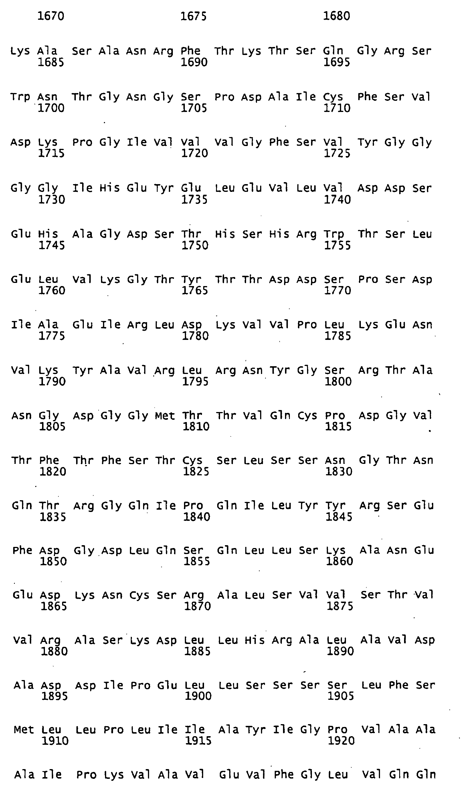
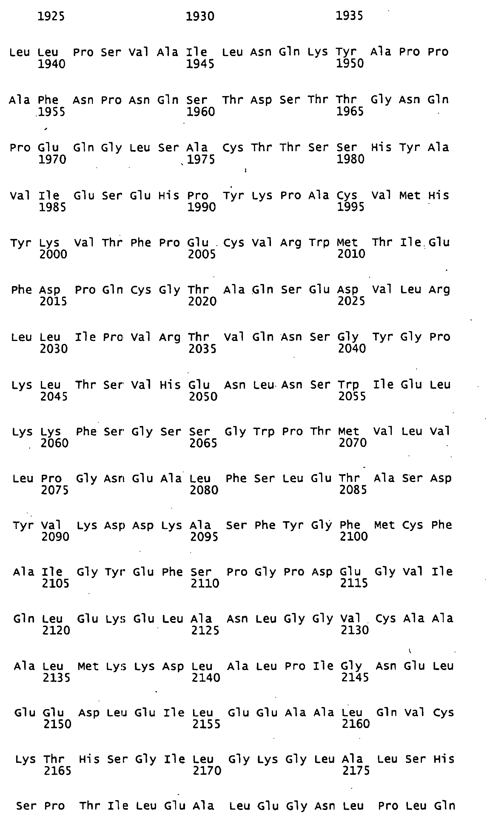

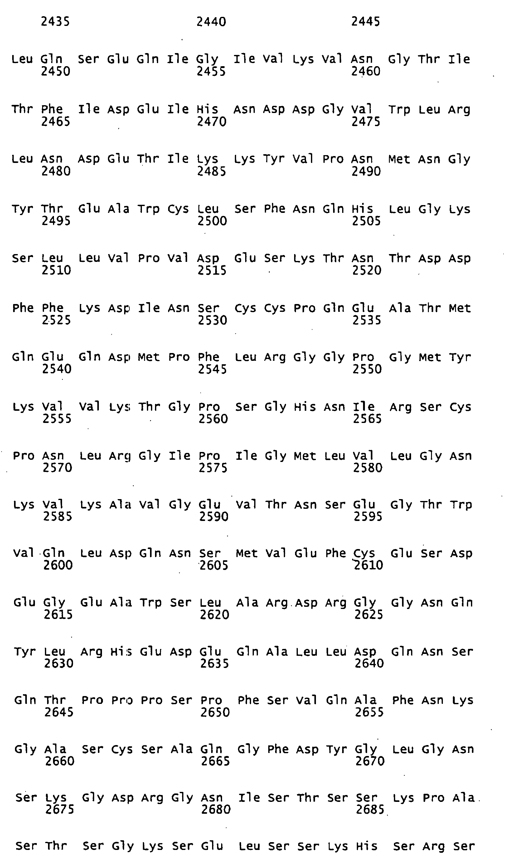
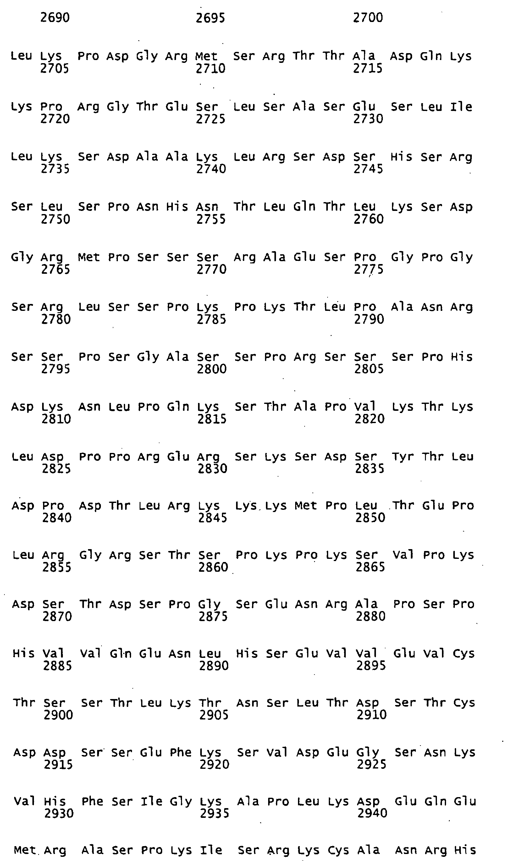







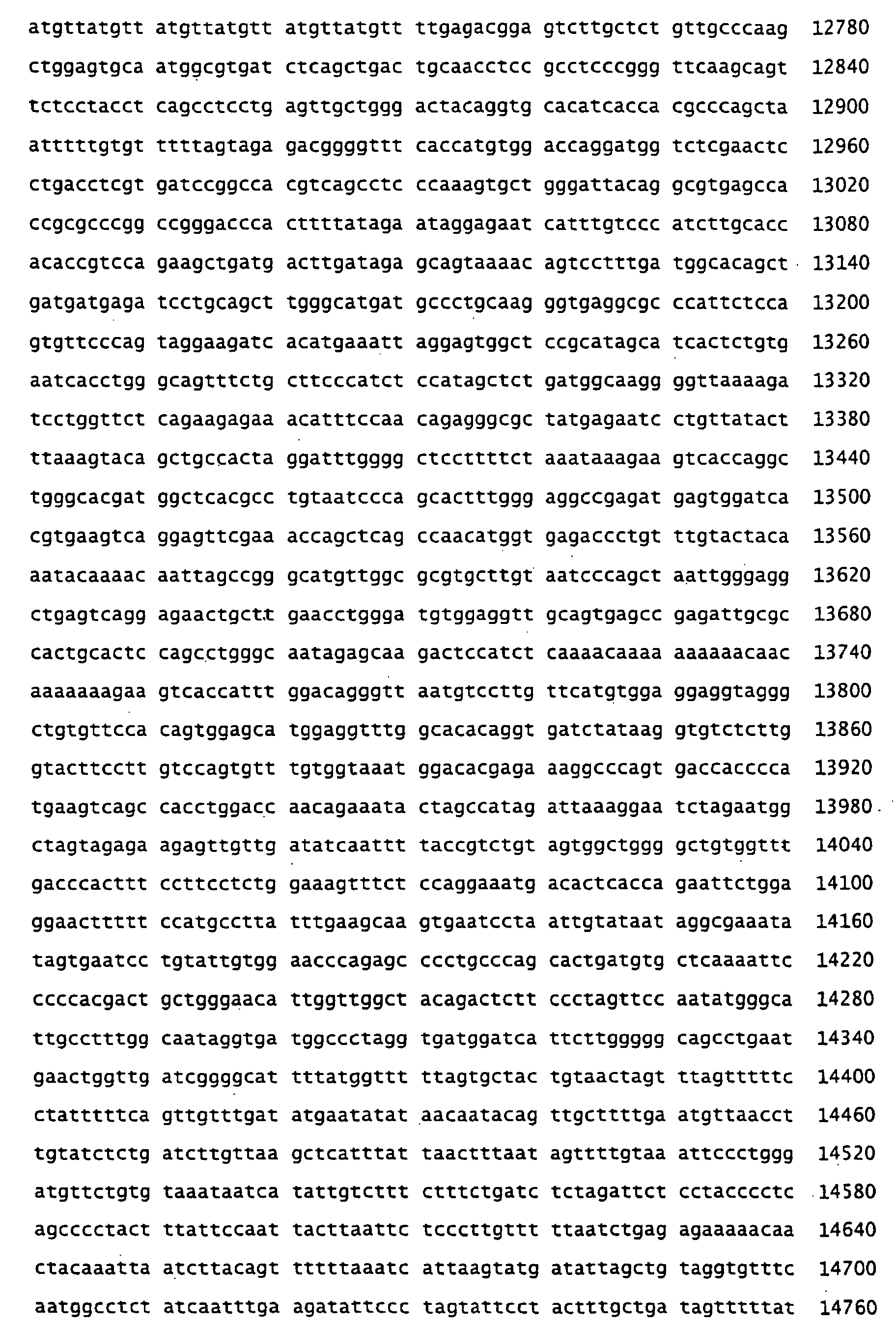
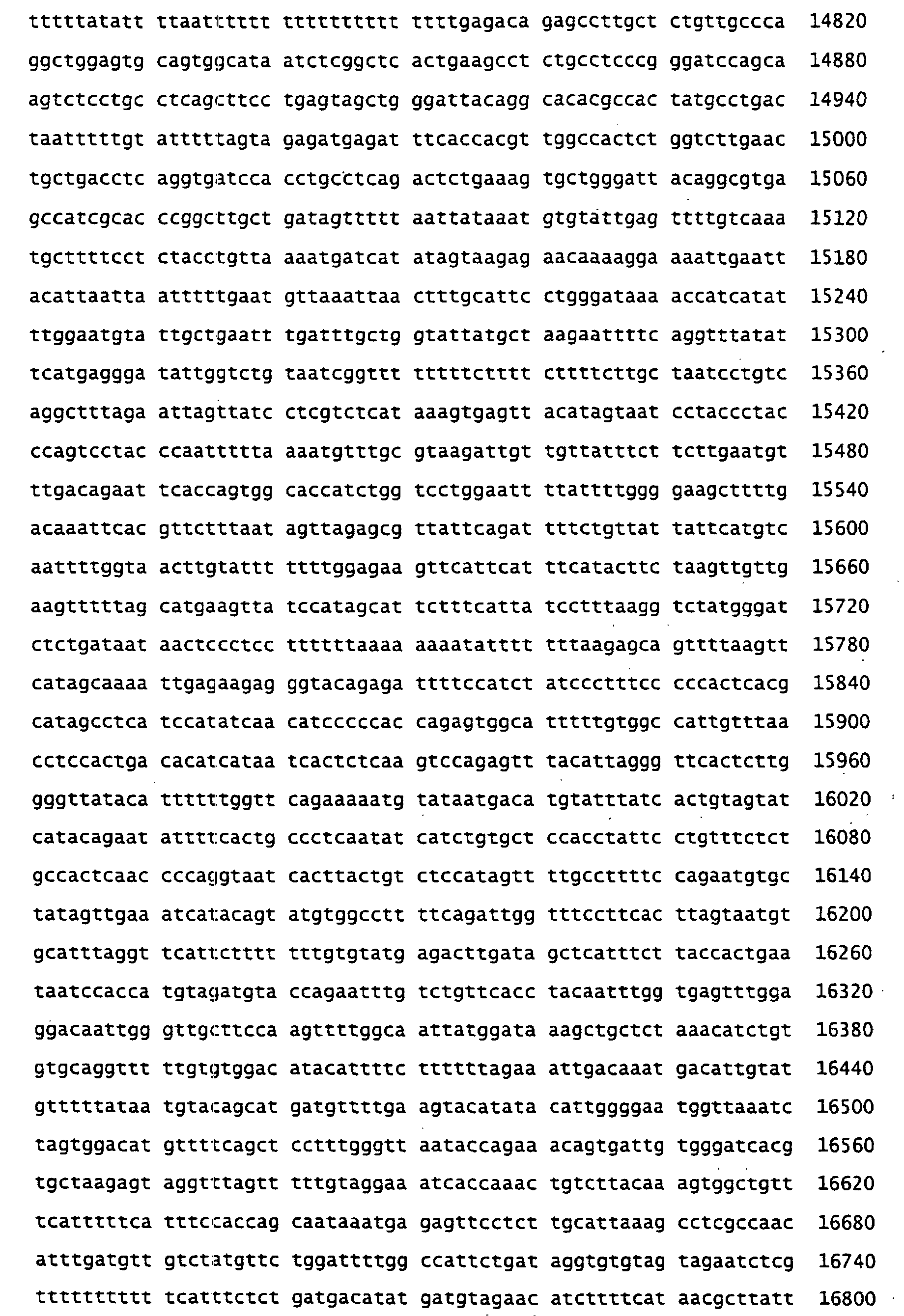
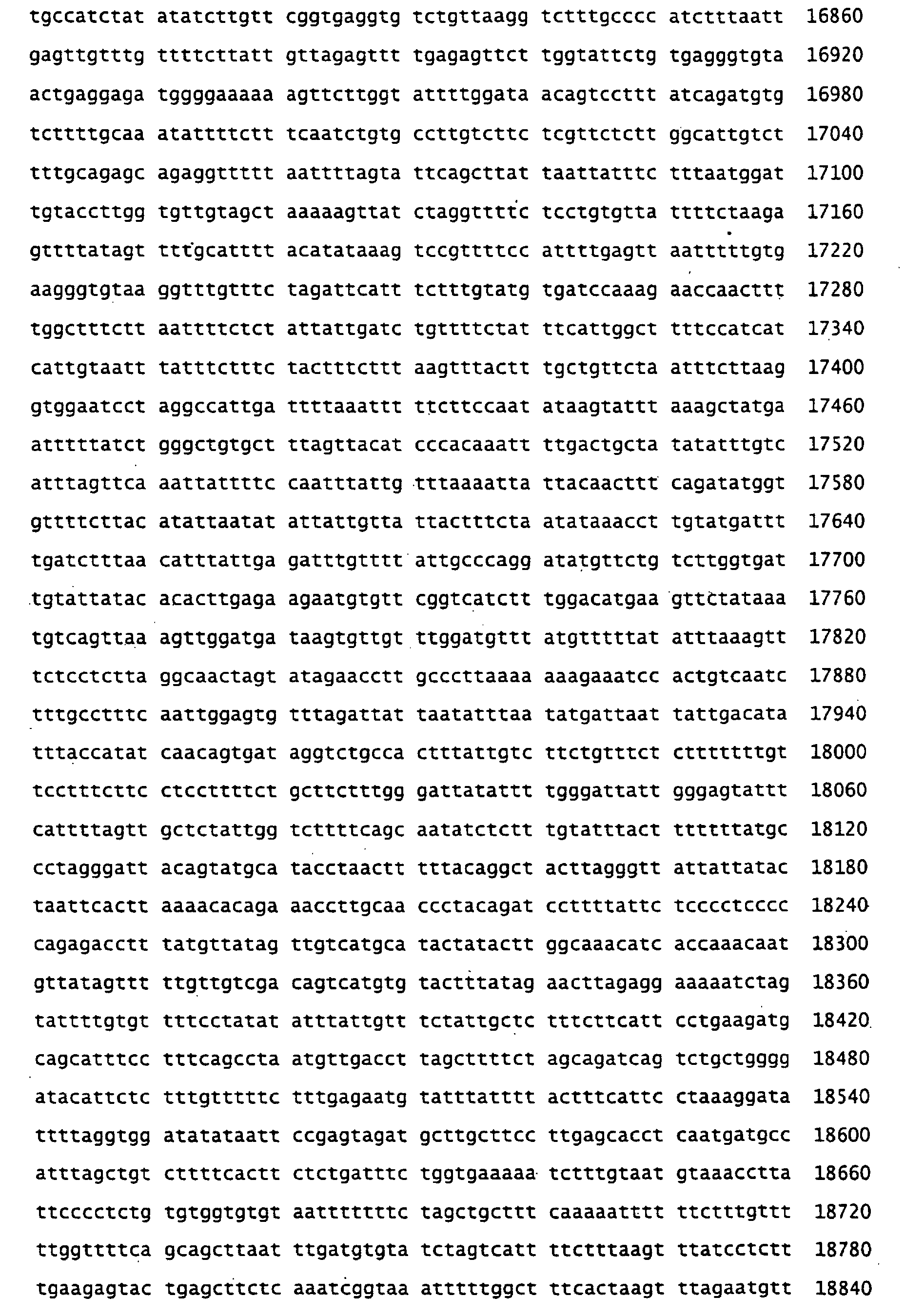

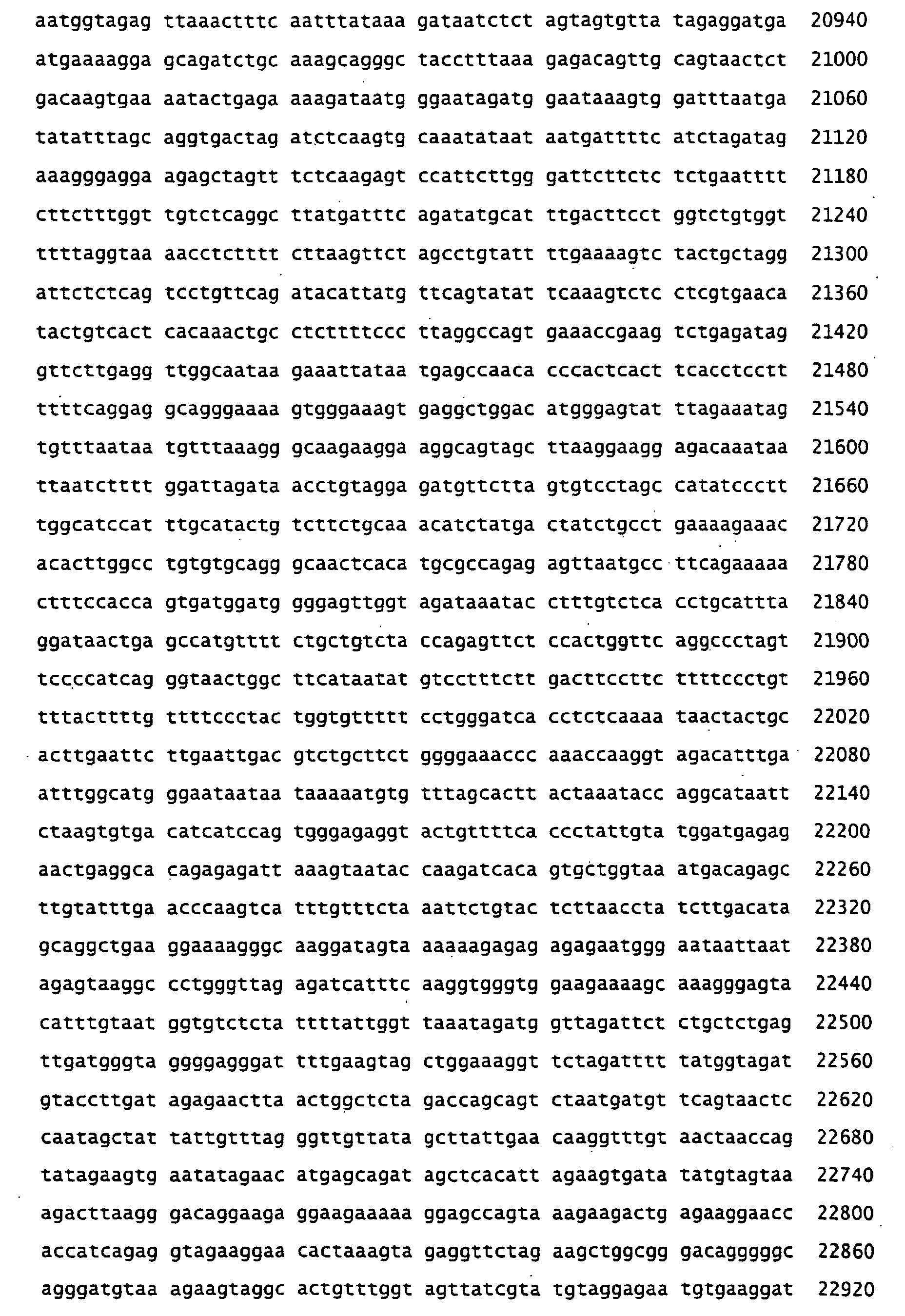
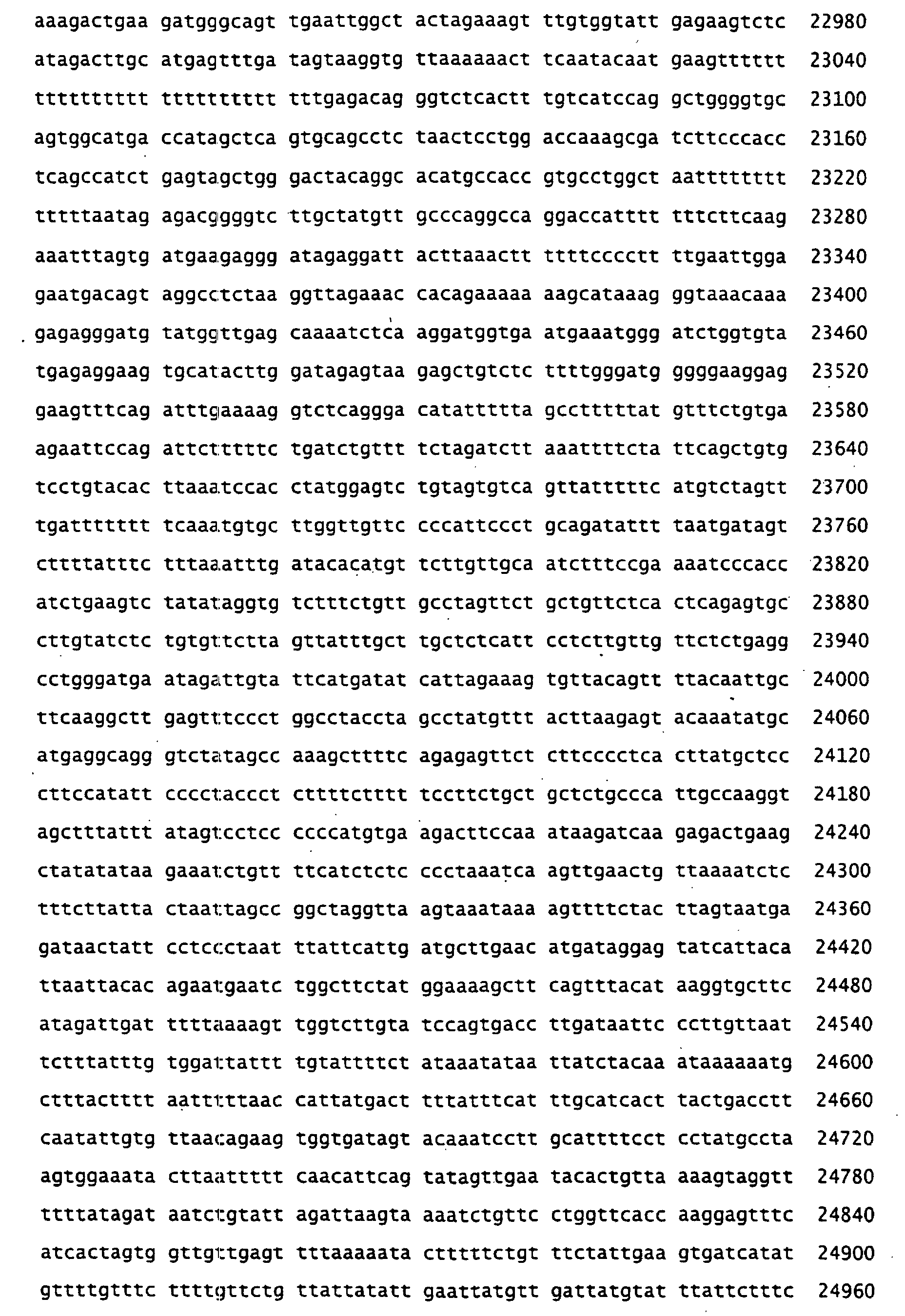
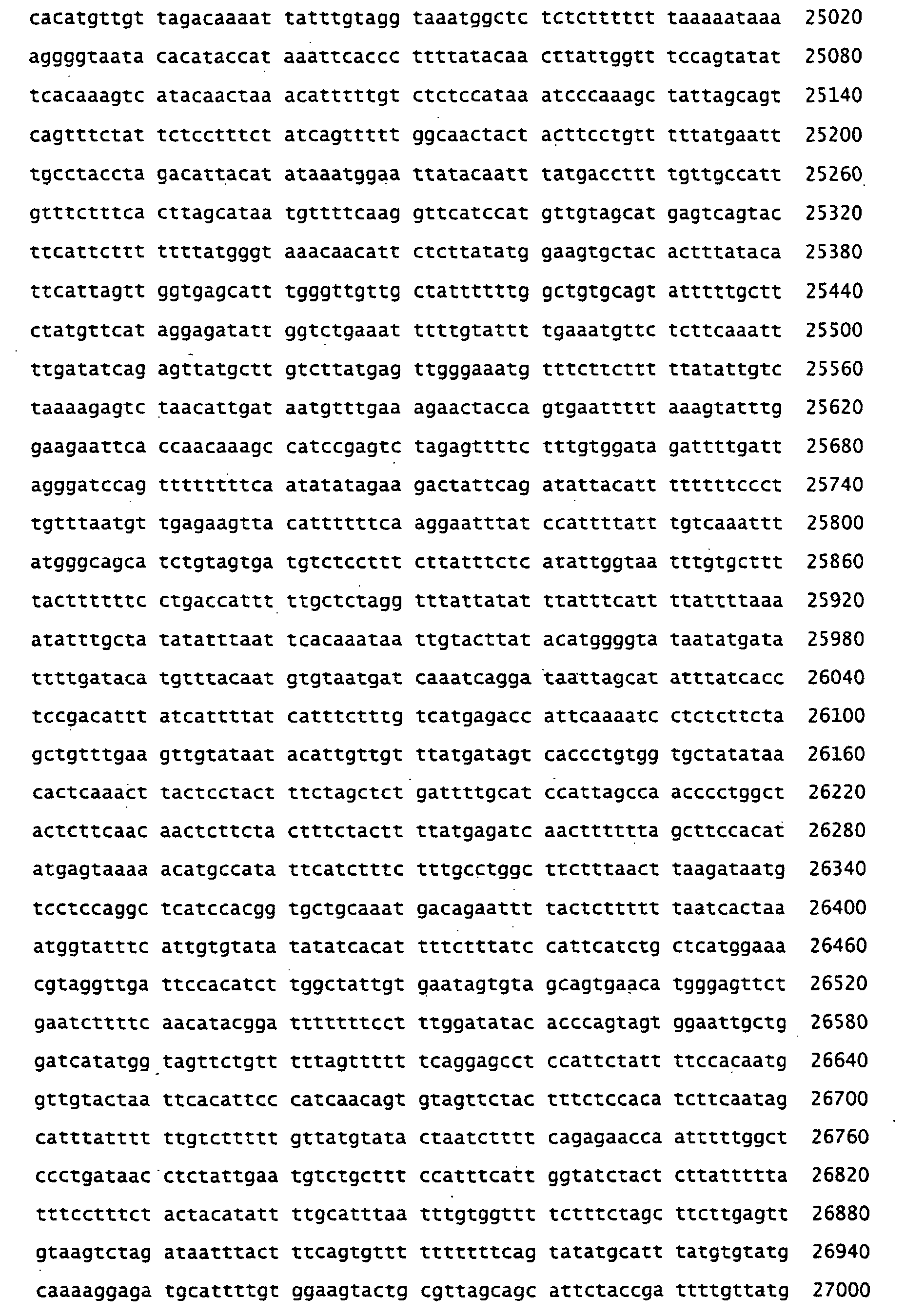
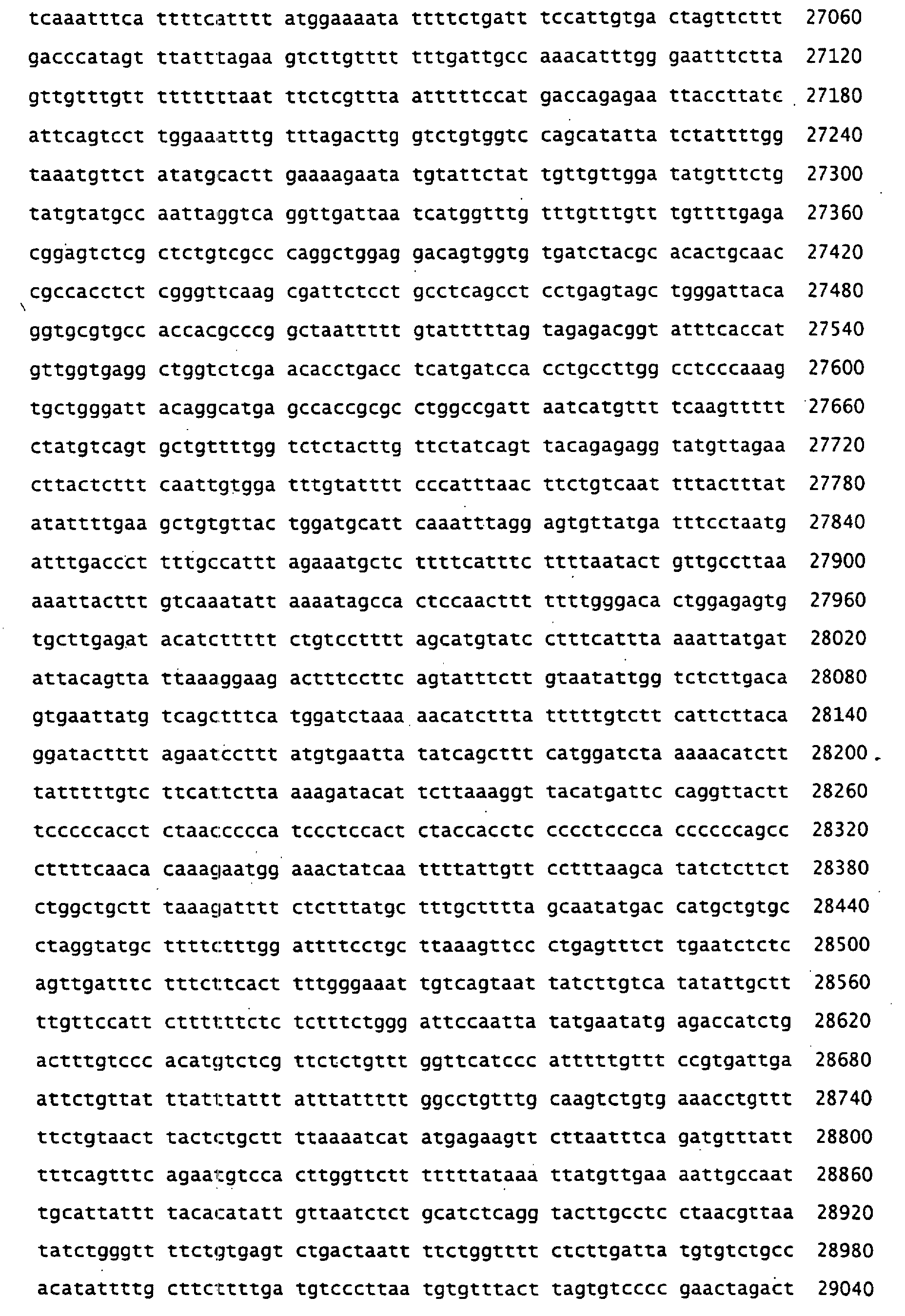
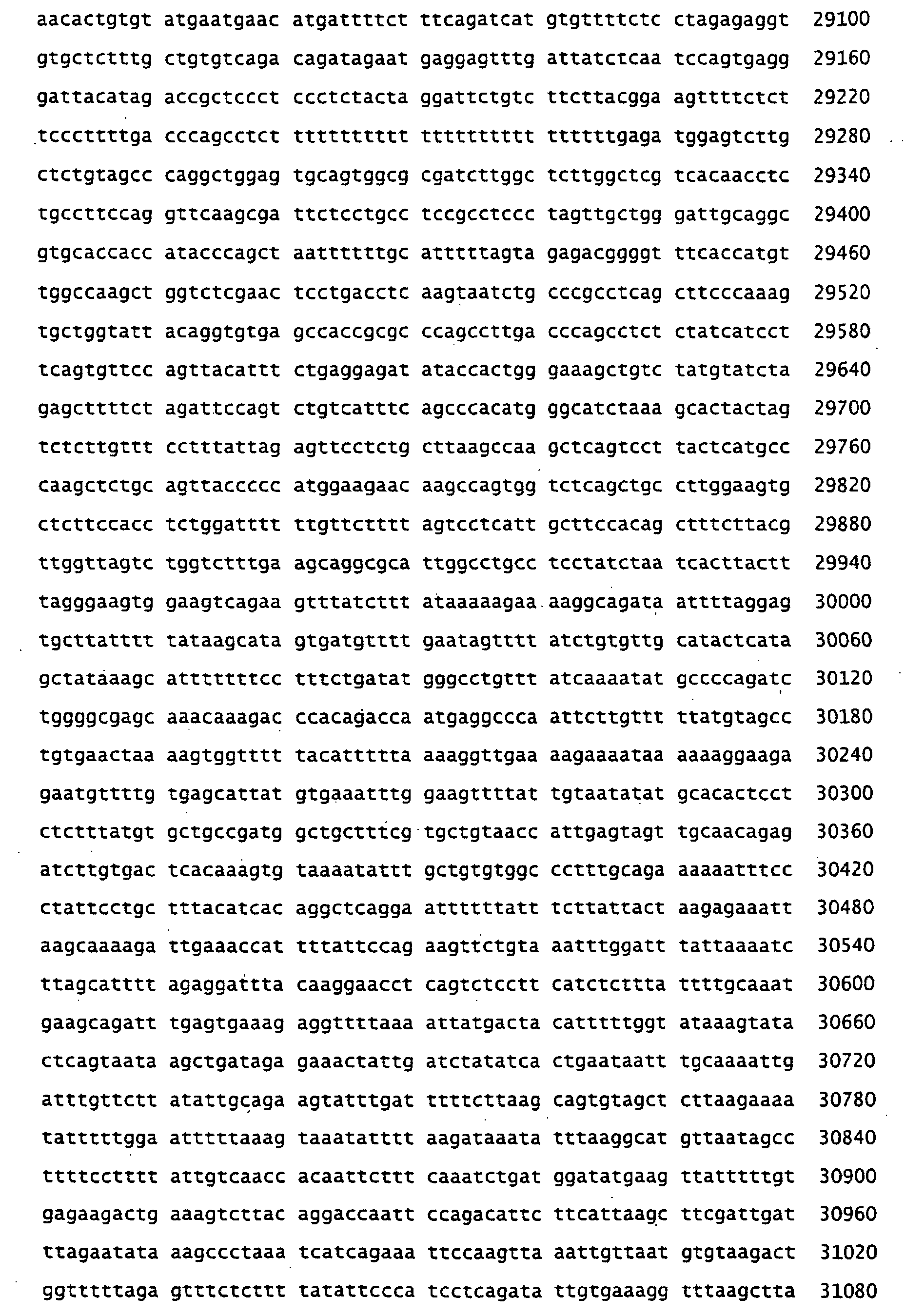





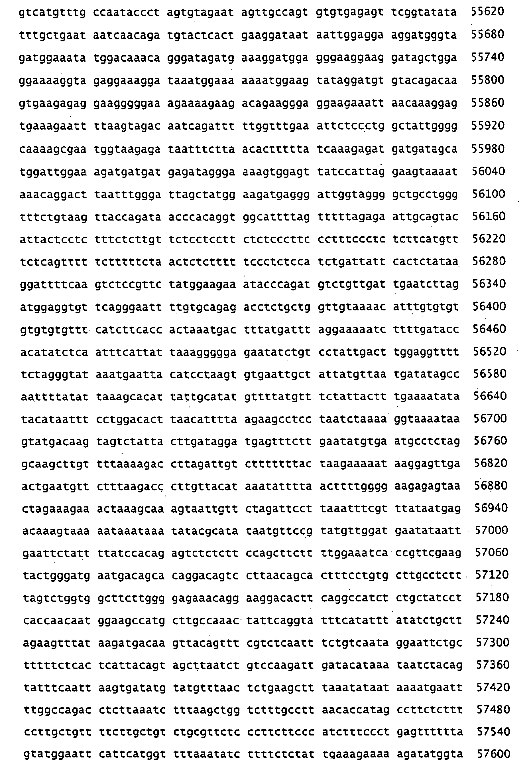


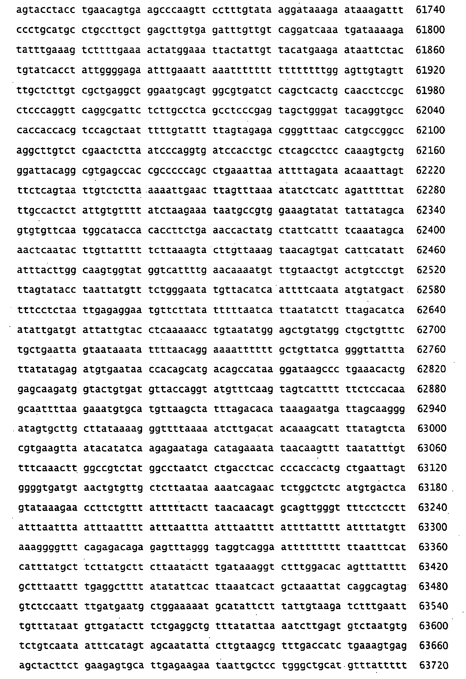
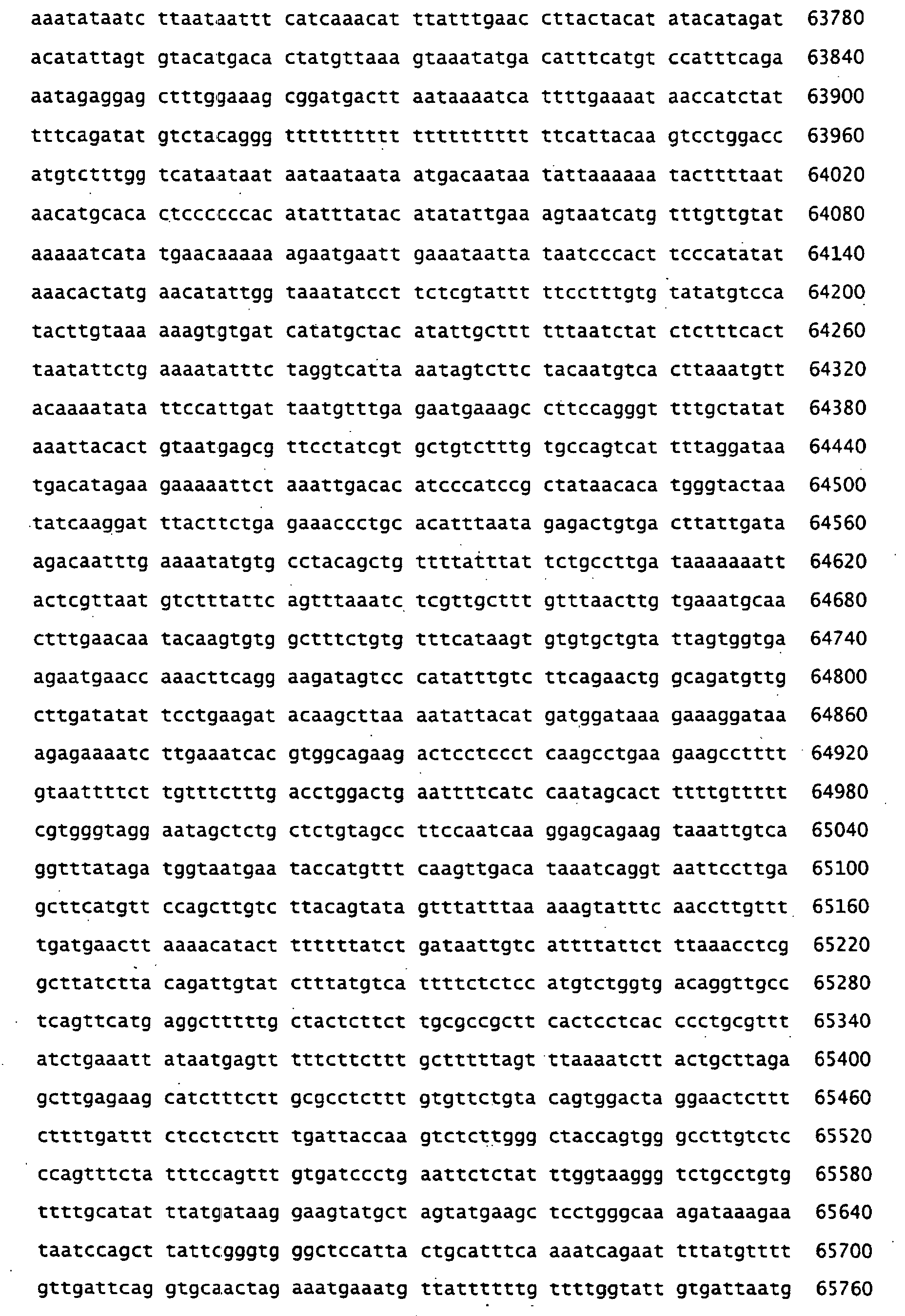
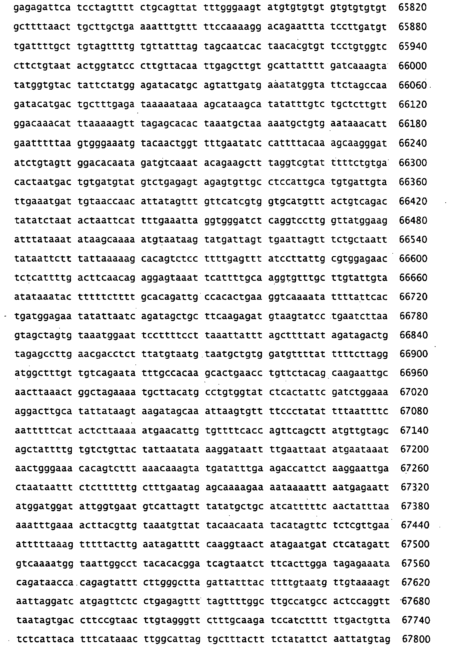






































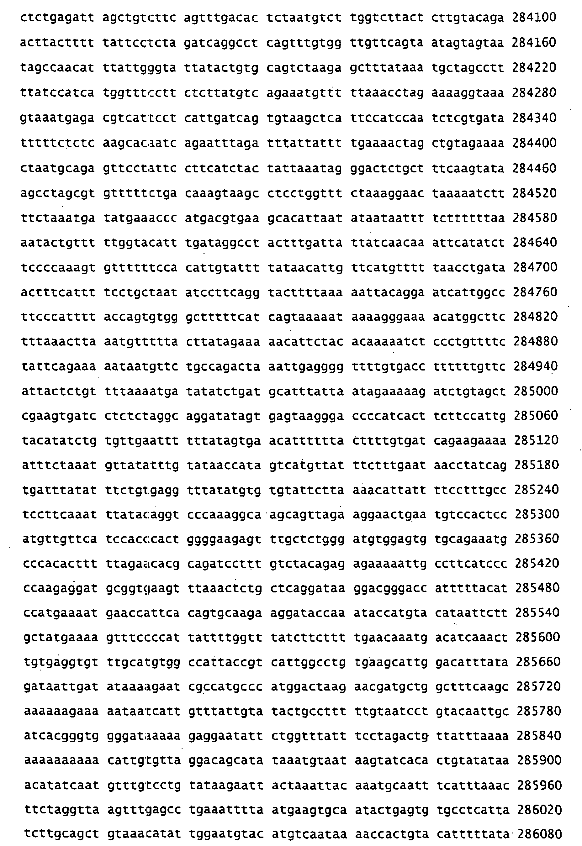


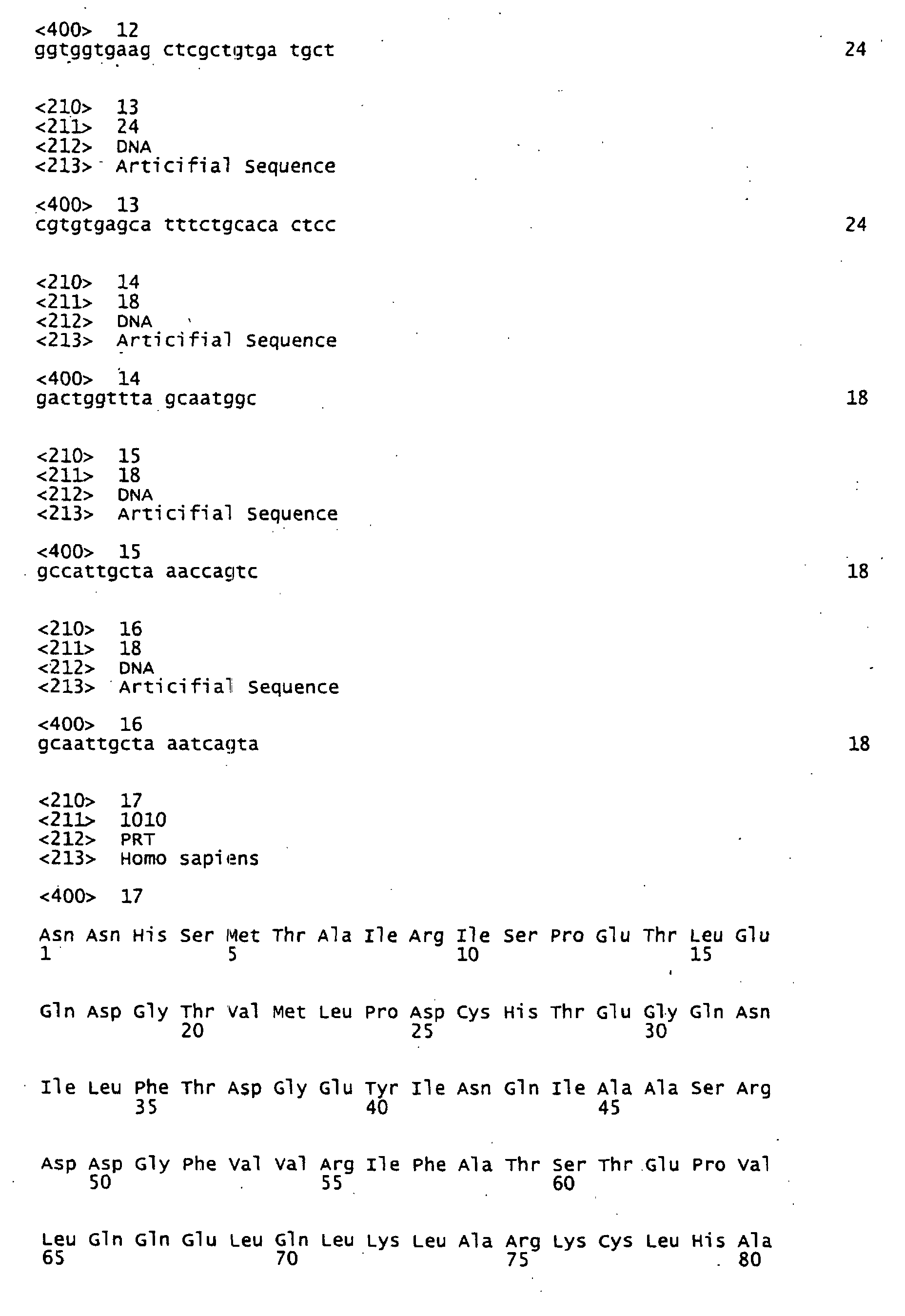
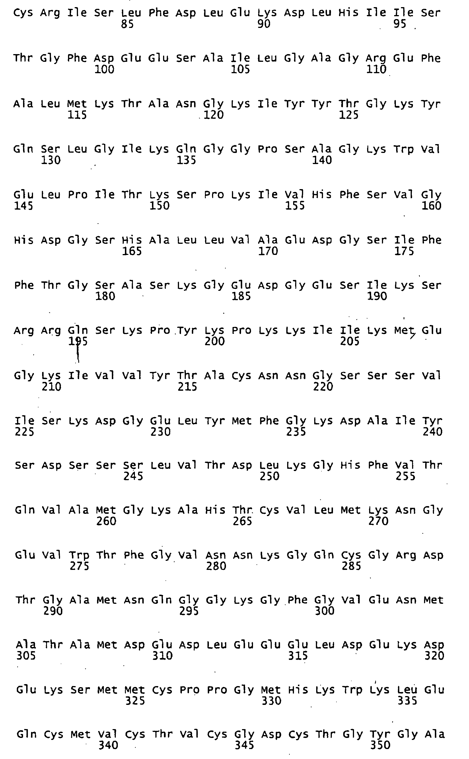
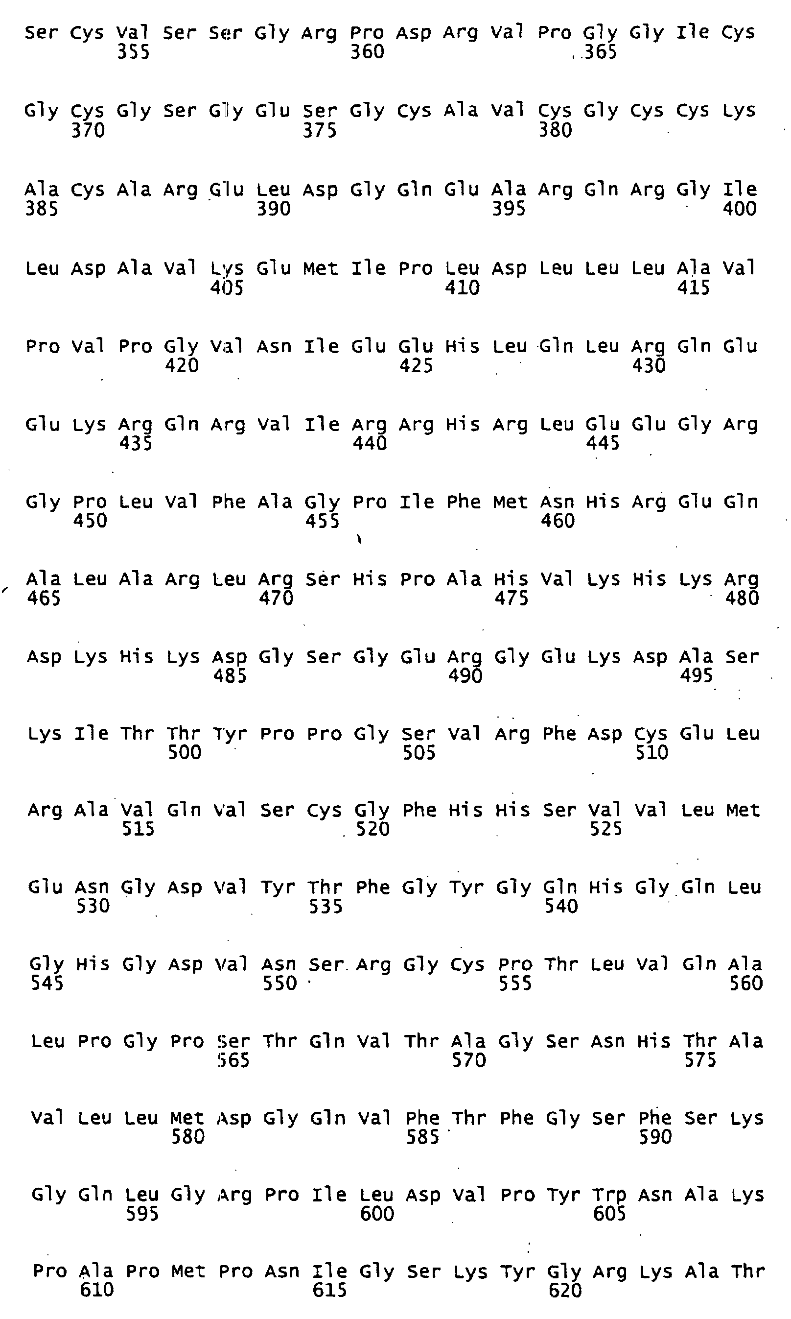
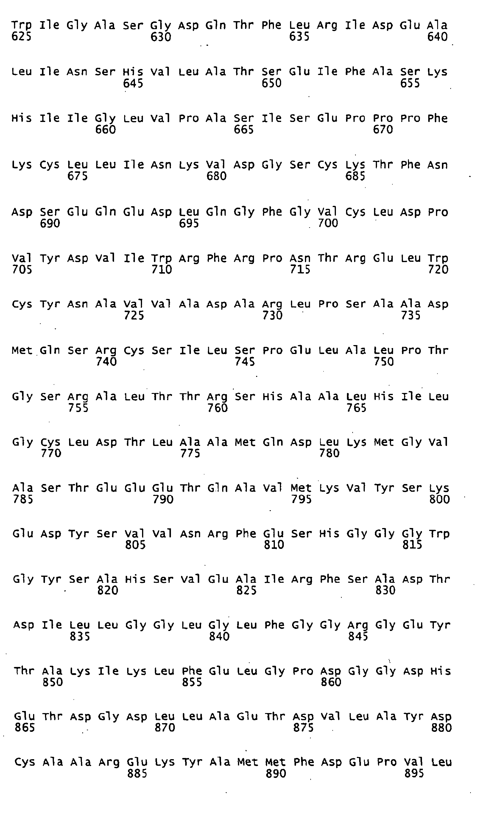

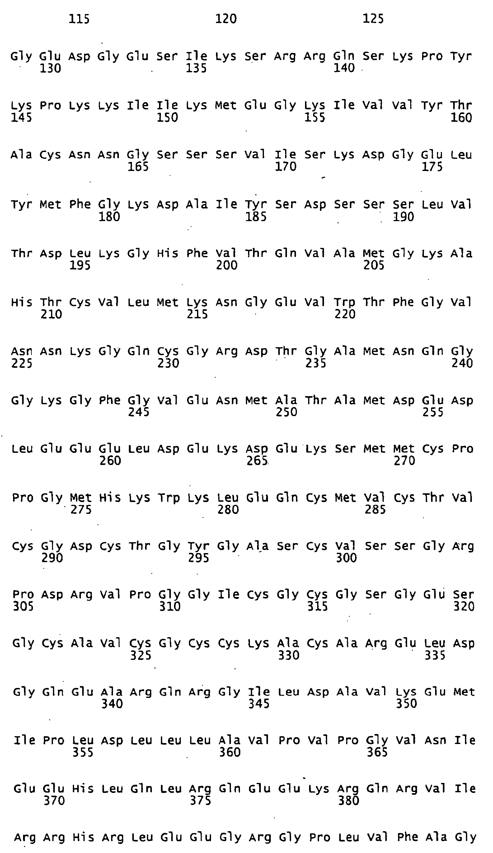


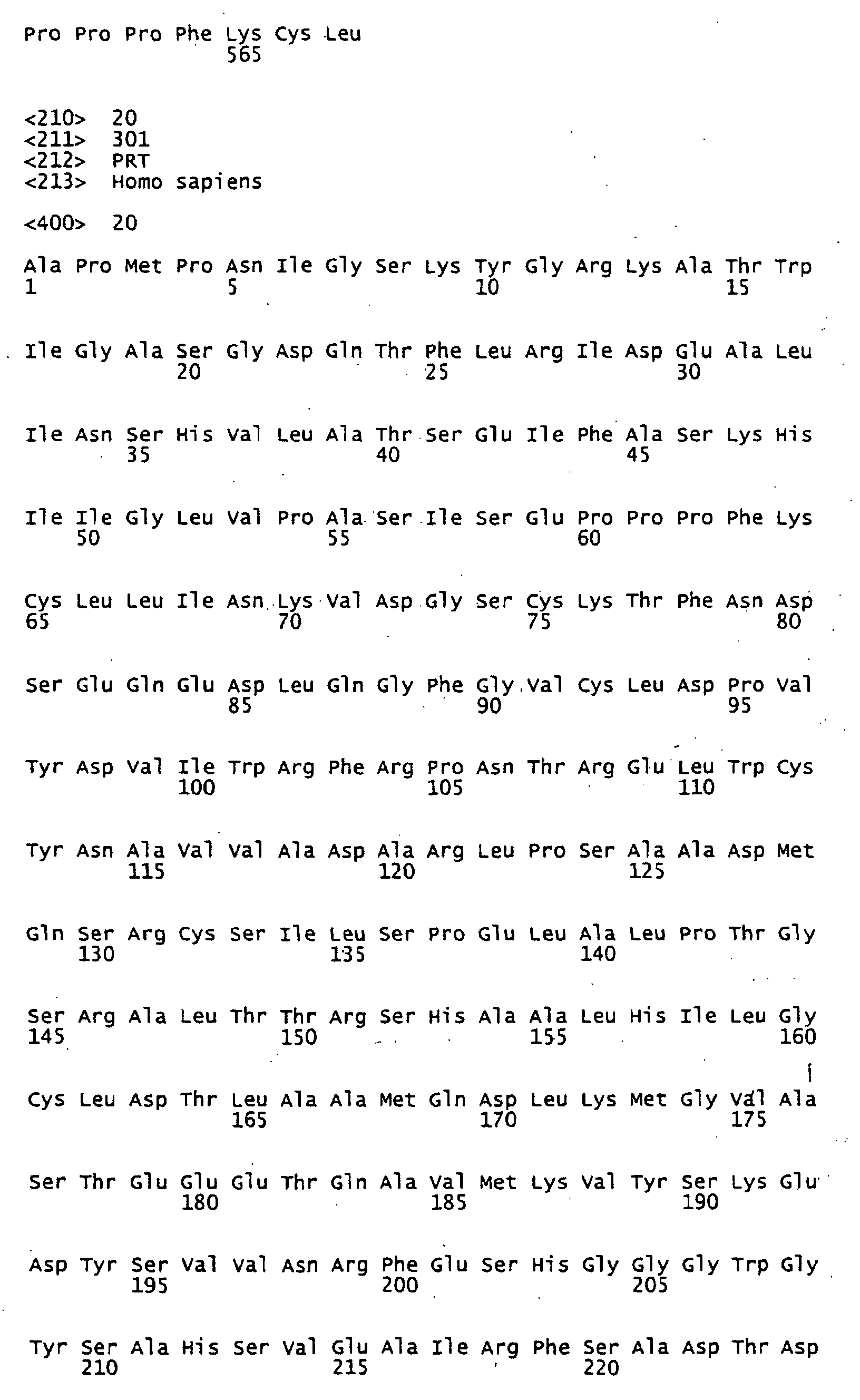
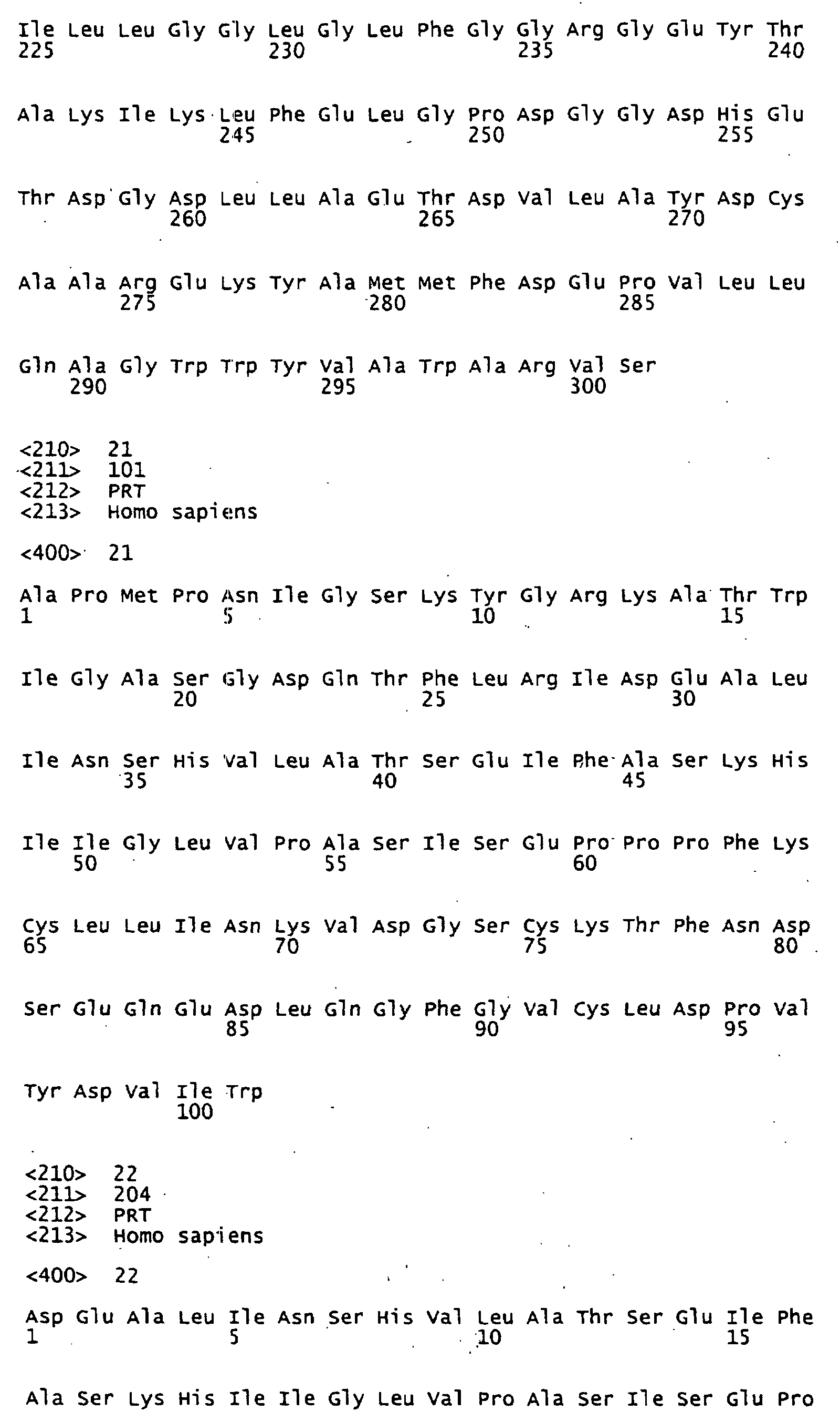
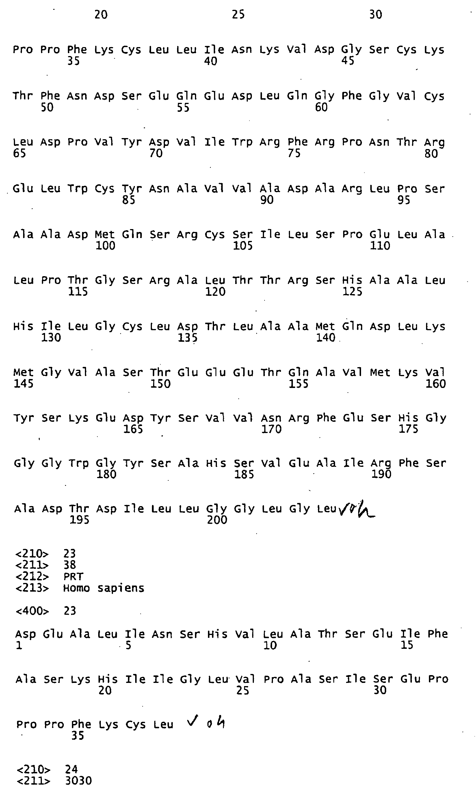
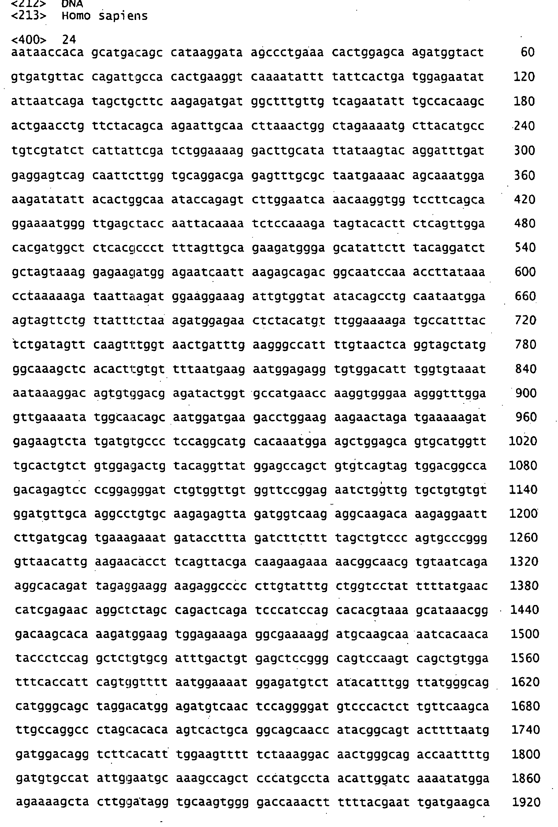
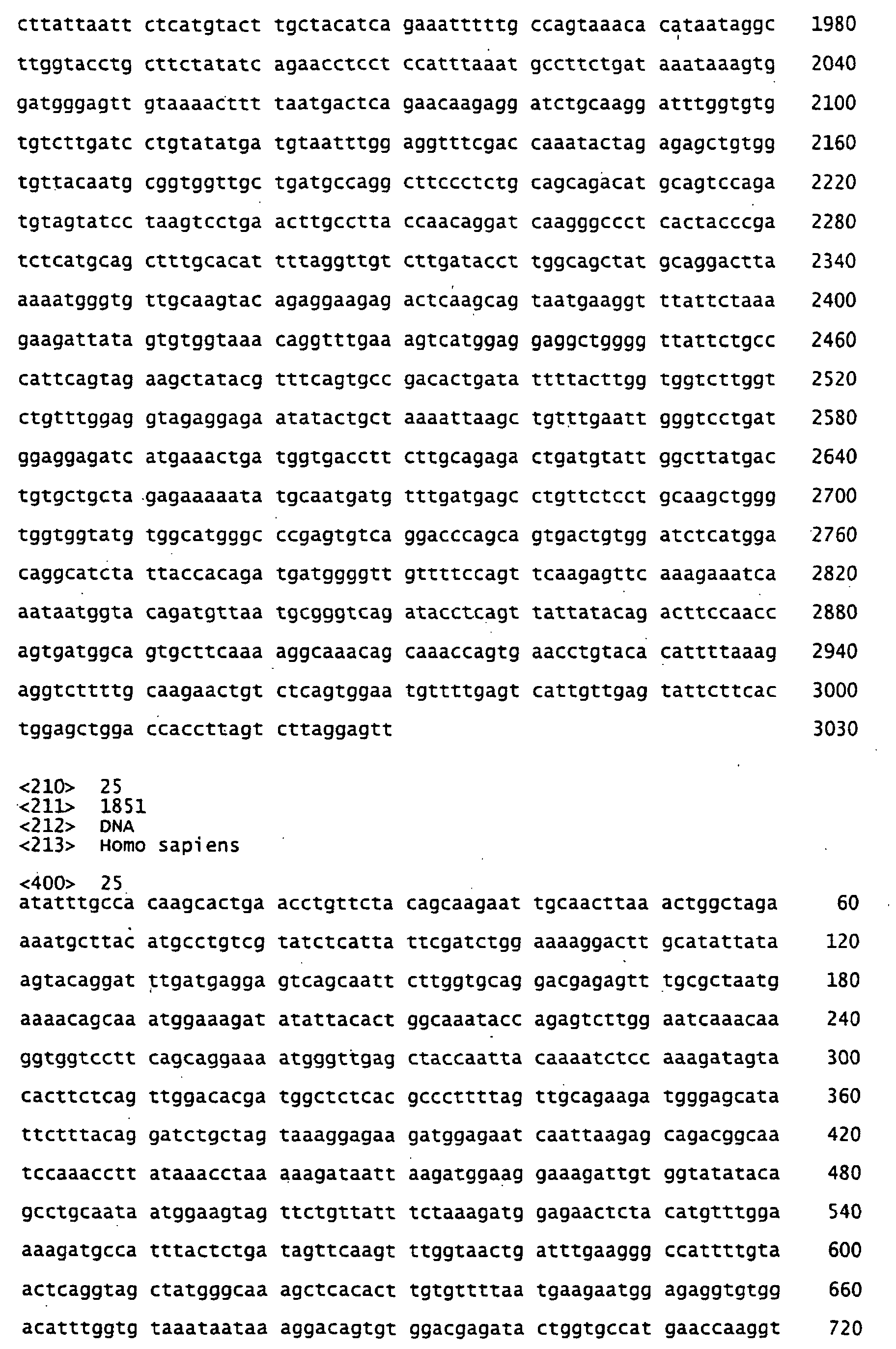
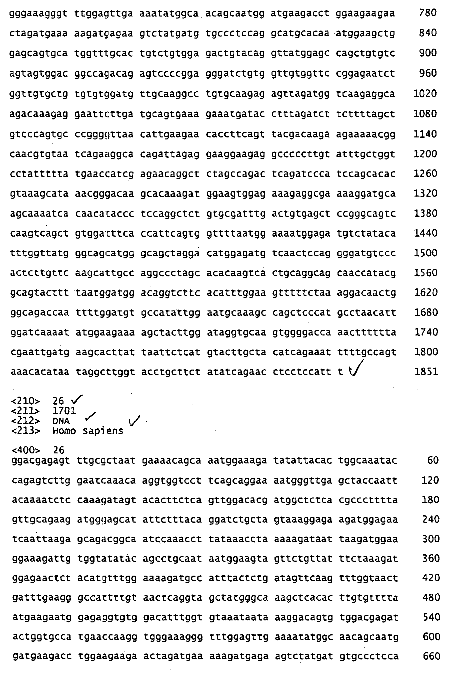

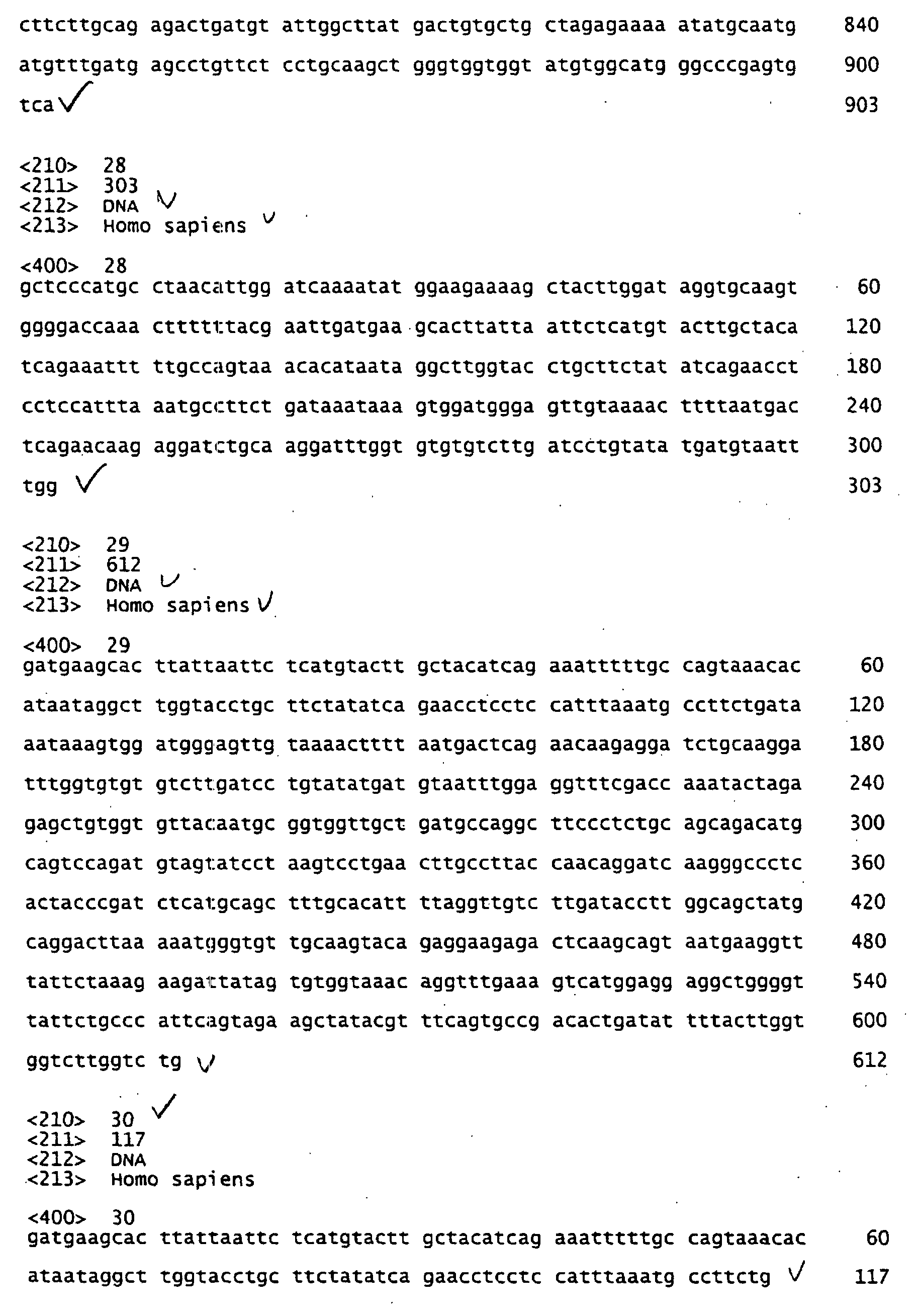
Claims (23)
- The use of S1P or functional fragments or derivatives thereof for the preparation of pharmaceutical compounds.
- The use of S1P or functional fragments or derivatives thereof for the modulation of pain.
- The use of S1P or functional fragments of derivatives thereof for identifying compounds that modulate pain.
- A method of lessening pain comprising administering a sufficient amount of S1P or a functional fragment or derivative thereof to an individual.
- S1P or a functional fragment or derivative thereof for the use as a medicament.
- S1P or a functional fragment or derivative thereof for the use as a medicament for the prevention or treatment of pain and/or inflammation.
- The use according to claim 1, wherein the pharmaceutical compounds are compounds for the prevention or treatment of pain and/or inflammation.
- The use according to claim 3, wherein the compounds lessen or abolish pain.
- A method of screening pharmaceuticals useful for modulating and/or preventing pain, comprising the stepsa. Providing a sample containing PAM or a functional fragment or derivative thereof and not containing S1Pb. Providing a second sample containing PAM or a functional fragment or derivative thereof containing as well S1Pc. Measuring the PAM activity in both samplesd. Contacting the first sample with a compounde. Determining the ability of the compound to mimic S1P function.
- A method of screening pharmaceuticals useful for modulating and/or preventing pain, comprising the stepsa) providing two samples comprising a cell expressing an S1P receptor or a functional fragment or derivative thereof, anb) contacting one sample with the compound, andc) measuring the receptor activity in both samples.
- A method of screening pharmaceuticals useful for modulating and/or preventing pain, comprising the stepsa) providing two samples comprising a cell expressing an S1P receptor or a functional fragment or derivative thereof, andb) contacting the samples with S1P, andc) contacting one sample with the compound, andd) measuring the receptor activity or the interaction of S1P and receptor in both samples.
- A method of screening pharmaceuticals useful for modulating and/or preventing pain, comprising the stepsb. Providing a cell expressing a S1P receptor or a functional fragment or derivative thereof and expressing PAM or a functional fragment or derivative thereof,c. Contacting the cell with a compound,f. Determining the S1P activity in the presence of compound,g. Determining the S1P activity in the absence of compound, andh. Comparing the S1P activity according to c) with that according to d).
- The method according to claim 10, further comprising a step wherein the cell is contacted with S1P instead of the compound and wherein the PAM activites according to c) and d) above are compared to the PAM activity in presence of S1P.
- A method of screening pharmaceuticals useful for modulating and/or preventing pain, comprising the stepsa. Providing two samples containing S1P or a functional fragment or derivative thereof and containing a S1P receptor or a functional fragment or derivative thereof,b. Contacting one of the samples with a compound,c. Determining and comparing S1P activity in both samples.
- The use, method or S1P according to one of the above claims, wherein the functional fragments or derivatives thereof are capable of inhibiting adenylyl cyclase activity.
- The use, method or S1P according to claim 10, wherein the adenylyl cyclase is adenylyl cyclase type I, V or Vf.
- The method according to one of the preceding claims, wherein the determination of S1P activity concerns its ability to interact with and / or activate a S1P receptor, to mediate PAM translocation, to activate PAM function or to inactivate AC function.
- A method of identifying a compound that modulates pain comprisinga) Selecting a compound that modulates or mimics the activity of S1P as a test compound, andb) Administering said test compound to a subject, andc) Determining whether the pain is modulated in the subject.
- Use of a compound having the ability to activate at least one of the S1P receptors and / or having the ability to activate PAM function for the preparation of a medicament that modulates or prevents pain.
- Use according to claim 19, wherein the modulation is a lessening or an abolishment of pain.
- Use according to one of the claims 19 or 20, wherein the compound is FTY 720 or a functional derivative, preferably a phosphorylated derivative thereof, or a salt of the compound of the derivative.
- A Method of lessening or preventing pain comprising administering a sufficient amount of a pharmaceutical compound with the ability to bind and activate at least one of the S1P receptors and/or the activity to activate PAM function to an individual.
- Method according to claim 22, wherein the compound is FTY 720 or a functional derivative, preferably a phosphorylated derivative thereof, or a physiologically acceptable salt of the compound or the derivative.
Priority Applications (19)
| Application Number | Priority Date | Filing Date | Title |
|---|---|---|---|
| EP20030012389 EP1481680A1 (en) | 2003-05-30 | 2003-05-30 | Use of S1P |
| CN200480015055.4A CN1838961B (en) | 2003-05-30 | 2004-05-15 | Use of s1p |
| AU2004243475A AU2004243475C1 (en) | 2003-05-30 | 2004-05-15 | Use of S1P |
| JP2006529836A JP4843492B2 (en) | 2003-05-30 | 2004-05-15 | Use of S1P |
| BRPI0410960-0A BRPI0410960A (en) | 2003-05-30 | 2004-05-15 | use of s1p |
| RU2005137176/15A RU2362565C2 (en) | 2003-05-30 | 2004-05-15 | S1p application |
| CA2809131A CA2809131A1 (en) | 2003-05-30 | 2004-05-15 | Use of s1p |
| DK04733258.0T DK1641468T3 (en) | 2003-05-30 | 2004-05-15 | Methods of screening for pharmaceutical agents for the treatment of pain |
| KR1020057022857A KR101169916B1 (en) | 2003-05-30 | 2004-05-15 | Use of S1P |
| EP04733258A EP1641468B1 (en) | 2003-05-30 | 2004-05-15 | Methods of screening pharmaceuticals for the treatment of pain |
| CA2527759A CA2527759C (en) | 2003-05-30 | 2004-05-15 | Use of s1p |
| PCT/EP2004/005236 WO2004105773A2 (en) | 2003-05-30 | 2004-05-15 | Use of s1p |
| AT04733258T ATE532522T1 (en) | 2003-05-30 | 2004-05-15 | SCREENING OF DRUGS FOR PAIN TREATMENT |
| US10/850,586 US7691563B2 (en) | 2003-05-30 | 2004-05-20 | Method for alleviating pain using sphingosine-1-phosphate and related compounds, and assays for identifying such compounds |
| IL172203A IL172203A (en) | 2003-05-30 | 2005-11-27 | Methods of screening pharmaceutical compounds useful for treating and/or preventing acute or chronic pain |
| NO20056028A NO20056028L (en) | 2003-05-30 | 2005-12-19 | Use of SIP |
| US12/696,250 US9271992B2 (en) | 2003-05-30 | 2010-01-29 | Method for alleviating pain using sphingosine-1-phosphate and related compounds, and assays for identifying such compounds |
| AU2010235846A AU2010235846B2 (en) | 2003-05-30 | 2010-10-15 | Use of S1P |
| JP2011030386A JP5460627B2 (en) | 2003-05-30 | 2011-02-16 | Use of S1P |
Applications Claiming Priority (1)
| Application Number | Priority Date | Filing Date | Title |
|---|---|---|---|
| EP20030012389 EP1481680A1 (en) | 2003-05-30 | 2003-05-30 | Use of S1P |
Publications (1)
| Publication Number | Publication Date |
|---|---|
| EP1481680A1 true EP1481680A1 (en) | 2004-12-01 |
Family
ID=33104118
Family Applications (2)
| Application Number | Title | Priority Date | Filing Date |
|---|---|---|---|
| EP20030012389 Withdrawn EP1481680A1 (en) | 2003-05-30 | 2003-05-30 | Use of S1P |
| EP04733258A Expired - Lifetime EP1641468B1 (en) | 2003-05-30 | 2004-05-15 | Methods of screening pharmaceuticals for the treatment of pain |
Family Applications After (1)
| Application Number | Title | Priority Date | Filing Date |
|---|---|---|---|
| EP04733258A Expired - Lifetime EP1641468B1 (en) | 2003-05-30 | 2004-05-15 | Methods of screening pharmaceuticals for the treatment of pain |
Country Status (14)
| Country | Link |
|---|---|
| US (2) | US7691563B2 (en) |
| EP (2) | EP1481680A1 (en) |
| JP (2) | JP4843492B2 (en) |
| KR (1) | KR101169916B1 (en) |
| CN (1) | CN1838961B (en) |
| AT (1) | ATE532522T1 (en) |
| AU (2) | AU2004243475C1 (en) |
| BR (1) | BRPI0410960A (en) |
| CA (2) | CA2809131A1 (en) |
| DK (1) | DK1641468T3 (en) |
| IL (1) | IL172203A (en) |
| NO (1) | NO20056028L (en) |
| RU (1) | RU2362565C2 (en) |
| WO (1) | WO2004105773A2 (en) |
Cited By (4)
| Publication number | Priority date | Publication date | Assignee | Title |
|---|---|---|---|---|
| WO2009053481A1 (en) * | 2007-10-25 | 2009-04-30 | Institut National De La Sante Et De La Recherche Medicale (Inserm) | Compositions and methods for modulating nk and t cell trafficking |
| CN113350363A (en) * | 2021-06-30 | 2021-09-07 | 中国矿业大学(北京) | Application of N-palmitoyl-D-sphingomyelin in preparation of medicine for preventing and/or treating neuropathic pain |
| US11359197B2 (en) | 2018-01-12 | 2022-06-14 | Bristol-Myers Squibb Company | Antisense oligonucleotides targeting alpha-synuclein and uses thereof |
| US12241065B2 (en) | 2018-01-12 | 2025-03-04 | Bristol-Myers Squibb Company | Antisense oligonucleotides targeting alpha-synuclein and uses thereof |
Families Citing this family (15)
| Publication number | Priority date | Publication date | Assignee | Title |
|---|---|---|---|---|
| US7241790B2 (en) * | 2002-07-30 | 2007-07-10 | University Of Virginia Patent Foundation | Compounds active in spinigosine 1-phosphate signaling |
| WO2005041899A2 (en) * | 2003-11-03 | 2005-05-12 | University Of Virginia Patent Foundation | Orally available sphingosine 1-phosphate receptor agonists and antagonists |
| JP2008522977A (en) * | 2004-12-06 | 2008-07-03 | ユニバーシティ オブ バージニア パテント ファンデーション | Sphingosine = 1-phosphate arylamide analog |
| MX2007009848A (en) * | 2005-02-14 | 2008-03-10 | Univ Virginia | Sphingosine 1- phos phate agonists comprising cycloalkanes and 5 -membered heterocycles substituted by amino and phenyl groups. |
| MX2008009579A (en) * | 2006-01-27 | 2008-09-25 | Univ Virginia | Method for treatment of neuropathic pain. |
| AU2007212193A1 (en) * | 2006-02-09 | 2007-08-16 | University Of Virginia Patent Foundation | Bicyclic sphingosine 1-phosphate analogs |
| EP2099741A2 (en) * | 2006-11-21 | 2009-09-16 | University Of Virginia Patent Foundation | Hydrindane analogs having sphingosine 1-phosphate receptor agonist activity |
| AU2007323618A1 (en) * | 2006-11-21 | 2008-05-29 | University Of Virginia Patent Foundation | Tetralin analogs having sphingosine 1-phosphate agonist activity |
| WO2008064337A2 (en) * | 2006-11-21 | 2008-05-29 | University Of Virginia Patent Foundation | Benzocycloheptyl analogs having sphingosine 1-phosphate receptor activity |
| EP2598126A2 (en) * | 2010-07-30 | 2013-06-05 | Saint Louis University | Methods of treating pain |
| WO2012162392A1 (en) * | 2011-05-23 | 2012-11-29 | Timothy Hla | Endothelium protective materials and methods of use |
| EP2769724A1 (en) * | 2013-02-22 | 2014-08-27 | Université d'Aix-Marseille | Inhibitors of Na(v) 1.9 channel activity and uses thereof for treating pain |
| WO2015025871A1 (en) * | 2013-08-20 | 2015-02-26 | Meiji Seikaファルマ株式会社 | Methods for evaluating and screening s1p1 receptor agonists |
| CN113444727B (en) * | 2021-06-30 | 2022-06-21 | 中国热带农业科学院热带生物技术研究所 | LncRNA and application thereof |
| CN117024565A (en) * | 2021-11-17 | 2023-11-10 | 罗赣 | Polypeptide modified body and application thereof in preparation of anti-escherichia coli infection drugs |
Citations (8)
| Publication number | Priority date | Publication date | Assignee | Title |
|---|---|---|---|---|
| US5663404A (en) * | 1992-04-03 | 1997-09-02 | Oncomembrane | Sphingosine-1-phosphate essentially free of L-threo isomer |
| US5712262A (en) * | 1996-11-21 | 1998-01-27 | Spiegel; Sarah | Use of sphingosine-1-phosphate to suppress programmed cell death |
| WO1999012533A1 (en) * | 1997-09-08 | 1999-03-18 | Medvet Science Pty. Ltd. | A method of modulating cellular activity |
| DE19810999A1 (en) * | 1998-03-13 | 1999-09-16 | Dermapharm Gmbh | Use of sphingosine compounds to treat inflammatory skin disorders, especially psoriasis, acne and dermatoheliosis |
| US6004565A (en) * | 1997-09-02 | 1999-12-21 | Yoshitomi Pharmaceutical Industries, Ltd. | Compositions and methods of using compositions with accelerated lymphocyte homing immunosuppressive properties |
| US20010041688A1 (en) * | 2000-03-13 | 2001-11-15 | Christian Waeber | Methods and compositions for the regulation of vasoconstriction |
| EP1195165A1 (en) * | 1999-07-12 | 2002-04-10 | Ono Pharmaceutical Co., Ltd. | Fibrosis inhibitors containing as the active ingredient sphingosine-1-phosphate receptor agonist or sphingosine-1-phosphate |
| WO2002064616A2 (en) * | 2001-01-30 | 2002-08-22 | University Of Virgina Patent Foundation | Agonists and antagonists of sphingosine-1-phosphate receptors |
Family Cites Families (2)
| Publication number | Priority date | Publication date | Assignee | Title |
|---|---|---|---|---|
| ES2151828B1 (en) * | 1998-07-10 | 2001-08-16 | Esteve Labor Dr | EMPLOYMENT OF TETRAHYDROPIRIDINAS DERIVATIVES (OHIDROXI-PIPERIDINAS) -B UTILAZOLES IN THE PREPARATION OF A MEDICINAL PRODUCT FOR THE TREATMENT OF PAIN. |
| JP2001278750A (en) * | 2000-03-29 | 2001-10-10 | Hideoki Ogawa | New hair-growing agent |
-
2003
- 2003-05-30 EP EP20030012389 patent/EP1481680A1/en not_active Withdrawn
-
2004
- 2004-05-15 BR BRPI0410960-0A patent/BRPI0410960A/en not_active IP Right Cessation
- 2004-05-15 WO PCT/EP2004/005236 patent/WO2004105773A2/en not_active Ceased
- 2004-05-15 EP EP04733258A patent/EP1641468B1/en not_active Expired - Lifetime
- 2004-05-15 KR KR1020057022857A patent/KR101169916B1/en not_active Expired - Fee Related
- 2004-05-15 AT AT04733258T patent/ATE532522T1/en active
- 2004-05-15 CN CN200480015055.4A patent/CN1838961B/en not_active Expired - Fee Related
- 2004-05-15 CA CA2809131A patent/CA2809131A1/en not_active Abandoned
- 2004-05-15 AU AU2004243475A patent/AU2004243475C1/en not_active Ceased
- 2004-05-15 JP JP2006529836A patent/JP4843492B2/en not_active Expired - Fee Related
- 2004-05-15 RU RU2005137176/15A patent/RU2362565C2/en not_active IP Right Cessation
- 2004-05-15 CA CA2527759A patent/CA2527759C/en not_active Expired - Fee Related
- 2004-05-15 DK DK04733258.0T patent/DK1641468T3/en active
- 2004-05-20 US US10/850,586 patent/US7691563B2/en not_active Expired - Fee Related
-
2005
- 2005-11-27 IL IL172203A patent/IL172203A/en active IP Right Grant
- 2005-12-19 NO NO20056028A patent/NO20056028L/en unknown
-
2010
- 2010-01-29 US US12/696,250 patent/US9271992B2/en not_active Expired - Fee Related
- 2010-10-15 AU AU2010235846A patent/AU2010235846B2/en not_active Ceased
-
2011
- 2011-02-16 JP JP2011030386A patent/JP5460627B2/en not_active Expired - Fee Related
Patent Citations (8)
| Publication number | Priority date | Publication date | Assignee | Title |
|---|---|---|---|---|
| US5663404A (en) * | 1992-04-03 | 1997-09-02 | Oncomembrane | Sphingosine-1-phosphate essentially free of L-threo isomer |
| US5712262A (en) * | 1996-11-21 | 1998-01-27 | Spiegel; Sarah | Use of sphingosine-1-phosphate to suppress programmed cell death |
| US6004565A (en) * | 1997-09-02 | 1999-12-21 | Yoshitomi Pharmaceutical Industries, Ltd. | Compositions and methods of using compositions with accelerated lymphocyte homing immunosuppressive properties |
| WO1999012533A1 (en) * | 1997-09-08 | 1999-03-18 | Medvet Science Pty. Ltd. | A method of modulating cellular activity |
| DE19810999A1 (en) * | 1998-03-13 | 1999-09-16 | Dermapharm Gmbh | Use of sphingosine compounds to treat inflammatory skin disorders, especially psoriasis, acne and dermatoheliosis |
| EP1195165A1 (en) * | 1999-07-12 | 2002-04-10 | Ono Pharmaceutical Co., Ltd. | Fibrosis inhibitors containing as the active ingredient sphingosine-1-phosphate receptor agonist or sphingosine-1-phosphate |
| US20010041688A1 (en) * | 2000-03-13 | 2001-11-15 | Christian Waeber | Methods and compositions for the regulation of vasoconstriction |
| WO2002064616A2 (en) * | 2001-01-30 | 2002-08-22 | University Of Virgina Patent Foundation | Agonists and antagonists of sphingosine-1-phosphate receptors |
Non-Patent Citations (4)
| Title |
|---|
| EHNERT CORINA ET AL: "Protein associated with Myc (PAM) is involved in spinal nociceptive processing.", JOURNAL OF NEUROCHEMISTRY. ENGLAND FEB 2004, vol. 88, no. 4, February 2004 (2004-02-01), pages 948 - 957, XP001179599, ISSN: 0022-3042 * |
| MANDALA S ET AL: "ALTERATION OF LYMPHOCYTE TRAFFICKING BY SPHINGOSINE-1-PHOSPHATE RECEPTOR AGONISTS", SCIENCE, AMERICAN ASSOCIATION FOR THE ADVANCEMENT OF SCIENCE,, US, vol. 296, no. 5566, 12 April 2002 (2002-04-12), pages 346 - 349, XP001128118, ISSN: 0036-8075 * |
| PYNE S ET AL: "SPINGOSINE 1-PHOSPHATE SIGNALLING VIA THE ENDOTHELIAL DIFFERENTIATION GENE FAMILY OF G-PROTEIN-COUPLED RECEPTORS", PHARMACOLOGY AND THERAPEUTICS, ELSEVIER, GB, vol. 88, 2000, pages 115 - 131, XP002254562, ISSN: 0163-7258 * |
| SCHOLICH K ET AL: "Protein associated with Myc (PAM) is a potent inhibitor of adenylyl cyclases.", THE JOURNAL OF BIOLOGICAL CHEMISTRY. UNITED STATES 14 DEC 2001, vol. 276, no. 50, 14 December 2001 (2001-12-14), pages 47583 - 47589, XP002271386, ISSN: 0021-9258 * |
Cited By (6)
| Publication number | Priority date | Publication date | Assignee | Title |
|---|---|---|---|---|
| WO2009053481A1 (en) * | 2007-10-25 | 2009-04-30 | Institut National De La Sante Et De La Recherche Medicale (Inserm) | Compositions and methods for modulating nk and t cell trafficking |
| US11359197B2 (en) | 2018-01-12 | 2022-06-14 | Bristol-Myers Squibb Company | Antisense oligonucleotides targeting alpha-synuclein and uses thereof |
| US11447775B2 (en) | 2018-01-12 | 2022-09-20 | Bristol-Myers Squibb Company | Antisense oligonucleotides targeting alpha-synuclein and uses thereof |
| US12180478B2 (en) | 2018-01-12 | 2024-12-31 | Bristol-Myers Squibb Company | Antisense oligonucleotides targeting alpha-synuclein and uses thereof |
| US12241065B2 (en) | 2018-01-12 | 2025-03-04 | Bristol-Myers Squibb Company | Antisense oligonucleotides targeting alpha-synuclein and uses thereof |
| CN113350363A (en) * | 2021-06-30 | 2021-09-07 | 中国矿业大学(北京) | Application of N-palmitoyl-D-sphingomyelin in preparation of medicine for preventing and/or treating neuropathic pain |
Also Published As
| Publication number | Publication date |
|---|---|
| US7691563B2 (en) | 2010-04-06 |
| US20050032744A1 (en) | 2005-02-10 |
| EP1641468B1 (en) | 2011-11-09 |
| ATE532522T1 (en) | 2011-11-15 |
| CN1838961B (en) | 2015-04-22 |
| AU2004243475C1 (en) | 2011-01-20 |
| KR101169916B1 (en) | 2012-08-06 |
| WO2004105773A3 (en) | 2006-04-27 |
| CA2527759A1 (en) | 2004-12-09 |
| KR20060054188A (en) | 2006-05-22 |
| IL172203A (en) | 2012-09-24 |
| US20100137257A1 (en) | 2010-06-03 |
| AU2010235846A1 (en) | 2010-11-04 |
| RU2362565C2 (en) | 2009-07-27 |
| CA2527759C (en) | 2014-04-15 |
| BRPI0410960A (en) | 2006-07-04 |
| EP1641468A2 (en) | 2006-04-05 |
| CA2809131A1 (en) | 2004-12-09 |
| JP4843492B2 (en) | 2011-12-21 |
| WO2004105773A2 (en) | 2004-12-09 |
| JP2011168589A (en) | 2011-09-01 |
| JP2007518698A (en) | 2007-07-12 |
| NO20056028L (en) | 2006-02-27 |
| AU2010235846B2 (en) | 2012-10-18 |
| US9271992B2 (en) | 2016-03-01 |
| RU2005137176A (en) | 2006-06-10 |
| CN1838961A (en) | 2006-09-27 |
| JP5460627B2 (en) | 2014-04-02 |
| IL172203A0 (en) | 2006-04-10 |
| AU2004243475B2 (en) | 2010-08-12 |
| DK1641468T3 (en) | 2012-02-27 |
| AU2004243475A1 (en) | 2004-12-09 |
Similar Documents
| Publication | Publication Date | Title |
|---|---|---|
| US9271992B2 (en) | Method for alleviating pain using sphingosine-1-phosphate and related compounds, and assays for identifying such compounds | |
| Nelson et al. | Phosphoproteomics reveals conserved exercise‐stimulated signaling and AMPK regulation of store‐operated calcium entry | |
| Du et al. | Inhibition of TRPC6 degradation suppresses ischemic brain damage in rats | |
| Dudek et al. | Pulmonary endothelial cell barrier enhancement by FTY720 does not require the S1P1 receptor | |
| Chen et al. | GARNL1, a major RalGAP α subunit in skeletal muscle, regulates insulin-stimulated RalA activation and GLUT4 trafficking via interaction with 14-3-3 proteins | |
| Germinario et al. | S1P3 receptor influences key physiological properties of fast-twitch extensor digitorum longus muscle | |
| AU2004243538B2 (en) | Use of PAM | |
| US20110105405A1 (en) | Method for alleviating pain using protein associated with myc and related compounds, and assays for identifying such compounds | |
| MXPA05012662A (en) | Use of s1p | |
| WO2006102653A2 (en) | Methods and compositions for treating insulin resistance and obesity-induced diseases | |
| Lee et al. | Meteorin-like (Metrnl) adipomyokine improves glucose tolerance in type 2 diabetes via AMPK pathway | |
| Scerif et al. | Ghrelin regulation of AMPK in the hypothalamus and peripheral tissues | |
| Mahmassani | α7β1 integrin regulation of skeletal muscle growth in response to mechanical stimulation | |
| Meza | Sphingosine Kinase 1 Inhibitor, A Novel Inducer of Autophagy | |
| Ramírez Flores | Hypothalamic Ceramide Levels regulated by CPT1C mediate the Orexigenic effect of Ghrelin | |
| Shah | Analysis of ADP receptor signaling pathways in nociceptors and implications for inflammatory pain | |
| Wong | The Effects of Saturated Fatty Acid Palmitate on Neuropeptide Gene Expression, Signal Transduction, and Insulin Signaling in an Immortalized Hypothalamic Neuronal Cell Model, mHypoA-NPY/GFP | |
| Munoz | Psychostimulant-induced plasticity of GIRK channel signaling in ventral tegmental area | |
| WO2012155070A1 (en) | Modulation of lipid homeostatis, methods and compositions related thereto |
Legal Events
| Date | Code | Title | Description |
|---|---|---|---|
| PUAI | Public reference made under article 153(3) epc to a published international application that has entered the european phase |
Free format text: ORIGINAL CODE: 0009012 |
|
| AK | Designated contracting states |
Kind code of ref document: A1 Designated state(s): AT BE BG CH CY CZ DE DK EE ES FI FR GB GR HU IE IT LI LU MC NL PT RO SE SI SK TR |
|
| AX | Request for extension of the european patent |
Extension state: AL LT LV MK |
|
| AKX | Designation fees paid | ||
| REG | Reference to a national code |
Ref country code: DE Ref legal event code: 8566 |
|
| STAA | Information on the status of an ep patent application or granted ep patent |
Free format text: STATUS: THE APPLICATION IS DEEMED TO BE WITHDRAWN |
|
| 18D | Application deemed to be withdrawn |
Effective date: 20050602 |