EP1481065B1 - Method for detection of leptin receptor ligands - Google Patents
Method for detection of leptin receptor ligands Download PDFInfo
- Publication number
- EP1481065B1 EP1481065B1 EP03720645A EP03720645A EP1481065B1 EP 1481065 B1 EP1481065 B1 EP 1481065B1 EP 03720645 A EP03720645 A EP 03720645A EP 03720645 A EP03720645 A EP 03720645A EP 1481065 B1 EP1481065 B1 EP 1481065B1
- Authority
- EP
- European Patent Office
- Prior art keywords
- leptin
- receptor
- cells
- protein
- leptin receptor
- Prior art date
- Legal status (The legal status is an assumption and is not a legal conclusion. Google has not performed a legal analysis and makes no representation as to the accuracy of the status listed.)
- Expired - Lifetime
Links
- 108010019813 leptin receptors Proteins 0.000 title claims abstract description 48
- 102000005861 leptin receptors Human genes 0.000 title claims abstract description 48
- 238000000034 method Methods 0.000 title claims description 53
- 239000003446 ligand Substances 0.000 title description 13
- 238000001514 detection method Methods 0.000 title description 4
- 108020001507 fusion proteins Proteins 0.000 claims abstract description 61
- 229940039781 leptin Drugs 0.000 claims abstract description 61
- 102000037865 fusion proteins Human genes 0.000 claims abstract description 59
- 102000016267 Leptin Human genes 0.000 claims abstract description 56
- 108010092277 Leptin Proteins 0.000 claims abstract description 56
- NRYBAZVQPHGZNS-ZSOCWYAHSA-N leptin Chemical compound O=C([C@H](CO)NC(=O)[C@H](CC(C)C)NC(=O)[C@H](CC(O)=O)NC(=O)[C@H](CC(C)C)NC(=O)[C@H](CCC(N)=O)NC(=O)[C@H](CC=1C2=CC=CC=C2NC=1)NC(=O)[C@H](CC(C)C)NC(=O)[C@@H](NC(=O)[C@H](CC(O)=O)NC(=O)[C@H](CCC(N)=O)NC(=O)[C@H](CC(C)C)NC(=O)[C@H](CO)NC(=O)CNC(=O)[C@H](CCC(N)=O)NC(=O)[C@@H](N)CC(C)C)CCSC)N1CCC[C@H]1C(=O)NCC(=O)N[C@@H](CS)C(O)=O NRYBAZVQPHGZNS-ZSOCWYAHSA-N 0.000 claims abstract description 56
- 108090000623 proteins and genes Proteins 0.000 claims abstract description 49
- 102000004169 proteins and genes Human genes 0.000 claims abstract description 46
- 239000012528 membrane Substances 0.000 claims abstract description 30
- 102000005962 receptors Human genes 0.000 claims abstract description 24
- 108020003175 receptors Proteins 0.000 claims abstract description 24
- 230000027455 binding Effects 0.000 claims abstract description 23
- 150000001875 compounds Chemical class 0.000 claims abstract description 23
- 230000003834 intracellular effect Effects 0.000 claims abstract description 17
- 239000012634 fragment Substances 0.000 claims abstract description 16
- 238000012216 screening Methods 0.000 claims abstract description 13
- 239000006166 lysate Substances 0.000 claims abstract description 12
- 239000001397 quillaja saponaria molina bark Substances 0.000 claims abstract description 10
- 229930182490 saponin Natural products 0.000 claims abstract description 10
- 150000007949 saponins Chemical class 0.000 claims abstract description 10
- 239000000556 agonist Substances 0.000 claims abstract description 7
- 239000005557 antagonist Substances 0.000 claims abstract description 5
- 101001129927 Homo sapiens Leptin receptor Proteins 0.000 claims description 57
- 102100031775 Leptin receptor Human genes 0.000 claims description 51
- 238000012546 transfer Methods 0.000 claims description 30
- 108010029485 Protein Isoforms Proteins 0.000 claims description 22
- 102000001708 Protein Isoforms Human genes 0.000 claims description 22
- 239000000758 substrate Substances 0.000 claims description 12
- 230000004927 fusion Effects 0.000 claims description 11
- 108060001084 Luciferase Proteins 0.000 claims description 9
- 239000005089 Luciferase Substances 0.000 claims description 9
- 238000012360 testing method Methods 0.000 claims description 7
- YHIPILPTUVMWQT-UHFFFAOYSA-N Oplophorus luciferin Chemical group C1=CC(O)=CC=C1CC(C(N1C=C(N2)C=3C=CC(O)=CC=3)=O)=NC1=C2CC1=CC=CC=C1 YHIPILPTUVMWQT-UHFFFAOYSA-N 0.000 claims description 6
- 150000001413 amino acids Chemical class 0.000 claims description 5
- 102000007318 human leptin receptor Human genes 0.000 claims description 5
- 102000004190 Enzymes Human genes 0.000 claims description 4
- 108090000790 Enzymes Proteins 0.000 claims description 4
- 108010021843 fluorescent protein 583 Proteins 0.000 claims description 3
- WKBPZYKAUNRMKP-UHFFFAOYSA-N 1-[2-(2,4-dichlorophenyl)pentyl]1,2,4-triazole Chemical compound C=1C=C(Cl)C=C(Cl)C=1C(CCC)CN1C=NC=N1 WKBPZYKAUNRMKP-UHFFFAOYSA-N 0.000 claims description 2
- 230000002265 prevention Effects 0.000 claims description 2
- 239000011031 topaz Substances 0.000 claims description 2
- 229910052853 topaz Inorganic materials 0.000 claims description 2
- 230000001747 exhibiting effect Effects 0.000 claims 5
- 230000001575 pathological effect Effects 0.000 claims 1
- 239000000018 receptor agonist Substances 0.000 claims 1
- 229940044601 receptor agonist Drugs 0.000 claims 1
- 239000002464 receptor antagonist Substances 0.000 claims 1
- 229940044551 receptor antagonist Drugs 0.000 claims 1
- 150000007523 nucleic acids Chemical class 0.000 abstract description 30
- 108020004707 nucleic acids Proteins 0.000 abstract description 29
- 102000039446 nucleic acids Human genes 0.000 abstract description 29
- 208000037265 diseases, disorders, signs and symptoms Diseases 0.000 abstract description 12
- 239000000203 mixture Substances 0.000 abstract description 12
- 201000010099 disease Diseases 0.000 abstract description 11
- 210000004027 cell Anatomy 0.000 description 72
- 108091005957 yellow fluorescent proteins Proteins 0.000 description 22
- 239000013598 vector Substances 0.000 description 18
- 108020004414 DNA Proteins 0.000 description 13
- 102000006503 Janus Kinase 2 Human genes 0.000 description 13
- 108010019437 Janus Kinase 2 Proteins 0.000 description 13
- 230000000694 effects Effects 0.000 description 13
- 101100189009 Schizosaccharomyces pombe (strain 972 / ATCC 24843) obr1 gene Proteins 0.000 description 12
- 238000009396 hybridization Methods 0.000 description 12
- 230000014509 gene expression Effects 0.000 description 11
- 230000004913 activation Effects 0.000 description 10
- 241001080024 Telles Species 0.000 description 9
- -1 EYFP Proteins 0.000 description 8
- NOESYZHRGYRDHS-UHFFFAOYSA-N insulin Chemical compound N1C(=O)C(NC(=O)C(CCC(N)=O)NC(=O)C(CCC(O)=O)NC(=O)C(C(C)C)NC(=O)C(NC(=O)CN)C(C)CC)CSSCC(C(NC(CO)C(=O)NC(CC(C)C)C(=O)NC(CC=2C=CC(O)=CC=2)C(=O)NC(CCC(N)=O)C(=O)NC(CC(C)C)C(=O)NC(CCC(O)=O)C(=O)NC(CC(N)=O)C(=O)NC(CC=2C=CC(O)=CC=2)C(=O)NC(CSSCC(NC(=O)C(C(C)C)NC(=O)C(CC(C)C)NC(=O)C(CC=2C=CC(O)=CC=2)NC(=O)C(CC(C)C)NC(=O)C(C)NC(=O)C(CCC(O)=O)NC(=O)C(C(C)C)NC(=O)C(CC(C)C)NC(=O)C(CC=2NC=NC=2)NC(=O)C(CO)NC(=O)CNC2=O)C(=O)NCC(=O)NC(CCC(O)=O)C(=O)NC(CCCNC(N)=N)C(=O)NCC(=O)NC(CC=3C=CC=CC=3)C(=O)NC(CC=3C=CC=CC=3)C(=O)NC(CC=3C=CC(O)=CC=3)C(=O)NC(C(C)O)C(=O)N3C(CCC3)C(=O)NC(CCCCN)C(=O)NC(C)C(O)=O)C(=O)NC(CC(N)=O)C(O)=O)=O)NC(=O)C(C(C)CC)NC(=O)C(CO)NC(=O)C(C(C)O)NC(=O)C1CSSCC2NC(=O)C(CC(C)C)NC(=O)C(NC(=O)C(CCC(N)=O)NC(=O)C(CC(N)=O)NC(=O)C(NC(=O)C(N)CC=1C=CC=CC=1)C(C)C)CC1=CN=CN1 NOESYZHRGYRDHS-UHFFFAOYSA-N 0.000 description 8
- 238000002474 experimental method Methods 0.000 description 7
- 108091026890 Coding region Proteins 0.000 description 6
- PEDCQBHIVMGVHV-UHFFFAOYSA-N Glycerine Chemical compound OCC(O)CO PEDCQBHIVMGVHV-UHFFFAOYSA-N 0.000 description 6
- 239000000539 dimer Substances 0.000 description 6
- 239000002773 nucleotide Substances 0.000 description 6
- 125000003729 nucleotide group Chemical group 0.000 description 6
- 241000282414 Homo sapiens Species 0.000 description 5
- 108010002386 Interleukin-3 Proteins 0.000 description 5
- 102000000646 Interleukin-3 Human genes 0.000 description 5
- 102000004889 Interleukin-6 Human genes 0.000 description 5
- 108090001005 Interleukin-6 Proteins 0.000 description 5
- 108010057464 Prolactin Proteins 0.000 description 5
- 102000003946 Prolactin Human genes 0.000 description 5
- 108010052090 Renilla Luciferases Proteins 0.000 description 5
- 108700008625 Reporter Genes Proteins 0.000 description 5
- FAPWRFPIFSIZLT-UHFFFAOYSA-M Sodium chloride Chemical compound [Na+].[Cl-] FAPWRFPIFSIZLT-UHFFFAOYSA-M 0.000 description 5
- 210000000170 cell membrane Anatomy 0.000 description 5
- 230000008859 change Effects 0.000 description 5
- 239000002158 endotoxin Substances 0.000 description 5
- 238000002866 fluorescence resonance energy transfer Methods 0.000 description 5
- 230000000977 initiatory effect Effects 0.000 description 5
- 229940076264 interleukin-3 Drugs 0.000 description 5
- 229940100601 interleukin-6 Drugs 0.000 description 5
- 229920006008 lipopolysaccharide Polymers 0.000 description 5
- 239000013612 plasmid Substances 0.000 description 5
- 229940097325 prolactin Drugs 0.000 description 5
- 239000006144 Dulbecco’s modified Eagle's medium Substances 0.000 description 4
- 102000003951 Erythropoietin Human genes 0.000 description 4
- 108090000394 Erythropoietin Proteins 0.000 description 4
- ZHNUHDYFZUAESO-UHFFFAOYSA-N Formamide Chemical compound NC=O ZHNUHDYFZUAESO-UHFFFAOYSA-N 0.000 description 4
- 108010017213 Granulocyte-Macrophage Colony-Stimulating Factor Proteins 0.000 description 4
- 102100039620 Granulocyte-macrophage colony-stimulating factor Human genes 0.000 description 4
- 102000004877 Insulin Human genes 0.000 description 4
- 108090001061 Insulin Proteins 0.000 description 4
- 208000008589 Obesity Diseases 0.000 description 4
- 208000001132 Osteoporosis Diseases 0.000 description 4
- 239000007983 Tris buffer Substances 0.000 description 4
- 108060008682 Tumor Necrosis Factor Proteins 0.000 description 4
- 102000000852 Tumor Necrosis Factor-alpha Human genes 0.000 description 4
- 238000006471 dimerization reaction Methods 0.000 description 4
- 230000002255 enzymatic effect Effects 0.000 description 4
- 229940105423 erythropoietin Drugs 0.000 description 4
- 229940125396 insulin Drugs 0.000 description 4
- 238000005259 measurement Methods 0.000 description 4
- 235000020824 obesity Nutrition 0.000 description 4
- OXCMYAYHXIHQOA-UHFFFAOYSA-N potassium;[2-butyl-5-chloro-3-[[4-[2-(1,2,4-triaza-3-azanidacyclopenta-1,4-dien-5-yl)phenyl]phenyl]methyl]imidazol-4-yl]methanol Chemical compound [K+].CCCCC1=NC(Cl)=C(CO)N1CC1=CC=C(C=2C(=CC=CC=2)C2=N[N-]N=N2)C=C1 OXCMYAYHXIHQOA-UHFFFAOYSA-N 0.000 description 4
- 238000002360 preparation method Methods 0.000 description 4
- 108090000765 processed proteins & peptides Proteins 0.000 description 4
- 238000002165 resonance energy transfer Methods 0.000 description 4
- 239000000243 solution Substances 0.000 description 4
- 230000000638 stimulation Effects 0.000 description 4
- 238000001890 transfection Methods 0.000 description 4
- LENZDBCJOHFCAS-UHFFFAOYSA-N tris Chemical compound OCC(N)(CO)CO LENZDBCJOHFCAS-UHFFFAOYSA-N 0.000 description 4
- 108010000239 Aequorin Proteins 0.000 description 3
- KCXVZYZYPLLWCC-UHFFFAOYSA-N EDTA Chemical compound OC(=O)CN(CC(O)=O)CCN(CC(O)=O)CC(O)=O KCXVZYZYPLLWCC-UHFFFAOYSA-N 0.000 description 3
- 102000009024 Epidermal Growth Factor Human genes 0.000 description 3
- 102000003746 Insulin Receptor Human genes 0.000 description 3
- 108010001127 Insulin Receptor Proteins 0.000 description 3
- 108010017324 STAT3 Transcription Factor Proteins 0.000 description 3
- 102100024040 Signal transducer and activator of transcription 3 Human genes 0.000 description 3
- HEMHJVSKTPXQMS-UHFFFAOYSA-M Sodium hydroxide Chemical compound [OH-].[Na+] HEMHJVSKTPXQMS-UHFFFAOYSA-M 0.000 description 3
- 102000000887 Transcription factor STAT Human genes 0.000 description 3
- 108050007918 Transcription factor STAT Proteins 0.000 description 3
- 238000007792 addition Methods 0.000 description 3
- 239000000872 buffer Substances 0.000 description 3
- 238000006243 chemical reaction Methods 0.000 description 3
- 238000000799 fluorescence microscopy Methods 0.000 description 3
- 230000006870 function Effects 0.000 description 3
- 238000011534 incubation Methods 0.000 description 3
- 230000003993 interaction Effects 0.000 description 3
- 230000037361 pathway Effects 0.000 description 3
- 230000026731 phosphorylation Effects 0.000 description 3
- 238000006366 phosphorylation reaction Methods 0.000 description 3
- 230000003449 preventive effect Effects 0.000 description 3
- 230000002285 radioactive effect Effects 0.000 description 3
- 230000001105 regulatory effect Effects 0.000 description 3
- 239000000523 sample Substances 0.000 description 3
- 239000011780 sodium chloride Substances 0.000 description 3
- 239000006228 supernatant Substances 0.000 description 3
- 238000013518 transcription Methods 0.000 description 3
- 230000035897 transcription Effects 0.000 description 3
- 108091032973 (ribonucleotides)n+m Proteins 0.000 description 2
- 241000972773 Aulopiformes Species 0.000 description 2
- 108020004705 Codon Proteins 0.000 description 2
- 102000003688 G-Protein-Coupled Receptors Human genes 0.000 description 2
- 108090000045 G-Protein-Coupled Receptors Proteins 0.000 description 2
- 108010076504 Protein Sorting Signals Proteins 0.000 description 2
- PXIPVTKHYLBLMZ-UHFFFAOYSA-N Sodium azide Chemical compound [Na+].[N-]=[N+]=[N-] PXIPVTKHYLBLMZ-UHFFFAOYSA-N 0.000 description 2
- 102000004142 Trypsin Human genes 0.000 description 2
- 108090000631 Trypsin Proteins 0.000 description 2
- 125000000539 amino acid group Chemical group 0.000 description 2
- 238000003556 assay Methods 0.000 description 2
- PXXJHWLDUBFPOL-UHFFFAOYSA-N benzamidine Chemical compound NC(=N)C1=CC=CC=C1 PXXJHWLDUBFPOL-UHFFFAOYSA-N 0.000 description 2
- 239000012148 binding buffer Substances 0.000 description 2
- 230000033228 biological regulation Effects 0.000 description 2
- 230000037182 bone density Effects 0.000 description 2
- 230000002308 calcification Effects 0.000 description 2
- 238000005119 centrifugation Methods 0.000 description 2
- 238000010367 cloning Methods 0.000 description 2
- 230000000295 complement effect Effects 0.000 description 2
- 230000009260 cross reactivity Effects 0.000 description 2
- 230000002950 deficient Effects 0.000 description 2
- 238000012217 deletion Methods 0.000 description 2
- 230000037430 deletion Effects 0.000 description 2
- 238000009826 distribution Methods 0.000 description 2
- 239000003814 drug Substances 0.000 description 2
- 238000000295 emission spectrum Methods 0.000 description 2
- 238000000695 excitation spectrum Methods 0.000 description 2
- 239000013604 expression vector Substances 0.000 description 2
- 239000000284 extract Substances 0.000 description 2
- 108091006047 fluorescent proteins Proteins 0.000 description 2
- 102000034287 fluorescent proteins Human genes 0.000 description 2
- 125000005842 heteroatom Chemical group 0.000 description 2
- 230000002401 inhibitory effect Effects 0.000 description 2
- 230000004807 localization Effects 0.000 description 2
- 238000004020 luminiscence type Methods 0.000 description 2
- 239000012139 lysis buffer Substances 0.000 description 2
- 238000004519 manufacturing process Methods 0.000 description 2
- 102000006240 membrane receptors Human genes 0.000 description 2
- 108020004084 membrane receptors Proteins 0.000 description 2
- 238000005457 optimization Methods 0.000 description 2
- 230000011164 ossification Effects 0.000 description 2
- 239000008188 pellet Substances 0.000 description 2
- 239000008194 pharmaceutical composition Substances 0.000 description 2
- 230000003169 placental effect Effects 0.000 description 2
- 229940093429 polyethylene glycol 6000 Drugs 0.000 description 2
- 108091033319 polynucleotide Proteins 0.000 description 2
- 102000040430 polynucleotide Human genes 0.000 description 2
- 239000002157 polynucleotide Substances 0.000 description 2
- 229920001184 polypeptide Polymers 0.000 description 2
- 230000008569 process Effects 0.000 description 2
- 102000004196 processed proteins & peptides Human genes 0.000 description 2
- 230000005180 public health Effects 0.000 description 2
- 235000019515 salmon Nutrition 0.000 description 2
- 238000009738 saturating Methods 0.000 description 2
- 210000002966 serum Anatomy 0.000 description 2
- 238000002415 sodium dodecyl sulfate polyacrylamide gel electrophoresis Methods 0.000 description 2
- UCSJYZPVAKXKNQ-HZYVHMACSA-N streptomycin Chemical compound CN[C@H]1[C@H](O)[C@@H](O)[C@H](CO)O[C@H]1O[C@@H]1[C@](C=O)(O)[C@H](C)O[C@H]1O[C@@H]1[C@@H](NC(N)=N)[C@H](O)[C@@H](NC(N)=N)[C@H](O)[C@H]1O UCSJYZPVAKXKNQ-HZYVHMACSA-N 0.000 description 2
- 239000000725 suspension Substances 0.000 description 2
- 239000012588 trypsin Substances 0.000 description 2
- 102000040650 (ribonucleotides)n+m Human genes 0.000 description 1
- JKMHFZQWWAIEOD-UHFFFAOYSA-N 2-[4-(2-hydroxyethyl)piperazin-1-yl]ethanesulfonic acid Chemical compound OCC[NH+]1CCN(CCS([O-])(=O)=O)CC1 JKMHFZQWWAIEOD-UHFFFAOYSA-N 0.000 description 1
- FWBHETKCLVMNFS-UHFFFAOYSA-N 4',6-Diamino-2-phenylindol Chemical compound C1=CC(C(=N)N)=CC=C1C1=CC2=CC=C(C(N)=N)C=C2N1 FWBHETKCLVMNFS-UHFFFAOYSA-N 0.000 description 1
- 244000105975 Antidesma platyphyllum Species 0.000 description 1
- 208000020084 Bone disease Diseases 0.000 description 1
- OYPRJOBELJOOCE-UHFFFAOYSA-N Calcium Chemical compound [Ca] OYPRJOBELJOOCE-UHFFFAOYSA-N 0.000 description 1
- 108020004635 Complementary DNA Proteins 0.000 description 1
- 102000004127 Cytokines Human genes 0.000 description 1
- 108090000695 Cytokines Proteins 0.000 description 1
- QRLVDLBMBULFAL-UHFFFAOYSA-N Digitonin Natural products CC1CCC2(OC1)OC3C(O)C4C5CCC6CC(OC7OC(CO)C(OC8OC(CO)C(O)C(OC9OCC(O)C(O)C9OC%10OC(CO)C(O)C(OC%11OC(CO)C(O)C(O)C%11O)C%10O)C8O)C(O)C7O)C(O)CC6(C)C5CCC4(C)C3C2C QRLVDLBMBULFAL-UHFFFAOYSA-N 0.000 description 1
- QXNVGIXVLWOKEQ-UHFFFAOYSA-N Disodium Chemical compound [Na][Na] QXNVGIXVLWOKEQ-UHFFFAOYSA-N 0.000 description 1
- 102400001368 Epidermal growth factor Human genes 0.000 description 1
- 101800003838 Epidermal growth factor Proteins 0.000 description 1
- 108090000331 Firefly luciferases Proteins 0.000 description 1
- 108091072458 GFP family Proteins 0.000 description 1
- WQZGKKKJIJFFOK-GASJEMHNSA-N Glucose Natural products OC[C@H]1OC(O)[C@H](O)[C@@H](O)[C@@H]1O WQZGKKKJIJFFOK-GASJEMHNSA-N 0.000 description 1
- 101000987586 Homo sapiens Eosinophil peroxidase Proteins 0.000 description 1
- 101000920686 Homo sapiens Erythropoietin Proteins 0.000 description 1
- 101001063991 Homo sapiens Leptin Proteins 0.000 description 1
- 101000617830 Homo sapiens Sterol O-acyltransferase 1 Proteins 0.000 description 1
- 108010001336 Horseradish Peroxidase Proteins 0.000 description 1
- 241000701109 Human adenovirus 2 Species 0.000 description 1
- 241001135569 Human adenovirus 5 Species 0.000 description 1
- 230000004163 JAK-STAT signaling pathway Effects 0.000 description 1
- 102100020880 Kit ligand Human genes 0.000 description 1
- XNSAINXGIQZQOO-UHFFFAOYSA-N L-pyroglutamyl-L-histidyl-L-proline amide Natural products NC(=O)C1CCCN1C(=O)C(NC(=O)C1NC(=O)CC1)CC1=CN=CN1 XNSAINXGIQZQOO-UHFFFAOYSA-N 0.000 description 1
- OUYCCCASQSFEME-QMMMGPOBSA-N L-tyrosine Chemical compound OC(=O)[C@@H](N)CC1=CC=C(O)C=C1 OUYCCCASQSFEME-QMMMGPOBSA-N 0.000 description 1
- 229940122363 Leptin receptor antagonist Drugs 0.000 description 1
- GDBQQVLCIARPGH-UHFFFAOYSA-N Leupeptin Natural products CC(C)CC(NC(C)=O)C(=O)NC(CC(C)C)C(=O)NC(C=O)CCCN=C(N)N GDBQQVLCIARPGH-UHFFFAOYSA-N 0.000 description 1
- FYYHWMGAXLPEAU-UHFFFAOYSA-N Magnesium Chemical compound [Mg] FYYHWMGAXLPEAU-UHFFFAOYSA-N 0.000 description 1
- 101001033276 Mus musculus Interleukin-3 Proteins 0.000 description 1
- 239000000020 Nitrocellulose Substances 0.000 description 1
- 108091028043 Nucleic acid sequence Proteins 0.000 description 1
- 229930040373 Paraformaldehyde Natural products 0.000 description 1
- 229930182555 Penicillin Natural products 0.000 description 1
- JGSARLDLIJGVTE-MBNYWOFBSA-N Penicillin G Chemical compound N([C@H]1[C@H]2SC([C@@H](N2C1=O)C(O)=O)(C)C)C(=O)CC1=CC=CC=C1 JGSARLDLIJGVTE-MBNYWOFBSA-N 0.000 description 1
- 102000007079 Peptide Fragments Human genes 0.000 description 1
- 108010033276 Peptide Fragments Proteins 0.000 description 1
- ZLMJMSJWJFRBEC-UHFFFAOYSA-N Potassium Chemical compound [K] ZLMJMSJWJFRBEC-UHFFFAOYSA-N 0.000 description 1
- KAEGGIFPLJZUOZ-UHFFFAOYSA-N Renilla luciferin Chemical compound C1=CC(O)=CC=C1C(N1)=CN2C(=O)C(CC=3C=CC=CC=3)=NC2=C1CC1=CC=CC=C1 KAEGGIFPLJZUOZ-UHFFFAOYSA-N 0.000 description 1
- 108091027981 Response element Proteins 0.000 description 1
- 241000256251 Spodoptera frugiperda Species 0.000 description 1
- 102100021993 Sterol O-acyltransferase 1 Human genes 0.000 description 1
- 101000697584 Streptomyces lavendulae Streptothricin acetyltransferase Proteins 0.000 description 1
- 210000001744 T-lymphocyte Anatomy 0.000 description 1
- 102000006601 Thymidine Kinase Human genes 0.000 description 1
- 108020004440 Thymidine kinase Proteins 0.000 description 1
- 239000000627 Thyrotropin-Releasing Hormone Substances 0.000 description 1
- 102400000336 Thyrotropin-releasing hormone Human genes 0.000 description 1
- 101800004623 Thyrotropin-releasing hormone Proteins 0.000 description 1
- 108091005971 Wild-type GFP Proteins 0.000 description 1
- 210000001789 adipocyte Anatomy 0.000 description 1
- 230000001800 adrenalinergic effect Effects 0.000 description 1
- 230000003321 amplification Effects 0.000 description 1
- 230000033115 angiogenesis Effects 0.000 description 1
- 208000022531 anorexia Diseases 0.000 description 1
- 238000013459 approach Methods 0.000 description 1
- 102000012740 beta Adrenergic Receptors Human genes 0.000 description 1
- 108010079452 beta Adrenergic Receptors Proteins 0.000 description 1
- 238000005415 bioluminescence Methods 0.000 description 1
- 230000029918 bioluminescence Effects 0.000 description 1
- 230000015572 biosynthetic process Effects 0.000 description 1
- BXYGSMRTHHSAHZ-WOJBJXKFSA-N bis-benzamidine Chemical compound C1=CC(C(=N)N)=CC=C1C[C@@H]1C(=O)[C@@H](CC=2C=CC(=CC=2)C(N)=N)CCCC1 BXYGSMRTHHSAHZ-WOJBJXKFSA-N 0.000 description 1
- 210000004369 blood Anatomy 0.000 description 1
- 239000008280 blood Substances 0.000 description 1
- 210000000988 bone and bone Anatomy 0.000 description 1
- UDSAIICHUKSCKT-UHFFFAOYSA-N bromophenol blue Chemical compound C1=C(Br)C(O)=C(Br)C=C1C1(C=2C=C(Br)C(O)=C(Br)C=2)C2=CC=CC=C2S(=O)(=O)O1 UDSAIICHUKSCKT-UHFFFAOYSA-N 0.000 description 1
- 210000004899 c-terminal region Anatomy 0.000 description 1
- 239000011575 calcium Substances 0.000 description 1
- 229910052791 calcium Inorganic materials 0.000 description 1
- 238000004113 cell culture Methods 0.000 description 1
- 238000002701 cell growth assay Methods 0.000 description 1
- 230000015861 cell surface binding Effects 0.000 description 1
- 239000006285 cell suspension Substances 0.000 description 1
- 230000001413 cellular effect Effects 0.000 description 1
- 230000030570 cellular localization Effects 0.000 description 1
- 238000012512 characterization method Methods 0.000 description 1
- 239000003153 chemical reaction reagent Substances 0.000 description 1
- 230000004186 co-expression Effects 0.000 description 1
- 238000010276 construction Methods 0.000 description 1
- 239000013632 covalent dimer Substances 0.000 description 1
- 102000003675 cytokine receptors Human genes 0.000 description 1
- 108010057085 cytokine receptors Proteins 0.000 description 1
- 210000000172 cytosol Anatomy 0.000 description 1
- 206010061428 decreased appetite Diseases 0.000 description 1
- 206010012601 diabetes mellitus Diseases 0.000 description 1
- UVYVLBIGDKGWPX-KUAJCENISA-N digitonin Chemical compound O([C@@H]1[C@@H]([C@]2(CC[C@@H]3[C@@]4(C)C[C@@H](O)[C@H](O[C@H]5[C@@H]([C@@H](O)[C@@H](O[C@H]6[C@@H]([C@@H](O[C@H]7[C@@H]([C@@H](O)[C@H](O)CO7)O)[C@H](O)[C@@H](CO)O6)O[C@H]6[C@@H]([C@@H](O[C@H]7[C@@H]([C@@H](O)[C@H](O)[C@@H](CO)O7)O)[C@@H](O)[C@@H](CO)O6)O)[C@@H](CO)O5)O)C[C@@H]4CC[C@H]3[C@@H]2[C@@H]1O)C)[C@@H]1C)[C@]11CC[C@@H](C)CO1 UVYVLBIGDKGWPX-KUAJCENISA-N 0.000 description 1
- UVYVLBIGDKGWPX-UHFFFAOYSA-N digitonine Natural products CC1C(C2(CCC3C4(C)CC(O)C(OC5C(C(O)C(OC6C(C(OC7C(C(O)C(O)CO7)O)C(O)C(CO)O6)OC6C(C(OC7C(C(O)C(O)C(CO)O7)O)C(O)C(CO)O6)O)C(CO)O5)O)CC4CCC3C2C2O)C)C2OC11CCC(C)CO1 UVYVLBIGDKGWPX-UHFFFAOYSA-N 0.000 description 1
- 229940042399 direct acting antivirals protease inhibitors Drugs 0.000 description 1
- 208000035475 disorder Diseases 0.000 description 1
- 231100000673 dose–response relationship Toxicity 0.000 description 1
- 230000020595 eating behavior Effects 0.000 description 1
- 238000005265 energy consumption Methods 0.000 description 1
- 238000005516 engineering process Methods 0.000 description 1
- 239000003623 enhancer Substances 0.000 description 1
- 230000007613 environmental effect Effects 0.000 description 1
- 229940116977 epidermal growth factor Drugs 0.000 description 1
- 210000003527 eukaryotic cell Anatomy 0.000 description 1
- 238000000605 extraction Methods 0.000 description 1
- MHMNJMPURVTYEJ-UHFFFAOYSA-N fluorescein-5-isothiocyanate Chemical compound O1C(=O)C2=CC(N=C=S)=CC=C2C21C1=CC=C(O)C=C1OC1=CC(O)=CC=C21 MHMNJMPURVTYEJ-UHFFFAOYSA-N 0.000 description 1
- 125000001153 fluoro group Chemical group F* 0.000 description 1
- 230000004907 flux Effects 0.000 description 1
- 235000013305 food Nutrition 0.000 description 1
- 108010074605 gamma-Globulins Proteins 0.000 description 1
- 230000002068 genetic effect Effects 0.000 description 1
- 239000008103 glucose Substances 0.000 description 1
- ZDXPYRJPNDTMRX-UHFFFAOYSA-N glutamine Natural products OC(=O)C(N)CCC(N)=O ZDXPYRJPNDTMRX-UHFFFAOYSA-N 0.000 description 1
- 238000000227 grinding Methods 0.000 description 1
- 239000001963 growth medium Substances 0.000 description 1
- 235000009424 haa Nutrition 0.000 description 1
- 102000044890 human EPO Human genes 0.000 description 1
- 102000049953 human LEP Human genes 0.000 description 1
- 230000028993 immune response Effects 0.000 description 1
- 239000012133 immunoprecipitate Substances 0.000 description 1
- 238000001114 immunoprecipitation Methods 0.000 description 1
- 230000006698 induction Effects 0.000 description 1
- 239000007972 injectable composition Substances 0.000 description 1
- 230000010354 integration Effects 0.000 description 1
- 230000002452 interceptive effect Effects 0.000 description 1
- 238000007918 intramuscular administration Methods 0.000 description 1
- 238000001990 intravenous administration Methods 0.000 description 1
- GDBQQVLCIARPGH-ULQDDVLXSA-N leupeptin Chemical compound CC(C)C[C@H](NC(C)=O)C(=O)N[C@@H](CC(C)C)C(=O)N[C@H](C=O)CCCN=C(N)N GDBQQVLCIARPGH-ULQDDVLXSA-N 0.000 description 1
- 108010052968 leupeptin Proteins 0.000 description 1
- 238000003670 luciferase enzyme activity assay Methods 0.000 description 1
- 239000011777 magnesium Substances 0.000 description 1
- 229910052749 magnesium Inorganic materials 0.000 description 1
- 239000000463 material Substances 0.000 description 1
- 238000000386 microscopy Methods 0.000 description 1
- 238000001823 molecular biology technique Methods 0.000 description 1
- 238000010369 molecular cloning Methods 0.000 description 1
- 239000003068 molecular probe Substances 0.000 description 1
- 238000012544 monitoring process Methods 0.000 description 1
- 229910000403 monosodium phosphate Inorganic materials 0.000 description 1
- 235000019799 monosodium phosphate Nutrition 0.000 description 1
- 230000035772 mutation Effects 0.000 description 1
- 229920001220 nitrocellulos Polymers 0.000 description 1
- 238000003199 nucleic acid amplification method Methods 0.000 description 1
- 238000007899 nucleic acid hybridization Methods 0.000 description 1
- SYXUBXTYGFJFEH-UHFFFAOYSA-N oat triterpenoid saponin Chemical compound CNC1=CC=CC=C1C(=O)OC1C(C=O)(C)CC2C3(C(O3)CC3C4(CCC5C(C)(CO)C(OC6C(C(O)C(OC7C(C(O)C(O)C(CO)O7)O)CO6)OC6C(C(O)C(O)C(CO)O6)O)CCC53C)C)C4(C)CC(O)C2(C)C1 SYXUBXTYGFJFEH-UHFFFAOYSA-N 0.000 description 1
- 230000003287 optical effect Effects 0.000 description 1
- 210000000963 osteoblast Anatomy 0.000 description 1
- 229920002866 paraformaldehyde Polymers 0.000 description 1
- 230000036961 partial effect Effects 0.000 description 1
- 230000007170 pathology Effects 0.000 description 1
- 230000035515 penetration Effects 0.000 description 1
- 229940049954 penicillin Drugs 0.000 description 1
- 239000000137 peptide hydrolase inhibitor Substances 0.000 description 1
- 230000035790 physiological processes and functions Effects 0.000 description 1
- 229920002401 polyacrylamide Polymers 0.000 description 1
- 239000011591 potassium Substances 0.000 description 1
- 229910052700 potassium Inorganic materials 0.000 description 1
- 230000004850 protein–protein interaction Effects 0.000 description 1
- 230000017854 proteolysis Effects 0.000 description 1
- XNSAINXGIQZQOO-SRVKXCTJSA-N protirelin Chemical compound NC(=O)[C@@H]1CCCN1C(=O)[C@@H](NC(=O)[C@H]1NC(=O)CC1)CC1=CN=CN1 XNSAINXGIQZQOO-SRVKXCTJSA-N 0.000 description 1
- 238000000746 purification Methods 0.000 description 1
- 230000005855 radiation Effects 0.000 description 1
- 238000000163 radioactive labelling Methods 0.000 description 1
- 239000002287 radioligand Substances 0.000 description 1
- 230000010076 replication Effects 0.000 description 1
- 230000001850 reproductive effect Effects 0.000 description 1
- 230000004044 response Effects 0.000 description 1
- 230000001177 retroviral effect Effects 0.000 description 1
- 238000012552 review Methods 0.000 description 1
- 239000012266 salt solution Substances 0.000 description 1
- 150000003839 salts Chemical class 0.000 description 1
- 235000019627 satiety Nutrition 0.000 description 1
- 230000036186 satiety Effects 0.000 description 1
- 229920006395 saturated elastomer Polymers 0.000 description 1
- 230000035807 sensation Effects 0.000 description 1
- 235000019615 sensations Nutrition 0.000 description 1
- 238000000926 separation method Methods 0.000 description 1
- 230000019491 signal transduction Effects 0.000 description 1
- 230000011664 signaling Effects 0.000 description 1
- 238000002741 site-directed mutagenesis Methods 0.000 description 1
- AJPJDKMHJJGVTQ-UHFFFAOYSA-M sodium dihydrogen phosphate Chemical compound [Na+].OP(O)([O-])=O AJPJDKMHJJGVTQ-UHFFFAOYSA-M 0.000 description 1
- 239000001488 sodium phosphate Substances 0.000 description 1
- 238000005063 solubilization Methods 0.000 description 1
- 230000007928 solubilization Effects 0.000 description 1
- 241000894007 species Species 0.000 description 1
- 238000001228 spectrum Methods 0.000 description 1
- 210000000130 stem cell Anatomy 0.000 description 1
- 238000003756 stirring Methods 0.000 description 1
- 229960005322 streptomycin Drugs 0.000 description 1
- 238000007920 subcutaneous administration Methods 0.000 description 1
- 238000003786 synthesis reaction Methods 0.000 description 1
- 229940034199 thyrotropin-releasing hormone Drugs 0.000 description 1
- 230000000699 topical effect Effects 0.000 description 1
- 230000005030 transcription termination Effects 0.000 description 1
- 238000010361 transduction Methods 0.000 description 1
- 230000026683 transduction Effects 0.000 description 1
- 239000012096 transfection reagent Substances 0.000 description 1
- 230000009466 transformation Effects 0.000 description 1
- 238000003146 transient transfection Methods 0.000 description 1
- 238000013519 translation Methods 0.000 description 1
- OUYCCCASQSFEME-UHFFFAOYSA-N tyrosine Natural products OC(=O)C(N)CC1=CC=C(O)C=C1 OUYCCCASQSFEME-UHFFFAOYSA-N 0.000 description 1
- VBEQCZHXXJYVRD-GACYYNSASA-N uroanthelone Chemical compound C([C@@H](C(=O)N[C@H](C(=O)N[C@@H](CS)C(=O)N[C@@H](CC(N)=O)C(=O)N[C@@H](CS)C(=O)N[C@H](C(=O)N[C@@H]([C@@H](C)CC)C(=O)NCC(=O)N[C@@H](CC=1C=CC(O)=CC=1)C(=O)N[C@@H](CO)C(=O)NCC(=O)N[C@@H](CC(O)=O)C(=O)N[C@@H](CCCNC(N)=N)C(=O)N[C@@H](CS)C(=O)N[C@@H](CCC(N)=O)C(=O)N[C@@H]([C@@H](C)O)C(=O)N[C@@H](CCCNC(N)=N)C(=O)N[C@@H](CC(O)=O)C(=O)N[C@@H](CC(C)C)C(=O)N[C@@H](CCCNC(N)=N)C(=O)N[C@@H](CC=1C2=CC=CC=C2NC=1)C(=O)N[C@@H](CC=1C2=CC=CC=C2NC=1)C(=O)N[C@@H](CCC(O)=O)C(=O)N[C@@H](CC(C)C)C(=O)N[C@@H](CCCNC(N)=N)C(O)=O)C(C)C)[C@@H](C)O)NC(=O)[C@H](CO)NC(=O)[C@H](CC(O)=O)NC(=O)[C@H](CC(C)C)NC(=O)[C@H](CO)NC(=O)[C@H](CCC(O)=O)NC(=O)[C@@H](NC(=O)[C@H](CC=1NC=NC=1)NC(=O)[C@H](CCSC)NC(=O)[C@H](CS)NC(=O)[C@@H](NC(=O)CNC(=O)CNC(=O)[C@H](CC(N)=O)NC(=O)[C@H](CC(C)C)NC(=O)[C@H](CS)NC(=O)[C@H](CC=1C=CC(O)=CC=1)NC(=O)CNC(=O)[C@H](CC(O)=O)NC(=O)[C@H](CC=1C=CC(O)=CC=1)NC(=O)[C@H](CO)NC(=O)[C@H](CO)NC(=O)[C@H]1N(CCC1)C(=O)[C@H](CS)NC(=O)CNC(=O)[C@H]1N(CCC1)C(=O)[C@H](CC=1C=CC(O)=CC=1)NC(=O)[C@H](CO)NC(=O)[C@@H](N)CC(N)=O)C(C)C)[C@@H](C)CC)C1=CC=C(O)C=C1 VBEQCZHXXJYVRD-GACYYNSASA-N 0.000 description 1
- LSGOVYNHVSXFFJ-UHFFFAOYSA-N vanadate(3-) Chemical compound [O-][V]([O-])([O-])=O LSGOVYNHVSXFFJ-UHFFFAOYSA-N 0.000 description 1
- XLYOFNOQVPJJNP-UHFFFAOYSA-N water Substances O XLYOFNOQVPJJNP-UHFFFAOYSA-N 0.000 description 1
- 230000004584 weight gain Effects 0.000 description 1
- 235000019786 weight gain Nutrition 0.000 description 1
- 230000004580 weight loss Effects 0.000 description 1
Images


Classifications
-
- C—CHEMISTRY; METALLURGY
- C07—ORGANIC CHEMISTRY
- C07K—PEPTIDES
- C07K14/00—Peptides having more than 20 amino acids; Gastrins; Somatostatins; Melanotropins; Derivatives thereof
- C07K14/435—Peptides having more than 20 amino acids; Gastrins; Somatostatins; Melanotropins; Derivatives thereof from animals; from humans
- C07K14/705—Receptors; Cell surface antigens; Cell surface determinants
- C07K14/715—Receptors; Cell surface antigens; Cell surface determinants for cytokines; for lymphokines; for interferons
-
- A—HUMAN NECESSITIES
- A61—MEDICAL OR VETERINARY SCIENCE; HYGIENE
- A61K—PREPARATIONS FOR MEDICAL, DENTAL OR TOILETRY PURPOSES
- A61K35/00—Medicinal preparations containing materials or reaction products thereof with undetermined constitution
- A61K35/66—Microorganisms or materials therefrom
-
- A—HUMAN NECESSITIES
- A61—MEDICAL OR VETERINARY SCIENCE; HYGIENE
- A61P—SPECIFIC THERAPEUTIC ACTIVITY OF CHEMICAL COMPOUNDS OR MEDICINAL PREPARATIONS
- A61P19/00—Drugs for skeletal disorders
- A61P19/08—Drugs for skeletal disorders for bone diseases, e.g. rachitism, Paget's disease
-
- A—HUMAN NECESSITIES
- A61—MEDICAL OR VETERINARY SCIENCE; HYGIENE
- A61P—SPECIFIC THERAPEUTIC ACTIVITY OF CHEMICAL COMPOUNDS OR MEDICINAL PREPARATIONS
- A61P3/00—Drugs for disorders of the metabolism
- A61P3/04—Anorexiants; Antiobesity agents
-
- A—HUMAN NECESSITIES
- A61—MEDICAL OR VETERINARY SCIENCE; HYGIENE
- A61P—SPECIFIC THERAPEUTIC ACTIVITY OF CHEMICAL COMPOUNDS OR MEDICINAL PREPARATIONS
- A61P9/00—Drugs for disorders of the cardiovascular system
-
- A—HUMAN NECESSITIES
- A61—MEDICAL OR VETERINARY SCIENCE; HYGIENE
- A61P—SPECIFIC THERAPEUTIC ACTIVITY OF CHEMICAL COMPOUNDS OR MEDICINAL PREPARATIONS
- A61P9/00—Drugs for disorders of the cardiovascular system
- A61P9/10—Drugs for disorders of the cardiovascular system for treating ischaemic or atherosclerotic diseases, e.g. antianginal drugs, coronary vasodilators, drugs for myocardial infarction, retinopathy, cerebrovascula insufficiency, renal arteriosclerosis
-
- C—CHEMISTRY; METALLURGY
- C07—ORGANIC CHEMISTRY
- C07K—PEPTIDES
- C07K19/00—Hybrid peptides, i.e. peptides covalently bound to nucleic acids, or non-covalently bound protein-protein complexes
-
- C—CHEMISTRY; METALLURGY
- C12—BIOCHEMISTRY; BEER; SPIRITS; WINE; VINEGAR; MICROBIOLOGY; ENZYMOLOGY; MUTATION OR GENETIC ENGINEERING
- C12N—MICROORGANISMS OR ENZYMES; COMPOSITIONS THEREOF; PROPAGATING, PRESERVING, OR MAINTAINING MICROORGANISMS; MUTATION OR GENETIC ENGINEERING; CULTURE MEDIA
- C12N15/00—Mutation or genetic engineering; DNA or RNA concerning genetic engineering, vectors, e.g. plasmids, or their isolation, preparation or purification; Use of hosts therefor
- C12N15/09—Recombinant DNA-technology
- C12N15/11—DNA or RNA fragments; Modified forms thereof; Non-coding nucleic acids having a biological activity
- C12N15/62—DNA sequences coding for fusion proteins
-
- G—PHYSICS
- G01—MEASURING; TESTING
- G01N—INVESTIGATING OR ANALYSING MATERIALS BY DETERMINING THEIR CHEMICAL OR PHYSICAL PROPERTIES
- G01N33/00—Investigating or analysing materials by specific methods not covered by groups G01N1/00 - G01N31/00
- G01N33/48—Biological material, e.g. blood, urine; Haemocytometers
- G01N33/50—Chemical analysis of biological material, e.g. blood, urine; Testing involving biospecific ligand binding methods; Immunological testing
- G01N33/68—Chemical analysis of biological material, e.g. blood, urine; Testing involving biospecific ligand binding methods; Immunological testing involving proteins, peptides or amino acids
- G01N33/6893—Chemical analysis of biological material, e.g. blood, urine; Testing involving biospecific ligand binding methods; Immunological testing involving proteins, peptides or amino acids related to diseases not provided for elsewhere
-
- G—PHYSICS
- G01—MEASURING; TESTING
- G01N—INVESTIGATING OR ANALYSING MATERIALS BY DETERMINING THEIR CHEMICAL OR PHYSICAL PROPERTIES
- G01N33/00—Investigating or analysing materials by specific methods not covered by groups G01N1/00 - G01N31/00
- G01N33/48—Biological material, e.g. blood, urine; Haemocytometers
- G01N33/50—Chemical analysis of biological material, e.g. blood, urine; Testing involving biospecific ligand binding methods; Immunological testing
- G01N33/74—Chemical analysis of biological material, e.g. blood, urine; Testing involving biospecific ligand binding methods; Immunological testing involving hormones or other non-cytokine intercellular protein regulatory factors such as growth factors, including receptors to hormones and growth factors
-
- C—CHEMISTRY; METALLURGY
- C07—ORGANIC CHEMISTRY
- C07K—PEPTIDES
- C07K2319/00—Fusion polypeptide
-
- G—PHYSICS
- G01—MEASURING; TESTING
- G01N—INVESTIGATING OR ANALYSING MATERIALS BY DETERMINING THEIR CHEMICAL OR PHYSICAL PROPERTIES
- G01N2500/00—Screening for compounds of potential therapeutic value
Definitions
- the present invention is a method of detecting leptin receptor ligands by performing energy transfer between fusion proteins composed of leptin receptors and energy donor or acceptor proteins.
- fusion proteins useful for carrying out this method.
- Leptin is a protein with a molecular weight of 16 kDa that is secreted by adipocytes. This protein is associated with the sensation of satiety, and plays a major role in the control of weight gain, energy consumption, bone formation, angiogenesis but also in other physiological functions such as triggering puberty and reproductive control or regulation of the immune response regulated by T lymphocytes.
- the leptin receptor (OBR) belongs to the family of cytokine receptors. It is composed, as illustrated in FIG. 1, of a single polypeptide chain comprising a transmembrane domain (Tartaglia et al., J. Biol Chem, 272, 6093-6096, 1995).
- the patent application WO 97/19952 relates to this receiver.
- a fusion protein consisting of the long form leptin receptor (OBR1) fused to EGFP (Enhanced Green Fluorescence Protein) was used by Lundin et al (Biochemica and Biophysica Acta, 1499, 130-138, 2000) to study the location of the receptor.
- the activation of the OBR would be via a tetrameric complex composed of two Janus kinase 2 (JAK2) and two OBRs.
- JAK2 Janus kinase 2
- the activation of OBR appears to be responsible for all known effects of leptin such as weight loss and all the phenomena involved in weight disorders.
- leptin works by inhibiting the activity of osteoblasts, a cell population responsible for bone formation. Modifying leptinemia could be used to treat diseases linked to a decrease in bone density, such as osteoporosis or, conversely, those linked to significant calcification.
- Xu et al Proc Natl Acad Sci USA 96, 151-156
- This method has moreover been the subject of the patent application WO99 / 66324.
- This method, called BRET Bioluminescent Resonance Energy Transfer
- BRET Bioluminescent Resonance Energy Transfer
- the enzymatic transformation, by Renilla luciferase (Luc), of a substrate that can cross the membrane generates a bioluminescence, which in turn excites an energy acceptor such as yellow fluorescent protein (YFP or yellow fluorescent protein).
- YFP yellow fluorescent protein
- This method corresponds to the LRET (for Luminescent Resonance Energy Transfer) described by Wang et al (Mol Gen Genet 264: 578-587 (2001)).
- LRET for Luminescent Resonance Energy Transfer
- the effectiveness of energy transfer depends on the physical proximity and respective orientations of the acceptor and the donor.
- the co-expression of luciferase and YFP is not sufficient to induce energy transfer because the distance between the two partners must be less than 100 ⁇ .
- the first protein was fused to luciferase and the second protein to YFP. If both proteins interact a transfer of energy can be observed. Since then, the BRET method has been implemented on a limited number of receptors, presenting a structure very different from the leptin receptor.
- GPCR G-protein coupled receptors
- the insulin receptor is composed of covalent dimers, not non-covalent complexes such as the leptin receptor. In addition, it comprises a rather long intracellular portion. Finally, the authors show that the insulin-induced BRET change can only be implemented on the solubilized receptor.
- leptin-induced BRET change could be carried out on one of the leptin receptor isoforms, but could not be implemented with all isoforms.
- the present invention is therefore a method of detecting leptin receptor ligands by implementing resonance energy transfer between a first fusion protein composed of a leptin receptor, or a substantial portion of it. a leptin receptor, and an energy donor protein, or a substantial and active portion of an energy donor protein, and a second fusion protein composed of a leptin receptor, or a a substantial portion of a leptin receptor, and an energy acceptor protein, or a substantial and active portion of an energy acceptor protein.
- It also contemplates a method of curative or preventive treatment of leptin-related diseases comprising administering a ligand selected by the method defined above to a patient suffering from said disease.
- a first object described in the patent is therefore a fusion protein characterized in that it is composed of a leptin receptor, or a substantial part of a leptin receptor and an acceptor or donor protein of energy, or a substantial and active portion of an acceptor protein or energy donor.
- the fusion proteins according to the present invention are substantially composed of a portion corresponding to part or all of the sequence of a leptin receptor and a portion corresponding to an energy donor or acceptor protein. They may, however, include other amino acid sequences, derived from other proteins, such as signal sequences.
- the sequence SEQ ID No. 4 consists of part of the sequence SEQ ID No. 2 and other sequences, and in particular of the signal sequence of mouse interleukin-3.
- the energy-donor protein is Renilla luciferase. It may, however, be any other energy-donor protein such that the donor's emission spectrum sufficiently overlaps the acceptor's excitation spectrum to allow efficient energy transfer between the two partners. It can be GFP, if the energy transfer is FRET, or aequorin if the energy transfer is CRET. Aequorin can be obtained and used as described in patent application EP0 187 519, or in the article by Inouye et al. (PNAS USA 82: 3154-3158 (1985)).
- the energy-accepting fluorescent protein is preferentially DsRed, GFP or a mutant of this protein, such as YFP, EYFP, wild-type GFP, GFPS65T, or Topaz. It may, however, be any other energy-accepting fluorescent protein such that the acceptor excitation spectrum and the donor emission spectrum overlap sufficiently to allow efficient energy transfer between the two partners.
- the isoform of the leptin receptor included in whole or in part in the fusion protein, is a short isoform, or having a short intracellular domain.
- Such an isoform advantageously comprises a Box1 intracellular domain, but does not include a Box 3 intracellular domain.
- this isoform is the OBRs isoform and even more preferably the human OBRs isoform.
- This isoform may nevertheless originate from any other species.
- OBR soluble form of OBR containing the leptin binding site described by Lee et al (Nature 379, 632-635 ( 1996)), Gavrilova et al (JBC 272: 30546-30551 (1997)) Maamr. et al. (Endocrinology 142: 4389-4393 (2001)) or Clement et al. (Nature 392: 398-401 (1998)).
- the isoform is the human OBRs isoform of sequence SEQ ID No. 2. It may also be a variant of this protein having an identity of at least 65%, preferably at least 75%, and even more preferably at least 85% or 95%, with the sequence SEQ ID No. 2.
- the fusion protein may comprise only a portion of the human OBRs isoform.
- it comprises the part between amino acids 46 and 866 of the sequence SEQ ID No. 2.
- the portion corresponding to the leptin receptor may thus have the sequence SEQ ID No. 4 or a variant of this sequence having an identity of at least 65%, preferably at least 75%, and even more preferably at least 85%. % or 95%.
- the donor fusion protein has the sequence SEQ ID No. 6 or a variant of this sequence having an identity of at least 65%.
- the acceptor fusion protein has the sequence SEQ ID No. 8 or a variant of this sequence having an identity of at least 65%.
- nucleic acids encoding these proteins may be complementary or genomic DNAs, or RNAs. These nucleic acids or polynucleotides may be in single chain form or in duplex form.
- the nucleic acid has an identity of at least 65%, preferably at least 75%, and even more preferably at least 85% or 95%, nucleotide identity with a sequence nucleic acid.
- the hybrid nucleic acid under high stringency hybridization conditions, with a nucleic acid such as defined above, and more particularly a nucleic acid of nucleotide sequences SEQ ID No. 5 and SEQ ID No. 7, or a nucleic acid of complementary sequence.
- the "percent identity" between two nucleotide or amino acid sequences can be determined by comparing two optimally aligned sequences, through a comparison window.
- the part of the nucleotide or polypeptide sequence in the comparison window may thus comprise additions or deletions (for example "gaps") with respect to the reference sequence (which does not include these additions or deletions) so as to obtain an optimal alignment of the two sequences.
- the percentage is calculated by determining the number of positions at which an identical nucleobase or amino acid residue is observed for the two (nucleic or peptide) sequences compared and then dividing the number of positions at which there is identity between the two bases. or amino acid residues by the total number of positions in the comparison window, then multiplying the result by 100 to obtain the percentage of sequence identity.
- the optimal alignment of the sequences for the comparison can be performed in a computer manner using known algorithms contained in the package of the company WISCONSIN GENETICS SOFTWARE PACKAGE, GENETICS COMPUTER GROUP (GCG), 575 Science Doctor, Madison, WISCONSIN.
- the percentage of sequence identity can be carried out using the BLAST software (BLAST versions 1.4.9 of March 1996, BLAST 2.0.4 of February 1998 and BLAST 2.0.6 of September 1998), using exclusively the default parameters (S. F Altschul et al., J. Mol Biol., 1990 215: 403-410, S. F Altschul et al, Nucleic Acids Res 1997 25: 3389-3402).
- Blast searches for similar / homologous sequences to a reference "query" sequence using the algorithm of Altschul et al.
- the query sequence and the databases used may be peptide or nucleic, any combination being possible.
- hybridization conditions described above are suitable for hybridization under conditions of high stringency, of a nucleic acid molecule of varying length from 20 nucleotides to several hundred nucleotides.
- hybridization conditions described above can be adapted as a function of the length of the nucleic acid whose hybridization is sought or of the type of marking chosen, according to techniques known to those skilled in the art. . Suitable hybridization conditions may for example be adapted according to the teaching contained in HAMES and HIGGINS (1985, “Nucleic Acid Hybridization: a Practical Approach", Hames and Higgins Ed., IRL Press, Oxford). in the work of F.AUSUBEL et al (1989, Current Protocols in Molecular Biology, Green Publishing Associates and Wiley Interscience, NY).
- the proteins useful for implementing the present invention may be obtained by any means known to those skilled in the art. They are nevertheless advantageously obtained by expression of the nucleic acids as described above, coding for these proteins, optionally inserted into expression vectors, in advantageously chosen cells, optionally followed by extraction and purification which may be complete or partial.
- the invention also relates to a recombinant vector comprising a nucleic acid according to the invention.
- vector will be understood to mean a circular or linear DNA or RNA molecule which is indifferently in the form of a single-stranded or double-stranded form.
- the expression vector comprises, besides a nucleic acid according to the invention, regulatory sequences making it possible to direct the transcription and / or translation thereof.
- the recombinant vectors may include one or more origins of replication in cell hosts in which their amplification or expression is sought, markers or markers of selection.
- eukaryotic cell promoters will include the HSV thymidine kinase promoter or the mouse L-metallothionein promoter.
- the preferred vectors according to the invention are plasmids, such as, for example, the vectors pCDNA3 (Invitrogen), pQE70, pQE60, pQE9 (Qiagen), psiX174, pBluescript SA, pNH8A, pNH16A, pNH18A, pNH46A, pWLNEO, pSV2CAT, pOG44, pXTI, pSG (Stratagene).
- plasmids such as, for example, the vectors pCDNA3 (Invitrogen), pQE70, pQE60, pQE9 (Qiagen), psiX174, pBluescript SA, pNH8A, pNH16A, pNH18A, pNH46A, pWLNEO, pSV2CAT, pOG44, pXTI, pSG (Stratagene).
- baculovirus- type vectors such as the vector pVL1392 / 1393 (Pharmingen) used to transfect the cells of the Sf9 line (ATCC No. CRL 1711) derived from Spodoptera frugiperda.
- adenoviral vectors such as human adenovirus type 2 or 5.
- a recombinant vector may also be a retroviral vector or an adeno-associated vector (AAV).
- AAV adeno-associated vector
- Such adeno-associated vectors are for example described by FLOTTE et al. (1992, Am J Respir Cell Mol Biol., 7 : 349-356.
- the patent further describes cells comprising a protein, a nucleic acid or a vector as described above, or fragments of these cells, lysates of these cells or membranes of these cells.
- Such cells may be cells isolated from an organism and cultured in a suitable growth medium. They are nevertheless preferentially cell lines. Thus, such lines are particularly advantageously the HEK 293, COS (ATCC No. CRL 1650), COS-M6 and HeLa (ATCC No. CCL2) or Cv 1 (ATCC No. CCL70), Sf-9 cell lines. (ATCC No. CRL 1711), CHO (ATCC No. CCL-61) or 3T3 (ATCC No. CRL-6361).
- the membranes of these cells can be prepared by any method known to those skilled in the art. Preferably they will be prepared by mechanical grinding of the cells and centrifugation of the resulting suspensions, as illustrated in the examples which follow.
- the present invention further relates to compositions comprising cells as described above and saponin.
- said method is carried out with cells treated with saponin.
- the energy donor fusion proteins and the energy acceptor fusion proteins are selected so that the energy resulting from the activation of the donor can be effectively transferred to the acceptor.
- the energy-donor fusion protein is a fusion protein between the leptin receptor, or a substantial part of the leptin receptor and the luciferase or a substantial part of the luciferase.
- the substrate is advantageously coelenterazine.
- the energy accepting fusion protein is a fusion protein between the leptin receptor, or a substantial part of the leptin receptor and the YFP or a substantial part of the YFP .
- the energy transfer measured in the presence of the test compound is compared with that measured in the absence of the test compound.
- the method is implemented on cell membranes, as described above.
- the donor and acceptor proteins according to the present invention are chosen so that the energy transfer is done by resonance BRET, or LRET.
- energy transfer can be effected by FRET (for Fluorescent Resonance Energy Transfer or resonance fluorescence energy transfer) or by CRET (for Chemiluminescent Resonance Energy Transfer or resonance chemiluminescence energy transfer).
- FRET Fluorescent Resonance Energy Transfer or resonance fluorescence energy transfer
- CRET Chemiluminescent Resonance Energy Transfer or resonance chemiluminescence energy transfer.
- the donor fusion protein / energy acceptor fusion protein pairs are chosen to allow such transfer.
- CRET consists of a transfer of energy between aequorin, which is a luciferase, and GFP.
- FRET consists of a transfer of energy between two proteins of the GFP family with different spectra.
- a person skilled in the art can refer to Baubet et al. (PNAS USA 97: 7260-7265 (2000)) for CRET, Matyus (J Photochem Photobiol B 12: 323-337 (1992)) and Pollok and Heim (Trends Cell Biol 9: 57-60 (1999)) for FRET.
- Such a method can be used for the screening of agonists or antagonists of the leptin receptor.
- the method according to the present invention is compatible with 96 or 384 well plates generally used. It does not require the use of radioactive molecules, is sensitive, reproducible, fast and the result is easy to read. Indeed, this process has a good signal / background noise ratio and low cross-reactivity with other ligands than leptin. This is at least partly explained by the fact that detection of OBR activation is directly performed at the receiver, thereby eliminating possible sources of cross-reactivity at other levels of the signaling pathways. signaling as can be observed in reporter-based assays or cell growth assays. In addition, this method is not limited to a transduction pathway having a specific signal but on the contrary is capable of detecting all the molecules interacting with the OBRs. This characteristic is particularly interesting for the implementation of large-scale screening, since more and more membrane receptor ligands turn out to activate certain pathways but not other pathways.
- Such diseases can be diseases related to a decrease in bone density such as osteoporosis or conversely those related to a significant calcification. They can also be diseases that have an effect on weight, such as obesity, diabetes or anorexia.
- Compounds identified by one of the methods of the invention may be formulated into pharmaceutical compositions for topical, oral, parenteral, intranasal, intravenous, intramuscular, subcutaneous, intraocular, etc. administration.
- the pharmaceutical compositions contain pharmaceutically acceptable vehicles for an injectable formulation.
- salt solutions monosodium phosphate, disodium, sodium chloride, potassium, calcium or magnesium, etc., or mixtures of such salts
- sterile, isotonic, or dry compositions especially freeze-dried, which, addition of sterilized water or saline, as appropriate, allow the constitution of injectable solutions.
- the method according to the present invention also allows the serum screening of obese patients for the presence or absence of non-functional leptin or the screening of molecules interfering with the dimerization of OBR.
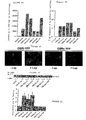
- FIG. 1 shows schematically the fusion proteins.
- Box1 represents the link site of JAK2;
- Box3 represents the STAT protein binding site;
- TM represents the trans-membrane domain.
- Figures 2a and 2b illustrate the expression of OBR constructs in COS cells estimated by radiolabeling experiments using 125 I-leptin as a radioligand.
- Figures 2a and 2b the total cellular content of OBR and the percentage of cell surface binding sites are respectively measured.
- Figure 2c illustrates the cellular localization of the expression of OBR1-YFP and OBRs-YFP constructs in the presence and absence of leptin.
- Figure 2d illustrates the activation of JAK2 by different OBR constructs.
- Figure 2e illustrates the effect of leptin stimulation of cells co-expressing the reporter gene for STAT3 and different OBR constructs.
- Figure 3 illustrates constitutive dimerization of OBR.
- HEK 293 cells expressing the various OBR constructs indicated are incubated in the presence of coelenterazine. The energy transfer is measured using a luminometer.
- FIGS. 4a and 4b illustrate the effect of leptin binding on the constitutive BRET of the OBRs.
- Figure 4a HeLa cells expressing the different OBR constructs indicated are incubated in the presence of leptin before initiating the luciferase reaction. The energy transfer is measured using a luminometer.
- Figure 4b The effect of leptin is compared in whole cells co-expressing OBRs-Luc and OBR-YFP, in the absence or presence of saponin, in total lysates and in membrane preparations.
- Figures 5a to 5e illustrate the optimization and characterization of leptin-induced BRET change on OBRs.
- Membranes prepared from HeLa or COS cells coexpressing OBRs-Luc and OBRs-YFP were pre-incubated with or without leptin before initiating the luciferase reaction.
- Figure 5a Optimization of relative and absolute OBRs-Luc and OBRs-YFP expression levels.
- Figure 5b Leptin-induced BRET signal variation as a function of time.
- Figure 5c BRET dose / response curves / leptin concentration on membrane and intact cells in the presence of saponin (0.05%).
- Figure 5d Competition binding of 125 I-leptin increase with increasing concentrations of leptin.
- FIG. 5e Specificity of leptin-induced BRET changes.
- the membranes were preincubated with saturating concentrations of erythropoietin (EPO, 10U / ml), trombopoietin (TPO, 10 nM), granulocyte macrophage colony stimulating factor (GM-CSF, 250 ng / ml), interleukin 3 (IL3, 280 ng / ml), interleukin 6 (IL6, 100 ng / ml), prolactin (PRL, 200 ng / ml), stem cell factor ⁇ (SCF ⁇ , 250 ng / ml), d epidermal growth factor (EGF, 100 ng / ml), insulin (Ins, 100 nM), lipopolysaccharide (LPS, 100 ng / ml) and tumor necrosis factor ⁇ (TNF ⁇ , 50 ng / ml).
- EPO erythropoie
- FIG. 6 Co-transfected COS cells with a constant amount of OB-Rs-Luc (50ng) and an increasing amount of OB-Rs-YFP: o, 200 ng; • 400 ng; ⁇ , 800 ⁇ , 1600; 0, 3200.
- the BRET measurements were made on the cells in the presence of saponin (0.015%), incubated or not with increasing doses of leptin, and are expressed in mBRET.
- the OB-R-YFP and OB-R-Luc fusion proteins were constructed by ligating YFP and Luc to the C-terminus of the receptors by standard molecular biology techniques.
- YFP coding regions obtained from the pGFPtpz-N1 vector Cytogem®-Topaze (Packard, Meriden, CT) were inserted into the EcoRV site of pcDNA3 / CMV (Invitrogen, Groningen, Netherlands) which contains a modified polylinker site.
- the coding region of Renilla luciferase was obtained from pRL-CMV (Promega, Madison, WI) and inserted into the EcoRV site of the modified pcDNA3.
- OBR1 and OBRs were inserted into the two vectors described above, respectively in the EcoR1 / BamH1 and NheI cloning sites. Stop codons were deleted by site-directed mutagenesis and the phase of the fusion protein was adjusted.
- the HEK 293, COS-M6 and HeLa cell lines were cultured in DMEM supplemented with the following components: 10% (v / v) FBS, 4.5 g / l glucose, 100 U / ml penicillin, 0.1 mg / ml streptomycin, 1 mM glutamine (Life Technologies, Gaithersburg, MD). Transient transfections were performed using the FuGene 6 transfection reagent (Roche, Basel, Switzerland).
- the cells were incubated with 100 nM leptin for 60 min and 0.01 mM bis-benzamidine for 15 min before being washed in PBS and fixed for 20 min at room temperature in a solution. cold 4% paraformaldehyde in PBS. The sections are observed by fluorescent microscopy using FITC and DAPI filters.
- the cells were placed in ice, washed twice in PBS at ice-temperature and mechanically detached in buffer 1 (5 mM Tris, 2 mM EDTA, pH 7.4, 5 mg / l of soy trypsin, 5 mg / liter leupeptin, and 10 mg / liter benzamidine) at ice temperature.
- buffer 1 5 mM Tris, 2 mM EDTA, pH 7.4, 5 mg / l of soy trypsin, 5 mg / liter leupeptin, and 10 mg / liter benzamidine
- the cell suspensions are homogenized with a Polytron homogenizer (Janke & Kunkel Ultra-Turrax T25) three times for 5 sec.
- the lysate is centrifuged at 450 X g for 5 min at 4 ° C and the supernatant is centrifuged at 48000 X g for 30 min at 4 ° C.
- the final pellet is washed twice in buffer 1 and resuspended in a solution (75 mM Tris (pH 7.4), 12.5 mM MgCl 2 , 5 mM EDTA with protease inhibitors, as described above) and immediately used in experiments. radioactive ligand binding or BRET experiments.
- HeLa cells were co-transfected with HA2-labeled JAK2 expressing plasmids (donated by Dr. Wojchowski, Pennsylvania State University, Pennsylvania, USA) and plasmids containing different OBR constructs.
- the cells were lysed in lysis buffer (10 mM Tris, 150 mM NaCl, 5 mM EDTA, 5% glycerol, 0.02% NaN3, 0.1% NP40, 1 mM orthovanadate, 5 mg / l of soy trypsin and 10 mg / liter of benzamidine) and centrifuged for 15 min at 13000 rpm.
- the soluble fraction was immunoprecipitated for 2 h with an anti-JAK2 polyclonal antibody (HR-758) (1 ⁇ g / ml) (Santa Cruz Biotechnology, Santa Cruz, CA).
- the JAK2 immunoprecipitates were denatured in the solution (62.5 mM Tris / HCl (pH 6.8), 5% SDS, 10% glycerol and 0.05% bromophenol blue) at 100 ° C for 10 minutes.
- the proteins were separated by SDS-PAGE in 7% polyacrylamide and transferred to nitrocellulose.
- the immuno-detection was carried out with an anti-phosphotyrosine antibody 4G10 (2 ⁇ g / ml) (Upstate Biotechnology, Lake Placid, NY). Immunoreactivity was revealed using a suitable horseradish peroxidase-coupled secondary antibody and the ECL chemiluminescent reagent (Amersham, Aylesbury, UK).
- Transfected cells were serum-deficient in DMEM (1% BSA) 24 h before the binding experiments.
- DMEM 1% BSA
- binding buffer DMEM, 25 mM Hepes pH 7.4, 1% BSA
- I-leptin 1 mM Hepes pH 7.4, 1% BSA
- 200 nM non-radioactive leptin recombinant human leptin (PeproTech Inc, USA) for 4 h at 4 ° C.
- the cells were washed twice with PBS at ice-temperature, lysed in 1N NaOH and the radioactivity determined in a gamma radiation counter.In order to measure the total amount of leptin binding in the extract, the cells were in suspension in 1.5 ml of binding buffer containing 0.15% digitonin for 2 h at 4 ° C. The extracts were centrifuged for 30 min in an Eppendorf centrifuge at maximum speed at 4 ° C. The supernatant (0.2 ml) was incubated with 100000 cpm of 125 I-lep in the presence or absence of 200 nM leptin in a total volume of 0.25 ml with stirring overnight at 4 ° C.
- 0.5 ml ⁇ -globulin (1.25 mg / ml) and 0.5 ml polyethylene glycol 6000 (25% w / v) were added to precipitate the receptor-ligand complexes, which were obtained by centrifugation (17000 xg for 3 min). The pellet is washed with 1 ml of polyethylene glycol 6000 12% (w / v) and counted.
- HeLa cells were co-transfected with 2.6 ⁇ g of plasmid carrying the reporter gene STAT3 (donated by Dr. Levy, New York University, New York, USA), 200 ⁇ g of pcDNA3 comprising the coding region of Renilla luciferase (used as internal control) and with 1.4 ⁇ g of the different OBR constructions or with the vehicle alone. 48 hours after transfection, the cells were deficient overnight in DMEM (1% BSA) before stimulation with 1nM leptin for 6-8 hours. The cells were then washed and lysed in passive lysis buffer (Promega Corporation, Madison, WI) for 15 minutes at room temperature.
- passive lysis buffer Promega Corporation, Madison, WI
- the total lysates were centrifuged for 2 minutes at 15,000 rpm and the supernatants were used in a Luciferase assay (Promega Corporation, Madison, WI) using a Berthold luminometer (Lumat LB 9507). The results are expressed by the ratio of firefly luciferase / Renilla luciferase activities .
- the substrate, coelenterazine h (Molecular Probes, Eugene, OR) is added at a final concentration of 5 ⁇ M and reads with a Fluoro / Luminometer Fusion TM (Packard Instrument Company, Meriden, CT) that allows sequential integration luminescence signals detected with two filters (Luc filter: 485 ⁇ 10 nm, YFP filter: 530 ⁇ 12.5 nm).
- the BRET ratio is defined as the difference of the 530 nm / 485 nm emission of co-transfected Luc and YFP fusion proteins and the 530 nm / 485 nm emission of the Luc fusion protein alone.
- mBU milliBRET units
- EPO erythropoietin
- Ins insulin
- LPS lipopolysaccharide
- TPO recombinant human trombopoietin
- GM-CSF interleukin-3 (IL3)
- IL6 interleukin-6
- PRL prolactin
- SCF EGF
- EGF EGF
- TNF ⁇ TNF ⁇
- the cell surface expression of fusion proteins and wild-type receptors expressed in COS cells vary between 5% and 10%, which is consistent with values already described. Similar values are obtained in HEK 293 cells expressing endogenous OBRs (14 ⁇ 3%).
- the location of OBR fusion proteins in HeLa cells was investigated by fluorescence microscopy using fusion proteins with YFP. The fluorescence due to OBR1-YFP is distributed sporadically in cells whereas that due to OBRs-YFP is localized in plaques. Leptin stimulation locates OBR1-YFP in large intracellular plaques probably corresponding to the endosomal compartment. The location of OBRs-YFP does not change significantly.
- the results obtained by fluorescence microscopy confirm the predominant localization of OBR in the intracellular compartment and are consistent with the already described localization of the OBR1-GFP fusion protein in COS cells.
- the functional expression of the fusion proteins is evaluated by measuring the activation of the JAK-STAT pathway.
- JAK2 kinases are associated with the intracellular domains of OBRs and OBR1.
- Ligand binding induces transphosphorylation of JAK2 and phosphorylation of OBR1 but not of OBRs.
- Phosphorylated OBR1 then provides an attachment site for STAT proteins, which are activated by tyrosine phosphorylation after receptor binding.
- the activated STAT proteins then dimerize and are translocated to the nucleus where they stimulate gene transcription via STAT response elements as described by Tartaglia (1997, J Biol Chem 272, 6093-6096). As shown in Figure 2c, all OBR constructs induce phosphorylation of JAK2 indicating activation of JAK2. STAT3 reporter gene activity is activated by a factor 2-4 by OBRI-wt and OBR1 fusion proteins while short isoforms have no effect on reporter gene activity. These results indicate that OBR fusion proteins are functionally expressed in HeLa cells.
- OBR-Luc and OBR-YFP The dimerization of OBR-Luc and OBR-YFP has been studied in living cells. Significant energy transfers were observed between OBRs-Luc and OBRs-YFP as well as between OBRI-Luc and OBRI-YFP, expressed in equimolar amounts, indicating that constitutive homo-dimers exist for both receptors ( Fig. 3 a, b). The existence of OBRs / OBR1 heterodimers in living cells is demonstrated by the detection of BRET between OBRs-Luc and OBR1-YFP as well as between OBRI-Luc and OBRs-YFP.
- the cells were pre-incubated with leptin before initiating the reaction of luciferase with its substrate. No changes in the constitutive BRET are observed with the OBR1 homodimers and the two OBRs / OBR1 hetero dimeric combinations while the
- BRET is increased with homo dimers OBRs (Fig. 4 a).
- BRET changes of leptin-induced OBR homo dimers were then measured in different cell preparations. Mechanical disruption of the cells in a hypotonic buffer significantly improves leptin BRET increase while basal BRET remains unchanged. Similar results were obtained with the membrane fraction after cytosol separation. While all OBRs-Luc / OBRs-YFP couples contribute to basal BRET, only receptors exposed to the cell surface (5-10%) can be stimulated by leptin that is membrane impermeable in intact cells. The disruption of cell membranes increases the leptin-accessible OBR fraction that is responsible for the leptin-induced increase in BRET.
- a saturated concentration of leptin induces a 2-2.5 fold increase in the basal BRET signal in cells incubated with saponin or membranes prepared from cells expressing OBRs homodimers. This increase is a function of time. Peak values are reached after 20 minutes of incubation with 1nM leptin at room temperature (Fig. 5b). For higher concentrations of leptin, the maximum values are obtained after 5 minutes of incubation at room temperature.
- the effect of leptin is dose-dependent with an EC50 of about 100 ⁇ M (Fig. 5c), which is in agreement with the Ki values obtained with the OBRs-Luc fusion proteins (116 ⁇ M) and OBRsYFP ( 35 ⁇ M) (FIG. 5 d).
- the specificity of the test is demonstrated by the absence of ligand-induced BRET by a saturating concentration of several cytokines and other membrane receptor ligands such as erythropoietin, trombopoietin, GM-CSF, IL3 IL6, PRL, SCF ⁇ , EGF, insulin, LPS and TNF ⁇ .
- the distribution of receptors in dimers follows statistical laws, and at a ratio 1/1 in number of receptors the following distribution is expected if all the receptors are in dimeric form: 1/4 Luc / Luc, 1/4 YFP / YFP and 1/2 receivers capable of generating a BRET signal (1/4 Luc / YFP, 1/4 YFP / Luc).
Abstract
Description
La présente invention est un procédé de détection de ligands du récepteur de la leptine par mise en oeuvre du transfert d'énergie entre des protéines de fusion composées de récepteurs de la leptine et de protéines donneurs ou accepteurs d'énergie.The present invention is a method of detecting leptin receptor ligands by performing energy transfer between fusion proteins composed of leptin receptors and energy donor or acceptor proteins.
Sont décrites également des protéines de fusion utiles pour la mise en oeuvre de ce procédé.Also described are fusion proteins useful for carrying out this method.
La leptine est une protéine présentant un poids moléculaire de 16 kDa qui est sécrétée par les adipocytes. Cette protéine est associée à la sensation de satiété, et joue un rôle majeur dans le contrôle de la prise de poids, la consommation d'énergie, la formation osseuse, l'angiogénèse mais aussi dans d'autres fonctions physiologiques telles que le déclenchement de la puberté et le contrôle de la reproduction ou la régulation de la réponse immunitaire régulée par les lymphocytes T.
Le récepteur de la leptine (OBR) appartient à la famille des récepteurs aux cytokines. Il est composé, comme l'illustre la figure 1 d'une chaine polypeptidique unique comprenant un domaine transmembranaire (Tartaglia et al., J. Biol. Chem, 272, 6093-6096, 1995). La demande de brevet WO 97/19952 est relative à ce récepteur.
Six isoformes différentes de l'OBR ayant des domaines C-terminaux de longueurs différentes ont été décrits. Ces isoformes dérivent toutes, par épissages alternatifs, d'un gène unique. Il existe également une forme soluble des OBR contenant le site de liaison à la leptine qui correspond au domaine extracellulaire des formes membranaires. Cette forme soluble générée de façon post-traductionelle par protéolyse à la membrane plasmique à partir des formes membranaires se retrouve dans le sang. Une autre forme soluble de l'OBR résultant d'une mutation générant un codon stop avant le domaine transmembranaire est également trouvée dans certaines cas très rares.
Une protéine de fusion constituée de la forme longue du récepteur de la leptine (OBR1) fusionnée à la EGFP (Enhanced Green Fluorescence Protein) a été utilisée par Lundin et al (Biochemica et Biophysica Acta, 1499, 130-138, 2000) pour étudier la localisation du récepteur.
L'activation de l'OBR se ferait par l'intermédiaire d'un complexe tétramèrique composé de deux janus kinase 2 (JAK2) et de deux OBR. L'activation du récepteur induite par la leptine induirait un changement dans la conformation de l'OBR, qui lui même activerait une JAK2, qui à son tour transphosphorylerait une autre JAK2 puis le récepteur OBR.
L'activation de l'OBR apparaît être responsable de tous les effets connus de la leptine tels que la perte de poids et tous les phénomènes impliqués dans les désordres pondéraux.
Les propriétés inhibitrices de la leptine vis à vis de la synthèse osseuse ont ainsi été récemment mises en évidence. La leptine agit en inhibant l'activité des ostéoblastes, une population de cellules responsables de la formation de l'os.
Modifier la leptinémie pourrait permettre de traiter les maladies liées à une diminution de la densité osseuse comme par exemple l'ostéoporose ou à l'inverse celles liées à une calcification importante.
En 1999 Xu et al (Proc Natl Acad Sci U S A 96, 151-156) ont décrit une méthode de détection des interactions protéine-protéine dans des cellules vivantes. Cette méthode a en outre fait l'objet de la demande de brevet WO99/66324.
Cette méthode, appelée BRET (pour Bioluminescent Resonance Energy Transfer ou transfert d'énergie de bioluminescence par résonance), est basée sur un phénomène naturel, l'émission de fluorescence par des organismes marins. La transformation enzymatique, par la luciférase de Renilla (Luc), d'un substrat qui peut traverser la membrane génère une bioluminescence, qui à son tour excite un accepteur d'énergie tel que la protéine fluorescente jaune (YFP ou yellow fluorescent protein en anglais). Cette méthode correspond à la LRET (pour Luminescent Resonance Energy Transfer) décrite par Wang et al (Mol Gen Genet 264 : 578-587 (2001)).
L'efficacité du transfert d'énergie dépend de la proximité physique et des orientations respectives de l'accepteur et du donneur. Ainsi la co-expression de la luciférase et de la YFP n'est pas suffisante pour induire un transfert d'énergie car la distance entre les deux partenaires doit être inférieure à 100Å. Afin d'étudier l'interaction entre deux partenaires d'interaction potentiels la première protéine a été fusionnée à la luciférase et la seconde protéine à la YFP. Si les deux protéines interagissent un transfert d'énergie peut être observé.
Depuis, la méthode de BRET a été mise en oeuvre sur un nombre limité de récepteurs, présentant une structure très différente du récepteur de la leptine.
Ainsi quelques auteurs décrivent la mise en oeuvre de la méthode sur des récepteurs de la famille des récepteurs couplés aux protéines G (GPCR) tels que les récepteurs β
2 adrénergiques (Angers et al, 2000, Proc. Natl. Acad. Sci. U. S. A. 10.1073), cholesystokines de type A (CCK ;Cheng et al, 2001 . Biol. Chem. 276: 48040-48047), et de la thyrotropin releasing hormon (Kroeger et al, 2001, J. Biol. Chem. 276: 12736-12743).
Ces récepteurs de taille importante présentent une structure complexe comprenant 7 domaines transmembranaires.
Enfin Boute et al (2001, Mol Pharmacol 60: 640-645) ont décrit le suivi de l'activation du récepteur de l'insuline en utilisant le BRET.
Le récepteur de l'insuline est constitué de dimères covalents, et non de complexes non covalents comme le récepteur de la leptine. En outre il comprend une partie intracellulaire assez longue. Enfin les auteurs montrent que le changement de BRET induit par l'insuline ne peut être mis en oeuvre que sur le récepteur solubilisé.Leptin is a protein with a molecular weight of 16 kDa that is secreted by adipocytes. This protein is associated with the sensation of satiety, and plays a major role in the control of weight gain, energy consumption, bone formation, angiogenesis but also in other physiological functions such as triggering puberty and reproductive control or regulation of the immune response regulated by T lymphocytes.
The leptin receptor (OBR) belongs to the family of cytokine receptors. It is composed, as illustrated in FIG. 1, of a single polypeptide chain comprising a transmembrane domain (Tartaglia et al., J. Biol Chem, 272, 6093-6096, 1995). The patent application WO 97/19952 relates to this receiver.
Six different isoforms of OBR having C-terminal domains of different lengths have been described. These isoforms all derive, by alternative splicing, a single gene. There is also a soluble form of OBR containing the leptin binding site which corresponds to the extracellular domain of the membrane forms. This soluble form generated post-translationally by proteolysis to the plasma membrane from the membrane forms is found in the blood. Another soluble form of OBR resulting from a mutation generating a stop codon before the transmembrane domain is also found in some very rare cases.
A fusion protein consisting of the long form leptin receptor (OBR1) fused to EGFP (Enhanced Green Fluorescence Protein) was used by Lundin et al (Biochemica and Biophysica Acta, 1499, 130-138, 2000) to study the location of the receptor.
The activation of the OBR would be via a tetrameric complex composed of two Janus kinase 2 (JAK2) and two OBRs. Leptin-induced receptor activation would induce a change in the conformation of OBR, which itself would activate a JAK2, which in turn would transphosphorylate another JAK2 and then the OBR receptor.
The activation of OBR appears to be responsible for all known effects of leptin such as weight loss and all the phenomena involved in weight disorders.
The inhibitory properties of leptin with respect to bone synthesis have thus recently been demonstrated. Leptin works by inhibiting the activity of osteoblasts, a cell population responsible for bone formation.
Modifying leptinemia could be used to treat diseases linked to a decrease in bone density, such as osteoporosis or, conversely, those linked to significant calcification.
In 1999 Xu et al (Proc Natl Acad Sci USA 96, 151-156) described a method for detecting protein-protein interactions in living cells. This method has moreover been the subject of the patent application WO99 / 66324.
This method, called BRET (for Bioluminescent Resonance Energy Transfer), is based on a natural phenomenon, the emission of fluorescence by marine organisms. The enzymatic transformation, by Renilla luciferase (Luc), of a substrate that can cross the membrane generates a bioluminescence, which in turn excites an energy acceptor such as yellow fluorescent protein (YFP or yellow fluorescent protein). ). This method corresponds to the LRET (for Luminescent Resonance Energy Transfer) described by Wang et al (Mol Gen Genet 264: 578-587 (2001)).
The effectiveness of energy transfer depends on the physical proximity and respective orientations of the acceptor and the donor. Thus the co-expression of luciferase and YFP is not sufficient to induce energy transfer because the distance between the two partners must be less than 100Å. To study the interaction between two potential interaction partners the first protein was fused to luciferase and the second protein to YFP. If both proteins interact a transfer of energy can be observed.
Since then, the BRET method has been implemented on a limited number of receptors, presenting a structure very different from the leptin receptor.
Thus some authors describe the implementation of the method on receptors of the family of G-protein coupled receptors (GPCR) such as β receptors
2 adrenergic (Angers et al., 2000, Proc Natl Acad Sci USA 10.1073), type A cholesystokins (CCK, Cheng et al., 2001 Biol Chem 276: 48040-48047), and thyrotropin releasing hormone (Kroeger et al., 2001, J. Biol Chem 276: 12736-12743).
These large receptors have a complex structure comprising 7 transmembrane domains.
Finally Boute et al (2001, Mol Pharmacol 60: 640-645) described the monitoring of insulin receptor activation using BRET.
The insulin receptor is composed of covalent dimers, not non-covalent complexes such as the leptin receptor. In addition, it comprises a rather long intracellular portion. Finally, the authors show that the insulin-induced BRET change can only be implemented on the solubilized receptor.
En quelques dizaines d'années, l'obésité est devenue un problème majeur de santé publique dans les pays industrialisés où elle touche maintenant 20 à 30 % de la population. Ces chiffres devraient encore croître de façon alarmante dans les années à venir. Du fait de ses origines multifactorielles qui prennent leurs sources à des degrés plus ou moins importants parmi des facteurs environnementaux d'une part (comportements alimentaires, accès à la nourriture, dépense énergétique,...) et des origines génétiques multiples d'autre part, l'obésité constitue un véritable défi pour la médecine.
De même les maladies de l'os, telles que l'ostéoporose, touchent une partie de plus en plus importante de la population.In a few decades, obesity has become a major public health problem in the industrialized countries, where it now affects 20-30% of the population. These numbers are expected to grow alarmingly in the coming years. Because of its multifactorial origins which take their sources to greater or lesser degrees among environmental factors on the one hand (eating behavior, access to food, energy expenditure, ...) and multiple genetic origins on the other hand Obesity is a real challenge for medicine.
Similarly, bone diseases, such as osteoporosis, affect an increasingly important part of the population.
La découverte de nouvelles molécules permettant de traiter les diverses maladies liées au récepteur de la leptine, telles que l'obésité et l'ostéoporose, est donc un enjeu majeur de la santé publique.
Il n'existe cependant pas de procédé de criblage spécifique d'agonistes ou d'antagonistes du récepteur de la leptine pouvant être mis en oeuvre à haut flux.The discovery of new molecules to treat various diseases related to the leptin receptor, such as obesity and osteoporosis, is therefore a major public health issue.
However, there is no specific method of screening agonists or leptin receptor antagonists that can be used at high flux.
Les demandeurs se sont donc attachés à la mise en oeuvre d'un test de criblage rapide, spécifique et efficace d'agonistes ou d'antagonistes du récepteur de la leptine.The applicants therefore attached themselves to the implementation of a rapid, specific and effective screening test for agonists or antagonists of the leptin receptor.
Ils ont montré de manière surprenante que le changement de BRET induit par la leptine pouvait être mis en oeuvre sur l'un des isoformes du récepteur de la leptine, mais qu'il ne pouvait être mis en oeuvre avec tous les isoformes.Surprisingly, they showed that leptin-induced BRET change could be carried out on one of the leptin receptor isoforms, but could not be implemented with all isoforms.
Ils ont en outre montré que la mise en oeuvre du BRET sur le récepteur de la leptine est optimale dans certaines conditions opératoires.They have furthermore shown that the use of BRET on the leptin receptor is optimal under certain operating conditions.
La présente invention est donc un procédé de détection de ligands du récepteur de la leptine par mise en oeuvre du transfert d'énergie de résonance entre une première protéine de fusion composée d'un récepteur de la leptine, ou d'une partie substantielle d'un récepteur de la leptine, et d'une protéine donneur d'énergie, ou d'une partie substantielle et active d'une protéine donneur d'énergie, et une seconde protéine de fusion composée d'un récepteur de la leptine, ou d'une partie substantielle d'un récepteur de la leptine, et d'une protéine accepteur d'énergie, ou d'une partie substantielle et active d'une protéine accepteur d'énergie.The present invention is therefore a method of detecting leptin receptor ligands by implementing resonance energy transfer between a first fusion protein composed of a leptin receptor, or a substantial portion of it. a leptin receptor, and an energy donor protein, or a substantial and active portion of an energy donor protein, and a second fusion protein composed of a leptin receptor, or a a substantial portion of a leptin receptor, and an energy acceptor protein, or a substantial and active portion of an energy acceptor protein.
Elle est en outre relative à des protéines de fusion pour la mise en oeuvre de ce procédé, ainsi qu'à des acides nucléiques codant ces protéines.It further relates to fusion proteins for carrying out this method, as well as to nucleic acids encoding these proteins.
Elle envisage également un procédé de traitement curatif ou préventif de maladies liées à la leptine consistant à administrer un ligand sélectionné par le procédé défini ci-dessus à un patient atteint par la dite maladie.It also contemplates a method of curative or preventive treatment of leptin-related diseases comprising administering a ligand selected by the method defined above to a patient suffering from said disease.
Un premier objet décrit dans le brevet est donc une protéine de fusion caractérisée en ce qu'elle est composée d'un récepteur de la leptine, ou d'une partie substantielle d'un récepteur de la leptine et d'une protéine accepteur ou donneur d'énergie, ou d'une partie substantielle et active d'une protéine accepteur ou donneur d'énergie.A first object described in the patent is therefore a fusion protein characterized in that it is composed of a leptin receptor, or a substantial part of a leptin receptor and an acceptor or donor protein of energy, or a substantial and active portion of an acceptor protein or energy donor.
Les protéines de fusion selon la présente invention sont composées en substance d'une partie correspondant à une partie ou la totalité de la séquence d'un récepteur de la leptine et d'une partie correspondant à une protéine donneur ou accepteur d'énergie. Elles peuvent néanmoins comprendre d'autres séquences d'acides aminés, issues d'autres protéines, telles que des séquences signal. Ainsi la séquence SEQ ID N°4 est constituée d'une partie de la séquence SEQ ID N°2 et d'autres séquences, et en particulier de la séquence signal de l'interleukine 3 de souris.The fusion proteins according to the present invention are substantially composed of a portion corresponding to part or all of the sequence of a leptin receptor and a portion corresponding to an energy donor or acceptor protein. They may, however, include other amino acid sequences, derived from other proteins, such as signal sequences. Thus, the sequence SEQ ID No. 4 consists of part of the sequence SEQ ID No. 2 and other sequences, and in particular of the signal sequence of mouse interleukin-3.
De manière avantageuse la protéine donneur d'énergie est la luciférase de Renilla. Elle peut néanmoins être toute autre protéine donneur d'énergie telle que le spectre d'émission du donneur chevauche suffisamment le spectre d'excitation de l'accepteur pour permettre un transfert d'énergie efficace entre les deux partenaires. Elle peut ainsi être la GFP, si le transfert d'énergie est le FRET, ou encore l'aequorine si le transfert d'énergie est le CRET. L'aequorine peut être obtenue et utilisée comme décrit dans la demande de brevet EP0 187 519, ou dans l'article de Inouye et al. (PNAS USA 82 : 3154-3158 (1985)).Advantageously, the energy-donor protein is Renilla luciferase. It may, however, be any other energy-donor protein such that the donor's emission spectrum sufficiently overlaps the acceptor's excitation spectrum to allow efficient energy transfer between the two partners. It can be GFP, if the energy transfer is FRET, or aequorin if the energy transfer is CRET. Aequorin can be obtained and used as described in patent application EP0 187 519, or in the article by Inouye et al. (PNAS USA 82: 3154-3158 (1985)).
La protéine fluorescente accepteur d'énergie est quant à elle préférentiellement la DsRed, la GFP ou un mutant de cette protéine, tel que la YFP, l'EYFP, la GFP sauvage, la GFPS65T, ou la Topaz.
Elle peut néanmoins être toute autre protéine fluorescente accepteur d'énergie telle que le spectre d'excitation de l'accepteur et le spectre d'émission du donneur se chevauchent suffisamment pour permettre un transfert d'énergie efficace entre les deux partenaires.The energy-accepting fluorescent protein is preferentially DsRed, GFP or a mutant of this protein, such as YFP, EYFP, wild-type GFP, GFPS65T, or Topaz.
It may, however, be any other energy-accepting fluorescent protein such that the acceptor excitation spectrum and the donor emission spectrum overlap sufficiently to allow efficient energy transfer between the two partners.
Ces protéines sont connues de l'homme du métier qui peut trouver leurs séquences dans la littérature, notamment dans la revue de Blinks et al. (Pharmacol. Rev. 28 : 1-93 (1976)). En particulier la GFP est décrite par Tsien (Annu. Rev. Biochem. 67 : 509-544 (1998)) et son clonage par Prasher et al. (Gene 111 : 229-233 (1992)). Le clonage de la DsRed est quant à lui décrit par Matz et al. (Nat. Biotechnol. 17 : 969-973 (1999)). Pour la Rluc, l'homme du métier peut se référer à Blinks et al. (Pharmacol. Rev. 28 : 1-93 (1976)) ou encore à Lorenz et al. (PNAS 88: 4438-4442 (1991)).These proteins are known to those skilled in the art who can find their sequences in the literature, in particular in the review of Blinks et al. (Pharmacol Rev. 28: 1-93 (1976)). In particular, GFP is described by Tsien (Annu Rev. Biochem 67: 509-544 (1998)) and cloned by Prasher et al. (Gene 111: 229-233 (1992)). Cloning of DsRed is described by Matz et al. (Nat Biotechnol 17: 969-973 (1999)). For Rluc, one skilled in the art can refer to Blinks et al. (Pharmacol Rev. 28: 1-93 (1976)) or Lorenz et al. (PNAS 88: 4438-4442 (1991)).
L'isoforme du récepteur de la leptine, compris en totalité ou en partie dans la protéine de fusion, est une isoforme courte, ou présentant un domaine intracellulaire court.
Une telle isoforme comprend avantageusement un domaine intracellulaire Box1, mais ne comprend pas de domaine intracellulaire Box 3.The isoform of the leptin receptor, included in whole or in part in the fusion protein, is a short isoform, or having a short intracellular domain.
Such an isoform advantageously comprises a Box1 intracellular domain, but does not include a Box 3 intracellular domain.
De manière préférentielle cette isoforme est l'isoforme OBRs et encore plus préférentiellement l'isoforme OBRs humain. Cette isoforme peut néanmoins être originaire de toute autre espèce.Preferably, this isoform is the OBRs isoform and even more preferably the human OBRs isoform. This isoform may nevertheless originate from any other species.
Elle peut aussi être toute autre isoforme courte, préférentiellement comprenant au moins le domaine extracellulaire de l'OBR, telle la forme soluble de l'OBR contenant le site de liaison à la leptine décrite par Lee et al (Nature 379, 632-635 (1996)), Gavrilova et al (JBC 272 :30546-30551 (1997)) Maamr. et al. (Endocrinology 142 :4389-4393 (2001)) ou Clement et al. (Nature 392 :398-401 (1998)).It can also be any other short isoform, preferably comprising at least the extracellular domain of OBR, such as the soluble form of OBR containing the leptin binding site described by Lee et al (Nature 379, 632-635 ( 1996)), Gavrilova et al (JBC 272: 30546-30551 (1997)) Maamr. et al. (Endocrinology 142: 4389-4393 (2001)) or Clement et al. (Nature 392: 398-401 (1998)).
Selon un mode de mise en oeuvre particulièrement préférentiel l'isoforme est l'isoforme OBRs humaine de séquence SEQ ID °2. Elle peut aussi être un variant de cette protéine présentant une identité d'au moins 65%, préférentiellement d'au moins 75%, et encore plus préférentiellement d'au moins 85% ou 95%, avec la séquence SEQ ID °2.According to a particularly preferred embodiment, the isoform is the human OBRs isoform of sequence SEQ ID No. 2. It may also be a variant of this protein having an identity of at least 65%, preferably at least 75%, and even more preferably at least 85% or 95%, with the sequence SEQ ID No. 2.
La protéine de fusion peut ne comprendre qu'une partie de l'isoforme OBRs humaine. Avantageusement elle comprend la partie comprise entre les acides aminés 46 et 866 de la séquence SEQ ID°2.
La partie correspondant au récepteur à la leptine peut ainsi présenter la séquence SEQ ID°4 ou un variant de cette séquence présentant une identité d'au moins 65%, préférentiellement d'au moins 75%, et encore plus préférentiellement d'au moins 85% ou 95%.The fusion protein may comprise only a portion of the human OBRs isoform. Advantageously it comprises the part between amino acids 46 and 866 of the sequence SEQ ID No. 2.
The portion corresponding to the leptin receptor may thus have the sequence SEQ ID No. 4 or a variant of this sequence having an identity of at least 65%, preferably at least 75%, and even more preferably at least 85%. % or 95%.
De manière particulièrement avantageuse la protéine de fusion donneur présente la séquence SEQ ID N°6 ou un variant de cette séquence présentant une identité d'au moins 65%.Particularly advantageously, the donor fusion protein has the sequence SEQ ID No. 6 or a variant of this sequence having an identity of at least 65%.
De manière particulièrement avantageuse la protéine de fusion accepteur présente la séquence SEQ ID N°8 ou un variant de cette séquence présentant une identité d'au moins 65%.Particularly advantageously, the acceptor fusion protein has the sequence SEQ ID No. 8 or a variant of this sequence having an identity of at least 65%.
D'autres objets décrits dans le brevet sont des acides nucléiques codant pour ces protéines. De tels acides nucléiques peuvent être des ADN complémentaires ou génomiques, ou des ARN. Ces acides nucléiques ou polynucléotides peuvent être sous forme simple chaîne ou sous la forme de duplex.Other objects described in the patent are nucleic acids encoding these proteins. Such nucleic acids may be complementary or genomic DNAs, or RNAs. These nucleic acids or polynucleotides may be in single chain form or in duplex form.
Ils sont de manière particulièrement avantageuse des ADN complémentaires.They are particularly advantageously complementary DNAs.
De manière préférentielle l'acide nucléique a une identité d'au moins 65%, préférentiellement d'au moins 75%, et encore plus préférentiellement d'au moins 85% ou 95%, d'identité en nucléotides avec un acide nucléique de séquence SEQ ID N°5 ou SEQ ID N°7.Preferably, the nucleic acid has an identity of at least 65%, preferably at least 75%, and even more preferably at least 85% or 95%, nucleotide identity with a sequence nucleic acid. SEQ ID No. 5 or SEQ ID No. 7.
Selon encore un autre aspect, l'acide nucléique hybride, dans des conditions d'hybridation de forte stringence, avec un acide nucléique tel que défini ci-avant, et plus particulièrement un acide nucléique de séquences nucléotidiques SEQ ID N°5 et SEQ ID N°7, ou un acide nucléique de séquence complémentaire.In yet another aspect, the hybrid nucleic acid, under high stringency hybridization conditions, with a nucleic acid such as defined above, and more particularly a nucleic acid of nucleotide sequences SEQ ID No. 5 and SEQ ID No. 7, or a nucleic acid of complementary sequence.
Le «pourcentage d'identité » entre deux séquences de nucléotides ou d'acides aminés, au sens de la présente invention, peut être déterminé en comparant deux séquences alignées de manière optimale, à travers une fenêtre de comparaison.The "percent identity" between two nucleotide or amino acid sequences, within the meaning of the present invention, can be determined by comparing two optimally aligned sequences, through a comparison window.
La partie de la séquence nucléotidique ou polypeptidique dans la fenêtre de comparaison peut ainsi comprendre des additions ou des délétions (par exemple des « gaps ») par rapport à la séquence de référence (qui ne comprend pas ces additions ou ces délétions) de manière à obtenir un alignement optimal des deux séquences.The part of the nucleotide or polypeptide sequence in the comparison window may thus comprise additions or deletions (for example "gaps") with respect to the reference sequence (which does not include these additions or deletions) so as to obtain an optimal alignment of the two sequences.
Le pourcentage est calculé en déterminant le nombre de positions auxquelles une base nucléique ou un résidu d'aminoacide identique est observé pour les deux séquences (nucléique ou peptidique) comparées, puis en divisant le nombre de positions auquel il y a identité entre les deux bases ou résidus d'aminoacides par le nombre total de positions dans la fenêtre de comparaison, puis en multipliant le résultat par 100 afin d'obtenir le pourcentage d'identité de séquence.The percentage is calculated by determining the number of positions at which an identical nucleobase or amino acid residue is observed for the two (nucleic or peptide) sequences compared and then dividing the number of positions at which there is identity between the two bases. or amino acid residues by the total number of positions in the comparison window, then multiplying the result by 100 to obtain the percentage of sequence identity.
L'alignement optimal des séquences pour la comparaison peut être réalisé de manière informatique à l'aide d'algorithmes connus contenus dans le package de la Société WISCONSIN GENETICS SOFTWARE PACKAGE, GENETICS COMPUTER GROUP (GCG), 575 Science Doctor , Madison, WISCONSIN.The optimal alignment of the sequences for the comparison can be performed in a computer manner using known algorithms contained in the package of the company WISCONSIN GENETICS SOFTWARE PACKAGE, GENETICS COMPUTER GROUP (GCG), 575 Science Doctor, Madison, WISCONSIN.
A titre d'illustration, le pourcentage d'identité de séquence pourra être effectué à l'aide du logiciel BLAST (versions BLAST 1.4.9 de mars 1996, BLAST 2.0.4 de février 1998 et BLAST 2.0.6 de septembre 1998), en utilisant exclusivement les paramètres par défaut (S. F Altschul et al, J. Mol. Biol. 1990 215 : 403-410, S. F Altschul et al, Nucleic Acids Res. 1997 25 : 3389-3402). Blast recherche des séquences similaires/homologues à une séquence « requête » de référence, à l'aide de l'algorithme d'Altschul et al. La séquence requête et les bases de données utilisées peuvent être peptidiques ou nucléiques, toute combinaison étant possible.By way of illustration, the percentage of sequence identity can be carried out using the BLAST software (BLAST versions 1.4.9 of March 1996, BLAST 2.0.4 of February 1998 and BLAST 2.0.6 of September 1998), using exclusively the default parameters (S. F Altschul et al., J. Mol Biol., 1990 215: 403-410, S. F Altschul et al, Nucleic Acids Res 1997 25: 3389-3402). Blast searches for similar / homologous sequences to a reference "query" sequence, using the algorithm of Altschul et al. The query sequence and the databases used may be peptide or nucleic, any combination being possible.
Par « conditions d'hybridation de forte stringence » au sens de la présente invention, on entendra les conditions suivantes :By "high stringency hybridization conditions" within the meaning of the present invention, the following conditions will be understood:
- Mélanger: 40µl ADN sperme de saumon (10mg/ml)+ 40 µl ADN placentaire humain (10mg/ml)Mix: 40μl salmon sperm DNA (10mg / ml) + 40μl human placental DNA (10mg / ml)
-
Dénaturer 5 mn à 96°C, puis plonger dans la glace le mélange.
Denature 5 minutes at 96 ° C, then immerse in the ice mixture. - Oter le SSC 2X et verser 4 ml de mix formamide dans le tube d'hybridation contenant les membranes.Remove the 2X SSC and pour 4 ml formamide mix into the hybridization tube containing the membranes.
- Ajouter le mélange des deux ADNs dénaturés.Add the mixture of the two denatured DNAs.
- Incubation à 42°C pendant 5 à 6 heures, avec rotation.Incubation at 42 ° C for 5 to 6 hours, with rotation.
-
Ajouter à la sonde marquée et purifiée 10 à 50 µl ADN Cot I, selon la quantité de répétitions.Add to the labeled and purified
probe 10 to 50 μl Cot I DNA, according to the amount of repeats. -
Dénaturer 7 à 10 mn à 95°C.
Denature 7 to 10 minutes at 95 ° C. - Incuber à 65°C pendant 2 à 5 heures.Incubate at 65 ° C for 2 to 5 hours.
- Oter le mix de pré hybridation.Remove the mix of pre hybridization.
-
Mélanger 40 µl ADN sperme de saumon + 40 µl ADN placentaire humain dénaturer 5 mn à 96°C, puis plonger dans la glace.
Mix 40 μl Salmon sperm + 40 μl human placental DNA denature for 5 minutes at 96 ° C, then immerse in ice. - Ajouter dans le tube d'hybridation 4 ml de mix formamide, le mélange des deux ADN et la sonde marquée/ADN Cot I dénaturée.Add to the hybridization tube 4 ml of formamide mix, the mixture of the two DNAs and the labeled probe / DNA Cot I denatured.
-
Incuber 15 à 20 heures à 42°C, avec rotation.
Incubate 15 to 20 hours at 42 ° C, with rotation.
- Un lavage à température ambiante dans du SSC 2X, pour rincer.
- 2
fois 5 minutes à température ambiante SSC2X et SDS - 2 fois 15 minutes à 65°C SSC
1X et SDS
- Wash at room temperature in 2 × SSC, to rinse.
- 2 x 5 minutes at room temperature 2X SSC and 0.1% SDS at 65 ° C.
- 2
times 15 minutes at 65 ° C. 1 × SSC and 0.1% SDS at 65 ° C.
Les conditions d'hybridation décrites plus haut sont adaptées à l'hybridation dans des conditions de forte stringence, d'une molécule d'acide nucléique d'une longueur variable de 20 nucléotides à plusieurs centaines de nucléotides.The hybridization conditions described above are suitable for hybridization under conditions of high stringency, of a nucleic acid molecule of varying length from 20 nucleotides to several hundred nucleotides.
Il va sans dire que les conditions d'hybridation ci-dessus décrites peuvent être adaptées en fonction de la longueur de l'acide nucléique dont l'hybridation est recherchée ou du type de marquage choisi, selon des techniques connues de l'homme du métier.
Les conditions convenables d'hybridation peuvent par exemple être adaptées selon l'enseignement contenu dans l'ouvrage de HAMES et HIGGINS (1985,"Nucleic acid hybridization : a practical approach", Hames and Higgins Ed., IRL Press, Oxford) ou encore dans l'ouvrage de F.AUSUBEL et al (1989, Current Protocols in Molecular Biology, Green Publishing Associates and Wiley Interscience, N.Y).It goes without saying that the hybridization conditions described above can be adapted as a function of the length of the nucleic acid whose hybridization is sought or of the type of marking chosen, according to techniques known to those skilled in the art. .
Suitable hybridization conditions may for example be adapted according to the teaching contained in HAMES and HIGGINS (1985, "Nucleic Acid Hybridization: a Practical Approach", Hames and Higgins Ed., IRL Press, Oxford). in the work of F.AUSUBEL et al (1989, Current Protocols in Molecular Biology, Green Publishing Associates and Wiley Interscience, NY).
Les protéines utiles pour mettre en oeuvre la présente invention peuvent être obtenues par tous moyens connus de l'homme du métier. Elles sont néanmoins avantageusement obtenues par expression des acides nucléiques tels que décrits ci-dessus, codant pour ces protéines, éventuellement insérés dans des vecteurs d'expression, dans des cellules avantageusement choisies, suivie éventuellement par une extraction et une purification qui peut être totale ou partielle.The proteins useful for implementing the present invention may be obtained by any means known to those skilled in the art. They are nevertheless advantageously obtained by expression of the nucleic acids as described above, coding for these proteins, optionally inserted into expression vectors, in advantageously chosen cells, optionally followed by extraction and purification which may be complete or partial.
L'invention est également relative à un vecteur recombinant comprenant un acide nucléique selon l'invention.The invention also relates to a recombinant vector comprising a nucleic acid according to the invention.
Avantageusement, un tel vecteur recombinant comprendra un acide nucléique choisi parmi les acides nucléiques suivants :
- a) un acide nucléique codant pour une protéine ayant au moins 65% d'identité en acides aminés avec une séquence SEQ ID N°6 ou SEQ ID N°8 ou un fragment peptidique ou un variant de cette dernière ;
- b) un acide nucléique comprenant un polynucléotide ayant une séquence SEQ ID N°5 ou SEQ ID N°7, ou un fragment ou un variant de ce dernier ;
- c) un acide nucléique ayant au moins 65% d'identité en nucléotides avec un acide nucléique ayant une séquence SEQ ID N°5 ou SEQ ID N°7 ou un fragment ou un variant de ce dernier ;
- d) un acide nucléique hybridant, dans des conditions d'hybridation de forte stringence, avec un acide nucléique de séquences SEQ ID N°5 ou SEQ ID N°7, ou un fragment ou un variant de ce dernier.
- a) a nucleic acid encoding a protein having at least 65% amino acid identity with a sequence SEQ ID No. 6 or SEQ ID No. 8 or a peptide fragment or a variant thereof;
- b) a nucleic acid comprising a polynucleotide having a sequence SEQ ID No. 5 or SEQ ID No. 7, or a fragment or a variant thereof;
- c) a nucleic acid having at least 65% nucleotide identity with a nucleic acid having a sequence SEQ ID NO: 5 or SEQ ID NO: 7 or a fragment or a variant thereof;
- d) a nucleic acid hybridizing, under high stringency hybridization conditions, with a nucleic acid of sequences SEQ ID No. 5 or SEQ ID No. 7, or a fragment or a variant thereof.
Par « vecteur » au sens de la présente invention on entendra une molécule d'ADN ou d'ARN circulaire ou linéaire qui est indifféremment sous forme de simple brin ou double brin.For the purposes of the present invention, the term "vector" will be understood to mean a circular or linear DNA or RNA molecule which is indifferently in the form of a single-stranded or double-stranded form.
Selon un mode de réalisation, le vecteur d'expression comprend, outre un acide nucléique conforme à l'invention, des séquences régulatrices permettant d'en diriger la transcription et/ou la traduction.According to one embodiment, the expression vector comprises, besides a nucleic acid according to the invention, regulatory sequences making it possible to direct the transcription and / or translation thereof.
Selon un mode de réalisation avantageux, un vecteur recombinant utile dans une méthode selon l'invention comprendra notamment les éléments suivants :
- (1) des éléments de régulation de l'expression de l'acide nucléique à insérer, tels que des promoteurs et des enhanceurs ;
- (2) la séquence codante comprise dans l'acide nucléique conforme à l'invention à insérer dans un tel vecteur, ladite séquence codante étant placée en phase avec les signaux de régulation décrits aux (1) ; et
- (3) des séquences d'initiation et d'arrêt de la transcription appropriées.
- (1) regulatory elements for expression of the nucleic acid to be inserted, such as promoters and enhancers;
- (2) the coding sequence comprised in the nucleic acid according to the invention to be inserted into such a vector, said coding sequence being placed in phase with the regulation signals described in (1); and
- (3) appropriate initiation and transcription termination sequences.
En outre, les vecteurs recombinants pourront inclure une ou plusieurs origines de réplication chez les hôtes cellulaires dans lesquels leur amplification ou leur expression est recherchée, des marqueurs ou des marqueurs de sélection.In addition, the recombinant vectors may include one or more origins of replication in cell hosts in which their amplification or expression is sought, markers or markers of selection.
A titre d'exemples, les promoteurs pour cellules eucaryotes comprendront le promoteur de la thymidine kinase du virus HSV ou encore le promoteur de la métallothionéine-L de souris.As examples, eukaryotic cell promoters will include the HSV thymidine kinase promoter or the mouse L-metallothionein promoter.
De manière générale, pour le choix d'un promoteur adapté, l'homme du métier pourra avantageusement se référer à l'ouvrage de SAMBROOK et al. (1989, "Molecular Cloning : A Laboratory Manual, " 2nd ed., Cold Spring Harbor Laboratory Press, Cold Spring Harbor, NY.) ou encore aux techniques décrites par FULLER et al. (1996, Immunology in Current Protocols in Molecular Biology, Ausubel et al).In general, for the choice of a suitable promoter, the skilled person can advantageously refer to the work of SAMBROOK et al. (1989, "Molecular Cloning: A Laboratory Manual," 2nd ed, Cold Spring Harbor Laboratory Press, Cold Spring Harbor, NY..) Or the techniques described by FULLER et al. (1996, Immunology in Current Protocols in Molecular Biology, Ausubel et al).
Les vecteurs préférés selon l'invention sont des plasmides, tels que par exemple les vecteurs pCDNA3 (Invitrogen), pQE70, pQE60, pQE9 (Qiagen), psiX174, pBluescript SA, pNH8A, pNH16A, pNH18A, pNH46A, pWLNEO, pSV2CAT, pOG44, pXTI, pSG(Stratagene).The preferred vectors according to the invention are plasmids, such as, for example, the vectors pCDNA3 (Invitrogen), pQE70, pQE60, pQE9 (Qiagen), psiX174, pBluescript SA, pNH8A, pNH16A, pNH18A, pNH46A, pWLNEO, pSV2CAT, pOG44, pXTI, pSG (Stratagene).
Il peut s'agir également de vecteurs de type baculovirus tel que le vecteur pVL1392/1393 (Pharmingen) utilisé pour transfecter les cellules de la lignée Sf9 (ATCC N°CRL 1711) dérivées de Spodoptera frugiperda. It may also be baculovirus- type vectors such as the vector pVL1392 / 1393 (Pharmingen) used to transfect the cells of the Sf9 line (ATCC No. CRL 1711) derived from Spodoptera frugiperda.
Il peut encore s'agir de vecteurs adénoviraux tels que l'adénovirus humain de type 2 ou 5.It can still be adenoviral vectors such as
Un vecteur recombinant peut aussi être un vecteur rétroviral ou encore un vecteur adéno-associé (AAV). De tels vecteurs adéno-associés sont par exemple décrits par FLOTTE et al. (1992, Am. J. Respir. Cell Mol. Biol., 7 : 349-356A recombinant vector may also be a retroviral vector or an adeno-associated vector (AAV). Such adeno-associated vectors are for example described by FLOTTE et al. (1992, Am J Respir Cell Mol Biol., 7 : 349-356.
Le brevet décrit en outre des cellules comprenant une protéine, un acide nucléique ou un vecteur tels que décrits ci dessus, ou des fragments de ces cellules, des lysats de ces cellules ou encore des membranes de ces cellules.The patent further describes cells comprising a protein, a nucleic acid or a vector as described above, or fragments of these cells, lysates of these cells or membranes of these cells.
De telles cellules peuvent être cellules isolées d'un organisme et cultivées dans un milieu de croissance adéquat. Elles sont néanmoins préférentiellement des lignées cellulaires. Ainsi de telles lignées sont de manière particulièrement avantageuse les lignées cellulaires HEK 293, COS (ATCC N°CRL 1650), COS-M6 et HeLa (ATCC N°CCL2), ou encore Cv 1 (ATCC N°CCL70), Sf-9 (ATCC N°CRL 1711), CHO (ATCC N°CCL-61) ou 3T3 (ATCC N°CRL-6361).Such cells may be cells isolated from an organism and cultured in a suitable growth medium. They are nevertheless preferentially cell lines. Thus, such lines are particularly advantageously the HEK 293, COS (ATCC No. CRL 1650), COS-M6 and HeLa (ATCC No. CCL2) or Cv 1 (ATCC No. CCL70), Sf-9 cell lines. (ATCC No. CRL 1711), CHO (ATCC No. CCL-61) or 3T3 (ATCC No. CRL-6361).
Les membranes de ces cellules peuvent être préparées par toute méthode connue de l'homme du métier.
Préférentiellement elles seront préparées par broyage mécanique des cellules puis centrifugation des suspensions obtenues, comme illustré dans les exemples qui suivent.The membranes of these cells can be prepared by any method known to those skilled in the art.
Preferably they will be prepared by mechanical grinding of the cells and centrifugation of the resulting suspensions, as illustrated in the examples which follow.
La présente invention est en outre relative à des compostions comprenant des cellules telles que décrites ci dessus et de la saponine.The present invention further relates to compositions comprising cells as described above and saponin.
La présente invention est un procédé de détermination de la liaison de composés au récepteur de la leptine comprenant les étapes consistant à :
- mettre en contact ledit composé avec une protéine de fusion donneur d'énergie telle que décrite ci dessus, et une protéine de fusion accepteur d'énergie telle que décrite ci dessus, ou des cellules, ou des fragments, ou des lysats, ou des membranes de cellules comprenant une telle protéine, et un substrat enzymatique adéquat, et
- à mesurer le transfert d'énergie.
- contacting said compound with an energy-donor fusion protein as described above, and an energy-accepting fusion protein as described above, or cells, or fragments, or lysates, or membranes cells comprising such a protein, and a suitable enzymatic substrate, and
- to measure the transfer of energy.
Préférentiellement ledit procédé est mis en oeuvre avec des cellules traitées par de la saponine.
Les protéines de fusion donneur d'énergie et les protéines de fusion accepteur d'énergie sont choisies de manière à ce que l'énergie résultant de l'activation du donneur puisse être transférée de manière efficace à l'accepteur.Preferably, said method is carried out with cells treated with saponin.
The energy donor fusion proteins and the energy acceptor fusion proteins are selected so that the energy resulting from the activation of the donor can be effectively transferred to the acceptor.
Dans un mode de mise en oeuvre avantageux dudit procédé, la protéine de fusion donneur d'énergie est une protéine de fusion entre le récepteur de la leptine, ou une partie substantielle du récepteur de la leptine et la luciférase ou une partie substantielle de la luciférase, auquel cas le substrat est avantageusement la coelenterazine.In an advantageous embodiment of said method, the energy-donor fusion protein is a fusion protein between the leptin receptor, or a substantial part of the leptin receptor and the luciferase or a substantial part of the luciferase. in which case the substrate is advantageously coelenterazine.
Dans un mode de mise en oeuvre préférentiel dudit procédé, la protéine de fusion accepteur d'énergie est une protéine de fusion entre le récepteur de la leptine, ou une partie substantielle du récepteur de la leptine et la YFP ou une partie substantielle de la YFP.In a preferred embodiment of said method, the energy accepting fusion protein is a fusion protein between the leptin receptor, or a substantial part of the leptin receptor and the YFP or a substantial part of the YFP .
Dans un mode de mise en oeuvre avantageux dudit procédé, le transfert d'énergie mesuré en présence du composé à tester est comparé à celui mesuré en absence du composé à tester.In an advantageous embodiment of said method, the energy transfer measured in the presence of the test compound is compared with that measured in the absence of the test compound.
Préférentiellement le procédé est mis en oeuvre sur des membranes des cellules, telles que décrites ci dessus.Preferentially, the method is implemented on cell membranes, as described above.
De manière préférentielle les protéines donneur et accepteur selon la présente invention sont choisies afin que le transfert d'énergie se fasse par résonance BRET, ou LRET. Néanmoins un tel transfert d'énergie peut être effectué par FRET (pour Fluorescent Résonance Energy Transfer ou transfert d'énergie de fluorescence par résonance) ou encore par CRET (pour Chemioluminescent Résonance Energy Transfer ou transfert d'énergie de chimioluminescence par résonance).
Quelque soit le type de transfert d'énergie les couples protéine de fusion donneur/protéine de fusion accepteur d'énergie sont choisis afin de permettre un tel transfert.Preferably, the donor and acceptor proteins according to the present invention are chosen so that the energy transfer is done by resonance BRET, or LRET. However, such energy transfer can be effected by FRET (for Fluorescent Resonance Energy Transfer or resonance fluorescence energy transfer) or by CRET (for Chemiluminescent Resonance Energy Transfer or resonance chemiluminescence energy transfer).
Whatever the type of energy transfer, the donor fusion protein / energy acceptor fusion protein pairs are chosen to allow such transfer.
Le CRET consiste en un transfert d'énergie entre l'aequorine, qui est une luciférase, et la GFP.
Le FRET consiste en un transfert d'énergie entre deux protéines de la famille des GFP ayant des spectres différents.
Pour la mise en oeuvre de ces transferts l'homme du métier peut se référer à Baubet et al. (PNAS USA 97 : 7260-7265 (2000)) pour la CRET, à Matyus (J Photochem Photobiol B 12: 323-337 (1992)) et Pollok et Heim (Trends Cell Biol 9:57-60 (1999)) pour la FRET.CRET consists of a transfer of energy between aequorin, which is a luciferase, and GFP.
FRET consists of a transfer of energy between two proteins of the GFP family with different spectra.
For the implementation of these transfers, a person skilled in the art can refer to Baubet et al. (PNAS USA 97: 7260-7265 (2000)) for CRET, Matyus (J Photochem Photobiol B 12: 323-337 (1992)) and Pollok and Heim (Trends Cell Biol 9: 57-60 (1999)) for FRET.
Un autre objet de la présente invention est un procédé de criblage ou de détection de composés destinés à la prévention et / ou au traitement de pathologies liées à la leptine comprenant les étapes consistant à :
- mettre en contact ledit composé avec une protéine de fusion donneur d'énergie telle que décrite ci dessus, et une protéine de fusion accepteur d'énergie telle que décrite ci dessus, ou des cellules en absence ou présence de saponine, ou des fragments, ou des lysats, ou des membranes de cellules comprenant de telles protéines, et éventuellement un substrat enzymatique adéquat, et
- à mesurer le transfert d'énergie.
- contacting said compound with an energy-donor fusion protein as described above, and an energy-accepting fusion protein as described herein above, or cells in the absence or presence of saponin, or fragments, or lysates, or membranes of cells comprising such proteins, and optionally an adequate enzyme substrate, and
- to measure the transfer of energy.
Un tel procédé peut être utilisé pour le criblage d'agonistes ou d'antagonistes du récepteur de la leptine.Such a method can be used for the screening of agonists or antagonists of the leptin receptor.
Le procédé selon la présente invention est compatible avec les plaques à 96 ou 384 puits généralement utilisées. Il ne nécessite pas l'utilisation de molécules radioactives, est sensible, reproductible, rapide et le résultat est facile à lire. En effet ce procédé a un bon rapport signal/bruit de fonds et une faible réactivité croisée avec d'autres ligands que la leptine. Ceci s'explique au moins en partie par le fait que la détection de l'activation de l'OBR est directement effectuée au niveau du récepteur, ce qui permet d'éliminer des sources possibles de réactivité croisée à d'autres niveaux des voies de signalisation comme on peut l'observer dans le cas de dosages basés sur des gènes rapporteur ou sur la croissance de cellules.
De plus ce procédé n'est pas limité à une voie de transduction ayant un signal spécifique mais au contraire est susceptible de détecter toutes les molécules interagissant avec les OBR.
Cette caractéristique est particulièrement intéressante pour la mise en oeuvre de criblage à grande échelle, puisque de plus en plus de ligands de récepteurs membranaires s'avèrent activer certaines voies mais pas d'autres voies.The method according to the present invention is compatible with 96 or 384 well plates generally used. It does not require the use of radioactive molecules, is sensitive, reproducible, fast and the result is easy to read. Indeed, this process has a good signal / background noise ratio and low cross-reactivity with other ligands than leptin. This is at least partly explained by the fact that detection of OBR activation is directly performed at the receiver, thereby eliminating possible sources of cross-reactivity at other levels of the signaling pathways. signaling as can be observed in reporter-based assays or cell growth assays.
In addition, this method is not limited to a transduction pathway having a specific signal but on the contrary is capable of detecting all the molecules interacting with the OBRs.
This characteristic is particularly interesting for the implementation of large-scale screening, since more and more membrane receptor ligands turn out to activate certain pathways but not other pathways.
La présente invention est en outre relative à l'utilisation de composés sélectionnés par un procédé consistant à :
- mettre en contact ledit composé avec une protéine de fusion donneur d'énergie, et une protéine de fusion accepteur d'énergie telle que décrite ci-dessus, ou des cellules, ou des fragments, ou des lysats, ou des membranes de cellules comprenant une telle protéine, et éventuellement un substrat enzymatique adéquat, et
- à mesurer le transfert d'énergie, pour la fabrication d'un médicament pour le traitement curatif ou préventif de maladies liées à la leptine ou à son récepteur.
- contacting said compound with an energy-donor fusion protein, and an energy-accepting fusion protein as described above, or cells, or fragments, or lysates, or cell membranes comprising a such a protein, and optionally an adequate enzymatic substrate, and
- to measure the energy transfer, for the manufacture of a medicament for the curative or preventive treatment of diseases related to leptin or its receptor.
Elle est enfin relative à un procédé de traitement curatif ou préventif de maladies liées à la leptine ou à son récepteur comprenant les étapes de:
- sélection dudit composé par un procédé consistant à :
- +mettre en contact ledit composé avec une protéine de fusion donneur d'énergie et une protéine de fusion accepteur d'énergie, ou des cellules, ou des fragments, des lysats, ou des membranes de cellules comprenant une telle protéine, et un substrat enzymatique adéquat, et
- +à mesurer le transfert d'énergie, et
- d'administration dudit composé à un patient atteint par la dite maladie.
- selecting said compound by a process consisting of:
- + contacting said compound with an energy-donor fusion protein and an energy-accepting fusion protein, or cells, or fragments, lysates, or cell membranes comprising such a protein, and an enzymatic substrate adequate, and
- + to measure the energy transfer, and
- administering said compound to a patient affected by said disease.
De telles maladies peuvent être des maladies liées à une diminution de la densité osseuse comme par exemple l'ostéoporose ou à l'inverse celles liées à une calcification importante.
Elles peuvent aussi être des maladies ayant un effet sur le poids, telles que l'obésité, le diabète ou l'anorexie.
Les composés identifiés par l'une des méthodes de l'invention peuvent être formulés dans des compositions pharmaceutiques en vue d'une administration par voie topique, orale, parentérale, intranasale, intraveineuse, intramusculaire, sous-cutanée, intraoculaire, etc. Préférentiellement, les compositions pharmaceutiques contiennent des véhicules pharmaceutiquement acceptables pour une formulation injectable. Il peut s'agir en particulier de solutions salines (phosphate monosodique, disodique, chlorure de sodium, potassium, calcium ou magnésium, etc, ou des mélanges de tels sels), stériles, isotoniques, ou de compositions sèches, notamment lyophilisées, qui, par addition selon le cas d'eau stérilisée ou de sérum physiologique, permettent la constitution de solutés injectables.Such diseases can be diseases related to a decrease in bone density such as osteoporosis or conversely those related to a significant calcification.
They can also be diseases that have an effect on weight, such as obesity, diabetes or anorexia.
Compounds identified by one of the methods of the invention may be formulated into pharmaceutical compositions for topical, oral, parenteral, intranasal, intravenous, intramuscular, subcutaneous, intraocular, etc. administration. Preferentially, the pharmaceutical compositions contain pharmaceutically acceptable vehicles for an injectable formulation. It may be in particular salt solutions (monosodium phosphate, disodium, sodium chloride, potassium, calcium or magnesium, etc., or mixtures of such salts), sterile, isotonic, or dry compositions, especially freeze-dried, which, addition of sterilized water or saline, as appropriate, allow the constitution of injectable solutions.
Enfin le procédé selon la présente invention permet aussi le criblage de sérum de patients obèses pour la présence ou l'absence de leptine non fonctionnelle ou encore le criblage de molécules interférant avec la dimèrisation de l'OBR.Finally, the method according to the present invention also allows the serum screening of obese patients for the presence or absence of non-functional leptin or the screening of molecules interfering with the dimerization of OBR.
La figure 1 représente schématiquement les protéines de fusion. Box1 représente le site de liaison de JAK2; Box3 représente le site de liaison des protéines STAT; TM représente le domaine trans-membranaire.Figure 1 shows schematically the fusion proteins. Box1 represents the link site of JAK2; Box3 represents the STAT protein binding site; TM represents the trans-membrane domain.
Les figures 2a et 2b illustrent l'expression des constructions OBR dans des cellules COS estimée par des expériences de radio-marquage utilisant la 125I-leptine comme radio-ligand. Dans les figures 2a et 2b le contenu cellulaire total en OBR et le pourcentage de sites de liaison à la surface des cellules sont respectivement mesurés.
La figure 2c illustre la localisation cellulaire de l'expression des constructions OBR1-YFP et OBRs-YFP en présence et en absence de leptine.
La figure 2d illustre l'activation de JAK2 par différentes constructions OBR.
La figure 2e illustre l'effet de la stimulation par la leptine de cellules co-exprimant le gène rapporteur pour STAT3 et différentes constructions OBR.Figures 2a and 2b illustrate the expression of OBR constructs in COS cells estimated by radiolabeling experiments using 125 I-leptin as a radioligand. In Figures 2a and 2b the total cellular content of OBR and the percentage of cell surface binding sites are respectively measured.
Figure 2c illustrates the cellular localization of the expression of OBR1-YFP and OBRs-YFP constructs in the presence and absence of leptin.
Figure 2d illustrates the activation of JAK2 by different OBR constructs.
Figure 2e illustrates the effect of leptin stimulation of cells co-expressing the reporter gene for STAT3 and different OBR constructs.
La figure 3 illustre la dimérisation constitutive de OBR. Des cellules HEK 293 exprimant les différentes constructions OBR indiquées sont incubées en présence de coelenterazine. Le transfert d'énergie est mesurée à l'aide d'un luminomètre.Figure 3 illustrates constitutive dimerization of OBR. HEK 293 cells expressing the various OBR constructs indicated are incubated in the presence of coelenterazine. The energy transfer is measured using a luminometer.
Les figures 4a et 4b illustrent l'effet de la liaison de la leptine sur le BRET constitutif des OBR.
Figure 4a: des cellules HeLa exprimant les différentes constructions OBR indiqués sont incubées en présence de leptine avant d'initier la réaction de la luciférase. Le transfert d'énergie est mesurée à l'aide d'un luminomètre.
Figure 4b: L'effet de la leptine est comparé dans des cellules entières co-exprimant OBRs-Luc et OBR-YFP, en absence ou présence de saponine, dans des lysats totaux et dans des préparations membranaires.FIGS. 4a and 4b illustrate the effect of leptin binding on the constitutive BRET of the OBRs.
Figure 4a: HeLa cells expressing the different OBR constructs indicated are incubated in the presence of leptin before initiating the luciferase reaction. The energy transfer is measured using a luminometer.
Figure 4b: The effect of leptin is compared in whole cells co-expressing OBRs-Luc and OBR-YFP, in the absence or presence of saponin, in total lysates and in membrane preparations.
Les figures 5a à 5e illustrent l'optimisation et la caractérisation du changement du BRET induit par la leptine sur les OBRs. Des membranes préparées à partir de cellules HeLa ou COS co-exprimant OBRs-Luc et OBRs-YFP ont été pré-incubées avec ou sans leptine avant d'initier la réaction de la luciférase.
Figure 5a: Optimisation des niveaux d'expression de OBRs-Luc et de OBRs-YFP relatifs et absolus.
Figure 5b : Variation du signal de BRET induit par la leptine en fonction du temps.
Figure 5c : Courbes dose-réponse BRET/concentration en leptine sur membrane et cellules intacte en présence de saponine (0.05%).
Figure 5d : Compétition de la liaison de la 125I-leptine par augmentation des concentrations croissantes de la leptine.
Figure 5e: Spécificité des changements de BRET induits par la leptine. Les membranes ont été pré-incubées avec des concentrations saturantes d'érythropoiétine (EPO, 10U /ml), de trombopoiétine (TPO, 10 nM), de granulocyte macrophage colony stimulating factor (GM-CSF, 250 ng/ml), d'interleukine 3 (IL3, 280 ng/ml), d'interleukine 6 (IL6, 100 ng/ml), de prolactine (PRL, 200 ng/ml), de stem cell factor α (SCFα, 250 ng/ml), d'epidermal growth factor (EGF, 100 ng/ml), d'insuline (Ins, 100 nM), de lipopolysaccharide (LPS, 100 ng/ml) et de tumor necrosis factor α (TNFα, 50ng/ml).
Figure 6 : Co-transfectées de cellules COS avec une quantité constante d'OB-Rs-Luc (50ng) et une quantité croissante d'OB-Rs-YFP : o, 200 ng ; •, 400 ng ; Δ, 800 ◆, 1600 ; 0, 3200. Les mesures de BRET ont été faites sur les cellules en présence de saponine (0,015%), incubées ou non avec des doses croissantes de leptine, et sont exprimées en mBRET.Figures 5a to 5e illustrate the optimization and characterization of leptin-induced BRET change on OBRs. Membranes prepared from HeLa or COS cells coexpressing OBRs-Luc and OBRs-YFP were pre-incubated with or without leptin before initiating the luciferase reaction.
Figure 5a: Optimization of relative and absolute OBRs-Luc and OBRs-YFP expression levels.
Figure 5b: Leptin-induced BRET signal variation as a function of time.
Figure 5c: BRET dose / response curves / leptin concentration on membrane and intact cells in the presence of saponin (0.05%).
Figure 5d: Competition binding of 125 I-leptin increase with increasing concentrations of leptin.
Figure 5e: Specificity of leptin-induced BRET changes. The membranes were preincubated with saturating concentrations of erythropoietin (EPO, 10U / ml), trombopoietin (TPO, 10 nM), granulocyte macrophage colony stimulating factor (GM-CSF, 250 ng / ml), interleukin 3 (IL3, 280 ng / ml), interleukin 6 (IL6, 100 ng / ml), prolactin (PRL, 200 ng / ml), stem cell factor α (SCFα, 250 ng / ml), d epidermal growth factor (EGF, 100 ng / ml), insulin (Ins, 100 nM), lipopolysaccharide (LPS, 100 ng / ml) and tumor necrosis factor α (TNFα, 50 ng / ml).
Figure 6: Co-transfected COS cells with a constant amount of OB-Rs-Luc (50ng) and an increasing amount of OB-Rs-YFP: o, 200 ng; • 400 ng; Δ, 800 ◆, 1600; 0, 3200. The BRET measurements were made on the cells in the presence of saponin (0.015%), incubated or not with increasing doses of leptin, and are expressed in mBRET.
La présente invention est illustrée sans pour autant être limitée par les exemples qui suivent.The present invention is illustrated without being limited by the following examples.
Les protéines de fusion OB-R-YFP et OB-R-Luc ont été construites par ligation de la YFP et de Luc à l'extrémité C-terminale des récepteurs par des techniques classiques de biologie moléculaire. Les régions codantes de YFP obtenues à partir du vecteur pGFPtpz-N1 Cytogem® -Topaze (Packard, Meriden, CT) ont été insérées dans le site EcoRV de pcDNA3/CMV (Invitrogen, Groningen, Netherlands) qui contient un site polylinker modifié. La région codante de Renilla luciferase a été obtenue à partir de pRL-CMV (Promega, Madison, WI) et insérée dans le site EcoRV du pcDNA3modifié. Les régions codantes de OBR1 et OBRs (don de Dr. Gainsford, Royal Melbourne Hospital, Victoria, Australia) ont été insérées dans les deux vecteurs décrits ci dessus, respectivement dans les sites de clonage EcoR1/BamH1 et Nhel. Les codons Stop ont été délétés par mutagenèse dirigée et la phase de la protéine de fusion a été ajustée.
Les lignées cellulaires HEK 293, COS-M6 et HeLa ont été cultivées dans du DMEM complété avec les composants suivants : 10% (v/v) FBS, 4.5 g/litre glucose, 100 U/ml pénicilline, 0.1 mg/ml streptomycine, 1 mM glutamine (Life Technologies, Gaithersburg, MD).
Les transfections transitoires ont été effectuées en utilisant le réactif de transfection FuGene 6 (Roche, Bale, Suisse).The OB-R-YFP and OB-R-Luc fusion proteins were constructed by ligating YFP and Luc to the C-terminus of the receptors by standard molecular biology techniques. YFP coding regions obtained from the pGFPtpz-N1 vector Cytogem®-Topaze (Packard, Meriden, CT) were inserted into the EcoRV site of pcDNA3 / CMV (Invitrogen, Groningen, Netherlands) which contains a modified polylinker site. The coding region of Renilla luciferase was obtained from pRL-CMV (Promega, Madison, WI) and inserted into the EcoRV site of the modified pcDNA3. The coding regions of OBR1 and OBRs (donated by Dr. Gainsford, Royal Melbourne Hospital, Victoria, Australia) were inserted into the two vectors described above, respectively in the EcoR1 / BamH1 and NheI cloning sites. Stop codons were deleted by site-directed mutagenesis and the phase of the fusion protein was adjusted.
The HEK 293, COS-M6 and HeLa cell lines were cultured in DMEM supplemented with the following components: 10% (v / v) FBS, 4.5 g / l glucose, 100 U / ml penicillin, 0.1 mg / ml streptomycin, 1 mM glutamine (Life Technologies, Gaithersburg, MD).
Transient transfections were performed using the
Deux jours après la transfection avec les constructions YFP, les cellule ont été incubées avec 100 nM leptine durant 60 min et 0.01 mM bis-benzamidine durant 15 min avant d'être lavées dans du PBS et fixées durant 20 min à température ambiante dans une solution froide de 4% paraformaldéhyde dans du PBS. Les coupes sont observées par microscopie fluorescente en utilisant des filtres FITC et DAPI.Two days after transfection with YFP constructs, the cells were incubated with 100 nM leptin for 60 min and 0.01 mM bis-benzamidine for 15 min before being washed in PBS and fixed for 20 min at room temperature in a solution. cold 4% paraformaldehyde in PBS. The sections are observed by fluorescent microscopy using FITC and DAPI filters.
Les cellules ont été placées dans la glace, lavées deux fois dans du PBS à la température de la glace et détachées de manière mécanique dans un tampon 1 (5 mM Tris, 2 mM EDTA, pH 7.4, 5 mg/litre d'inhibiteur de la trypsine de soja, 5 mg/litre de leupeptin, et 10 mg/litre de benzamidine) à la température de la glace.The cells were placed in ice, washed twice in PBS at ice-temperature and mechanically detached in buffer 1 (5 mM Tris, 2 mM EDTA, pH 7.4, 5 mg / l of soy trypsin, 5 mg / liter leupeptin, and 10 mg / liter benzamidine) at ice temperature.
Les suspensions cellulaires sont homogénéisées avec un homogénéiseur Polytron (Janke & Kunkel Ultra-Turrax T25) trois fois durant 5 sec. Le lysat est centrifugé à 450 X g durant 5 min à 4°C et le surnageant est centrifugé à 48000 X g durant 30 min à 4°C. Le culot final est lavé deux fois dans du tampon 1 et resuspendu dans une solution (75 mM Tris (pH 7.4), 12.5 mM MgCl2, 5 mM EDTA avec des inhibiteurs de protéases, comme décrits ci dessus) et immédiatement utilisé dans des expériences de liaison de ligands radioactifs ou des expériences de BRET.The cell suspensions are homogenized with a Polytron homogenizer (Janke & Kunkel Ultra-Turrax T25) three times for 5 sec. The lysate is centrifuged at 450 X g for 5 min at 4 ° C and the supernatant is centrifuged at 48000 X g for 30 min at 4 ° C. The final pellet is washed twice in
Des cellules HeLa ont été co-transfectées avec des plasmides exprimant JAK2 marquée par HA2 (don de Dr. Wojchowski, Pennsylvania State University, Pennsylvania, USA) et des plasmides contenant différentes constructions d'OBR. Les cellules ont été lysées dans du tampon de lyse (10 mM Tris, 150 mM NaCl, 5 mM EDTA, 5% glycerol, 0,02% NaN3, NP40 0,1%, orthovanadate 1mM, 5 mg/litre d'inhibiteur de la trypsine de soja et 10 mg/litre de benzamidine) et centrifugées durant 15 min à 13000 rpm. La fraction soluble a été immunoprécipitée durant 2h avec un anticorps polyclonal anti-JAK2 (HR-758) (1µg/ml) (Santa-Cruz Biotechnology, Santa Cruz, CA).HeLa cells were co-transfected with HA2-labeled JAK2 expressing plasmids (donated by Dr. Wojchowski, Pennsylvania State University, Pennsylvania, USA) and plasmids containing different OBR constructs. The cells were lysed in lysis buffer (10 mM Tris, 150 mM NaCl, 5 mM EDTA, 5% glycerol, 0.02% NaN3, 0.1% NP40, 1 mM orthovanadate, 5 mg / l of soy trypsin and 10 mg / liter of benzamidine) and centrifuged for 15 min at 13000 rpm. The soluble fraction was immunoprecipitated for 2 h with an anti-JAK2 polyclonal antibody (HR-758) (1 μg / ml) (Santa Cruz Biotechnology, Santa Cruz, CA).
Les immunoprécipitats JAK2 ont été dénaturés dans la solution (62.5 mM Tris/HCl (pH 6.8), 5% SDS, 10% glycerol et 0.05% bleu de bromophenol), à 100°C durant 10 minutes. Les protéines ont été séparées par SDS-PAGE en 7 % de poly-acrylamide et transferées sur nitrocellulose. L'immuno-détection a été effectuée avec un anticorps anti-phosphotyrosine 4G10 (2 µg/ml) (Upstate Biotechnology, Lake Placid, NY). L'immunoréactivité a été révélée en utilisant un anticorps secondaire approprié couplé à la peroxydase de raifort et le réactif chimio-luminescent ECL (Amersham, Aylesbury, UK).The JAK2 immunoprecipitates were denatured in the solution (62.5 mM Tris / HCl (pH 6.8), 5% SDS, 10% glycerol and 0.05% bromophenol blue) at 100 ° C for 10 minutes. The proteins were separated by SDS-PAGE in 7% polyacrylamide and transferred to nitrocellulose. The immuno-detection was carried out with an anti-phosphotyrosine antibody 4G10 (2 μg / ml) (Upstate Biotechnology, Lake Placid, NY). Immunoreactivity was revealed using a suitable horseradish peroxidase-coupled secondary antibody and the ECL chemiluminescent reagent (Amersham, Aylesbury, UK).
Des cellules transfectées ont été carencées en sérum dans du DMEM (1% BSA) 24 h avant les expériences de liaison. Pour mesurer la liaison de la leptine à la surface des cellules, des cellules ont été lavées deux fois avec du PBS à la température de la glace et incubées dans un tampon de liaison (DMEM, 25 mM Hepes pH 7.4, 1% BSA) contenant 100000 cpm/puits de 125I-leptine (PerkinElmer life sciences, Paris, France) en absence ou en présence de 200 nM de leptin non radioactive (leptine humaine recombinante (PeproTech Inc, USA) durant 4 h à 4°C. Les cellules ont été lavées deux fois avec du PBS à la température de la glace, lysées dans 1N NaOH et la radioactivité déterminée dans un compteur à radiations gammma. Afin de mesurer la quantité totale de leptine se liant dans l'extrait, les cellules ont été mises en suspension dans 1.5 ml de tampon de liaison contenant 0.15% de digitonine durant 2h à 4°C. Les extraits ont été centrifugés durant 30 min dans une centrifugeuse Eppendorf à vitesse maximale à 4°C. Le surnageant (0.2 ml) a été incubé avec 100000 cpm de 125I-leptine en présence ou en absence de 200 nM de leptine dans un volume total de 0.25 ml avec agitation toute la nuit à 4°C.
0.5 ml de γ-globuline (1.25 mg/ml) et 0.5 ml de polyethylene glycol 6000 (25% p/v) ont été ajoutés pour précipiter les complexes récepteur-ligand , qui sont obtenus par centrifugation (17000 x g durant 3 min). Le culot est lavé avec 1 ml de polyéthylene glycol 6000 12 % (p/v) puis compté.Transfected cells were serum-deficient in DMEM (1% BSA) 24 h before the binding experiments. To measure the binding of leptin to the cell surface, cells were washed twice with PBS at ice-temperature and incubated in binding buffer (DMEM, 25 mM Hepes pH 7.4, 1% BSA) containing 100000 cpm / well of 125 I-leptin (PerkinElmer life sciences, Paris, France) in the absence or in the presence of 200 nM non-radioactive leptin (recombinant human leptin (PeproTech Inc, USA) for 4 h at 4 ° C. The cells were washed twice with PBS at ice-temperature, lysed in 1N NaOH and the radioactivity determined in a gamma radiation counter.In order to measure the total amount of leptin binding in the extract, the cells were in suspension in 1.5 ml of binding buffer containing 0.15% digitonin for 2 h at 4 ° C. The extracts were centrifuged for 30 min in an Eppendorf centrifuge at maximum speed at 4 ° C. The supernatant (0.2 ml) was incubated with 100000 cpm of 125 I-lep in the presence or absence of 200 nM leptin in a total volume of 0.25 ml with stirring overnight at 4 ° C.
0.5 ml γ-globulin (1.25 mg / ml) and 0.5 ml polyethylene glycol 6000 (25% w / v) were added to precipitate the receptor-ligand complexes, which were obtained by centrifugation (17000 xg for 3 min). The pellet is washed with 1 ml of polyethylene glycol 6000 12% (w / v) and counted.
Des cellules HeLa ont été co-transfectées avec 2.6 µg de plasmide portant le gène reporteur STAT3 (don de Dr. Levy, New York University, New York, USA), 200 pg de pcDNA3 comprenant la région codante de la Renilla luciferase (utilisée comme témoin interne) et avec 1.4 µg des différentes constructions OBR ou avec le véhicule seul. 48 heures après la transfection, les cellules ont été carencées durant la nuit en DMEM (1% BSA) avant la stimulation par 1nM de leptine durant 6 - 8 heures. Les cellules ont alors été lavées et lysées dans un tampon de lyse passive (Promega Corporation, Madison, WI) durant 15 minutes à température ambiante. Les lysats totaux ont été centrifugé durant 2 minutes à 15000 rpm et les surnageants ont été utilisés dans un test de dosage de la Luciferase ((Promega Corporation, Madison, WI) en utilisant un luminomètre Berthold (Lumat LB 9507). Les résultats sont exprimés par le rapport des activités luciférase de luciole ((firefly) / luciférase de Renilla. HeLa cells were co-transfected with 2.6 μg of plasmid carrying the reporter gene STAT3 (donated by Dr. Levy, New York University, New York, USA), 200 μg of pcDNA3 comprising the coding region of Renilla luciferase (used as internal control) and with 1.4 μg of the different OBR constructions or with the vehicle alone. 48 hours after transfection, the cells were deficient overnight in DMEM (1% BSA) before stimulation with 1nM leptin for 6-8 hours. The cells were then washed and lysed in passive lysis buffer (Promega Corporation, Madison, WI) for 15 minutes at room temperature. The total lysates were centrifuged for 2 minutes at 15,000 rpm and the supernatants were used in a Luciferase assay (Promega Corporation, Madison, WI) using a Berthold luminometer (Lumat LB 9507). The results are expressed by the ratio of firefly luciferase / Renilla luciferase activities .
48 heures après la transfection des cellules HeLa, COS et HEK 293 exprimant OBR ont été détachées et lavées avec du PBS. 1-2x10⁵ cellules ont été distribuées dans des plaques optiques à 96 puits (Packard Instrument Company, Meriden, CT) en absence ou en présence de ligands, à 25°C. Des membranes préparées à partir de cellules exprimant OBR ont été utilisées pour les mesures de BRET. Le substrat, la coelenterazine h (Molecular Probes, Eugene, OR) est ajouté à une concentration finale de 5 µM et la lecture est faite avec un fluoro/luminométre Fusion™ (Packard Instrument Company, Meriden, CT) qui permet l'intégration séquentielle des signaux de luminescence détectés avec deux filtres (filtre Luc: 485 ± 10 nm; filtre YFP: 530 ± 12.5 nm). Le rapport BRET est défini comme la différence de l'émission à 530 nm/485 nm des protéine de fusion Luc et YFP co-transfectées et l'émission à 530 nm/485 nm de la protéine de fusion Luc seule. Les résultats sont exprimés en unités milliBRET (mBU), 1 unité mBRET correspondant à la valeur du rapport BRET multiplié par 1000. Les ligands suivants ont été utilisés pour déterminer la spécificité du dosage: érythropoiétine humaine recombinante (EPO), insuline (Ins), lipopolysaccharide (LPS, Sigma Aldrich, St Louis, USA), trombopoiétine Humaine recombinante (TPO), GM-CSF, interleukine 3 (IL3), interleukine 6 (IL6), prolactine (PRL), SCF, EGF et TNFα.48 hours after transfection of HeLa cells, COS and HEK 293 expressing OBR were detached and washed with PBS. 1-2x10 5 cells were dispensed into 96-well optical plates (Packard Instrument Company, Meriden, CT) in the absence or presence of ligands at 25 ° C. Membranes prepared from cells expressing OBR were used for BRET measurements. The substrate, coelenterazine h (Molecular Probes, Eugene, OR) is added at a final concentration of 5 μM and reads with a Fluoro / Luminometer Fusion ™ (Packard Instrument Company, Meriden, CT) that allows sequential integration luminescence signals detected with two filters (Luc filter: 485 ± 10 nm, YFP filter: 530 ± 12.5 nm). The BRET ratio is defined as the difference of the 530 nm / 485 nm emission of co-transfected Luc and YFP fusion proteins and the 530 nm / 485 nm emission of the Luc fusion protein alone. The results are expressed in milliBRET units (mBU), 1 mBRET unit corresponding to the value of the BRET ratio multiplied by 1000. The following ligands were used to determine the specificity of the assay: recombinant human erythropoietin (EPO), insulin (Ins), lipopolysaccharide (LPS, Sigma Aldrich, St. Louis, USA), recombinant human trombopoietin (TPO), GM-CSF, interleukin-3 (IL3), interleukin-6 (IL6), prolactin (PRL), SCF, EGF, and TNFα.
La forme longue (OBR1) et la forme courte (OBRs) des OBR on été fusionnées à leurs extrémités C-terminale avec la YFP ou la Luc (Fig. 1). L'expression de ces protéines de fusion a été confirmée dans des cellules COS transfectées dans des expériences de liaison avec la 125I-leptin (Fig. 2a). Des résultats similaires ont été obtenus dans des cellules HeLa transfectées.The long form (OBR1) and the short form (OBRs) of the OBRs were fused at their C-termini with YFP or Luc (Fig. 1). Expression of these fusion proteins was confirmed in COS cells transfected in 125 I-leptin binding experiments (Figure 2a). Similar results were obtained in transfected HeLa cells.
L'expression à la surface des cellules des protéines de fusion et des récepteurs de type sauvage exprimés dans les cellules COS varient entre 5 et 10 %, ce qui est en accord avec des valeurs déjà décrites. Des valeurs similaires sont obtenues dans des cellules HEK 293 exprimant des OBR endogènes (14 ± 3 %).
La localisation des protéines de fusion OBR dans les cellules HeLa a été étudiée par microscopie de fluorescence en utilisant les protéines de fusion avec YFP. La fluorescence due à OBR1-YFP est repartie ponctuellement dans les cellules tandis que celle due à OBRs-YFP est localisée dans des plaques. La stimulation par la leptine localise OBR1-YFP dans de grandes plaques intracellulaires correspondant probablement au compartiment endosomal. La localisation de OBRs-YFP ne change pas de manière significative. Les résultats obtenus par microscopie à fluorescence confirment la localisation prédominante de OBR dans le compartiment intracellulaire et sont cohérents avec la localisation déjà décrite de la protéine de fusion OBR1-GFP dans des cellules COS.
L'expression fonctionnelle des protéines de fusion est évaluée par mesure de l'activation de la voie JAK-STAT. Les kinases JAK2 sont associées avec les domaines intracellulaires de OBRs et OBR1. La liaison de ligands induit la transphosphorylation de JAK2 et la phosphorylation de OBR1 mais pas d'OBRs. OBRl phosphorylé fournit ensuite un site d'accrochage pour les protéines STAT, qui sont activées par phosphorylation de la tyrosine après liaison au récepteur. Les protéines STAT activées dimèrisent alors et sont transloquées au noyau où elles stimulent la transcription de gènes par l'intermédiaire d'éléments de réponse au STAT comme décrit par Tartaglia (1997, J Biol Chem 272, 6093-6096).
Comme le montre la figure 2c toutes les constructions OBRs induisent la phosphorylation de JAK2 ce qui indique une activation de JAK2. L'activité du gène rapporteur STAT3 est activée d'un facteur 2-4 par OBRI-wt et les protéines de fusion OBR1 tandis que les isoformes courtes n'ont pas d'effet sur l'activité du gène rapporteur. Ces résultats indiquent que les protéines de fusion OBR sont fonctionnellement exprimées dans les cellules HeLa.The cell surface expression of fusion proteins and wild-type receptors expressed in COS cells vary between 5% and 10%, which is consistent with values already described. Similar values are obtained in HEK 293 cells expressing endogenous OBRs (14 ± 3%).
The location of OBR fusion proteins in HeLa cells was investigated by fluorescence microscopy using fusion proteins with YFP. The fluorescence due to OBR1-YFP is distributed sporadically in cells whereas that due to OBRs-YFP is localized in plaques. Leptin stimulation locates OBR1-YFP in large intracellular plaques probably corresponding to the endosomal compartment. The location of OBRs-YFP does not change significantly. The results obtained by fluorescence microscopy confirm the predominant localization of OBR in the intracellular compartment and are consistent with the already described localization of the OBR1-GFP fusion protein in COS cells.
The functional expression of the fusion proteins is evaluated by measuring the activation of the JAK-STAT pathway. JAK2 kinases are associated with the intracellular domains of OBRs and OBR1. Ligand binding induces transphosphorylation of JAK2 and phosphorylation of OBR1 but not of OBRs. Phosphorylated OBR1 then provides an attachment site for STAT proteins, which are activated by tyrosine phosphorylation after receptor binding. The activated STAT proteins then dimerize and are translocated to the nucleus where they stimulate gene transcription via STAT response elements as described by Tartaglia (1997, J Biol Chem 272, 6093-6096).
As shown in Figure 2c, all OBR constructs induce phosphorylation of JAK2 indicating activation of JAK2. STAT3 reporter gene activity is activated by a factor 2-4 by OBRI-wt and OBR1 fusion proteins while short isoforms have no effect on reporter gene activity. These results indicate that OBR fusion proteins are functionally expressed in HeLa cells.
La dimèrisation de OBR-Luc et OBR-YFP a été étudiée dans des cellules vivantes.
Des transferts d'énergie significatifs ont été observés entre OBRs-Luc et OBRs-YFP ainsi qu'entre OBRI-Luc et OBRI-YFP, exprimés dans des quantités équimolaires, ce qui indique que des homo-dimères constitutifs existent pour les deux récepteurs (Fig. 3 a, b). L'existence des hétéro-dimères OBRs/OBR1 dans les cellules vivantes est démontrée par la détection de BRET entre OBRs-Luc et OBR1-YFP ainsi qu'entre OBRI-Luc et OBRs-YFP. La spécificité de ces interactions est illustrée par l'absence de transfert significatif entre OBRs-Luc et OBRI-Luc et une protéine fusion entre l'YFP et le récepteur de l'insuline décrite récemment (Boute et al., 2001 précédemment cités).
Ces résultats indiquent que les isoformes courtes et longues sont impliqués dans des hétéro et homo complexes dans les cellules vivantes.The dimerization of OBR-Luc and OBR-YFP has been studied in living cells.
Significant energy transfers were observed between OBRs-Luc and OBRs-YFP as well as between OBRI-Luc and OBRI-YFP, expressed in equimolar amounts, indicating that constitutive homo-dimers exist for both receptors ( Fig. 3 a, b). The existence of OBRs / OBR1 heterodimers in living cells is demonstrated by the detection of BRET between OBRs-Luc and OBR1-YFP as well as between OBRI-Luc and OBRs-YFP. The specificity of these interactions is illustrated by the absence of significant transfer between OBRs-Luc and OBRI-Luc and a fusion protein between YFP and the insulin receptor described recently (Boute et al., 2001 cited above).
These results indicate that short and long isoforms are involved in hetero and homo complexes in living cells.
Afin d'évaluer les effets agonistes sur le BRET constitutif, les cellules ont été pré-incubées avec la leptine avant d'initier la réaction de la luciférase avec son substrat. Aucun changement dans le BRET constitutif n'est observé avec les homodimères OBR1 et les deux combinaisons d'hétéro dimères OBRs/OBR1 alors que leIn order to evaluate the agonist effects on constitutive BRET, the cells were pre-incubated with leptin before initiating the reaction of luciferase with its substrate. No changes in the constitutive BRET are observed with the OBR1 homodimers and the two OBRs / OBR1 hetero dimeric combinations while the
BRET est augmenté avec les homo dimères OBRs (Fig. 4 a). Les changements de BRET des homo dimères OBRs induits par la leptine ont ensuite été mesurés dans différentes préparations cellulaires.
La rupture mécanique des cellules dans un tampon hypotonique améliore de manière significative l'augmentation du BRET par la leptine tandis que le BRET basal reste inchangé.
Des résultats similaires ont été obtenus avec la fraction membranaire après séparation du cytosol. Tandis que tous les couples OBRs-Luc/OBRs-YFP contribuent au BRET basal, seuls les récepteurs exposés à la surface de la cellule (5-10 %) peuvent être stimulés par la leptine qui est imperméable aux membranes dans des cellules intactes. La disruption des membranes cellulaires augmente la fraction d'OBR accessible à la leptine et qui est responsable de l'augmentation du BRET induit par la leptine.BRET is increased with homo dimers OBRs (Fig. 4 a). BRET changes of leptin-induced OBR homo dimers were then measured in different cell preparations.
Mechanical disruption of the cells in a hypotonic buffer significantly improves leptin BRET increase while basal BRET remains unchanged.
Similar results were obtained with the membrane fraction after cytosol separation. While all OBRs-Luc / OBRs-YFP couples contribute to basal BRET, only receptors exposed to the cell surface (5-10%) can be stimulated by leptin that is membrane impermeable in intact cells. The disruption of cell membranes increases the leptin-accessible OBR fraction that is responsible for the leptin-induced increase in BRET.
Des résultats similaires ont été obtenus sur cellules traitées avec la saponine. Ce composant fait des trous dans les membranes et permet la pénétration des protéines comme la leptine dans les compartiments intracellulaires ou se trouve la majorité des OBRs.
Aucun changement de BRET induit par la leptine n'a été observé dans des expériences analogues effectuées avec des préparations à partir de cellules exprimant des homo-dimères OBR1 -ou des hétéro-dimères OBRs/OBR1.
Les quantités de et les rapports de OBRs-Luc et OBRs-YFP ont ensuite été modulés afin d'optimiser le BRET induit par la leptine (Fig. 5a). Les meilleurs résultats sont obtenus quand 500 ng d'ADN codant pour OBRs-Luc et 250 ng d'ADN codant pour OBRs-YFP sont utilisés.
Dans ces conditions optimisées une concentration saturée de leptine induit une augmentation d'un facteur 2 - 2.5 du signal BRET basal dans des cellules incubées avec la saponine ou membranes préparées à partir de cellules exprimant des homodimères OBRs. Cette augmentation est fonction du temps. Les valeurs maximales sont atteintes après 20 minutes d'incubation avec 1nM leptine à température ambiante(Fig. 5b). Pour des concentrations supérieures en leptine, les valeurs maximales sont obtenues après 5 minutes d'incubation à température ambiante.
L'effet de la leptine est dose-dépendant avec un EC50 d'environ 100 pM (Fig. 5c), ce qui est en accord avec les valeurs de Ki obtenues avec les protéines de fusion OBRs-Luc (116 pM) et OBRsYFP (35 pM) (Fig 5 d). La spécificité du test est démontrée par l'absence de BRET induite par le ligand par une concentration saturante de plusieurs cytokines et d'autres ligands de récepteurs membranaires tels que l'érythropoiétine, la trombopoiétine, le GM-CSF, l'IL3, l'IL6, la PRL, le SCFα, l'EGF, l'insuline, le LPS et le TNFα.
La répartition des récepteurs en dimères suit des lois statistiques, et à un rapport 1/1 en nombre de récepteurs on s'attend à la répartition suivante si tous les récepteurs sont sous forme dimérique: 1/4 Luc/Luc, 1/4 YFP/YFP et 1/2 de récepteurs capables d'engendrer un signal de BRET (1/4 Luc/YFP, 1/4 YFP/Luc). Cependant lors des mesures de BRET, l'ensemble des molécules fusionnées à la Luc donnent un signal de luminescence et donc à un rapport 1/1 on observe la moitié des récepteurs capables de BRET, sur une population donneuse totale. Aussi, pour augmenter le signal de BRET, on a réalisé des expériences de saturation des molécules Luc par les molécules YFP de façon à avoir l'ensemble des molécules Luc sous forme de dimères avec les molécules YFP (capables de BRET). Les résultats de la figure 6, montrent que le signal de BRET basal augmente lors de la saturation et que l'induction par la leptine est proportionnelle au signal basal, avec une stimulation de 2-2,5 fois le BRET basal. A saturation on obtient une meilleure résolution du BRET basal et induit, permettant un criblage plus aisé pour la recherche de molécules.Similar results were obtained on cells treated with saponin. This component makes holes in the membranes and allows the penetration of proteins such as leptin into the intracellular compartments where most of the OBRs are found.
No leptin-induced BRET changes were observed in analogous experiments performed with preparations from cells expressing OBR1 homo-dimers or OBRs / OBR1 heterodimers.
The amounts of and the OBRs-Luc and OBRs-YFP ratios were then modulated to optimize leptin-induced BRET (Fig. 5a). The best results are obtained when 500 ng of OBRs-Luc encoding DNA and 250 ng of OBRs-YFP-encoding DNA are used.
Under these optimized conditions, a saturated concentration of leptin induces a 2-2.5 fold increase in the basal BRET signal in cells incubated with saponin or membranes prepared from cells expressing OBRs homodimers. This increase is a function of time. Peak values are reached after 20 minutes of incubation with 1nM leptin at room temperature (Fig. 5b). For higher concentrations of leptin, the maximum values are obtained after 5 minutes of incubation at room temperature.
The effect of leptin is dose-dependent with an EC50 of about 100 μM (Fig. 5c), which is in agreement with the Ki values obtained with the OBRs-Luc fusion proteins (116 μM) and OBRsYFP ( 35 μM) (FIG. 5 d). The specificity of the test is demonstrated by the absence of ligand-induced BRET by a saturating concentration of several cytokines and other membrane receptor ligands such as erythropoietin, trombopoietin, GM-CSF, IL3 IL6, PRL, SCFα, EGF, insulin, LPS and TNFα.
The distribution of receptors in dimers follows statistical laws, and at a
-
<110> AVENTIS PHARMA S.A.
INSERM<110> AVENTIS PHARMA SA
INSERM - <120> Procédé de criblage d'agonistes et d'antagonistes de la leptine<120> Screening method for leptin agonists and antagonists
- <130> RecepteurLeptineBRET<130> ReceptorLeptinBRET
-
<140>
<141><140>
<141> - <160> 8<160> 8
- <170> PatentIn Ver. 2.1<170> PatentIn Ver. 2.1
-
<210> 1
<211> 2691
<212> ADN
<213> Homo sapiens<210> 1
<211> 2691
<212> DNA
<213> Homo sapiens -
<220>
<221> CDS
<222> (1)..(2691)<220>
<221> CDS
<222> (1) .. (2691) -
<400> 1


 <400> 1
-
<210> 2
<211> 896
<212> PRT
<213> Homo sapiens<210> 2
<211> 896
<212> PRT
<213> Homo sapiens -
<400> 2

 <400> 2
-
<210> 3
<211> 2751
<212> ADN
<213> Séquence artificielle<210> 3
<211> 2751
<212> DNA
<213> Artificial sequence -
<220>
<221> CDS
<222> (1)..(2757)<220>
<221> CDS
<222> (1) .. (2757) -
<220>
<223> Description de la séquence artificielle:Protéine de fusion comprenant une partie d'OBRs<220>
<223> Description of the Artificial Sequence: Fusion Protein Containing a Portion of OBRs -
<400> 3


 <400> 3
-
<210> 4
<211> 916
<212> PRT
<213> Séquence artificielle
<223> Description de la séquence artificielle:Protéine de fusion comprenant une partie d'OBRs<210> 4
<211> 916
<212> PRT
<213> Artificial sequence
<223> Description of the Artificial Sequence: Fusion Protein Containing a Portion of OBRs -
<400> 4


 <400> 4
-
<210> 5
<211> 3705
<212> ADN
<213> Séquence artificielle<210> 5
<211> 3705
<212> DNA
<213> Artificial sequence -
<220>
<223> Description de la séquence artificielle:fusionOBRluc<220>
<223> Description of the artificial sequence: fusionOBRluc -
<220>
<221> CDS
<222> (1)..(3705)<220>
<221> CDS
<222> (1) .. (3705) -
<400> 5


 <400> 5
-
<210> 6
<211> 1234
<212> PRT
<213> Séquence artificielle
<223> Description de la séquence artificielle:fusionOBRluc<210> 6
<211> 1234
<212> PRT
<213> Artificial sequence
<223> Description of the artificial sequence: fusionOBRluc -
<400> 6


 <400> 6
-
<210> 7
<211> 3486
<212> ADN
<213> Séquence artificielle<210> 7
<211> 3486
<212> DNA
<213> Artificial sequence -
<220>
<223> Description de la séquence artificielle:fusionOBRyfp<220>
<223> Description of the artificial sequence: fusionOBRyfp -
<220>
<221> CDS
<222> (1)..(3486)<220>
<221> CDS
<222> (1) .. (3486) -
<400> 7


 <400> 7
-
<210> 8
<211> 1161
<212> PRT
<213> Séquence artificielle
<223> Description de la séquence artificielle:fusionOBRyfp<210> 8
<211> 1161
<212> PRT
<213> Artificial sequence
<223> Description of the artificial sequence: fusionOBRyfp -
<400> 8


 <400> 8
Claims (17)
- Method for determining the binding of compounds to the leptin receptor, comprising the steps consisting in:a) - bringing said compounds into contact with a fusion protein composed of a leptin receptor having a short intracellular domain or of a soluble form of such a receptor containing the leptin-binding site, and of an energy donor protein, and with a fusion protein composed of a leptin receptor having a short intracellular domain or of a soluble form of such a receptor containing the leptin-binding site, and of an energy acceptor protein, or bringing said compounds into contact with cells, or fragments or lysates or membranes of cells comprising such proteins, and optionally an appropriate enzyme substrate, andb) - measuring the energy transfer.
- Method according to Claim 1, characterized in that the energy transfer measured in the presence of the test compound is compared to that measured in the absence of the test compound.
- Method for screening or detecting compounds intended for the prevention and/or treatment of pathological conditions, comprising the steps consisting in:a) - bringing said compounds into contact with a fusion protein composed of a leptin receptor having a short intracellular domain or of a soluble form of such a receptor containing the leptin-binding site, and of an energy donor protein, and with a fusion protein composed of a leptin receptor having a short intracellular domain or of a soluble form of such a receptor containing the leptin-binding site, and of an energy acceptor protein, or bringing said compounds into contact with cells, or fragments or lysates or membranes of cells comprising such proteins, and optionally an appropriate enzyme substrate, andb) - measuring the energy transfer.
- Method for screening leptin receptor agonists or antagonists, comprising the steps consisting in:a) - bringing the potential agonists or antagonists into contact with a fusion protein composed of a leptin receptor having a short intracellular domain or of a soluble form of such a receptor containing the leptin-binding site, and of an energy donor protein, and with a fusion protein composed of a leptin receptor having a short intracellular domain or of a soluble form of such a receptor containing the leptin-binding site, and of an energy acceptor protein, or bringing said compounds into contact with cells, or fragments or lysates or membranes of cells comprising such fusion proteins, and optionally an appropriate enzyme substrate, andb) - measuring the energy transfer.
- Method according to one of Claims 1 to 4, characterized in that the leptin receptor is a short isoform.
- Method according to one of Claims 1 to 5, characterized in that the leptin receptor is an isoform comprising a Box1 intracellular domain, but not comprising a Box 3 intracellular domain.
- Method according to one of Claims 1 to 6, characterized in that the leptin receptor is the OBRs isoform.
- Method according to one of Claims 1 to 4, characterized in that the leptin receptor is the human OBRs isoform of sequence SEQ ID No. 2, or a variant of this receptor exhibiting at least 65% identity with the sequence SEQ ID No. 2.
- Method according to one of Claims 1 to 4, characterized in that the sequence of the leptin receptor is the sequence of amino acids 46 to 866 of the human OBRs isoform of sequence SEQ ID No. 2, or a variant of this sequence exhibiting at least 65% identity.
- Method according to one of Claims 1 to 4, characterized in that the leptin receptor has the sequence SEQ ID No. 4, or a variant of this sequence exhibiting at least 65% identity.
- Method according to one of Claims 1 to 10, characterized in that the energy donor fusion protein is a protein from fusion between the leptin receptor and luciferase.
- Method according to one of Claims 1 to 11, characterized in that the energy acceptor fusion protein is a protein from fusion between the leptin receptor and GFP or a mutant of this protein or DsRed.
- Method according of Claim 12, characterized in that the GFP mutant is YFP, EYFP, GFPS65T, or Topaz.
- Method according to one of Claims 1 to 10, characterized in that the energy donor fusion protein has the sequence SEQ ID No. 6, or a variant of this sequence exhibiting at least 65% identity.
- Method according to one of Claims 1 to 10, characterized in that the energy donor fusion protein has the sequence SEQ ID No. 8, or a variant of this sequence exhibiting at least 65% identity.
- Method according to one of Claims 1 to 15, characterized in that it is used with the cells treated with saponin.
- Method according to one of Claims 1 to 16, characterized in that the substrate is coelenterazine.
Applications Claiming Priority (3)
| Application Number | Priority Date | Filing Date | Title |
|---|---|---|---|
| FR0202431A FR2836475B1 (en) | 2002-02-26 | 2002-02-26 | METHOD FOR DETECTING LEPTIN RECEPTOR LIGANDS |
| FR0202431 | 2002-02-26 | ||
| PCT/FR2003/000610 WO2003072787A2 (en) | 2002-02-26 | 2003-02-25 | Method for detection of leptin receptor ligands |
Publications (2)
| Publication Number | Publication Date |
|---|---|
| EP1481065A2 EP1481065A2 (en) | 2004-12-01 |
| EP1481065B1 true EP1481065B1 (en) | 2006-07-26 |
Family
ID=27676092
Family Applications (1)
| Application Number | Title | Priority Date | Filing Date |
|---|---|---|---|
| EP03720645A Expired - Lifetime EP1481065B1 (en) | 2002-02-26 | 2003-02-25 | Method for detection of leptin receptor ligands |
Country Status (20)
| Country | Link |
|---|---|
| US (2) | US7211399B2 (en) |
| EP (1) | EP1481065B1 (en) |
| JP (1) | JP4463562B2 (en) |
| KR (1) | KR101053425B1 (en) |
| CN (1) | CN100523198C (en) |
| AT (1) | ATE334210T1 (en) |
| AU (1) | AU2003224223B2 (en) |
| BR (1) | BR0307956A (en) |
| CA (1) | CA2476846C (en) |
| DE (1) | DE60307061T2 (en) |
| ES (1) | ES2270011T3 (en) |
| FR (1) | FR2836475B1 (en) |
| HK (1) | HK1075274A1 (en) |
| IL (1) | IL163682A0 (en) |
| MX (1) | MXPA04006491A (en) |
| NO (1) | NO20043503L (en) |
| NZ (1) | NZ535562A (en) |
| RU (1) | RU2373534C2 (en) |
| WO (1) | WO2003072787A2 (en) |
| ZA (1) | ZA200406134B (en) |
Families Citing this family (10)
| Publication number | Priority date | Publication date | Assignee | Title |
|---|---|---|---|---|
| FR2850971B1 (en) * | 2003-02-10 | 2006-08-11 | Aventis Pharma Sa | ANTISENSE OLIGONUCLEOTIDE INHIBITING OB-RGRP PROTEIN EXPRESSION AND METHOD OF DETECTING COMPOUNDS THAT MODIFY THE INTERACTION BETWEEN THE OB-RGRP PROTEIN FAMILY AND THE LEPTIN RECEPTOR |
| EP2078037A4 (en) * | 2006-11-10 | 2010-01-27 | Dimerix Bioscience Pty Ltd | Thyrotropin releasing hormone receptor-orexin receptor hetero-dimers/-oligomers |
| AU2010207880B2 (en) * | 2009-01-29 | 2012-03-29 | Commonwealth Scientific Industrial Research Organisation | Measuring G protein coupled receptor activation |
| CN103562719B (en) | 2011-03-16 | 2016-01-13 | 国立大学法人东北大学 | Bio-tissue analysis probe and using method thereof |
| RU2517161C2 (en) * | 2012-04-02 | 2014-05-27 | Максим Петрович Никитин | Method of determining ligand content in sample (versions) |
| US8877434B2 (en) * | 2012-04-19 | 2014-11-04 | Hsinyu Lee | Method and system of detecting dioxin-like compounds |
| CN102890155B (en) * | 2012-09-12 | 2015-04-29 | 暨南大学 | Fluorescent test strip based on resonance energy transfer, and preparation method and application for fluorescent test strip |
| JP6666848B2 (en) * | 2014-02-18 | 2020-03-18 | アンスティチュ ナショナル ドゥ ラ サンテ エ ドゥ ラ ルシェルシュ メディカル | Methods and pharmaceutical compositions for the treatment of diseases mediated by the NRP-1 / OBR complex signaling pathway |
| CN104360086B (en) * | 2014-12-05 | 2016-06-15 | 重庆中元生物技术有限公司 | The latex enhancing immune of a kind of soluble leptin receptor is than turbid detection kit |
| KR102533483B1 (en) | 2021-03-30 | 2023-05-17 | 계명대학교 산학협력단 | Sandwich ELISA-Based Electrochemical Biosensor for leptin and leptin detecting method using the same |
Family Cites Families (8)
| Publication number | Priority date | Publication date | Assignee | Title |
|---|---|---|---|---|
| NZ214563A (en) | 1984-12-31 | 1991-05-28 | Univ Georgia | Production of apoaeqorin using recombinant dna |
| US6355237B1 (en) * | 1994-09-14 | 2002-03-12 | Progenitor, Inc. | Methods for using the obese gene and its gene product to stimulate hematopoietic development |
| US5643748A (en) * | 1994-09-14 | 1997-07-01 | Progenitor, Inc. | HU-B1.219, a novel human hematopoietin receptor |
| US5949948A (en) * | 1995-11-20 | 1999-09-07 | Imedia Corporation | Method and apparatus for implementing playback features for compressed video data |
| US6482927B1 (en) * | 1995-11-27 | 2002-11-19 | Millennium Pharmaceuticals, Inc. | Chimeric proteins comprising the extracellular domain of murine Ob receptor |
| ES2244990T3 (en) * | 1996-01-04 | 2005-12-16 | Amgen Inc. | RECEIVER OF PROTEIN OB AND RELATED COMPOSITIONS AND METHODS. |
| WO1999066324A2 (en) * | 1998-06-16 | 1999-12-23 | Biosignal Packard Inc. | A bioluminescence resonance energy transfer (bret) system and its use |
| US6864359B1 (en) * | 1999-02-11 | 2005-03-08 | Xencor | Structure-based screening techniques for drug discovery |
-
2002
- 2002-02-26 FR FR0202431A patent/FR2836475B1/en not_active Expired - Fee Related
-
2003
- 2003-02-25 MX MXPA04006491A patent/MXPA04006491A/en active IP Right Grant
- 2003-02-25 BR BR0307956-2A patent/BR0307956A/en not_active Application Discontinuation
- 2003-02-25 US US10/373,624 patent/US7211399B2/en not_active Expired - Fee Related
- 2003-02-25 IL IL16368203A patent/IL163682A0/en not_active IP Right Cessation
- 2003-02-25 RU RU2004128457/13A patent/RU2373534C2/en not_active IP Right Cessation
- 2003-02-25 AU AU2003224223A patent/AU2003224223B2/en not_active Ceased
- 2003-02-25 DE DE60307061T patent/DE60307061T2/en not_active Expired - Lifetime
- 2003-02-25 NZ NZ535562A patent/NZ535562A/en not_active IP Right Cessation
- 2003-02-25 KR KR1020047013379A patent/KR101053425B1/en not_active IP Right Cessation
- 2003-02-25 CA CA2476846A patent/CA2476846C/en not_active Expired - Fee Related
- 2003-02-25 JP JP2003571468A patent/JP4463562B2/en not_active Expired - Fee Related
- 2003-02-25 ES ES03720645T patent/ES2270011T3/en not_active Expired - Lifetime
- 2003-02-25 CN CNB038044560A patent/CN100523198C/en not_active Expired - Fee Related
- 2003-02-25 WO PCT/FR2003/000610 patent/WO2003072787A2/en active IP Right Grant
- 2003-02-25 EP EP03720645A patent/EP1481065B1/en not_active Expired - Lifetime
- 2003-02-25 AT AT03720645T patent/ATE334210T1/en not_active IP Right Cessation
-
2004
- 2004-07-30 ZA ZA2004/06134A patent/ZA200406134B/en unknown
- 2004-08-23 NO NO20043503A patent/NO20043503L/en not_active Application Discontinuation
-
2005
- 2005-08-25 HK HK05107452.1A patent/HK1075274A1/en not_active IP Right Cessation
-
2007
- 2007-04-09 US US11/697,907 patent/US7618818B2/en not_active Expired - Fee Related
Non-Patent Citations (1)
| Title |
|---|
| COUTURIER C.; JOCKERS R., J.BIOL.CHEM., vol. 278, no. 29, 18 July 2003 (2003-07-18), pages 26604 - 26611 * |
Also Published As
| Publication number | Publication date |
|---|---|
| FR2836475A1 (en) | 2003-08-29 |
| RU2373534C2 (en) | 2009-11-20 |
| ATE334210T1 (en) | 2006-08-15 |
| KR20040096620A (en) | 2004-11-16 |
| CN1639343A (en) | 2005-07-13 |
| MXPA04006491A (en) | 2005-03-31 |
| JP4463562B2 (en) | 2010-05-19 |
| US7211399B2 (en) | 2007-05-01 |
| US7618818B2 (en) | 2009-11-17 |
| US20070178560A1 (en) | 2007-08-02 |
| ES2270011T3 (en) | 2007-04-01 |
| WO2003072787A3 (en) | 2004-03-04 |
| RU2004128457A (en) | 2005-04-10 |
| NO20043503L (en) | 2004-08-23 |
| AU2003224223B2 (en) | 2008-12-04 |
| NZ535562A (en) | 2008-04-30 |
| ZA200406134B (en) | 2005-10-26 |
| DE60307061T2 (en) | 2007-04-26 |
| EP1481065A2 (en) | 2004-12-01 |
| FR2836475B1 (en) | 2007-02-23 |
| CA2476846C (en) | 2013-04-16 |
| JP2005525111A (en) | 2005-08-25 |
| HK1075274A1 (en) | 2005-12-09 |
| DE60307061D1 (en) | 2006-09-07 |
| US20040132093A1 (en) | 2004-07-08 |
| BR0307956A (en) | 2004-12-21 |
| CN100523198C (en) | 2009-08-05 |
| WO2003072787A2 (en) | 2003-09-04 |
| AU2003224223A1 (en) | 2003-09-09 |
| CA2476846A1 (en) | 2003-09-04 |
| IL163682A0 (en) | 2005-12-18 |
| KR101053425B1 (en) | 2011-08-03 |
Similar Documents
| Publication | Publication Date | Title |
|---|---|---|
| US7618818B2 (en) | Method for detecting leptin receptor ligands | |
| JP5296986B2 (en) | Means and methods for the determination of cAMP in vitro and in vivo | |
| KR20210117271A (en) | Use of annexin to prevent and treat myofascial injuryUse of annexin to prevent and treat muscle damage | |
| EP2553453B1 (en) | Tools for the identification of Lingo-1-, Lingo-2-, Lingo-3- and Lingo-4- ligands, and uses thereof | |
| Inoue et al. | Lack of p11 expression facilitates acidity‐sensing function of TASK1 channels in mouse adrenal medullary cells | |
| EP1594964B1 (en) | Oligonucleotides which inhibit the expression of the ob-rgrp protein and method for detection of compounds modifying the interaction between the proteins of the ob-rgrp family and the leptin receptor | |
| EP1493035B1 (en) | Method for selecting bioactive agents by detecting variation induced in cyclic amp intracellular concentration of a sensitive living cell | |
| Perhal | Elucidation of Signal Transduction Mechanisms with Pathological Relevance for Cardiovascular Diseases in-vitro and in-vivo | |
| EP1379876A2 (en) | Novel method for screening inhibitors of the linkage between the neuronal nitric oxide synthase associated protein and the protein inhibiting neuronal nitric oxide synthase | |
| EP4281563A1 (en) | Fndc4 fusion protein and uses thereof | |
| EP3322821A1 (en) | Pathology prognosis method | |
| EP1161687A1 (en) | Grb14 and the insulin receptor and screening of novel medicines | |
| Jabr et al. | WC1E 6JF UK Phone: 44 (0) 207 679 6180 Fax: 44 (0) 207 679 6250 e-mail: l. clapp@ ucl. ac. uk |
Legal Events
| Date | Code | Title | Description |
|---|---|---|---|
| PUAI | Public reference made under article 153(3) epc to a published international application that has entered the european phase |
Free format text: ORIGINAL CODE: 0009012 |
|
| 17P | Request for examination filed |
Effective date: 20040927 |
|
| AK | Designated contracting states |
Kind code of ref document: A2 Designated state(s): AT BE BG CH CY CZ DE DK EE ES FI FR GB GR HU IE IT LI LU MC NL PT SE SI SK TR |
|
| AX | Request for extension of the european patent |
Extension state: AL LT LV MK RO |
|
| 17Q | First examination report despatched |
Effective date: 20050217 |
|
| GRAP | Despatch of communication of intention to grant a patent |
Free format text: ORIGINAL CODE: EPIDOSNIGR1 |
|
| GRAS | Grant fee paid |
Free format text: ORIGINAL CODE: EPIDOSNIGR3 |
|
| GRAA | (expected) grant |
Free format text: ORIGINAL CODE: 0009210 |
|
| AK | Designated contracting states |
Kind code of ref document: B1 Designated state(s): AT BE BG CH CY CZ DE DK EE ES FI FR GB GR HU IE IT LI LU MC NL PT SE SI SK TR |
|
| PG25 | Lapsed in a contracting state [announced via postgrant information from national office to epo] |
Ref country code: IT Free format text: LAPSE BECAUSE OF FAILURE TO SUBMIT A TRANSLATION OF THE DESCRIPTION OR TO PAY THE FEE WITHIN THE PRESCRIBED TIME-LIMIT;WARNING: LAPSES OF ITALIAN PATENTS WITH EFFECTIVE DATE BEFORE 2007 MAY HAVE OCCURRED AT ANY TIME BEFORE 2007. THE CORRECT EFFECTIVE DATE MAY BE DIFFERENT FROM THE ONE RECORDED. Effective date: 20060726 Ref country code: FI Free format text: LAPSE BECAUSE OF FAILURE TO SUBMIT A TRANSLATION OF THE DESCRIPTION OR TO PAY THE FEE WITHIN THE PRESCRIBED TIME-LIMIT Effective date: 20060726 Ref country code: AT Free format text: LAPSE BECAUSE OF FAILURE TO SUBMIT A TRANSLATION OF THE DESCRIPTION OR TO PAY THE FEE WITHIN THE PRESCRIBED TIME-LIMIT Effective date: 20060726 Ref country code: SI Free format text: LAPSE BECAUSE OF FAILURE TO SUBMIT A TRANSLATION OF THE DESCRIPTION OR TO PAY THE FEE WITHIN THE PRESCRIBED TIME-LIMIT Effective date: 20060726 Ref country code: NL Free format text: LAPSE BECAUSE OF FAILURE TO SUBMIT A TRANSLATION OF THE DESCRIPTION OR TO PAY THE FEE WITHIN THE PRESCRIBED TIME-LIMIT Effective date: 20060726 Ref country code: CZ Free format text: LAPSE BECAUSE OF FAILURE TO SUBMIT A TRANSLATION OF THE DESCRIPTION OR TO PAY THE FEE WITHIN THE PRESCRIBED TIME-LIMIT Effective date: 20060726 Ref country code: IE Free format text: LAPSE BECAUSE OF FAILURE TO SUBMIT A TRANSLATION OF THE DESCRIPTION OR TO PAY THE FEE WITHIN THE PRESCRIBED TIME-LIMIT Effective date: 20060726 Ref country code: SK Free format text: LAPSE BECAUSE OF FAILURE TO SUBMIT A TRANSLATION OF THE DESCRIPTION OR TO PAY THE FEE WITHIN THE PRESCRIBED TIME-LIMIT Effective date: 20060726 |
|
| REG | Reference to a national code |
Ref country code: GB Ref legal event code: FG4D Free format text: NOT ENGLISH |
|
| REG | Reference to a national code |
Ref country code: CH Ref legal event code: EP |
|
| REG | Reference to a national code |
Ref country code: IE Ref legal event code: FG4D Free format text: LANGUAGE OF EP DOCUMENT: FRENCH |
|
| REF | Corresponds to: |
Ref document number: 60307061 Country of ref document: DE Date of ref document: 20060907 Kind code of ref document: P |
|
| PG25 | Lapsed in a contracting state [announced via postgrant information from national office to epo] |
Ref country code: DK Free format text: LAPSE BECAUSE OF FAILURE TO SUBMIT A TRANSLATION OF THE DESCRIPTION OR TO PAY THE FEE WITHIN THE PRESCRIBED TIME-LIMIT Effective date: 20061026 Ref country code: SE Free format text: LAPSE BECAUSE OF FAILURE TO SUBMIT A TRANSLATION OF THE DESCRIPTION OR TO PAY THE FEE WITHIN THE PRESCRIBED TIME-LIMIT Effective date: 20061026 Ref country code: BG Free format text: LAPSE BECAUSE OF FAILURE TO SUBMIT A TRANSLATION OF THE DESCRIPTION OR TO PAY THE FEE WITHIN THE PRESCRIBED TIME-LIMIT Effective date: 20061026 |
|
| GBT | Gb: translation of ep patent filed (gb section 77(6)(a)/1977) |
Effective date: 20061030 |
|
| PG25 | Lapsed in a contracting state [announced via postgrant information from national office to epo] |
Ref country code: PT Free format text: LAPSE BECAUSE OF FAILURE TO SUBMIT A TRANSLATION OF THE DESCRIPTION OR TO PAY THE FEE WITHIN THE PRESCRIBED TIME-LIMIT Effective date: 20061226 |
|
| NLV1 | Nl: lapsed or annulled due to failure to fulfill the requirements of art. 29p and 29m of the patents act | ||
| REG | Reference to a national code |
Ref country code: IE Ref legal event code: FD4D |
|
| PG25 | Lapsed in a contracting state [announced via postgrant information from national office to epo] |
Ref country code: MC Free format text: LAPSE BECAUSE OF NON-PAYMENT OF DUE FEES Effective date: 20070228 |
|
| REG | Reference to a national code |
Ref country code: ES Ref legal event code: FG2A Ref document number: 2270011 Country of ref document: ES Kind code of ref document: T3 |
|
| PLBE | No opposition filed within time limit |
Free format text: ORIGINAL CODE: 0009261 |
|
| STAA | Information on the status of an ep patent application or granted ep patent |
Free format text: STATUS: NO OPPOSITION FILED WITHIN TIME LIMIT |
|
| 26N | No opposition filed |
Effective date: 20070427 |
|
| BERE | Be: lapsed |
Owner name: INSTITUT NATIONAL DE LA SANTE ET DE LA RECHERCHE Effective date: 20070228 Owner name: AVENTIS PHARMA S.A. Effective date: 20070228 Owner name: CENTRE NATIONAL DE LA RECHERCHE SCIENTIFIQUE (CNR Effective date: 20070228 |
|
| PG25 | Lapsed in a contracting state [announced via postgrant information from national office to epo] |
Ref country code: BE Free format text: LAPSE BECAUSE OF NON-PAYMENT OF DUE FEES Effective date: 20070228 |
|
| PG25 | Lapsed in a contracting state [announced via postgrant information from national office to epo] |
Ref country code: GR Free format text: LAPSE BECAUSE OF FAILURE TO SUBMIT A TRANSLATION OF THE DESCRIPTION OR TO PAY THE FEE WITHIN THE PRESCRIBED TIME-LIMIT Effective date: 20061027 |
|
| PG25 | Lapsed in a contracting state [announced via postgrant information from national office to epo] |
Ref country code: EE Free format text: LAPSE BECAUSE OF FAILURE TO SUBMIT A TRANSLATION OF THE DESCRIPTION OR TO PAY THE FEE WITHIN THE PRESCRIBED TIME-LIMIT Effective date: 20060726 |
|
| PG25 | Lapsed in a contracting state [announced via postgrant information from national office to epo] |
Ref country code: CY Free format text: LAPSE BECAUSE OF FAILURE TO SUBMIT A TRANSLATION OF THE DESCRIPTION OR TO PAY THE FEE WITHIN THE PRESCRIBED TIME-LIMIT Effective date: 20060726 Ref country code: LU Free format text: LAPSE BECAUSE OF NON-PAYMENT OF DUE FEES Effective date: 20070225 |
|
| PG25 | Lapsed in a contracting state [announced via postgrant information from national office to epo] |
Ref country code: TR Free format text: LAPSE BECAUSE OF FAILURE TO SUBMIT A TRANSLATION OF THE DESCRIPTION OR TO PAY THE FEE WITHIN THE PRESCRIBED TIME-LIMIT Effective date: 20060726 Ref country code: HU Free format text: LAPSE BECAUSE OF FAILURE TO SUBMIT A TRANSLATION OF THE DESCRIPTION OR TO PAY THE FEE WITHIN THE PRESCRIBED TIME-LIMIT Effective date: 20070127 |
|
| REG | Reference to a national code |
Ref country code: FR Ref legal event code: PLFP Year of fee payment: 13 |
|
| PGFP | Annual fee paid to national office [announced via postgrant information from national office to epo] |
Ref country code: DE Payment date: 20150218 Year of fee payment: 13 Ref country code: IT Payment date: 20150217 Year of fee payment: 13 Ref country code: CH Payment date: 20150213 Year of fee payment: 13 Ref country code: ES Payment date: 20150113 Year of fee payment: 13 |
|
| PGFP | Annual fee paid to national office [announced via postgrant information from national office to epo] |
Ref country code: GB Payment date: 20150225 Year of fee payment: 13 Ref country code: FR Payment date: 20150210 Year of fee payment: 13 |
|
| REG | Reference to a national code |
Ref country code: DE Ref legal event code: R119 Ref document number: 60307061 Country of ref document: DE |
|
| REG | Reference to a national code |
Ref country code: CH Ref legal event code: PL |
|
| GBPC | Gb: european patent ceased through non-payment of renewal fee |
Effective date: 20160225 |
|
| PG25 | Lapsed in a contracting state [announced via postgrant information from national office to epo] |
Ref country code: CH Free format text: LAPSE BECAUSE OF NON-PAYMENT OF DUE FEES Effective date: 20160229 Ref country code: LI Free format text: LAPSE BECAUSE OF NON-PAYMENT OF DUE FEES Effective date: 20160229 |
|
| REG | Reference to a national code |
Ref country code: FR Ref legal event code: ST Effective date: 20161028 |
|
| PG25 | Lapsed in a contracting state [announced via postgrant information from national office to epo] |
Ref country code: IT Free format text: LAPSE BECAUSE OF NON-PAYMENT OF DUE FEES Effective date: 20160225 |
|
| PG25 | Lapsed in a contracting state [announced via postgrant information from national office to epo] |
Ref country code: FR Free format text: LAPSE BECAUSE OF NON-PAYMENT OF DUE FEES Effective date: 20160229 Ref country code: GB Free format text: LAPSE BECAUSE OF NON-PAYMENT OF DUE FEES Effective date: 20160225 Ref country code: DE Free format text: LAPSE BECAUSE OF FAILURE TO SUBMIT A TRANSLATION OF THE DESCRIPTION OR TO PAY THE FEE WITHIN THE PRESCRIBED TIME-LIMIT Effective date: 20160901 |
|
| PG25 | Lapsed in a contracting state [announced via postgrant information from national office to epo] |
Ref country code: ES Free format text: LAPSE BECAUSE OF NON-PAYMENT OF DUE FEES Effective date: 20160226 |