EP1470779A1 - Ophthalmologic image pickup system - Google Patents
Ophthalmologic image pickup system Download PDFInfo
- Publication number
- EP1470779A1 EP1470779A1 EP20040008865 EP04008865A EP1470779A1 EP 1470779 A1 EP1470779 A1 EP 1470779A1 EP 20040008865 EP20040008865 EP 20040008865 EP 04008865 A EP04008865 A EP 04008865A EP 1470779 A1 EP1470779 A1 EP 1470779A1
- Authority
- EP
- European Patent Office
- Prior art keywords
- image
- image pickup
- image data
- device information
- processing
- Prior art date
- Legal status (The legal status is an assumption and is not a legal conclusion. Google has not performed a legal analysis and makes no representation as to the accuracy of the status listed.)
- Withdrawn
Links
- 238000012545 processing Methods 0.000 claims abstract description 128
- 238000002583 angiography Methods 0.000 claims description 10
- 238000003672 processing method Methods 0.000 claims description 6
- 238000006243 chemical reaction Methods 0.000 claims description 5
- GNBHRKFJIUUOQI-UHFFFAOYSA-N fluorescein Chemical compound O1C(=O)C2=CC=CC=C2C21C1=CC=C(O)C=C1OC1=CC(O)=CC=C21 GNBHRKFJIUUOQI-UHFFFAOYSA-N 0.000 claims description 5
- MOFVSTNWEDAEEK-UHFFFAOYSA-M indocyanine green Chemical compound [Na+].[O-]S(=O)(=O)CCCCN1C2=CC=C3C=CC=CC3=C2C(C)(C)C1=CC=CC=CC=CC1=[N+](CCCCS([O-])(=O)=O)C2=CC=C(C=CC=C3)C3=C2C1(C)C MOFVSTNWEDAEEK-UHFFFAOYSA-M 0.000 claims description 5
- 229960004657 indocyanine green Drugs 0.000 claims description 5
- 239000000203 mixture Substances 0.000 claims description 3
- 230000002194 synthesizing effect Effects 0.000 claims 1
- 238000004891 communication Methods 0.000 description 4
- 238000003745 diagnosis Methods 0.000 description 3
- 238000003384 imaging method Methods 0.000 description 3
- 238000000034 method Methods 0.000 description 3
- 238000010586 diagram Methods 0.000 description 2
- 230000015572 biosynthetic process Effects 0.000 description 1
- 210000004204 blood vessel Anatomy 0.000 description 1
- 230000000694 effects Effects 0.000 description 1
- 230000003287 optical effect Effects 0.000 description 1
- GGCZERPQGJTIQP-UHFFFAOYSA-N sodium;9,10-dioxoanthracene-2-sulfonic acid Chemical compound [Na+].C1=CC=C2C(=O)C3=CC(S(=O)(=O)O)=CC=C3C(=O)C2=C1 GGCZERPQGJTIQP-UHFFFAOYSA-N 0.000 description 1
- 238000003786 synthesis reaction Methods 0.000 description 1
Images


Classifications
-
- A—HUMAN NECESSITIES
- A61—MEDICAL OR VETERINARY SCIENCE; HYGIENE
- A61B—DIAGNOSIS; SURGERY; IDENTIFICATION
- A61B3/00—Apparatus for testing the eyes; Instruments for examining the eyes
- A61B3/10—Objective types, i.e. instruments for examining the eyes independent of the patients' perceptions or reactions
- A61B3/12—Objective types, i.e. instruments for examining the eyes independent of the patients' perceptions or reactions for looking at the eye fundus, e.g. ophthalmoscopes
-
- B—PERFORMING OPERATIONS; TRANSPORTING
- B25—HAND TOOLS; PORTABLE POWER-DRIVEN TOOLS; MANIPULATORS
- B25F—COMBINATION OR MULTI-PURPOSE TOOLS NOT OTHERWISE PROVIDED FOR; DETAILS OR COMPONENTS OF PORTABLE POWER-DRIVEN TOOLS NOT PARTICULARLY RELATED TO THE OPERATIONS PERFORMED AND NOT OTHERWISE PROVIDED FOR
- B25F1/00—Combination or multi-purpose hand tools
- B25F1/02—Combination or multi-purpose hand tools with interchangeable or adjustable tool elements
-
- B—PERFORMING OPERATIONS; TRANSPORTING
- B25—HAND TOOLS; PORTABLE POWER-DRIVEN TOOLS; MANIPULATORS
- B25G—HANDLES FOR HAND IMPLEMENTS
- B25G3/00—Attaching handles to the implements
- B25G3/02—Socket, tang, or like fixings
- B25G3/12—Locking and securing devices
- B25G3/24—Locking and securing devices comprising clamping or contracting means acting transversely on the handle or socket
Definitions
- the present invention mainly relates to an ophthalmologic image pickup system that performs image processing on images picked up by plural kinds of eye fundus cameras.
- an ophthalmologic apparatus such as an eye fundus camera that observes an eye fundus of an eye to be examined and picks up an image thereof or a slit lamp (slit lamp microscope) that observes an anterior segment of the eye and picks up an image thereof
- a 35 mm-film or an instant camera is used as a recording member.
- an image pickup device is designed such that each film becomes the same composition.
- the shape of an aperture portion is stored as numerical data to reduce a necessary memory capacity.
- JP 2000-023918 A information indicating whether or not an image is required to be stored during image pickup is inputted by an additional information input unit. After the completion of the image pickup, whether or not the image is deleted is determined based on the information inputted by the additional information input unit.
- Fluorescein fundus angiography (hereinafter referred to as an "FAG image pickup") using the eye fundus camera, it originally becomes a greenish color image.
- FOG image pickup Fluorescein fundus angiography
- the greenish color image is converted into a white-and-black image, and ⁇ characteristic adjustment and contrast processing are performed thereon for diagnosis.
- ICG image pickup Indocyanine green angiography
- the ⁇ characteristic adjustment and the contrast processing are performed for diagnosis in many cases.
- the operator when the FAG image pickup is performed, the operator should perform operations such as conversion of color information into white-and-black information, setting of the ⁇ characteristic, and contrast enhancement with respect to the picked up color electronic image using the image processing apparatus. Therefore, operation efficiency is low. Even in the ICG image pickup, operations other than the color information conversion are performed as in the FAG image pickup.
- an object of the present invention is to provide an ophthalmologic image pickup system capable of processing information obtained by a plurality of image pickup devices by using a single processing apparatus.
- an ophthalmologic image pickup system includes: a plurality of image pickup devices, each of which has image data generating means for picking up an image of an eye to be examined to generate image data; device information determination means for determining inputted device information related to the image pickup devices; and image processing means for performing different image processings on the image data in accordance with a result of the device information determination means.
- outputs of digital eye fundus cameras A and B are connected with an image processing apparatus X.
- the eye fundus camera A includes an image data generation unit 1 and a device information generation unit 3.
- the eye fundus camera B includes an image data generation unit 2 and a device information generation unit 4.
- the image data generation units 1 and 2 each pick up an eye fundus image of an eye to be examined and generate an eye fundus image as image data.
- the device information generation units 3 and 4 each generate information related to a device (eye fundus camera) that has performed image pickup and add the information to the generated eye fundus image.
- the common image processing apparatus X is provided for the eye fundus cameras A and B.
- Outputs of the image data generation units 1 and 2 of the eye fundus cameras A and B are connected with a device information determination unit 5 of the image processing apparatus X.
- An output of the device information determination unit 5 is connected with a control unit 7 through an image processing unit 6.
- the control unit 7 is connected with a patient information input unit 8, a memory unit 9, and a monitor 10.
- the eye fundus image which is the image data generated by the eye fundus camera A is as shown in Fig. 2 in a stage in which the eye fundus image has been transmitted to the image processing apparatus X. That is, an aperture mask indicating an image pickup area is provided in the eye fundus camera A (in the device). Note that the top, the bottom, the right, and the left of the eye fundus region are picked up as in a conventional case.
- the aperture mask is not provided in the eye fundus camera B (in the device).
- the eye fundus image is formed on an image pickup device in a vertically and horizontally reversed state with respect to the case of the eye fundus camera A, even when an image of the same eye fundus is picked up, image data in which the aperture mask indicating the image pickup area does not exist is obtained as shown in Fig. 3.
- the device information generation units 3 and 4 each generate device information for specifying which processing should be performed on the eye fundus image and transmit the image data to which the device information is added as shown in Fig. 4 to the image processing apparatus X.
- the device information data generated by each of the device information generation units 3 and 4 includes "T O P Q R". More specifically, codes of processings O and P related to reverse processing of the image, a code of processing Q related to the aperture mask, and a code of processing R related to an image pickup mode follow an identification code T indicating the presence of information related to processing, and are defined as shown in Table 1. Processing Numerical value 0 Numerical value 1 Numerical value 2 O Image horizontal reverse not needed Image horizontal reverse executed P Vertical reverse not needed Vertical reverse executed Q Aperture mask synthesization not needed Aperture mask additional synthesization executed R Color image FAG image ICG image
- any processing is not performed in the case of a color image.
- an FAG image while considering ease of diagnosis, it is converted into a white-and-black image, and ⁇ characteristic adjustment and contrast processing are performed thereon.
- ⁇ characteristic adjustment and contrast processing are performed thereon.
- ICG image even in the case of an ICG image, it is considered to perform the ⁇ characteristic adjustment, the contrast processing, and the like.
- Step S1 patient information such as a patient ID number of a subject, a name thereof, a birth date thereof, or a sex thereof is inputted from the patient information input unit 8 to the control unit 7 of the image processing apparatus X before image pickup by a person that performs image pickup.
- Step S2 an image of the eye fundus of the eye to be examined is picked up using the image data generation unit 1 of the eye fundus camera A.
- the image data generation unit 1 adds onto the image data the device information generated by the device information generation unit 3 connected therewith as additional information. In this case, the device information becomes "T 0 0 0 1" because the eye fundus camera A is used.
- Step S5 the image data to which the device information is added is inputted to the device information determination unit 5 of the image processing apparatus X.
- Step S6 the device information is read out from the inputted image data by the device information determination unit 5 and processing required for the image data is determined thereby.
- Step S7 processing is selected based on the determination result.
- the device information is "T 0 0 0 1". Therefore, in the image processing unit 6, the horizontal reverse and the vertical reverse are not performed and it is unnecessary to synthesize the aperture mask. However, because of the FAG image pickup, in Step S8, the color image is converted into the white-and-black image, the ⁇ characteristic is adjusted, and the contrast processing is performed.
- Fig. 6A shows the image transmitted from the eye fundus camera A and Fig. 6B shows the image processed by the image processing unit 6. Note that it is originally considered in Fig. 6B that a blood vessel is displayed in white and an eye fundus segment is displayed in gray. However, they are reversely shown in Fig. 6B to easily understand a change.
- Step S7 the image data processed by the image processing unit 6 is transmitted to the control unit 7, and then stored in relation to the patient information such as the patient ID number in the memory unit 9 and displayed on the monitor 10 if necessary.
- Step S1 in Fig. 5 the color image pickup is performed by the image data generation unit 2 of the eye fundus camera B in Step S2.
- Step S3 and S4 device information "T 1 1 1 0" is generated by the device information generation unit 4 and added to image data.
- Step S5 The image data is transmitted in Step S5. After that, in Step S6, device information is read out by the device information determination unit 5 and adequate processing is performed on the transmitted image data thereby.
- Step S8 is selected in Step S7. Therefore, in the image processing unit 6, the horizontal reverse and the vertical reverse are performed and the aperture mask is synthesized. However, the ⁇ characteristic adjustment and the like are not performed because of the color image pickup.
- Fig. 7A shows the image which is transmitted from the eye fundus camera B and overlapped with addresses 1, 2, 3, 4, ... for explanation.
- Fig. 7B shows the image which is horizontally reversed by the image processing unit 6. In the reverse processing, only the arrangement may be reversed without changing rows of addresses.
- the horizontally reversed image may be located such that a first row address thereof becomes a final row. Further, the image is electronically synthesized with an aperture mask, thereby obtaining the image as shown in Fig. 7D.
- the image shown in Fig. 7D is transmitted to the control unit 7 in Step S8, in Step S9, the image is stored in relation to the patient information such as the patient ID number in the memory unit 9 and displayed on the monitor 10 if necessary.
- the system is specialized to processing on the image data according to the image pickup mode as follows: an image pickup information generation unit that generates information related to an image pickup mode is used instead of the device information generation unit described in this embodiment and the device information determination unit 5 is replaced by an image pickup information determination unit.
- the horizontal reverse processing and the vertical reverse processing are separately performed in this embodiment. However, because it is determined whether or not the two processings are performed at a time when the device information is read, it is needless to say that the processings may be performed at the same time.
- the image data to which the device information is added as tag information is transmitted to the image processing apparatus X.
- the image data is transmitted using, for example, serial communication or parallel communication.
- an image data generation unit 11 and a device information generation unit 12 are provided in an eye fundus camera C and the image processing apparatus X has the same structure as shown in Fig. 1.
- the image data "001" picked up by the image data generation unit 11 is transmitted to the image processing unit 6 through the device information determination unit 5 of the image processing apparatus X using, for example, a USB or an IEEE1394.
- the device information "001"' corresponding to the image data "001” is generated by the device information generation unit 12 and transmitted to the device information determination unit 5 using, for example, an RS232C in the case of serial communication or a PC-bus in the case of parallel communication.
- a content of the device information "001"' is based on a format described earlier in Table 1: first data (code) T corresponds to a command indicating the device information and the following correspond to the content.
- the image processing unit 6 processes the image data according to the result of the device information determination unit 5. After that, the image data is stored in relation to the patient information such as the patient ID number in the memory unit 9.
- TS is used as the information generated by the device information generation unit 12. That is, only a content which can specify the apparatus is used as shown in Table 2 below.
- T S U is used as device information and the device information is transmitted to the image pickup mode generation unit 11.
- Numerical value 0 Numerical value 1
- Numerical value 2 S Eye fundus camera A Eye fundus camera B Eye fundus camera C V Color image FAG image ICG image
- the extent of the ⁇ characteristic adjustment, the contrast processing, and the like can be set in detail for each image pickup device.
- the device is constructed that the device information or the image pickup information is generated therein. It is also conceivable that the information is directly inputted by an examiner.
- One aspect of the present invention relates to an ophthalmologic image pickup system, including: a plurality of image pickup devices, each of which has image data generating means for picking up an image of an eye to be examined to generate image data; device information determination means for determining inputted device information related to the image pickup devices; and image processing means for performing different image processings on the image data in accordance with a result of the device information determination means.
- the image pickup device information includes information related to at least one of processing for vertically reversing the image data and processing for horizontally reversing the image data.
- the image pickup device information includes information related to synthesis of the image data with an electronic aperture mask.
- the aperture mask is not necessarily provided in the image pickup device. Therefore, cost reduction can be achieved.
- an ophthalmologic image pickup system including: a plurality of image pickup devices having image data generation means, each of which picks up an image of an eye to be examined to generate image data thereof; device information determination means for determining inputted device information related to the image pickup device; a processing table in which the corresponding image pickup device and an image data processing method therefor are shown; and image processing means for performing different image processings on the image data in accordance with a determination result of the device information determination means and the processing table.
- the image processing apparatus that captures image data for each image pickup device and performs image processing thereon. That is, the plurality of image pickup devices can be treated by the single image processing apparatus, thereby achieving cost reduction. In addition, it is unnecessary to take into account a kind of device, so that an error is prevented and the operation is easy.
- the image pickup device When the image pickup device is designed, it can be designed without considering an output image of the image data, so that the degree of freedom of the design is increased.
- a new image pickup device it is necessary to merely update the processing table.
- the device information includes a description of a kind of the image pickup device.
- the device information can be simplified.
- the processing method shown in the processing table relates to at least one of processing for horizontally reversing the image data and processing for vertically reversing the image data.
- the image pickup device when the image pickup device is designed, it is unnecessary to take into account the number of imaging of the image and the number of reflection by mirrors. Therefore, the degree of freedom of the design is improved and cost reduction can be achieved.
- the processing method shown in the processing table is to synthesize an electric aperture mask with the image data.
- the image pickup device when the image pickup device is designed, it is unnecessary to take into account the number of imaging of the image and the number of reflection by mirrors. Therefore, the degree of freedom of the design is improved and cost reduction can be achieved.
- the image pickup device includes device information generation means for generating device information
- the device information generation means is connected with the image data generation means
- the image data generation means adds the device information generated by the device information generation means to the image data and inputs the image data to which the device information is added to the device information determination means.
- the number of cables provided between the image pickup device and the image processing apparatus can be reduced and a general-purpose format related to the image data can be utilized as it is.
- erroneous processing based on erroneous input can be prevented.
- the image pickup device includes device information generation means for generating device information, and the image data and the device information are separately inputted to the device information determination means.
- an ophthalmologic image pickup system including: an image pickup device including image data generation means for picking up an image of an eye to be examined to generate image data thereof; and image pickup information determination means for determining inputted image pickup information; and image processing means for performing different image processings on the image data in accordance with a result of the image pickup information determination means.
- the image pickup device further includes image pickup information generation means for generating image pickup information related to an image pickup mode upon image pickup, the image pickup information generation means is connected with the image data generation means, and the image data generation means adds the image pickup information generated by the image pickup information generation means to the image data and inputs the image data to which the image pickup information is added to the image pickup information determination means.
- the image pickup mode is one of a color image pickup mode, a visible Fluorescein fundus angiography mode, and an Indocyanine green angiography mode.
- the different image processings include at least one of conversion of the image data into a white-and-black image, ⁇ characteristic adjustment thereof, and contrast processing thereof when the image pickup mode is one of the Fluorescein fundus angiography mode and the Indocyanine green angiography mode.
- the number of cables provided between the image pickup device and the image processing apparatus can be reduced and the general-purpose format related to the image data can be utilized as it is.
- erroneous processing based on erroneous input can be prevented.
- the image pickup device further includes image pickup information generation means for generating image pickup information related to an image pickup mode upon image pickup, and the image data and the image pickup information are separately inputted to the image pickup information determination means.
- the extent of the ⁇ characteristic adjustment, the contrast processing, and the like can be set in detail for each image pickup device.
- the ophthalmologic image pickup system it is unnecessary to use the image processing apparatus that captures the image data for each image pickup device and performs image processing thereon.
- the plurality of image pickup devices can be treated by the single image processing apparatus, whereby cost reduction is achieved.
- the image pickup device When the image pickup device is designed, it can be designed without considering an output image of the image data, so that the degree of freedom of the design is increased.
- device information used for image pickup is added to picked-up-image information, and different image processings such as image reverse and aperture synthesization are performed based on the added device information.
Landscapes
- Health & Medical Sciences (AREA)
- Life Sciences & Earth Sciences (AREA)
- Engineering & Computer Science (AREA)
- Medical Informatics (AREA)
- Surgery (AREA)
- Biophysics (AREA)
- Biomedical Technology (AREA)
- Heart & Thoracic Surgery (AREA)
- Physics & Mathematics (AREA)
- Molecular Biology (AREA)
- Ophthalmology & Optometry (AREA)
- Animal Behavior & Ethology (AREA)
- General Health & Medical Sciences (AREA)
- Public Health (AREA)
- Veterinary Medicine (AREA)
- Mechanical Engineering (AREA)
- Eye Examination Apparatus (AREA)
- Image Processing (AREA)
Abstract
Description
- The present invention mainly relates to an ophthalmologic image pickup system that performs image processing on images picked up by plural kinds of eye fundus cameras.
- Up to now, in an ophthalmologic apparatus such as an eye fundus camera that observes an eye fundus of an eye to be examined and picks up an image thereof or a slit lamp (slit lamp microscope) that observes an anterior segment of the eye and picks up an image thereof, a 35 mm-film or an instant camera is used as a recording member. In order to prevent an image reader from image-reading and diagnosing an eye to be image-picked-up in a state in which the top, the bottom, the right, and the left thereof are erroneously judged, an image pickup device is designed such that each film becomes the same composition.
- On the other hand, in recent years, there has been proposed a system in which a CCD camera is connected and incorporated and a picked-up image of the eye to be examined is displayed on a screen and outputted to a recording device such as a magneto optical disk or a video printer. Therefore, according as digitization of the image progresses, image reverse, contrast adjustment, and light-and-dark adjustment are electronically performed on the picked-up image. Alternatively, a protrusion of an aperture mask provided in a silver-salt film so as to facilitate distinguishment of the eye to be image-picked-up is electronically synthesized.
- According to an example disclosed in, for example, JP 10-179523 A, when the digital camera is connected with the ophthalmologic apparatus such as the eye fundus camera, the picked-up image is subjected to reverse processing and displayed. On the other hand, when it is not connected, the reverse processing is not performed.
- According to a method disclosed in JP 07-124121 A, the shape of an aperture portion is stored as numerical data to reduce a necessary memory capacity.
- Also, according to an apparatus that adds additional information to an electronic image and uses a resultant image, as disclosed in JP 2000-023918 A, information indicating whether or not an image is required to be stored during image pickup is inputted by an additional information input unit. After the completion of the image pickup, whether or not the image is deleted is determined based on the information inputted by the additional information input unit.
- Further, when an image of the eye fundus of the eye to be examined is picked up by Fluorescein fundus angiography (hereinafter referred to as an "FAG image pickup") using the eye fundus camera, it originally becomes a greenish color image. In general, because of ease of image reading, the greenish color image is converted into a white-and-black image, and γ characteristic adjustment and contrast processing are performed thereon for diagnosis. In addition, even in Indocyanine green angiography (hereinafter referred to as an "ICG image pickup"), similarly, the γ characteristic adjustment and the contrast processing are performed for diagnosis in many cases.
- However, in order to prevent an image reader from image-reading and diagnosing the eye to be image-picked-up in a state in which the top, the bottom, the right, and the left thereof are erroneously judged, not all kinds of current ophthalmologic apparatuses are designed such that each film becomes the same composition. For example, as regards the aperture mask as well, there are various apparatuses; some apparatuses include a member but other apparatuses do not include the member.
- Therefore, in order that the electronic image picked up by each ophthalmologic apparatus is captured, an image processing apparatus specialized to each ophthalmologic apparatus is required.
- If a single image processing apparatus is used for all ophthalmologic apparatuses, a problem in which the operator needs to set necessary image processing for each ophthalmologic apparatus is caused. In addition, a fear that erroneous setting is performed by the operator cannot be eliminated.
- Also, when the FAG image pickup is performed, the operator should perform operations such as conversion of color information into white-and-black information, setting of the γ characteristic, and contrast enhancement with respect to the picked up color electronic image using the image processing apparatus. Therefore, operation efficiency is low. Even in the ICG image pickup, operations other than the color information conversion are performed as in the FAG image pickup.
- The present invention has been made to solve the above-mentioned problems and accordingly, an object of the present invention is to provide an ophthalmologic image pickup system capable of processing information obtained by a plurality of image pickup devices by using a single processing apparatus.
- In order to achieve the above-mentioned object, an ophthalmologic image pickup system according to the present invention includes: a plurality of image pickup devices, each of which has image data generating means for picking up an image of an eye to be examined to generate image data; device information determination means for determining inputted device information related to the image pickup devices; and image processing means for performing different image processings on the image data in accordance with a result of the device information determination means.
-
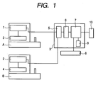
- Fig. 1 is a structural diagram showing an embodiment in which the present invention is applied to an eye fundus camera;
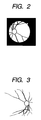
- Fig. 2 is an explanatory view showing an image picked up by an eye fundus camera A;
- Fig. 3 is an explanatory view showing an image picked up by an eye fundus camera B;
- Fig. 4 is an explanatory view showing data to be transmitted;
- Fig. 5 is a flow chart showing image processing;
- Figs. 6A and 6B are explanatory views each showing processing of the image picked up by the eye fundus camera A;
- Figs. 7A, 7B, 7C and 7D are explanatory views each showing processing of the image picked up by the eye fundus camera B; and
- Fig. 8 is a structural diagram showing another embodiment of the present invention.
-
- The present invention will be described in detail with reference to embodiments shown in the drawings.
- In Fig. 1, outputs of digital eye fundus cameras A and B are connected with an image processing apparatus X. The eye fundus camera A includes an image
data generation unit 1 and a deviceinformation generation unit 3. The eye fundus camera B includes an imagedata generation unit 2 and a deviceinformation generation unit 4. The imagedata generation units information generation units data generation units information determination unit 5 of the image processing apparatus X. An output of the deviceinformation determination unit 5 is connected with acontrol unit 7 through animage processing unit 6. Thecontrol unit 7 is connected with a patientinformation input unit 8, amemory unit 9, and amonitor 10. - For example, when an image of the left eye of a subject is picked up, the eye fundus image which is the image data generated by the eye fundus camera A is as shown in Fig. 2 in a stage in which the eye fundus image has been transmitted to the image processing apparatus X. That is, an aperture mask indicating an image pickup area is provided in the eye fundus camera A (in the device). Note that the top, the bottom, the right, and the left of the eye fundus region are picked up as in a conventional case.
- On the other hand, the aperture mask is not provided in the eye fundus camera B (in the device). In addition to this, because the eye fundus image is formed on an image pickup device in a vertically and horizontally reversed state with respect to the case of the eye fundus camera A, even when an image of the same eye fundus is picked up, image data in which the aperture mask indicating the image pickup area does not exist is obtained as shown in Fig. 3.
- Also, the device
information generation units - Here, the device information data generated by each of the device
information generation units Processing Numerical value 0 Numerical value 1Numerical value 2 O Image horizontal reverse not needed Image horizontal reverse executed P Vertical reverse not needed Vertical reverse executed Q Aperture mask synthesization not needed Aperture mask additional synthesization executed R Color image FAG image ICG image - According to the processing R, any processing is not performed in the case of a color image. In the case of an FAG image, while considering ease of diagnosis, it is converted into a white-and-black image, and γ characteristic adjustment and contrast processing are performed thereon. In addition, even in the case of an ICG image, it is considered to perform the γ characteristic adjustment, the contrast processing, and the like.
- Next, operations will be described according to an operation flow of a flow chart of Fig. 5. First, it is considered that the FAG image pickup is performed using the eye fundus camera A. In Step S1, patient information such as a patient ID number of a subject, a name thereof, a birth date thereof, or a sex thereof is inputted from the patient
information input unit 8 to thecontrol unit 7 of the image processing apparatus X before image pickup by a person that performs image pickup. - In Step S2, an image of the eye fundus of the eye to be examined is picked up using the image
data generation unit 1 of the eye fundus camera A. In Steps S3 and S4, the imagedata generation unit 1 adds onto the image data the device information generated by the deviceinformation generation unit 3 connected therewith as additional information. In this case, the device information becomes "T 0 0 0 1" because the eye fundus camera A is used. - In Step S5, the image data to which the device information is added is inputted to the device
information determination unit 5 of the image processing apparatus X. In Step S6, the device information is read out from the inputted image data by the deviceinformation determination unit 5 and processing required for the image data is determined thereby. - In Step S7, processing is selected based on the determination result.
- In this case, the device information is "T 0 0 0 1". Therefore, in the
image processing unit 6, the horizontal reverse and the vertical reverse are not performed and it is unnecessary to synthesize the aperture mask. However, because of the FAG image pickup, in Step S8, the color image is converted into the white-and-black image, the γ characteristic is adjusted, and the contrast processing is performed. - Fig. 6A shows the image transmitted from the eye fundus camera A and Fig. 6B shows the image processed by the
image processing unit 6. Note that it is originally considered in Fig. 6B that a blood vessel is displayed in white and an eye fundus segment is displayed in gray. However, they are reversely shown in Fig. 6B to easily understand a change. In Step S7, the image data processed by theimage processing unit 6 is transmitted to thecontrol unit 7, and then stored in relation to the patient information such as the patient ID number in thememory unit 9 and displayed on themonitor 10 if necessary. - In the case of color image pickup using the eye fundus camera B, after Step S1 in Fig. 5, the color image pickup is performed by the image
data generation unit 2 of the eye fundus camera B in Step S2. In Steps S3 and S4, device information "T 1 1 1 0" is generated by the deviceinformation generation unit 4 and added to image data. - The image data is transmitted in Step S5. After that, in Step S6, device information is read out by the device
information determination unit 5 and adequate processing is performed on the transmitted image data thereby. - In this case, because the device information is "
T 1 1 1 0", Step S8 is selected in Step S7. Therefore, in theimage processing unit 6, the horizontal reverse and the vertical reverse are performed and the aperture mask is synthesized. However, the γ characteristic adjustment and the like are not performed because of the color image pickup. - Fig. 7A shows the image which is transmitted from the eye fundus camera B and overlapped with
addresses image processing unit 6. In the reverse processing, only the arrangement may be reversed without changing rows of addresses. - Subsequently, vertical reverse processing is performed. As shown in Fig. 7C, the horizontally reversed image may be located such that a first row address thereof becomes a final row. Further, the image is electronically synthesized with an aperture mask, thereby obtaining the image as shown in Fig. 7D. Finally, as in the above-mentioned case, after the image shown in Fig. 7D is transmitted to the
control unit 7 in Step S8, in Step S9, the image is stored in relation to the patient information such as the patient ID number in thememory unit 9 and displayed on themonitor 10 if necessary. - Also, it is possible that the system is specialized to processing on the image data according to the image pickup mode as follows: an image pickup information generation unit that generates information related to an image pickup mode is used instead of the device information generation unit described in this embodiment and the device
information determination unit 5 is replaced by an image pickup information determination unit. - Further, the horizontal reverse processing and the vertical reverse processing are separately performed in this embodiment. However, because it is determined whether or not the two processings are performed at a time when the device information is read, it is needless to say that the processings may be performed at the same time.
- Also, in this embodiment, the image data to which the device information is added as tag information is transmitted to the image processing apparatus X. In addition to this, it is considered that the image data is transmitted using, for example, serial communication or parallel communication. As shown in Fig. 8, in this case, an image
data generation unit 11 and a deviceinformation generation unit 12 are provided in an eye fundus camera C and the image processing apparatus X has the same structure as shown in Fig. 1. - The image data "001" picked up by the image
data generation unit 11 is transmitted to theimage processing unit 6 through the deviceinformation determination unit 5 of the image processing apparatus X using, for example, a USB or an IEEE1394. In addition, the device information "001"' corresponding to the image data "001" is generated by the deviceinformation generation unit 12 and transmitted to the deviceinformation determination unit 5 using, for example, an RS232C in the case of serial communication or a PC-bus in the case of parallel communication. A content of the device information "001"' is based on a format described earlier in Table 1: first data (code) T corresponds to a command indicating the device information and the following correspond to the content. - The
image processing unit 6 processes the image data according to the result of the deviceinformation determination unit 5. After that, the image data is stored in relation to the patient information such as the patient ID number in thememory unit 9. - Further, with respect to another embodiment, "TS" is used as the information generated by the device
information generation unit 12. That is, only a content which can specify the apparatus is used as shown in Table 2 below.S Numerical value 0 Numerical value 1Numerical value 2Eye fundus camera A Eye fundus camera B Eye fundus camera C - On the other hand, when the image processing apparatus X has an image processing table corresponding to a processing content of the image data as shown in Table 3 below, the same effect can be obtained.
Processing content Eye fundus camera A no processing Eye fundus camera B Horizontal reverse and vertical reverse, and electronic mask synthesization Eye fundus camera C Vertical reverse - Alternatively, as shown in Table 4, it is considered that "T S U" is used as device information and the device information is transmitted to the image pickup
mode generation unit 11.Numerical value 0 Numerical value 1Numerical value 2 S Eye fundus camera A Eye fundus camera B Eye fundus camera C V Color image FAG image ICG image - Note that the device information in Table 4 includes detail processing settings as shown in Table 5 below.

- Therefore, for example, even in processing related to the same FAG image pickup, the extent of the γ characteristic adjustment, the contrast processing, and the like can be set in detail for each image pickup device.
- Also, in the above-mentioned embodiment, the device is constructed that the device information or the image pickup information is generated therein. It is also conceivable that the information is directly inputted by an examiner.
- Here, aspects of the present invention are described below. One aspect of the present invention relates to an ophthalmologic image pickup system, including: a plurality of image pickup devices, each of which has image data generating means for picking up an image of an eye to be examined to generate image data; device information determination means for determining inputted device information related to the image pickup devices; and image processing means for performing different image processings on the image data in accordance with a result of the device information determination means.
- In the image pickup system, preferably, the image pickup device information includes information related to at least one of processing for vertically reversing the image data and processing for horizontally reversing the image data.
- According to the aspects, when an image pickup device is designed, it is unnecessary to take into consideration the number of imaging of an image and the number of reflection by mirrors. Therefore, the flexibility in design is improved and cost reduction can be achieved.
- Also, in the ophthalmologic image pickup system, preferably, the image pickup device information includes information related to synthesis of the image data with an electronic aperture mask.
- According to this aspect, the aperture mask is not necessarily provided in the image pickup device. Therefore, cost reduction can be achieved.
- Further, another aspect of the present invention relates to an ophthalmologic image pickup system, including: a plurality of image pickup devices having image data generation means, each of which picks up an image of an eye to be examined to generate image data thereof; device information determination means for determining inputted device information related to the image pickup device; a processing table in which the corresponding image pickup device and an image data processing method therefor are shown; and image processing means for performing different image processings on the image data in accordance with a determination result of the device information determination means and the processing table.
- According to this aspect, it is unnecessary to use an image processing apparatus that captures image data for each image pickup device and performs image processing thereon. That is, the plurality of image pickup devices can be treated by the single image processing apparatus, thereby achieving cost reduction. In addition, it is unnecessary to take into account a kind of device, so that an error is prevented and the operation is easy. When the image pickup device is designed, it can be designed without considering an output image of the image data, so that the degree of freedom of the design is increased. In addition, when a new image pickup device is used, it is necessary to merely update the processing table.
- Further, in the ophthalmologic image pickup system, preferably, the device information includes a description of a kind of the image pickup device.
- According to this aspect, the device information can be simplified.
- Further, in the ophthalmologic image pickup system, preferably, the processing method shown in the processing table relates to at least one of processing for horizontally reversing the image data and processing for vertically reversing the image data.
- According to this aspect, when the image pickup device is designed, it is unnecessary to take into account the number of imaging of the image and the number of reflection by mirrors. Therefore, the degree of freedom of the design is improved and cost reduction can be achieved.
- Further, in the ophthalmologic image pickup system, preferably, the processing method shown in the processing table is to synthesize an electric aperture mask with the image data.
- According to this aspect, when the image pickup device is designed, it is unnecessary to take into account the number of imaging of the image and the number of reflection by mirrors. Therefore, the degree of freedom of the design is improved and cost reduction can be achieved.
- In the ophthalmologic image pickup system according to any one of the aspects of the invention, preferably, the image pickup device includes device information generation means for generating device information, the device information generation means is connected with the image data generation means, and the image data generation means adds the device information generated by the device information generation means to the image data and inputs the image data to which the device information is added to the device information determination means.
- According to this aspect, the number of cables provided between the image pickup device and the image processing apparatus can be reduced and a general-purpose format related to the image data can be utilized as it is. In addition, erroneous processing based on erroneous input can be prevented.
- In the ophthalmologic image pickup system according to any one of the aspects of the invention, preferably, the image pickup device includes device information generation means for generating device information, and the image data and the device information are separately inputted to the device information determination means.
- According to this aspect, it is unnecessary to provide a unit that describes information in a tab of the image, so that the device information can be relatively easily transmitted using a command.
- Further, another aspect of the present invention relates to an ophthalmologic image pickup system, including: an image pickup device including image data generation means for picking up an image of an eye to be examined to generate image data thereof; and image pickup information determination means for determining inputted image pickup information; and image processing means for performing different image processings on the image data in accordance with a result of the image pickup information determination means.
- According to this aspect, complicated procedure of performing image processing on the image data after the completion of the image pickup is unnecessary, which is time-saving.
- In the ophthalmologic image pickup system, preferably, the image pickup device further includes image pickup information generation means for generating image pickup information related to an image pickup mode upon image pickup, the image pickup information generation means is connected with the image data generation means, and the image data generation means adds the image pickup information generated by the image pickup information generation means to the image data and inputs the image data to which the image pickup information is added to the image pickup information determination means.
- According to this aspect, based on the three image pickup modes, no special processing is separately performed or such processing can be performed.
- Further, in the ophthalmologic image pickup system, preferably, the image pickup mode is one of a color image pickup mode, a visible Fluorescein fundus angiography mode, and an Indocyanine green angiography mode.
- According to this aspect, it is unnecessary for an operator to perform the operations such as the conversion of color information into white-and-black information, setting of the γ characteristic, and the contrast processing with respect to the picked up visible fluorescence electronic image using the image processing apparatus.
- In the ophthalmologic image pickup system, preferably, the different image processings include at least one of conversion of the image data into a white-and-black image, γ characteristic adjustment thereof, and contrast processing thereof when the image pickup mode is one of the Fluorescein fundus angiography mode and the Indocyanine green angiography mode.
- According to this aspect, the number of cables provided between the image pickup device and the image processing apparatus can be reduced and the general-purpose format related to the image data can be utilized as it is. In addition, erroneous processing based on erroneous input can be prevented.
- In the ophthalmologic image pickup system, preferably, the image pickup device further includes image pickup information generation means for generating image pickup information related to an image pickup mode upon image pickup, and the image data and the image pickup information are separately inputted to the image pickup information determination means.
- According to this aspect, it is unnecessary to provide a unit that describes information in a tab of the image, so that the device information can be relatively easily transmitted using a command.
- Also, in the above-mentioned system, it is preferable that different image processing to the image data differs according to the image pickup device that has performed the image pickup.
- According to this aspect, even in processing related to the same FAG image pickup, the extent of the γ characteristic adjustment, the contrast processing, and the like can be set in detail for each image pickup device.
- As described above, in the ophthalmologic image pickup system according to the present invention, it is unnecessary to use the image processing apparatus that captures the image data for each image pickup device and performs image processing thereon. The plurality of image pickup devices can be treated by the single image processing apparatus, whereby cost reduction is achieved. In addition, it is unnecessary to take into account a kind of device, so that an error is prevented and the operation is easy. When the image pickup device is designed, it can be designed without considering an output image of the image data, so that the degree of freedom of the design is increased.
- With respect to image data generated by plural kinds of eye fundus cameras, device information used for image pickup is added to picked-up-image information, and different image processings such as image reverse and aperture synthesization are performed based on the added device information.
Claims (17)
- An ophthalmologic image pickup system, comprising:an image pickup device including;image data generation means for generating image data of an eye fundus to be examined;the device information generation means for generating the device information to identify the device; anddata output means for outputting the image data and the device information, andan image processing device including;data input means for inputting the image data and the device information outputted from said data output means of the image pickup device;device information determination means for determining the image pickup device based on the device information inputted through the data input means; andimage processing means for performing different image processings on the image data in accordance with a determination result of the device information determination means.
- An ophthalmologic image pickup system according to claim 1, wherein the image pickup device information includes information indicating whether or not at least one of processing for vertically reversing the image data and processing for horizontally reversing the image data with the image processing means should be performed by the image processing apparatus.
- An ophthalmologic image pickup system according to claim 1 or 2,
wherein the image pickup device information includes information indicating whether or not the image data should be synthesized with an electronic aperture mask by means of the image processing means. - An ophthalmologic image pickup system, comprising:a plurality of image pickup devices, each of which picks up an image of an eye to be examined to generate image data thereof;device information determination means for determining inputted device information related to the image pickup device;a processing table showing an image data processing method corresponding to each of the plurality of image pickup devices; andimage processing means for performing different image processings on the image data in accordance with a determination result of the device information determination means and the processing table.
- An ophthalmologic image pickup system according to claim 4, wherein the device information includes a description of a kind of the image pickup device.
- An ophthalmologic image pickup system according to claim 4, wherein the processing method shown in the processing table relates to whether or not at least one of the processing in which the image data is horizontally or vertically reversed should be performed.
- An ophthalmologic image pickup system according to claim 6, wherein the processing method shown in the processing table is whether the composition of an electric aperture mask with the image data should be performed or not.
- An ophthalmologic image pickup system according to claim 1 or 4, wherein the device information generation means is connected with the image data generation means, and the image data generation means adds the device information generated by the device information generation means to the image data and outputs the image data to which the device information is added to the device information determination means.
- An ophthalmologic image pickup system according to claim 1 or 4, wherein the image data and the device information are separately inputted to the device information determination means.
- An ophthalmologic image pickup system, comprising:an image pickup device including image data generation means for picking up an image of an eye to be examined to generate image data thereof; andan image processing apparatus including: image pickup information determination means for determining inputted image pickup information; and image processing means for performing different image processings on the image data in accordance with a result of the image pickup information determination means.
- An ophthalmologic image pickup system according to claim 10, wherein the image pickup device further comprises image pickup information generation means for generating image pickup information related to an image pickup mode upon image pickup, the image pickup information generation means is connected with the image data generation means, and the image data generation means adds the image pickup information generated by the image pickup information generation means to the image data and outputs the image data to which the image pickup information is added to the image pickup information determination means.
- An ophthalmologic image pickup system according to claim 10, wherein the image pickup mode is one of a color image pickup mode, a Fluorescein fundus angiography mode, and an Indocyanine green angiography mode.
- An ophthalmologic image pickup system according to claim 10, wherein the different image processing include at least one of conversion of the image data into a white-and-black image, γ characteristic adjustment thereof, and contrast processing thereof when the image pickup mode is one of the Fluorescein fundus angiography mode and the Indocyanine green angiography mode.
- An ophthalmologic image pickup system according to claim 10, wherein the image pickup device further comprises image pickup information generation means for generating image pickup information related to an image pickup mode upon image pickup, and the image data and the image pickup information are separately inputted to the image pickup information determination means.
- An ophthalmologic image processing apparatus, comprising:wherein the image data is processed in accordance with the determined device information.image processing means for processing image data outputted from an ophthalmologic image pickup device; anddevice information determination means for determining device information inputted from the ophthalmologic image pickup device,
- An ophthalmologic image processing apparatus according to claim 15, wherein at least one of processing for vertically reversing the image data, processing for horizontally reversing the image data, and processing for synthesizing an aperture with the image data is performed in accordance with the device information.
- An ophthalmologic image pickup device, comprising:image data generation means for picking up an eye fundus image of an eye to be examined to generate image data thereof;device information generation means for generating device information of the ophthalmologic image pickup device; andoutput means for adding the device information to the image data and outputting the image data to which the device information is added.
Applications Claiming Priority (2)
| Application Number | Priority Date | Filing Date | Title |
|---|---|---|---|
| JP2003121961A JP4181915B2 (en) | 2003-04-25 | 2003-04-25 | Ophthalmic imaging system |
| JP2003121961 | 2003-04-25 |
Publications (1)
| Publication Number | Publication Date |
|---|---|
| EP1470779A1 true EP1470779A1 (en) | 2004-10-27 |
Family
ID=32959703
Family Applications (1)
| Application Number | Title | Priority Date | Filing Date |
|---|---|---|---|
| EP20040008865 Withdrawn EP1470779A1 (en) | 2003-04-25 | 2004-04-14 | Ophthalmologic image pickup system |
Country Status (5)
| Country | Link |
|---|---|
| US (1) | US7472994B2 (en) |
| EP (1) | EP1470779A1 (en) |
| JP (1) | JP4181915B2 (en) |
| KR (1) | KR20040092461A (en) |
| CN (1) | CN1323636C (en) |
Families Citing this family (5)
| Publication number | Priority date | Publication date | Assignee | Title |
|---|---|---|---|---|
| JP4636841B2 (en) * | 2004-09-29 | 2011-02-23 | キヤノン株式会社 | Ophthalmic image photographing apparatus and photographing method |
| JP5021007B2 (en) * | 2009-08-27 | 2012-09-05 | キヤノン株式会社 | Ophthalmic photographing apparatus and camera used in the ophthalmic photographing apparatus |
| JP2012050621A (en) * | 2010-08-31 | 2012-03-15 | Canon Inc | Imaging unit and control method thereof |
| KR101643724B1 (en) * | 2016-04-29 | 2016-07-29 | 세종대학교산학협력단 | Method and Apparatus for identifying eyeball position, Method and Apparatus for tracking eyeball position |
| WO2024202094A1 (en) * | 2023-03-27 | 2024-10-03 | オリンパスメディカルシステムズ株式会社 | Medical image processing device, medical image processing method, and medical image processing program |
Citations (8)
| Publication number | Priority date | Publication date | Assignee | Title |
|---|---|---|---|---|
| JPH07124121A (en) | 1993-11-08 | 1995-05-16 | Canon Inc | Image processing device |
| JPH10179523A (en) | 1996-12-26 | 1998-07-07 | Canon Inc | Digital camera |
| JP2000023918A (en) | 1998-07-10 | 2000-01-25 | Canon Inc | Ophthalmic imaging equipment |
| JP2000175869A (en) * | 2000-01-01 | 2000-06-27 | Topcon Corp | Fundus image processing device |
| JP2001094714A (en) * | 1999-09-22 | 2001-04-06 | Fuji Photo Film Co Ltd | Image processing unit and image processing method |
| US6325511B1 (en) * | 1999-01-28 | 2001-12-04 | Kowa Company Ltd. | Ophthalmic photographic apparatus |
| US20020140825A1 (en) * | 1998-01-30 | 2002-10-03 | Takaaki Terashita | Image processing method and apparatus for carrying out image processing on digital image signals under different image processing conditions |
| US20030058349A1 (en) * | 2001-09-26 | 2003-03-27 | Fuji Photo Film Co., Ltd. | Method, apparatus, and program for image processing |
Family Cites Families (21)
| Publication number | Priority date | Publication date | Assignee | Title |
|---|---|---|---|---|
| JPS62211040A (en) | 1986-03-11 | 1987-09-17 | 富士写真光機株式会社 | Endoscope |
| JPS62211010A (en) | 1986-03-12 | 1987-09-17 | 富士通株式会社 | Meeting table for sound meeting |
| US5291231A (en) * | 1990-11-29 | 1994-03-01 | Kabushiki Kaisha Topcon | Opthalmological image processing system |
| JP3058680B2 (en) * | 1990-11-30 | 2000-07-04 | 株式会社トプコン | Fundus image processing device |
| JP2555693Y2 (en) * | 1991-03-28 | 1997-11-26 | 株式会社ニコン | Ophthalmic equipment |
| JPH04317629A (en) | 1991-04-16 | 1992-11-09 | Canon Inc | Eye ground camera |
| US5225859A (en) * | 1991-10-10 | 1993-07-06 | Hemozoin Scientific, Inc. | Apparatus and method for capture and processing of ocular and retinal images |
| US5455644A (en) * | 1992-02-28 | 1995-10-03 | Canon Kabushiki Kaisha | Ophthalmologic apparatus having an examining system for examining the fundus of an eye |
| JP3461203B2 (en) | 1994-07-18 | 2003-10-27 | キヤノン株式会社 | Ophthalmic image recording system |
| JPH08154244A (en) | 1994-11-28 | 1996-06-11 | Canon Inc | Image storage |
| JP3618877B2 (en) | 1996-02-05 | 2005-02-09 | キヤノン株式会社 | Ophthalmic image processing device |
| US5886767A (en) * | 1996-10-09 | 1999-03-23 | Snook; Richard K. | Keratometry system and method for measuring physical parameters of the cornea |
| US5912720A (en) * | 1997-02-13 | 1999-06-15 | The Trustees Of The University Of Pennsylvania | Technique for creating an ophthalmic augmented reality environment |
| JP3630973B2 (en) * | 1998-02-27 | 2005-03-23 | 株式会社ニデック | Ophthalmic apparatus and method for storing optometry information in ophthalmic apparatus |
| JP2001275977A (en) * | 2000-03-31 | 2001-10-09 | Canon Inc | Fundus blood flow meter |
| DE10047237A1 (en) * | 2000-09-23 | 2002-04-11 | Physoptics Opto Electronic Gmb | System for recording the retinal reflex image |
| US6736507B2 (en) * | 2001-01-22 | 2004-05-18 | Alexis Kudryashov | High resolution, multispectral, wide field of view retinal imager |
| US6361170B1 (en) * | 2001-02-14 | 2002-03-26 | Heidelberg Engineering Optische Messysteme Gmbh | Aberration-free imaging of the fundus of the human eye |
| JP2002360521A (en) | 2001-06-07 | 2002-12-17 | Hitachi Medical Corp | Image processing device |
| DE602004001140D1 (en) * | 2003-02-03 | 2006-07-27 | Canon Kk | Ophthalmic device |
| US7708403B2 (en) * | 2003-10-30 | 2010-05-04 | Welch Allyn, Inc. | Apparatus and method for diagnosis of optically identifiable ophthalmic conditions |
-
2003
- 2003-04-25 JP JP2003121961A patent/JP4181915B2/en not_active Expired - Fee Related
-
2004
- 2004-03-31 US US10/815,479 patent/US7472994B2/en not_active Expired - Lifetime
- 2004-04-02 CN CNB2004100316829A patent/CN1323636C/en not_active Expired - Fee Related
- 2004-04-14 EP EP20040008865 patent/EP1470779A1/en not_active Withdrawn
- 2004-04-22 KR KR1020040027789A patent/KR20040092461A/en not_active Ceased
Patent Citations (8)
| Publication number | Priority date | Publication date | Assignee | Title |
|---|---|---|---|---|
| JPH07124121A (en) | 1993-11-08 | 1995-05-16 | Canon Inc | Image processing device |
| JPH10179523A (en) | 1996-12-26 | 1998-07-07 | Canon Inc | Digital camera |
| US20020140825A1 (en) * | 1998-01-30 | 2002-10-03 | Takaaki Terashita | Image processing method and apparatus for carrying out image processing on digital image signals under different image processing conditions |
| JP2000023918A (en) | 1998-07-10 | 2000-01-25 | Canon Inc | Ophthalmic imaging equipment |
| US6325511B1 (en) * | 1999-01-28 | 2001-12-04 | Kowa Company Ltd. | Ophthalmic photographic apparatus |
| JP2001094714A (en) * | 1999-09-22 | 2001-04-06 | Fuji Photo Film Co Ltd | Image processing unit and image processing method |
| JP2000175869A (en) * | 2000-01-01 | 2000-06-27 | Topcon Corp | Fundus image processing device |
| US20030058349A1 (en) * | 2001-09-26 | 2003-03-27 | Fuji Photo Film Co., Ltd. | Method, apparatus, and program for image processing |
Non-Patent Citations (3)
| Title |
|---|
| PATENT ABSTRACTS OF JAPAN vol. 1996, no. 10 31 October 1996 (1996-10-31) * |
| PATENT ABSTRACTS OF JAPAN vol. 2000, no. 09 13 October 2000 (2000-10-13) * |
| PATENT ABSTRACTS OF JAPAN vol. 2000, no. 21 3 August 2001 (2001-08-03) * |
Also Published As
| Publication number | Publication date |
|---|---|
| JP4181915B2 (en) | 2008-11-19 |
| CN1539371A (en) | 2004-10-27 |
| CN1323636C (en) | 2007-07-04 |
| JP2004321567A (en) | 2004-11-18 |
| KR20040092461A (en) | 2004-11-03 |
| US7472994B2 (en) | 2009-01-06 |
| US20040212780A1 (en) | 2004-10-28 |
Similar Documents
| Publication | Publication Date | Title |
|---|---|---|
| JP5449816B2 (en) | Image processing apparatus, image processing program, and method of operating image processing apparatus | |
| US9872610B2 (en) | Image processing device, imaging device, computer-readable storage medium, and image processing method | |
| JPH08195951A (en) | Endoscope device | |
| JP2017213058A (en) | Image processing device, endoscope device, image processing method, and image processing program | |
| US7472994B2 (en) | Ophthalmologic image pickup system | |
| JP4447849B2 (en) | Imaging apparatus and program | |
| JP2000287203A (en) | Electronic endoscope capable of digital output | |
| JP2006055326A (en) | Ultrasonic diagnostic equipment | |
| JP2001119625A (en) | Image processing method and apparatus | |
| JPH10155736A (en) | Signal processing apparatus for endoscope | |
| JP4027842B2 (en) | Medical image recording device | |
| JPH1198497A (en) | Endoscope device | |
| JP4459564B2 (en) | Endoscope system having an image signal processing device | |
| JP2000287202A (en) | Electronic endoscope | |
| JP2021169051A (en) | Image clipping device and method | |
| JP4033746B2 (en) | Microscope equipment | |
| JPH119547A (en) | Endoscope system | |
| JP2003299620A (en) | Fundus image processing device | |
| US20080259406A1 (en) | Endoscope image filing system | |
| JP4827414B2 (en) | Electronic endoscope system and filing system | |
| JP4124875B2 (en) | Electronic endoscope device capable of digital output | |
| JP2661182B2 (en) | Image position adjustment method for electronic endoscopes | |
| JP2003024319A (en) | X-ray image display device | |
| JPH1147090A (en) | Endoscope imaging device | |
| WO2024202094A1 (en) | Medical image processing device, medical image processing method, and medical image processing program |
Legal Events
| Date | Code | Title | Description |
|---|---|---|---|
| PUAI | Public reference made under article 153(3) epc to a published international application that has entered the european phase |
Free format text: ORIGINAL CODE: 0009012 |
|
| AK | Designated contracting states |
Kind code of ref document: A1 Designated state(s): AT BE BG CH CY CZ DE DK EE ES FI FR GB GR HU IE IT LI LU MC NL PL PT RO SE SI SK TR |
|
| AX | Request for extension of the european patent |
Extension state: AL HR LT LV MK |
|
| 17P | Request for examination filed |
Effective date: 20050310 |
|
| AKX | Designation fees paid |
Designated state(s): DE FR GB IT NL |
|
| 17Q | First examination report despatched |
Effective date: 20100216 |
|
| STAA | Information on the status of an ep patent application or granted ep patent |
Free format text: STATUS: THE APPLICATION HAS BEEN WITHDRAWN |
|
| 18W | Application withdrawn |
Effective date: 20150702 |