EP1449538A1 - Inhibition of TACE or amphiregulin for the modulation of EGF receptor signal transactivation - Google Patents
Inhibition of TACE or amphiregulin for the modulation of EGF receptor signal transactivation Download PDFInfo
- Publication number
- EP1449538A1 EP1449538A1 EP03003935A EP03003935A EP1449538A1 EP 1449538 A1 EP1449538 A1 EP 1449538A1 EP 03003935 A EP03003935 A EP 03003935A EP 03003935 A EP03003935 A EP 03003935A EP 1449538 A1 EP1449538 A1 EP 1449538A1
- Authority
- EP
- European Patent Office
- Prior art keywords
- tace
- amphiregulin
- protein
- cell
- transactivation
- Prior art date
- Legal status (The legal status is an assumption and is not a legal conclusion. Google has not performed a legal analysis and makes no representation as to the accuracy of the status listed.)
- Withdrawn
Links
Images


Classifications
-
- C—CHEMISTRY; METALLURGY
- C12—BIOCHEMISTRY; BEER; SPIRITS; WINE; VINEGAR; MICROBIOLOGY; ENZYMOLOGY; MUTATION OR GENETIC ENGINEERING
- C12N—MICROORGANISMS OR ENZYMES; COMPOSITIONS THEREOF; PROPAGATING, PRESERVING, OR MAINTAINING MICROORGANISMS; MUTATION OR GENETIC ENGINEERING; CULTURE MEDIA
- C12N9/00—Enzymes; Proenzymes; Compositions thereof; Processes for preparing, activating, inhibiting, separating or purifying enzymes
- C12N9/14—Hydrolases (3)
- C12N9/48—Hydrolases (3) acting on peptide bonds (3.4)
- C12N9/50—Proteinases, e.g. Endopeptidases (3.4.21-3.4.25)
- C12N9/64—Proteinases, e.g. Endopeptidases (3.4.21-3.4.25) derived from animal tissue
- C12N9/6421—Proteinases, e.g. Endopeptidases (3.4.21-3.4.25) derived from animal tissue from mammals
- C12N9/6489—Metalloendopeptidases (3.4.24)
-
- A—HUMAN NECESSITIES
- A61—MEDICAL OR VETERINARY SCIENCE; HYGIENE
- A61K—PREPARATIONS FOR MEDICAL, DENTAL OR TOILETRY PURPOSES
- A61K38/00—Medicinal preparations containing peptides
- A61K38/005—Enzyme inhibitors
-
- A—HUMAN NECESSITIES
- A61—MEDICAL OR VETERINARY SCIENCE; HYGIENE
- A61P—SPECIFIC THERAPEUTIC ACTIVITY OF CHEMICAL COMPOUNDS OR MEDICINAL PREPARATIONS
- A61P17/00—Drugs for dermatological disorders
- A61P17/06—Antipsoriatics
-
- A—HUMAN NECESSITIES
- A61—MEDICAL OR VETERINARY SCIENCE; HYGIENE
- A61P—SPECIFIC THERAPEUTIC ACTIVITY OF CHEMICAL COMPOUNDS OR MEDICINAL PREPARATIONS
- A61P35/00—Antineoplastic agents
-
- A—HUMAN NECESSITIES
- A61—MEDICAL OR VETERINARY SCIENCE; HYGIENE
- A61P—SPECIFIC THERAPEUTIC ACTIVITY OF CHEMICAL COMPOUNDS OR MEDICINAL PREPARATIONS
- A61P43/00—Drugs for specific purposes, not provided for in groups A61P1/00-A61P41/00
-
- C—CHEMISTRY; METALLURGY
- C07—ORGANIC CHEMISTRY
- C07K—PEPTIDES
- C07K14/00—Peptides having more than 20 amino acids; Gastrins; Somatostatins; Melanotropins; Derivatives thereof
- C07K14/435—Peptides having more than 20 amino acids; Gastrins; Somatostatins; Melanotropins; Derivatives thereof from animals; from humans
- C07K14/475—Growth factors; Growth regulators
- C07K14/485—Epidermal growth factor [EGF], i.e. urogastrone
Definitions
- the present invention relates to the modulation of transactivation of receptor tyrosine kinases by G protein or G protein-coupled receptor (GPCR) mediated signal transduction in a cell or an organism comprising inhibiting the activity of the metalloprotease TACE/ADAM17 and/or the activity of the receptor tyrosine kinase ligand amphiregulin.
- GPCR G protein-coupled receptor
- GPCR G protein-coupled receptor
- GPCRs G protein-coupled receptors
- EGFR-characteristic, intracellular signal (5) Due to the rapid kinetics of the EGFR transactivation signal and the fact that release of EGFR ligands was not detectable after GPCR stimulation, the mechanism of EGFR transactivation was proposed to exclusively rely on intracellular elements (5, 6).
- HB-EGF heparin-binding EGF-like growth factor
- TGF ⁇ transforming growth factor alpha
- AR amphiregulin
- EGFR signal transactivation has gained further experimental support by studies on GPCR mitogenic signalling in vascular smooth muscle cells (8), cardiac endothelial cells (9) and cardiomyocytes (10). Importantly, recent data have implicated EGFR signal transactivation pathways in the etiology of pathobiological processes such as cystic fibrosis (3), cardiac (2) and gastrointestinal hypertrophy (11). Furthermore, increasing evidence argues for a direct correlation between aberrant GPCR signalling and the development and progression of human cancers (12).
- HNSCC head and neck squamous cell carcinoma
- lysophosphatidic acid LPA
- carbachol specifically results in metalloprotease-dependent cleavage and release of the EGFR ligand amphiregulin (AR).
- AR gene silencing by small interfering RNA (siRNA) or inhibition of AR biological activity by neutralizing antibodies prevents GPCR-induced EGFR tyrosine phosphorylation, downstream mitogenic signalling events, activation of Akt/PKB, cell proliferation and migration.
- TACE and/or AR can function as an effector of GPCR-mediated signalling and therefore represents a key element of the cellular receptor cross-talk network.
- the invention relates to a method for modulating transactivation of receptor tyrosine kinases by G protein or G protein-coupled receptor mediated signal transduction in a cell comprising inhibiting the activity of the metalloprotease TACE/ADAM17 and/or the activity of the receptor tyrosine kinase ligand amphiregulin.
- inhibitors preferably relate to a "specific" inhibition, wherein the activity of TACE/ADAM17 and/or amphiregulin is selectively inhibited, i.e. the activity of other metalloproteases such as ADAM 12 or other receptor tyrosine kinase ligands such as HB-EGF is not significantly inhibited.
- TACE/ADAM 17 and/or amphiregulin By means of selective inhibition of TACE/ADAM 17 and/or amphiregulin a highly specific disruption of receptor tyrosine kinase transactivation may be achieved which is important for pharmaceutical applications in that the occurance of undesired side effects may be reduced.
- the term “inhibition” preferably relates to a "direct” inhibition, wherein the inhibitor directly binds to TACE/ADAM 17 and/or amphiregulin or a nucleic acid molecule coding therefor.
- the invention also encompasses an "indirect” inhibtion wherein the inhibitor does not directly bind to TACE/ADAM17 and/or amphiregulin but to a precursor or metabolite thereof, particularly the amphiregulin precursor proamphiregulin.
- the term "activity" preferably relates to the cleavage of proamphiregulin by TACE/ADAM17 and/or the activation of a receptor tyrosine kinase, e.g. EGFR by amphiregulin.
- a TACE/ADAM17 inhibitor of the present invention is preferably capable of inhibiting the cleavage and release of the receptor tyrosine kinase ligand amphiregulin.
- An amphiregulin inhibitor of the present invention is preferably capable of inhibiting biological activity of amphiregulin, particularly EGFR tyrosine phosphorylation, downstream mitogenic signaling events, activation of Akt/PKB, cell proliferation and/or migration.
- a further aspect of the present invention is the use of an inhibitor of the metalloprotease TACE/ADAM17 and/or an inhibitor of the receptor tyrosine kinase ligand amphiregulin for the prevention and/or treatment of a disorder which is caused by or associated with a transactivation of receptor tyrosine kinases by G protein oder G protein-coupled receptor mediated signal transduction.
- the presence of such a type of disorder may be determined by measuring G protein and/or GPCR expression, e.g. on the mRNA level (cDNA array analysis, SAGE, Northern blot, etc.) and/or on the protein level (Western blot analysis, Immunofluorescence Microscopy, in situ hybridisation techniques, etc.).
- the presence of such a type of disorder may also be determined by examining the occurrence of activating mutations in genomic and/or mRNA molecules encoding G proteins or GPCRs and/or the presence of virally encoded GPCRs. Further, elevated levels of GPCR agonists such as LPA and/or amphiregulin in serum and/or disease-affected tissues may be determined. It should be pointed out that this type of disorder need not be associated with enhanced receptor tyrosine kinase expression.
- the disorder may be a hyperproliferative disorder such as cancer, e.g. squamous cell carcinoma or another disorder such as a hyperproliferative skin disease, e.g. psoriasis.
- a hyperproliferative disorder such as cancer, e.g. squamous cell carcinoma or another disorder such as a hyperproliferative skin disease, e.g. psoriasis.
- the activity of TACE/ADAM 17 and/or amphiregulin may be inhibited on the nucleic acid level, e.g. on the gene level or on the transcription level. Inhibition on the gene level may comprise a partial or complete gene inactivation, i.e. by gene disruption. On the other hand, inhibition may occur on the transcript level, e.g. by application of anti-sense molecules, e.g. DNA molecules, RNA molecules or nucleic acid analogues, ribozymes, e.g. RNA molecules or nucleic acid analogues or small RNA molecules capable of RNA interference (RNAi), e.g. RNA molecules or nucleic acid analogues, directed against TACE/ADAM17 and/or amphiregulin mRNA.
- anti-sense molecules e.g. DNA molecules, RNA molecules or nucleic acid analogues
- ribozymes e.g. RNA molecules or nucleic acid analogues or small RNA molecules capable of
- TACE/ADAM 17 and/or amphiregulin may be inhibited on the protein level, e.g. by application of compounds which result in a specific inhibition of TACE/ADAM17 and/or amphiregulin acitivity.
- the inhbition on the protein level may comprise for example the application of antibodies or antibody fragments directed against TACE/ADAM17 and/or amphiregulin.
- the antibodies may be polyclonal antibodies or monoclonal antibodies, recombinant antibodies, e.g. single chain antibodies or fragments of such antibodies which contain at least one antigen-binding site, e.g.
- proteolytic antibody fragments such as Fab, Fab' or F(ab') 2 fragments or recombinant antibody fragments such as scFv fragments.
- Fab fragment antigen binding fragment
- Fab' fragment antigen binding fragment
- F(ab') 2 fragments fragment antigen binding fragment
- recombinant antibody fragments such as scFv fragments.
- chimeric antibodies, humanized antibodies or human antibodies is especially preferred.
- low-molecular weight inhibitors of TACE/ADAM17 and/or amphiregulin may be used. These inhibitors may be identified by screening procedures as outlined in detail below.
- the medicament is administered in the form of a pharmaceutical composition which additionally comprises pharmaceutically acceptable carriers, diluents and/or adjuvants.
- compositions suitable for use in the present invention include compositions wherein the active ingredients are contained in an effective amount to achieve its intended purpose.
- a therapeutically effective dose refers to that amount of the compound that results in amelioration of symptoms or a prolongation of survival in a patient. Toxicity and therapeutic efficacy of such compounds can be determined by standard pharmaceutical procedures in cell cultures or experimental animals, e.g. for determining the LD50 (the dose lethal to 50% of the population) and the ED50 (the dose therapeutically effective in 50% of the population).
- the therapeutically effective dose can be estimated initially from cell culture assays.
- a dose can be formulated in animal models to achieve a circulating concentration range that includes the IC50 as determined in cell culture (i.e.
- the concentration of the test compound which achieves a half-maximal inhibition of the growth-factor receptor activity can be used to more accurately determine useful doses in humans.
- the dose ratio between toxic and therapeutic effects is the therapeutic index and it can be expressed as the ratio between LD50 and ED50. Compounds which exhibit high therapeutic indices are preferred.
- the exact formulation, route of administration and dosage can be chosen by the individual physician in view of the patient's condition (see e.g. Fingl et al., 1975, in "The Pharmacological Basis of Therapeutics", Ch. 1, p. 1). Dosage amount and interval may be adjusted individually to provide plasma levels of the active moiety which are sufficient to maintain the receptor modulating effects, or minimal effective concentration (MEC).
- MEC minimal effective concentration
- the MEC will vary for each compound but can be estimated from in vitro data, e.g. the concentration necessary to achieve a 50-90% inhibition of the receptor using the assays described herein.
- Compounds should be administered using a regimen which maintains plasma levels above the MEC for 10-90% of the time, preferably between 30-90% and most preferably between 50-90%. Dosages necessary to achieve the MEC will depend on individual characteristics and route of administration. In cases of local administration or selective uptake, the effective local concentration of the drug may not be related to plasma concentration.
- compositions administered will, of course, be dependent on the subject being treated, on the subject's weight, the severity of the affliction, the manner of administration and the judgement of the prescribing physician.
- a daily dosage of 0,001 to 100 mg/kg, particularly 0,01 to 10 mg/kg per day is suitable.
- Suitable routes of administration may, for example, include oral, rectal, transmucosal, or intestinal administration; parenteral delivery, including intramuscular, subcutaneous, intramedullary injections, as well as intrathecal, direct intraventricular, intravenous, intraperitoneal, intranasal, or intraocular injections.
- the liposomes will be targeted to and taken up selectively by the tumor.
- Still a further aspect of the present invention is a method for identifying modulators of receptor tyrosine kinase transactivation by G protein or G protein-coupled receptor mediated signal transduction, comprising determining, if a test compound is capable of inhibiting the activity of TACE/ADAM17 and/or the activity of amphiregulin.
- This method is suitable as a screening procedure, e.g. a high-throughput screening procedure for identifiying novel compounds or classes of compounds which are capable of modulating G protein signal transduction.
- the method is suitable as a validation procedure for characterizing the pharmaceutical efficacy and/or the side effects of compounds.
- the method may comprise the use of isolated proteins, cell extracts, recombinant cells or transgenic non-human animals.
- the recombinant cells or transgenic non-human animals preferably exhibit an altered TACE/ADAM17 and/or amphiregulin expression compared to a corresponding wild-type cell or animal.
- Suitable receptor tyrosine kinases are EGFR and other members of the EGFR family such as HER2, HER3 or HER4, PDGFR, the vascular endothelial growth factor receptor KDR/Fik-1, the Trk receptor, FGFR-1 or IGF-1 receptor but also other types of growth-factor receptors such as TNF receptor 1, TNF receptor 2, CD30 and IL-6 receptor are targets for the G protein/GPCR mediated signal transduction.
- FIG. 1 GPCR stimulation of the EGFR involves a ligand-dependent mechanism and is accompanied by AR release from the cell surface.
- EGFR signal transactivation requires metalloprotease activity and the EGFR extracellular domain.
- SCC-9 cells were pre-incubated with marimastat (BB2516, 10 ⁇ M; 20 min), anti-EGFR antibody ICR-3R (20 ⁇ g/mL; 60 min) or PTX (100 ng/mL; 18 h) and treated with LPA ( 10 ⁇ M), carbachol (Car, 1 mM), EGF (7.5 ng/mL) or pervanadate (PV, 1 mM) for 3 min.
- IP immunoprecipitation
- SCC-9 cells were collected and stained for surface HB-EGF, TGF ⁇ or AR and analyzed by flow cytometry. Control cells were labelled with FITC-conjugated secondary antibody alone.
- c LPA-induced proteolytic processing of proAR. SCC-9 cells were pre-incubated with batimastat (BB94, 10 ⁇ M) or PTX and stimulated with LPA or TPA (1 ⁇ M) for 5 min.
- RNA interference Blockade of EGF-like growth factor precursor expression by RNA interference (RNAi).
- SCC-9 cells were transfected with siRNA for proAR, proHB-EGF or proTGF ⁇ cultured for 2 days and analyzed for gene expression by RT-PCR as indicated or
- b stimulated with LPA or carbachol and assayed for EGFR tyrosine phosphorylation content.
- c Requirement of AR for LPA-induced cell migration.
- SiRNA-transfected SCC-9 cells were analyzed for transwell migration toward fibronectin as chemoattractant. Each bar is the average of quadruplicate values (mean ⁇ s.d.).
- SCC-9 cells were pre-incubated with inhibitors and stimulated for 5 min as indicated. Lysates were incubated with GST-Grb-2 fusion protein or GST alone. Proteins were immunoblotted with monoclonal anti-SHC antibody.
- AR is required for GPCR-induced ERK/MAPK activation and Akt/PKB phosphorylation.
- SCC-9 or SCC-15 cells were pre-incubated with inhibitors and stimulated for 7'min. Phosphorylated ERK1/2 was detected by immunoblotting total lysates with anti-phospho-ERK antibody. The same filters were re-probed with anti-ERK antibody.
- TACE is expressed in HNSCC cell lines. TACE was immunoprecipitated from lysates with monoclonal TACE/ADAM17 antibody. HEK-293 cells transfected with human TACE cDNA served as a positive control. b , Timp-3 but not Timp-1 inhibits EGFR signal transactivation. SCC-9 cells were infected with retrovirus encoding human Timp-1 or Timp-3. EGFR activation was determined by immunoblot after stimulation with agonists as indicated (left panel).
- Timp-1 /3 carrying C-terminal VSV-tag was confirmed by immunoblotting total cell lysates with anti-VSV antibody (right panel).
- c Expression of wild type and dominant negative TACE or HA-tagged ADAM 12 in SCC-9 cells after retroviral gene transfer. Total lysates were immunoblotted as indicated.
- d Dominant negative TACE abrogates LPA-induced proAR cleavage (left panel) and AR release into cell culture medium (right panel) as determined by flow cytometric analysis and AR ELISA, respectively.
- e Effect of dominant negative TACE on GPCR stimulated EGFR signal transactivation.
- TACE siRNA inhibits EGFR signal transmission and cell migration by GPCR agonists.
- a TACE siRNA blocks endogenous TACE expression.
- SCC-9 cells were transfected with TACE or ADAM 12 siRNA. Gene expression was analyzed by RT-PCR (left panel) or immunoblot (right panel) with polyclonal anti-TACE antibody.
- b Knockdown of TACE results in accumulation of proAR at the cell surface.
- siRNA-transfected SCC-9 cells were analyzed for AR cell surface content by FACS.
- c EGFR signal transmission upon GPCR activation requires TACE.
- SCC-9 cells were transfected with siRNA and stimulated with agonists as indicated.
- Squamous cancer cell motility in response to LPA depends on TACE.
- siRNA-transfected SCC-9 cells were treated with LPA or AR and analyzed in transwell migration assay.
- cDNAs encoding ADAM 10, 12, 15 and 17 were amplified by PCR from a human placenta cDNA library and subcloned into pcDNA3 (Invitrogen, Carlsbad, CA) and pLXSN vectors (Clontech, Palo Alto, CA).
- pcDNA3 Invitrogen, Carlsbad, CA
- pLXSN vectors Clontech, Palo Alto, CA.
- dominant negative protease constructs lacking the pro- and metalloprotease domains were generated as described before (2,26). All protease constructs included a C-terminal hemagglutinin (HA) tag, detectable with an anti-HA monoclonal antibody (Babco, Richmond, CA).
- amphotropic packaging cell line Phoenix was transfected with pLXSN retroviral expression plasmids by the calcium phosphate/ chloroquine method as described previously(29). At 24 h after transfection, the viral supernatant was collected and used to infect subconfluent SCC-9 cells (5 ⁇ 10 4 cells/ 6-Well plate).
- FACS analysis was performed as described before (1). Cells were stained with ectodomain-specific antibodies against HB-EGF, AR (R&D Systems) or TGF ⁇ (Oncogene, Boston, MA). After washing with PBS, cells were incubated with FITC-conjugated secondary antibody and analyzed on a Becton Dickinson FACScalibur flow cytometer.
- Concentrations of free AR were determined by sandwich ELISA (R&D Systems) using monoclonal anti-AR capture antibody and biotinylated polyclonal detection antibody. Standards were recombinant human AR diluted in culture medium. For statistical analysis Student's t-test was used to compare data between two groups. Values are expressed as mean ⁇ s. d. of at least triplicate samples. P ⁇ 0.05 was considered statistically significant.
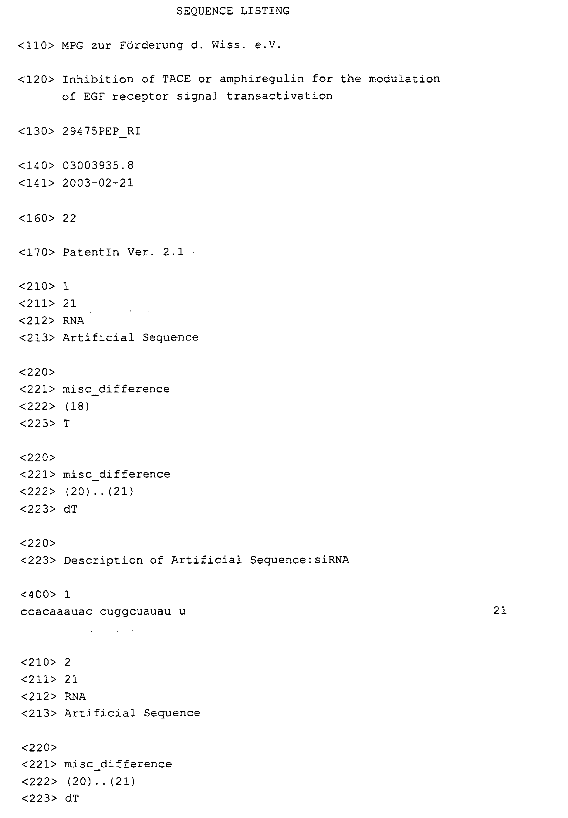
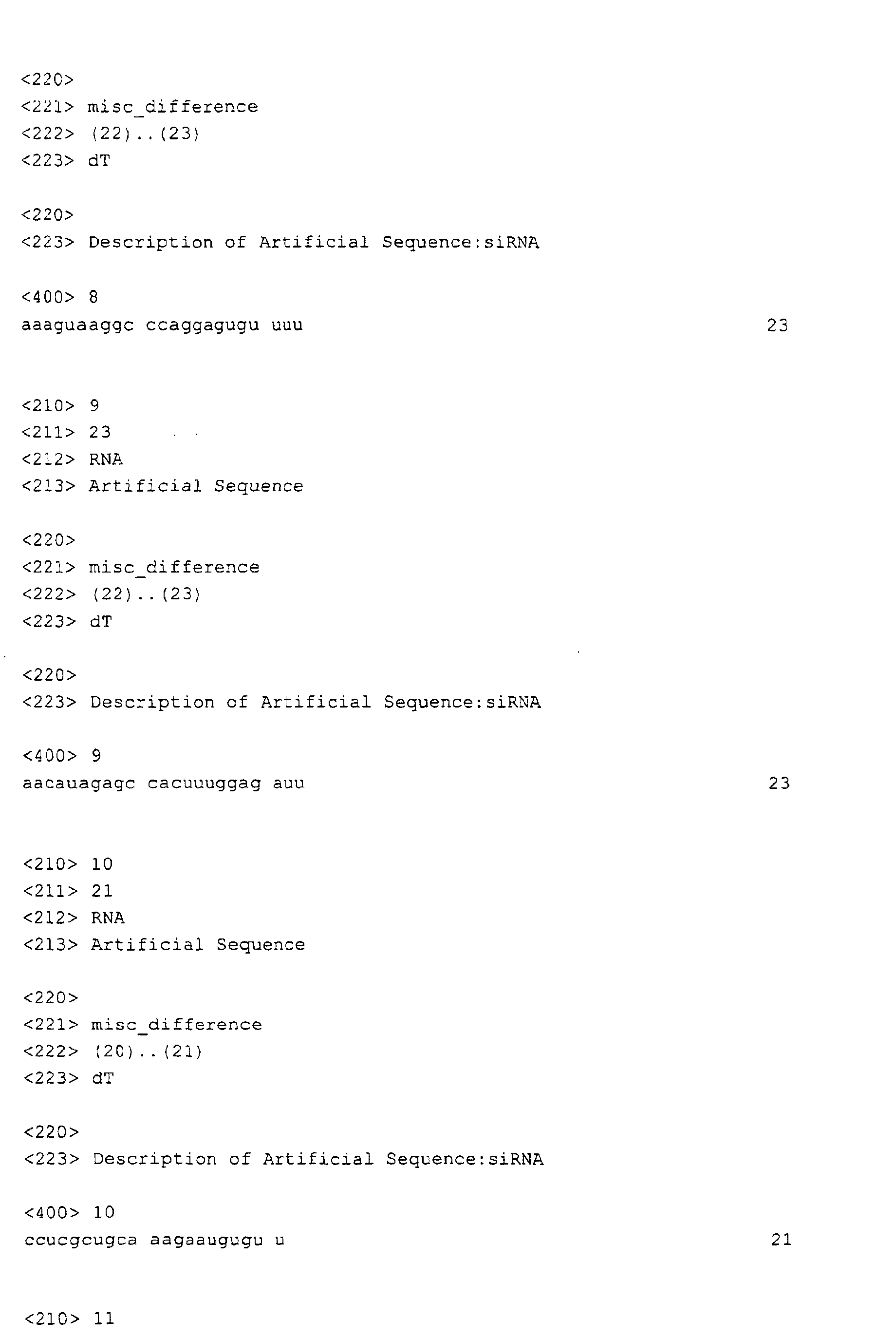
- siRNA duplexes 21-nucleotide siRNA duplexes (Dharmacon Research, Lafayette, CO, USA) for targeting endogenous genes was carried out using Oligofectamine (Invitrogen) and 4.2 ⁇ g siRNA duplex per 6-well plate as previously described (30). Transfected SCC-9 cells were serum-starved and assayed 4 d after transfection. Highest efficiencies in silencing target genes were obtained by using mixtures of siRNA duplexes targeting different regions of the gene of interest. Sequences of siRNA used were
- RNA isolated using RNeasy Mini Kit (Qiagen, Hilden, Germany) was reverse transcribed using AMV Reverse Transcriptase (Roche, Mannheim, Germany).
- PuReTaq Ready-To'-Go PCR Beads (Amersham Biosciences, Piscataway, NJ) were used for PCR amplification.
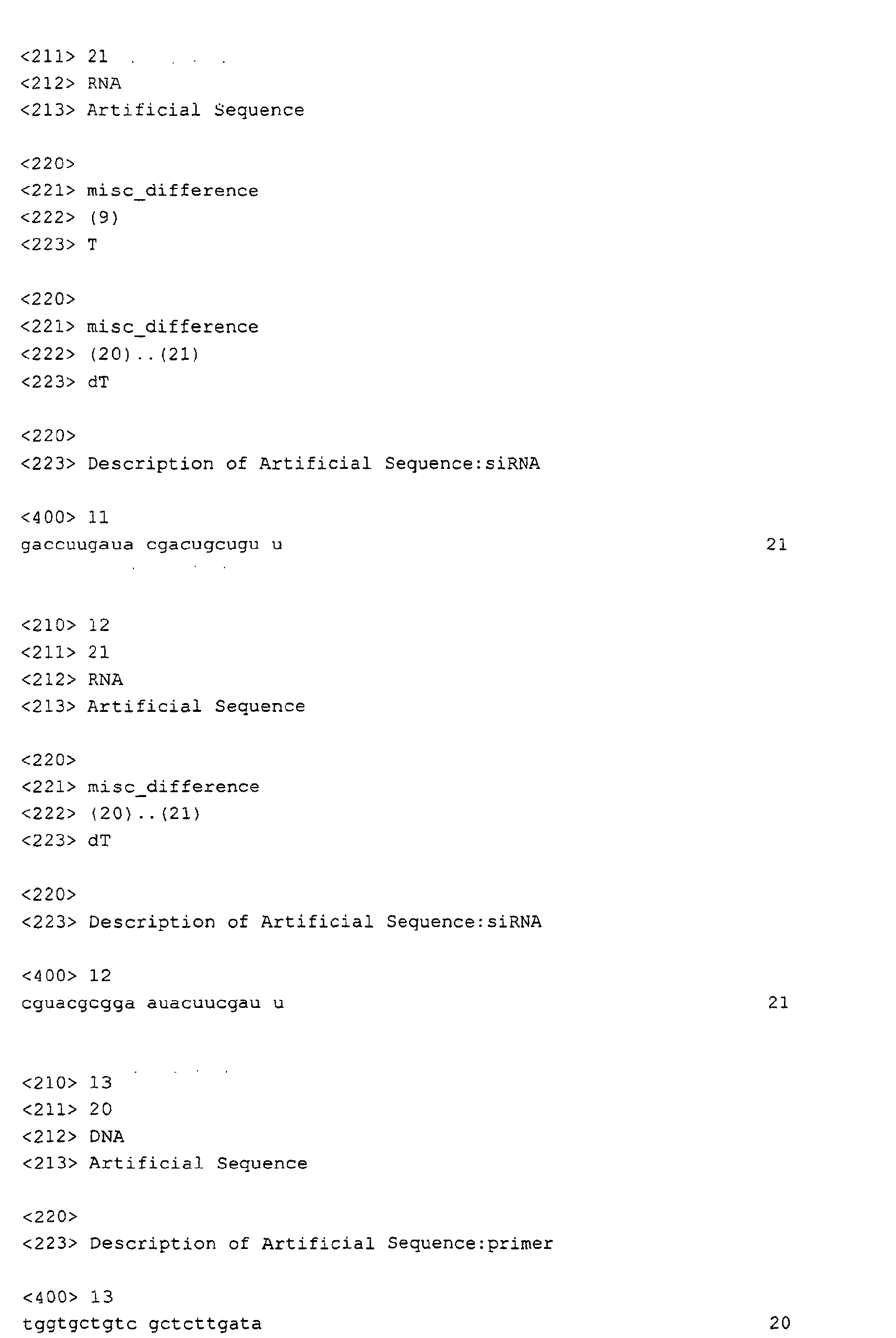
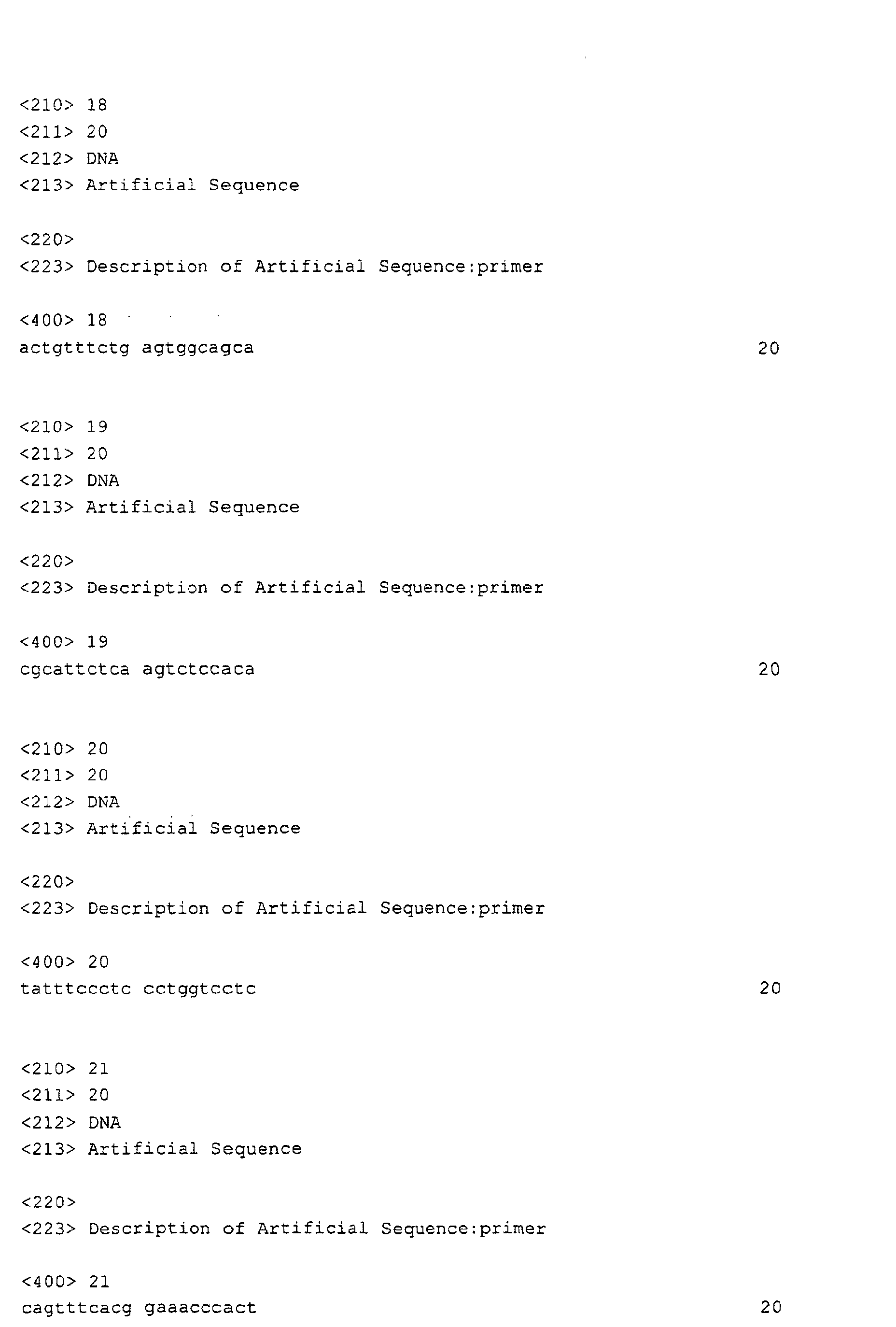
- Custom primers (Sigma Ark, Steinheim, Germany) were proAR, and and and and PCR products were subjected to electrophoresis on a 2.5 % agarose gel and DNA was visualized by ethidium bromide staining. Location of the products and their sizes were determined by using a 100-bp ladder (GIBCO, Gaithersburg, Maryland) under ultraviolet illumination.
- 3 H-thymidine incorporation assay (5) SCC-15 cells were seeded into 12-well plates at 3 x 10 4 cells/well. Upon serum deprivation for 48 h, cells were subjected to pre-incubation and stimulation as indicated. After 18 h cells were pulse-labelled with 3H-thymidine (1 ⁇ Ci/ml) for 4 h, and thymidine incorporation was measured by trichloroacetic acid precipitation and subsequent liquid-scintillation counting.
- the GPCR-induced transactivation signal in HNSCC cells is sensitive to broad-spectrum metalloprotease inhibitors such as batimastat (BB94) (13) and marimastat (BB2516; Fig. 1A). Consistent with a ligand-dependent mechanism of EGFR signal transactivation we found that the monoclonal anti-EGFR antibody ICR-3R which prevents binding of EGF-like growth factors to the extracellular domain of the receptor (14) abrogated GPCR- and EGF-induced EGFR tyrosine phosphorylation in SCC-9 cells (Fig. 1A).
- ICR-3R did not interfere with responses triggered by pervanadate, a potent tyrosine phosphatase inhibitor (15) which increases the tyrosine phosphorylation content of many intracellular proteins.
- a potent tyrosine phosphatase inhibitor (15) which increases the tyrosine phosphorylation content of many intracellular proteins.
- Previous reports demonstrating that GPCR-induced EGFR tyrosine phosphorylation requires proteolytic cleavage of HB-EGF (1-3) prompted us to ask whether HB-EGF or other EGF-like growth factors are involved in the EGFR transactivation pathway in head and neck cancer cells.
- cDNA microarray analysis we found the expression of HB-EGF, TGF ⁇ and AR mRNAs in SCC-4, SCC-9, SCC-15 and SCC-25 cells (data not shown).
- siRNA small interfering RNA
- SiRNAs to proHB-EGF and proTGF ⁇ did not significantly alter the transactivation signal demonstrating specific requirement for proAR.
- proAR knockdown affects the GPCR-induced motility of head and neck cancer cells.
- proAR siRNA significantly suppressed LPA-induced chemotactic migration in vitro (Fig. 2C).
- HNSCC cells displayed a significant reduction in the rate of DNA synthesis triggered by LPA upon AR inhibition suggesting that a full proliferative response by LPA requires AR.
- batimastat and the EGFR-specific inhibitor tyrphostin AG 1478 decreased DNA synthesis by LPA to below basal level.
- TACE siRNA-directed inhibition of TACE resulted in the accumulation of proAR at the cell surface of SCC-9 cells (Fig. 4B) supporting the view that TACE is involved in basal proAR ectodomain processing.
- TACE siRNA specifically suppressed GPCR-induced EGFR, SHC, ERK/MAPK and Akt/PKB activation (Fig. 4C).
- TACE siRNA also prevented migration of SCC-9 cells in response to LPA (Fig. 4D).
- transmembrane proAR is cleaved in response to GPCR stimulation and also that AR is functionally relevant for mediating hallmark cancer cell characteristics by GPCR agonists.
- TACE-dependent AR release is a prerequisite to GPCR-induced EGFR stimulation, activation of the ERK/MAPK pathway, phosphorylation of Akt/PKB, induction of cell proliferation and migration.
Landscapes
- Health & Medical Sciences (AREA)
- Life Sciences & Earth Sciences (AREA)
- Chemical & Material Sciences (AREA)
- Organic Chemistry (AREA)
- Engineering & Computer Science (AREA)
- Medicinal Chemistry (AREA)
- General Health & Medical Sciences (AREA)
- Bioinformatics & Cheminformatics (AREA)
- Zoology (AREA)
- Genetics & Genomics (AREA)
- Pharmacology & Pharmacy (AREA)
- Animal Behavior & Ethology (AREA)
- Public Health (AREA)
- Veterinary Medicine (AREA)
- Biomedical Technology (AREA)
- Molecular Biology (AREA)
- Proteomics, Peptides & Aminoacids (AREA)
- Gastroenterology & Hepatology (AREA)
- Wood Science & Technology (AREA)
- Biochemistry (AREA)
- Nuclear Medicine, Radiotherapy & Molecular Imaging (AREA)
- General Chemical & Material Sciences (AREA)
- Chemical Kinetics & Catalysis (AREA)
- General Engineering & Computer Science (AREA)
- Microbiology (AREA)
- Toxicology (AREA)
- Biophysics (AREA)
- Biotechnology (AREA)
- Immunology (AREA)
- Epidemiology (AREA)
- Dermatology (AREA)
- Medicines That Contain Protein Lipid Enzymes And Other Medicines (AREA)
- Medicines Containing Antibodies Or Antigens For Use As Internal Diagnostic Agents (AREA)
- Pharmaceuticals Containing Other Organic And Inorganic Compounds (AREA)
- Measuring Or Testing Involving Enzymes Or Micro-Organisms (AREA)
- Investigating Or Analysing Biological Materials (AREA)
- Micro-Organisms Or Cultivation Processes Thereof (AREA)
- Peptides Or Proteins (AREA)
Abstract
The present invention relates to the modulation of transactivation of
receptor tyrosine kinases by G protein or G protein-coupled receptor
(GPCR) mediated signal transduction in a cell or an organism comprising
inhibiting the activity of the metalloprotease TACE/ADAM17 and/or the
activity of the receptor tyrosine kinase ligand amphiregulin.
Description
- The present invention relates to the modulation of transactivation of receptor tyrosine kinases by G protein or G protein-coupled receptor (GPCR) mediated signal transduction in a cell or an organism comprising inhibiting the activity of the metalloprotease TACE/ADAM17 and/or the activity of the receptor tyrosine kinase ligand amphiregulin.
- Communication between G protein-coupled receptor (GPCR) and EGFR signalling systems involves cell surface proteolysis of the growth factor precursor proHB-EGF (1-3). The molecular mechanism of EGFR signal transactivation in human cancer cells, however, is largely unknown.
- Interreceptor communication between G protein-coupled receptors (GPCRs) and the EGFR occurs in diverse cell types including fibroblasts, keratinocytes and smooth muscle cells (4). Treatment of cells with GPCR agonists results in activation and tyrosine phosphorylation of the EGFR and subsequently leads to the generation of an EGFR-characteristic, intracellular signal (5). Due to the rapid kinetics of the EGFR transactivation signal and the fact that release of EGFR ligands was not detectable after GPCR stimulation, the mechanism of EGFR transactivation was proposed to exclusively rely on intracellular elements (5, 6). In contrast, a novel mechanistic concept of EGFR transactivation involves the proteolytic release of heparin-binding EGF-like growth factor (HB-EGF) at the cell surface of GPCR stimulated cells (1). HB-EGF, as well as transforming growth factor alpha (TGFα) and amphiregulin (AR) belong to a family of EGF-like ligands that directly activate the EGFR. These molecules are synthesized as transmembrane precursors and are subject to proteolytic cleavage to produce the soluble and diffusible growth factors (7). The HB-EGF-dependent mechanism of EGFR signal transactivation has gained further experimental support by studies on GPCR mitogenic signalling in vascular smooth muscle cells (8), cardiac endothelial cells (9) and cardiomyocytes (10). Importantly, recent data have implicated EGFR signal transactivation pathways in the etiology of pathobiological processes such as cystic fibrosis (3), cardiac (2) and gastrointestinal hypertrophy (11). Furthermore, increasing evidence argues for a direct correlation between aberrant GPCR signalling and the development and progression of human cancers (12). We have recently demonstrated that GPCR-EGFR cross-talk pathways are widely established in head and neck squamous cell carcinoma (HNSCC) cells and that GPCR agonists such as LPA and carbachol regulate the proliferative and migratory behavior of HNSCC cells via transactivation of the EGFR (13). Elucidation of the molecular mechanisms underlying EGFR signal transactivation may thus lead to new strategies for the prevention and treatment of carcinomas, e.g. squamous cell carcinomas.
- Here, we demonstrate that in squamous cell carcinoma cells stimulation with the GPCR agonists lysophosphatidic acid (LPA) or carbachol specifically results in metalloprotease-dependent cleavage and release of the EGFR ligand amphiregulin (AR). Moreover, AR gene silencing by small interfering RNA (siRNA) or inhibition of AR biological activity by neutralizing antibodies prevents GPCR-induced EGFR tyrosine phosphorylation, downstream mitogenic signalling events, activation of Akt/PKB, cell proliferation and migration. Furthermore, we present evidence that in squamous cell carcinoma cells blockade of the metalloprotease-disintegrin TACE/ADAM17 by expression of a dominant negative mutant or by RNA interference suppresses GPCR stimulated AR release and EGFR-dependent cellular responses. Thus, TACE and/or AR can function as an effector of GPCR-mediated signalling and therefore represents a key element of the cellular receptor cross-talk network.
- In a first aspect, the invention relates to a method for modulating transactivation of receptor tyrosine kinases by G protein or G protein-coupled receptor mediated signal transduction in a cell comprising inhibiting the activity of the metalloprotease TACE/ADAM17 and/or the activity of the receptor tyrosine kinase ligand amphiregulin.
- The term "inhibition" according to the present invention preferably relates to a "specific" inhibition, wherein the activity of TACE/ADAM17 and/or amphiregulin is selectively inhibited, i.e. the activity of other metalloproteases such as
ADAM 12 or other receptor tyrosine kinase ligands such as HB-EGF is not significantly inhibited. By means of selective inhibition of TACE/ADAM 17 and/or amphiregulin a highly specific disruption of receptor tyrosine kinase transactivation may be achieved which is important for pharmaceutical applications in that the occurance of undesired side effects may be reduced. - Further, the term "inhibition" preferably relates to a "direct" inhibition, wherein the inhibitor directly binds to TACE/
ADAM 17 and/or amphiregulin or a nucleic acid molecule coding therefor. The invention, however, also encompasses an "indirect" inhibtion wherein the inhibitor does not directly bind to TACE/ADAM17 and/or amphiregulin but to a precursor or metabolite thereof, particularly the amphiregulin precursor proamphiregulin. - The term "activity" preferably relates to the cleavage of proamphiregulin by TACE/ADAM17 and/or the activation of a receptor tyrosine kinase, e.g. EGFR by amphiregulin. A TACE/ADAM17 inhibitor of the present invention is preferably capable of inhibiting the cleavage and release of the receptor tyrosine kinase ligand amphiregulin. An amphiregulin inhibitor of the present invention is preferably capable of inhibiting biological activity of amphiregulin, particularly EGFR tyrosine phosphorylation, downstream mitogenic signaling events, activation of Akt/PKB, cell proliferation and/or migration.
- A further aspect of the present invention is the use of an inhibitor of the metalloprotease TACE/ADAM17 and/or an inhibitor of the receptor tyrosine kinase ligand amphiregulin for the prevention and/or treatment of a disorder which is caused by or associated with a transactivation of receptor tyrosine kinases by G protein oder G protein-coupled receptor mediated signal transduction. The presence of such a type of disorder may be determined by measuring G protein and/or GPCR expression, e.g. on the mRNA level (cDNA array analysis, SAGE, Northern blot, etc.) and/or on the protein level (Western blot analysis, Immunofluorescence Microscopy, in situ hybridisation techniques, etc.). The presence of such a type of disorder may also be determined by examining the occurrence of activating mutations in genomic and/or mRNA molecules encoding G proteins or GPCRs and/or the presence of virally encoded GPCRs. Further, elevated levels of GPCR agonists such as LPA and/or amphiregulin in serum and/or disease-affected tissues may be determined. It should be pointed out that this type of disorder need not be associated with enhanced receptor tyrosine kinase expression.
- For example, the disorder may be a hyperproliferative disorder such as cancer, e.g. squamous cell carcinoma or another disorder such as a hyperproliferative skin disease, e.g. psoriasis.
- The activity of TACE/
ADAM 17 and/or amphiregulin may be inhibited on the nucleic acid level, e.g. on the gene level or on the transcription level. Inhibition on the gene level may comprise a partial or complete gene inactivation, i.e. by gene disruption. On the other hand, inhibition may occur on the transcript level, e.g. by application of anti-sense molecules, e.g. DNA molecules, RNA molecules or nucleic acid analogues, ribozymes, e.g. RNA molecules or nucleic acid analogues or small RNA molecules capable of RNA interference (RNAi), e.g. RNA molecules or nucleic acid analogues, directed against TACE/ADAM17 and/or amphiregulin mRNA. - Further, the activity of TACE/
ADAM 17 and/or amphiregulin may be inhibited on the protein level, e.g. by application of compounds which result in a specific inhibition of TACE/ADAM17 and/or amphiregulin acitivity. The inhbition on the protein level may comprise for example the application of antibodies or antibody fragments directed against TACE/ADAM17 and/or amphiregulin. The antibodies may be polyclonal antibodies or monoclonal antibodies, recombinant antibodies, e.g. single chain antibodies or fragments of such antibodies which contain at least one antigen-binding site, e.g. proteolytic antibody fragments such as Fab, Fab' or F(ab')2 fragments or recombinant antibody fragments such as scFv fragments. For therapeutic purposes, particularly for the treatment of humans, the application of chimeric antibodies, humanized antibodies or human antibodies is especially preferred. - Furthermore, low-molecular weight inhibitors of TACE/ADAM17 and/or amphiregulin may be used. These inhibitors may be identified by screening procedures as outlined in detail below.
- For therapeutic purposes, the medicament is administered in the form of a pharmaceutical composition which additionally comprises pharmaceutically acceptable carriers, diluents and/or adjuvants.
- Pharmaceutical compositions suitable for use in the present invention include compositions wherein the active ingredients are contained in an effective amount to achieve its intended purpose. A therapeutically effective dose refers to that amount of the compound that results in amelioration of symptoms or a prolongation of survival in a patient. Toxicity and therapeutic efficacy of such compounds can be determined by standard pharmaceutical procedures in cell cultures or experimental animals, e.g. for determining the LD50 (the dose lethal to 50% of the population) and the ED50 (the dose therapeutically effective in 50% of the population). For any compound used in the method of the invention, the therapeutically effective dose can be estimated initially from cell culture assays. For example, a dose can be formulated in animal models to achieve a circulating concentration range that includes the IC50 as determined in cell culture (i.e. the concentration of the test compound which achieves a half-maximal inhibition of the growth-factor receptor activity). Such information can be used to more accurately determine useful doses in humans. The dose ratio between toxic and therapeutic effects is the therapeutic index and it can be expressed as the ratio between LD50 and ED50. Compounds which exhibit high therapeutic indices are preferred. The exact formulation, route of administration and dosage can be chosen by the individual physician in view of the patient's condition (see e.g. Fingl et al., 1975, in "The Pharmacological Basis of Therapeutics", Ch. 1, p. 1). Dosage amount and interval may be adjusted individually to provide plasma levels of the active moiety which are sufficient to maintain the receptor modulating effects, or minimal effective concentration (MEC). The MEC will vary for each compound but can be estimated from in vitro data, e.g. the concentration necessary to achieve a 50-90% inhibition of the receptor using the assays described herein. Compounds should be administered using a regimen which maintains plasma levels above the MEC for 10-90% of the time, preferably between 30-90% and most preferably between 50-90%. Dosages necessary to achieve the MEC will depend on individual characteristics and route of administration. In cases of local administration or selective uptake, the effective local concentration of the drug may not be related to plasma concentration.
- The actual amount of composition administered will, of course, be dependent on the subject being treated, on the subject's weight, the severity of the affliction, the manner of administration and the judgement of the prescribing physician. For antibodies or therapeutically active nucleic acid molecules, and other compounds e.g. a daily dosage of 0,001 to 100 mg/kg, particularly 0,01 to 10 mg/kg per day is suitable.
- Suitable routes of administration may, for example, include oral, rectal, transmucosal, or intestinal administration; parenteral delivery, including intramuscular, subcutaneous, intramedullary injections, as well as intrathecal, direct intraventricular, intravenous, intraperitoneal, intranasal, or intraocular injections.
- Alternatively, one may administer the compound in a local rather than a systematic manner, for example, via injection of the compound directly into a solid tumor, often in a depot or sustained release formulation.
- Furthermore, one may administer the drug in a targeted drug delivery system, for example in a liposome coated with a tumor-specific antibody. The liposomes will be targeted to and taken up selectively by the tumor.
- Still a further aspect of the present invention is a method for identifying modulators of receptor tyrosine kinase transactivation by G protein or G protein-coupled receptor mediated signal transduction, comprising determining, if a test compound is capable of inhibiting the activity of TACE/ADAM17 and/or the activity of amphiregulin. This method is suitable as a screening procedure, e.g. a high-throughput screening procedure for identifiying novel compounds or classes of compounds which are capable of modulating G protein signal transduction. Further, the method is suitable as a validation procedure for characterizing the pharmaceutical efficacy and/or the side effects of compounds. The method may comprise the use of isolated proteins, cell extracts, recombinant cells or transgenic non-human animals. The recombinant cells or transgenic non-human animals preferably exhibit an altered TACE/ADAM17 and/or amphiregulin expression compared to a corresponding wild-type cell or animal.
- Examples of suitable receptor tyrosine kinases are EGFR and other members of the EGFR family such as HER2, HER3 or HER4, PDGFR, the vascular endothelial growth factor receptor KDR/Fik-1, the Trk receptor, FGFR-1 or IGF-1 receptor but also other types of growth-factor receptors such as
TNF receptor 1,TNF receptor 2, CD30 and IL-6 receptor are targets for the G protein/GPCR mediated signal transduction. - Furthermore, the invention should be explained by the following Figures and Examples.
- Figure 1 GPCR stimulation of the EGFR involves a ligand-dependent mechanism and is accompanied by AR release from the cell surface. a, EGFR signal transactivation requires metalloprotease activity and the EGFR extracellular domain. SCC-9 cells were pre-incubated with marimastat (BB2516, 10 µM; 20 min), anti-EGFR antibody ICR-3R (20 µg/mL; 60 min) or PTX (100 ng/mL; 18 h) and treated with LPA ( 10 µM), carbachol (Car, 1 mM), EGF (7.5 ng/mL) or pervanadate (PV, 1 mM) for 3 min. Following immunoprecipitation (IP) of cell extracts with anti-EGFR antibody proteins were immunoblotted (IB) with anti-phosphotyrosine antibody and re-probed with anti-EGFR antibody. b, Flow cytometric analysis of EGF-like precursor expression. SCC-9 cells were collected and stained for surface HB-EGF, TGFα or AR and analyzed by flow cytometry. Control cells were labelled with FITC-conjugated secondary antibody alone. c, LPA-induced proteolytic processing of proAR. SCC-9 cells were pre-incubated with batimastat (BB94, 10 µM) or PTX and stimulated with LPA or TPA (1 µM) for 5 min. Cells were collected and analyzed for cell surface AR density by flow cytometry. d, GPCR-induced proteolytic release of AR. SCC-9 cells were pre-incubated with batimastat or vehicle followed by stimulation with agonists as indicated for 120 min. Conditioned medium was collected and analyzed for total AR amount by ELISA. Each bar is the average of triplicate values (mean ± s.d.). *, P < 0.03 for the difference between agonists vs. BB94 + agonists.
- Figure 2 GPCR stimulation requires AR to trigger EGFR-dependent signals and biological responses. a, Blockade of EGF-like growth factor precursor expression by RNA interference (RNAi). SCC-9 cells were transfected with siRNA for proAR, proHB-EGF or proTGFα cultured for 2 days and analyzed for gene expression by RT-PCR as indicated or b, stimulated with LPA or carbachol and assayed for EGFR tyrosine phosphorylation content. c, Requirement of AR for LPA-induced cell migration. SiRNA-transfected SCC-9 cells were analyzed for transwell migration toward fibronectin as chemoattractant. Each bar is the average of quadruplicate values (mean ± s.d.). *, P < 0.001 for control siRNA + LPA vs. proAR siRNA + LPA. d, Effect of anti-AR neutralizing antibody and heparin on GPCR-induced EGFR and SHC tyrosine phosphorylation. SCC-9 cells were pre-treated with anti-AR antibody (αAR Ab, 50 µg/mL, 60 min) or heparin (100 ng/mL, 15 min), and stimulated for 3'min (EGFR) or 5 min (SHC) as indicated. Precipitated EGFR and SHC were immunoblotted with anti-phosphotyrosine antibody followed by reprobing of the same filters with anti-EGFR and anti-SHC antibody, respectively. e, Association of Grb2 with SHC in vitro. SCC-9 cells were pre-incubated with inhibitors and stimulated for 5 min as indicated. Lysates were incubated with GST-Grb-2 fusion protein or GST alone. Proteins were immunoblotted with monoclonal anti-SHC antibody. f, AR is required for GPCR-induced ERK/MAPK activation and Akt/PKB phosphorylation. SCC-9 or SCC-15 cells were pre-incubated with inhibitors and stimulated for 7'min. Phosphorylated ERK1/2 was detected by immunoblotting total lysates with anti-phospho-ERK antibody. The same filters were re-probed with anti-ERK antibody. Quantitative analysis of ERK phosphorylation from three independent experiments (mean ± s.d.). *, P < 0.05 for the difference between LPA vs. inhibitors + LPA. Stimulation of Akt/PKB. Cell lysates were immunoblotted with anti-phospho-Akt/PKB antibody followed by reprobing of the same filters with anti-Akt/PKB antibody. g, Effect of AR inhibition on LPA-induced DNA synthesis. SCC-15 cells were treated with inhibitors as indicated and incubated in the presence or absence of ligands (LPA; AR, 10 ng/ml) for 18 h. Cells were then pulse-labelled with 3H-thymidine and thymidine incorporation was measured by liquid-scintillation counting. Quantitative analysis from three independent experiments (mean ± s.d.). *, P < 0.001 for LPA vs. inhibitors + LPA.
- Figure 3 Dominant negative TACE suppresses GPCR-induced AR release and EGFR signal transactivation. a, TACE is expressed in HNSCC cell lines. TACE was immunoprecipitated from lysates with monoclonal TACE/ADAM17 antibody. HEK-293 cells transfected with human TACE cDNA served as a positive control. b, Timp-3 but not Timp-1 inhibits EGFR signal transactivation. SCC-9 cells were infected with retrovirus encoding human Timp-1 or Timp-3. EGFR activation was determined by immunoblot after stimulation with agonists as indicated (left panel). Expression of Timp-1 /3 carrying C-terminal VSV-tag was confirmed by immunoblotting total cell lysates with anti-VSV antibody (right panel). c, Expression of wild type and dominant negative TACE or HA-tagged
ADAM 12 in SCC-9 cells after retroviral gene transfer. Total lysates were immunoblotted as indicated. d, Dominant negative TACE abrogates LPA-induced proAR cleavage (left panel) and AR release into cell culture medium (right panel) as determined by flow cytometric analysis and AR ELISA, respectively. e, Effect of dominant negative TACE on GPCR stimulated EGFR signal transactivation. - Figure 4 TACE siRNA inhibits EGFR signal transmission and cell migration by GPCR agonists. a, TACE siRNA blocks endogenous TACE expression. SCC-9 cells were transfected with TACE or
ADAM 12 siRNA. Gene expression was analyzed by RT-PCR (left panel) or immunoblot (right panel) with polyclonal anti-TACE antibody. b, Knockdown of TACE results in accumulation of proAR at the cell surface. siRNA-transfected SCC-9 cells were analyzed for AR cell surface content by FACS. c, EGFR signal transmission upon GPCR activation requires TACE. SCC-9 cells were transfected with siRNA and stimulated with agonists as indicated. Activation of EGFR, SHC, ERK and Akt was determined as described above. d, Squamous cancer cell motility in response to LPA depends on TACE. siRNA-transfected SCC-9 cells were treated with LPA or AR and analyzed in transwell migration assay. - All cell lines (American Type Culture Collection, Manassas, VA) were routinely grown according to the supplier's instructions. Transfections of HEK-293 cells were carried out by calcium phosphate coprecipitation as previously described (1). Anti-amphiregulin (AR), anti-HB-EGF neutralizing antibodies (R&D Systems, Minneapolis, MN), PTX, heparin (Sigma, St. Louis, MO), marimastat (BB2516, Sugen Inc., South San Francisco, CA), batimastat (BB94, British Biotech, Oxford, UK) were added to serum-starved cells before the respective growth factor.
- Full-length
cDNAs encoding ADAM - Cells were lysed and proteins immunoprecipitated as described (13). Western blots were performed according to standard methods. The antibodies against human EGFR (108.1) and SHC (1), as well as a GST-Grb2 fusion protein (5), have been characterized before. Phosphotyrosine was detected with the 4G 10 monoclonal antibody (UBI, Lake Placid, NY). Polyclonal anti-phospho-p44/p42 (Thr202/Tyr204) MAPK antibody and anti-phospho-Akt (Ser473) antibody were purchased from New England Biolabs (Beverly, MA). Polyclonal anti-Akt1/2 and anti-ERK2 antibody was from Santa Cruz Biotechnology (Santa Cruz, CA), anti-TACE antibodies from Chemicon (Harrow, UK).
- FACS analysis was performed as described before (1). Cells were stained with ectodomain-specific antibodies against HB-EGF, AR (R&D Systems) or TGFα (Oncogene, Boston, MA). After washing with PBS, cells were incubated with FITC-conjugated secondary antibody and analyzed on a Becton Dickinson FACScalibur flow cytometer.
- Concentrations of free AR were determined by sandwich ELISA (R&D Systems) using monoclonal anti-AR capture antibody and biotinylated polyclonal detection antibody. Standards were recombinant human AR diluted in culture medium. For statistical analysis Student's t-test was used to compare data between two groups. Values are expressed as mean ± s. d. of at least triplicate samples. P < 0.05 was considered statistically significant.
- Transfection of 21-nucleotide siRNA duplexes (Dharmacon Research, Lafayette, CO, USA) for targeting endogenous genes was carried out using Oligofectamine (Invitrogen) and 4.2 µg siRNA duplex per 6-well plate as previously described (30). Transfected SCC-9 cells were serum-starved and assayed 4 d after transfection. Highest efficiencies in silencing target genes were obtained by using mixtures of siRNA duplexes targeting different regions of the gene of interest. Sequences of siRNA used were








- Specific silencing of targeted genes was confirmed by Western blot (TACE) and RT-PCR analysis. RNA isolated using RNeasy Mini Kit (Qiagen, Hilden, Germany) was reverse transcribed using AMV Reverse Transcriptase (Roche, Mannheim, Germany). PuReTaq Ready-To'-Go PCR Beads (Amersham Biosciences, Piscataway, NJ) were used for PCR amplification. Custom primers (Sigma Ark, Steinheim, Germany) were proAR,and

 and
and
 and
and
 and
and

 PCR products were subjected to electrophoresis on a 2.5 % agarose gel and DNA was visualized by ethidium bromide staining. Location of the products and their sizes were determined by using a 100-bp ladder (GIBCO, Gaithersburg, Maryland) under ultraviolet illumination.
- For the 3H-thymidine incorporation assay (5), SCC-15 cells were seeded into 12-well plates at 3 x 10⁴ cells/well. Upon serum deprivation for 48 h, cells were subjected to pre-incubation and stimulation as indicated. After 18 h cells were pulse-labelled with 3H-thymidine (1 µCi/ml) for 4 h, and thymidine incorporation was measured by trichloroacetic acid precipitation and subsequent liquid-scintillation counting.
- Analysis of cell motility was performed as described before (13) using a modified Boyden chamber. 24 h after transfection with siRNAs SCC-9 cells were seeded into polycarbonate membrane inserts (6.5 mm diameter and 8 µm pore size) in 24-transwell dishes at 1 x 10⁵ cells/ well in the presence or absence of agonist. The lower chamber was filled with standard medium without FCS containing 10 µg/ml fibronectin as chemoattractant. Cells were permitted to migrate for 36 h. Following incubation, nonmigrated cells were removed from the upper surface of the membranes. The cells that had migrated to the lower surface were fixed and stained with crystal violet. The stained cells were solubilized in 10 % acetic acid, absorbance at 570 nm was measured in a micro-plate reader.
- The GPCR-induced transactivation signal in HNSCC cells is sensitive to broad-spectrum metalloprotease inhibitors such as batimastat (BB94) (13) and marimastat (BB2516; Fig. 1A). Consistent with a ligand-dependent mechanism of EGFR signal transactivation we found that the monoclonal anti-EGFR antibody ICR-3R which prevents binding of EGF-like growth factors to the extracellular domain of the receptor (14) abrogated GPCR- and EGF-induced EGFR tyrosine phosphorylation in SCC-9 cells (Fig. 1A). In contrast, ICR-3R did not interfere with responses triggered by pervanadate, a potent tyrosine phosphatase inhibitor (15) which increases the tyrosine phosphorylation content of many intracellular proteins. Previous reports demonstrating that GPCR-induced EGFR tyrosine phosphorylation requires proteolytic cleavage of HB-EGF (1-3) prompted us to ask whether HB-EGF or other EGF-like growth factors are involved in the EGFR transactivation pathway in head and neck cancer cells. By cDNA microarray analysis we found the expression of HB-EGF, TGFα and AR mRNAs in SCC-4, SCC-9, SCC-15 and SCC-25 cells (data not shown). Moreover, expression and cell surface localization of these ligands were confirmed by flow cytometry using ectodomain specific antibodies (Fig. 1 B, representative data shown for SCC-9). Surprisingly, treatment of head and neck cancer cells with LPA (10 µM) or the phorbol ester TPA (1 mM), which acts as a general inductor of shedding events, reduced the cell surface content of endogenous proAR (Fig. 1C). However, in this cellular context, LPA was not able to induce the proteolytic cleavage of proTGFα or proHB-EGF, while stimulation with TPA resulted in ectodomain cleavage of both EGF-like growth factor precursors (data not shown). These findings suggested that LPA stimulation selectively induces shedding of proAR in HNSCC. In addition, batimastat (10 µM) completely abolished LPA-induced ectodomain cleavage of proAR (Fig. 1C) confirming the requirement of metalloprotease activity for proAR shedding. In agreement with the observation that predominantly pertussis toxin (PTX)-sensitive G proteins of the Gi/o family are mediators of LPA-induced EGFR tyrosine phosphorylation (Fig. 1A), PTX (100 ng/mL) partially inhibited proAR shedding at the cell surface of SCC-9 cells (Fig. 1C).
- In addition to the decrease of cell-surface proAR, GPCR stimulation resulted in the accumulation of mature AR in cell culture medium as determined by sandwich-ELISA (Fig. 1D). The finding that AR release in response to carbachol was substantially lower compared to LPA stimulation suggested a direct correlation between the amount of released AR and EGFR tyrosine phosphorylation content in response to GPCR ligands (Fig. 1 A). Moreover, pre-incubation with batimastat completely prevented GPCR- and TPA-induced accumulation of AR in cell culture medium (Fig. 1 D), confirming metalloprotease-dependency of AR release.
- We used three approaches to determine if AR function is required for GPCR-induced EGFR tyrosine phosphorylation and downstream cellular responses. First, we used small interfering RNA (siRNA) to silence the endogenous expression of proAR, proHB-EGF and proTGFα in SCC-9 cells. Efficient and specific knockdown of target gene expression was monitored by RT-PCR (Fig. 2A) confirming that gene silencing occurred by mRNA degradation. Concomitantly, the effect of siRNAs on the EGFR transactivation signal was examined. As shown in Figure 2B, siRNA to proAR completely blocked GPCR-induced EGFR tyrosine phosphorylation. SiRNAs to proHB-EGF and proTGFα, however, did not significantly alter the transactivation signal demonstrating specific requirement for proAR. In addition, we examined whether proAR knockdown affects the GPCR-induced motility of head and neck cancer cells. In fact, proAR siRNA significantly suppressed LPA-induced chemotactic migration in vitro (Fig. 2C).
- Second, we examined the effect of AR neutralizing antibodies on EGFR tyrosine phosphorylation by LPA in the squamous cell carcinoma cell lines SCC-4, SCC-9, SCC-15 and SCC-25. The results show that pre-treatment with either a polyclonal goat or a monoclonal mouse antibody raised against the ectodomain of human AR inhibited the EGFR transactivation signal (Fig. 2D, representative data shown for the polyclonal anti-AR antibody in SCC-9 cells). Similar results were obtained upon stimulation of head and neck cancer cells with carbachol (data not shown). In contrast, specific inhibition of HB-EGF by using the diphtheria toxin mutant CRM 197 or anti-HB-EGF neutralizing antibodies showed no effect on LPA- or carbachol-induced EGFR transactivation (data not shown).
- Third, since AR contains a heparin-binding domain and the glycosaminoglycan heparin prevents AR-triggered mitogenic responses in keratinocytes (16) and MCF-10A cells (17) we evaluated the effect of heparin on the EGFR transactivation signal. As expected, heparin (100 ng/mL) completely blocked EGFR tyrosine phosphorylation caused by LPA (Fig. 2D). Based on these findings we next examined whether AR function is required for SHC activation downstream of the transactivated EGFR, since tyrosine phosphorylation of the adaptor protein SHC and formation of a SHC-Grb2-Sos complex is known to be a critical step in linking the activated EGFR to the Ras/MAPK cascade (18). In fact, AR blockade completely prevented LPA-induced SHC tyrosine phosphorylation (Fig. 2D) and association with a glutathione-S-transferase (GST) Grb2 fusion protein (Fig. 2E).
- Several studies have previously demonstrated that EGFR transactivation is one important mechanism whereby GPCR agonists activate the ERK/MAPK pathway (4,12,19,20). To determine whether AR was required for LPA stimulated ERK/MAPK activation in HNSCC cells, the effect of AR inhibition on ERK1/2 activation was studied. As shown on Figure 2F, AR neutralizing antibodies, heparin and batimastat prevented LPA-induced ERK activation in SCC-9 and SCC-15 cells. In addition to its mitogenic effect, LPA can act as a survival factor by activating both the ERK/MAPK pathway and the phosphoinositide 3-kinase (Pl3K)-dependent phosphorylation of Akt/PKB (21,22). We therefore raised the question whether LPA stimulation induces phosphorylation of Akt/PKB in head and neck cancer cells. The results indicate that LPA markedly increased phosphorylation of Akt/PKB at Ser-473 (Fig. 2F). Moreover, Akt/PKB phosphorylation by LPA was sensitive to PI3K inhibition by wortmannin or LY294002 (data not shown) and was also abrogated by AR blockade or batimastat treatment (Fig. 2F).
- To further extend our studies on AR function for growth-promoting GPCR signalling we assessed the effect of AR inhibition on LPA-induced DNA synthesis. As shown in Figure 2G, HNSCC cells displayed a significant reduction in the rate of DNA synthesis triggered by LPA upon AR inhibition suggesting that a full proliferative response by LPA requires AR. Moreover, batimastat and the EGFR-specific inhibitor tyrphostin AG 1478 decreased DNA synthesis by LPA to below basal level. Collectively, these data substantiate the requirement of AR for the generation of an EGFR-characteristic, mitogenic and motility-promoting transactivation signal in HNSCC.
- Recent observations have suggested a role of the metalloprotease-disintegrin TACE/ ADAM17 in constitutive shedding of proAR and other EGF-like growth factor'precursors in mouse fibroblasts (23,24). Moreover, the proteolytic activity of TACE has been shown to be inhibited by the tissue inhibitor of metalloprotease-3 (Timp-3) but not Timp-1 in vitro (25). As TACE is widely expressed in HNSCC cell lines (Fig. 3A) we investigated the effect of Timp-1 and Timp-3 on the EGFR transactivation signal. Indeed, ectopic expression of Timp-3 but not Timp-1 by retroviral transduction inhibited GPCR-induced EGFR tyrosine phosphorylation in SCC-9 cells (Fig. 3B). Furthermore, ectopic expression of dominant negative TACE which lacks the pro- and metalloprotease domain (26) (Fig. 3C) suppressed GPCR-induced proAR cleavage, release of mature AR (Fig. 3D) and EGFR signal transactivation in SCC-9 cells (Fig. 3E). In contrast, neither dominant negative mutants of ADAM10 (3) and ADAM 12 (2) which have been shown to be involved in GPCR-triggered proHB-EGF processing nor an analogous ADAM15 mutant affected the GPCR-induced responses (Fig. 3E, representative data shown for ADAM 12).
- To independently verify the requirement of TACE for the EGFR transactivation pathway in HNSCC we blocked endogenous expression of TACE by RNA interference. Suppression of TACE expression was monitored by RT-PCR and Western blot analysis (Fig. 4A). Interestingly, siRNA-directed inhibition of TACE resulted in the accumulation of proAR at the cell surface of SCC-9 cells (Fig. 4B) supporting the view that TACE is involved in basal proAR ectodomain processing. In addition, TACE siRNA specifically suppressed GPCR-induced EGFR, SHC, ERK/MAPK and Akt/PKB activation (Fig. 4C). Finally, TACE siRNA also prevented migration of SCC-9 cells in response to LPA (Fig. 4D).
- An increasing amount of experimental evidence supports the concept that the EGFR functions as a central integrator of diverse GPCR signals which are thereby funnelled to downstream pathways (4,6,12). The data presented here support an unexpected mechanism of EGFR transactivation in human cancer cells and identify a novel biological function for TACE in GPCR signalling. Our results demonstrate that GPCR-induced activation of TACE has biological consequences that can be attributed to an increase in the amount of free AR. Other mechanisms, in which HB-EGF-dependent transactivation of the EGFR is mediated by ADAM10 in lung epithelial cells (3) and COS-7 ceils (27) or by
ADAM 12 in cardiomyocytes (2) have been described. This is the first demonstration, however, that transmembrane proAR is cleaved in response to GPCR stimulation and also that AR is functionally relevant for mediating hallmark cancer cell characteristics by GPCR agonists. We demonstrate that TACE-dependent AR release is a prerequisite to GPCR-induced EGFR stimulation, activation of the ERK/MAPK pathway, phosphorylation of Akt/PKB, induction of cell proliferation and migration. - How TACE is activated by heterotrimeric G proteins is not known. Although ERK has been shown to bind to and phosphorylate the cytoplasmic domain of TACE at threonine 735 in response to TPA stimulation (28), GPCR-induced AR release and EGFR tyrosine phosphorylation is insensitive to MEK inhibitors in HNSCC cells (unpublished observation) suggesting ERK not to be involved upstream of the EGFR. An important issue of future studies will be to determine how GPCR signal transmission is defined to be mediated by either ADAM10/HB-EGF, ADAM12/HB-EGF or TACE/AR modules in a cell-type or physiology-dependent manner. Thus, our experimental results represent compelling evidence for the relevance of physiologically important GPCR ligands, TACE and AR in the mediation of critical cancer cell characteristics.
-
- 1. Prenzel, N. et al. EGF receptor transactivation by G-protein-coupled receptors requires metalloproteinase cleavage of proHB-EGF. Nature 402, 884-8. (1999).
- 2. Asakura, M. et al. Cardiac hypertrophy is inhibited by antagonism of ADAM12 processing of HB-EGF: metalloproteinase inhibitors as a new therapy. Nat Med 8, 35-40. (2002).
- 3. Lemjabbar, H. & Basbaum, C. Platelet-activating factor receptor and ADAM10 mediate responses to Staphylococcus aureus in epithelial cells. Nat Med 8, 41-6. (2002).
- 4. Gschwind, A., Zwick, E., Prenzel, N., Leserer, M. & Ullrich, A. Cell communication networks: epidermal growth factor receptor transactivation as the paradigm for interreceptor signal transmission. Oncogene 20, 1594-600. (2001).
- 5. Daub, H., Weiss, F. U., Wallasch, C. & Ullrich, A. Role of transactivation of the EGF receptor in signalling by G-protein-coupled receptors. Nature 379, 557-60. (1996).
- 6. Carpenter, G. Employment of the epidermal growth factor receptor in growth factor-independent signaling pathways. J Cell Biol 146, 697-702. (1999).
- 7. Massague, J. & Pandiella, A. Membrane-anchored growth factors. Annu Rev Biochem 62, 515-41. (1993).
- 8. Eguchi, S., Dempsey, P. J., Frank, G. D., Motley, E. D. & Inagami, T. Activation of MAPKs by angiotensin II in vascular smooth muscle cells. Metalloprotease-dependent EGF receptor activation is required for activation of ERK and p38 MAPK but not for JNK. J Biol Chem 276, 7957-62. (2001).
- 9. Fujiyama, S. et al. Angiotensin AT(1) and AT(2) receptors differentially regulate angiopoietin-2 and vascular endothelial growth factor expression and angiogenesis by modulating heparin binding-epidermal growth factor (EGF)-mediated EGF receptor transactivation. Circ Res 88, 22-9. (2001).
- 10. Kodama, H. et al. Role of EGF Receptor and Pyk2 in Endothelin-1-induced ERK Activation in Rat Cardiomyocytes. J Mol Cell Cardiol 34, 139-50. (2002).
- 11. Keates, S. et al. cag + Helicobacter pylori induce transactivation of the epidermal growth factor receptor in AGS gastric epithelial cells. J Biol Chem 276, 48127-34. (2001).
- 12. Marinissen, M. J. & Gutkind, J. S. G-protein-coupled receptors and signaling networks: emerging paradigms. Trends Pharmacol Sci 22, 368-76. (2001).
- 13. Gschwind, A., Prenzel, N. & Ullrich, A. Lysophosphatidic Acid-induced Squamous Cell Carcinoma Cell Proliferation and Motility Involves Epidermal Growth Factor Receptor Signal Transactivation. Cancer Res 62, 6329-6336. (2002).
- 14. Mateo, C. et al. Humanization of a mouse monoclonal antibody that
blocks the epidermal growth factor receptor: recovery of
antagonistic activity.
Immunotechnology 3, 71-81. (1997). - 15. Huyer, G. et al. Mechanism of inhibition of protein-tyrosine phosphatases by vanadate and pervanadate. J Biol Chem 272, 843-51. (1997).
- 16. Cook, P. W. et al. A heparin sulfate-regulated human keratinocyte autocrine factor is similar or identical to amphiregulin. Mol Cell Biol 11,2547-57. (1991).
- 17. Johnson, G. R. & Wong, L. Heparan sulfate is essential to amphiregulin-induced mitogenic signaling by the epidermal growth factor receptor. J Biol Chem 269, 27149-54. (1994).
- 18. Bar-Sagi, D. & Hall, A. Ras and Rho GTPases: a family reunion. Cell 103, 227-38. (2000).
- 19. Faure, M., Voyno-Yasenetskaya, T. A. & Bourne, H. R. CAMP and beta gamma subunits of heterotrimeric G proteins stimulate the mitogen-activated protein kinase pathway in COS-7 cells. J Biol Chem 269, 7851-4. (1994).
- 20. Pierce, K. L. et al. Epidermal growth factor (EGF) receptor-dependent ERK activation by G protein-coupled receptors: a co-culture system for identifying intermediates upstream and downstream of heparin-binding EGF shedding. J Biol Chem 276, 23155-60. (2001).
- 21. Sautin, Y. Y., Crawford, J. M. & Svetlov, S. 1. Enhancement of survival by LPA via Erk1/Erk2 and PI 3-kinase/Akt pathways in a murine hepatocyte cell line. Am J Physiol Cell Physiol 281, C2010-9. (2001).
- 22. Yart, A. et al. A function for phosphoinositide 3-kinase beta lipid products in coupling beta gamma to Ras activation in response to lysophosphatidic acid. J Biol Chem 26, 26 (2002).
- 23. Peschon, J. J. et al. An essential role for ectodomain shedding in mammalian development. Science 282, 1281-4. (1998).
- 24. Sunnarborg, S. W. et al. Tumor necrosis factor-alpha converting enzyme (TACE) regulates epidermal growth factor receptor ligand availability. J Biol Chem 31, 31 (2002).
- 25. Amour, A. et al. TNF-alpha converting enzyme (TACE) is inhibited by TIMP-3. FEBS Lett 435, 39-44. (1998).
- 26. Solomon, K. A., Pesti, N., Wu, G. & Newton, R. C. Cutting edge: a dominant negative form of TNF-alpha converting enzyme inhibits proTNF and TNFRII secretion. J Immunol 163, 4105-8. (1999).
- 27. Yan, Y., Shirakabe, K. & Werb, Z. The metalloprotease Kuzbanian (ADAM 10) mediates the transactivation of EGF receptor by G protein-coupled receptors. J Cell Biol 158, 221-6. (2002).
- 28. Diaz-Rodriguez, E., Montero, J. C., Esparis-Ogando, A., Yuste, L. & Pandiella, A. Extracellular Signal-regulated Kinase Phosphorylates Tumor Necrosis Factor alpha-converting Enzyme at Threonine 735: A Potential Role in Regulated Shedding. Mol Biol Cell 13, 2031-44. (2002).
- 29. Kinsella, T. M. & Nolan, G. P. Episomal vectors rapidly and stably produce high-titer recombinant retrovirus. Hum Gene Ther 7, 1405-13. (1996).
- 30. Elbashir, S. M. et al. Duplexes of 21-nucleotide RNAs mediate RNA interference in cultured mammalian cells. Nature 411, 494-8. (2001).
-




Claims (19)
- A method for modulating transactivation of receptor tyrosine kinases by G-protein or G protein-coupled receptor mediated signal transduction in a cell comprising inhibiting the activity of TACE/ADAM 17 and/or amphiregulin.
- The method of claim 1 wherein the cell is a human cell.
- The method of claim 1 or 2 wherein the cell is a carcinoma cell.
- The method of claim 3 wherein the cell is a squamous carcinoma cell.
- The method of any one of claims 1 - 4 wherein the activity of TACE/ADAM 17 and/or amphiregulin is inhibited on the nucleic acid level.
- The method of claim 5 wherein the inhibition comprises a specific transcription inhibition.
- The method of claim 6 wherein the inhibition comprises application of anti-sense molecules, ribozymes or RNAi molecules directed against TACE/ADAM 17 and/or amphiregulin mRNA.
- The method of claim 5 wherein the inhibition comprises a gene inactivation.
- The method of any one of claims 1 - 4 wherein the activity of TACE/ADAM 17 and/or amphiregulin is inhibited on the protein level.
- The method of claim 9 wherein the inhibition comprises a specific protein inhibition.
- The method of claim 9 or 10 wherein the inhibition comprises application of antibodies or antibody fragments directed against TACE/ADAM17 and/or amphiregulin.
- The method of claim 9 or 10 wherein the inhibition comprises application of low-molecular weight inhibitors of TACE/ADAM17 and/or amphiregulin.
- Use of an inhibitor of TACE/ADAM17 and/or an inhibitor of amphiregulin for the manufacture of a medicament for the prevention and or treatment of a disorder which is caused by or associated with a transactivation of receptor tyrosine kinases by G protein or G protein-coupled receptor mediated signal transduction.
- The use of claim 13 wherein the disorder is a hyperproliferative disorder.
- The use of claim 14 wherein the disorder is cancer.
- The use of claim 15 wherein the disorder is squamous cell carcinoma.
- The use of any one of claims 13 - 16 wherein the medicament additionally comprises pharmaceutically acceptable carriers, diluents and/or adjuvants.
- A method for the prevention and or treatment of a disorder which is caused by or associated with a transactivation of receptor tyrosine kinases by G protein or G protein-coupled receptor mediated signal transduction comprising administering a subject in need thereof an effective amount of an inhibitor of TACE/ADAM17 and/or an inhibitor of amphiregulin.
- A method for identifying modulators of receptor tyrosine kinase transactivation by G protein or G protein-coupled receptor mediated signal transduction, comprising determining, if a test compound is capable of inhibiting the activity of TACE/ADAM17 and/or the activity of amphiregulin.
Priority Applications (11)
| Application Number | Priority Date | Filing Date | Title |
|---|---|---|---|
| EP03003935A EP1449538A1 (en) | 2003-02-21 | 2003-02-21 | Inhibition of TACE or amphiregulin for the modulation of EGF receptor signal transactivation |
| AU2004212710A AU2004212710B2 (en) | 2003-02-21 | 2004-02-20 | Inhibition of TACE or amphiregulin for the modulation of EGF receptor signal transactivation |
| EP04713015A EP1599224A1 (en) | 2003-02-21 | 2004-02-20 | Inhibition of tace or amphiregulin for the modulation of egf receptor signal transactivation |
| CNA2004800047515A CN1753690A (en) | 2003-02-21 | 2004-02-20 | Inhibition of TACE or amphiregulin for the modulation of EGF receptor signal transactivation |
| CA002515965A CA2515965A1 (en) | 2003-02-21 | 2004-02-20 | Inhibition of tace or amphiregulin for the modulation of egf receptor signal transactivation |
| PCT/EP2004/001691 WO2004073734A1 (en) | 2003-02-21 | 2004-02-20 | Inhibition of tace or amphiregulin for the modulation of egf receptor signal transactivation |
| US10/546,160 US7524495B2 (en) | 2003-02-21 | 2004-02-20 | Inhibition of TACE or amphiregulin for the modulation of EGF receptor signal transactivation |
| JP2006501913A JP4723474B2 (en) | 2003-02-21 | 2004-02-20 | Inhibition of TACE or amphiregulin for modulation of EGF receptor signaling |
| IL170126A IL170126A (en) | 2003-02-21 | 2005-08-04 | Modulation of transactivation of epidermal growth factor receptor (egfr) by inhibiting the activity of amphiregulin and uses thereof |
| US12/400,122 US8591903B2 (en) | 2003-02-21 | 2009-03-09 | Inhibition of TACE or amphiregulin for the modulation of EGF receptor signal transactivation |
| JP2011025668A JP5473960B2 (en) | 2003-02-21 | 2011-02-09 | Inhibition of TACE or amphiregulin for modulation of EGF receptor signaling |
Applications Claiming Priority (1)
| Application Number | Priority Date | Filing Date | Title |
|---|---|---|---|
| EP03003935A EP1449538A1 (en) | 2003-02-21 | 2003-02-21 | Inhibition of TACE or amphiregulin for the modulation of EGF receptor signal transactivation |
Publications (1)
| Publication Number | Publication Date |
|---|---|
| EP1449538A1 true EP1449538A1 (en) | 2004-08-25 |
Family
ID=32731560
Family Applications (2)
| Application Number | Title | Priority Date | Filing Date |
|---|---|---|---|
| EP03003935A Withdrawn EP1449538A1 (en) | 2003-02-21 | 2003-02-21 | Inhibition of TACE or amphiregulin for the modulation of EGF receptor signal transactivation |
| EP04713015A Withdrawn EP1599224A1 (en) | 2003-02-21 | 2004-02-20 | Inhibition of tace or amphiregulin for the modulation of egf receptor signal transactivation |
Family Applications After (1)
| Application Number | Title | Priority Date | Filing Date |
|---|---|---|---|
| EP04713015A Withdrawn EP1599224A1 (en) | 2003-02-21 | 2004-02-20 | Inhibition of tace or amphiregulin for the modulation of egf receptor signal transactivation |
Country Status (8)
| Country | Link |
|---|---|
| US (2) | US7524495B2 (en) |
| EP (2) | EP1449538A1 (en) |
| JP (2) | JP4723474B2 (en) |
| CN (1) | CN1753690A (en) |
| AU (1) | AU2004212710B2 (en) |
| CA (1) | CA2515965A1 (en) |
| IL (1) | IL170126A (en) |
| WO (1) | WO2004073734A1 (en) |
Cited By (3)
| Publication number | Priority date | Publication date | Assignee | Title |
|---|---|---|---|---|
| WO2016147194A1 (en) * | 2015-03-19 | 2016-09-22 | Yeda Research And Development Co. Ltd. | Anti amphiregulin antibodies, compositions comprising same and uses thereof |
| US10208309B2 (en) | 2014-04-04 | 2019-02-19 | Bioneer Corporation | Double-stranded oligo RNA targeted to amphiregulin and pharmaceutical composition comprising same for preventing or treating fibrosis or respiratory diseases |
| CN113004390A (en) * | 2019-12-20 | 2021-06-22 | 中国科学院动物研究所 | Application of ADAM17 as receptor of hog cholera virus |
Families Citing this family (19)
| Publication number | Priority date | Publication date | Assignee | Title |
|---|---|---|---|---|
| WO2007016597A2 (en) * | 2005-07-29 | 2007-02-08 | The Regents Of The University Of California | Targeting tnf-alpha converting enzyme (tace)-dependent growth factor shedding in cancer therapy |
| JP2007135581A (en) * | 2005-10-20 | 2007-06-07 | Japan Science & Technology Agency | Blood cell specific genes in patients with autoimmune thrombocytopenic purpura (ITP) |
| JP5919593B2 (en) * | 2006-10-11 | 2016-05-18 | フージョン アンティボディーズ リミテッド | Combination therapy |
| JP5513378B2 (en) * | 2007-06-15 | 2014-06-04 | ユニバーシティ オブ フロリダ リサーチ ファウンデーション,インコーポレイテッド | Therapeutic compound |
| US20090309617A1 (en) * | 2007-08-24 | 2009-12-17 | Ye Fang | Biosensor antibody functional mapping |
| GB0807018D0 (en) | 2008-04-17 | 2008-05-21 | Fusion Antibodies Ltd | Antibodies and treatment |
| WO2010137012A1 (en) | 2009-05-25 | 2010-12-02 | Ramot At Tel-Aviv University Ltd. | Peptide therapy for amphiregulin mediated diseases |
| AU2012213240A1 (en) | 2011-02-01 | 2013-08-15 | Cancer Research Technology Limited | Anti-TACE antibody molecules and their uses |
| KR20150064065A (en) * | 2012-10-05 | 2015-06-10 | (주)바이오니아 | AMPHIREGULIN-SPECIFIC DOUBLE-HELICAL OLIGO-RNA, DOUBLE-HELICAL OLIGO-RNA STRUCTURE COMPRISING DOUBLE-HELICAL OLIGO-RNA, AND COMPOSITION FOR PREVENTING OR TREATING RESPlRATORY DISEASES CONTAINING SAME |
| WO2014157229A1 (en) * | 2013-03-28 | 2014-10-02 | 国立大学法人東北大学 | Tace peptide epitope, antihuman tace protein antibody and hybridoma producing antibody |
| CA2947997A1 (en) * | 2014-05-09 | 2015-11-12 | The Jackson Laboratory | Methods for identifying compounds that alter the activity of irhom polypeptides and use thereof |
| CA2971361A1 (en) * | 2014-12-24 | 2016-06-30 | Pierre Fabre Medicament | Novel humanized adam17 antibody |
| EP3070101A1 (en) | 2015-03-16 | 2016-09-21 | Universität Heidelberg | Agents for use in the therapeutic or prophylactic treatment of myopia or hyperopia |
| US20190008828A1 (en) | 2015-12-28 | 2019-01-10 | The U.S.A., As Represented By The Secretary Department Of Health And Human Services | Methods for inhibiting human immunodeficiency virus (hiv) release from infected cells |
| AU2019272190B2 (en) | 2018-05-25 | 2022-12-01 | Bioneer Corporation | Amphiregulin gene-specific double-stranded oligonucleotide and composition for preventing and treating fibrosis-related diseases and respiratory diseases, comprising same |
| WO2020097318A1 (en) * | 2018-11-09 | 2020-05-14 | Dana-Farber Cancer Institute, Inc. | Targeting the oncogenic transcription factor stat5 with mineralocorticoid analogues |
| KR102473989B1 (en) * | 2018-11-28 | 2022-12-07 | (주)바이오니아 | Double-stranded oligo nucleotide structure comprising androgen receptor specific sequences and composition comprising the same for preventing hair loss and hair growth |
| JP7564882B2 (en) * | 2020-03-27 | 2024-10-09 | ナショナル インスティテュート オブ バイオロジカル サイエンシズ,ベイジン | Antibodies to AREG and uses thereof |
| US11933781B2 (en) | 2020-12-09 | 2024-03-19 | The Regents Of The University Of Colorado | Fibrosis model and methods of use thereof |
Citations (3)
| Publication number | Priority date | Publication date | Assignee | Title |
|---|---|---|---|---|
| WO1992002822A1 (en) * | 1990-08-06 | 1992-02-20 | Cetus Corporation | Methods for the identification of cytokine convertase inhibitors |
| US6180403B1 (en) * | 1999-10-28 | 2001-01-30 | Isis Pharmaceuticals Inc. | Antisense inhibition of tumor necrosis factor alpha converting enzyme (TACE) expression |
| EP1279674A2 (en) * | 1999-01-27 | 2003-01-29 | American Cyanamid Company | Acetylenic ortho-sulfonamido and phosphinic acid amido bicyclic heteroaryl hydroxamic acids as TACE inhibitors |
Family Cites Families (12)
| Publication number | Priority date | Publication date | Assignee | Title |
|---|---|---|---|---|
| FI890312L (en) * | 1988-01-25 | 1989-07-26 | Oncogen | AMFIREGULIN: NOW A BIFUNCTIONAL TILLVAEXT MODULARANDE GLYCOPROTEIN. |
| ATE274346T1 (en) * | 1995-06-08 | 2004-09-15 | Immunex Corp | TNF-ALPHA CONVERTING ENZYME |
| WO1998003166A1 (en) * | 1996-07-22 | 1998-01-29 | Monsanto Company | Thiol sulfonamide metalloprotease inhibitors |
| ATE226590T1 (en) * | 1996-08-28 | 2002-11-15 | Procter & Gamble | PHOSPHINIC ACID AMIDES AS MATRIX METALLOPROTEASE INHIBITORS |
| AU743898B2 (en) * | 1996-10-16 | 2002-02-07 | American Cyanamid Company | The preparation and use of ortho-sulfonamido heteroaryl hydroxamic acids as matrix metalloproteinase and tace inhibitors |
| HUP0000641A3 (en) * | 1996-10-16 | 2001-02-28 | American Cyanamid Company Madi | Ortho-sulfonamido aryl hydroxamic acid derivatives, their use, as matrix metalloproteinase and tace inhibitors and pharmaceutical compositions containing them |
| EP1100523A4 (en) * | 1998-08-07 | 2005-02-02 | Gen Hospital Corp | TREATMENT OF ISCHEMIA OR INJURY OF THE CENTRAL NERVOUS SYSTEM WITH EPIDERMAL GROWTH FACTOR-SIMILAR POLYPEPTIDES |
| JP4152030B2 (en) | 1999-02-12 | 2008-09-17 | 株式会社フジシールインターナショナル | Film sticking device |
| US6245759B1 (en) * | 1999-03-11 | 2001-06-12 | Merck & Co., Inc. | Tyrosine kinase inhibitors |
| WO2002018326A1 (en) * | 2000-08-31 | 2002-03-07 | Wakunaga Pharmaceutical Co., Ltd. | Novel propenohydroxamic acid derivatives |
| US20040248196A1 (en) * | 2001-08-03 | 2004-12-09 | Adams Timothy Edward | Methods of screening based on the egf receptor crystal structure |
| JP2006517109A (en) * | 2003-02-07 | 2006-07-20 | プロテイン デザイン ラブス インコーポレイテッド | Amphiregulin antibodies and their use to treat cancer and psoriasis |
-
2003
- 2003-02-21 EP EP03003935A patent/EP1449538A1/en not_active Withdrawn
-
2004
- 2004-02-20 EP EP04713015A patent/EP1599224A1/en not_active Withdrawn
- 2004-02-20 JP JP2006501913A patent/JP4723474B2/en not_active Expired - Fee Related
- 2004-02-20 WO PCT/EP2004/001691 patent/WO2004073734A1/en not_active Ceased
- 2004-02-20 AU AU2004212710A patent/AU2004212710B2/en not_active Ceased
- 2004-02-20 CA CA002515965A patent/CA2515965A1/en not_active Abandoned
- 2004-02-20 CN CNA2004800047515A patent/CN1753690A/en active Pending
- 2004-02-20 US US10/546,160 patent/US7524495B2/en not_active Expired - Fee Related
-
2005
- 2005-08-04 IL IL170126A patent/IL170126A/en not_active IP Right Cessation
-
2009
- 2009-03-09 US US12/400,122 patent/US8591903B2/en not_active Expired - Fee Related
-
2011
- 2011-02-09 JP JP2011025668A patent/JP5473960B2/en not_active Expired - Fee Related
Patent Citations (3)
| Publication number | Priority date | Publication date | Assignee | Title |
|---|---|---|---|---|
| WO1992002822A1 (en) * | 1990-08-06 | 1992-02-20 | Cetus Corporation | Methods for the identification of cytokine convertase inhibitors |
| EP1279674A2 (en) * | 1999-01-27 | 2003-01-29 | American Cyanamid Company | Acetylenic ortho-sulfonamido and phosphinic acid amido bicyclic heteroaryl hydroxamic acids as TACE inhibitors |
| US6180403B1 (en) * | 1999-10-28 | 2001-01-30 | Isis Pharmaceuticals Inc. | Antisense inhibition of tumor necrosis factor alpha converting enzyme (TACE) expression |
Non-Patent Citations (3)
| Title |
|---|
| GSCHWIND A ET AL: "LYSOPHOSPHADITIDIC ACID-INDUCED SQUAMOUS CELL CARCINOMA CELL PROLIFERATION AND MOTILITY INVOLVES EPIDERMAL GROWTH FACTOR RECEPTOR SIGNAL TRANSACTIVATION", CANCER RESEARCH, AMERICAN ASSOCIATION FOR CANCER RESEARCH, BALTIMORE, MD, US, vol. 62, no. 21, 1 November 2002 (2002-11-01), pages 6329 - 6336, XP001148928, ISSN: 0008-5472 * |
| GSCHWIND A. ET AL: "TACE cleavage of proamphiregulin regulates GPCR-induced proliferation and motility of cancer cells", THE EMBO JOURNAL, vol. 22, no. 10, 15 May 2003 (2003-05-15), pages 2411 - 2421, XP002242854 * |
| SUNNARBORG S.W. ET AL: "Tumor necrosis factor-alpha converting enzyme (TACE) regulates epidermal growth factor receptor ligand availability", THE JOURNAL OF BIOLOGICAL CHEMISTRY, vol. 277, no. 15, 12 April 2002 (2002-04-12), pages 12838 - 12845, XP002242853 * |
Cited By (6)
| Publication number | Priority date | Publication date | Assignee | Title |
|---|---|---|---|---|
| US10208309B2 (en) | 2014-04-04 | 2019-02-19 | Bioneer Corporation | Double-stranded oligo RNA targeted to amphiregulin and pharmaceutical composition comprising same for preventing or treating fibrosis or respiratory diseases |
| WO2016147194A1 (en) * | 2015-03-19 | 2016-09-22 | Yeda Research And Development Co. Ltd. | Anti amphiregulin antibodies, compositions comprising same and uses thereof |
| EP3271726B1 (en) * | 2015-03-19 | 2019-10-02 | Yeda Research and Development Co., Ltd. | Anti amphiregulin antibodies, compositions comprising same and uses thereof |
| US10640556B2 (en) | 2015-03-19 | 2020-05-05 | Yeda Research And Development Co. Ltd. | Anti amphiregulin antibodies, compositions comprising same and uses thereof |
| CN113004390A (en) * | 2019-12-20 | 2021-06-22 | 中国科学院动物研究所 | Application of ADAM17 as receptor of hog cholera virus |
| CN113004390B (en) * | 2019-12-20 | 2022-08-26 | 中国科学院动物研究所 | Application of ADAM17 as receptor of hog cholera virus |
Also Published As
| Publication number | Publication date |
|---|---|
| US20090292007A1 (en) | 2009-11-26 |
| EP1599224A1 (en) | 2005-11-30 |
| US20060246448A1 (en) | 2006-11-02 |
| US8591903B2 (en) | 2013-11-26 |
| AU2004212710B2 (en) | 2009-04-02 |
| IL170126A (en) | 2012-10-31 |
| US7524495B2 (en) | 2009-04-28 |
| CA2515965A1 (en) | 2004-09-02 |
| JP5473960B2 (en) | 2014-04-16 |
| AU2004212710A1 (en) | 2004-09-02 |
| JP2011148798A (en) | 2011-08-04 |
| JP2006518352A (en) | 2006-08-10 |
| CN1753690A (en) | 2006-03-29 |
| WO2004073734A1 (en) | 2004-09-02 |
| JP4723474B2 (en) | 2011-07-13 |
Similar Documents
| Publication | Publication Date | Title |
|---|---|---|
| US8591903B2 (en) | Inhibition of TACE or amphiregulin for the modulation of EGF receptor signal transactivation | |
| Gschwind et al. | TACE cleavage of proamphiregulin regulates GPCR‐induced proliferation and motility of cancer cells | |
| US11939374B2 (en) | Treatment of fibrosis | |
| Vecchi et al. | Constitutive proteolysis of the ErbB-4 receptor tyrosine kinase by a unique, sequential mechanism | |
| Grandis et al. | Inhibition of epidermal growth factor receptor gene expression and function decreases proliferation of head and neck squamous carcinoma but not normal mucosal epithelial cells | |
| EP1493445A1 (en) | Inhibition of stress-induced ligand-dependent EGFR activation | |
| EP2342230A1 (en) | Materials and methods for inhibiting cancer cell invasion related to fgfr4 | |
| Mężyk-Kopeć et al. | Identification of ADAM10 as a major TNF sheddase in ADAM17-deficient fibroblasts | |
| US7049410B2 (en) | Antibodies to a novel EGF-receptor related protein (ERRP) | |
| Granchi et al. | In vitro blockade of receptor activator of nuclear factor‐κB ligand prevents osteoclastogenesis induced by neuroblastoma cells | |
| Zhu et al. | The activation of protease-activated receptor 1 mediates proliferation and invasion of nasopharyngeal carcinoma cells | |
| EP1773879B1 (en) | Inhibitors of l1 and adam10 for the treatment of carcinomas | |
| KR20160116848A (en) | Composition for treating bone diseases or inhibiting osteoclast formation comprising TSPAN7 inhibitor | |
| CN106795556B (en) | Target of VGSC β3 protein for cancer prevention, treatment and diagnostic testing | |
| WO2007075655A2 (en) | Compositions and methods for altering rnai | |
| US20050265997A1 (en) | Cancer treatment method by inhibiting MAGE gene expression or function | |
| US20100130585A1 (en) | Methods and compositions for investigation and treatment of cancer using a g-protein coupled eicosanoid receptor | |
| KR20090068376A (en) | 4-1BB ligand in inflammatory diseases | |
| IL323505A (en) | Antibody, nucleic acid, cell and drug | |
| Furuta | IL-25 Causes Apoptosis of IL-25R | |
| Zajac | GPR54 Transactivates EGFR to Promote Breast Cancer Cell Invasiveness | |
| Soulet et al. | Furin-cleaved ELA/Apela precursor displays tumor suppressor function in renal cell carcinoma through mTORC1 activation | |
| HK1134304A (en) | 4-1 bb ligand in inflammatory diseases |
Legal Events
| Date | Code | Title | Description |
|---|---|---|---|
| PUAI | Public reference made under article 153(3) epc to a published international application that has entered the european phase |
Free format text: ORIGINAL CODE: 0009012 |
|
| AK | Designated contracting states |
Kind code of ref document: A1 Designated state(s): AT BE BG CH CY CZ DE DK EE ES FI FR GB GR HU IE IT LI LU MC NL PT SE SI SK TR |
|
| AX | Request for extension of the european patent |
Extension state: AL LT LV MK RO |
|
| AKX | Designation fees paid | ||
| REG | Reference to a national code |
Ref country code: DE Ref legal event code: 8566 |
|
| STAA | Information on the status of an ep patent application or granted ep patent |
Free format text: STATUS: THE APPLICATION IS DEEMED TO BE WITHDRAWN |
|
| 18D | Application deemed to be withdrawn |
Effective date: 20050226 |