EP1430152B1 - Gene and protein relating to hepatocellular carcinoma - Google Patents
Gene and protein relating to hepatocellular carcinoma Download PDFInfo
- Publication number
- EP1430152B1 EP1430152B1 EP02765617A EP02765617A EP1430152B1 EP 1430152 B1 EP1430152 B1 EP 1430152B1 EP 02765617 A EP02765617 A EP 02765617A EP 02765617 A EP02765617 A EP 02765617A EP 1430152 B1 EP1430152 B1 EP 1430152B1
- Authority
- EP
- European Patent Office
- Prior art keywords
- protein
- znfn3a1
- cells
- gene
- seq
- Prior art date
- Legal status (The legal status is an assumption and is not a legal conclusion. Google has not performed a legal analysis and makes no representation as to the accuracy of the status listed.)
- Expired - Lifetime
Links
- 108090000623 proteins and genes Proteins 0.000 title claims abstract description 336
- 102000004169 proteins and genes Human genes 0.000 title claims abstract description 273
- 206010073071 hepatocellular carcinoma Diseases 0.000 title claims description 50
- 231100000844 hepatocellular carcinoma Toxicity 0.000 title claims description 29
- 101000708574 Homo sapiens Histone-lysine N-methyltransferase SMYD3 Proteins 0.000 claims abstract description 219
- 230000014509 gene expression Effects 0.000 claims abstract description 68
- 108090000944 RNA Helicases Proteins 0.000 claims abstract description 27
- 102000004409 RNA Helicases Human genes 0.000 claims abstract description 27
- 230000001105 regulatory effect Effects 0.000 claims abstract description 13
- 238000000034 method Methods 0.000 claims description 97
- 108020004414 DNA Proteins 0.000 claims description 90
- 150000001875 compounds Chemical class 0.000 claims description 57
- 239000000523 sample Substances 0.000 claims description 38
- 125000003729 nucleotide group Chemical group 0.000 claims description 34
- 239000002773 nucleotide Substances 0.000 claims description 33
- 108090000765 processed proteins & peptides Proteins 0.000 claims description 33
- 230000000694 effects Effects 0.000 claims description 32
- 238000012216 screening Methods 0.000 claims description 31
- 125000003275 alpha amino acid group Chemical group 0.000 claims description 29
- 206010028980 Neoplasm Diseases 0.000 claims description 25
- 201000011510 cancer Diseases 0.000 claims description 20
- 238000012360 testing method Methods 0.000 claims description 17
- 238000013518 transcription Methods 0.000 claims description 17
- 230000035897 transcription Effects 0.000 claims description 17
- 230000036961 partial effect Effects 0.000 claims description 16
- 108020005029 5' Flanking Region Proteins 0.000 claims description 15
- 102000009572 RNA Polymerase II Human genes 0.000 claims description 15
- 108010009460 RNA Polymerase II Proteins 0.000 claims description 15
- 230000035755 proliferation Effects 0.000 claims description 12
- 239000012190 activator Substances 0.000 claims description 9
- 238000000338 in vitro Methods 0.000 claims description 9
- 108700008625 Reporter Genes Proteins 0.000 claims description 7
- 230000004913 activation Effects 0.000 claims description 5
- 230000015572 biosynthetic process Effects 0.000 claims description 5
- 238000012258 culturing Methods 0.000 claims description 4
- 230000001093 anti-cancer Effects 0.000 claims description 3
- 239000012472 biological sample Substances 0.000 claims description 3
- 230000002103 transcriptional effect Effects 0.000 claims description 2
- 102000052116 epidermal growth factor receptor activity proteins Human genes 0.000 claims 5
- 108700015053 epidermal growth factor receptor activity proteins Proteins 0.000 claims 5
- YOHYSYJDKVYCJI-UHFFFAOYSA-N n-[3-[[6-[3-(trifluoromethyl)anilino]pyrimidin-4-yl]amino]phenyl]cyclopropanecarboxamide Chemical compound FC(F)(F)C1=CC=CC(NC=2N=CN=C(NC=3C=C(NC(=O)C4CC4)C=CC=3)C=2)=C1 YOHYSYJDKVYCJI-UHFFFAOYSA-N 0.000 claims 5
- 241000282414 Homo sapiens Species 0.000 abstract description 53
- 102000004163 DNA-directed RNA polymerases Human genes 0.000 abstract description 11
- 108090000626 DNA-directed RNA polymerases Proteins 0.000 abstract description 11
- HCHKCACWOHOZIP-UHFFFAOYSA-N Zinc Chemical compound [Zn] HCHKCACWOHOZIP-UHFFFAOYSA-N 0.000 abstract description 8
- 229910052725 zinc Inorganic materials 0.000 abstract description 8
- 239000011701 zinc Substances 0.000 abstract description 8
- 102000051614 SET domains Human genes 0.000 abstract description 6
- 108700039010 SET domains Proteins 0.000 abstract description 6
- 210000005228 liver tissue Anatomy 0.000 abstract description 5
- 235000018102 proteins Nutrition 0.000 description 261
- 102100032804 Histone-lysine N-methyltransferase SMYD3 Human genes 0.000 description 188
- 210000004027 cell Anatomy 0.000 description 159
- 230000027455 binding Effects 0.000 description 41
- 239000013598 vector Substances 0.000 description 39
- 239000002299 complementary DNA Substances 0.000 description 37
- 102000001301 EGF receptor Human genes 0.000 description 26
- 108060006698 EGF receptor Proteins 0.000 description 26
- 239000013615 primer Substances 0.000 description 26
- 239000002987 primer (paints) Substances 0.000 description 26
- 235000001014 amino acid Nutrition 0.000 description 24
- 150000001413 amino acids Chemical class 0.000 description 24
- 108091034117 Oligonucleotide Proteins 0.000 description 23
- 101001083769 Homo sapiens Probable helicase with zinc finger domain Proteins 0.000 description 22
- 102100031018 Probable helicase with zinc finger domain Human genes 0.000 description 22
- 239000000074 antisense oligonucleotide Substances 0.000 description 20
- 238000012230 antisense oligonucleotides Methods 0.000 description 20
- 108020004999 messenger RNA Proteins 0.000 description 20
- JLCPHMBAVCMARE-UHFFFAOYSA-N [3-[[3-[[3-[[3-[[3-[[3-[[3-[[3-[[3-[[3-[[3-[[5-(2-amino-6-oxo-1H-purin-9-yl)-3-[[3-[[3-[[3-[[3-[[3-[[5-(2-amino-6-oxo-1H-purin-9-yl)-3-[[5-(2-amino-6-oxo-1H-purin-9-yl)-3-hydroxyoxolan-2-yl]methoxy-hydroxyphosphoryl]oxyoxolan-2-yl]methoxy-hydroxyphosphoryl]oxy-5-(5-methyl-2,4-dioxopyrimidin-1-yl)oxolan-2-yl]methoxy-hydroxyphosphoryl]oxy-5-(6-aminopurin-9-yl)oxolan-2-yl]methoxy-hydroxyphosphoryl]oxy-5-(6-aminopurin-9-yl)oxolan-2-yl]methoxy-hydroxyphosphoryl]oxy-5-(6-aminopurin-9-yl)oxolan-2-yl]methoxy-hydroxyphosphoryl]oxy-5-(6-aminopurin-9-yl)oxolan-2-yl]methoxy-hydroxyphosphoryl]oxyoxolan-2-yl]methoxy-hydroxyphosphoryl]oxy-5-(5-methyl-2,4-dioxopyrimidin-1-yl)oxolan-2-yl]methoxy-hydroxyphosphoryl]oxy-5-(4-amino-2-oxopyrimidin-1-yl)oxolan-2-yl]methoxy-hydroxyphosphoryl]oxy-5-(5-methyl-2,4-dioxopyrimidin-1-yl)oxolan-2-yl]methoxy-hydroxyphosphoryl]oxy-5-(5-methyl-2,4-dioxopyrimidin-1-yl)oxolan-2-yl]methoxy-hydroxyphosphoryl]oxy-5-(6-aminopurin-9-yl)oxolan-2-yl]methoxy-hydroxyphosphoryl]oxy-5-(6-aminopurin-9-yl)oxolan-2-yl]methoxy-hydroxyphosphoryl]oxy-5-(4-amino-2-oxopyrimidin-1-yl)oxolan-2-yl]methoxy-hydroxyphosphoryl]oxy-5-(4-amino-2-oxopyrimidin-1-yl)oxolan-2-yl]methoxy-hydroxyphosphoryl]oxy-5-(4-amino-2-oxopyrimidin-1-yl)oxolan-2-yl]methoxy-hydroxyphosphoryl]oxy-5-(6-aminopurin-9-yl)oxolan-2-yl]methoxy-hydroxyphosphoryl]oxy-5-(4-amino-2-oxopyrimidin-1-yl)oxolan-2-yl]methyl [5-(6-aminopurin-9-yl)-2-(hydroxymethyl)oxolan-3-yl] hydrogen phosphate Polymers Cc1cn(C2CC(OP(O)(=O)OCC3OC(CC3OP(O)(=O)OCC3OC(CC3O)n3cnc4c3nc(N)[nH]c4=O)n3cnc4c3nc(N)[nH]c4=O)C(COP(O)(=O)OC3CC(OC3COP(O)(=O)OC3CC(OC3COP(O)(=O)OC3CC(OC3COP(O)(=O)OC3CC(OC3COP(O)(=O)OC3CC(OC3COP(O)(=O)OC3CC(OC3COP(O)(=O)OC3CC(OC3COP(O)(=O)OC3CC(OC3COP(O)(=O)OC3CC(OC3COP(O)(=O)OC3CC(OC3COP(O)(=O)OC3CC(OC3COP(O)(=O)OC3CC(OC3COP(O)(=O)OC3CC(OC3COP(O)(=O)OC3CC(OC3COP(O)(=O)OC3CC(OC3COP(O)(=O)OC3CC(OC3COP(O)(=O)OC3CC(OC3CO)n3cnc4c(N)ncnc34)n3ccc(N)nc3=O)n3cnc4c(N)ncnc34)n3ccc(N)nc3=O)n3ccc(N)nc3=O)n3ccc(N)nc3=O)n3cnc4c(N)ncnc34)n3cnc4c(N)ncnc34)n3cc(C)c(=O)[nH]c3=O)n3cc(C)c(=O)[nH]c3=O)n3ccc(N)nc3=O)n3cc(C)c(=O)[nH]c3=O)n3cnc4c3nc(N)[nH]c4=O)n3cnc4c(N)ncnc34)n3cnc4c(N)ncnc34)n3cnc4c(N)ncnc34)n3cnc4c(N)ncnc34)O2)c(=O)[nH]c1=O JLCPHMBAVCMARE-UHFFFAOYSA-N 0.000 description 18
- 239000000427 antigen Substances 0.000 description 18
- 108091007433 antigens Proteins 0.000 description 18
- 102000036639 antigens Human genes 0.000 description 18
- 239000012634 fragment Substances 0.000 description 18
- 239000013604 expression vector Substances 0.000 description 17
- 241000588724 Escherichia coli Species 0.000 description 16
- 102000004196 processed proteins & peptides Human genes 0.000 description 16
- 241001465754 Metazoa Species 0.000 description 15
- 102000037865 fusion proteins Human genes 0.000 description 15
- 108020001507 fusion proteins Proteins 0.000 description 15
- 239000013612 plasmid Substances 0.000 description 15
- 238000003757 reverse transcription PCR Methods 0.000 description 15
- 230000000692 anti-sense effect Effects 0.000 description 14
- 230000000875 corresponding effect Effects 0.000 description 13
- 238000003119 immunoblot Methods 0.000 description 13
- FAPWRFPIFSIZLT-UHFFFAOYSA-M Sodium chloride Chemical compound [Na+].[Cl-] FAPWRFPIFSIZLT-UHFFFAOYSA-M 0.000 description 12
- LOKCTEFSRHRXRJ-UHFFFAOYSA-I dipotassium trisodium dihydrogen phosphate hydrogen phosphate dichloride Chemical compound P(=O)(O)(O)[O-].[K+].P(=O)(O)([O-])[O-].[Na+].[Na+].[Cl-].[K+].[Cl-].[Na+] LOKCTEFSRHRXRJ-UHFFFAOYSA-I 0.000 description 12
- 239000003814 drug Substances 0.000 description 12
- 239000000284 extract Substances 0.000 description 12
- 239000002953 phosphate buffered saline Substances 0.000 description 12
- 108091033319 polynucleotide Proteins 0.000 description 12
- 102000040430 polynucleotide Human genes 0.000 description 12
- 239000002157 polynucleotide Substances 0.000 description 12
- 210000001519 tissue Anatomy 0.000 description 12
- 101000600434 Homo sapiens Putative uncharacterized protein encoded by MIR7-3HG Proteins 0.000 description 11
- 102100037401 Putative uncharacterized protein encoded by MIR7-3HG Human genes 0.000 description 11
- 229940079593 drug Drugs 0.000 description 11
- 230000012010 growth Effects 0.000 description 11
- 238000009396 hybridization Methods 0.000 description 11
- 210000004940 nucleus Anatomy 0.000 description 11
- 238000003752 polymerase chain reaction Methods 0.000 description 11
- LFQSCWFLJHTTHZ-UHFFFAOYSA-N Ethanol Chemical compound CCO LFQSCWFLJHTTHZ-UHFFFAOYSA-N 0.000 description 10
- 241000283973 Oryctolagus cuniculus Species 0.000 description 10
- 238000001114 immunoprecipitation Methods 0.000 description 10
- 150000007523 nucleic acids Chemical group 0.000 description 10
- 108020000948 Antisense Oligonucleotides Proteins 0.000 description 9
- 230000022131 cell cycle Effects 0.000 description 9
- 230000003993 interaction Effects 0.000 description 9
- 108091032973 (ribonucleotides)n+m Proteins 0.000 description 8
- 108020004705 Codon Proteins 0.000 description 8
- 241000699666 Mus <mouse, genus> Species 0.000 description 8
- 229920002684 Sepharose Polymers 0.000 description 8
- 210000004408 hybridoma Anatomy 0.000 description 8
- 230000003053 immunization Effects 0.000 description 8
- 239000007924 injection Substances 0.000 description 8
- 238000002347 injection Methods 0.000 description 8
- 239000002609 medium Substances 0.000 description 8
- 239000000047 product Substances 0.000 description 8
- 108091026890 Coding region Proteins 0.000 description 7
- 102000004190 Enzymes Human genes 0.000 description 7
- 108090000790 Enzymes Proteins 0.000 description 7
- 102000005720 Glutathione transferase Human genes 0.000 description 7
- 108010070675 Glutathione transferase Proteins 0.000 description 7
- 241000124008 Mammalia Species 0.000 description 7
- 108091028043 Nucleic acid sequence Proteins 0.000 description 7
- 102000007056 Recombinant Fusion Proteins Human genes 0.000 description 7
- 108010008281 Recombinant Fusion Proteins Proteins 0.000 description 7
- 230000003321 amplification Effects 0.000 description 7
- NOFOAYPPHIUXJR-APNQCZIXSA-N aphidicolin Chemical compound C1[C@@]23[C@@]4(C)CC[C@@H](O)[C@@](C)(CO)[C@@H]4CC[C@H]3C[C@H]1[C@](CO)(O)CC2 NOFOAYPPHIUXJR-APNQCZIXSA-N 0.000 description 7
- SEKZNWAQALMJNH-YZUCACDQSA-N aphidicolin Natural products C[C@]1(CO)CC[C@]23C[C@H]1C[C@@H]2CC[C@H]4[C@](C)(CO)[C@H](O)CC[C@]34C SEKZNWAQALMJNH-YZUCACDQSA-N 0.000 description 7
- 239000000872 buffer Substances 0.000 description 7
- 230000004663 cell proliferation Effects 0.000 description 7
- 238000004587 chromatography analysis Methods 0.000 description 7
- 238000001514 detection method Methods 0.000 description 7
- 229940088598 enzyme Drugs 0.000 description 7
- 239000006166 lysate Substances 0.000 description 7
- 238000003199 nucleic acid amplification method Methods 0.000 description 7
- 102000039446 nucleic acids Human genes 0.000 description 7
- 108020004707 nucleic acids Proteins 0.000 description 7
- 238000000746 purification Methods 0.000 description 7
- 210000002966 serum Anatomy 0.000 description 7
- 230000004960 subcellular localization Effects 0.000 description 7
- 210000001550 testis Anatomy 0.000 description 7
- 108700026244 Open Reading Frames Proteins 0.000 description 6
- 101000702488 Rattus norvegicus High affinity cationic amino acid transporter 1 Proteins 0.000 description 6
- 230000018199 S phase Effects 0.000 description 6
- 101710120037 Toxin CcdB Proteins 0.000 description 6
- 239000002246 antineoplastic agent Substances 0.000 description 6
- 239000011324 bead Substances 0.000 description 6
- 230000010261 cell growth Effects 0.000 description 6
- 238000010293 colony formation assay Methods 0.000 description 6
- 210000000805 cytoplasm Anatomy 0.000 description 6
- 238000012217 deletion Methods 0.000 description 6
- 230000037430 deletion Effects 0.000 description 6
- 201000010099 disease Diseases 0.000 description 6
- 208000037265 diseases, disorders, signs and symptoms Diseases 0.000 description 6
- 238000002474 experimental method Methods 0.000 description 6
- 229920001184 polypeptide Polymers 0.000 description 6
- 239000011780 sodium chloride Substances 0.000 description 6
- 238000002415 sodium dodecyl sulfate polyacrylamide gel electrophoresis Methods 0.000 description 6
- 238000010396 two-hybrid screening Methods 0.000 description 6
- 238000005406 washing Methods 0.000 description 6
- 108091060211 Expressed sequence tag Proteins 0.000 description 5
- 230000010190 G1 phase Effects 0.000 description 5
- 101001116774 Homo sapiens Methionine-R-sulfoxide reductase B2, mitochondrial Proteins 0.000 description 5
- 102100024862 Methionine-R-sulfoxide reductase B2, mitochondrial Human genes 0.000 description 5
- 206010035226 Plasma cell myeloma Diseases 0.000 description 5
- 241000700159 Rattus Species 0.000 description 5
- 101710185494 Zinc finger protein Proteins 0.000 description 5
- 102100023597 Zinc finger protein 816 Human genes 0.000 description 5
- 239000002775 capsule Substances 0.000 description 5
- 238000006243 chemical reaction Methods 0.000 description 5
- 238000010367 cloning Methods 0.000 description 5
- 238000000684 flow cytometry Methods 0.000 description 5
- BRZYSWJRSDMWLG-CAXSIQPQSA-N geneticin Natural products O1C[C@@](O)(C)[C@H](NC)[C@@H](O)[C@H]1O[C@@H]1[C@@H](O)[C@H](O[C@@H]2[C@@H]([C@@H](O)[C@H](O)[C@@H](C(C)O)O2)N)[C@@H](N)C[C@H]1N BRZYSWJRSDMWLG-CAXSIQPQSA-N 0.000 description 5
- 239000003550 marker Substances 0.000 description 5
- 201000000050 myeloid neoplasm Diseases 0.000 description 5
- 230000002246 oncogenic effect Effects 0.000 description 5
- 230000029279 positive regulation of transcription, DNA-dependent Effects 0.000 description 5
- 238000011160 research Methods 0.000 description 5
- 238000010561 standard procedure Methods 0.000 description 5
- 230000001629 suppression Effects 0.000 description 5
- FWBHETKCLVMNFS-UHFFFAOYSA-N 4',6-Diamino-2-phenylindol Chemical compound C1=CC(C(=N)N)=CC=C1C1=CC2=CC=C(C(N)=N)C=C2N1 FWBHETKCLVMNFS-UHFFFAOYSA-N 0.000 description 4
- 241000282693 Cercopithecidae Species 0.000 description 4
- 238000002965 ELISA Methods 0.000 description 4
- -1 Enhancer-of-zeste Proteins 0.000 description 4
- 108700020796 Oncogene Proteins 0.000 description 4
- 108091081024 Start codon Proteins 0.000 description 4
- 239000002671 adjuvant Substances 0.000 description 4
- 238000001042 affinity chromatography Methods 0.000 description 4
- 239000000556 agonist Substances 0.000 description 4
- 238000004458 analytical method Methods 0.000 description 4
- 210000004102 animal cell Anatomy 0.000 description 4
- 239000005557 antagonist Substances 0.000 description 4
- 238000003556 assay Methods 0.000 description 4
- 210000004899 c-terminal region Anatomy 0.000 description 4
- 101150043915 cbs2 gene Proteins 0.000 description 4
- 230000006369 cell cycle progression Effects 0.000 description 4
- 230000007910 cell fusion Effects 0.000 description 4
- 239000003795 chemical substances by application Substances 0.000 description 4
- 230000000295 complement effect Effects 0.000 description 4
- 210000004748 cultured cell Anatomy 0.000 description 4
- 230000004927 fusion Effects 0.000 description 4
- 210000002865 immune cell Anatomy 0.000 description 4
- 238000002649 immunization Methods 0.000 description 4
- 238000003365 immunocytochemistry Methods 0.000 description 4
- 230000002401 inhibitory effect Effects 0.000 description 4
- 230000005764 inhibitory process Effects 0.000 description 4
- 238000004255 ion exchange chromatography Methods 0.000 description 4
- 239000007788 liquid Substances 0.000 description 4
- 210000004698 lymphocyte Anatomy 0.000 description 4
- 239000012528 membrane Substances 0.000 description 4
- 238000002493 microarray Methods 0.000 description 4
- 238000002360 preparation method Methods 0.000 description 4
- 230000001737 promoting effect Effects 0.000 description 4
- 230000002441 reversible effect Effects 0.000 description 4
- 239000002904 solvent Substances 0.000 description 4
- 210000005253 yeast cell Anatomy 0.000 description 4
- QKNYBSVHEMOAJP-UHFFFAOYSA-N 2-amino-2-(hydroxymethyl)propane-1,3-diol;hydron;chloride Chemical compound Cl.OCC(N)(CO)CO QKNYBSVHEMOAJP-UHFFFAOYSA-N 0.000 description 3
- WVDDGKGOMKODPV-UHFFFAOYSA-N Benzyl alcohol Chemical compound OCC1=CC=CC=C1 WVDDGKGOMKODPV-UHFFFAOYSA-N 0.000 description 3
- 230000004668 G2/M phase Effects 0.000 description 3
- 102100039556 Galectin-4 Human genes 0.000 description 3
- 102100031181 Glyceraldehyde-3-phosphate dehydrogenase Human genes 0.000 description 3
- 101000608765 Homo sapiens Galectin-4 Proteins 0.000 description 3
- 108010021625 Immunoglobulin Fragments Proteins 0.000 description 3
- 102000008394 Immunoglobulin Fragments Human genes 0.000 description 3
- 102100034343 Integrase Human genes 0.000 description 3
- 101710175625 Maltose/maltodextrin-binding periplasmic protein Proteins 0.000 description 3
- OKKJLVBELUTLKV-UHFFFAOYSA-N Methanol Chemical compound OC OKKJLVBELUTLKV-UHFFFAOYSA-N 0.000 description 3
- 238000000636 Northern blotting Methods 0.000 description 3
- 239000002202 Polyethylene glycol Substances 0.000 description 3
- DNIAPMSPPWPWGF-UHFFFAOYSA-N Propylene glycol Chemical compound CC(O)CO DNIAPMSPPWPWGF-UHFFFAOYSA-N 0.000 description 3
- 229940124158 Protease/peptidase inhibitor Drugs 0.000 description 3
- 108010076504 Protein Sorting Signals Proteins 0.000 description 3
- 108010092799 RNA-directed DNA polymerase Proteins 0.000 description 3
- 240000004808 Saccharomyces cerevisiae Species 0.000 description 3
- 241000700605 Viruses Species 0.000 description 3
- HMNZFMSWFCAGGW-XPWSMXQVSA-N [3-[hydroxy(2-hydroxyethoxy)phosphoryl]oxy-2-[(e)-octadec-9-enoyl]oxypropyl] (e)-octadec-9-enoate Chemical compound CCCCCCCC\C=C\CCCCCCCC(=O)OCC(COP(O)(=O)OCCO)OC(=O)CCCCCCC\C=C\CCCCCCCC HMNZFMSWFCAGGW-XPWSMXQVSA-N 0.000 description 3
- 238000002835 absorbance Methods 0.000 description 3
- 230000004071 biological effect Effects 0.000 description 3
- 210000004369 blood Anatomy 0.000 description 3
- 239000008280 blood Substances 0.000 description 3
- 230000037396 body weight Effects 0.000 description 3
- 238000004113 cell culture Methods 0.000 description 3
- 239000003153 chemical reaction reagent Substances 0.000 description 3
- 238000000749 co-immunoprecipitation Methods 0.000 description 3
- 238000004440 column chromatography Methods 0.000 description 3
- 238000007796 conventional method Methods 0.000 description 3
- 239000012228 culture supernatant Substances 0.000 description 3
- 230000003247 decreasing effect Effects 0.000 description 3
- 238000003745 diagnosis Methods 0.000 description 3
- 238000005516 engineering process Methods 0.000 description 3
- 238000010353 genetic engineering Methods 0.000 description 3
- 108020004445 glyceraldehyde-3-phosphate dehydrogenase Proteins 0.000 description 3
- 238000004128 high performance liquid chromatography Methods 0.000 description 3
- 229910052739 hydrogen Inorganic materials 0.000 description 3
- 230000002209 hydrophobic effect Effects 0.000 description 3
- 238000001727 in vivo Methods 0.000 description 3
- 206010022000 influenza Diseases 0.000 description 3
- 238000002955 isolation Methods 0.000 description 3
- 230000004807 localization Effects 0.000 description 3
- 210000004962 mammalian cell Anatomy 0.000 description 3
- 238000005259 measurement Methods 0.000 description 3
- 238000001000 micrograph Methods 0.000 description 3
- 238000012986 modification Methods 0.000 description 3
- 230000004048 modification Effects 0.000 description 3
- 230000035772 mutation Effects 0.000 description 3
- 210000000056 organ Anatomy 0.000 description 3
- 210000001672 ovary Anatomy 0.000 description 3
- 239000000137 peptide hydrolase inhibitor Substances 0.000 description 3
- 239000000546 pharmaceutical excipient Substances 0.000 description 3
- 239000012071 phase Substances 0.000 description 3
- 239000002504 physiological saline solution Substances 0.000 description 3
- 229920001223 polyethylene glycol Polymers 0.000 description 3
- 230000008569 process Effects 0.000 description 3
- 238000001742 protein purification Methods 0.000 description 3
- 238000004366 reverse phase liquid chromatography Methods 0.000 description 3
- 238000002741 site-directed mutagenesis Methods 0.000 description 3
- 239000000243 solution Substances 0.000 description 3
- 239000003381 stabilizer Substances 0.000 description 3
- 208000024891 symptom Diseases 0.000 description 3
- 230000001360 synchronised effect Effects 0.000 description 3
- XLYOFNOQVPJJNP-UHFFFAOYSA-N water Substances O XLYOFNOQVPJJNP-UHFFFAOYSA-N 0.000 description 3
- 238000001262 western blot Methods 0.000 description 3
- YBJHBAHKTGYVGT-ZKWXMUAHSA-N (+)-Biotin Chemical compound N1C(=O)N[C@@H]2[C@H](CCCCC(=O)O)SC[C@@H]21 YBJHBAHKTGYVGT-ZKWXMUAHSA-N 0.000 description 2
- 102000040650 (ribonucleotides)n+m Human genes 0.000 description 2
- BFSVOASYOCHEOV-UHFFFAOYSA-N 2-diethylaminoethanol Chemical compound CCN(CC)CCO BFSVOASYOCHEOV-UHFFFAOYSA-N 0.000 description 2
- 101710186708 Agglutinin Proteins 0.000 description 2
- 102100026189 Beta-galactosidase Human genes 0.000 description 2
- 241000283690 Bos taurus Species 0.000 description 2
- 241000283707 Capra Species 0.000 description 2
- 108010047041 Complementarity Determining Regions Proteins 0.000 description 2
- 108020004635 Complementary DNA Proteins 0.000 description 2
- 108091035707 Consensus sequence Proteins 0.000 description 2
- 229920002261 Corn starch Polymers 0.000 description 2
- 235000019750 Crude protein Nutrition 0.000 description 2
- FBPFZTCFMRRESA-KVTDHHQDSA-N D-Mannitol Chemical compound OC[C@@H](O)[C@@H](O)[C@H](O)[C@H](O)CO FBPFZTCFMRRESA-KVTDHHQDSA-N 0.000 description 2
- 230000004544 DNA amplification Effects 0.000 description 2
- 206010059866 Drug resistance Diseases 0.000 description 2
- 239000006144 Dulbecco’s modified Eagle's medium Substances 0.000 description 2
- KCXVZYZYPLLWCC-UHFFFAOYSA-N EDTA Chemical compound OC(=O)CN(CC(O)=O)CCN(CC(O)=O)CC(O)=O KCXVZYZYPLLWCC-UHFFFAOYSA-N 0.000 description 2
- 101150039808 Egfr gene Proteins 0.000 description 2
- 241000282326 Felis catus Species 0.000 description 2
- 238000012413 Fluorescence activated cell sorting analysis Methods 0.000 description 2
- 230000035519 G0 Phase Effects 0.000 description 2
- 108010010803 Gelatin Proteins 0.000 description 2
- 102000003886 Glycoproteins Human genes 0.000 description 2
- 108090000288 Glycoproteins Proteins 0.000 description 2
- ZRALSGWEFCBTJO-UHFFFAOYSA-N Guanidine Chemical compound NC(N)=N ZRALSGWEFCBTJO-UHFFFAOYSA-N 0.000 description 2
- 101001030211 Homo sapiens Myc proto-oncogene protein Proteins 0.000 description 2
- 101710146024 Horcolin Proteins 0.000 description 2
- FBOZXECLQNJBKD-ZDUSSCGKSA-N L-methotrexate Chemical compound C=1N=C2N=C(N)N=C(N)C2=NC=1CN(C)C1=CC=C(C(=O)N[C@@H](CCC(O)=O)C(O)=O)C=C1 FBOZXECLQNJBKD-ZDUSSCGKSA-N 0.000 description 2
- 241000283953 Lagomorpha Species 0.000 description 2
- 101710189395 Lectin Proteins 0.000 description 2
- 101710179758 Mannose-specific lectin Proteins 0.000 description 2
- 101710150763 Mannose-specific lectin 1 Proteins 0.000 description 2
- 101710150745 Mannose-specific lectin 2 Proteins 0.000 description 2
- 241000699670 Mus sp. Species 0.000 description 2
- 101710135898 Myc proto-oncogene protein Proteins 0.000 description 2
- 102100038895 Myc proto-oncogene protein Human genes 0.000 description 2
- 108091008758 NR0A5 Proteins 0.000 description 2
- PXHVJJICTQNCMI-UHFFFAOYSA-N Nickel Chemical compound [Ni] PXHVJJICTQNCMI-UHFFFAOYSA-N 0.000 description 2
- 239000000020 Nitrocellulose Substances 0.000 description 2
- 241000282579 Pan Species 0.000 description 2
- ISWSIDIOOBJBQZ-UHFFFAOYSA-N Phenol Chemical compound OC1=CC=CC=C1 ISWSIDIOOBJBQZ-UHFFFAOYSA-N 0.000 description 2
- 241000288906 Primates Species 0.000 description 2
- 102000006382 Ribonucleases Human genes 0.000 description 2
- 108010083644 Ribonucleases Proteins 0.000 description 2
- 241000283984 Rodentia Species 0.000 description 2
- 239000012722 SDS sample buffer Substances 0.000 description 2
- 108010041897 SU(VAR)3-9 Proteins 0.000 description 2
- 101100221606 Saccharomyces cerevisiae (strain ATCC 204508 / S288c) COS7 gene Proteins 0.000 description 2
- IQFYYKKMVGJFEH-XLPZGREQSA-N Thymidine Chemical compound O=C1NC(=O)C(C)=CN1[C@@H]1O[C@H](CO)[C@@H](O)C1 IQFYYKKMVGJFEH-XLPZGREQSA-N 0.000 description 2
- 101710150448 Transcriptional regulator Myc Proteins 0.000 description 2
- 239000013504 Triton X-100 Substances 0.000 description 2
- 229920004890 Triton X-100 Polymers 0.000 description 2
- 102000044209 Tumor Suppressor Genes Human genes 0.000 description 2
- 108700025716 Tumor Suppressor Genes Proteins 0.000 description 2
- 239000002253 acid Substances 0.000 description 2
- 150000007513 acids Chemical class 0.000 description 2
- 238000005377 adsorption chromatography Methods 0.000 description 2
- 239000000910 agglutinin Substances 0.000 description 2
- 125000000539 amino acid group Chemical group 0.000 description 2
- 210000000628 antibody-producing cell Anatomy 0.000 description 2
- SESFRYSPDFLNCH-UHFFFAOYSA-N benzyl benzoate Chemical compound C=1C=CC=CC=1C(=O)OCC1=CC=CC=C1 SESFRYSPDFLNCH-UHFFFAOYSA-N 0.000 description 2
- 108010005774 beta-Galactosidase Proteins 0.000 description 2
- 239000011230 binding agent Substances 0.000 description 2
- 239000012620 biological material Substances 0.000 description 2
- 238000009835 boiling Methods 0.000 description 2
- 238000004422 calculation algorithm Methods 0.000 description 2
- 239000013592 cell lysate Substances 0.000 description 2
- 230000003833 cell viability Effects 0.000 description 2
- 238000012512 characterization method Methods 0.000 description 2
- HVYWMOMLDIMFJA-DPAQBDIFSA-N cholesterol Chemical compound C1C=C2C[C@@H](O)CC[C@]2(C)[C@@H]2[C@@H]1[C@@H]1CC[C@H]([C@H](C)CCCC(C)C)[C@@]1(C)CC2 HVYWMOMLDIMFJA-DPAQBDIFSA-N 0.000 description 2
- 230000002759 chromosomal effect Effects 0.000 description 2
- 230000005757 colony formation Effects 0.000 description 2
- 230000001332 colony forming effect Effects 0.000 description 2
- 238000012790 confirmation Methods 0.000 description 2
- 239000008120 corn starch Substances 0.000 description 2
- 238000000326 densiometry Methods 0.000 description 2
- 238000011161 development Methods 0.000 description 2
- 239000000032 diagnostic agent Substances 0.000 description 2
- 229940039227 diagnostic agent Drugs 0.000 description 2
- 238000000502 dialysis Methods 0.000 description 2
- 238000010790 dilution Methods 0.000 description 2
- 239000012895 dilution Substances 0.000 description 2
- 239000002552 dosage form Substances 0.000 description 2
- 238000004520 electroporation Methods 0.000 description 2
- 108700021358 erbB-1 Genes Proteins 0.000 description 2
- 239000012091 fetal bovine serum Substances 0.000 description 2
- 239000000796 flavoring agent Substances 0.000 description 2
- 229910052731 fluorine Inorganic materials 0.000 description 2
- 235000013355 food flavoring agent Nutrition 0.000 description 2
- 239000000499 gel Substances 0.000 description 2
- 239000008273 gelatin Substances 0.000 description 2
- 229920000159 gelatin Polymers 0.000 description 2
- 235000019322 gelatine Nutrition 0.000 description 2
- 235000011852 gelatine desserts Nutrition 0.000 description 2
- 238000002523 gelfiltration Methods 0.000 description 2
- 238000001415 gene therapy Methods 0.000 description 2
- 230000002068 genetic effect Effects 0.000 description 2
- RWSXRVCMGQZWBV-WDSKDSINSA-N glutathione Chemical compound OC(=O)[C@@H](N)CCC(=O)N[C@@H](CS)C(=O)NCC(O)=O RWSXRVCMGQZWBV-WDSKDSINSA-N 0.000 description 2
- 230000009036 growth inhibition Effects 0.000 description 2
- 239000001963 growth medium Substances 0.000 description 2
- 208000006454 hepatitis Diseases 0.000 description 2
- 230000004730 hepatocarcinogenesis Effects 0.000 description 2
- 125000000487 histidyl group Chemical group [H]N([H])C(C(=O)O*)C([H])([H])C1=C([H])N([H])C([H])=N1 0.000 description 2
- 102000053563 human MYC Human genes 0.000 description 2
- FDGQSTZJBFJUBT-UHFFFAOYSA-N hypoxanthine Chemical compound O=C1NC=NC2=C1NC=N2 FDGQSTZJBFJUBT-UHFFFAOYSA-N 0.000 description 2
- 238000011532 immunohistochemical staining Methods 0.000 description 2
- 238000003364 immunohistochemistry Methods 0.000 description 2
- 239000012133 immunoprecipitate Substances 0.000 description 2
- 238000011534 incubation Methods 0.000 description 2
- 239000003112 inhibitor Substances 0.000 description 2
- 238000003780 insertion Methods 0.000 description 2
- 230000037431 insertion Effects 0.000 description 2
- 238000002372 labelling Methods 0.000 description 2
- 101150066555 lacZ gene Proteins 0.000 description 2
- 239000002502 liposome Substances 0.000 description 2
- 239000007791 liquid phase Substances 0.000 description 2
- 208000014018 liver neoplasm Diseases 0.000 description 2
- HQKMJHAJHXVSDF-UHFFFAOYSA-L magnesium stearate Chemical compound [Mg+2].CCCCCCCCCCCCCCCCCC([O-])=O.CCCCCCCCCCCCCCCCCC([O-])=O HQKMJHAJHXVSDF-UHFFFAOYSA-L 0.000 description 2
- 239000000463 material Substances 0.000 description 2
- 230000007246 mechanism Effects 0.000 description 2
- 229960000485 methotrexate Drugs 0.000 description 2
- 244000005700 microbiome Species 0.000 description 2
- 238000010369 molecular cloning Methods 0.000 description 2
- 229930014626 natural product Natural products 0.000 description 2
- 229920001220 nitrocellulos Polymers 0.000 description 2
- 239000003921 oil Substances 0.000 description 2
- 235000019198 oils Nutrition 0.000 description 2
- 102000027450 oncoproteins Human genes 0.000 description 2
- 108091008819 oncoproteins Proteins 0.000 description 2
- 238000010647 peptide synthesis reaction Methods 0.000 description 2
- 229910052698 phosphorus Inorganic materials 0.000 description 2
- 210000002826 placenta Anatomy 0.000 description 2
- 239000000419 plant extract Substances 0.000 description 2
- 238000007747 plating Methods 0.000 description 2
- 229910052700 potassium Inorganic materials 0.000 description 2
- 238000001556 precipitation Methods 0.000 description 2
- 239000003755 preservative agent Substances 0.000 description 2
- 230000002062 proliferating effect Effects 0.000 description 2
- XJMOSONTPMZWPB-UHFFFAOYSA-M propidium iodide Chemical compound [I-].[I-].C12=CC(N)=CC=C2C2=CC=C(N)C=C2[N+](CCC[N+](C)(CC)CC)=C1C1=CC=CC=C1 XJMOSONTPMZWPB-UHFFFAOYSA-M 0.000 description 2
- 238000012514 protein characterization Methods 0.000 description 2
- 230000009145 protein modification Effects 0.000 description 2
- 238000003127 radioimmunoassay Methods 0.000 description 2
- 150000003839 salts Chemical class 0.000 description 2
- 238000000926 separation method Methods 0.000 description 2
- 239000012064 sodium phosphate buffer Substances 0.000 description 2
- 238000010186 staining Methods 0.000 description 2
- 238000007447 staining method Methods 0.000 description 2
- 239000000758 substrate Substances 0.000 description 2
- 229910052717 sulfur Inorganic materials 0.000 description 2
- 238000002198 surface plasmon resonance spectroscopy Methods 0.000 description 2
- 239000000725 suspension Substances 0.000 description 2
- 230000002194 synthesizing effect Effects 0.000 description 2
- 239000003826 tablet Substances 0.000 description 2
- 238000001890 transfection Methods 0.000 description 2
- 239000012096 transfection reagent Substances 0.000 description 2
- 238000013519 translation Methods 0.000 description 2
- 229910052721 tungsten Inorganic materials 0.000 description 2
- 238000003160 two-hybrid assay Methods 0.000 description 2
- 238000000108 ultra-filtration Methods 0.000 description 2
- 239000003981 vehicle Substances 0.000 description 2
- 238000001086 yeast two-hybrid system Methods 0.000 description 2
- 229910052727 yttrium Inorganic materials 0.000 description 2
- FTZIQBGFCYJWKA-UHFFFAOYSA-N 3-(4,5-dimethylthiazol-2-yl)-2,5-diphenyltetrazolium Chemical compound S1C(C)=C(C)N=C1[N+]1=NC(C=2C=CC=CC=2)=NN1C1=CC=CC=C1 FTZIQBGFCYJWKA-UHFFFAOYSA-N 0.000 description 1
- XZKIHKMTEMTJQX-UHFFFAOYSA-N 4-Nitrophenyl Phosphate Chemical compound OP(O)(=O)OC1=CC=C([N+]([O-])=O)C=C1 XZKIHKMTEMTJQX-UHFFFAOYSA-N 0.000 description 1
- TVZGACDUOSZQKY-LBPRGKRZSA-N 4-aminofolic acid Chemical compound C1=NC2=NC(N)=NC(N)=C2N=C1CNC1=CC=C(C(=O)N[C@@H](CCC(O)=O)C(O)=O)C=C1 TVZGACDUOSZQKY-LBPRGKRZSA-N 0.000 description 1
- OCKGFTQIICXDQW-ZEQRLZLVSA-N 5-[(1r)-1-hydroxy-2-[4-[(2r)-2-hydroxy-2-(4-methyl-1-oxo-3h-2-benzofuran-5-yl)ethyl]piperazin-1-yl]ethyl]-4-methyl-3h-2-benzofuran-1-one Chemical compound C1=C2C(=O)OCC2=C(C)C([C@@H](O)CN2CCN(CC2)C[C@H](O)C2=CC=C3C(=O)OCC3=C2C)=C1 OCKGFTQIICXDQW-ZEQRLZLVSA-N 0.000 description 1
- OPIFSICVWOWJMJ-YGEXGZRRSA-N 5-bromo-4-chloro-3-indolyl alpha-D-galactoside Chemical compound O[C@@H]1[C@@H](O)[C@@H](O)[C@@H](CO)O[C@@H]1OC1=CNC2=CC=C(Br)C(Cl)=C12 OPIFSICVWOWJMJ-YGEXGZRRSA-N 0.000 description 1
- 229920000936 Agarose Polymers 0.000 description 1
- 102000002260 Alkaline Phosphatase Human genes 0.000 description 1
- 108020004774 Alkaline Phosphatase Proteins 0.000 description 1
- GUBGYTABKSRVRQ-XLOQQCSPSA-N Alpha-Lactose Chemical compound O[C@@H]1[C@@H](O)[C@@H](O)[C@@H](CO)O[C@H]1O[C@@H]1[C@@H](CO)O[C@H](O)[C@H](O)[C@H]1O GUBGYTABKSRVRQ-XLOQQCSPSA-N 0.000 description 1
- 208000002109 Argyria Diseases 0.000 description 1
- 206010003445 Ascites Diseases 0.000 description 1
- 101100136076 Aspergillus oryzae (strain ATCC 42149 / RIB 40) pel1 gene Proteins 0.000 description 1
- 108090001008 Avidin Proteins 0.000 description 1
- 102100035682 Axin-1 Human genes 0.000 description 1
- 244000063299 Bacillus subtilis Species 0.000 description 1
- 235000014469 Bacillus subtilis Nutrition 0.000 description 1
- DWRXFEITVBNRMK-UHFFFAOYSA-N Beta-D-1-Arabinofuranosylthymine Natural products O=C1NC(=O)C(C)=CN1C1C(O)C(O)C(CO)O1 DWRXFEITVBNRMK-UHFFFAOYSA-N 0.000 description 1
- 241000167854 Bourreria succulenta Species 0.000 description 1
- 108091003079 Bovine Serum Albumin Proteins 0.000 description 1
- 238000009010 Bradford assay Methods 0.000 description 1
- CPELXLSAUQHCOX-UHFFFAOYSA-M Bromide Chemical compound [Br-] CPELXLSAUQHCOX-UHFFFAOYSA-M 0.000 description 1
- UXVMQQNJUSDDNG-UHFFFAOYSA-L Calcium chloride Chemical compound [Cl-].[Cl-].[Ca+2] UXVMQQNJUSDDNG-UHFFFAOYSA-L 0.000 description 1
- 241000282472 Canis lupus familiaris Species 0.000 description 1
- 208000005623 Carcinogenesis Diseases 0.000 description 1
- 102000053642 Catalytic RNA Human genes 0.000 description 1
- 108090000994 Catalytic RNA Proteins 0.000 description 1
- 241000282692 Catarrhini Species 0.000 description 1
- 241000700198 Cavia Species 0.000 description 1
- 102100025064 Cellular tumor antigen p53 Human genes 0.000 description 1
- 206010008909 Chronic Hepatitis Diseases 0.000 description 1
- 108090000317 Chymotrypsin Proteins 0.000 description 1
- 108700010070 Codon Usage Proteins 0.000 description 1
- 241000699800 Cricetinae Species 0.000 description 1
- 241001559589 Cullen Species 0.000 description 1
- FBPFZTCFMRRESA-FSIIMWSLSA-N D-Glucitol Natural products OC[C@H](O)[C@H](O)[C@@H](O)[C@H](O)CO FBPFZTCFMRRESA-FSIIMWSLSA-N 0.000 description 1
- FBPFZTCFMRRESA-JGWLITMVSA-N D-glucitol Chemical compound OC[C@H](O)[C@@H](O)[C@H](O)[C@H](O)CO FBPFZTCFMRRESA-JGWLITMVSA-N 0.000 description 1
- 101150074155 DHFR gene Proteins 0.000 description 1
- 238000000018 DNA microarray Methods 0.000 description 1
- 230000004568 DNA-binding Effects 0.000 description 1
- 229920002307 Dextran Polymers 0.000 description 1
- 238000003718 Dual-Luciferase Reporter Assay System Methods 0.000 description 1
- 239000006145 Eagle's minimal essential medium Substances 0.000 description 1
- 241000196324 Embryophyta Species 0.000 description 1
- 101710202200 Endolysin A Proteins 0.000 description 1
- 102000003951 Erythropoietin Human genes 0.000 description 1
- 108090000394 Erythropoietin Proteins 0.000 description 1
- 108010074860 Factor Xa Proteins 0.000 description 1
- 241000272496 Galliformes Species 0.000 description 1
- 241000287828 Gallus gallus Species 0.000 description 1
- 244000059224 Gaultheria adenothrix Species 0.000 description 1
- 235000001721 Gaultheria adenothrix Nutrition 0.000 description 1
- WQZGKKKJIJFFOK-GASJEMHNSA-N Glucose Natural products OC[C@H]1OC(O)[C@H](O)[C@@H](O)[C@@H]1O WQZGKKKJIJFFOK-GASJEMHNSA-N 0.000 description 1
- 102000004366 Glucosidases Human genes 0.000 description 1
- 108010056771 Glucosidases Proteins 0.000 description 1
- 108010024636 Glutathione Proteins 0.000 description 1
- 206010053759 Growth retardation Diseases 0.000 description 1
- 101150009006 HIS3 gene Proteins 0.000 description 1
- 206010019695 Hepatic neoplasm Diseases 0.000 description 1
- 208000005176 Hepatitis C Diseases 0.000 description 1
- 108010068250 Herpes Simplex Virus Protein Vmw65 Proteins 0.000 description 1
- 241000238631 Hexapoda Species 0.000 description 1
- 241000282412 Homo Species 0.000 description 1
- 101000874566 Homo sapiens Axin-1 Proteins 0.000 description 1
- 108090000144 Human Proteins Proteins 0.000 description 1
- 102000003839 Human Proteins Human genes 0.000 description 1
- UGQMRVRMYYASKQ-UHFFFAOYSA-N Hypoxanthine nucleoside Natural products OC1C(O)C(CO)OC1N1C(NC=NC2=O)=C2N=C1 UGQMRVRMYYASKQ-UHFFFAOYSA-N 0.000 description 1
- 108010058683 Immobilized Proteins Proteins 0.000 description 1
- 102000009786 Immunoglobulin Constant Regions Human genes 0.000 description 1
- 108010009817 Immunoglobulin Constant Regions Proteins 0.000 description 1
- 108010067060 Immunoglobulin Variable Region Proteins 0.000 description 1
- 102000017727 Immunoglobulin Variable Region Human genes 0.000 description 1
- GUBGYTABKSRVRQ-QKKXKWKRSA-N Lactose Natural products OC[C@H]1O[C@@H](O[C@H]2[C@H](O)[C@@H](O)C(O)O[C@@H]2CO)[C@H](O)[C@@H](O)[C@H]1O GUBGYTABKSRVRQ-QKKXKWKRSA-N 0.000 description 1
- 108060001084 Luciferase Proteins 0.000 description 1
- 108010053229 Lysyl endopeptidase Proteins 0.000 description 1
- 238000000134 MTT assay Methods 0.000 description 1
- 231100000002 MTT assay Toxicity 0.000 description 1
- 241000282567 Macaca fascicularis Species 0.000 description 1
- 241000282560 Macaca mulatta Species 0.000 description 1
- 244000246386 Mentha pulegium Species 0.000 description 1
- 235000016257 Mentha pulegium Nutrition 0.000 description 1
- 235000004357 Mentha x piperita Nutrition 0.000 description 1
- CHJJGSNFBQVOTG-UHFFFAOYSA-N N-methyl-guanidine Natural products CNC(N)=N CHJJGSNFBQVOTG-UHFFFAOYSA-N 0.000 description 1
- 229930193140 Neomycin Natural products 0.000 description 1
- 102000043276 Oncogene Human genes 0.000 description 1
- 238000012408 PCR amplification Methods 0.000 description 1
- 102100035593 POU domain, class 2, transcription factor 1 Human genes 0.000 description 1
- 101710084414 POU domain, class 2, transcription factor 1 Proteins 0.000 description 1
- 241000609499 Palicourea Species 0.000 description 1
- 108090000526 Papain Proteins 0.000 description 1
- 241000282520 Papio Species 0.000 description 1
- 241000282515 Papio hamadryas Species 0.000 description 1
- 229930040373 Paraformaldehyde Natural products 0.000 description 1
- 241001494479 Pecora Species 0.000 description 1
- 102000057297 Pepsin A Human genes 0.000 description 1
- 108090000284 Pepsin A Proteins 0.000 description 1
- 108091005804 Peptidases Proteins 0.000 description 1
- 102000035195 Peptidases Human genes 0.000 description 1
- 102000010292 Peptide Elongation Factor 1 Human genes 0.000 description 1
- 108010077524 Peptide Elongation Factor 1 Proteins 0.000 description 1
- 102000003992 Peroxidases Human genes 0.000 description 1
- 241000235648 Pichia Species 0.000 description 1
- HCBIBCJNVBAKAB-UHFFFAOYSA-N Procaine hydrochloride Chemical compound Cl.CCN(CC)CCOC(=O)C1=CC=C(N)C=C1 HCBIBCJNVBAKAB-UHFFFAOYSA-N 0.000 description 1
- 239000004365 Protease Substances 0.000 description 1
- 101800004937 Protein C Proteins 0.000 description 1
- 102000001253 Protein Kinase Human genes 0.000 description 1
- 101500027983 Rattus norvegicus Octadecaneuropeptide Proteins 0.000 description 1
- 108020005091 Replication Origin Proteins 0.000 description 1
- 101800001700 Saposin-D Proteins 0.000 description 1
- 102400000827 Saposin-D Human genes 0.000 description 1
- 102100022056 Serum response factor Human genes 0.000 description 1
- 229930006000 Sucrose Natural products 0.000 description 1
- CZMRCDWAGMRECN-UGDNZRGBSA-N Sucrose Chemical compound O[C@H]1[C@H](O)[C@@H](CO)O[C@@]1(CO)O[C@@H]1[C@H](O)[C@@H](O)[C@H](O)[C@@H](CO)O1 CZMRCDWAGMRECN-UGDNZRGBSA-N 0.000 description 1
- 241000282887 Suidae Species 0.000 description 1
- 101710137500 T7 RNA polymerase Proteins 0.000 description 1
- 244000247617 Teramnus labialis var. labialis Species 0.000 description 1
- 239000004098 Tetracycline Substances 0.000 description 1
- 241000053227 Themus Species 0.000 description 1
- 108090000190 Thrombin Proteins 0.000 description 1
- 229920001615 Tragacanth Polymers 0.000 description 1
- 108091023040 Transcription factor Proteins 0.000 description 1
- 102000040945 Transcription factor Human genes 0.000 description 1
- 102000009618 Transforming Growth Factors Human genes 0.000 description 1
- GLNADSQYFUSGOU-GPTZEZBUSA-J Trypan blue Chemical compound [Na+].[Na+].[Na+].[Na+].C1=C(S([O-])(=O)=O)C=C2C=C(S([O-])(=O)=O)C(/N=N/C3=CC=C(C=C3C)C=3C=C(C(=CC=3)\N=N\C=3C(=CC4=CC(=CC(N)=C4C=3O)S([O-])(=O)=O)S([O-])(=O)=O)C)=C(O)C2=C1N GLNADSQYFUSGOU-GPTZEZBUSA-J 0.000 description 1
- 108090000631 Trypsin Proteins 0.000 description 1
- 102000004142 Trypsin Human genes 0.000 description 1
- 102000004243 Tubulin Human genes 0.000 description 1
- 108090000704 Tubulin Proteins 0.000 description 1
- 102000015098 Tumor Suppressor Protein p53 Human genes 0.000 description 1
- 108010078814 Tumor Suppressor Protein p53 Proteins 0.000 description 1
- 241000711975 Vesicular stomatitis virus Species 0.000 description 1
- 101100068489 Vicia faba AGPC gene Proteins 0.000 description 1
- 210000000683 abdominal cavity Anatomy 0.000 description 1
- 230000005856 abnormality Effects 0.000 description 1
- 235000010489 acacia gum Nutrition 0.000 description 1
- 239000001785 acacia senegal l. willd gum Substances 0.000 description 1
- 239000004480 active ingredient Substances 0.000 description 1
- 239000000654 additive Substances 0.000 description 1
- 235000010443 alginic acid Nutrition 0.000 description 1
- 239000000783 alginic acid Substances 0.000 description 1
- 229920000615 alginic acid Polymers 0.000 description 1
- 229960001126 alginic acid Drugs 0.000 description 1
- 150000004781 alginic acids Chemical class 0.000 description 1
- 125000001931 aliphatic group Chemical group 0.000 description 1
- 125000005600 alkyl phosphonate group Chemical group 0.000 description 1
- 230000004075 alteration Effects 0.000 description 1
- 150000001408 amides Chemical class 0.000 description 1
- 229960003896 aminopterin Drugs 0.000 description 1
- 238000012870 ammonium sulfate precipitation Methods 0.000 description 1
- 229960000723 ampicillin Drugs 0.000 description 1
- AVKUERGKIZMTKX-NJBDSQKTSA-N ampicillin Chemical compound C1([C@@H](N)C(=O)N[C@H]2[C@H]3SC([C@@H](N3C2=O)C(O)=O)(C)C)=CC=CC=C1 AVKUERGKIZMTKX-NJBDSQKTSA-N 0.000 description 1
- 238000012197 amplification kit Methods 0.000 description 1
- 239000003708 ampul Substances 0.000 description 1
- 230000001857 anti-mycotic effect Effects 0.000 description 1
- 239000002543 antimycotic Substances 0.000 description 1
- 239000003963 antioxidant agent Substances 0.000 description 1
- 230000003078 antioxidant effect Effects 0.000 description 1
- 235000006708 antioxidants Nutrition 0.000 description 1
- 230000006907 apoptotic process Effects 0.000 description 1
- 239000007864 aqueous solution Substances 0.000 description 1
- 125000003118 aryl group Chemical group 0.000 description 1
- 239000000305 astragalus gummifer gum Substances 0.000 description 1
- 235000019445 benzyl alcohol Nutrition 0.000 description 1
- 229960002903 benzyl benzoate Drugs 0.000 description 1
- WQZGKKKJIJFFOK-VFUOTHLCSA-N beta-D-glucose Chemical compound OC[C@H]1O[C@@H](O)[C@H](O)[C@@H](O)[C@@H]1O WQZGKKKJIJFFOK-VFUOTHLCSA-N 0.000 description 1
- IQFYYKKMVGJFEH-UHFFFAOYSA-N beta-L-thymidine Natural products O=C1NC(=O)C(C)=CN1C1OC(CO)C(O)C1 IQFYYKKMVGJFEH-UHFFFAOYSA-N 0.000 description 1
- 230000003115 biocidal effect Effects 0.000 description 1
- 230000031018 biological processes and functions Effects 0.000 description 1
- 229960002685 biotin Drugs 0.000 description 1
- 235000020958 biotin Nutrition 0.000 description 1
- 239000011616 biotin Substances 0.000 description 1
- 210000004204 blood vessel Anatomy 0.000 description 1
- 210000004900 c-terminal fragment Anatomy 0.000 description 1
- 238000010805 cDNA synthesis kit Methods 0.000 description 1
- 239000001110 calcium chloride Substances 0.000 description 1
- 229910001628 calcium chloride Inorganic materials 0.000 description 1
- 239000001506 calcium phosphate Substances 0.000 description 1
- 229910000389 calcium phosphate Inorganic materials 0.000 description 1
- 235000011010 calcium phosphates Nutrition 0.000 description 1
- 230000036952 cancer formation Effects 0.000 description 1
- 229910052799 carbon Inorganic materials 0.000 description 1
- 125000003178 carboxy group Chemical group [H]OC(*)=O 0.000 description 1
- 150000001732 carboxylic acid derivatives Chemical class 0.000 description 1
- 231100000504 carcinogenesis Toxicity 0.000 description 1
- 239000000969 carrier Substances 0.000 description 1
- 101150055766 cat gene Proteins 0.000 description 1
- 230000015556 catabolic process Effects 0.000 description 1
- 230000030833 cell death Effects 0.000 description 1
- 230000001413 cellular effect Effects 0.000 description 1
- 239000001913 cellulose Substances 0.000 description 1
- 229920002678 cellulose Polymers 0.000 description 1
- 210000003679 cervix uteri Anatomy 0.000 description 1
- 230000008859 change Effects 0.000 description 1
- 238000001311 chemical methods and process Methods 0.000 description 1
- 229940044683 chemotherapy drug Drugs 0.000 description 1
- 235000019693 cherries Nutrition 0.000 description 1
- 210000004978 chinese hamster ovary cell Anatomy 0.000 description 1
- 229960005091 chloramphenicol Drugs 0.000 description 1
- WIIZWVCIJKGZOK-RKDXNWHRSA-N chloramphenicol Chemical compound ClC(Cl)C(=O)N[C@H](CO)[C@H](O)C1=CC=C([N+]([O-])=O)C=C1 WIIZWVCIJKGZOK-RKDXNWHRSA-N 0.000 description 1
- 235000012000 cholesterol Nutrition 0.000 description 1
- 239000013611 chromosomal DNA Substances 0.000 description 1
- 210000000349 chromosome Anatomy 0.000 description 1
- 229960002376 chymotrypsin Drugs 0.000 description 1
- 208000019425 cirrhosis of liver Diseases 0.000 description 1
- 239000007979 citrate buffer Substances 0.000 description 1
- 208000029742 colonic neoplasm Diseases 0.000 description 1
- 230000009918 complex formation Effects 0.000 description 1
- 239000002131 composite material Substances 0.000 description 1
- 230000021615 conjugation Effects 0.000 description 1
- 230000002596 correlated effect Effects 0.000 description 1
- 238000005520 cutting process Methods 0.000 description 1
- 238000006731 degradation reaction Methods 0.000 description 1
- 239000002274 desiccant Substances 0.000 description 1
- 238000013461 design Methods 0.000 description 1
- 239000003599 detergent Substances 0.000 description 1
- 239000005546 dideoxynucleotide Substances 0.000 description 1
- 230000029087 digestion Effects 0.000 description 1
- SWSQBOPZIKWTGO-UHFFFAOYSA-N dimethylaminoamidine Natural products CN(C)C(N)=N SWSQBOPZIKWTGO-UHFFFAOYSA-N 0.000 description 1
- 238000004821 distillation Methods 0.000 description 1
- 239000012153 distilled water Substances 0.000 description 1
- 239000008298 dragée Substances 0.000 description 1
- 238000007876 drug discovery Methods 0.000 description 1
- 238000001962 electrophoresis Methods 0.000 description 1
- 239000003995 emulsifying agent Substances 0.000 description 1
- 239000000839 emulsion Substances 0.000 description 1
- 108010048367 enhanced green fluorescent protein Proteins 0.000 description 1
- 229940105423 erythropoietin Drugs 0.000 description 1
- ZMMJGEGLRURXTF-UHFFFAOYSA-N ethidium bromide Chemical compound [Br-].C12=CC(N)=CC=C2C2=CC=C(N)C=C2[N+](CC)=C1C1=CC=CC=C1 ZMMJGEGLRURXTF-UHFFFAOYSA-N 0.000 description 1
- 229960005542 ethidium bromide Drugs 0.000 description 1
- 239000013613 expression plasmid Substances 0.000 description 1
- HJUFTIJOISQSKQ-UHFFFAOYSA-N fenoxycarb Chemical compound C1=CC(OCCNC(=O)OCC)=CC=C1OC1=CC=CC=C1 HJUFTIJOISQSKQ-UHFFFAOYSA-N 0.000 description 1
- 210000002950 fibroblast Anatomy 0.000 description 1
- 238000002073 fluorescence micrograph Methods 0.000 description 1
- 238000001943 fluorescence-activated cell sorting Methods 0.000 description 1
- 235000003599 food sweetener Nutrition 0.000 description 1
- 238000004108 freeze drying Methods 0.000 description 1
- 239000012737 fresh medium Substances 0.000 description 1
- 125000000524 functional group Chemical group 0.000 description 1
- 238000001641 gel filtration chromatography Methods 0.000 description 1
- 230000004077 genetic alteration Effects 0.000 description 1
- 231100000118 genetic alteration Toxicity 0.000 description 1
- 239000008103 glucose Substances 0.000 description 1
- 229960003180 glutathione Drugs 0.000 description 1
- 239000008187 granular material Substances 0.000 description 1
- 231100000001 growth retardation Toxicity 0.000 description 1
- 230000002440 hepatic effect Effects 0.000 description 1
- 231100000283 hepatitis Toxicity 0.000 description 1
- 208000002672 hepatitis B Diseases 0.000 description 1
- 235000014304 histidine Nutrition 0.000 description 1
- 229940088597 hormone Drugs 0.000 description 1
- 239000005556 hormone Substances 0.000 description 1
- 235000001050 hortel pimenta Nutrition 0.000 description 1
- 125000002887 hydroxy group Chemical group [H]O* 0.000 description 1
- 238000003018 immunoassay Methods 0.000 description 1
- 238000012760 immunocytochemical staining Methods 0.000 description 1
- 238000010166 immunofluorescence Methods 0.000 description 1
- 230000005847 immunogenicity Effects 0.000 description 1
- 229940027941 immunoglobulin g Drugs 0.000 description 1
- 238000012744 immunostaining Methods 0.000 description 1
- 230000002779 inactivation Effects 0.000 description 1
- 230000006698 induction Effects 0.000 description 1
- 239000004615 ingredient Substances 0.000 description 1
- 238000001361 intraarterial administration Methods 0.000 description 1
- 238000007918 intramuscular administration Methods 0.000 description 1
- 239000007928 intraperitoneal injection Substances 0.000 description 1
- 238000001990 intravenous administration Methods 0.000 description 1
- 230000009545 invasion Effects 0.000 description 1
- BPHPUYQFMNQIOC-NXRLNHOXSA-N isopropyl beta-D-thiogalactopyranoside Chemical compound CC(C)S[C@@H]1O[C@H](CO)[C@H](O)[C@H](O)[C@H]1O BPHPUYQFMNQIOC-NXRLNHOXSA-N 0.000 description 1
- 239000007951 isotonicity adjuster Substances 0.000 description 1
- 229960000318 kanamycin Drugs 0.000 description 1
- 229930027917 kanamycin Natural products 0.000 description 1
- SBUJHOSQTJFQJX-NOAMYHISSA-N kanamycin Chemical compound O[C@@H]1[C@@H](O)[C@H](O)[C@@H](CN)O[C@@H]1O[C@H]1[C@H](O)[C@@H](O[C@@H]2[C@@H]([C@@H](N)[C@H](O)[C@@H](CO)O2)O)[C@H](N)C[C@@H]1N SBUJHOSQTJFQJX-NOAMYHISSA-N 0.000 description 1
- 229930182823 kanamycin A Natural products 0.000 description 1
- 239000008101 lactose Substances 0.000 description 1
- 230000000670 limiting effect Effects 0.000 description 1
- 229940040145 liniment Drugs 0.000 description 1
- 239000000865 liniment Substances 0.000 description 1
- 238000004811 liquid chromatography Methods 0.000 description 1
- 201000007270 liver cancer Diseases 0.000 description 1
- 238000011068 loading method Methods 0.000 description 1
- 239000000314 lubricant Substances 0.000 description 1
- 238000003670 luciferase enzyme activity assay Methods 0.000 description 1
- 239000012139 lysis buffer Substances 0.000 description 1
- 229920002521 macromolecule Polymers 0.000 description 1
- 235000019359 magnesium stearate Nutrition 0.000 description 1
- 235000010355 mannitol Nutrition 0.000 description 1
- 238000004519 manufacturing process Methods 0.000 description 1
- 230000001404 mediated effect Effects 0.000 description 1
- 239000003094 microcapsule Substances 0.000 description 1
- 238000002156 mixing Methods 0.000 description 1
- 238000002715 modification method Methods 0.000 description 1
- 108091005573 modified proteins Proteins 0.000 description 1
- 102000035118 modified proteins Human genes 0.000 description 1
- 239000012120 mounting media Substances 0.000 description 1
- 210000004898 n-terminal fragment Anatomy 0.000 description 1
- 229940100662 nasal drops Drugs 0.000 description 1
- 239000005445 natural material Substances 0.000 description 1
- 230000017066 negative regulation of growth Effects 0.000 description 1
- 229960004927 neomycin Drugs 0.000 description 1
- 229910052759 nickel Inorganic materials 0.000 description 1
- 229910052757 nitrogen Inorganic materials 0.000 description 1
- 239000002736 nonionic surfactant Substances 0.000 description 1
- 239000002777 nucleoside Substances 0.000 description 1
- 229940046166 oligodeoxynucleotide Drugs 0.000 description 1
- 230000008266 oncogenic mechanism Effects 0.000 description 1
- 238000010397 one-hybrid screening Methods 0.000 description 1
- 229940055729 papain Drugs 0.000 description 1
- 235000019834 papain Nutrition 0.000 description 1
- 229920002866 paraformaldehyde Polymers 0.000 description 1
- 230000037361 pathway Effects 0.000 description 1
- 101150040383 pel2 gene Proteins 0.000 description 1
- 101150050446 pelB gene Proteins 0.000 description 1
- 229940111202 pepsin Drugs 0.000 description 1
- 210000001322 periplasm Anatomy 0.000 description 1
- 108040007629 peroxidase activity proteins Proteins 0.000 description 1
- 239000000825 pharmaceutical preparation Substances 0.000 description 1
- WVDDGKGOMKODPV-ZQBYOMGUSA-N phenyl(114C)methanol Chemical compound O[14CH2]C1=CC=CC=C1 WVDDGKGOMKODPV-ZQBYOMGUSA-N 0.000 description 1
- 239000008363 phosphate buffer Substances 0.000 description 1
- PTMHPRAIXMAOOB-UHFFFAOYSA-N phosphoramidic acid Chemical compound NP(O)(O)=O PTMHPRAIXMAOOB-UHFFFAOYSA-N 0.000 description 1
- 239000010773 plant oil Substances 0.000 description 1
- 239000013600 plasmid vector Substances 0.000 description 1
- 229920000729 poly(L-lysine) polymer Polymers 0.000 description 1
- 229920002401 polyacrylamide Polymers 0.000 description 1
- 238000002264 polyacrylamide gel electrophoresis Methods 0.000 description 1
- 238000007694 polyacrylamide gel isoelectric focusing Methods 0.000 description 1
- 229920002704 polyhistidine Polymers 0.000 description 1
- 239000000244 polyoxyethylene sorbitan monooleate Substances 0.000 description 1
- 235000010482 polyoxyethylene sorbitan monooleate Nutrition 0.000 description 1
- 229940068968 polysorbate 80 Drugs 0.000 description 1
- 229920000053 polysorbate 80 Polymers 0.000 description 1
- OXCMYAYHXIHQOA-UHFFFAOYSA-N potassium;[2-butyl-5-chloro-3-[[4-[2-(1,2,4-triaza-3-azanidacyclopenta-1,4-dien-5-yl)phenyl]phenyl]methyl]imidazol-4-yl]methanol Chemical compound [K+].CCCCC1=NC(Cl)=C(CO)N1CC1=CC=C(C=2C(=CC=CC=2)C2=N[N-]N=N2)C=C1 OXCMYAYHXIHQOA-UHFFFAOYSA-N 0.000 description 1
- 230000003389 potentiating effect Effects 0.000 description 1
- 239000000843 powder Substances 0.000 description 1
- 238000003825 pressing Methods 0.000 description 1
- 230000002265 prevention Effects 0.000 description 1
- 230000003449 preventive effect Effects 0.000 description 1
- 125000002924 primary amino group Chemical group [H]N([H])* 0.000 description 1
- 229960001309 procaine hydrochloride Drugs 0.000 description 1
- 235000019833 protease Nutrition 0.000 description 1
- 229960000856 protein c Drugs 0.000 description 1
- 230000004853 protein function Effects 0.000 description 1
- 238000000164 protein isolation Methods 0.000 description 1
- 108060006633 protein kinase Proteins 0.000 description 1
- 238000011002 quantification Methods 0.000 description 1
- 230000002285 radioactive effect Effects 0.000 description 1
- 102000005962 receptors Human genes 0.000 description 1
- 108020003175 receptors Proteins 0.000 description 1
- 230000006798 recombination Effects 0.000 description 1
- 238000001953 recrystallisation Methods 0.000 description 1
- 230000002829 reductive effect Effects 0.000 description 1
- 230000022532 regulation of transcription, DNA-dependent Effects 0.000 description 1
- 108091008146 restriction endonucleases Proteins 0.000 description 1
- 238000010839 reverse transcription Methods 0.000 description 1
- 108091092562 ribozyme Proteins 0.000 description 1
- CVHZOJJKTDOEJC-UHFFFAOYSA-N saccharin Chemical compound C1=CC=C2C(=O)NS(=O)(=O)C2=C1 CVHZOJJKTDOEJC-UHFFFAOYSA-N 0.000 description 1
- 229940081974 saccharin Drugs 0.000 description 1
- 235000019204 saccharin Nutrition 0.000 description 1
- 239000000901 saccharin and its Na,K and Ca salt Substances 0.000 description 1
- 230000028327 secretion Effects 0.000 description 1
- 230000035945 sensitivity Effects 0.000 description 1
- 238000012163 sequencing technique Methods 0.000 description 1
- 239000008159 sesame oil Substances 0.000 description 1
- 235000011803 sesame oil Nutrition 0.000 description 1
- 210000002027 skeletal muscle Anatomy 0.000 description 1
- 239000007974 sodium acetate buffer Substances 0.000 description 1
- 238000010532 solid phase synthesis reaction Methods 0.000 description 1
- 238000000638 solvent extraction Methods 0.000 description 1
- 229960002920 sorbitol Drugs 0.000 description 1
- 239000003549 soybean oil Substances 0.000 description 1
- 235000012424 soybean oil Nutrition 0.000 description 1
- 230000009870 specific binding Effects 0.000 description 1
- 210000000952 spleen Anatomy 0.000 description 1
- 238000007619 statistical method Methods 0.000 description 1
- 239000008174 sterile solution Substances 0.000 description 1
- 239000007929 subcutaneous injection Substances 0.000 description 1
- 238000010254 subcutaneous injection Methods 0.000 description 1
- 239000000126 substance Substances 0.000 description 1
- 238000006467 substitution reaction Methods 0.000 description 1
- 239000005720 sucrose Substances 0.000 description 1
- 150000005846 sugar alcohols Polymers 0.000 description 1
- 239000007940 sugar coated tablet Substances 0.000 description 1
- 125000004434 sulfur atom Chemical group 0.000 description 1
- 239000006228 supernatant Substances 0.000 description 1
- 239000004094 surface-active agent Substances 0.000 description 1
- 239000000375 suspending agent Substances 0.000 description 1
- 239000003765 sweetening agent Substances 0.000 description 1
- 230000008961 swelling Effects 0.000 description 1
- 238000003786 synthesis reaction Methods 0.000 description 1
- 229960002180 tetracycline Drugs 0.000 description 1
- 229930101283 tetracycline Natural products 0.000 description 1
- 235000019364 tetracycline Nutrition 0.000 description 1
- 150000003522 tetracyclines Chemical class 0.000 description 1
- 230000001225 therapeutic effect Effects 0.000 description 1
- 229940126622 therapeutic monoclonal antibody Drugs 0.000 description 1
- 238000002560 therapeutic procedure Methods 0.000 description 1
- RYYWUUFWQRZTIU-UHFFFAOYSA-K thiophosphate Chemical compound [O-]P([O-])([O-])=S RYYWUUFWQRZTIU-UHFFFAOYSA-K 0.000 description 1
- 229960004072 thrombin Drugs 0.000 description 1
- 229940104230 thymidine Drugs 0.000 description 1
- 238000012546 transfer Methods 0.000 description 1
- 230000009466 transformation Effects 0.000 description 1
- 230000009261 transgenic effect Effects 0.000 description 1
- 230000010474 transient expression Effects 0.000 description 1
- 230000014616 translation Effects 0.000 description 1
- QORWJWZARLRLPR-UHFFFAOYSA-H tricalcium bis(phosphate) Chemical compound [Ca+2].[Ca+2].[Ca+2].[O-]P([O-])([O-])=O.[O-]P([O-])([O-])=O QORWJWZARLRLPR-UHFFFAOYSA-H 0.000 description 1
- 239000001226 triphosphate Substances 0.000 description 1
- 235000011178 triphosphate Nutrition 0.000 description 1
- 239000012588 trypsin Substances 0.000 description 1
- 210000004881 tumor cell Anatomy 0.000 description 1
- 238000005199 ultracentrifugation Methods 0.000 description 1
- 241000701161 unidentified adenovirus Species 0.000 description 1
- 241000701447 unidentified baculovirus Species 0.000 description 1
- 241001529453 unidentified herpesvirus Species 0.000 description 1
- 241001430294 unidentified retrovirus Species 0.000 description 1
- 230000003827 upregulation Effects 0.000 description 1
- 229910052720 vanadium Inorganic materials 0.000 description 1
- 238000010792 warming Methods 0.000 description 1
- 239000012130 whole-cell lysate Substances 0.000 description 1
- 238000003158 yeast two-hybrid assay Methods 0.000 description 1
Images


Classifications
-
- C—CHEMISTRY; METALLURGY
- C07—ORGANIC CHEMISTRY
- C07K—PEPTIDES
- C07K14/00—Peptides having more than 20 amino acids; Gastrins; Somatostatins; Melanotropins; Derivatives thereof
- C07K14/435—Peptides having more than 20 amino acids; Gastrins; Somatostatins; Melanotropins; Derivatives thereof from animals; from humans
- C07K14/46—Peptides having more than 20 amino acids; Gastrins; Somatostatins; Melanotropins; Derivatives thereof from animals; from humans from vertebrates
- C07K14/47—Peptides having more than 20 amino acids; Gastrins; Somatostatins; Melanotropins; Derivatives thereof from animals; from humans from vertebrates from mammals
- C07K14/4701—Peptides having more than 20 amino acids; Gastrins; Somatostatins; Melanotropins; Derivatives thereof from animals; from humans from vertebrates from mammals not used
- C07K14/4748—Tumour specific antigens; Tumour rejection antigen precursors [TRAP], e.g. MAGE
-
- A—HUMAN NECESSITIES
- A61—MEDICAL OR VETERINARY SCIENCE; HYGIENE
- A61P—SPECIFIC THERAPEUTIC ACTIVITY OF CHEMICAL COMPOUNDS OR MEDICINAL PREPARATIONS
- A61P35/00—Antineoplastic agents
-
- C—CHEMISTRY; METALLURGY
- C12—BIOCHEMISTRY; BEER; SPIRITS; WINE; VINEGAR; MICROBIOLOGY; ENZYMOLOGY; MUTATION OR GENETIC ENGINEERING
- C12Q—MEASURING OR TESTING PROCESSES INVOLVING ENZYMES, NUCLEIC ACIDS OR MICROORGANISMS; COMPOSITIONS OR TEST PAPERS THEREFOR; PROCESSES OF PREPARING SUCH COMPOSITIONS; CONDITION-RESPONSIVE CONTROL IN MICROBIOLOGICAL OR ENZYMOLOGICAL PROCESSES
- C12Q1/00—Measuring or testing processes involving enzymes, nucleic acids or microorganisms; Compositions therefor; Processes of preparing such compositions
- C12Q1/68—Measuring or testing processes involving enzymes, nucleic acids or microorganisms; Compositions therefor; Processes of preparing such compositions involving nucleic acids
- C12Q1/6809—Methods for determination or identification of nucleic acids involving differential detection
-
- C—CHEMISTRY; METALLURGY
- C12—BIOCHEMISTRY; BEER; SPIRITS; WINE; VINEGAR; MICROBIOLOGY; ENZYMOLOGY; MUTATION OR GENETIC ENGINEERING
- C12Q—MEASURING OR TESTING PROCESSES INVOLVING ENZYMES, NUCLEIC ACIDS OR MICROORGANISMS; COMPOSITIONS OR TEST PAPERS THEREFOR; PROCESSES OF PREPARING SUCH COMPOSITIONS; CONDITION-RESPONSIVE CONTROL IN MICROBIOLOGICAL OR ENZYMOLOGICAL PROCESSES
- C12Q1/00—Measuring or testing processes involving enzymes, nucleic acids or microorganisms; Compositions therefor; Processes of preparing such compositions
- C12Q1/68—Measuring or testing processes involving enzymes, nucleic acids or microorganisms; Compositions therefor; Processes of preparing such compositions involving nucleic acids
- C12Q1/6876—Nucleic acid products used in the analysis of nucleic acids, e.g. primers or probes
- C12Q1/6883—Nucleic acid products used in the analysis of nucleic acids, e.g. primers or probes for diseases caused by alterations of genetic material
- C12Q1/6886—Nucleic acid products used in the analysis of nucleic acids, e.g. primers or probes for diseases caused by alterations of genetic material for cancer
-
- C—CHEMISTRY; METALLURGY
- C12—BIOCHEMISTRY; BEER; SPIRITS; WINE; VINEGAR; MICROBIOLOGY; ENZYMOLOGY; MUTATION OR GENETIC ENGINEERING
- C12Q—MEASURING OR TESTING PROCESSES INVOLVING ENZYMES, NUCLEIC ACIDS OR MICROORGANISMS; COMPOSITIONS OR TEST PAPERS THEREFOR; PROCESSES OF PREPARING SUCH COMPOSITIONS; CONDITION-RESPONSIVE CONTROL IN MICROBIOLOGICAL OR ENZYMOLOGICAL PROCESSES
- C12Q2600/00—Oligonucleotides characterized by their use
- C12Q2600/158—Expression markers
Definitions
- the present invention relates to the field of biological science, more specifically to the field of cancer research.
- the present invention relates a novel protein, ZNFN3A1, involved in the proliferation mechanism of hepatocellular carcinoma cells.
- the proteins of the present invention can be used, for example, as target molecules for developing drugs against liver cancer.
- Hepatocellular carcinoma is one of the most common cancers worldwide and its incidence is gradually increasing in Japan as well as in United States (Akriviadis EA, et al., Br J Surg. 1998 Oct;85(10):1319-31). Although recent medical advances have made great progress in diagnosis, a large number of patients with HCCs are still diagnosed at advanced stages and their complete cures from the disease remain difficult. In addition, since patients with hepatic cirrhosis or chronic hepatitis have a high risk to HCCs, they may develop multiple liver tumors, or new tumors even after complete removal of initial tumors. Therefore development of highly effective chemotherapeutic drugs and preventive strategies are matters of pressing concern.
- the present inventors previously analyzed genome-wide expression of genes in hepatocellular carcinomas and identified a number of genes that appeared to be involved in hepatocarcinogenesis.
- inactivation of some tumor suppressor genes, such as p53 and p161NK4A has been identified, until now no known oncogene has been shown to be commonly activated in hepatocellular carcinoma.
- the present application provides an isolated gene, ZNFN3A1, that is overexpressed in hepatocellular carcinomas and an oncoprotein has been identified having of zinc finger domain and SET domain.
- the expression of the gene, ZNFN3A1 is frequently up-regulated in HCCs, and appears to confer an oncogenic activity to cancer cells through a transactivating RNA polymerase 11 complex, which, in turn, enhances transcription of target genes, including EGFR.
- ZNFN3A1 serves as a novel molecular target for HCCs.
- nucleic acid sequence and amino acid sequence of ZNFN3A1 have previously been reported in WO 00/44900 (Young et al ).
- SEQ ID NO:66 of WO 00/44900 discloses a nucleic acid sequence which encodes the amino acid sequence of the ZNFN3A 1 protein.
- Related nucleic acid sequences have been reported in two online sequence database entries available at http://www.ebi.ac.uk , database accession numbers:
- An object of the present invention is to provide a transaction activation complex involved in the proliferation mechanism of hepatocellular carcinoma cells as well as methods for using the same in the diagnosis of hepatocellular carcinoma (HCC).
- the inventors set out to discover novel targets of drug discovery for the treatment of HCC. Accordingly, they analyzed expression profiles of HCCs using a genome-wide cDNA microarray. Reported herein is the identification of a novel human gene ZNFN3A1 whose expression is markedly elevated in a great majority of HCCs compared to corresponding non-cancerous liver tissues.
- the ZNFN3A1 cDNA consists of 1622 nucleotides that contain an open reading frame of 1284 nucleotides encoding a putative 428-amino acid protein with a zinc finger motif.
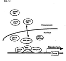
- ZNFN3A1 directly associates with a RNA helicase KIAA0054, and forms a complex with RNA polymerase 11, which activates transcription of downstream genes including epidermal growth factor receptor (EGFR) through a direct binding of the complex with an element of "(C)CCCTCC(T)" in the 5' flanking region.
- EGFR epidermal growth factor receptor
- ZNFN3A1 renders oncogenic activities to cancer cells by transcriptional activation of target genes including EGFR through a complex with RNA helicase and RNA polymerase 11, and that inhibition of the activity of the complex could be a promising strategy for the treatment of HCC.
- the present invention relates to ZNFN3A1 proteins involved in cell proliferation and the genes encoding them.
- the ZNFN3A1 protein preferably includes the amino acid sequence set forth in SEQ ID NO:2.
- the ZNFN3A1 gene includes a polynucleotide sequence as described in SEQ ID NO:1. More specifically, the present invention provides the following:
- a transcription activation complex that includes a protein consisting of the amino acid sequence of SEQ ID NO:2 and at least one co-activator thereof.
- the co-activator may be an RNA helicase and/or an RNA polymerase.
- the RNA helicase may be RNA helicase KIAA0054.
- the RNA polymerase may be RNApolymerase 11.
- the complex includes the protein consisting of the amino acid sequence of SEQ ID NO:2, RNA helicase and RNA polymerase 11.
- the transcription activation complex may activate transcription of genes including epidermal growth factor receptor (EGFR) through a direct binding of the complex with an element of "(C)CCCTCC (T)" in the 5' flanking region of the EGFR gene.
- EGFR epidermal growth factor receptor
- a method of screening for a compound that inhibits the activity of a protein consisting of the amino acid sequence of SEQ ID NO:2, comprising culturing cells which express the protein consisting of the amino acid sequence of SEQ ID NO:2, or a partial peptide thereof, in the presence of a subject sample which contains at least one test compound; detecting the proliferation of the cell and selecting the test compound that inhibits the proliferation as compared to the proliferation detected in the absence of the subject sample.
- An in vitro method of screening for a candidate compound for an anti-cancer agent includes contacting the protein consisting of the amino acid SEQ ID NO:2, a co-activator thereof, and a DNA containing the target sequence of the polypeptide with candidate compounds under the suitable condition for the formation of the complex of the protein consisting of amino acid SEQ ID NO:2 and the DNA, and selecting compounds that inhibit the formation of the complex.
- the target sequence comprises any one of the following (a) to (d) sequences flainking the 5' region of EGFR.
- An in vitro method of screening for a candidate compound for an anti-cancer agent includes contacting the protein consisting of amino acid SEQ ID NO:2, a co-activator thereof, and a reporter gene with a transcriptional regulatory region recognized by the complex of the protein consisting of amino acid SEQ ID NO:2 and a co-activator with candidate compounds under the suitable condition for the expression of the reporter gene, and selecting compounds that inhibit the expression of the reporter gene.
- An in vitro method for diagnosis of cancer that includes determining an expression level of a ZNFN3A1 gene comprising the nucleotide sequence of SEQ ID NO:1 in biological sample of specimen, comparing the expression level said ZNFN3A1 gene comprising the nucleotide sequence of SEQ ID NO: 1 with that in normal sample, and defining a high expression level of said ZNFN3A1 gene comprising the nucleotide sequence of SEQ ID NO:1 in the sample as having a cancer.
- the cancer is suitably a hepatocellular carcinoma.
- the present application identifies a novel human gene ZNFN3A1 whose expression is markedly elevated in HCCs compared to corresponding non-cancerous liver tissues.
- the ZNFN3A1 cDNA consists of 1622 nucleotides that contain an open reading frame of 1284 nucleotides as set forth in SEQ. ID. NO. 1.
- the open reading frame encodes a putative 428-amino acid protein with a zinc finger motif.
- This protein has been named ZNFN3A1 (zinc finger protein, subfamily 3A (MYND containing), 1) by a nomenclature committee.
- ZNFN3A1 directly associates with a RNA helicase KIAA0054, and forms a complex with RNA polymerase II, which activates transcription of downstream genes including epidermal growth factor receptor (EGFR) through a direct binding of the complex with an element of "(C)CCCTCC(T)" in the 5' flanking region of the EGFR gene.
- EGFR epidermal growth factor receptor
- exogenous expression of ZNFN3A1 into NIH3T3 cells conferred increased cell growth, while suppression of its expression with antisense S-oligonucleotides resulted in a significant growth-inhibition of hepatoma cells.
- ZNFN3A1 renders oncogenic activities to cancer cells by transcriptional activation of target genes including EGFR through a complex with RNA helicase and RNA polymerase II, and that inhibition of the activity of the complex could be a promising strategy for the treatment of HCC.
- the present invention encompasses novel human gene ZNFN3A1, including a polynucleotide sequence as described in SEQ. ID. NO. 1, as well as degenerates and mutants thereof, to the extent that they encode a ZNFN3A1 protein, including the amino acid sequence set forth in SEQ. ID. NO.2 or its functional equivalent.
- proteins functionally equivalent to ZNFN3A1 include, for example, homologous proteins of other organisms corresponding to the human ZNFN3A1 protein, as well as mutants of human ZNFN3A1 proteins.
- the term "functionally equivalent” means that the subject protein has the activity to promote cell proliferation like ZNFN3A1 proteins and to confer oncogenic activity to cancer cells by forming a transactivating complex with an RNA helicase or an RNA polymerase or both, which, in turn, enhances the transcription of target genes, such as EGFR.
- Whether the subject protein has a cell proliferation activity or not can be judged by introducing the DNA encoding the subject protein into a cell, such as NIH3T3, expressing the protein, and detecting promotion of proliferation of the cells or increase in colony forming activity.
- RNA polymerase an RNA polymerase
- RNA helicase an RNA helicase
- co-immunoprecipitation such as those described in the Examples below.
- the enhancement of EGFR transcription may be further assayed using reporter plasmids such as those described in the Examples below.
- proteins functionally equivalent to a given protein are well known by a person skilled in the art and include known methods of introducing mutations into the protein.
- one skilled in the art can prepare proteins functionally equivalent to the human ZNFN3A1 protein by introducing an appropriate mutation in the amino acid sequence of the human ZNFN3A1 protein by site-directed mutagenesis (Hashimoto-Gotoh, T. et al. (1995), Gene 152, 271-275; Zoller, MJ, and Smith, M. (1983), Methods Enzymol. 100, 468-500; Kramer, W. et al. (1984), Nucleic Acids Res. 12, 9441-9456; Kramer W, and Fritz HJ. (1987) Methods.
- the protein of the present invention includes those proteins having the amino acid sequences of the human ZNFN3A1 protein in which one or more amino acids are mutated, provided the resulting mutated proteins are functionally equivalent to the human ZNFN3A1 protein.
- the number of amino acids to be mutated in such a mutant is generally 10 amino acids or less, preferably 6 amino acids or less, and more preferably 3 amino acids or less.
- Mutated or modified proteins proteins having amino acid sequences modified by deleting, adding and/or replacing one or more amino acid residues of a certain amino acid sequence, have been known to retain the original biological activity (Mark, D. F. et al., Proc. Natl. Acad. Sci. USA (1984) 81, 5662-5666, Zoller, M. J. & Smith, M., Nucleic Acids Research (1982) 10, 6487-6500, Wang, A. et al., Science 224, 1431-1433, Dalbadie-McFarland, G. et al., Proc. Natl. Acad. Sci. USA (1982) 79, 6409-6413).
- the amino acid residue to be mutated is preferably mutated into a different amino acid in which the properties of the amino acid side-chain are conserved (a process known as conservative amino acid substitution).
- properties of amino acid side chains are hydrophobic amino acids (A, I, L, M, F, P, W, Y, V), hydrophilic amino acids (R, D, N, C, E, Q, G, H, K, S, T), and side chains having the following functional groups or characteristics in common: an aliphatic side-chain (G, A, V, L, I, P); a hydroxyl group containing side-chain (S, T, Y); a sulfur atom containing side-chain (C, M); a carboxylic acid and amide containing side-chain (D, N, E, Q); a base containing side-chain (R, K, H); and an aromatic containing side-chain (H, F, Y, W).
- the parenthetic letters indicate the one-letter codes of amino acids.
- a protein to which one or more amino acids residues are added to the amino acid sequence of human ZNFN3A1 protein is a fusion protein containing the human ZNFN3A1 protein.
- Fusion proteins are, fusions of the human ZNFN3A1 protein and other peptides or proteins, and are included in the present invention. Fusion proteins can be made by techniques well known to a person skilled in the art, such as by linking the DNA encoding the human ZNFN3A1 protein of the invention with DNA encoding other peptides or proteins, so that the frames match, inserting the fusion DNA into an expression vector and expressing it in a host. There is no restriction as to the peptides or proteins fused to the protein of the present invention.
- peptides that can be used as peptides that are fused to the protein of the present invention include, for example, FLAG (Hopp, T. P. et al., Biotechnology (1988) 6, 1204-1210), 6xHis containing six His (histidine) residues, 10xHis, Influenza agglutinin (HA), human c-myc fragment, VSP-GP fragment, p18HIV fragment, T7-tag, HSV-tag, E-tag, SV40T antigen fragment, lck tag, ⁇ -tubulin fragment, B-tag, Protein C fragment, and the like.
- FLAG Hopp, T. P. et al., Biotechnology (1988) 6, 1204-1210
- 6xHis containing six His (histidine) residues 10xHis
- Influenza agglutinin (HA) Influenza agglutinin
- human c-myc fragment VSP-GP fragment
- p18HIV fragment T7-tag
- Fusion proteins can be prepared by fusing commercially available DNA, encoding the fusion peptides or proteins discussed above, with the DNA encoding the protein of the present invention and expressing the fused DNA prepared.
- proteins include mammal homologues corresponding to the protein derived from human or mouse (for example, a protein encoded by a monkey, rat, rabbit and bovine gene).
- mammal homologues corresponding to the protein derived from human or mouse (for example, a protein encoded by a monkey, rat, rabbit and bovine gene).
- isolating a cDNA highly homologous to the DNA encoding the human ZNFN3A1 protein from animals it is particularly preferable to use tissues from ovary or testis.
- hybridization for isolating a DNA encoding a protein functionally equivalent to the human ZNFN3A1 protein can be routinely selected by a person skilled in the art.
- hybridization may be performed by conducting prehybridization at 68°C for 30 min or longer using "Rapid-hyb buffer" (Amersham LIFE SCIENCE), adding a labeled probe, and warming at 68°C for 1 hour or longer.
- the following washing step can be conducted, for example, in a low stringent condition.
- a low stringent condition is, for example, 42°C, 2X SSC, 0.1% SDS, or preferably 50°C, 2X SSC, 0.1% SDS. More preferably, high stringent conditions is used.
- a high stringent condition is, for example, washing 3 times in 2X SSC, 0.01% SDS at room temperature for 20 min, then washing 3 times in 1x SSC, 0.1 % SDS at 37°C for 20 min, and washing twice in 1 x SSC, 0.1 % SDS at 50°C for 20 min.
- factors such as temperature and salt concentration can influence the stringency of hybridization and one skilled in the art can suitably select the factors to achieved the requisite stringency.
- a gene amplification method for example, the polymerase chain reaction (PCR) method, can be utilized to isolate a DNA encoding a protein functionally equivalent to the human ZNFN3A1 protein, using a primer synthesized based on the sequence information of the DNA (SEQ. ID. NO. 1) encoding the human ZNFN3A1 protein.
- PCR polymerase chain reaction
- Proteins that are functionally equivalent to the human ZNFN3A1 protein encoded by the DNA isolated through the above hybridization techniques or gene amplification techniques normally have a high homology to the amino acid sequence of the human ZNFN3A1 protein.
- “High homology” typically refers to a homology of 40% or higher, preferably 60% or higher, more preferably 80% or higher, even more preferably 95% or higher.
- the homology of a protein can be determined by following the algorithm in "Wilbur, W. J. and Lipman, D. J. Proc. Natl. Acad. Sci. USA (1983) 80, 726-730".
- a protein of the present invention may have variations in amino acid sequence, molecular weight, isoelectric point, the presence or absence of sugar chains, or form, depending on the cell or host used to produce it or the purification method utilized. Nevertheless, so long as it has a function equivalent to that of the human ZNFN3A1 protein (SEQ. ID. NO. 2) of the present invention, it is within the scope of the present invention.
- the proteins of the present invention can be prepared as recombinant proteins or natural proteins, by methods well known to those skilled in the art.
- a recombinant protein can be prepared by inserting a DNA, which encodes the protein of the present invention (for example, the DNA comprising the nucleotide sequence of SEQ. ID. NO. 1), into an appropriate expression vector, introducing the vector into an appropriate host cell, obtaining the extract, and purifying the protein by subjecting the extract to chromatography, for example, ion exchange chromatography, reverse phase chromatography, gel filtration, or affinity chromatography utilizing a column to which antibodies against the protein of the present invention is fixed, or by combining more than one of aforementioned columns.
- chromatography for example, ion exchange chromatography, reverse phase chromatography, gel filtration, or affinity chromatography utilizing a column to which antibodies against the protein of the present invention is fixed, or by combining more than one of aforementioned columns.
- the protein of the present invention when expressed within host cells (for example, animal cells and E . coli ) as a fusion protein with glutathione-S-transferase protein or as a recombinant protein supplemented with multiple histidines, the expressed recombinant protein can be purified using a glutathione column or nickel column.
- host cells for example, animal cells and E . coli
- the expressed recombinant protein can be purified using a glutathione column or nickel column.
- a natural protein can be isolated by methods known to a person skilled in the art, for example, by contacting the affinity column, in which antibodies binding to the ZNFN3A1 protein described below are bound, with the extract of tissues or cells expressing the protein of the present invention.
- the antibodies can be polyclonal antibodies or a monoclonal antibodies.
- the present invention also encompasses partial peptides of the protein of the present invention.
- the partial peptide has an amino acid sequence specific to the protein of the present invention and consists of at least 7 amino acids, preferably 8 amino acids or more, and more preferably 9 amino acids or more.
- the partial peptide can be used, for example, for preparing antibodies against the protein of the present invention, screening for a compound that binds to the protein of the present invention, and screening for accelerators or inhibitors of the protein of the present invention.
- a partial peptide of the invention can be produced by genetic engineering, by known methods of peptide synthesis, or by digesting the protein of the invention with an appropriate peptidase.
- peptide synthesis for example, solid phase synthesis or liquid phase synthesis may be used.
- the isolated ZNFN3A1 protein includes a putative 428-amino acid protein with a zinc finger motif encoded by the open reading frame of the ZNFN3A1 polynucleotide sequence.
- the zinc finger domain (MYND) is positioned at codons 49-87 and the SET (Su 3-9, Enhancer-of-zeste, Trihorrax) domain is positioned at codons 117-246.
- the ZNFN3A1 binding region resides in the SET domain. Therefore, the partial peptide of ZNFN3A1 preferably includes the SET domain.
- the present invention provides DNA encoding the proteins of the present invention.
- the DNA of the present invention can be used for the in vivo or in vitro production of the protein of the present invention as described above, or can be applied to gene therapy for diseases attributed to genetic abnormality in the gene encoding the protein of the present invention. Any form of the DNA of the present invention can be used, so long as it encodes the protein of the present invention. Specifically, cDNA synthesized from the mRNA, genomic DNA, and chemically synthesized DNA can be used.
- the DNA of the present invention include a DNA comprising a given nucleotide sequences as well as its degenerate sequences, so long as the resulting DNA encodes a protein of the present invention.
- the DNA of the present invention can be prepared by methods known to a person skilled in the art.
- the DNA of the present invention can be prepared by: preparing a cDNA library from cells which express the protein of the present invention, and conducting hybridization using a partial sequence of the DNA of the present invention (for example, SEQ. ID. NO. 1) as a probe.
- a cDNA library can be prepared, for example, by the method described in Sambrook J. et al., Molecular Cloning, Cold Spring Harbor Laboratory Press (1989); alternatively, commercially available cDNA libraries may be used.
- a cDNA library can be also prepared by: extracting RNAs from cells expressing the protein of the present invention, synthesizing oligo DNAs based on the sequence of the DNA of the present invention (for example, SEQ. ID. NO. 1), conducting PCR by using the oligos as primers, and amptifying cDNAs encoding the protein of the present invention.
- the translation region encoded by the cDNA can be routinely determined, and the amino acid sequence of the protein of the present invention can be easily obtained.
- the genomic DNA can be isolated.
- mRNAs may first be prepared from a cell, tissue, or organ (for example, ovary, testis, placenta, etc.) in which the protein of the invention is expressed.
- Known methods can be used to isolate mRNAs; for instance, total RNA may be prepared by guanidine ultracentrifugation (Chirgwin J. M. et al. Biochemistry 18:5294-5299 (1979)) or AGPC method (Chomczynski P. and Sacchi N. Anal. Biochem. 162:156-159 (1987)).
- mRNA may be purified from total RNA using mRNA Purification Kit (Pharmacia) and such or, alternatively, mRNA may be directly purified by QuickPrep mRNA Purification Kit (Pharmacia).
- cDNA is used to synthesize cDNA using reverse transcriptase.
- cDNA may be synthesized by using a commercially available kit, such as the AMV Reverse Transcriptase First-strand cDNA Synthesis Kit (Seikagaku Kogyo).
- cDNA may be synthesized and amplified following the 5'-RACE method(Frohman M. A. et al. Proc. Natl. Acad. Sci. U.S.A. 85:8998-9002 (1988); Belyavsky A. et al. Nucleic Acids Res. 17:2919-2932 (1989)), which uses a primer and such, described herein, the 5'-Ampli FINDER RACE Kit (Clontech), and polymerase chain reaction (PCR).
- 5'-RACE method Frohman M. A. et al. Proc. Natl. Acad. Sci. U.S.A. 85:8998-9002 (1988); Be
- a desired DNA fragment is prepared from the PCR products and ligated with a vector DNA.
- the recombinant vectors are used to transform E. coli and such, and a desired recombinant vector is prepared from a selected colony.
- the nucleotide sequence of the desired DNA can be verified by conventional methods, such as dideoxynucleotide chain termination.
- the nucleotide sequence of a DNA of the invention may be designed to be expressed more efficiently by taking into account the frequency of codon usage in the host to be used for expression (Grantham R. et al. Nucleic Acids Res. 9:43-74 (1981)).
- the DNA of the present invention may be altered by a commercially available kit or a conventional method. For instance, the DNA may be altered by digestion with restriction enzymes, insertion of a synthetic oligonucleotide or an appropriate DNA fragment, addition of a linker, or insertion of the initiation codon (ATG) and/or the stop codon (TAA, TGA, or TAG).
- the DNA of the present invention encompasses the DNA comprising the nucleotide sequence encoding the zinc finger domain, positioned at codons 49-87 of SEQ. ID. NO.2 and the DET domain, positioned at codons 117-246 of SEQ. ID. NO.2
- the present invention provides a DNA that hybridizes under stringent conditions with a DNA having a nucleotide sequence of SEQ. ID. NO. 1, and encodes a protein functionally equivalent to the protein of the invention described above.
- stringent conditions For example, low stringent condition can be used. More preferably, high stringent condition can be used. These conditions are the same as that described above.
- the hybridizing DNA above is preferably a cDNA or a chromosomal DNA.
- the present invention also provides a vector into which a DNA of the present invention is inserted.
- a vector of the present invention is useful to keep a DNA of the present invention in host cell, or to express the protein of the present invention.
- E. coli is a host cell and the vector is amplified and produced in a large amount in E. coli (e.g., JM109, DH5 ⁇ , HB101, or XL1 Blue)
- the vector should have "ori" to be amplified in E. coli and a marker gene for selecting transformed E. coli - (e.g., a drug-resistance gene selected by a drug such as ampicillin, tetracycline, kanamycin, chloramphenicol or the like).
- M13-series vectors for example, M13-series vectors, pUC-series vectors, pBR322, pBluescript, pCR-Script, etc. can be used.
- pGEM-T, pDIRECT, and pT7 can also be used for subcloning and extracting cDNA as well as the vectors described above.
- an expression vector is especially useful.
- an expression vector to be expressed in E . coli should have the above characteristics to be amplified in E . coli.
- the vector should have a promoter, for example, lacZ promoter (Ward et al., Nature (1989) 341, 544-546; FASEB J (1992) 6. 2422-2427), araB promoter (Better et al., Science (1988) 240, 1041-1043), or T7 promoter or the like, that can efficiently express the desired gene in E . coli.
- a promoter for example, lacZ promoter (Ward et al., Nature (1989) 341, 544-546; FASEB J (1992) 6. 2422-2427), araB promoter (Better et al., Science (1988) 240, 1041-1043), or T7 promoter or the like, that can efficiently express the desired gene in E . coli.
- pGEX-5X-1 Pulcoa
- QIAexpress system Qiagen
- pEGFP pEGFP
- pET pET
- the host is preferably BL21 which expresses T7 RNA polymerase,for example, can be used instead of the above vectors.
- the vector may also contain a signal sequence for polypeptide secretion.
- An exemplary signal sequence that directs the protein to be secreted to the periplasm of the E. coli is the pelB signal sequence (Lei, S. P. et al J. Bacteriol. (1987) 169, 4379).
- Means for introducing of the vectors into the target host cells include, for example, the calcium chloride method, and the electroporation method.
- expression vectors derived from mammals for example, pcDNA3 (Invitrogen) and pEGF-BOS (Nucleic Acids. Res. 1990, 18 (17), p5322), pEF, pCDM8), expression vectors derived from insect cells (for example, "Bac-to-BAC baculovirus expression system” (GIBCO BRL), pBacPAK8), expression vectors derived from plants (for example pMH1, pMH2), expression vectors derived from animal viruses (for example, pHSV, pMV, pAdexLcw), expression vectors derived from retroviruses (for example, pZlpneo), expression vector derived from yeast (for example, "Pichia Expression Kit” (Invitrogen), pNV11, SP-Q01), and expression vectors derived from Bacillus subtilis (for example, pPL608, pKTH50) can be used for producing the protein of the mammals (for example, pcDNA3 (In
- the vector In order to express the vector in animal cells, such as CHO, COS, or NIH3T3 cells, the vector should have a promoter necessary for expression in such cells, for example, the SV40 promoter (Mulligan et al., Nature (1979) 277, 108), the MMLV-LTR promoter, the EF1 ⁇ promoter (Mizushima et al., Nucleic Acids Res. (1990) 18, 5322), the CMV promoter, and the like, and preferably a marker gene for selecting transformants (for example, a drug resistance gene selected by a drug (e.g., neomycin, G418)).
- a promoter necessary for expression in such cells for example, the SV40 promoter (Mulligan et al., Nature (1979) 277, 108), the MMLV-LTR promoter, the EF1 ⁇ promoter (Mizushima et al., Nucleic Acids Res. (1990) 18, 5322
- a vector comprising the complementary DHFR gene may be introduced into CHO cells in which the nucleic acid synthesizing pathway is deleted, and then amplified by methotrexate (MTX).
- MTX methotrexate
- the method wherein a vector comprising a replication origin of SV40 (pcD, etc.) is transformed into COS cells comprising the SV40 T antigen expressing gene on the chromosome can be used.
- a protein of the present invention obtained as above may be isolated from inside or outside (such as medium) of host cells, and purified as a substantially pure homogeneous protein.
- substantially pure as used herein in reference to a given polypeptide means that the polypeptide is substantially free from other biological macromolecules.
- the substantially pure polypeptide is at least 75% (e.g., at least 80, 85, 95, or 99%) pure by dry weight. Purity can be measured by any appropriate standard method, for example by column chromatography, polyacrylamide gel electrophoresis, or HPLC analysis. The method for protein isolation and purification is not limited to any specific method; in fact, any standard method may be used.
- column chromatography filter, ultrafiltration, salt precipitation, solvent precipitation, solvent extraction, distillation, immunoprecipitation, SDS-polyacrylamide gel electrophoresis, isoelectric point electrophoresis, dialysis, and recrystallization may be appropriately selected and combined to isolate and purify the protein.
- chromatography examples include, for example, affinity chromatography, ion-exchange chromatography, hydrophobic chromatography, gel filtration, reverse phase chromatography, adsorption chromatography, and such (Strategies for Protein Purification and Characterization: A Laboratory Course Manual. Ed. Daniel R. Marshak et al., Cold Spring Harbor Laboratory Press (1996)). These chromatographies may be performed by liquid chromatography, such as HPLC and FPLC. Thus, the present invention provides for highly purified proteins prepared by the above methods.
- a protein of the present invention may be optionally modified or partially deleted by treating it with an appropriate protein modification enzyme before or after purification.
- useful protein modification enzymes include, but are not limited to, trypsin, chymotrypsin, lysylendopeptidase, protein kinase, glucosidase and so on.
- the present invention provides an antibody that binds to the protein of the invention.
- the antibody of the invention can be used in any form, such as monoclonal or polyclonal antibodies, and includes antiserum obtained by immunizing an animal such as a rabbit with the protein of the invention, all classes of polyclonal and monoclonal antibodies, human antibodies, and humanized antibodies produced by genetic recombination.
- a protein of the invention used as an antigen to obtain an antibody may be derived from any animal species, but preferably is derived from a mammal such as a human, mouse, or rat, more preferably from a human.
- a human-derived protein may be obtained from the nucleotide or amino acid sequences disclosed herein.
- the protein to be used as an immunization antigen may be a complete protein or a partial peptide of the protein.
- a partial peptide may comprise, for example, the amino (N)-terminal or carboxy (C)-terminal fragment of a protein of the present invention.
- an antibody is defined as a protein that reacts with either the full length or a fragment of a protein of the present invention.
- a gene encoding a protein of the invention or its fragment may be inserted into a known expression vector, which is then used to transform a host cell as described herein.
- the desired protein or its fragment may be recovered from the outside or inside of host cells by any standard method, and may subsequently be used as an antigen.
- whole cells expressing the protein or their lysates, or a chemically synthesized protein may be used as the antigen.
- Any mammalian animal may be immunized with the antigen, but preferably the compatibility with parental cells used for cell fusion is taken into account.
- animals of Rodentia, Lagomorpha, or Primates are used.
- Animals of Rodentia include, for example, mouse, rat, and hamster.
- Animals of Lagomorpha include, for example, rabbit.
- Animals of Primates include, for example, a monkey of Catarrhini (old world monkey) such as Macaca fascicularis, rhesus monkey, sacred baboon, and chimpanzees.
- antigens may be diluted and suspended in an appropriate amount of phosphate buffered saline (PBS), physiological saline, etc.
- PBS phosphate buffered saline
- the antigen suspension may be mixed with an appropriate amount of a standard adjuvant, such as Freund's complete adjuvant, made into emulsion, and then administered to mammalian animals.
- a standard adjuvant such as Freund's complete adjuvant
- an appropriately amount of Freund's incomplete adjuvant every 4 to 21 days.
- An appropriate carrier may also be used for immunization.
- serum is examined by a standard method for an increase in the amount of desired antibodies.
- Polyclonal antibodies against the proteins of the present invention may be prepared by collecting blood from the immunized mammal examined for the increase of desired antibodies in the serum, and by separating serum from the blood by any conventional method.
- Polyclonal antibodies include serum containing the polyclonal antibodies, as well as the fraction containing the polyclonal antibodies may be isolated from the serum.
- Immunoglobulin G or M can be prepared from a fraction which recognizes only the protein of the present invention using, for example, an affinity column coupled with the.protein of the present invention, and further purifying this fraction by using protein A or protein G column.
- immune cells are collected from the mammal immunized with the antigen and checked for the increased level of desired antibodies in the serum as described above, and are subjected to cell fusion.
- the immune cells used for cell fusion are preferably obtained from spleen.
- Other preferred parental cells to be fused with the above immunocyte include, for example, myeloma cells of mammalians, and more preferably myeloma cells having an acquired property for the selection of fused cells by drugs.
- the above immunocyte and myeloma cells can be fused according to known methods, for example, the method of Milstein et al. (Galfre, G. and Milstein, C., Methods Enzymol. (1981) 73, 3-46).
- Resulting hybridomas obtained by the cell fusion may be selected by cultivating them in a standard selection medium, such as HAT medium (hypoxanthine, aminopterin, and thymidine containing medium).
- HAT medium hyperxanthine, aminopterin, and thymidine containing medium.
- the cell culture is typically continued in the HAT medium for severa! days to several weeks, the time being sufficient to allow all the other cells, with the exception of the desired hybridoma (non-fused cells), to die.
- the standard limiting dilution is performed to screen and clone a hybridoma cell producing the desired antibody.
- human lymphocytes such as those infected by EB virus may be immunized with a protein, protein expressing cells, or their lysates in vitro. Then, the immunized lymphocytes are fused with human-derived myeloma cells that are capable of indefinitely dividing, such as U266, to yield a hybridoma producing a desired human antibody that is able to bind to the protein can be obtained (Unexamined Published Japanese Patent Application No. (JP-A) Sho 63-17688).
- JP-A Japanese Patent Application No.
- the obtained hybridomas are subsequently transplanted into the abdominal cavity of a mouse and the ascites are extracted.
- the obtained monoclonal antibodies can be purified by, for example, ammonium sulfate precipitation, a protein A or protein G column, DEAE ion exchange chromatography, or an affinity column to which the protein of the present invention is coupled.
- the antibody of the present invention can be used not only for purification and detection of the protein of the present invention, but also as a candidate for agonists and antagonists of the protein of the present invention.
- this antibody can be applied to the antibody treatment for diseases related to the protein of the present invention.
- a human antibody or a humanized antibody is preferable for reducing immunogenicity.
- transgenic animals having a repertory of human antibody genes may be immunized with an antigen selected from a protein, protein expressing cells, or their lysates.
- Antibody producing cells are then collected from the animals and fused with myeloma cells to obtain hybridoma, from which human antibodies against the protein can be prepared (see W092-03918, W093-2227, WO94-02602, W094-25585, W096-33735, and W096-34096).
- an immune cell such as an immunized lymphocyte, producing antibodies may be immortalized by an oncogene and used for preparing monoclonal antibodies.
- Monoclonal antibodies thus obtained can be also recombinantly prepared using genetic engineering techniques (see, for example, Borrebaeck C. A. K. and Larrick J. W. Therapeutic Monoclonal Antibodies, published in the United Kingdom by MacMillan Publishers LTD (1990)).
- a DNA encoding an antibody may be cloned from an immune cell, such as a hybridoma or an immunized lymphocyte producing the antibody, inserted into an appropriate vector, and introduced into host cells to prepare a recombinant antibody.
- the present invention also provides recombinant antibodies prepared as described above.
- an antibody of the present invention may be a fragment of an antibody or modified antibody, so long as it binds to one or more of the proteins of the invention.
- the antibody fragment may be Fab, F(ab') 2 , Fv, or single chain Fv (scFv), in which Fv fragments from H and L chains are ligated by an appropriate linker (Huston J. S. et al. Proc. Natl. Acad. Sci. U.S.A. 85:5879-5883 (1988)).
- an antibody fragment may be generated by treating an antibody with an enzyme, such as papain or pepsin.
- a gene encoding the antibody fragment may be constructed, inserted into an expression vector, and expressed in an appropriate host cell (see, for example, Co M. S. et al. J. Immunol. 152:2968-2976 (1994); Better M. and Horwitz A. H. Methods Enzymol. 178:476-496 (1989); Pluckthun A. and Skerra A. Methods Enzymol. 178:497-515 (1989); Lamoyi E. Methods Enzymol. 121:652-663 (1986); Rousseaux J. et al. Methods Enzymol. 121:663-669 (1986); Bird R. E. and Walker B. W. Trends Biotechnol. 9:132-137 (1991)).
- An antibody may be modified by conjugation with a variety of molecules, such as polyethylene glycol (PEG).
- PEG polyethylene glycol
- the present invention provides for such modified antibodies.
- the modified antibody can be obtained by chemically modifying an antibody. These modification methods are conventional in the field.
- an antibody of the present invention may be obtained as a chimeric antibody, between a variable region derived from nonhuman antibody and the constant region derived from human antibody, or as a humanized antibody, comprising the complementarity determining region (CDR) derived from nonhuman antibody, the frame work region (FR) derived from human antibody, and the constant region.
- CDR complementarity determining region
- FR frame work region
- Antibodies obtained as above may be purified to homogeneity.
- the separation and purification of the antibody can be performed according to separation and purification methods used for general proteins.
- the antibody may be separated and isolated by the appropriately selected and combined use of column chromatographies, such as affinity chromatography, filter, ultrafiltration, satting-out, dialysis, SDS polyacrylamide gel electrophoresis, isoelectric focusing, and others (Antibodies: A Laboratory Manual. Ed Hanow and David Lane, Cold Spring Harbor Laboratory, 1988), but are not limited thereto.
- a protein A column and protein G column can be used as the affinity column.
- Exemplary protein A columns to be used include, for example, Hyper D, POROS, and Sepharose F.F. (Pharmacia).
- Exemplary chromatography with the exception of affinity includes, for example, ion-exchange chromatography, hydrophobic chromatography, gel filtration, reverse-phase chromatography, adsorption chromatography, and the like (Strategies for Protein Purification and Characterization: A Laboratory Course Manual. Ed Daniel R. Marshak et al., Cold Spring Harbor Laboratory Press, 1996).
- the chromatographic procedures can be carried out by liquid-phase chromatography, such as HPLC, FPLC.
- ELISA enzyme-linked immunosorbent assay
- EIA enzyme immunoassay
- RIA radioimmunoassay
- immunofluorescence may be used to measure the antigen binding activity of the antibody of the invention.
- the antibody of the present invention is immobilized on a plate, protein of the invention is applied to the plate, and then a sample containing a desired antibody, such as culture supernatant of antibody producing cells or purified antibodies, is applied. Then, a secondary antibody that recognizes the primary antibody and is labeled with an enzyme, such as alkaline phosphatase, is applied, and the plate is incubated.
- a desired antibody such as culture supernatant of antibody producing cells or purified antibodies
- an enzyme substrate such as p -nitrophenyl phosphate
- the absorbance is measured to evaluate the antigen binding activity of the sample.
- a fragment of the protein such as a C-terminal or N-terminal fragment, may be used as a protein.
- BIAcore Pharmacia
- the above methods allow for the detection or measurement of the protein of the invention, by exposing the antibody of the invention to a sample assumed to contain the protein of the invention, and detecting or measuring the immune complex formed by the antibody and the protein.
- the method of detection or measurement of the protein according to the invention can specifically detect or measure a protein, the method may be useful in a variety of experiments in which the protein is used.
- the present invention also provides a polynucleotide which hybridizes with the DNA encoding human ZNFN3A1 protein (SEQ. ID. NO.1) or the complementary strand thereof, and which comprises at least 15 nucleotides.
- the polynucleotide of the present invention is preferably a polynucleotide which specifically hybridizes with the DNA encoding the protein of the present invention.
- the term "specifically hybridize” as used herein, means that cross-hybridization does not occur significantly with DNA encoding other proteins, under the usual hybridizing conditions, preferably under stringent hybridizing conditions.
- Such polynucleotides include, probes, primers, nucleotides and nucleotide derivatives (for example, antisense oligonucleotides and ribozymes), which specifically hybridize with DNA encoding the protein of the invention or its complementary strand. Moreover, such polynucleotide can be utilized for the preparation of DNA chip.
- the present invention includes an antisense oligonucleotide that hybridizes with any site within the nucleotide sequence of SEQ. ID. NO. 1.
- This antisense oligonucleotide is preferably against at least 15 continuous nucleotides of the nucleotide sequence of SEQ. ID. NO. 1.
- the above-mentioned antisense oligonucleotide which contains an initiation codon in the above-mentioned at least 15 continuous nucleotides, is even more preferred.
- Derivatives or modified products of antisense oligonucleotides can be used as antisense oligonucleotides.
- modified products include lower alkyl phosphonate modifications such as methyl-phosphonate-type or ethyl-phosphonate-type, phosphorothioate modifications and phosphoroamidate modifications.
- antisense oligonucleotides means, not only those in which the nucleotides corresponding to those constituting a specified region of a DNA or mRNA are entirely complementary, but also those having a mismatch of one or more nucleotides, as long as the DNA or mRNA and the antisense oligonucleotide can specifically hybridize with the nucleotide sequence of SEQ. ID. NO.1.
- Such polynucleotides are contained as those having, in the "at least 15 continuous nucleotide sequence region", a homology of at least 70% or higher, preferably at 80% or higher, more preferably 90% or higher, even more preferably 95% or higher.
- the algorithm stated herein can be used to determine the homology.
- Such polynucleotides are useful as probes for the isolation or detection of DNA encoding the protein of the invention as stated in a later example or as a primer used for amplifications.
- the antisense oligonucleotide derivatives of the present invention act upon cells producing the protein of the invention by binding to the DNA or mRNA encoding the protein, inhibiting its transcription or translation, promoting the degradation of the mRNA, and inhibiting the expression of the protein of the invention, thereby resulting in the inhibition of the protein's function.
- An antisense oligonucleotide derivative of the present invention can be made into an external preparation, such as a liniment or a poultice, by mixing with a suitable base material which is inactive against the derivatives.
- the derivatives can be formulated into tablets, powders, granules, capsules, liposome capsules, injections, solutions, nose-drops and freeze-drying agents by adding excipients, isotonic agents, solubilizers, stabilizers, preservatives, pain-killers, and such. These can be prepared by following usual methods.
- the antisense oligonucleotide derivative is given to the patient by directly applying onto the ailing site or by injecting into a blood vessel so that it will reach the site of ailment.
- An antisense-mounting medium can also be used to increase durability and membrane-permeability. Examples are, liposome, poly-L-lysine, lipid, cholesterol; lipofectin or derivatives of these.
- the dosage of the antisense oligonucleotide derivative of the present invention can be adjusted suitably according to the patient's condition and used in desired amounts. For example, a dose range of 0.1 to 100 mg/kg, preferably 0.1 to 50 mg/kg can be administered.
- the antisense oligonucleotide of the invention inhibits the expression of the protein of the invention and is thereby useful for suppressing the biological activity of the protein of the invention. Also, expression-inhibitors, comprising the antisense oligonucleotide of the invention, are useful in the point that they can inhibit the biological activity of the protein of the invention.
- the present invention provides a method of screening for a compound that binds to the protein of the present invention by using the protein of the present invention.
- This screening method comprises the steps of: (a) contacting the protein of the present invention or a partial peptide thereof with a subject sample, (b) detecting the binding activity between the protein of the present invention or the partial peptide thereof and the subject sample, and (c) selecting a compound that binds to the protein of the present invention or the partial peptide thereof.
- the protein of the present invention to be used for screening may be a recombinant protein or a protein derived from the nature, or a partial peptide thereof.
- Any subject sample for example, cell extracts, cell culture supernatant, products of fermenting microorganism, extracts from marine organism, plant extracts, purified or crude proteins, peptides, non-peptide compounds, synthetic micromolecular compounds and natural compounds, can be used.
- the protein of the present invention to be contacted with a subject sample can be, for example, a purified protein, a soluble protein, a form bound to a carrier, or a fusion protein fused with other proteins.
- a method of screening for proteins for example, that bind to the protein of the present invention using the protein of the present invention
- many methods well known by a person skilled in the art can be used.
- Such a screening can be conducted by, for example, immunoprecipitation method, specifically, in the following manner.
- the gene encoding the protein of the present invention is expressed in animal cells and so on by inserting the gene to an expression vector for foreign genes, such as pSV2neo, pcDNA I, and pCD8.
- the promoter to be used for the expression may be any promoter that can be used commonly and include, for example, the SV40 early promoter (Rigby in Williamson (ed.), Genetic Engineering, vol. 3. Academic Press, London, p.
- the EF-1 ⁇ promoter Kim et al., Gene 91 , p217-223 (1990)), the CAG promoter (Niwa et al. Gene 108 , p. 193-200 (1991)), the RSV LTR promoter (Cullen Methods in Enzymology 152 , p. 684-704 (1987)) the SR ⁇ promoter (Takebe et al., Mol. Cell. Biol. 8 , p. 466 (1988), the CMV immediate early promoter (Seed and Aruffo Proc. Natl. Acad. Sci. USA 84 , p.
- the introduction of the gene into animal cells to express a foreign gene can be performed according to any methods, for example, the electroporation method (Chu G. et al. Nucl. Acids Res. 15 , 1311-1326 (1987)), the calcium phosphate method (Chen, C and Okayama, H. Mol. Cell. Biol.
- the protein of the present invention can be expressed as a fusion protein comprising a recognition site (epitope) of a monoclonal antibody by introducing the epitope of the monoclonal antibody, whose specificity has been revealed, to the N- or C- terminus of the protein of the present invention.
- a commercially available epitope-antibody system can be used (Experimental Medicine 13 , 85-90 (1995)).
- Vectors which can express a fusion protein with, for example, ⁇ -galactosidase, maltose binding protein, glutathione S-transferase, green florescence protein (GFP) and so on by the use of its multiple cloning sites are commercially available.
- a fusion protein prepared by introducing only small epitopes consisting of several to a dozen amino acids so as not to change the property of the protein of the present invention by the fusion is also reported.
- Epitopes such as polyhistidine (His-tag), influenza aggregate HA, human c-myc, FLAG, Vesicular stomatitis virus glycoprotein (VSV-GP), T7 gene 10 protein (T7-tag), human simple herpes virus glycoprotein (HSV-tag), E-tag (an epitope on monoclonal phage), and such, and monoclonal antibodies recognizing them can be used as the epitope-antibody system for screening proteins binding to the protein of the present invention (Experimental Medicine 13 , 85-90 (1995)).
- an immune complex is formed by adding these antibodies to cell lysate prepared by using an appropriate detergent.
- the immune complex consists of the protein of the present invention, a protein comprising the binding ability with the protein, and an antibody. Immunoprecipitation can be also conducted by using antibodies against the protein of the present invention, besides using antibodies against the above epitopes.
- An antibody against the protein of the present invention can be prepared, for example, by introducing a gene encoding the protein of the present invention to an appropriate E. coli expression vector, expressing the gene in E. coli, purifying the expressed protein, and immunizing rabbits, mice, rats, goats, domestic fowls and such against the protein. The antibody can be also prepared by immunizing the above animals against a synthesized partial peptide of the protein of the present invention.
- An immune complex can be precipitated, for example by Protein A Sepharose or Protein G sepharose when the antibody is a mouse IgG antibody. If the protein of the present invention is prepared as a fusion protein with an epitope, such as GST, an immune complex can be formed in the same manner as in the use of the antibody against the protein of the present invention, by using a substance specifically binding to these epitopes, such as glutathione-Sepharose 4B.
- Immunoprecipitation can be performed by following or according to, for example, the methods in the literature (Harlow, E. and Lane, D.: Antibodies pp. 511-552, Cold Spring Harbor Laboratory publications, New York (1988))
- SDS-PAGE is commonly used for analysis of immunoprecipitated proteins and the bound protein can be analyzed by the molecular weight of the protein by using gels with an appropriate concentration. Since the protein bound to the protein of the present invention is difficult to detect by a common staining method, such as Coomassie staining or silver staining, the detection sensitivity for the protein can be improved by culturing cells in culture medium containing radioactive isotope, 35 S-methionine or 35 S-cystein, labeling proteins in the cells, and detecting the proteins. The target protein can be purified directly from the SDS-polyacrylamide gel and its sequence can be determined, when the molecular weight of a protein has been revealed.
- a common staining method such as Coomassie staining or silver staining
- a protein binding to the protein of the present invention can be obtained by preparing a cDNA library from cells, tissues, organs (for example, tissues such as ovary, testis, and placenta or cultured cells) expected to express a protein binding to the protein of the present invention by using a phage vector (e.g., ZAP), expressing the protein on LB-agarose, fixing the protein expressed on a filter, reacting the purified and labeled protein of the present invention with the above filter, and detecting the plaques expressing proteins bound to the protein of the present invention according to the label.
- a phage vector e.g., ZAP
- the protein of the invention may be labeled by utilizing the binding between biotin and avidin, or by utilizing an antibody that specifically binds to the protein of the present invention, or a peptide or polypeptide (for example, GST) that is fused to the protein of the present invention. Methods using radioisotope or fluorescence and such may be also used.
- a two-hybrid system utilizing cells may be used ("MATCHMAKER Two-Hybrid system", “Mammalian MATCHMAKER Two-Hybrid Assay Kit”, “MATCHMAKER one-Hybrid system” (Clontech); “HybriZAP Two-Hybrid Vector System” (Stratagene); the references “Dalton S, and Treisman R (1992) Cell 68, 597-612", “Fields S. and Stemglanz R. Trends Genet. (1994) 10:286-292").
- the protein of the invention is fused to the SRF-binding region or GAL4-binding region and expressed in yeast cells.
- a cDNA library is prepared from cells expected to express a protein binding to the protein of the invention, such that the library, when expressed, is fused to the VP16 or GAL4 transcriptional activation region.
- the cDNA library is then introduced into the above yeast cells and the cDNA derived from the library is isolated from the positive clones detected (when a protein binding to the protein of the invention is expressed in yeast cells, the binding of the two activates a reporter gene, making positive clones detectable).
- a protein encoded by the cDNA can be prepared by introducing the cDNA isolated above to E.coli and expressing the protein.
- reporter gene for example, Ade2 gene, lacZ gene, CAT gene, luciferase gene and such can be used besides HIS3 gene.
- a compound binding to the protein of the present invention can be screened using affinity chromatography.
- the protein of the invention may be immobilized on a carrier of an affinity column, and a test sample, containing a protein capable of binding to the protein of the invention is supposed to be expressed, is applied to the column.
- a test sample herein may be, for example, cell extracts, cell lysates, etc. After loading the test sample, the column is washed, and proteins bound to the protein of the invention can be prepared.
- a biosensor using the surface plasmon resonance phenomenon may be used as a mean for detecting or quantifying the bound compound in the present invention.
- the interaction between the protein of the invention and a test compound can be observed real-time as a surface plasmon resonance signal, using only a minute amount of protein and without labeling (for example, BIAcore, Pharmacia). Therefore, it is possible to evaluate the binding between the protein of the invention and a test compound using a biosensor such as BIAcore.
- the methods of screening for molecules that bind when the immobilized protein of the present invention is exposed to synthetic chemical compounds, or natural substance banks, or a random phage peptide display library, or the methods of screening using high-throughput based on combinatorial chemistry techniques (Wrighton Nc, Farrel FX, Chang R, Kashyap AK, Barbone FP, Mulcahy LS, Johnson DL, Barret RW, Jolliffe LK, Dower WJ; Small peptides as potent mimetics of the protein hormone erythropoietin, Science (UNITED STATES) Jul 26 1996, 273 p458-64, Verdine GL., The combinatorial chemistry of nature.
- a compound isolated by the screening is a candidate for drugs which promote or inhibit the activity of the protein of the present invention, for treating or preventing diseases attributed to, for example, cell proliferative diseases such as cancer.
- the present invention provides a method for screening a compound which promotes or inhibits the activity of the protein of the present invention. Since the ZNFN3A1 protein of the present invention has the activity of promoting cell proliferation, a compound which promotes or inhibits this activity of a ZNFN3A1 protein of the present invention can be screened using this activity as an index.
- This screening method includes the steps of: (a) culturing cells which express ZNFN3A1 protein in the presence of the subject sample, (b) detecting the proliferation of the cells, and (c) selecting a compound which promotes or inhibits the proliferation in comparison with the proliferation detected in the absence of the subject sample.
- Compounds that inhibit the expression and/or activity of ZNFN3A1 find utility as anti-cancer agents.
- ZNFN3A1 proteins can be used for screening so long as they comprise the activity of inhibiting cell proliferation.
- a human ZNFN3A1 protein can be used and proteins functionally equivalent to these proteins can also be used.
- ZNFN3A1 proteins may be expressed endogenously or exogenously by cells.
- Any subject samples for example, cell extracts, cell culture supernatant, products of fermenting microorganism, extracts of marine organism, plant extracts, purified or crude proteins, peptides, non-peptide compounds, synthetic micromolecular compounds, natural compounds, can be used.
- a compound obtained by the above screening for compounds that bind to the protein of the present invention can be also used as the subject compound.
- the compound isolated by this screening is a candidate for agonists or antagonists of the protein of the present invention.
- agonist refers to molecules that activate the function of the protein of the present invention by binding thereto.
- antagonist refers to molecules that inhibit the function of the protein of the present invention by binding thereto.
- a compound isolated by this screening is a candidate for compounds which inhibit the in vivo interaction of the protein of the present invention with molecules (including DNAs and proteins).
- Cell proliferation can be detected, for example, by determining the speed of cell proliferation, measuring the cell cycle and such, as well as by measuring the colony forming activity as described in the Examples.
- the compound isolated by the screening is a candidate for drugs which inhibit the activity of the protein of the present invention and can be applied to the treatment of diseases associated with the protein of the present invention, for example, cancer, more particularly hepatocellular carcinoma.
- compound in which a part of the structure of the compound inhibiting the activity of ZNFN3A1 proteins is converted by addition, deletion and/or replacement are also included in the compounds obtainable by the screening method of the present invention.
- the isolated compound When administrating the compound isolated by the method of the invention as a pharmaceutical for humans and other mammals, such as mice, rats, guinea-pigs, rabbits, chicken, cats, dogs, sheep, pigs, cattle, monkeys, baboons, chimpanzees, the isolated compound can be directly administered or can be formulated into a dosage form using known pharmaceutical preparation methods.
- the drugs can be taken orally, as sugar-coated tablets, capsules, elixirs and microcapsules, or non-orally, in the form of injections of sterile solutions or suspensions with water or any other pharmaceutically acceptable liquid.
- the compounds can be mixed with pharmacologically acceptable carriers or medium, specifically, sterilized water, physiological saline, plant-oil, emulsifiers, suspending agents, surfactants. stabilizers, flavoring agents, excipients, vehicles, preservatives, binders and such, in a unit dose form required for generally accepted drug implementation.
- pharmacologically acceptable carriers or medium specifically, sterilized water, physiological saline, plant-oil, emulsifiers, suspending agents, surfactants. stabilizers, flavoring agents, excipients, vehicles, preservatives, binders and such, in a unit dose form required for generally accepted drug implementation.
- the amount of active ingredients in these preparations makes a suitable dosage within the indicated range acquirable.
- additives that can be mixed to tablets and capsules are, binders such as gelatin, corn starch, tragacanth gum and arabic gum; excipients such as crystalline cellulose; swelling agents such as corn starch, gelatin and alginic acid; lubricants such as magnesium stearate; sweeteners such as sucrose, lactose or saccharin; flavoring agents such as peppermint, Gaultheria adenothrix oil and cherry.
- a liquid carrier such as oil, can also be further included in the above ingredients.
- Sterile composites for injections can be formulated following normal drug implementations using vehicles such as distilled water used for injections.
- Physiological saline, glucose, and other isotonic liquids including adjuvants can be used as aqueous solutions for injections.
- adjuvants such as D-sorbitol, D-mannnose, D-mannitol, and sodium chloride
- Suitable solubilizers such as alcohol, specifically ethanol, polyalcohols such as propylene glycol and polyethylene glycol, non-ionic surfactants, such as Polysorbate 80 (TM) and HCO-50.
- Sesame oil or Soy-bean oil can be used as a oleaginous liquid and may be used in conjunction with benzyl benzoate or benzyl alcohol as a solubilizers and may be formulated with a buffer, such as phosphate buffer and sodium acetate buffer; a pain-killer, such as procaine hydrochloride; a stabilizer, such as benzyl alcohol, phenol; and an anti-oxidant.
- the prepared injection may be filled into a suitable ampule.
- Methods well known to one skilled in the art may be used to administer the inventive pharmaceutical compound to patients, for example as intraarterial, intravenous, percutaneous injections and also as intranasal, transbronchial, intramuscular or oral administrations.
- the dosage and method of administration vary according to the body-weight and age of a patient and the administration method; however, one skilled in the art can routinely select them. If said compound is encodable by a DNA, the DNA can be inserted into a vector for gene therapy and the vector administered to perform the therapy.
- the dosage and method of administration vary according to the body-weight, age, and symptoms of a patient but one skilled in the art can select them suitably.
- the dose of a compound that binds with the protein of the present invention and regulates its activity is about 0.1 mg to about 100 mg per day, preferably about 1.0 mg to about 50 mg per day and more preferably about 1.0 mg to about 20 mg per day, when administered orally to a normal adult (weight 60 kg).
- the present invention provides a method of diagnosing cancer using ZNFN3A1 gene as a diagnostics marker.
- This diagnosing method comprises the steps of:
- the expression levels of ZNFN3A1 gene in a particular specimen can be estimated by quantifying mRNA corresponding to or protein encoded by the ZNFN3A1 gene. Quantification methods for mRNA are known to those skilled in the art. For example, the levels of mRNAs corresponding to the ZNFN3A1 gene can be estimated by Northern blotting or RT-PCR. Since all the nucleotide sequences of the ZNFN3A1 gene is shown in SEQ ID NO:1, anyone skilled in the art can design the nucleotide sequences for probes or primers to quantify the ZNFN3A1 gene.
- the expression level of the ZNFN3A1 gene can be analyzed based on activity or quantity of protein encoded by the ZNFN3A1 gene.
- a method for determining the quantity of the ZNFN3A1 protein is shown in bellow.
- immunoasssay method is useful for determination of the protein in biological material. Any biological materials can be used for the determination of the protein or it's activity. For example, blood sample is analyzed for estimation of the protein encoded by serum marker.
- a suitable method can be selected for the determination of the activity protein encoded by the ZNFN3A1 gene according to the activity of each protein to be analyzed.
- Expression levels of the ZNFN3A1 gene in a specimen are estimated and compared with those in a nomal sample. When such a comparison shows that the expression level of ZNFN3A1 gene is higher than those in the nomal sample, the subject is judged to be affected with a cancer.
- the expression level of ZNFN3A1 gene in the specimens from the nomal sample and subject may be determined at the same time. Alternatively, normal ranges of the expression levels can be determined by a statistical method based on the results obtained by analyzing the expression level of ZNFN3A1 gene in specimens previously collected from a control group.
- the cancer to be diagnosed is preferably a hepatocellular carcinoma.
- a diagnostic agent for diagnosing hepatocellular carcinoma comprising a compound that binds to the DNA or the protein of the present invention.
- the oligonucleotide that hybridize to polynucleotide of the present invention, or the antibodies may that bind to the protein of the present invention may be used as these compound.
- a variety of mammalian host cells including human hepatoma cell line Huh7 and Alexander, human cervix cell line HeLa and mouse fibroblast cell line NIH3T3, all of which were obtained from the American Type Culture Collection (ATCC) were used to isolate and characterize ZNFN3A1.
- Human hepatoma cell lines SNU423 and SNU475 were also used and were obtained from the Korea cell-line bank.
- All cell lines were grown in monolayers in appropriate media: Dulbecco's modified Eagle's medium for Huh7 Alexander and NIH3T3; Eagle's Minimum Essential Medium for HeLa; RPM1640 for SNU423 and SNU475 supplemented with 10% fetal bovine serum and 1% antibiotic/antimycotic solution (Sigma) and grown at 37°C in air containing 5% CO 2 .
- ZNFN3A1 Cloning of ZNFN3A1 was generally done by PCR using KOD-plus (TOYOBO). For E. Coli expression, coding region of ZNFN3A1 was cloned in the Eco R I- Kpn I site of pET21 a.
- coding region of ZNFN3A1 was cloned in the Eco R I- Kpn I site of pcDNA3.1 (+) and (-) (Invitrogen), Eco R I- Kpn I site ot pFLAG and Eco R I-Kpn I site of pEGFP (Clontech). Coding region of KIAA0054 was cloned in the Eco R I- Xho I site of pCMV-HA (Clontech).
- RNA was prepared in the following experiments by extracting total RNA with the Qiagen RNeasy kit (Qiagen) or Trizol reagent (Life Technologies) according to the manufacturer's protocol.
- RNA was amplified herein by RT-PCR. 10 ⁇ g of total RNA was reversely transcribed for single-stranded cDNAs using poly dT 12,18 primer (Amersham Biosciences) with Superscript II reverse transcriptase (Life Technologies). Each single-stranded cDNA was diluted for subsequent PCR amplification.
- Standard RT-PCR was carried out in a 20 ⁇ l volume of PCR buffer (TAKARA), and amplified for 4 min at 94°C for denaturing, followed by 20 (for GAPDH ) or 30 (for ZNFN3A1 ) cycles of 94°C for 30 s, 56°C for 30 s, 72°C for 30 s, in the Gene Amp PCR system 9700 (Perkin-Elmer).
- Primer sequence were as follows, for GAPDH forward; 5'-ACAACAGCCTCAAGATCATCAG (SEQ. ID. NO. 4) and reverse; 5'- GGTCCACCACTGACACGTTG(SEQ. ID. NO. 5), for ZNFN3A1 forward; 5'-TTCCCGATATCAACATCTACCAG(SEQ. ID. NO. 6) and reverse; 5'-AGTGTGTGACCTCAATAAGGCAT(SEQ. ID. NO. 7).
- ZNFN3A For detection of ZNFN3A1, rabbit anti-ZNFN3A polyclonal antibody was generated. Full coding sequence of ZNFN3A1 was amplified by PCR reaction using testis cDNA as a template and cloned in pET21 a (Novagen). The cloned vector was transfected into BL21-CodonPlus® competent cells (Stratagene). Recombinant ZNFN3A1 protein was induced by 1.0 mM IPTG at 30°C for 6 h. His-ZNFN3A1 fusion protein was purified using Pro Bond TM Resin (Invitrogen). Rabbits were immunized ten times with purified His-ZNFN3A1. Immunoblotting with this polyclonal antibody showed single 50 kD band of FLAG-tagged ZNFN3A1, which was identical pattern to that detected using anti-FLAG monoclonal antibody (Sigma) (data not shown).
- the effect of ZNFN3A1 in cell cycle progression was determined by flow cytometry.
- Cells were plated at a density of 1X10 5 cells/100 mm dish. The cells were trypsinized at the given time course, collected in PBS and fixed in 70% cold ethanol. After RNase treatment, cells were stained with propidium iodide (50 ⁇ g/ml) in PBS.
- Flow cytometry was performed on a Becton Dickinson FACScan and analyzed by CellQuest and ModFit software (Verity Software House), The percentages of nuclei in GO/G1, S and G2/M phases of the cell cycle, and any sub-G1 population were determined from at least 20,000 ungated cells.
- Immunoblotting and immunohistochemistry was also performed in various experiments.
- the cells were washed twice with PBS and harvested in lysis buffer (150 mM NaCl, 1% Triton X-100, 50mM Tris-HCl pH 7.4, 1mM DTT, and 1X complete Protease Inhibitor Cocktail (Roche)). After the cells were homogenized and centrifuged at 10,000xg for 30 min, the supernatant were standardized for protein concentration by the Bradford assay (Bio-Rad). Proteins were separated by 10% SDS-PAGE and Immunoblotted with mouse anti-FLAG. rabbit anti-RNA polymerase II, rabbit anti-HA antibody and rabbit anti-ZNFN3A1 antibody. HRP-conjugated goat anti-mouse IgG and anti-rabbit IgG served as the secondary antibody for the ECL Detection System (Amersham Biosciences).
- immunohistochemical staining was carried out using anti-ZNFN3A1 antibody.
- Paraffin-embedded tissue sections were subjected to the SAB-PO peroxidase immunostaining system (Nichirei, Tokyo, Japan) according to the manufacturer's recommended method.
- Antigens were retrieved from deparaffinized and re-hydrated tissues by pretreating the slides in citrate buffer (pH6) in a microwave oven for 10 mm at 700W.
- Example 1 Identification of a novel gene frequently up-regulated in HCCs
- Multi-tissue northern blots from Clonetech were used to hybridize a 32 P labeled partial A6681 cDNA with an approximately 1.7-kb transcript expressed in testis and skeletal muscle purchased from Clontech.
- Figure 1C The A6681 probe was prepared by RT-PCR using a set of primers, 5'-TTCCCGATATCAACATCTACCAG-3' (SEQ. ID. NO. 6) and 5'-AGTGTGTGACCTCAATAAGGCAT-3'(SEQ. ID. NO. 7) . Pre-hybridization, hybridization and washing were performed according to the supplier's recommendations. The blots were autoradiographed with intensifying screens at - 80°C for 24 h. The approximately 1.7 kb polynucleotide transcript was termed the ZNFN3A1 gene.
- the sequence of the 5' region of the transcript was determined by 5' rapid amplification of cDNA ends (5'RACE) which was performed using the Marathon cDNA amplification kit by Clontech in accordance with the manufacturer's instructions.
- a gene specific reverse primer 5'-CTGCCAAGAAGTCGGAGTCTGGAG [SEQ. ID. NO. 8] was used.
- the cDNA template was synthesized from human testis mRNA by RT-PCR.
- the PCR products were cloned using TA cloning kit by Invitrogen and their sequences were determined with an ABI PRISM 377 DNA sequencer from Applied Biosystems.
- 1622 nucleotides sequence was assembled and obtained as shown and described in SEQ. ID. NO: 1 that contained an open reading frame of 1284 nucleotides encoding 428 amino acids.
- Genomic sequences were searched corresponding to the ZNFN3A1 cDNA in the genomic databases. Cosmid sequences were found in the genomic sequence comparison that were assigned to chromosomal band 1q44, which also included the ZNFN3A1 gene.
- GENSCAN and Gene Recognition GENSCAN and Gene Recognition (GENSCAN from MIT ( http://genes.mit.edu/GENSCAN.html ) and GRAIL 2 from IMS (http://www.genome.ad.jp)
- Assembly Internet Link program AutoAssembler 2.1
- SMART Simple Modular Architecture Research Tool
- ZNFN3A1 The amino acid sequence of the ZNFN3A1 protein shared 94% identity with amino acid sequence of the Mus musculus ES cells cDNA (GenBank Accession number: AK010447) ( Figure 2B) and the gene encoding the protein was termed "ZNFN3A1"/ (zinc finger protein, subfamily 3A (MYND domain containing), 1) by nomenclature committee.
- ZNFN3A1 The entire coding region corresponding to ZNFN3A1 was cloned into a pEGFP-N1 vector and a pFLAG-CMV-5a vector, and these constructs were transfected into SNU475 cells and expressed. Expression of EGFP tagged ZNFN3A1 and FLAG tagged ZNFN3A1 was confirmed by western blotting (data not shown). Both ZNFN3A1-EGFP fusion protein and FLAG-tagged ZNFN3A1 protein were detected homogenously in cytoplasm and nucleus by fluorescent immunocytochemistry (Figure 3A-F). Sub-cellular localization of endogenous ZNFN3A1 protein was observed by using specific antibody against ZNFN3A1.
- ZNFN3A1 accumulates in the nucleus when cells are in middle to late S phase or cultured in sparse condition, while ZNFN3A1 localizes in the cytoplasm as well as nucleus when they are in other phases or grown in dense condition.
- the immunocytochemistry was performed by fixing cultured cells on chamber slides with PBS containing 4% paraformaldehyde for 15 min, then rendered permeable with PBS containing 0.1% Triton X-100 for 2.5 min at RT.
- the cells were covered with 2% BSA in PBS for 24 h at 4°C to block non-specific hybridization and subsequently incubated with mouse anti-FLAG antibody at 1:2000 dilution and rabbit anti-ZNFN3A1 antibody at 1:3000 for the first antibody.
- Antibodies were stained fluorescent substrate conjugated anti-mouse IgG and anti-rabbit IgG second antibody (ICN/Cappel and Jackson Immuno Research). Nuclei were counter stained by 4', 6'-diamidine-2'-phenylindole dihydrochioride (DAPI). Fluorescence image was obtained with an ECLIPSE E800 microscope.
- ZNFN3A1 may be dependant on cell cycle and thus cell cycle was analyzed using flow cytometry in different cell concentration of SNU475 cells.
- Cells were plated at a density of 1X10 5 cells/100 mm dish. The cells were trypsinized at the given time course, collected in PBS and fixed in 70% cold ethanol. After RNase treatment, cells were stained with propidium iodide (50 ⁇ g/ml) in PBS.
- Flow cytometry was performed on a Becton Dickinson FACScan and analyzed by CellQuest and ModFit software (Verity Software House). The percentages of nuclei in G0/G1, S and G2/M phases of the cell cycle, and any sub-G1 population were determined from at least 20,000 ungated cells.
- Example 4 Promotion of growth of normal tissue cell line NIH3T3 cells by ZNFN3A1
- normal tissue cell line NIH3T3 cells were transfected with an expression plasmid pcDNA3.1 containing sense ZNFN3A1 and anitsense- ZNFN3A1.
- NIH3T3 cells were transfected by pcDNA3.1 vectors (Invitrogen) containing full coding sequence of ZNFN3A1 using FuGENE 6 transfection reagent according to the supplier's recommendations. Cells were maintained in DMEM containing 10% FBS and 0.9 mg/ml geneticin, and single colonies were selected.
- the NIH3T3 cells generally showed no endogenous expression of ZNFN3A1 mRNA.
- the sense ZNFN3A1 expression vector promoted colony formation in NIH3T3 cells compared with mock and antisense- ZNFN3A1 vectors demonstrated no growth as shown in Figures 5A, 5B.
- Colony formation assays were performed on the plating cells at a density of 1X10 5 cells/100mm dish. After 24 h, the cells were transfected by plasmid vector using FuGENE 6 transfection reagent (Roche) and were cultured with appropriate concentration of geneticin for 2 weeks. Cells were fixed with 100% methanol and stained by Giemsa solution. These colony formation assays were confirmed by three independent experiments.
- Example 5 Reduced expression of ZNFN3A1 by antisense oligonucleotides suppresses growth of hepatoma cells
- antisense S-oligonucleotides were synthesized to suppress ZNFN3A1 expression.
- Sense or antisense S-oligonucleotides of ZNFN3A1 encompassing initiation codon was transfected using LIPOFECTIN Reagent (GIBCO-BRL).
- the sequences of phosphorothioate-modified ODNs were as follows: antisense S-oligonucleotides; 5'-GCGGGAGGAT GGAGCC (SEQ. ID. NO.3).
- the cells were cultured with the antisense and sense S-oligonucleotides for 24 hours, and analyzed for their expression of ZNFN3A1 and ZNFN3A1 by RT-PCR and western blotting using anti-ZNFN3A1 antibody.
- the cells transfected with antisense S-oligonucleotides encompassing the initiation codon significantly decreased endogenous expression of ZNFN3A1 in SNU475 and Huh7 cells that constitutively express abundant amount of ZNFN3A1 ( Figure 6A, B).
- Transfection of antisense S-oligonucleotides also significantly suppressed cell numbers of the Huh7 and SNU475 cells as determined by a colony formation assay as previously described.
- An MTT assay was performed by plating cells at a density of 5X10 5 cells/100 mm dish. On the next day, the cells were transfected in triplicate with sense or antisense S-oligonucleotides of ZNFN3A1. After 72 h of culture, the medium was replaced with 10 ml of fresh medium containing 5 mg 3-(4,5-dimethylthiazol-2-yl)-2,5-diphenyltetrazolium in bromide (MTT) (SIGMA). After further 4 h of incubation at 37°C, cells were lysed by the addition of 1 ml of 0.01 N HCl/10%SDS. The color reaction was quantified with an ELISA plate reader at a test wavelength of 570 nm (reference, 630 nm). The cell viability was represented by the absorbance compared to the control.
- ZNFN3A1-interacting proteins were searched using yeast two-hybrid screening system. Yeast two-hybrid assay was performed with MATCHMAKER GAL4 Two-Hybrid System 2 and System 3 by Clontech according to the manufacturer's protocols. The full coding sequence of ZNFN3A1 was cloned in Eco R I site of pAS2-1 as a bait vector. For library screening, a human testis library was cloned in pACT2 (Clontech).
- RNA helicases constitute a family of proteins that unwind double-stranded RNA by using nucleoside triphosphates as a source of energy. It is clear that RNA helicases are a widely dispersed group of proteins found in virtually all biological processes.
- ZNFN3A1 FLAG-tagged ZNFN3A1 protein was prepared.
- HeLa cells were transfected with 8 ⁇ g pFLAG-CMV-ZNFN3A1 and pCMV-HA-KIAA0054 DNA per 10-cm dish and collected after an additional 48 h. Cells were washed once with 10 mM sodium phosphate buffer (pH 7.4) containing 150 mM NaCl (PBS) and lysed in NET-N buffer (150 mM NaCl, 0.5% NP-40, 20mM Tris-HCl pH8.0, 1 mM EDTA, and 1X complete Protease Inhibitor Cocktail).
- Example 7 Interaction among ZNFN3A1, RNA helicase and RNA polymerase II
- RNA helicase plays a crucial role for transcription by binding to transcription factor and RNA polymerase II.
- RNA polymerase II specific band was detected strongly in the case of expressing FLAG-ZNFN3A1 protein and HA-KIAA0054 protein together, and detected marginally in the case of expressing only FLAG-ZNFN3A1 protein.
- RNA polymerase specific band was detected strongly not only in the case of expressing FLAG-ZNFN3A1 protein and HA-KIAA0054 protein - together, but also expressing HA-KIAA0054 alone.
- co-immunoprecipitation was worked reversely by changing antibody for immunoprecipitation and immunoblotting. The results were similar in the case of using opposite antibody. These results show that RNA helicase KIAA0054 can mediate complex formation between ZNFN3A1 protein and RNA polymerase II via contacts with each protein.
- FIG. 12 is an illustration of complex formed between ZNFN3A1, RNA helicase, and RNA polymerase II to regulate transcription of gene.
- a consensus DNA binding site for ZNFN3A1 was determined by using an oligonucleotide construct containing a core of 20 random nucleotides. The putative consensus sequences were searched that were able to associate with ZNFN3A1 protein in vitro by means of DNA selection using random oligonucleotides.
- a GST-ZNFN3A1 fusion protein was prepared and immobilized with Sepharose 4B, and double-stranded random oligonucleotide DNAs were selected that associated with the protein. After ten times of selection and subsequent amplification, the amplified DNA was cloned into pCR vector (Clontech) and 92 clones were sequenced.
- Recombinant fusion protein was prepared and purified by Sepharose 46 column as described previously. Oligonucleotides with the sequence "5'-GGGAGAATTCCGACACGCGT(N20)CTCGAGCGTCTACATGGATCCTCA-3''' [SEQ. ID. NO. 11], were used for selection and amplification as previously described.
- ZNFN3A1 whose consensus target sequence is 5'-(C)CCCTCC(T) or (A)GGAGGG(G)-3', were identified in the 5' flanking region of EGFR, between -213-bp and -207-bp (CBS1), between -106-bp and -100-bp (CBS2), between -65-bp and -59-bp (CBS3) and between -46-bp and -40-bp (CBS4).
- a wild-type reporter plasmid (P1) containing CBS1 CBS2, cBS3, and CBS4 as well as four deletion constructs of the plasmid (P2, P3, P4 and P5) were prepared by cloning each sequence into the appropriate enzyme sites of pGL3-Basic vector (Promega). Plasmids P1, P2, P3, P4 and P5 were constructed by amplification of P1-F (5'-GGGGTACCCAGTGCTGGGAACGCCCCTCTCG-3') [SEQ. ID. NO. 12], P2-F (5'-GGGGTACCCACTCCCGCCGGAGACTAGGTCC-3') [SEQ. ID. NO.
- Each reporter plasmid (2 ⁇ g) was co-transfected with 0.2 ⁇ g of pRL-TK plasmid (Promega) using FuGENE6 Reagent (Boehringer) according to the manufacturer's instructions. Luciferase assays were carried out using a Dual-Luciferase Reporter Assay System (Promega) following the manufacturer's protocol.
- novel human gene ZNFN3A1 is markedly elevated in hepatocellular carcinoma as compared to non-cancerous liver tissues. Accordingly, this gene may serve as a diagnostic marker of HCC and the protein encoded thereby may be used in diagnostic assays therefore.
- novel protein ZNFN3A1 promotes cell growth whereas cell growth is suppressed by antisense oligonucleotides corresponding to the ZNFN3A1.
- ZNFN3A1 stimulates oncogenic activity.
- this novel oncoprotein is a useful target for the development of anti-cancer pharmaceuticals.
- agents that block the expression of ZNFN3A1 or prevent its activity may find therapeutic utility as anti-cancer agents, particularly anti-cancer agents for the treatment of HCC. Examples of such agents include antisense oligonucleotides and antibodies that recognize ZNFN3A1.
- ZNFN3A1 directly associates with an RNA helicase and forms a complex with RNA polymerase II. This complex then activates transcription of downstream target genes, including EGFR, through direct binding of the complex with an element "(C)CCCTCC(T)'' in the 5' flanking region.
- agents that inhibit the activity of the complex may also find utility in the treatment and prevention of HCC.
Abstract
Description
- The present invention relates to the field of biological science, more specifically to the field of cancer research. In particular, the present invention relates a novel protein, ZNFN3A1, involved in the proliferation mechanism of hepatocellular carcinoma cells. The proteins of the present invention can be used, for example, as target molecules for developing drugs against liver cancer.
- Hepatocellular carcinoma (HCC) is one of the most common cancers worldwide and its incidence is gradually increasing in Japan as well as in United States (Akriviadis EA, et al., Br J Surg. 1998 Oct;85(10):1319-31). Although recent medical advances have made great progress in diagnosis, a large number of patients with HCCs are still diagnosed at advanced stages and their complete cures from the disease remain difficult. In addition, since patients with hepatic cirrhosis or chronic hepatitis have a high risk to HCCs, they may develop multiple liver tumors, or new tumors even after complete removal of initial tumors. Therefore development of highly effective chemotherapeutic drugs and preventive strategies are matters of pressing concern.
- Recent advances in molecular biology suggest that multi-step processes underlie hepatocarcinogenesis as they do the genesis and progression of colon tumors. These processes involve qualitative and quantitative alterations of various gene products. Genetic alterations were observed in tumor suppressor genes including TP53 and AXIN1, and in oncogenes such as c-myc, cyclinD1, and s-catenin, in a subset number of HCCs (Tanaka S, et al., Cancer Res. 1993 Jun 15;53(12):2884-7; Satoh S, et al., Nat Genet. 2000 Mar;24(3):245-50). Frequent loss of heterozygosity (LOH) has been reported in chromosomal regions 4q, 6q, 8p, 8q, 9p, 9q, 13q, 16p, 16q, and 17p, in hepatic tumor cells (Feitelson MA, et al., Oncogene 2002 Apr 11;21(16):2593-604). In addition to these genetic changes, altered expression of a number of genes such as TGF α play a role in hepatocarcingenesis (Lee GH, et al., Cancer Res 1992 Oct 1;52(19):5162-70).
- The present inventors previously analyzed genome-wide expression of genes in hepatocellular carcinomas and identified a number of genes that appeared to be involved in hepatocarcinogenesis. We also disclosed genes associated with type of hepatitis virus in the patients, histological grade of the tumors, and status of their vessel invasion (Okabe H, et al., Cancer Res. 2001 Mar 1;61(5):2129-37). Although inactivation of some tumor suppressor genes, such as p53 and p161NK4A, has been identified, until now no known oncogene has been shown to be commonly activated in hepatocellular carcinoma.
- The present application provides an isolated gene, ZNFN3A1, that is overexpressed in hepatocellular carcinomas and an oncoprotein has been identified having of zinc finger domain and SET domain. The expression of the gene, ZNFN3A1, is frequently up-regulated in HCCs, and appears to confer an oncogenic activity to cancer cells through a transactivating
RNA polymerase 11 complex, which, in turn, enhances transcription of target genes, including EGFR. Thus, ZNFN3A1 serves as a novel molecular target for HCCs. - The nucleic acid sequence and amino acid sequence of ZNFN3A1 have previously been reported in WO 00/44900 (Young et al). SEQ ID NO:66 of WO 00/44900 (Young et al) discloses a nucleic acid sequence which encodes the amino acid sequence of the ZNFN3A 1 protein. Related nucleic acid sequences have been reported in two online sequence database entries available at http://www.ebi.ac.uk, database accession numbers:
- AK024733 and AL557360.
- An object of the present invention is to provide a transaction activation complex involved in the proliferation mechanism of hepatocellular carcinoma cells as well as methods for using the same in the diagnosis of hepatocellular carcinoma (HCC).
- The inventors set out to discover novel targets of drug discovery for the treatment of HCC. Accordingly, they analyzed expression profiles of HCCs using a genome-wide cDNA microarray. Reported herein is the identification of a novel human gene ZNFN3A1 whose expression is markedly elevated in a great majority of HCCs compared to corresponding non-cancerous liver tissues. The ZNFN3A1 cDNA consists of 1622 nucleotides that contain an open reading frame of 1284 nucleotides encoding a putative 428-amino acid protein with a zinc finger motif. Interestingly subcellular localization of ZNFN3A1 protein is altered during cell cycle progression or due to the density of cultured cells ; it accumulates in the nucleus when cells are in middle to late S phase or cultured in sparse condition, while it localizes in the cytoplasm as well as nucleus when they are in other phases or grown in dense condition. Furthermore, ZNFN3A1 directly associates with a RNA helicase KIAA0054, and forms a complex with
RNA polymerase 11, which activates transcription of downstream genes including epidermal growth factor receptor (EGFR) through a direct binding of the complex with an element of "(C)CCCTCC(T)" in the 5' flanking region. Consistently, exogenous expression of ZNFN3A1 into N1H3T3 cells conferred increased cell growth, while suppression of its expression with antisense S-oligonucleotides resulted in a significant growthinhibition of hepatoma cells. These findings suggest that ZNFN3A1 renders oncogenic activities to cancer cells by transcriptional activation of target genes including EGFR through a complex with RNA helicase andRNA polymerase 11, and that inhibition of the activity of the complex could be a promising strategy for the treatment of HCC. - Accordingly, the present invention relates to ZNFN3A1 proteins involved in cell proliferation and the genes encoding them. The ZNFN3A1 protein preferably includes the amino acid sequence set forth in SEQ ID NO:2. The ZNFN3A1 gene includes a polynucleotide sequence as described in SEQ ID NO:1. More specifically, the present invention provides the following:
- A transcription activation complex that includes a protein consisting of the amino acid sequence of SEQ ID NO:2 and at least one co-activator thereof. In one embodiment, the co-activator may be an RNA helicase and/or an RNA polymerase. The RNA helicase may be RNA helicase KIAA0054. The RNA polymerase may be
RNApolymerase 11. In one form, the complex includes the protein consisting of the amino acid sequence of SEQ ID NO:2, RNA helicase andRNA polymerase 11. The transcription activation complex may activate transcription of genes including epidermal growth factor receptor (EGFR) through a direct binding of the complex with an element of "(C)CCCTCC (T)" in the 5' flanking region of the EGFR gene. - A method of screening for a compound that inhibits the activity of a protein consisting of the amino acid sequence of SEQ ID NO:2, comprising culturing cells which express the protein consisting of the amino acid sequence of SEQ ID NO:2, or a partial peptide thereof, in the presence of a subject sample which contains at least one test compound; detecting the proliferation of the cell and selecting the test compound that inhibits the proliferation as compared to the proliferation detected in the absence of the subject sample.
- An in vitro method of screening for a candidate compound for an anti-cancer agent, wherein the method includes contacting the protein consisting of the amino acid SEQ ID NO:2, a co-activator thereof, and a DNA containing the target sequence of the polypeptide with candidate compounds under the suitable condition for the formation of the complex of the protein consisting of amino acid SEQ ID NO:2 and the DNA, and selecting compounds that inhibit the formation of the complex. The target sequence comprises any one of the following (a) to (d) sequences flainking the 5' region of EGFR.
- (a) a sequence which is identified between -213-bp and -207-bp (cccctcc) in the 5' flanking region of EGFR;
- (b) a sequence which is identified between -106-bp and -100-bp (ccctcct) in the 5' flanking region of EGFR;
- (c) a sequence which is identified between -65-bp and -59-bp (ccctcct) in the 5' flanking region of EGFR;
- (d) a sequence which is identified between -46-bp and -40-bp (ccctcct) in the 5' flanking region of EGFR.
- An in vitro method of screening for a candidate compound for an anti-cancer agent, wherein the method includes contacting the protein consisting of amino acid SEQ ID NO:2, a co-activator thereof, and a reporter gene with a transcriptional regulatory region recognized by the complex of the protein consisting of amino acid SEQ ID NO:2 and a co-activator with candidate compounds under the suitable condition for the expression of the reporter gene, and selecting compounds that inhibit the expression of the reporter gene.
- An in vitro method for diagnosis of cancer that includes determining an expression level of a ZNFN3A1 gene comprising the nucleotide sequence of SEQ ID NO:1 in biological sample of specimen, comparing the expression level said ZNFN3A1 gene comprising the nucleotide sequence of SEQ ID NO: 1 with that in normal sample, and defining a high expression level of said ZNFN3A1 gene comprising the nucleotide sequence of SEQ ID NO:1 in the sample as having a cancer. The cancer is suitably a hepatocellular carcinoma.
-
- Figures 1A-1C depict the expression of A6681 andZNFN3A1 in HCCs. Figure 1A depicts the relative expression ratios of A6681 in 20 primary HCCs examined by cDNA microarray. Its expression was significantly up-regulated in eleven of the twelve (91.7%) clinical HCCs that passed through the cutoff filter (both Cy3 and Cy5 signals greater than 25,000). Figure1B presents photographs depicting the expression of HCC candidate genes shown by ethidium bromide staining of RT-PCR products (T, tumor tissue; N, normal tissue). Expression of GAPDH served as an internal control. Figure 1C is a photograph depicting multiple-tissue northern blot analysis of A6681 in various adult human tissues.
- Figure 2A depicts the predicted protein structure and protein motifs of ZNFN3A1. The protein motifs of ZNFN3A1 were predicted by Simple Modular Architecture Research Tool (SMART). Figure 2B depicts the homology between the deduced amino acid sequence of ZNFN3A1 and AK010447. The zf-MYND[zinc finger protein (MYND domain containing)] domain (A), and SET [(Su (var) 3-9, Enhancer-of-zeste, Trithorax)] domain (B) showed 94% and 95% identity, respectively.
- Figures 3A-3R presents photographs depicting thesubcellular localization of ZNFN3A1 observed by immunocytochemistry, particularly through fluorescent immunohistochemical staining. Figures 3A-3C depict SNU475 cells transfected with plasmid DNA designed to express EGFP-tagged ZNFN3A1 (pEGFP- ZNFN3A1). Fluorescent micrographs ofFITC (g, j, m, p), DAPI (h, k, n, q), merge (1, 1, o, r). Figures 3D-3F depict SNU475 cells transfected with plasmid DNA designed to express FLAG-tagged ZNFN3A1 (pFLAG-ZNFN3A1). Fluorescent micrographs of anti-FLAG (d), DAPI (e), merge (f). Figures 3G-3R depict endogenous expression of ZNFN3A1 in SNU475cells (g-1) and SNU423 cells (m-r). Fluorescent micrographs of anti-ZNFN3A1 (g, j, m, p), DAPI (h, k, n, q), merge(i, l, o, r). Cells were inoculated in the low concentration (1.25 x10⁴ cells/well) (g-i, m-o) and high concentration (1.0 x 10⁵ cells/well) (j-I, p-r).
- Figures 4A and 4B depict the effect of cell cycle progression on the sub-cellular localization of ZNFN3A1. Figure 4A depicts the FACS analysis of Huh7 cells synchronized by aphidicolin. The cells were growth-arrested in G1 phase by incubation with 7.5 µg/ml aphidicolin for 36 h and released from G1 by removal of aphidicolin. FACS was performed 0, 4, 8, and 12 h later. Figure 4B presents photographs depicting the fluorescent immunocytochemical staining of endogenous ZNFN3A1 protein in Huh7 synchronized by aphidicolin.
- Figures 5A - 5D depict the growth promoting effect of ZNFN3A1 in NIH3T3. Figure 5A is a photograph depicting the results of a colony formation assay of ZNFN3A1 in NIH3T3 cells, comparing the expression of ZNFN3A1 in mock, NIH3T3-antisense ZNFN3A1, and NIH3T3-sense ZNFN3A1. Figure 5B depicts the number of colonies counted by electric densitometry. Colonies were counted by electric densitometry. Colony numbers are presented as mean ± SD of triplicate plates. A (*) denotes a significant difference (p< 0.05) as determined by a Fisher's protected least significant test Figure 5C presents photographs depicting the growth induction of NIH3T3 cells by expressing ZNFN3A1 stably. Expression of ZNFN3A1 mRNA in stable-transfectant (NIH3T3-ZNFN3A1) cells determined by RT-PCR. Figure 5D depicts the time course of cell numbers measured by trypan blue staining method.
- Figures 6A - 6E depicts the growth suppressive effect of antisense S-oligonucleotides designated to suppress ZNFN3A1. Figure 6A depicts the construct of sense (Se) or antisense (As) oligonucleotides designed to suppress ZNFN3A1. Figure 6B presents photographs depicting the expression of ZNFN3A1 in SNU475 cells treated with either sense (Se) or antisense (As) oligonucleotides for 24 h and examined by RT-PCR and western blotting using anti-ZNFN3A1 antibody. Figure 6C presents photographs depicting the results of a colony formation assay using sense or antisense oligonucleotides against ZNFN3A1 mRNA in Huh7, Alexander, SNU423, and SNU475 cells. Antisense oligonucleotides (As) suppressed growth. Figure 6D depicts the results of an MIT assay, which assesses cell viability, 72 hours after sense or antisense oligonucleotide treatment in Huh7, Alexander, SNU423, and SNU475 cells. Figure 6E depicts the results of FACS analysis, which assesses cell cycle, of
Huh7 cells 72 hours after treatment with sense (Se) or antisense (As) oligonucleotides. - Figures 7A - 7C depict the interaction between ZNFN3A1 and RNA Helicase KIAA0054. In Figure 7A, the conserved domain of KIAA0054 and the regions for binding to ZNFN3A1 are mapped. Figure 7B presents photographs depicting the results of a yeast two-hybrid experiment, wherein pAS2-1 containing ZNFN3A1 were co-transfected into yeast strain AH109 with library vectors containing two different length of KIAA0054. The results show the confirmation of binding between ZNFN3A1 and KIAA0054 in yeast strain AH109. Figure 7C presents photographs depicting the confirmation of binding between ZNFN3A1 and KIAA0054 in mammalian cells. Lysates from HeLa cells were immunoprecipitated with anti-FLAG or anti-HA antibody. The immunoprecipitates were analysed by immunoblotting with anti-HA or anti-FLAG antibody. Lysate was directly analysed by immunoblotting as a control.
- Figure 8 presents photographs demonstrating that the interaction of ZNFN3A1 with KIAA0054 is mediated by SET domain and C-terminal region of KLAA0054. Deletion constructs of ZNFN3A1 were analysed for their ability to interact with KIAA0054 C-terminal region in the two-hybrid system.
- Figure 9 presents photographs demonstrating that RNA helicase KIAA0054 associates with ZNFN3A1 and RNA polymerase II in vivo. Cellular extracts were prepared from HeLa cells transfected with 8 µg of the pFLAG-CMV-ZNFN3A1 (Full) and the pCMV-HA-KIAA0054 (Full length) expression vector. The extracts was immunoprecipitated with anti-RNA polymerase II antibody, anti-HA antibody or anti-FLAG antibody. The immunoprecipitates were analysed by immunoblotting with anti-RNA polymerase II antibody, anti-HA antibody or anti-FLAG antibody. Lysate was directly analysed by immunoblotting as a control.
- Figures 10A and 10B demonstrate that candidate down-stream genes regulated by ZNFN3A1. Figure 10A depicts the sequence of oligonucleotides isolated by binding and amplification reactions with GST fusion proteins containing full length ZNFN3A1. The sequences of the core containing the random nucleotide region are shown. Figure 10B presents photographs depicting the reverse transcription analysis of extended transcripts of several genes in COS7 stable transformant that expressed ZNFN3A1 exogenously.
- Figure11 (A) Schematic presentation of various reporter plasmids containing putative ZNFN3A1-binding elements in the 5' flanking region of EGFR. Nucleotide positions relative to the putative transcription-initiating site are indicated by plus or minus numbers. (B) Assay of the EGFR promoter in SNU475 cells using the indicated reporter plasmids. Bars, SD. *, a significant difference (p < 0.05) as determined by a Fisher's protected least-significant test.
- Figure 12 is an illustration of complex formed between ZNFN3A1, RNA helicase, and RNA polymerase II to regulate transcription of gene.
- The words "a", "an", and "the" as used herein mean "at least one" unless otherwise specifically indicated.
- The present application identifies a novel human gene ZNFN3A1 whose expression is markedly elevated in HCCs compared to corresponding non-cancerous liver tissues. The ZNFN3A1 cDNA consists of 1622 nucleotides that contain an open reading frame of 1284 nucleotides as set forth in SEQ. ID. NO. 1. The open reading frame encodes a putative 428-amino acid protein with a zinc finger motif. This protein has been named ZNFN3A1 (zinc finger protein, subfamily 3A (MYND containing), 1) by a nomenclature committee. Furthermore, ZNFN3A1 directly associates with a RNA helicase KIAA0054, and forms a complex with RNA polymerase II, which activates transcription of downstream genes including epidermal growth factor receptor (EGFR) through a direct binding of the complex with an element of "(C)CCCTCC(T)" in the 5' flanking region of the EGFR gene. Consistently, exogenous expression of ZNFN3A1 into NIH3T3 cells conferred increased cell growth, while suppression of its expression with antisense S-oligonucleotides resulted in a significant growth-inhibition of hepatoma cells. These findings suggest that ZNFN3A1 renders oncogenic activities to cancer cells by transcriptional activation of target genes including EGFR through a complex with RNA helicase and RNA polymerase II, and that inhibition of the activity of the complex could be a promising strategy for the treatment of HCC.
- The present invention encompasses novel human gene ZNFN3A1, including a polynucleotide sequence as described in SEQ. ID. NO. 1, as well as degenerates and mutants thereof, to the extent that they encode a ZNFN3A1 protein, including the amino acid sequence set forth in SEQ. ID. NO.2 or its functional equivalent. Examples of proteins functionally equivalent to ZNFN3A1 include, for example, homologous proteins of other organisms corresponding to the human ZNFN3A1 protein, as well as mutants of human ZNFN3A1 proteins.
- In the present invention, the term "functionally equivalent" means that the subject protein has the activity to promote cell proliferation like ZNFN3A1 proteins and to confer oncogenic activity to cancer cells by forming a transactivating complex with an RNA helicase or an RNA polymerase or both, which, in turn, enhances the transcription of target genes, such as EGFR. Whether the subject protein has a cell proliferation activity or not can be judged by introducing the DNA encoding the subject protein into a cell, such as NIH3T3, expressing the protein, and detecting promotion of proliferation of the cells or increase in colony forming activity. The ability of a subject protein to form a complex with either an RNA polymerase or an RNA helicase or both may be assayed by co-immunoprecipitation, such as those described in the Examples below. The enhancement of EGFR transcription may be further assayed using reporter plasmids such as those described in the Examples below.
- Methods for preparing proteins functionally equivalent to a given protein are well known by a person skilled in the art and include known methods of introducing mutations into the protein. For example, one skilled in the art can prepare proteins functionally equivalent to the human ZNFN3A1 protein by introducing an appropriate mutation in the amino acid sequence of the human ZNFN3A1 protein by site-directed mutagenesis (Hashimoto-Gotoh, T. et al. (1995), Gene 152, 271-275; Zoller, MJ, and Smith, M. (1983), Methods Enzymol. 100, 468-500; Kramer, W. et al. (1984), Nucleic Acids Res. 12, 9441-9456; Kramer W, and Fritz HJ. (1987) Methods. Enzymol. 154, 350-367; Kunkel, TA (1985), Proc. Natl. Acad. Sci. USA. 82, 488-492; Kunkel (1988), Methods Enzymol. 85, 2763-2766). Amino acid mutations can occur in nature, too. The protein of the present invention includes those proteins having the amino acid sequences of the human ZNFN3A1 protein in which one or more amino acids are mutated, provided the resulting mutated proteins are functionally equivalent to the human ZNFN3A1 protein. The number of amino acids to be mutated in such a mutant is generally 10 amino acids or less, preferably 6 amino acids or less, and more preferably 3 amino acids or less.
- Mutated or modified proteins, proteins having amino acid sequences modified by deleting, adding and/or replacing one or more amino acid residues of a certain amino acid sequence, have been known to retain the original biological activity (Mark, D. F. et al., Proc. Natl. Acad. Sci. USA (1984) 81, 5662-5666, Zoller, M. J. & Smith, M., Nucleic Acids Research (1982) 10, 6487-6500, Wang, A. et al., Science 224, 1431-1433, Dalbadie-McFarland, G. et al., Proc. Natl. Acad. Sci. USA (1982) 79, 6409-6413).
- The amino acid residue to be mutated is preferably mutated into a different amino acid in which the properties of the amino acid side-chain are conserved (a process known as conservative amino acid substitution). Examples of properties of amino acid side chains are hydrophobic amino acids (A, I, L, M, F, P, W, Y, V), hydrophilic amino acids (R, D, N, C, E, Q, G, H, K, S, T), and side chains having the following functional groups or characteristics in common: an aliphatic side-chain (G, A, V, L, I, P); a hydroxyl group containing side-chain (S, T, Y); a sulfur atom containing side-chain (C, M); a carboxylic acid and amide containing side-chain (D, N, E, Q); a base containing side-chain (R, K, H); and an aromatic containing side-chain (H, F, Y, W). Note, the parenthetic letters indicate the one-letter codes of amino acids.
- An example of a protein to which one or more amino acids residues are added to the amino acid sequence of human ZNFN3A1 protein (SEQ. ID. NO. 2) is a fusion protein containing the human ZNFN3A1 protein. Fusion proteins are, fusions of the human ZNFN3A1 protein and other peptides or proteins, and are included in the present invention. Fusion proteins can be made by techniques well known to a person skilled in the art, such as by linking the DNA encoding the human ZNFN3A1 protein of the invention with DNA encoding other peptides or proteins, so that the frames match, inserting the fusion DNA into an expression vector and expressing it in a host. There is no restriction as to the peptides or proteins fused to the protein of the present invention.
- Known peptides that can be used as peptides that are fused to the protein of the present invention include, for example, FLAG (Hopp, T. P. et al., Biotechnology (1988) 6, 1204-1210), 6xHis containing six His (histidine) residues, 10xHis, Influenza agglutinin (HA), human c-myc fragment, VSP-GP fragment, p18HIV fragment, T7-tag, HSV-tag, E-tag, SV40T antigen fragment, lck tag, α-tubulin fragment, B-tag, Protein C fragment, and the like. Examples of proteins that may be fused to a protein of the invention include GST (glutathione-S-transferase), Influenza agglutinin (HA), immunoglobulin constant region, β-galactosidase, MBP (maltose-binding protein), and such.
- Fusion proteins can be prepared by fusing commercially available DNA, encoding the fusion peptides or proteins discussed above, with the DNA encoding the protein of the present invention and expressing the fused DNA prepared.
- An alternative method known in the art to isolate functionally equivalent proteins is, for example, the method using a hybridization technique (Sambrook, J. et al., Molecular Cloning 2nd ed. 9.47-9.58, Cold Spring Harbor Lab. Press, 1989). One skilled in the art can readily isolate a DNA having high homology with a whole or part of the DNA sequence (SEQ. ID. NO. 1) encoding the human ZNFN3A1 protein, and isolate functionally equivalent proteins to the human ZNFN3A1 protein from the isolated DNA. The proteins of the present invention include those that are encoded by DNA that hybridize with a whole or part of the DNA sequence encoding the human ZNFN3A1 protein and are functionally equivalent to the human ZNFN3A1 protein. These proteins include mammal homologues corresponding to the protein derived from human or mouse (for example, a protein encoded by a monkey, rat, rabbit and bovine gene). In isolating a cDNA highly homologous to the DNA encoding the human ZNFN3A1 protein from animals, it is particularly preferable to use tissues from ovary or testis.
- The condition of hybridization for isolating a DNA encoding a protein functionally equivalent to the human ZNFN3A1 protein can be routinely selected by a person skilled in the art. For example, hybridization may be performed by conducting prehybridization at 68°C for 30 min or longer using "Rapid-hyb buffer" (Amersham LIFE SCIENCE), adding a labeled probe, and warming at 68°C for 1 hour or longer. The following washing step can be conducted, for example, in a low stringent condition. A low stringent condition is, for example, 42°C, 2X SSC, 0.1% SDS, or preferably 50°C, 2X SSC, 0.1% SDS. More preferably, high stringent conditions is used. A high stringent condition is, for example, washing 3 times in 2X SSC, 0.01% SDS at room temperature for 20 min, then washing 3 times in 1x SSC, 0.1 % SDS at 37°C for 20 min, and washing twice in 1 x SSC, 0.1 % SDS at 50°C for 20 min. However, several factors such as temperature and salt concentration can influence the stringency of hybridization and one skilled in the art can suitably select the factors to achieved the requisite stringency.
- In place of hybridization, a gene amplification method, for example, the polymerase chain reaction (PCR) method, can be utilized to isolate a DNA encoding a protein functionally equivalent to the human ZNFN3A1 protein, using a primer synthesized based on the sequence information of the DNA (SEQ. ID. NO. 1) encoding the human ZNFN3A1 protein.
- Proteins that are functionally equivalent to the human ZNFN3A1 protein encoded by the DNA isolated through the above hybridization techniques or gene amplification techniques, normally have a high homology to the amino acid sequence of the human ZNFN3A1 protein. "High homology" typically refers to a homology of 40% or higher, preferably 60% or higher, more preferably 80% or higher, even more preferably 95% or higher. The homology of a protein can be determined by following the algorithm in "Wilbur, W. J. and Lipman, D. J. Proc. Natl. Acad. Sci. USA (1983) 80, 726-730".
- A protein of the present invention may have variations in amino acid sequence, molecular weight, isoelectric point, the presence or absence of sugar chains, or form, depending on the cell or host used to produce it or the purification method utilized. Nevertheless, so long as it has a function equivalent to that of the human ZNFN3A1 protein (SEQ. ID. NO. 2) of the present invention, it is within the scope of the present invention.
- The proteins of the present invention can be prepared as recombinant proteins or natural proteins, by methods well known to those skilled in the art. A recombinant protein can be prepared by inserting a DNA, which encodes the protein of the present invention (for example, the DNA comprising the nucleotide sequence of SEQ. ID. NO. 1), into an appropriate expression vector, introducing the vector into an appropriate host cell, obtaining the extract, and purifying the protein by subjecting the extract to chromatography, for example, ion exchange chromatography, reverse phase chromatography, gel filtration, or affinity chromatography utilizing a column to which antibodies against the protein of the present invention is fixed, or by combining more than one of aforementioned columns.
- Also when the protein of the present invention is expressed within host cells (for example, animal cells and E. coli) as a fusion protein with glutathione-S-transferase protein or as a recombinant protein supplemented with multiple histidines, the expressed recombinant protein can be purified using a glutathione column or nickel column.
- After purifying the fusion protein, it is also possible to exclude regions other than the objective protein by cutting with thrombin or factor-Xa as required.
- A natural protein can be isolated by methods known to a person skilled in the art, for example, by contacting the affinity column, in which antibodies binding to the ZNFN3A1 protein described below are bound, with the extract of tissues or cells expressing the protein of the present invention. The antibodies can be polyclonal antibodies or a monoclonal antibodies.
- The present invention also encompasses partial peptides of the protein of the present invention. The partial peptide has an amino acid sequence specific to the protein of the present invention and consists of at least 7 amino acids, preferably 8 amino acids or more, and more preferably 9 amino acids or more. The partial peptide can be used, for example, for preparing antibodies against the protein of the present invention, screening for a compound that binds to the protein of the present invention, and screening for accelerators or inhibitors of the protein of the present invention.
- A partial peptide of the invention can be produced by genetic engineering, by known methods of peptide synthesis, or by digesting the protein of the invention with an appropriate peptidase. For peptide synthesis, for example, solid phase synthesis or liquid phase synthesis may be used.
- The isolated ZNFN3A1 protein includes a putative 428-amino acid protein with a zinc finger motif encoded by the open reading frame of the ZNFN3A1 polynucleotide sequence. The zinc finger domain (MYND) is positioned at codons 49-87 and the SET (Su 3-9, Enhancer-of-zeste, Trihorrax) domain is positioned at codons 117-246. As discussed in detail below, the ZNFN3A1 binding region resides in the SET domain. Therefore, the partial peptide of ZNFN3A1 preferably includes the SET domain.
- Furthermore, the present invention provides DNA encoding the proteins of the present invention. The DNA of the present invention can be used for the in vivo or in vitro production of the protein of the present invention as described above, or can be applied to gene therapy for diseases attributed to genetic abnormality in the gene encoding the protein of the present invention. Any form of the DNA of the present invention can be used, so long as it encodes the protein of the present invention. Specifically, cDNA synthesized from the mRNA, genomic DNA, and chemically synthesized DNA can be used. The DNA of the present invention include a DNA comprising a given nucleotide sequences as well as its degenerate sequences, so long as the resulting DNA encodes a protein of the present invention.
- The DNA of the present invention can be prepared by methods known to a person skilled in the art. For example, the DNA of the present invention can be prepared by: preparing a cDNA library from cells which express the protein of the present invention, and conducting hybridization using a partial sequence of the DNA of the present invention (for example, SEQ. ID. NO. 1) as a probe. A cDNA library can be prepared, for example, by the method described in Sambrook J. et al., Molecular Cloning, Cold Spring Harbor Laboratory Press (1989); alternatively, commercially available cDNA libraries may be used. A cDNA library can be also prepared by: extracting RNAs from cells expressing the protein of the present invention, synthesizing oligo DNAs based on the sequence of the DNA of the present invention (for example, SEQ. ID. NO. 1), conducting PCR by using the oligos as primers, and amptifying cDNAs encoding the protein of the present invention.
- In addition, by sequencing the nucleotides of the obtained cDNA, the translation region encoded by the cDNA can be routinely determined, and the amino acid sequence of the protein of the present invention can be easily obtained. Moreover, by screening the genomic DNA library using the obtained cDNA as a probe, the genomic DNA can be isolated.
- More specifically, mRNAs may first be prepared from a cell, tissue, or organ (for example, ovary, testis, placenta, etc.) in which the protein of the invention is expressed. Known methods can be used to isolate mRNAs; for instance, total RNA may be prepared by guanidine ultracentrifugation (Chirgwin J. M. et al. Biochemistry 18:5294-5299 (1979)) or AGPC method (Chomczynski P. and Sacchi N. Anal. Biochem. 162:156-159 (1987)). In addition, mRNA may be purified from total RNA using mRNA Purification Kit (Pharmacia) and such or, alternatively, mRNA may be directly purified by QuickPrep mRNA Purification Kit (Pharmacia).
- The obtained mRNA is used to synthesize cDNA using reverse transcriptase. cDNA may be synthesized by using a commercially available kit, such as the AMV Reverse Transcriptase First-strand cDNA Synthesis Kit (Seikagaku Kogyo). Alternatively, cDNA may be synthesized and amplified following the 5'-RACE method(Frohman M. A. et al. Proc. Natl. Acad. Sci. U.S.A. 85:8998-9002 (1988); Belyavsky A. et al. Nucleic Acids Res. 17:2919-2932 (1989)), which uses a primer and such, described herein, the 5'-Ampli FINDER RACE Kit (Clontech), and polymerase chain reaction (PCR).
- A desired DNA fragment is prepared from the PCR products and ligated with a vector DNA. The recombinant vectors are used to transform E. coli and such, and a desired recombinant vector is prepared from a selected colony. The nucleotide sequence of the desired DNA can be verified by conventional methods, such as dideoxynucleotide chain termination.
- The nucleotide sequence of a DNA of the invention may be designed to be expressed more efficiently by taking into account the frequency of codon usage in the host to be used for expression (Grantham R. et al. Nucleic Acids Res. 9:43-74 (1981)). The DNA of the present invention may be altered by a commercially available kit or a conventional method. For instance, the DNA may be altered by digestion with restriction enzymes, insertion of a synthetic oligonucleotide or an appropriate DNA fragment, addition of a linker, or insertion of the initiation codon (ATG) and/or the stop codon (TAA, TGA, or TAG).
- Specifically, the DNA of the present invention encompasses the DNA comprising the nucleotide sequence encoding the zinc finger domain, positioned at codons 49-87 of SEQ. ID. NO.2 and the DET domain, positioned at codons 117-246 of SEQ. ID. NO.2
- Furthermore, the present invention provides a DNA that hybridizes under stringent conditions with a DNA having a nucleotide sequence of SEQ. ID. NO. 1, and encodes a protein functionally equivalent to the protein of the invention described above. One skilled in the art may appropriately choose stringent conditions. For example, low stringent condition can be used. More preferably, high stringent condition can be used. These conditions are the same as that described above. The hybridizing DNA above is preferably a cDNA or a chromosomal DNA.
- The present invention also provides a vector into which a DNA of the present invention is inserted. A vector of the present invention is useful to keep a DNA of the present invention in host cell, or to express the protein of the present invention.
When E. coli is a host cell and the vector is amplified and produced in a large amount in E. coli (e.g., JM109, DH5α, HB101, or XL1 Blue), the vector should have "ori" to be amplified in E. coli and a marker gene for selecting transformed E. coli - (e.g., a drug-resistance gene selected by a drug such as ampicillin, tetracycline, kanamycin, chloramphenicol or the like). For example, M13-series vectors, pUC-series vectors, pBR322, pBluescript, pCR-Script, etc. can be used. In addition, pGEM-T, pDIRECT, and pT7 can also be used for subcloning and extracting cDNA as well as the vectors described above. When a vector is used to produce the protein of the present invention, an expression vector is especially useful. For example, an expression vector to be expressed in E. coli should have the above characteristics to be amplified in E. coli. When E. coli, such as JM109, DH5α, HB101, or XL1 Blue, are used as a host cell, the vector should have a promoter, for example, lacZ promoter (Ward et al., Nature (1989) 341, 544-546; FASEB J (1992) 6. 2422-2427), araB promoter (Better et al., Science (1988) 240, 1041-1043), or T7 promoter or the like, that can efficiently express the desired gene in E. coli. In that respect, pGEX-5X-1 (Pharmacia), "QIAexpress system" (Qiagen), pEGFP and pET (in this case, the host is preferably BL21 which expresses T7 RNA polymerase),for example, can be used instead of the above vectors. - Additionally, the vector may also contain a signal sequence for polypeptide secretion. An exemplary signal sequence that directs the protein to be secreted to the periplasm of the E. coli is the pelB signal sequence (Lei, S. P. et al J. Bacteriol. (1987) 169, 4379). Means for introducing of the vectors into the target host cells include, for example, the calcium chloride method, and the electroporation method.
- In addition to E. coli, for example, expression vectors derived from mammals (for example, pcDNA3 (Invitrogen) and pEGF-BOS (Nucleic Acids. Res. 1990, 18 (17), p5322), pEF, pCDM8), expression vectors derived from insect cells (for example, "Bac-to-BAC baculovirus expression system" (GIBCO BRL), pBacPAK8), expression vectors derived from plants (for example pMH1, pMH2), expression vectors derived from animal viruses (for example, pHSV, pMV, pAdexLcw), expression vectors derived from retroviruses (for example, pZlpneo), expression vector derived from yeast (for example, "Pichia Expression Kit" (Invitrogen), pNV11, SP-Q01), and expression vectors derived from Bacillus subtilis (for example, pPL608, pKTH50) can be used for producing the protein of the present invention.
- In order to express the vector in animal cells, such as CHO, COS, or NIH3T3 cells, the vector should have a promoter necessary for expression in such cells, for example, the SV40 promoter (Mulligan et al., Nature (1979) 277, 108), the MMLV-LTR promoter, the EF1α promoter (Mizushima et al., Nucleic Acids Res. (1990) 18, 5322), the CMV promoter, and the like, and preferably a marker gene for selecting transformants (for example, a drug resistance gene selected by a drug (e.g., neomycin, G418)). Examples of known vectors with these characteristics include, for example, pMAM, pDR2, pBK-RSV, pBK-CMV, pOPRSV, and pOP13.
- In addition, methods may be used to express a gene stably and, at the same time, to amplify the copy number of the gene in cells. For example, a vector comprising the complementary DHFR gene (for example pCHO I) may be introduced into CHO cells in which the nucleic acid synthesizing pathway is deleted, and then amplified by methotrexate (MTX). Furthermore, in case of transient expression of a gene, the method wherein a vector comprising a replication origin of SV40 (pcD, etc.) is transformed into COS cells comprising the SV40 T antigen expressing gene on the chromosome can be used.
- A protein of the present invention obtained as above may be isolated from inside or outside (such as medium) of host cells, and purified as a substantially pure homogeneous protein. The term "substantially pure" as used herein in reference to a given polypeptide means that the polypeptide is substantially free from other biological macromolecules. The substantially pure polypeptide is at least 75% (e.g., at least 80, 85, 95, or 99%) pure by dry weight. Purity can be measured by any appropriate standard method, for example by column chromatography, polyacrylamide gel electrophoresis, or HPLC analysis. The method for protein isolation and purification is not limited to any specific method; in fact, any standard method may be used.
- For instance, column chromatography, filter, ultrafiltration, salt precipitation, solvent precipitation, solvent extraction, distillation, immunoprecipitation, SDS-polyacrylamide gel electrophoresis, isoelectric point electrophoresis, dialysis, and recrystallization may be appropriately selected and combined to isolate and purify the protein.
- Examples of chromatography include, for example, affinity chromatography, ion-exchange chromatography, hydrophobic chromatography, gel filtration, reverse phase chromatography, adsorption chromatography, and such (Strategies for Protein Purification and Characterization: A Laboratory Course Manual. Ed. Daniel R. Marshak et al., Cold Spring Harbor Laboratory Press (1996)). These chromatographies may be performed by liquid chromatography, such as HPLC and FPLC. Thus, the present invention provides for highly purified proteins prepared by the above methods.
- A protein of the present invention may be optionally modified or partially deleted by treating it with an appropriate protein modification enzyme before or after purification. Useful protein modification enzymes include, but are not limited to, trypsin, chymotrypsin, lysylendopeptidase, protein kinase, glucosidase and so on.
- The present invention provides an antibody that binds to the protein of the invention. The antibody of the invention can be used in any form, such as monoclonal or polyclonal antibodies, and includes antiserum obtained by immunizing an animal such as a rabbit with the protein of the invention, all classes of polyclonal and monoclonal antibodies, human antibodies, and humanized antibodies produced by genetic recombination.
- A protein of the invention used as an antigen to obtain an antibody may be derived from any animal species, but preferably is derived from a mammal such as a human, mouse, or rat, more preferably from a human. A human-derived protein may be obtained from the nucleotide or amino acid sequences disclosed herein.
- According to the present invention, the protein to be used as an immunization antigen may be a complete protein or a partial peptide of the protein. A partial peptide may comprise, for example, the amino (N)-terminal or carboxy (C)-terminal fragment of a protein of the present invention. Herein, an antibody is defined as a protein that reacts with either the full length or a fragment of a protein of the present invention.
- A gene encoding a protein of the invention or its fragment may be inserted into a known expression vector, which is then used to transform a host cell as described herein. The desired protein or its fragment may be recovered from the outside or inside of host cells by any standard method, and may subsequently be used as an antigen. Alternatively, whole cells expressing the protein or their lysates, or a chemically synthesized protein may be used as the antigen.
- Any mammalian animal may be immunized with the antigen, but preferably the compatibility with parental cells used for cell fusion is taken into account. In general, animals of Rodentia, Lagomorpha, or Primates are used.
- Animals of Rodentia include, for example, mouse, rat, and hamster. Animals of Lagomorpha include, for example, rabbit. Animals of Primates include, for example, a monkey of Catarrhini (old world monkey) such as Macaca fascicularis, rhesus monkey, sacred baboon, and chimpanzees.
- Methods for immunizing animals with antigens are known in the art. Intraperitoneal injection or subcutaneous injection of antigens is a standard method for immunization of mammals. More specifically, antigens may be diluted and suspended in an appropriate amount of phosphate buffered saline (PBS), physiological saline, etc. If desired, the antigen suspension may be mixed with an appropriate amount of a standard adjuvant, such as Freund's complete adjuvant, made into emulsion, and then administered to mammalian animals. Preferably, it is followed by several administrations of antigen mixed with an appropriately amount of Freund's incomplete adjuvant every 4 to 21 days. An appropriate carrier may also be used for immunization. After immunization as above, serum is examined by a standard method for an increase in the amount of desired antibodies.
- Polyclonal antibodies against the proteins of the present invention may be prepared by collecting blood from the immunized mammal examined for the increase of desired antibodies in the serum, and by separating serum from the blood by any conventional method. Polyclonal antibodies include serum containing the polyclonal antibodies, as well as the fraction containing the polyclonal antibodies may be isolated from the serum. Immunoglobulin G or M can be prepared from a fraction which recognizes only the protein of the present invention using, for example, an affinity column coupled with the.protein of the present invention, and further purifying this fraction by using protein A or protein G column.
- To prepare monoclonal antibodies, immune cells are collected from the mammal immunized with the antigen and checked for the increased level of desired antibodies in the serum as described above, and are subjected to cell fusion. The immune cells used for cell fusion are preferably obtained from spleen. Other preferred parental cells to be fused with the above immunocyte include, for example, myeloma cells of mammalians, and more preferably myeloma cells having an acquired property for the selection of fused cells by drugs.
- The above immunocyte and myeloma cells can be fused according to known methods, for example, the method of Milstein et al. (Galfre, G. and Milstein, C., Methods Enzymol. (1981) 73, 3-46).
- Resulting hybridomas obtained by the cell fusion may be selected by cultivating them in a standard selection medium, such as HAT medium (hypoxanthine, aminopterin, and thymidine containing medium). The cell culture is typically continued in the HAT medium for severa! days to several weeks, the time being sufficient to allow all the other cells, with the exception of the desired hybridoma (non-fused cells), to die. Then, the standard limiting dilution is performed to screen and clone a hybridoma cell producing the desired antibody.
- In addition to the above method, in which a non human animal is immunized with an antigen for preparing hybridoma, human lymphocytes such as those infected by EB virus may be immunized with a protein, protein expressing cells, or their lysates in vitro. Then, the immunized lymphocytes are fused with human-derived myeloma cells that are capable of indefinitely dividing, such as U266, to yield a hybridoma producing a desired human antibody that is able to bind to the protein can be obtained (Unexamined Published Japanese Patent Application No. (JP-A) Sho 63-17688).
- The obtained hybridomas are subsequently transplanted into the abdominal cavity of a mouse and the ascites are extracted. The obtained monoclonal antibodies can be purified by, for example, ammonium sulfate precipitation, a protein A or protein G column, DEAE ion exchange chromatography, or an affinity column to which the protein of the present invention is coupled. The antibody of the present invention can be used not only for purification and detection of the protein of the present invention, but also as a candidate for agonists and antagonists of the protein of the present invention. In addition, this antibody can be applied to the antibody treatment for diseases related to the protein of the present invention. When the obtained antibody is to be administered to the human body (antibody treatment), a human antibody or a humanized antibody is preferable for reducing immunogenicity.
- For example, transgenic animals having a repertory of human antibody genes may be immunized with an antigen selected from a protein, protein expressing cells, or their lysates. Antibody producing cells are then collected from the animals and fused with myeloma cells to obtain hybridoma, from which human antibodies against the protein can be prepared (see W092-03918, W093-2227, WO94-02602, W094-25585, W096-33735, and W096-34096).
- Alternatively, an immune cell, such as an immunized lymphocyte, producing antibodies may be immortalized by an oncogene and used for preparing monoclonal antibodies.
- Monoclonal antibodies thus obtained can be also recombinantly prepared using genetic engineering techniques (see, for example, Borrebaeck C. A. K. and Larrick J. W. Therapeutic Monoclonal Antibodies, published in the United Kingdom by MacMillan Publishers LTD (1990)). For example, a DNA encoding an antibody may be cloned from an immune cell, such as a hybridoma or an immunized lymphocyte producing the antibody, inserted into an appropriate vector, and introduced into host cells to prepare a recombinant antibody. The present invention also provides recombinant antibodies prepared as described above.
- Furthermore, an antibody of the present invention may be a fragment of an antibody or modified antibody, so long as it binds to one or more of the proteins of the invention. For instance, the antibody fragment may be Fab, F(ab')2, Fv, or single chain Fv (scFv), in which Fv fragments from H and L chains are ligated by an appropriate linker (Huston J. S. et al. Proc. Natl. Acad. Sci. U.S.A. 85:5879-5883 (1988)). More specifically, an antibody fragment may be generated by treating an antibody with an enzyme, such as papain or pepsin. Alternatively, a gene encoding the antibody fragment may be constructed, inserted into an expression vector, and expressed in an appropriate host cell (see, for example, Co M. S. et al. J. Immunol. 152:2968-2976 (1994); Better M. and Horwitz A. H. Methods Enzymol. 178:476-496 (1989); Pluckthun A. and Skerra A. Methods Enzymol. 178:497-515 (1989); Lamoyi E. Methods Enzymol. 121:652-663 (1986); Rousseaux J. et al. Methods Enzymol. 121:663-669 (1986); Bird R. E. and Walker B. W. Trends Biotechnol. 9:132-137 (1991)).
- An antibody may be modified by conjugation with a variety of molecules, such as polyethylene glycol (PEG). The present invention provides for such modified antibodies. The modified antibody can be obtained by chemically modifying an antibody. These modification methods are conventional in the field.
- Alternatively, an antibody of the present invention may be obtained as a chimeric antibody, between a variable region derived from nonhuman antibody and the constant region derived from human antibody, or as a humanized antibody, comprising the complementarity determining region (CDR) derived from nonhuman antibody, the frame work region (FR) derived from human antibody, and the constant region. Such antibodies can be prepared by using known technology.
- Antibodies obtained as above may be purified to homogeneity. For example, the separation and purification of the antibody can be performed according to separation and purification methods used for general proteins. For example, the antibody may be separated and isolated by the appropriately selected and combined use of column chromatographies, such as affinity chromatography, filter, ultrafiltration, satting-out, dialysis, SDS polyacrylamide gel electrophoresis, isoelectric focusing, and others (Antibodies: A Laboratory Manual. Ed Hanow and David Lane, Cold Spring Harbor Laboratory, 1988), but are not limited thereto.
- A protein A column and protein G column can be used as the affinity column. Exemplary protein A columns to be used include, for example, Hyper D, POROS, and Sepharose F.F. (Pharmacia).
- Exemplary chromatography, with the exception of affinity includes, for example, ion-exchange chromatography, hydrophobic chromatography, gel filtration, reverse-phase chromatography, adsorption chromatography, and the like (Strategies for Protein Purification and Characterization: A Laboratory Course Manual. Ed Daniel R. Marshak et al., Cold Spring Harbor Laboratory Press, 1996). The chromatographic procedures can be carried out by liquid-phase chromatography, such as HPLC, FPLC.
- For example, measurement of absorbance, enzyme-linked immunosorbent assay (ELISA), enzyme immunoassay (EIA), radioimmunoassay (RIA), and/or immunofluorescence may be used to measure the antigen binding activity of the antibody of the invention. In ELISA, the antibody of the present invention is immobilized on a plate, protein of the invention is applied to the plate, and then a sample containing a desired antibody, such as culture supernatant of antibody producing cells or purified antibodies, is applied. Then, a secondary antibody that recognizes the primary antibody and is labeled with an enzyme, such as alkaline phosphatase, is applied, and the plate is incubated. Next, after washing, an enzyme substrate, such as p-nitrophenyl phosphate, is added to the plate, and the absorbance is measured to evaluate the antigen binding activity of the sample. A fragment of the protein, such as a C-terminal or N-terminal fragment, may be used as a protein. BIAcore (Pharmacia) may be used to evaluate the activity of the antibody according to the present invention.
- The above methods allow for the detection or measurement of the protein of the invention, by exposing the antibody of the invention to a sample assumed to contain the protein of the invention, and detecting or measuring the immune complex formed by the antibody and the protein.
- Because the method of detection or measurement of the protein according to the invention can specifically detect or measure a protein, the method may be useful in a variety of experiments in which the protein is used.
- The present invention also provides a polynucleotide which hybridizes with the DNA encoding human ZNFN3A1 protein (SEQ. ID. NO.1) or the complementary strand thereof, and which comprises at least 15 nucleotides. The polynucleotide of the present invention is preferably a polynucleotide which specifically hybridizes with the DNA encoding the protein of the present invention. The term "specifically hybridize" as used herein, means that cross-hybridization does not occur significantly with DNA encoding other proteins, under the usual hybridizing conditions, preferably under stringent hybridizing conditions. Such polynucleotides include, probes, primers, nucleotides and nucleotide derivatives (for example, antisense oligonucleotides and ribozymes), which specifically hybridize with DNA encoding the protein of the invention or its complementary strand. Moreover, such polynucleotide can be utilized for the preparation of DNA chip.
- The present invention includes an antisense oligonucleotide that hybridizes with any site within the nucleotide sequence of SEQ. ID. NO. 1. This antisense oligonucleotide is preferably against at least 15 continuous nucleotides of the nucleotide sequence of SEQ. ID. NO. 1. The above-mentioned antisense oligonucleotide, which contains an initiation codon in the above-mentioned at least 15 continuous nucleotides, is even more preferred.
- Derivatives or modified products of antisense oligonucleotides can be used as antisense oligonucleotides. Examples of such modified products include lower alkyl phosphonate modifications such as methyl-phosphonate-type or ethyl-phosphonate-type, phosphorothioate modifications and phosphoroamidate modifications.
- The term "antisense oligonucleotides" as used herein means, not only those in which the nucleotides corresponding to those constituting a specified region of a DNA or mRNA are entirely complementary, but also those having a mismatch of one or more nucleotides, as long as the DNA or mRNA and the antisense oligonucleotide can specifically hybridize with the nucleotide sequence of SEQ. ID. NO.1.
- Such polynucleotides are contained as those having, in the "at least 15 continuous nucleotide sequence region", a homology of at least 70% or higher, preferably at 80% or higher, more preferably 90% or higher, even more preferably 95% or higher. The algorithm stated herein can be used to determine the homology. Such polynucleotides are useful as probes for the isolation or detection of DNA encoding the protein of the invention as stated in a later example or as a primer used for amplifications.
- The antisense oligonucleotide derivatives of the present invention act upon cells producing the protein of the invention by binding to the DNA or mRNA encoding the protein, inhibiting its transcription or translation, promoting the degradation of the mRNA, and inhibiting the expression of the protein of the invention, thereby resulting in the inhibition of the protein's function.
- An antisense oligonucleotide derivative of the present invention can be made into an external preparation, such as a liniment or a poultice, by mixing with a suitable base material which is inactive against the derivatives.
- Also, as needed, the derivatives can be formulated into tablets, powders, granules, capsules, liposome capsules, injections, solutions, nose-drops and freeze-drying agents by adding excipients, isotonic agents, solubilizers, stabilizers, preservatives, pain-killers, and such. These can be prepared by following usual methods.
- The antisense oligonucleotide derivative is given to the patient by directly applying onto the ailing site or by injecting into a blood vessel so that it will reach the site of ailment. An antisense-mounting medium can also be used to increase durability and membrane-permeability. Examples are, liposome, poly-L-lysine, lipid, cholesterol; lipofectin or derivatives of these.
- The dosage of the antisense oligonucleotide derivative of the present invention can be adjusted suitably according to the patient's condition and used in desired amounts. For example, a dose range of 0.1 to 100 mg/kg, preferably 0.1 to 50 mg/kg can be administered.
- The antisense oligonucleotide of the invention inhibits the expression of the protein of the invention and is thereby useful for suppressing the biological activity of the protein of the invention. Also, expression-inhibitors, comprising the antisense oligonucleotide of the invention, are useful in the point that they can inhibit the biological activity of the protein of the invention.
- Moreover, the present invention provides a method of screening for a compound that binds to the protein of the present invention by using the protein of the present invention. This screening method comprises the steps of: (a) contacting the protein of the present invention or a partial peptide thereof with a subject sample, (b) detecting the binding activity between the protein of the present invention or the partial peptide thereof and the subject sample, and (c) selecting a compound that binds to the protein of the present invention or the partial peptide thereof.
- The protein of the present invention to be used for screening may be a recombinant protein or a protein derived from the nature, or a partial peptide thereof. Any subject sample, for example, cell extracts, cell culture supernatant, products of fermenting microorganism, extracts from marine organism, plant extracts, purified or crude proteins, peptides, non-peptide compounds, synthetic micromolecular compounds and natural compounds, can be used. The protein of the present invention to be contacted with a subject sample can be, for example, a purified protein, a soluble protein, a form bound to a carrier, or a fusion protein fused with other proteins.
- As a method of screening for proteins, for example, that bind to the protein of the present invention using the protein of the present invention, many methods well known by a person skilled in the art can be used. Such a screening can be conducted by, for example, immunoprecipitation method, specifically, in the following manner. The gene encoding the protein of the present invention is expressed in animal cells and so on by inserting the gene to an expression vector for foreign genes, such as pSV2neo, pcDNA I, and pCD8. The promoter to be used for the expression may be any promoter that can be used commonly and include, for example, the SV40 early promoter (Rigby in Williamson (ed.), Genetic Engineering, vol. 3. Academic Press, London, p. 83-141 (1982)), the EF-1α promoter (Kim et al., Gene 91, p217-223 (1990)), the CAG promoter (Niwa et al. Gene 108, p. 193-200 (1991)), the RSV LTR promoter (Cullen Methods in Enzymology 152, p. 684-704 (1987)) the SRα promoter (Takebe et al., Mol. Cell. Biol. 8, p. 466 (1988), the CMV immediate early promoter (Seed and Aruffo Proc. Natl. Acad. Sci. USA 84, p. 3365-3369 (1987)), the SV40 late promoter (Gheysen and Fiers J. Mol. Appl. Genet. 1, p. 385-394 (1982)), the Adenovirus late promoter (Kaufman et al., Mol. Cell. Biol. 9, p. 946 (1989)), the HSV TK promoter and so on. The introduction of the gene into animal cells to express a foreign gene can be performed according to any methods, for example, the electroporation method (Chu G. et al. Nucl. Acids Res. 15, 1311-1326 (1987)), the calcium phosphate method (Chen, C and Okayama, H. Mol. Cell. Biol. Z, 2745-2752 (1987)), the DEAE dextran method (Lopata, M. A. et al. Nucl. Acids Res. 12, 5707-5717 (1984)), Sussman, D. J. and Milman, G. Mol. Cell. Biol. 4, 1642-1643 (1985)), the Lipofectin method (Derijard,
B. Cell 7, 1025-1037 (1994); Lamb, B. T. et al.Nature Genetics 5, 22-30 (1993): Rabindran, S. K. et al. Science 259, 230-234 (1993)), and so on. The protein of the present invention can be expressed as a fusion protein comprising a recognition site (epitope) of a monoclonal antibody by introducing the epitope of the monoclonal antibody, whose specificity has been revealed, to the N- or C- terminus of the protein of the present invention. A commercially available epitope-antibody system can be used (Experimental Medicine 13, 85-90 (1995)). Vectors which can express a fusion protein with, for example, β-galactosidase, maltose binding protein, glutathione S-transferase, green florescence protein (GFP) and so on by the use of its multiple cloning sites are commercially available. - A fusion protein prepared by introducing only small epitopes consisting of several to a dozen amino acids so as not to change the property of the protein of the present invention by the fusion is also reported. Epitopes, such as polyhistidine (His-tag), influenza aggregate HA, human c-myc, FLAG, Vesicular stomatitis virus glycoprotein (VSV-GP),
T7 gene 10 protein (T7-tag), human simple herpes virus glycoprotein (HSV-tag), E-tag (an epitope on monoclonal phage), and such, and monoclonal antibodies recognizing them can be used as the epitope-antibody system for screening proteins binding to the protein of the present invention (Experimental Medicine 13, 85-90 (1995)). - In immunoprecipitation, an immune complex is formed by adding these antibodies to cell lysate prepared by using an appropriate detergent. The immune complex consists of the protein of the present invention, a protein comprising the binding ability with the protein, and an antibody. Immunoprecipitation can be also conducted by using antibodies against the protein of the present invention, besides using antibodies against the above epitopes. An antibody against the protein of the present invention can be prepared, for example, by introducing a gene encoding the protein of the present invention to an appropriate E. coli expression vector, expressing the gene in E. coli, purifying the expressed protein, and immunizing rabbits, mice, rats, goats, domestic fowls and such against the protein. The antibody can be also prepared by immunizing the above animals against a synthesized partial peptide of the protein of the present invention.
- An immune complex can be precipitated, for example by Protein A Sepharose or Protein G sepharose when the antibody is a mouse IgG antibody. If the protein of the present invention is prepared as a fusion protein with an epitope, such as GST, an immune complex can be formed in the same manner as in the use of the antibody against the protein of the present invention, by using a substance specifically binding to these epitopes, such as glutathione-Sepharose 4B.
- Immunoprecipitation can be performed by following or according to, for example, the methods in the literature (Harlow, E. and Lane, D.: Antibodies pp. 511-552, Cold Spring Harbor Laboratory publications, New York (1988))
- SDS-PAGE is commonly used for analysis of immunoprecipitated proteins and the bound protein can be analyzed by the molecular weight of the protein by using gels with an appropriate concentration. Since the protein bound to the protein of the present invention is difficult to detect by a common staining method, such as Coomassie staining or silver staining, the detection sensitivity for the protein can be improved by culturing cells in culture medium containing radioactive isotope, 35S-methionine or 35S-cystein, labeling proteins in the cells, and detecting the proteins. The target protein can be purified directly from the SDS-polyacrylamide gel and its sequence can be determined, when the molecular weight of a protein has been revealed.
- As a method for isolating proteins binding to the protein of the present invention by using the protein, for example, West-Westem blotting analysis (Skolnik, E. Y. et al., Cell (1991) 65, 83-90) can be used. Specifically, a protein binding to the protein of the present invention can be obtained by preparing a cDNA library from cells, tissues, organs (for example, tissues such as ovary, testis, and placenta or cultured cells) expected to express a protein binding to the protein of the present invention by using a phage vector (e.g., ZAP), expressing the protein on LB-agarose, fixing the protein expressed on a filter, reacting the purified and labeled protein of the present invention with the above filter, and detecting the plaques expressing proteins bound to the protein of the present invention according to the label. The protein of the invention may be labeled by utilizing the binding between biotin and avidin, or by utilizing an antibody that specifically binds to the protein of the present invention, or a peptide or polypeptide (for example, GST) that is fused to the protein of the present invention. Methods using radioisotope or fluorescence and such may be also used.
- Alternatively, in another embodiment of the screening method of the present invention, a two-hybrid system utilizing cells may be used ("MATCHMAKER Two-Hybrid system", "Mammalian MATCHMAKER Two-Hybrid Assay Kit", "MATCHMAKER one-Hybrid system" (Clontech); "HybriZAP Two-Hybrid Vector System" (Stratagene); the references "Dalton S, and Treisman R (1992) Cell 68, 597-612", "Fields S. and Stemglanz R. Trends Genet. (1994) 10:286-292").
- In the two-hybrid system, the protein of the invention is fused to the SRF-binding region or GAL4-binding region and expressed in yeast cells. A cDNA library is prepared from cells expected to express a protein binding to the protein of the invention, such that the library, when expressed, is fused to the VP16 or GAL4 transcriptional activation region. The cDNA library is then introduced into the above yeast cells and the cDNA derived from the library is isolated from the positive clones detected (when a protein binding to the protein of the invention is expressed in yeast cells, the binding of the two activates a reporter gene, making positive clones detectable). A protein encoded by the cDNA can be prepared by introducing the cDNA isolated above to E.coli and expressing the protein.
- As a reporter gene, for example, Ade2 gene, lacZ gene, CAT gene, luciferase gene and such can be used besides HIS3 gene.
- A compound binding to the protein of the present invention can be screened using affinity chromatography. For example, the protein of the invention may be immobilized on a carrier of an affinity column, and a test sample, containing a protein capable of binding to the protein of the invention is supposed to be expressed, is applied to the column. A test sample herein may be, for example, cell extracts, cell lysates, etc. After loading the test sample, the column is washed, and proteins bound to the protein of the invention can be prepared.
- The amino acid sequence of the obtained protein is analyzed, an oligo DNA is synthesized based on the sequence, and cDNA libraries are screened using the oligo DNA as a probe to obtain a DNA encoding the protein. A biosensor using the surface plasmon resonance phenomenon may be used as a mean for detecting or quantifying the bound compound in the present invention. When such a biosensor is used, the interaction between the protein of the invention and a test compound can be observed real-time as a surface plasmon resonance signal, using only a minute amount of protein and without labeling (for example, BIAcore, Pharmacia). Therefore, it is possible to evaluate the binding between the protein of the invention and a test compound using a biosensor such as BIAcore.
- The methods of screening for molecules that bind when the immobilized protein of the present invention is exposed to synthetic chemical compounds, or natural substance banks, or a random phage peptide display library, or the methods of screening using high-throughput based on combinatorial chemistry techniques (Wrighton Nc, Farrel FX, Chang R, Kashyap AK, Barbone FP, Mulcahy LS, Johnson DL, Barret RW, Jolliffe LK, Dower WJ; Small peptides as potent mimetics of the protein hormone erythropoietin, Science (UNITED STATES) Jul 26 1996, 273 p458-64, Verdine GL., The combinatorial chemistry of nature. Nature (ENGLAND)
Nov 7 1996, 384, p11-13, Hogan JC Jr., Directed combinatorial chemistry. Nature (ENGLAND)Nov 7 1996, 384 p17-9) to isolate not only proteins but chemical compounds that bind to protein of the present invention (including agonist and antagonist) are well known to one skilled in the art. - A compound isolated by the screening is a candidate for drugs which promote or inhibit the activity of the protein of the present invention, for treating or preventing diseases attributed to, for example, cell proliferative diseases such as cancer. A compound in which a part of the structure of the compound obtained by the present screening method having the activity of binding to the protein of the present invention is converted by addition, deletion and/or replacement, is included in the compounds obtained by the screening method of the present invention.
- Moreover the present invention provides a method for screening a compound which promotes or inhibits the activity of the protein of the present invention. Since the ZNFN3A1 protein of the present invention has the activity of promoting cell proliferation, a compound which promotes or inhibits this activity of a ZNFN3A1 protein of the present invention can be screened using this activity as an index.
- This screening method includes the steps of: (a) culturing cells which express ZNFN3A1 protein in the presence of the subject sample, (b) detecting the proliferation of the cells, and (c) selecting a compound which promotes or inhibits the proliferation in comparison with the proliferation detected in the absence of the subject sample. Compounds that inhibit the expression and/or activity of ZNFN3A1 find utility as anti-cancer agents.
- Any ZNFN3A1 proteins can be used for screening so long as they comprise the activity of inhibiting cell proliferation. For example, a human ZNFN3A1 protein can be used and proteins functionally equivalent to these proteins can also be used. ZNFN3A1 proteins may be expressed endogenously or exogenously by cells.
- Any subject samples, for example, cell extracts, cell culture supernatant, products of fermenting microorganism, extracts of marine organism, plant extracts, purified or crude proteins, peptides, non-peptide compounds, synthetic micromolecular compounds, natural compounds, can be used. A compound obtained by the above screening for compounds that bind to the protein of the present invention can be also used as the subject compound.
- The compound isolated by this screening is a candidate for agonists or antagonists of the protein of the present invention. The term "agonist" refers to molecules that activate the function of the protein of the present invention by binding thereto. Likewise, the term "antagonist" refers to molecules that inhibit the function of the protein of the present invention by binding thereto. Moreover, a compound isolated by this screening is a candidate for compounds which inhibit the in vivo interaction of the protein of the present invention with molecules (including DNAs and proteins).
- Cell proliferation can be detected, for example, by determining the speed of cell proliferation, measuring the cell cycle and such, as well as by measuring the colony forming activity as described in the Examples.
- The compound isolated by the screening is a candidate for drugs which inhibit the activity of the protein of the present invention and can be applied to the treatment of diseases associated with the protein of the present invention, for example, cancer, more particularly hepatocellular carcinoma.
- Moreover, compound in which a part of the structure of the compound inhibiting the activity of ZNFN3A1 proteins is converted by addition, deletion and/or replacement are also included in the compounds obtainable by the screening method of the present invention.
- When administrating the compound isolated by the method of the invention as a pharmaceutical for humans and other mammals, such as mice, rats, guinea-pigs, rabbits, chicken, cats, dogs, sheep, pigs, cattle, monkeys, baboons, chimpanzees, the isolated compound can be directly administered or can be formulated into a dosage form using known pharmaceutical preparation methods. For example, according to the need, the drugs can be taken orally, as sugar-coated tablets, capsules, elixirs and microcapsules, or non-orally, in the form of injections of sterile solutions or suspensions with water or any other pharmaceutically acceptable liquid. For example, the compounds can be mixed with pharmacologically acceptable carriers or medium, specifically, sterilized water, physiological saline, plant-oil, emulsifiers, suspending agents, surfactants. stabilizers, flavoring agents, excipients, vehicles, preservatives, binders and such, in a unit dose form required for generally accepted drug implementation. The amount of active ingredients in these preparations makes a suitable dosage within the indicated range acquirable.
- Examples of additives that can be mixed to tablets and capsules are, binders such as gelatin, corn starch, tragacanth gum and arabic gum; excipients such as crystalline cellulose; swelling agents such as corn starch, gelatin and alginic acid; lubricants such as magnesium stearate; sweeteners such as sucrose, lactose or saccharin; flavoring agents such as peppermint, Gaultheria adenothrix oil and cherry. When the unit dosage form is a capsule, a liquid carrier, such as oil, can also be further included in the above ingredients. Sterile composites for injections can be formulated following normal drug implementations using vehicles such as distilled water used for injections.
- Physiological saline, glucose, and other isotonic liquids including adjuvants, such as D-sorbitol, D-mannnose, D-mannitol, and sodium chloride, can be used as aqueous solutions for injections. These can be used in conjunction with suitable solubilizers, such as alcohol, specifically ethanol, polyalcohols such as propylene glycol and polyethylene glycol, non-ionic surfactants, such as Polysorbate 80 (TM) and HCO-50. Sesame oil or Soy-bean oil can be used as a oleaginous liquid and may be used in conjunction with benzyl benzoate or benzyl alcohol as a solubilizers and may be formulated with a buffer, such as phosphate buffer and sodium acetate buffer; a pain-killer, such as procaine hydrochloride; a stabilizer, such as benzyl alcohol, phenol; and an anti-oxidant. The prepared injection may be filled into a suitable ampule.
- Methods well known to one skilled in the art may be used to administer the inventive pharmaceutical compound to patients, for example as intraarterial, intravenous, percutaneous injections and also as intranasal, transbronchial, intramuscular or oral administrations. The dosage and method of administration vary according to the body-weight and age of a patient and the administration method; however, one skilled in the art can routinely select them. If said compound is encodable by a DNA, the DNA can be inserted into a vector for gene therapy and the vector administered to perform the therapy. The dosage and method of administration vary according to the body-weight, age, and symptoms of a patient but one skilled in the art can select them suitably.
- For example, although there are some differences according to the symptoms, the dose of a compound that binds with the protein of the present invention and regulates its activity is about 0.1 mg to about 100 mg per day, preferably about 1.0 mg to about 50 mg per day and more preferably about 1.0 mg to about 20 mg per day, when administered orally to a normal adult (
weight 60 kg). - When administering parenterally, in the form of an injection to a normal adult (
weight 60 kg), although there are some differences according to the patient, target organ, symptoms and method of administration, it is convenient to intravenously inject a dose of about 0.01 mg to about 30 mg per day, preferably about 0.1 to about 20 mg per day and more preferably about 0.1 to about 10 mg per day. Also, in the case of other animals too, it is possible to administer an amount converted to 60kgs of body-weight. - Moreover, the present invention provides a method of diagnosing cancer using ZNFN3A1 gene as a diagnostics marker. This diagnosing method comprises the steps of:
- (a) determining a expression level of the ZNFN3A1 gene in biological sample of specimen:
- (b) comparing the expression level of ZNFN3A1 gene with that in normal sample, and
- (c) defining a high expression level of the ZNFN3A1 gene in the sample as having a cancer.
- The expression levels of ZNFN3A1 gene in a particular specimen can be estimated by quantifying mRNA corresponding to or protein encoded by the ZNFN3A1 gene. Quantification methods for mRNA are known to those skilled in the art. For example, the levels of mRNAs corresponding to the ZNFN3A1 gene can be estimated by Northern blotting or RT-PCR. Since all the nucleotide sequences of the ZNFN3A1 gene is shown in SEQ ID NO:1, anyone skilled in the art can design the nucleotide sequences for probes or primers to quantify the ZNFN3A1 gene.
- Also the expression level of the ZNFN3A1 gene can be analyzed based on activity or quantity of protein encoded by the ZNFN3A1 gene. A method for determining the quantity of the ZNFN3A1 protein is shown in bellow. For example, immunoasssay method is useful for determination of the protein in biological material. Any biological materials can be used for the determination of the protein or it's activity. For example, blood sample is analyzed for estimation of the protein encoded by serum marker. Another hand, a suitable method can be selected for the determination of the activity protein encoded by the ZNFN3A1 gene according to the activity of each protein to be analyzed.
- Expression levels of the ZNFN3A1 gene in a specimen (test sample) are estimated and compared with those in a nomal sample. When such a comparison shows that the expression level of ZNFN3A1 gene is higher than those in the nomal sample, the subject is judged to be affected with a cancer. The expression level of ZNFN3A1 gene in the specimens from the nomal sample and subject may be determined at the same time. Alternatively, normal ranges of the expression levels can be determined by a statistical method based on the results obtained by analyzing the expression level of ZNFN3A1 gene in specimens previously collected from a control group. A result obtained by examining the sample of a subject is compared with the normal range; when the result does not fall within the normal range, the subject is judged to be affected with the cancer. In the present invention, the cancer to be diagnosed is preferably a hepatocellular carcinoma.
- In the present invention, a diagnostic agent for diagnosing hepatocellular carcinoma is also provided. The diagnostic agent of the present invention comprising a compound that binds to the DNA or the protein of the present invention. Preferably, the oligonucleotide that hybridize to polynucleotide of the present invention, or the antibodies may that bind to the protein of the present invention may be used as these compound.
- The following examples are presented to illustrate the present invention and to assist one of ordinary skill in making and using the same. The examples are not intended in any way to otherwise limit the scope of the invention.
- The present invention is illustrated in details by following Examples, but are not restricted to these Examples.
- A variety of mammalian host cells including human hepatoma cell line Huh7 and Alexander, human cervix cell line HeLa and mouse fibroblast cell line NIH3T3, all of which were obtained from the American Type Culture Collection (ATCC) were used to isolate and characterize ZNFN3A1. Human hepatoma cell lines SNU423 and SNU475 were also used and were obtained from the Korea cell-line bank. All cell lines were grown in monolayers in appropriate media: Dulbecco's modified Eagle's medium for Huh7 Alexander and NIH3T3; Eagle's Minimum Essential Medium for HeLa; RPM1640 for SNU423 and SNU475 supplemented with 10% fetal bovine serum and 1% antibiotic/antimycotic solution (Sigma) and grown at 37°C in air containing 5% CO2.
- Cloning of ZNFN3A1 was generally done by PCR using KOD-plus (TOYOBO). For E. Coli expression, coding region of ZNFN3A1 was cloned in the EcoR I-Kpn I site of pET21 a.
- For mammalian cell expression, coding region of ZNFN3A1 was cloned in the EcoR I-Kpn I site of pcDNA3.1 (+) and (-) (Invitrogen), EcoR I-Kpn I site ot pFLAG and EcoR I-Kpn I site of pEGFP (Clontech). Coding region of KIAA0054 was cloned in the EcoR I-Xho I site of pCMV-HA (Clontech).
- The RNA was prepared in the following experiments by extracting total RNA with the Qiagen RNeasy kit (Qiagen) or Trizol reagent (Life Technologies) according to the manufacturer's protocol.
- The RNA was amplified herein by RT-PCR. 10 µg of total RNA was reversely transcribed for single-stranded cDNAs using poly dT12,18 primer (Amersham Biosciences) with Superscript II reverse transcriptase (Life Technologies). Each single-stranded cDNA was diluted for subsequent PCR amplification. Standard RT-PCR was carried out in a 20 µl volume of PCR buffer (TAKARA), and amplified for 4 min at 94°C for denaturing, followed by 20 (for GAPDH) or 30 (for ZNFN3A1) cycles of 94°C for 30 s, 56°C for 30 s, 72°C for 30 s, in the Gene Amp PCR system 9700 (Perkin-Elmer). Primer sequence were as follows, for GAPDH forward; 5'-ACAACAGCCTCAAGATCATCAG (SEQ. ID. NO. 4) and reverse; 5'- GGTCCACCACTGACACGTTG(SEQ. ID. NO. 5), for ZNFN3A1 forward; 5'-TTCCCGATATCAACATCTACCAG(SEQ. ID. NO. 6) and reverse; 5'-AGTGTGTGACCTCAATAAGGCAT(SEQ. ID. NO. 7).
- For detection of ZNFN3A1, rabbit anti-ZNFN3A polyclonal antibody was generated. Full coding sequence of ZNFN3A1 was amplified by PCR reaction using testis cDNA as a template and cloned in pET21 a (Novagen). The cloned vector was transfected into BL21-CodonPlus® competent cells (Stratagene). Recombinant ZNFN3A1 protein was induced by 1.0 mM IPTG at 30°C for 6 h. His-ZNFN3A1 fusion protein was purified using Pro Bond™ Resin (Invitrogen). Rabbits were immunized ten times with purified His-ZNFN3A1. Immunoblotting with this polyclonal antibody showed single 50 kD band of FLAG-tagged ZNFN3A1, which was identical pattern to that detected using anti-FLAG monoclonal antibody (Sigma) (data not shown).
- The effect of ZNFN3A1 in cell cycle progression was determined by flow cytometry. Cells were plated at a density of 1X10⁵ cells/100 mm dish. The cells were trypsinized at the given time course, collected in PBS and fixed in 70% cold ethanol. After RNase treatment, cells were stained with propidium iodide (50 µg/ml) in PBS. Flow cytometry was performed on a Becton Dickinson FACScan and analyzed by CellQuest and ModFit software (Verity Software House), The percentages of nuclei in GO/G1, S and G2/M phases of the cell cycle, and any sub-G1 population were determined from at least 20,000 ungated cells.
- Immunoblotting and immunohistochemistry was also performed in various experiments. For immunoblotting, the cells were washed twice with PBS and harvested in lysis buffer (150 mM NaCl, 1% Triton X-100, 50mM Tris-HCl pH 7.4, 1mM DTT, and 1X complete Protease Inhibitor Cocktail (Roche)). After the cells were homogenized and centrifuged at 10,000xg for 30 min, the supernatant were standardized for protein concentration by the Bradford assay (Bio-Rad). Proteins were separated by 10% SDS-PAGE and Immunoblotted with mouse anti-FLAG. rabbit anti-RNA polymerase II, rabbit anti-HA antibody and rabbit anti-ZNFN3A1 antibody. HRP-conjugated goat anti-mouse IgG and anti-rabbit IgG served as the secondary antibody for the ECL Detection System (Amersham Biosciences).
- For immunohistochemistry, immunohistochemical staining was carried out using anti-ZNFN3A1 antibody. Paraffin-embedded tissue sections were subjected to the SAB-PO peroxidase immunostaining system (Nichirei, Tokyo, Japan) according to the manufacturer's recommended method. Antigens were retrieved from deparaffinized and re-hydrated tissues by pretreating the slides in citrate buffer (pH6) in a microwave oven for 10 mm at 700W.
- Using genome-wide cDNA microarray with 23040 genes, we identified an expressed sequence tag (EST), which was commonly up-regulated in hepatitis B-positive and/or hepatitis C-positive HCCs. Among them, we focused on a gene, A6681, corresponding to an EST (Hs. 8109), because its expression was significantly up-regulated in eleven of twelve (91.7%) clinical HCCs compared with the corresponding noncancerous liver tissues (Figure 1A). The elevated expression of the gene A6681 was also confirmed in another 10 HCC cases by RT-PCR (Figure 1B). The relative expression confirmed by semi-quantitative RT-PCR were well correlated to those confirmed by cDNA microarray.
- Multi-tissue northern blots from Clonetech were used to hybridize a 32P labeled partial A6681 cDNA with an approximately 1.7-kb transcript expressed in testis and skeletal muscle purchased from Clontech. (Figure 1C) The A6681 probe was prepared by RT-PCR using a set of primers, 5'-TTCCCGATATCAACATCTACCAG-3' (SEQ. ID. NO. 6) and 5'-AGTGTGTGACCTCAATAAGGCAT-3'(SEQ. ID. NO. 7) . Pre-hybridization, hybridization and washing were performed according to the supplier's recommendations. The blots were autoradiographed with intensifying screens at - 80°C for 24 h. The approximately 1.7 kb polynucleotide transcript was termed the ZNFN3A1 gene.
- The sequence of the 5' region of the transcript was determined by 5' rapid amplification of cDNA ends (5'RACE) which was performed using the Marathon cDNA amplification kit by Clontech in accordance with the manufacturer's instructions. For the amplification of the 5' part of ZNFN3A1 cDNA, a gene specific reverse primer (5'-CTGCCAAGAAGTCGGAGTCTGGAG) [SEQ. ID. NO. 8] was used. The cDNA template was synthesized from human testis mRNA by RT-PCR. The PCR products were cloned using TA cloning kit by Invitrogen and their sequences were determined with an ABI PRISM 377 DNA sequencer from Applied Biosystems. As a result, 1622 nucleotides sequence was assembled and obtained as shown and described in SEQ. ID. NO: 1 that contained an open reading frame of 1284 nucleotides encoding 428 amino acids.
- Because no EST clones containing the 5' part of the ZNFN3A1 gene was identified in EST data bases: Genomic sequences were searched corresponding to the ZNFN3A1 cDNA in the genomic databases. Cosmid sequences were found in the genomic sequence comparison that were assigned to chromosomal band 1q44, which also included the ZNFN3A1 gene. Using GENSCAN and Gene Recognition (GENSCAN from MIT (http://genes.mit.edu/GENSCAN.html) and
GRAIL 2 from IMS (http://www.genome.ad.jp)) and Assembly Internet Link program (AutoAssembler 2.1) provided by ABI, the candidate-exon sequences were predicted and exon-connection was performed. - With the 1622 nucleotide sequence that contained an open reading frame encoding 428 amino acids, the protein ZNFN3A1 was identified using a Simple Modular Architecture Research Tool (SMART) from EMBL (http://smart.embl-heidelberg.de). SMART suggested that the ZNFN3A1 protein contained a zf-MYND [zinc finger protein (MYND domain containing)] domain (codons 49-87) as well as a SET [(Su (var) 3-9, Enhancer-of-zeste, Trithorax)] domain (codons 117-246) (Figure 2A). The amino acid sequence of the ZNFN3A1 protein shared 94% identity with amino acid sequence of the Mus musculus ES cells cDNA (GenBank Accession number: AK010447) (Figure 2B) and the gene encoding the protein was termed "ZNFN3A1"/ (zinc finger protein, subfamily 3A (MYND domain containing), 1) by nomenclature committee.
- The entire coding region corresponding to ZNFN3A1 was cloned into a pEGFP-N1 vector and a pFLAG-CMV-5a vector, and these constructs were transfected into SNU475 cells and expressed. Expression of EGFP tagged ZNFN3A1 and FLAG tagged ZNFN3A1 was confirmed by western blotting (data not shown). Both ZNFN3A1-EGFP fusion protein and FLAG-tagged ZNFN3A1 protein were detected homogenously in cytoplasm and nucleus by fluorescent immunocytochemistry (Figure 3A-F). Sub-cellular localization of endogenous ZNFN3A1 protein was observed by using specific antibody against ZNFN3A1.
- Interestingly subcellular localization of ZNFN3A1 protein is altered during cell cycle progression or due to the density of cultured cells. Performing immunocytochemistry for endogenous ZNFN3A1 protein, some amount of proteins were localized in nucleus at the case of low cell concentration culture (Figure 3G-I and Figure 3M-O). However, in the case of high cell concentration culture, most of ZNFN3A1 protein localized in cytoplasm (Figure 3J-L and Figure 3P-R). These results revealed that sub-localization of ZNFN3A1 depends on cell concentration. ZNFN3A1 accumulates in the nucleus when cells are in middle to late S phase or cultured in sparse condition, while ZNFN3A1 localizes in the cytoplasm as well as nucleus when they are in other phases or grown in dense condition.
- The immunocytochemistry was performed by fixing cultured cells on chamber slides with PBS containing 4% paraformaldehyde for 15 min, then rendered permeable with PBS containing 0.1% Triton X-100 for 2.5 min at RT. The cells were covered with 2% BSA in PBS for 24 h at 4°C to block non-specific hybridization and subsequently incubated with mouse anti-FLAG antibody at 1:2000 dilution and rabbit anti-ZNFN3A1 antibody at 1:3000 for the first antibody. Antibodies were stained fluorescent substrate conjugated anti-mouse IgG and anti-rabbit IgG second antibody (ICN/Cappel and Jackson Immuno Research). Nuclei were counter stained by 4', 6'-diamidine-2'-phenylindole dihydrochioride (DAPI). Fluorescence image was obtained with an ECLIPSE E800 microscope.
- Localization of ZNFN3A1 may be dependant on cell cycle and thus cell cycle was analyzed using flow cytometry in different cell concentration of SNU475 cells. Cells were plated at a density of 1X10⁵ cells/100 mm dish. The cells were trypsinized at the given time course, collected in PBS and fixed in 70% cold ethanol. After RNase treatment, cells were stained with propidium iodide (50 µg/ml) in PBS. Flow cytometry was performed on a Becton Dickinson FACScan and analyzed by CellQuest and ModFit software (Verity Software House). The percentages of nuclei in G0/G1, S and G2/M phases of the cell cycle, and any sub-G1 population were determined from at least 20,000 ungated cells.
- Compared with low cell concentration and high cell concentration, the population of cells in G0/G1 phase increased in the case of high cell concentration, however the population of cells in S and G2/M phase decreased drastically. To determine the effect of cell cycle to localization of ZNFN3A in detail, Huh7 cells were synchronized using aphidicolin and sub-cellular localization of ZNFN3A1 was observed (Figure 4a, b). Most of the Huh7 cells stayed G0/G1 phase 36 h after treatment of aphidicolin and the ZNFN3A1 was localized in the cytoplasm. When aphidicolin was removed from culture medium, cell cycle was progressed and ZNFN3A1 protein moved to nucleus from cytoplasm. These data showed that sub-cellular localization of ZNFN3A1 protein was regulated by cell cycle status, and ZNFN3A1 protein moved to nucleus in the proliferative condition.
- To analyze the effects of ZNFN3A1 gene transfer on growth of hepatoma cell lines, normal tissue cell line NIH3T3 cells were transfected with an expression plasmid pcDNA3.1 containing sense ZNFN3A1 and anitsense-ZNFN3A1.
- NIH3T3 cells were transfected by pcDNA3.1 vectors (Invitrogen) containing full coding sequence of ZNFN3A1 using FuGENE 6 transfection reagent according to the supplier's recommendations. Cells were maintained in DMEM containing 10% FBS and 0.9 mg/ml geneticin, and single colonies were selected.
- Constitutive ZNFN3A1 expression was determined by RT-PCR.
- The NIH3T3 cells generally showed no endogenous expression of ZNFN3A1 mRNA. Upon colony formation, the sense ZNFN3A1 expression vector promoted colony formation in NIH3T3 cells compared with mock and antisense-ZNFN3A1 vectors demonstrated no growth as shown in Figures 5A, 5B.
- Colony formation assays were performed on the plating cells at a density of 1X10⁵ cells/100mm dish. After 24 h, the cells were transfected by plasmid vector using FuGENE 6 transfection reagent (Roche) and were cultured with appropriate concentration of geneticin for 2 weeks. Cells were fixed with 100% methanol and stained by Giemsa solution. These colony formation assays were confirmed by three independent experiments.
- To further investigate the growth promotive effects of ZNFN3A1. stable NIH3T3 transfectant cells of ZNFN3A1 were further examined. The sense ZNFN3A1 stable transfectant cells expressed constitutive ZNFN3A1 mRNA (Figure 5C, 5D). Expression was determined by RT-PCR. As shown Figure 5C, 5D, the clone which expressed ZNFN3A1 constantly showed high growth ability compared with antisense ZNFN3A1 and control vector transfectant cells showing that ZNFN3A1 plays an important role for growth promotion of hepatocellular carcinoma cells.
- To examine whether suppression of ZNFN3A1 may induce growth retardation and/or cell death to HCC cells, various antisense S-oligonucleotides were synthesized to suppress ZNFN3A1 expression. Sense or antisense S-oligonucleotides of ZNFN3A1 encompassing initiation codon was transfected using LIPOFECTIN Reagent (GIBCO-BRL). The sequences of phosphorothioate-modified ODNs were as follows: antisense S-oligonucleotides; 5'-GCGGGAGGAT GGAGCC (SEQ. ID. NO.3).
- The cells were cultured with the antisense and sense S-oligonucleotides for 24 hours, and analyzed for their expression of ZNFN3A1 and ZNFN3A1 by RT-PCR and western blotting using anti-ZNFN3A1 antibody.
- The cells transfected with antisense S-oligonucleotides, encompassing the initiation codon significantly decreased endogenous expression of ZNFN3A1 in SNU475 and Huh7 cells that constitutively express abundant amount of ZNFN3A1 (Figure 6A, B). Transfection of antisense S-oligonucleotides also significantly suppressed cell numbers of the Huh7 and SNU475 cells as determined by a colony formation assay as previously described.
- An MTT assay was performed by plating cells at a density of 5X10⁵ cells/100 mm dish. On the next day, the cells were transfected in triplicate with sense or antisense S-oligonucleotides of ZNFN3A1. After 72 h of culture, the medium was replaced with 10 ml of fresh medium containing 5 mg 3-(4,5-dimethylthiazol-2-yl)-2,5-diphenyltetrazolium in bromide (MTT) (SIGMA). After further 4 h of incubation at 37°C, cells were lysed by the addition of 1 ml of 0.01 N HCl/10%SDS. The color reaction was quantified with an ELISA plate reader at a test wavelength of 570 nm (reference, 630 nm). The cell viability was represented by the absorbance compared to the control.
- Suppression of cell growth from transfection of antisense-ZNFN3A1 was also seen in other HCC cell lines including Alexander cells and SNU423 cells, both of which also constitutively express abundant amount of ZNFN3A1 compared with that of control S-oligonucleotides as shown in Figure 6C, 6D. The results of the colony formation assay was confirmed by three independent experiments. Furthermore, flow-cytometry demonstrated that inhibition of ZNFN3A1 expression significantry decreased numbers of cells in S phase population and increased numbers of sub-G1 phase (Figure 6E). These results revealed that the suppression of ZNFN3A1 expression induced inhibition of growth and promotion of apoptosis of HCC.
- To examine the oncogenic mechanism of ZNFN3A1, ZNFN3A1-interacting proteins were searched using yeast two-hybrid screening system. Yeast two-hybrid assay was performed with MATCHMAKER GAL4 Two-
Hybrid System 2 andSystem 3 by Clontech according to the manufacturer's protocols. The full coding sequence of ZNFN3A1 was cloned in EcoR I site of pAS2-1 as a bait vector. For library screening, a human testis library was cloned in pACT2 (Clontech). Simultaneously co-transformed AH109 yeast cells (with pAS2-1 bait vector and a variety of pACT2 prey vectors) were plated on SD minimal Medium (-Ade/-His/-Leu/-Trp) in addition of 25 mg/L X-α-Gal (Clontech) and 25 mM 3-amino-1,2,4,-trizole (Sigma). Library plasmid was isolated from the positive colonies and the sequence and the frame was confirmed. - Among the clones identified, the C-terminal region of RNA helicase KIAA0054 interacted with ZNFN3A1 by simultaneous transformation using pAS2.1-ZNFN3A1 and pACT2-KIAA0054 (Figure 7A, 7B). RNA helicases constitute a family of proteins that unwind double-stranded RNA by using nucleoside triphosphates as a source of energy. It is clear that RNA helicases are a widely dispersed group of proteins found in virtually all biological processes.
- To further confirm the interaction of ZNFN3A1 with KIAA0054, FLAG-tagged ZNFN3A1 protein was prepared. HeLa cells were transfected with 8 µg pFLAG-CMV-ZNFN3A1 and pCMV-HA-KIAA0054 DNA per 10-cm dish and collected after an additional 48 h. Cells were washed once with 10 mM sodium phosphate buffer (pH 7.4) containing 150 mM NaCl (PBS) and lysed in NET-N buffer (150 mM NaCl, 0.5% NP-40, 20mM Tris-HCl pH8.0, 1 mM EDTA, and 1X complete Protease Inhibitor Cocktail). In a typical immunoprecipitation reaction, 300 µg of the HeLa whole-cell lysate extract was incubated with 1µg of the desired antibody and 20 µl of protein A or protein G Sepharose beads (Zymed) at 4°C for 1-2 hr. Beads were washed four times in 1 ml of NET-N buffer. Proteins bound to the beads were eluted by boiling in SDS sample buffer, separated by SDS-PAGE, and transferred to nitrocellulose membranes. The membranes were used for immunoblotting as described above. The lysate was directly analyzed by immunoblotting with anti-FLAG antibody and anti-HA antibody as a control. The ZNFN3A1 protein revealed its association to HA-tagged KIAA0054 protein expressed in HeLa mammalian cells (Figure 7C).
- To identify the region of ZNFN3A1 responsible for its interaction with KIAA0054, two-hybrid assay was performed using deletion fragments of ZNFN3A1. Mutants lacking
amino acids 1 to 250 or 100 to 428 were negative for interaction with KIAA0054 C-terminal region, whereas a fragment containingamino acids 100 to 250 was positive (Figure 8). This indicates that the ZNFN3A1-binding region resides in the region of the SET domain. - RNA helicase plays a crucial role for transcription by binding to transcription factor and RNA polymerase II. These results revealed that there was a possibility that zinc finger protein ZNFN3A1 regulated transcription through association with not only RNA helicase KIAA0054 but also RNA polymerase II. The association among ZNFN3A1, RNA helicase KIAA0054 and RNA polymerase II was tested by co-immunoprecipitation (Figure 9). HeLa cells were transfected with 8 µg pFLAG-CMV-ZNFN3A1 and pCMV-HA-KIAA0054 DNA per 10-cm dish and collected after an additional 48 h. Cells were washed once with 10 mM sodium phosphate buffer (pH 7.4) containing 150 mM NaCl (PBS) and lysed in NET-N buffer (150 mM NaCl, 0.5% NP-40, 20mM Tris-HCl pH8.0, 1 mM EDTA, and 1X complete Protease Inhibitor Cocktail). In a typical immunoprecipitation reaction, 300 µg of whole-cell extract was incubated with 1µg of antibody and 20 µl of protein A or protein G Sepharose beads (Zymed) at 4°C for 1-2 hr. Beads were washed four times in 1 ml of NET-N buffer. Proteins bound to the beads were eluted by boiling in SDS sample buffer, separated by SDS-PAGE, and transferred to nitrocellulose membranes. The membranes were used for immunoblotting as described above.
- An association with endogenous RNA polymerase II was detected by using anti-FLAG antibody, anti-HA antibody and anti-RNA polymerase II antibody. When an anti-FLAG antibody was used for immunoprecipitation and an anti-RNA polymerase II antibody was used for immunoblotting, RNA polymerase II specific band was detected strongly in the case of expressing FLAG-ZNFN3A1 protein and HA-KIAA0054 protein together, and detected marginally in the case of expressing only FLAG-ZNFN3A1 protein. On the other hand, when anti-HA antibody was used for immunoprecipitation and anti-RNA polymerase II antibody was used for immunoblotting, RNA polymerase specific band was detected strongly not only in the case of expressing FLAG-ZNFN3A1 protein and HA-KIAA0054 protein - together, but also expressing HA-KIAA0054 alone. Moreover, co-immunoprecipitation was worked reversely by changing antibody for immunoprecipitation and immunoblotting. The results were similar in the case of using opposite antibody. These results show that RNA helicase KIAA0054 can mediate complex formation between ZNFN3A1 protein and RNA polymerase II via contacts with each protein. Thus, transcriptional regulation by ZNPN3A1, RNA helicase KIAA0054 and RNA polymerase II may play an important role for promotion of cell growth in hepatocellular carcinogenesis. Figure 12 is an illustration of complex formed between ZNFN3A1, RNA helicase, and RNA polymerase II to regulate transcription of gene.
- A consensus DNA binding site for ZNFN3A1 was determined by using an oligonucleotide construct containing a core of 20 random nucleotides. The putative consensus sequences were searched that were able to associate with ZNFN3A1 protein in vitro by means of DNA selection using random oligonucleotides. A GST-ZNFN3A1 fusion protein was prepared and immobilized with Sepharose 4B, and double-stranded random oligonucleotide DNAs were selected that associated with the protein. After ten times of selection and subsequent amplification, the amplified DNA was cloned into pCR vector (Clontech) and 92 clones were sequenced. Among the 92 clones, 32 (34.8%) contained a consensus sequence of 5'-CCCTCC-3', which is 102-fold higher incidence than calculated probability (Figure 10A). To prepare recombinant protein expressing the zinc finger domain of ZNFN3A1, a cDNA fragment corresponding codons full length of ZNFN3A1 was amplified by RT-PCR using a set of primers, 5'-CGGAATTCATGGAGCCGCTGAAGGTGGAAAAG-3' [SEQ. ID. NO. 9] and 5'-CCGCTCGAGGGATGCTCTGATGTTGGCGTCG-3' [SEQ. ID. NO. 10], which was subcloned into an appropriate cloning site of pGEX-6P plasmid. Recombinant fusion protein was prepared and purified by
Sepharose 46 column as described previously. Oligonucleotides with the sequence "5'-GGGAGAATTCCGACACGCGT(N20)CTCGAGCGTCTACATGGATCCTCA-3''' [SEQ. ID. NO. 11], were used for selection and amplification as previously described. - By using Eukaryotic Promoter Database (http://www.epd.isb-slb.ch/index.html), candidate downstream genes of ZNFN3A1 was searched and five candidate genes were picked. First, the expression level of each genes was determined by semi-quantitative RT-PCR in COS7 stable transformant that expressed ZNFN3A1 exogenously (Figure 10B). These results revealed that the expression of EGFR, c-myc and Bc12 was up-regulated by ZNFN3A1.
- Four putative binding sites for ZNFN3A1, whose consensus target sequence is 5'-(C)CCCTCC(T) or (A)GGAGGG(G)-3', were identified in the 5' flanking region of EGFR, between -213-bp and -207-bp (CBS1), between -106-bp and -100-bp (CBS2), between -65-bp and -59-bp (CBS3) and between -46-bp and -40-bp (CBS4). A wild-type reporter plasmid (P1) containing CBS1 CBS2, cBS3, and CBS4 as well as four deletion constructs of the plasmid (P2, P3, P4 and P5) were prepared by cloning each sequence into the appropriate enzyme sites of pGL3-Basic vector (Promega). Plasmids P1, P2, P3, P4 and P5 were constructed by amplification of P1-F (5'-GGGGTACCCAGTGCTGGGAACGCCCCTCTCG-3') [SEQ. ID. NO. 12], P2-F (5'-GGGGTACCCACTCCCGCCGGAGACTAGGTCC-3') [SEQ. ID. NO. 13], 3-F (5'-GGGGTACCCTCGCATTCTCCTCCTCCTCTGC-3') [SEQ. ID. NO. 14]. 4-F (5'- GGGGTACCTGGTCCCTCCTCCTCCCGCCCTG-3') [SEQ. ID. NO. 15] or P5-F (5'- GGGGTACCTCCCGCCCTGCCTCCCGCGCCTC-3') [SEQ. ID. NO. 16] with the same reverse primer (P1-R; 5'-GAAGATCTAG GTGGCCTGTC GTCCGGTCTG G-3') [SEQ. ID. NO. 17]. Site-directed mutagenesis was carried out for both putative ZNFN3A1 binding sites, replacing CCCTCC by CATTCC using the QuickChange Site-Directed Mutagenesis Kit according to the supplier's recommendations (Stratagene).
- Each reporter plasmid (2 µg) was co-transfected with 0.2 µg of pRL-TK plasmid (Promega) using FuGENE6 Reagent (Boehringer) according to the manufacturer's instructions. Luciferase assays were carried out using a Dual-Luciferase Reporter Assay System (Promega) following the manufacturer's protocol.
- Since enhanced expression of EGFR had been reported in HCCs, we focused on that receptor molecule and tested whether its promoter was regulated by the ZNFN3A1-binding sequences. We identified four possible ZNFN3A1-binding motifs (CBS1, 2, 3 and 4) in its 5' flanking region and prepared reporter plasmids containing the four motifs (P1) as well as various deletion forms (P2, P3, P4 and P5). When these reporter plasmids were transfected into SNU475 cells, the activity of P1 was significantly higher than that of P2, P3, P4 or P5. Given the fact that the activity of P4 was very similar to that of P5, we suspected that a region between -261 and -50, containing CBS1, CBS2 and CBS3, might be associated with transcriptional activation of EGFR. To disclose the roles of these domains, we constructed reporter plasmids containing mutant CBS1 (P1m1) and mutant CBS2 (P1m2) or mutant CBS3 (P1m3) in which each of the candidate binding motifs was changed from 5'-CCCTCC-3' to 5'-CATTCC-3' (Figure 11A). Reporter assays revealed that fragments containing mutated motifs (P1m1, P1m2 and P1m3) activated transcription of EGFR much more weakly than did P1 (Figure 11B). These results implied that the three putative ZNFN3A1-binding motifs were involved in transcriptional activation of EGFR.
- The expression of novel human gene ZNFN3A1 is markedly elevated in hepatocellular carcinoma as compared to non-cancerous liver tissues. Accordingly, this gene may serve as a diagnostic marker of HCC and the protein encoded thereby may be used in diagnostic assays therefore.
- The present inventors have also shown that the expression of novel protein ZNFN3A1 promotes cell growth whereas cell growth is suppressed by antisense oligonucleotides corresponding to the ZNFN3A1. These findings suggest that ZNFN3A1 stimulates oncogenic activity. Thus, this novel oncoprotein is a useful target for the development of anti-cancer pharmaceuticals. For example, agents that block the expression of ZNFN3A1 or prevent its activity may find therapeutic utility as anti-cancer agents, particularly anti-cancer agents for the treatment of HCC. Examples of such agents include antisense oligonucleotides and antibodies that recognize ZNFN3A1.
- Furthermore, the present inventors have shown that ZNFN3A1 directly associates with an RNA helicase and forms a complex with RNA polymerase II. This complex then activates transcription of downstream target genes, including EGFR, through direct binding of the complex with an element "(C)CCCTCC(T)'' in the 5' flanking region. Thus, agents that inhibit the activity of the complex may also find utility in the treatment and prevention of HCC.
-
- <110> JAPAN AS REPRESENTED BY THE PRESIDENT OF THE UNIVERSITY OF TOKYO ONCOTHERAPY SCIENCE, INC.
- <120> Gene and Protein Relating To Hepatocellular Carcinoma
- <130> ONC-A0206P
- <150>
US 60/324.261
<151> 2001-09-25 - <150>
US 60/391, 666
<151> 2002-06-26 - <150> CA
<151> 2002-08-23 - <160> 17
- <170> PatentIn version 3.1
- <210> 1
<211> 1622
<212> DNA
<213> Homo sapiens - <220>
<221> CDS
<222> (96) .. (1382)
<223> - <400> 1


- <210> 2
<211> 428
<212> PRT
<213> Homo sapiens - <400> 2


- <210> 3
<211> 16
<212> DNA
<213> Artificial - <220>
<223> an artificially synthesized primer sequence - <400> 3
gcgggaggat ggagcc 16 - <210> 4
<211> 22
<212> DNA
<213> Artificial - <220>
<223> an artificially synthesized primer sequence - <400> 4
acaacagcct caagatcatc ag 22 - <210> 5
<211> 20
<212> DNA
<213> Artificial - <220>
<223> an artificially synthesized primer sequence - <400> 5
ggtccaccac tgacacgttg 20 - <210> 6
<211> 23
<212> DNA
<213> Artificial - <220>
<223> an artificially synthesized primer sequence - <400> 6
ttcccgatat caacatctac cag 23 - <210> 7 .
<211> 23
<212> DNA
<213> Artificial - <220>
<223> an artificially synthesized primer sequence - <400> 7
agtgtgtgac ctcaataagg cat 23 - <210> 8
<211> 24
<212> DNA
<213> Artificial - <220>
<223> an artificially synthesized primer sequence .. - <400> 8
ctgccaagaa gtcggagtct ggag 24 - <210> 9
<211> 32
<212> DNA
C213> Artificial - <220>
<223> an artificially synthesized primer sequence - <400> 9
cggaattcat ggagccgctg aaggtggaaa ag 32 - <210> 10
<211> 31
<212> DNA
<213> Artificial - <220>
<223> an artificially synthesized primer sequence - <400> 10
ccgctcgagg gatgctctga tgttggcgtc g 31 - <210> 11
<211> 64
<212> DNA
<213> Artificial - <220>
<223> artificially synthesized oligonucleotides - <220>
<221> misc_feature
<222> (21).. (40)
<223> "n"=A. G, C or T - <400> 11

- <210> 12
<211> 31
<212> DNA
<213> Artificial - <220>
<223> an artificially synthesized primer sequence - <400> 12
gggtaccca gtgctggaa cgcccctctc g 31 - <210> 13
<211> 31
<212> DNA
<213> Artificial - <220>
<223> an artificially synthesized primer sequence - <400> 13
ggggtaccca ctcccgccgg agactaggtc 31 - <210> 14
<211> 31
<212>DNA
<213> Artificial - <220>
<223>an artificially synthesized primer sequence - <400> 14
ggggtaccct cgcattctcc tcctcctctg c 31 - <210>15
<211> 31
<212> DNA
<213> an artificially synthesized primer sequence - <400> 15
ggggtacctg gtccctcctc ctcccgccct g 31 - <210> 16
<211> 31
<212> DNA
<213> Artificial - <220>
<223> an artificially synthesized primer sequence - <400> 16
ggggtacctc ccgccctgcc tcccgcgcct c 31 - <210> 17
<211>31
<212> DNA
<213>Artificial - <200>
<223> an artificially synthesized primer sequence - <400> 17
gaagatctag gtggcctgtc gtccggtcg g 31
Claims (8)
- A transcription activation complex comprising a protein consisting of the amino acid sequence of SEQ ID NO. 2 and at least one co-activator thereof.
- The complex of claim 1 the co-activator is selected from the group consisting of an RNA helicase, and an RNA polymerase II.
- A method of screening for a compound that inhibits the activity of a protein consisting of the amino acid sequence of SEQ ID NO. 2, comprising the steps of:(a) culturing cells which express the protein consisting of the amino acid sequence of SEQ ID NO. 2 or a partial peptide thereof in the presence of a subject sample which contains at least one test compound;(b) detecting the proliferation of the cell; and(c) electing the test compound that inhibits the proliferation as compared to the proliferation detected in the absence of the subject sample.
- An in vitro method of screening a compound for anti-cancer activity, comprising the steps of:(a) contacting a subject sample, containing at least one test compound, with a protein consisting of the amino acid sequence of SEQ ID NO. 2, a co-activator thereof and a DNA containing the target sequence of said protein under suitable conditions to allow formation of the complex of said protein with the DNA; and(b) selecting the test compound that inhibits the formation of the complex.
- The method of claim 4, wherein said target sequence comprises any one of the following sequences (a) to (d) flanking the 5' region of EGFR:(a) a sequence which is identified between -213-bp and -207-bp (cccctcc) in the 5' flanking region of EGFR;(b) a sequence which is identified between -106-bp and -100-bp (ccctcct) in the 5' flanking region of EGFR;(c) a sequence which is identified between -65-bp and -59-bp (ccctcct) in the 5' flanking region of EGFR;(d) a sequence which is identified between -46-bp and -40-bp (ccctcct) in the 5' flanking region of EGFR.
- An in vitro method of screening a compound for anti-cancer activity, comprising the steps of:(a) contacting a subject sample, containing at least one test compound, with a transcription activation complex comprising a protein consisting of the amino acid sequence of SEQ ID NO. 2 and at least one co-activator thereof and a reporter gene with a transcriptional regulatory region recognized by said complex; and(b) selecting the test compound that inhibits the expression of the reporter gene.
- An in vitro method of diagnosing cancer, wherein said method comprises the steps of:(a) determining the expression level of a ZNFN3A1 gene comprising the nucleotide sequence of SEQ ID NO. 1 in a biological sample of specimen;(b) comparing the expression level of said ZNFN3A1 gene comprising the nucleotide sequence of SEQ ID NO. 1 with that in normal sample, and(c) defining a high expression level of said ZNFN3A1 gene comprising the nucleotide sequence of SEQ ID NO. 1 in the sample as having a cancer.
- The method of claim 7, wherein the cancer is hepatocellular carcinoma.
Applications Claiming Priority (7)
| Application Number | Priority Date | Filing Date | Title |
|---|---|---|---|
| US32426101P | 2001-09-25 | 2001-09-25 | |
| US324261P | 2001-09-25 | ||
| US39166602P | 2002-06-26 | 2002-06-26 | |
| US391666P | 2002-06-26 | ||
| CA2399569 | 2002-08-23 | ||
| CA 2399569 CA2399569A1 (en) | 2001-09-25 | 2002-08-23 | Diagnostic markers and drug targets for treatment of cancer |
| PCT/JP2002/009876 WO2003027143A2 (en) | 2001-09-25 | 2002-09-25 | Gene and protein relating to hepatocellular carcinoma |
Publications (2)
| Publication Number | Publication Date |
|---|---|
| EP1430152A2 EP1430152A2 (en) | 2004-06-23 |
| EP1430152B1 true EP1430152B1 (en) | 2007-03-07 |
Family
ID=27171637
Family Applications (1)
| Application Number | Title | Priority Date | Filing Date |
|---|---|---|---|
| EP02765617A Expired - Lifetime EP1430152B1 (en) | 2001-09-25 | 2002-09-25 | Gene and protein relating to hepatocellular carcinoma |
Country Status (9)
| Country | Link |
|---|---|
| US (3) | US7767392B2 (en) |
| EP (1) | EP1430152B1 (en) |
| JP (1) | JP4353798B2 (en) |
| CN (1) | CN1628179B (en) |
| AT (1) | ATE356223T1 (en) |
| AU (1) | AU2002329061A1 (en) |
| DE (1) | DE60218710T2 (en) |
| ES (1) | ES2283592T3 (en) |
| WO (1) | WO2003027143A2 (en) |
Families Citing this family (14)
| Publication number | Priority date | Publication date | Assignee | Title |
|---|---|---|---|---|
| CA2399569A1 (en) | 2001-09-25 | 2003-03-25 | Yusuke Nakamura | Diagnostic markers and drug targets for treatment of cancer |
| WO2003027143A2 (en) | 2001-09-25 | 2003-04-03 | Japan As Represented By The President Of The University Of Tokyo | Gene and protein relating to hepatocellular carcinoma |
| CN1780912B (en) * | 2003-02-28 | 2011-06-15 | 肿瘤疗法科学股份有限公司 | Method for inhibiting cell growth and composition therefor |
| JP2007506425A (en) * | 2003-09-24 | 2007-03-22 | オンコセラピー・サイエンス株式会社 | Methods for diagnosing hepatocellular carcinoma |
| TWI350373B (en) * | 2004-01-23 | 2011-10-11 | Oncotherapy Science Inc | Methods of detecting methyl transferase activity and methods of screening formethyl transferase activity modulators |
| US20060189560A1 (en) * | 2005-01-27 | 2006-08-24 | Ajit Kumar | Molecular method to augment RNA mediated gene silencing |
| CA2599337A1 (en) | 2005-02-28 | 2006-09-08 | Oncotherapy Science, Inc. | Breast cancer related gene znfn3a1 |
| WO2006121208A1 (en) * | 2005-05-12 | 2006-11-16 | Oncotherapy Science, Inc. | Polymorphisms of the e2f-1 binding element and methods of determining cancer susceptibility |
| TW200741009A (en) | 2005-07-01 | 2007-11-01 | Oncotherapy Science Inc | Methods of modulating SMYD3 for treatment of cancer |
| EP2171082B1 (en) | 2007-06-14 | 2012-11-28 | Oncotherapy Science, Inc. | Methods of identifying agents that modulate methylation of vegfr1 by smyd3 |
| EP2405269B8 (en) | 2009-03-05 | 2016-10-12 | National Institute of Advanced Industrial Science And Technology | Method for detecting and distinguishing intrahepatic cholangiocarcinoma |
| WO2011011366A2 (en) * | 2009-07-20 | 2011-01-27 | Constellation Pharmaceuticals | Agents for stimulating activity of methyl modifying enzymes and methods of use thereof |
| EP2785870B1 (en) | 2011-12-02 | 2017-05-03 | OncoTherapy Science, Inc. | Smyd2 as a target gene for cancer therapy and diagnosis |
| WO2014141652A1 (en) * | 2013-03-11 | 2014-09-18 | Oncotherapy Science, Inc. | Smyd3 peptides and vaccines containing the same |
Family Cites Families (20)
| Publication number | Priority date | Publication date | Assignee | Title |
|---|---|---|---|---|
| AU2480799A (en) | 1998-01-29 | 1999-08-16 | Thomas Jefferson University | Molecular markers for diagnosing hepatocellular carcinoma |
| US6783961B1 (en) * | 1999-02-26 | 2004-08-31 | Genset S.A. | Expressed sequence tags and encoded human proteins |
| WO2000017355A2 (en) | 1998-09-18 | 2000-03-30 | Incyte Pharmaceuticals, Inc. | Human cytoskeleton associated proteins |
| WO2000044900A2 (en) * | 1999-01-29 | 2000-08-03 | Incyte Pharmaceuticals, Inc. | Nucleic-acid binding proteins |
| CA2296792A1 (en) | 1999-02-26 | 2000-08-26 | Genset S.A. | Expressed sequence tags and encoded human proteins |
| AU1220001A (en) | 1999-10-20 | 2001-04-30 | Zymogenetics Inc. | Novel proteins and polynucleotides encoding them |
| EP1240178A4 (en) | 1999-12-23 | 2004-06-30 | Nuvelo Inc | Methods and materials relating to prothrombinase-like polypeptides and polynucleotides. |
| EP1591448A3 (en) | 2000-04-18 | 2006-01-11 | Nuvelo, Inc. | Novel nucleic acids and polypeptides |
| JP2005503760A (en) | 2001-01-24 | 2005-02-10 | プロテイン デザイン ラブス, インコーポレイテッド | Breast cancer diagnosis method, composition and breast cancer modulator screening method |
| GB0111218D0 (en) | 2001-05-08 | 2001-06-27 | Cancer Res Ventures Ltd | Assays methods and means |
| US6989245B2 (en) | 2001-05-11 | 2006-01-24 | The Burnham Institute | Screening, diagnostic and therapeutic methods relating to RIZ |
| CA2936534C (en) | 2001-07-23 | 2021-01-26 | The Board Of Trustees Of Leland Stanford Junior University | Methods and compositions for rnai mediated inhibition of gene expression in mammals |
| CA2399569A1 (en) | 2001-09-25 | 2003-03-25 | Yusuke Nakamura | Diagnostic markers and drug targets for treatment of cancer |
| WO2003027143A2 (en) | 2001-09-25 | 2003-04-03 | Japan As Represented By The President Of The University Of Tokyo | Gene and protein relating to hepatocellular carcinoma |
| US8003315B2 (en) * | 2001-12-13 | 2011-08-23 | Exelixis, Inc. | TAOJIKs as modifiers of the beta-catenin pathway and methods of use |
| CN1780912B (en) | 2003-02-28 | 2011-06-15 | 肿瘤疗法科学股份有限公司 | Method for inhibiting cell growth and composition therefor |
| TWI350373B (en) * | 2004-01-23 | 2011-10-11 | Oncotherapy Science Inc | Methods of detecting methyl transferase activity and methods of screening formethyl transferase activity modulators |
| CA2599337A1 (en) * | 2005-02-28 | 2006-09-08 | Oncotherapy Science, Inc. | Breast cancer related gene znfn3a1 |
| TW200741009A (en) * | 2005-07-01 | 2007-11-01 | Oncotherapy Science Inc | Methods of modulating SMYD3 for treatment of cancer |
| EP2171082B1 (en) | 2007-06-14 | 2012-11-28 | Oncotherapy Science, Inc. | Methods of identifying agents that modulate methylation of vegfr1 by smyd3 |
-
2002
- 2002-09-25 WO PCT/JP2002/009876 patent/WO2003027143A2/en active IP Right Grant
- 2002-09-25 JP JP2003530730A patent/JP4353798B2/en not_active Expired - Fee Related
- 2002-09-25 AT AT02765617T patent/ATE356223T1/en not_active IP Right Cessation
- 2002-09-25 ES ES02765617T patent/ES2283592T3/en not_active Expired - Lifetime
- 2002-09-25 AU AU2002329061A patent/AU2002329061A1/en not_active Abandoned
- 2002-09-25 CN CN028234014A patent/CN1628179B/en not_active Expired - Fee Related
- 2002-09-25 DE DE2002618710 patent/DE60218710T2/en not_active Expired - Lifetime
- 2002-09-25 EP EP02765617A patent/EP1430152B1/en not_active Expired - Lifetime
-
2004
- 2004-02-27 US US10/788,847 patent/US7767392B2/en not_active Expired - Fee Related
-
2010
- 2010-03-19 US US12/727,370 patent/US8148080B2/en not_active Expired - Fee Related
-
2012
- 2012-02-23 US US13/403,623 patent/US20120149762A1/en not_active Abandoned
Also Published As
| Publication number | Publication date |
|---|---|
| JP4353798B2 (en) | 2009-10-28 |
| DE60218710D1 (en) | 2007-04-19 |
| US7767392B2 (en) | 2010-08-03 |
| ATE356223T1 (en) | 2007-03-15 |
| WO2003027143A3 (en) | 2003-10-30 |
| WO2003027143A2 (en) | 2003-04-03 |
| CN1628179B (en) | 2011-04-20 |
| JP2005511023A (en) | 2005-04-28 |
| ES2283592T3 (en) | 2007-11-01 |
| US8148080B2 (en) | 2012-04-03 |
| DE60218710T2 (en) | 2007-12-06 |
| US20040235018A1 (en) | 2004-11-25 |
| EP1430152A2 (en) | 2004-06-23 |
| CN1628179A (en) | 2005-06-15 |
| AU2002329061A1 (en) | 2003-04-07 |
| US20100248240A1 (en) | 2010-09-30 |
| US20120149762A1 (en) | 2012-06-14 |
Similar Documents
| Publication | Publication Date | Title |
|---|---|---|
| US8124341B2 (en) | Genes and polypeptides relating to hepatocellular or colorectal carcinoma | |
| US8148080B2 (en) | Gene and protein relating to hepatocellular carcinoma and methods of use thereof | |
| US8242245B2 (en) | Fetal polypeptides from human liver | |
| US8124733B2 (en) | Tumor suppressor gene | |
| US8058408B2 (en) | Transcription factor having zinc finger domains | |
| US8569454B2 (en) | YS68 polypeptide involved in primitive hematopoiesis | |
| EP1241255A1 (en) | Novel human rna helicase, helicain | |
| WO2003085134A2 (en) | Methods of diagnosing and treating colorectal cancer |
Legal Events
| Date | Code | Title | Description |
|---|---|---|---|
| PUAI | Public reference made under article 153(3) epc to a published international application that has entered the european phase |
Free format text: ORIGINAL CODE: 0009012 |
|
| 17P | Request for examination filed |
Effective date: 20040422 |
|
| AK | Designated contracting states |
Kind code of ref document: A2 Designated state(s): AT BE BG CH CY CZ DE DK EE ES FI FR GB GR IE IT LI LU MC NL PT SE SK TR |
|
| AX | Request for extension of the european patent |
Extension state: AL LT LV MK RO SI |
|
| GRAP | Despatch of communication of intention to grant a patent |
Free format text: ORIGINAL CODE: EPIDOSNIGR1 |
|
| GRAS | Grant fee paid |
Free format text: ORIGINAL CODE: EPIDOSNIGR3 |
|
| GRAA | (expected) grant |
Free format text: ORIGINAL CODE: 0009210 |
|
| AK | Designated contracting states |
Kind code of ref document: B1 Designated state(s): AT BE BG CH CY CZ DE DK EE ES FI FR GB GR IE IT LI LU MC NL PT SE SK TR |
|
| PG25 | Lapsed in a contracting state [announced via postgrant information from national office to epo] |
Ref country code: BE Free format text: LAPSE BECAUSE OF FAILURE TO SUBMIT A TRANSLATION OF THE DESCRIPTION OR TO PAY THE FEE WITHIN THE PRESCRIBED TIME-LIMIT Effective date: 20070307 Ref country code: NL Free format text: LAPSE BECAUSE OF FAILURE TO SUBMIT A TRANSLATION OF THE DESCRIPTION OR TO PAY THE FEE WITHIN THE PRESCRIBED TIME-LIMIT Effective date: 20070307 Ref country code: FI Free format text: LAPSE BECAUSE OF FAILURE TO SUBMIT A TRANSLATION OF THE DESCRIPTION OR TO PAY THE FEE WITHIN THE PRESCRIBED TIME-LIMIT Effective date: 20070307 Ref country code: AT Free format text: LAPSE BECAUSE OF FAILURE TO SUBMIT A TRANSLATION OF THE DESCRIPTION OR TO PAY THE FEE WITHIN THE PRESCRIBED TIME-LIMIT Effective date: 20070307 |
|
| REG | Reference to a national code |
Ref country code: GB Ref legal event code: FG4D |
|
| REG | Reference to a national code |
Ref country code: CH Ref legal event code: EP |
|
| RAP2 | Party data changed (patent owner data changed or rights of a patent transferred) |
Owner name: ONCOTHERAPY SCIENCE, INC. |
|
| REF | Corresponds to: |
Ref document number: 60218710 Country of ref document: DE Date of ref document: 20070419 Kind code of ref document: P |
|
| REG | Reference to a national code |
Ref country code: IE Ref legal event code: FG4D |
|
| NLT2 | Nl: modifications (of names), taken from the european patent patent bulletin |
Owner name: ONCOTHERAPY SCIENCE, INC. Effective date: 20070418 |
|
| REG | Reference to a national code |
Ref country code: CH Ref legal event code: NV Representative=s name: R. A. EGLI & CO. PATENTANWAELTE |
|
| REG | Reference to a national code |
Ref country code: SE Ref legal event code: TRGR |
|
| PG25 | Lapsed in a contracting state [announced via postgrant information from national office to epo] |
Ref country code: PT Free format text: LAPSE BECAUSE OF FAILURE TO SUBMIT A TRANSLATION OF THE DESCRIPTION OR TO PAY THE FEE WITHIN THE PRESCRIBED TIME-LIMIT Effective date: 20070807 |
|
| ET | Fr: translation filed | ||
| NLV1 | Nl: lapsed or annulled due to failure to fulfill the requirements of art. 29p and 29m of the patents act | ||
| REG | Reference to a national code |
Ref country code: ES Ref legal event code: FG2A Ref document number: 2283592 Country of ref document: ES Kind code of ref document: T3 |
|
| PG25 | Lapsed in a contracting state [announced via postgrant information from national office to epo] |
Ref country code: SK Free format text: LAPSE BECAUSE OF FAILURE TO SUBMIT A TRANSLATION OF THE DESCRIPTION OR TO PAY THE FEE WITHIN THE PRESCRIBED TIME-LIMIT Effective date: 20070307 |
|
| PG25 | Lapsed in a contracting state [announced via postgrant information from national office to epo] |
Ref country code: CZ Free format text: LAPSE BECAUSE OF FAILURE TO SUBMIT A TRANSLATION OF THE DESCRIPTION OR TO PAY THE FEE WITHIN THE PRESCRIBED TIME-LIMIT Effective date: 20070307 |
|
| PLBE | No opposition filed within time limit |
Free format text: ORIGINAL CODE: 0009261 |
|
| STAA | Information on the status of an ep patent application or granted ep patent |
Free format text: STATUS: NO OPPOSITION FILED WITHIN TIME LIMIT |
|
| PG25 | Lapsed in a contracting state [announced via postgrant information from national office to epo] |
Ref country code: DK Free format text: LAPSE BECAUSE OF FAILURE TO SUBMIT A TRANSLATION OF THE DESCRIPTION OR TO PAY THE FEE WITHIN THE PRESCRIBED TIME-LIMIT Effective date: 20070307 |
|
| 26N | No opposition filed |
Effective date: 20071210 |
|
| PG25 | Lapsed in a contracting state [announced via postgrant information from national office to epo] |
Ref country code: GR Free format text: LAPSE BECAUSE OF FAILURE TO SUBMIT A TRANSLATION OF THE DESCRIPTION OR TO PAY THE FEE WITHIN THE PRESCRIBED TIME-LIMIT Effective date: 20070608 Ref country code: MC Free format text: LAPSE BECAUSE OF NON-PAYMENT OF DUE FEES Effective date: 20070930 |
|
| PG25 | Lapsed in a contracting state [announced via postgrant information from national office to epo] |
Ref country code: IE Free format text: LAPSE BECAUSE OF NON-PAYMENT OF DUE FEES Effective date: 20070925 |
|
| PG25 | Lapsed in a contracting state [announced via postgrant information from national office to epo] |
Ref country code: EE Free format text: LAPSE BECAUSE OF FAILURE TO SUBMIT A TRANSLATION OF THE DESCRIPTION OR TO PAY THE FEE WITHIN THE PRESCRIBED TIME-LIMIT Effective date: 20070307 |
|
| PG25 | Lapsed in a contracting state [announced via postgrant information from national office to epo] |
Ref country code: CY Free format text: LAPSE BECAUSE OF FAILURE TO SUBMIT A TRANSLATION OF THE DESCRIPTION OR TO PAY THE FEE WITHIN THE PRESCRIBED TIME-LIMIT Effective date: 20070307 |
|
| PG25 | Lapsed in a contracting state [announced via postgrant information from national office to epo] |
Ref country code: LU Free format text: LAPSE BECAUSE OF NON-PAYMENT OF DUE FEES Effective date: 20070925 Ref country code: BG Free format text: LAPSE BECAUSE OF FAILURE TO SUBMIT A TRANSLATION OF THE DESCRIPTION OR TO PAY THE FEE WITHIN THE PRESCRIBED TIME-LIMIT Effective date: 20070607 |
|
| PG25 | Lapsed in a contracting state [announced via postgrant information from national office to epo] |
Ref country code: TR Free format text: LAPSE BECAUSE OF FAILURE TO SUBMIT A TRANSLATION OF THE DESCRIPTION OR TO PAY THE FEE WITHIN THE PRESCRIBED TIME-LIMIT Effective date: 20070307 |
|
| REG | Reference to a national code |
Ref country code: FR Ref legal event code: PLFP Year of fee payment: 15 |
|
| REG | Reference to a national code |
Ref country code: FR Ref legal event code: PLFP Year of fee payment: 16 |
|
| REG | Reference to a national code |
Ref country code: FR Ref legal event code: PLFP Year of fee payment: 17 |
|
| PGFP | Annual fee paid to national office [announced via postgrant information from national office to epo] |
Ref country code: DE Payment date: 20180920 Year of fee payment: 17 Ref country code: IT Payment date: 20180925 Year of fee payment: 17 Ref country code: FR Payment date: 20180924 Year of fee payment: 17 |
|
| PGFP | Annual fee paid to national office [announced via postgrant information from national office to epo] |
Ref country code: GB Payment date: 20180919 Year of fee payment: 17 Ref country code: CH Payment date: 20180919 Year of fee payment: 17 Ref country code: SE Payment date: 20180919 Year of fee payment: 17 |
|
| PGFP | Annual fee paid to national office [announced via postgrant information from national office to epo] |
Ref country code: ES Payment date: 20181022 Year of fee payment: 17 |
|
| REG | Reference to a national code |
Ref country code: DE Ref legal event code: R119 Ref document number: 60218710 Country of ref document: DE |
|
| PG25 | Lapsed in a contracting state [announced via postgrant information from national office to epo] |
Ref country code: SE Free format text: LAPSE BECAUSE OF NON-PAYMENT OF DUE FEES Effective date: 20190926 |
|
| REG | Reference to a national code |
Ref country code: SE Ref legal event code: EUG |
|
| REG | Reference to a national code |
Ref country code: CH Ref legal event code: PL |
|
| PG25 | Lapsed in a contracting state [announced via postgrant information from national office to epo] |
Ref country code: DE Free format text: LAPSE BECAUSE OF NON-PAYMENT OF DUE FEES Effective date: 20200401 Ref country code: CH Free format text: LAPSE BECAUSE OF NON-PAYMENT OF DUE FEES Effective date: 20190930 Ref country code: LI Free format text: LAPSE BECAUSE OF NON-PAYMENT OF DUE FEES Effective date: 20190930 |
|
| PG25 | Lapsed in a contracting state [announced via postgrant information from national office to epo] |
Ref country code: IT Free format text: LAPSE BECAUSE OF NON-PAYMENT OF DUE FEES Effective date: 20190925 |
|
| GBPC | Gb: european patent ceased through non-payment of renewal fee |
Effective date: 20190925 |
|
| PG25 | Lapsed in a contracting state [announced via postgrant information from national office to epo] |
Ref country code: FR Free format text: LAPSE BECAUSE OF NON-PAYMENT OF DUE FEES Effective date: 20190930 Ref country code: GB Free format text: LAPSE BECAUSE OF NON-PAYMENT OF DUE FEES Effective date: 20190925 |
|
| REG | Reference to a national code |
Ref country code: ES Ref legal event code: FD2A Effective date: 20210128 |
|
| PG25 | Lapsed in a contracting state [announced via postgrant information from national office to epo] |
Ref country code: ES Free format text: LAPSE BECAUSE OF NON-PAYMENT OF DUE FEES Effective date: 20190926 |