EP1411982B1 - Modulators of p-selectin glycoprotein ligand 1 - Google Patents
Modulators of p-selectin glycoprotein ligand 1 Download PDFInfo
- Publication number
- EP1411982B1 EP1411982B1 EP02733839A EP02733839A EP1411982B1 EP 1411982 B1 EP1411982 B1 EP 1411982B1 EP 02733839 A EP02733839 A EP 02733839A EP 02733839 A EP02733839 A EP 02733839A EP 1411982 B1 EP1411982 B1 EP 1411982B1
- Authority
- EP
- European Patent Office
- Prior art keywords
- cell
- psgl
- antibody
- cells
- activated
- Prior art date
- Legal status (The legal status is an assumption and is not a legal conclusion. Google has not performed a legal analysis and makes no representation as to the accuracy of the status listed.)
- Expired - Lifetime
Links
Images


Classifications
-
- C—CHEMISTRY; METALLURGY
- C07—ORGANIC CHEMISTRY
- C07K—PEPTIDES
- C07K16/00—Immunoglobulins [IGs], e.g. monoclonal or polyclonal antibodies
- C07K16/18—Immunoglobulins [IGs], e.g. monoclonal or polyclonal antibodies against material from animals or humans
- C07K16/28—Immunoglobulins [IGs], e.g. monoclonal or polyclonal antibodies against material from animals or humans against receptors, cell surface antigens or cell surface determinants
-
- A—HUMAN NECESSITIES
- A61—MEDICAL OR VETERINARY SCIENCE; HYGIENE
- A61K—PREPARATIONS FOR MEDICAL, DENTAL OR TOILETRY PURPOSES
- A61K39/00—Medicinal preparations containing antigens or antibodies
- A61K39/395—Antibodies; Immunoglobulins; Immune serum, e.g. antilymphocytic serum
-
- A—HUMAN NECESSITIES
- A61—MEDICAL OR VETERINARY SCIENCE; HYGIENE
- A61P—SPECIFIC THERAPEUTIC ACTIVITY OF CHEMICAL COMPOUNDS OR MEDICINAL PREPARATIONS
- A61P1/00—Drugs for disorders of the alimentary tract or the digestive system
-
- A—HUMAN NECESSITIES
- A61—MEDICAL OR VETERINARY SCIENCE; HYGIENE
- A61P—SPECIFIC THERAPEUTIC ACTIVITY OF CHEMICAL COMPOUNDS OR MEDICINAL PREPARATIONS
- A61P1/00—Drugs for disorders of the alimentary tract or the digestive system
- A61P1/04—Drugs for disorders of the alimentary tract or the digestive system for ulcers, gastritis or reflux esophagitis, e.g. antacids, inhibitors of acid secretion, mucosal protectants
-
- A—HUMAN NECESSITIES
- A61—MEDICAL OR VETERINARY SCIENCE; HYGIENE
- A61P—SPECIFIC THERAPEUTIC ACTIVITY OF CHEMICAL COMPOUNDS OR MEDICINAL PREPARATIONS
- A61P1/00—Drugs for disorders of the alimentary tract or the digestive system
- A61P1/16—Drugs for disorders of the alimentary tract or the digestive system for liver or gallbladder disorders, e.g. hepatoprotective agents, cholagogues, litholytics
-
- A—HUMAN NECESSITIES
- A61—MEDICAL OR VETERINARY SCIENCE; HYGIENE
- A61P—SPECIFIC THERAPEUTIC ACTIVITY OF CHEMICAL COMPOUNDS OR MEDICINAL PREPARATIONS
- A61P11/00—Drugs for disorders of the respiratory system
- A61P11/06—Antiasthmatics
-
- A—HUMAN NECESSITIES
- A61—MEDICAL OR VETERINARY SCIENCE; HYGIENE
- A61P—SPECIFIC THERAPEUTIC ACTIVITY OF CHEMICAL COMPOUNDS OR MEDICINAL PREPARATIONS
- A61P15/00—Drugs for genital or sexual disorders; Contraceptives
- A61P15/02—Drugs for genital or sexual disorders; Contraceptives for disorders of the vagina
-
- A—HUMAN NECESSITIES
- A61—MEDICAL OR VETERINARY SCIENCE; HYGIENE
- A61P—SPECIFIC THERAPEUTIC ACTIVITY OF CHEMICAL COMPOUNDS OR MEDICINAL PREPARATIONS
- A61P17/00—Drugs for dermatological disorders
-
- A—HUMAN NECESSITIES
- A61—MEDICAL OR VETERINARY SCIENCE; HYGIENE
- A61P—SPECIFIC THERAPEUTIC ACTIVITY OF CHEMICAL COMPOUNDS OR MEDICINAL PREPARATIONS
- A61P17/00—Drugs for dermatological disorders
- A61P17/06—Antipsoriatics
-
- A—HUMAN NECESSITIES
- A61—MEDICAL OR VETERINARY SCIENCE; HYGIENE
- A61P—SPECIFIC THERAPEUTIC ACTIVITY OF CHEMICAL COMPOUNDS OR MEDICINAL PREPARATIONS
- A61P17/00—Drugs for dermatological disorders
- A61P17/12—Keratolytics, e.g. wart or anti-corn preparations
-
- A—HUMAN NECESSITIES
- A61—MEDICAL OR VETERINARY SCIENCE; HYGIENE
- A61P—SPECIFIC THERAPEUTIC ACTIVITY OF CHEMICAL COMPOUNDS OR MEDICINAL PREPARATIONS
- A61P19/00—Drugs for skeletal disorders
- A61P19/02—Drugs for skeletal disorders for joint disorders, e.g. arthritis, arthrosis
-
- A—HUMAN NECESSITIES
- A61—MEDICAL OR VETERINARY SCIENCE; HYGIENE
- A61P—SPECIFIC THERAPEUTIC ACTIVITY OF CHEMICAL COMPOUNDS OR MEDICINAL PREPARATIONS
- A61P21/00—Drugs for disorders of the muscular or neuromuscular system
-
- A—HUMAN NECESSITIES
- A61—MEDICAL OR VETERINARY SCIENCE; HYGIENE
- A61P—SPECIFIC THERAPEUTIC ACTIVITY OF CHEMICAL COMPOUNDS OR MEDICINAL PREPARATIONS
- A61P25/00—Drugs for disorders of the nervous system
-
- A—HUMAN NECESSITIES
- A61—MEDICAL OR VETERINARY SCIENCE; HYGIENE
- A61P—SPECIFIC THERAPEUTIC ACTIVITY OF CHEMICAL COMPOUNDS OR MEDICINAL PREPARATIONS
- A61P27/00—Drugs for disorders of the senses
- A61P27/02—Ophthalmic agents
-
- A—HUMAN NECESSITIES
- A61—MEDICAL OR VETERINARY SCIENCE; HYGIENE
- A61P—SPECIFIC THERAPEUTIC ACTIVITY OF CHEMICAL COMPOUNDS OR MEDICINAL PREPARATIONS
- A61P27/00—Drugs for disorders of the senses
- A61P27/16—Otologicals
-
- A—HUMAN NECESSITIES
- A61—MEDICAL OR VETERINARY SCIENCE; HYGIENE
- A61P—SPECIFIC THERAPEUTIC ACTIVITY OF CHEMICAL COMPOUNDS OR MEDICINAL PREPARATIONS
- A61P29/00—Non-central analgesic, antipyretic or antiinflammatory agents, e.g. antirheumatic agents; Non-steroidal antiinflammatory drugs [NSAID]
-
- A—HUMAN NECESSITIES
- A61—MEDICAL OR VETERINARY SCIENCE; HYGIENE
- A61P—SPECIFIC THERAPEUTIC ACTIVITY OF CHEMICAL COMPOUNDS OR MEDICINAL PREPARATIONS
- A61P3/00—Drugs for disorders of the metabolism
- A61P3/08—Drugs for disorders of the metabolism for glucose homeostasis
- A61P3/10—Drugs for disorders of the metabolism for glucose homeostasis for hyperglycaemia, e.g. antidiabetics
-
- A—HUMAN NECESSITIES
- A61—MEDICAL OR VETERINARY SCIENCE; HYGIENE
- A61P—SPECIFIC THERAPEUTIC ACTIVITY OF CHEMICAL COMPOUNDS OR MEDICINAL PREPARATIONS
- A61P35/00—Antineoplastic agents
-
- A—HUMAN NECESSITIES
- A61—MEDICAL OR VETERINARY SCIENCE; HYGIENE
- A61P—SPECIFIC THERAPEUTIC ACTIVITY OF CHEMICAL COMPOUNDS OR MEDICINAL PREPARATIONS
- A61P35/00—Antineoplastic agents
- A61P35/02—Antineoplastic agents specific for leukemia
-
- A—HUMAN NECESSITIES
- A61—MEDICAL OR VETERINARY SCIENCE; HYGIENE
- A61P—SPECIFIC THERAPEUTIC ACTIVITY OF CHEMICAL COMPOUNDS OR MEDICINAL PREPARATIONS
- A61P37/00—Drugs for immunological or allergic disorders
-
- A—HUMAN NECESSITIES
- A61—MEDICAL OR VETERINARY SCIENCE; HYGIENE
- A61P—SPECIFIC THERAPEUTIC ACTIVITY OF CHEMICAL COMPOUNDS OR MEDICINAL PREPARATIONS
- A61P37/00—Drugs for immunological or allergic disorders
- A61P37/02—Immunomodulators
-
- A—HUMAN NECESSITIES
- A61—MEDICAL OR VETERINARY SCIENCE; HYGIENE
- A61P—SPECIFIC THERAPEUTIC ACTIVITY OF CHEMICAL COMPOUNDS OR MEDICINAL PREPARATIONS
- A61P37/00—Drugs for immunological or allergic disorders
- A61P37/02—Immunomodulators
- A61P37/06—Immunosuppressants, e.g. drugs for graft rejection
-
- A—HUMAN NECESSITIES
- A61—MEDICAL OR VETERINARY SCIENCE; HYGIENE
- A61P—SPECIFIC THERAPEUTIC ACTIVITY OF CHEMICAL COMPOUNDS OR MEDICINAL PREPARATIONS
- A61P37/00—Drugs for immunological or allergic disorders
- A61P37/08—Antiallergic agents
-
- A—HUMAN NECESSITIES
- A61—MEDICAL OR VETERINARY SCIENCE; HYGIENE
- A61P—SPECIFIC THERAPEUTIC ACTIVITY OF CHEMICAL COMPOUNDS OR MEDICINAL PREPARATIONS
- A61P43/00—Drugs for specific purposes, not provided for in groups A61P1/00-A61P41/00
-
- A—HUMAN NECESSITIES
- A61—MEDICAL OR VETERINARY SCIENCE; HYGIENE
- A61P—SPECIFIC THERAPEUTIC ACTIVITY OF CHEMICAL COMPOUNDS OR MEDICINAL PREPARATIONS
- A61P5/00—Drugs for disorders of the endocrine system
- A61P5/14—Drugs for disorders of the endocrine system of the thyroid hormones, e.g. T3, T4
-
- A—HUMAN NECESSITIES
- A61—MEDICAL OR VETERINARY SCIENCE; HYGIENE
- A61P—SPECIFIC THERAPEUTIC ACTIVITY OF CHEMICAL COMPOUNDS OR MEDICINAL PREPARATIONS
- A61P7/00—Drugs for disorders of the blood or the extracellular fluid
- A61P7/04—Antihaemorrhagics; Procoagulants; Haemostatic agents; Antifibrinolytic agents
-
- A—HUMAN NECESSITIES
- A61—MEDICAL OR VETERINARY SCIENCE; HYGIENE
- A61P—SPECIFIC THERAPEUTIC ACTIVITY OF CHEMICAL COMPOUNDS OR MEDICINAL PREPARATIONS
- A61P7/00—Drugs for disorders of the blood or the extracellular fluid
- A61P7/06—Antianaemics
-
- A—HUMAN NECESSITIES
- A61—MEDICAL OR VETERINARY SCIENCE; HYGIENE
- A61K—PREPARATIONS FOR MEDICAL, DENTAL OR TOILETRY PURPOSES
- A61K39/00—Medicinal preparations containing antigens or antibodies
- A61K2039/505—Medicinal preparations containing antigens or antibodies comprising antibodies
-
- C—CHEMISTRY; METALLURGY
- C07—ORGANIC CHEMISTRY
- C07K—PEPTIDES
- C07K2319/00—Fusion polypeptide
- C07K2319/30—Non-immunoglobulin-derived peptide or protein having an immunoglobulin constant or Fc region, or a fragment thereof, attached thereto
-
- G—PHYSICS
- G01—MEASURING; TESTING
- G01N—INVESTIGATING OR ANALYSING MATERIALS BY DETERMINING THEIR CHEMICAL OR PHYSICAL PROPERTIES
- G01N2500/00—Screening for compounds of potential therapeutic value
- G01N2500/10—Screening for compounds of potential therapeutic value involving cells
Landscapes
- Health & Medical Sciences (AREA)
- Chemical & Material Sciences (AREA)
- Medicinal Chemistry (AREA)
- Life Sciences & Earth Sciences (AREA)
- Organic Chemistry (AREA)
- General Health & Medical Sciences (AREA)
- Veterinary Medicine (AREA)
- Public Health (AREA)
- Animal Behavior & Ethology (AREA)
- Pharmacology & Pharmacy (AREA)
- General Chemical & Material Sciences (AREA)
- Nuclear Medicine, Radiotherapy & Molecular Imaging (AREA)
- Chemical Kinetics & Catalysis (AREA)
- Bioinformatics & Cheminformatics (AREA)
- Engineering & Computer Science (AREA)
- Immunology (AREA)
- Diabetes (AREA)
- Hematology (AREA)
- Pulmonology (AREA)
- Endocrinology (AREA)
- Dermatology (AREA)
- Neurology (AREA)
- Biochemistry (AREA)
- Physical Education & Sports Medicine (AREA)
- Orthopedic Medicine & Surgery (AREA)
- Rheumatology (AREA)
- Proteomics, Peptides & Aminoacids (AREA)
- Molecular Biology (AREA)
- Genetics & Genomics (AREA)
- Biophysics (AREA)
- Gastroenterology & Hepatology (AREA)
- Microbiology (AREA)
- Neurosurgery (AREA)
- Obesity (AREA)
- Biomedical Technology (AREA)
- Emergency Medicine (AREA)
- Pain & Pain Management (AREA)
- Transplantation (AREA)
- Oncology (AREA)
- Gynecology & Obstetrics (AREA)
Abstract
Description
- The invention relates to compositions and methods for controlling immune responses.
- The control of unwanted immune responses is a critical issue in the treatment of diseases such as autoimmune diseases, transplant rejection, allergic diseases, and some cancers. The activity of overly aggressive T cells can be controlled by immunosuppression or by the induction of immunological tolerance. Tolerance is defined as a state where the immune system is made unresponsive to an antigen, whereas immunosuppression, which decreases the immune response to antigens, usually requires the continued use of medication. In organ transplantation, T cells play an essential role in the immune response to alloantigens. Current immunosuppressive regimes commonly involve the use of corticosteroid, cyclosporin or rapamycin, which block the transcription of IL-2, a key growth factor for T cells, or inhibit IL-2 dependent proliferation. However, a number of monoclonal antibodies which either act as T cell-depleting agents (e.g. CD3, CD4, CD8), or as inhibitors of the cytokine signaling or the co-stimulatory pathways of T cells (e.g. CD25, B7-1, B7-2, CD152, CTLA4) have demonstrated effectiveness in reducing the incidence of rejection with limited side effects or toxicity. Some of these agents have been shown to have some degree of success in treating autoimmune disease and in prolonging graft survival.
- Apoptosis is widely believed to be of vital importance for maintaining the proper function of the immune system and a major mechanism to remove unwanted cells (Kabelitz et al. Immunol. Today 14:338-340 (1993); Raff, Nature:356:397-399 (1992)). Various signals originating from either inside or outside a cell influence the life and death of the cell. Antibodies against T cell surface molecules such as Fas (or CD95, MW = 43 kD), TNFR2 (MW = 75 kD), CD2 (MW = 45 kD) and CTLA-4 (MW = 33 kD) induce the apoptosis of T cells (Osborne, Curr. Opin. Immunol. 8:245-248 (1996); Lin et al. J. Immunol. 158:598-603 (1997); Zhang et al. Nature:377:348-350 (1995); Lai et al. Eur. J. Immunol. 25:3243-3248 (1995); Mollereau et al. J. Immunol. 156:3184-3190 (1996); Gribben et al. Proc. Natl. Acad. Sci. USA 92:811-815 (1995)). Attempts to use Fas and TNFR2 molecules to control unwanted T cells have been hampered by the fact that these two molecules are expressed not only on immune cells, but also on several other important organ systems like liver. This expression pattern potentially limits the therapeutic application of these two antibodies (Ogasawara et al. Nature 364:806-809 (1993); Pfeffer et al. Celt:73:457-467 (1993); Engelmann et al. J. Biological Chemistry 265:14497-14504 (1990)).
- P-Selectin Glycoprotein Ligand-1 Monoclonal Antibodies are known (Snapp et al. Blood. 91(1):154-64 (1998)).
- Diacovo et al. J. Exp. Med.:183;1193-1203 (1996) discloses that both α/β and γ/δ T lymphocytes form rolling adhesions on E- and P-selectin substrates.
- A rat anti-human CD2 mAb is known to inhibit in vitro T-cell proliferation induced by allogenic or OKT3-mediated activation (Nizet et al. Transplantation. 69(7);1420-8 (2000)).
- Administration of mAbs to a4-integrin and VACM- has been shown to prevent spontaneous and adoptively transferred diabetes in NOD mice (Tsukamoto et al. Cell Immunol. 165(2):193-201 (1995)).
- The invention is based on the discovery that T cells can be depleted and/or induced to undergo apoptosis by the engagement of the T cell surface antigen P-Selectin Glycoprotein Ligand-1 (PSGL-1). T cell depletion can be particularly useful for the treatment of conditions associated with an excessive or unwanted T cell-mediated immune response or excessive or unwanted T cell proliferation. For example, the depletion of T cells can cause the reduction or elimination of undesirable T cell activity or proliferation associated with autoimmune diseases, transplant rejection, allergic diseases, and/or T cell-derived cancers. The invention encompasses methods of using modulators of PSGL-1 function to prevent or reduce a T cell-mediated immune response as well as methods of screening for modulators of PSGL-1 function.
- In one aspect, the invention features an antibody or antigen binding fragment thereof that specifically binds to P-Selectin Glycoprotein Ligand-1 (PSGL-1) on the surface of an activated T cell and induces death of the activated T cell for use as a medicament. In a preferred embodiment, the antibody is a monoclonal antibody.
- In a further aspect, the invention provides the use of an antibody or antigen binding fragment thereof that specifically binds to P-Selectin Glycoprotein Ligand-1 (PSGL-1) on the surface of an activated T cell and induces death of the activated T cell in the manufacture of a medicament for the treatment or prevention of a T cell-mediated autoimmune disease. In one embodiment, the T cell-mediated autoimmune disease is autoimmune diabetes. In one embodiment, the T cell-mediated autoimmune disease is rheumatoid arthritis. In one embodiment, the T cell-mediated autoimmune disease is psoriasis. In one embodiment, the T cell-mediated autoimmune disease is multiple sclerosis.
- In a further aspect, the invention provides use of an antibody or antigen binding fragment thereof that specifically binds to P-Selectin Glycoprotein Ligand -1 (PSGL-1) on the surface of an activated T cell and induces death of the activated T cell in the manufacture of a medicament for the treatment or prevention of allogeneic or xenogeneic transplant rejection.
- In a further aspect, the invention provides use of an antibody or antigen binding fragment thereof that specifically binds to P-Selectin Glycoprotein Ligand -1 (PSGL-1) on the surface of an activated T cell and induces death of the activated T cell in the manufacture of a medicament for the treatment or prevention of graft-versus-host-disease.
- In one embodiment, the medicament further comprises an agent that binds to the monoclonal antibody and induces the cross-linking of a plurality of PSGL-1 antigens on the surface of the T cell, for administration in combination to said individual.
- In one embodiment, the T cell is an activated T cell. In one example, the T cell is a CD4+ T cell. In another example, the T cell is a CD8+ T cell.
- The description also discloses a method of inducing the death of a T cell or a natural killer (NK) cell. The method includes the steps of: providing a T cell or NK cell expressing PSGL- on its cell surface; and contacting the T cell or NK cell with a compound that binds to PSGL- on the surface of the T cell or NK cell, wherein the binding of the compound to PSGL-1 on the surface of the T cell or NK cell induces a signal transduction pathway that results in the death of the T cell or NK cell.
- The compound used in such a method can include an antibody or antigen binding fragment thereof that specifically binds to PSGL-1. In one example of the disclosure, the compound is a monoclonal antibody that specifically binds to PSGL-1. In one embodiment of the disclosure, the method includes a step of contacting the monoclonal antibody with an agent that binds to the monoclonal antibody and induces the cross-linking of a plurality of PSGL-1 antigens on the surface of the T cell or NK cell.
- In one embodiment of the disclosure, the method includes a step of inducing the cross-linking of a plurality of PSGL-1 antigens on the surface of the T cell or NK cell, wherein the cross-linking induces the signal transduction pathway that results in the death of the T cell or NK cell.
- In one embodiment of the disclosure, the T cell is an activated T cell. In one example, the T cell is a CD4+ T cell. In another example, the T cell is a CD8+ T cell.
- In one embodiment of the disclosure, the method includes a step of assessing the viability of the T cell or NK cell after the contacting with the compound.
- In one embodiment of the disclosure, the method includes a step of assessing a biological activity of the T cell or NK cell after the contacting with the compound.
- In another aspect, the invention features a method of screening for an antibody or antigen binding fragment thereof that specifically binds to PSGL-1_that induces death of an activated T cell. The method includes the steps of: providing an activated T cell expressing PSGL-1 on the surface of the cell; contacting the activated T cell with an antibody or antigen binding fragment thereof that specifically binds to PSGL-1; and measuring the viability of the cell after contacting the cell with the test substance to thereby determine if the test substance is a modulator of PSGL-1 function.
- In one example, the antibody is a monoclonal antibody that specifically binds to PSGL-1. In one embodiment, the method includes the step of contacting the monoclonal antibody with an agent that binds to the monoclonal antibody and induces the cross-linking of a plurality of PSGL-1 antigens on the surface of the cell.
- In one embodiment, the method includes the step of inducing the cross-linking of a plurality of PSGL-1 antigens on the surface of the cell, wherein the cross-linking induces the signal transduction pathway that results in the death of the cell.
- In one embodiment, the T cell is an activated T cell. In one example, the T cell is a CD4+ T cell. In another example, the T cell is a CD8+T cell.
- In one embodiment, the method includes the step of manufacturing bulk quantities of the test substance and formulating the test substance in a pharmaceutically acceptable carrier.
- In another aspect, the disclosure features a kit containing: a compound that binds to PSGL-1 on the surface of a T cell, wherein the binding of the compound to PSGL-1 on the surface of the T cell induces a signal transduction pathway that results in the death of the T cell; and instructions for use of the compound to treat autoimmunity, transplant rejection, an allergic condition, or a T cell cancer. In other embodiments, the kit includes instructions for use to treat any disease or disorder described herein.
- An advantage of the invention is that it allows for the depletion of T cells and/or the induction of apoptosis in T cells without causing an associated unwanted or harmful immune response. For example, the administration of an anti-PSGL-1 antibody to an individual does not result in an unwanted elevation in the levels of inflammatory cytokines such as IL-2 or TNF-α.
- Another advantage of the invention is that it allows for the targeting of a cell surface protein, PSGL-1, whose expression is largely limited to leukocytes, and in particular T cells and NK cells. Therefore, the compounds described herein do not induce significant levels of apoptosis of other cell types such as liver cells. The targeting of T cells and NK cells (an important CD3- cell type involved in transplantation rejection) for selective depletion, without significantly inducing life-threatening systemic cytokine responses and damaging other organ systems, is a desired characteristic of an immunosuppressive agent.
- Unless otherwise defined, all technical and scientific terms used herein have the same meaning as commonly understood by those of ordinary skill in the art to which this invention belongs. Although methods and materials similar or equivalent to those described herein can be used in the practice or testing of the invention, suitable methods and materials are described below. All publications, patent applications, patents, and other references mentioned herein are incorporated by reference in their entirety. In case of a conflict in terminology, the present specification will control. In addition, the described materials and methods are illustrative only and are not intended to be limiting.
- Other features and advantages of the invention will be apparent from the following detailed description and the claims.
-
-
Fig. 1 depicts the results of a time-course experiment that investigated when activated T cells acquire sensitivity to TAB4 (an anti-PSGL-1 monoclonal antibody)-mediated apoptotic signals. -
Fig. 2 depicts the results of cell surface biotinylation and immunoprecipitation of the antigen recognized by the TAB4 antibody. -
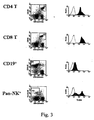
Fig. 3 depicts the expression of the PSGL-1 antigen on spleen CD4+ T cells, CD8+ T cells, CD19+ B cells, and NK cells. -
Fig. 4 depicts the expression of the PSGL-1 antigen on CD4+, CD8+, andCD4 +8+, andCD4 -8- thymocytes. -
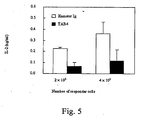
Fig. 5 depicts the levels of IL-2 produced in mixed lymphocyte culture using spleen cells isolated from TAB4 (or hamster Ig)-treated Balb/c mice as the responders and H2-mismatched C3H spleen cells as the stimulator. -
Fig. 6 depicts western blot analyses demonstrating that (A) proteins immunoprecipitated with the TAB4 antibody can be recognized by a commercially available anti-PSGL-1 antibody and (B) preclearing of T cell lysate with anti-PSGL-1 antibody can deplete the proteins recognized by the TAB4. -
Fig. 7 depicts the percentage of surviving grafts in C57BL/6 mice that received a skin graft from Balb/c mice and were treated with an anti-PSGL-1 antibody (closed diamond) or a control antibody (open square). -
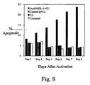
Fig. 8 depicts the time course of the percentage of apoptotic T cells following the treatment of activated human peripheral blood mononuclear cells with an anti-human PSGL-1 antibody. -
Fig. 9 depicts the incidence of diabetes in autoimmune non-obese diabetic (NOD) male mice that were treated with anti-PSGL-1 antibody (closed square) or a control antibody (open square). - The invention is directed to methods of modulating T cell activity by modulating the function of PSGL-1 molecules residing on the surface of a T cell. Engagement of PSGL-1 with compositions described herein can cause the depletion of T cells and/or induce T cells to undergo apoptosis. These compositions are therefore useful as therapeutic agents for controlling immune-related conditions such as autoimmune diseases, transplant rejection, allergic diseases, and/or T cell-derived cancers. The compositions are also useful in causing the depletion of T cells from any biological sample where the presence or activity of T cells is not desired.
- PSGL-1 is a cell surface adhesion molecule that is expressed on neutrophils, T and B-lymphocytes, NK cells, monocytes, dendritic cells, and primitive human CD34 hematopoietic progenitor cells. Through its ability to interact with selectins, PSGL-1 mediates the rolling of leukocytes on the endothelium and the extravasation of leukocytes into inflamed tissues. PSGL-1-mediated binding of T cells to E- and P-selectin, or migration, is differentially regulated. For instance, the appearance of CLA (cutaneous lymphocyte antigen) epitope is thought to be induced on T cells undergoing native to memory transition. Only activated
helper 1 but not helper 2 T cells express functional PSGL-1 and is capable of migration into the inflamed area of the skin. - PSGL-1 is a sialomucin that must be specifically sialylated, fucosylated, and sulfated to bind P-selectin. The PSGL-1 molecule exists in isoforms characterized by different degree of glycosylation and sulfation sites at their N-termini. Resting peripheral blood T and B cells, lymphoid cell lines, and in vitro activated peripheral blood T cells express similar level of PGSL-1. Yet, only activated T cells display a functional form of PSGL-1 and bind avidly to P-selectin. Such activation-dependent binding activity appears to be a result of differential post-translational modification, as suggested by elevated levels of alpha (1,3) fucosyltransferases activities in activated T cells. PSGL-1 isoforms also show differential affinity to L-selectin and E-selectin. For instance, human T cells exhibiting the CLA-positive isoform can tether and roll on both E- and P-selectin, while T cells expressing PSGL-1 without the CLA epitope only bind to P-selectin. Furthermore, binding of PSGL-1 to P-selectin is contingent upon the presence of the terminal decapeptide that contains three tyrosine residues for sulfation and one threonine residue for glycosylation.
- A PSGL-1 protein can be prepared by recombinant methods and/or by isolating a native PSGL-1 protein from biological material. A recombinant PSGL-1 protein can be produced in prokaryotic or eukaryotic cells, either in vitro or in vivo. Nucleic acids encoding PSGL-1 can be used for recombinant production of the protein (see, e.g., GenBank Accession NM_003006 for an example of a nucleic acid encoding a PSGL-1 polypeptide). Antibodies directed to PSGL-1 are also well known and can be used for purification of the antigen (see, e.g., Herron et al. (2000) ;
WO 00/25808 - For recombinant production of PSGL-1, the simultaneous expression of both PSGL-1 and its modifying alpha- (1,3) fucosyltransferase, Fuc-TVII, may be required for the functional expression of PSGL-1. In addition or alternatively, recombinant production of PSGL-1 may be accompanied by co-transfection with a nucleic acid encoding PACE for removing the propeptide and/or or a nucleic acid encoding tyrosine sulfotransferase.
- An anti-PSGL-1 antibody can be used to isolate and purify a PSGL-1 antigen from biological material. Any cell type expressing a PSGL-1 protein, e.g., T cells derived from an individual or a T cell line, can be used as a source of the protein. Once purified, the protein can be used in a variety of methods as described herein. For example, the purified PSGL-1 protein can be used in an in vitro screen of modulators of PSGL-1 function on T cells or as an immunogen to prepare antibodies directed against the protein.
- In addition, a PSGL-1 antigen can be purified using a selectin-Fc fusion, e.g., a P-selectin-Fc fusion.
- PSGL-1 polypeptides (or immunogenic fragments or analogs thereof) can be used to generate antibodies useful in the methods of the invention. As described above, PSGL-1 polypeptides or peptide fragments thereof can be produced by recombinant techniques or synthesized using solid phase synthesis methods. The recombinant PSGL-1 polypeptides or a peptide fragment thereof can be used as an immunogen to produce anti-PSGL-1 antibodies. In addition, an anti-PSGL-1 antibody, such as the TAB4 monoclonal antibody, can be used to purify a PSGL-1 polypeptide, e.g., a PSGL-1 polypeptide in its natural conformation, which can then be used as an immunogen to produce additional anti-PSGL-1 antibodies.
- An antibody of the invention can be a monoclonal, polyclonal, or engineered antibody that specifically binds to a PSGL-1 polypeptide. An antibody that "specifically binds" to a particular antigen, e.g., a PSGL-1 polypeptide, will not substantially recognize or bind to other molecules in a sample. Thus, the invention also features methods for identifying a test compound (e.g., an antibody) that binds to a polypeptide of the invention by contacting the polypeptide with a test compound and determining whether the polypeptide binds to the test compound (e.g., by direct detection of the binding, detection of a competitor molecule which disrupts binding of the test compound to the polypeptide, and/or detection of binding using an assay for apoptosis-inducing activity).
- In general, PSGL-1 polypeptides can be coupled to a carrier protein, such as KLH, mixed with an adjuvant, and injected into a host mammal. Antibodies produced in that animal can then be purified by peptide antigen affinity chromatography.
- In particular, various host animals can be immunized by injection with a PSGL-1 polypeptide or an antigenic fragment thereof. Commonly employed host animals include rabbits, mice, guinea pigs, and rats. Various adjuvants that can be used to increase the immunological response depend on the host species and include Freund's adjuvant (complete and incomplete), mineral gels such as aluminum hydroxide, surface active substances such as lysolecithin, pluronic polyols, polyanions, peptides, oil emulsions, keyhole limpet hemocyanin, and dinitrophenol. Potentially useful human adjuvants include BCG (bacille Calmette-Guerin) and Corynebacterium parvum. Polyclonal antibodies are heterogeneous populations of antibody molecules that are contained in the sera of the immunized animals.
- Antibodies within the invention therefore include polyclonal antibodies and, in addition, monoclonal antibodies, humanized or chimeric antibodies, single chain antibodies, Fab fragments, F(ab')2 fragments, and molecules produced using a Fab expression library.
- Monoclonal antibodies, which are homogeneous populations of antibodies to a particular antigen, can be prepared using the PSGL-1 polypeptides described above and standard hybridoma technology (see, for example, Kohler et al., Nature 256:495 [1975]; Kohler et al., Eur J Immunol 6:511 [1976]; Kohler et al., Eur J Immunol 6:292 [1976]; Hammerling et al., Monoclonal Antibodies and T Cell Hybridomas, Elsevier, N.Y. [1981]).
- In particular, monoclonal antibodies can be obtained by any technique that provides for the production of antibody molecules by continuous cell lines in culture such as described in Kohler et al., Nature 256:495 (1975), and
U.S. Pat. No. 4,376,110 ; the human B-cell hybridoma technique (Kosbor et al., Immunology Today 4:72 [1983]; Cole et al., Proc Natl Acad Sci USA 80:2026 [1983]), and the EBV-hybridoma technique (Cole et al., Monoclonal Antibodies and Cancer Therapy, Alan R. Liss, Inc., pp. 77-96 [1983]). Such antibodies can be of any immunoglobulin class including IgG, IgM, IgE, IgA, IgD and any subclass thereof. The hybridoma producing the mAb of this invention may be cultivated in vitro or in vivo. The ability to produce high titers of mAbs in vivo makes this a particularly useful method of production. - Once produced, polyclonal or monoclonal antibodies are tested for specific PSGL-1 recognition by Western blot or immunoprecipitation analysis by standard methods, for example, as described in Ausubel et al., supra. Antibodies that specifically recognize and bind to PSGL-1 are useful in the invention. Anti-PSGL-1 antibodies that bind to the PSGL-1 antigen on the surface of a T cell, e.g., a CD3+ cell, and induce the depletion and/or apoptosis of T cells in an individual are particularly useful.
The antibodies can be used, for example, as part of a therapeutic regimen (e.g., to reduce or eliminate an undesirable immune response, such as a T cell mediated immune response, associated with conditions such as autoimmune diseases, transplant rejection, allergic diseases, and T cell-derived cancers). Antibodies also can be used in a screening assay to measure the ability of a candidate compound to bind to PSGL-1. - In addition, techniques developed for the production of "chimeric antibodies" (Morrison et al., Proc Natl Acad Sci USA 81:6851 [1984]; Neuberger et al., Nature 312:604 [1984]; Takeda et al., Nature 314:452 [1984]) by splicing the genes from a mouse antibody molecule of appropriate antigen specificity together with genes from a human antibody molecule of appropriate biological activity can be used. A chimeric antibody is a molecule in which different portions are derived from different animal species, such as those having a variable region derived from a murine monoclonal antibody and a human immunoglobulin constant region.
- Alternatively, techniques described for the production of single chain antibodies (
U.S. Pat. Nos. 4,946,778 ,4,946,778 , and4,704,692 ) can be adapted to produce single chain antibodies against a PSGL-1 polypeptide, or a fragment thereof. Single chain antibodies are formed by linking the heavy and light chain fragments of the Fv region via an amino acid bridge, resulting in a single chain polypeptide. - Antibody fragments that recognize and bind to specific epitopes can be generated by known techniques. For example, such fragments include but are not limited to F(ab')2 fragments that can be produced by pepsin digestion of the antibody molecule, and Fab fragments that can be generated by reducing the disulfide bridges of F(ab')2 fragments. Alternatively, Fab expression libraries can be constructed (Huse et al., Science 246:1275 [1989]) to allow rapid and easy identification of monoclonal Fab fragments with the desired specificity.
- Antibodies can be humanized by methods known in the art. For example, monoclonal antibodies with a desired binding specificity can be commercially humanized (Scotgene, Scotland; Oxford Molecular, Palo Alto, Calif.). Fully human antibodies, such as those expressed in transgenic animals are also features of the invention (Green et al., Nature Genetics 7:13 [1994]; and
U.S. Pat. Nos. 5,545,806 and5,569,825 ). - The invention also encompasses methods for identifying compounds that interact with PSGL-1 (or a domain of PSGL-1) including, but not limited to, compounds that induce T cell depletion and/or T cell apoptosis upon binding to PSGL-1. Also included are compounds that modulate the interaction of PSGL-1 with transmembrane, extracellular, or iritracellular proteins that regulate PSGL-1 activity and compounds which modulate PSGL-1 activity.
- The compounds that may be screened in accordance with the invention include, but are not limited to peptides, antibodies and fragments thereof, and other organic compounds that bind to PSGL-1 and modulate a biological function mediated by PSGL-1, as described herein.
- Such compounds may include, but are not limited to, peptides such as, for example, soluble peptides, including but not limited to members of random peptide libraries; (Lam et al., Nature 354:82 [1991]; Houghten et al., Nature 354:84 [1991]), and combinatorial chemistry-derived molecular library made of D- and/or L configuration amino acids, phosphopeptides (including, but not limited to, members of random or partially degenerate, directed phosphopeptide libraries; Songyang et al., Cell 72:767 [1993]), antibodies (including, but not limited to, polyclonal, monoclonal, humanized, anti-idiotypic, chimeric or single chain antibodies, and Fab, F(ab')2 and Fab expression library fragments, and epitope-binding fragments thereof), and small organic or inorganic molecules.
- Other compounds which can be screened in accordance with the invention include but are not limited to small organic molecules that affect an activity of the PSGL-1 protein, as described herein.
- Computer modeling and searching technologies permit identification of compounds, or the improvement of already identified compounds, that can modulate PSGL-1 expression or activity. Having identified such a compound or composition, the active sites or regions are identified. Such active sites might typically be a binding site for a natural modulator of activity. The active site can be identified using methods known in the art including, for example, from the amino acid sequences of peptides, from the nucleotide sequences of nucleic acids, or from study of complexes of the relevant compound or composition with its natural ligand. In the latter case, chemical or X-ray crystallographic methods can be used to find the active site by finding where on the factor the modulator (or ligand) is found.
- Although described above with reference to design and generation of compounds which could alter binding, one could also screen libraries of known compounds, including natural products or synthetic chemicals, and biologically active materials, including proteins, for compounds which bind to a PSGL-1 protein and cause T cell depletion and/or induce T cell apoptosis.
- In vitro systems may be designed to identify compounds capable of interacting with PSGL-1 (or a domain of PSGL-1). Compounds identified may be useful, for example, in modulating T cell activity as described herein and thus may be useful for the treatment of conditions such as autoimmune diseases, transplant rejection, allergic diseases, and T cell-derived cancers.
- The principle of the assays used to identify compounds that bind to PSGL-1 involves preparing a reaction mixture of PSGL-1 (or a domain thereof) and the test compound under conditions and for a time sufficient to allow the two components to interact and bind, thus forming a complex which can be removed and/or detected in the reaction mixture. The PSGL-1 species used can vary depending upon the goal of the screening assay. In some situations it is preferable to employ a peptide corresponding to a domain of PSGL-1 fused to a heterologous protein or polypeptide that affords advantages in the assay system (e.g., labeling, isolation of the resulting complex, etc.) can be utilized.
- The screening assays can be conducted in a variety of ways. For example, one method to conduct such an assay involves anchoring PSGL-1 protein, polypeptide, peptide or fusion protein or the test substance onto a solid phase and detecting PSGL-1 /test compound complexes anchored on the solid phase at the end of the reaction. In one embodiment of such a method, the PSGL-1 reactant may be anchored onto a solid surface, and the test compound, which is not anchored, may be labeled, either directly or indirectly.
- In practice, microtiter plates may conveniently be utilized as the solid phase. The anchored component may be immobilized by non-covalent or covalent attachments. Non-covalent attachment may be accomplished by simply coating the solid surface with a solution of the protein and drying. Alternatively, an immobilized antibody, preferably a monoclonal antibody, specific for the protein to be immobilized may be used to anchor the protein to the solid surface. The surfaces may be prepared in advance and stored.
- In order to conduct the assay, the nonimmobilized component is added to the coated surface containing the anchored component. After the reaction is complete, unreacted components are removed (e.g., by washing) under conditions such that any complexes formed will remain immobilized on the solid surface. The detection of complexes anchored on the solid surface can be accomplished in a number of ways. Where the previously non-immobilized component is pre-labeled, the detection of label immobilized on the surface indicates that complexes were formed. Where the previously non-immobilized component is not pre-labeled, an indirect label can be used to detect complexes anchored on the surface; e.g., using a labeled antibody specific for the previously non-immobilized component (the antibody, in turn, may be directly labeled or indirectly labeled with a labeled anti-Ig antibody).
- Alternatively, a reaction can be conducted in a liquid phase, the reaction products separated from unreacted components, and complexes detected; e.g., using an immobilized antibody specific for PSGL-1 protein, polypeptide, peptide or fusion protein or the test compound to anchor any complexes formed in solution, and a labeled antibody specific for the other component of the possible complex to detect anchored complexes.
- Alternatively, cell-based assays can be used to identify compounds that interact with PSGL-1. To this end, cell lines that express PSGL-1, or cell lines that have been genetically engineered to express PSGL-1 can be used. Cell based assays are particularly useful for evaluating the functional effects of a compound identified by a screen described herein. For example, once a compound is identified based upon its ability to bind to a PSGL-1 protein, the compound can then be tested for its ability to, e.g., induce T cell apoptosis in vitro or in vivo or deplete T cells in vitro or in vivo.
- Given that an object of the present invention is to alter an immune response in an individual, a pharmaceutical composition containing, for example, antibodies, small molecules, or other compounds that specifically bind PSGL-1 polypeptides are also a feature of the invention. In a preferred example, the compound functions as an agonist of PSGL-1.
- Pharmaceutical compositions for use in accordance with the present invention can be formulated in a conventional manner using one or more physiologically acceptable carriers or excipients. Thus, the compounds and their physiologically acceptable salts and solvates may be formulated for administration by a variety of routes of administration.
- The compounds may be formulated for parenteral administration by injection, for example, by bolus injection or continuous infusion. Formulations for injection may be presented in unit dosage form, for example, in ampoules or in multi-dose containers, with an added preservative. The compositions may take such forms as suspensions, solutions, or emulsions in oily or aqueous vehicles, and may contain formulatory agents such as suspending, stabilizing and/or dispersing agents. Alternatively, the active ingredient can be in powder form for constitution with a suitable vehicle, for example, sterile pyrogen-free water, before use.
- Compounds such as those detailed in the screening assays described herein may be useful, for example, in modulating a biological function mediated by a PSGL-1 polypeptide and/or for the treatment of disorders associated an excessive or unwanted immune response, e.g., a T cell-mediated immune response. These compounds include, but are not limited to peptides, antibodies and fragments thereof, and other organic compounds that bind to PSGL-1 on the surface of a T cell and induce a signal transduction pathway that results in the death of the T cell. The methods of the invention optionally include the addition of a cross-linking agent that induces the cross-linking of PSGL-1 on the surface of a cell. The compounds described herein can be used in any instance wherein the depletion or elimination of T cell activity is desired. Particularly useful conditions that can be treated with the compounds of the invention include autoimmune diseases, transplant rejection, allergic diseases, and T cell-derived cancers.
- Examples of conditions that can be treated with the anti-PSGL-1 compounds described herein include, but are not limited to, diabetes mellitus, arthritis (including rheumatoid arthritis, juvenile rheumatoid arthritis, osteoarthritis, and psoriatic arthritis), multiple sclerosis, encephalomyelitis, myasthenia gravis, systemic lupus erythematosis, autoimmune thyroiditis, dermatitis (including atopic dermatitis and eczematous dermatitis), psoriasis, Sjögren's Syndrome, Crohn's disease, aphthous ulcer, iritis, conjunctivitis, keratoconjunctivitis, type I diabetes, inflammatory bowel diseases, ulcerative colitis, asthma, allergic asthma, cutaneous lupus erythematosus, scleroderma, vaginitis, proctitis, drug eruptions, leprosy reversal reactions, erythema nodosum leprosum, autoimmune uveitis, allergic encephalomyelitis, acute necrotizing hemorrhagic encephalopathy, idiopathic bilateral progressive sensorineural hearing loss, aplastic anemia, pure red cell anemia, idiopathic thrombocytopenia, polychondritis, Wegener's granulomatosis, chronic active hepatitis, Stevens-Johnson syndrome, idiopathic sprue, lichen planus, Graves' disease, sarcoidosis, primary biliary cirrhosis, uveitis posterior, interstitial lung fibrosis, graft-versus-host disease, cases of transplantation (including transplantation using allogeneic or xenogeneic tissues) such as bone marrow transplantation, liver transplantation, or the transplantation of any organ or tissue, allergies such as atopic allergy, AIDS, and T cell neoplasms such as leukemias and/or lymphomas.
- The methods of the invention can be used to deplete T cells from a cell population, either in vitro or in vivo. For example, a biological sample derived from an individual can be depleted of T cells in vitro by contacting the sample with an anti-PSGL-1 compound described herein, optionally together with a cross-linking agent. This method can be useful, e.g., by allowing for the enrichment of non-T cells in a cell population as well as by reducing or eliminating T cell activity from a cell population.
- The following are examples of the practice of the invention.
- A TAIP-specific monoclonal antibody was generated by applying the well known cell fusion methods of Kohler and Milstein ((1976) European Journal of Immunology 6:511-519) to produce a hybridoma secreting desired antibodies. Antibody-producing cells from a hamster injected with Concanavalin A (Con A)-activated Balb/c spleen T cells were fused with a myeloma cell line to form an antibody secreting hybridoma. The two populations of cells were fused with polyethylene glycol, and the resulting antibody producing cells were cloned and propagated by standard tissue culture methods. One hybridoma generated according to these methods secreted a monoclonal antibody, designated TAB4, that was able to induce T cell apoptosis in vitro and deplete T cells in vivo. The protein recognized by TAB4 was designated T cell apoptosis inducing protein (TAIP).
- C57BL/6J (B6) and BALB/c mice were purchased from the Jackson lab (Bar Harbor, ME). Syrian hamsters were purchased from the Animal Core Facility, National Taiwan University Medical College.
- Concentrated culture supernatant of the TAB4 hybridoma was spun at 20,000 x g for 10 minutes and the supernatant was diluted at a 1:1 ratio with the binding buffer (0.1 M sodium acetate, pH 5.0). A protein G column (approximately 1 ml bed volume) was washed three times with 3-5 ml of the binding buffer. The cleared culture supernatant was loaded to the protein G column and the flow-through was collected and reloaded to the column. The column was washed with 6-10 ml of the binding buffer and the bound antibody was eluted from the column with 5 ml of the elution buffer (0.1 M glycine-HCl, pH 2.8). Each fraction contained 1 ml of the eluted antibody and the eluted fraction was adjusted to neutral pH by mixing each 1, ml fraction with 50 microliters of 1 M Tris-HCl, pH 7.5. Fractions containing the antibody were pooled and dialyzed against 2 liters of PBS, pH 7.4 three times at three hours for each dialysis. Protein concentration in the antibody samples were determined with the procedure described by Bradford using the Bio-Rad Protein Assay (BIO-RAD, Hercules, CA).
- Mouse spleen was immersed in 8 ml of Hank's balanced salt solution (HBSS), gently minced with a sterile cover slip, transferred to a 15 ml centrifuge tube (Costar), and spun at 200 x g for 5 minutes. The supernatant was discarded and the cell pellet was resuspended in the residual buffer by gently tapping the wall. The contaminating red blood cells (RBC) were lysed by the addition of 1 ml of RBC lysis buffer (0.6 M NH4Cl, 0.17 M Tris-base, pH 7.65), followed by a 2 min incubation at room temperature and rapid quenching with 9 ml of HBSS. The cells were pelleted at 200 x g for 5 minutes, washed twice and resuspended in RPMI medium. The concentration and viability of cells in the mixture were determined with a hemocytometer (Cambridge Scientific Inc.) and Trypan blue exclusion.
- The spleen cells were adjusted to a final concentration of 3
X 106/ml with RPMI medium and Concanavalin A was added to a final concentration of 2 micrograms/ml to activate the T cells. The cell suspension was transferred to a 6-well culture plate (5 ml/well) or a 10-cm culture dish (10 ml/dish) and incubated at 37°C, 5% CO2 for 48 hours before harvesting. The activated spleen cells, including activated T cells, were resuspended in 5 ml of HBSS and carefully overlaid on top of a 5 ml 55% cushion of Percoll solution in a centrifuge tube. Care was taken not to disturb the separated layers. The cells were spun at 1,900 x g for 13 minutes at 25°C without the brake. The enriched T cells were collected from the interface of the two layers, washed twice with HBSS, and were ready for experiments. - Activated T cells (see Example 2) were resuspended to a final concentration of 5
X 105 cells/ml in RPMI medium containing 5 ng/ml of IL-2, and treated with control Ig, TAB4, or anti-CD3 according to the conditions shown in Table 1.TABLE 1 Experiment groups Treatment* Negative control 3 ug/ ml hamster Ig 5 ng/ml IL-2 3 ug/ml cross-linker antibody (anti-hamster Ig) TAB4 3 ug/ml TUB4 hamster mAb 5 ng/ml IL-2 3 ug/ml cross-linker antibody (anti-hamster Ig) Positive control 1 ug/ ml anti-CD3 mAb 5 ng/ml IL-2 1 ug/ml cross-linker antibody (anti-mouse Ig) *: Final concentration of the designated reagents in the medium. - After an incubation period of 18-24 hours, the extent of apoptosis in each culture was determined using the 7-AAD apoptosis assay. The treated cells were transferred to FACS tubes (Falcon), washed twice with ice-cold FACS solution (1% fetal bovine serum, 0.05% sodium azide in PBS), pelleted at 200 x g at 4°C. The cells were resuspended in ice-cold FACS solution to a final concentration of 1-2
X 107 cells/ml. For staining, 0.1 ml of the resuspended cells were mixed with 7-AAD to a final concentration of 2 ug/ml and then incubated at 4°C in the dark for 20 minutes. Finally, the stained cells were washed twice with ice-cold FACS solution, resuspended in 0.5 ml of FACS solution and analyzed with BD LSR flow cytometer (Becton Dickinson). -
Fig. 1 depicts the results of a representative time-course experiment that investigated when activated T cells acquire sensitivity to TAB4 (anti-TAIP)-mediated apoptotic signals. Mouse splenocytes were activated with Con-A and maintained in IL-2 containing medium. Activated T cells were harvested, resuspended, and challenged with TAB4 monoclonal antibody or control hamster IgG in the presence of anti-hamster IgG antibody as cross-linker. The ability of TAEP cross-linking to induce low level (6.5%) of apoptotic cell death was evident on day one. However, the extent of TAB4-induced apoptosis increased from 17% onday 2, peaked at 52% onday 4, and declined to 44% onday 6. The control hamster IgG did not induce specific apoptotic T cell death, as compared with the cultures that received only IL-2. Anti-CD3 (as positive control) induced apoptosis in 38% of T cell after 48 hours of activation (data not shown). - Cells were washed twice with ice-cold FACS solution (1% fetal bovine serum, 0.05% sodium azide in PBS) and spun at 200 x g at 4°C in a FACS tube (Falcon). The cells were resuspended in ice-cold FACS solution to a final concentration of 1 x 10⁷ cells/ml and a 0.1 ml aliquot of the resuspended cells in a FACS tube (Falcon) was used for each assay. For surface staining, the TAB4 monoclonal antibody or a control hamster Ig at a final concentration of 2 ug/ml were added to the cells and the mixtures were incubated at 4°C for 30 minutes in the dark. The cells were washed once with ice-cold FACS and then stained with: (1) for spleen cells, cychrome-conjugated anti-CD3 antibody (2 ug/ml), FITC-conjugated anti-hamster Ig, and PE-conjugated anti-CD8/CD4/CD19/CD11b/pan-NK/I-A/I-E/Mac-3 antibody (2 ug/ml) in 100 ul of ice-cold FACS solution; and (2) for thymus cells, FITC-conjugated anti-hamster Ig, PE-conjugated anti-CD8, and cychrome-conjugated anti-CD4 antibodies (2 ug/ml) in 100 ul of ice-cold FACS solution. The reaction was performed at 4°C for 30 minutes in the dark. Finally, the stained cells were washed twice with ice-cold FACS solution, resuspended in 1 ml of FACS solution and analyzed with BD LSR flow cytometer (Becton Dickinson).
-
Figs. 3 and4 demonstrate by FACS analysis the distribution of TAIP antigen on the various subpopulations of splenocytes and thymocytes. As shown inFig. 3 , CD19+ B cells expressed low but detectable amounts of TAIP proteins on the surface. Significantly higher amounts of TAIP proteins were detected on CD3+ T cells and a fraction ofNK cells. Most of the CD4+, CD8+, andCD4 +8+ thymus T cells expressed significant amounts of TAIP proteins. In contrast, the TAIP proteins were expressed only on a small population ofCD4 -8- thymus T cells (Fig. 4 ). - Tissues from B6 and BALB/c mice, including brain, thymus, heart, lung, liver, stomach, kidney, spleen, and skin, were collected, fixed in 10% formaldehyde overnight at room temperature, and embedded in paraffin blocks. Tissue sections, at a 4 um thickness, were prepared from the paraffin block with Leica RM2135 microtome, spread in 45°C water, and laid on a coated slide. The slides were dried in 37°C and were ready for subsequent experiments.
- Slides containing the tissue paraffin sections were dewaxed and dried through a xylenes-100% ethanol series according to standard protocol and were finally kept in 100% ethanol. The sections were rehydrated through a 100% ethanol-90% ethanol-85% ethanol-70% ethanol-PBS serial incubation according to standard protocol to a final PBS solution. The following reactions were all performed in a humidified box. Non-specific binding were blocked by incubating the tissue sections in blocking buffer (1% normal goat serum) for 1 hour at room temperature (or 4°C overnight). The blocking buffer was removed and TAB4 or normal hamster Ig (1:200 dilution) was added to the sections and incubation continued for another hour at room temperature (or 4°C overnight). The sections were washed twice in PBS, for 5 minutes each, to remove the primary antibody, reacted with 1:250 diluted alkaline phosphatase-conjugated goat anti-hamster Ig, and incubated at room temperature for 1 hour. The sections were again washed twice with PBS, 5 minutes each, to remove the antibody-enzyme conjugate and the color reaction was developed with BCIP/NBT substrate solution at room temperature for 30 minutes in the dark. The sections were washed again with PBS to remove excess enzyme substrate, dehydrated through the PBS-ethanol-xylenes series, and mounted for microscopy.
- The results indicated that the TAIP proteins expression were detected only in bone marrow derived-tissues but not on the rest of the tissues tested.
- 5 x 10⁷ RL♂1 or NIH-3T3 cells were surface biotinylated in 1 ml of PBS containing 0.5 mg/ml Sulfo-NHS-biotin (Pierce) for 30 minutes on ice. The reaction was terminated by incubating the cells with 0.5 ml of Dulbecco's modified Eagle's medium (Life Technologies, Inc.) for 10 minutes on ice. Cells were washed with 1 ml of Dulbecco's modified Eagle's medium once and with 1 ml of phosphate-buffered saline twice.
- Labeled cells were lysed at a density of 5.0 x 10⁷ cells/ml in cold lysis buffer (1% Triton X-100, 20 mM Tris-HCl, pH 8.0, 160 mM NaCl, 1 mM CaCl2) containing complete protease inhibitor cocktail (Roche) for 15 minutes, and insoluble material was pelleted at 10,000 x g for 10 minutes; these and all subsequent steps were performed at 4°C. For immunoprecipitation, the lysate was preincubated for 30 minutes with 50 µl of packed protein G-Sepharose (Amersham Pharmacia Biotech) to remove non-specifically binding proteins. Beads were pelleted, and aliquots of the supernatant (routinely corresponding to 5.0 × 10⁷ cells) were incubated with 20 µl of protein G-Sepharose preloaded with 10 µg of mAb TAB4 or IgG from normal hamster serum. After incubation for 4 h at 4°C, the resin was washed four times with washing buffer (0.05% Triton X-100, 50 mM Tris-HCl, pH 8.5, 400 mM NaCl, 1 mM CaCl2, 1 mg/ml ovalbumin), twice with a similar washing buffer, containing 250 mM instead of 400 mM NaCl. Proteins specifically bound to the TAB4 were eluted with 50 µl of 1xSDS sample buffer. Eluted proteins were separated by 8 % SDS-PAGE and transferred to nitrocellulose membrane (Millipore). Filters were analyzed for biotinylated proteins with peroxidase-conjugated Avidin (PharMingen) and developed with the Chemiluminescence reagent (NEN™ Life Science Products).
- As shown in
Fig. 2 , a biotinylated surface protein with a molecular weight of approximately 120-kD was identified by TAB4 in RL♂1 cells (TAIP+ T cells), but not in 3T3 cells (TAIP- cells). In contrast, protein G sepharose coated with hamster normal serum could not retrieve this 120-kDa protein. These results suggest that this 120-kDa protein is the antigen recognized by monoclonal antibody TAB4 on the cell surface of T cells. - To examine the effects of TAB4 on populations of T cells and other cells in vivo, mice were injected with 300 ug of TAB4 or control hamster Ig intraperitoneally and, on
day 4, splenocytes, thymocytes, and peripheral blood mononuclear cells were harvested for the total cell count and for the analyses of cell surface markers by FACS. - For FACS assays, the cells were fixed with 2% paraformaldehyde at 4°C for 20 minutes, washed twice, and resuspended in ice-cold FACS solution to a final concentration of 1 x 10⁷ cells/ml. A 100 ul aliquot of the resuspended cells in a FACS tube (Falcon) was used for each assay. TAB4 or control hamster Ig at a final concentration of 2 ug/ml were added to the cells and the mixtures were incubated at 4°C for 30 minutes in the dark. The cells were washed once with ice-cold FACS and reacted with: (1) for spleen cells, cychrome-conjugated anti-CD3 antibody (2 ug/ml), FITC-conjugated anti-hamster Ig and PE-conjugated anti-CD8/CD4/CD19/CDl 1b/pan-NK/I-A/I-E/Mac-3 antibody (2 ug/ml) in 100 ul of ice-cold FACS solution; and (2) for thymus cells, FITC-conjugated anti-hamster Ig, PE-conjugated anti-CD8, and cychrome-conjugated anti-CD4 antibodies (2 ug/ml) in 100 ul of ice-cold FACS solution. The reaction was performed at 4°C for 30 minutes in the dark. Finally, the stained cells were washed twice with ice-cold FACS solution, resuspended in 1,000 ul of FACS solution and analyzed with BD LSR flow cytometer (Becton Dickinson).
- Four days after the injection, the percentages of CD3+ T cells in peripheral blood leukocytes (PBL) decreased from 36.7% in control mice to 4.1% in TAB4-treated mice (Table 2). TAB4 treatment caused a slight reduction in the total number of splenocytes. However, in TAB4 treated mice, there was a 62% decrease in the number of CD3+ T cells, a 50% decrease in the number of NK cells, and a slight increase in the total number of CD19+ B cells. The total number of thymocytes recovered from TAB4 treated mice was only 48% of the level seen in control (52% reduced). Moreover, except for CD4+ T cells, all other CD8+, CD4+CD8+, and CD4-CD8- T cells were reduced, with CD4+CD8+ subpopulation being the most profoundly affected (64.7% reduction).
TABLE 2 Spleen x 106 No Treatment Normal Hamster Ig TAB4-treated Depletion (%) Total Splenocytes 123 93.3 105 14.6 CD3+ T cells 32.8 28.4 12.4 62.2 CD3- CD 19+ 72.2 53.4 72.9 - 0.8 CD3- NK+ 3.6 2.4 1.80 50 Peripheral Blood Leukocytes No Treatment Normal Hamster Ig TAB4-treated Depletion (%) CD3+ T cells 36.7% 36% 4.1% 88.8% Thymus x 106 No Treatment Normal Hamster Ig TAB4-treated Depletion (%) Total Thymocytes 94 229 45 52.1 CD4+ 9.3 28.4 10.9 -16.6 CD8+ 5.2 7.7 3.6 30.3 CD4+ CD8+ 73.8 182 26 64.7 CD4- CD8- 5.6 10.5 4.5 19.3 (representative data from three experiments) - Balb/c mice. (H-2d) were intraperitoneally injected with 300 micrograms of TAB4 or control hamster Ig. Splenocytes were isolated 7 days after injection, and used as responders in culture with mitomycin C-treated C3H (H-2k) splenocytes (as stimulators). Three days later, the culture supernatants were harvested and the IL-2 content was measured by ELISA set (PharMingen). As shown in
Fig. 5 , the IL-2 production was suppressed in responder cells derived from TAB4-treated mice as compared with that of control mice. The plasma levels of IL-2 and TNF-α were also analyzed and no significant difference was noted in the levels of IL-2 (or TNF-α) in the sera of the control and the TAB4 treated mice. Since production of IL-2 is central to the activity of T cells, the results show that a TAIP-specific antibody, such as TAB4, can be used in vivo to manipulate T cells and control unwanted T cell-mediated immune responses such as those associated with autoimmune diseases and transplantation rejection. - Mice (obtained from Jackson Laboratory) at 8 to 12 weeks of age were anesthetized with Acepromazin maleate (Fermenta Animal Health Co., Kansas City, MO). Prior to skin grafting, non-thymectomized recipient C57BL/6 mice (H-2b) were injected intraperitoneally with 500 ug of TAB4 or isotype control antibodies seven days before skin transplant surgery. Seven days later, a lateral flank of skin from fully allogeneic mismatched Balb/cj mice (H-2d) was grafted on the lateral flank of the antibody pre-treated C57BL/6 mice. Seven days post transplantation, the mice were again injected with 500 ug of TAB4 or isotype control antibody. The mice were monitored every day after graft transplantation. The grafts were considered rejected when 50% donor skin was necrotic. The percent of graft survival is shown in
Fig. 7 (n=8). The data show that TAB4 antibody treatments prolonged the survival of the allogeneic skin grafts. - P-selectin glycoprotein ligand-1 (PSGL-1), also named CD162, is the main P-selectin ligand expressed on leukocytes, including T cells (Sako et al. (1993) Cell 75:1179; Vachino et al. (1995) J. Biol. Chem. 270:21966; Veldman et al. (1995) J. Biol. Chem. 270:16470). Biochemical characteristics of TAIP, such as its molecular weight and its tendency for dimerization suggested the possibility that TAIP may be analogous to PSGL-1. To investigate the relationship between these two antigens, the following were tested: 1) whether the antigen precipitated by TAB4 can be recognized by a commercially-available anti-PSGL-1 antibody; and 2) whether an anti-PSGL-1 antibody can deplete TAIP from the cell lysate.
- RL♂1 T cells were lysed at a density of 1.0 × 10⁸ cells/ml in lysis buffer (1% Triton X-100, 20 mM Tris-HCl, pH 8.0, 160 mM NaCl, 1 mM CaCl2) containing complete protease inhibitor cocktail for 1 hour, and insoluble material was pelleted at 10,000, × g for 10 minutes. These and all subsequent steps were performed at 4°C. The lysate corresponding to 5.0 × 10⁷ cells was incubated with 20 ul of protein G-Sepharose reloaded with 10 ug of anti-PSGL-1 mAb (clone 2P-1, PharMingen, San Diego, CA), anti-TAIP mAb, TAB4, or IgG from normal hamster serum. After incubation for 4 hours at 4°C, the beads were washed five times with washing buffer (0.05% Triton X-100, 50 mM Tris-HCl, pH 8.5, 400 mM NaCl, 1 mM CaCl2, 1 mg/ml ovalbumin), and twice with a similar washing buffer, containing 250 mM instead of 400 mM NaCl. Bound proteins were eluted with 40 ul of 1×SDS sample buffer. Eluted proteins were separated by 6 % SDS-PAGE and transferred to a nitrocellulose membrane. The membranes were immunoblotted with anti-PSGL-1 mAb, and revealed by peroxidase-conjugated goat anti-rat IgG (H+L) followed by chemiluminescence (Renaissance, NEN).
- Surface biotinylated RL♂1 T cells were lysed at a density of 1.0 × 10⁸ cells/ml in lysis buffer. The cell extract was incubated with 20 ug of antibody bound to 40 ul of protein G-Sepharose at 4°C overnight. Depletions were done with anti-PSGL-1 mAb (2PH1) or control rat IgG, with TAB4 or control normal hamster serum. Depleted lysates were further subjected to immunoprecipitation with TAB4 or anti-PSGL-1 mAb, respectively. Immunoprecipitates were separated on 6% SDS-polyacrylamide gel and detected by fluorography. As shown in
Fig. 6 , anti-PSGL-1 antibody can deplete TAIP protein from T cell lysates. In addition, proteins immunoprecipitated with anti-TAIP antibody (TAB4) can be recognized by anti-PSGL-1 antibody by western analysis. - To determine the role played by PSGL-1 in the apoptosis of human T cells, time-course experiments were carried out to investigate when activated human T cells acquire sensitivity toward PSGL-1-mediated apoptotic signals. Human T cells were stimulated with phytohemagglutinin (PHA) mitogen and further expanded in IL-2-containing medium. Activated T cells were harvested and then challenged with anti-PSGL-1 in the presence of IL-2 and cross-linking antibodies.
- Human peripheral blood was taken from healthy adults, heprinized, and enriched for peripheral blood mononuclear cells (PBMC) based on differential density using Ficoll-Paque Plus (Pharmacia Biotech). The PBMC were activated with 1% PHA (Life Technologies, GibcoBRL) for 48 hours and subsequently maintained in recombinant human IL-2 (5 ng/ml) through the assay period. To assess the apoptosis-inducing ability an anti-human PSGL-1 antibody, the activated cells were treated with: (1) 1 ug/ml of the anti-PSGL-1 antibody clone KPL-1 (BD PharMingen) plus cross-linker rabbit anti-mouse Ig (0.5 ug /ml) (Jackson ImmunoResearch Laboratories); (2) isotype control purified mouse Ig plus cross-linker rabbit anti-mouse Ig; or (3) cross-linker rabbit anti-mouse Ig alone. After six hours of treatment, the percentage of early apoptotic cells was determined by FACS, staining with anti-Annexin V (BD PharMingen) and PI (Sigma).
- As shown in
Fig. 8 , signaling triggered by PSGL-1 using an anti-PSGL-1 antibody plus the crosslinker triggered significant level of apoptosis in PHA-activated human PBMC (mainly T cells). The percentage of apoptotic cells increased from 8.5% onday 3 to 24% onday 8 in anti-PSGL-1 treated cultures. Neither isotype-matched control, nor the cross-linking antibodies alone, had any effect on these cells. - Non-obese diabetic (NOD) mice, a well-accepted autoimmune diabetes animal, were bred under standard conditions. Spontaneous diabetes developed in the NOD mice at the age of about 20 weeks. In the experimental group, the mice received three doses of anti-PSGL-1 antibody (TAB4) intraperitoneally at 300 µg per mouse at age of 14, 15 and 17 weeks. Two additional injections with the same dose were given at the ages of 24 and 26 weeks. The control group was given hamster Ig at the same dose. Mice were monitored for glucose uria by Medi-Test Glucose strips (Macherey-Nagel, Germany) twice every week after the age of 15 weeks. Non-fasting urine glucose levels over 300 mg/dl for two consecutive measurements were considered diabetic.
- As shown in
Fig. 9 , TAB4 (anti-PSGL-1) antibody treatment yielded significant protection as compared with control antibody treatment. Thus an anti-PSGL-1 antibody treatment can dampen the activity of autoimmune T cells and delay the onset of type I-diabetes.
Claims (17)
- An antibody or antigen binding fragment thereof that specifically binds to P-Selectin Glycoprotein Ligand -1 (PSGL-1) on the surface of an activated T cell and induces death of the activated T cell for use as a medicament.
- Use of an antibody or antigen binding fragment thereof that specifically binds to P-Selectin Glycoprotein Ligand -1 (PSGL-1) on the surface of an activated T cell and induces death of the activated T cell in the manufacture of a medicament for the treatment or prevention of a T cell-mediated autoimmune disease.
- An antibody of claim 1 or the use of claim 2, wherein the antibody is a monoclonal antibody.
- The use of claim 3, wherein the medicament further comprises an agent that binds to the monoclonal antibody and induces cross-linking of a plurality of PSGL-1 antigens on the surface of the activated T cell for administration in combination.
- The use of claim 2, wherein the T cell-mediated autoimmune disease is autoimmune diabetes.
- The use of claim 2, wherein the T cell-mediated autoimmune disease is rheumatoid arthritis.
- The use of claim 2, wherein the T cell-mediated autoimmune disease is psoriasis.
- The use of claim 2, wherein the T cell-mediated autoimmune disease is multiple sclerosis.
- Use, of an antibody or antigen binding fragment thereof that specifically binds to P-Selectin Glycoprotein Ligand -1 (PSGL-1) on the surface of an activated T cell and induces death of the activated T cell in the manufacture of a medicament for the treatment or prevention of allogeneic or xenogeneic transplant rejection.
- Use of an antibody or antigen binding fragment thereof that specifically binds to P-Selectin Glycoprotein Ligand -1 (PSGL-1) on the surface of an activated T cell and induces death of the activated T cell in the manufacture of a medicament for the treatment or prevention of graft-versus-host-disease.
- The antibody of claim 1 or the use of claim 2, wherein the activated T cell is a CD4+ T cell.
- The antibody of claim 1 or the use of claim 2, wherein the activated T cell is a CD8+ T cell.
- A method of screening in vitro for an antibody or antigen binding fragment thereof that specifically binds to PSGL-1 that induces death of an activated T cell, the method comprising:providing an activated T cell expressing PSGL-1 on the surface of the activated T cell;contacting the activated T cell in vitro with an antibody or antigen binding fragment thereof that specifically binds to PSGL-1; andmeasuring the viability of the activated T cell after contacting the activated T cell with the antibody or antigen binding fragment thereof to thereby determine if the antibody or antigen binding fragment thereof is an anti-PSGL-1 antibody that induces death of an activated T cell.
- The method of claim 13, wherein the antibody is a monoclonal antibody.
- The method of claim 14, further comprising contacting the monoclonal antibody with an antibody that binds to the monoclonal antibody and induces the cross-linking of a plurality of PSGL-1 antigens on the surface of the activated T cell.
- The method of claim 13, wherein the activated T cell is a CD4+ T cell.
- The method of claim 13, wherein the activated T cell is a CD8+ T cell.
Priority Applications (3)
| Application Number | Priority Date | Filing Date | Title |
|---|---|---|---|
| DK08012718.6T DK1985634T3 (en) | 2001-08-03 | 2002-03-13 | Antibody-binding p-selectin glycoprotein ligand 1 (PSGL-1) for use in the treatment of an allergic disease |
| EP08012718A EP1985634B1 (en) | 2001-08-03 | 2002-03-13 | Antibody binding p-selectin glycoprotein ligand 1 (PSGL-1) for use in treating allergic disease |
| CY20091100020T CY1108701T1 (en) | 2001-08-03 | 2009-01-13 | R-SELECTIN Glycoprotein 1 Regulators. |
Applications Claiming Priority (5)
| Application Number | Priority Date | Filing Date | Title |
|---|---|---|---|
| US31019601P | 2001-08-03 | 2001-08-03 | |
| US310196P | 2001-08-03 | ||
| US51497 | 2002-01-18 | ||
| US10/051,497 US7744888B2 (en) | 2001-08-03 | 2002-01-18 | Methods of modulating T cell or natural killer cell activity with anti-P-selectin glycoprotein ligand 1 antibodies |
| PCT/US2002/007498 WO2003013603A1 (en) | 2001-08-03 | 2002-03-13 | Modulators of p-selectin glycoprotein ligand 1 |
Related Child Applications (1)
| Application Number | Title | Priority Date | Filing Date |
|---|---|---|---|
| EP08012718A Division EP1985634B1 (en) | 2001-08-03 | 2002-03-13 | Antibody binding p-selectin glycoprotein ligand 1 (PSGL-1) for use in treating allergic disease |
Publications (3)
| Publication Number | Publication Date |
|---|---|
| EP1411982A1 EP1411982A1 (en) | 2004-04-28 |
| EP1411982A4 EP1411982A4 (en) | 2004-10-06 |
| EP1411982B1 true EP1411982B1 (en) | 2008-10-15 |
Family
ID=26729479
Family Applications (2)
| Application Number | Title | Priority Date | Filing Date |
|---|---|---|---|
| EP02733839A Expired - Lifetime EP1411982B1 (en) | 2001-08-03 | 2002-03-13 | Modulators of p-selectin glycoprotein ligand 1 |
| EP08012718A Expired - Lifetime EP1985634B1 (en) | 2001-08-03 | 2002-03-13 | Antibody binding p-selectin glycoprotein ligand 1 (PSGL-1) for use in treating allergic disease |
Family Applications After (1)
| Application Number | Title | Priority Date | Filing Date |
|---|---|---|---|
| EP08012718A Expired - Lifetime EP1985634B1 (en) | 2001-08-03 | 2002-03-13 | Antibody binding p-selectin glycoprotein ligand 1 (PSGL-1) for use in treating allergic disease |
Country Status (20)
| Country | Link |
|---|---|
| US (3) | US7744888B2 (en) |
| EP (2) | EP1411982B1 (en) |
| JP (3) | JP4815101B2 (en) |
| KR (1) | KR100850731B1 (en) |
| CN (2) | CN1304051C (en) |
| AT (2) | ATE531734T1 (en) |
| AU (1) | AU2002305041B2 (en) |
| CA (1) | CA2428822C (en) |
| CY (2) | CY1108701T1 (en) |
| DE (1) | DE60229394D1 (en) |
| DK (2) | DK1985634T3 (en) |
| ES (2) | ES2316569T3 (en) |
| HK (2) | HK1065489A1 (en) |
| IL (4) | IL160168A0 (en) |
| MX (1) | MXPA04001047A (en) |
| NO (2) | NO333456B1 (en) |
| NZ (1) | NZ531199A (en) |
| PT (2) | PT1985634E (en) |
| TW (1) | TWI327070B (en) |
| WO (1) | WO2003013603A1 (en) |
Families Citing this family (20)
| Publication number | Priority date | Publication date | Assignee | Title |
|---|---|---|---|---|
| US20040116333A1 (en) * | 2001-08-03 | 2004-06-17 | Rong-Hwa Lin | Modulators of P-selectin glycoprotein ligand 1 |
| US7744888B2 (en) | 2001-08-03 | 2010-06-29 | Abgenomics Cooperatief U.A. | Methods of modulating T cell or natural killer cell activity with anti-P-selectin glycoprotein ligand 1 antibodies |
| AU2004273807B2 (en) * | 2003-09-15 | 2011-06-09 | Abgenomics Cooperatief U.A. | Modulators of P-Selectin Glycoprotein Ligand 1 |
| US7459523B2 (en) * | 2003-11-12 | 2008-12-02 | Wisconsin Alumni Research Foundation | Equine P-selectin glycoprotein ligand-1 and uses thereof |
| AU2012201999C1 (en) * | 2004-05-10 | 2016-05-12 | Abgenomics Cooperatief U.A. | Antibodies |
| MY148646A (en) * | 2004-05-10 | 2013-05-15 | Abgenomics Cooperatief Ua | Anti-psgl-1 antibodies |
| CN105777867A (en) * | 2004-05-11 | 2016-07-20 | 艾比吉诺米克斯合作公司 | T-cell death-inducing epitopes |
| WO2007033959A2 (en) * | 2005-09-19 | 2007-03-29 | Vib Vzw | Use of p-selectin glycoprotein ligand 1 in modulation of hematogenous metastasis of lymphomas |
| WO2007067984A2 (en) * | 2005-12-09 | 2007-06-14 | Wyeth | Neutralizing antibodies against primate psgl-1 and uses therefor |
| WO2009140623A2 (en) * | 2008-05-15 | 2009-11-19 | Selexys Pharmaceuticals Corporation | Anti-psgl-1 antibodies and methods of identification and use |
| ES2387156B1 (en) * | 2010-12-01 | 2013-07-04 | Universidad Autónoma de Madrid | USE OF PSGL-1 FOR THE TREATMENT OF INFLAMMATORY AND AUTOIMMUNE DISEASES |
| JP6180408B2 (en) | 2011-06-13 | 2017-08-16 | アブゲノミクス コーペラティフ ユー.エー. | Anti-PSGL-1 antibody and use thereof |
| CA2953706A1 (en) * | 2014-07-08 | 2016-01-14 | Sanford-Burnham Medical Research Institute | Psgl-1 modulators and uses thereof |
| EP3400239B1 (en) | 2016-01-08 | 2021-06-02 | AltruBio Inc. | Tetravalent anti-psgl-1 antibodies and uses thereof |
| US20170241415A1 (en) * | 2016-02-19 | 2017-08-24 | Muzan, Inc. | Sanitary check valve |
| KR102630011B1 (en) | 2017-01-11 | 2024-01-26 | 브리스톨-마이어스 스큅 컴퍼니 | PSGL-1 antagonists and their uses |
| JP7211961B2 (en) | 2017-03-14 | 2023-01-24 | ファイヴ プライム セラピューティクス インク | Antibody that binds to VISTA at acidic pH |
| AU2019306165A1 (en) | 2018-07-20 | 2021-02-25 | Pierre Fabre Medicament | Receptor for vista |
| EP3963333A4 (en) * | 2019-05-03 | 2023-07-19 | Versitech Limited | Method for peritoneal metastatic cell detection and isolation thereof |
| WO2020247371A1 (en) * | 2019-06-04 | 2020-12-10 | Verseau Therapeutics, Inc. | Anti-psgl-1 compositions and methods for modulating myeloid cell inflammatory phenotypes and uses thereof |
Family Cites Families (31)
| Publication number | Priority date | Publication date | Assignee | Title |
|---|---|---|---|---|
| US2002A (en) * | 1841-03-12 | Tor and planter for plowing | ||
| US4376110A (en) | 1980-08-04 | 1983-03-08 | Hybritech, Incorporated | Immunometric assays using monoclonal antibodies |
| US4704692A (en) | 1986-09-02 | 1987-11-03 | Ladner Robert C | Computer based system and method for determining and displaying possible chemical structures for converting double- or multiple-chain polypeptides to single-chain polypeptides |
| US4946778A (en) | 1987-09-21 | 1990-08-07 | Genex Corporation | Single polypeptide chain binding molecules |
| US5378464A (en) * | 1989-03-08 | 1995-01-03 | Board Of Regents Of The University Of Oklahoma | Modulation of inflammatory responses by administration of GMP-140 or antibody to GMP-140 |
| US5464778A (en) * | 1989-03-08 | 1995-11-07 | Board Of Regents Of The University Of Oklahoma | Glycoprotein ligand for P-selectin and methods of use thereof |
| US5690933A (en) * | 1989-05-31 | 1997-11-25 | Glaxo Wellcome Inc. | Monoclonal antibodies for inducing tolerance |
| US5545806A (en) | 1990-08-29 | 1996-08-13 | Genpharm International, Inc. | Ransgenic non-human animals for producing heterologous antibodies |
| ES2108048T3 (en) | 1990-08-29 | 1997-12-16 | Genpharm Int | PRODUCTION AND USE OF LOWER TRANSGENIC ANIMALS CAPABLE OF PRODUCING HETEROLOGICAL ANTIBODIES. |
| US5709859A (en) * | 1991-01-24 | 1998-01-20 | Bristol-Myers Squibb Company | Mixed specificity fusion proteins |
| US6124267A (en) * | 1991-02-05 | 2000-09-26 | Southpac Trust Internationals, Inc. | O-glycan inhibitors of selectin mediated inflammation derived from PSGL-1 |
| US6309639B1 (en) * | 1991-02-05 | 2001-10-30 | The Board Of Regents Of The University Of Oklahoma | Method for inhibiting an inflammatory response using antibodies to P-selectin glycoprotein ligand |
| US5972625A (en) * | 1991-05-06 | 1999-10-26 | The Regents Of The University Of California | Assays for inhibitors of leukocyte adhesion |
| US5843707A (en) * | 1992-10-23 | 1998-12-01 | Genetics Institute, Inc. | Nucleic acid encoding a novel P-selectin ligand protein |
| US5710123A (en) * | 1992-12-18 | 1998-01-20 | Centocor, Inc. | Peptide inhibitors of selectin binding |
| SG48892A1 (en) * | 1993-01-25 | 1998-05-18 | Dana Faber Cancer Inst Inc | Chimeric L- and P-selectin by exchange of domains-uses thereof |
| WO1995014787A1 (en) * | 1993-11-22 | 1995-06-01 | Centocor, Inc. | Peptide inhibitors of selecting binding |
| US5686426A (en) * | 1994-11-17 | 1997-11-11 | Bristol-Myers Squibb Company | Dicarboxymethylated glycolipid derivatives as cell adhesion inhibitors |
| EP0850243B1 (en) | 1995-08-03 | 2003-10-08 | Board Of Regents Of The University Of Oklahoma | o-glycan inhibitors of selectin mediated inflammation |
| JPH09227410A (en) | 1996-02-21 | 1997-09-02 | Sumitomo Pharmaceut Co Ltd | Suppressant for rejection in organ transplantation |
| WO1999043353A2 (en) * | 1998-02-26 | 1999-09-02 | Boehringer Ingelheim Pharmaceuticals, Inc. | Combination anti-selectin and immunosuppressant therapy |
| JP2002528513A (en) | 1998-10-30 | 2002-09-03 | ジェネティックス・インスチチュート・インコーポレーテッド | Inhibition of cytotoxic T-cell differentiation by P-selectin ligand (PSGL) antagonists |
| US20040001839A1 (en) * | 2000-12-29 | 2004-01-01 | Avigdor Levanon | Multimers - isolated molecules comprising epitopes containing sulfated moieties, antibodies to such epitopes, and uses thereof |
| US20040002450A1 (en) * | 2000-12-29 | 2004-01-01 | Janette Lazarovits | Y17 - isolated molecules comprising epitopes containing sulfated moieties, antibodies to such epitopes, and uses thereof |
| EP1406930A4 (en) | 2000-12-29 | 2007-01-10 | Savient Pharmaceuticals Inc | Isolated molecules comprising epitopes containing sulfated moieties, antibodies to such epitopes, and uses thereof |
| US20040116333A1 (en) * | 2001-08-03 | 2004-06-17 | Rong-Hwa Lin | Modulators of P-selectin glycoprotein ligand 1 |
| US7744888B2 (en) * | 2001-08-03 | 2010-06-29 | Abgenomics Cooperatief U.A. | Methods of modulating T cell or natural killer cell activity with anti-P-selectin glycoprotein ligand 1 antibodies |
| US20050152906A1 (en) * | 2003-06-30 | 2005-07-14 | Avigdor Levanon | Specific human antibodies |
| MY148646A (en) | 2004-05-10 | 2013-05-15 | Abgenomics Cooperatief Ua | Anti-psgl-1 antibodies |
| CN105777867A (en) | 2004-05-11 | 2016-07-20 | 艾比吉诺米克斯合作公司 | T-cell death-inducing epitopes |
| JP6180408B2 (en) | 2011-06-13 | 2017-08-16 | アブゲノミクス コーペラティフ ユー.エー. | Anti-PSGL-1 antibody and use thereof |
-
2002
- 2002-01-18 US US10/051,497 patent/US7744888B2/en not_active Expired - Fee Related
- 2002-03-13 KR KR1020047001711A patent/KR100850731B1/en active IP Right Grant
- 2002-03-13 DK DK08012718.6T patent/DK1985634T3/en active
- 2002-03-13 EP EP02733839A patent/EP1411982B1/en not_active Expired - Lifetime
- 2002-03-13 CA CA2428822A patent/CA2428822C/en not_active Expired - Lifetime
- 2002-03-13 JP JP2003518607A patent/JP4815101B2/en not_active Expired - Fee Related
- 2002-03-13 ES ES02733839T patent/ES2316569T3/en not_active Expired - Lifetime
- 2002-03-13 DE DE60229394T patent/DE60229394D1/en not_active Expired - Lifetime
- 2002-03-13 NZ NZ531199A patent/NZ531199A/en not_active IP Right Cessation
- 2002-03-13 AT AT08012718T patent/ATE531734T1/en active
- 2002-03-13 PT PT08012718T patent/PT1985634E/en unknown
- 2002-03-13 IL IL16016802A patent/IL160168A0/en active IP Right Grant
- 2002-03-13 EP EP08012718A patent/EP1985634B1/en not_active Expired - Lifetime
- 2002-03-13 CN CNB028029844A patent/CN1304051C/en not_active Expired - Fee Related
- 2002-03-13 AU AU2002305041A patent/AU2002305041B2/en not_active Ceased
- 2002-03-13 WO PCT/US2002/007498 patent/WO2003013603A1/en active Application Filing
- 2002-03-13 PT PT02733839T patent/PT1411982E/en unknown
- 2002-03-13 CN CN200710003904XA patent/CN101045161B/en not_active Expired - Fee Related
- 2002-03-13 DK DK02733839T patent/DK1411982T3/en active
- 2002-03-13 MX MXPA04001047A patent/MXPA04001047A/en active IP Right Grant
- 2002-03-13 ES ES08012718T patent/ES2376948T3/en not_active Expired - Lifetime
- 2002-03-13 AT AT02733839T patent/ATE411045T1/en active
- 2002-04-10 TW TW091107216A patent/TWI327070B/en not_active IP Right Cessation
-
2004
- 2004-02-02 IL IL160168A patent/IL160168A/en unknown
- 2004-02-02 NO NO20040460A patent/NO333456B1/en not_active IP Right Cessation
- 2004-10-27 HK HK04108412.9A patent/HK1065489A1/en not_active IP Right Cessation
-
2008
- 2008-03-07 JP JP2008058501A patent/JP2008260759A/en not_active Abandoned
-
2009
- 2009-01-13 CY CY20091100020T patent/CY1108701T1/en unknown
- 2009-04-22 HK HK09103684.6A patent/HK1124624A1/en not_active IP Right Cessation
- 2009-06-29 US US12/494,018 patent/US8298540B2/en not_active Expired - Fee Related
-
2010
- 2010-02-18 IL IL204030A patent/IL204030A/en active IP Right Grant
-
2011
- 2011-01-11 IL IL210557A patent/IL210557A0/en unknown
-
2012
- 2012-02-01 CY CY20121100115T patent/CY1112966T1/en unknown
- 2012-02-03 JP JP2012022511A patent/JP2012092144A/en active Pending
- 2012-09-14 US US13/616,749 patent/US8557579B2/en not_active Expired - Fee Related
-
2013
- 2013-01-14 NO NO20130069A patent/NO334850B1/en not_active IP Right Cessation
Non-Patent Citations (2)
| Title |
|---|
| NIZET Y. ET AL: "The experimental (in vitro) and clinical (in vivo) immunosuppressive effects of a rat IgG2b anti human CD2 mAb, LO-CD2a/BTI-322", TRANSPLANTATION, vol. 69, 2000, pages 1420 - 1428 * |
| TSUKAMOTO K. ET AL: "Adhesion of monoclonal antibodies against vascular cell adhesion molecule-1 / very late antigen-4 abrogates predisposing autoimmune diabetesin NOD mice", CELLULAR IMMUNOLOGY, vol. 165, 1995, pages 193 - 201 * |
Also Published As
Similar Documents
| Publication | Publication Date | Title |
|---|---|---|
| US8557579B2 (en) | Screening for modulators of PSGL-1 with respect to T cell or NK cell death | |
| US8628775B2 (en) | Methods of reducing T cell-mediated immune responses with multimeric P-selectin and/or E-selectin compounds | |
| AU2002305041A1 (en) | Modulators of P-selectin glycoprotein ligand 1 | |
| IL174172A (en) | Multimeric compound that binds psgl-1 proteins, uses thereof and kits comprising the same | |
| WO2024090458A1 (en) | Method for avoiding immune rejection using agonist for inhibitory kir | |
| MXPA06002968A (en) | Modulators of p-selectin glycoprotein ligand 1 |
Legal Events
| Date | Code | Title | Description |
|---|---|---|---|
| PUAI | Public reference made under article 153(3) epc to a published international application that has entered the european phase |
Free format text: ORIGINAL CODE: 0009012 |
|
| 17P | Request for examination filed |
Effective date: 20030414 |
|
| AK | Designated contracting states |
Kind code of ref document: A1 Designated state(s): AT BE CH CY DE DK ES FI FR GB GR IE IT LI LU MC NL PT SE TR |
|
| AX | Request for extension of the european patent |
Extension state: AL LT LV MK RO SI |
|
| RIC1 | Information provided on ipc code assigned before grant |
Ipc: 7A 61P 37/06 B Ipc: 7A 61P 37/08 B Ipc: 7A 61K 39/395 A Ipc: 7C 07K 16/28 B Ipc: 7A 61P 35/02 B |
|
| A4 | Supplementary search report drawn up and despatched |
Effective date: 20040823 |
|
| REG | Reference to a national code |
Ref country code: HK Ref legal event code: DE Ref document number: 1065489 Country of ref document: HK |
|
| 17Q | First examination report despatched |
Effective date: 20050323 |
|
| 17Q | First examination report despatched |
Effective date: 20050323 |
|
| GRAP | Despatch of communication of intention to grant a patent |
Free format text: ORIGINAL CODE: EPIDOSNIGR1 |
|
| RIN1 | Information on inventor provided before grant (corrected) |
Inventor name: PEI-LING, HSU Inventor name: RONG-HWA, LIN Inventor name: CHUNG-HSIUN, WU |
|
| GRAS | Grant fee paid |
Free format text: ORIGINAL CODE: EPIDOSNIGR3 |
|
| RIN1 | Information on inventor provided before grant (corrected) |
Inventor name: PEI-LING, HSU Inventor name: CHUNG-HSIUN, WU Inventor name: RONG-HWA, LIN |
|
| RAP1 | Party data changed (applicant data changed or rights of an application transferred) |
Owner name: ABGENOMICS CORPORATION |
|
| GRAA | (expected) grant |
Free format text: ORIGINAL CODE: 0009210 |
|
| RIN1 | Information on inventor provided before grant (corrected) |
Inventor name: WU, CHUNG-HSIUN Inventor name: LIN, RONG-HWA Inventor name: HSU, PEI-LING |
|
| AK | Designated contracting states |
Kind code of ref document: B1 Designated state(s): AT BE CH CY DE DK ES FI FR GB GR IE IT LI LU MC NL PT SE TR |
|
| REG | Reference to a national code |
Ref country code: CH Ref legal event code: EP Ref country code: GB Ref legal event code: FG4D |
|
| RAP2 | Party data changed (patent owner data changed or rights of a patent transferred) |
Owner name: BIOALLIANCE C.V. |
|
| RAP2 | Party data changed (patent owner data changed or rights of a patent transferred) |
Owner name: ABGENOMICS CORPORATION |
|
| REG | Reference to a national code |
Ref country code: IE Ref legal event code: FG4D |
|
| REF | Corresponds to: |
Ref document number: 60229394 Country of ref document: DE Date of ref document: 20081127 Kind code of ref document: P |
|
| REG | Reference to a national code |
Ref country code: PT Ref legal event code: SC4A Free format text: AVAILABILITY OF NATIONAL TRANSLATION Effective date: 20081212 |
|
| NLT2 | Nl: modifications (of names), taken from the european patent patent bulletin |
Owner name: ABGENOMICS CORPORATION Effective date: 20081119 Owner name: BIOALLIANCE C.V. Effective date: 20081112 |
|
| REG | Reference to a national code |
Ref country code: SE Ref legal event code: TRGR |
|
| REG | Reference to a national code |
Ref country code: CH Ref legal event code: NV Representative=s name: TROESCH SCHEIDEGGER WERNER AG |
|
| REG | Reference to a national code |
Ref country code: DK Ref legal event code: T3 Ref country code: GR Ref legal event code: EP Ref document number: 20090400081 Country of ref document: GR |
|
| REG | Reference to a national code |
Ref country code: HK Ref legal event code: GR Ref document number: 1065489 Country of ref document: HK |
|
| REG | Reference to a national code |
Ref country code: ES Ref legal event code: FG2A Ref document number: 2316569 Country of ref document: ES Kind code of ref document: T3 |
|
| PLBE | No opposition filed within time limit |
Free format text: ORIGINAL CODE: 0009261 |
|
| STAA | Information on the status of an ep patent application or granted ep patent |
Free format text: STATUS: NO OPPOSITION FILED WITHIN TIME LIMIT |
|
| 26N | No opposition filed |
Effective date: 20090716 |
|
| REG | Reference to a national code |
Ref country code: PT Ref legal event code: PC4A Owner name: ABGENOMICS COOPERATIEF U.A., NL Effective date: 20091109 |
|
| REG | Reference to a national code |
Ref country code: GB Ref legal event code: 732E Free format text: REGISTERED BETWEEN 20091105 AND 20091111 |
|
| REG | Reference to a national code |
Ref country code: FR Ref legal event code: TP |
|
| REG | Reference to a national code |
Ref country code: CH Ref legal event code: PUE Owner name: BGENOMICS COOEPERATIEF U.A. Free format text: ABGENOMICS CORPORATION#2F, NO. 32 LN. 358, JUIKUANG RD. NEIHU#TAIPEI, TAIWAN 114 (TW) -TRANSFER TO- ABGENOMICS COOEPERATIEF U.A.#STRAWINSKYLAAN 3111#1077 ZX AMSTERDAM (NL) |
|
| REG | Reference to a national code |
Ref country code: FR Ref legal event code: PLFP Year of fee payment: 15 |
|
| REG | Reference to a national code |
Ref country code: ES Ref legal event code: PC2A Owner name: ABGENOMICS COOEPERATIEF U.A. Effective date: 20160706 |
|
| REG | Reference to a national code |
Ref country code: NL Ref legal event code: PD Owner name: ABGENOMICS COOEPERATIEF U.A.; NL Free format text: DETAILS ASSIGNMENT: VERANDERING VAN EIGENAAR(S), OVERDRACHT; FORMER OWNER NAME: ABGENOMICS CORPORATION Effective date: 20160906 |
|
| REG | Reference to a national code |
Ref country code: FR Ref legal event code: PLFP Year of fee payment: 16 |
|
| REG | Reference to a national code |
Ref country code: AT Ref legal event code: PC Ref document number: 411045 Country of ref document: AT Kind code of ref document: T Owner name: ABGENOMICS COOEPERATIEF U.A., NL Effective date: 20171005 |
|
| REG | Reference to a national code |
Ref country code: FR Ref legal event code: PLFP Year of fee payment: 17 |
|
| PGFP | Annual fee paid to national office [announced via postgrant information from national office to epo] |
Ref country code: NL Payment date: 20180326 Year of fee payment: 17 Ref country code: LU Payment date: 20180327 Year of fee payment: 17 Ref country code: DK Payment date: 20180326 Year of fee payment: 17 |
|
| PGFP | Annual fee paid to national office [announced via postgrant information from national office to epo] |
Ref country code: GR Payment date: 20180329 Year of fee payment: 17 Ref country code: SE Payment date: 20180328 Year of fee payment: 17 Ref country code: TR Payment date: 20180227 Year of fee payment: 17 Ref country code: MC Payment date: 20180222 Year of fee payment: 17 Ref country code: IE Payment date: 20180328 Year of fee payment: 17 Ref country code: PT Payment date: 20180221 Year of fee payment: 17 Ref country code: AT Payment date: 20180221 Year of fee payment: 17 Ref country code: BE Payment date: 20180327 Year of fee payment: 17 Ref country code: IT Payment date: 20180322 Year of fee payment: 17 |
|
| PGFP | Annual fee paid to national office [announced via postgrant information from national office to epo] |
Ref country code: ES Payment date: 20180402 Year of fee payment: 17 Ref country code: CY Payment date: 20180228 Year of fee payment: 17 |
|
| REG | Reference to a national code |
Ref country code: DK Ref legal event code: EBP Effective date: 20190331 |
|
| REG | Reference to a national code |
Ref country code: SE Ref legal event code: EUG |
|
| PG25 | Lapsed in a contracting state [announced via postgrant information from national office to epo] |
Ref country code: CY Free format text: LAPSE BECAUSE OF NON-PAYMENT OF DUE FEES Effective date: 20190313 Ref country code: FI Free format text: LAPSE BECAUSE OF NON-PAYMENT OF DUE FEES Effective date: 20190313 Ref country code: SE Free format text: LAPSE BECAUSE OF NON-PAYMENT OF DUE FEES Effective date: 20190314 Ref country code: MC Free format text: LAPSE BECAUSE OF NON-PAYMENT OF DUE FEES Effective date: 20190401 Ref country code: PT Free format text: LAPSE BECAUSE OF NON-PAYMENT OF DUE FEES Effective date: 20190913 |
|
| REG | Reference to a national code |
Ref country code: NL Ref legal event code: MM Effective date: 20190401 |
|
| REG | Reference to a national code |
Ref country code: AT Ref legal event code: MM01 Ref document number: 411045 Country of ref document: AT Kind code of ref document: T Effective date: 20190313 |
|
| PG25 | Lapsed in a contracting state [announced via postgrant information from national office to epo] |
Ref country code: LU Free format text: LAPSE BECAUSE OF NON-PAYMENT OF DUE FEES Effective date: 20190313 |
|
| REG | Reference to a national code |
Ref country code: BE Ref legal event code: MM Effective date: 20190331 |
|
| PG25 | Lapsed in a contracting state [announced via postgrant information from national office to epo] |
Ref country code: AT Free format text: LAPSE BECAUSE OF NON-PAYMENT OF DUE FEES Effective date: 20190313 Ref country code: NL Free format text: LAPSE BECAUSE OF NON-PAYMENT OF DUE FEES Effective date: 20190401 Ref country code: IE Free format text: LAPSE BECAUSE OF NON-PAYMENT OF DUE FEES Effective date: 20190313 |
|
| PG25 | Lapsed in a contracting state [announced via postgrant information from national office to epo] |
Ref country code: GR Free format text: LAPSE BECAUSE OF NON-PAYMENT OF DUE FEES Effective date: 20191007 Ref country code: IT Free format text: LAPSE BECAUSE OF NON-PAYMENT OF DUE FEES Effective date: 20190313 Ref country code: BE Free format text: LAPSE BECAUSE OF NON-PAYMENT OF DUE FEES Effective date: 20190331 |
|
| PG25 | Lapsed in a contracting state [announced via postgrant information from national office to epo] |
Ref country code: DK Free format text: LAPSE BECAUSE OF NON-PAYMENT OF DUE FEES Effective date: 20190331 |
|
| PGFP | Annual fee paid to national office [announced via postgrant information from national office to epo] |
Ref country code: GB Payment date: 20200327 Year of fee payment: 19 Ref country code: DE Payment date: 20200327 Year of fee payment: 19 |
|
| PGFP | Annual fee paid to national office [announced via postgrant information from national office to epo] |
Ref country code: FR Payment date: 20200325 Year of fee payment: 19 |
|
| REG | Reference to a national code |
Ref country code: ES Ref legal event code: FD2A Effective date: 20200724 |
|
| PGFP | Annual fee paid to national office [announced via postgrant information from national office to epo] |
Ref country code: CH Payment date: 20200401 Year of fee payment: 19 |
|
| PG25 | Lapsed in a contracting state [announced via postgrant information from national office to epo] |
Ref country code: ES Free format text: LAPSE BECAUSE OF NON-PAYMENT OF DUE FEES Effective date: 20190314 |
|
| REG | Reference to a national code |
Ref country code: DE Ref legal event code: R119 Ref document number: 60229394 Country of ref document: DE |
|
| REG | Reference to a national code |
Ref country code: CH Ref legal event code: PL |
|
| GBPC | Gb: european patent ceased through non-payment of renewal fee |
Effective date: 20210313 |
|
| PG25 | Lapsed in a contracting state [announced via postgrant information from national office to epo] |
Ref country code: FR Free format text: LAPSE BECAUSE OF NON-PAYMENT OF DUE FEES Effective date: 20210331 Ref country code: GB Free format text: LAPSE BECAUSE OF NON-PAYMENT OF DUE FEES Effective date: 20210313 Ref country code: DE Free format text: LAPSE BECAUSE OF NON-PAYMENT OF DUE FEES Effective date: 20211001 Ref country code: CH Free format text: LAPSE BECAUSE OF NON-PAYMENT OF DUE FEES Effective date: 20210331 Ref country code: LI Free format text: LAPSE BECAUSE OF NON-PAYMENT OF DUE FEES Effective date: 20210331 |
|
| PG25 | Lapsed in a contracting state [announced via postgrant information from national office to epo] |
Ref country code: TR Free format text: LAPSE BECAUSE OF NON-PAYMENT OF DUE FEES Effective date: 20190313 |