The present invention relates generally to the field of medical imaging and,
more particularly, to the subject of diffusion weighted imaging using magnetic
resonance techniques.
This section is intended to introduce the reader to various aspects of art that
may be related to various aspects of the present invention, which are
described and/or claimed below. This discussion is believed to be helpful in
providing the reader with background information to facilitate a better
understanding of the various aspects of the present invention. Accordingly, it
should be understood that these statements are to be read in this light, and
not as admissions of prior art.
In the field of magnetic resonance imaging (MRI), specialized radio frequency
(RF) pulses are used to stimulate susceptible protons so that image information
may be collected. Three axes, X, Y, and Z, are employed to acquire sufficient
positional information about each proton to construct a three dimensional
image. Further, each of the three axes is not uniform but comprises a magnetic
gradient, allowing each proton to be measured relative to the axis by its position
within the gradient. Absent the gradient, no meaningful positional information
could be obtained. Use of specialized RF pulses and three gradient axes is
common in MRI techniques.
Because MRI acquires information about susceptible protons, typically
hydrogen protons, water and water containing fluids are a common imaging
target. In the realm of medical imaging this allows the imaging of diffusion
processes involving blood, cerebrospinal fluid, or other water containing bodily
fluids. Because fluids are relatively mobile compared to other bodily tissues,
special imaging techniques must often be employed.
The overall diffusion weighting, or b-value, describes the sensitivity of an MRI
sequence to a diffusion process. In the most common diffusion encoding
method, two large gradient pulses are separated by a 180 degree RF
refocusing pulse. The b-value for this sequence is given by the Stesjkal-Tanner
(S-T) equation:
b = (2πγ)2 g 2 (δ2(Δ-δ/3))
where γ is the gyromagnetic ratio, g is the amplitude of a diffusion lobe
associated with the pulse sequence, δ is the duration of a single diffusion
lobe, and Δ is the interval between the start of a first diffusion lobe to the start
of a second diffusion lobe. Typically an operator configures the MRI
apparatus with a prescribed b-value to obtain a desired degree of sensitivity
to diffusion processes.
However several factors influence the accuracy of the b-values calculated
using this equation, and thereby the actual degree of sensitivity obtained in
diffusion imaging. First, the S-T equation only considers square gradient
waveforms. Square waveforms, however, cannot be achieved in practice due
to the finite inductance of the gradient coil. In practice, gradient waveforms
are instead trapezoidal, sinusoidal, or some other waveform shape which
complicates determining the b-value analytically. These non-square
waveforms creates deviations from the desired b-value when the S-T
equation is used as an approximation. Therefore, configuring the degree of
diffusion sensitivity is problematic, as is knowing the actual degree of diffusion
sensitivity associated with an acquired diffusion image.
A second factor that influences the accuracy of the b-values calculated from the
S-T equation are the imaging gradients used to localize the proton signal in
space. The S-T equation does not take into account the contribution of the
imaging gradients to the b-value. Since the b-value is used to calculate various
diagnostic measures, such as Apparent Diffusion Coefficients (ADC) and
diffusion anisotropy indices, the amount of error contributed by the imaging
gradients is particularly important.
A third factor which contributes to the accuracy of the b-value and diffusion
sensitivity in an image is the interactions between imaging gradients on each
axis. These interactions lead to off-diagonal b-value terms and as a
consequence, the diffusion weighting in a sequence is more accurately
described by a b-matrix where the on-diagonal b-value terms represent the
diffusion weighting along a particular axis and the off-diagonal b-value terms
represent interactions between gradients across axes.
There is a need for techniques permitting more precise knowledge of b-values
for use in MR imaging applications.
The present technique teaches the calculation of true b-values in an MR
imaging system. This calculation is generally performed by determining linear
segments between corner points of a gradient waveform. Integration of the
linear segments is then performed and a true b-value is calculated from the
sum.
Further steps may be performed to correct the MR imaging process, and
particularly to correct the actual b-value to the desired b-value. Correction may
be performed by means of a minimization routine or an iterative search with
recalculation of the actual b-value performed after each iteration. Calculation
and correction of the b-value is performed upon each axis of the gradient
waveform.
The invention will now be described in greater detail, by way of example, with
reference to the drawings, in which:-
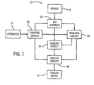
Turning now to the drawings and referring initially to Fig. 1, there is depicted a
diagrammatical representation of a magnetic resonance system 10 and its
constituent components. An operator 14 employs MR system 10 to perform
imaging operations upon patient 18. The operator 14 performs these
operations via control circuit 22 which controls the operations performed by
such constituent components as MR scanner 26, analysis circuit 30, and
display circuit 38. Certain utilitarian functions of memory circuit 34 may also
be controlled by control circuit 22 but, for purposes of this discussion, the
operation of memory circuit 34 will be presumed to be automatic in response
to the needs of other component circuits.
The operator 14 performs the imaging operations on patient 18 by instructing
MR scanner 26 to execute a series of radio frequency (RF) pulses according
to both automated routines and variables configured at control circuit 22. MR
scanner 26 collects data derived from these RF pulses which is then passed
either directly to an analysis circuit 30 or to a memory circuit 34 which will
subsequently provide the data to analysis circuit 30. Analysis circuit 30
applies analytical routines on the data in accordance with the parameters set
by operator 14 at control circuit 22 to produce image data. The image data is
in turn passed either to memory circuit 34 for later display or directly to display
circuit 38 for immediate display in accordance with instructions received from
operator 14 via control circuit 22. Display circuit 38 then displays the data on
display device 42 for review by the operator or appropriate medical personnel.
One particular type of imaging which MR system 10 is suited for is echo planar
imaging (EPI). EPI allows successive echoes from a single TR interval to be
acquired, each providing position information, as opposed to phase encoding a
separate line of data associated with numerous TR intervals. In this manner
EPI drastically reduces the time required for an MRI scan.
As with other types of MRI, to obtain positional information EPI utilizes
magnetic gradients along three axes (X, Y, and Z) to provide specific positional
data for each proton being imaged. Each gradient imparts differential spin
information upon each proton being observed. The spin information, once
acquired, allows each proton to be located along each gradient, thus allowing
three dimensional positioning of each proton within the derived image. Thus
gradients are useful in systems, such as MR system 10, for constructing either
two dimensional or three dimensional diagnostic images.
One area in which EPI techniques have proven valuable is in diffusion imaging.
Diffusion imaging allows diagnosticians to examine the diffusion patterns in the
tissues of a patient. MR diffusion imaging using EPI typically utilizes two large
gradient pulses after excitation of the system to perform diffusion weighted
imaging. The large gradient pulses are designed to impart phase shifts to
proton spins which cancel each other out if the spins are stationary, while spins
which have moved undergo an observable phase shift. Therefore, in tissues
where the fluid is undergoing random or non-directional motion, the phase
shifts within an imaging voxel are partially canceled, creating an attenuated
image. In tissues where the fluid undergoes directional motion, the phase shifts
do not cancel resulting in a complete signal loss in the image. However, in
tissue where the fluid is stationary, the phase shifts cancel and an image
highlighting differences in diffusion between tissues can be created. By
manipulating the diffusion gradient pulses on the X, Y, and Z axes, the diffusion
along particular directions in space can be imaged.
Referring now to Fig. 2, an EPI diffusion imaging mode is illustrated by
reference to an EPI pulse sequence 60. EPI pulse sequence 60 depicts a spin-echo
sequence where the timing of a 90° excitation pulse 68 and a 180°
refocusing pulse 72 are shown on a RF axis 64. The spin-echo sequence is
used to readout the signal. EPI pulse sequence 60 also depicts an X-gradient
axis 76, a Y-gradient axis 80, and a Z-gradient axis 84, each demonstrating the
events occurring along those gradients over the same time scale. For example,
Z-gradient axis 84 is the slice select gradient axis and reflects the slice select
pulse 88 as well as a slice select refocus pulse 92. X-gradient axis 76 is the
frequency encoding gradient axis and depicts the readout 96. The readout
occurs at echo time (TE) 74, measured from the time of the excitation pulse 68.
All three axes demonstrate the presence of the two large diffusion gradient
pulses 100 timed before and after refocusing pulse 72. Diffusion gradient
pulses 100 introduce diffusion sensitivity to the EPI sequence. Diffusion
encoding is performed by modulating the diffusion gradient pulses 100 on X-axis
76, Y-axis 80, and Z-axis 84 for each diffusion measurement. Also
depicted are diffusion lobe amplitude 108, commonly denoted g, diffusion lobe
width 112, commonly denoted δ, and diffusion lobe interval 116, commonly
denoted Δ.
A second EPI diffusion imaging mode is illustrated in Fig. 3 which depicts a dual
spin-echo sequence 104 with similar events represented by identical reference
numerals. In dual spin-echo sequence 104, an additional refocusing pulse 72
is added and bipolar diffusion lobes 102 are placed around both refocusing
pulses 72. The dual spin-echo approach doubles the number of gradient
transitions and provides intrinsic refocusing of eddy currents over a fairly wide
range of time constants. The dual spin-echo approach increases the minimum
echo time (TE) 74, however image distortion that would normally arise from
eddy currents induced by diffusion gradients is largely removed. Either imaging
mode sequence is capable of producing a diffusion weighted image.
In diffusion weighted imaging, the degree of sensitivity is controlled by a b-value,
generally input by operator 14. The b-value is generally given by the
Stesjkal-Tanner equation: bi,i = (2πγ)2 gi,j gi,j (δ2(Δ-δ/3)). Numerous factors,
however, can lead to inaccuracies in a b-value calculated using the above
equation and, in fact, the diffusion weighting in a sequence is preferably
described by a 3 X 3 matrix of b-values, or b-matrix. Such a b-matrix describes
the diffusion sensitivity from on-axis gradients, the on-diagonal terms, and
diffusion sensitivity generated by interactions across gradient axes, the off-diagonal
terms or cross-terms.
The b-matrix values may be determined by integrating waveforms and including
imaging gradients. Integration of the products of the k-space trajectory from
the 90°
excitation pulse 68 to the echo time (TE) 74 on each gradient axis is
given by the equation:
where the k-space trajectory is given by the equation:
where i and j each signify gradient axes, k signifies a detected value, y
signifies the gyromagnetic constant, and g signifies the gradient. Equation (2)
calculates the k-space trajectory on a gradient axis by integrating the
waveform over time. Equation 1 may then be used to integrate the dot-product
of the k-space trajectory at each point in time up to TE. This integral
is the b-value for the gradient waveform at that point in the matrix. On-diagonal
values for the b-matrix are calculated where
i =
j and represent the
diffusional weighting along an axis. Off-diagonal values, where
i ≠
j, represent
the interactions between gradients across the axes.
One method of compensating for inaccuracies in the b-value involves
calculating the actual b-value on each axis (the on-diagonal b-matrix elements)
and correcting for deviations from the desired, user configured b-value. After
calculating the actual b-value, corrections for the diffusion lobe amplitudes are
performed until the on-diagonal b-matrix values are within a given tolerance.
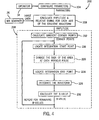
Referring now to Fig. 4, exemplary stages in the calculation of an actual b-value
are illustrated and designated generally by the reference numeral 200. First,
the parameters are configured as indicated at step 204. This configuration
includes setting the prescribed resolution, echo time, field of view, or any other
relevant imaging parameters. Subsequently, the gradient amplitudes and their
respective timing on each axis of the waveform must be calculated, as
represented in step 208. To calculate the amplitudes, the image data, from MR
scanner 26 and the image parameters, from operator 14 via input step 204, are
utilized.
After the amplitudes have been calculated, that information is then used to
calculate gradient corner points along each axis in step 212. Corner points are
calculated by taking the derivative of the amplitudes along the axes. Those
points where slope changes occur are corner points and are kept for further
calculation. Non-corner points are discarded.
A starting point for integration along an axis is determined next in step 216
using the corner point information from step 212. To determine the integration
starting point, the 90° excitation pulse 68 (see Figs. 2 and 3) is located in the
pulse sequence. If excitation pulse 68 corresponds to a corner point on the
gradient axis, integration begins at that point. If excitation pulse 68 does not
correspond to a corner point on the gradient axis, an interpolated point is
placed between adjacent corner points and integration begins at the
interpolated point.
The location of each 180° refocusing pulse 72 (see Figs. 2 and 3), if any are
present, is then determined in step 220. At each refocusing pulse 72, a change
in sign of the area occurs since each 180° pulse is basically an inversion. If the
refocusing pulse 72 corresponds to a corner point, the change of sign occurs at
the corner point. Otherwise an interpolated point is generated between
adjacent corner points and the change of sign occurs at the interpolated point.
An integration end point is then calculated in step 224. The end point is
calculated by locating TE 74 (see Figs. 2 and 3) and determining if a corner
point corresponds to the TE 74. If the TE 74 corresponds to a corner point,
integration stops at that corner point. Otherwise an interpolated point is
generated between adjacent corner points and integration stops at the
interpolated point.
Once the start point, end point and sign changes have been determined, the
gradient waveform along the axis is integrated in step 228. Integration involves
determining the k-space trajectory (ki(t)), as given by equation (2), at each
corner point using numerical integration of each linear waveform segment. The
linear waveform segments are defined as the lines connecting adjacent corner
points along the gradient axis. The result of this integration is a vector of
trajectory values, k, at various times, t, along the axis.
Once the k-space trajectory vector has been determined in step 228, the actual
b-value is determined in step 232. The b-value for a gradient waveform along
an axis is determined by solving equation (1 ) where i = j, i.e. for the on-diagonal
element. This value represents the cumulative area of the square of the k-space
trajectory calculated in step 228, scaled appropriately. Steps 216
through 232 are repeated until the actual b-value has been calculated for each
remaining axis such that the three on-diagonal b-values of the b-matrix (bxx, byy,
and bzz) are determined for all three axes, X, Y, and Z.
It should also be noted that the off-diagonal b-matrix values may also be
calculated in step 232 by integrating over the product of two separate k-space
trajectory vectors where i ≠ j, as may be determined in step 228. The off-diagonal
b-matrix values may provide valuable information regarding pairwise
interactions of gradient waveforms across axes.
Once the actual b-values of the image have been calculated, deviations from
the desired b-values are corrected. The correction process is described by Fig.
5 and is generally referred to by the reference numeral 250. The correction
process begins at step 254 by scaling the gradient diffusion lobes of an axis
using the amplitude and b-value information from the previous steps. In step
254, the amplitudes of the gradient diffusion lobes on a gradient axis are scaled
by the square root of the prescribed b-value to the calculated b-value
according to the equation:
(3) G' diffusion = G 0 diffusion * b - value prescribed b-valuecalculated .
The scaling process is then repeated for each remaining axis, as needed, until
the amplitudes of the diffusion lobes on each gradient axis, X, Y, and Z, have
been scaled.
After the diffusion lobes have been scaled, b-values for each axis are
recalculated in step 258 by repeating steps 212 through 232 (as outlined in Fig.
4) using the scaled amplitude values. The recalculated b-values are then
compared to the prescribed b-value in step 262. If the recalculated b-values
(bxx, byy, and bzz) are within a given tolerance of the prescribed b-value, no
further compensation is performed and the process is ended in step 268.
However, if one or more of the recalculated b-values are outside the given
tolerance, further fine adjustments are made to the b-value in step 272.
The fine adjustments to the b-value are accomplished by iteratively adjusting
the diffusion lobe amplitude using a numeric search in step 272, as depicted in
Fig. 6. In one embodiment, the numeric search may take the form of a
bisection search, as illustrated in Fig. 6, however a variety of numeric searches
which allow fine, corrective adjustments to be made may be used as well. In
Fig. 6, at step 280, the minimum and maximum diffusion amplitude points that
bracket the desired b-value of an axis are determined. The maximum diffusion
amplitude point is set to some number greater than the corrected diffusion
amplitude determined in step 254 of Fig. 5, such as 110% of the corrected
diffusion amplitude. The minimum diffusion amplitude point is set to some
number less than the corrected diffusion amplitude, such as 90% of the
corrected diffusion amplitude. Therefore the corrected diffusion amplitude is an
intermediate amplitude bracketed by the maximum and minimum diffusion
amplitudes, typically the midpoint. Indeed, in the bisection search described by
Fig. 6 the intermediate point is the midpoint diffusion amplitude.
Next, the calculated b-value, based upon this midpoint diffusion amplitude, is
checked against the prescribed b-value, in step 284, to determine whether the
calculated b-value is within the desired tolerance level. Note that in the first
iteration, the calculated b-value is known to be outside the tolerance. For
subsequent iterations, however, a b-value is re-calculated, in step 258, and
may be within the desire tolerance, thereby ending processing.
If the calculated b-value is less than the prescribed b-value minus the tolerance
value, the minimum amplitude is set equal to the midpoint amplitude in step
288. Likewise, if the calculated b-value is greater than the prescribed b-value
plus the tolerance value, the maximum amplitude is set equal to the midpoint
amplitude in step 290.
Next, in step 292, the midpoint amplitude is revised such that:
(4) Amplitudemidpoint = Amplitudeminimum + (.5 * (Amplitudemaximum -
Amplitudeminimum)).
The revised midpoint amplitude is then used to recalculate the b-value in step
258 (referring back to Fig. 4). The revised b-value is then checked against the
prescribed b-value at step 284. If the revised b-value is within tolerance, any
remaining axes are processed. However if the revised b-value is still outside of
the tolerance range, the amplitude midpoint is again iteratively adjusted as per
step 284. The process ends at step 268 when the b-value is within tolerance
and there are no remaining axes to be processed. While the bisection search
depicted in Fig. 6 and described in the preceding text illustrates one
embodiment of an acceptable numeric search, those skilled in the art will
recognize that other numeric searches may be used to obtain the same type of
b-value correction.
The result of b-value correction can be seen in Figs. 7 and 8. Figure 7 is a
graph showing b-value plotted against time in microseconds before correction
where an operator has configured a b-value of 1000. Shaded deviation 300
illustrates the uncorrected error, here approximately 15%. Figure 8
demonstrates the same data after calculation and correction of the b-value by
the present technique. After scaling of the diffusion lobe amplitudes, b-value
error is less than 1% from the prescribed value. Reduction of the b-value error
allows for greater operator control in MR diffusion weighted imaging by allowing
more precise control of the sensitivity of the device to diffusion processes.
For the sake of good order, various aspects of the invention are set out in the
following clauses:-