EP1384487A1 - Drugs containing genetically modified antibody against ganglioside gd3 - Google Patents
Drugs containing genetically modified antibody against ganglioside gd3 Download PDFInfo
- Publication number
- EP1384487A1 EP1384487A1 EP02713258A EP02713258A EP1384487A1 EP 1384487 A1 EP1384487 A1 EP 1384487A1 EP 02713258 A EP02713258 A EP 02713258A EP 02713258 A EP02713258 A EP 02713258A EP 1384487 A1 EP1384487 A1 EP 1384487A1
- Authority
- EP
- European Patent Office
- Prior art keywords
- antibody
- human
- cdr
- manufactured
- amino acid
- Prior art date
- Legal status (The legal status is an assumption and is not a legal conclusion. Google has not performed a legal analysis and makes no representation as to the accuracy of the status listed.)
- Withdrawn
Links
Images




Classifications
-
- C—CHEMISTRY; METALLURGY
- C07—ORGANIC CHEMISTRY
- C07K—PEPTIDES
- C07K16/00—Immunoglobulins [IG], e.g. monoclonal or polyclonal antibodies
- C07K16/18—Immunoglobulins [IG], e.g. monoclonal or polyclonal antibodies against material from animals or humans
- C07K16/28—Immunoglobulins [IG], e.g. monoclonal or polyclonal antibodies against material from animals or humans against receptors, cell surface antigens or cell surface determinants
-
- A—HUMAN NECESSITIES
- A61—MEDICAL OR VETERINARY SCIENCE; HYGIENE
- A61K—PREPARATIONS FOR MEDICAL, DENTAL OR TOILETRY PURPOSES
- A61K39/00—Medicinal preparations containing antigens or antibodies
- A61K39/395—Antibodies; Immunoglobulins; Immune serum, e.g. antilymphocytic serum
- A61K39/39533—Antibodies; Immunoglobulins; Immune serum, e.g. antilymphocytic serum against materials from animals
- A61K39/39558—Antibodies; Immunoglobulins; Immune serum, e.g. antilymphocytic serum against materials from animals against tumor tissues, cells, antigens
-
- A—HUMAN NECESSITIES
- A61—MEDICAL OR VETERINARY SCIENCE; HYGIENE
- A61P—SPECIFIC THERAPEUTIC ACTIVITY OF CHEMICAL COMPOUNDS OR MEDICINAL PREPARATIONS
- A61P35/00—Antineoplastic agents
-
- A—HUMAN NECESSITIES
- A61—MEDICAL OR VETERINARY SCIENCE; HYGIENE
- A61P—SPECIFIC THERAPEUTIC ACTIVITY OF CHEMICAL COMPOUNDS OR MEDICINAL PREPARATIONS
- A61P35/00—Antineoplastic agents
- A61P35/04—Antineoplastic agents specific for metastasis
-
- A—HUMAN NECESSITIES
- A61—MEDICAL OR VETERINARY SCIENCE; HYGIENE
- A61K—PREPARATIONS FOR MEDICAL, DENTAL OR TOILETRY PURPOSES
- A61K39/00—Medicinal preparations containing antigens or antibodies
- A61K2039/505—Medicinal preparations containing antigens or antibodies comprising antibodies
-
- C—CHEMISTRY; METALLURGY
- C07—ORGANIC CHEMISTRY
- C07K—PEPTIDES
- C07K2317/00—Immunoglobulins specific features
- C07K2317/20—Immunoglobulins specific features characterized by taxonomic origin
- C07K2317/24—Immunoglobulins specific features characterized by taxonomic origin containing regions, domains or residues from different species, e.g. chimeric, humanized or veneered
-
- C—CHEMISTRY; METALLURGY
- C07—ORGANIC CHEMISTRY
- C07K—PEPTIDES
- C07K2317/00—Immunoglobulins specific features
- C07K2317/70—Immunoglobulins specific features characterized by effect upon binding to a cell or to an antigen
- C07K2317/73—Inducing cell death, e.g. apoptosis, necrosis or inhibition of cell proliferation
- C07K2317/732—Antibody-dependent cellular cytotoxicity [ADCC]
-
- C—CHEMISTRY; METALLURGY
- C07—ORGANIC CHEMISTRY
- C07K—PEPTIDES
- C07K2317/00—Immunoglobulins specific features
- C07K2317/70—Immunoglobulins specific features characterized by effect upon binding to a cell or to an antigen
- C07K2317/73—Inducing cell death, e.g. apoptosis, necrosis or inhibition of cell proliferation
- C07K2317/734—Complement-dependent cytotoxicity [CDC]
Definitions
- the present invention relates to a medicament which comprises a gene recombinant antibody against ganglioside GD3 or the antibody fragment thereof in combination with at least one of a substance which activates an immunocompetent cell and a substance having an antitumor activity.
- Ganglioside which is one of glycolipids containing sialic acid is a constituent of animal cell membrane. Also, ganglioside has a sugar chain as a hydrophilic side chain and a sphingosine and fatty acid as hydrophobic side chains. It is known that kinds and expression levels of ganglioside vary depending on the cell types, organ species, animal species and the like. It is also known that the expression of ganglioside changes quantitatively and qualitatively in the process of malignant transformation of cells [ Cancer Res ., 45 , 2405 (1985)].
- GD3 is present in an extremely small amount in normal cells but in a large amount in cancer cells, particularly melanoma, and is present together with ganglioside GD2 in cancer cells such as sarcoma, glioma and neuroblastoma [ Proc. Natl. Acad Sci. U.S.A ., 77 , 6114 (1980); J. Exp.
- anti-GD3 antibody an antibody against GD3 (hereinafter referred to as "anti-GD3 antibody”) is considered to be useful for the treatment of these cancers [ Melanoma Research , 7 , S115 (1997)].
- anti-GD3 mouse antibody R24 as an anti-GD3 mouse monoclonal antibody
- HAMA human anti mouse antibody
- anti-GD3 human chimeric antibody KM871 Japanese Unexamined Patent Application No. 304989/93
- anti-GD2 human chimeric antibody ch14.18 J. Immunol ., 144 , 1382, (1990)
- the ch14.18 has already been used in clinical studies and its good blood retentivity has been observed [ Human Antibodies & Hybridomas , 3 , 19 (1992)].
- ADCC activity antibody-dependent cell-mediated cytotoxic activity
- CDC activity complement-dependent cytotoxicity
- Cytokine is a general term for various humoral factors which relate to cytoclesis in immune response.
- ADCC activity as one of cytotoxic activities is induced by effector cells having Fc receptor on the cell surface such as monocyte, macrophage and NK cell [ J. Immunol ., 138 , 1992 (1987)]. Since various species of cytokine activate these effector cells, administration in combination with an antibody has been tried in order to increase ADCC activity and the like of the antibody.
- anti-GD3 mouse antibody R24 a possibility of combined use effects with IL-2 has been suggested based on in vitro evaluation (U.S. Patent 5,104,652). Also, clinical studies have been carried out on the combined administration of the anti-GD3 mouse antibody R24 with IL-2, the anti-GD3 mouse antibody R24 with IFN ⁇ , the anti-GD3 mouse antibody R24 with M-CSF, the anti-GD3 mouse antibody R24 with GM-CSF, and the like [ Medical Oncology , 15 , 191 (1998), Clinical Cancer Res ., 3 , 17 (1997), Cancer , 75 , 2251 (1995), Journal Of Immunotherapy With Emphasis On Tumor Immunology , 16 , 132 (1994)].
- Melanoma is a malignant tumor in chromocytes and is known as a tumor having high malignancy, which is apt to generate metastasis and has high resistance against chemotherapy and the like.
- Immunotherapy by IL-2 or interferon, chemotherapy mainly using dacarbazine and cisplatin, combined therapy thereof and the like has so far been carried out.
- a combination of dacarbazine, cisplatin, carmustine and tamoxifen [ Cancer Treat. Rep ., 68 , 1403 (1984), Cancer , 63 , 1292 (1989)]
- a combination of dacarbazine, cisplatin and vinblastine Proc. ASCO , 12 , 1328 (1993)
- a combination of dacarbazine, fotemustine and vindesine Proc. ASCO , 33 , 1336 (1992)] and the like are known.
- dacarbazine and IL-2 [ J. Natl. Cancer Inst ., 82 , 1345 (1990)], dacarbazine and IFN ⁇ [ J. Clin. Oncol ., 9 , 1403 (1991), J. Clin. Oncol ., 12 , 806 (1994)], cisplatin and IL-2 [ J. Clin. Oncol ., 9 , 1821 (1991)], cisplatin and IFN ⁇ [ J. Clin. Oncol ., 10 , 1574 (1992)], cisplatin, dacarbazine, IL-2 and IFN ⁇ [ Proc. ASCO , 10 , 1029 (1991)] and the like are known.
- An object of the present invention is to provide a medicament which can provide higher therapeutic effects than any one of a gene recombinant antibody against ganglioside GD3 or the antibody fragment thereof, and a substance which activates an immunocompetent cell and a substance having an antitumor activity as single agents, by combining the gene recombinant antibody against ganglioside GD3 or the antibody fragment thereof with at least one of the substance which activates an immunocompetent cell and the substance having an antitumor activity.
- the medicament is expected to relieve side effects which were problems in the case of single administration.
- the present invention relates to the following (1) to (12):
- the present invention relates to a medicament which comprises a gene recombinant antibody against ganglioside GD3 or the antibody fragment thereof in combination with at least one of a substance which activates an immunocompetent cell and a substance having an antitumor activity.
- the antibody in combination with one of a substance which activates a single immunocompetent cell and a substance having an antitumor activity; the antibody in combination with two or more of substances which activate a immunocompetent cell and substances having an antitumor activity; a medicament which comprises the antibody and at least one of a substance which activates a immunocompetent cell and a substance having an antitumor activity; or a medicament which comprises the antibody and at least one of a substance which activates a immunocompetent cell and a substance having an antitumor activity and which is administered to the living body simultaneously or separately.
- the gene recombinant antibody includes a humanized antibody, a human antibody and the like.
- the humanized antibody includes a human chimeric antibody, a human CDR-grafted antibody and the like.
- a human chimeric antibody is an antibody which comprises an H chain V region (hereinafter referred to as "HV” or “VH”) and an L chain V region (hereinafter referred to as “LV” or “VL”) of an antibody derived from a non-human animal, an H chain C region (hereinafter referred to as "CH”) of a human antibody and an L chain C region (hereinafter referred to as "CL”) of a human antibody.
- HV H chain V region
- LV L chain V region
- CH H chain C region
- CL L chain C region
- the human chimeric antibody of the present invention can be produced by obtaining cDNAs encoding VH and VL from a hybridoma capable of producing a monoclonal antibody which specifically reacts with GD3, constructing a human chimeric antibody expression vector by inserting them into an expression vector for animal cells having genes encoding human antibody CH and human antibody CL, and expressing the expression vector by introducing it into an animal cell.
- the CH of a human chimeric antibody may be any region which belongs to human immunoglobulin (hereinafter referred to as "hIg"). Those of hIgG class are preferred and any one of subclasses belonging to hIgG class such as hIgG 1, hIgG2, hIgG3 and hIgG4 can also be used. Also, as the CL of a human chimeric antibody, it may be any region which belongs to hIg, and those of ⁇ class or ⁇ class can be used.
- the anti-GD3 human chimeric antibody includes a human chimeric antibody which comprises CDR1, CDR2 and CDR3 of VH comprising the amino acid sequences represented by SEQ ID NOs:3, 4 and 5, respectively, and CDR1, CDR2 and CDR3 of VL comprising the amino acid sequences represented by SEQ ID NOs:6, 7 and 8, respectively; specifically, a human chimeric antibody which comprises VH and VL comprising the amino acid sequences represented by SEQ ID NOs:49 and 50, respectively; and more specifically, a human chimeric antibody which comprises VH comprising the amino acid sequence represented by SEQ ID NO:49, an H chain C region comprising an amino acid sequence of a human antibody IgG1 subclass, an L chain V region comprising the amino acid sequence represented by SEQ ID NO:50, and an L chain C region comprising an amino acid sequence of a human antibody ⁇ class.
- Examples include anti-GD3 human chimeric antibody KM871 described in Japanese Published Unexamined Patent Application No. 304989/93
- a human CDR-grafted antibody is an antibody in which amino acid sequences of CDRs in VH and VL derived from a non-human animal antibody are grafted to appropriate positions of VH and VL of a human antibody.
- the anti-CD3 CDR-grafted antibody of the present invention can be produced by constructing cDNAs encoding V regions in which CDR sequences of VH and VL of an antibody derived from a non-human animal which specifically reacts with GD3 have been grafted to CDR sequences of VH and VL of any human antibody, constructing a human CDR-grafted antibody expression vector by inserting them into an expression vector for animal cells having genes encoding human antibody CH and human antibody CL, and expressing the human CDR-grafted antibody by introducing the expression vector into an animal cell.
- the CH of a human CDR-grafted antibody it may be any region which belongs to hIg. Those of hIgG class are preferred and any one of subclasses belonging to hIgG class such as hIgG1, hIgG2, hIgG3 and hIgG4 can also be used. Also, as the CL of a human chimeric antibody, it may be any region which belongs to hIg, and those of ⁇ class or ⁇ class can be used.
- the anti-GD3 CDR-grafted antibody includes a human CDR-grafted antibody which comprises CDR1, CDR2 and CDR3 of VH comprising the amino acid sequences represented by SEQ ID NOs:3, 4 and 5, respectively, and CDR1, CDR2 and CDR3 of VL comprising the amino acid sequences represented by SEQ ID NOs:6, 7 and 8, respectively; specifically, a human CDR-grafted antibody which comprises VH and VL comprising the amino acid sequences represented by SEQ ID NOs:9 and 10, respectively; and more specifically, a human CDR-grafted antibody which comprises VH comprising the amino acid sequence represented by SEQ ID NO:9, an H chain C region comprising an amino acid sequence of a human antibody IgG1 subclass, an L chain V region comprising the amino acid sequence represented by SEQ ID NO:10, and an L chain C region comprising an amino acid sequence of a human antibody ⁇ class.
- Examples include anti-GD3 human CDR-grafted antibody KM8871 produced by transformed cell clone KM8871 which has been internationally deposited as FERM BP-6790 on July 22, 1999, in National Institute of Bioscience and Human Technology, Agency of Industrial Science and Technology, the Ministry of International Trade and Industry (present name: International Patent Organism Depositary, National Institute of Advanced Industrial Science and Technology (AIST Tsukuba Central 6, 1-1, Higashi 1-Chome Tsukuba-shi, Ibaraki-ken, Japan).
- a human antibody is an antibody naturally present in the human body. It also includes antibodies obtained from a human antibody phage library and a human antibody-producing transgenic animal which are produced based on the recent advance in genetic engineering, cell engineering and developmental engineering techniques.
- a lymphocyte capable of producing the antibody can be cultured by isolating a human peripheral blood lymphocyte and infecting it with EB virus or the like to thereby immortalize it, followed by cloning, and the antibody can be purified from the culture mixture.
- the human antibody phage library is a library in which antibody fragments such as Fab and scFv are expressed on the phage surface by inserting an antibody gene prepared from human B cell into a phage gene. From the library, a phage expressing a desired antibody fragment having an antigen binding activity can be recovered, using its activity to bind to an antigen-immobilized substrate as the marker. The antibody fragment can be further converted genetic engineeringly into a human antibody molecule having two complete H chains and two complete L chains.
- a human antibody-producing transgenic non-human animal is an animal in which a human antibody gene is introducing into cells.
- a human antibody-producing transgenic animal can be produced by introducing a human antibody gene into a mouse ES cell, inoculating the ES cell into an early stage embryo of other mouse and then developing it.
- the human antibody can be produced and accumulated in a culture by obtaining a human antibody-producing hybridoma according to a hybridoma production method usually carried out in non-human mammals and then culturing it.
- the antibody fragment includes Fab, Fab', F(ab') 2 , scFv, dsFv, a peptide comprising CDR and the like.
- An Fab is an antibody fragment having a molecular weight of approximately 50,000 and an antigen binding activity, in which about a half of the N-terminal side of H chain and a full L chain, among fragments obtained by treating IgG with a protease, papain (cut at the amino acid residue at position 224 in the H chain), are bound together through a disulfide bond.
- the Fab of the present invention can be obtained by treating an antibody which specifically reacts with GD3 with a protease, papain. Also, the Fab can be produced by inserting a DNA encoding Fab of the antibody into an expression vector, and introducing the vector into a host cell to express the Fab.
- An F(ab') 2 is an antibody fragment having a molecular weight of approximately 100,000 and an antigen binding activity, which is slightly larger than the Fab bound via a disulfide bond of the hinge region, among fragments obtained by treating IgG with a protease, pepsin (cut at the amino acid residue at position 234 in the H chain).
- the F(ab') 2 of the present invention can be obtained by treating an antibody which specifically reacts with GD3, with a protease, pepsin. Also, the F(ab') 2 can be produced by binding Fab' described below via a thioether bond or a disulfide bond.
- An Fab' is an antibody fragment having a molecular weight of approximately 50,000 and an antigen binding activity, which is obtained by cutting a disulfide bond of the hinge region of the F(ab') 2 .
- the Fab' of the present invention can be obtained by treating F(ab') 2 which specifically reacts with GD3 with a reducing agent, dithiothreitol. Also, the Fab' can be produced by inserting DNA encoding an Fab' fragment of the antibody into an expression vector, and introducing the vector into a host cell to express the Fab'.
- An scFv is a VH-P-VL or VL-P-VH polypeptide in which one chain VH and one chain VL are linked using an appropriate peptide linker (hereinafter referred to as "P").
- the VH and VL in the scFv used in the present invention may be derived from any one of antibodies produced by a hybridoma, a humanized antibody and a human antibody.
- the scFv of the present invention can be produced by obtaining cDNAs encoding the VH and VL of an antibody which specifically reacts with GD3, constructing DNA encoding scFv, inserting the DNA into an expression vector, and then introducing the expression vector into a host cell to express the scFv.
- a dsFv is obtained by binding polypeptides in which one amino acid residue of each of VH and VL is substituted with a cysteine residue via a disulfide bond between the cysteine residues.
- the amino acid residue substituted with a cysteine residue can be selected based on a three-dimensional structure estimation of the antibody in accordance with the method shown by Reiter et al . [ Protein Engineering , 7 , 697 (1994)].
- the VH and VL in the dsFv of the present invention may be derived from any one of antibodies produced by a hybridoma, a humanized antibody and a human antibody.
- the dsFv of the present invention can be produced by obtaining cDNAs encoding the VH and VL of an antibody which specifically reacts with GD3, constructing DNA encoding dsFv, inserting the DNA into an expression vector, and then introducing the expression vector into a host cell to express the dsFv.
- a peptide comprising CDR is constituted by at least one region of H chain and L chain CDRs. Plural CDRs can be bound directly or via an appropriate peptide linker.
- the peptide comprising CDR of the present invention can be produced by obtaining cDNA encoding the VH and VL of an antibody which specifically reacts with GD3, inserting the cDNA into an expression vector, and then introducing the expression vector into a host cell to express the peptide comprising CDR.
- the peptide comprising CDR can be produced by a chemical synthesis method such as Fmoc method (fluorenylmethoxycarbonyl method) or tBoc method (t-butyloxycarbonyl method).
- the substance which activates an immunocompetent cell or the substance having an antitumor activity used in the present invention includes cytokine, a chemotherapeutant, a hormonotherapeutant and the like.
- cytokine any cytokine can be used, so long as it activates an effector cell relating to ADCC activity caused by an antibody, such as NK cell, macrophage, monocyte and granulocyte.
- the cytokine which activates NK cell includes IL-2, IFN ⁇ , IL-12, IL-18 and the like, and IL-2 is preferred.
- the cytokine which activates macrophage includes M-CSF.
- the cytokine which activates monocyte and granulocyte includes GM-CSF.
- the cytokine having an antitumor activity includes interferons, TNF ⁇ , TNF ⁇ , IL-1 ⁇ , IL-1 ⁇ and the like, and IFN ⁇ is preferred.
- the chemotherapeutant and hormonotherapeutant used in the present invention include dacarbazine, cisplatin, fotemustine, carmustine, nimustine, lomustine, semustine, pacritaxel, dosetaxel, vindesine, vinblastine, vincristine, carboplatin, bleomycin, doxorubicin, tamoxifen and the like.
- the medicament of the present invention includes anti-GD3 human chimeric antibody KM871 or anti-GD3 CDR-grafted antibody KM8871 in combination with at least one agent selected from IL-2, IFN ⁇ , dacarbazine and cisplatin.
- the medicament of the present invention can relieve side effects because it can be used at a lower dose.
- the medicament of the present invention can be used against tumors such as melanoma, sarcoma, glioma and neuroblastoma. Particularly, the medicament is preferably used against tumors in which ganglioside GD3 is highly expressed.
- Antitumor effects can be examined using an animal model.
- the animal model includes a xenograft model, a syngeneic graft model and the like.
- a xenograft model can be prepared by inoculating a culture cell line derived from a human cancer tissue, into various regions of an immunodeficiency mouse such as a nude mouse, for example, subcutaneously, intracutaneously, intraperitoneally, intravenously, etc.
- a syngeneic graft model can be prepared by inoculating a cultured mouse cancer cell line into various regions of a wild-type mouse having a normal immune system, for example, subcutaneously, intracutaneously, intraperitoneally, intravenously, etc ..
- evaluation is preferably carried out using a syngeneic graft model having no T cell defection.
- a syngeneic graft model for evaluation of a GD3 monoclonal antibody is prepared as follows.
- a syngeneic graft model for evaluation of an anti-GD3 monoclonal antibody can be prepared by inoculating a GD-3 positive mouse cell into a wild-type mouse having a normal immune system.
- the GD3-positive mouse can be prepared by introducing a GM3-specific ⁇ -2,8-sialyltransferase (GD3 synthase) gene into a mouse culture cell line which expresses a GD3 biosynthesis precursor, GM3, to prepare a GD3-positive transformant [ Proc. Natl. Acad Sci. U.S.A ., 91 , 10455 (1994)].
- a xenograft model for evaluation of a GD3 monoclonal antibody can be prepared by inoculating into a nude mouse a tumor section collected from a tumor mass of a GD3-positive human malignant myeloma cell line, such as H-187 cell or G-361.
- the antitumor effects of the medicament of the present invention can be evaluated using the above animal model by comparing the effects of the medicament of the present invention with effects of the administration of an antibody alone and administration of a substance which activates an immunocompetent cell alone or a substance having an antitumor activity alone.
- the medicament of the present invention can be administered alone. Generally, it is preferred to provide it in the form of a pharmaceutical formulation produced by mixing it with at least one pharmaceutically acceptable carrier in accordance with a method well known in the technical field of pharmaceutics.
- a route of administration which is the most effective in carrying out the intended treatment such as oral administration or parenteral administration, e.g., intraorally, tracheally, rectally, subcutaneously, intramuscularly, intravenously, etc .
- parenteral administration e.g., intraorally, tracheally, rectally, subcutaneously, intramuscularly, intravenously, etc .
- intravenous administration is preferred.
- the dosage form includes sprays, capsules, tablets, granules, syrups, emulsions, suppositories, injections, ointments, tapes and the like.
- Formulations suitable for oral administration include emulsions, syrups, capsules, tablets, powders, granules and the like.
- Liquid preparations such as emulsions and syrups, can be produced using additives, for example, water; saccharides such as sucrose, sorbitol and fructose; glycols such as polyethylene glycol and propylene glycol; oils such as sesame oil, olive oil and soybean oil; antiseptics such as p-hydroxybenzoate; flavors such as strawberry flavor and peppermint; and the like.
- additives for example, water; saccharides such as sucrose, sorbitol and fructose; glycols such as polyethylene glycol and propylene glycol; oils such as sesame oil, olive oil and soybean oil; antiseptics such as p-hydroxybenzoate; flavors such as strawberry flavor and peppermint; and the like.
- Capsules, tablets, powders, granules and the like can be produced using additives, for example, excipients such as lactose, glucose, sucrose and mannitol; disintegrants such as starch and sodium alginate; lubricants such as magnesium stearate; binders such as polyvinyl alcohol, hydroxypropylcellulose and gelatin; surfactants such as fatty acid ester; plasticizers such as glycerine; and the like.
- excipients such as lactose, glucose, sucrose and mannitol
- disintegrants such as starch and sodium alginate
- lubricants such as magnesium stearate
- binders such as polyvinyl alcohol, hydroxypropylcellulose and gelatin
- surfactants such as fatty acid ester
- plasticizers such as glycerine
- Formulations suitable for parenteral administration include injections, suppositories, sprays and the like.
- Injections can be prepared using a carrier such as a salt solution, a glucose solution or a mixture thereof
- Suppositories can be prepared using a carrier such as cacao butter, hydrogenated fat or carboxylic acid.
- sprays can be prepared from the medicament itself or using a carrier or the like which does not stimulate oral and airway mucous membranes of patients and can facilitate absorption of the medicament by dispersing it as minute particles.
- the carrier includes lactose, glycerine and the like. Depending on the properties of the medicament and the used carrier, aerosols, dry powders and the like can be produced.
- the additives exemplified in the oral preparations can also be added to the parenteral preparations.
- the dose and frequency of administration vary depending on intended therapeutic effects, administration method, treating period, age, body weight and the like, and the dose for adult patient is generally from 0.1 to 20 mg/kg in terms of anti-GD3 antibody per one administration.
- the agent used in combination with the antibody is administered at a dose equal to or lower than the dose when the agent is used alone in clinic.
- the humanized antibody expression vector is an expression vector for animal cells to which genes encoding CH and CL of a human antibody are inserted, and can be constructed by cloning the genes encoding CH and CL of a human antibody into an expression vector for animal cells.
- the C regions of a human antibody can be any human antibody CH and CL, and examples include a C region belonging to IgG1 subclass in an H chain of a human antibody (hereinafter referred to as "hC ⁇ 1”) and a C region belonging to ⁇ class in an L chain of a human antibody (hereinafter referred to as "hC ⁇ ").
- a chromosomal DNA comprising an exon and an intron can be used as the gene encoding CH and CL of a human antibody, and cDNA can also be used.
- Any expression vector for animal cells can be used, so long as a gene encoding the human antibody C region can be inserted and expressed.
- Examples include pAGE107 [ Cytotechnology , 3 , 133 (1990)], pAGE103 [ J. Biochem ., 101 , 1307 (1987)], pHSG274 [ Gene , 27 , 223 (1984)], pKCR [ Proc. Natl. Acad Sci. U.S.A ., 78 , 1527 (1981)], pSG1 ⁇ d2-4 [ Cytotechnology , 4 , 173 (1990)] and the like.
- the promoter and the enhancer used in the expression vector for animal cell include early promoter and enhancer of SV40 [ J.
- the humanized antibody expression vector may be either of a type in which a gene encoding an antibody H chain and a gene encoding an antibody L chain exist on separate vectors or of a type in which both genes exist on the same vector (hereinafter referred to as "tandem type").
- tandem type In respect of easiness of construction of a humanized antibody expression vector, easiness of introduction into animal cells, and balance between the expression levels of antibody H and L chains in animal cells, a tandem type of the humanized antibody expression vector is more preferred [ J. Immunol. Methods , 167 , 271 (1994)].
- the tandem type humanized antibody expression vector includes pKANTEX93 (WO 97/10354), pEE18 [ HYBRIDOMA , 17 , 559 (1998)] and the like.
- the thus constructed humanized antibody expression vector can be used for the expression of the human chimeric antibody and human CDR-grafted antibody in animal cells.
- cDNAs encoding VH and VL of an antibody derived from an non-human animal such as a mouse antibody are obtained as follows.
- mRNA is extracted from hybridoma cells producing a mouse antibody or the like to synthesize cDNA.
- the synthesized cDNA is inserted into a vector such as a phage or a plasmid to prepare a cDNA library.
- a recombinant phage or recombinant plasmid containing cDNA encoding VH and a recombinant phage or recombinant plasmid containing cDNA encoding VL is isolated from the library using a C region part or a V region part of a mouse antibody as the DNA probe.
- the full nucleotide sequences of the VH and VL of the mouse antibody of interest on the recombinant phage or recombinant plasmid are determined, and the full amino acid sequences of the VH and VL are deduced from the nucleotide sequences.
- the non-human animal may be any animal such as a mouse, a rat, a hamster or a rabbit, so long as a hybridoma cell can be produced therefrom.
- the method for preparing total RNA from a hybridoma cell includes a guanidine thiocyanate-cesium trifluoroacetate method [ Methods in Enzymol ., 154 , 3 (1987)] and the like.
- the method for preparing mRNA from total RNA includes an oligo (dT) immobilized cellulose column method [ Molecular Cloning: A Laboratory Manual , Cold Spring Harbor Lab. Press, New York (1989), hereinafter referred to as " Molecular Cloning: A Laboratory Manual "] and the like.
- kits for preparing mRNA from a hybridoma cell includes Fast Track mRNA Isolation Kit (manufactured by Invitrogen), Quick Prep mRNA Purification Kit (manufactured by Pharmacia) and the like.
- the method for synthesizing cDNA and preparing a cDNA library includes known methods ( Molecular Cloning: A Laboratory Manual ; Current Protocols in Molecular Biology , Supplement 1-34, hereinafter referred to as " Current Protocols in Molecular Biology "); a method using a commercially available kit such as Super ScriptTM Plasmid System for cDNA Synthesis and Plasmid Cloning (manufactured by GIBCO BRL) or ZAP-cDNA Kit (manufactured by Stratagene); and the like.
- the vector into which the cDNA synthesized using mRNA extracted from a hybridoma cell as the template is inserted for preparing a cDNA library may be any vector, so long as the cDNA can be inserted.
- Examples include ZAP Express [ Strategies , 5 , 58 (1992)], pBluescript II SK(+) [ Nucleic Acids Research , 17 , 9494 (1989)], ⁇ zapII (manufactured by Stratagene), ⁇ gt10 and ⁇ gt11 [ DNA Cloning: A Practical Approach , I, 49 (1985)], Lambda BlueMid (manufactured by Clontech), ⁇ ExCell and pT7T3 18U (manufactured by Pharmacia), pcD2 [ Mol. Cell. Biol ., 3 , 280 (1983)], pUC18 [ Gene , 33 , 103 (1985)] and the like.
- E. coli for introducing the cDNA library constructed by a phage or plasmid vector can be used, so long as the cDNA library can be introduced, expressed and maintained.
- Examples include XL1-Blue MRF' [ Strategies , 5 , 81 (1992)], C600 [ Genetics , 39 , 440 (1954)], Y1088 and Y1090 [ Science , 222 , 778 (1983)], NM522 [ J. Mol. Biol ., 166 , 1 (1983)], K802 [ J. Mol. Biol ., 16 ; 118 (1966)], JM105 [ Gene , 38 , 275 (1985)] and the like.
- a colony hybridization or plaque hybridization method using an isotope- or fluorescence-labeled probe can be used ( Molecular Cloning: A Laboratory Manual ).
- the cDNAs encoding VH and VL can be prepared through polymerase chain reaction (hereinafter referred to as "PCR"; Molecular Cloning: A Laboratory Manual; Current Protocols in Molecular Biology ) by preparing primers and using cDNA prepared from mRNA or a cDNA library as the template.
- the nucleotide sequence of the cDNA can be determined by digesting the cDNA selected by the above method with appropriate restriction enzymes and the like, cloning the fragments into a plasmid such as pBluescript SK(-) (manufactured by Stratagene), carrying out the reaction according to a usually used nucleotide analyzing method such as the dideoxy method of Sanger, F. et al . [ Proc. Natl. Acad Sci. USA , 74 , 5463 (1977)], and then analyzing the sequence using an automatic nucleotide sequence analyzer such as A.L.F. DNA sequencer (manufactured by Pharmacia).
- a plasmid such as pBluescript SK(-) (manufactured by Stratagene)
- a usually used nucleotide analyzing method such as the dideoxy method of Sanger, F. et al . [ Proc. Natl. Acad Sci. USA , 74 ,
- Whether the obtained cDNAs encode the full amino acid sequences of the VH and VL of the antibody containing a secretory signal sequence can be confirmed by estimating the full amino acid sequences of the VH and VL from the determined nucleotide sequence and comparing them with full amino acid sequences of the VH and VL of known antibodies [ Sequences of Proteins of Immunological Interest , US Dept. Health and Human Services (1991), hereinafter referred to as " Sequences of Proteins of Immunological Interest "].
- the length of the secretory signal sequence and N-terminal amino acid sequences can be deduced and subgroups to which they belong can also be known, by comparing with full amino acid sequences of VH and VL of known antibodies ( Sequences of Proteins of Immunological Interest ). Also, regarding the amino acid sequence of each CDR of VH and VL, it can be found by comparing it with amino acid sequences of VH and VL of known antibodies ( Sequences of Proteins of Immunological Interest ).
- a human chimeric antibody expression vector can be constructed by cloning cDNAs encoding VH and VL of an antibody derived from a non-human animal in the region upstream of genes encoding CH and CL of the human antibody on the humanized antibody expression vector as described in the item 1(1).
- each of cDNAs encoding VH and VL of an antibody derived from a non-human animal is linked with a synthesized DNA comprising nucleotide sequences at the 3' terminals of VH and VL of an antibody derived from a non-human animal and nucleotide sequences at the 5' terminals of CH and CL of a human antibody and having a recognition sequence of an appropriate restriction enzyme at both terminals, and each cDNA is cloned so that it is appropriately expressed in upstream of genes encoding the CH and CL of the humanized antibody expression vector as described in the item 1(1) to thereby construct a human chimeric antibody expression vector.
- cDNAs encoding VH and VL of a human CDR-grafted antibody can be obtained as follows. First, amino acid sequences of FRs in VH and VL of a human antibody to which amino acid sequences of CDRs in VH and VL of an antibody derived from a non-human animal antibody are grafted are selected. Any amino acid sequences of FRs in VH and VL of a human antibody can be used, so long as they are derived from human. Examples include amino acid sequences of FRs in VH and VL of human antibodies registered in database such as Protein Data Bank, amino acid sequences common to subgroups of FRs in the VH and VL of human antibodies ( Sequences of Proteins of Immunological Interest ) and the like.
- amino acid sequences having high homology (at least 60% or more) with amino acid sequence of FRs of VH and VL of an antibody of interest derived from a non-human animal is preferably selected. Then, amino acid sequences of CDRs of VH and VL of the antibody derived from a non-human animal are grafted to the selected amino acid sequences of FRs of VH and VL of a human antibody to design amino acid sequences of the VH and VL of a human CDR-grafted antibody.
- the designed amino acid sequences are converted to DNA sequences by considering the frequency of codon usage found in nucleotide sequences of genes of antibodies ( Sequence of Proteins of Immunological Interest ), and the DNA sequences encoding the amino acid sequences of the VH and VL of a human CDR-grafted antibody are designed.
- Several synthetic DNAs having a length of about 100 nucleotides are synthesized, and PCR is carried out using them. In this case, it is preferred in each of the H chain and the L chain that 6 synthetic DNAs are designed in view of the reaction efficiency of PCR and the lengths of DNAs which can be synthesized.
- the humanized antibody expression vector constructed in the item 1(1) can be easily cloned into the humanized antibody expression vector constructed in the item 1(1) by introducing the recognition sequence of an appropriate restriction enzyme to the 5' terminal of the synthetic DNAs present on the both terminals.
- an amplified product is cloned into a plasmid such as pBluescript SK (-) (manufactured by Stratagene), and the nucleotide sequences are determined according to the method described in the item 1(2) to obtain a plasmid having DNA sequences encoding the VH and VL of a designed human CDR-grafted antibody.
- Substitution of the amino acid residues of the FR in the VH and VL of a human antibody can be achieved by carrying out the PCR described in the item 1(5) using synthetic DNA for modification.
- the nucleotide sequence is determined according to the method described in the item 1(2) so that whether the objective modification has been carried out is confirmed.
- a human CDR-grafted antibody expression vector can be constructed by cloning cDNAs encoding VH and VL of the human CDR-grafted antibody constructed in the items 1(5) and 1(6) into upstream of the genes encoding CH and CL of the human antibody in the humanized antibody expression vector as described in the item 1(1).
- a human CDR-grafted antibody expression vector can be constructed by carrying out cloning so that they are expressed in an appropriate form in upstream of genes encoding CH and CL of the human antibody in the humanized antibody expression vector as described in the item 1(1).
- the humanized antibodies can be expressed transiently using the humanized antibody expression vector as described in the items 1(4) and 1(7) or the modified expression vector thereof.
- Any cell can be used as a host cell, so long as the host cell can express a humanized antibody.
- COS-7 cell (ATCC CRL1651) is used in view of its high expression level [ Methods in Nucleic Acids Res ., CRC Press, 283 (1991)].
- the method for introducing the expression vector into COS-7 cell includes a DEAE-dextran method [ Methods in Nucleic Acids. Res ., CRC Press, 283 (1991)], a lipofection method [ Proc. Natl. Acad Sci. USA , 84 , 7413 (1987)], and the like.
- the expression level and antigen binding activity of the humanized antibody in the culture supernatant can be measured by enzyme-linked immunosorbent assay [hereinafter referred to as "ELISA”; Antibodies: A Laboratory Manual , Cold Spring Harbor Laboratory, Chapter 14 (1988), hereinafter referred to as “ Antibodies: A Laboratory Manual “, Monoclonal Antibodies: Principles and Practice , Academic Press Limited (1996), hereinafter referred to as “ Monoclonal Antibodies " (1996)] and the like.
- ELISA enzyme-linked immunosorbent assay
- a transformant which produces a humanized antibody stably can be obtained by introducing into an appropriate host cell the humanized antibody expression vector described in the items 1 (4) and 1 (7).
- the method for introducing the expression vector into a host cell includes electroporation [Japanese Published Unexamined Patent Application No. 257891/90, Cytotechnology , 3 , 133 (1990)] and the like.
- Any cell can be used as the host cell into which the humanized antibody expression vector is to be introduced, so long as it can express a humanized antibody.
- Examples include mouse SP2/0-Ag14 cell (ATCC CRL1581), mouse P3X63-Ag8.653 cell (ATCC CRL1580), CHO cell in which a dihydrofolate reductase gene (hereinafter referred to as " dhfr ”) is defective [ Proc. Natl. Acad Sci. U.S.A ., 77 , 4216 (1980)], rat YB2/3HL.P2.G11.16Ag.20 cell (ATCC CRL1662, hereinafter referred to as "YB2/0 cell”), and the like.
- dhfr dihydrofolate reductase gene
- a host cell which expresses an antibody having high ADCC activity includes host cells in which activity of an enzyme protein relating to synthesis of an intracellular sugar nucleotide, GDP-fucose, an enzyme protein relating to modification of a sugar chain in which 1-position of fucose is bound to 6-position of N-acetylglucosamine in the reducing end through ⁇ -bond in a complex N-glycoside-linked sugar chain, or a protein relating to transport of an intracellular sugar nucleotide, GDP-fucose to the Golgi body or the like is decreased or deleted, preferably YB2/0 cell, and the like.
- a transformant which expresses a humanized antibody stably is selected in accordance with the method disclosed in Japanese Published Unexamined Patent Application No. 257891/90, by culturing it in a medium for animal cell culture containing an agent such as G418 sulfate (hereinafter referred to as "G418", manufactured by SIGMA).
- G418 G418 sulfate
- the medium for animal cell culture includes PRMI1640 medium (manufactured by Nissui Pharmaceutical), GIT medium (manufactured by Nihon Pharmaceutical), EX-CELL302 medium (manufactured by JRH), IMDM medium (manufactured by GIBCO BRL), Hybridoma-SFM medium (manufactured by GIBCO BRL), media obtained by adding various additives such as fetal bovine serum (hereinafter referred to as "FBS”) to these media, and the like.
- FBS fetal bovine serum
- the expression level and antigen binding activity of the humanized antibody in the culture supernatant can be measured by ELISA or the like. Also, in the transformant, the expression level of the humanized antibody can be increased by using the dhfr amplification system or the like according to the method disclosed in Japanese Published Unexamined Patent Application No. 257891/90.
- the humanized antibody can be purified from the culture supernatant of the transformant by using a protein A column [ Antibodies, A Laboratory Manual, Monoclonal Antibodies (1996)]. Any other conventional methods for protein purification can be used.
- the humanized antibody can be purified by a combination of gel filtration, ion-exchange chromatography, ultrafiltration and the like.
- the molecular weight of the H chain or the L chain of the purified humanized antibody or the antibody molecule as a whole is determined by polyacrylamide gel electrophoresis [hereinafter referred to as "SDS-PAGE", Nature , 227 , 680 (1970)], Western blotting [ Antibodies: A Laboratory Mamial, Monoclonal Antibodies (1996)], and the like.
- the binding activity to an antigen and the binding activity to a cultured cancer cell line of the purified humanized antibody can be measured by ELISA, an immunofluorescent method [ Cancer Immunol. Immunother ., 36 , 373 (1993)] and the like.
- the cytotoxic activity against an antigen positive culture cell line can be evaluated by measuring the CDC activity, the ADCC activity or the like [ Cancer Immunol. Immunother ., 36 , 373 (1993)].
- a 2 to 3 mm square tumor section prepared from a tumor mass of a GD3-positive human malignant myeloma cell line H-187 which had been passaged under the dorsal skin of a Balb/c nude mice (male, CLEA Japan) was inoculated using a inoculation needle under the dorsal skin of a 6-week-old male nude mouse. Ten days after the inoculation, the tumor diameter was measured using slide calipers and the tumor volume was calculated by the following equation: Tumor volume width ⁇ length ⁇ height ⁇ 0.5
- V/V0 (tumor volume of each individual on the measured day) /(tumor volume of each individual on the measurement starting day)
- Results of periodical changes in the mean value of V/V0 in each group are shown in Fig. 1.
- the combined administration of KM871 with IL-2 showed higher growth inhibitory effects than the administration of IL-2 alone or the antibody alone.
- a significance test was carried out between the value of V/V0 in the combined administration group and the values of V/V0 in other groups, and the results of obtained p values are shown in Table 1.
- the combined administration group showed significant tumor growth inhibitory effects on and after the 17th day of the inoculation, in comparison with the negative control group.
- significant growth inhibitory effects were found during from the 21st day to the 27th day for the antibody alone, and on and after the 21st day for the IL-2 alone.
- a two-tailed t-test was carried out for the judgment of significance, and p ⁇ 0.05 was used as the standard of significance judgment.
- NK cell and T cell are known as the main cell population to be activated by IL-2. Accordingly, in order to examine antitumor effects by combined administration of IL-2 with the antibody, a syngeneic graft model was constructed for inoculating a mouse-derived tumor cell into a normal mouse having no T cell defection.
- a cell was prepared by constructing an animal cell stable expression vector for a GD3 synthase which catalyses the reaction of synthesizing GD3 by adding sialic acid to a GD3 biosynthesis precursor GM3, and introducing the vector into a mouse melanoma B16-F10 cell capable of expressing GM3.
- Antitumor effects by the combined administration of KM871 with human IL-2 were measured with the syngeneic graft model constructed using the cell. Details are shown below.
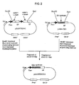
- FIG. 2 A schematic illustration regarding the method for constructing a stable expression vector is shown in Fig. 2.
- a buffer comprising 10 mM Tris-HCl (pH 7.5), 50 mM sodium chloride, 10 mM magnesium chloride and 1 mM DTT
- 3 ⁇ g of a vector pAMo-GD3 (WO 94/23020) for expressing GD3 synthase gene in animal cells was added, and 10 units of a restriction enzyme Hin dIII (manufactured by Takara Shuzo) was further added thereto to carry out the reaction at 37°C for 1 hour.
- Hin dIII manufactured by Takara Shuzo
- the reaction solution was precipitated with ethanol and added to 10 ⁇ l of a buffer comprising 50 mM Tris-HCl (pH 7.5), 100 mM sodium chloride, 10 mM magnesium chloride, 1 mM DTT, 100 ⁇ g/ml BSA and 0.01% Triton X-100, and 10 units of a restriction enzyme Not I (manufactured by Takara Shuzo) was added thereto to carry out the reaction at 37°C for 1 hour.
- the reaction solution was precipitated with ethanol, and the 5'-protruding-end formed by the restriction enzyme digestion was changed into blunt-end using DNA Blunting Kit (manufactured by Takara Shuzo).
- reaction solution was fractionated by agarose gel electrophoresis to recover about 2 ⁇ g of a blunt-ended Hin dIII- Not I fragment having about 2.1 kb using QIAquick Gel Extraction Kit (manufactured by QIAGEN) in accordance with the manufacture's instructions.
- an anti-GD3 human chimeric antibody KM871 expression vector pKANTEX641 Japanese Published Unexamined Patent Application No. 304989/93 was added to 10 ⁇ l of a buffer comprising 50 mM Tris-HCl (pH 7.5), 100 mM sodium chloride, 10 mM magnesium chloride and 1 mM DTT, and 10 units of a restriction enzyme Eco RI (manufactured by Takara Shuzo) was further added thereto to carry out the reaction at 37°C for 1 hour.
- the reaction solution was precipitated with ethanol, and the 5'-protruding-end formed by the restriction enzyme digestion was changed into blunt-end using DNA Blunting Kit (manufactured by Takara Shuzo).
- the reaction solution was precipitated with ethanol and dissolved in 10 ⁇ l of a buffer comprising 50 mM Tris-HCl (pH 7.5), 100 mM sodium chloride, 10 mM magnesium chloride and 1 mM DTT, and 10 units of a restriction enzyme Nru I (manufactured by Takara Shuzo) was further added thereto to carry out the reaction at 37°C for 1 hour.
- reaction solution was precipitated with ethanol, 10 ⁇ l of a buffer comprising 50 mM Tris-HCl (pH 7.5) and 10 mM magnesium chloride were added thereto, and 10 units of a 5'-terminal dephosphorylation enzyme, calf intestine-derived alkaline phosphatase (manufactured by Takara Shuzo), was further added thereto to carry out the reaction at 37°C for 1 hour for dephosphorylation of the 5'-terminal.
- a buffer comprising 50 mM Tris-HCl (pH 7.5) and 10 mM magnesium chloride
- reaction solution was treated with phenol and then fractionated by agarose gel electrophoresis to recover about 2 ⁇ g of a blunt end-dephosphorylated Eco RI- Nru I fragment having about 9.4 kb using QIAquick Gel Extraction Kit (manufactured by QIAGEN) in accordance with the manufacture's instructions.
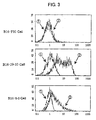
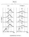
- Each of 1 ⁇ 10 6 cells of the GD3 synthase gene transfectants B16-8-3 and B16-29-10 obtained in b. of the item (2) of Example 1 and the parent cell line B16-F10 cell were suspended in PBS, put into a microtube (manufactured by Treff) and centrifuged (2,000 rpm for 2 minutes) for washing the cells, 50 ⁇ l of the anti-GD3 human chimeric antibody KM871 (a solution prepared by adjusting to 5 ⁇ g/ml with 1% BSA-PBS, negative control was 1% BSA-PBS alone) was added thereto, followed by stirring, and the reaction was carried out at 4°C for 1 hour.
- the anti-GD3 human chimeric antibody KM871 a solution prepared by adjusting to 5 ⁇ g/ml with 1% BSA-PBS, negative control was 1% BSA-PBS alone
- the GD3 expressing B16 cell B16 ⁇ 29-10 obtained in c. of the item (2) of Example 1 was suspended in PBS to give a density of 1 ⁇ 10 8 cells/ml, and 50 ⁇ l of the suspension was inoculated under the abdominal side skin of a C57 BL/6 mouse (male, 7-week-old, manufactured by Charles River).
- the tumor diameter was measured using slide calipers to calculate the tumor volume by the following equation, and individuals having the value within the range of 10 to 80 mm 3 were selected.
- Tumor volume (width) 2 ⁇ length ⁇ 0.5
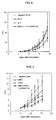
- the test was carried out using 5 animals for each group. Each agent was prepared to 200 ⁇ l per animal by diluting with a citrate buffer (an aqueous solution of 10 mM citric acid and 150 mM sodium chloride, adjusted to pH 6 with sodium hydroxide). From the 6th day after the inoculation, the tumor volume was periodically measured, and the antitumor effects were judged by comparing the mean value of specific volume V/V0 in each group. Results of periodical changes in the mean value of V/V0 in each group are shown in Fig. 4. As shown in Fig. 4, the antitumor effects were not found by IL-2 alone or the antibody alone, but the combined administration of KM871 with IL-2 showed growth inhibitory effects.
- the combined administration group showed significant tumor growth inhibitory effects on and after the 8th day of the inoculation, in comparison with the negative control group.
- significant growth inhibitory effects were found on the 8th, 10th, 12th, 13th, 14th and 17th days for the antibody alone, and on and after the 8th day for the IL-2 alone.
- a two-tailed t-test was carried out for the judgment of significance, and p ⁇ 0.05 was used as the standard of significance judgment.
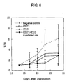
- the test was carried out using 6 animals for each group. Each agent was prepared by diluting with saline (manufactured by Otsuka Pharmaceutical) and administered from the tail vein. From the 10th day after the inoculation, the tumor volume was periodically measured, and the antitumor effects were judged by comparing the mean value of specific volume V/V0 in each group.
- the combined administration group showed significant tumor growth inhibitory effects on and after the 14th day of the inoculation, in comparison with the negative control group. In addition, significant difference was found on the 21 st and 24th days for the IFN ⁇ alone.
- the combined administration group showed significant difference in the growth inhibitory effects against the negative control in all of the measuring days on and after the 14th day, while significant difference was not obtained on the 14th day in the antibody alone group. Based on the above, it was found that significantly higher tumor growth inhibitory effects can be obtained by the combined administration of KM871 with IFN ⁇ in comparison with the individual single agents. Also, a two-tailed t-test was carried out for the judgment of significance, and p ⁇ 0.05 was used as the standard of significance judgment.
- a GD3-positive human malignant melanoma cell line G-361 (ATCC CRL-1424) which had been cultured in vitro using McCoy's 5A medium (manufactured by Gibco BRL) containing 10% immobilized fetal bovine serum (manufactured by Gibco BRL) was suspended in phosphate buffered saline (manufactured by Gibco BRL) to give a density of 1 ⁇ 10 8 cells/ml, and 50 ⁇ l of the suspension was inoculated under the abdominal side skin of a Balb/c nude mouse (male, 7-week-old, manufactured by CLEA Japan). Thirteen days after the tumor inoculation, the tumor diameter was measured using slide calipers to calculate the tumor volume by the above equation.
- the test was carried out using 5 animals for each group. Each agent was prepared by diluting with saline (manufactured by Otsuka Pharmaceutical) and administered from the tail vein. From the 13th day after the inoculation, the tumor volume was periodically measured, and the antitumor effects were judged by comparing a mean value of specific volume V/V0 in each group.
- the combined administration group showed significant tumor growth inhibitory effects on and after the 7th day of the inoculation, in comparison with the negative control group.
- a significance test was carried out between the negative control group and each of other groups, and the results of obtained p values are shown in Table 6.
- Day 4 10 15 Negative control vs. KM871 0.167 0.0298 0.101 0.0252
- Negative control vs. DTIC 0.145 0.198 0.205 0.0367
- Negative control vs. combined use 0.112 0.0114 0.0238 0.0289 (Day: the number of days after inoculation, bold figures: p ⁇ 0.05)
- the combined administration group showed significant difference against the negative control in all of the measuring days on and after the 7th day, whereas significant difference was obtained only on the 7th and 15th days regarding the antibody alone group and on the 15th day regarding the dacarbazine alone group. Based on the above, it was found that significantly higher tumor growth inhibitory effects can be obtained by the combined administration of KM871 with dacarbazine in comparison with the individual single agents. Also, a two-tailed t-test was carried out for the judgment of significance, and p ⁇ 0.05 was used as the standard of significance judgment.
- a full amino acid sequence of VH of anti-GD3 mouse antibody KM641 disclosed in Japanese Published Unexamined Patent Application No. 304989/93 is represented by SEQ ID NO:1, and a full amino acid sequence of VL thereof is represented by SEQ ID NO:2.
- SEQ ID NO:1 A full amino acid sequence of VH of anti-GD3 mouse antibody KM641 disclosed in Japanese Published Unexamined Patent Application No. 304989/93 is represented by SEQ ID NO:1
- SEQ ID NO:2 A full amino acid sequence of VH of anti-GD3 mouse antibody KM641 disclosed in Japanese Published Unexamined Patent Application No. 304989/93
- SEQ ID NO:1 A full amino acid sequence of VH of anti-GD3 mouse antibody KM641 disclosed in Japanese Published Unexamined Patent Application No. 304989/93 is represented by SEQ ID NO:1
- SEQ ID NO:2 A full amino acid sequence of VH of anti-GD3 mouse antibody KM641 disclosed in Japanese Published Unexamined Patent Application No.
- the binding activity of antibodies to various gangliosides was measured as follows.
- Each ganglioside (2 nmol) was dissolved in 2 ml of ethanol solution containing 10 ⁇ g of dipalmitoylphosphatidylcholine (manufactured by SIGMA) and 5 ⁇ g of cholesterol (manufactured by SIGMA).
- 1% BSA-PBS PBS containing 1% BSA
- Tween-PBS PBS containing 0.05% Tween 20
- a peroxidase-labeled goat anti-human IgG( ⁇ ) antibody solution manufactured by Kirkegaard & Perry Laboratories
- BSA-PBS 1% BSA-PBS
- an ABTS substrate solution (a solution prepared by dissolving 0.55 g of 2,2'-azinobis(3-ethylbenzothiazoline-6-sulfonic acid) in 1 L of 0.1 M citrate buffer (pH 4.2) and adding 1 ⁇ l/ml hydrogen peroxide just before the use) was added in 50 ⁇ l/well for color development and the absorbance at 415 nm (hereinafter referred to as "OD415") was measured.
- an amino acid sequence of VH of an anti-GD3 CDR-grafted antibody was designed as follows. In order to graft the CDR amino acid sequences of VH of the anti-GD3 mouse antibody KM641 identified in the item 1 of Reference Example, an FR amino acid sequence of VH of a human antibody was selected. Kabat et al . have classified the VH of various known human antibodies into three subgroups (HSG I to III) based on the homology of the amino acid sequences and then reported consensus sequences among respective subgroups ( Sequences of Proteins of Immunological Interest ).
- an amino acid sequence HV.0 of VH of the anti-GD3 CDR-grafted antibody was designed by grafting the CDR amino acid sequence of VH of the anti-GD3 mouse antibody KM641 to an appropriate position of the amino acid sequence of FR of the consensus sequence of subgroup III of VH of the human antibody.
- an amino acid sequence of VL of an anti-GD3 CDR-grafted antibody was designed as follows.
- an FR amino acid sequence of VL of a human antibody was selected. Kabat et al . have classified the VL of various known human antibodies into four subgroups (HSG I to IV) based on the homology of the amino acid sequences and then reported consensus sequences among respective subgroups ( Sequences of Proteins of Immunological Interest ).
- an FR amino acid sequence having the highest homology with the FR amino acid sequence of VL of KM641 was selected from FR amino acid sequences of consensus sequences of the four subgroups of VL of human antibodies. Results of the homology search are shown in Table 8. As shown in Table 8, the FR amino acid sequence of VL of KM641 showed the highest homology with the subgroup I. Homology between FR amino acid sequence of consensus sequence of each subgroup of human antibody L chain V region and FR amino acid sequence of L chain V region of KM641 HSG I HSG II HSG III HSG IV 76.2% 60.0% 62.5% 67.5%
- an amino acid sequence LV.0 of VL of the anti-GD3 CDR-grafted antibody was designed by grafting the CDR amino acid sequence of VL of the anti-GD3 mouse antibody KM641 to an appropriate position of the amino acid sequence of FR of the consensus sequence of subgroup I of VL of the human antibody.
- amino acid sequence HV.0 of VH and amino acid sequence LV.0 of VL of the anti-GD3 CDR-grafted antibody are sequences in which only the CDR amino acid sequence of the anti-GD3 mouse antibody is grafted to the FR amino acid sequence of the selected human antibody.
- the antigen binding activity is reduced in many cases by grafting of a mouse antibody CDR amino acid sequence alone.
- amino acid residues considered to have influences on the antigen binding activity are grafted together with a CDR amino acid sequence. Accordingly, FR amino acid residues considered to have influences on the activity were identified in this Reference Example.
- HV0LV0 a three-dimensional structure of an antibody V region comprised of the amino acid sequence HV.0 of VH and amino acid sequence LV.0 of VL of anti-GD3 CDR-grafted antibody designed in the above (hereinafter referred to as "HV0LV0") was constructed using computer-modeling.
- the three-dimensional structure coordinates was carried out using a software AbM (manufactured by Oxford Molecular) and the display of three-dimensional structure was carried out using a software Pro-Explore (manufactured by Oxford Molecular) in accordance with the respective manufacture's instructions.
- a computer model of the three-dimensional structure of the V region of anti-GD3 mouse antibody KM641 was constructed in the same manner.
- a three-dimensional structure model of a modified HV0LV0 having an amino acid sequence in which at least one amino acid residue different from anti-GD3 mouse antibody KM641 in the FR amino acid sequences of VH and VL of HV0LV0 was substituted by the amino acid residues of positions corresponding to the anti-GD3 mouse antibody KM641 in order was also constructed.
- Three-dimensional structures of V regions of the anti-GD3 mouse antibody KM641, HV0LV0 and modified product were compared.
- amino acid residues considered to have influences on the antigen binding activity by changing three-dimensional structure of the antigen-binding region were selected from the FR amino acid residues of HV0LV0.
- an amino acid sequence hKM641H of VH of the anti-GD3 CDR-grafted antibody represented by SEQ ID NO:9 and an amino acid sequence hKM641L of VL of the anti-GD3 CDR-grafted antibody represented by SEQ ID NO:10 were designed.
- a cDNA encoding the anti-GD3 CDR-grafted antibody VH amino acid sequence hKM641H designed in the item 3(1) of Reference Example was constructed as follows.
- the designed amino acid sequence was ligated with the secretory signal sequence of H chain of anti-GD3 mouse antibody KM641 represented by SEQ ID NO:1 to produce a full antibody amino acid sequence.
- the amino acid sequence is converted into genetic codons. When two or more genetic codons were present for one amino acid residue, corresponding genetic codon was determined by taking the frequency of codon usage found in nucleotide sequences of antibody genes into consideration ( Sequences of Proteins of Immunological Interest ).
- a nucleotide sequence of cDNA encoding the complete antibody V region amino acid sequence was designed by ligating the thus determined genetic codons, and nucleotide sequences of primer binding site for the PCR amplification (including restriction enzyme recognition sequences for cloning into a humanized antibody expression vector) were added to the 5'-terminal and 3'-terminal.
- nucleotide sequence was divided into a total of 6 nucleotide sequences from the 5'-terminal side, each having about 100 bases (adjoining nucleotide sequences are designed such that the termini have an overlapping sequence of about 20 bases), and they were synthesized in alternating order of the sense chain and the antisense chain using an automatic DNA synthesizer (380A, manufactured by Applied Biosystems).
- an automatic DNA synthesizer (380A, manufactured by Applied Biosystems).
- each DNA was added to 50 ⁇ l of a buffer comprising 10 mM Tris-HCl (pH 8.3), 50 mM potassium chloride, 1.5 mM magnesium chloride, 0.001% gelatin, 200 ⁇ M dNTPs, 0.5 ⁇ M M13 primer RV (manufactured by Takara Shuzo), 0.5 ⁇ M M13 primer M4 (manufactured by Takara Shuzo) and 2 units of TaKaRa Taq DNA polymerase (manufactured by Takara Shuzo) to give a final concentration of 0.1 ⁇ M, and the solution was covered with 50 ⁇ l of mineral oil and set to a DNA thermal cycler (PJ480, manufactured by PERKIN ELMER) to carry out the reaction of 30 cycles of heating at 94°C for 2 minutes, at 55°C for 2 minutes and at 72°C for 2 minutes as one cycle.
- PJ480 manufactured by PERKIN ELMER
- the reaction solution was purified using QIAquick PCR Purification Kit (manufactured by QIAGEN) in accordance with the manufacture's instructions and made into 30 ⁇ l of a buffer comprising 10 mM Tris-HCl (pH 7.5), 10 mM magnesium chloride and 1 mM DTT, and 10 units of a restriction enzyme Apa I (manufactured by Takara Shuzo) was further added thereto to carry out the reaction at 37°C for 1 hour.
- QIAquick PCR Purification Kit manufactured by QIAGEN
- a buffer comprising 10 mM Tris-HCl (pH 7.5), 10 mM magnesium chloride and 1 mM DTT, and 10 units of a restriction enzyme Apa I (manufactured by Takara Shuzo) was further added thereto to carry out the reaction at 37°C for 1 hour.
- the reaction solution was precipitated with ethanol, added to 10 ⁇ l of a buffer comprising 50 mM Tris-HCl (pH 7.5), 100 mM sodium chloride, 10 mM magnesium chloride, 1 mM DTT, 100 ⁇ g/ml BSA and 0.01% Triton X-100, and 10 units of a restriction enzyme Not I (manufactured by Takara Shuzo) was further added to carry out the reaction at 37°C for 1 hour.
- the reaction solution was fractionated by agarose gel electrophoresis to recover about 0.2 ⁇ g of an Apa I- Not I fragment having about 0.44 kb using QIAquick Gel Extraction Kit (manufactured by QIAGEN) in accordance with the manufacture's instructions.
- a plasmid pBluescript SK(-) was added to 10 ⁇ l of a buffer comprising 10 mM Tris-HCl (pH 7.5), 10 mM magnesium chloride and 1 mM DTT, and 10 units of a restriction enzyme Apa I (manufactured by Takara Shuzo) was further added thereto to carry out the reaction at 37°C for 1 hour.
- the reaction solution was precipitated with ethanol and added to 10 ⁇ l of a buffer comprising 50 mM Tris-HCl (pH 7.5), 100 mM sodium chloride, 10 mM magnesium chloride, 1 mM DTT, 100 ⁇ g/ml BSA and 0.01% Triton X-100, and 10 units of a restriction enzyme Not I (manufactured by Takara Shuzo) was further added thereto to carry out the reaction at 37°C for 1 hour.
- a buffer comprising 50 mM Tris-HCl (pH 7.5), 100 mM sodium chloride, 10 mM magnesium chloride, 1 mM DTT, 100 ⁇ g/ml BSA and 0.01% Triton X-100, and 10 units of a restriction enzyme Not I (manufactured by Takara Shuzo) was further added thereto to carry out the reaction at 37°C for 1 hour.
- reaction solution was fractionated by agarose gel electrophoresis to recover about 2 ⁇ g of an Apa I- Not I fragment having about 2.95 kb using QIAquick Gel Extraction Kit (manufactured by QIAGEN) in accordance with the manufacture's instructions.
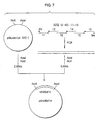
- Each plasmid DNA was produced from 10 clones of the transformants, allowed to react in accordance with the manufacture's instructions attached to AutoRead Sequencing Kit (manufactured by Pharmacia) and then subjected to electrophoresis using A.L.F. DNA Sequencer (manufactured by Pharmacia) to determine the nucleotide sequence, and as a result, the plasmid phKM641H shown in Fig. 7 having the nucleotide sequence of interest was obtained.
- a cDNA encoding the anti-GD3 CDR-grafted antibody VL amino acid sequence hKM641L designed in the item 3(1) of Reference Example was constructed as follows using the PCR similar to the VH. In this case, the amino acid sequence of L chain of anti-GD3 mouse antibody KM641 represented by SEQ ID NO:2 was used as the secretory signal sequence.
- the reaction solution was purified using QIAquick PCR Purification Kit (manufactured by QIAGEN) in accordance with the manufacture's instructions and made into 30 ⁇ l of a buffer comprising 50 mM Tris-HCl (pH 7.5), 100 mM sodium chloride, 10 mM magnesium chloride, 1 mM DTT and 100 ⁇ g/ml BSA, and 10 units of a restriction enzyme Eco RI (manufactured by Takara Shuzo) and 10 units of a restriction enzyme Spl I (manufactured by Takara Shuzo) were further added thereto to carry out the reaction at 37°C for 1 hour.
- a buffer comprising 50 mM Tris-HCl (pH 7.5), 100 mM sodium chloride, 10 mM magnesium chloride, 1 mM DTT and 100 ⁇ g/ml BSA, and 10 units of a restriction enzyme Eco RI (manufactured by Takara Shuzo) and 10 units of a restriction enzyme Spl
- reaction solution was fractionated by agarose gel electrophoresis to recover about 0.2 ⁇ g of an Eco RI- Spl I fragment having about 0.39 kb using QIAquick Gel Extraction Kit (manufactured by QIAGEN) in accordance with the manufacture's instructions.
- a plasmid pBSL3 described in Japanese Published Unexamined Patent Application No. 257893/98 was added to 10 ⁇ l of a buffer comprising 50 mM Tris-HCl (pH 7.5), 100 mM sodium chloride, 10 mM magnesium chloride, 1 mM DTT and 100 ⁇ g/ml BSA, and 10 units of a restriction enzyme Eco RI (manufactured by Takara Shuzo) and 10 units of a restriction enzyme Spl I (manufactured by Takara Shuzo) were further added thereto to carry out the reaction at 37°C for 1 hour.
- a buffer comprising 50 mM Tris-HCl (pH 7.5), 100 mM sodium chloride, 10 mM magnesium chloride, 1 mM DTT and 100 ⁇ g/ml BSA, and 10 units of a restriction enzyme Eco RI (manufactured by Takara Shuzo) and 10 units of a restriction enzyme Spl I (manufactured by Takara
- reaction solution was fractionated by agarose gel electrophoresis to recover about 2 ⁇ g of an Eco RI- Spl I fragment having about 2.95 kb using QIAquick Gel Extraction Kit (manufactured by QIAGEN) in accordance with the manufacture's instructions.
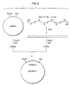
- Each plasmid DNA was prepared from 10 clones of the transformants, allowed to react in accordance with the manufacture's instructions attached to AutoRead Sequencing Kit (manufactured by Pharmacia) and then subjected to electrophoresis using A.L.F. DNA Sequencer (manufactured by Pharmacia) to determine the nucleotide sequence, and as a result, the plasmid phKM641L shown in Fig. 8 having the nucleotide sequence of interest was obtained.
- transient expression of anti-GD3 CDR-grafted antibody was carried out using COS-7 cell (ATCC CRL 1651) as follows.
- transient expression vector pT641 for anti-GD3 chimeric antibody KM871 was constructed as follows.
- the transient expression vector pT641 for anti-GD3 chimeric antibody KM871 was constructed as follows using the transient expression vector pT796 for anti-ganglioside GM2 chimeric antibody KM966 described in Japanese Published Unexamined Patent Application No. 257893/98 and the plasmid pKM641HF1 having VH of anti-GD3 mouse antibody KM641 and plasmid pKM641LA2 having VL of anti-GD3 mouse antibody KM641 described in Japanese Published Unexamined Patent Application No. 304989/93.
- the reaction solution was precipitated with ethanol and added to 10 ⁇ l of a buffer comprising 50 mM Tris-HCl (pH 7.5), 100 mM sodium chloride, 10 mM magnesium chloride and 1 mM DTT, and 10 units of a restriction enzyme Eco RI (manufactured by Takara Shuzo) was further added to carry out the reaction at 37°C for 1 hour.
- the reaction solution was fractionated by agarose gel electrophoresis to recover about 0.2 ⁇ g of a Hin dIII- Eco RI fragment having about 0.35 kb using QIAquick Gel Extraction Kit (manufactured by QIAGEN) in accordance with the manufacture's instructions.
- 3 ⁇ g of the transient expression vector pT796 for anti-ganglioside GM2 chimeric antibody KM966 described in Japanese Published Unexamined Patent Application No. 257893/98 was added to 10 ⁇ l of a buffer comprising 50 mM Tris-HCl (pH 7.5), 100 mM sodium chloride, 10 mM magnesium chloride, 1 mM DTT and 100 ⁇ g/ml BSA, and 10 units of a restriction enzyme Eco RI (manufactured by Takara Shuzo) and 10 units of a restriction enzyme Spl I (manufactured by Takara Shuzo) were added thereto to carry out the reaction at 37°C for 1 hour.
- a buffer comprising 50 mM Tris-HCl (pH 7.5), 100 mM sodium chloride, 10 mM magnesium chloride, 1 mM DTT and 100 ⁇ g/ml BSA, and 10 units of a restriction enzyme Eco RI (manufactured by Takara Shuzo
- reaction solution was fractionated by agarose gel electrophoresis to recover about 2 ⁇ g of an Eco RI- Spl I fragment having about 9.20 kb using QIAquick Gel Extraction Kit (manufactured by QIAGEN) in accordance with the manufacture's instructions.
- the thus obtained plasmid (10 ⁇ g) was allowed to react in accordance with the manufacture's instructions attached to AutoRead Sequencing Kit (manufactured by Pharmacia) and then subjected to an electrophoresis using A.L.F. DNA Sequencer (manufactured by Pharmacia) to determine the nucleotide sequence, and as a result, it was confirmed that a plasmid into which the DNA of interest was cloned was obtained.
- a plasmid pBluescript SK(-) (manufactured by Stratagene) was added to 10 ⁇ l of a buffer comprising 10 mM Tris-HCl (pH 7.5), 50 mM sodium chloride, 10 mM magnesium chloride, 1 mM DTT and 100 ⁇ g/ml BSA, and 10 units of a restriction enzyme Xba I (manufactured by Takara Shuzo) was further added thereto to carry out the reaction at 37°C for 1 hour.
- a buffer comprising 10 mM Tris-HCl (pH 7.5), 50 mM sodium chloride, 10 mM magnesium chloride, 1 mM DTT and 100 ⁇ g/ml BSA, and 10 units of a restriction enzyme Xba I (manufactured by Takara Shuzo) was further added thereto to carry out the reaction at 37°C for 1 hour.
- reaction solution was precipitated with ethanol, added to 30 ⁇ l of a buffer comprising 30 mM sodium acetate (pH 5.0), 100 mM sodium chloride, 1 mM zinc acetate and 5% glycerol, and 30 units of a modification enzyme Mung Bean Nuclease (manufactured by Takara Shuzo) were further added thereto to carry out the reaction at 25°C for 15 minutes.
- a buffer comprising 30 mM sodium acetate (pH 5.0), 100 mM sodium chloride, 1 mM zinc acetate and 5% glycerol, and 30 units of a modification enzyme Mung Bean Nuclease (manufactured by Takara Shuzo) were further added thereto to carry out the reaction at 25°C for 15 minutes.
- the reaction solution was precipitated with ethanol, added to 10 ⁇ l of a buffer comprising 10 mM Tris-HCl (pH 7.5), 10 mM magnesium chloride and 1 mM DTT, and 10 units of a restriction enzyme Apa I (manufactured by Takara Shuzo) was further added thereto to carry out the reaction at 37°C for 1 hour.
- the reaction solution was fractionated by agarose gel electrophoresis to recover about 2 ⁇ g of a blunt end- Apa I fragment having about 2.95 kb using QIAquick Gel Extraction Kit (manufactured by QIAGEN) in accordance with the manufacture's instructions.
- plasmid pKM641HF1 was added to 10 ⁇ l of a buffer comprising 50 mM Tris-HCl (pH 7.5), 100 mM sodium chloride, 10 mM magnesium chloride and 1 mM DTT, and 10 units of a restriction enzyme Eco RI (manufactured by Takara Shuzo) was further added thereto to carry out the reaction at 37°C for 1 hour.
- the reaction solution was precipitated with ethanol, and the 5' protruding end formed by the restriction enzyme digestion was blunt-ended using DNA Blunting Kit (manufactured by Takara Shuzo).
- the reaction solution was precipitated with ethanol, added to 10 ⁇ l of a buffer comprising 10 mM Tris-HCl (pH 7.5), 10 mM magnesium chloride and 1 mM DTT, and 10 units of a restriction enzyme Apa I (manufactured by Takara Shuzo) was further added thereto to carry out the reaction at 37°C for 1 hour.
- the reaction solution was fractionated by agarose gel electrophoresis to recover about 0.2 ⁇ g of a blunt end- Apa I fragment having about 0.44 kb using QIAquick Gel Extraction Kit (manufactured by QIAGEN) in accordance with the manufacture's instructions.
- 0.1 ⁇ g of the blunt end- Apa I fragment derived from the plasmid pBluescript SK(-) and 0.1 ⁇ g of the blunt end- Apa I fragment derived from the plasmid pKM641HF1, obtained in the above, were added to 20 ⁇ l in total volume of sterile water and ligated using Ready-To-Go T4 DNA Ligase (manufactured by Pharmacia).
- E. coli HB101 was transformed to obtain the plasmid pBS641H shown in Fig. 10.
- the reaction solution was precipitated with ethanol, added to 10 ⁇ l of a buffer comprising 50 mM Tris-HCl (pH 7.5), 100 mM sodium chloride, 10 mM magnesium chloride, 1 mM DTT, 100 ⁇ g/ml BSA and 0.01% Triton X-100, and 10 units of a restriction enzyme Not I (manufactured by Takara Shuzo) was further added thereto to carry out the reaction at 37°C for 1 hour.
- a buffer comprising 50 mM Tris-HCl (pH 7.5), 100 mM sodium chloride, 10 mM magnesium chloride, 1 mM DTT, 100 ⁇ g/ml BSA and 0.01% Triton X-100
- reaction solution was fractionated by agarose gel electrophoresis to recover about 0.2 ⁇ g of an Apa I- Not I fragment having about 9.16 kb using QIAquick Gel Extraction Kit (manufactured by QIAGEN) in accordance with the manufacture's instructions.
- the reaction solution was precipitated with ethanol, added to 10 ⁇ l of a buffer comprising 50 mM Tris-HCl (pH 7.5), 100 mM sodium chloride, 10 mM magnesium chloride, 1 mM DTT, 100 ⁇ g/ml BSA and 0.01% Triton X-100, and 10 units of a restriction enzyme Not I (manufactured by Takara Shuzo) was further added thereto to carry out the reaction at 37°C for 1 hour.
- a buffer comprising 50 mM Tris-HCl (pH 7.5), 100 mM sodium chloride, 10 mM magnesium chloride, 1 mM DTT, 100 ⁇ g/ml BSA and 0.01% Triton X-100
- reaction solution was fractionated by agarose gel electrophoresis to recover about 2 ⁇ g of an Apa I- Not I fragment having about 0.44 kb using QIAquick Gel Extraction Kit (manufactured by QIAGEN) in accordance with the manufacture's instructions.
- a transient expression vector for anti-GD3 CDR-grafted antibody was constructed as follows using the transient expression vector pT641 for anti-GD3 chimeric antibody described in the item 4(1) of Reference Example and the plasmids phKM641H and phKM641L described in the items 3(2) and(3) of Reference Example.
- the reaction solution was precipitated with ethanol, added to 10 ⁇ l of a buffer comprising 50 mM Tris-HCl (pH 7.5), 100 mM sodium chloride, 10 mM magnesium chloride, 1 mM DTT, 100 ⁇ g/ml BSA and 0.01% Triton X-100, and 10 units of a restriction enzyme Not I (manufactured by Takara Shuzo) was further added thereto to carry out the reaction at 37°C for 1 hour.
- a buffer comprising 50 mM Tris-HCl (pH 7.5), 100 mM sodium chloride, 10 mM magnesium chloride, 1 mM DTT, 100 ⁇ g/ml BSA and 0.01% Triton X-100
- reaction solution was fractionated by agarose gel electrophoresis to recover about 0.2 ⁇ g of an Apa I- Not I fragment having about 0.44 kb using QIAquick Gel Extraction Kit (manufactured by QIAGEN) in accordance with the manufacture's instructions.
- the reaction solution was precipitated with ethanol, added to 10 ⁇ l of a buffer comprising 50 mM Tris-HCl (pH 7.5), 100 mM sodium chloride, 10 mM magnesium chloride, 1 mM DTT, 100 ⁇ g/ml BSA and 0.01% Triton X-100, and 10 units of a restriction enzyme Not I (manufactured by Takara Shuzo) was further added thereto to carry out the reaction at 37°C for 1 hour.
- a buffer comprising 50 mM Tris-HCl (pH 7.5), 100 mM sodium chloride, 10 mM magnesium chloride, 1 mM DTT, 100 ⁇ g/ml BSA and 0.01% Triton X-100
- reaction solution was fractionated by agarose gel electrophoresis to recover about 2 ⁇ g of an Apa I- Not I fragment having about 9.16 kb using QIAquick Gel Extraction Kit (manufactured by QIAGEN) in accordance with the manufacture's instructions.
- 0.1 ⁇ g of the Apa I- Not I fragment derived from the plasmid phKM641H and 0.1 ⁇ g of the Apa I- Not I fragment derived from the plasmid pT641 were added to 20 ⁇ l in total volume of sterile water and ligated using Ready-To-Go T4 DNA Ligase (manufactured by Pharmacia).
- E. coli HB101 was transformed to obtain the plasmid pT641HCDR shown in Fig. 12.
- 3 ⁇ g of the plasmid phKM641L obtained in the item 3(3) of Reference Example was added to 10 ⁇ l of a buffer comprising 50 mM Tris-HCl (pH 7.5), 100 mM sodium chloride, 10 mM magnesium chloride, 1 mM DTT and 100 ⁇ g/ml BSA, and 10 units of a restriction enzyme Eco RI (manufactured by Takara Shuzo) and 10 units of a restriction enzyme Spl I (manufactured by Takara Shuzo) were further added thereto to carry out the reaction at 37°C for 1 hour.
- a buffer comprising 50 mM Tris-HCl (pH 7.5), 100 mM sodium chloride, 10 mM magnesium chloride, 1 mM DTT and 100 ⁇ g/ml BSA, and 10 units of a restriction enzyme Eco RI (manufactured by Takara Shuzo) and 10 units of a restriction enzyme Spl I (manufactured by Takara Shuz
- reaction solution was fractionated by agarose gel electrophoresis to recover about 0.2 ⁇ g of an Eco RI- Spl I fragment having about 0.39 kb using QIAquick Gel Extraction Kit (manufactured by QIAGEN) in accordance with the manufacture's instructions.
- reaction solution was fractionated by agarose gel electrophoresis to recover about 2 ⁇ g of an Eco RI- Spl I fragment having about 9.20 kb derived from each plasmid using QIAquick Gel Extraction Kit (manufactured by QIAGEN) in accordance with the manufacture's instructions.
- Transient expression of antibodies was carried out using the transient expression vector pT641 for anti-GD3 chimeric antibody, the transient expression vector pT641HLCDR for anti-GD3 CDR-grafted antibody and the transient expression vectors pT641HCDR and pT641LCDR for anti-GD3 hybrid antibodies having V regions of mouse antibody and human CDR-grafted antibody, obtained in the items 4(1) and (2) of Reference Example.
- COS-7 cells (ATCC CRL 1651) were dispensed in 2 ml at a density of 1 ⁇ 10 5 cells/ml into a 6 well plate (manufactured by Falcon) and cultured overnight at 37°C.
- OPTI-MEM medium manufactured by GIBCO BRL
- a solution in which 10 ⁇ l of LIPOFECTAMINE Reagent was added to 100 ⁇ l of the OPTI-MEM medium
- the reaction was carried out at room temperature for 40 minutes for formation of a DNA-liposome complex.
- the overnight-cultured COS-7 cells were washed twice with 2 ml of the OPTI-MEM medium (manufactured by GIBCO BRL), a solution prepared by adding 0.8 ml of OPTI-MEM medium to the complex-containing solution was added thereto, followed by culturing at 37°C for 7 hours, the solution was discarded and then the cells were mixed with 2 ml of 10% FBS-containing DME medium (manufactured by GIBCO BRL) and cultured at 37°C.
- the OPTI-MEM medium manufactured by GIBCO BRL
- the culture supernatant was recovered 72 hours thereafter and its concentration operation was carried out as occasion demands, and then binding activity of the anti-GD3 humanized antibody for GD3 (manufactured by DIA-IATRON) in the culture supernatant was measured by the ELISA described in the item 2 of Reference Example, by measuring concentration of the anti-GD3 humanized antibody in the culture supernatant by the ELISA described in the item 4 which will be described later and calculating the activity from the measured values as relative activity (%) when the activity of the positive control anti-GD3 chimeric antibody is defined as 100. Results are shown in Fig. 14. As shown in Fig.
- the anti-GD3 hybrid antibody derived from the transient expression vector pT641HCDR (VH is derived from a human CDR-grafted antibody and VL is derived from a chimeric antibody) showed almost the same binding activity of that of the anti-GD3 hybrid antibody derived from the transient expression vector pT641, but the anti-GD3 hybrid antibody derived from the transient expression vector pT641LCDR (VH is derived from a chimeric antibody and VL is derived from a human CDR-grafted antibody) showed a binding activity of about 50% of the anti-GD3 chimeric antibody.
- the anti-GD3 CDR-grafted antibody derived from the transient expression vector pT641HLCDR showed a binding activity of only about 5% of the anti-GD3 chimeric antibody, showing considerable decrease in the activity.
- binding activity of the anti-GD3 CDR-grafted antibody having VH and VL designed in the item 3(1) of Reference Example is considerably reduced and the main cause of the activity reduction is considered to be due to VL, but since about 50% of the activity is found by its combination with the VH of anti-GD3 chimeric antibody, it was suggested that there is a problem particularly at the interaction region with VH. Accordingly, an attempt was made to increase the antibody activity by further modifying amino acid residues of the VL designed in the item 3(1) of Reference Example.
- the transient expression culture supernatants obtained in the item 4(3) of Reference Example or diluted solutions of the purified anti-GD3 chimeric antibody KM871 were added in 50 ⁇ l/well and allowed to react at room temperature for 1 hour. After the reaction, each well was washed with Tween-PBS and then a solution prepared by diluting a peroxidase-labeled mouse anti-human ⁇ L chain antibody (manufactured by Zymed) 500 folds with PBS was added in 50 ⁇ l/well and allowed to react at room temperature for 1 hour.
- an ABTS substrate solution (a solution prepared by dissolving 0.55 g of 2,2'-azinobis(3-ethylbenzothiazoline-6-sulfonic acid) in 1 L of 0.1 M citrate buffer (pH 4.2) and adding 1 ⁇ l/ml hydrogen peroxide just before the use) was added in 50 ⁇ l/well for color development and OD415 was measured.
- amino acid residues which are amino acid residues positioned in the interaction region of VH and VL and amino acid residues which are considered to have influence on the three-dimensional structure of each CDR of VL and are also different from the amino acid residues of mouse antibody in the VL of human CDR-grafted antibody were identified from computer models of the three-dimensional structures of various antibodies constructed in the item 3(1) of Reference Example.
- hKM641L as the amino acid sequence of VL of anti-GD3 CDR-grafted antibody represented by SEQ ID NO:10 Ser at position 7, Pro at position 8, Ser at position 12, Gly at position 41, Pro at position 44, Thr at position 72, Ser at position 77, Phe at position 83 and Tyr at position 87 were identified.
- VLs of modified anti-GD3 CDR-grafted antibody were designed. That is, in hKM641NL, Pro at position 8, Ser at position 12, Pro at position 44 and Tyr at position 87 in the hKM641L amino sequence were replaced by Ala, Pro, Val and Phe, respectively. In hKM641Lm-1, Ser at position 7, Pro at position 8 and Ser at position 12 in the hKM641L amino sequence were replaced by Thr, Ala and Pro, respectively. In hKM641Lm-4, Tyr at position 87 in the hKM641L amino sequence was replaced by Phe.
- hKM641Lm-6 Gly at position 41 and Pro at position 44 in the hKM641L amino sequence were represented by Asp and Val, respectively.
- hKM641Lm-7 Thr at position 72, Ser at position 77, Phe at position 83 and Tyr at position 87 in the hKM641L amino sequence were replaced by Ser, Asn, Ile and Phe, respectively.
- hKM641Lm-8 Ser at position 77 in the hKM641L amino sequence was replaced by Asn.
- Phe at position 83 and Tyr at position 87 in the hKM641L amino sequence were replaced by Ile and Phe, respectively.
- hKM641Lm-69 Gly at position 41, Pro at position 44 and Phe at position 83 in the hKM641L amino sequence were replaced by Asp, Val and Ile, respectively.
- antisense chain and sense chain DNA primers were synthesized using an automatic DNA synthesizer (380A, manufactured by Applied Biosystems) for introducing mutation, and a first PCR was carried out in accordance with the method described in the item 3(2) of Reference Example using 1 ng of the plasmid phKM641L as the template and 0.5 ⁇ M in final concentration of M13 primer RV (manufactured by Takara Shuzo) and the antisense chain DNA primer and M13 primer M4 (manufactured by Takara Shuzo) and the sense chain DNA primer.
- an automatic DNA synthesizer (380A, manufactured by Applied Biosystems) for introducing mutation
- reaction solutions were purified using QIAquick PCR Purification Kit (manufactured by QIAGEN) in accordance with the manufacture's instructions by eluting with 20 ⁇ l of 10 mM Tris-HCl (pH 8.0), and then a second PCR was carried out using 5 ⁇ l of each eluate in accordance with the method described in the item 3(2) of Reference Example.
- QIAquick PCR Purification Kit manufactured by QIAGEN
- the reaction solution was purified using QIAquick PCR Purification Kit (manufactured by QIAGEN) in accordance with the manufacture's instructions and made into 30 ⁇ l of a buffer comprising 50 mM Tris-HCl (pH 7.5), 100 mM sodium chloride, 10 mM magnesium chloride, 1 mM DTT and 100 ⁇ g/ml BSA, and 10 units of a restriction enzyme Eco RI (manufactured by Takara Shuzo) and 10 units of a restriction enzyme Spl I (manufactured by Takara Shuzo) were further added thereto to carry out the reaction at 37°C for 1 hour.
- a buffer comprising 50 mM Tris-HCl (pH 7.5), 100 mM sodium chloride, 10 mM magnesium chloride, 1 mM DTT and 100 ⁇ g/ml BSA, and 10 units of a restriction enzyme Eco RI (manufactured by Takara Shuzo) and 10 units of a restriction enzyme Spl
- reaction solution was fractionated by agarose gel electrophoresis to recover about 0.2 ⁇ g of an Eco RI- Spl I fragment having about 0.39 kb using QIAquick Gel Extraction Kit (manufactured by QIAGEN) in accordance with the manufacture's instructions.
- Each plasmid DNA was produced from 10 clones of the transformants, allowed to react in accordance with the manufacture's instructions attached to AutoRead Sequencing Kit (manufactured by Pharmacia) and then subjected to electrophoresis using A.L.F. DNA Sequencer (manufactured by Pharmacia) to determine the nucleotide sequence to thereby obtain a plasmid having cDNA to which the intended modification was applied.
- AutoRead Sequencing Kit manufactured by Pharmacia
- A.L.F. DNA Sequencer manufactured by Pharmacia
- a plasmid phKM641Lm-1 having the nucleotide sequence represented by SEQ ID NO:27 was obtained by carrying out a series of the above operations using the synthetic DNA of SEQ ID NO:25 as the antisense chain DNA primer and the synthetic DNA of SEQ ID NO:26 as the sense chain DNA primer.
- the hKM641Lm-1 as an amino acid sequence encoded by the nucleotide sequence is also represented by SEQ ID NO:27.
- a plasmid phKM641Lm-4 having the nucleotide sequence represented by SEQ ID NO:30 was obtained by carrying out a series of the above operations using the synthetic DNA of SEQ ID NO:28 as the antisense chain DNA primer and the synthetic DNA of SEQ ID NO:29 as the sense chain DNA primer.
- the hKM641Lm-4 as an amino acid sequence encoded by the nucleotide sequence is also represented by SEQ ID NO:30.
- a plasmid phKM641Lm-6 having the nucleotide sequence represented by SEQ ID NO:33 was obtained by carrying out a series of the above operations using the synthetic DNA of SEQ ID NO:31 as the antisense chain DNA primer and the synthetic DNA of SEQ ID NO:32 as the sense chain DNA primer.
- the hKM641Lm-6 as an amino acid sequence encoded by the nucleotide sequence is also represented by SEQ ID NO:33.
- a plasmid phKM641Lm-7 having the nucleotide sequence represented by SEQ ID NO:36 was obtained by carrying out a series of the above operations using the synthetic DNA of SEQ ID NO:34 as the antisense chain DNA primer and the synthetic DNA of SEQ ID NO:35 as the sense chain DNA primer.
- the hKM641Lm-7 as an amino acid sequence encoded by the nucleotide sequence is also represented by SEQ ID NO:36.
- a plasmid phKM641Lm-8 having the nucleotide sequence represented by SEQ ID NO:39 was obtained by carrying out a series of the above operations using the synthetic DNA of SEQ ID NO:37 as the antisense chain DNA primer and the synthetic DNA of SEQ ID NO:38 as the sense chain DNA primer.
- the hKM641Lm-8 as an amino acid sequence encoded by the nucleotide sequence is also represented by SEQ ID NO:39.
- a plasmid phKM641Lm-9 having the nucleotide sequence represented by SEQ ID NO:42 was obtained by carrying out a series of the above operations using the synthetic DNA of SEQ ID NO:40 as the antisense chain DNA primer and the synthetic DNA of SEQ ID NO:41 as the sense chain DNA primer.
- the hKM641Lm-9 as an amino acid sequence encoded by the nucleotide sequence is also represented by SEQ ID NO:42.
- cDNA encoding the hKM641NL was constructed by synthesizing 6 synthetic DNA fragments of SEQ ID NOs:17, 22 and 43 to 46 using an automatic DNA synthesizer (380A, manufactured by Applied Biosystems) and carrying out the procedure described in the item 3(3) of Reference Example using these fragments.
- an automatic DNA synthesizer (380A, manufactured by Applied Biosystems) and carrying out the procedure described in the item 3(3) of Reference Example using these fragments.
- a plasmid phKM641NL having the nucleotide sequence represented by SEQ ID NO:47 in which the modification of interest was carried out was obtained.
- the hKM641NL as an amino acid sequence encoded by the nucleotide sequence is also represented by SEQ ID NO:47.
- cDNA encoding the hKM641Lm-69 was constructed as follows using the plasmids phKM641Lm-6 and phKM641Lm-9 having cDNA of modified VL obtained in the above.
- 3 ⁇ g of the plasmid phKM641Lm-6 was added to 10 ⁇ l of a buffer comprising 50 mM Tris-HCl (pH 7.5), 100 mM sodium chloride, 10 mM magnesium chloride and 1 mM DTT and 10 units of a restriction enzyme Eco RI (manufactured by Takara Shuzo) and 10 units of a restriction enzyme Pst I (manufactured by Takara Shuzo) were further added thereto to carry out the reaction at 37°C for 1 hour.
- a buffer comprising 50 mM Tris-HCl (pH 7.5), 100 mM sodium chloride, 10 mM magnesium chloride and 1 mM DTT and 10 units of a restriction enzyme Eco RI (manufactured by Takara Shuzo) and 10 units of a restriction enzyme Pst I (manufactured by Takara Shuzo) were further added thereto to carry out the reaction at 37°C for 1 hour.
- reaction solution was fractionated by agarose gel electrophoresis to recover about 0.3 ⁇ g of an Eco RI- Pst I fragment having about 0.30 kb using QIAquick Gel Extraction Kit (manufactured by QIAGEN) in accordance with the manufacture's instructions.
- plasmid phKM641Lm-9 was added to 10 ⁇ l of a buffer comprising 50 mM Tris-HCl (pH 7.5), 100 mM sodium chloride, 10 mM magnesium chloride and 1 mM DTT and 10 units of a restriction enzyme Eco RI (manufactured by Takara Shuzo) and 10 units of a restriction enzyme Pst I (manufactured by Takara Shuzo) were further added thereto to carry out the reaction at 37°C for 1 hour.
- a buffer comprising 50 mM Tris-HCl (pH 7.5), 100 mM sodium chloride, 10 mM magnesium chloride and 1 mM DTT and 10 units of a restriction enzyme Eco RI (manufactured by Takara Shuzo) and 10 units of a restriction enzyme Pst I (manufactured by Takara Shuzo) were further added thereto to carry out the reaction at 37°C for 1 hour.
- reaction solution was fractionated by agarose gel electrophoresis to recover about 2 ⁇ g of an Eco RI- Pst I fragment having about 3.05 kb using QIAquick Gel Extraction Kit (manufactured by QIAGEN) in accordance with the manufacture's instructions.
- the reaction was carried out using 10 ⁇ g of the thus obtained plasmid in accordance with the manufacture's instructions attached to AutoRead Sequencing Kit (manufactured by Pharmacia) and then the resulting mixture was subjected to electrophoresis using A.L.F. DNA Sequencer (manufactured by Pharmacia) to determine the nucleotide sequence, and as a result, it was confirmed that it has the nucleotide sequence represented by SEQ ID NO:48 in which the modification of interest was carried out.
- the hKM641Lm-69 as an amino acid sequence encoded by the nucleotide sequence is also represented by SEQ ID NO:48
- Transient expression vectors for anti-GD3 CDR-grafted antibodies having various modified VLs were constructed as follows using the plasmids having cDNA molecules encoding various modified VLs, obtained in the item 5(1) of Reference Example, and the transient expression vector pT641HCDR for anti-GD3 hybrid antibodies obtained in the item 4(2) of Reference Example.
- reaction solution was fractionated by agarose gel electrophoresis to recover about 0.2 ⁇ g of an Eco RI- Spl I fragment having about 0.39 kb using QIAquick Gel Extraction Kit (manufactured by QIAGEN) in accordance with the manufacture's instructions.
- coli HB101 was transformed to obtain the transient expression vectors for anti-GD3 CDR-grafted antibodies having various modified VLs, pT641HLCDRNL, pT641HLCDRLm-1, pT641HLCDRLm-4, pT641HLCDRLm-6, pT641HLCDRLm-7, pT641HLCDRLm-8, pT641HLCDRLm-9 and pT641HLCDRLm-69, shown in Fig. 16.
- Transient expression and evaluation of activity of each antibody were carried out in accordance with the method described in the item 4(3) of Reference Example using the transient expression vector pT641 for ant-GD3 chimeric antibody and the transient expression vector pT641HLCDR for anti-GD3 CDR-grafted antibody, both obtained in the item 4(2) of Reference Example, and the transient expression vectors, pT641HLCDRNL, pT641HLCDRLm-1, pT641HLCDRLm-4, pT641HLCDRLm-6, pT641HLCDRLm-7, pT641HLCDRLm-8, pT641HLCDRLm-9 and pT641HLCDRLm-69, for anti-GD3 CDR-grafted antibodies having various modified VLs, obtained in the item 5(2) of Reference Example.
- results are shown in Fig. 17.
- the modified anti-GD3 CDR-grafted antibodies derived from the transient expression vectors, pT641HLCDRLm-6, pT641HLCDRLm-7, pT641HLCDRLm-9 and pT641HLCDRLm-69 showed increased binding activity in comparison with the anti-GD3 CDR-grafted antibodies before modification, particularly, the modified anti-GD3 CDR-grafted antibody derived from the transient expression vector pT641HLCDRLm-69 showed its binding activity of about 80% of the anti-GD3 chimeric antibody.
- the modified anti-GD3 CDR-grafted antibodies derived from other transient expression vectors showed no increase in the binding activity in comparison with the anti-GD3 CDR-grafted antibodies before modification. Based on these results, it was found that, among the modified amino acid residues of VL identified in the item 5(1) of Reference Example, amino acid residues as positions 41, 44, 83 and 87 greatly contribute to the increase of binding activity. It was also found that simultaneous substitution of two amino acid residues at positions 41 and 44 or at positions 83 and 87 contributes to synergistic increase in the activity, and simultaneous modification of four amino acid residues at positions 41, 44, 83 and 87 also contributes to synergistic increase in the activity.
- the anti-GD3 CDR-grafted antibodies derived from transient expression vectors pT641HLCDRLm-9 and pT641HLCDRLm-69 have about 20% and about 80% of binding activity for GD3, respectively, in comparison with anti-GD3 chimeric antibodies. Accordingly, in order to evaluate the activity of these anti-GD3 CDR-grafted antibodies more in detail, transformed cell lines capable of stably expressing anti-GD3 CDR-grafted antibodies were obtained by the following method, and anti-GD3 CDR-grafted antibodies from culture supernatants of the transformed cell lines were purified. Also, for comparison, stable expression and purification of the anti-GD3 CDR-grafted antibody derived from the transient expression vector pT641HLCDR were carried out in the same manner.
- Stable expression vectors were constructed by introducing a resistance gene against an agent G418 and the dhfr gene into various transient expression vectors constructed in the item 5(2) of Reference Example.
- 3 ⁇ g of the humanized antibody expression vector pKANTEX93 described in WO 97/10354 was added to 10 ⁇ l of a buffer comprising 20 mM Tris-HCl (pH 8.5), 10 mM magnesium chloride, 1 mM DTT and 100 mM potassium chloride, and 10 units of a restriction enzyme Bam HI (manufactured by Takara Shuzo) and 10 units of a restriction enzyme Xho I (manufactured by Takara Shuzo) were further added thereto to carry out the reaction at 37°C for 1 hour.
- a buffer comprising 20 mM Tris-HCl (pH 8.5), 10 mM magnesium chloride, 1 mM DTT and 100 mM potassium chloride, and 10 units of a restriction enzyme Bam HI (manufactured by Takara Shuzo) and 10 units of a restriction enzyme Xho I (manufactured by Takara Shuzo) were further added thereto to carry out the reaction at
- reaction solution was fractionated by agarose gel electrophoresis to recover about 1 ⁇ g of a Bam HI- Xho I fragment having about 8.68 kb using QIAquick Gel Extraction Kit (manufactured by QIAGEN) in accordance with the manufacture's instructions.
- reaction solution was fractionated by agarose gel electrophoresis to recover about 1 ⁇ g of a Bam HI- Xho I fragment having about 4.90 kb using QIAquick Gel Extraction Kit (manufactured by QIAGEN) in accordance with the manufacture's instructions.
- coli HB101 was transformed to obtain the anti-GD3 CDR-grafted antibody stable expression vectors, pKANTEX641HLCDRLm-9, pKANTEX641HLCDRLm-69 and pKANTEX641HLCDR, shown in Fig. 18.
- Each of the anti-GD3 CDR-grafted antibody expression vectors (4 ⁇ g) was introduced into 4 ⁇ 10 6 cells of a rat myeloma cell line YB2/0 cell (ATCC CRL 1662) by the electroporation method [ Cytotechnology , 3 , 133 (1990)], and the cells were suspended in 40 ml of RPMI640-FBS(10) [RPMI1640 medium containing 10% of fetal bovine serum (FBS)] and dispensed in 200 ⁇ l/well into a 96 well microtiter plate (manufactured by Sumitomo Bakelite).
- G418 was added to give a concentration of 0.5 mg/ml, followed by culturing for 1 to 2 weeks.
- Culture supernatants were recovered from wells in which colonies of transformants showing G418-resistance were formed and became confluent, and the antigen binding activity of various anti-GD3 CDR-grafted antibodies in the supernatants was measured by the ELISA shown in the item 2 of Reference Example (a peroxidase-labeled goat anti-human IgG( ⁇ ) antibody was used as the secondary antibody).
- the MTX concentration was increased to 100 nM and then to 200 nM by the method similar to the above, and transformants capable of growing in the RPMI1640-FBS(10) medium containing 0.5 mg/ml G418 and 200 nM MTX and of highly expressing anti-GD3 CDR-grafted antibodies were finally obtained.
- single cell isolation (cloning) was carried out twice by limiting dilution analysis.
- the transformed cell clones obtained by introducing the stable expression vectors, pKANTEX641HLCDRLm-9, pKANTEX641HLCDRLm-69 and pKANTEX641HLCDR, were named KM8870, KM8871 and KM8869, respectively, and expression levels of anti-GD3 CDR-grafted antibody by transformed cell clones were about 5 ⁇ g/10 6 cells/24 hr, about 10 ⁇ g/10 6 cells/24 hr and about 30 ⁇ g/10 6 cells/24 hr, respectively.
- Each of the transformed cell clones, KM8870, KM8871 and KM8869, obtained in the item 6(2) of Reference Example which express various anti-GD3 CDR-grafted antibodies was suspended in GIT medium (manufactured by Nippon Pharmaceutical) containing 200 nM MTX to give a density of 1 to 2 ⁇ 10 5 cells/ml, and dispensed in 200 ml into 175 cm 2 flasks (manufactured by Greiner). The cells were cultured at 37°C for 5 to 7 days until they became confluent and then the culture supernatant was recovered.
- GIT medium manufactured by Nippon Pharmaceutical
- Each anti-GD3 CDR-grafted antibody was purified from the culture supernatant using Prosep-A (manufactured by Bioprocessing) column in accordance with the manufacture's instructions.
- About 3 mg of anti-GD3 CDR-grafted antibody KM8870 was obtained from 500 ml of the culture supernatant of KM8870, and about 25 mg of anti-GD3 CDR-grafted antibody KM8871 from 1,600 ml of the culture supernatant of KM8871 and about 65 mg of anti-GD3 CDR-grafted antibody KM8869 from 1,000 ml of the culture supernatant of KM8869.
- N-terminal amino acid sequences of the H chain and L chain of each of the purified anti-GD3 CDR-grafted antibodies were analyzed by Edman degradation using a protein sequencer (470A, manufactured by Applied Biosystems), it was confirmed that they coincide with the H chain and L chain N-terminal amino acid sequences deduced from respective cDNA nucleotide sequences.
- Fig. 20 shows a result of the examination of the reactivity carried out by fixing the amount of GD3 to be adsorbed to each well of a plate for ELISA to 20 pmol/well and changing the concentration of anti-GD3 chimeric antibody KM871 and anti-GD3 CDR-grafted antibodies KM8869, KM8870 and KM8871 to be added. As shown in Fig.
- KM8871 showed the highest binding activity which was about 1/2 of the anti-GD3 chimeric antibody KM871.
- Fig. 21 shows a result of the examination of the reactivity of a constant concentration (10 ⁇ g/ml) of anti-GD3 chimeric antibody KM871 and anti-GD3 CDR-grafted antibodies KM8869, KM8870 and KM8871, carried out by changing the amount of GD3 to be adsorbed to each well of a plate for ELISA.
- KM8871 showed the highest binding activity which was equivalent to the anti-GD3 chimeric antibody KM871.
- GM1 and GD1a were purified from bovine brain, and N-glycolylGM2 and N-glycolylGM3 from mouse liver, N-acetylGM3 from canine erythrocyte and GD2 from a human neuroblastoma culture cell line IMR32 (ATCC CCL 127), respectively in accordance with the known method [ J. Biol. Chem ., 263 , 10915 (1988)]. As shown in Fig.
- each of the anti-GD3 CDR-grafted antibodies KM8869, KM8870 and KM8871 bound strongly to GD3 and weakly to GQ1b similar to the case of the anti-GD3 chimeric antibody KM871 but did not bind to other gangliosides. Thus, it was shown that they maintain the binding specificity of chimeric antibody KM871.
- Reactivity of purified anti-GD3 CDR-grafted antibodies KM8869, KM8870 and KM8871 with human cancer cells was measured as follows. That is, 1 ⁇ 10 6 cells of each of human melanoma culture cell lines G-361 (ATCC CRL 1424) and SK-MEL-28 (ATCC HTB72) were suspended in PBS, put into microtubes and centrifuged (2,000 rpm for 2 minutes), and the thus washed cells were stirred after adding 50 ⁇ l of the anti-GD3 chimeric antibody KM871 or the anti-GD3 CDR-grafted antibody, KM8869, KM8870 or KM8871 (a solution adjusted to 5 ⁇ g/ml with 1% BSA-PBS) and then allowed to react at 4°C for 1 hour.
- the CDC activity was measured by the following method.
- Sera of three healthy persons were mixed and used as the human complement source. At the time of its use, it was diluted to 15% vol/vol with RPMI1640-FBS(10) medium and used as the complement solution.
- the amount of 51 Cr released in the supernatant was measured by a ⁇ -counter.
- the amount of the spontaneously dissociated 51 Cr was calculated by carrying out the same procedure using the medium alone instead of the antibody solution and complement solution and measuring the amount of 51 Cr in the supernatant.
- Amount of the total dissociated 51 Cr was calculated by carrying out the same procedure by adding the medium alone instead of the antibody solution, and 5 N sodium hydroxide instead of the complement solution, and measuring the amount of 51 Cr in the supernatant.
- Results are shown in Fig. 24.
- Fig. 24 it was found that all of the anti-GD3 chimeric antibody KM871 and the anti-GD3 CDR-grafted antibodies KM8869, KM8870 and KM8871 show the CDC activity for both cell lines, particularly show remarkably strong cytotoxic activity for the human melanoma culture cell line G-361.
- KM8871 showed the highest cytotoxic activity which was about 1/3 of the anti-GD3 chimeric antibody KM871.
- ADCC activity In vitro cytotoxic activity of anti-GD3 CDR-grafted antibody
- the ADCC activity was measured by the following method.
- Healthy human vein blood 50 ml was collected, and 0.5 ml of heparin sodium (manufactured by Takeda Pharmaceutical) was gently added thereto.
- the resulting mixture was centrifuged (1,500 to 1,800 ⁇ g for 30 minutes) using Polymorphprep (manufactured by Nycomed Pharma AS) in accordance with the manufacture's instructions to separate a mononuclear cell layer.
- the separated cells were centrifuged (1,500 to 1,800 ⁇ g for 5 minutes) three times with RPMI1640-FBS(10) medium for washing and then re-suspended in the medium to give a density of 5 ⁇ 10 6 cells/ml to be used as the effector cell suspension.
- each well of a 96 well U bottom plate 50 ⁇ l of the target cell suspension prepared in "a.” was dispensed (1 ⁇ 10 4 cells/well). Next, 100 ⁇ l of the effector cell suspension prepared in "b.” was added (5 ⁇ 10 5 cells/well, ratio of the effector cells to the target cells becomes 50:1). Thereafter, each of the anti-GD3 chimeric antibody KM871 and the anti-GD3 CDR-grafted antibodies KM8869, KM8870 and KM8871 was added to give a final concentration of 0.05 to 50 ⁇ g/ml and allowed to react at 37°C for 4 hours.
- the amount of the spontaneously dissociated 51 Cr was calculated by carrying out the same procedure using the medium alone instead of the effector cell suspension and antibody solution and measuring the amount of 51 Cr in the supernatant.
- the amount of the total dissociated 51 Cr was calculated by carrying out the same procedure by adding the medium alone instead of the antibody solution, and 5 N sodium hydroxide instead of the effector cell suspension, and measuring the amount of 51 Cr in the supernatant.
- Results are shown in Fig. 25.
- Fig. 25 it was found that all of the anti-GD3 chimeric antibody KM871 and the anti-GD3 CDR-grafted antibodies KM8869, KM8870 and KM8871, show strong ADCC activity for both cell lines.
- the anti-GD3 chimeric antibody KM871 and the anti-GD3 CDR-grafted antibodies KM8869, KM8870 and KM8871 difference in the cytotoxic activity was not found.
- the anti-GD3 CDR-grafted antibodies are useful in the diagnosis, treatment and the like of GD3-positive human tumors similar to the case of the anti-GD3 chimeric antibody KM871.
- immunogenicity of the anti-GD3 CDR-grafted antibodies is reduced in human and the effects are further prolonged.
- the transformed cell clone KM8871 capable of producing KM8871 which showed the highest activity among the anti-GD3 CDR-grafted antibodies has been deposited on July 22, 1999, as FERM BP-6790 in International Patent Organism Depositary, National Institute of Advanced Industrial Science and Technology (AIST Tsukuba Central 6, 1-1, Higashi 1-Chome Tsukuba-shi, Ibaraki-ken, Japan).
- the present invention relates to a medicament which comprises a gene recombinant antibody against ganglioside GD3 or the antibody fragment thereof in combination with at least one of a substance which activates an immunocompetent cell and a substance having an antitumor activity.
- the medicament of the present invention show higher therapeutic effects at a lower dose than the case where the antibody, the substance which activates an immunocompetent cell or the substance having an antitumor activity is administered alone.
Landscapes
- Health & Medical Sciences (AREA)
- Chemical & Material Sciences (AREA)
- Life Sciences & Earth Sciences (AREA)
- Medicinal Chemistry (AREA)
- General Health & Medical Sciences (AREA)
- Immunology (AREA)
- Organic Chemistry (AREA)
- Animal Behavior & Ethology (AREA)
- Pharmacology & Pharmacy (AREA)
- Engineering & Computer Science (AREA)
- Public Health (AREA)
- Veterinary Medicine (AREA)
- Oncology (AREA)
- Biochemistry (AREA)
- Chemical Kinetics & Catalysis (AREA)
- Biophysics (AREA)
- Genetics & Genomics (AREA)
- Molecular Biology (AREA)
- Proteomics, Peptides & Aminoacids (AREA)
- General Chemical & Material Sciences (AREA)
- Nuclear Medicine, Radiotherapy & Molecular Imaging (AREA)
- Biomedical Technology (AREA)
- Bioinformatics & Cheminformatics (AREA)
- Microbiology (AREA)
- Mycology (AREA)
- Epidemiology (AREA)
- Medicines Containing Antibodies Or Antigens For Use As Internal Diagnostic Agents (AREA)
- Peptides Or Proteins (AREA)
- Pharmaceuticals Containing Other Organic And Inorganic Compounds (AREA)
Abstract
In order to obtain high therapeutic effects in treating malignant tumors,
particularly melanoma, a new therapeutic method having less side effects, or a new
therapeutic method which can provide further high therapeutic effects at conventional
doses of agents has been desired.
An object of the present invention is to provide a medicament which can
provide higher therapeutic effects than any one of a gene recombinant antibody against
ganglioside GD3 or the antibody fragment thereof alone, and a substance which
activates an immunocompetent cell and a substance having an antitumor activity alone,
by combining a gene recombinant antibody against ganglioside GD3 or the antibody
fragment thereof with at least one of a substance which activates an immunocompetent
cell and a substance having an antitumor activity. Also, the medicament is expected to
relieve side effects which were problems in the case of administration of individual
single agents.
Description
The present invention relates to a medicament which comprises a gene
recombinant antibody against ganglioside GD3 or the antibody fragment thereof in
combination with at least one of a substance which activates an immunocompetent cell
and a substance having an antitumor activity.
Ganglioside which is one of glycolipids containing sialic acid is a
constituent of animal cell membrane. Also, ganglioside has a sugar chain as a
hydrophilic side chain and a sphingosine and fatty acid as hydrophobic side chains. It
is known that kinds and expression levels of ganglioside vary depending on the cell
types, organ species, animal species and the like. It is also known that the expression
of ganglioside changes quantitatively and qualitatively in the process of malignant
transformation of cells [Cancer Res., 45, 2405 (1985)].
Particularly, it is known that GD3 is present in an extremely small amount
in normal cells but in a large amount in cancer cells, particularly melanoma, and is
present together with ganglioside GD2 in cancer cells such as sarcoma, glioma and
neuroblastoma [Proc. Natl. Acad Sci. U.S.A., 77, 6114 (1980); J. Exp. Med., 155, 1133
(1982); Cancer Res., 45, 4401 (1985); Cancer, 70, 633 (1992); Acta Neuropathologica,
82, 45 (1991)], and an antibody against GD3 (hereinafter referred to as "anti-GD3
antibody") is considered to be useful for the treatment of these cancers [Melanoma
Research, 7, S115 (1997)].
Administration tests to melanoma patients have already been carried out
using R24 (hereinafter referred to as "anti-GD3 mouse antibody R24") as an anti-GD3
mouse monoclonal antibody, and its clinical responses were observed in a part of the
patients but not to an expected degree because of considerably rapid disappearance of
the antibody from blood due to induction of a human antibody against the mouse
antibody in the body of the patient (human anti mouse antibody: hereinafter referred to
as "HAMA") [Melanoma Research, 7, S115 (1997)].
In order to prevent decrease of therapeutic effects derived from antigenicity
such as generation of HAMA when a mouse antibody is administered to human,
methods for the production of humanized antibodies for attenuating antigenicity for
human have so far been known, such as a human chimeric antibody in which a variable
region derived from an animal other than human and a constant region derived from
human are bound [Proc. Natl. Acad Sci. U.S.A., 81, 6851 (1984)] and a human CDR-grafted
antibody in which, among the variable regions, all of framework regions other
than the ultra-variable region which directly relate to binding to the antigen are further
changed to human-derived sequences [Nature, 321, 522 (1986)]. Regarding antibodies
for melanoma, anti-GD3 human chimeric antibody KM871 (Japanese Published
Unexamined Patent Application No. 304989/93) and anti-GD2 human chimeric
antibody ch14.18 [J. Immunol., 144, 1382, (1990)] are known. The ch14.18 has
already been used in clinical studies and its good blood retentivity has been observed
[Human Antibodies & Hybridomas, 3, 19 (1992)].
Also, since increase in cytotoxic activity via a constant region can be found
by the administration of humanized antibodies to the living body, it is considered that
they are advantageous in view of therapeutic effects particularly for antibodies against
cancers and the like. Since antibody-dependent cell-mediated cytotoxic activity
(hereinafter referred to as "ADCC activity") using human peripheral blood monocyte as
an effector and complement-dependent cytotoxicity (hereinafter referred to as "CDC
activity") using human serum as the complement source are increased by the anti-GD3
human chimeric antibody KM871 in comparison with the original mouse-derived
antibody [Cancer Immunol. Immunother., 36, 373 (1993)], the antibody is considered to
be useful in treating GD3-positive melanoma.
In general chemotherapy, there is a case in which a good therapeutic result
is obtained by its combined use with an antibody even when it is difficult to introduce
remission due to appearance of a drug-resistance tumor cell and the like. Significant
therapeutic results have been obtained from combined use of an anti-HER2/neu
humanized antibody for breast cancer, rhuMAb HER2 (Herceptin/trastuzumab) with a
taxane-series antitumor agent [Clinical Therapeutics, 21, 309 (1999)] and from
combined use of an anti-CD20 human chimeric antibody for B cell lymphoma, IDEC-C2B8
(Rituxan, manufactured by Roche) with polypharmacy [J. Clin. Oncol., 17, 268
(1999)].
Regarding combined use of an antibody with a substance having an
antitumor activity in melanoma treatment, it is known that CDC sensitivity of an anti-GD3
mouse antibody R24 against a complement non-sensitive melanoma cell line is
increased by combined use of an anti-GD3 mouse antibody R24 with doxorubicin [Proc.
Natl. Acad Sci. U.S.A., 83, 9144 (1986)]. However, no significant therapeutic effect
was observed in the phase I clinical study of the combined use of the anti-GD3 mouse
antibody R24 with dacarbazine, combined use of the anti-GD3 mouse antibody R24
with cisplatin or combined use of the anti-GD3 mouse antibody R24 with doxorubicin
[Melanoma Res., 7, Supple 2, S155 (1997)].
Combined therapy using an antibody with cytokine is also expected as new
immunotherapy. Cytokine is a general term for various humoral factors which relate
to cytoclesis in immune response. ADCC activity as one of cytotoxic activities is
induced by effector cells having Fc receptor on the cell surface such as monocyte,
macrophage and NK cell [J. Immunol., 138, 1992 (1987)]. Since various species of
cytokine activate these effector cells, administration in combination with an antibody
has been tried in order to increase ADCC activity and the like of the antibody.
Regarding the anti-GD3 mouse antibody R24, a possibility of combined use
effects with IL-2 has been suggested based on in vitro evaluation (U.S. Patent
5,104,652). Also, clinical studies have been carried out on the combined
administration of the anti-GD3 mouse antibody R24 with IL-2, the anti-GD3 mouse
antibody R24 with IFNα, the anti-GD3 mouse antibody R24 with M-CSF, the anti-GD3
mouse antibody R24 with GM-CSF, and the like [Medical Oncology, 15, 191 (1998),
Clinical Cancer Res., 3, 17 (1997), Cancer, 75, 2251 (1995), Journal Of
Immunotherapy With Emphasis On Tumor Immunology, 16, 132 (1994)]. However,
effects to be expected were not found by these combined therapies due to antigenicity of
the mouse antibody or side effects of cytokine. It is important to predict therapeutic
effects when used in the clinical field, by in vivo evaluation using a tumor model animal
having high extrapolation ability to human melanoma treatment. However, only
combined use of the anti-GD3 mouse antibody R24 with M-CSF is known regarding
examination of antitumor effects by combined administration of the anti-GD3 mouse
antibody R24 with cytokine to animal tumor models [Journal of Immunotherapy with
Emphasis on Tumor Immunology, 19, 317 (1996)].
Melanoma is a malignant tumor in chromocytes and is known as a tumor
having high malignancy, which is apt to generate metastasis and has high resistance
against chemotherapy and the like. Immunotherapy by IL-2 or interferon,
chemotherapy mainly using dacarbazine and cisplatin, combined therapy thereof and the
like has so far been carried out.
As an example of the immunotherapy, a combination of IL-2 with IFNα is
known [J. Clin. Oncol., 7, 1863 (1989), J. Clin. Oncol., 11, 1969 (1993), J.
Immunother., 13, 117 (1993)].
As examples of the chemotherapy, a combination of dacarbazine, cisplatin,
carmustine and tamoxifen [Cancer Treat. Rep., 68, 1403 (1984), Cancer, 63, 1292
(1989)], a combination of dacarbazine, cisplatin and vinblastine [Proc. ASCO, 12, 1328
(1993)], a combination of dacarbazine, fotemustine and vindesine [Proc. ASCO, 33,
1336 (1992)] and the like are known.
As examples of the combined therapy of immunotherapy and chemotherapy,
dacarbazine and IL-2 [J. Natl. Cancer Inst., 82, 1345 (1990)], dacarbazine and IFNα [J.
Clin. Oncol., 9, 1403 (1991), J. Clin. Oncol., 12, 806 (1994)], cisplatin and IL-2 [J. Clin.
Oncol., 9, 1821 (1991)], cisplatin and IFNα [J. Clin. Oncol., 10, 1574 (1992)], cisplatin,
dacarbazine, IL-2 and IFNα [Proc. ASCO, 10, 1029 (1991)] and the like are known.
However, although a slight improvement is recognized by the chemotherapy
regarding the efficacy, great improvement is not still found on the survival rate of 5
years or more [Jpn. J. Cancer Chemotherapy, 27, S238 (2000)]. Also, since IL-2
[Immunology Today, 9, 58 (1998)] and chemotherapy show strong side effects, a large
dose cannot be administered to patients.
In order to obtain high therapeutic effects in treating malignant tumors,
particularly melanoma, a new therapeutic method having less side effects, or a new
therapeutic method which can provide further high therapeutic effects at conventional
doses of agents has been desired. An object of the present invention is to provide a
medicament which can provide higher therapeutic effects than any one of a gene
recombinant antibody against ganglioside GD3 or the antibody fragment thereof, and a
substance which activates an immunocompetent cell and a substance having an
antitumor activity as single agents, by combining the gene recombinant antibody against
ganglioside GD3 or the antibody fragment thereof with at least one of the substance
which activates an immunocompetent cell and the substance having an antitumor
activity. Also, the medicament is expected to relieve side effects which were problems
in the case of single administration.
The present invention relates to the following (1) to (12):
The present invention relates to a medicament which comprises a gene
recombinant antibody against ganglioside GD3 or the antibody fragment thereof in
combination with at least one of a substance which activates an immunocompetent cell
and a substance having an antitumor activity. As the medicament of the present
invention, mention may be made of the antibody in combination with one of a substance
which activates a single immunocompetent cell and a substance having an antitumor
activity; the antibody in combination with two or more of substances which activate a
immunocompetent cell and substances having an antitumor activity; a medicament
which comprises the antibody and at least one of a substance which activates a
immunocompetent cell and a substance having an antitumor activity; or a medicament
which comprises the antibody and at least one of a substance which activates a
immunocompetent cell and a substance having an antitumor activity and which is
administered to the living body simultaneously or separately.
The gene recombinant antibody includes a humanized antibody, a human
antibody and the like.
The humanized antibody includes a human chimeric antibody, a human
CDR-grafted antibody and the like.
A human chimeric antibody is an antibody which comprises an H chain V
region (hereinafter referred to as "HV" or "VH") and an L chain V region (hereinafter
referred to as "LV" or "VL") of an antibody derived from a non-human animal, an H
chain C region (hereinafter referred to as "CH") of a human antibody and an L chain C
region (hereinafter referred to as "CL") of a human antibody. As the non-human
animal, any animal such as a mouse, a rat, a hamster or a rabbit can be used, so long as a
hybridoma can be prepared therefrom.
The human chimeric antibody of the present invention can be produced by
obtaining cDNAs encoding VH and VL from a hybridoma capable of producing a
monoclonal antibody which specifically reacts with GD3, constructing a human
chimeric antibody expression vector by inserting them into an expression vector for
animal cells having genes encoding human antibody CH and human antibody CL, and
expressing the expression vector by introducing it into an animal cell.
As the CH of a human chimeric antibody, it may be any region which
belongs to human immunoglobulin (hereinafter referred to as "hIg"). Those of hIgG
class are preferred and any one of subclasses belonging to hIgG class such as hIgG 1,
hIgG2, hIgG3 and hIgG4 can also be used. Also, as the CL of a human chimeric
antibody, it may be any region which belongs to hIg, and those of κ class or λ class can
be used.
The anti-GD3 human chimeric antibody includes a human chimeric
antibody which comprises CDR1, CDR2 and CDR3 of VH comprising the amino acid
sequences represented by SEQ ID NOs:3, 4 and 5, respectively, and CDR1, CDR2 and
CDR3 of VL comprising the amino acid sequences represented by SEQ ID NOs:6, 7
and 8, respectively; specifically, a human chimeric antibody which comprises VH and
VL comprising the amino acid sequences represented by SEQ ID NOs:49 and 50,
respectively; and more specifically, a human chimeric antibody which comprises VH
comprising the amino acid sequence represented by SEQ ID NO:49, an H chain C
region comprising an amino acid sequence of a human antibody IgG1 subclass, an L
chain V region comprising the amino acid sequence represented by SEQ ID NO:50, and
an L chain C region comprising an amino acid sequence of a human antibody κ class.
Examples include anti-GD3 human chimeric antibody KM871 described in Japanese
Published Unexamined Patent Application No. 304989/93.
A human CDR-grafted antibody is an antibody in which amino acid
sequences of CDRs in VH and VL derived from a non-human animal antibody are
grafted to appropriate positions of VH and VL of a human antibody.
The anti-CD3 CDR-grafted antibody of the present invention can be
produced by constructing cDNAs encoding V regions in which CDR sequences of VH
and VL of an antibody derived from a non-human animal which specifically reacts with
GD3 have been grafted to CDR sequences of VH and VL of any human antibody,
constructing a human CDR-grafted antibody expression vector by inserting them into an
expression vector for animal cells having genes encoding human antibody CH and
human antibody CL, and expressing the human CDR-grafted antibody by introducing
the expression vector into an animal cell.
As the CH of a human CDR-grafted antibody, it may be any region which
belongs to hIg. Those of hIgG class are preferred and any one of subclasses belonging
to hIgG class such as hIgG1, hIgG2, hIgG3 and hIgG4 can also be used. Also, as the
CL of a human chimeric antibody, it may be any region which belongs to hIg, and those
of κ class or λ class can be used.
The anti-GD3 CDR-grafted antibody includes a human CDR-grafted
antibody which comprises CDR1, CDR2 and CDR3 of VH comprising the amino acid
sequences represented by SEQ ID NOs:3, 4 and 5, respectively, and CDR1, CDR2 and
CDR3 of VL comprising the amino acid sequences represented by SEQ ID NOs:6, 7
and 8, respectively; specifically, a human CDR-grafted antibody which comprises VH
and VL comprising the amino acid sequences represented by SEQ ID NOs:9 and 10,
respectively; and more specifically, a human CDR-grafted antibody which comprises
VH comprising the amino acid sequence represented by SEQ ID NO:9, an H chain C
region comprising an amino acid sequence of a human antibody IgG1 subclass, an L
chain V region comprising the amino acid sequence represented by SEQ ID NO:10, and
an L chain C region comprising an amino acid sequence of a human antibody κ class.
Examples include anti-GD3 human CDR-grafted antibody KM8871 produced by
transformed cell clone KM8871 which has been internationally deposited as FERM BP-6790
on July 22, 1999, in National Institute of Bioscience and Human Technology,
Agency of Industrial Science and Technology, the Ministry of International Trade and
Industry (present name: International Patent Organism Depositary, National Institute of
Advanced Industrial Science and Technology (AIST Tsukuba Central 6, 1-1, Higashi 1-Chome
Tsukuba-shi, Ibaraki-ken, Japan).
Originally, a human antibody is an antibody naturally present in the human
body. It also includes antibodies obtained from a human antibody phage library and a
human antibody-producing transgenic animal which are produced based on the recent
advance in genetic engineering, cell engineering and developmental engineering
techniques.
With regard to the antibody present in the human body, a lymphocyte
capable of producing the antibody can be cultured by isolating a human peripheral
blood lymphocyte and infecting it with EB virus or the like to thereby immortalize it,
followed by cloning, and the antibody can be purified from the culture mixture.
The human antibody phage library is a library in which antibody fragments
such as Fab and scFv are expressed on the phage surface by inserting an antibody gene
prepared from human B cell into a phage gene. From the library, a phage expressing a
desired antibody fragment having an antigen binding activity can be recovered, using its
activity to bind to an antigen-immobilized substrate as the marker. The antibody
fragment can be further converted genetic engineeringly into a human antibody
molecule having two complete H chains and two complete L chains.
A human antibody-producing transgenic non-human animal is an animal in
which a human antibody gene is introducing into cells. Specifically, a human
antibody-producing transgenic animal can be produced by introducing a human
antibody gene into a mouse ES cell, inoculating the ES cell into an early stage embryo
of other mouse and then developing it. As the preparation method of a human
antibody from the human antibody-producing transgenic animal, the human antibody
can be produced and accumulated in a culture by obtaining a human antibody-producing
hybridoma according to a hybridoma production method usually carried out in non-human
mammals and then culturing it.
The antibody fragment includes Fab, Fab', F(ab')2, scFv, dsFv, a peptide
comprising CDR and the like.
An Fab is an antibody fragment having a molecular weight of approximately
50,000 and an antigen binding activity, in which about a half of the N-terminal side of H
chain and a full L chain, among fragments obtained by treating IgG with a protease,
papain (cut at the amino acid residue at position 224 in the H chain), are bound together
through a disulfide bond.
The Fab of the present invention can be obtained by treating an antibody
which specifically reacts with GD3 with a protease, papain. Also, the Fab can be
produced by inserting a DNA encoding Fab of the antibody into an expression vector,
and introducing the vector into a host cell to express the Fab.
An F(ab')2 is an antibody fragment having a molecular weight of
approximately 100,000 and an antigen binding activity, which is slightly larger than the
Fab bound via a disulfide bond of the hinge region, among fragments obtained by
treating IgG with a protease, pepsin (cut at the amino acid residue at position 234 in the
H chain).
The F(ab')2 of the present invention can be obtained by treating an antibody
which specifically reacts with GD3, with a protease, pepsin. Also, the F(ab')2 can be
produced by binding Fab' described below via a thioether bond or a disulfide bond.
An Fab' is an antibody fragment having a molecular weight of
approximately 50,000 and an antigen binding activity, which is obtained by cutting a
disulfide bond of the hinge region of the F(ab')2.
The Fab' of the present invention can be obtained by treating F(ab')2 which
specifically reacts with GD3 with a reducing agent, dithiothreitol. Also, the Fab' can
be produced by inserting DNA encoding an Fab' fragment of the antibody into an
expression vector, and introducing the vector into a host cell to express the Fab'.
An scFv is a VH-P-VL or VL-P-VH polypeptide in which one chain VH
and one chain VL are linked using an appropriate peptide linker (hereinafter referred to
as "P"). The VH and VL in the scFv used in the present invention may be derived
from any one of antibodies produced by a hybridoma, a humanized antibody and a
human antibody.
The scFv of the present invention can be produced by obtaining cDNAs
encoding the VH and VL of an antibody which specifically reacts with GD3,
constructing DNA encoding scFv, inserting the DNA into an expression vector, and
then introducing the expression vector into a host cell to express the scFv.
A dsFv is obtained by binding polypeptides in which one amino acid residue
of each of VH and VL is substituted with a cysteine residue via a disulfide bond
between the cysteine residues. The amino acid residue substituted with a cysteine
residue can be selected based on a three-dimensional structure estimation of the
antibody in accordance with the method shown by Reiter et al. [Protein Engineering, 7,
697 (1994)]. The VH and VL in the dsFv of the present invention may be derived
from any one of antibodies produced by a hybridoma, a humanized antibody and a
human antibody.
The dsFv of the present invention can be produced by obtaining cDNAs
encoding the VH and VL of an antibody which specifically reacts with GD3,
constructing DNA encoding dsFv, inserting the DNA into an expression vector, and
then introducing the expression vector into a host cell to express the dsFv.
A peptide comprising CDR is constituted by at least one region of H chain
and L chain CDRs. Plural CDRs can be bound directly or via an appropriate peptide
linker.
The peptide comprising CDR of the present invention can be produced by
obtaining cDNA encoding the VH and VL of an antibody which specifically reacts with
GD3, inserting the cDNA into an expression vector, and then introducing the expression
vector into a host cell to express the peptide comprising CDR.
Also, the peptide comprising CDR can be produced by a chemical synthesis
method such as Fmoc method (fluorenylmethoxycarbonyl method) or tBoc method (t-butyloxycarbonyl
method).
The substance which activates an immunocompetent cell or the substance
having an antitumor activity used in the present invention includes cytokine, a
chemotherapeutant, a hormonotherapeutant and the like.
Any cytokine can be used, so long as it activates an effector cell relating to
ADCC activity caused by an antibody, such as NK cell, macrophage, monocyte and
granulocyte. The cytokine which activates NK cell includes IL-2, IFNα, IL-12, IL-18
and the like, and IL-2 is preferred. The cytokine which activates macrophage includes
M-CSF. The cytokine which activates monocyte and granulocyte includes GM-CSF.
The cytokine having an antitumor activity includes interferons, TNFα,
TNFβ, IL-1α, IL-1β and the like, and IFNα is preferred.
The chemotherapeutant and hormonotherapeutant used in the present
invention include dacarbazine, cisplatin, fotemustine, carmustine, nimustine, lomustine,
semustine, pacritaxel, dosetaxel, vindesine, vinblastine, vincristine, carboplatin,
bleomycin, doxorubicin, tamoxifen and the like.
The medicament of the present invention includes anti-GD3 human
chimeric antibody KM871 or anti-GD3 CDR-grafted antibody KM8871 in combination
with at least one agent selected from IL-2, IFNα, dacarbazine and cisplatin.
Although the above substance which activates an immunocompetent cell or
substance having an antitumor activity has a possibility of inducing side effects when it
is administered alone to the living body at a high dose, the medicament of the present
invention can relieve side effects because it can be used at a lower dose.
The medicament of the present invention can be used against tumors such as
melanoma, sarcoma, glioma and neuroblastoma. Particularly, the medicament is
preferably used against tumors in which ganglioside GD3 is highly expressed.
Antitumor effects can be examined using an animal model.
The animal model includes a xenograft model, a syngeneic graft model and
the like. A xenograft model can be prepared by inoculating a culture cell line derived
from a human cancer tissue, into various regions of an immunodeficiency mouse such
as a nude mouse, for example, subcutaneously, intracutaneously, intraperitoneally,
intravenously, etc.
A syngeneic graft model can be prepared by inoculating a cultured mouse
cancer cell line into various regions of a wild-type mouse having a normal immune
system, for example, subcutaneously, intracutaneously, intraperitoneally, intravenously,
etc..
When antitumor effects by the combined use with cytokine such as IL-2
capable of activating T cells are examined, evaluation is preferably carried out using a
syngeneic graft model having no T cell defection.
A syngeneic graft model for evaluation of a GD3 monoclonal antibody is
prepared as follows.
A syngeneic graft model for evaluation of an anti-GD3 monoclonal
antibody can be prepared by inoculating a GD-3 positive mouse cell into a wild-type
mouse having a normal immune system. The GD3-positive mouse can be prepared by
introducing a GM3-specific α-2,8-sialyltransferase (GD3 synthase) gene into a mouse
culture cell line which expresses a GD3 biosynthesis precursor, GM3, to prepare a
GD3-positive transformant [Proc. Natl. Acad Sci. U.S.A., 91, 10455 (1994)].
A xenograft model for evaluation of a GD3 monoclonal antibody can be
prepared by inoculating into a nude mouse a tumor section collected from a tumor mass
of a GD3-positive human malignant myeloma cell line, such as H-187 cell or G-361.
The antitumor effects of the medicament of the present invention can be
evaluated using the above animal model by comparing the effects of the medicament of
the present invention with effects of the administration of an antibody alone and
administration of a substance which activates an immunocompetent cell alone or a
substance having an antitumor activity alone.
The medicament of the present invention can be administered alone.
Generally, it is preferred to provide it in the form of a pharmaceutical formulation
produced by mixing it with at least one pharmaceutically acceptable carrier in
accordance with a method well known in the technical field of pharmaceutics.
It is preferred to select a route of administration which is the most effective
in carrying out the intended treatment such as oral administration or parenteral
administration, e.g., intraorally, tracheally, rectally, subcutaneously, intramuscularly,
intravenously, etc. In the case of a protein formulation, intravenous administration is
preferred.
The dosage form includes sprays, capsules, tablets, granules, syrups,
emulsions, suppositories, injections, ointments, tapes and the like.
Formulations suitable for oral administration include emulsions, syrups,
capsules, tablets, powders, granules and the like.
Liquid preparations such as emulsions and syrups, can be produced using
additives, for example, water; saccharides such as sucrose, sorbitol and fructose; glycols
such as polyethylene glycol and propylene glycol; oils such as sesame oil, olive oil and
soybean oil; antiseptics such as p-hydroxybenzoate; flavors such as strawberry flavor
and peppermint; and the like.
Capsules, tablets, powders, granules and the like can be produced using
additives, for example, excipients such as lactose, glucose, sucrose and mannitol;
disintegrants such as starch and sodium alginate; lubricants such as magnesium stearate;
binders such as polyvinyl alcohol, hydroxypropylcellulose and gelatin; surfactants such
as fatty acid ester; plasticizers such as glycerine; and the like.
Formulations suitable for parenteral administration include injections,
suppositories, sprays and the like.
Injections can be prepared using a carrier such as a salt solution, a glucose
solution or a mixture thereof
Suppositories can be prepared using a carrier such as cacao butter,
hydrogenated fat or carboxylic acid. Also, sprays can be prepared from the
medicament itself or using a carrier or the like which does not stimulate oral and airway
mucous membranes of patients and can facilitate absorption of the medicament by
dispersing it as minute particles.
The carrier includes lactose, glycerine and the like. Depending on the
properties of the medicament and the used carrier, aerosols, dry powders and the like
can be produced. The additives exemplified in the oral preparations can also be added
to the parenteral preparations. The dose and frequency of administration vary
depending on intended therapeutic effects, administration method, treating period, age,
body weight and the like, and the dose for adult patient is generally from 0.1 to 20
mg/kg in terms of anti-GD3 antibody per one administration. The agent used in
combination with the antibody is administered at a dose equal to or lower than the dose
when the agent is used alone in clinic.
Processes for producing a humanized antibody against ganglioside GD3 are
explained below.
The humanized antibody expression vector is an expression vector for
animal cells to which genes encoding CH and CL of a human antibody are inserted, and
can be constructed by cloning the genes encoding CH and CL of a human antibody into
an expression vector for animal cells. The C regions of a human antibody can be any
human antibody CH and CL, and examples include a C region belonging to IgG1
subclass in an H chain of a human antibody (hereinafter referred to as "hCγ1") and a C
region belonging to κ class in an L chain of a human antibody (hereinafter referred to as
"hCκ"). A chromosomal DNA comprising an exon and an intron can be used as the
gene encoding CH and CL of a human antibody, and cDNA can also be used.
Any expression vector for animal cells can be used, so long as a gene
encoding the human antibody C region can be inserted and expressed. Examples
include pAGE107 [Cytotechnology, 3, 133 (1990)], pAGE103 [J. Biochem., 101, 1307
(1987)], pHSG274 [Gene, 27, 223 (1984)], pKCR [Proc. Natl. Acad Sci. U.S.A., 78,
1527 (1981)], pSG1βd2-4 [Cytotechnology, 4, 173 (1990)] and the like. The promoter
and the enhancer used in the expression vector for animal cell include early promoter
and enhancer of SV40 [J. Biochem., 101, 1307 (1987)], LTR promoter and enhancer of
Moloney mouse leukemia virus [Biochem. Biophys. Res. Comun., 149, 960 (1987)],
promoter [Cell, 41, 479 (1985)] and enhancer [Cell, 33, 717 (1983)] of immunoglobulin
H chain, and the like.
The humanized antibody expression vector may be either of a type in which
a gene encoding an antibody H chain and a gene encoding an antibody L chain exist on
separate vectors or of a type in which both genes exist on the same vector (hereinafter
referred to as "tandem type"). In respect of easiness of construction of a humanized
antibody expression vector, easiness of introduction into animal cells, and balance
between the expression levels of antibody H and L chains in animal cells, a tandem type
of the humanized antibody expression vector is more preferred [J. Immunol. Methods,
167, 271 (1994)]. The tandem type humanized antibody expression vector includes
pKANTEX93 (WO 97/10354), pEE18 [HYBRIDOMA, 17, 559 (1998)] and the like.
The thus constructed humanized antibody expression vector can be used for
the expression of the human chimeric antibody and human CDR-grafted antibody in
animal cells.
cDNAs encoding VH and VL of an antibody derived from an non-human
animal such as a mouse antibody are obtained as follows.
mRNA is extracted from hybridoma cells producing a mouse antibody or
the like to synthesize cDNA. The synthesized cDNA is inserted into a vector such as a
phage or a plasmid to prepare a cDNA library. Each of a recombinant phage or
recombinant plasmid containing cDNA encoding VH and a recombinant phage or
recombinant plasmid containing cDNA encoding VL is isolated from the library using a
C region part or a V region part of a mouse antibody as the DNA probe.
The full nucleotide sequences of the VH and VL of the mouse antibody of
interest on the recombinant phage or recombinant plasmid are determined, and the full
amino acid sequences of the VH and VL are deduced from the nucleotide sequences.
The non-human animal may be any animal such as a mouse, a rat, a hamster
or a rabbit, so long as a hybridoma cell can be produced therefrom.
The method for preparing total RNA from a hybridoma cell includes a
guanidine thiocyanate-cesium trifluoroacetate method [Methods in Enzymol., 154, 3
(1987)] and the like. The method for preparing mRNA from total RNA includes an
oligo (dT) immobilized cellulose column method [Molecular Cloning: A Laboratory
Manual, Cold Spring Harbor Lab. Press, New York (1989), hereinafter referred to as
"Molecular Cloning: A Laboratory Manual"] and the like. Also, a kit for preparing
mRNA from a hybridoma cell includes Fast Track mRNA Isolation Kit (manufactured
by Invitrogen), Quick Prep mRNA Purification Kit (manufactured by Pharmacia) and
the like.
The method for synthesizing cDNA and preparing a cDNA library includes
known methods (Molecular Cloning: A Laboratory Manual; Current Protocols in
Molecular Biology, Supplement 1-34, hereinafter referred to as "Current Protocols in
Molecular Biology"); a method using a commercially available kit such as Super
Script™ Plasmid System for cDNA Synthesis and Plasmid Cloning (manufactured by
GIBCO BRL) or ZAP-cDNA Kit (manufactured by Stratagene); and the like.
The vector into which the cDNA synthesized using mRNA extracted from a
hybridoma cell as the template is inserted for preparing a cDNA library may be any
vector, so long as the cDNA can be inserted. Examples include ZAP Express
[Strategies, 5, 58 (1992)], pBluescript II SK(+) [Nucleic Acids Research, 17, 9494
(1989)], λzapII (manufactured by Stratagene), λgt10 and λgt11 [DNA Cloning: A
Practical Approach, I, 49 (1985)], Lambda BlueMid (manufactured by Clontech),
λExCell and pT7T3 18U (manufactured by Pharmacia), pcD2 [Mol. Cell. Biol., 3, 280
(1983)], pUC18 [Gene, 33, 103 (1985)] and the like.
Any E. coli for introducing the cDNA library constructed by a phage or
plasmid vector can be used, so long as the cDNA library can be introduced, expressed
and maintained. Examples include XL1-Blue MRF' [Strategies, 5, 81 (1992)], C600
[Genetics, 39, 440 (1954)], Y1088 and Y1090 [Science, 222, 778 (1983)], NM522 [J.
Mol. Biol., 166, 1 (1983)], K802 [J. Mol. Biol., 16; 118 (1966)], JM105 [Gene, 38, 275
(1985)] and the like.
For selecting cDNA clones encoding VH and VL of an antibody derived
from a non-human animal in the cDNA library, a colony hybridization or plaque
hybridization method using an isotope- or fluorescence-labeled probe can be used
(Molecular Cloning: A Laboratory Manual). Also, the cDNAs encoding VH and VL
can be prepared through polymerase chain reaction (hereinafter referred to as "PCR";
Molecular Cloning: A Laboratory Manual; Current Protocols in Molecular Biology) by
preparing primers and using cDNA prepared from mRNA or a cDNA library as the
template.
The nucleotide sequence of the cDNA can be determined by digesting the
cDNA selected by the above method with appropriate restriction enzymes and the like,
cloning the fragments into a plasmid such as pBluescript SK(-) (manufactured by
Stratagene), carrying out the reaction according to a usually used nucleotide analyzing
method such as the dideoxy method of Sanger, F. et al. [Proc. Natl. Acad Sci. USA, 74,
5463 (1977)], and then analyzing the sequence using an automatic nucleotide sequence
analyzer such as A.L.F. DNA sequencer (manufactured by Pharmacia).
Whether the obtained cDNAs encode the full amino acid sequences of the
VH and VL of the antibody containing a secretory signal sequence can be confirmed by
estimating the full amino acid sequences of the VH and VL from the determined
nucleotide sequence and comparing them with full amino acid sequences of the VH and
VL of known antibodies [Sequences of Proteins of Immunological Interest, US Dept.
Health and Human Services (1991), hereinafter referred to as "Sequences of Proteins of
Immunological Interest"].
Regarding the complete amino acid sequences of VH and VL of the
antibody comprising a secretory signal sequence, the length of the secretory signal
sequence and N-terminal amino acid sequences can be deduced and subgroups to which
they belong can also be known, by comparing with full amino acid sequences of VH
and VL of known antibodies (Sequences of Proteins of Immunological Interest). Also,
regarding the amino acid sequence of each CDR of VH and VL, it can be found by
comparing it with amino acid sequences of VH and VL of known antibodies (Sequences
of Proteins of Immunological Interest).
A human chimeric antibody expression vector can be constructed by cloning
cDNAs encoding VH and VL of an antibody derived from a non-human animal in the
region upstream of genes encoding CH and CL of the human antibody on the
humanized antibody expression vector as described in the item 1(1). For example,
each of cDNAs encoding VH and VL of an antibody derived from a non-human animal
is linked with a synthesized DNA comprising nucleotide sequences at the 3' terminals of
VH and VL of an antibody derived from a non-human animal and nucleotide sequences
at the 5' terminals of CH and CL of a human antibody and having a recognition
sequence of an appropriate restriction enzyme at both terminals, and each cDNA is
cloned so that it is appropriately expressed in upstream of genes encoding the CH and
CL of the humanized antibody expression vector as described in the item 1(1) to thereby
construct a human chimeric antibody expression vector.
cDNAs encoding VH and VL of a human CDR-grafted antibody can be
obtained as follows. First, amino acid sequences of FRs in VH and VL of a human
antibody to which amino acid sequences of CDRs in VH and VL of an antibody derived
from a non-human animal antibody are grafted are selected. Any amino acid
sequences of FRs in VH and VL of a human antibody can be used, so long as they are
derived from human. Examples include amino acid sequences of FRs in VH and VL
of human antibodies registered in database such as Protein Data Bank, amino acid
sequences common to subgroups of FRs in the VH and VL of human antibodies
(Sequences of Proteins of Immunological Interest) and the like. In order to produce a
human CDR-grafted antibody having potent activity, amino acid sequences having high
homology (at least 60% or more) with amino acid sequence of FRs of VH and VL of an
antibody of interest derived from a non-human animal is preferably selected. Then,
amino acid sequences of CDRs of VH and VL of the antibody derived from a non-human
animal are grafted to the selected amino acid sequences of FRs of VH and VL of
a human antibody to design amino acid sequences of the VH and VL of a human CDR-grafted
antibody. The designed amino acid sequences are converted to DNA
sequences by considering the frequency of codon usage found in nucleotide sequences
of genes of antibodies (Sequence of Proteins of Immunological Interest), and the DNA
sequences encoding the amino acid sequences of the VH and VL of a human CDR-grafted
antibody are designed. Several synthetic DNAs having a length of about 100
nucleotides are synthesized, and PCR is carried out using them. In this case, it is
preferred in each of the H chain and the L chain that 6 synthetic DNAs are designed in
view of the reaction efficiency of PCR and the lengths of DNAs which can be
synthesized.
Furthermore, they can be easily cloned into the humanized antibody
expression vector constructed in the item 1(1) by introducing the recognition sequence
of an appropriate restriction enzyme to the 5' terminal of the synthetic DNAs present on
the both terminals. After the PCR, an amplified product is cloned into a plasmid such
as pBluescript SK (-) (manufactured by Stratagene), and the nucleotide sequences are
determined according to the method described in the item 1(2) to obtain a plasmid
having DNA sequences encoding the VH and VL of a designed human CDR-grafted
antibody.
It is known that when a human CDR-grafted antibody is produced by simply
grafting only CDRs in VH and VL of an antibody derived from a non-human animal in
FRs of the VH and VL of a human antibody, its antigen binding activity is reduced in
comparison with that of the original antibody derived from a non-human animal
[BIO/TECHNOLOGY, 9, 266 (1991)]. As the reason, it is considered that several
amino acid residues in not only CDRs but also FRs directly or indirectly relate to the
antigen binding activity in the VH and VL of the original antibody derived from a non-human
animal, and that these amino acid residues are changed to different amino acid
residues derived from FRs of the VH and VL of the human antibody, which leads to
influence on the antigen binding activity. In order to resolve the problem, in human
CDR-grafted antibodies, attempts have been made to identify, among amino acid
sequences of the FR of the VH and VL of human antibodies, an amino acid residue
which directly relates to binding to the antibody, an amino acid residue which is
interacted with an amino acid residue of CDR or an amino acid residue which keeps
three-dimensional structure of the antibody and directly relates to its binding to the
antigen, and to increase the reduced antigen binding activity by changing them into
amino acid residues of the original antibody derived from a non-human animal
[BIO/TECHNOLOGY, 9, 266 (1991)]. In producing a human CDR-grafted antibody, it
is the most important point to efficiently identify these FR amino acid residues related
to the antigen binding activity, so that construction and analysis of the three-dimensional
structure of antibodies have been carried out by X-ray crystallography [J.
Mol. Biol., 112, 535 (1977)], computer-modeling [Protein Engineering, 7, 1501 (1994)]
and the like. Although the information of the three-dimensional structure of antibodies
has been useful in the production of a human CDR-grafted antibody, no method for
producing a human CDR-grafted antibody which can be applied to any antibodies has
been established yet. Therefore, various attempts must be currently be necessary, for
example, several modified antibodies of each antibody are produced and the relationship
between each of the modified antibodies and its antibody binding activity is examined.
Substitution of the amino acid residues of the FR in the VH and VL of a
human antibody can be achieved by carrying out the PCR described in the item 1(5)
using synthetic DNA for modification. With regard to an amplified product obtained
by the PCR, the nucleotide sequence is determined according to the method described in
the item 1(2) so that whether the objective modification has been carried out is
confirmed.
A human CDR-grafted antibody expression vector can be constructed by
cloning cDNAs encoding VH and VL of the human CDR-grafted antibody constructed
in the items 1(5) and 1(6) into upstream of the genes encoding CH and CL of the human
antibody in the humanized antibody expression vector as described in the item 1(1).
For example, when recognition sites for an appropriate restriction enzymes
are introduced to the 5'-terminal of synthetic DNAs positioned at both terminals among
synthetic DNAs used in the construction of VH and VL of the human CDR-grafted
antibody in the items 1(5) and 1(6), a human CDR-grafted antibody expression vector
can be constructed by carrying out cloning so that they are expressed in an appropriate
form in upstream of genes encoding CH and CL of the human antibody in the
humanized antibody expression vector as described in the item 1(1).
In order to efficiently evaluate the antigen binding activity of various
humanized antibodies produced, the humanized antibodies can be expressed transiently
using the humanized antibody expression vector as described in the items 1(4) and 1(7)
or the modified expression vector thereof. Any cell can be used as a host cell, so long
as the host cell can express a humanized antibody. Generally, COS-7 cell (ATCC
CRL1651) is used in view of its high expression level [Methods in Nucleic Acids Res.,
CRC Press, 283 (1991)]. The method for introducing the expression vector into COS-7
cell includes a DEAE-dextran method [Methods in Nucleic Acids. Res., CRC Press,
283 (1991)], a lipofection method [Proc. Natl. Acad Sci. USA, 84, 7413 (1987)], and
the like.
After introduction of the expression vector, the expression level and antigen
binding activity of the humanized antibody in the culture supernatant can be measured
by enzyme-linked immunosorbent assay [hereinafter referred to as "ELISA";
Antibodies: A Laboratory Manual, Cold Spring Harbor Laboratory, Chapter 14 (1988),
hereinafter referred to as "Antibodies: A Laboratory Manual", Monoclonal Antibodies:
Principles and Practice, Academic Press Limited (1996), hereinafter referred to as
"Monoclonal Antibodies" (1996)] and the like.
A transformant which produces a humanized antibody stably can be
obtained by introducing into an appropriate host cell the humanized antibody expression
vector described in the items 1 (4) and 1 (7).
The method for introducing the expression vector into a host cell includes
electroporation [Japanese Published Unexamined Patent Application No. 257891/90,
Cytotechnology, 3, 133 (1990)] and the like.
Any cell can be used as the host cell into which the humanized antibody
expression vector is to be introduced, so long as it can express a humanized antibody.
Examples include mouse SP2/0-Ag14 cell (ATCC CRL1581), mouse P3X63-Ag8.653
cell (ATCC CRL1580), CHO cell in which a dihydrofolate reductase gene (hereinafter
referred to as "dhfr") is defective [Proc. Natl. Acad Sci. U.S.A., 77, 4216 (1980)], rat
YB2/3HL.P2.G11.16Ag.20 cell (ATCC CRL1662, hereinafter referred to as "YB2/0
cell"), and the like.
A host cell which expresses an antibody having high ADCC activity
includes host cells in which activity of an enzyme protein relating to synthesis of an
intracellular sugar nucleotide, GDP-fucose, an enzyme protein relating to modification
of a sugar chain in which 1-position of fucose is bound to 6-position of
N-acetylglucosamine in the reducing end through α-bond in a complex N-glycoside-linked
sugar chain, or a protein relating to transport of an intracellular sugar nucleotide,
GDP-fucose to the Golgi body or the like is decreased or deleted, preferably YB2/0 cell,
and the like.
After introduction of the expression vector, a transformant which expresses
a humanized antibody stably is selected in accordance with the method disclosed in
Japanese Published Unexamined Patent Application No. 257891/90, by culturing it in a
medium for animal cell culture containing an agent such as G418 sulfate (hereinafter
referred to as "G418", manufactured by SIGMA). The medium for animal cell culture
includes PRMI1640 medium (manufactured by Nissui Pharmaceutical), GIT medium
(manufactured by Nihon Pharmaceutical), EX-CELL302 medium (manufactured by
JRH), IMDM medium (manufactured by GIBCO BRL), Hybridoma-SFM medium
(manufactured by GIBCO BRL), media obtained by adding various additives such as
fetal bovine serum (hereinafter referred to as "FBS") to these media, and the like. The
resulting transformant is cultured in a medium to thereby form and accumulate the
humanized antibody in the culture supernatant. The expression level and antigen
binding activity of the humanized antibody in the culture supernatant can be measured
by ELISA or the like. Also, in the transformant, the expression level of the humanized
antibody can be increased by using the dhfr amplification system or the like according
to the method disclosed in Japanese Published Unexamined Patent Application No.
257891/90.
The humanized antibody can be purified from the culture supernatant of the
transformant by using a protein A column [Antibodies, A Laboratory Manual,
Monoclonal Antibodies (1996)]. Any other conventional methods for protein
purification can be used. For example, the humanized antibody can be purified by a
combination of gel filtration, ion-exchange chromatography, ultrafiltration and the like.
The molecular weight of the H chain or the L chain of the purified humanized antibody
or the antibody molecule as a whole is determined by polyacrylamide gel
electrophoresis [hereinafter referred to as "SDS-PAGE", Nature, 227, 680 (1970)],
Western blotting [Antibodies: A Laboratory Mamial, Monoclonal Antibodies (1996)],
and the like.
The binding activity to an antigen and the binding activity to a cultured
cancer cell line of the purified humanized antibody can be measured by ELISA, an
immunofluorescent method [Cancer Immunol. Immunother., 36, 373 (1993)] and the
like. The cytotoxic activity against an antigen positive culture cell line can be
evaluated by measuring the CDC activity, the ADCC activity or the like [Cancer
Immunol. Immunother., 36, 373 (1993)].
A 2 to 3 mm square tumor section prepared from a tumor mass of a GD3-positive
human malignant myeloma cell line H-187 which had been passaged under the
dorsal skin of a Balb/c nude mice (male, CLEA Japan) was inoculated using a
inoculation needle under the dorsal skin of a 6-week-old male nude mouse. Ten days
after the inoculation, the tumor diameter was measured using slide calipers and the
tumor volume was calculated by the following equation:
Tumor volume = width × length × height × 0.5
Individuals having a tumor volume of around 100 mm3 were selected and
divided into groups in such a manner that the average tumor volume became uniform,
and then the following administration groups of A to D were arranged.
The test was carried out using 6 animals for each group. Each agent was
prepared by diluting with saline (manufactured by Otsuka Pharmaceutical) and
administered from the tail vein. From the 10th day after the inoculation, the tumor
volume was periodically measured, and the antitumor effects were judged by comparing
the mean value of specific volume V/V0 in each group. The calculation method of
V/V0 is shown in the following equation:
V/V0 = (tumor volume of each individual on the measured day)
/(tumor volume of each individual on the measurement starting day)
Results of periodical changes in the mean value of V/V0 in each group are
shown in Fig. 1. As shown in Fig. 1, the combined administration of KM871 with IL-2
showed higher growth inhibitory effects than the administration of IL-2 alone or the
antibody alone. Further, in order to show that the combined use of KM871 with IL-2
shows more significant effects than the individual single agents, a significance test was
carried out between the value of V/V0 in the combined administration group and the
values of V/V0 in other groups, and the results of obtained p values are shown in Table 1.
| | 14 | 17 | 21 | 24 | 27 | 31 |
| Combined use vs negative control | 0.318 | 0.00233 | 0.003 | 0.00701 | 0.0118 | 0.00822 |
| Combined use vs KM871 | 0.0981 | 0.0629 | 0.0144 | 0.0107 | 0.0295 | 0.0622 |
| Combined use vs IL-2 | 0.548 | 0.0966 | 0.0179 | 0.00273 | 0.012 | 0.0181 |
| (Day: the number of days after inoculation, bold figures: p < 0.05) |
As shown in Table 1, the combined administration group showed significant
tumor growth inhibitory effects on and after the 17th day of the inoculation, in
comparison with the negative control group. In addition, significant growth inhibitory
effects were found during from the 21st day to the 27th day for the antibody alone, and
on and after the 21st day for the IL-2 alone. Based on the above, it was found that
significantly higher tumor growth inhibitory effects can be obtained by the combined
administration of KM871 with IL-2 in comparison with the individual single agents.
Also, a two-tailed t-test was carried out for the judgment of significance, and p < 0.05
was used as the standard of significance judgment.
Among various effector cells relating to the antitumor immunity, NK cell
and T cell are known as the main cell population to be activated by IL-2. Accordingly,
in order to examine antitumor effects by combined administration of IL-2 with the
antibody, a syngeneic graft model was constructed for inoculating a mouse-derived
tumor cell into a normal mouse having no T cell defection. Since there is no available
mouse-derived tumor cell line capable of expressing GD3, a cell was prepared by
constructing an animal cell stable expression vector for a GD3 synthase which catalyses
the reaction of synthesizing GD3 by adding sialic acid to a GD3 biosynthesis precursor
GM3, and introducing the vector into a mouse melanoma B16-F10 cell capable of
expressing GM3. Antitumor effects by the combined administration of KM871 with
human IL-2 were measured with the syngeneic graft model constructed using the cell.
Details are shown below.
A schematic illustration regarding the method for constructing a stable
expression vector is shown in Fig. 2. To 10 µl of a buffer comprising 10 mM Tris-HCl
(pH 7.5), 50 mM sodium chloride, 10 mM magnesium chloride and 1 mM DTT, 3 µg of
a vector pAMo-GD3 (WO 94/23020) for expressing GD3 synthase gene in animal cells
was added, and 10 units of a restriction enzyme HindIII (manufactured by Takara
Shuzo) was further added thereto to carry out the reaction at 37°C for 1 hour. The
reaction solution was precipitated with ethanol and added to 10 µl of a buffer
comprising 50 mM Tris-HCl (pH 7.5), 100 mM sodium chloride, 10 mM magnesium
chloride, 1 mM DTT, 100 µg/ml BSA and 0.01% Triton X-100, and 10 units of a
restriction enzyme NotI (manufactured by Takara Shuzo) was added thereto to carry out
the reaction at 37°C for 1 hour. The reaction solution was precipitated with ethanol,
and the 5'-protruding-end formed by the restriction enzyme digestion was changed into
blunt-end using DNA Blunting Kit (manufactured by Takara Shuzo). The reaction
solution was fractionated by agarose gel electrophoresis to recover about 2 µg of a
blunt-ended HindIII-NotI fragment having about 2.1 kb using QIAquick Gel Extraction
Kit (manufactured by QIAGEN) in accordance with the manufacture's instructions.
Next, 3 µg of an anti-GD3 human chimeric antibody KM871 expression
vector pKANTEX641 (Japanese Published Unexamined Patent Application No.
304989/93) was added to 10 µl of a buffer comprising 50 mM Tris-HCl (pH 7.5), 100
mM sodium chloride, 10 mM magnesium chloride and 1 mM DTT, and 10 units of a
restriction enzyme EcoRI (manufactured by Takara Shuzo) was further added thereto to
carry out the reaction at 37°C for 1 hour. The reaction solution was precipitated with
ethanol, and the 5'-protruding-end formed by the restriction enzyme digestion was
changed into blunt-end using DNA Blunting Kit (manufactured by Takara Shuzo).
The reaction solution was precipitated with ethanol and dissolved in 10 µl of a buffer
comprising 50 mM Tris-HCl (pH 7.5), 100 mM sodium chloride, 10 mM magnesium
chloride and 1 mM DTT, and 10 units of a restriction enzyme NruI (manufactured by
Takara Shuzo) was further added thereto to carry out the reaction at 37°C for 1 hour.
The reaction solution was precipitated with ethanol, 10 µl of a buffer comprising 50
mM Tris-HCl (pH 7.5) and 10 mM magnesium chloride were added thereto, and 10
units of a 5'-terminal dephosphorylation enzyme, calf intestine-derived alkaline
phosphatase (manufactured by Takara Shuzo), was further added thereto to carry out the
reaction at 37°C for 1 hour for dephosphorylation of the 5'-terminal. The reaction
solution was treated with phenol and then fractionated by agarose gel electrophoresis to
recover about 2 µg of a blunt end-dephosphorylated EcoRI-NruI fragment having about
9.4 kb using QIAquick Gel Extraction Kit (manufactured by QIAGEN) in accordance
with the manufacture's instructions.
Next, 0.1 µg of the thus obtained EcoRI-NruI fragment derived from the
plasmid pKANTEX641 and 0.1 µg of the HindIII-NotI fragment derived from the
plasmid pAMoGD3 were added to 10 µl in total volume of sterile water and ligated
using DNA Ligation Kit Ver. 2 (manufactured by Takara Shuzo) in accordance with the
manufacture's instructions. Using the thus obtained recombinant plasmid DNA
solution, Escherichia coli DH5α was transformed to obtain GD3 synthase stable
expression vector pKANTEXGD3 shown in Fig. 2.
To B16-F10 cell (ATCC CRL 1581) suspended at a density of 4×10⁶
cells/200 µl with PBS (manufactured by Gibco BRL), 4 µg of the GD3 synthase gene
stable expression vector pKANTEXGD3 obtained in a. of the item (2) of Example 1
was added, and the gene was introduced by electroporation using Gene Pulsar II
(manufactured by Bio Rad). The selection was carried out finally by G418 (0.5
mg/ml) to obtain transformed cell clones B16-8-3 and B16-29-10. RPMI 1640
medium (manufactured by Gibco BRL) containing 10% immobilized fetal bovine serum
(manufactured by Gibco BRL) was used for culturing the cells in vitro.
Each of 1 ×10⁶ cells of the GD3 synthase gene transfectants B16-8-3 and
B16-29-10 obtained in b. of the item (2) of Example 1 and the parent cell line B16-F10
cell were suspended in PBS, put into a microtube (manufactured by Treff) and
centrifuged (2,000 rpm for 2 minutes) for washing the cells, 50 µl of the anti-GD3
human chimeric antibody KM871 (a solution prepared by adjusting to 5 µg/ml with 1%
BSA-PBS, negative control was 1% BSA-PBS alone) was added thereto, followed by
stirring, and the reaction was carried out at 4°C for 1 hour. After the reaction and
subsequent washing with PBS by centrifugation three times, 100 µl of biotin-labeled
goat anti-human IgG (H + L) solution (manufactured by VECTOR, used by diluting 400
folds with 1% BSA-PBS) was added thereto, followed by stirring, and the reaction was
carried out at 4°C for 1 hour. After washing with PBS by centrifugation three times,
100 µl of a streptoavidin-labeled Cy5 pigment solution (Streptoavidin-RED670
manufactured by GIBCO, used by diluting 100 folds with 1% BSA-PBS) was added
thereto, followed by stirring, and the reaction was carried out at 4°C for 30 minutes.
After the reaction and subsequent washing with PBS by centrifugation three times, the
cells were again suspended in PBS to carry out the analysis using a flow cytometer
EPICS Elite (manufactured by COULTER). The results are shown in Fig. 3. As
shown in Fig. 3, the anti-GD3 human chimeric antibody KM871 reacted with only B16-8-3
and B16-29-10 but did not show reactivity with the parent cell line B16-F10 cell.
Since the above results showed that both of the transfectants B16-8-3 and B16-29-10
expressed GD3 on the surface, an animal model for measuring in vivo antitumor effects
of KM871 was prepared by inoculating them into C57 BL/6 mice as the origin of B16
cell.
The GD3 expressing B16 cell B16·29-10 obtained in c. of the item (2) of
Example 1 was suspended in PBS to give a density of 1×10⁸ cells/ml, and 50 µl of the
suspension was inoculated under the abdominal side skin of a C57 BL/6 mouse (male,
7-week-old, manufactured by Charles River). Six days after the tumor inoculation, the
tumor diameter was measured using slide calipers to calculate the tumor volume by the
following equation, and individuals having the value within the range of 10 to 80 mm3
were selected.
Tumor volume = (width)2 × length × 0.5
They were divided into groups in such a manner that the average tumor
volume became uniform, and then the following administration groups of A to D were
arranged to carry out continuous administration of the following agents from the tail
vein once a day for 8 days (from the 6th day to 14th day).
The test was carried out using 5 animals for each group. Each agent was
prepared to 200 µl per animal by diluting with a citrate buffer (an aqueous solution of
10 mM citric acid and 150 mM sodium chloride, adjusted to pH 6 with sodium
hydroxide). From the 6th day after the inoculation, the tumor volume was periodically
measured, and the antitumor effects were judged by comparing the mean value of
specific volume V/V0 in each group. Results of periodical changes in the mean value
of V/V0 in each group are shown in Fig. 4. As shown in Fig. 4, the antitumor effects
were not found by IL-2 alone or the antibody alone, but the combined administration of
KM871 with IL-2 showed growth inhibitory effects.
Further, in order to show that the combined use of KM871 with IL-2 shows
more significant effects than the individual single agents, a significance test was carried
out between the value of V/V0 in the combined administration group and the values of
V/V0 in other groups, and the results of obtained p values are shown in Table 2.
| | 8 | 10 | 12 | 13 | 14 | 15 | 17 | 19 |
| Combined use vs negative control | 0.0347 | 0.0392 | 0.0293 | 0.0323 | 0.0455 | 0.0282 | 0.021 | 0.0399 |
| Combined use vs KM871 | 0.036 | 0.00107 | 0.0206 | 0.0151 | 0.0333 | 0.0507 | 0.0378 | 0.0822 |
| Combined use vs IL-2 | 0.00459 | 0.000868 | 0.000624 | 0.00199 | 0.00326 | 0.00222 | 0.00188 | 0.00355 |
| (Day: the number of days after inoculation, bold figures: p < 0.05) |
As shown in Table 2, the combined administration group showed significant
tumor growth inhibitory effects on and after the 8th day of the inoculation, in
comparison with the negative control group. In addition, significant growth inhibitory
effects were found on the 8th, 10th, 12th, 13th, 14th and 17th days for the antibody
alone, and on and after the 8th day for the IL-2 alone. Based on the above, it was
found that significantly higher tumor growth inhibitory effects can be obtained by the
combined administration of KM871 with IL-2, in comparison with the individual single
agents, in the syngeneic graft model. Also, a two-tailed t-test was carried out for the
judgment of significance, and p < 0.05 was used as the standard of significance
judgment.
A 2 to 3 mm square tumor section prepared from a tumor mass of a GD3-positive
human malignant myeloma cell line H-187 which had been passaged under the
dorsal skin of a Balb/c nude mouse (male, manufactured by CLEA Japan) was
inoculated using a inoculation needle under the dorsal skin of a 6-week-old male nude
mouse. Ten days after the inoculation, the tumor diameter was measured using slide
calipers and the tumor volume was calculated using the above equation.
Individuals having a tumor volume of around 100 mm3 were selected and
divided into groups in such a manner that the average tumor volume became uniform,
and then the following administration groups of A to D were arranged.
The test was carried out using 6 animals for each group. Each agent was
prepared by diluting with saline (manufactured by Otsuka Pharmaceutical) and
administered from the tail vein. From the 10th day after the inoculation, the tumor
volume was periodically measured, and the antitumor effects were judged by comparing
the mean value of specific volume V/V0 in each group.
Results of periodical changes in the mean value of V/V0 in each group are
shown in Fig. 5. As shown in Fig. 5, the combined administration of KM871 with
IFNα showed further higher growth inhibitory effects than the administration of IFNα
alone or the antibody alone.
In addition, in order to show that the combined use of KM871 with IFNα
shows more significant effects than their single administration, a two-tailed t-test was
carried out between the value of V/V0 in the combined administration group and the
values of V/V0 in other groups, and the results of obtained p values are shown in Table
3.
| | 14 | 17 | 21 | 24 | 27 | 31 |
| Combined use vs. negative control | 0.0425 | 0.00033 | 0.000341 | 0.00168 | 0.00358 | 0.00243 |
| Combined use vs. KM871 | 0.583 | 0.335 | 0.071 | 0.0658 | 0.0611 | 0.232 |
| Combined use vs. IFNα | 0.529 | 0.189 | 0.0235 | 0.0294 | 0.06 | 0.0596 |
| (Day: the number of days after inoculation, bold figures: p < 0.05) |
As shown in Table 3, the combined administration group showed significant
tumor growth inhibitory effects on and after the 14th day of the inoculation, in
comparison with the negative control group. In addition, significant difference was
found on the 21 st and 24th days for the IFNα alone.
Since significant difference was not found in the above results between the
combined administration group and the antibody single administration group, a
significance test was carried out between the negative control group and the antibody
alone group or the combined administration group, with the results of obtained p values
shown in Table 4.
| | 14 | 17 | 21 | 24 | 27 | 31 |
| Negative control vs. KM871 | 0.195 | 0.0117 | 0.0151 | 0.0302 | 0.0417 | 0.0162 |
| Negative control vs. combined use | 0.0425 | 0.00033 | 0.000341 | 0.00168 | 0.00358 | 0.00243 |
| (Day: the number of days after inoculation, bold figures: p < 0.05) |
As shown in Table 4, the combined administration group showed significant
difference in the growth inhibitory effects against the negative control in all of the
measuring days on and after the 14th day, while significant difference was not obtained
on the 14th day in the antibody alone group. Based on the above, it was found that
significantly higher tumor growth inhibitory effects can be obtained by the combined
administration of KM871 with IFNα in comparison with the individual single agents.
Also, a two-tailed t-test was carried out for the judgment of significance, and p < 0.05
was used as the standard of significance judgment.
A GD3-positive human malignant melanoma cell line G-361 (ATCC CRL-1424)
which had been cultured in vitro using McCoy's 5A medium (manufactured by
Gibco BRL) containing 10% immobilized fetal bovine serum (manufactured by Gibco
BRL) was suspended in phosphate buffered saline (manufactured by Gibco BRL) to
give a density of 1×10⁸ cells/ml, and 50 µl of the suspension was inoculated under the
abdominal side skin of a Balb/c nude mouse (male, 7-week-old, manufactured by CLEA
Japan). Thirteen days after the tumor inoculation, the tumor diameter was measured
using slide calipers to calculate the tumor volume by the above equation.
Individuals having the tumor volume within the range of 5:0 to 60 mm3
were selected and divided into groups in such a manner that the average tumor volume
became uniform, and then the following administration groups of A to D were arranged.
The test was carried out using 5 animals for each group. Each agent was
prepared by diluting with saline (manufactured by Otsuka Pharmaceutical) and
administered from the tail vein. From the 13th day after the inoculation, the tumor
volume was periodically measured, and the antitumor effects were judged by comparing
a mean value of specific volume V/V0 in each group.
Results of periodical changes in the mean value of V/V0 in each group are
shown in Fig. 6. As shown in Fig. 6, the combined administration of KM871 with
dacarbazine showed higher growth inhibitory effects than the dacarbazine alone or the
antibody alone.
Further, in order to clarify the effects of the combined use of KM871 with
dacarbazine, a significance test was carried out between the value of V/V0 in the
combined administration group and the values of V/V0 in other groups, and the results
of obtained p values are shown in Table 5.
| | 4 | 7 | 10 | 15 |
| Combined use vs. negative control | 0.112 | 0.0114 | 0.0238 | 0.0289 |
| Combined use vs. KM871 | 0.753 | 0.422 | 0.0963 | 0.994 |
| Combined use vs. DTIC | 0.626 | 0.214 | 0.0703 | 0.681 |
| (Day: the number of days after inoculation, bold figures: p < 0.05) |
As shown in Table 5, the combined administration group showed significant
tumor growth inhibitory effects on and after the 7th day of the inoculation, in
comparison with the negative control group. However, since significant difference
was not found between the combined administration group and the antibody alone group
or dacarbazine alone group, a significance test was carried out between the negative
control group and each of other groups, and the results of obtained p values are shown
in Table 6.
| | 4 | 7 | 10 | 15 |
| Negative control vs. KM871 | 0.167 | 0.0298 | 0.101 | 0.0252 |
| Negative control vs. DTIC | 0.145 | 0.198 | 0.205 | 0.0367 |
| Negative control vs. combined use | 0.112 | 0.0114 | 0.0238 | 0.0289 |
| (Day: the number of days after inoculation, bold figures: p < 0.05) |
As shown in Table 6, the combined administration group showed significant
difference against the negative control in all of the measuring days on and after the 7th
day, whereas significant difference was obtained only on the 7th and 15th days
regarding the antibody alone group and on the 15th day regarding the dacarbazine alone
group. Based on the above, it was found that significantly higher tumor growth
inhibitory effects can be obtained by the combined administration of KM871 with
dacarbazine in comparison with the individual single agents. Also, a two-tailed t-test
was carried out for the judgment of significance, and p < 0.05 was used as the standard
of significance judgment.
A full amino acid sequence of VH of anti-GD3 mouse antibody KM641
disclosed in Japanese Published Unexamined Patent Application No. 304989/93 is
represented by SEQ ID NO:1, and a full amino acid sequence of VL thereof is
represented by SEQ ID NO:2. Based on the comparison of both sequences with
analytical results of amino acid sequences of known antibodies (Sequences of Proteins
of Immunological Interest) and N-terminal amino acid sequences of H chain and L
chain of purified anti-GD3 mouse antibody KM641, it was confirmed that positions -19
to -1 in the amino acid sequence of H chain and positions -20 to -1 in the amino acid
sequence of L chain are. Full amino acid sequences of the secretory VH and VL are
represented by SEQ ID NOs:49 and 50, respectively. Also, it was found that CDRs 1,
2 and 3 of VH have the amino acid sequences represented by SEQ ID NOs:3, 4 and 5,
and CDRs 1, 2 and 3 of VL have those represented by SEQ ID NOs:6, 7 and 8.
The binding activity of antibodies to various gangliosides was measured as
follows.
Each ganglioside (2 nmol) was dissolved in 2 ml of ethanol solution
containing 10 µg of dipalmitoylphosphatidylcholine (manufactured by SIGMA) and 5
µg of cholesterol (manufactured by SIGMA). In each well of a 96 well plate for
ELISA (manufactured by Greiner), 20 µl of the solution (becomes 20 pmol/well) or 20
µl of the solution diluted with ethanol was respectively dispensed, followed by airdrying,
and then PBS containing 1% BSA (hereinafter referred to as "1% BSA-PBS")
was added in 100 µl/well and allowed to react at room temperature for 1 hour to thereby
block remaining active groups. After discarding 1% BSA-PBS, culture supernatants of
transformants or diluted solutions of humanized antibodies were added in 50 µl/well
and allowed to react at room temperature for 1 hour. After the reaction, each well was
washed with PBS containing 0.05% Tween 20 (hereinafter referred to as "Tween-PBS")
and then a peroxidase-labeled goat anti-human IgG(γ) antibody solution (manufactured
by Kirkegaard & Perry Laboratories) diluted 1,000 folds with 1% BSA-PBS was added
as a secondary antibody solution in 50 µl/well and allowed to react at room temperature
for 1 hour. After the reaction and subsequent washing with Tween-PBS, an ABTS
substrate solution (a solution prepared by dissolving 0.55 g of 2,2'-azinobis(3-ethylbenzothiazoline-6-sulfonic
acid) in 1 L of 0.1 M citrate buffer (pH 4.2) and adding
1 µl/ml hydrogen peroxide just before the use) was added in 50 µl/well for color
development and the absorbance at 415 nm (hereinafter referred to as "OD415") was
measured.
First, an amino acid sequence of VH of an anti-GD3 CDR-grafted antibody
was designed as follows. In order to graft the CDR amino acid sequences of VH of the
anti-GD3 mouse antibody KM641 identified in the item 1 of Reference Example, an FR
amino acid sequence of VH of a human antibody was selected. Kabat et al. have
classified the VH of various known human antibodies into three subgroups (HSG I to
III) based on the homology of the amino acid sequences and then reported consensus
sequences among respective subgroups (Sequences of Proteins of Immunological
Interest). Since these consensus sequences have a possibility that the immunogenicity
is reduced in human, it was decided to design a VH amino acid sequence of an anti-GD3
CDR-grafted antibody based on these consensus sequences. In order to produce
an anti-GD3 CDR-grafted antibody having higher activity in designing it, it was decided
to select an FR amino acid sequence having the highest homology with the FR amino
acid sequence of VH of KM641, among FR amino acid sequences of consensus
sequences of the three subgroups of VH of human antibodies. Results of the
homology search are shown in Table 7. As shown in Table 7, the FR amino acid
sequence of VH of KM641 showed the highest homology with the subgroup III.
| Homology between FR amino acid sequence of consensus sequence of each subgroup of human antibody H chain V region and FR amino acid sequence of H chain V region of KM641 | ||
| HSG I | HSG II | HSG III |
| 62.1% | 56.3% | 78.2% |
Based on the above results, an amino acid sequence HV.0 of VH of the anti-GD3
CDR-grafted antibody was designed by grafting the CDR amino acid sequence of
VH of the anti-GD3 mouse antibody KM641 to an appropriate position of the amino
acid sequence of FR of the consensus sequence of subgroup III of VH of the human
antibody.
Next, an amino acid sequence of VL of an anti-GD3 CDR-grafted antibody
was designed as follows. In order to graft the CDR amino acid sequence of VL of the
anti-GD3 mouse antibody KM641 identified in the item 1 of Reference Example, an FR
amino acid sequence of VL of a human antibody was selected. Kabat et al. have
classified the VL of various known human antibodies into four subgroups (HSG I to IV)
based on the homology of the amino acid sequences and then reported consensus
sequences among respective subgroups (Sequences of Proteins of Immunological
Interest). Accordingly, similar to the H chain, an FR amino acid sequence having the
highest homology with the FR amino acid sequence of VL of KM641 was selected from
FR amino acid sequences of consensus sequences of the four subgroups of VL of human
antibodies. Results of the homology search are shown in Table 8. As shown in Table
8, the FR amino acid sequence of VL of KM641 showed the highest homology with the
subgroup I.
| Homology between FR amino acid sequence of consensus sequence of each subgroup of human antibody L chain V region and FR amino acid sequence of L chain V region of KM641 | |||
| HSG I | HSG II | HSG III | HSG IV |
| 76.2% | 60.0% | 62.5% | 67.5% |
Based on the above results, an amino acid sequence LV.0 of VL of the anti-GD3
CDR-grafted antibody was designed by grafting the CDR amino acid sequence of
VL of the anti-GD3 mouse antibody KM641 to an appropriate position of the amino
acid sequence of FR of the consensus sequence of subgroup I of VL of the human
antibody.
The thus designed amino acid sequence HV.0 of VH and amino acid
sequence LV.0 of VL of the anti-GD3 CDR-grafted antibody are sequences in which
only the CDR amino acid sequence of the anti-GD3 mouse antibody is grafted to the FR
amino acid sequence of the selected human antibody. In general, in the human CDR-grafted
antibodies, the antigen binding activity is reduced in many cases by grafting of a
mouse antibody CDR amino acid sequence alone. Thus, in order to avoid the
reduction of the antigen binding activity of antibodies, among FR amino acid residues
different between a human antibody and a mouse antibody, amino acid residues
considered to have influences on the antigen binding activity are grafted together with a
CDR amino acid sequence. Accordingly, FR amino acid residues considered to have
influences on the activity were identified in this Reference Example.
First, a three-dimensional structure of an antibody V region comprised of
the amino acid sequence HV.0 of VH and amino acid sequence LV.0 of VL of anti-GD3
CDR-grafted antibody designed in the above (hereinafter referred to as "HV0LV0") was
constructed using computer-modeling. The three-dimensional structure coordinates
was carried out using a software AbM (manufactured by Oxford Molecular) and the
display of three-dimensional structure was carried out using a software Pro-Explore
(manufactured by Oxford Molecular) in accordance with the respective manufacture's
instructions. Also, a computer model of the three-dimensional structure of the V
region of anti-GD3 mouse antibody KM641 was constructed in the same manner. In
addition, a three-dimensional structure model of a modified HV0LV0 having an amino
acid sequence in which at least one amino acid residue different from anti-GD3 mouse
antibody KM641 in the FR amino acid sequences of VH and VL of HV0LV0 was
substituted by the amino acid residues of positions corresponding to the anti-GD3
mouse antibody KM641 in order was also constructed. Three-dimensional structures
of V regions of the anti-GD3 mouse antibody KM641, HV0LV0 and modified product
were compared. As a result, amino acid residues considered to have influences on the
antigen binding activity by changing three-dimensional structure of the antigen-binding
region were selected from the FR amino acid residues of HV0LV0. As a result of
substituting the thus selected FR amino acid residues of HV0LV0 by the amino acid
residues found in the mouse antibody KM641, an amino acid sequence hKM641H of
VH of the anti-GD3 CDR-grafted antibody represented by SEQ ID NO:9 and an amino
acid sequence hKM641L of VL of the anti-GD3 CDR-grafted antibody represented by
SEQ ID NO:10 were designed. In the hKM641H, Gly at position 10, Leu at position
11, Leu at position 20, Thr at position 28, Asn at position 84, Thr at position 91, Tyr at
position 95, Ala at position 97 and Val at position 115 in the FR amino acid sequence of
HV.0 were replaced by Asp, Phe, Val, Ala, Arg, Ser, Phe, Thr and Leu, respectively, as
amino acid residues of positions corresponding to the VH of anti-GD3 mouse antibody
KM641. In the hKM641L, Tyr at posiition 49, Ser at position 65 and Phe at position
71 in the FR amino acid sequence of LV.0 were replaced by Tyr, Ser and Phe,
respectively, as amino acid residues of positions corresponding to the VL of anti-GD3
mouse antibody KM641.
A cDNA encoding the anti-GD3 CDR-grafted antibody VH amino acid
sequence hKM641H designed in the item 3(1) of Reference Example was constructed as
follows.
First, the designed amino acid sequence was ligated with the secretory
signal sequence of H chain of anti-GD3 mouse antibody KM641 represented by SEQ ID
NO:1 to produce a full antibody amino acid sequence. Next, the amino acid sequence
is converted into genetic codons. When two or more genetic codons were present for
one amino acid residue, corresponding genetic codon was determined by taking the
frequency of codon usage found in nucleotide sequences of antibody genes into
consideration (Sequences of Proteins of Immunological Interest). A nucleotide
sequence of cDNA encoding the complete antibody V region amino acid sequence was
designed by ligating the thus determined genetic codons, and nucleotide sequences of
primer binding site for the PCR amplification (including restriction enzyme recognition
sequences for cloning into a humanized antibody expression vector) were added to the
5'-terminal and 3'-terminal. The thus designed nucleotide sequence was divided into a
total of 6 nucleotide sequences from the 5'-terminal side, each having about 100 bases
(adjoining nucleotide sequences are designed such that the termini have an overlapping
sequence of about 20 bases), and they were synthesized in alternating order of the sense
chain and the antisense chain using an automatic DNA synthesizer (380A, manufactured
by Applied Biosystems).
Specifically, 6 synthetic DNA fragments of SEQ ID NOs:11 to 16 were
synthesized. Each DNA was added to 50 µl of a buffer comprising 10 mM Tris-HCl
(pH 8.3), 50 mM potassium chloride, 1.5 mM magnesium chloride, 0.001% gelatin, 200
µM dNTPs, 0.5 µM M13 primer RV (manufactured by Takara Shuzo), 0.5 µM M13
primer M4 (manufactured by Takara Shuzo) and 2 units of TaKaRa Taq DNA
polymerase (manufactured by Takara Shuzo) to give a final concentration of 0.1 µM,
and the solution was covered with 50 µl of mineral oil and set to a DNA thermal cycler
(PJ480, manufactured by PERKIN ELMER) to carry out the reaction of 30 cycles of
heating at 94°C for 2 minutes, at 55°C for 2 minutes and at 72°C for 2 minutes as one
cycle. The reaction solution was purified using QIAquick PCR Purification Kit
(manufactured by QIAGEN) in accordance with the manufacture's instructions and
made into 30 µl of a buffer comprising 10 mM Tris-HCl (pH 7.5), 10 mM magnesium
chloride and 1 mM DTT, and 10 units of a restriction enzyme ApaI (manufactured by
Takara Shuzo) was further added thereto to carry out the reaction at 37°C for 1 hour.
The reaction solution was precipitated with ethanol, added to 10 µl of a buffer
comprising 50 mM Tris-HCl (pH 7.5), 100 mM sodium chloride, 10 mM magnesium
chloride, 1 mM DTT, 100 µg/ml BSA and 0.01% Triton X-100, and 10 units of a
restriction enzyme NotI (manufactured by Takara Shuzo) was further added to carry out
the reaction at 37°C for 1 hour. The reaction solution was fractionated by agarose gel
electrophoresis to recover about 0.2 µg of an ApaI-NotI fragment having about 0.44 kb
using QIAquick Gel Extraction Kit (manufactured by QIAGEN) in accordance with the
manufacture's instructions.
Next, 3 µg of a plasmid pBluescript SK(-) was added to 10 µl of a buffer
comprising 10 mM Tris-HCl (pH 7.5), 10 mM magnesium chloride and 1 mM DTT,
and 10 units of a restriction enzyme ApaI (manufactured by Takara Shuzo) was further
added thereto to carry out the reaction at 37°C for 1 hour. The reaction solution was
precipitated with ethanol and added to 10 µl of a buffer comprising 50 mM Tris-HCl
(pH 7.5), 100 mM sodium chloride, 10 mM magnesium chloride, 1 mM DTT, 100
µg/ml BSA and 0.01% Triton X-100, and 10 units of a restriction enzyme NotI
(manufactured by Takara Shuzo) was further added thereto to carry out the reaction at
37°C for 1 hour. The reaction solution was fractionated by agarose gel electrophoresis
to recover about 2 µg of an ApaI-NotI fragment having about 2.95 kb using QIAquick
Gel Extraction Kit (manufactured by QIAGEN) in accordance with the manufacture's
instructions.
Next, 0.1 µg of the ApaI-NotI fragment of the PCR product of VH of anti-GD3
CDR-grafted antibody and 0.1 µg of the ApaI-NotI fragment of the plasmid
pBluescript SK(-), both obtained in the above, were added to 20 µl in total volume of
sterile water and ligated using Ready-To-Go T4 DNA Ligase (manufactured by
Pharmacia). Using the thus obtained recombinant plasmid DNA solution, an
Escherichia coli line HB101 was transformed. Each plasmid DNA was produced from
10 clones of the transformants, allowed to react in accordance with the manufacture's
instructions attached to AutoRead Sequencing Kit (manufactured by Pharmacia) and
then subjected to electrophoresis using A.L.F. DNA Sequencer (manufactured by
Pharmacia) to determine the nucleotide sequence, and as a result, the plasmid
phKM641H shown in Fig. 7 having the nucleotide sequence of interest was obtained.
A cDNA encoding the anti-GD3 CDR-grafted antibody VL amino acid
sequence hKM641L designed in the item 3(1) of Reference Example was constructed as
follows using the PCR similar to the VH. In this case, the amino acid sequence of L
chain of anti-GD3 mouse antibody KM641 represented by SEQ ID NO:2 was used as
the secretory signal sequence.
First, 6 synthetic DNA fragments of SEQ ID NOs:17 to 22 were synthesized
using an automatic DNA synthesizer (380A, manufactured by Applied Biosystems).
Each of the thus synthesized DNA fragments was added to 50 µl of a buffer comprising
10 mM Tris-HCl (pH 8.3), 50 mM potassium chloride, 1.5 mM magnesium chloride,
0.001% gelatin, 200 µM dNTPs, 0.5 µM M13 primer RV (manufactured by Takara
Shuzo), 0.5 µM M13 primer M4 (manufactured by Takara Shuzo) and 2 units of
TaKaRa Taq DNA polymerase (manufactured by Takara Shuzo) to give a final
concentration of 0.1 µM, and the solution was covered with 50 µl of mineral oil and set
to a DNA thermal cycler (PJ480, manufactured by PERKIN ELMER) to carry out the
reaction of 30 cycles of heating at 94°C for 2 minutes, at 55°C for 2 minutes and at
72°C for 2 minutes as one cycle. The reaction solution was purified using QIAquick
PCR Purification Kit (manufactured by QIAGEN) in accordance with the manufacture's
instructions and made into 30 µl of a buffer comprising 50 mM Tris-HCl (pH 7.5), 100
mM sodium chloride, 10 mM magnesium chloride, 1 mM DTT and 100 µg/ml BSA,
and 10 units of a restriction enzyme EcoRI (manufactured by Takara Shuzo) and 10
units of a restriction enzyme SplI (manufactured by Takara Shuzo) were further added
thereto to carry out the reaction at 37°C for 1 hour. The reaction solution was
fractionated by agarose gel electrophoresis to recover about 0.2 µg of an EcoRI- SplI
fragment having about 0.39 kb using QIAquick Gel Extraction Kit (manufactured by
QIAGEN) in accordance with the manufacture's instructions.
Next, 3 µg of a plasmid pBSL3 described in Japanese Published
Unexamined Patent Application No. 257893/98 was added to 10 µl of a buffer
comprising 50 mM Tris-HCl (pH 7.5), 100 mM sodium chloride, 10 mM magnesium
chloride, 1 mM DTT and 100 µg/ml BSA, and 10 units of a restriction enzyme EcoRI
(manufactured by Takara Shuzo) and 10 units of a restriction enzyme SplI
(manufactured by Takara Shuzo) were further added thereto to carry out the reaction at
37°C for 1 hour. The reaction solution was fractionated by agarose gel electrophoresis
to recover about 2 µg of an EcoRI-SplI fragment having about 2.95 kb using QIAquick
Gel Extraction Kit (manufactured by QIAGEN) in accordance with the manufacture's
instructions.
Next, 0.1 µg of the EcoRI-SplI fragment of the PCR product of VL of anti-GD3
CDR-grafted antibody and 0.1 µg of the EcoRI-SplI fragment of the plasmid
pBSL3, both obtained in the above, were added to 20 µl in total volume of sterile water
and ligated using Ready-To-Go T4 DNA Ligase (manufactured by Pharmacia). Using
the thus obtained recombinant plasmid DNA solution, E. coli HB101 was transformed.
Each plasmid DNA was prepared from 10 clones of the transformants, allowed to react
in accordance with the manufacture's instructions attached to AutoRead Sequencing Kit
(manufactured by Pharmacia) and then subjected to electrophoresis using A.L.F. DNA
Sequencer (manufactured by Pharmacia) to determine the nucleotide sequence, and as a
result, the plasmid phKM641L shown in Fig. 8 having the nucleotide sequence of
interest was obtained.
In order to evaluate activity of anti-GD3 CDR-grafted antibody more
quickly, transient expression of anti-GD3 CDR-grafted antibody was carried out using
COS-7 cell (ATCC CRL 1651) as follows.
In order to use as a positive control in the activity evaluation of anti-GD3
CDR-grafted antibody by transient expression, a transient expression vector pT641 for
anti-GD3 chimeric antibody KM871 was constructed as follows.
Since the efficiency of transient expression using animal cells generally
depends on the copy number of introduced expression vector, it was considered that
smaller expression vector has higher expression efficiency. Accordingly, the transient
expression vector pT641 for anti-GD3 chimeric antibody KM871 was constructed as
follows using the transient expression vector pT796 for anti-ganglioside GM2 chimeric
antibody KM966 described in Japanese Published Unexamined Patent Application
No. 257893/98 and the plasmid pKM641HF1 having VH of anti-GD3 mouse antibody
KM641 and plasmid pKM641LA2 having VL of anti-GD3 mouse antibody KM641
described in Japanese Published Unexamined Patent Application No. 304989/93.
First, 3 µg of the plasmid pKM641LA2 having VL of anti-GD3 mouse
antibody KM641 described in Japanese Published Unexamined Patent Application
No. 304989/93 was added to 10 µl of a buffer comprising 10 mM Tris-HCl (pH 7.5), 50
mM sodium chloride, 10 mM magnesium chloride and 1 mM DTT, and 10 units of a
restriction enzyme HindIII (manufactured by Takara Shuzo) was further added thereto
to carry out the reaction at 37°C for 1 hour. The reaction solution was precipitated
with ethanol and added to 10 µl of a buffer comprising 50 mM Tris-HCl (pH 7.5), 100
mM sodium chloride, 10 mM magnesium chloride and 1 mM DTT, and 10 units of a
restriction enzyme EcoRI (manufactured by Takara Shuzo) was further added to carry
out the reaction at 37°C for 1 hour. The reaction solution was fractionated by agarose
gel electrophoresis to recover about 0.2 µg of a HindIII-EcoRI fragment having about
0.35 kb using QIAquick Gel Extraction Kit (manufactured by QIAGEN) in accordance
with the manufacture's instructions.
Next, 3 µg of the transient expression vector pT796 for anti-ganglioside
GM2 chimeric antibody KM966 described in Japanese Published Unexamined Patent
Application No. 257893/98 was added to 10 µl of a buffer comprising 50 mM Tris-HCl
(pH 7.5), 100 mM sodium chloride, 10 mM magnesium chloride, 1 mM DTT and 100
µg/ml BSA, and 10 units of a restriction enzyme EcoRI (manufactured by Takara
Shuzo) and 10 units of a restriction enzyme SplI (manufactured by Takara Shuzo) were
added thereto to carry out the reaction at 37°C for 1 hour. The reaction solution was
fractionated by agarose gel electrophoresis to recover about 2 µg of an EcoRI-SplI
fragment having about 9.20 kb using QIAquick Gel Extraction Kit (manufactured by
QIAGEN) in accordance with the manufacture's instructions.
Next, synthetic DNA fragments respectively having the nucleotide
sequences represented by SEQ ID NOs:23 and 24 were synthesized using an automatic
DNA synthesizer (380A, manufactured by Applied Biosystems). To 15 µl of sterile
water, 0.3 µg of each of the thus obtained synthetic DNA fragments was added,
followed by heating at 65°C for 5 minutes. After allowing the reaction solution to
stand at room temperature for 30 minutes, 2 µl of a 10 × buffer (500 mM Tris-HCl (pH
7.6), 100 mM magnesium chloride, 50 mM DTT) and 2 µl of 10 mM ATP were added
thereto, and 10 units of T4 Polynucleotide Kinase (manufactured by Takara Shuzo) was
further added thereto to carry out the reaction at 37°C for 30 minutes to thereby
phosphorylate the 5'-terminal.
Next, 0.1 µg of the HindIII-EcoRI fragment derived from the plasmid
pKM641LA2, 0.1 µg of the EcoRI-SplI fragment derived from the plasmid pT796 and
0.05 µg of the phosphorylated synthetic DNA, obtained in the above, were added to 20
µl in total volume of sterile water and ligated using Ready-To-Go T4 DNA Ligase
(manufactured by Pharmacia). Using the thus obtained recombinant plasmid DNA
solution, E. coli HB101 was transformed to obtain the plasmid pT796H641L shown in
Fig. 9. The thus obtained plasmid (10 µg) was allowed to react in accordance with the
manufacture's instructions attached to AutoRead Sequencing Kit (manufactured by
Pharmacia) and then subjected to an electrophoresis using A.L.F. DNA Sequencer
(manufactured by Pharmacia) to determine the nucleotide sequence, and as a result, it
was confirmed that a plasmid into which the DNA of interest was cloned was obtained.
Next, 3 µg of a plasmid pBluescript SK(-) (manufactured by Stratagene)
was added to 10 µl of a buffer comprising 10 mM Tris-HCl (pH 7.5), 50 mM sodium
chloride, 10 mM magnesium chloride, 1 mM DTT and 100 µg/ml BSA, and 10 units of
a restriction enzyme XbaI (manufactured by Takara Shuzo) was further added thereto to
carry out the reaction at 37°C for 1 hour. The reaction solution was precipitated with
ethanol, added to 30 µl of a buffer comprising 30 mM sodium acetate (pH 5.0), 100 mM
sodium chloride, 1 mM zinc acetate and 5% glycerol, and 30 units of a modification
enzyme Mung Bean Nuclease (manufactured by Takara Shuzo) were further added
thereto to carry out the reaction at 25°C for 15 minutes. The reaction solution was
precipitated with ethanol, added to 10 µl of a buffer comprising 10 mM Tris-HCl (pH
7.5), 10 mM magnesium chloride and 1 mM DTT, and 10 units of a restriction enzyme
ApaI (manufactured by Takara Shuzo) was further added thereto to carry out the
reaction at 37°C for 1 hour. The reaction solution was fractionated by agarose gel
electrophoresis to recover about 2 µg of a blunt end-ApaI fragment having about 2.95
kb using QIAquick Gel Extraction Kit (manufactured by QIAGEN) in accordance with
the manufacture's instructions.
Next, 3 µg of the plasmid pKM641HF1 was added to 10 µl of a buffer
comprising 50 mM Tris-HCl (pH 7.5), 100 mM sodium chloride, 10 mM magnesium
chloride and 1 mM DTT, and 10 units of a restriction enzyme EcoRI (manufactured by
Takara Shuzo) was further added thereto to carry out the reaction at 37°C for 1 hour.
The reaction solution was precipitated with ethanol, and the 5' protruding end formed
by the restriction enzyme digestion was blunt-ended using DNA Blunting Kit
(manufactured by Takara Shuzo). The reaction solution was precipitated with ethanol,
added to 10 µl of a buffer comprising 10 mM Tris-HCl (pH 7.5), 10 mM magnesium
chloride and 1 mM DTT, and 10 units of a restriction enzyme ApaI (manufactured by
Takara Shuzo) was further added thereto to carry out the reaction at 37°C for 1 hour.
The reaction solution was fractionated by agarose gel electrophoresis to recover about
0.2 µg of a blunt end-ApaI fragment having about 0.44 kb using QIAquick Gel
Extraction Kit (manufactured by QIAGEN) in accordance with the manufacture's
instructions.
Next, 0.1 µg of the blunt end-ApaI fragment derived from the plasmid
pBluescript SK(-) and 0.1 µg of the blunt end-ApaI fragment derived from the plasmid
pKM641HF1, obtained in the above, were added to 20 µl in total volume of sterile
water and ligated using Ready-To-Go T4 DNA Ligase (manufactured by Pharmacia).
Using the thus obtained recombinant plasmid DNA solution, E. coli HB101 was
transformed to obtain the plasmid pBS641H shown in Fig. 10.
Next, 3 µg of the plasmid pT796H641L obtained in the above was added to
10 µl of a buffer comprising 10 mM Tris-HCl (pH 7.5), 10 mM magnesium chloride
and 1 mM DTT, and 10 units of a restriction enzyme ApaI (manufactured by Takara
Shuzo) was further added thereto to carry out the reaction at 37°C for 1 hour. The
reaction solution was precipitated with ethanol, added to 10 µl of a buffer comprising 50
mM Tris-HCl (pH 7.5), 100 mM sodium chloride, 10 mM magnesium chloride, 1 mM
DTT, 100 µg/ml BSA and 0.01% Triton X-100, and 10 units of a restriction enzyme
NotI (manufactured by Takara Shuzo) was further added thereto to carry out the
reaction at 37°C for 1 hour. The reaction solution was fractionated by agarose gel
electrophoresis to recover about 0.2 µg of an ApaI-NotI fragment having about 9.16 kb
using QIAquick Gel Extraction Kit (manufactured by QIAGEN) in accordance with the
manufacture's instructions.
Next, 3 µg of the plasmid pBS641H obtained in the above was added to 10
µl of a buffer comprising 10 mM Tris-HCl (pH 7.5), 10 mM magnesium chloride and 1
mM DTT, and 10 units of a restriction enzyme ApaI (manufactured by Takara Shuzo)
was further added thereto to carry out the reaction at 37°C for 1 hour. The reaction
solution was precipitated with ethanol, added to 10 µl of a buffer comprising 50 mM
Tris-HCl (pH 7.5), 100 mM sodium chloride, 10 mM magnesium chloride, 1 mM DTT,
100 µg/ml BSA and 0.01% Triton X-100, and 10 units of a restriction enzyme NotI
(manufactured by Takara Shuzo) was further added thereto to carry out the reaction at
37°C for 1 hour. The reaction solution was fractionated by agarose gel electrophoresis
to recover about 2 µg of an ApaI-NotI fragment having about 0.44 kb using QIAquick
Gel Extraction Kit (manufactured by QIAGEN) in accordance with the manufacture's
instructions.
Next, 0.1 µg of the ApaI-NotI fragment derived from the plasmid
pT796H641L and 0.1 µg of the ApaI-NotI fragment derived from the plasmid pBS641H,
obtained in the above, were added to 20 µl in total volume of sterile water and ligated
using Ready-To-Go T4 DNA Ligase (manufactured by Pharmacia). Using the thus
obtained recombinant plasmid DNA solution, E. coli HB101 was transformed to obtain
the plasmid pT641 shown in Fig. 11.
A transient expression vector for anti-GD3 CDR-grafted antibody was
constructed as follows using the transient expression vector pT641 for anti-GD3
chimeric antibody described in the item 4(1) of Reference Example and the plasmids
phKM641H and phKM641L described in the items 3(2) and(3) of Reference Example.
First, 3 µg of the plasmid phKM641H obtained in the item 3(2) of
Reference Example was added to 10 µl of a buffer comprising 10 mM Tris-HCl (pH
7.5), 10 mM magnesium chloride and 1 mM DTT, and 10 units of a restriction enzyme
ApaI (manufactured by Takara Shuzo) was further added thereto to carry out the
reaction at 37°C for 1 hour. The reaction solution was precipitated with ethanol, added
to 10 µl of a buffer comprising 50 mM Tris-HCl (pH 7.5), 100 mM sodium chloride, 10
mM magnesium chloride, 1 mM DTT, 100 µg/ml BSA and 0.01% Triton X-100, and 10
units of a restriction enzyme NotI (manufactured by Takara Shuzo) was further added
thereto to carry out the reaction at 37°C for 1 hour. The reaction solution was
fractionated by agarose gel electrophoresis to recover about 0.2 µg of an ApaI-NotI
fragment having about 0.44 kb using QIAquick Gel Extraction Kit (manufactured by
QIAGEN) in accordance with the manufacture's instructions.
Next, 3 µg of the plasmid pT641 obtained in the item 4(1) of Reference
Example was added to 10 µl of a buffer comprising 10 mM Tris-HCl (pH 7.5), 10 mM
magnesium chloride and 1 mM DTT, and 10 units of a restriction enzyme ApaI
(manufactured by Takara Shuzo) was further added thereto to carry out the reaction at
37°C for 1 hour. The reaction solution was precipitated with ethanol, added to 10 µl of
a buffer comprising 50 mM Tris-HCl (pH 7.5), 100 mM sodium chloride, 10 mM
magnesium chloride, 1 mM DTT, 100 µg/ml BSA and 0.01% Triton X-100, and 10
units of a restriction enzyme NotI (manufactured by Takara Shuzo) was further added
thereto to carry out the reaction at 37°C for 1 hour. The reaction solution was
fractionated by agarose gel electrophoresis to recover about 2 µg of an ApaI-NotI
fragment having about 9.16 kb using QIAquick Gel Extraction Kit (manufactured by
QIAGEN) in accordance with the manufacture's instructions.
Next, 0.1 µg of the ApaI-NotI fragment derived from the plasmid
phKM641H and 0.1 µg of the ApaI-NotI fragment derived from the plasmid pT641
were added to 20 µl in total volume of sterile water and ligated using Ready-To-Go T4
DNA Ligase (manufactured by Pharmacia). Using the thus obtained recombinant
plasmid DNA solution, E. coli HB101 was transformed to obtain the plasmid
pT641HCDR shown in Fig. 12.
Next, 3 µg of the plasmid phKM641L obtained in the item 3(3) of
Reference Example was added to 10 µl of a buffer comprising 50 mM Tris-HCl (pH
7.5), 100 mM sodium chloride, 10 mM magnesium chloride, 1 mM DTT and 100 µg/ml
BSA, and 10 units of a restriction enzyme EcoRI (manufactured by Takara Shuzo) and
10 units of a restriction enzyme SplI (manufactured by Takara Shuzo) were further
added thereto to carry out the reaction at 37°C for 1 hour. The reaction solution was
fractionated by agarose gel electrophoresis to recover about 0.2 µg of an EcoRI-SplI
fragment having about 0.39 kb using QIAquick Gel Extraction Kit (manufactured by
QIAGEN) in accordance with the manufacture's instructions.
Next, 3 µg of each of the plasmid pT641 obtained in the item 4(1) of
Reference Example and the plasmid pT641HCDR obtained in the above was added to
10 µl of a buffer comprising 50 mM Tris-HCl (pH 7.5), 100 mM sodium chloride, 10
mM magnesium chloride, 1 mM DTT and 100 µg/ml BSA, and 10 units of a restriction
enzyme EcoRI (manufactured by Takara Shuzo) and 10 units of a restriction enzyme
SplI (manufactured by Takara Shuzo) were further added thereto to carry out the
reaction at 37°C for 1 hour. The reaction solution was fractionated by agarose gel
electrophoresis to recover about 2 µg of an EcoRI-SplI fragment having about 9.20 kb
derived from each plasmid using QIAquick Gel Extraction Kit (manufactured by
QIAGEN) in accordance with the manufacture's instructions.
Next, 0.1 µg of the EcoRI-SplI fragment derived from the plasmid
phKM641L and 0.1 µg of the EcoRI-SplI fragment derived from the plasmid pT641,
obtained in the above, were added to 20 µl in total volume of sterile water and ligated
using Ready-To-Go T4 DNA Ligase (manufactured by Pharmacia). Also, 0.1 µg of
the EcoRI-SplI fragment derived from the plasmid phKM641L and 0.1 µg of the EcoRI-SplI
fragment derived from the plasmid pT641HCDR, obtained in the above, were
added to 20 µl in total volume of sterile water and ligated using Ready-To-Go T4 DNA
Ligase (manufactured by Pharmacia). Using each of the thus obtained recombinant
plasmid DNA solutions, E. coli HB101 was transformed to obtain the plasmids
pT641LCDR and pT641HLCDR shown in.Fig. 13.
Transient expression of antibodies was carried out using the transient
expression vector pT641 for anti-GD3 chimeric antibody, the transient expression
vector pT641HLCDR for anti-GD3 CDR-grafted antibody and the transient expression
vectors pT641HCDR and pT641LCDR for anti-GD3 hybrid antibodies having V
regions of mouse antibody and human CDR-grafted antibody, obtained in the items 4(1)
and (2) of Reference Example.
COS-7 cells (ATCC CRL 1651) were dispensed in 2 ml at a density of
1×10⁵ cells/ml into a 6 well plate (manufactured by Falcon) and cultured overnight at
37°C. By adding 2 µg of each expression vector to 100 µl of OPTI-MEM medium
(manufactured by GIBCO BRL) and further adding a solution in which 10 µl of
LIPOFECTAMINE Reagent (manufactured by GIBCO BRL) was added to 100 µl of
the OPTI-MEM medium, the reaction was carried out at room temperature for 40
minutes for formation of a DNA-liposome complex. The overnight-cultured COS-7
cells were washed twice with 2 ml of the OPTI-MEM medium (manufactured by
GIBCO BRL), a solution prepared by adding 0.8 ml of OPTI-MEM medium to the
complex-containing solution was added thereto, followed by culturing at 37°C for 7
hours, the solution was discarded and then the cells were mixed with 2 ml of 10% FBS-containing
DME medium (manufactured by GIBCO BRL) and cultured at 37°C. After
the introduction of each expression vector, the culture supernatant was recovered 72
hours thereafter and its concentration operation was carried out as occasion demands,
and then binding activity of the anti-GD3 humanized antibody for GD3 (manufactured
by DIA-IATRON) in the culture supernatant was measured by the ELISA described in
the item 2 of Reference Example, by measuring concentration of the anti-GD3
humanized antibody in the culture supernatant by the ELISA described in the item 4
which will be described later and calculating the activity from the measured values as
relative activity (%) when the activity of the positive control anti-GD3 chimeric
antibody is defined as 100. Results are shown in Fig. 14. As shown in Fig. 14, the
anti-GD3 hybrid antibody derived from the transient expression vector pT641HCDR
(VH is derived from a human CDR-grafted antibody and VL is derived from a chimeric
antibody) showed almost the same binding activity of that of the anti-GD3 hybrid
antibody derived from the transient expression vector pT641, but the anti-GD3 hybrid
antibody derived from the transient expression vector pT641LCDR (VH is derived from
a chimeric antibody and VL is derived from a human CDR-grafted antibody) showed a
binding activity of about 50% of the anti-GD3 chimeric antibody. In addition, the anti-GD3
CDR-grafted antibody derived from the transient expression vector pT641HLCDR
showed a binding activity of only about 5% of the anti-GD3 chimeric antibody, showing
considerable decrease in the activity.
Based on these results, binding activity of the anti-GD3 CDR-grafted
antibody having VH and VL designed in the item 3(1) of Reference Example is
considerably reduced and the main cause of the activity reduction is considered to be
due to VL, but since about 50% of the activity is found by its combination with the VH
of anti-GD3 chimeric antibody, it was suggested that there is a problem particularly at
the interaction region with VH. Accordingly, an attempt was made to increase the
antibody activity by further modifying amino acid residues of the VL designed in the
item 3(1) of Reference Example.
A solution (50 µl) prepared by diluting a goat anti-human IgG (γ-chain)
antibody (manufactured by Medical & Biological Laboratories) 400 folds with PBS was
dispensed in each well of a 96 well plate for ELISA (manufactured by Greiner) and
allowed to react at 4°C overnight. After discarding the antibody solution, 1% BSA-PBS
was added in 100 µl/well and allowed to react at room temperature for 1 hour to
thereby block the remaining active groups. After discarding 1% BSA-PBS, the
transient expression culture supernatants obtained in the item 4(3) of Reference
Example or diluted solutions of the purified anti-GD3 chimeric antibody KM871 were
added in 50 µl/well and allowed to react at room temperature for 1 hour. After the
reaction, each well was washed with Tween-PBS and then a solution prepared by
diluting a peroxidase-labeled mouse anti-human κ L chain antibody (manufactured by
Zymed) 500 folds with PBS was added in 50 µl/well and allowed to react at room
temperature for 1 hour. After the reaction and subsequent washing with Tween-PBS,
an ABTS substrate solution (a solution prepared by dissolving 0.55 g of 2,2'-azinobis(3-ethylbenzothiazoline-6-sulfonic
acid) in 1 L of 0.1 M citrate buffer (pH 4.2) and adding
1 µl/ml hydrogen peroxide just before the use) was added in 50 µl/well for color
development and OD415 was measured.
Increase of the binding activity by modifying amino acid residues of VL of
the anti-GD3 CDR-grafted antibody designed in the item 3(1) of Reference Example
was carried out as follows.
First, amino acid residues which are amino acid residues positioned in the
interaction region of VH and VL and amino acid residues which are considered to have
influence on the three-dimensional structure of each CDR of VL and are also different
from the amino acid residues of mouse antibody in the VL of human CDR-grafted
antibody were identified from computer models of the three-dimensional structures of
various antibodies constructed in the item 3(1) of Reference Example. As a result, in
hKM641L as the amino acid sequence of VL of anti-GD3 CDR-grafted antibody
represented by SEQ ID NO:10, Ser at position 7, Pro at position 8, Ser at position 12,
Gly at position 41, Pro at position 44, Thr at position 72, Ser at position 77, Phe at
position 83 and Tyr at position 87 were identified. By replacing these amino acid
residues by amino acid residues found in the mouse antibody, 8 VLs of modified anti-GD3
CDR-grafted antibody were designed. That is, in hKM641NL, Pro at position 8,
Ser at position 12, Pro at position 44 and Tyr at position 87 in the hKM641L amino
sequence were replaced by Ala, Pro, Val and Phe, respectively. In hKM641Lm-1, Ser
at position 7, Pro at position 8 and Ser at position 12 in the hKM641L amino sequence
were replaced by Thr, Ala and Pro, respectively. In hKM641Lm-4, Tyr at position 87
in the hKM641L amino sequence was replaced by Phe. In the case of hKM641Lm-6,
Gly at position 41 and Pro at position 44 in the hKM641L amino sequence were
represented by Asp and Val, respectively. In hKM641Lm-7, Thr at position 72, Ser at
position 77, Phe at position 83 and Tyr at position 87 in the hKM641L amino sequence
were replaced by Ser, Asn, Ile and Phe, respectively. In hKM641Lm-8, Ser at position
77 in the hKM641L amino sequence was replaced by Asn. In hKM641Lm-9, Phe at
position 83 and Tyr at position 87 in the hKM641L amino sequence were replaced by
Ile and Phe, respectively. In hKM641Lm-69, Gly at position 41, Pro at position 44
and Phe at position 83 in the hKM641L amino sequence were replaced by Asp, Val and
Ile, respectively. Among these modified VLs, cDNA encoding each of 6 modified
VLs excluding hKM641NL and hKM641Lm-69 was constructed as follows by PCR-aided
mutagenesis. That is, antisense chain and sense chain DNA primers were
synthesized using an automatic DNA synthesizer (380A, manufactured by Applied
Biosystems) for introducing mutation, and a first PCR was carried out in accordance
with the method described in the item 3(2) of Reference Example using 1 ng of the
plasmid phKM641L as the template and 0.5 µM in final concentration of M13 primer
RV (manufactured by Takara Shuzo) and the antisense chain DNA primer and M13
primer M4 (manufactured by Takara Shuzo) and the sense chain DNA primer. Each of
the reaction solutions was purified using QIAquick PCR Purification Kit (manufactured
by QIAGEN) in accordance with the manufacture's instructions by eluting with 20 µl of
10 mM Tris-HCl (pH 8.0), and then a second PCR was carried out using 5 µl of each
eluate in accordance with the method described in the item 3(2) of Reference Example.
The reaction solution was purified using QIAquick PCR Purification Kit (manufactured
by QIAGEN) in accordance with the manufacture's instructions and made into 30 µl of
a buffer comprising 50 mM Tris-HCl (pH 7.5), 100 mM sodium chloride, 10 mM
magnesium chloride, 1 mM DTT and 100 µg/ml BSA, and 10 units of a restriction
enzyme EcoRI (manufactured by Takara Shuzo) and 10 units of a restriction enzyme
SplI (manufactured by Takara Shuzo) were further added thereto to carry out the
reaction at 37°C for 1 hour. The reaction solution was fractionated by agarose gel
electrophoresis to recover about 0.2 µg of an EcoRI-SplI fragment having about 0.39 kb
using QIAquick Gel Extraction Kit (manufactured by QIAGEN) in accordance with the
manufacture's instructions.
Next, 0.1 µg of the EcoRI-SplI fragment of the PCR product of VL of the
modified anti-GD3 CDR-grafted antibody obtained in the above and 0.1 µg of the
EcoRI-SplI fragment of the plasmid pBSL3 obtained in the item 3(3) of Reference
Example were added to 20 µl in total volume of sterile water and ligated using Ready-To-Go
T4 DNA Ligase (manufactured by Pharmacia). Using the thus obtained
recombinant plasmid DNA solution, E. coli HB101 was transformed. Each plasmid
DNA was produced from 10 clones of the transformants, allowed to react in accordance
with the manufacture's instructions attached to AutoRead Sequencing Kit (manufactured
by Pharmacia) and then subjected to electrophoresis using A.L.F. DNA Sequencer
(manufactured by Pharmacia) to determine the nucleotide sequence to thereby obtain a
plasmid having cDNA to which the intended modification was applied.
Specifically, a plasmid phKM641Lm-1 having the nucleotide sequence
represented by SEQ ID NO:27 was obtained by carrying out a series of the above
operations using the synthetic DNA of SEQ ID NO:25 as the antisense chain DNA
primer and the synthetic DNA of SEQ ID NO:26 as the sense chain DNA primer. The
hKM641Lm-1 as an amino acid sequence encoded by the nucleotide sequence is also
represented by SEQ ID NO:27.
A plasmid phKM641Lm-4 having the nucleotide sequence represented by
SEQ ID NO:30 was obtained by carrying out a series of the above operations using the
synthetic DNA of SEQ ID NO:28 as the antisense chain DNA primer and the synthetic
DNA of SEQ ID NO:29 as the sense chain DNA primer. The hKM641Lm-4 as an
amino acid sequence encoded by the nucleotide sequence is also represented by SEQ ID
NO:30.
A plasmid phKM641Lm-6 having the nucleotide sequence represented by
SEQ ID NO:33 was obtained by carrying out a series of the above operations using the
synthetic DNA of SEQ ID NO:31 as the antisense chain DNA primer and the synthetic
DNA of SEQ ID NO:32 as the sense chain DNA primer. The hKM641Lm-6 as an
amino acid sequence encoded by the nucleotide sequence is also represented by SEQ ID
NO:33.
A plasmid phKM641Lm-7 having the nucleotide sequence represented by
SEQ ID NO:36 was obtained by carrying out a series of the above operations using the
synthetic DNA of SEQ ID NO:34 as the antisense chain DNA primer and the synthetic
DNA of SEQ ID NO:35 as the sense chain DNA primer. The hKM641Lm-7 as an
amino acid sequence encoded by the nucleotide sequence is also represented by SEQ ID
NO:36.
A plasmid phKM641Lm-8 having the nucleotide sequence represented by
SEQ ID NO:39 was obtained by carrying out a series of the above operations using the
synthetic DNA of SEQ ID NO:37 as the antisense chain DNA primer and the synthetic
DNA of SEQ ID NO:38 as the sense chain DNA primer. The hKM641Lm-8 as an
amino acid sequence encoded by the nucleotide sequence is also represented by SEQ ID
NO:39.
A plasmid phKM641Lm-9 having the nucleotide sequence represented by
SEQ ID NO:42 was obtained by carrying out a series of the above operations using the
synthetic DNA of SEQ ID NO:40 as the antisense chain DNA primer and the synthetic
DNA of SEQ ID NO:41 as the sense chain DNA primer. The hKM641Lm-9 as an
amino acid sequence encoded by the nucleotide sequence is also represented by SEQ ID
NO:42.
Among the modified VLs, cDNA encoding the hKM641NL was constructed
by synthesizing 6 synthetic DNA fragments of SEQ ID NOs:17, 22 and 43 to 46 using
an automatic DNA synthesizer (380A, manufactured by Applied Biosystems) and
carrying out the procedure described in the item 3(3) of Reference Example using these
fragments. As a result, a plasmid phKM641NL having the nucleotide sequence
represented by SEQ ID NO:47 in which the modification of interest was carried out was
obtained. The hKM641NL as an amino acid sequence encoded by the nucleotide
sequence is also represented by SEQ ID NO:47.
Among the modified VLs, cDNA encoding the hKM641Lm-69 was
constructed as follows using the plasmids phKM641Lm-6 and phKM641Lm-9 having
cDNA of modified VL obtained in the above.
First, 3 µg of the plasmid phKM641Lm-6 was added to 10 µl of a buffer
comprising 50 mM Tris-HCl (pH 7.5), 100 mM sodium chloride, 10 mM magnesium
chloride and 1 mM DTT and 10 units of a restriction enzyme EcoRI (manufactured by
Takara Shuzo) and 10 units of a restriction enzyme PstI (manufactured by Takara
Shuzo) were further added thereto to carry out the reaction at 37°C for 1 hour. The
reaction solution was fractionated by agarose gel electrophoresis to recover about 0.3 µg
of an EcoRI-PstI fragment having about 0.30 kb using QIAquick Gel Extraction Kit
(manufactured by QIAGEN) in accordance with the manufacture's instructions.
Next, 3 µg of the plasmid phKM641Lm-9 was added to 10 µl of a buffer
comprising 50 mM Tris-HCl (pH 7.5), 100 mM sodium chloride, 10 mM magnesium
chloride and 1 mM DTT and 10 units of a restriction enzyme EcoRI (manufactured by
Takara Shuzo) and 10 units of a restriction enzyme PstI (manufactured by Takara
Shuzo) were further added thereto to carry out the reaction at 37°C for 1 hour. The
reaction solution was fractionated by agarose gel electrophoresis to recover about 2 µg
of an EcoRI-PstI fragment having about 3.05 kb using QIAquick Gel Extraction Kit
(manufactured by QIAGEN) in accordance with the manufacture's instructions.
Next, 0.1 µg of the EcoRI-PstI fragment derived from the plasmid
phKM641Lm-6 and 0.1 µg of the EcoRI-PstI fragment derived from the plasmid
phKM641Lm-9, both obtained in the above, were added to 20 µl in total volume of
sterile water and ligated using Ready-To-Go T4 DNA Ligase (manufactured by
Pharmacia). Using the thus obtained recombinant plasmid DNA solution, E. coli
HB101 was transformed to obtain the plasmid phKM641Lm-69 shown in Fig. 15. The
reaction was carried out using 10 µg of the thus obtained plasmid in accordance with the
manufacture's instructions attached to AutoRead Sequencing Kit (manufactured by
Pharmacia) and then the resulting mixture was subjected to electrophoresis using A.L.F.
DNA Sequencer (manufactured by Pharmacia) to determine the nucleotide sequence,
and as a result, it was confirmed that it has the nucleotide sequence represented by SEQ
ID NO:48 in which the modification of interest was carried out. The hKM641Lm-69
as an amino acid sequence encoded by the nucleotide sequence is also represented by
SEQ ID NO:48
Transient expression vectors for anti-GD3 CDR-grafted antibodies having
various modified VLs were constructed as follows using the plasmids having cDNA
molecules encoding various modified VLs, obtained in the item 5(1) of Reference
Example, and the transient expression vector pT641HCDR for anti-GD3 hybrid
antibodies obtained in the item 4(2) of Reference Example.
First, 3 µg of each of the plasmids phKM641NL, phKM641Lm-1,
phKM641Lm-4, phKM641Lm-6, phKM641Lm-7, phKM641Lm-8, phKM641Lm-9 and
phKM641Lm-69 obtained in the item 5(1) of Reference Example was added to 10 µl of
a buffer comprising 50 mM Tris-HCl (pH 7.5), 100 mM sodium chloride, 10 mM
magnesium chloride, 1 mM DTT and 100 µg/ml BSA, and 10 units of a restriction
enzyme EcoRI (manufactured by Takara Shuzo) and 10 units of a restriction enzyme
SplI (manufactured by Takara Shuzo) were further added thereto to carry out the
reaction at 37°C for 1 hour. The reaction solution was fractionated by agarose gel
electrophoresis to recover about 0.2 µg of an EcoRI-SplI fragment having about 0.39 kb
using QIAquick Gel Extraction Kit (manufactured by QIAGEN) in accordance with the
manufacture's instructions.
Next, 0.1 µg of each of the thus obtained EcoRI-SplI fragments of various
modified VLs and 0.1 µg of the EcoRI-SplI fragment of the transient expression vector
pT641HCDR for anti-GD3 hybrid antibody obtained in the item 4(2) of Reference
Example, were added to 20 µl in total volume of sterile water and ligated using Ready-To-Go
T4 DNA Ligase (manufactured by Pharmacia). Using the thus obtained
respective recombinant plasmid DNA solutions, E. coli HB101 was transformed to
obtain the transient expression vectors for anti-GD3 CDR-grafted antibodies having
various modified VLs, pT641HLCDRNL, pT641HLCDRLm-1, pT641HLCDRLm-4,
pT641HLCDRLm-6, pT641HLCDRLm-7, pT641HLCDRLm-8, pT641HLCDRLm-9
and pT641HLCDRLm-69, shown in Fig. 16.
Transient expression and evaluation of activity of each antibody were
carried out in accordance with the method described in the item 4(3) of Reference
Example using the transient expression vector pT641 for ant-GD3 chimeric antibody
and the transient expression vector pT641HLCDR for anti-GD3 CDR-grafted antibody,
both obtained in the item 4(2) of Reference Example, and the transient expression
vectors, pT641HLCDRNL, pT641HLCDRLm-1, pT641HLCDRLm-4,
pT641HLCDRLm-6, pT641HLCDRLm-7, pT641HLCDRLm-8, pT641HLCDRLm-9
and pT641HLCDRLm-69, for anti-GD3 CDR-grafted antibodies having various
modified VLs, obtained in the item 5(2) of Reference Example. Results are shown in
Fig. 17. As shown in Fig. 17, the modified anti-GD3 CDR-grafted antibodies derived
from the transient expression vectors, pT641HLCDRLm-6, pT641HLCDRLm-7,
pT641HLCDRLm-9 and pT641HLCDRLm-69, showed increased binding activity in
comparison with the anti-GD3 CDR-grafted antibodies before modification, particularly,
the modified anti-GD3 CDR-grafted antibody derived from the transient expression
vector pT641HLCDRLm-69 showed its binding activity of about 80% of the anti-GD3
chimeric antibody. On the other hand, the modified anti-GD3 CDR-grafted antibodies
derived from other transient expression vectors showed no increase in the binding
activity in comparison with the anti-GD3 CDR-grafted antibodies before modification.
Based on these results, it was found that, among the modified amino acid residues of
VL identified in the item 5(1) of Reference Example, amino acid residues as positions
41, 44, 83 and 87 greatly contribute to the increase of binding activity. It was also
found that simultaneous substitution of two amino acid residues at positions 41 and 44
or at positions 83 and 87 contributes to synergistic increase in the activity, and
simultaneous modification of four amino acid residues at positions 41, 44, 83 and 87
also contributes to synergistic increase in the activity. Based on the computer models
of three-dimensional structures of V regions of various antibodies constructed in the
item 3(1) of Reference Example, it was suggested that amino acid residues at position
41 and 44 have influence on the interaction with VH, and amino acid residues at
positions 83 and 87 have influence on the three-dimensional structure of CDR3 of VL,
and it was considered that three-dimensional structure of an antibody as a whole is
suitably maintained by modifying these amino acid residues into amino acid residues
found in a mouse antibody and, as a result, its binding activity increases. This
information shows that it is necessary to take the interaction regions of VH and VL also
into consideration in preparing human CDR-grafted antibodies and that it is necessary to
carry out various examinations on the identification of these interaction regions based
on the information from three-dimensional structures of antibody V regions. With
regard to these interaction regions, it can be easily analogize that they are different in
individual antibodies and it is difficult to find a general law at present, so that it is
necessary to carry out trial and error in response to each antibody of interest.
Based on the results described in the item 5(3) of Reference Example, it was
suggested that the anti-GD3 CDR-grafted antibodies derived from transient expression
vectors pT641HLCDRLm-9 and pT641HLCDRLm-69 have about 20% and about 80%
of binding activity for GD3, respectively, in comparison with anti-GD3 chimeric
antibodies. Accordingly, in order to evaluate the activity of these anti-GD3 CDR-grafted
antibodies more in detail, transformed cell lines capable of stably expressing
anti-GD3 CDR-grafted antibodies were obtained by the following method, and anti-GD3
CDR-grafted antibodies from culture supernatants of the transformed cell lines
were purified. Also, for comparison, stable expression and purification of the anti-GD3
CDR-grafted antibody derived from the transient expression vector pT641HLCDR
were carried out in the same manner.
Stable expression vectors were constructed by introducing a resistance gene
against an agent G418 and the dhfr gene into various transient expression vectors
constructed in the item 5(2) of Reference Example.
First, 3 µg of the humanized antibody expression vector pKANTEX93
described in WO 97/10354 was added to 10 µl of a buffer comprising 20 mM Tris-HCl
(pH 8.5), 10 mM magnesium chloride, 1 mM DTT and 100 mM potassium chloride,
and 10 units of a restriction enzyme BamHI (manufactured by Takara Shuzo) and 10
units of a restriction enzyme XhoI (manufactured by Takara Shuzo) were further added
thereto to carry out the reaction at 37°C for 1 hour. The reaction solution was
fractionated by agarose gel electrophoresis to recover about 1 µg of a BamHI-XhoI
fragment having about 8.68 kb using QIAquick Gel Extraction Kit (manufactured by
QIAGEN) in accordance with the manufacture's instructions.
Next, 3 µg of each of the transient expression vectors, pT641HLCDRLm-9,
pT641HLCDRLm-69 and pT641HLCDR, was added to 10 µl of a buffer comprising 20
mM Tris-HCl (pH 8.5), 10 mM magnesium chloride, 1 mM DTT and 100 mM
potassium chloride, and 10 units of a restriction enzyme BamHI (manufactured by
Takara Shuzo), 10 units of a restriction enzyme XhoI (manufactured by Takara Shuzo)
and 10 units of a restriction enzyme StuI (manufactured by Takara Shuzo) were further
added thereto to carry out the reaction at 37°C for 1 hour. The reaction solution was
fractionated by agarose gel electrophoresis to recover about 1 µg of a BamHI-XhoI
fragment having about 4.90 kb using QIAquick Gel Extraction Kit (manufactured by
QIAGEN) in accordance with the manufacture's instructions.
Next, 0.1 µg of the BamHI-XhoI fragment derived from the plasmid
pKANTEX93 and 0.1 µg of the BamHI-XhoI fragment derived from each of the
transient expression vectors, obtained in the above, were added to 20 µl in total volume
of sterile water and ligated using Ready-To-Go T4 DNA Ligase (manufactured by
Pharmacia). Using each of the thus obtained recombinant plasmid DNA solutions, E.
coli HB101 was transformed to obtain the anti-GD3 CDR-grafted antibody stable
expression vectors, pKANTEX641HLCDRLm-9, pKANTEX641HLCDRLm-69 and
pKANTEX641HLCDR, shown in Fig. 18.
Using the various stable expression vectors for anti-GD3 CDR-grafted
antibody obtained in the item 6(1) of Reference Example, expression of anti-GD3 CDR-grafted
antibody in animal cells was carried out as follows.
Each of the anti-GD3 CDR-grafted antibody expression vectors (4 µg) was
introduced into 4×10⁶ cells of a rat myeloma cell line YB2/0 cell (ATCC CRL 1662) by
the electroporation method [Cytotechnology, 3, 133 (1990)], and the cells were
suspended in 40 ml of RPMI640-FBS(10) [RPMI1640 medium containing 10% of fetal
bovine serum (FBS)] and dispensed in 200 µl/well into a 96 well microtiter plate
(manufactured by Sumitomo Bakelite). After culturing at 37°C for 24 hours in a 5%
CO2 incubator, G418 was added to give a concentration of 0.5 mg/ml, followed by
culturing for 1 to 2 weeks. Culture supernatants were recovered from wells in which
colonies of transformants showing G418-resistance were formed and became confluent,
and the antigen binding activity of various anti-GD3 CDR-grafted antibodies in the
supernatants was measured by the ELISA shown in the item 2 of Reference Example (a
peroxidase-labeled goat anti-human IgG(γ) antibody was used as the secondary
antibody).
With regard to the transformants of wells in which expression of anti-GD3
CDR-grafted antibodies was found in the culture supernatants, in order to increase
antibody expression quantity by using a dhfr gene amplification system, they were
suspended in the RPMI1640-FBS(10) medium containing 0.5 mg/ml G418 and 50 nM
methotrexate (hereinafter referred to as "MTX": manufactured by SIGMA) as an
inhibitor of the dhfr gene product dihydrofolate reductase (hereinafter referred to as
"DHFR") to give a density of 1 to 2×10⁵ cells/ml, and dispensed in 2 ml into a 24 well
plate (manufactured by Greiner). By culturing at 37°C for 1 to 2 weeks in a 5% CO2
incubator, transformants showing 50 nM MTX resistance were induced. When the
transformants became confluent, the antigen binding activity of various anti-GD3 CDR-grafted
antibodies in the supernatants was measured by the ELISA shown in the item 2
of Reference Example. With regard to the transformants of wells in which expression
of anti-GD3 CDR-grafted antibodies was found in the culture supernatants, the MTX
concentration was increased to 100 nM and then to 200 nM by the method similar to the
above, and transformants capable of growing in the RPMI1640-FBS(10) medium
containing 0.5 mg/ml G418 and 200 nM MTX and of highly expressing anti-GD3 CDR-grafted
antibodies were finally obtained. With regard to the thus obtained
transformants, single cell isolation (cloning) was carried out twice by limiting dilution
analysis. The transformed cell clones obtained by introducing the stable expression
vectors, pKANTEX641HLCDRLm-9, pKANTEX641HLCDRLm-69 and
pKANTEX641HLCDR, were named KM8870, KM8871 and KM8869, respectively,
and expression levels of anti-GD3 CDR-grafted antibody by transformed cell clones
were about 5 µg/106 cells/24 hr, about 10 µg/106 cells/24 hr and about
30 µg/106 cells/24 hr, respectively.
Each of the transformed cell clones, KM8870, KM8871 and KM8869,
obtained in the item 6(2) of Reference Example which express various anti-GD3 CDR-grafted
antibodies was suspended in GIT medium (manufactured by Nippon
Pharmaceutical) containing 200 nM MTX to give a density of 1 to 2×10⁵ cells/ml, and
dispensed in 200 ml into 175 cm2 flasks (manufactured by Greiner). The cells were
cultured at 37°C for 5 to 7 days until they became confluent and then the culture
supernatant was recovered. Each anti-GD3 CDR-grafted antibody was purified from
the culture supernatant using Prosep-A (manufactured by Bioprocessing) column in
accordance with the manufacture's instructions. About 3 mg of anti-GD3 CDR-grafted
antibody KM8870 was obtained from 500 ml of the culture supernatant of KM8870, and
about 25 mg of anti-GD3 CDR-grafted antibody KM8871 from 1,600 ml of the culture
supernatant of KM8871 and about 65 mg of anti-GD3 CDR-grafted antibody KM8869
from 1,000 ml of the culture supernatant of KM8869. About 4 µg of each of the thus
obtained anti-GD3 CDR-grafted antibodies was subjected to an electrophoresis in
accordance with the known method [Nature, 227, 680 (1970)] to examine its molecular
weight and purification degree. Results are shown in Fig. 19. As shown in Fig. 19,
molecular weight of each purified anti-GD3 CDR-grafted antibody was about 150
kilodaltons (hereinafter referred to as "Kd") under non-reducing conditions, and two
bands of about 50 Kd and about 25 Kd were found under reducing conditions. The
molecular weights almost coincided with the molecular weights deduced from the
cDNA nucleotide sequences of H chain and L chain of the antibody (H chain: about 49
Kd, L chain: about 24 Kd, whole molecule: about 146 Kd), and also coincided with the
reports that the IgG type antibody has a molecular weight of about 150 Kd under non-reducing
conditions and is degraded into H chains having a molecular weight of about
50 Kd and L chains having a molecular weight of about 25 Kd under reducing
conditions due to cutting of the disulfide bond (hereinafter referred to as "S-S bond") in
the molecule (Antibodies: A Laboratory Manual, Monoclonal Antibodies: Principles
and Practice), so that it was confirmed that each anti-GD3 CDR-grafted antibody was
expressed as an antibody molecule of a correct structure. Also, when N-terminal
amino acid sequences of the H chain and L chain of each of the purified anti-GD3 CDR-grafted
antibodies were analyzed by Edman degradation using a protein sequencer
(470A, manufactured by Applied Biosystems), it was confirmed that they coincide with
the H chain and L chain N-terminal amino acid sequences deduced from respective
cDNA nucleotide sequences.
Reactivity of purified anti-GD3 CDR-grafted antibodies KM8869, KM8870
and KM8871 with GD3 (manufactured by DIA-IATRON) was measured by the ELISA
shown in the item 2 of Reference Example. Fig. 20 shows a result of the examination
of the reactivity carried out by fixing the amount of GD3 to be adsorbed to each well of
a plate for ELISA to 20 pmol/well and changing the concentration of anti-GD3 chimeric
antibody KM871 and anti-GD3 CDR-grafted antibodies KM8869, KM8870 and
KM8871 to be added. As shown in Fig. 20, among the anti-GD3 CDR-grafted
antibodies, KM8871 showed the highest binding activity which was about 1/2 of the
anti-GD3 chimeric antibody KM871. Fig. 21 shows a result of the examination of the
reactivity of a constant concentration (10 µg/ml) of anti-GD3 chimeric antibody KM871
and anti-GD3 CDR-grafted antibodies KM8869, KM8870 and KM8871, carried out by
changing the amount of GD3 to be adsorbed to each well of a plate for ELISA. As
shown in Fig. 21, among the anti-GD3 CDR-grafted antibodies, KM8871 showed the
highest binding activity which was equivalent to the anti-GD3 chimeric antibody
KM871. Fig. 22 shows a result of the examination of the reactivity of a constant
concentration (10 µg/ml) of anti-GD3 chimeric antibody KM871 and anti-GD3 CDR-grafted
antibodies KM8869, KM8870 and KM8871, carried out by changing the kind of
ganglioside to be adsorbed to each well of a plate for ELISA (adsorption amount: 20
pmol/well). A total of 11 kinds of ganglioside, GM1, N-acetylGM2 (manufactured by
Boehringer Mannheim, hereinafter referred to as "AcGM2"), N-glycolylGM2
(hereinafter referred to as "GcGM2"), N-acetylGM3 (hereinafter referred to as
"AcGM3"), N-glycolylGM3 (hereinafter referred to as "GcGM3"), GD1a and GD1b
(manufactured by DIA-IATRON), GD2 and GD3 (manufactured by DIA-IATRON),
GQ1b (manufactured by DIA-IATRON) and GT1b (manufactured by Funakoshi), were
used. In this case, GM1 and GD1a were purified from bovine brain, and N-glycolylGM2
and N-glycolylGM3 from mouse liver, N-acetylGM3 from canine
erythrocyte and GD2 from a human neuroblastoma culture cell line IMR32 (ATCC
CCL 127), respectively in accordance with the known method [J. Biol. Chem., 263,
10915 (1988)]. As shown in Fig. 22, each of the anti-GD3 CDR-grafted antibodies
KM8869, KM8870 and KM8871 bound strongly to GD3 and weakly to GQ1b similar to
the case of the anti-GD3 chimeric antibody KM871 but did not bind to other
gangliosides. Thus, it was shown that they maintain the binding specificity of
chimeric antibody KM871.
Reactivity of purified anti-GD3 CDR-grafted antibodies KM8869, KM8870
and KM8871 with human cancer cells was measured as follows. That is, 1×10⁶ cells
of each of human melanoma culture cell lines G-361 (ATCC CRL 1424) and SK-MEL-28
(ATCC HTB72) were suspended in PBS, put into microtubes and centrifuged (2,000
rpm for 2 minutes), and the thus washed cells were stirred after adding 50 µl of the anti-GD3
chimeric antibody KM871 or the anti-GD3 CDR-grafted antibody, KM8869,
KM8870 or KM8871 (a solution adjusted to 5 µg/ml with 1% BSA-PBS) and then
allowed to react at 4°C for 1 hour. After the reaction, followed by centrifugation three
times with PBS for washing, 20 µl of rabbit anti-human IgG (H + L) F(ab')2 solution
(manufactured by Wako Pure chemical Industries, used by diluting 30 folds with 1%
BSA-PBS) fluorescence-labeled with fluorescein isothiocyanate (hereinafter referred to
as "FITC") was added thereto, followed by stirring, and then the reaction was carried
out at 4°C for 1 hour. After the reaction, followed by centrifugation three times with
PBS for washing, the cells were again suspended in PBS to carry out the analysis using
a flow cytometer EPICS Elite (manufactured by COULTER). As a control, the
analysis was carried out by the same procedure without adding antibodies. Results are
shown in Fig. 23. As shown in Fig. 23, all of the anti-GD3 chimeric antibody KM871
and the anti-GD3 CDR-grafted antibodies KM8869, KM8870 and KM8871 showed the
reactivity with both cell lines. Among the anti-GD3 CDR-grafted antibodies, KM8871
showed the highest reactivity. The above results show that the anti-GD3 CDR-grafted
antibodies are useful in the diagnosis, treatment and the like of GD3-positive human
tumors including melanoma.
In order to evaluate in vitro cytotoxic activity of the purified anti-GD3
CDR-grafted antibodies KM8869, KM8870 and KM8871, the CDC activity was
measured by the following method.
Each of human melanoma culture cell lines, G-361 (ATCC CRL 1424) and
SK-MEL-28 (ATCC HTB72), was cultured in RPMI1640-FBS(10) medium and
adjusted to 5×10⁶ cells, and the cells were isotope-labeled by adding 3.7 MBq
equivalent of a radioactive substance Na2 51CrO4 and carrying out the reaction at 37°C
for 1 hour. After the reaction, the cells were washed three times by their suspension in
RPMI1640-FBS(10) medium and centrifugation, re-suspended in the medium and then
incubated in ice at 4°C for 30 minutes to thereby spontaneously release the radioactive
substance. After centrifugation, the cells were adjusted to 1×10⁶ cells/ml by adding 5
ml of RPMI1640-FBS(10) medium and used as the target cell suspension.
Sera of three healthy persons were mixed and used as the human
complement source. At the time of its use, it was diluted to 15% vol/vol with
RPMI1640-FBS(10) medium and used as the complement solution.
To each well of a 96 well U bottom plate (manufactured by Falcon), 50 µl
of the target cell suspension prepared in "a." was added (5×10⁴ cells/well), and then the
anti-GD3 chimeric antibody KM871 or the anti-GD3 CDR-grafted antibody, KM8869,
KM8870 or KM8871, was added to give a final concentration of 0.05 to 50 µg/ml and
allowed to react at room temperature for 30 minutes. After the reaction, the plate was
centrifuged to remove the supernatant, and 150 µl of the human complement solution
prepared in "b." was added thereto and allowed to react at 37°C for 1 hour. After
centrifugation, the amount of 51Cr released in the supernatant was measured by a γ-counter.
The amount of the spontaneously dissociated 51Cr was calculated by carrying
out the same procedure using the medium alone instead of the antibody solution and
complement solution and measuring the amount of 51Cr in the supernatant. Amount of
the total dissociated 51Cr was calculated by carrying out the same procedure by adding
the medium alone instead of the antibody solution, and 5 N sodium hydroxide instead of
the complement solution, and measuring the amount of 51Cr in the supernatant. The
CDC activity was calculated by the following equation.
CDC activity (%) = 51 Cr in sample supernatant - spontaneously released 51 Cr total released 51 Cr - spontaneously released 51 Cr X 100
Results are shown in Fig. 24. As shown in Fig. 24, it was found that all of
the anti-GD3 chimeric antibody KM871 and the anti-GD3 CDR-grafted antibodies
KM8869, KM8870 and KM8871 show the CDC activity for both cell lines, particularly
show remarkably strong cytotoxic activity for the human melanoma culture cell line G-361.
Among the anti-GD3 CDR-grafted antibodies, KM8871 showed the highest
cytotoxic activity which was about 1/3 of the anti-GD3 chimeric antibody KM871.
In order to evaluate in vitro cytotoxic activity of the purified anti-GD3
CDR-grafted antibodies KM8869, KM8870 and KM8871, the ADCC activity was
measured by the following method.
Each of human melanoma culture cell lines, G-361 (ATCC CRL 1424) and
SK-MEL-28 (ATCC HTB72), was cultured in RPMI1640-FBS(10) medium and
adjusted to 1×10⁶ cells, and the cells were isotope-labeled by adding 1.85 MBq
equivalent of a radioactive substance Na2 51CrO4 and carrying out the reaction at 37°C
for 1 hour. After the reaction, the cells were washed three times by their suspension in
RPMI1640-FBS(10) medium and centrifugation, re-suspended in the medium and then
incubated in ice at 4°C for 30 minutes to thereby spontaneously release the radioactive
substance. After centrifugation, the cells were adjusted to 2×10⁵ cells/ml by adding 5
ml of RPMI1640-FBS(10) medium and used as the target cell suspension.
Healthy human vein blood (50 ml) was collected, and 0.5 ml of heparin
sodium (manufactured by Takeda Pharmaceutical) was gently added thereto. The
resulting mixture was centrifuged (1,500 to 1,800 × g for 30 minutes) using
Polymorphprep (manufactured by Nycomed Pharma AS) in accordance with the
manufacture's instructions to separate a mononuclear cell layer. The separated cells
were centrifuged (1,500 to 1,800 × g for 5 minutes) three times with RPMI1640-FBS(10)
medium for washing and then re-suspended in the medium to give a density of
5×10⁶ cells/ml to be used as the effector cell suspension.
Into each well of a 96 well U bottom plate (manufactured by Falcon), 50 µl
of the target cell suspension prepared in "a." was dispensed (1×10⁴ cells/well). Next,
100 µl of the effector cell suspension prepared in "b." was added (5×10⁵ cells/well, ratio
of the effector cells to the target cells becomes 50:1). Thereafter, each of the anti-GD3
chimeric antibody KM871 and the anti-GD3 CDR-grafted antibodies KM8869,
KM8870 and KM8871 was added to give a final concentration of 0.05 to 50 µg/ml and
allowed to react at 37°C for 4 hours. After the reaction, the plate was centrifuged and
the amount of 51Cr in the supernatant was measured by a γ-counter. The amount of the
spontaneously dissociated 51Cr was calculated by carrying out the same procedure using
the medium alone instead of the effector cell suspension and antibody solution and
measuring the amount of 51Cr in the supernatant. The amount of the total dissociated
51Cr was calculated by carrying out the same procedure by adding the medium alone
instead of the antibody solution, and 5 N sodium hydroxide instead of the effector cell
suspension, and measuring the amount of 51Cr in the supernatant. The ADCC activity
was calculated by the following equation.
ADCCC activity (%) = 51 Cr in sample supernatant - spontaneously released 51 Cr total released 51 Cr - spontaneously released 51 Cr X 100
Results are shown in Fig. 25. As shown in Fig. 25, it was found that all of
the anti-GD3 chimeric antibody KM871 and the anti-GD3 CDR-grafted antibodies
KM8869, KM8870 and KM8871, show strong ADCC activity for both cell lines.
Among the anti-GD3 chimeric antibody KM871 and the anti-GD3 CDR-grafted
antibodies KM8869, KM8870 and KM8871, difference in the cytotoxic activity was not
found.
The above results show that the anti-GD3 CDR-grafted antibodies are useful
in the diagnosis, treatment and the like of GD3-positive human tumors similar to the
case of the anti-GD3 chimeric antibody KM871. In addition, it is expected that
immunogenicity of the anti-GD3 CDR-grafted antibodies is reduced in human and the
effects are further prolonged.
The transformed cell clone KM8871 capable of producing KM8871 which
showed the highest activity among the anti-GD3 CDR-grafted antibodies has been
deposited on July 22, 1999, as FERM BP-6790 in International Patent Organism
Depositary, National Institute of Advanced Industrial Science and Technology (AIST
Tsukuba Central 6, 1-1, Higashi 1-Chome Tsukuba-shi, Ibaraki-ken, Japan).
The present invention relates to a medicament which comprises a gene
recombinant antibody against ganglioside GD3 or the antibody fragment thereof in
combination with at least one of a substance which activates an immunocompetent cell
and a substance having an antitumor activity. The medicament of the present
invention show higher therapeutic effects at a lower dose than the case where the
antibody, the substance which activates an immunocompetent cell or the substance
having an antitumor activity is administered alone.

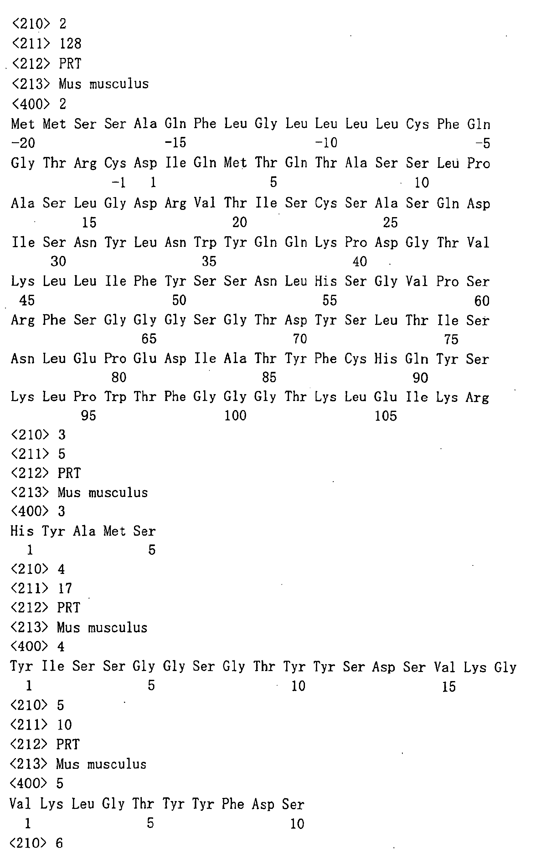


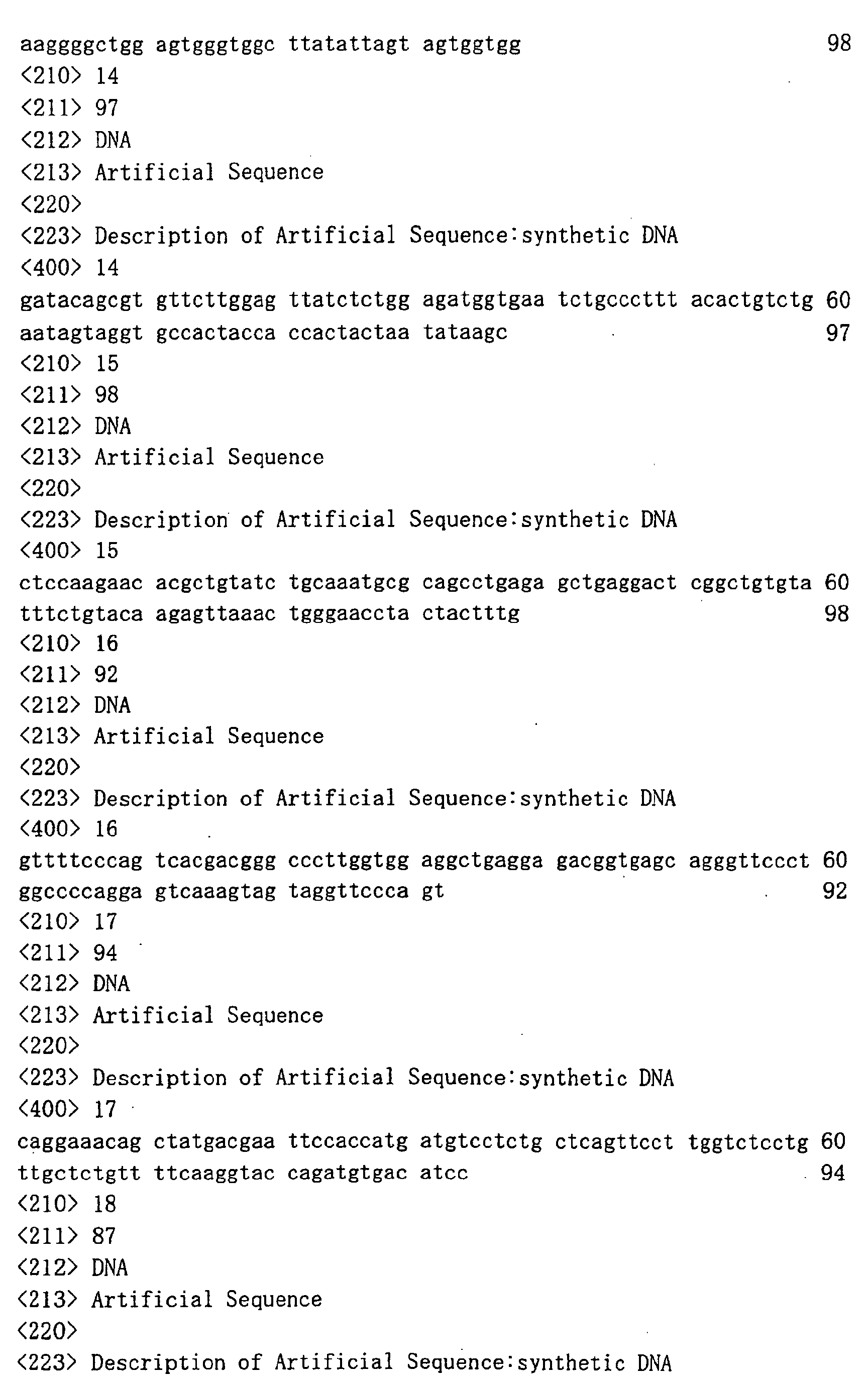
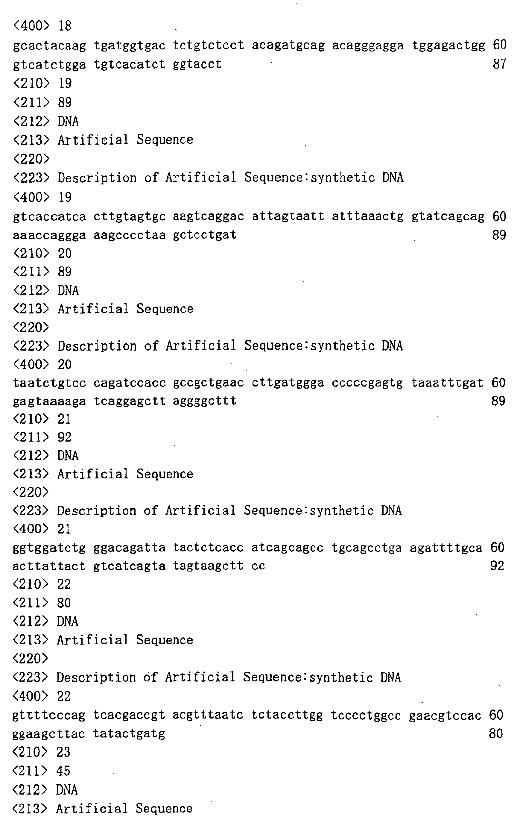


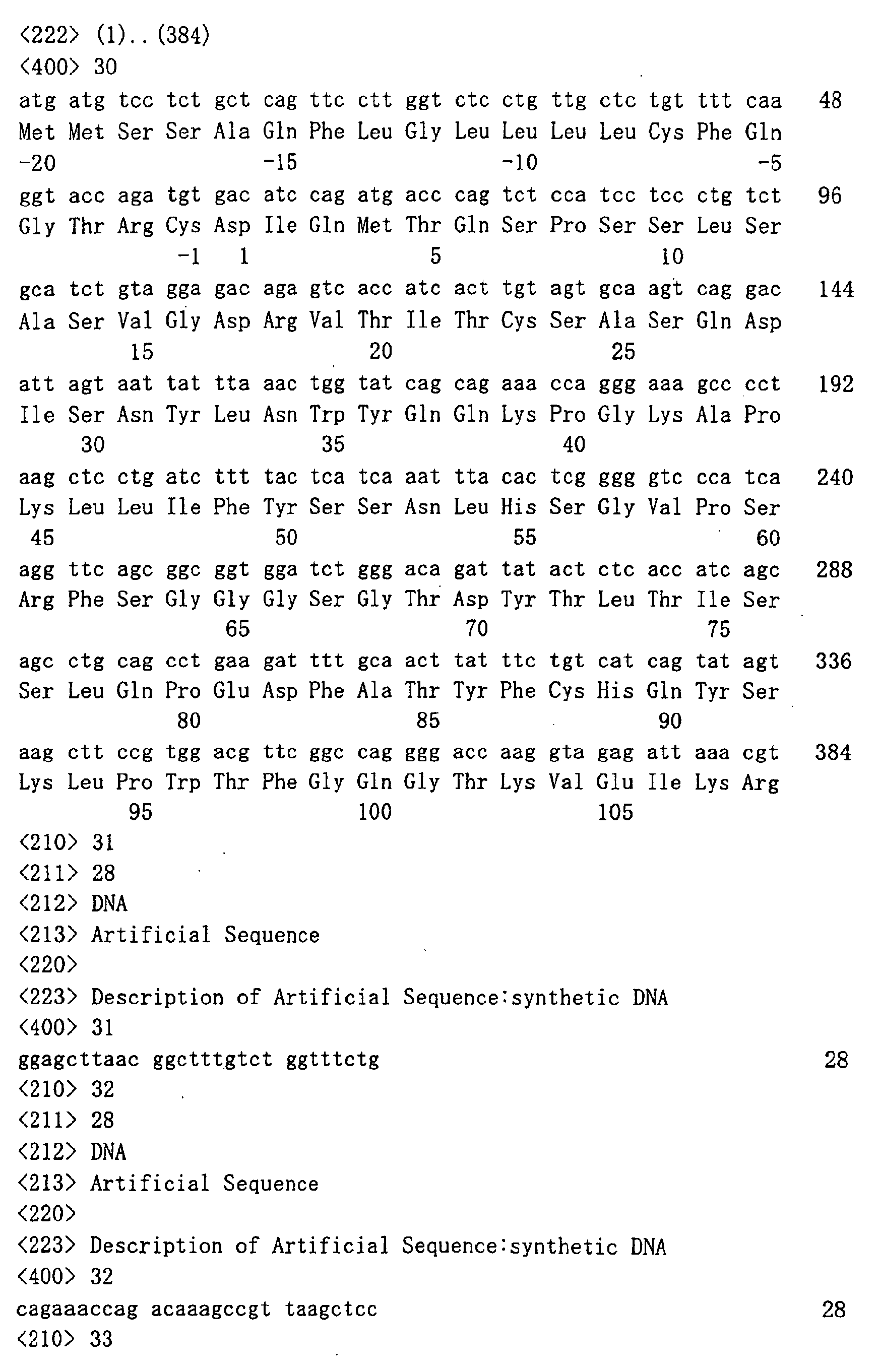
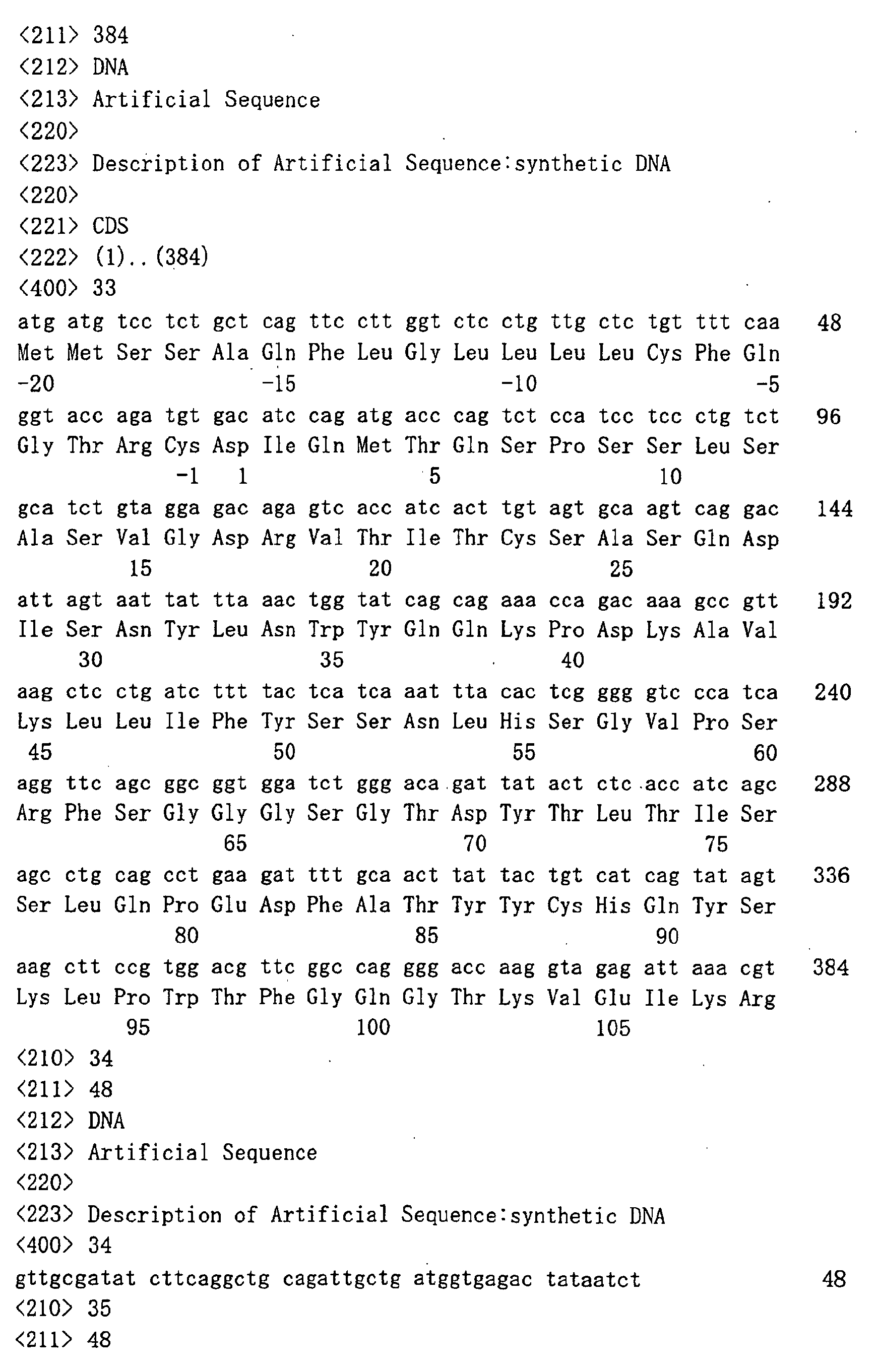
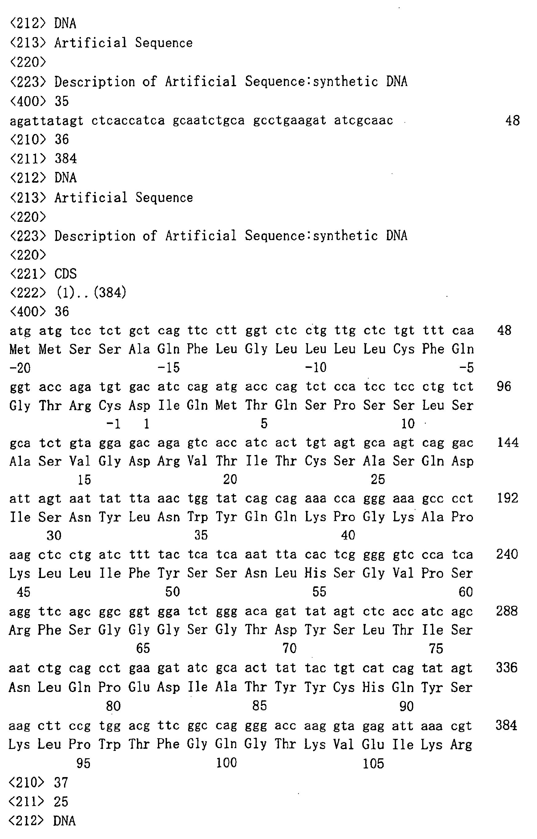


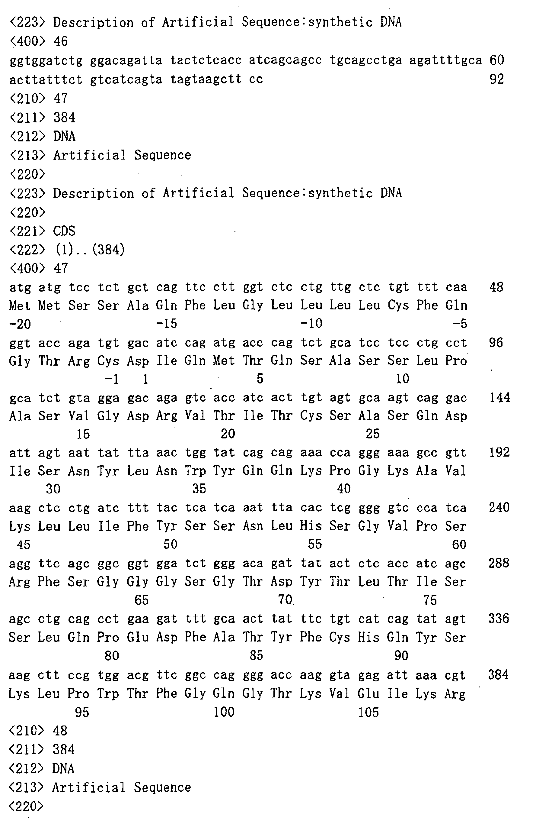
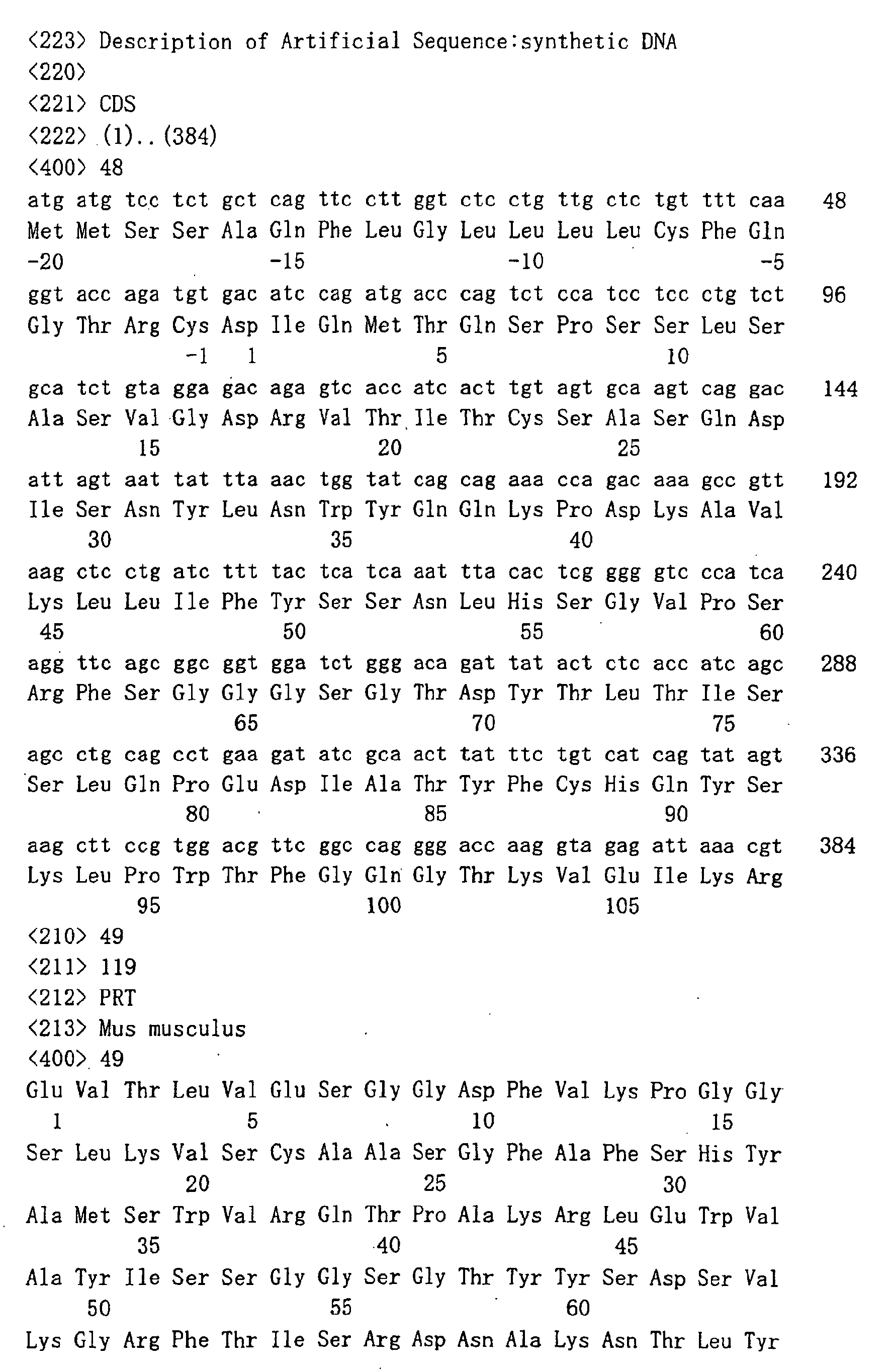

Claims (12)
- A medicament which comprises a gene recombinant antibody against ganglioside GD3 or the antibody fragment thereof in combination with at least one of a substance which activates an immunocompetent cell and a substance having an antitumor activity.
- An antitumor agent which comprises a gene recombinant antibody against ganglioside GD3 or the antibody fragment thereof in combination with at least one of a substance which activates an immunocompetent cell and a substance having an antitumor activity.
- The antitumor agent according to claim 2, wherein the tumor is melanoma.
- The agent according to any one of claims 1 to 3, wherein the gene recombinant antibody against ganglioside GD3 is a humanized antibody against ganglioside GD3.
- The agent according to claim 4, wherein the humanized antibody is a human chimeric antibody or a human complementarity determining region (CDR)-grafted antibody.
- The agent according to claim 5, wherein the humanized antibody is a humanized antibody which comprises CDR1, CDR2 and CDR3 of a heavy chain (H chain) variable region (V region) comprising the amino acid sequences represented by SEQ ID NOs:3, 4 and 5, respectively, and CDR1, CDR2 and CDR3 of a light chain (L chain) variable region (V region) comprising the amino acid sequences represented by SEQ ID NOs:6, 7 and 8, respectively.
- The agent according to claim 5, wherein the human chimeric antibody is a human chimeric antibody which comprises an H chain V region and an L chain V region comprising the amino acid sequences represented by SEQ ID NOs:49 and 50, respectively.
- The agent according to claim 5, wherein the human CDR-grafted antibody is a human CDR-grafted antibody which comprises an H chain V region and an L chain V region of the human CDR-grafted antibody comprising the amino acid sequences represented by SEQ ID NOs:9 and 10, respectively.
- The agent according to any one of claims 1 to 8, wherein the substance which activates an immunocompetent cell is cytokine.
- The agent according to claim 9, wherein the cytokine is selected from the group consisting of IFNα, IFNβ, IFNγ, TNFα, TNFβ, IL-1α, IL-1β, IL-2, IL-3, IL-4, IL-6, IL-7, IL-9, IL-12, IL-15, IL-17, IL-18, GM-CSF, G-CSF, M-CSF and SCF.
- The agent according to any one of claims 1 to 8, wherein the substance having an antitumor activity is a hormonotherapeutant agent or a chemotherapy agent.
- The agent according to claim 11, wherein the hormonotherapeutant agent or the chemotherapy agent is an agent selected from the group consisting of dacarbazine, cisplatin, fotemustine, carmustine, nimustine, lomustine, semustine, pacritaxel, dosetaxel, vindesine, vinblastine, vincristine, carboplatin, bleomycin, doxorubicin and tamoxifen.
Applications Claiming Priority (3)
| Application Number | Priority Date | Filing Date | Title |
|---|---|---|---|
| JP2001097483 | 2001-03-29 | ||
| JP2001097483 | 2001-03-29 | ||
| PCT/JP2002/003170 WO2002078739A1 (en) | 2001-03-29 | 2002-03-29 | Drugs containing genetically modified antibody against ganglioside gd3 |
Publications (2)
| Publication Number | Publication Date |
|---|---|
| EP1384487A1 true EP1384487A1 (en) | 2004-01-28 |
| EP1384487A4 EP1384487A4 (en) | 2007-07-11 |
Family
ID=18951264
Family Applications (1)
| Application Number | Title | Priority Date | Filing Date |
|---|---|---|---|
| EP02713258A Withdrawn EP1384487A4 (en) | 2001-03-29 | 2002-03-29 | MEDICAMENTS CONTAINING A GENETICALLY MODIFIED ANTIBODY AGAINST GD3 GANGLIOSIDE |
Country Status (6)
| Country | Link |
|---|---|
| US (3) | US20050260206A1 (en) |
| EP (1) | EP1384487A4 (en) |
| JP (1) | JPWO2002078739A1 (en) |
| AU (1) | AU2002244946B2 (en) |
| CA (1) | CA2441863A1 (en) |
| WO (1) | WO2002078739A1 (en) |
Cited By (1)
| Publication number | Priority date | Publication date | Assignee | Title |
|---|---|---|---|---|
| WO2006112556A1 (en) * | 2005-04-21 | 2006-10-26 | Kyowa Hakko Kogyo Co., Ltd. | Method for treating cancer using premedication |
Families Citing this family (6)
| Publication number | Priority date | Publication date | Assignee | Title |
|---|---|---|---|---|
| EP1384487A4 (en) * | 2001-03-29 | 2007-07-11 | Kyowa Hakko Kogyo Kk | MEDICAMENTS CONTAINING A GENETICALLY MODIFIED ANTIBODY AGAINST GD3 GANGLIOSIDE |
| JPWO2005053742A1 (en) * | 2003-12-04 | 2007-06-28 | 協和醗酵工業株式会社 | Medicament containing antibody composition |
| EP1918304A1 (en) * | 2006-11-02 | 2008-05-07 | Institut National De La Sante Et De La Recherche Medicale (Inserm) | Monoclonal antibodies against the human anti-müllerian hormone type II receptor (AMHR-II) |
| EP2370819A4 (en) * | 2008-12-01 | 2012-09-19 | Univ Johns Hopkins | METHODS OF DIAGNOSING AND TREATING CANCER BASED ON IMMUNE INHIBITORS |
| AU2011292149A1 (en) | 2010-08-17 | 2013-03-14 | Allergan, Inc. | Method of stabilizing and sterilizing peptides or proteins |
| CN113164570B (en) | 2018-08-08 | 2024-05-14 | 蜻蜓疗法股份有限公司 | Proteins binding NKG2D, CD and tumor-associated antigens |
Family Cites Families (6)
| Publication number | Priority date | Publication date | Assignee | Title |
|---|---|---|---|---|
| CA1339798C (en) * | 1985-02-01 | 1998-04-07 | Alan N. Houghton | Method for treatment of neuroectodermal malignancies and epithelial carcinomas in humans |
| EP0267615A3 (en) * | 1986-11-13 | 1989-05-03 | Sloan-Kettering Institute For Cancer Research | Compositions and method for treatment of cancer using monoclonal antibody against gd3 ganglioside together with il-2 |
| CA2078539C (en) * | 1991-09-18 | 2005-08-02 | Kenya Shitara | Process for producing humanized chimera antibody |
| EP1056861A2 (en) * | 1998-02-27 | 2000-12-06 | Novopharm Biotech, Inc. | Human monoclonal antibodies capable of oligospecifically recognizing the major tumor-associated gangliosides and methods of use thereof |
| WO2001023432A1 (en) * | 1999-09-30 | 2001-04-05 | Kyowa Hakko Kogyo Co., Ltd. | Human type complementarity determining region transplantation antibody against ganglioside gd3 and derivatives of antibody against ganglioside gd3 |
| EP1384487A4 (en) * | 2001-03-29 | 2007-07-11 | Kyowa Hakko Kogyo Kk | MEDICAMENTS CONTAINING A GENETICALLY MODIFIED ANTIBODY AGAINST GD3 GANGLIOSIDE |
-
2002
- 2002-03-29 EP EP02713258A patent/EP1384487A4/en not_active Withdrawn
- 2002-03-29 AU AU2002244946A patent/AU2002244946B2/en not_active Ceased
- 2002-03-29 WO PCT/JP2002/003170 patent/WO2002078739A1/en not_active Ceased
- 2002-03-29 CA CA002441863A patent/CA2441863A1/en not_active Abandoned
- 2002-03-29 JP JP2002577004A patent/JPWO2002078739A1/en active Pending
- 2002-03-29 US US10/473,037 patent/US20050260206A1/en not_active Abandoned
-
2008
- 2008-02-19 US US12/033,516 patent/US20080166345A1/en not_active Abandoned
-
2009
- 2009-04-08 US US12/420,142 patent/US20090226399A1/en not_active Abandoned
Cited By (1)
| Publication number | Priority date | Publication date | Assignee | Title |
|---|---|---|---|---|
| WO2006112556A1 (en) * | 2005-04-21 | 2006-10-26 | Kyowa Hakko Kogyo Co., Ltd. | Method for treating cancer using premedication |
Also Published As
| Publication number | Publication date |
|---|---|
| US20050260206A1 (en) | 2005-11-24 |
| US20090226399A1 (en) | 2009-09-10 |
| EP1384487A4 (en) | 2007-07-11 |
| AU2002244946B2 (en) | 2007-05-10 |
| JPWO2002078739A1 (en) | 2004-07-22 |
| WO2002078739A1 (en) | 2002-10-10 |
| CA2441863A1 (en) | 2002-10-10 |
| US20080166345A1 (en) | 2008-07-10 |
Similar Documents
| Publication | Publication Date | Title |
|---|---|---|
| EP1238985B1 (en) | Human type complementarity determining region transplantation antibody against ganglioside gd3 and derivatives of antibody against ganglioside gd3 | |
| JP4368530B2 (en) | Methods for modulating the activity of immune function molecules | |
| EP2138576A1 (en) | Anti-claudin-4 antibody | |
| JPWO2001023432A1 (en) | Human complementarity-determining region-grafted antibody against ganglioside GD3 and derivative of antibody against ganglioside GD3 | |
| JPWO2000061739A1 (en) | Methods for modulating the activity of immune function molecules | |
| JPWO2008090960A1 (en) | Recombinant antibody composition that specifically binds to ganglioside GM2 | |
| US20090226399A1 (en) | Medicaments comprising gene recombinant antibody against ganglioside gd3 | |
| EP1422243B1 (en) | Humanized antibody against fibroblast growth factor-8 and fragment of the antibody | |
| JP5651893B2 (en) | Anti-PERP antibody | |
| US8093362B2 (en) | Anti-PERP recombinant antibody | |
| US20070003557A1 (en) | Method for treating cancer using premedication | |
| JPWO2001023573A1 (en) | Human complementarity-determining region-grafted antibody against ganglioside GD2 and derivatives of said antibody | |
| JPWO2001023431A1 (en) | Derivatives of antibodies against ganglioside GM2 |
Legal Events
| Date | Code | Title | Description |
|---|---|---|---|
| PUAI | Public reference made under article 153(3) epc to a published international application that has entered the european phase |
Free format text: ORIGINAL CODE: 0009012 |
|
| 17P | Request for examination filed |
Effective date: 20031029 |
|
| AK | Designated contracting states |
Kind code of ref document: A1 Designated state(s): AT BE CH CY DE DK ES FI FR GB GR IE IT LI LU MC NL PT SE TR |
|
| AX | Request for extension of the european patent |
Extension state: AL LT LV MK RO SI |
|
| A4 | Supplementary search report drawn up and despatched |
Effective date: 20070612 |
|
| 17Q | First examination report despatched |
Effective date: 20070924 |
|
| STAA | Information on the status of an ep patent application or granted ep patent |
Free format text: STATUS: THE APPLICATION IS DEEMED TO BE WITHDRAWN |
|
| 18D | Application deemed to be withdrawn |
Effective date: 20080405 |