EP1362062B1 - Concatameric immunoadhesion - Google Patents

Concatameric immunoadhesion Download PDF

Info

Publication number
EP1362062B1
EP1362062B1 EP02753273A EP02753273A EP1362062B1 EP 1362062 B1 EP1362062 B1 EP 1362062B1 EP 02753273 A EP02753273 A EP 02753273A EP 02753273 A EP02753273 A EP 02753273A EP 1362062 B1 EP1362062 B1 EP 1362062B1
Authority
EP
European Patent Office
Prior art keywords
protein
seq
fusion
proteins
sequence
Prior art date
Legal status (The legal status is an assumption and is not a legal conclusion. Google has not performed a legal analysis and makes no representation as to the accuracy of the status listed.)
Expired - Lifetime
Application number
EP02753273A
Other languages
German (de)
French (fr)
Other versions
EP1362062A1 (en
EP1362062A4 (en
Inventor
Yong-Hoon Chung
Ji-Woong Han
Hye-Ja Lee
Eun-Yong Choi
Jin-Mi Kim
Current Assignee (The listed assignees may be inaccurate. Google has not performed a legal analysis and makes no representation or warranty as to the accuracy of the list.)
Medexgen Inc
Original Assignee
Medexgen Inc
Priority date (The priority date is an assumption and is not a legal conclusion. Google has not performed a legal analysis and makes no representation as to the accuracy of the date listed.)
Filing date
Publication date
Application filed by Medexgen Inc filed Critical Medexgen Inc
Publication of EP1362062A1 publication Critical patent/EP1362062A1/en
Publication of EP1362062A4 publication Critical patent/EP1362062A4/en
Application granted granted Critical
Publication of EP1362062B1 publication Critical patent/EP1362062B1/en
Anticipated expiration legal-status Critical
Expired - Lifetime legal-status Critical Current

Links

Images

Classifications

    • CCHEMISTRY; METALLURGY
    • C07ORGANIC CHEMISTRY
    • C07KPEPTIDES
    • C07K16/00Immunoglobulins [IG], e.g. monoclonal or polyclonal antibodies
    • C07K16/46Hybrid immunoglobulins
    • CCHEMISTRY; METALLURGY
    • C07ORGANIC CHEMISTRY
    • C07KPEPTIDES
    • C07K14/00Peptides having more than 20 amino acids; Gastrins; Somatostatins; Melanotropins; Derivatives thereof
    • C07K14/435Peptides having more than 20 amino acids; Gastrins; Somatostatins; Melanotropins; Derivatives thereof from animals; from humans
    • C07K14/705Receptors; Cell surface antigens; Cell surface determinants
    • C07K14/70503Immunoglobulin superfamily
    • C07K14/70507CD2
    • AHUMAN NECESSITIES
    • A61MEDICAL OR VETERINARY SCIENCE; HYGIENE
    • A61PSPECIFIC THERAPEUTIC ACTIVITY OF CHEMICAL COMPOUNDS OR MEDICINAL PREPARATIONS
    • A61P19/00Drugs for skeletal disorders
    • A61P19/02Drugs for skeletal disorders for joint disorders, e.g. arthritis, arthrosis
    • AHUMAN NECESSITIES
    • A61MEDICAL OR VETERINARY SCIENCE; HYGIENE
    • A61PSPECIFIC THERAPEUTIC ACTIVITY OF CHEMICAL COMPOUNDS OR MEDICINAL PREPARATIONS
    • A61P29/00Non-central analgesic, antipyretic or antiinflammatory agents, e.g. antirheumatic agents; Non-steroidal antiinflammatory drugs [NSAID]
    • AHUMAN NECESSITIES
    • A61MEDICAL OR VETERINARY SCIENCE; HYGIENE
    • A61PSPECIFIC THERAPEUTIC ACTIVITY OF CHEMICAL COMPOUNDS OR MEDICINAL PREPARATIONS
    • A61P37/00Drugs for immunological or allergic disorders
    • CCHEMISTRY; METALLURGY
    • C07ORGANIC CHEMISTRY
    • C07KPEPTIDES
    • C07K14/00Peptides having more than 20 amino acids; Gastrins; Somatostatins; Melanotropins; Derivatives thereof
    • C07K14/435Peptides having more than 20 amino acids; Gastrins; Somatostatins; Melanotropins; Derivatives thereof from animals; from humans
    • C07K14/705Receptors; Cell surface antigens; Cell surface determinants
    • C07K14/70503Immunoglobulin superfamily
    • C07K14/70521CD28, CD152
    • CCHEMISTRY; METALLURGY
    • C07ORGANIC CHEMISTRY
    • C07KPEPTIDES
    • C07K14/00Peptides having more than 20 amino acids; Gastrins; Somatostatins; Melanotropins; Derivatives thereof
    • C07K14/435Peptides having more than 20 amino acids; Gastrins; Somatostatins; Melanotropins; Derivatives thereof from animals; from humans
    • C07K14/705Receptors; Cell surface antigens; Cell surface determinants
    • C07K14/715Receptors; Cell surface antigens; Cell surface determinants for cytokines; for lymphokines; for interferons
    • C07K14/7151Receptors; Cell surface antigens; Cell surface determinants for cytokines; for lymphokines; for interferons for tumor necrosis factor [TNF], for lymphotoxin [LT]
    • AHUMAN NECESSITIES
    • A61MEDICAL OR VETERINARY SCIENCE; HYGIENE
    • A61KPREPARATIONS FOR MEDICAL, DENTAL OR TOILETRY PURPOSES
    • A61K39/00Medicinal preparations containing antigens or antibodies
    • A61K2039/505Medicinal preparations containing antigens or antibodies comprising antibodies
    • CCHEMISTRY; METALLURGY
    • C07ORGANIC CHEMISTRY
    • C07KPEPTIDES
    • C07K2319/00Fusion polypeptide
    • CCHEMISTRY; METALLURGY
    • C07ORGANIC CHEMISTRY
    • C07KPEPTIDES
    • C07K2319/00Fusion polypeptide
    • C07K2319/30Non-immunoglobulin-derived peptide or protein having an immunoglobulin constant or Fc region, or a fragment thereof, attached thereto

Definitions

  • Soluble receptors used as functional inhibitors of cytokine that fused to heavy chains of human immunoglobulins were disclosed by Capon et al. (Nature 337:5254, 1989), and thereafter many patents were disclosed inventions related to fusion proteins of soluble receptors and immunoglobulins (US patent 5,521,288, 5,844,095, 6,046,310, 6,090,914, 6,100,383, 6,225,448).
  • the present invention generally relates to concatameric proteins where C-terminal end of a soluble domain of a biologically active protein is fused to an N-terminal end of a soluble domain of the same or other biologically active proteins.
  • the present invention provides DNA constructs that encode monomeric fusion proteins whose concatamerized domain is fused to hinge region of Fc fragment of immunoglobulins.
  • the present invention provides DNA plasmids comprising a DNA construct that encodes monomeric fusion protein whose concatamerized part is fused to hinge region ofFc fragment of immunoglobulin.
  • the present invention provides host cells transfected or transformed with recombinant DNA plasmids including a DNA construct that encodes monomeric fusion protein whose concatamerized part is fused to hinge region of Fc fragment of immunoglobulin.
  • the present invention provides the glycosylated dimers formed by disulfide bond at hinge region of two monomeric proteins whose concatamerized part involving immune response is fused to hinge region of Fc fragment of immunoglobulins.
  • the present invention is generally directed to concatameric proteins, and more particularly, to immunoadhesion molecules.
  • Immunoadhesion molecules are typically formed by fusion of the Fc fragment of immunoglobulin (Ig) to a ligand-binding region of a receptor or an adhesion molecule, and thus have a structure similar to that of an antibody.
  • the typical immunoadhesion molecules known in the art have a structure of an antibody in which the variable region is substituted with a ligand-binding region of a receptor while retaining the Fc fragment.
  • a wide variety of immunoadhesion molecules are suggested in the literature. However, immunoadhesion molecules according to the present invention have different structure with the conventional immunoadhesion molecules, and there is also no prior art predicting or describing preparation of the immunoadhesion molecules according to the present invention.
  • immunoglobulin refers to protein molecules being produced in B cells and serving as antigen receptors specifically recognizing a wide variety of antigens.
  • the molecules have a Y-shaped structure consisting of two identical light chains (L chains) and two identical heavy chains (H chains), in which the four chains are held together by a number of disulfide bonds, including the disulfide bridge between the H chains at the hinge region.
  • L chains two identical light chains
  • H chains heavy chains
  • the L and H chains comprise variable and constant regions.
  • the L chain variable region associates with the H chain variable region, thus producing two identical antigen-binding regions.
  • Fc fragment of an immunoglobulin molecule refers to a fragment having no antigen-binding activity and being easily crystallized, which comprises a hinge region and CH2 and CH3 domains, and a portion responsible for binding of an antibody to effector materials and cells. Therefore, the Fc fragment mentioned in the present invention can be different from that described in some literatures, but includes the hinge region. Such description of the Fc fragment is given to supply convenience in describing the present invention, and will be fully understood by those of ordinary skill in the art with reference to the specification of the present invention and the accompanying drawings.
  • the chemokine receptors are exemplified by CCR1 and CXCR1-4.
  • Examples of the receptor tyrosine kinases include TrkA, TrkB, TrkC, Htk, REK7, Rse/Tyro-3, hepatocyte growth factor R, platelet-derived growth factor R, and Flt-1.
  • simple fusion dimeric protein refers to a fusion protein having a dimeric structure, in which two simple fusion monomeric proteins are joined by formation of intermolecular disulfide bonds at the hinge region.
  • Such a simple fusion dimeric protein may be designated "[protein name/Fc] 2 " for convenience in the present invention.
  • the resulting fusion protein having dimeric structure is designated [TNFR1/Fc] 2 .
  • the origin of the Fc fragment may be specified in the designation, if desired. For example, in the case that the Fc fragment is derived from IgG1, the dimeric protein is designated [TNFR1/IgG1Fc] 2 .
  • Host cells used in the present invention may be prokaryotic or eukaryotic.
  • host cells having high introduction efficiency of foreign DNA and having high expression levels of an introduced gene may be typically used.
  • Examples of the host cells useful in the present invention include prokaryotic and eukaryotic cells such as E. coli, Pseudomonas sp., Bacillus sp., Streptomyces sp., fungi or yeast, insect.cells such as Spodoptera frugiperda (Sf9), animal cells such as Chinese hamster ovary cells (CHO) or mouse cells, African green monkey cells such as COS 1, COS 7, human embryonic kidney cells, BSC 1, BSC 40 or BMT 10, and tissue-cultured human cells.
  • prokaryotic and eukaryotic cells such as E. coli, Pseudomonas sp., Bacillus sp., Streptomyces sp., fungi or yeast, insect.cells such as Spo
  • transfection refers to the taking up of an expression vector by a suitable host cell, whether any coding sequences are in fact expressed or not.
  • PCR polymerase chain reaction
  • the immunoglobulin fusion gene may further include a signal sequence to stimulate extracellular secretion of a protein encoded thereby.
  • the CTLA-4 molecule contains a unique leader sequence having highly hydrophilic redundancy at its N-terminus, and which is abnormally long and highly water-soluble (Harper, K. et al., J. Immunol. 147:1037-1044; and Brunet, J.F. Nature 328:267-270, 1987).
  • leader sequence comprising 20-24 highly hydrophobic amino acids at their N-termini.
  • a DNA fragment is prepared by performing PCR using a primer encoding a leader sequence of a soluble extracellular domain and EcoRI restriction site, and an antisense primer in which a portion of a nucleotide sequence encoding a portion of the 3' end of a first soluble extracellular domain and a portion of the 5' end of a second soluble extracellular domain is substituted with glycosylation motif sequences.
  • Another DNA fragment is prepared by performing PCR using a primer in which a portion of a nucleotide sequence encoding a portion of the 3' end of a first soluble extracellular domain and a portion of the 5' end of a second soluble extracellulular domain is substituted with glycosylation motif sequences, and an antisense primer encoding the 3' end of Fc portion of IgG1 and XbaI restriction site. Then, secondary PCR is carried out in a test tube using the two DNA fragments.
  • Tumor necrosis factor-alpha which is known as the hormone cachectin
  • tumor necrosis factor-beta TNF- ⁇
  • lymphotoxin tumor necrosis factor-beta
  • cytokines inducing inflammation, cellular immune response, septicemia, cytotoxicity, cachexia, rheumatoid arthritis, inflammation-related diseases (Tartaglia, L.A. et al., Immunol. Today 13:151,1992), and antiviral reaction (Butler, P., Peptide Growth Factor II, 1990, Springer-Verlag, Berlin, pp.39-70).
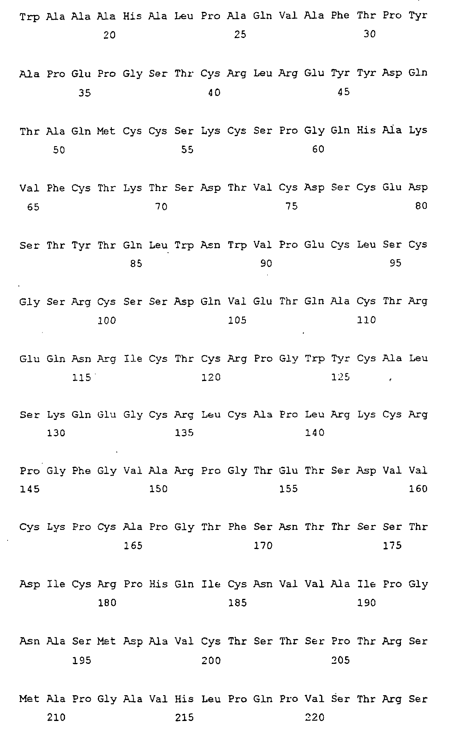
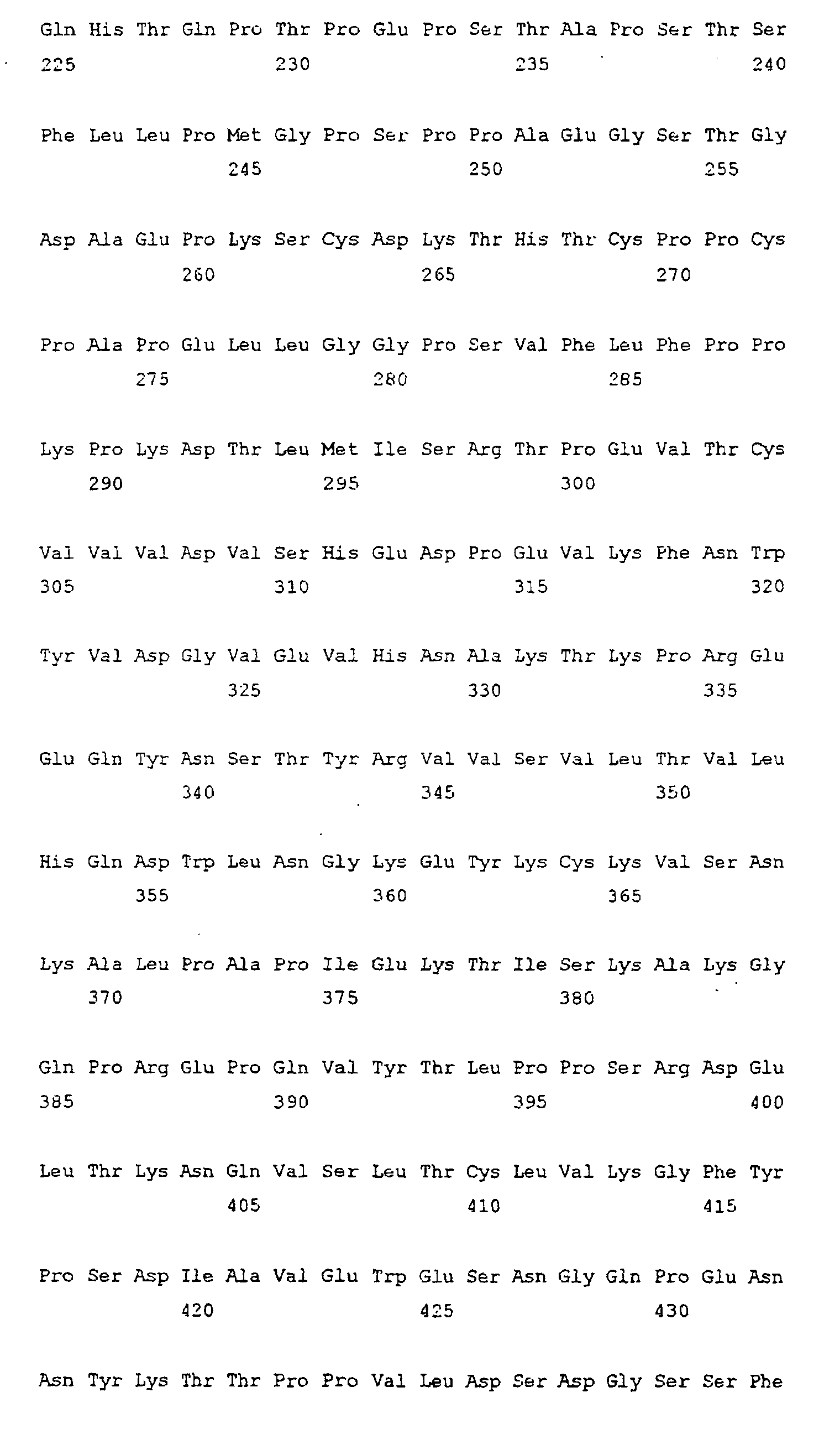
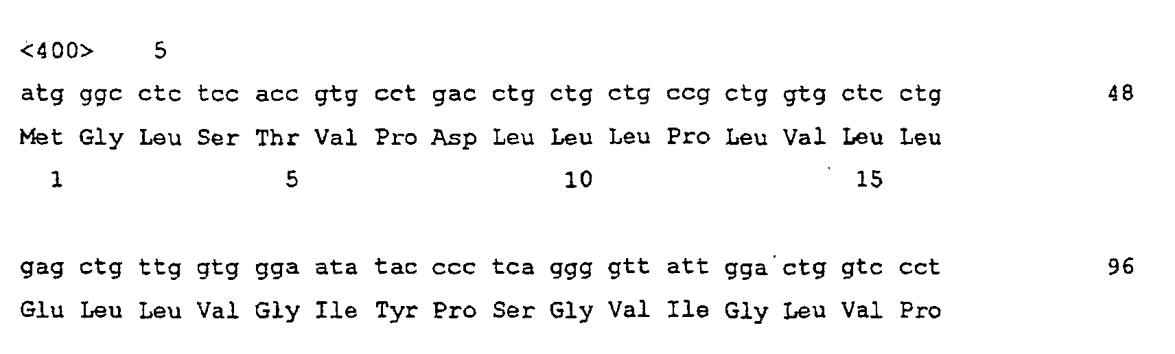
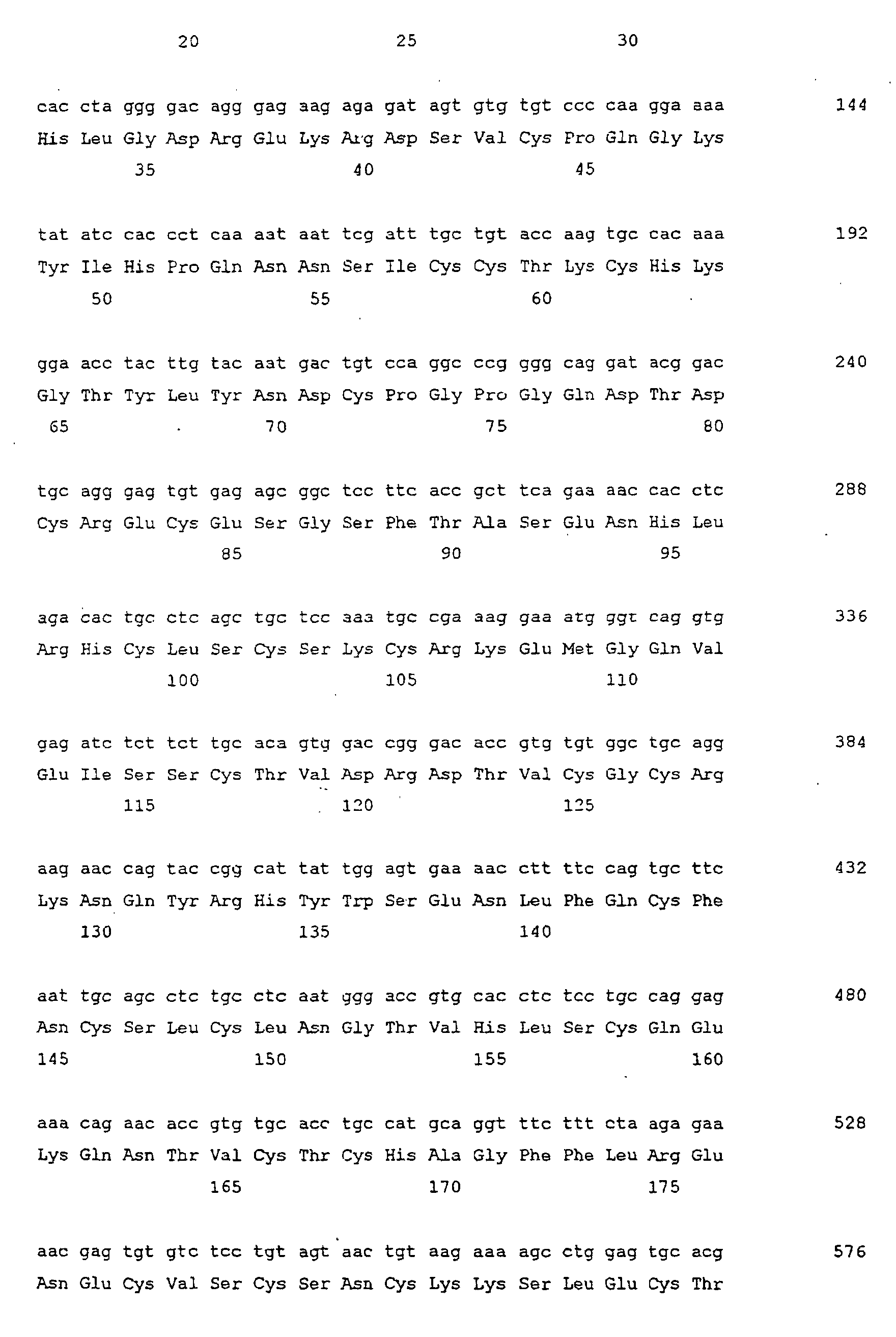
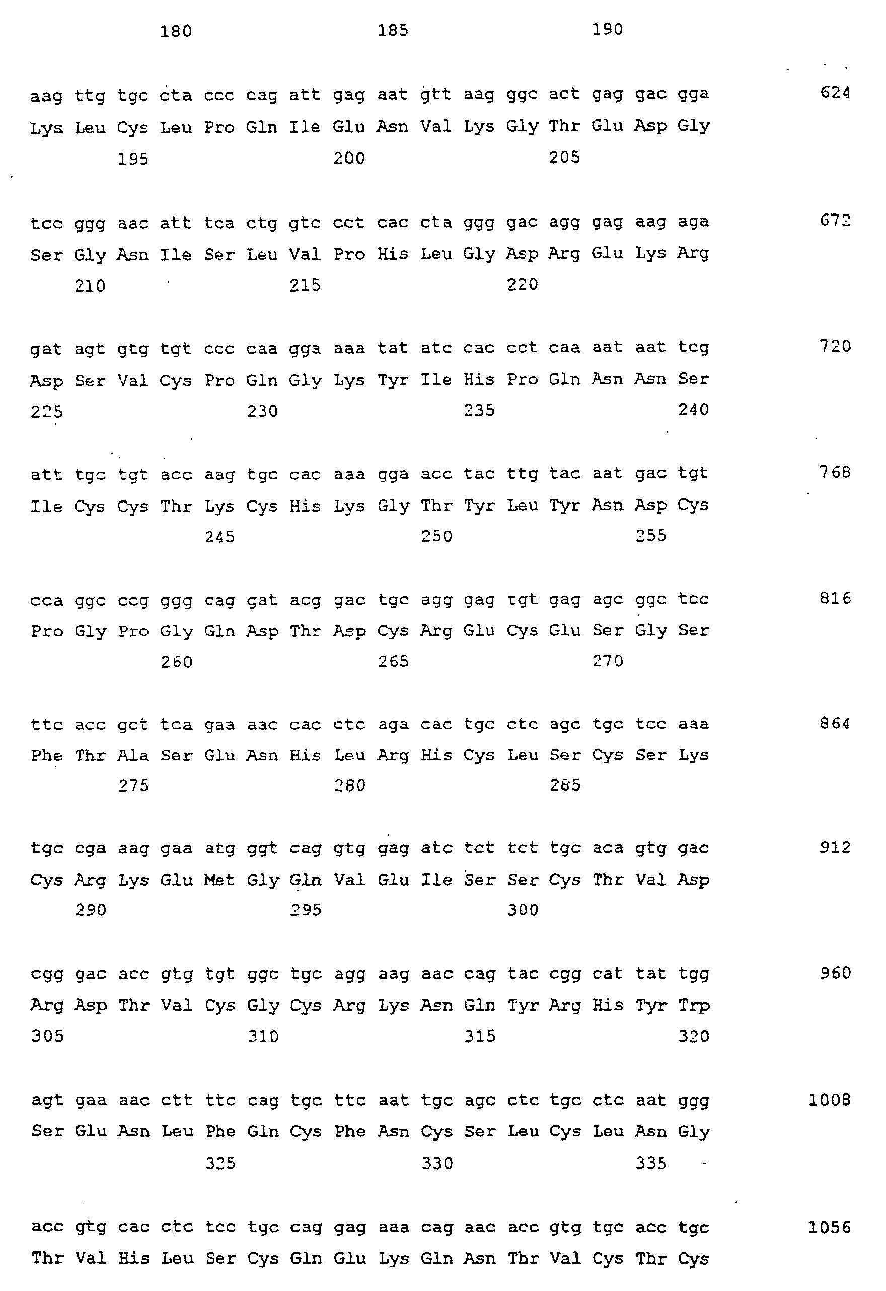
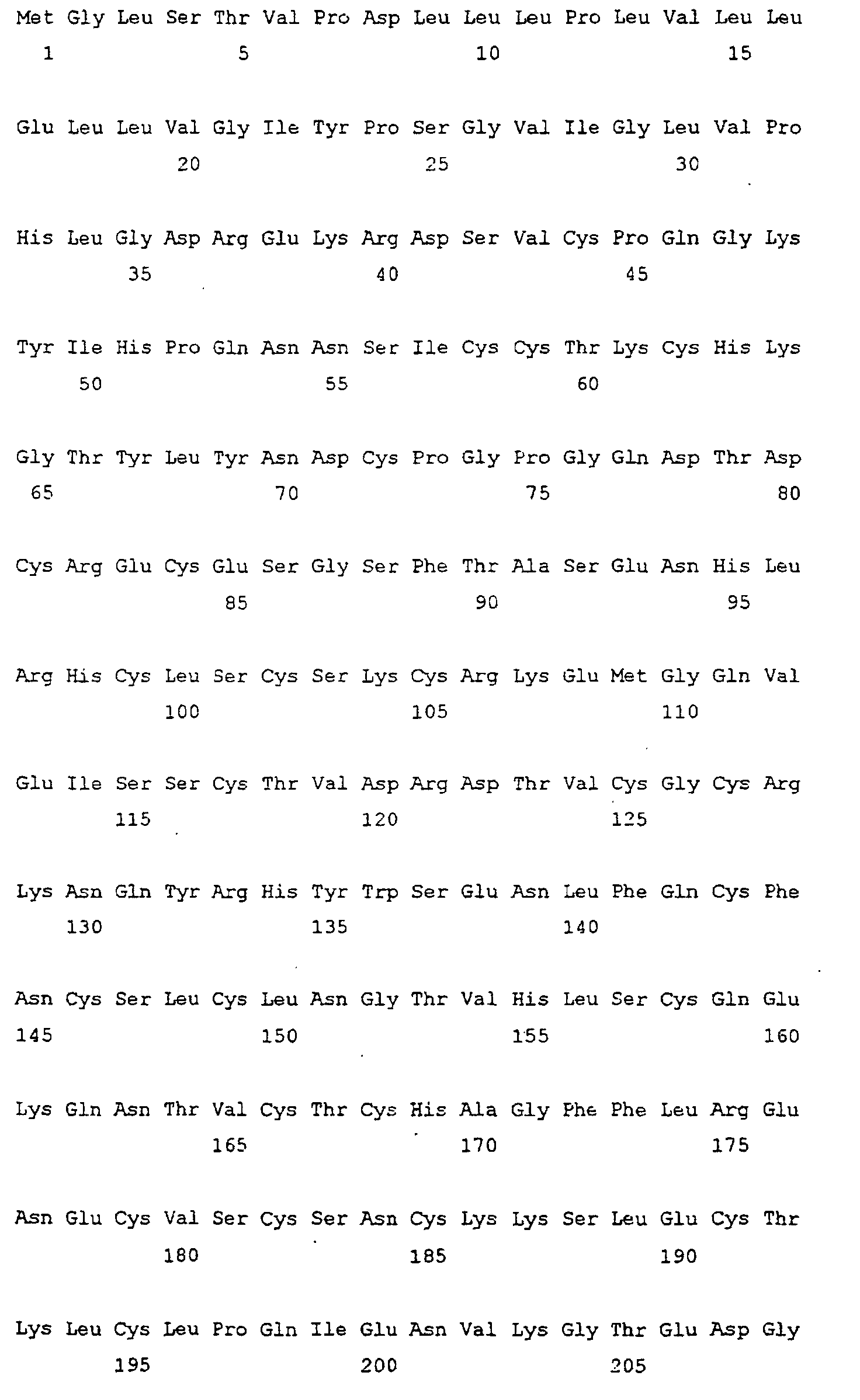
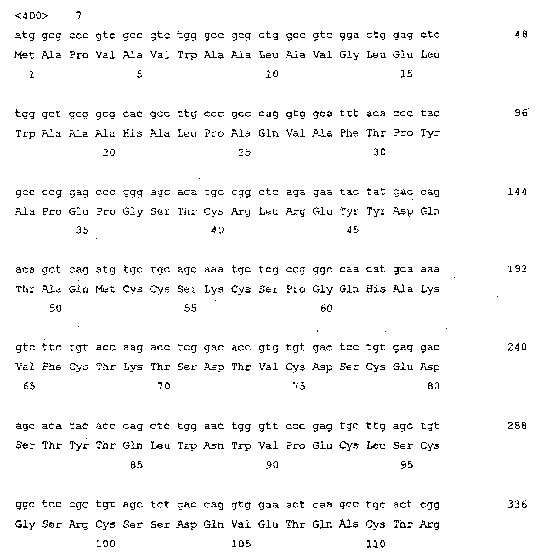
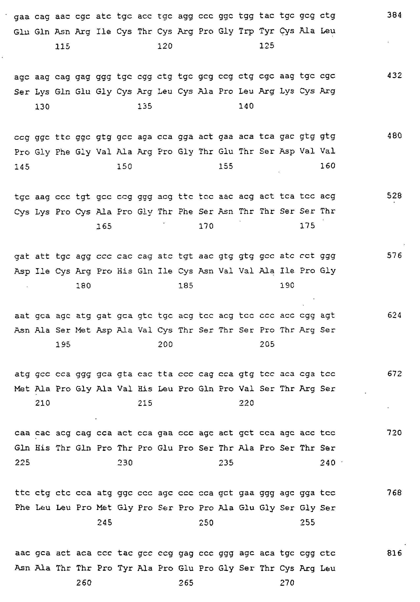
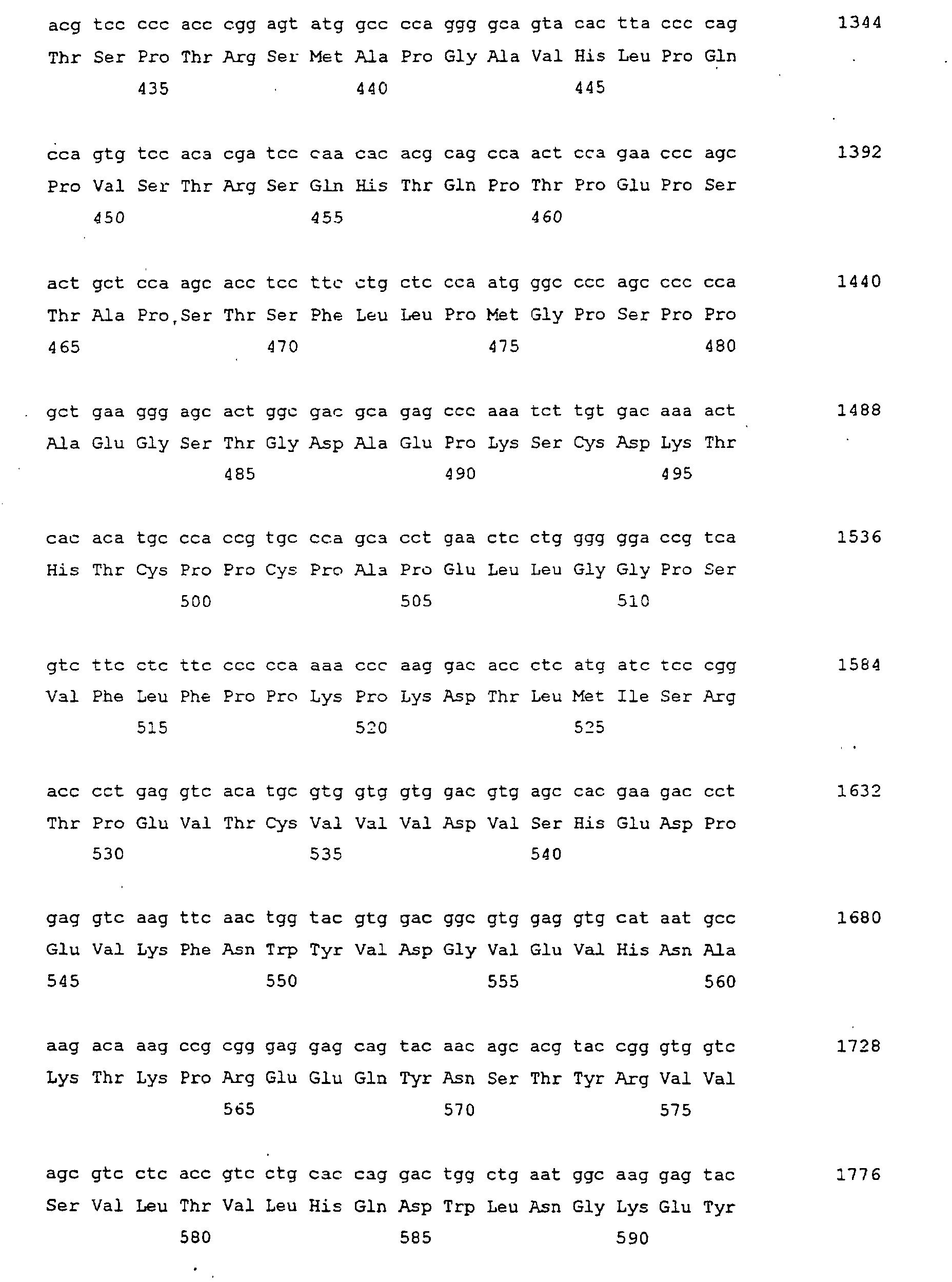
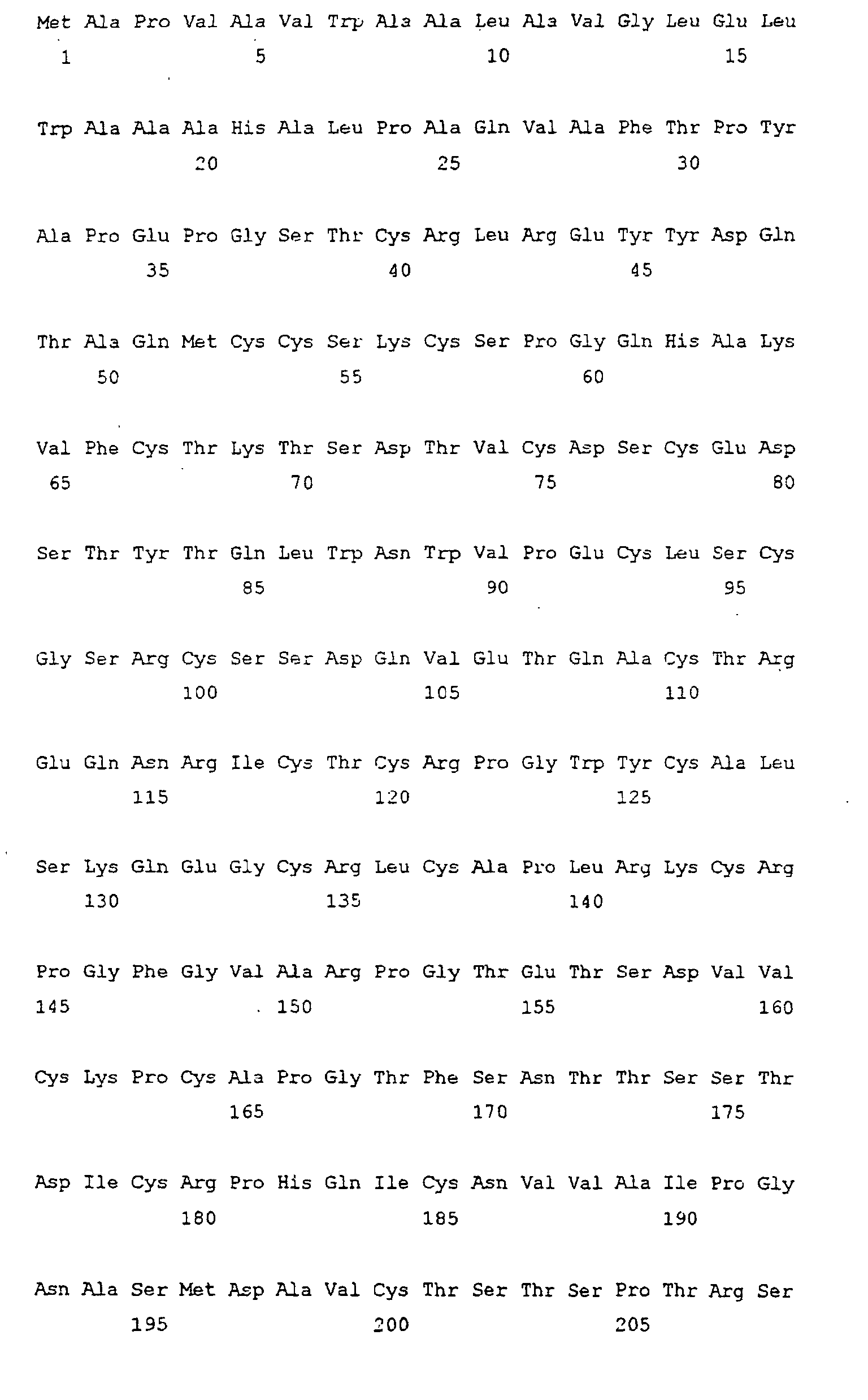
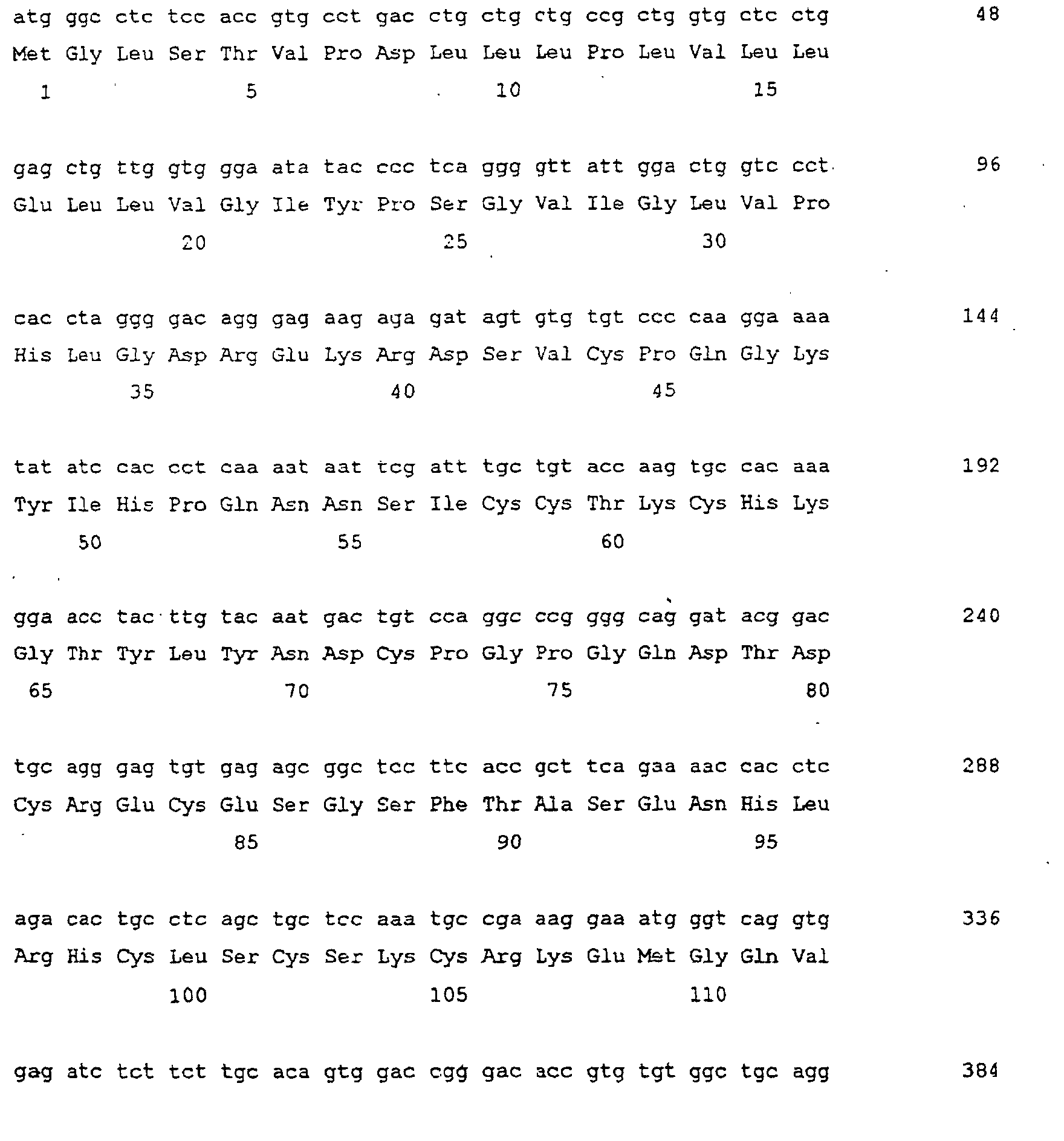
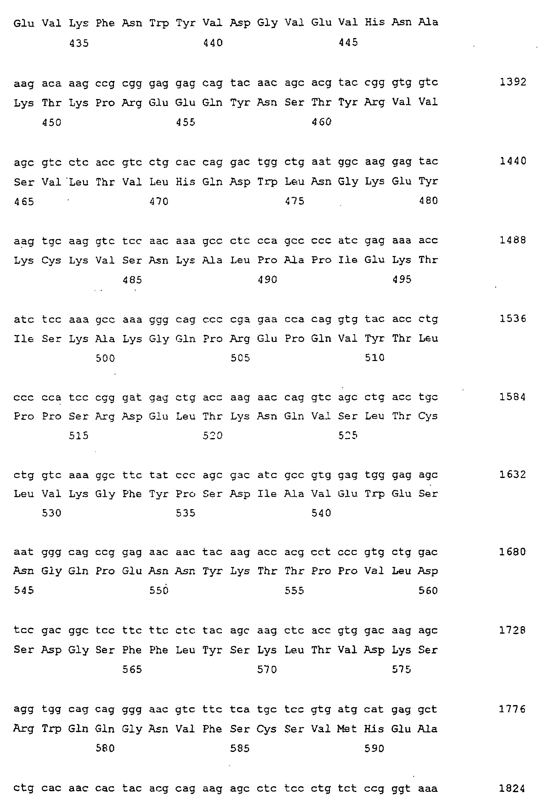
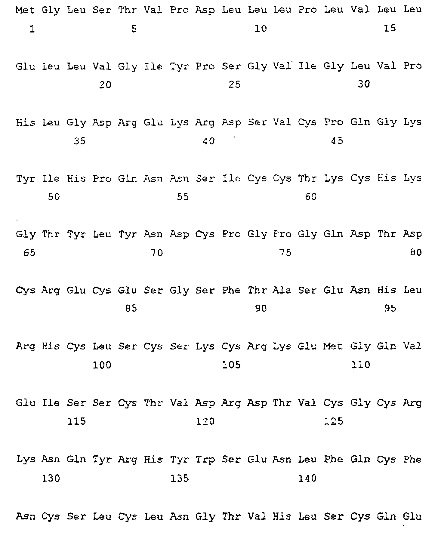
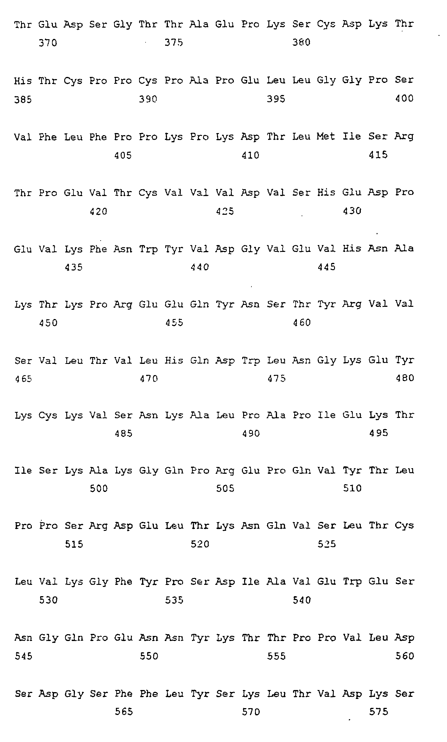
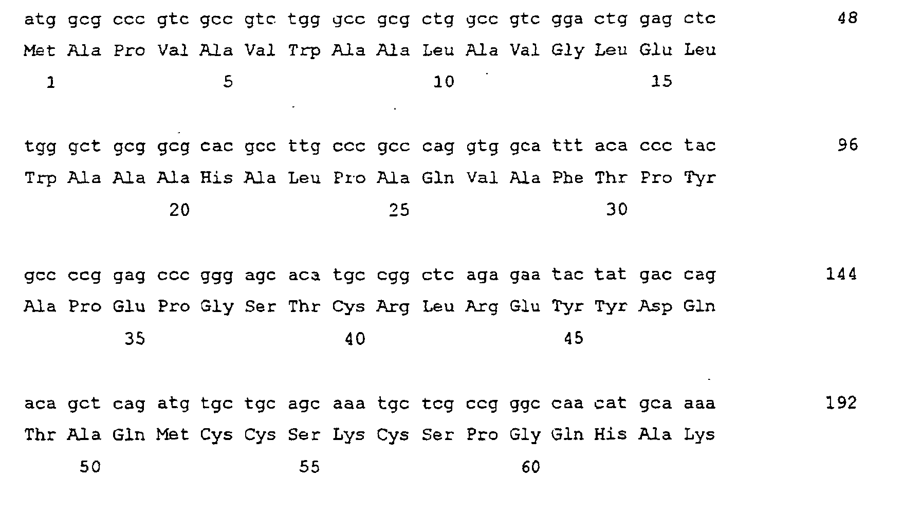
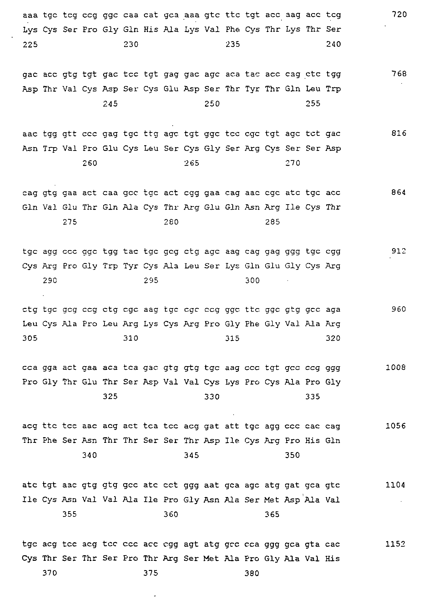
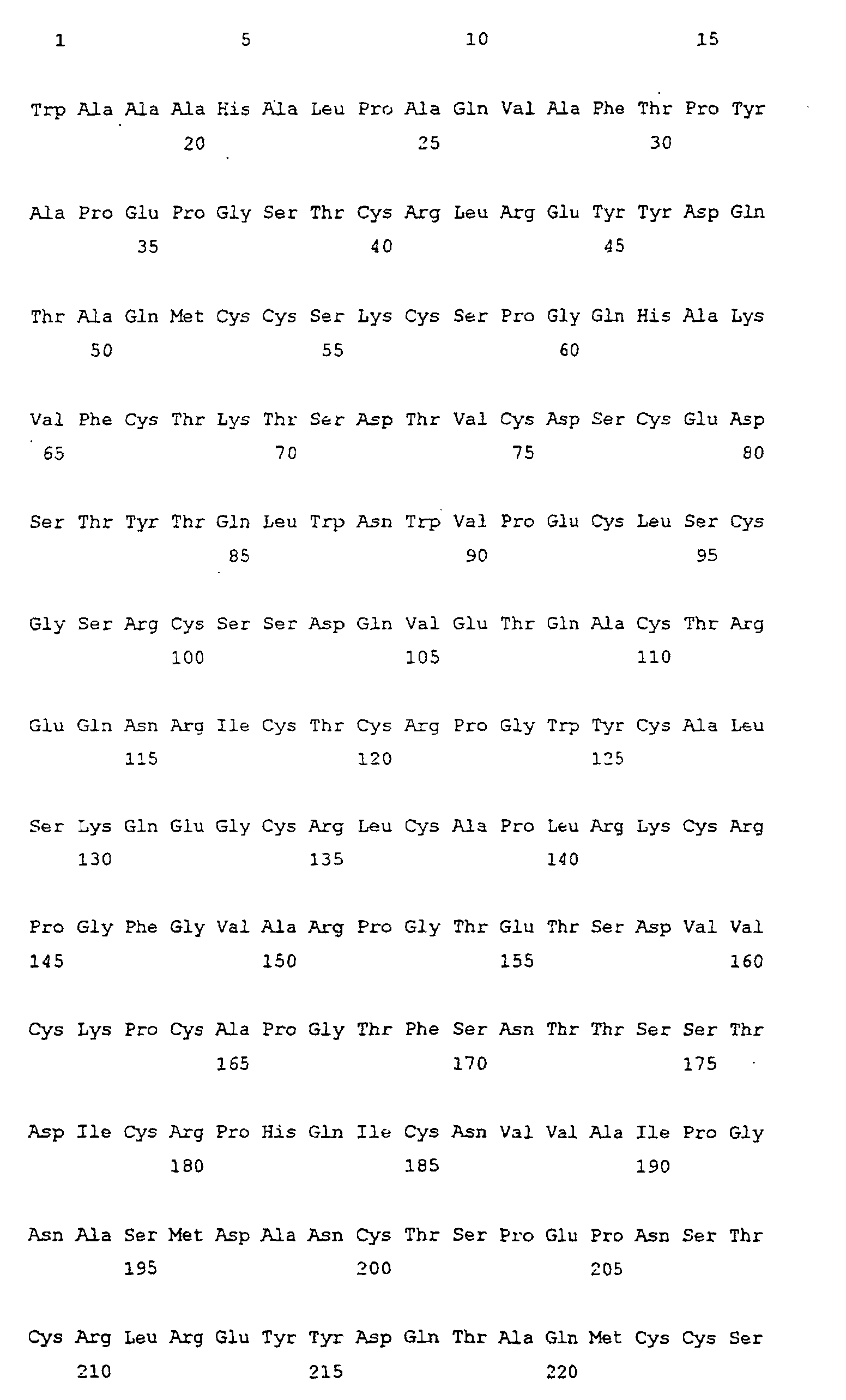
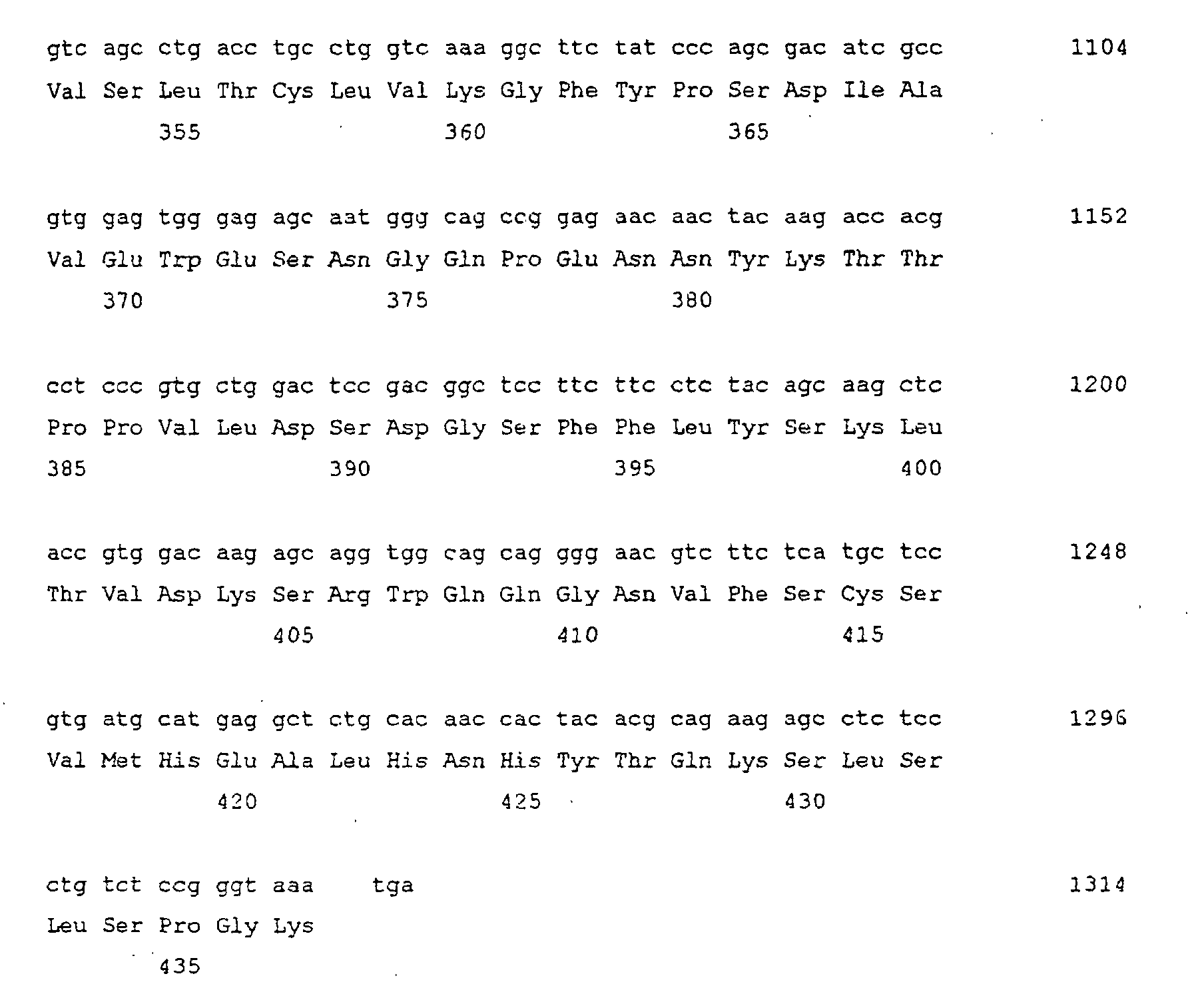
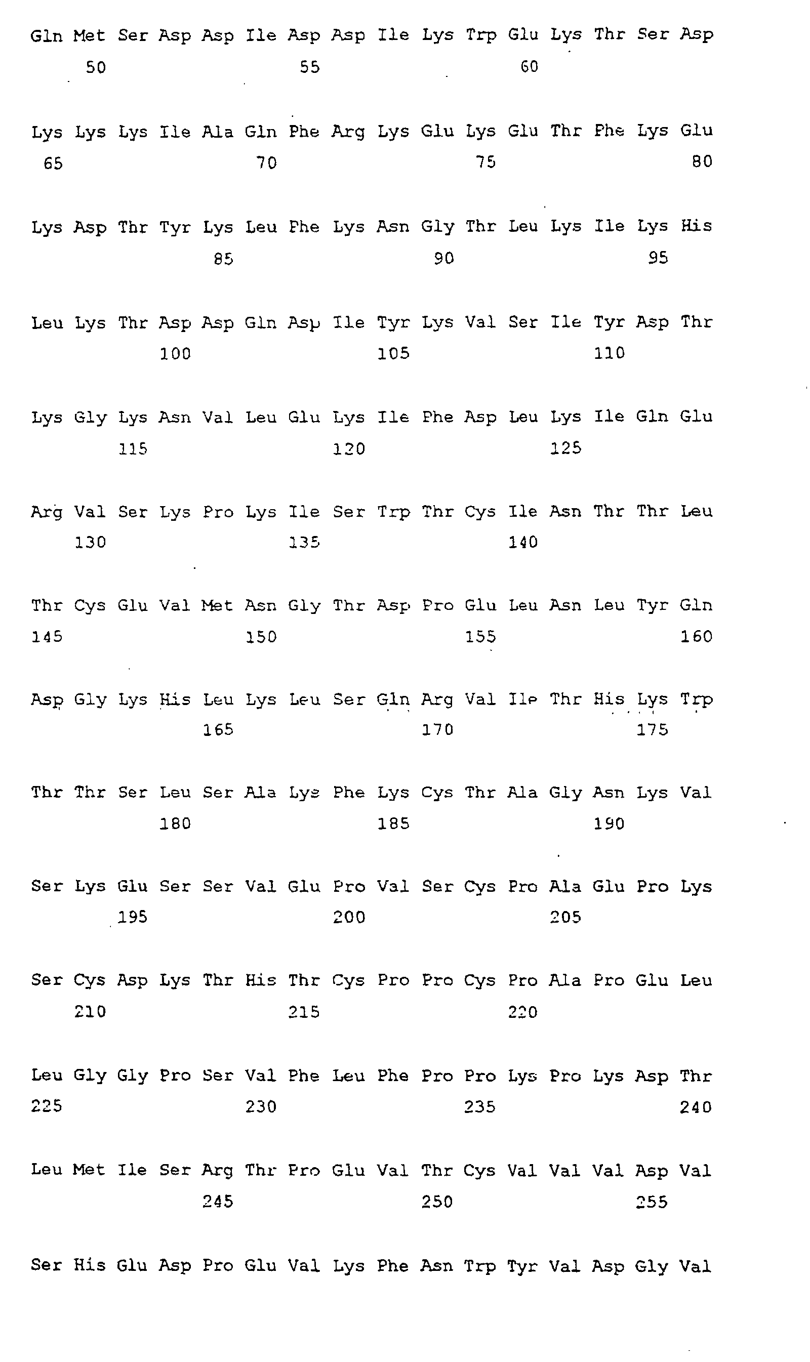
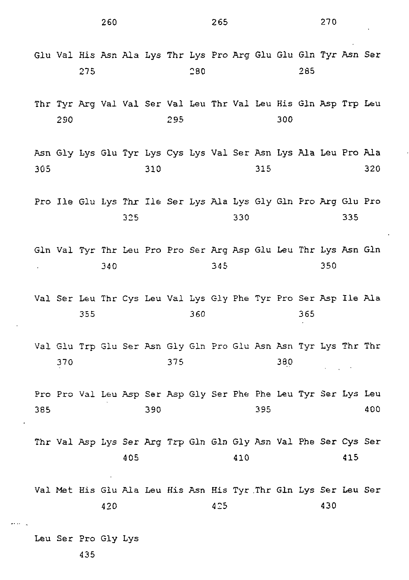
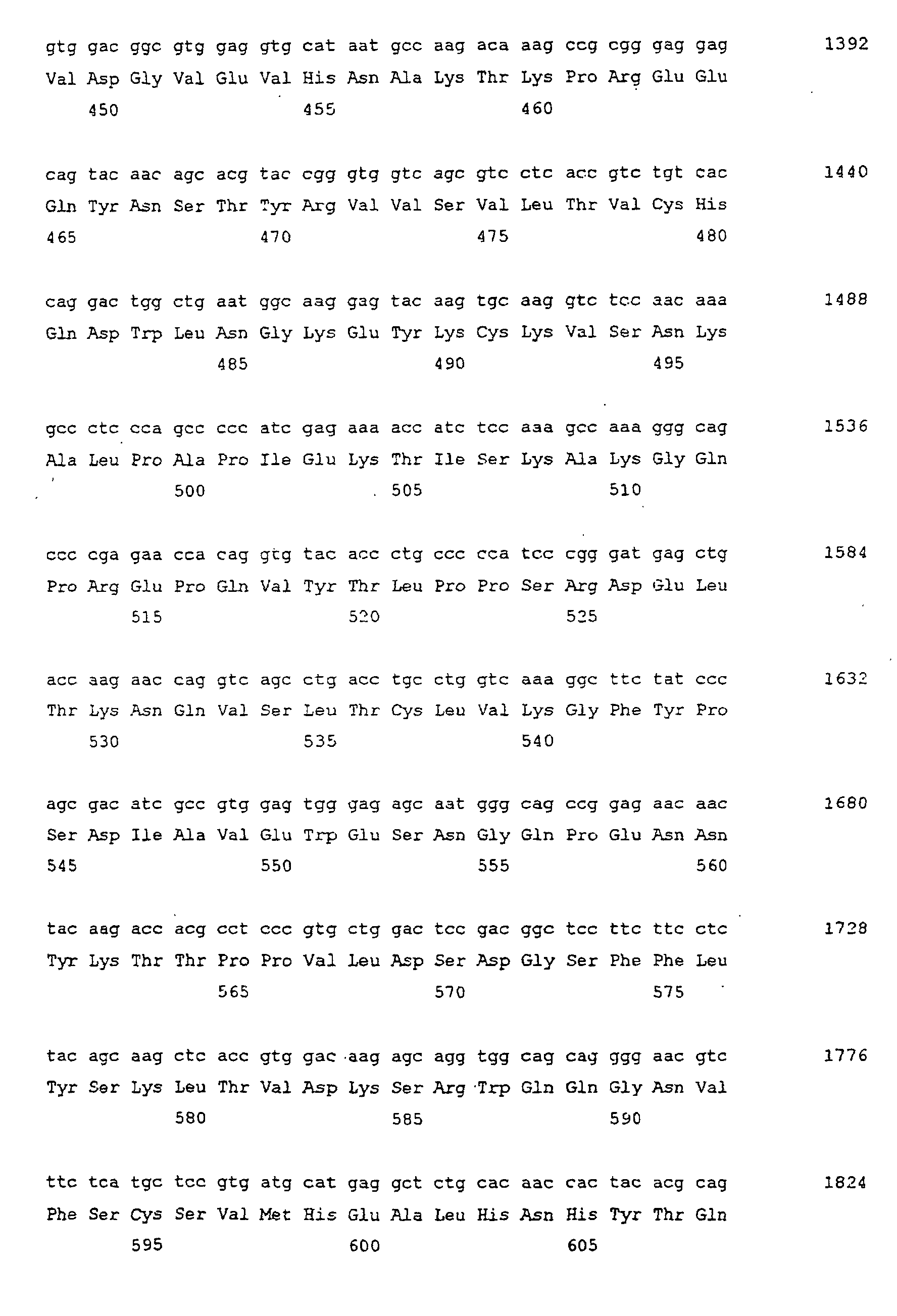
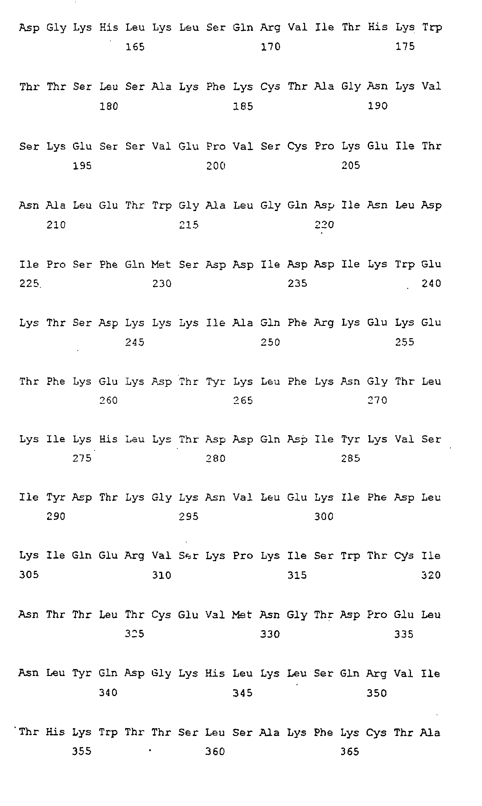
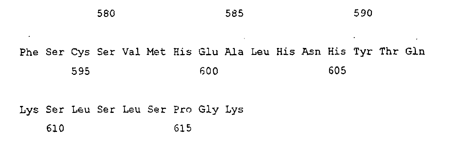
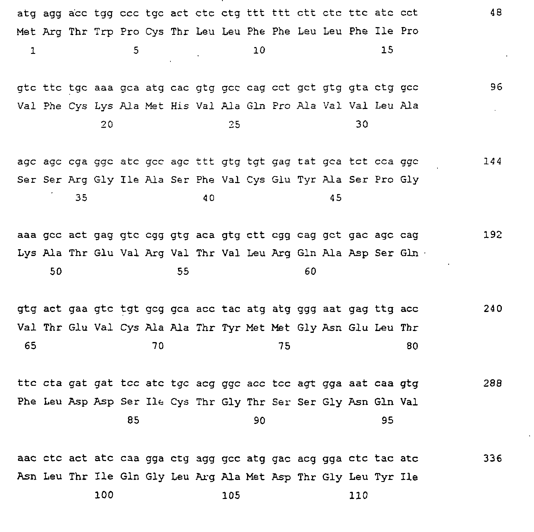
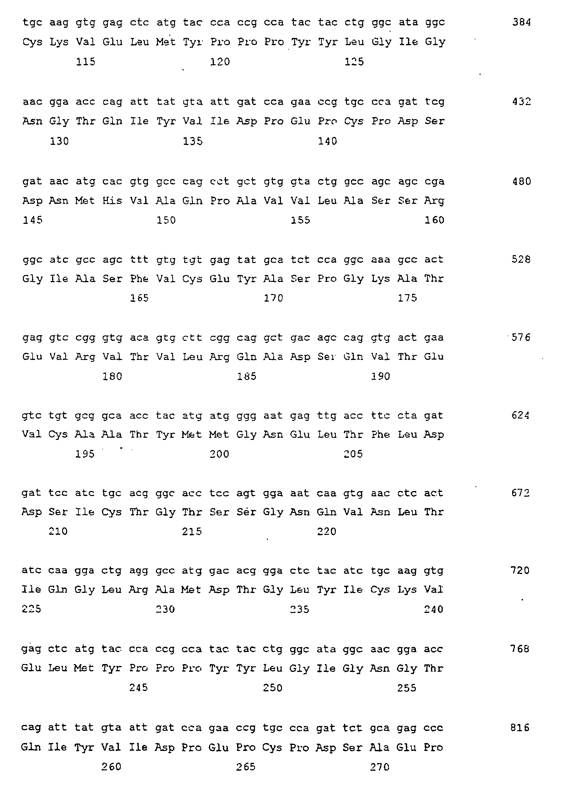
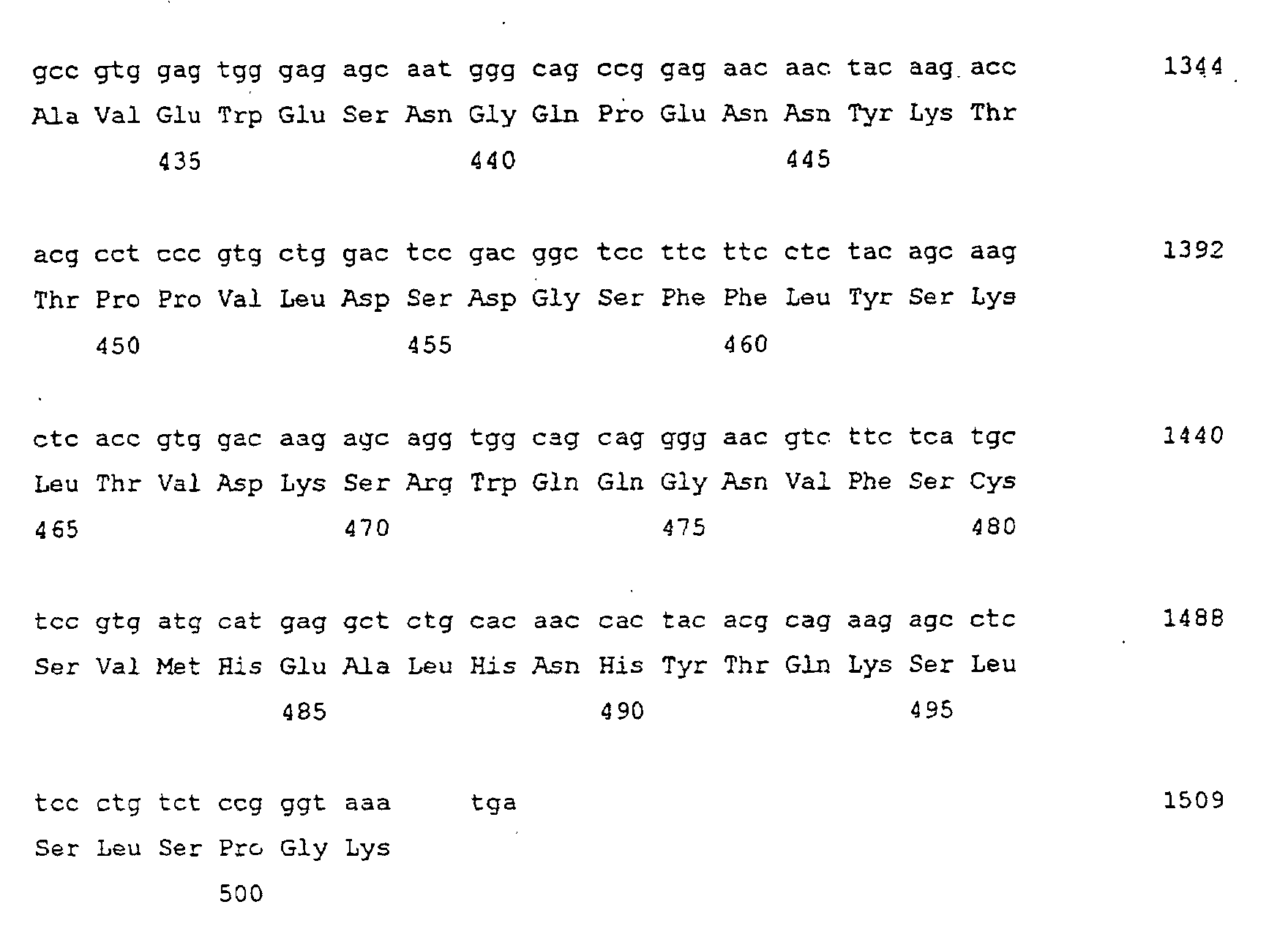
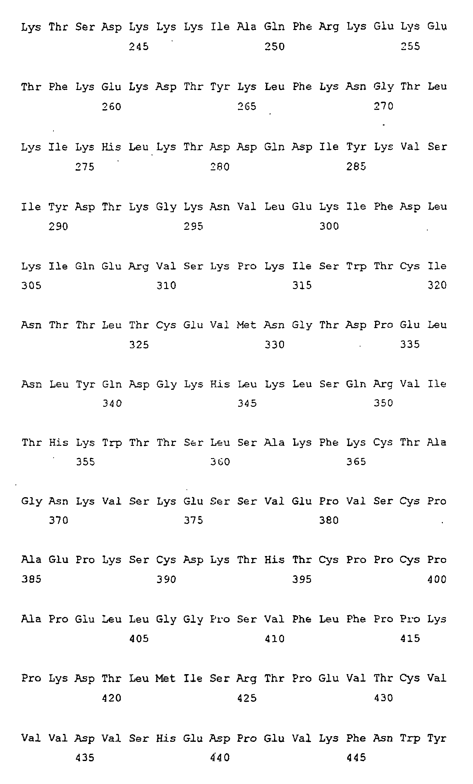
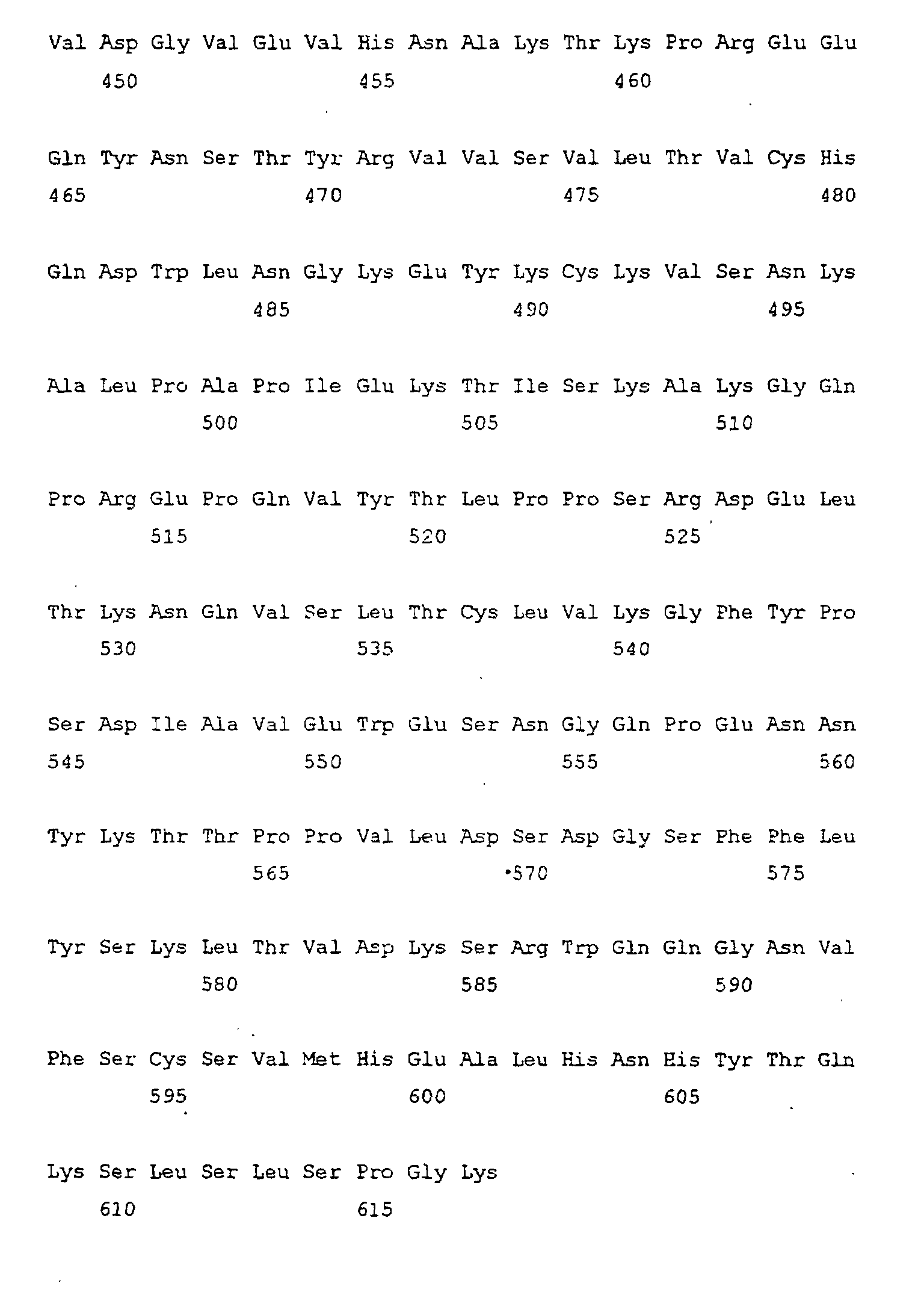
  • a concatameric fusion monomeric protein TNFR1-TNFR1/Fc designated by SEQ ID NO: 6
  • a concatameric fusion monomeric protein TNFR2-TNFR2/Fc designated by SEQ ID NO: 8
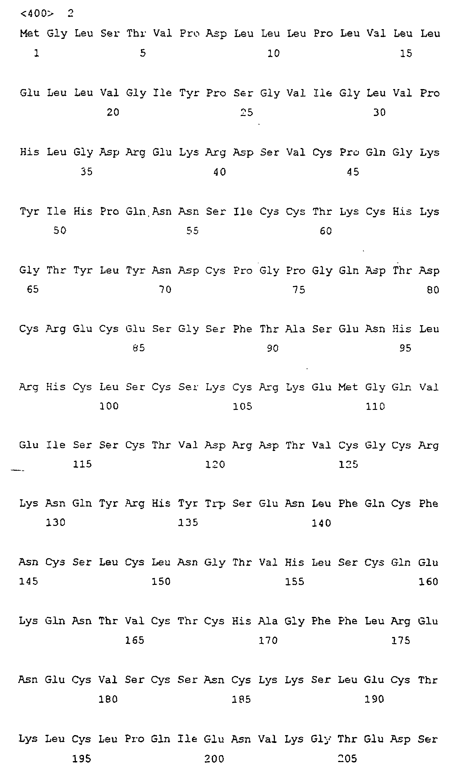
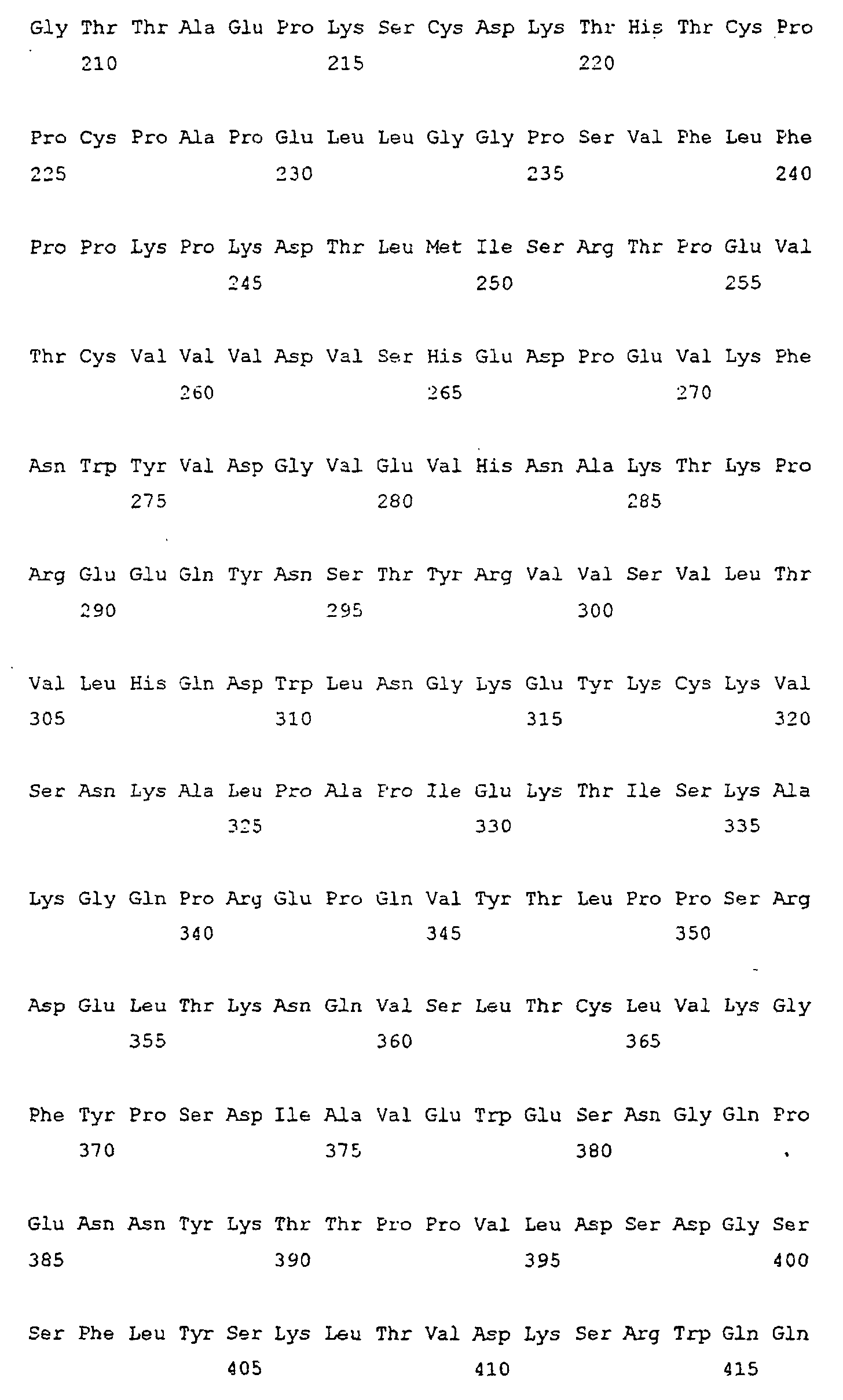
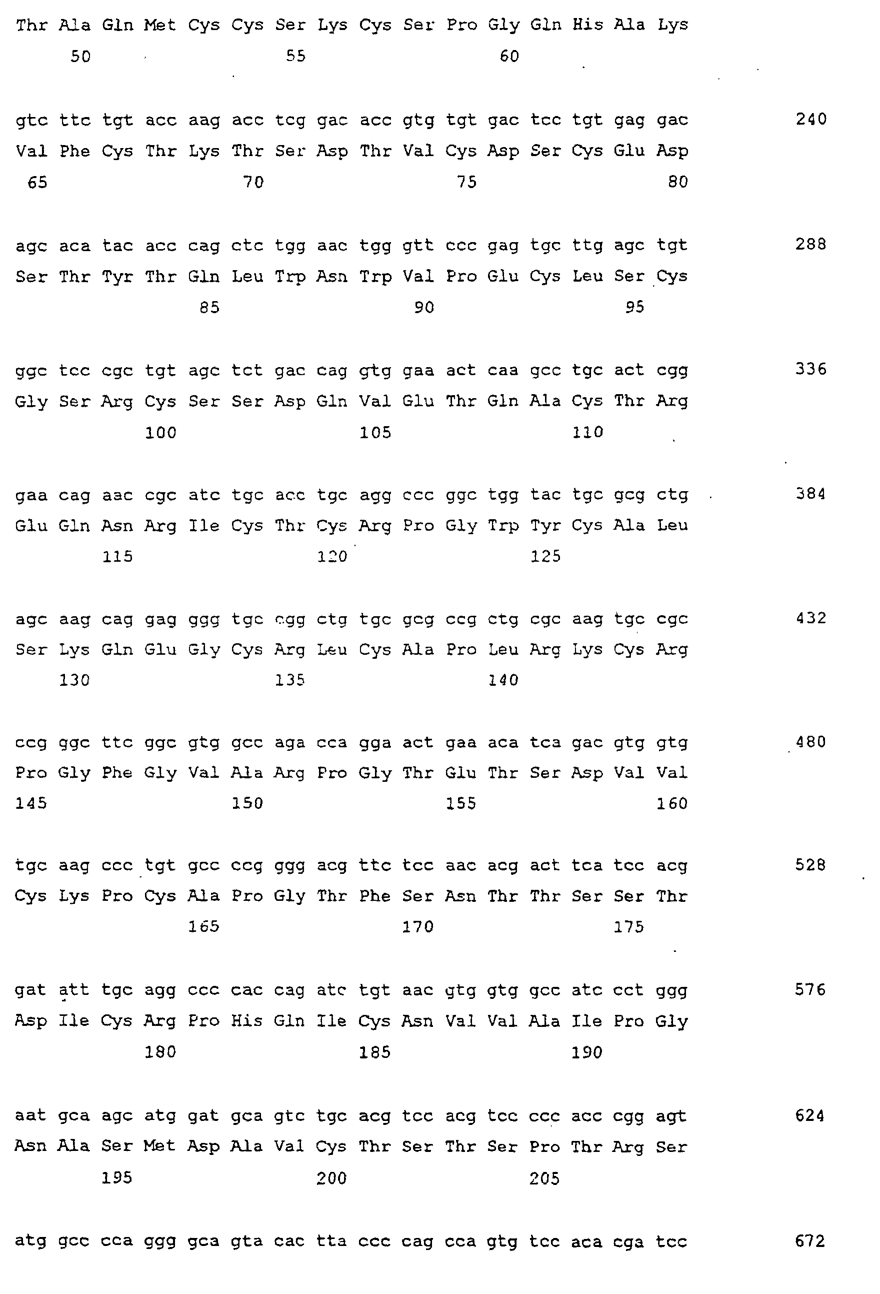
  • a concatameric fusion monomeric protein CD2-CD2/Fc designated by SEQ ID NO: 18
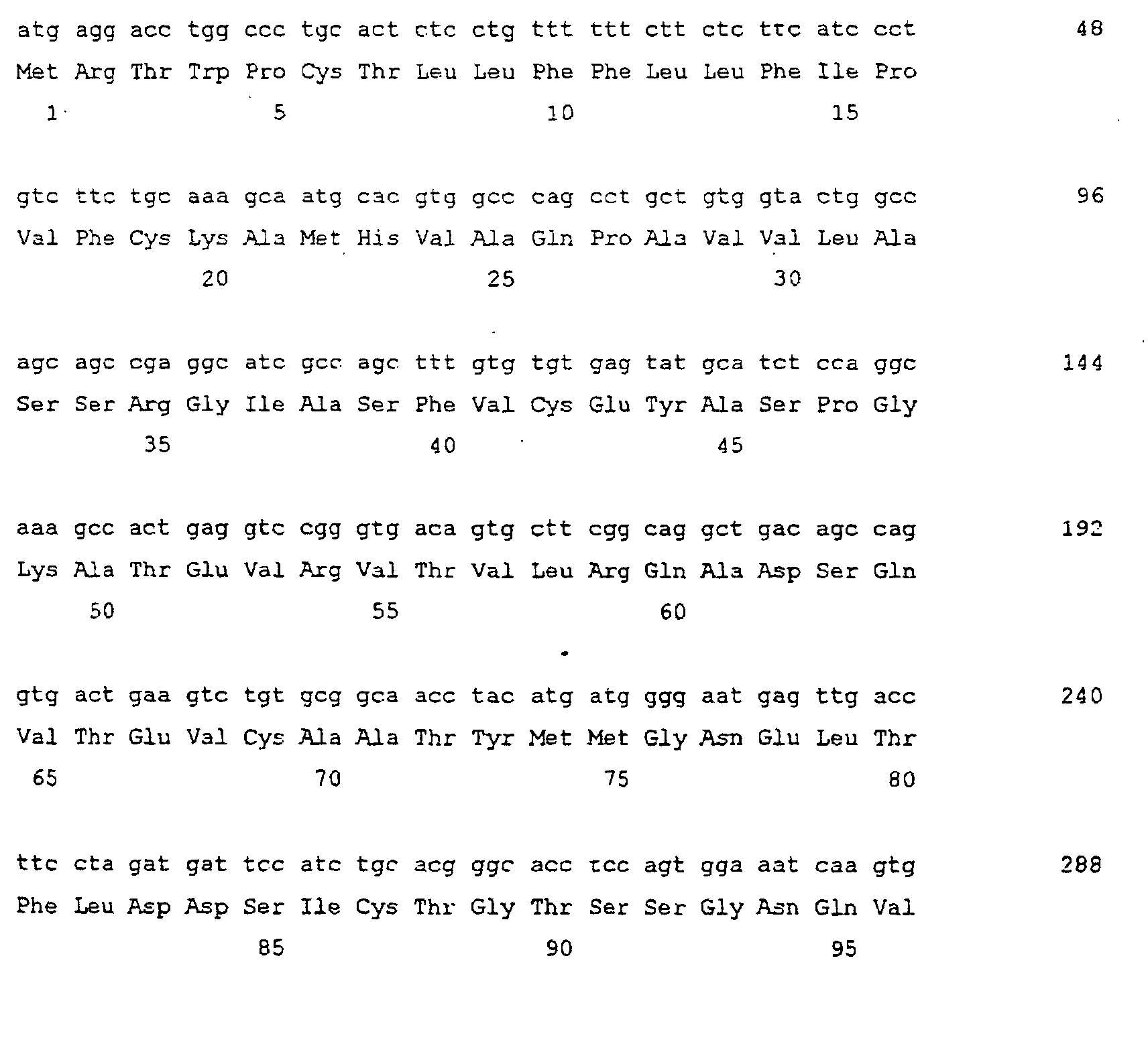
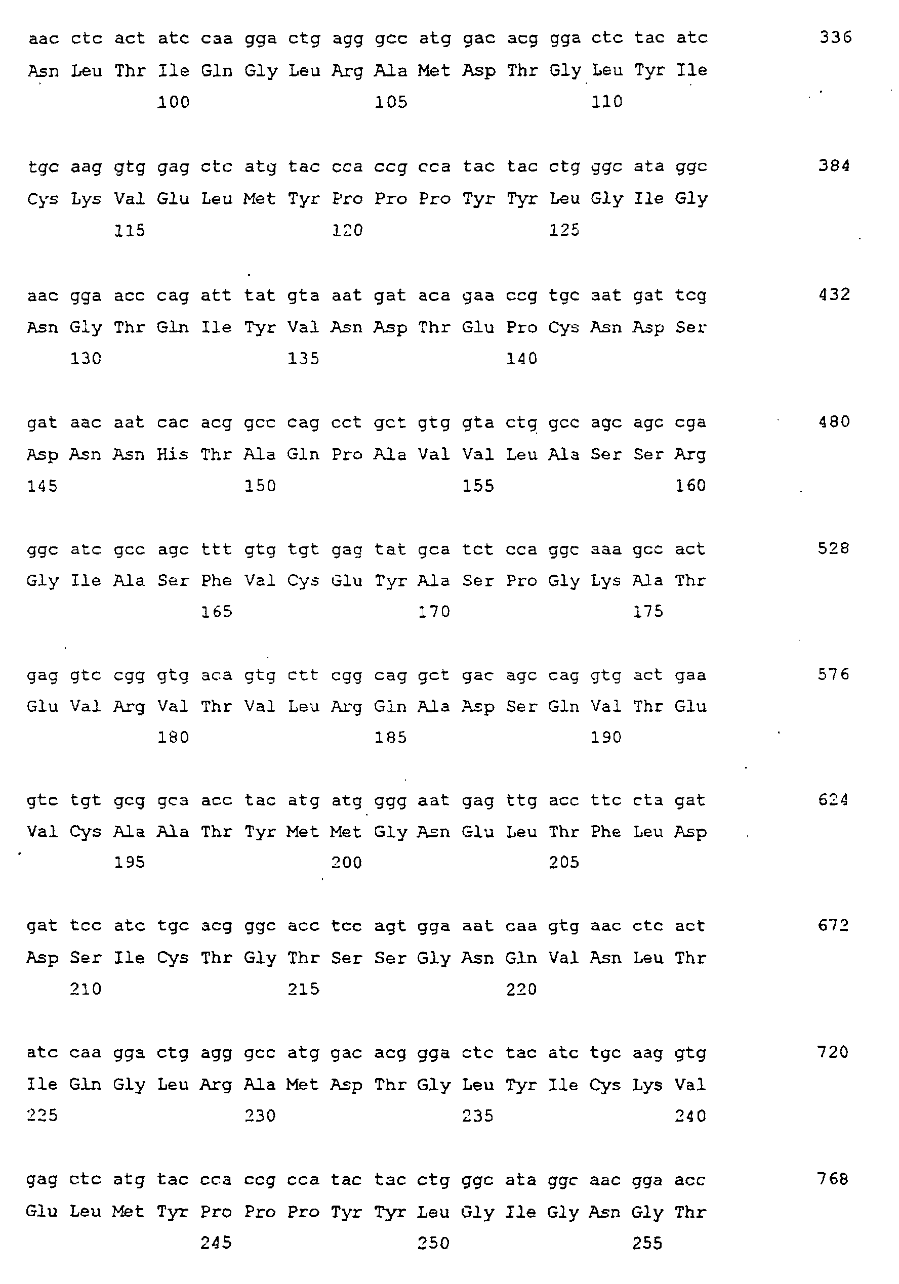
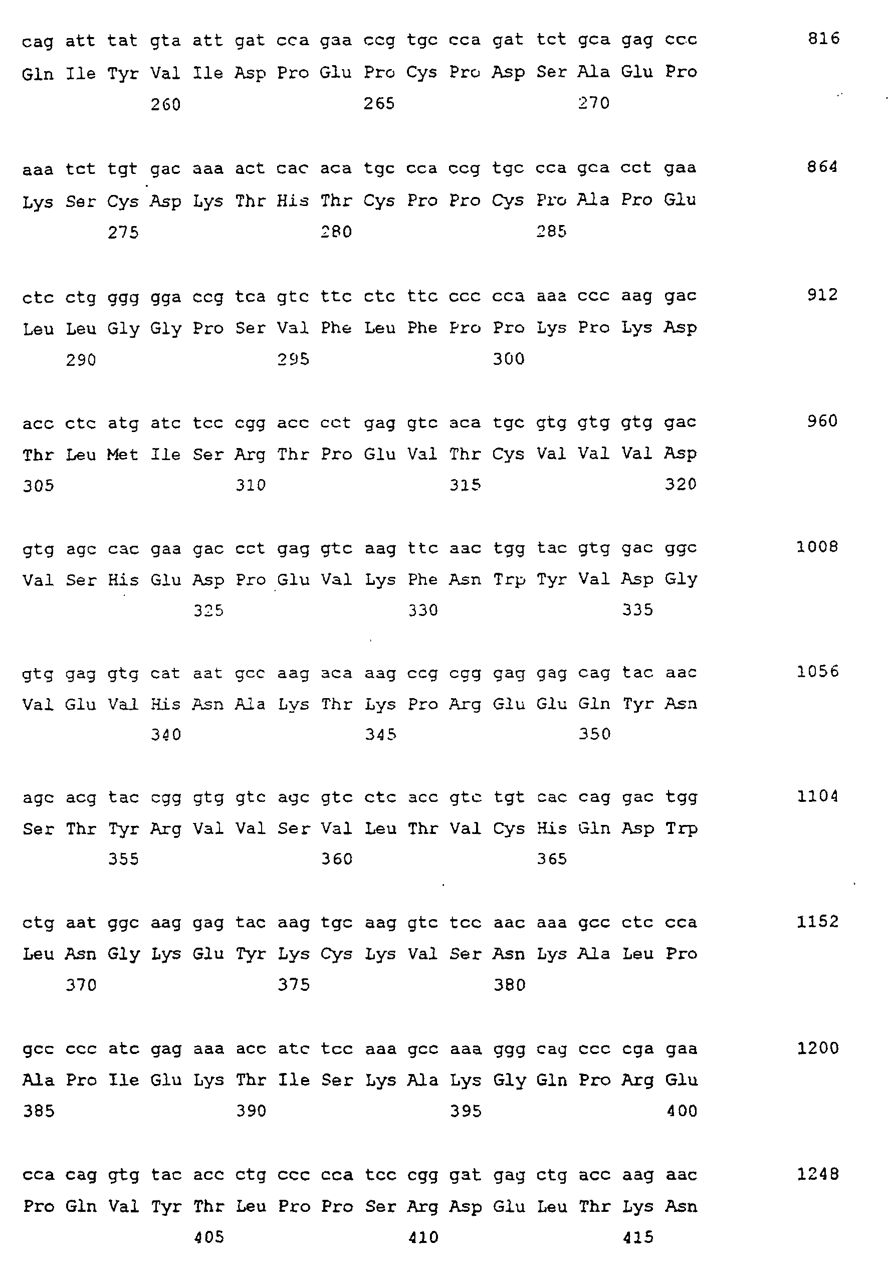
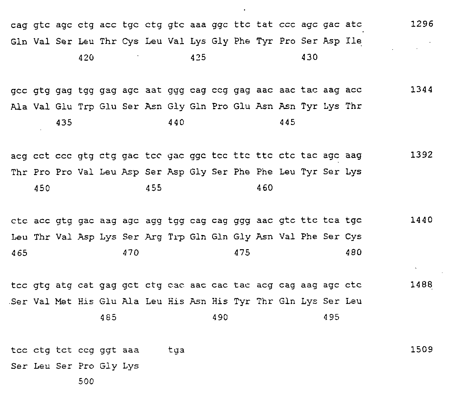
  • a concatameric fusion monomeric protein CTLA4-CTLA4/Fc designated by SEQ ID NO: 20.
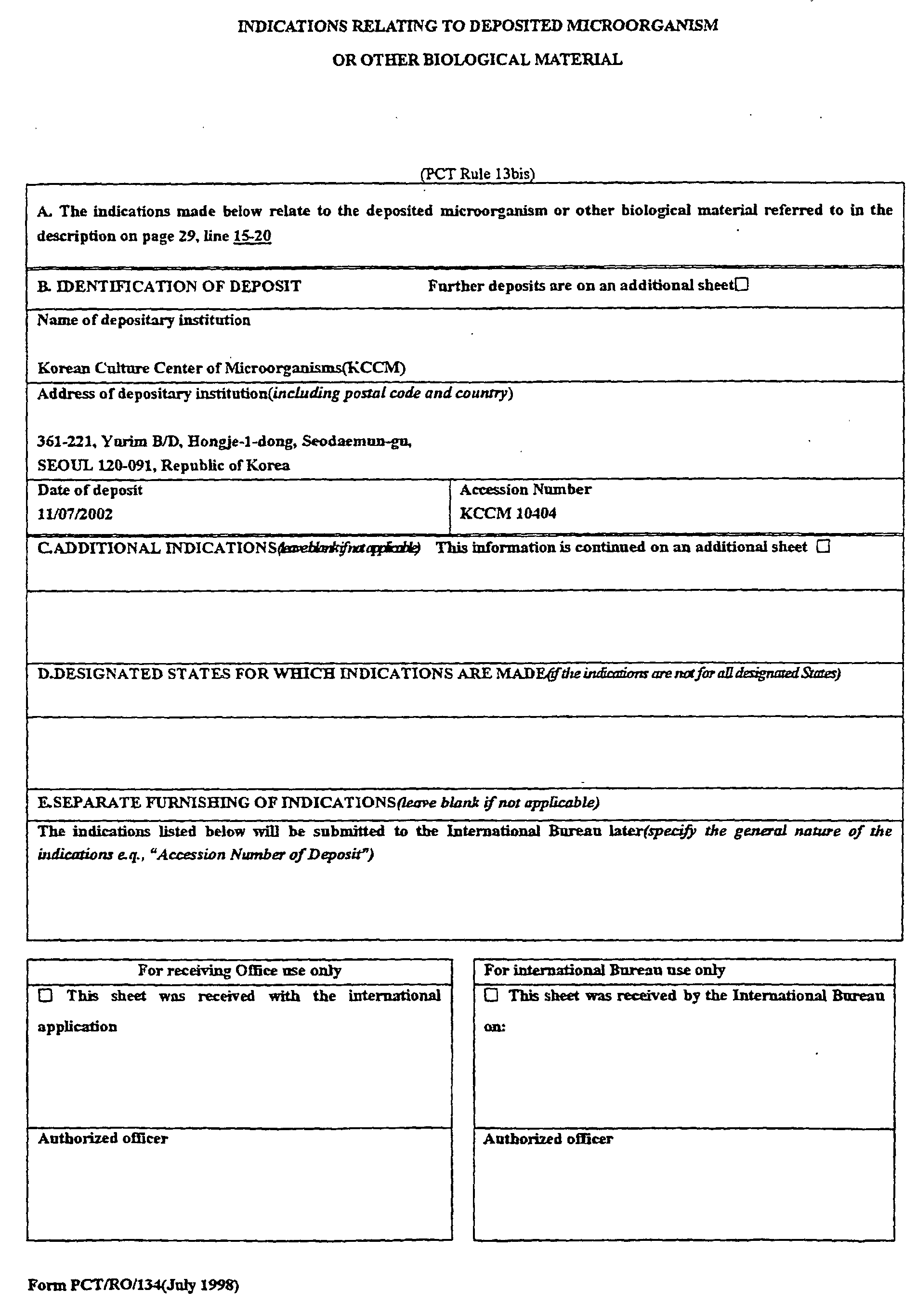
  • the recombination expression plasmids are deposited in Korean Culture Center of Microorganisms (KCCM) and are assigned accession Nos. KCCM-10288, KCCM-10291, KCCM-10402 and KCCM-10400, respectively.
  • the KCCM deposit will be maintained under the terms of the Budapest Treaty on the International Recognition of the Deposit of Microorganisms for the Purposes of Patent Procedure.
  • the concatameric fusion dimeric proteins of the present invention may be isolated from culture medium after culturing the transformants or transfectants according to the present invention.
  • the concatameric fusion dimeric proteins may participate in immune response, as described in Table 1, above, and are thus useful as therapeutic agents, diagnostic agents and laboratory tools according to the kinds of the protein, and their use is well known to those of ordinary skill in the art.
  • RPMI-1640 Gibco BRL, USA
  • the layer of T lymphocyte which formed at upper part was obtained by density gradient centrifugation using Ficoll-hypaque (Amersham, USA).
  • the cell was washed with RPMI-1640 for 3 times, and RPMI-1640 culture media containing 10% Fetal Bovine Serum (FBS, Gibco BRL, USA) was added, then cultured at 37°C for two days in the 5% CO 2 incubator after adding leukoagglutinin to 3.5ug/ml (Pharmacia, USA).
  • a fusion gene encoding soluble extracellular domain of type II human TNF receptor (TNFR2, p75) and Fc fragment of human immunoglobulin G1 was constructed by the same method as that of TNFR1/Fc.
  • a fusion gene comprising the concatameric shape in soluble extracellular domain of TNFR1, i.e. the DNA construct encoding concatameric fusion monomeric protein of TNFR1-TNFR1/Fc
  • BamHI restriction site was inserted respectively into the sequence of soluble extracellular domain of TNFR1 and DNA construct as produced as above encoding simple fusion monomeric protein of TNFR1/Fc by PCR, and then regions of each fragments restricted by BamHI were linked by ligase.
  • PCR was performed by adding 1 ⁇ l of primary cDNA, 2U of Pfu DNA polymerase (Stratagene, USA), 10 ⁇ l of 10X reaction buffer [200mM Tris-HCl, pH 8.75, 100mM (NH 4 ) 2 SO 4 , 100mM KCl, 20mM MgCl 2 ], 1% TritonTM X-100, 1mg/ml BSA, 3 ⁇ l primer 1 (10 ⁇ M), 3 ⁇ l primer 2 (10 ⁇ M), 2 ⁇ l dNTP (10mM each), and 3° distilled water to 100 ⁇ l.
  • the reaction condition was as follows; 94 °C, 5 min; 95°C, 1 min; 58°C, 1 min 30 sec; 72°C, 1 min for 31 cycles; and 72 °C, 15 min to make PCR product with complete blunt end.
  • the PCR product was purified by Qiaex II gel extraction kit (Qiagen, USA).
  • the purified PCR product was restricted by BamHI and extracted by phenol-chloroform extraction methods. Subsequently, two kinds of DNA fragments restricted by BamHI were linked by ligase.
  • DNA fragments encoding soluble extracellular domain of CD2 and CTLA4 were constructed by PCR using a primer [CD2(the sequence of nucleotide of SEQ ID NO: 40), and CTLA4(the sequence of nucleotide of SEQ ID NO: 43)] with EcoRI restriction site and the coding sequence [CD2 (the sequence of nucleotide of SEQ ID NO: 13), and CTLA4 (the sequence of nucleotide of SEQ ID NO: 15)] encoding the leader sequence [CD2(the sequence of amino acid 1-24 of SEQ ID NO: 14), and CTLA4(the sequence of amino acid 1-21 of SEQ ID NO: 16)], and an antisense primer [CD2(the sequence of nucleotide of SEQ ID NO: 41), and CTLA4(the sequence of nucleotide of SEQ ID NO: 44)] with PstI restriction site and the sequence [CD2(the sequence of nucleotide of SEQ ID NO: 13), and CTLA4(the sequence of nu
  • a DNA fragment encoding Fc fragment of immunoglobulin G1 was constructed by PCR using a primer (the sequence of nucleotide of SEQ ID NO: 42) with PstI restriction site and the sequence encoding 5' ends of constant region of IgGI, and an antisense primer (the sequence of nucleotide of SEQ ID NO: 28) with XbaI restriction site and the sequence encoding 3' ends of TgG1 Fc.
  • the template cDNA for this reaction was constructed by RT-PCR of mRNA extracted from peripheral blood cell (B lymphocyte) of convalescent patients with unknown fever.
  • the glycosylation mofit was inserted by secondary PCR with mixing in the same tube of a DNA fragment produced by PCR using a primer including EcoRI restriction site and the soluble extracellular domain with leader sequence, and an antisense primer with the sequence encoding the part of 3' end of the first soluble extracellular domain of concatameric shape of fusion gene and the part of 5' end of the second soluble extracellular domain with the nucleotide of substituted glycosylation motif; and other DNA fragment produced by PCR using a primer with the sequence encoding the part of 3' end of the first soluble extracellular domain of concatameric shape of fusion gene and the part of 5' end of the second soluble extracellular domain with the nucleotide of substituted glycosylation motif, and an antisense primer with the sequence encoding 3' end of Fc fragment of immunoglobulin G1 and XbaI restriction site.
  • WT100B1S a cell line of B lymphocyte which was made by transfection of pyrexia patient's B lymphocyte with Ebstein-Barr virus was incubated in RPMI 1640 supplemented with 10% FBS to use as antigen presenting cell of T lymphocyte. After centrifuged at 2,000rpm for 2 min to precipitate, this cells were resuspended in RPMI 1640 supplemented with 10% FBS to make 5.0X10 5 cells/ml, then irradiated by 3,000 rd of ⁇ -ray.

Landscapes

  • Health & Medical Sciences (AREA)
  • Chemical & Material Sciences (AREA)
  • Life Sciences & Earth Sciences (AREA)
  • Organic Chemistry (AREA)
  • Immunology (AREA)
  • General Health & Medical Sciences (AREA)
  • Medicinal Chemistry (AREA)
  • Proteomics, Peptides & Aminoacids (AREA)
  • Molecular Biology (AREA)
  • Genetics & Genomics (AREA)
  • Biophysics (AREA)
  • Biochemistry (AREA)
  • Cell Biology (AREA)
  • Toxicology (AREA)
  • Zoology (AREA)
  • Gastroenterology & Hepatology (AREA)
  • General Chemical & Material Sciences (AREA)
  • Public Health (AREA)
  • Veterinary Medicine (AREA)
  • Animal Behavior & Ethology (AREA)
  • Chemical Kinetics & Catalysis (AREA)
  • Pharmacology & Pharmacy (AREA)
  • Nuclear Medicine, Radiotherapy & Molecular Imaging (AREA)
  • Bioinformatics & Cheminformatics (AREA)
  • Rheumatology (AREA)
  • Engineering & Computer Science (AREA)
  • Pain & Pain Management (AREA)
  • Orthopedic Medicine & Surgery (AREA)
  • Physical Education & Sports Medicine (AREA)
  • Peptides Or Proteins (AREA)
  • Preparation Of Compounds By Using Micro-Organisms (AREA)
  • Medicines That Contain Protein Lipid Enzymes And Other Medicines (AREA)
  • Micro-Organisms Or Cultivation Processes Thereof (AREA)
  • Pharmaceuticals Containing Other Organic And Inorganic Compounds (AREA)
  • Enzymes And Modification Thereof (AREA)
  • Acyclic And Carbocyclic Compounds In Medicinal Compositions (AREA)
  • Dental Preparations (AREA)

Abstract

Disclosed are concatameric proteins comprising two soluble domains, in which the C-terminus of a soluble domain of a biologically active protein is linked to the N-terminus of an identical soluble domain or a distinct soluble domain of a biologically active protein. Also, the present invention discloses dimeric proteins formed by formation of intermolecular disulfide bonds at the hinge region of two monomeric proteins formed by linkage of a concatamer of two identical soluble extracellular regions of proteins involving immune response to an Fc fragment of an immunoglobulin molecule, their glycosylated proteins, DNA constructs encoding the monomeric proteins, recombinant expression plasmids containing the DNA construct, host cells transformed or transfected with the recombinant expression plasmids, and a method of preparing the dimeric proteins by culturing the host cells. Further, the present invention discloses pharmaceutical or diagnostic compositions comprising the dimeric protein or its glycosylated form.

Description

TECHNICAL FIELD
The present invention relates to concatameric proteins, and more specifically, concatamerized structure of biologically active protein domains where C-terminal end of extracellular soluble domain of biologically active protein is fused to N-terminal end of the same or other extracellular soluble domain of biologically active protein, and dimerization of two concatamers by coupling to hinge region of Fc fragment of immunoglobulin, and glycosylated forms of the concatameric proteins.
BACKGROUND ART
The activity of cytokine is associated with pathologic severity of inflammatory and /or immune response to various antigenic stimulations. Many antigen specific antibodies and soluble receptors which could recognize cytokines are currently in use to inhibit the function of cytokines for the therapeutic purposes (WO 93/016184, WO 96/02576, WO 96/023067, WO 1997/03682, and US 5,434,131, 5,656,272, 5,977,318, 6,210,661, 6,225,117). Antibodies and soluble receptors inhibit cytokine signal transduction by disturbing interaction between cytokines and their receptors on cell surface.
Soluble receptors used as functional inhibitors of cytokine that fused to heavy chains of human immunoglobulins were disclosed by Capon et al. (Nature 337:5254, 1989), and thereafter many patents were disclosed inventions related to fusion proteins of soluble receptors and immunoglobulins (US patent 5,521,288, 5,844,095, 6,046,310, 6,090,914, 6,100,383, 6,225,448).
Generally, fusion proteins of soluble receptors and immunoglobulins have following advantages (Capon et al., Nature 337:5254, 1989)
  • 1. Increase in total avidity to ligand by forming bivalency via dimerization.
  • 2. Increase in blood half-life of proteins, that is, increase in molecular stability
  • 3. Activation of effecter cells by Fc fragment of immunoglobulin heavy chain
  • 4. Convenience of purification by using affinity column, e.g. using protein A Most fusion proteins of receptor extracellular domain and immunoglobulin heavy chain are composed of heavy chain without CH1 domain, which result in dimers not binding to light chains. This structure is more desirable for the function of proteins and receptors involving immune response. For example, TNFR(WO92/16221, WO95/34326)-immunoglobulin fusion proteins disclosed in WO94/06476 and US 5,447,851 have been used for the inhibition of TNF-mediated inflammation. It is well known that TNFR-immunoglobulin fusion proteins have a higher affinity than original monomeric molecules (Lesslauer et al., Eur. J. Immunol. 21:2883, 1991; Ashkenazi et al., Proc. Natl. Acad. Sci. 88:10535, 1991; Peppe et al., J. Exp. Med. 174:1483, 1991; Mohler et al., J. Immunol. 151:1548, 1993).
  • For the improved inhibition of TNF mediated response, one can increase efficacy by multimerizing soluble extracellular domains of TNFR, CD2, and CTLA-4. For example, when fusion proteins of TNFR's extracellular domains bound with immunoglobulin heavy chain(heavy chain fusion protein) and with light chain(light chain fusion protein) respectively are coexpressed in the same cell, one can produce fusion proteins as a tetrameric form by linking heavy chain to heavy and light chains. This tetramer showed much more increased efficacy than monomeric or dimeric forms as presented by Scallon et al. (Cytokine 7:759, 1995).
    However, this method had many difficulties for commercialization such as simultaneous expression of two different fusion genes in the same cell line, remarkably lower production yields of multimeric form; and difficulty in purifying multimeric high molecular weight forms. For these reasons, immunoglobulin fusion proteins currently in use are only heavy chain fused form.
    Therefore, there is considerable demand for the development of methods of producing multimeric protein therapeutics with high yield and efficient purification procedures.
    Further references relating to multimeric proteins and vectors pertinent to the field of the invention are: US 5 073 672, EP 464 533, US 5 861 151, US 5 349 053, US 5 428 130, US 6 165 476, WO 01/77324 and Cornish et al., Journal of Cell Science 1999, 1213-1223.
    DISCLOSURE OF INVENTION
    The present inventors have manufactured concatameric proteins by fusing the C-terminal end of soluble domain of biologically active protein to the N-terminal end of soluble domain of the same or other biologically active protein by using DNA recombination techniques. Also, the present inventors have dimerized this concatamers by linking it to the hinge region of Fc fragment of immunoglobulin and added more glycosylations by using DNA mutagenesis techniques. And the present inventors have found that concatamerized protein dimers and their glycosylated forms show increased efficacy and stability compared to conventional monomeric fusion proteins.
    Therefore, the present invention generally relates to concatameric proteins where C-terminal end of a soluble domain of a biologically active protein is fused to an N-terminal end of a soluble domain of the same or other biologically active proteins.
    In one aspect, the present invention provides a concatemeric fusion dimeric protein comprising two monomeric proteins formed by linkage of a concatemer of two identical soluble extracellular domains of proteins involving immune response to a hinge region of an Fc fragment of an immunoglobulin molecule, wherein said monomeric proteins are linked by intermolecular disulfide bonds at the hinge region, and having improved stability and therapeutic effects.
    Also in another aspect, the present invention provides DNA constructs that encode monomeric fusion proteins whose concatamerized domain is fused to hinge region of Fc fragment of immunoglobulins.
    Also in another aspect, the present invention provides DNA plasmids comprising a DNA construct that encodes monomeric fusion protein whose concatamerized part is fused to hinge region ofFc fragment of immunoglobulin.
    Also in another aspect, the present invention provides host cells transfected or transformed with recombinant DNA plasmids including a DNA construct that encodes monomeric fusion protein whose concatamerized part is fused to hinge region of Fc fragment of immunoglobulin.
    Also in another aspect, the present invention provides a method for culturing the host cells, which were transfected or transformed with recombinant DNA plasmids including a DNA construct that encodes monomeric fusion protein whose concatamerized part is fused to hinge region ofFc fragment of immunoglobulin, under culture condition for expression of DNA constructs encoding concatameric fusion protein coupled to hinge region ofFc fragment of immunoglobulin , and manufacturing dimeric concatamers formed by disulfide bond at hinge region of two monomeric concatamers described as above including the process of purification of the proteins described as above from cell culture.
    Also in another aspect, the present invention provides a method for culturing the host cells, which were transfected or transformed with recombinant DNA plasmids including a DNA construct that encodes monomeric fusion protein whose concatamerized part of immunomudulatory function is fused to hinge region of Fc fragment of immunoglobulin and is inserted with glycosylation motifs, under the best condition which is suitable for expression of DNA constructs that encode monomeric fusion protein whose concatamerized part of immune function is fused to hinge region of Fc fragment of immunoglobulin, and for manufacturing glycosylated dimers formed by disulfide bond at hinge region of two monomeric proteins described as above including the process of purification of the glycosylated proteins described as above from cell culture.
    Also in another aspect, the present invention provides DNA primers for inserting glycosylation motif into the DNA constructs that encode monomeric fusion proteins whose concatamerized part is fused to hinge region of Fc fragment of immunoglobulins.
    Also in another aspect, the present invention provides the glycosylated dimers formed by disulfide bond at hinge region of two monomeric proteins whose concatamerized part involving immune response is fused to hinge region of Fc fragment of immunoglobulins.
    Also in another aspect, the present invention provides the pharmaceutical compositions comprising dimers formed by disulfide bond at hinge region of two monomeric proteins whose concatamerized part involving immune response is fused to hinge region of Fc fragment of immunoglobulins in a pharmaceutically effective amount and in a pharmaceutically acceptable carrier.
    Also in another aspect, the present invention provides the pharmaceutical compositions comprising glycosylated dimers formed by disulfide bond at hinge region of two monomeric proteins whose concatamerized part involving immune response is fused to hinge region of Fc fragment of immunoglobulins in a pharmaceutically effective amount and in a pharmaceutically acceptable carrier.
    BRIEF DESCRIPTION OF THE DRAWINGS
    The above and other objects, features and other advantages of the present invention will be more clearly understood from the following detailed description taken in conjunction with the accompanying drawings, in which:
  • Fig. 1 is a schematic view showing a process of preparing a DNA construct encoding a conventional simple fusion monomeric protein through polymerase chain reaction (PCR);
  • Fig. 2 is a schematic view showing a process of preparing a DNA construct encoding a concatameric fusion monomeric protein according to the present invention through PCR;
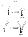
  • Fig. 3a shows structures of [TNFR/Fc]2, [CD2/Fc]2 or [CTLA4/Fc]2 fusion proteins, which are simple fusion dimeric proteins formed through homodimerization in cells of TNFR/Fc, CD2/Fc or CTLA4/Fc fusion proteins as examples of conventional simple fusion monomeric proteins;
  • Fig. 3b shows structures of [TNFR-TNFR/Fc]2, [CD2-CD2/Fc]2 or [CTLA4-CTLA4/Fc]2 fusion proteins, which are concatameric fusion dimeric proteins formed through homodimerization in cells of TNFR-TNFR/Fc, CD2-CD2/Fc or CTLA4-CTLA4/Fc fusion proteins as embodiments of the concatameric fusion dimeric protein according to the present invention;
  • Fig. 4a shows a structure of [TNFR1-TNFR1/Fc]2, as an embodiment of a concatameric fusion dimeric protein according to the present invention;
  • Fig. 4b shows a structure of [TNFR2-TNFR2/Fc]2, as another embodiment of the concatameric fusion dimeric protein according to the present invention;
  • Fig. 4c shows a structure of [CD2-CD2/Fc]2, as a further embodiment of the concatameric fusion dimeric protein according to the present invention;
  • Fig. 4d shows a structure of [CTLA4-CTLA4/Fc]2, as a still further embodiment of the concatameric fusion dimeric protein according to the present invention;
  • Fig. 5 is a diagram showing a process of constructing a recombinant expression plasmid pTR11Ig-Top10' expressing a concatameric fusion monomeric protein TNFR1-TNFR1/Fc according to the present invention;
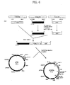
  • Fig. 6 is a diagram showing a process of constructing a recombinant expression plasmid pCD22Ig expressing a concatameric fusion monomeric protein CD2-CD2/Fc according to the present invention;
  • Fig. 7 is a map of a recombinant expression plasmid pTR11Ig-Top10' expressing a concatameric fusion monomeric protein TNFR1-TNFR1/Fc according to the present invention;
  • Fig. 8 is a map of a recombinant expression plasmid pTR22Ig-Top10' expressing a concatameric fusion monomeric protein TNFR1-TNFR1/Fc according to the present invention;
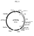
  • Fig. 9 is a map of a recombinant expression plasmid pCD22Ig expressing a concatameric fusion monomeric protein CD2-CD2/Fc according to the present invention;
  • Fig. 10 is a map of a recombinant expression plasmid pCT44Ig expressing a concatameric fusion monomeric protein CTLA4-CTLA4/Fc according to the present invention;
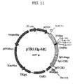
  • Fig. 11 is a map of a recombinant expression plasmid pTR11Ig-MG expressing a concatameric fusion monomeric protein mgTNFR1-TNFR1/Fc containing four glycosylation motif peptides according to the present invention;
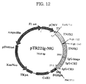
  • Fig. 12 is a map of a recombinant expression plasmid pTR22Ig-MG expressing a concatameric fusion monomeric protein mgTNFR2-TNFR2/Fc containing two glycosylation motif peptides according to the present invention;
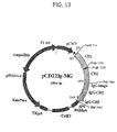
  • Fig. 13 is a map of a recombinant expression plasmid pCD22Ig-MG expressing a concatameric fusion monomeric protein mgCD2-CD2/Fc containing two glycosylation motif peptides according to the present invention;
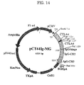
  • Fig. 14 is a map of a recombinant expression plasmid pCT44Ig-MG expressing a concatameric fusion monomeric protein mgCTLA4-CTLA4/Fc containing three glycosylation motif peptides according to the present invention;
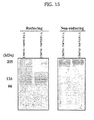
  • Fig. 15 shows a result of SDS-PAGE of purified concatameric fusion dimeric proteins [TNFR1-TNFR1/Fc]2 and [TNFR2-TNFR2/Fc]2 under reducing or non-reducing conditions;
  • Fig. 16 is a graph showing inhibitory effect of the conventional simple fusion dimeric proteins [TNFR1/Fc]2() and [TNFR2/Fc]2(○) and the concatameric fusion dimeric proteins [TNFR1-RNFR1/Fc]2(▾) and [TNFR2-TR2Fc]2(▿) according to the present invention against cytotoxic activity of TNF-alpha;
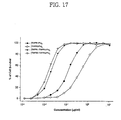
  • Fig. 17 is a graph showing inhibitory effect of the conventional simple fusion dimeric proteins [TNFR1/Fc]2() and [TNFR2/Fc]2(○) and the concatameric fusion dimeric proteins [TNFR1-RNFR1/Fc]2(▾) and [TNFR2-TR2Fc]2(▿) according to the present invention against cytotoxic activity of TNF-beta;
  • Fig. 18 is a graph showing inhibitory effect of the conventional simple fusion dimeric protein [CD2/Fc]2(), the known immunosuppressive agent cyclosporin A (▾) and the concatameric fusion dimeric protein [CD2-CD2/Fc]2(○) according to the present invention on the proliferation of active T lymphocytes;
  • Fig. 19 is a graph showing inhibitory effect of the conventional simple fusion dimeric protein [CTLA4/Fc]2(), the known immunosuppressive agent cyclosporin A (▾) and the concatameric fusion dimeric protein [CTLA4- CTLA4/Fc]2 (○) according to the present invention on the proliferation of active T lymphocytes;
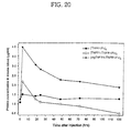
  • Fig. 20 is a graph showing blood half-life of the conventional simple fusion dimeric protein [TNFR1/Fc]2(), the concatameric dimeric protein [TNFR1-TNFR1/Fc]2 (○) and a glycosylated concatameric fusion dimeric protein [mgTNFR1-TNFR1/Fc]2 (▿) according to the present invention;
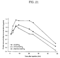
  • Fig. 21 is a graph showing blood half-life of the conventional simple fusion dimeric protein [CD2/Fc]2(), the concatameric fusion dimeric protein [CD2-CD2/Fc]2 (○) and a glycosylated concatameric fusion dimeric protein [mgCD2-CD2/Fc]2 (▿) according to the present invention;
  • Fig. 22 is a graph showing blood half-life of the conventional simple fusion dimeric protein [CTLA4/Fc]2(), the concatameric fusion dimeric protein [CTLA4-CTLA4/Fc]2 (○) and a glycosylated concatameric fusion dimeric protein [mgCTLA4-CTLA4/Fc]2 (▿) according to the present invention; and
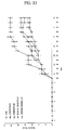
  • Fig. 23 is a graph showing inhibitory effect of PBS () as a control, the conventional simple fusion dimeric proteins [TNFR1/Fc]2 (▪) and [TNFR2/Fc]2 (▴), and concatameric fusion dimeric proteins [TNFR1-TNFR2/Fc]2 (×) and [TNFR2-TNFR2/Fc]2 (Δ) according to the present invention on the induction of collagen-induced arthritis (CIA) in DBA/1 mice.
  • BEST MODE FOR CARRYING OUT THE INVENTION
    The present invention is generally directed to concatameric proteins, and more particularly, to immunoadhesion molecules. Immunoadhesion molecules are typically formed by fusion of the Fc fragment of immunoglobulin (Ig) to a ligand-binding region of a receptor or an adhesion molecule, and thus have a structure similar to that of an antibody. The typical immunoadhesion molecules known in the art have a structure of an antibody in which the variable region is substituted with a ligand-binding region of a receptor while retaining the Fc fragment. A wide variety of immunoadhesion molecules are suggested in the literature. However, immunoadhesion molecules according to the present invention have different structure with the conventional immunoadhesion molecules, and there is also no prior art predicting or describing preparation of the immunoadhesion molecules according to the present invention.
    Definition of Terms
    For full understanding of the characteristic structure of the immunoadhesion molecules according to the present invention, exact definitions of the terms used in the present invention are given as follows. In general, all of the technical and scientific terms being not additionally defined in the present invention have meanings commonly used in the art. However, although having meanings commonly used in the art, the following terms are defined to give a clearer understanding of their meanings and make the scope of the present invention clear, as follows.
    The term "immunoglobulin", as used herein, refers to protein molecules being produced in B cells and serving as antigen receptors specifically recognizing a wide variety of antigens. The molecules have a Y-shaped structure consisting of two identical light chains (L chains) and two identical heavy chains (H chains), in which the four chains are held together by a number of disulfide bonds, including the disulfide bridge between the H chains at the hinge region. The L and H chains comprise variable and constant regions. The L chain variable region associates with the H chain variable region, thus producing two identical antigen-binding regions. According to features of the constant regions of H chains, immunoglobulins (Ig) are classified into five isotypes, A (IgA), D (IgD), E (IgE), G (IgG) and M (IgM). Each subtype possesses unique structural and biological properties. For example, IgG has slightly different Fc structure, compared with other isotypes. In addition, IgG and IgA have a number of subtypes. For example, the human IgG isotype has four subtypes, IgG1, IgG2, IgG3 and IgG4, which have γ1, γ2, γ3 and γ4 H chains, respectively. Biological functions of immunoglobulin molecules, such as complement activation, Fc receptor-mediated phagocytosis and antigen-dependent cytotoxicity, are mediated by structural determinants (complementarity-determining regions) in the Fc region of H chains. Such an Fc region of H chains is used for construction of dimeric proteins according to the present invention, and may be derived from all isotypes and subtypes of immunoglobulin as described above.
    The term "Fc fragment of an immunoglobulin molecule", as used herein, refers to a fragment having no antigen-binding activity and being easily crystallized, which comprises a hinge region and CH2 and CH3 domains, and a portion responsible for binding of an antibody to effector materials and cells. Therefore, the Fc fragment mentioned in the present invention can be different from that described in some literatures, but includes the hinge region. Such description of the Fc fragment is given to supply convenience in describing the present invention, and will be fully understood by those of ordinary skill in the art with reference to the specification of the present invention and the accompanying drawings.
    The term "biologically active protein", as used herein, refers to a protein, peptide or polypeptide having generally physiological or pharmaceutical activities, which retains a part of its native activities after forming a concatamer or immunoadhesion molecule. The term "biological activity", as used herein, is not limited in meaning to physiological or pharmaceutical activities. For example, some concatamers, such as those containing an enzyme can catalyze a reaction in an organic solvent. Similarly, some high-molecular weight fusion molecules containing concanavalin A or an immunoglobulin molecule are useful as diagnostic agents in laboratories.
    Non-limiting examples of the protein, peptide or polypeptide include hemoglobin, serum proteins (e.g., blood factors including factor VII, VIII and factor IX), immunoglobulin, cytokines (e.g., interleukin), α-, β- and γ-interferon, colony-stimulating agent (e.g., G-CSF and GM-CSF), platelet-derived growth factor (PDGF), and phospholipase activating proteins (PLAPs). Other typical biological or therapeutic proteins include insulin, plant proteins (e.g., lectin and ricin), tumor necrosis factor (TNF) and its related alleles, growth factors (e.g., tissue growth factors and endothelial growth factors such as TGFα or TGFβ), hormones (e.g., follicle-stimulating hormone, thyroid-stimulating hormone, antidiuretic hormone, pigment-concentrating or dispersing hormones and parathyroid hormone, luteinizing hormone-releasing hormone and its derivatives, calcitonin, calcitonin gene related peptide (CGRP), synthetic enkephalin, somatomedin, erythropoietin, hypothalamus releasing factors, prolactin, chronic gonadotrophin, tissue plasminogen-activating agents, growth hormone-releasing peptide (GHRP), and thymic humoral factor (THF). The immunoglobulins include IgG, IgE, IgM, IgA, IgD and fragments thereof. Some proteins such as interleukin, interferon or colony-stimulating factor may be produced in a non-glycosylated form using DNA recombinant techniques. The non-glycosylated proteins may be useful as biologically active materials in the present invention.
    In addition, the biologically active materials useful in the present invention include any polypeptide, which has bioactivity in vivo. Examples of the biologically active materials include peptides or polypeptides, fragments of an antibody, single chain-binding proteins (see U.S. Pat. No. 4,946,778), binding molecules including fusion polypeptides of antibodies or their fragments, polyclonal antibodies, monoclonal antibodies, and catalytic antibodies. Other examples of the biologically active materials include allergen proteins, such as ragweed, antigen E, honeybee venom, or allergen of mites.
    In addition, the biologically active material useful in the present invention includes enzymes. Examples of the enzymes include carbohydrate-specific enzymes, proteinases, oxidoreductases, transferases, hydrolases, lyases, isomerases, and ligases. In detail, non-limiting examples of the enzymes include asparaginase, arginase, arginine deaminase, adenosine deaminase, peroxide dismutase, endotoxinase, catalase, chymotrypsin, lipase, uricase, adenosine dephosphatase, tyrosinase, and bilirubin oxidase. Examples of the carbohydrate-specific enzymes include glucose oxidase, glucodase, galactosidase, glucocerebrosidase, and glucouronidase.
    The term "proteins involving immune response", as used herein, refers to all proteins mediating cell-to-cell signal transduction during cellular or humoral immune response and thus activating or suppressing immune response. Immunity is a process of protecting "self" from "non-self" such as bacteria or viruses. Immune response is largely divided into cellular and humoral immune response, where T and B lymphocytes play the most important role. T cells, mainly mediating cellular immune response, directly attack and kill virus-infected cells or tumor cells, or help other immune cells by secreting cytokines functioning to induce or activate immune response or inflammation. B cells produce antibodies against non-self foreign materials (antigens) that enter a body, such as bacteria or viruses, and such immune response is called cellular immune response. Cell-to-cell signal transduction is an essential process in both cellular and humoral immune responses, in which a signal molecule, that is, a ligand, interacts with a cell surface receptor acting to transduce a specific signal into a cell.
    Representative examples of the proteins involving the immune response according to the present invention include cytokines, cytokine receptors, adhesion molecules, tumor necrosis factor receptor (TNFR), enzymes, receptor tyrosine kinases, chemokine receptors, other cell surface proteins, and soluble ligands. Non-limiting examples of the cytokines include IL-1, TL-2, IL-3, IL-4, IL-5, IL-6, IL-7, IL-10, IL-12, IL-17, TNF, TGF, IFN, GM-CSF, G-CSF, EPO, TPO, and M-CSF. Examples of the cytokine receptors, but are not limited to, include growth hormone receptors (GHRs), IL-13R, IL-IR, IL-2R, IL-3R, IL-4R, IL-5R, IL-6R, IL-7R, IL-9R, IL-15R, TNFR, TGFR, IFNR (e.g., IFN-γ R α-chain and IFN-γ R β-chain), interferon-α R, -β R and -γ R, GM-CSFR, G-CSFR, EPOR, cMp1, gp130, and Fas (Apo 1). Non-limiting examples of the enzymes include influenza C hemaglutinin esterase and urokinase. The chemokine receptors are exemplified by CCR1 and CXCR1-4. Examples of the receptor tyrosine kinases, but are not limited to, include TrkA, TrkB, TrkC, Htk, REK7, Rse/Tyro-3, hepatocyte growth factor R, platelet-derived growth factor R, and Flt-1. Examples of other cell surface proteins includes CD2, CD4, CD5, CD6, CD22, CD27, CD28, CD30, CD31, CD40, CD44, CD100, CD137, CD150, LAG-3, B7, B61, β-neurexin, CTLA-4, ICOS, ICAM-1, complement R-2 (CD21), IgER, lysosomal membrane gp-1, α2-microglobulin receptor-related proteins, and sodium-releasing peptide R Non-limiting examples of the soluble ligands include IL-10, heregulin, and keratinocyte growth factors.
    Ligands for the proteins involving immune response according to the present invention and use thereof are well known to those of ordinary skill in the art, as summarized in Tables 1 to 7, below.
    Proteins involving immune response: Adhesion molecules
    Adhesion molecules Ligands Uses
    CD4 HIV gp120 Inhibition of in vivo HIV infection; and identification of CD4 domain participating in ligand binding
    L-Selectin GlyCAM-1, CD34 Prevention of neutrophile-mediated lung damage; determination of position in tissues of a ligand by histochemical staining; and isolation and cloning of ligands and determination of their properties
    E-Selectin Sialyl Lewisx Prevention of neutrophile-mediated lung damage; and determination of thermodynamic properties in ligand-binding
    P-Selectin Sialyl Lewisx Prevention of neutrophile-mediated lung damage; and study of functions of individual of amino acid residues in binding to cell surface
    ICAM-1 CD11a/CD18 Phagocytosis of erythrocytes in malaria; inhibition of infection with rhinovirus; and anti-inflammation in diabetes
    ICAM-2 CD11a/CD18 Study of activation of T cells mediated by T cell receptor
    ICAM-3 CD11a/CD18 Identification of receptor domains binding to a ligand
    VCAM-1 VLA-4 Study of role of VLA-4 in T lymphocyte migration to dermal inflammation sites
    LFA-3 CD2 Study of role of CD2 in costimulation of T cells
    L1 glycoprotein Fibroblast growth factor receptor Stimulation of nerve reproduction after repair, and functional comparison with FGF
    Proteins involving immune response: Enzymes
    Enzymes Ligands Uses
    Influenza C hemaglutinin esterase 9-0-acetylated sialic acid Inactive enzyme used in study of tissue-specific expression of ligands
    Urokinase Urokinase receptor Inactive enzyme developed to inhibit cancer metastasis by disturbing urokinase activation
    Proteins involving immune response: Cytokine receptors
    Cytokine receptors Ligands Uses
    IFN-γ R α-chain IFN-γ Inhibition of IFN-mediated autoimmunity
    IFN-γ R β-chain IFN-γ Study of structure of subunits of a ligand-receptor complex
    IL1R IL-1 Inhibition of IL-1-mediated inflammation
    IL4R IL-4 Identification of receptor domains participating in ligand binding
    Erythropoietin R Erythropoietin Map design of epitopes of anti-ligand antibodies
    cMp1 Thrombopoietin Isolation and cloning of ligands
    gp130 IL-6-IL6R complex Study of structure of subunits of a ligand-receptor complex
    Proteins involving immune response: Tumor necrosis factor receptors
    TNF receptors Ligands Uses
    TNF R-1 TNF, lymphotoxin-α Treatment of septic shock, rheumatoid arthritis and other inflammatory diseases; and identification of domains participating in ligand binding
    TNF R-2 TNF, lymphotoxin-α Inhibition of TNF-enriched HIV replication; and prevention of collagen-induced arthritis in mice
    Lymphotoxin-β R Lymphotoxin-β Study of structure of subunits of cell surface lymphotoxin-β
    Fas/Apo-1/CD95 Fas/Apo-1/CD95 ligand Treatment of excessive apoptosis and related diseases (e.g., AIDS); and resistance to apoptosis of lymphocytes and peripheral immune tolerance; roles of Fas ligand in T cell-mediated cytotoxicity, and isolation and cloning of ligands
    CD27 CD27 ligand Isolation and cloning of ligands
    CD30 CD30 ligand Isolation and cloning of ligands
    CD40 gp39 Isolation and cloning of ligands
    4-1BB 4-1BB ligand Identification of tissues containing ligands by histochemical staining; isolation and cloning of ligands; and Study of structural determinant of potential ligand
    OX40 gp34 Isolation and cloning of ligands
    Proteins involving immune response: Receptor tyrosine kinases
    Receptor tyrosine kinases Ligands Uses
    TrkA, B, C Neutropin Determination of properties of neutropin binding
    Htk Htk ligand Isolation and cloning of ligands
    REK7 AL-1 Isolation and cloning of ligands
    Rse/Tyro-3 Protein S, Gas6 Identification of ligands and determination of their properties
    Hepatocyte growth factor R Hepatocyte growth factor Identification of receptor domains participating in ligand binding
    Platelet-derived growth factor R Platelet-derived growth factor Identification of receptor domains participating in ligand binding
    Flt-1 Vesicular endothelial growth factor (VEGF) Determination of properties of ligand binding of receptors
    Flk-1/KDR VEGF Evaluation of selectivity of receptors for VEGF versus placenta growth factor
    Proteins involving immune response: Other cell surface proteins
    Other cell surface proteins Ligands Uses
    B7 CD28 Study of T cell stimulation by B cells
    B61 Eck Roles of Eck in inflammation
    β-neurexin β-neurexin ligand Determination of properties of a signal sequence from β-neurexin
    CD2 LFA-3, CD48 Identification of ligands
    CD5 CD5 ligand Study of T cell stimulation by B cells
    CD6 ALCAM Study of binding activities of cloned ligands
    CD22 CD45, other sialoglycoproteins Identificaiotn of ligands; study on roles of CD22 in T-B-cell interaction; and determination of properties of binding determinants of sialo-oligo sugar ligands
    CD28 B7, B7-2 Study of T cell stimulation by B cells
    CD31 CD31 Identification of CD31 domains related to homotype binding
    CD44 Hyaluronate Screening of tissues containing ligands by histochemical staining; and determination of properties of structural determinants of ligands
    Complement R-2 (CD21) C3 fragment Inhibition of reactivity of antibody to immunosuppressive and cancer therapeutic agents
    CTLA-4 B7 Identification of CTLA-4 as a secondary receptor of B7
    IgE R IgE Inhibition of mast cell-binding of IgE as therapy of allergic diseases
    Lisosome membrane gp-1 LAMP-1 ligand Design of epitope maps of anti-ligand antibodies
    α2-microglobulin receptor-bound proteins gp330 Determination of position of ligands in tissues by histochemical staining
    Sodium-releasing peptide R Sodium-releasing peptide Design of epitope maps of anti-ligand antibodies; and preparation of recombinant receptors for structural study
    Proteins involving immune response: Soluble ligands
    Soluble ligands Ligands Uses
    IL-2 IL-2R Extension of half-life of IL-2 in the circulation system
    IL-10 IL-10R Therapy of septic shock and transplantation rejection; and extension of half-life of IL-10 in the circulation system
    Heregulin Her4/p180erbB4 Study of signal transduction by Her4
    Keratinocyte growth factor Keratinocyte growth factor R Determination of position of receptors by histochemical staining
    The term "soluble extracellular domain", as used herein, refers to a portion exposed to the extracellular region of an integral membrane protein penetrating the cell membrane comprising phospholipid, wherein the integral membrane protein contains one or more transmembrane domain made up predominantly of hydrophobic amino acids. Such an extracellular domain mainly comprises hydrophilic amino acids, which are typically positioned at the surface of a folded structure of a protein, and thus is soluble in an aqueous environment. For most cell surface receptor proteins, extracellular domains serve to bind specific ligands, while intracellular domains play an important role in signal transduction.
    The term "concatamer-linked", as used herein, refers to a state in which two soluble domains of biologically active proteins are linked and thus form a long polypeptide.
    The term "concatameric protein", as used herein, means a concatamer-linked protein. For example, the N-terminus of a soluble extracellular domain of a protein involving immune response is linked to the C-terminus of an identical soluble extracellular domain of the protein involving immune response, wherein the C-terminus of the former soluble extracellular domain is linked to the hinge region of an Fc fragment of an immunoglobulin molecule. Thus, two identical soluble extracellular domains of a protein involving immune response form a long polypeptide.
    The term "simple fusion monomeric protein", as used herein, refers to a fusion protein having a monomeric structure consisting of a single polypeptide formed by linkage of a soluble extracellular domain of a protein involving immune response to the hinge region of an Fc fragment of an immunoglobulin molecule. A simple fusion monomeric protein may be designated "protein name/Fc" for convenience in the present invention. For example, a simple fusion monomeric protein produced by linkage of an soluble extracellular domain of TNFR1 protein involving immune response to an Fc fragment of an immunoglobulin molecule is designated TNFR1/Fc. If desired, the origin of the Fc fragment may be also specified in the designation. For example, in the case that the Fc fragment is derived from IgGl, the monomeric protein is called TNFRl/IgGlFc.
    The term "simple fusion dimeric protein", as used herein, refers to a fusion protein having a dimeric structure, in which two simple fusion monomeric proteins are joined by formation of intermolecular disulfide bonds at the hinge region. Such a simple fusion dimeric protein may be designated "[protein name/Fc]2" for convenience in the present invention. For example, when fused by formation of intermolecular disulfide bonds at the hinge region of two simple fusion monomeric proteins produced by linkage of an soluble extracellular domain of TNFR1 protein and an Fc fragment of an immunoglobulin molecule, the resulting fusion protein having dimeric structure is designated [TNFR1/Fc]2. In addition, the origin of the Fc fragment may be specified in the designation, if desired. For example, in the case that the Fc fragment is derived from IgG1, the dimeric protein is designated [TNFR1/IgG1Fc]2.
    The term "concatameric fusion monomeric protein", as used herein, refers to a fusion protein having a monomeric structure consisting of a single polypeptide, in which the N-terminus of a soluble extracellular domain of a protein involving immune response is linked to the C-terminus of an identical soluble extracellular domain of the protein involving immune response, wherein the C-terminus of the former soluble extracellular domain is linked to the hinge region of an Fc fragment of an immunoglobulin molecule. A concatameric fusion monomeric protein may be designated "protein name-protein name/Fc" for convenience in the present invention. For example, when an soluble extracellular domain of TNFR1 of a simple fusion monomeric protein, produced by linkage of the soluble extracellular domain of TNFR1 protein involving immune response and an Fc fragment of an immunoglobulin molecule, is linked to an identical soluble extracellular domain of TNFR1, the resulting concatameric fusion monomeric protein is designated TNFR1-TNFR1/Fc. If desired, the origin of the Fc fragment may be specified in the designation. For example, in the case that the Fc fragment is derived from IgG1, the monomeric protein is designated TNFR1-TNFR1/IgG1Fc.
    The term "concatameric fusion dimeric protein", as used herein, refers to a fusion protein having a dimeric structure, in which two concatameric fusion monomeric proteins are fused by formation of intermolecular disulfide bonds at the hinge region. A concatameric fusion dimeric protein may be designated "[protein name-protein name/Fc]2" for convenience in the present invention. For example, when two concatameric fusion monomeric proteins, each of which is produced by linkage of a TNFR1 soluble extracellular domain of a simple fusion monomeric protein to an identical soluble extracellular domain of TNFR1 protein involving immune response, are fused by formation of intermolecular disulfide bonds at the hinge region, the resulting fusion protein having dimeric structure is designated [TNFR1-TNFR1/Fc]2, wherein the simple fusion monomeric protein is formed by linkage of the TNFR1 soluble extracellular domain to an Fc fragment from an immunoglobulin molecule. If desired, the origin of the Fc fragment may be specified in the designation. For example, in the case that the Fc fragment is derived from IgG1, the fusion protein is designated [TNFRl-TNFR1/IgG1Fc]2.
    The term "vector", as used herein, means a DNA molecule serving as a vehicle capable of stably carrying exogeneous genes into host cells. For useful application, a vector should be able to replicate, have a system for introducing itself into a host cell, and possess selectable markers. The exogeneous genes, for example, include, a DNA construct encoding a concatameric fusion monomeric protein.
    The term "recombinant expression plasmid", as used herein, refers to a circular DNA molecule carrying exogeneous genes operably linked thereto to be expressed in a host cell. When introduced into a host cell, the recombinant expression plasmid has the ability to replicate regardless of host chromosomal DNA, copy itself at a high copy number, and to produce heterogeneous DNA. As generally known in the art, in order to increase the expression level of a transfected gene in a host cell, the gene should be operably linked to transcription and translation regulatory sequences functional in a host cell selected as an expression system. Preferably, the expression regulation sequences and the exogeneous genes may be carried in a single expression vector containing bacteria-selectable markers and a replication origin. In case that eukaryotic cells are used as an expression system, the expression vector should further comprise expression markers useful in the eukaryotic host cells.
    The term "operably linked", as used herein, means an arrangement of elements of a vector, in which each element is capable of performing its innate function. Therefore, a control sequence operably linked to a coding sequence can influence expression of the coding sequence. A control sequence acting to induce expression of a coding sequence does not have to be adjacent to the coding sequence. For example, when an intervening sequence is present between a promoter sequence and a coding sequence, the promoter sequence may still be "operably linked" to the coding sequence.
    Host cells used in the present invention may be prokaryotic or eukaryotic. In addition, host cells having high introduction efficiency of foreign DNA and having high expression levels of an introduced gene may be typically used. Examples of the host cells useful in the present invention include prokaryotic and eukaryotic cells such as E. coli, Pseudomonas sp., Bacillus sp., Streptomyces sp., fungi or yeast, insect.cells such as Spodoptera frugiperda (Sf9), animal cells such as Chinese hamster ovary cells (CHO) or mouse cells, African green monkey cells such as COS 1, COS 7, human embryonic kidney cells, BSC 1, BSC 40 or BMT 10, and tissue-cultured human cells. When cloning a DNA construct encoding the fusion protein according to the present invention, host cells are preferably animal cells. When using COS cells, since SV40 large T antigen is expressed in COS cells, a plasmid carrying a SV 40 replication origin may be present as a multicopy episome and thus allows high expression of an exogeneous gene. A DNA sequence introduced into a host cell may be homogeneous or heterogeneous to the host cell, or a hybrid DNA sequence containing a homogenous or heterogeneous DNA sequence.
    In order to express a DNA sequence encoding the concatameric fusion protein according to the present invention, a wide variety of combinations of host cells as an expression system and vectors may be used. Expression vectors useful for transforming eukaryotic host cells contain expression regulation sequences from, for example, SV40, bovine papillomavirus, adenovirus, adeno-associated viruses, cytomegalovirus and retroviruses. Expression vectors useful in bacterial host cells include bacterial plasmids from E. coli, which are exemplified by pBluescript, pGEX2T, pUC, pCR1, pBR322, pMB9 and derivatives thereof, plasmids having a broad range of host cells, such as RP4, phage DNAs, exemplified by a wide variety of λ phage derivatives including λ gt10, λ gt11 and NM989, and other DNA phages, exemplified by filamentous single-stranded DNA phages such as M13. Expression vectors useful in yeast cells include 2µ plasmid and derivatives thereof. Expression vectors useful in insect cells include pVL 941.
    The term "transformation", as used herein, means introducing DNA into a suitable host cell so that the DNA is replicable, either as an extrachromosomal element, or by chromosomal integration.
    The term "transfection", as used herein, refers to the taking up of an expression vector by a suitable host cell, whether any coding sequences are in fact expressed or not.
    The term "signal sequence", as used herein, means an amino acid sequence mediating transport of an expressed protein to the outside of the cell membrane, and is also called a "leader sequence". Cell surface proteins or secretory proteins, which are transported to the outside of the cell membrane, have an N-terminal sequence typically cut by signal peptidase in the cell membrane. Such a N-terminal sequence is called a signal sequence or signal peptide, or a leader sequence or leader peptide. Secretory (or transported) proteins or all proteins present outside of the cell membrane or in the extracellular environment have a specific signal sequence. There is no specific homology between such signal sequences and same proteins have different signal sequences according to their origin. Secondary structure or distribution of nonpolar and charged residues is more important for proper function of the signal sequences than primary structures thereof. Although not having specific homology, the signal sequences share several common features, as follows. The signal sequences contain an N domain at their N-termini, which is a hydrophilic region comprising one or more positively charged residues, and an H-domain follows the N domain, which is a somewhat long hydrophobic region. In the case of E. coli, the signal sequence comprises about 18-30 amino acids. The N domain contains many cationic amino acids such as Lys or Arg, and thus has a net positive charge. Many hydrophobic amino acids such as Ala or Leu are found in the H domain, and polar or charged amino acids such as Pro, Lys, Arg, Asn or Glu are rarely in the H domain. A large number of amino acids such as Ala and Leu residues form an α-helical structure to facilitate membrane penetration. A C domain is positioned between the H domain and an actually secreted portion of a protein. The C domain is less hydrophobic, and contains a sequence capable of being recognized by signal peptidase such as LebB or LspA. There have been no reports about an exact site cleaved by the signal peptidase, but the signal peptidase is typically known to mostly cleave behind the Ala-X-Ala sequence in the C domain. Preproteins containing the above-mentioned signal sequence arrive at the cell membrane through interaction with several proteins, and fold to their mature forms through cleavage of a specific region of a signal peptide. Such a signal sequence is very important in strategies to express a desired protein on the cell surface or in the extracellular environment. Foreign proteins and fusion proteins should be stably transported to the extracellular environment at high efficiency. Typically, cell surface proteins having excellent secretory ability are useful for cell surface expression of foreign proteins or fusion proteins, which typically have secretory signal sequences capable of offering excellent secretion efficiency.
    Preparation of the concatameric fusion dimeric protein according to the present invention
    The concatameric fusion dimeric protein according to the present invention is generally prepared by (a) preparing a DNA construct encoding a simple fusion monomeric protein using a gene encoding an Fc fragment of an immunoglobulin molecule and a gene encoding a soluble extracellular domain of a protein involving immune response; (b) inserting by polymerase chain reaction (PCR) a recognition sequence of a restriction enzyme into the prepared simple fusion monomeric protein-encoding DNA construct and an identical gene to the gene encoding a soluble extracellular domain of a protein involving immune response, respectively; (c) cleaving the recognition sequence of a restriction enzyme in the simple fusion monomeric protein-coding DNA construct and the gene encoding a soluble extracellular domain of a protein involving immune response using the restriction enzyme recognizing the recognition sequence; (d) ligating the cleaved DNA fragments using ligase to produce a DNA construct encoding a concatameric fusion monomeric protein (see, Fig. 2); (e) operably linking the prepared DNA construct encoding a concatameric fusion monomeric protein to a vector to produce a recombinant expression plasmid; (f) transforming or transfecting a host cell with the recombinant expression plasmid; and (g) culturing the transformant or transfectant under conditions suitable for expression of the DNA construct encoding a concatameric fusion monomeric protein and then isolating and purifying a concatameric fusion dimeric protein of interest.
    A DNA fragment encoding a soluble extracellular domain of a protein involving immune response is produced by PCR using a primer containing a recognition sequence of a specific restriction enzyme and a sequence encoding a leader sequence, and a primer containing an antisense sequence encoding the 3' end of the soluble extracellular domain and a portion of the 5' end of a specific region of Fc fragment of an immunoglobulin molecule.
    A DNA fragment encoding a specific region of the Fc fragment of an immunoglobulin molecule is produced by PCR using a primer having a sequence encoding a portion of the 3' end of the soluble extracellular domain of the protein involving immune response and a sequence encoding the 5' end of the specific region of the Fc fragment of an immunoglobulin molecule, and another primer having an antisense sequence encoding a recognition sequence of a specific restriction enzyme and the 3' end of a specific region of the Fc fragment of an immunoglobulin molecule.
    The DNA fragment encoding a soluble extracellular domain of a protein involving the immune response and the DNA fragment encoding a specific region of Fc fragment of an immunoglobulin molecule, as described above, are mixed in a test tube. After denaturation, the DNA is re-annealed. Then, a complete double-stranded DNA fragment is produced by polymerization using DNA polymerase at the 3' end of each DNA hybrid. Using the resulting double-stranded DNA fragment, another polymerase chain reaction (PCR) is carried out with the primer having a sequence encoding a soluble extracellular domain of a protein involving immune response and the primer encoding the 3' end of a specific region of the Fc fragment of an immunoglobulin molecule, in order to amplify a immunoglobulin fusion gene comprising a sequence corresponding to the DNA fragment encoding a soluble extracellular domain of a protein involving immune response and a sequence corresponding to the DNA fragment encoding a specific region of the Fc fragment of an immunoglobulin molecule.
    An recognition sequence of a restriction enzyme is introduced by PCR into the amplified immunoglobulin fusion gene and the DNA fragment having a sequence encoding a soluble extracellular domain of a protein involving the immune response. The recognition sequence is then cleaved with the restriction enzyme and the cleaved regions are ligated using ligase, thus producing a concatameric immunoglobulin fusion gene.
    The immunoglobulin fusion gene may further include a signal sequence to stimulate extracellular secretion of a protein encoded thereby. For example, the CTLA-4 molecule contains a unique leader sequence having highly hydrophilic redundancy at its N-terminus, and which is abnormally long and highly water-soluble (Harper, K. et al., J. Immunol. 147:1037-1044; and Brunet, J.F. Nature 328:267-270, 1987). Generally, most cell surface proteins or secretory proteins have a leader sequence comprising 20-24 highly hydrophobic amino acids at their N-termini. However, the CTLA-4 molecule used in the present invention comprises a total of 37 residues: 16 hydrophilic amino acids at its N-terminus, and 21 highly hydrophobic amino acids typical in its transmembrane regions. In the conventional method of preparing CTLA4Ig fusion proteins, the leader sequence of the CTLA-4 molecule was substituted with a leader sequence of oncostatin M (Linsley, P.S. et al., J. Exp. Med. 174:561-569, 1991) or IL-6 (Yamada, A, et al., Microbiol. Immunol. 40:513-518, 1996). The present inventors demonstrated that a CTLA-4 molecule containing a leader sequence having a "MRTWPCTLLFFIPVFCKA" sequence instead of the amino acid sequence consisting of 16 amino acids, "ACLGFQRHKAQKNLAA", is preferable, and the secretion of an expressed protein to the extracellular environment is easily achieved, as disclosed in International Pat. Publication No. WO98/31820.
    A recombinant expression plasmid is prepared by inserting the immunoglobulin fusion gene into a vector, and then introduced to a host cell to produce a transformant or transfectant. A concatameric fusion dimeric protein of interest may be obtained by culturing the transformant or transfectant cell and isolating and purifying a concatameric fusion protein.
    A host cell useful for preparation of the concatameric fusion dimeric protein according to the present invention is preferably selected from among bone marrow cell lines, CHO cells, monkey COS cells, human embryonic kidney 293 cells, and baculovirus-infected insect cells. A polypeptide of interest, produced in such an expression system, is secreted to culture medium as an inclusion body. Then, the concatameric fusion dimeric protein can be purified by affinity chromatography using a protein A or protein G column. In fact, effective mammalian expression systems and such purification systems are very useful in expressing proteins involving immune response in a dimeric form, and isolation of such proteins.
    Preparation of the glycosylated concatameric fusion dimeric protein according to the present invention
    Secretory proteins produced in eukaryotic cells as host cells are modified by glycosylation. Glycosylation is known to influence in vivo stability and functionality as well as physical properties of a protein. Therefore, a preferred aspect of the present invention includes facilitating production of a concatameric fusion dimeric protein of interest using recombinant DNA techniques and the above-mentioned animal cell lines as host cells, and linking additional sugar chains to a soluble extracellular domain of a protein involving immune response.
    Two glycosylation patterns are known. One is O-linked glycosylation, in which an oligosaccharide is linked to a serine or threonine residue, and the other is N-linked glycosylation, in which an oligosaccharide is linked to asparagine residue. N-linked glycosylation occurs at a specific amino acid sequence, particularly, Asn-X-Ser/Thr, wherein X is any amino acid excluding proline. N-linked oligosaccharide has a structure distinct from O-linked oligosaccharide, and glycosylated residues found in the N-linked type also differ from the O-linked type. For example, N-acetylgalactosamine is invariably linked to serine or threonine in O-linked oligosaccharide, while N-acetylglucosamine is linked to asparagines in all of N-linked oligosaccharides. The O-linked oligosaccharides generally contain only 1-4 sugar residues. In contrast, the N-linked oligosaccharides comprise 5 or more sugar residues, essentially including N-acetylglucosamine and mannose.
    In accordance with the present invention, to allow additional O-linked or N-linked glycosylation, one or more nucleotides in a DNA sequence encoding a soluble extracellular domain of a protein involving immune response are altered, and the resulting DNA is expressed in a suitable animal host cell to induce glycosylation using the host system. In accordance with an aspect of the present invention, the glycosylated concatameric fusion dimeric protein according to the present invention may be prepared by altering a DNA sequence encoding a soluble extracellular domain of a protein involving immune response to induce or increase N-linked glycosylation by adding the sequence Asn-X-Ser/Thr.
    Alteration of a DNA sequence to introduce glycosylation may be performed according to the conventional method common in the art. In a preferred aspect of the present invention, to protect the concatameric fusion protein, especially the two soluble extracellular domains, from attack of intercellular proteinases and thus increase its half-life in serum, a DNA construct encoding a multiglycosylated concatameric fusion monomeric protein may be prepared using PCR, which introduces multiglycosylation sites to the joint region between two soluble extracellular domains. In a specific aspect of the present invention, glycosylation motif peptide sequences may be introduced into the concatameric fusion protein, as follows. A DNA fragment is prepared by performing PCR using a primer encoding a leader sequence of a soluble extracellular domain and EcoRI restriction site, and an antisense primer in which a portion of a nucleotide sequence encoding a portion of the 3' end of a first soluble extracellular domain and a portion of the 5' end of a second soluble extracellular domain is substituted with glycosylation motif sequences. Another DNA fragment is prepared by performing PCR using a primer in which a portion of a nucleotide sequence encoding a portion of the 3' end of a first soluble extracellular domain and a portion of the 5' end of a second soluble extracellulular domain is substituted with glycosylation motif sequences, and an antisense primer encoding the 3' end of Fc portion of IgG1 and XbaI restriction site. Then, secondary PCR is carried out in a test tube using the two DNA fragments.
    In accordance with an embodiment of the present invention, the soluble extracellular domains useful in the present invention include soluble extracellular domains of TNFR1, TNFR2, CD2 and CTLA-4. Their application will be described in detail with reference to accompanying figures, sequence listing and examples.
    Tumor necrosis factor-alpha (TNF-α), which is known as the hormone cachectin, and tumor necrosis factor-beta (TNF-β), which is also known as lymphotoxin, are multifunctional cytokines, inducing inflammation, cellular immune response, septicemia, cytotoxicity, cachexia, rheumatoid arthritis, inflammation-related diseases (Tartaglia, L.A. et al., Immunol. Today 13:151,1992), and antiviral reaction (Butler, P., Peptide Growth Factor II, 1990, Springer-Verlag, Berlin, pp.39-70). Such actions of TNF-α and TNF-β, including cytotoxic activity, originate from their binding to TNF receptors in a trimeric form (Eck, M.J. et al., J. Biol. Chem. 267:2119, 1992). As TNF receptors, 55 kDa-type I (TNFR1 or p55) and about 75 kDa-type II (TNFR2 or p75) are known (Smith, C.A et al., Science 248:1019, 1990; Loetscher, H. et al., Cell 61:351, 1990; and Schall et al., Cell 61:361, 1990). The two receptors have similar affinity for TNF-α and TNF-β (Schall et al., Cell 61:361, 1990). Immunoglobulin fusion proteins of such soluble receptors have effects of inhibiting the action of TNF-α and TNF-β by inhibiting binding of TNF-α and TNF-β to their receptors on the cell surface, which is known to be effective in reducing TNF-dependent inflammation.
    Among cell surface antigens regulating immune response, the costimulatory molecule CD2 and CTLA-4, inducing secondary stimulation to give sufficient activation of T cells, when being in a soluble form, also can be used for therapy of diverse immunological diseases according to the same method as TNF receptors. Immune response is accomplished by binding of cell surface antigen molecules of antigen presenting cells (APC) to specific receptors of T lymphocytes, that is, T lymphocytes and leukocyte-function-antigen molecules of APC, and when a costimulatory signal as a secondary signal is not produced during antigen-presenting, T lymphocytes are removed by apoptosis or inhibition of clonal activation. CD2 is a leukocyte-function-antigen on T lymphocytes, binding to LFA-3 on APC, and participates in adhesion and costimulation of leukocytes, as well as stimulating T cell activation through costimulation with CD28. CTLA-4 is expressed after activation of T lymphocytes, and its expression level is increased in the resting phase. CTLA-4 has a binding affinity to the B7 molecule of APC over 20 times higher than that of CD28, and transduces signals inhibiting T lymphocyte activation after binding to B7.
    In a specific aspect of the present invention, there are provided a concatameric fusion monomeric protein TNFR1-TNFR1/Fc, designated by SEQ ID NO: 6; a concatameric fusion monomeric protein TNFR2-TNFR2/Fc, designated by SEQ ID NO: 8; a concatameric fusion monomeric protein CD2-CD2/Fc, designated by SEQ ID NO: 18; and a concatameric fusion monomeric protein CTLA4-CTLA4/Fc, designated by SEQ ID NO: 20.
    In another specific aspect of the present invention, there are provided a DNA construct (TNFR1-TNFR1-IgG) encoding a concatameric fusion monomeric protein TNFR1-TNFR1/Fc, designated by SEQ ID NO: 5; a DNA construct (TNFR2-TNFR2-IgG) encoding a concatameric fusion monomeric protein TNFR2-TNFR2/Fc, designated by SEQ ID NO: 7; a DNA construct (CD2-CD2-IgG) encoding a concatameric fusion monomeric protein CD2-CD2/Fc, designated by SEQ ID NO: 17; and a DNA construct (CTLA4-CTLA4-IgG) encoding a concatameric fusion monomeric protein CTLA4-CTLA4/Fc, designated by SEQ ID NO: 19.
    In a further specific aspect of the present invention, there are provided a recombinant expression plasmid pTR11Ig-Top10' operably linked to a DNA construct encoding a concatameric fusion monomeric protein TNFR1-TNFR1/Fc, designated by SEQ ID NO: 5; a recombinant expression plasmid pTR22Ig-Top10' operably linked to a DNA construct encoding a concatameric fusion monomeric protein TNFR-2-TNFR2/Fc, designated by SEQ ID NO: 7; a recombinant expression plasmid pCD22Ig operably linked to a DNA construct encoding a concatameric fusion monomeric protein CD2-CD2/Fc, designated by SEQ ID NO: 17; and a recombinant expression plasmid pCT44Ig operably linked to a DNA construct encoding a concatameric fusion monomeric protein CTLA4-CTLA4/Fc, designated by SEQ ID NO: 19. The recombination expression plasmids are deposited in Korean Culture Center of Microorganisms (KCCM) and are assigned accession Nos. KCCM-10288, KCCM-10291, KCCM-10402 and KCCM-10400, respectively. The KCCM deposit will be maintained under the terms of the Budapest Treaty on the International Recognition of the Deposit of Microorganisms for the Purposes of Patent Procedure.
    In a further specific aspect of the present invention, there are provided a mammalian host cell (e.g., TR11Ig-CHO) transformed or transfected with a recombinant expression plasmid pTR11Ig-Top10' operably linked to a DNA construct encoding a concatameric fusion monomeric protein TNFR1-TNFR1/Fc, designated by SEQ ID NO: 5; a mammalian host cell (e.g., TR22Ig-CHO) transformed or transfected with a recombinant expression plasmid pTR22Ig-Top10' operably linked to a DNA construct encoding a concatameric fusion monomeric protein TNFR2-TNFR2/Fc, designated by SEQ ID NO: 7; a mammalian host cell transformed or transfected with a recombinant expression plasmid pCD22Ig operably linked to a DNA construct encoding a concatameric fusion monomeric protein CD2-CD2/Fc, designated by SEQ ID NO: 17; and a mammalian host cell transformed or transfected with a recombinant expression plasmid pCT44Ig operably linked to a DNA construct encoding a concatameric fusion monomeric protein CTLA4-CTLA4/Fc, designated by SEQ ID NO: 19. Chinese hamster ovary cell line TR11Ig-CHO transfected with the recombinant expression plasmid pTR11Ig-Top10' and Chinese hamster ovary cell line TR22Ig-CHO transfected with the recombinant expression plasmid pTR22Ig-Top10' are deposited in KCCM, the first cell line having been assigned accession No. KCLRF-BP-00046. The KCCM deposit will be maintained. under the terms of the Budapest Treaty on the International Recognition of the Deposit of Microorganisms for the Purposes of Patent Procedure.
    In a still further specific aspect of the present invention, there are provided a concatameric fusion monomeric protein mgTNFR1-TNFR1/Fc containing glycosylation motif peptides, designated by SEQ ID NO: 10; a concatameric fusion monomeric protein mgTNFR2-TNFR2/Fc containing glycosylation motif peptides, designated by SEQ ID NO: 12; a concatameric fusion monomeric protein mgCD2-CD2/Fc containing glycosylation motif peptides, designated by SEQ ID NO: 22; and a concatameric fusion monomeric protein mgCTLA4-CTLA4/Fc containing glycosylation motif peptides, designated by SEQ ID NO: 24.
    In a still further specific aspect of the present invention, there are provided a DNA construct encoding a concatameric fusion monomeric protein mgTNFR1-TNFR1/Fc containing glycosylation motif peptides, designated by SEQ ID NO: 9; a DNA construct encoding a concatameric fusion monomeric protein mgTNFR2-TNFR2/Fc containing glycosylation motif peptides, designated by SEQ ID NO: 11; a DNA construct encoding a concatameric fusion monomeric protein mgCD2-CD2/Fc containing glycosylation motif peptides, designated by SEQ ID NO: 21; and a DNA construct encoding a concatameric fusion monomeric protein mgCTLA4-CTLA4/Fc containing glycosylation motif peptides, designated by SEQ ID NO: 23. In order to produce a glycosylation motif peptide, a primer set (forward and reverse primers) is designed, which are complementary to a nucleotide sequence corresponding to the joint region between soluble extracellular domains of concatameric fusion proteins of TNFR/Fc, CD2/Fc and CTLA4/Fc, as well as containing codons encoding asparagine (N) (ATT and AAC) or codons encoding serine (S) and threonine (T) (TCC; and ACC, ACG and ACA, respectively), with which any codon in the concatameric fusion protein gene may be substituted. When designing the primer, selection of one among a plurality of amino acid sequences may be determined depending on a condition allowing minimum substitution of the nucleotide sequence and melting temperature (Tm) of each primer.
    In a still further specific aspect of the present invention, there are provided a recombinant expression plasmid pTR11Ig-MG operably linked to a DNA construct encoding a concatameric fusion monomeric protein mgTNFR1-TNFR1/Fc containing glycosylation motif peptides, designated by SEQ ID NO: 9; a recombinant expression plasmid pTR22Ig-MG operably linked to a DNA construct encoding a concatameric fusion monomeric protein mgTNFR2-TNFR2/Fc containing glycosylation motif peptides, designated by SEQ ID NO: 11; a recombinant expression plasmid pCD22Ig-MG operably linked to a DNA construct encoding a concatameric fusion monomeric protein mgCD2-CD2/Fc containing glycosylation motif peptides, designated by SEQ ID NO: 21,; and a recombinant expression plasmid Pct44Ig-MG operably linked to a DNA construct encoding a concatameric fusion monomeric protein mgCTLA4-CTLA4/Fc containing glycosylation motif peptides, designated by SEQ ID NO: 23. The recombination expression plasmids are deposited in Korean Culture Center of Microorganisms (KCCM) and are assigned accession Nos. KCCM-10404, KCCM-10407, KCCM-10401 and KCCM-10399, respectively. The KCCM deposit will be maintained under the terms of the Budapest Treaty on the International Recognition of the Deposit of Microorganisms for the Purposes of Patent Procedure.
    In a still further specific aspect of the present invention, there are provided a mammalian host cell transformed or transfected with a recombinant expression plasmid pTR11Ig-MG operably linked to a DNA construct encoding a concatameric fusion monomeric protein mgTNFR1-TNFR1/Fc containing glycosylation motif peptides, designated by SEQ ID NO: 9; a mammalian host cell transformed or transfected with a recombinant expression plasmid pTR22Ig-MG operably linked to a DNA construct encoding a concatameric fusion monomeric protein mgTNFR2-TNFR2/Fc containing glycosylation motif peptides, designated by SEQ ID NO: 11; a mammalian host cell transformed or transfected with a recombinant expression plasmid pCD22Ig-MG operably linked to a DNA construct encoding a concatameric fusion monomeric protein mgCD2-CD2/Fc containing glycosylation motif peptides, designated by SEQ ID NO: 21; and a mammalian host cell transformed or transfected with a recombinant expression plasmid Pct44Ig-MG operably linked to a DNA construct encoding a concatameric fusion monomeric protein mgCTLA4-CTLA4/Fc containing glycosylation motif peptides, designated by SEQ ID NO: 23.
    The concatameric fusion dimeric proteins of the present invention may be isolated from culture medium after culturing the transformants or transfectants according to the present invention. The concatameric fusion dimeric proteins may participate in immune response, as described in Table 1, above, and are thus useful as therapeutic agents, diagnostic agents and laboratory tools according to the kinds of the protein, and their use is well known to those of ordinary skill in the art. In particular, when being used as therapeutic agents, the concatameric fusion dimeric proteins may be applied at an therapeutically effective amount common in the art, and it will be understood that such an amount may vary depending on diverse factors including activity of the used compound, patient's age, body weight, health state, sex and diet, administration time, administration route, combination of drugs, and pathogenic state of a specific disease to be prevented or treated. In addition, when being used as therapeutic agents, it will be understood that the concatameric fusion dimeric proteins according to the present invention may be applied by the typical methods and routes for administration of proteins involving immune response, which are known to those of ordinary skill in the art.
    The present invention will be explained in more detail with reference to the following examples in conjunction with the accompanying drawings. However, the following examples are provided only to illustrate the present invention, and the present invention is not limited to them. For convenience in describing the present invention, information on DNA constructs, recombinant expression plasmids and transformed cell lines, which are prepared according to the Examples, below, and the used primers and accession numbers is summarized in Tables 8 and 9, below.
    Information on DNA constructs and accession Nos.
    DNA construct name SEQ ID No. Deposition of genes Deposition of cell lines
    DNA Protein Designation Accession No. Designation Accession No.
    TNFR1-IgG 1 2
    INFR2-IgG 3 4
    TNFR1-TNFR1-IgG 5 6 pTR11Ig-Top10' KCCM 10288 TR11Ig-CHO KCLRF-BP-00046
    TNFR2-TNFR2-IgG 7 8 pTR22Ig-Top10' KCCM 10291 TR22Ig-CHO
    mgTNFR1-TNFR1-IgG 9 10 pTR11Ig-MG KCCM 10404
    mgTNFR2-TNFR2-IgG 11 12 PTR22Ig-MG KCCM 10407
    CD2-IgG 13 14
    CTLA4-IgG 15 16
    CD2-CD2-IgG 17 18 pCD22Ig KCCM 10402
    CTLA4-CTLA4-IgG 19 20 pCT44Ig KCCM 10400
    mgCD2-CD2-IgG 21 22 pCD22Ig-MG KCCM 10401
    mgCTLA4-CTLA4-IgG 23 24 pGT44Ig-MG KCCM 10399
    Information for primers
    Primer name SEQ ID No. Description
    Oligo TNFR-EDF-EcoRI 25 Containing 5' end of the extracellular domain of TNFR1 and an EcoRI site
    Oligo TNFR-EDR-IgGh 26 Reverse primer containing 3' end of the extracellular domain of TNFR1 and the hinge region of IgG
    Oligo IgG1-T1F 27 Containing 5' end of the hinge region of IgG and 3' end of TNFR1
    Oligo IgG1-R-XbaI 28 Reverse primer containing 3' end of the hinge region of IgG and a XbaI site
    Oligo TNFR2-EDF-EcoRI 29 Containing 5' end of the extracellular domain of TNFR2 and an EcoRI site
    Oligo TNFR2-EDR-IgGh 30 Reverse primer containing 3' end of the extracellular domain of TNFR2 and the hinge region of IgG
    Oligo IgG1-T2F 31 Containing 5' end of the hinge region of IgG and 3' end of TNFR2
    Oligo TNFR1-CF-BamHI 32 Containing 5' end of the extracellular domain of TNFR1 and a BamHI site; and used for preparation of a concatamer
    Oligo TNFR1-NR-BamHI 33 Reverse primer containing 3' end of the extracellular domain of TNFR1 and a BamHI site; and used for preparation of a concatamer
    Oligo TNFR2-CF-BamHI 34 Containing 5' end of the extracellular domain of TNFR2 and a BamHI site; and used for preparation of a concatamer
    Oligo TNFR2-NR-BamHI 35 Reverse primer containing 3' end of the extracellular domain of TNFR2 and a BamHI site; and used for preparation of a concatamer
    Oligo mgTNFR1-TNFR1-IgG-F 36 Primer for mutagenesis, containing a sequence capable of inserting glycosylation sites into the joint region of TNFR1-TNFR1, and sequences corresponding to 3' end and 5' end of TNFR1; and used for preparation of a MG (multiglycosylation) form
    Oligo mgTNFR1-TNFR1-IgG-R 37 Reverse primer for mutagenesis, containing a sequence capable of inserting glycosylation sites into the joint region of TNFR1-TNFR1, and sequences corresponding to 3' end and 5' end of TNFR1; and used for preparation of a MG form
    Oligo mgTNFR2-TNFR2-IgG-F 38 Primer for mutagenesis, containing a sequence capable of inserting glycosylation sites into the joint region of TNFR2-TNFR2, and sequences corresponding to 3' end and 5' end of TNFR2; and used for preparation of a MG form
    Oligo mgTNFR2-TNFR2-IgG-R 39 Reverse primer for mutation, containing a sequence capable of inserting glycosylation sites into the joint region of TNFR2-TNFR2, and sequences corresponding to 3' end and 5' end of TNFR2; and used for preparation of a MG form
    Oligo CD2F-EcoRI 40 Containing 5' end of the extracellular domain of CD2 and a EcoRI site
    Oligo CD2R-RstI 41 Containing 3' end of the extracellular domain of CD2 and a PstI site
    Oligo IgG-F-PstI 42 Containing 5' end of the hinge region of IgG and a PstI site
    Oligo CTLA4F-EcoRI 43 Containing 5' end of the extracellular domain of CTLA-4 and a EcoRI site
    Oligo CTLA4R-PstI 44 Containing 3' end of the extracellular domain of CTLA-4 and a PstI site
    Oligo CD2-NT-F 45 Containing 5' end of the extracellular domain of CD2; and used for preparation of a concatamer
    Oligo CD2-CT-R 46 Reverse primer containing 3' end of the extracellular domain of CD2; and used for preparation of a concatamer
    Oligo CTLA4-NT-F 47 Containing 5' end of the extracellular domain of CTLA-4; and used for preparation of a concatamer
    Oligo CTLA4-CT-R 48 Reverse primer containing 3' end of the extracellular domain of CTLA-4; and used for preparation of a concatamer
    Oligo mgCD2-CD2-IgG-F 49 Used for preparation of a MG (multiglycosylation) form of CD2-CD2-IgG
    Oligo mgCD2-CD2-IgG-R 50 Reverse primer used for preparation of a MG (multiglycosylation) form of CD2-CD2-IgG
    Oligo mgCTLA4-CTLA4-IgG-F 51 Used for preparation of a MG (multiglycosylation) form of CTLA4-CTLA4-IgG
    Oligo mgCTLA4-CTLA4-IgG-R 52 Reverse primer used for preparation of a MG (multiglycosylation) form of CTLA4-CTLA4-IgG
    EXAMPLE 1 Human TNFR A. Manufacture of a DNA construct encoding simple fusion monomeric protein of TNFR1/Fc (Fig. 1 and Fig. 5) a. DNA fragment encoding soluble extracellular domain of TNFR1
    A fusion gene encoding soluble extracellular domain of type I human TNF receptor (TNFR1, p55) and Fc fragment of human immunoglobulin G1 was constructed by the Polymerase Chain Reaction (PCR) method described in the prior art (Holten et al., Biotechniques 8:528, 1990).
    A DNA fragment encoding soluble extracellular domain of TNFR1 was constructed by PCR using a primer (the sequence of nucleotide of SEQ ID NO: 25) with EcoRI restriction site and the sequence encoding leader sequence (the sequence of amino acids 1-20 of SEQ ID NO: 2), and an antisense primer (the sequence of nucleotide of SEQ ID NO: 26) with the sequence encoding a part of 3' ends of the said soluble extracellular domain of TNFR1 (TNFR1-ED) and 5' ends of the hinge region of immunoglobulin G1 (IgGI). The template cDNA for this reaction was constructed by reverse transcription PCR (RT-PCR) of mRNA extracted from monocyte (T lymphocyte) of healthy adults.
    After blood of healthy adults was extracted and diluted to 1:1 with RPMI-1640 (Gibco BRL, USA), the layer of T lymphocyte which formed at upper part was obtained by density gradient centrifugation using Ficoll-hypaque (Amersham, USA). In order to make the concentration of the cell to 5X10⁵ cells/ml, the cell was washed with RPMI-1640 for 3 times, and RPMI-1640 culture media containing 10% Fetal Bovine Serum (FBS, Gibco BRL, USA) was added, then cultured at 37°C for two days in the 5% CO2 incubator after adding leukoagglutinin to 3.5ug/ml (Pharmacia, USA).
    The mRNAs were purified using Tri-Reagent (MRC, USA) mRNA purification kit. First, 2X10⁷ of human T lymphocyte was washed with Phosphate Buffered Saline (PBS, pH7.2) for 3 times, and then 1ml of Tri-Reagent was mixed for several times to dissolve RNA. After adding 0.2ml of chloroform to this tube and mixing thoroughly, this tube was incubated at room temperature (RT) for 15 min, then centrifuged at 15,000 rpm, 4°C for 15 min. The upper part of the solution was transferred to a 1.5ml tube, and 0.5ml of isopropanol was added, and then centrifuged at 15,000 rpm, 4°C for 15 min. After the supernatant was discarded, the pellet was resuspended with 1ml of 3° distilled water treated with 75% ethanol-25% DEPC (Sigma, USA), and then centrifuged at 15,000 rpm, 4°C for 15 min. After the supernatant was removed completely and dried in the air to remove ethanol residue, RNA was resuspended with 50µl of 3° distilled water treated with DEPC.
    The primary cDNA was synthesized by mixing 2µg of purified mRNA and 1µl of oligo dT (dT30, Promega, USA) primer to 10µM in 1.5ml tube, heating at 70°C for 2 min, and cooling in ice for 2 min. After that, this mixture was added with 200U of M-MLV reverse transcriptase (Promega, USA), 10µl of 5 x reaction buffer (250mM Tris-HCl, pH 8.3, 375mM KCl, 15mM MgCl2, and 50mM DTT), 1µl of dNTP (10mM each, Takara, Japan), and DEPC-treated 3° distilled water to 50µl, then reacted at 42°C for 1 hour.
    b. DNA fragment encoding Fc fragment of immunoglobulin
    A DNA fragment encoding Fc fragment of immunoglobulin G1 was constructed by PCR using a primer (the sequence of nucleotide of SEQ ID NO: 27) with the sequence encoding a part of 3' ends of the said soluble extracellular domain of TNFR and 5' end of the hinge region of immunoglobulin G1 (IgG1), and an antisense primer (the sequence of nucleotide of SEQ ID NO: 28) with XbaI restriction site and the sequence encoding 3' ends of IgG1 Fc. The template cDNA for this reaction was constructed by RT-PCR of mRNA extracted from peripheral blood cell (B lymphocyte) of convalescent patients with pyrexia of unknown origin.
    c. DNA construct encoding simple fusion monomeric protein of TNFR1/Fc
    After DNA fragment encoding soluble extracellular domain of TNFR1 and DNA fragment encoding Fc fragment of immunoglobulin produced as described above were mixed in the same tube, complementary binding between the common sequence (the sequence including 3' end of soluble extracellular domain of TNFR1 and 5' end of IgG1 hinge region) was induced. Using this mixture as a template, DNA construct including DNA fragment encoding soluble extracellular domain of TNFR1 and DNA fragment encoding IgG1 Fc fragment was amplified by PCR using a primer (the sequence of nucleotide of SEQ ID NO: 25) with the sequence encoding 5' end of TNFR1 and another primer (the sequence of nucleotide of SEQ ID NO: 28) with the sequence encoding 3' end of IgG1 Fc. The constructed gene included a leader sequence to faciliate secretion of protein after expression.
    d. Cloning of the DNA construct encoding simple fusion monomeric protein of TNFR1/Fc
    DNA construct encoding simple fusion monomeric protein of TNFR1/Fc as described above was restricted with EcoRI and XbaI, and cloned by inserting into a commercially available cloning vector, pBluescript KS II (+) (Stratagene, USA), at EcoRI/XbaI site. The sequence of a total coding region was identified by DNA sequencing (SEQ ID NO: 1). This produced fusion protein was designated TNFR1/Fc as simple fusion monomeric protein, and the elliptical shape shown in Figure 1 represents the structure of a primary expression product of the fusion gene. The deduced amino acid sequence of simple fusion monomeric of TNFR1/Fc corresponded to SEQ ID NO: 2.
    B. Manufacture of a DNA construct encoding simple fusion monomeric protein of TNFR2/Fc (Fig. 1 and Fig. 5) a. DNA fragment encoding soluble extracellular domain of TNFR2
    A fusion gene encoding soluble extracellular domain of type II human TNF receptor (TNFR2, p75) and Fc fragment of human immunoglobulin G1 was constructed by the same method as that of TNFR1/Fc.
    A DNA fragment encoding soluble extracellular domain of TNFR2 was constructed by PCR using a primer (the sequence of nucleotide of SEQ ID NO: 29) with EcoRI restriction site and the sequence encoding leader sequence (the sequence of amino acids 1-22 of SEQ ID NO: 4), and an antisense primer (the sequence of nucleotide of SEQ ID NO: 30) with the sequence encoding a part of 3' ends of said soluble extracellular domain of TNFR2 (TNFR2-ED) and 5' ends of the hinge region of immunoglobulin G1 (IgG1). The template cDNA for this reaction was constructed by RT-PCR of mRNA extracted from monocyte (T lymphocyte) of healthy adults.
    b. DNA construct encoding simple fusion monomeric protein of TNFR2/Fc
    After DNA fragment encoding soluble extracellular domain of TNFR2 and DNA fragment encoding Fc fragment of immunoglobulin G1 produced as described above were mixed in the same tube, complementary binding between the common sequence (the sequence including 3' end of soluble extracellular domain of TNFR2 and 5' end of IgG1 hinge region) was induced. Using this mixture as a template, DNA construct including DNA fragment encoding soluble extracellular domain of TNFR2 and encoding and DNA fragment encoding IgG1 Fc fragment was amplified by PCR using a primer (the sequence of nucleotide of SEQ ID NO: 29) with the sequence encoding 5' end of TNFR2 and another primer (the sequence of nucleotide of SEQ ID NO: 28) with the sequence encoding 3' end of IgG1 Fc. The constructed gene includes a sequence to faciliate secretion of protein after expression.
    c. Cloning of the DNA construct encoding simple fusion monomeric protein of TNFR2/Fc
    DNA construct encoding simple fusion monomeric protein of TNFR2/Fc as described above was restricted with EcoRI and XbaI, and cloned by inserting into a commercially available cloning vector, pBluescript KS II (+) (Stratagene, USA), at EcoRI/XbaI site. The sequence of a total coding region was identified by DNA sequencing (SEQ ID NO: 3). This produced fusion protein was designated TNFR2/Fc as simple fusion monomeric protein, and the elliptical shape shown in Figure 1 represents the structure of a primary expression product of the fusion gene. The deduced amino acid sequence of simple fusion monomeric of TNFR2/Fc corresponded to SEQ ID NO: 4.
    C. Manufacture of a DNA construct encoding concatameric fusion monomeric protein of TNFR1-TNFR1/Fc (Fig. 2 and Fig. 5)
    In order to manufacture a fusion gene comprising the concatameric shape in soluble extracellular domain of TNFR1, i.e. the DNA construct encoding concatameric fusion monomeric protein of TNFR1-TNFR1/Fc, BamHI restriction site was inserted respectively into the sequence of soluble extracellular domain of TNFR1 and DNA construct as produced as above encoding simple fusion monomeric protein of TNFR1/Fc by PCR, and then regions of each fragments restricted by BamHI were linked by ligase. The DNA construct, encoding simple fusion monomeric protein of TNFR1/Fc produced as above, was used as the template of this reaction.
    The fragment of the soluble extracellular domain of TNFR1 with BamHI restriction site at 3' end was amplified by PCR using a primer corresponding to the nucleotide of SEQ ID NO: 25 and another primer corresponding to the nucleotide sequence of SEQ ID NO: 33, and the other fragment of simple fusion monomeric protein of TNFR1/Fc with BamHI restriction site at 5' end was amplified by PCR using a primer corresponding to the nucleotide of SEQ ID NO: 28 and another primer corresponding to the nucleotide sequence of SEQ ID NO: 32, respectively. PCR was performed by adding 1µl of primary cDNA, 2U of Pfu DNA polymerase (Stratagene, USA), 10µl of 10X reaction buffer [200mM Tris-HCl, pH 8.75, 100mM (NH4)2SO4, 100mM KCl, 20mM MgCl2], 1% Triton™ X-100, 1mg/ml BSA, 3µl primer 1 (10µM), 3µl primer 2 (10µM), 2µl dNTP (10mM each), and 3° distilled water to 100µl. The reaction condition was as follows; 94 °C, 5 min; 95°C, 1 min; 58°C, 1 min 30 sec; 72°C, 1 min for 31 cycles; and 72 °C, 15 min to make PCR product with complete blunt end.
    After electrophorized on 0.8% agarose gel, the PCR product was purified by Qiaex II gel extraction kit (Qiagen, USA). The purified PCR product was restricted by BamHI and extracted by phenol-chloroform extraction methods. Subsequently, two kinds of DNA fragments restricted by BamHI were linked by ligase.
    D. Manufacture of a DNA construct encoding concatameric fusion monomeric protein of TNFR2-TNPR2/Fc (Fig. 2 and Fig. 5)
    After a BamHI restriction site was inserted respectively into the sequence of the soluble extracellular domain of TNFR21 and the DNA construct produced as described above encoding simple fusion monomeric protein of TNFR2/Fc by PCR, a DNA construct encoding concatameric fusion monomeric protein of TNFR2-TNFR2/Fc was manufactured by linking the regions of each fragments restricted by BamHI by ligase.
    A fragment of soluble extracellular domain of TNFR2 with BamHI restriction site at 3' end was amplified using a primer corresponding the sequence of SEQ ID NO: 34 and SEQ ID NO: 35. PCR was performed as that of TNFR1, except that a DNA construct encoding simple fusion monomeric protein of SEQ ID NO: 3 produced as above was used as a template. The PCR product was purified by the method as that of TNFR1.
    E. DNA construct encoding concatameric fusion monomeric protein of TNFR1-TNFR1/Fc with glycosylation motif.
    A DNA fragment was manufactured by PCR using an antisense primer (the sequence of nucleotide of SEQ ID NO: 37) with the sequence encoding the part (the sequence of nucleotide 565-591 of SEQ ID NO: 5) of 3' end of the first soluble extracellular domain of TNFR1, except the sequence of hydrophobic peptide region (the sequence of amino acid 197-216 of SEQ ID NO: 6) at the junction of soluble extracellular domain of TNFR1 and the part (the sequence of nucleotide 649-681 of SEQ ID NO: 5) of 5' end of the second soluble extracellular domain of TNFR1, and another primer (the sequence of nucleotide of SEQ ID NO: 25) with the sequence encoding EcoRI restriction site and leader sequence.
    In addition, the total four amino acid sequences encoding glycosylation site (the sequence of amino acids 189-191, 192-194, 198-200, and 204-206 of SEQ ID NO: 10) were inserted by manufacturing the primer as above (the sequence of nucleotide of SEQ ID NO: 36 and 37) corresponding the substitution of the nucleotide 565-567 (CTG, Leu), 574-576 (ACG, Thr), 652-654 (CTA, Leu), and 670-672 (AGA, Arg) of SEQ ID NO: 5 with the nucleotide of AAC (Asn, N); the nucleotide of 571-573 (TGC, Cys) and 580-582 (TTG, Leu) of SEQ ID NO: 5 with the nucleotide of ACC (Thr, T); the nucleotide of 658-660 (GAC, Asp) with the nucleotide of TCC (Ser, S).
    In this reaction, the gene (the nucleotide of SEQ ID NO: 5) encoding concatameric shape of TNFR1-TNFR1/Fc was used as a template. During the primary PCR, only the half of the antisense primer was induced to bind the gene encoding concatameric shape of TNFR1-TNFR1/Fc used as a template, and, as chain reaction was proceeding, the unbound part to the template was induced to form a complete double-stranded DNA by polymerase, and then this was capable of producing the DNA fragment with state of linkage of the sequence of 5' end encoding the part of the second soluble extracellular domain and the sequence of 3' end encoding TNFR1 extracellular domain including leader sequence. Therefore, a part of the sequence of 5' end encoding the second soluble extracellular domain has the function that was capable of binding to the second DNA fragment as follows.
    The second DNA fragment was manufactured by PCR using a primer (the sequence of nucleotide of SEQ ID NO: 36) with the sequence encoding the part (the sequence of nucleotide 565-591 of SEQ ID NO: 5) of 3' end of the first soluble extracellular domain of TNFR1 and the part (the sequence of nucleotide 649-681 of SEQ ID NO: 5) of 5' end of the second soluble extracellular domain of TNFR1, and an antisense primer (the sequence of nucleotide of SEQ ID NO: 28) with the sequence encoding a XbaI restriction site and 3' end of IgG1 Fc. This reaction was also performed as described above, that is, only the half of antisense primer was induced to bind the template, and consequently, DNA fragment like that described above had the sequence encoding 5' end of TNFR1 extracellular including the part of 3' end of the first soluble extracellular domain.
    Subsequently, resulting from two kinds of DNA fragments as PCR described as above were mixed in the same tube, induced to bind between common sequences, and fused by PCR using primers (the sequence of nucleotide of SEQ ID NO: 25 and 28) encoding 5' and 3' end of each concatameric genes, and the product was designated mgTNFR1-TNFR1-IgG.
    F. DNA construct encoding concatameric fusion monomeric protein of TNFR2-TNFR2/Fc with glycosylation motif.
    A DNA fragment was manufactured by PCR using an antisense primer (the sequence of nucleotide of SEQ ID NO: 39) with the sequence encoding the part (the sequence of nucleotide 586-606 of SEQ ID NO: 7) of 3' end of first soluble extracellular domain of TNFR2, except the sequence of hydrophobic peptide region (the sequence of amino acid 203-263 of SEQ ID NO: 8) at the junction of soluble extracellular domain of TNFR2 and the part (the sequence of nucleotide 790-807 of SEQ ID NO: 7) of 5' end of second soluble extracellular domain of TNFR2, and another primer (the sequence of nucleotide of SEQ ID NO: 29) with the sequence encoding EcoRI restriction site and leader sequence.
    In addition ,the total two amino acid sequences encoding glycosylation site (the sequence of amino acids 199-201 and 206-208 of SEQ ID NO: 12) were inserted by manufacturing the primer as described above (the sequence of nucleotide of SEQ ID NO: 38 and 39) corresponding to the substitution of the nucleotide 595-597 (GTC, Val) and 799-801 (GGG, Gly) SEQ ID NO: 7 with the nucleotide of AAC (Asn, N).
    In this reaction, the gene (the nucleotide of SEQ ID NO: 7) encoding concatameric shape of TNFR2-TNFR2/Fc was used as a template. During the primary PCR, only the half of antisense primer was induced to bind the gene encoding concatameric shape of TNFR2-TNFR2/Fc used as a template, and, as the chain reaction was proceeding, the unbound part to the template was induced to form a complete double-stranded DNA by polymerase, and thus this was capable of producing the DNA fragment with a state of linkage of the sequence of 5' end encoding the part of the second soluble extracellular domain and the sequence of 3' end encoding TNFR2 extracellular domain including the leader sequence. Therefore, a part of the sequence of 5' end encoding the second soluble extracellular domain has the function that was capable of binding to the second DNA fragment as follows.
    The second DNA fragment was manufactured by PCR using a primer (the sequence of nucleotide of SEQ ID NO: 38) with the sequence encoding the part (the sequence of nucleotide 586-606 of SEQ ID NO: 7) of 3' end of the first soluble extracellular domain of TNFR2 and the part (the sequence of nucleotide 790-807 of SEQ ID NO: 7) of 5' end of the second soluble extracellular domain of TNFR2, and an antisense primer (the sequence of nucleotide of SEQ ID NO: 28) with the sequence encoding a XbaI restriction site and 3' end of IgG1 Fc. This reaction was also performed, that is, only the half of antisense primer was induced to bind the template, and consequently, DNA fragment like that described above had the sequence encoding 5' end of TNFR2 extracellular including the part of 3' end of first soluble extracellular domain.
    Subsequently, resulting from two kinds of DNA fragments as PCR produced as above were mixed in the same tube, induced to bind between common sequences, and fused by PCR using primers (the sequence of nucleotide of SEQ ID NO: 29 and 28) encoding 5' and 3' end of each concatameric genes, and the product was designated mgTNFR2-TNFR2-IgG.
    G. Cloning of DNA constructs encoding concatameric fusion monomeric protein of TNFR-TNFR/Fc and their glycosylated forms
    DNA constructs encoding concatameric fusion monomeric protein of TNFR-TNFR/Fc and their glycosylated forms as above were cloned by inserting into pBluescript KS II (+) (Stratagene, USA) at EcoRI/XbaI site. These produced fusion proteins were designated TNFR1-TNFR1/Fc and TNFR2-TNFR2/Fc as concatameric fusion monomeric protein, and designated mgTNFR1-TNFR1/Fc and mgTNFR2-TNFR2/Fc as their glycosylated forms. The deduced amino acid sequences corresponded to SEQ ID NO: 6, 8, 10, and 12.
    After 10µg of-pBluescript KS II (+) (Stratagene, USA) used as a vector was mixed with 15U of EcoRI, 15U of XbaI, 5µl of 10X reaction buffer (100mM Tris-HCl, pH 7.5, 100mM MgCl2, 10mM DTT, 500nM NaCl), 5µl of 0.1% BSA (Takara, Japan), and 3° distilled water to 50µl, DNA was restricted by incubation at 37°C for 2 hrs. After electrophorized on 0.8% agarose gel, the PCR product was purified by Qiaex II gel extraction kit (Qiagen, USA).
    After 100ng of pBluescript KS II (+) (Stratagene, USA) restricted by EcoRI and XbaI was mixed with 20ng of PCR product restricted by the restriction enzyme, 0.5U of T4 DNA ligase (Amersham, USA), 1µl of 10X reaction buffer (300mM Tris-HCl, pH 7.8, 100mM MgCl2, 100mM DTT, 10mM ATP) and 3° distilled water were added to 10µl, and the mixture was incubated in the water bath at 16°C for 16 hrs. E. coli Top 10 (Novex, USA) was made to competent cell by the method of rubidium chloride (RbCl, Sigma, USA) and transformed, then spread on the solid LB media including 50µg/ml of ampicillin (Sigma, USA) and incubated at 37°C for 16 hrs. Formed colonies were inoculated in 4ml of liquid LB media including 50µg/ml of ampicillin and incubated at 37°C for 16 hrs. Plasmid was purified by the method of alkaline lysis according to Sambrook et al. (Molecular cloning, Cold Spring Harbor Laboratory press, p1.25-1.31, p1.63-1.69, p7.26-7.29, 1989) from 1.5ml of that, and the existence of cloning was confirmed by the restriction of EcoRI and XbaI.
    The sequence of a total coding region was identified by the DNA sequencing method of dideoxy chain termination method (Sanger et al., Proc. Natl. Acad. Sci., 74:5483, 1977) as follows. The DNA sequencing reaction was performed according to the manual using a plasmid purified by alkaline lysis method as described above and Sequenase™ ver 2.0 (Amersham, USA). After the reaction mixture as above was loaded on 6% polyacrylamide gel and electrophorized for 2 hrs at constant voltage of 1,800∼2,000 V and 50°C, DNA sequence was identified by exposing to X-ray film (Kodak, USA) after the gel was dried out.
    EXAMPLE 2 AND 3 CD2 and CTLA4
    DNA fragments encoding soluble extracellular domain of CD2 and CTLA4 were constructed by PCR using a primer [CD2(the sequence of nucleotide of SEQ ID NO: 40), and CTLA4(the sequence of nucleotide of SEQ ID NO: 43)] with EcoRI restriction site and the coding sequence [CD2 (the sequence of nucleotide of SEQ ID NO: 13), and CTLA4 (the sequence of nucleotide of SEQ ID NO: 15)] encoding the leader sequence [CD2(the sequence of amino acid 1-24 of SEQ ID NO: 14), and CTLA4(the sequence of amino acid 1-21 of SEQ ID NO: 16)], and an antisense primer [CD2(the sequence of nucleotide of SEQ ID NO: 41), and CTLA4(the sequence of nucleotide of SEQ ID NO: 44)] with PstI restriction site and the sequence [CD2(the sequence of nucleotide of SEQ ID NO: 13), and CTLA4(the sequence of nucleotide of SEQ ID NO: 15)] encoding 3' end of the soluble extracellular domain of the proteins as described above. The template cDNA for this reaction was constructed by reverse transcription PCR (RT-PCR) of mRNA extracted from the monocyte (T lymphocyte) of healthy adults.
    Also, a DNA fragment encoding Fc fragment of immunoglobulin G1 was constructed by PCR using a primer (the sequence of nucleotide of SEQ ID NO: 42) with PstI restriction site and the sequence encoding 5' ends of constant region of IgGI, and an antisense primer (the sequence of nucleotide of SEQ ID NO: 28) with XbaI restriction site and the sequence encoding 3' ends of TgG1 Fc. The template cDNA for this reaction was constructed by RT-PCR of mRNA extracted from peripheral blood cell (B lymphocyte) of convalescent patients with unknown fever.
    Subsequently, both DNA fragment encoding soluble extracellular domain of CD2 and CTLA4 and DNA fragment encoding Fc fragment of immunoglobulin G1 produced as described above were restricted by PstI, and then the simple dimeric shape of CD2/Fc and CTLA4/Fc genes were constructed by linkages using T4 DNA ligase. The constructed genes included a leader sequence to faciliate secretion of protein after expression.
    DNA constructs as described above were restricted by restriction enzyme of EcoRI and XbaI, and cloned by inserting into a commercially available cloning vector, pBluescript KS II (+) (Stratagene, USA) at EcoRI/XbaI site. The sequence of a total coding region was identified by DNA sequencing (SEQ ID NO: 13 and 15). These produced fusion proteins were designated CD2/Fc and CTLA4/Fc, and the deduced amino acid sequences of these corresponded to SEQ ID NO: 14 and 16.
    PCR was performed by adding 1µl of primary cDNA, 2U of Pfu DNA polymerase (Stratagene, USA), 10µl of 10X reaction buffer [200mM Tris-HCl, pH 8.75, 100mM (NH4)2SO4, 100mM KCl, 20mM MgCl2], 1% Triton™ X-100, 1mg/ml BSA, 3µl primer 1 (10µM), 3µl primer 2 (10µM), 2µl dNTP (10mM each), and 3° distilled water to 100µl. The reaction condition was as follows; 94°C, 5 min; 95°C, 1 min; 58°C, 1 min 30 sec; 72°C, 1 min for 31 cycles; and 72°C, 15 min to make PCR product with complete blunt end.
    The fusion genes with concatameric shape of CD2-CD2/Fc and CTLA4-CTLA4/Fc were constructed as follows.
    In order to manufacture fusion gene comprising the concatameric shape in soluble extracellular domain of CD2 and CTLA4, the sequences of soluble extracellular domain of CD2 and CTLA4 were inserted by blunt-end ligation using ligase at the junction between extracellular domain and immunoglobulin of fusion genes in the shape of simple dimer with blunt end, using PstI restriction enzyme and T4 DNA polymerase. Specifically, DNA constructs were constructed by PCR using a primer [CD2(the sequence of nucleotide of SEQ ID NO: 13) and CTLA4(the sequence of nucleotide of SEQ ID NO: 48)] with the coding sequence [CD2(the sequence of nucleotide of SEQ ID NO: 13) and CTLA4(the sequence of nucleotide of SEQ ID NO: 15)] encoding the end of leader sequence [CD2(the sequence of amino acid 25 of SEQ ID NO: 14) and CTLA4(the sequence of amino acid 22 of SEQ ID NO: 16)] of soluble extracellular domain, and an antisense primer [CD2(SEQ ID NO: 46) and CTLA4(SEQ ID NO: 48)] with the sequence [CD2(the sequence of nucleotide of SEQ ID NO: 13) and CTLA4(the sequence of nucleotide of SEQ ID NO: 15)] encoding 3' end of soluble extracellular domain as above. The simple fusion monomeric genes [CD2/Fc (the sequence of nucleotide of SEQ ID NO: 13) and CTLA4/Fc (the sequence of nucleotide of SEQ ID NO: 15)] described as above were used as the template of this reaction.
    Also, CD2/Fc and CTLA4/Fc, which were inserted in pBluescript KS II (+) in the shape of simple monomeric form, were made to have 3' overhang end using the restriction enzyme of PstI. The cut end of 3' overhang was partially deleted to form a blunt end by treating T4 DNA polymerase. In order to manufacture fusion genes in the shape of concatamer in soluble extracellular domain, the soluble extracellular domains of CD2 and CTLA4 produced by PCR as described above were cloned by inserting into cut ends of simple monomeric gene made as blunt end. These produced fusion proteins were designated CD2-CD2/Fc and CTLA4-CTLA4/Fc as concatameric fusion monomeric protein, and their deduced amino acid sequences corresponded SEQ ID NO: 18 and 20, respectively.
    The concatameric fusion genes in the shape of multiglycosylated form were constructed as follows.
    The glycosylation mofit was inserted by secondary PCR with mixing in the same tube of a DNA fragment produced by PCR using a primer including EcoRI restriction site and the soluble extracellular domain with leader sequence, and an antisense primer with the sequence encoding the part of 3' end of the first soluble extracellular domain of concatameric shape of fusion gene and the part of 5' end of the second soluble extracellular domain with the nucleotide of substituted glycosylation motif; and other DNA fragment produced by PCR using a primer with the sequence encoding the part of 3' end of the first soluble extracellular domain of concatameric shape of fusion gene and the part of 5' end of the second soluble extracellular domain with the nucleotide of substituted glycosylation motif, and an antisense primer with the sequence encoding 3' end of Fc fragment of immunoglobulin G1 and XbaI restriction site.
    In the case of concatameric fusion gene of CD2/Fc and CTLA4/Fc, the glycosylation motif was inserted by PCR using modified primers as the same methods as that of TNFR/Fc described as above, but it was different from the case of TNFR/Fc that the amino acid sequence of binding to soluble extracellular domain of CD2 and CTLA4 was retained as the same.
    In the process of multiglycosylatin of the concatameric fusion protein of CD2/Fc and CTLA4/Fc, the case of CD2/Fc was completed by inserting the total two glycosylation motif peptide region (the sequence of amino acid of 200-202 and 206-208 of SEQ ID NO: 22) using a manufactured primer including the substitution of the nucleotide of 598-600 (CCT, Pro) and 616-618 (GAG, Glu) of SEQ ID NO: 17 with AAT (Asn, N), and the case of CTLA4/Fc was completed by inserting the total three glycosylation motif peptide region (the sequence of amino acid of 136-138, 142-144, and 147-149 of SEQ ID NO: 24) using a manufactured primer(SEQ ID NO: 51 and 52) including the substitution of the nucleotide of 403-405 (GTA, Val) and 424-426 (CCA, Pro) of SEQ ID NO: 19 with AAT (Asn, N); the nucleotide of 409-411 (GAT, Asp) and 445-447 (GTG, Val) with ACA (Thr, T) and ACG (Thr, T), respectively. These produced fusion proteins were designated mgCD2-CD2/Fc and mgCTLA4-CTLA4/Fc as concatameric fusion monomeric protein, and their deduced amino acid sequences corresponded to SEQ ID NO: 22 and 24, respectively.
    EXAMPLE 4 Expression and purification of simple/concatameric fusion dimeric protein of TNFR/Fc
    In order to express the fusion proteins in CHO-K1 cell (ATCC CCL-61, Ovary, Chinese hamster, Cricetulus griseus), after pBluescript KS II (+) plasmid DNA including TNFR/Fc fusion gene was purified from transformed E. coli, an animal cell expression vectors were constructed as TNFR/Fc fragment produced by restriction using EcoRI and XbaI was inserted at EcoRI/XbaI site of an animal cell expression vector, pCR™3 (Invitrogen, USA) plasmid. And these were designated plasmid pTR11-Top10' and plasmid pTR22-Top10', and deposited as accession numbers of KCCM 10288 and KCCM 10291, respectively, at Korean Culture Center of Microorganisms (KCCM) on Jul. 10. 2001.
    Transfection was performed by mixing either the plasmid pTR11-Top10' or plasmid pTR22-Top10' DNA including TNFR/Fc fusion genes as described above with the reagent of Lipofectamin™ (Gibco BRL, USA). CHO-K1 cells with the concentration of 1∼3 X 10⁵ cells/well were inoculated in 6-well tissue culture plate (Nunc, USA), and incubated to 50∼80% in 10% FBS - DMEM media, then the DNA-liposome complex, which was reacted for 15∼45 min with 1∼2µg of either the plasmid pTR11-Top10' or plasmid pTR22-Top10' DNA including TNFR/Fc fusion genes as described above and 2∼25µl of Lipofectamin™ (Gibco BRL, USA), were added to the cell culture plate in the serum-free DMEM media. After incubation for 5 hrs, DMEM media with 20% serum was added and cells were incubated further for 18∼24 hrs. After primary transfection, cells were incubated for 3 weeks in 10% FBS - DMEM media with 1.5mg/ml of Geneticin (G418, Gibco BRL, USA), and formed colonies was selected for amplified incubation. The expression of fusion proteins was analyzed by ELISA using a peroxidase labeled goat anti-human IgG (KPL, USA).
    ELISA was performed as follows. First, 1mg/ml of a peroxidase labeled goat anti-human IgG (KPL, USA) was diluted to 1:2,000 with 0.1M sodium bicarbonate, 100 µl of that was aliquoted into 96-well flexible plate (Falcon, USA) and sealed with plastic wrap, then incubated at 4°C over 16 hrs to be coated on the surface of the plate. After this, it was washed for 3 times with washing buffer (0.1% Tween-20 in 1X PBS) and dilution buffer (48.5ml 1XPBS, 1.5ml FBS, 50ul Tween-20), and then was aliquoted to 180l. After 20µl of culture supernatant was dropped in the first well, then serially diluted using a micropipette, and 0.01µg/µℓ of human immunoglobulin G (Sigma, USA) as the positive control and the culture media of untransfected CHO K-1 cell as the negative was equally diluted. After dilution, 96-well ELISA plate (Falcon, USA) was wrapped with aluminum foil and incubated at 37°C for 1 hr 30 min, washed for 3 times with washing buffer. Peroxidase conjugated goat anti-human IgG (KPL, USA) was diluted to 1:5,000 with dilution buffer, aliquoted to 100µl, wrapped with aluminum foil, and reacted at 37°C for 1 hr. After reaction, this plate was washed for 3 times, colorized using TMB microwell peroxidase substrate system (KPL, USA) and existence of expression was confirmed by measurement of absorbance at 655nm wavelength using microplate reader (Bio-Rad, Model 550, Japan).
    Transfectants manufactured as above were designated TR11Ig-CHO and TR22Ig-CHO, the first of which was deposited as accession number of KCLRF-BP-00046 at Korean Cell Line Research Foundation (KCLRF) on Jul. 7. 2001. And adaptation for transfectants as described above to one of the serum free media, CHO-S-SFM II (Gibco BRL, USA), was proceeded to purify the proteins produced by those transfectants as follows. After about 3X10⁵ of cells were inoculated into the 6-well plate, cells were cultured at 5% CO2, 37°C for over 16 hrs to adhere, and it was checked under a microscope that cells were adhered at about 30∼50% area of the plate, then cells were cultured in a media consisting of 10% FBS DMEM and CHO-S-SFM II in the ratio of 8:2. After culturing 3 times serial passage at this ratio, it was cultured 3 times at the ratio of 6:4; 3 times at 4:6; 3 times at 3:7; 3 times at 2:8; 3 times at 1:9; and finally cultured in 100% CHO-S-SFM II media. And the level of expression was measured by ELISA.
    After these transfectant cells were cultured on a large scale in CHO-S-SFM II, the supernatants including each fusion proteins were centrifuged at 200X g for 12min to remove cell debris, and proteins were purified by the method using HiTrap protein A column (Amersham, USA) as follows. After 20mM of sodium phosphate (pH 7.0, Sigma, USA) was passed at the velocity of 1ml/min for 2 min, 10ml of supernatant was passed at the same velocity to bind fusion protein to protein A. After 20mM of sodium phosphate (pH 7.0) was passed at the same velocity for 2 min to wash, 500µl of the extracts were serially fractionated in a 1.5ml tube as 0.1M of citric acid (pH 3.0, Sigma, USA) was passed at the the same velocity for 3 min. This was adjusted to pH 7.0 using 1M of Tris (pH 11.0, USB, USA), the existence of fusion proteins in tube was confirmed through ELISA as described above. The purified proteins were concentrated by centrifugation at 2000Xg, 4 °C for 30min using Centricon 30 (Amicon, USA)
    Example 5. SDS-PAGE of purified TNFR1-TNFR1/Fc and TNFR2-TNFR2/Fc (Fig. 15)
    Proteins purified using protein A column were electrophorized by the method of SDS-PAGE in reducing condition added by DTT, reducing reagent (which destroy disulfide bond), and in a non-reducing condition excluding DTT. The result of the estimation of molecular weight on SDS-PAGE is shown in Table 10. It was possible to confirm that TNFR/Fc proteins were the shape of a dimer in the cell. The molecular weight deduced from the amino acid sequence of TNFR1-TNFR1-Ig was about 70kDa, and was estimated as about 102kDa on SDS-PAGE. As this difference could be regarded as a general phenomenon which generate on the electrophoresis of glycoproteins, this feature seemed to occurr as the result from decrease in mobility on the electrophoresis by the site of glycosylation.
    Molecular weight of TNFR-TNFR/Fc on the SDS-PAGE.
    Proteins Molecular weight (kDa)
    Reducing condition Non-reducing condition
    TNFR1-TNFR1/Fc 102 200
    TNFR2-TNFR2/Fc 115 220
    Example 6. Experiment of neutralization effect of simple/concatameric fusion dimeric TNFR/Fc fusion proteins on the cytotoxicity of TNFα and TNFβ
    An L929 cell [ATCC, Mus musculus (mouse), NCTC clone 929 (derivative of strain L; L-929; L cell) was used for testing the effect of TNFR/Fc fusion protein on the inhibition of cytotoxicity induced by TNFα and TNFβ. This analysis was based on the TNFR activity of inhibiting cytotoxicity induced by TNF (Scallon et al., Cytokine 7:759, 1995).
    L929 cells were inoculated to be 3X10⁴ cells/well in 96-well plates, and incubated at 37°C for 24 hrs in a CO2 incubator. Subsequently, actinomycin D (Sigma, USA) was added to 3µg/ml, and cells were incubated for 16∼18 hrs with TNFα and TNFβ in the concentration of expressing 100% cytotoxicity (0.5∼2ng/ml), and with serially 10 times diluted TNFR sample. Then, the cells in the 96-well plate were stained by the staining reagent, crystal violet (Wako Pure Chemical Industries, Japan) and the activity of the cells was estimated by the degree of absorbance at 595 nm wavelength using a spectrophotometer (Bio-Rad, Model-550, Japan).
    As shown in Table 11 represented by IC50 of each TNFR/Fc fusion protein, concatameric fusion proteins (TNFR1-TNFR1/Ig and TNFR2-TNFR2/Ig) have shown the higher inhibitory effect on the cytotoxicity induced by two kinds of TNF than simple dimeric fusion proteins (TNFR1/Ig and TNFR2/Ig). Also, as compared with the effects of existing simple fusion dimer and concatameric shaped TNFR/Fc fusion protein dimer of the present invention on the inhibition of cytotoxicity of TNFα (Fig. 16) and TNFβ (Fig. 17), it more clearly appeared that concatameric shaped TNFR/Fc fusion protein dimers of the present invention remarkably inhibited the TNFα and TNFβ cytotoxicity.
    IC50 of cytotoxicity inhibition
    Fusion proteins IC50 (ug/ml)
    TNFα treated TNFβ treated
    Simple dimer [TNFR1/Fc]2 63 129
    [TNFR2/Fc]2 189 469
    Concatameric dimer [TNFR1-TNFR1/Fc]2 9 20
    [TNFR2-TNFR2/Fc]2 15 15
    Example 7 Experiment of suppressive effect of simple/concatameric fusion dimeric CD2/Fc fusion protein and CTLA4/Fc fusion protein on the proliferation of active immune cell
    WT100B1S, a cell line of B lymphocyte which was made by transfection of pyrexia patient's B lymphocyte with Ebstein-Barr virus was incubated in RPMI 1640 supplemented with 10% FBS to use as antigen presenting cell of T lymphocyte. After centrifuged at 2,000rpm for 2 min to precipitate, this cells were resuspended in RPMI 1640 supplemented with 10% FBS to make 5.0X10⁵ cells/ml, then irradiated by 3,000 rd of γ-ray.
    T lymphocytes were isolated from blood of healthy adult using Ficoll-hypaque (Amersham, USA), then incubated RPMI 1640 supplemented with 10% FBS to 2.0X10⁶ cells/ml.
    To perform primary Mixed Lymphocyte Reaction (MLR), each 15ml of WT100B1S and T lymphocyte were mixed in 150mm cell culture dish, and incubated for 3 days, then added by 15ml of RPMI 1640 supplemented with 10% FBS and incubated for 3 days further. After incubated for total 6 days, live T lymphocytes were purified using Ficoll-hypaque (Amersham, USA) as described above, and purified T lymphocytes were stored in liquid nitrogen after freezing it by using the media comprising 45% FBS, 45% RPMI 1640, and 10% DMSO.
    After T lymphocytes which were reacted by primary MLR were thawed to perform secondary MLR, the cells were washed with RPMI 1640 media for 2 times and made to be 3.0X10⁵ cells/ml in RPMI 1640 supplemented with 10% FBS.
    WT100B1S using as antigen presenting cell was newly cultured by the method as described above, then prepared by irradiation of 3,000 rd of γ-ray and to be 7.5X10⁴ cells/ml in RPMI 1640 supplemented with 10% FBS. After 100µl of prepared WT100B1S was added in 96-well flat bottom cell culture plate and mixed with CD2/Fc and CTLA4/Fc fusion protein at final concentration of 10, 1, 10-1, 10-2, 10-3, and 10-4µg/ml, 100µl of primary MLR reacted T lymphocytes as above was added. After incubated for 2 days in 5% CO2, 37°C incubator, 100µl of RPMI 1640 supplemented with 10% FBS was added and incubated for 2 days further. In the last 6 hrs of the total 6 days culture, cells were incubated with addition of 1.2µCi/ml of 3H-thymidine (Amersham, USA).
    At the end of culturing, supernatants were removed after centrifugation of 96-well plate was performed at 4°C, 110Xg for 10 min to precipitate T lymphocytes, and pellets were washed with 200µl of 1XPBS. Centrifugation was performed in the same condition and PBS was removed, then 200µl of ice-cold trichloridic acid (TCA, Merck, USA) was added and mixed for 2 min, then reacted at 4°C for 5 min to remove residue of 3H-thymidine.
    After centrifugation in the same condition as described above, supernatants were removed and T lymphocytes were fixed by incubation at 4°C for 5 min after 200µl of ice-cold 70% ethanol was added. Supernatants were removed after centrifugation, and 3H-thymidine (Amersham, USA) residue was completely removed by treatment of 10% TCA in the same method as described above.
    Cell lysis was performed by reaction with 100µl of 2% SDS (pH 8.0) and 0.5N of NaOH at 37°C for 30min, and T lymphocytes were precipitated by centrifugation at 25 °C, 110Xg for 10min, and then 50µl of supernatants was transferred to 96-well sample plate (Wallac, USA). After 1.5 volume of OptiPhase SuperMix (Wallac, USA) was added into the supernatants and mixed for 5 min, the existence of T lymphocyte proliferation was confirmed by measurement of cpm value of 3H using 1450 MicroBeta TriLux microplate liquid scintillation andluminescence counter (Wallac, USA).
    Example 8 Experiment of effect on increase of plasma half-life of glycosylated concatameric fusion dimeric proteins in mouse
    The measurement of plasma half-life of glycosylated concatameric fusion dimeric proteins, [mgTNFR1-TNFR1/Fc]2, [mgTNrR2-TNFR2/Fc]2, [mgCD2-CD2/Fc]2, and [mgCTLA4-CTLA4/Fc]2 was performed by measuring the concentration of proteins using ELISA after 5µg of purified fusion proteins was i.p. injected into mouse (ICR, Samtako, Korea) and bloods were extracted at regular interval for 120 hrs (5 days) as maximum. As shown Fig. 20, Fig. 21, and Fig 22, it could be seen that the plasma half-life of glycosylated concatameric fusion dimeric proteins have been increased in comparison of the corresponding simple fusion dimeric proteins of native shape, and the increase in efficacy through continuous effect could be expected.
    Example 9 Experiment of effects of simple/concatameric TNFR/Fc fusion protein dimers on collagen-induced arthritis of DBA/1 mouse
    Collagen Induced Arthritis (CIA) was developed by injection with 100µg per DBA/1 mouse of type II collagen dissolved at 2mg/ml concentration in 0.05M acetic acid and Arthrogen-CIA adjuvant (Chondrex, USA) into tail. Boosting was performed after 3 weeks, and incomplete Freund's adjuvant (Difco, USA) was used.
    Arthritis was developed 3∼4 weeks after immunization with 100µg of type II collagen in the DBA/1 mice. Red and swollen paws of mice had been observed 3∼5 days after onset, and inflammatory arthritis lasted more than 3 - 4 weeks. Although inflammation was eventually alleviated, damaged joints remained rigid permanently. The degree of arthritis was measured 2∼3 times per week on the basis of table 12 which represented subjective index of arthritis severity (measure average of five mice in each experiment). To measure the effects of simple and concatameric fusion dimeric TNFR/Fc on CIA, TNFR/Fc orPBS was i.p. injected into the mice. TNFR/Fc was injected with 10µg at every 2 days for 19∼45 days into 5 mice per experiments (arrows in Fig. 23). PBS was injected into 5 mice as control. As shown in Fig. 7, in the case of mice injected with existing simple dimeric shaped TNFR/Fc fusion protein, it could be seen that the effect decreased to about 26-38% in comparison with the figures of arthritis index in mice injected with PBS as control, but 42-55% decreased in case of concatameric shaped dimer, [TNFR1-TNFR1/Fc]2 and [TNFR2-TNFR2/Fc]2 were injected. Therefore, it could be shown that concatameric fusion dimeric TNFR/Fc fusion proteins have remarkably decreased arthritis of mouse than existing simple fusion dimeric TNFR/Fc fusion proteins.
    Severity score of arthritis
    Severity score Condition of disease
    0 No erythema and swelling
    1 Erythema and mild swelling limited to ankle and tarsal
    2 Erythema and mild swelling spread from ankle to tarsal
    3 Erythema and mild swelling spread from ankle to metatarsal joint
    4 Erythema and severe swelling expend to ankle, legs, and digits
    The results as above represented that concatameric shaped dimeric TNFR/Fc fusion proteins were more effective in decreasing the rate of CIA development than existing simple dimeric fusion proteins, therefore, as use in arthritis therapy, concatameric shaped protein compositions could be more effective therapeutics than existing protein compositions.
    The concatameric proteins, concatameric fusion dimeric proteins and their glycosylated proteins of the present invention were able to express increased efficacy and high stability, and to be produced with high yield.
    INDUSTRIAL APPLICABILITY
    Figure 00570001
    Figure 00580001
    Figure 00590001
    Figure 00600001
    Figure 00610001
    Figure 00620001
    Figure 00630001
    Figure 00640001
    Figure 00650001
    Sequence Listing
  • <110> MeDexGen Inc.
    CHUNG, Yong Hoon
    HAN, Ji Woong
    LEE, Hye Ja
    CHOI, Eun Yong
    KIM, Jin Mi
    YIM, Soo Bin
  • <120> Method of manufacturing Ig-fusion proteins by concatamerization, TNFR/Fc, CD2/Fc, CTLA4/Fc fusion proteins manufactured by the method, DNA coding the proteins, vectors including the DNA, and cells transformed by the vectorTOR
  • <160> 52
  • <170> KopatentIn 1.71
  • <210> 1
    <211> 1335
    <212> DNA
    <213> Homo sapiens
  • <220>
    <221> CDS
    <222> (1)..(1332)
    <223> TNFR1-IgG
  • <220>
    <221> C_region
    <222> (634)..(1335)
    <223> Hinge, CH2, CH3 region
  • <220>
    <221> misc_signal
    <222> (160)..(168)
    <223> N-linked glycosylation site
  • <220>
    <221> misc_signal
    <222> (433)..(441)
    <223> N-linked glycosylation site
  • <220>
    <221> misc_signal
    <222> (451)..(459)
    <223> N-linked glycosylation site
  • <220>
    <221> primer_bind
    <222> (1)..(15)
    <223> PCR primer SEQ ID : 25 binding site
  • <220>
    <221>. primer_bind
    <222> (616)..(652)
    <223> PCR primer SEQ ID : 26(antisense) binding site
  • <220>
    <221> primer_bind
    <222> (616)..(651)
    <223> PCR primer SEQ ID : 27 binding site
  • <220>
    <221> primer_bind
    <222> (1312)..(1335)
    <223> PCR primer SEQ ID : 28(antisense) binding site
  • <220>
    <221> sig_peptide
    <222> (1)..(60)
    <223> signal peptide
    Figure 00680001
    Figure 00690001
    Figure 00700001
  • <210> 2
    <211> 444
    <212> PRT
    <213> Homo sapiens
    Figure 00710001
    Figure 00720001
    Figure 00730001
  • <210> 3
    <211> 1473
    <212> DNA
    <213> Homo sapiens
  • <220>
    <221> CDS
    <222> (1)..(1470)
    <223> TNFR2-IgG
  • <220>
    <221> C_region
    <222> (772)..(1473)
    <223> Hinge, CH2, CH3 region
  • <220>
    <221> misc_signal
    <222> (511)..(519)
    <223> N-linked glycosylation site
  • <220>
    <221> misc_signal
    <222> (577)..(585)
    <223> N-linked glycosylation site
  • <220>
    <221> primer_bind
    <222> (1)..(15)
    <223> PCR primer SEQ ID : 29 binding site
  • <220>
    <221> primer_bind
    <222> (754)..(790)
    <223> PCR primer SEQ ID : 30(antisense) binding site
  • <220>
    <221> primer_bind
    <222> (754)..(790)
    <223> PCR primer SEQ ID : 31 binding site
  • <220>
    <221> primer_bind
    <222> (1451)..(1473)
    <223> PCR primer SEQ ID : 28(antisense) binding site
  • <220>
    <221> sig_peptide
    <222> (1)..(66)
    <223> signal peptide
  • <400> 3
    Figure 00740001
    Figure 00750001
    Figure 00760001
    Figure 00770001
  • <210> 4
    <211> 490
    <212> PRT
    <213> Homo sapiens
  • <400> 4
    Figure 00770002
    Figure 00780001
    Figure 00790001
    Figure 00800001
  • <210> 5
    <211> 1887
    <212> DNA
    <213> Homo sapiens
  • <220>
    <221> CDS
    <222> (1)..(1884)
    <223> TNFR1-TNFR1-IgG
  • <220>
    <221> C_region
    <222> (1716)..(1887)
    <223> Hinge, CH2, CH3 region
  • <220>
    <221> misc_signal
    <222> (160)..(168)
    <223> N-linked glycosylation site
  • <220>
    <221> misc_signal
    <222> (433)..(441)
    <223> N-linked glycosylation site
  • <220>
    <221> misc_signal
    <222> (451)..(459)
    <223> N-linked glycosylation site
  • <220>
    <221> misc_signal
    <222> (631)..(639)
    <223> N-linked glycosylation site
  • <220>
    <221> misc_signal
    <222> (712)..(720)
    <223> N-linked glycosylation site
  • <220>
    <221> misc_signal
    <222> (985)..(993)
    <223> N-linked glycosylation site
  • <220>
    <221> misc_signal
    <222> (1003)..(1011)
    <223> N-linked glycosylation site
  • <220>
    <221> primer_bind
    <222> (1)..(15)
    <223> PCR primer SEQ ID : 25 binding site
  • <220>
    <221> primer_bind
    <222> (592)..(628)
    <223> PCR primer SEQ ID : 33(antisense) binding site
  • <220>
    <221> primer_bind
    <222> (622)..(655)
    <223> PCR primer SEQ ID : 32 binding site
  • <220>
    <221> primer_bind
    <222> (1168)..(1204)
    <223> PCR primer SEQ ID : 26(antisense) binding site
  • <220>
    <221> primer_bind
    <222> (1168)..(1204)
    <223> PCR primer SEQ ID : 27 binding site
  • <220>
    <221> primer_bind
    <222> (1864)..(1887)
    <223> PCR primer SEQ ID : 28(antisense) binding site
  • <220>
    <221> sig_peptide
    <222> (1)..(60)
    <223> signal peptide
    Figure 00820001
    Figure 00830001
    Figure 00840001
    Figure 00850001
    Figure 00860001
  • <210> 6
    <211> 628
    <212> PRT
    <213> Homo sapiens
  • <400> 6
    Figure 00870001
    Figure 00880001
    Figure 00890001
  • <210> 7
    <211> 2163
    <212> DNA
    <213> Homo sapiens
  • <220>
    <221> CDS
    <222> (1)..(2160)
    <223> TNFR2-TNFR2-IgG
  • <220>
    <221> C_region
    <222> (1462)..(2163)
    <223> Hinge, CH2, CH3 region
  • <220>
    <221> misc_signal
    <222> (511)..(519)
    <223> N-linked glycosylation site
  • <220>
    <221> misc_signal
    <222> (577)..(585)
    <223> N-linked glycosylation site
  • <220>
    <221> misc_signal
    <222> (769)..(777)
    <223> N-linked glycosylation site
  • <220>
    <221> misc_signal
    <222> (1201)..(1209)
    <223> N-linked glycosylation site
  • <220>
    <221> misc_signal
    <222> (126-7)..(1275)
    <223> N-linked glycosylation site
  • <220>
    <221> primer_bind
    <222> (1)..(15)
    <223> PCR primer SEQ ID : 29 binding site
  • <220>
    <221> primer_bind
    <222> (761)..(795)
    <223> PCR primer SEQ ID : 35(antisense) binding site
  • <220>
    <221> primer_bind
    <222> (741)..(768)
    <223> PCR primer SEQ ID : 34 binding site
  • <220>
    <221> primer_bind
    <222> (1444)..(1480)
    <223> PCR primer SEQ ID : 30(antisense) binding site
  • <220>
    <221> primer_bind
    <222> (1444)..(1480)
    <223> PCR primer SEQ ID : 31 binding site
  • <220>
    <221> primer_bind
    <222> (2141)..(2163)
    <223> PCR primer SEQ ID : 28(antisense) binding site
  • <220>
    <221> sig_peptide
    <222> (1)..(66)
    <223> signal peptide
    Figure 00920001
    Figure 00930001
    Figure 00940001
    Figure 00950001
    Figure 00960001
  • <210> 8
    <211> 720
    <212> PRT
    <213> Homo sapiens
  • <400> 8
    Figure 00970001
    Figure 00980001
    Figure 00990001
    Figure 01000001
  • <210> 9
    <211> 1827
    <212> DNA
    <213> Homo sapiens
  • <220>
    <221> CDS
    <222> (1)..(1824)
    <223> mgTNFR1-TNFR1-IgG
  • <220>
    <221> C_region
    <222> (1126)..(1827)
    <223> Hinge, CH2, CH3 region
  • <220>
    <221> misc_signal
    <222> (160)..(168)
    <223> N-linked glycosylation site
  • <220>
    <221> misc_signal
    <222> (433)..(441)
    <223> N-linked glycosylation site
  • <220>
    <221> misc_signal
    <222> (451)..(459)
    <223> N-linked glycosylation site
  • <220>
    <221> misc_signal
    <222> (565)..(573)
    <223> N-linked glycosylation site
  • <220>
    <221> misc_signal
    <222> (574)..(582)
    <223> N-linked glycosylation site
  • <220>
    <221> misc_signal
    <222> (592)..(600)
    <223> N-linked glycosylation site
  • <220>
    <221> misc_signal
    <222> (610)..(618)
    <223> N-linked glycosylation site
  • <220>
    <221> misc_signal
    <222> (925)..(933)
    <223> N-linked glycosylation site
  • <220>
    <221> misc_signal
    <222> (943)..(951)
    <223> N-linked glycosylation site
  • <220>
    <221> primer_bind
    <222> (1)..(15)
    <223> PCR primer SEQ ID : 25 binding site
  • <220>
    <221> primer_bind
    <222> (545)..(606)
    <223> PCR primer SEQ ID : 37(antisense) binding site
  • <220>
    <221> primer_bind
    <222> (559)..(621)
    <223> PCR primer SEQ ID : 36 binding site
  • <220>
    <221> primer_bind
    <222> (1108)..(1144)
    <223> PCR primer SEQ ID : 26(antisense) binding site
  • <220>
    <221> primer_bind
    <222> (1108)..(1144)
    <223> PCR primer SEQ ID : 27 binding site
  • <220>
    <221> primer_bind
    <222> (1804)..(1827)
    <223> PCR primer SEQ ID : 28(antisense) binding site
  • <220>
    <221> sig_peptide
    <222> (1)..(60)
    <223> signal peptide
  • <400> 9
    Figure 01030001
    Figure 01040001
    Figure 01050001
    Figure 01060001
    Figure 01070001
  • <210> 10
    <211> 608
    <212> PRT
    <213> Homo sapiens
  • <400> 10
    Figure 01070002
    Figure 01080001
    Figure 01090001
    Figure 01100001
  • <210> 11
    <211> 1980
    <212> DNA
    <213> Homo sapiens
  • <220>
    <221> CDS
    <222> (1)..(1977)
    <223> mgTNFR2-TNFR2-IgG
  • <220>
    <221> C_region
    <222> (1279)..(1980)
    <223> Hinge, CH2, CH3 region
  • <220>
    <221> misc_signal
    <222> (511)..(519)
    <223> N-linked glycosylation site
  • <220>
    <221> misc_signal
    <222> (577)..(585)
    <223> N-linked glycosylation site
  • <220>
    <221> misc_signal
    <222> (595)..(603)
    <223> N-linked glycosylation site
  • <220>
    <221> misc_signal
    <222> (616)..(624)
    <223> N-linked glycosylation site
  • <220>
    <221> misc_signal
    <222> (1018)..(1026)
    <223> N-linked glycosylation site
  • <220>
    <221> misc_signal
    <222> (1084)..(1092)
    <223> N-linked glycosylation site
  • <220>
    <221> primer_bind
    <222> (1)..(15)
    <223> PCR primer SEQ ID : 29 binding site
  • <220>
    <221> primer_bind
    <222> (586)..(627)
    <223> PCR primer SEQ ID : 39(antisense) binding site
  • <220>
    <221> primer_bind
    <222> (586)..(630)
    <223> PCR primer SEQ ID : 38 binding site
  • <220>
    <221> primer_bind
    <222> (1261)..(1296)
    <223> PCR primer SEQ ID : 30(antisense) binding site
  • <220>
    <221> primer_bind
    <222> (1261)..(1296)
    <223> PCR primer SEQ ID : 31 binding site
  • <220>
    <221> primer_bind
    <222> (1957)..(1980)
    <223> PCR primer SEQ ID : 28(antisense) binding site
  • <220>
    <221> sig_peptide
    <222> (1)..(66)
    <223> signal peptide
  • <400> 11
    Figure 01120001
    Figure 01130001
    Figure 01140001
    Figure 01150001
    Figure 01160001
  • <210> 12
    <211> 659
    <212> PRT
    <213> Homo sapiens

  • <400> 12
    Figure 01160002
    Figure 01170001
    Figure 01180001
    Figure 01190001
    Figure 01200001
  • <210> 13
    <211> 1314
    <212> DNA
    <213> Homo sapiens
  • <220>
    <221> CDS
    <222> (1)..(1311)
    <223> CD2-IgG
  • <220>
    <221> C_region
    <222> (613)..(1314)
    <223> Hinge, CH2, CH3 region
  • <220>
    <221> misc_signal
    <222> (265)..(273)
    <223> N-linked glycosylation site
  • <220>
    <221> misc_signal
    <222> (421)..(429)
    <223> N-linked glycosylation site
  • <220>
    <221> misc_signal
    <222> (448)..(456)
    <223> N-linked glycosylation site
  • <220>
    <221> primer_bind
    <222> (1)..(27)
    <223> PCR primer SEQ ID : 40 binding site
  • <220>
    <221> primer_bind
    <222> (589)..(618)
    <223> PCR primer SEQ ID : 41(antisense) binding site
  • <220>
    <221> primer_bind
    <222> (611)..(633)
    <223> PCR primer SEQ ID : 42 binding site
  • <220>
    <221> primer_bind
    <222> (1292)..(1314)
    <223> PCR primer SEQ ID : 28(antisense) binding site
  • <220>
    <221> sig_peptide
    <222> (1)..(72)
    <223> signal peptide
  • <400> 13
    Figure 01210001
    Figure 01220001
    Figure 01230001
    Figure 01240001
  • <210> 14
    <211> 437
    <212> PRT
    <213> Homo sapiens
  • <400> 14
    Figure 01240002
    Figure 01250001
    Figure 01260001
  • <210> 15
    <211> 1134
    <212> DNA
    <213> Homo sapiens
  • <220>
    <221> CDS
    <222> (1)..(1131)
    <223> CTLA4-IgG
  • <220>
    <221> C_region
    <222> (433)..(1134)
    <223> Hinge, CH2, CH3 region
  • <220>
    <221> misc_signal
    <222> (289)..(297)
    <223> N-linked glycosylation site
  • <220>
    <221> misc_signal
    <222> (385)..(393)
    <223> N-linked glycosylation site
  • <220>
    <221> primer_bind
    <222> (1)..(15)
    <223> PCR primer SEQ ID : 43 binding site
  • <220>
    <221> primer_bind
    <222> (409)..(438)
    <223> PCR primer SEQ ID : 44(antisense) binding site
  • <220>
    <221> primer_bind
    <222> (430)..(453)
    <223> PCR primer SEQ ID : 42 binding site
  • <220>
    <221> primer_bind
    <222> (1111)..(1134)
    <223> PCR primer SEQ ID : 28(antisense) binding site
  • <220>
    <221> sig_peptide
    <222> (1)..(63)
    <223> signal peptide
  • <400> 15
    Figure 01280001
    Figure 01290001
    Figure 01300001
  • <210> 16
    <211> 377
    <212> PRT
    <213> Homo sapiens
  • <400> 16
    Figure 01300002
    Figure 01310001
    Figure 01320001
  • <210> 17
    <211> 1854
    <212> DNA
    <213> Homo sapiens
  • <220>
    <221> CDS
    <222> (1)..(1851)
    <223> CD2-CD2-IgG
  • <220>
    <221> C_region
    <222> (1153)..(1854)
    <223> Hinge, CH2, CH3 region
  • <220>
    <221> misc_signal
    <222> (265)..(273)
    <223> N-linked glycosylation site
  • <220>
    <221> misc_signal
    <222> (931)..(429)
    <223> N-linked glycosylation site
  • <220>
    <221> misc_signal
    <222> (448)..(456)
    <223> N-linked glycosylation site
  • <220>
    <221> misc_signal
    <222> (805)..(813)
    <223> N-linked glycosylation site
  • <220>
    <221> misc_signal
    <222> (961)..(969)
    <223> N-linked glycosylation site
  • <220>
    <221> misc_signal
    <222> (988)..(996)
    <223> N-linked glycosylation site
  • <220>
    <221> primer_bind
    <222> (1)..(27)
    <223> PCR primer SEQ ID : 40 binding site
  • <220>
    <221> primer_bind
    <222> (598)..(612)
    <223> PCR primer SEQ ID : 46(antisense) binding site
  • <220>
    <221> primer_bind
    <222> (612)..(630)
    <223> PCR primer SEQ ID : 45 binding site
  • <220>
    <221> primer_bind
    <222> (1128)..(1158)
    <223> PCR primer SEQ ID : 41(antisense) binding site
  • <220>
    <221> primer_bind
    <222> (1151)..(1173)
    <223> PCR primer SEQ ID : 42 binding site
  • <220>
    <221> primer_bind
    <222> (1832)..(1854)
    <223> PCR primer SEQ ID : 28(antisense) binding site
  • <220>
    <221> sig_peptide
    <222> (1)..(72)
    <223> signal peptide
  • <400> 17
    Figure 01350001
    Figure 01360001
    Figure 01370001
    Figure 01380001
    Figure 01390001
  • <210> 18
    <211> 617
    <212> PRT
    <213> Homo sapiens
  • <400> 18
    Figure 01390002
    Figure 01400001
    Figure 01410001
    Figure 01420001
  • <210> 19
    <211> 1509
    <212> DNA
    <213> Homo sapiens
  • <220>
    <221> CDS
    <222> (1)..(1506)
    <223> CTLA4-CTLA4-IgG
  • <220>
    <221> C_region
    <222> (808)..(1509)
    <223> Hinge, CH2, CH3 region
  • <220>
    <221> misc_signal
    <222> (289)..(297)
    <223> N-linked glycosylation site
  • <220>
    <221> misc_signal
    <222> (385)..(393)
    <223> N-linked glycosylation site
  • <220>
    <221> misc_signal
    <222> (664)..(672)
    <223> N-linked glycosylation site
  • <220>
    <221> misc_signal
    <222> (760)..(768)
    <223> N-linked glycosylation site
  • <220>
    <221> primer_bind
    <222> (1)..(15)
    <223> PCR primer SEQ ID : 43 binding site
  • <220>
    <221> primer_bind
    <222> (418)..(431)
    <223> PCR primer SEQ ID : 48(antisense) binding site
  • <220>
    <221> primer_bind
    <222> (432)..(453)
    <223> PCR primer SEQ ID : 47 binding site
  • <220>
    <221> primer_bind
    <222> (784)..(813)
    <223> PCR primer SEQ ID : 44(antisense) binding site
  • <220>
    <221> primer_bind
    <222> (805)..(826)
    <223> PCR primer SEQ ID : 42 binding site
  • <220>
    <221> primer_bind
    <222> (1486)..(1509)
    <223> PCR primer SEQ ID : 28(antisense) binding site
  • <220>
    <221> sig_peptide
    <222> (1)..(63)
    <223> signal peptide
  • <400> 19
    Figure 01440001
    Figure 01450001
    Figure 01460001
    Figure 01470001
  • <210> 20
    <211> 502
    <212> PRT
    <213> Homo sapiens
  • <400> 20
    Figure 01470002
    Figure 01480001
    Figure 01490001
    Figure 01500001
  • <210> 21
    <211> 1854
    <212> DNA
    <213> Homo sapiens
  • <220>
    <221> CDS
    <222> (1)..(1851)
    <223> mgCD2-CD2-IgG
  • <220>
    <221> C_region
    <222> (1153)..(1854)
    <223> Hinge, CH2, CH3 region
  • <220>
    <221> misc_signal
    <222> (265)..(273)
    <223> N-linked glycosylation site
  • <220>
    <221> misc_signal
    <222> (421)..(429)
    <223> N-linked glycosylation site
  • <220>
    <221> misc_signal
    <222> (448)..(456)
    <223> N-linked glycosylation site
  • <220>
    <221> misc_signal
    <222> (598)..(606)
    <223> N-linked glycosylation site
  • <220>
    <221> misc_signal
    <222> (616)..(624)
    <223> N-linked glycosylation site
  • <220>
    <221> misc_signal
    <222> (805)..(813)
    <223> N-linked glycosylation site
  • <220>
    <221> misc_signal
    <222> (961)..(969)
    <223> N-linked glycosylation site
  • <220>
    <221> misc_signal
    <222> (988)..(996)
    <223> N-linked glycosylation site
  • <220>
    <221> primer_bind
    <222> (1)..(27)
    <223> PCR primer SEQ ID : 40 binding site
  • <220>
    <221> primer_bind
    <222> (588)..(630)
    <223> PCR primer SEQ ID : 50(antisense) binding site
  • <220>
    <221> primer_bind
    <222> (588)..(630)
    <223> PCR primer SEQ ID : 49 binding site
  • <220>
    <221> primer_bind
    <222> (1228)..(1158)
    <223> PCR primer SEQ ID : 41(antisense) binding site
  • <220>
    <221> primer_bind
    <222> (1151)..(1173)
    <223> PCR primer SEQ ID : 42 binding site
  • <220>
    <221> primer_bind
    <222> (1832)..(1854)
    <223> PCR primer SEQ ID : 28(antisense) binding site
  • <220>
    <221> sig_peptide
    <222> (1)..(72)
    <223> signal peptide
  • <400> 21
    Figure 01520001
    Figure 01530001
    Figure 01540001
    Figure 01550001
    Figure 01560001
  • <210> 22
    <211> 617
    <212> PRT
    <213> Homo sapiens
  • <400> 22
    Figure 01560002
    Figure 01570001
    Figure 01580001
    Figure 01590001
  • <210> 23
    <211> 1509
    <212> DNA
    <213> Homo sapiens
  • <220>
    <221> CDS
    <222> (1)..(1506)
    <223> mgCTLA4-CTLA4-IgG
  • <220>
    <221> C_region
    <222> (808)..(1509)
    <223> Hinge, CH2, CH3 region
  • <220>
    <221> misc_signal
    <222> (289)..(297)
    <223> N-linked glycosylation site
  • <220>
    <221> misc_signal
    <222> (385)..(393)
    <223> N-linked glycosylation site
  • <220>
    <221> misc_signal
    <222> (403)..(411)
    <223> N-linked glycosylation site
  • <220>
    <221> misc_signal
    <222> (424)..(432)
    <223> N-linked glycosylation site
  • <220>
    <221> misc_signal
    <222> (439)..(447)
    <223> N-linked glycosylation site
  • <220>
    <221> misc_signal
    <222> (664)..(672)
    <223> N-linked glycosylation site
  • <220>
    <221> misc_signal
    <222> (760)..(768)
    <223> N-linked glycosylation site
  • <220>
    <221> primer_bind
    <222> (1)..(15)
    <223> PCR primer SEQ ID : 43 binding site
  • <220>
    <221> primer_bind
    <222> (394)..(456)
    <223> PCR primer SEQ ID : 52(antisense) binding site
  • <220>
    <221> primer_bind
    <222> (397)..(460)
    <223> PCR primer SEQ ID : 51 binding site
  • <220>
    <221> primer_bind
    <222> (784)..(813)
    <223> PCR primer SEQ ID : 44(antisense) binding site
  • <220>
    <221> primer_bind
    <222> (805)..(826)
    <223> PCR primer SEQ ID : 42 binding site
  • <220>
    <221> primer_bind
    <222> (1986)..(1509)
    <223> PCR primer SEQ ID : 2B(antisense) binding site
  • <220>
    <221> sig_peptide
    <222> (1)..(63)
    <223> signal peptide
  • <400> 23
    Figure 01620001
    Figure 01630001
    Figure 01640001
    Figure 01650001
  • <210> 24
    <211> 502
    <212> PRT
    <213> Homo sapiens
  • <400> 24
    Figure 01650002
    Figure 01660001
    Figure 01670001
    Figure 01680001
  • <210> 25
    <211> 33
    <212> DNA
    <213> Artificial Sequence
  • <220>
    <223> PCR primer, oligonucleotide TNFR1-EDF-EcoRI

  • <400> 25
    Figure 01680002
  • <210> 26
    <211> 37
    <212> DNA
    <213> Artificial Sequence
  • <220>
    <223> PCR primer, oligonucleotide TNFR1-EDR-IgGh

  • <400> 26
    Figure 01680003
  • <210> 27
    <211> 37
    <212> DNA
    <213> Artificial Sequence
  • <220>
    <223> PCR primer, oligonucleotide IgG1-T1F

  • <400> 27
    Figure 01690001
  • <210> 28
    <211> 34
    <212> DNA
    <213> Artificial Sequence
  • <220>
    <223> PCR primer, oligonucleotide IgG1-R-XbaI

  • <400> 28
    Figure 01690002
  • <210> 29
    <211> 33
    <212> DNA
    <213> Artificial Sequence
  • <220>
    <223> PCR primer, oligonucleotide TNFR2-EDF-EcoRI
  • <400> 29
    Figure 01690003
  • <210> 30
    <211> 37
    <212> DNA
    <213> Artificial Sequence
  • <220>
    <223> PCR primer, oligonucleotide TNFR2-EDR-IgGh

  • <400> 30
    Figure 01700001
  • <210> 31
    <211> 37
    <212> DNA
    <213> Artificial Sequence
  • <220>
    <223> PCR primer, oligonucleotide IgG-T2F

  • <400> 31
    Figure 01700002
  • <210> 32
    <211> 37
    <212> DNA
    <213> Artificial Sequence
  • <220>
    <223> PCR primer, oligonucleotide TNFR1-CF-BamHI
  • <400> 32
    Figure 01700003
  • <210> 33
    <211> 39
    <212> DNA
    <213> Artificial Sequence
  • <220>
    <223> PCR primer, oligonucleotide TNFR1-NR-BamHI

  • <400> 33
    Figure 01710001
  • <210> 34
    <211> 36
    <212> DNA
    <213> Artificial Sequence
  • <220>
    <223> PCR primer, oligonucleotide TNFR2-CF-BamHI
  • <400> 34
    Figure 01710002
  • <210> 35
    <211> 31
    <212> DNA
    <213> Artificial Sequence
  • <220>
    <223> PCR primer, oligonucleotide TNFR2-NR-BamHI
  • <400> 35
    Figure 01710003
  • <210> 36
    <211> 63
    <212> DNA
    <213> Artificial Sequence
  • <220>
    <223> PCR primer, oligonucleotide mgTNFR1-TNFR1-IgG-F

  • <400> 36
    Figure 01720001
  • <210> 37
    <211> 62
    <212> DNA
    <213> Artificial Sequence
  • <220>
    <223> PCR primer, oligonucleotide mgTNFR1-TNFR1-IgG-R
  • <400> 37
    Figure 01720002
  • <210> 38
    <211> 45
    <212> DNA
    <213> Artificial Sequence
  • <220>
    <223> PCR primer, oligonucleotide mgTNFR2-TNFR2-IgG-F
  • <400> 38
    Figure 01720003
  • <210> 39
    <211> 42
    <212> DNA
    <213> Artificial Sequence
  • <220>
    <223> PCR primer, oligonucleotide mgTNFR2-TNFR2-IgG-R

  • <400> 39
    Figure 01730001
  • <210> 40
    <211> 36
    <212> DNA
    <213> Artificial Sequence
  • <220>
    <223> PCR primer, oligonucleotide CD2F-EcoRI
  • <400> 40
    Figure 01730002
  • <210> 41
    <211> 30
    <212> DNA
    <213> Artificial Sequence
  • <220>
    <223> PCR primer, oligonucleotide CD2R-PstI
  • <400> 41
    Figure 01730003
  • <210> 42
    <211> 25
    <212> DNA
    <213> Artificial Sequence
  • <220>
    <223> PCR primer, oligonucleotide IgG-F-PstI
  • <400> 42
    Figure 01740001
  • <210> 43
    <211> 24
    <212> DNA
    <213> Artificial Sequence
  • <220>
    <223> PCR primer, oligonucleotide CTLA4F-EcoRI

  • <400> 43
    Figure 01740002
  • <210> 44
    <211> 30
    <212> DNA
    <213> Artificial Sequence
  • <220>
    <223> PCR primer, oligonucleotide CTLA4R-PstI

  • <400> 44
    Figure 01740003
  • <210> 45
    <211> 19
    <212> DNA
    <213> Artificial Sequence
  • <220>
    <223> PCR primer, oligonucleotide CD2-NT-F
  • <400> 45
    Figure 01750001
  • <210> 46
    <211> 18
    <212> DNA
    <213> Artificial Sequence
  • <220>
    <223> PCR primer, oligonucleotide CD2-CT-R

  • <400> 46
    Figure 01750002
  • <210> 47
    <211> 23
    <212> DNA
    <213> Artificial Sequence
  • <220>
    <223> PCR primer, oligonucleotide CTLA4-NT-F
  • <400> 47
    Figure 01750003
  • <210> 48
    <211> 18
    <212> DNA
    <213> Artificial Sequence
  • <220>
    <223> PCR primer, oligonucleotide CTLA4-CT-R

  • <400> 48
    Figure 01760001
  • <210> 49
    <211> 43
    <212> DNA
    <213> Artificial Sequence
  • <220>
    <223> PCR primer, oligonucleotide mgCD2-CD2-IgG-F

  • <400> 49
    Figure 01760002
  • <210> 50
    <211> 43
    <212> DNA
    <213> Artificial Sequence
  • <220>
    <223> PCR primer, oligonucleotide mgCD2-CD2-IgG-R
  • <400> 50
    Figure 01760003
  • <210> 51
    <211> 64
    <212> DNA
    <213> Artificial Sequence
  • <220>
    <223> PCR primer, oligonucleotide mgCTLA4-CTLA4-IgG-F
  • <400> 51
    Figure 01770001
  • <210> 52
    <211> 63
    <212> DNA
    <213> Artificial Sequence
  • <220>
    <223> PCR primer, oligonucleotide mgCTLA4-CTLA4-IgG-R

  • <400> 52
    Figure 01770002
  • Claims (34)

    1. A concatemeric fusion dimeric protein comprising two monomeric proteins formed by linkage of a concatemer of two identical soluble extracellular domains of proteins involving immune response to a hinge region of an Fc fragment of an immunoglobulin molecule, wherein said monomeric proteins are linked by intermolecular disulfide bonds at the hinge region, and having improved stability and therapeutic effects.
    2. The concatemeric fusion dimeric protein as set forth in claim 1, wherein the immunoglobulin molecule is IgG.
    3. The concatemeric fusion dimeric protein as set forth in claim 1, wherein the protein involving immune response is selected from the group consisting of cytokines, cytokine receptors, adhesion molecules, tumor necrosis factor receptors, receptor tyrosine kinases, chemokine receptors and other cell surface proteins which contain a soluble extracellular domain.
    4. The concatemeric fusion dimeric protein as set forth in claim 3, wherein. the protein is selected from the group consisting of IL-1, IL-2, IL-3, IL-4, IL-5, IL-6, IL-7, IL-10, IL-12, IL-17, TNF, TGF, IFN, GM-CSF, G-CSF, EPO, TPO, M-CSF, GHR, IL-13R, IL-1R, IL-2R, IL-3R, IL-4R, IL-5R, IL-6R, IL-7R, IL-9R, IL-15R, TNFR, TGFR, IFNR, interferon-α R, -β R and -γ R, GM-CSFR, G-CSFR, EPOR, cMpl, gp130, Fas (Apo 1), CCR1, CXCR1-4, TrkA, TrkB, TrkC, Htk, REK7, Rse/Tyro-3, hepatocyte growth factor R, platelet-derived growth factor R, Flt-I, CD2, CD4, CD5, CD6, CD22, CD27, CD28, CD30, CD31, CD40, CD44, CD100, CD137, CD150, LAG-3, B7, B61, β-neurexin, CTLA-4, ICOS, ICAM-1, complement R-2 (CD21), IgER, lysosomal membrane gp-1, α2-microglobulin receptor-related proteins, and sodium-releasing peptide R.
    5. The concatemeric fusion dimeric protein as set forth in claim 1, wherein the monomeric protein contains an amino acid sequence of SEQ ID NO: 6, SEQ ID NO: 8, SEQ ID NO: 18, or SEQ ID NO: 20.
    6. A DNA construct encoding a monomeric protein formed by linkage of a concatemer of two identical soluble extracellular domains of a protein involving immune response to a hinge region of an Fc fragment of an immunoglobulin molecule.
    7. The DNA construct as set forth in claim 6, wherein the immunoglobulin molecule is IgG.
    8. The DNA construct as set forth in claim 6, wherein the protein involving immune response is selected from the group consisting of cytokines, cytokine receptors, adhesion molecules, tumor necrosis factor receptors, receptor tyrosine kinases, chemokine receptors and other cell surface proteins which contain a soluble extracellular domain.
    9. The DNA construct as set forth in claim 8, wherein the protein is selected from the group consisting of IL-1, IL-2, IL-3, IL-4, IL-5, IL-6, IL-7, IL-10, IL-12, IL-17, TNF, TGF, IFN, GM-CSF, G-CSF, EPO, TPO, M-CSF, GHR, IL-13R, IL-1R, IL-2R, IL-3R, IL-4R, IL-5R, IL-6R, IL-7R, IL-9R, IL-15R, TNFR, TGFR, IFNR, interferon-α R, -β R and -γ R, GM-CSFR, G-CSFR, EPOR, cMpl, gp130, Fas (Apo 1), CCR1, CXCR1-4, TrkA, TrkB, TrkC, Htk, REK7, Rse/Tyro-3, hepatocyte growth factor R, platelet-derived growth factor R, Flt-1, CD2, CD4, CD5, CD6, CD22, CD27, CD28, CD30, CD31, CD40, CD44, CD100, CD137, CD150, LAG-3, 87, 861, β-neurexin, CTLA-4, ICOS, ICAM-1, complement R-2 (CD21), IgER, lysosomal membrane gp-1, α2-microglobulin receptor-related proteins, and sodium-releasing peptide R.
    10. The DNA construct as set forth in claim 6, wherein the DNA construct contains a nucleotide sequence of SEQ ID NO: 5, SEQ ID NO:7, SEQ ID NO: 17, or SEQ ID NO: 19.
    11. A recombinant expression plasmid comprising the DNA construct of claim 6 operably linked thereto.
    12. The recombinant expression plasmid as set forth in claim 11, wherein the recombinant expression plasmid is a pTR11-Top10' plasmid (accession No.: KCCM 10288), a pTR22-Top10' plasmid (accession No.: KCCM 10291), a pCD22Ig plasmid (accession No.: KCCM 10402), or a pCT44Ig plasmid (accession No.: KCCM 10400).
    13. A host cell transformed or transfected with the recombinant expression plasmid of claim 11.
    14. The host cell as set forth in claim 13, wherein the host cell is a mammalian cell.
    15. The host cell as set forth in claim 13 or 14, wherein the recombinant expression plasmid is a pTR11-Top10' plasmid (accession No.: KCCM 10288), a pTR22-Top10' plasmid (accession No.: KCCM 10291), a pCD22Ig plasmid (accession No.: KCCM 10402), or a pCT44Ig plasmid (accession No.: KCCM 10400).
    16. The host cell as set forth in claim 15, wherein the host cell is a TR11Ig-CHO cell line (accession No.: KCLRF-BP-00046).
    17. A method of preparing a concatemeric fusion dimeric protein in which disulfide bonds are formed between the hinge regions of two monomeric proteins, comprising the steps of: culturing the transformed or transfected host cell of claim 13 under conditions suitable for expression of a DNA construct encoding a concatemeric fusion monomeric protein in which a concatemer of two identical soluble extracellular domains of proteins involving immune response is linked to a hinge region of an Fc fragment of an immunoglobulin molecule; and isolating and purifying a dimeric protein formed by dimerization of the produced monomeric proteins from culture medium.
    18. The method as set forth in claim 17, wherein the DNA construct encoding a concatemeric fusion monomeric protein is prepared by preparing a DNA construct encoding a simple fusion monomeric protein formed by joining a DNA fragment encoding an Fc fragment of an immunoglobulin molecule and a DNA fragment encoding a soluble extracellular domain of a protein involving immune response; and joining the prepared DNA construct and a second DNA fragment identical to the DNA fragment encoding a soluble extracellular domain of a protein involving immune response.
    19. The method as set forth in claim 18, wherein the DNA construct encoding a concatemeric fusion monomeric protein contains a glycosylation motif sequence.
    20. The method as set forth in claim 19, wherein the glycosylation motif sequence is inserted to a region at which two soluble extracellular domains are joined.
    21. The method as set forth in claim 18, wherein the concatemeric fusion monomeric protein contains a leader sequence.
    22. The method as set forth in claim 21, wherein the concatemeric fusion monomeric protein is CTLA-4, and the leader sequence has an amino acid sequence of MACLGFQRHKAQKNLAARTWPCTLLFFIPVFCKA.
    23. The method as set forth in claim 22, wherein the leader sequence has an amino acid sequence of MRTWPCTLLFFIPVFCKA excluding ACLGFQRHKAQKNLAA.
    24. The method as set forth in any of claims 17 to 23, wherein the host cell is a mammalian cell.
    25. The concatemeric fusion dimeric protein as set forth in claim 1, wherein said monomeric proteins are glycosylated.
    26. The concatemeric fusion dimeric protein as set forth in claim 25, wherein the monomeric protein contains an amino acid sequence of SEQ ID NO: 10, SEQ ID NO: 12, SEQ ID NO: 22, or SEQ ID NO: 24.
    27. The DNA construct as set forth in claim 6 and containing glycosylation motif sequences.
    28. The DNA construct as set forth in claim 27, wherein the DNA construct contains an amino acid sequence of SEQ ID NO: 9, SEQ ID NO: 11, SEQ ID NO: 21, or SEQ ID NO: 23.
    29. A recombinant expression plasmid operably linked to the DNA construct of claim 27.
    30. The recombinant expression plasmid as set forth in claim 29, wherein the recombinant expression plasmid is a pTR11Ig-MG plasmid (accession No.: KCCM 10404), a pTR22Ig-MG plasmid (accession No.: KCCM 10407), a pCD22Ig-MG plasmid (accession No.: KCCM 10401), or a pCT44Ig-MG plasmid (accession No.: KCCM 10399).
    31. A host cell transformed or transfected with the recombinant expression plasmid of claim 29.
    32. The host cell as set forth in claim 31, wherein the host cell is a mammalian cell.
    33. A pharmaceutical or diagnostic composition comprising the dimeric protein of claim 1.
    34. A pharmaceutical or diagnostic composition comprising the glycosylated dimeric protein of claim 25.
    EP02753273A 2001-07-26 2002-07-26 Concatameric immunoadhesion Expired - Lifetime EP1362062B1 (en)

    Applications Claiming Priority (3)

    Application Number Priority Date Filing Date Title
    KR2001045028 2001-07-26
    KR10-2001-0045028A KR100453877B1 (en) 2001-07-26 2001-07-26 METHOD OF MANUFACTURING Ig-FUSION PROTEINS BY CONCATAMERIZATION, TNFR/Fc FUSION PROTEINS MANUFACTURED BY THE METHOD, DNA CODING THE PROTEINS, VECTORS INCLUDING THE DNA, AND CELLS TRANSFORMED BY THE VECTOR
    PCT/KR2002/001427 WO2003010202A1 (en) 2001-07-26 2002-07-26 Concatameric immunoadhesion

    Publications (3)

    Publication Number Publication Date
    EP1362062A1 EP1362062A1 (en) 2003-11-19
    EP1362062A4 EP1362062A4 (en) 2004-04-14
    EP1362062B1 true EP1362062B1 (en) 2005-05-04

    Family

    ID=36638258

    Family Applications (1)

    Application Number Title Priority Date Filing Date
    EP02753273A Expired - Lifetime EP1362062B1 (en) 2001-07-26 2002-07-26 Concatameric immunoadhesion

    Country Status (11)

    Country Link
    US (3) US7229962B2 (en)
    EP (1) EP1362062B1 (en)
    JP (1) JP4288159B2 (en)
    KR (1) KR100453877B1 (en)
    CN (1) CN1293098C (en)
    AT (1) ATE294817T1 (en)
    AU (1) AU2002313952B2 (en)
    CA (1) CA2429384C (en)
    DE (1) DE60203996T2 (en)
    ES (1) ES2239242T3 (en)
    WO (1) WO2003010202A1 (en)

    Cited By (7)

    * Cited by examiner, † Cited by third party
    Publication number Priority date Publication date Assignee Title
    WO2011068993A1 (en) * 2009-12-02 2011-06-09 Acceleron Pharma Inc. Compositions and methods for increasing serum half-life of fc fusion proteins.
    WO2012170938A1 (en) * 2011-06-08 2012-12-13 Acceleron Pharma Inc. Compositions and methods for increasing serum half-life
    US8629109B2 (en) 2005-11-23 2014-01-14 Acceleron Pharma Inc. Method for promoting bone growth using activin-actriia antagonists
    US9439945B2 (en) 2008-08-14 2016-09-13 Acceleron Pharma Inc. Isolated nucleotide sequences encoding GDF traps
    US9526759B2 (en) 2007-02-01 2016-12-27 Acceleron Pharma Inc. Activin-actriia antagonists and uses for treating or preventing breast cancer
    US10195249B2 (en) 2012-11-02 2019-02-05 Celgene Corporation Activin-ActRII antagonists and uses for treating bone and other disorders
    US11725034B2 (en) 2019-12-20 2023-08-15 Regeneron Pharmaceuticals, Inc. IL2 agonists and methods of use thereof

    Families Citing this family (95)

    * Cited by examiner, † Cited by third party
    Publication number Priority date Publication date Assignee Title
    AU2003222427B8 (en) * 2000-11-17 2010-04-29 Vascular Biogenics Ltd. Promoters exhibiting endothelial cell specificity and methods of using same
    US8071740B2 (en) 2000-11-17 2011-12-06 Vascular Biogenics Ltd. Promoters exhibiting endothelial cell specificity and methods of using same for regulation of angiogenesis
    US6838452B2 (en) * 2000-11-24 2005-01-04 Vascular Biogenics Ltd. Methods employing and compositions containing defined oxidized phospholipids for prevention and treatment of atherosclerosis
    KR100453877B1 (en) 2001-07-26 2004-10-20 메덱스젠 주식회사 METHOD OF MANUFACTURING Ig-FUSION PROTEINS BY CONCATAMERIZATION, TNFR/Fc FUSION PROTEINS MANUFACTURED BY THE METHOD, DNA CODING THE PROTEINS, VECTORS INCLUDING THE DNA, AND CELLS TRANSFORMED BY THE VECTOR
    US7173003B2 (en) 2001-10-10 2007-02-06 Neose Technologies, Inc. Granulocyte colony stimulating factor: remodeling and glycoconjugation of G-CSF
    US8008252B2 (en) 2001-10-10 2011-08-30 Novo Nordisk A/S Factor VII: remodeling and glycoconjugation of Factor VII
    US7214660B2 (en) * 2001-10-10 2007-05-08 Neose Technologies, Inc. Erythropoietin: remodeling and glycoconjugation of erythropoietin
    US7157277B2 (en) 2001-11-28 2007-01-02 Neose Technologies, Inc. Factor VIII remodeling and glycoconjugation of Factor VIII
    CA2463816C (en) 2001-10-19 2014-07-08 Vascular Biogenics Ltd. Polynucleotide constructs, pharmaceutical compositions and methods for targeted downregulation of angiogenesis and anticancer therapy
    AU2003210996A1 (en) 2002-02-11 2003-09-04 Zymogenetics, Inc. Materials and methods for preparing dimeric growth factors
    MXPA04012496A (en) 2002-06-21 2005-09-12 Novo Nordisk Healthcare Ag Pegylated factor vii glycoforms.
    KR100418329B1 (en) * 2002-07-26 2004-02-14 메덱스젠 주식회사 Concatameric Immunoadhesin
    JP2006523211A (en) 2003-03-14 2006-10-12 ネオス テクノロジーズ インコーポレイテッド Branched water-soluble polymers and their composites
    NZ542586A (en) 2003-04-09 2009-11-27 Biogenerix Ag Method of forming a covalent conjugate between a G-CSF peptide and PEG
    WO2006127896A2 (en) 2005-05-25 2006-11-30 Neose Technologies, Inc. Glycopegylated factor ix
    US8791070B2 (en) 2003-04-09 2014-07-29 Novo Nordisk A/S Glycopegylated factor IX
    AU2004240553A1 (en) * 2003-05-09 2004-12-02 Neose Technologies, Inc. Compositions and methods for the preparation of human growth hormone glycosylation mutants
    US9005625B2 (en) * 2003-07-25 2015-04-14 Novo Nordisk A/S Antibody toxin conjugates
    US20080305992A1 (en) 2003-11-24 2008-12-11 Neose Technologies, Inc. Glycopegylated erythropoietin
    US8633157B2 (en) 2003-11-24 2014-01-21 Novo Nordisk A/S Glycopegylated erythropoietin
    US20080015142A1 (en) * 2003-12-03 2008-01-17 Defrees Shawn Glycopegylated Follicle Stimulating Hormone
    US20060040856A1 (en) 2003-12-03 2006-02-23 Neose Technologies, Inc. Glycopegylated factor IX
    US7338933B2 (en) 2004-01-08 2008-03-04 Neose Technologies, Inc. O-linked glycosylation of peptides
    KR20050082389A (en) * 2004-02-18 2005-08-23 메덱스젠 주식회사 Pharmaceutical composition for treatment of transplantation rejection comprising concatameric immunoadhesin
    ATE517914T1 (en) 2004-03-08 2011-08-15 Zymogenetics Inc DIMERIC FUSION PROTEINS AND MATERIALS AND METHODS FOR THE PRODUCTION THEREOF
    KR100545720B1 (en) * 2004-05-31 2006-01-24 메덱스젠 주식회사 Glycated immunoglobulins and immunoconjugates comprising them
    WO2006010143A2 (en) 2004-07-13 2006-01-26 Neose Technologies, Inc. Branched peg remodeling and glycosylation of glucagon-like peptide-1 [glp-1]
    US20090292110A1 (en) * 2004-07-23 2009-11-26 Defrees Shawn Enzymatic modification of glycopeptides
    EP3489257A1 (en) 2004-07-23 2019-05-29 Acceleron Pharma Inc. Actrii receptor polypeptides, methods and compositions
    EP1781684A4 (en) * 2004-08-25 2008-05-21 Amprotein Corp A novel chimeric polypeptide and use thereof
    EP1799249A2 (en) 2004-09-10 2007-06-27 Neose Technologies, Inc. Glycopegylated interferon alpha
    EP3061461A1 (en) 2004-10-29 2016-08-31 ratiopharm GmbH Remodeling and glycopegylation of fibroblast growth factor (fgf)
    US9029331B2 (en) 2005-01-10 2015-05-12 Novo Nordisk A/S Glycopegylated granulocyte colony stimulating factor
    EP1871795A4 (en) 2005-04-08 2010-03-31 Biogenerix Ag Compositions and methods for the preparation of protease resistant human growth hormone glycosylation mutants
    ATE445644T1 (en) * 2005-04-15 2009-10-15 Rappaport Family Inst For Res APPLICATION OF MOLECULES AND METHODS TO TREAT MCP-1/CCR2 ASSOCIATED DISEASES
    JP5224707B2 (en) * 2005-04-28 2013-07-03 持田製薬株式会社 Anti-platelet membrane glycoprotein VI monoclonal antibody
    WO2006117910A1 (en) 2005-04-28 2006-11-09 Mochida Pharmaceutical Co., Ltd. Monoclonal antibody against platelet membrane glycoprotein vi
    US20110003744A1 (en) * 2005-05-25 2011-01-06 Novo Nordisk A/S Glycopegylated Erythropoietin Formulations
    US20090291091A1 (en) * 2005-07-29 2009-11-26 Laboratoires Serono Sa Use of the globular domain of acrp30 for the preparation of a medicament for the prevention and/or treatment of thrombosis-related diseases
    US20070105755A1 (en) * 2005-10-26 2007-05-10 Neose Technologies, Inc. One pot desialylation and glycopegylation of therapeutic peptides
    WO2007056191A2 (en) 2005-11-03 2007-05-18 Neose Technologies, Inc. Nucleotide sugar purification using membranes
    WO2007056812A1 (en) * 2005-11-16 2007-05-24 Apollo Life Sciences Limited A molecule and chimeric molecules thereof
    US8128933B2 (en) 2005-11-23 2012-03-06 Acceleron Pharma, Inc. Method of promoting bone growth by an anti-activin B antibody
    WO2007059574A1 (en) * 2005-11-23 2007-05-31 Apollo Life Sciences Limited A molecule and chimeric molecules thereof
    US8354109B2 (en) 2005-12-13 2013-01-15 Suppremol Gmbh Multimeric Fc receptor polypeptides
    EP2468765B1 (en) * 2006-03-03 2015-04-22 ONO Pharmaceutical Co., Ltd. Tetramer of extracellular domain of PD-L1
    ES2395839T3 (en) * 2006-03-27 2013-02-15 Medimmune Limited Binding member for the GM-CSF receiver
    ATE555383T1 (en) * 2006-06-22 2012-05-15 Novo Nordisk As SOLUBLE HETERODIMERIC RECEPTORS AND THEIR USE
    WO2007147901A1 (en) * 2006-06-22 2007-12-27 Novo Nordisk A/S Production of bispecific antibodies
    US20080242607A1 (en) 2006-07-21 2008-10-02 Neose Technologies, Inc. Glycosylation of peptides via o-linked glycosylation sequences
    US8969532B2 (en) 2006-10-03 2015-03-03 Novo Nordisk A/S Methods for the purification of polypeptide conjugates comprising polyalkylene oxide using hydrophobic interaction chromatography
    BRPI0721078A2 (en) * 2006-12-13 2014-10-07 Suppremol Gmbh MULTIMERIC Fc RECEIVER POLYPEPTIDES INCLUDING A MODIFIED Fc DOMAIN
    US8895016B2 (en) 2006-12-18 2014-11-25 Acceleron Pharma, Inc. Antagonists of activin-actriia and uses for increasing red blood cell levels
    TW201803890A (en) 2007-02-02 2018-02-01 艾瑟勒朗法瑪公司 Variants derived from ActRIIB and their uses
    EP2481415B1 (en) 2007-02-09 2019-09-11 Acceleron Pharma Inc. Pharmaceutical compositions comprising Activin-ActRIIA antagonists
    CN101796063B (en) 2007-04-03 2017-03-22 拉蒂奥法姆有限责任公司 methods of treatment using glycopegylated G-CSF
    CN103897063B (en) * 2007-06-01 2017-04-12 马里兰大学巴尔的摩分校 Immunoglobulin constant region Fc receptor binding agents
    US9493499B2 (en) 2007-06-12 2016-11-15 Novo Nordisk A/S Process for the production of purified cytidinemonophosphate-sialic acid-polyalkylene oxide (CMP-SA-PEG) as modified nucleotide sugars via anion exchange chromatography
    US7968811B2 (en) * 2007-06-29 2011-06-28 Harley-Davidson Motor Company Group, Inc. Integrated ignition and key switch
    US8207112B2 (en) 2007-08-29 2012-06-26 Biogenerix Ag Liquid formulation of G-CSF conjugate
    JP2010539236A (en) 2007-09-18 2010-12-16 アクセルロン ファーマ, インコーポレイテッド Activin-ActRIIa Antagonist and Use for Reducing or Inhibiting FSH Secretion
    JP5647899B2 (en) * 2008-01-08 2015-01-07 ラツィオファルム ゲーエムベーハーratiopharm GmbH Glycoconjugation of polypeptides using oligosaccharyltransferase
    DK2257311T3 (en) 2008-02-27 2014-06-30 Novo Nordisk As Conjugated Factor VIII Molecules
    TWI626945B (en) 2008-08-14 2018-06-21 艾瑟勒朗法瑪公司 Use GDF traps to increase red blood cell levels
    CN101747440B (en) * 2008-12-18 2014-05-07 嘉和生物药业有限公司 TNFR-Fc fusion protein and usage thereof
    AU2010204985A1 (en) 2009-01-13 2011-08-04 Acceleron Pharma Inc. Methods for increasing adiponectin
    JP5912078B2 (en) 2009-06-08 2016-04-27 アクセルロン ファーマ, インコーポレイテッド Methods for increasing thermogenic adipocytes
    EP3290439B1 (en) 2009-06-12 2020-09-02 Acceleron Pharma Inc. Truncated actriib-fc fusion proteins
    EP3332796A1 (en) 2009-11-17 2018-06-13 Acceleron Pharma Inc. Actriib proteins and variants and uses therefore relating to utrophin induction for muscular dystrophy therapy
    KR101810257B1 (en) * 2010-01-05 2017-12-18 바스큘라 바이오제닉스 리미티드 Compositions and methods for treating glioblastoma gbm
    EP2545073B1 (en) 2010-03-12 2015-09-30 AbbVie Biotherapeutics Inc. Ctla4 proteins and their uses
    EP3543257A1 (en) 2010-07-28 2019-09-25 Gliknik Inc. Fusion proteins of natural human protein fragments to create orderly multimerized immunoglobulin fc compositions
    KR20130132824A (en) 2010-11-08 2013-12-05 악셀레론 파마 인코포레이티드 Actriia binding agents and uses thereof
    WO2013162050A1 (en) 2012-04-23 2013-10-31 株式会社Nrlファーマ Lactoferrin fusion protein and method for producing same
    CN104231086B (en) * 2013-08-27 2019-12-13 北京韩美药品有限公司 Bifunctional fusion protein, preparation method and application thereof
    RU2016139006A (en) 2014-03-05 2018-04-05 Юсб Биофарма Спрл MULTI-DIMENSIONAL FC PROTEINS
    EP3131926A1 (en) * 2014-04-16 2017-02-22 UCB Biopharma SPRL Multimeric fc proteins
    MA40008A (en) 2014-06-13 2021-05-05 Acceleron Pharma Inc ANTAGONIST ACTRII FOR THE TREATMENT AND PREVENTION OF SKIN ULCER IN A SUBJECT WITH ANEMIA
    MA41052A (en) 2014-10-09 2017-08-15 Celgene Corp TREATMENT OF CARDIOVASCULAR DISEASE USING ACTRII LIGAND TRAPS
    TWI730949B (en) 2014-12-03 2021-06-21 美商西建公司 Activin-actrii antagonists and uses for treating anemia
    RS61943B1 (en) 2015-04-17 2021-07-30 Alpine Immune Sciences Inc Immunomodulatory proteins with tunable affinities
    SI3325011T1 (en) 2015-07-24 2021-02-26 Gliknik Inc. Fusion proteins of human protein fragments to create orderly multimerized immunoglobulin fc compositions with enhanced complement binding
    CA3026420A1 (en) 2016-06-07 2017-12-14 Gliknik Inc. Cysteine-optimized stradomers
    EP3491013A1 (en) 2016-07-28 2019-06-05 Alpine Immune Sciences, Inc. Cd155 variant immunomodulatory proteins and uses thereof
    US11041014B2 (en) 2016-10-28 2021-06-22 S & K Biopharma, Inc. Lactoferrin/albumin fusion protein and production method therefor
    EP3551647A4 (en) 2016-12-09 2021-01-13 Gliknik Inc. MANUFACTURING OPTIMIZATION OF GL-2045, MULTIMERIZING STRADOMER
    IL266988B2 (en) 2016-12-09 2024-11-01 Gliknik Inc Methods for the treatment of inflammatory disorders with multivalent FC compounds
    BR112019019973A2 (en) * 2017-03-24 2020-04-28 Orpheus Bioscience Inc pantlds for treatment of autoimmune disorders
    CA3059731A1 (en) 2017-04-21 2018-10-25 Kindred Biosciences, Inc. Il4/il13 receptor molecule for veterinary use
    WO2019074983A1 (en) 2017-10-10 2019-04-18 Alpine Immune Sciences, Inc. Ctla-4 variant immunomodulatory proteins and uses thereof
    WO2019136179A1 (en) 2018-01-03 2019-07-11 Alpine Immune Sciences, Inc. Multi-domain immunomodulatory proteins and methods of use thereof
    JP7667931B2 (en) 2018-10-19 2025-04-24 イメディカ・ファーマ・アー・ベー Arginine depletion therapy for the treatment of GAMT deficiency
    EP3870601A4 (en) * 2018-10-25 2022-07-13 Kindred Biosciences, Inc. Il4/il13 receptor molecule for veterinary use
    CA3149592A1 (en) 2019-08-30 2021-03-04 Scott W. Rowlinson Methods for production of human recombinant arginase 1 and uses thereof
    WO2021226551A1 (en) 2020-05-08 2021-11-11 Alpine Immune Sciences, Inc. April and baff inhibitory immunomodulatory proteins and methods of use thereof

    Family Cites Families (39)

    * Cited by examiner, † Cited by third party
    Publication number Priority date Publication date Assignee Title
    US53539A (en) * 1866-03-27 Improvement in revolving fire-arms
    US4946778A (en) 1987-09-21 1990-08-07 Genex Corporation Single polypeptide chain binding molecules
    US5225538A (en) * 1989-02-23 1993-07-06 Genentech, Inc. Lymphocyte homing receptor/immunoglobulin fusion proteins
    US5073627A (en) * 1989-08-22 1991-12-17 Immunex Corporation Fusion proteins comprising GM-CSF and IL-3
    US6641809B1 (en) 1990-03-26 2003-11-04 Bristol-Myers Squibb Company Method of regulating cellular processes mediated by B7 and CD28
    US6552170B1 (en) 1990-04-06 2003-04-22 Amgen Inc. PEGylation reagents and compounds formed therewith
    US5349053A (en) * 1990-06-01 1994-09-20 Protein Design Labs, Inc. Chimeric ligand/immunoglobulin molecules and their uses
    ATE169030T1 (en) 1990-06-28 1998-08-15 Hoechst Ag FUSION PROTEINS WITH IMMUNGLOBULIN COMPONENTS, THEIR PRODUCTION AND USE
    US7253264B1 (en) 1990-06-28 2007-08-07 Sanofi-Arentideutschland GmbH Immunoglobulin fusion proteins, their production and use
    GEP20033082B (en) 1991-03-15 2003-10-27 Amgen Inc Pegylation of Polypeptides
    US5656272A (en) 1991-03-18 1997-08-12 New York University Medical Center Methods of treating TNF-α-mediated Crohn's disease using chimeric anti-TNF antibodies
    US5851795A (en) 1991-06-27 1998-12-22 Bristol-Myers Squibb Company Soluble CTLA4 molecules and uses thereof
    US5637481A (en) 1993-02-01 1997-06-10 Bristol-Myers Squibb Company Expression vectors encoding bispecific fusion proteins and methods of producing biologically active bispecific fusion proteins in a mammalian cell
    US5844095A (en) 1991-06-27 1998-12-01 Bristol-Myers Squibb Company CTLA4 Ig fusion proteins
    US6090914A (en) 1991-06-27 2000-07-18 Bristol-Myers Squibb Company CTLA4/CD28Ig hybrid fusion proteins and uses thereof
    EP0606217B2 (en) 1991-06-27 2008-12-03 Bristol-Myers Squibb Company Ctl4a receptor, fusion proteins containing it and uses thereof
    US5861151A (en) * 1991-12-20 1999-01-19 Bristol-Myers Squibb Co Soluble fusion molecules with binding specificity for cell adhesion molecules
    US6100383A (en) 1992-01-27 2000-08-08 Icos Corporation Fusion proteins comprising ICAM-R polypeptides and immunoglobulin constant regions
    SG49597A1 (en) 1992-02-06 1998-06-15 Schering Corp Design cloning and expression of humanized monoclonal antibodies against human interleukin-5
    DE4322330A1 (en) 1992-08-31 1994-03-03 Behringwerke Ag Use of the IL-4 receptor for the therapy, prophylaxis and diagnosis of allergic, viral, parasitic and bacterial diseases as well as fungal infections
    US5447851B1 (en) 1992-04-02 1999-07-06 Univ Texas System Board Of Dna encoding a chimeric polypeptide comprising the extracellular domain of tnf receptor fused to igg vectors and host cells
    EP0620739A4 (en) 1992-09-15 1997-01-15 Immunex Corp Method of treating tnf-dependent inflammation using tumor necrosis factor antagonists.
    US5470952A (en) 1993-10-20 1995-11-28 Regeneron Pharmaceuticals, Inc. CNTF and IL-6 antagonists
    CA2194907A1 (en) 1994-07-13 1996-02-01 Kouji Matsushima Reshaped human antibody against human interleukin-8
    PE64396A1 (en) 1995-01-23 1997-01-28 Hoffmann La Roche INTERLEUKIN 1 RECEIVER ACCESSORY PROTEIN
    WO1996031229A1 (en) 1995-04-05 1996-10-10 Beth Israel Hospital Association Inhibiting rejection of a graft
    CA2223489A1 (en) 1995-07-17 1997-02-06 Joseph A. Fontana Method for treating cancer using ahpn
    US6750334B1 (en) * 1996-02-02 2004-06-15 Repligen Corporation CTLA4-immunoglobulin fusion proteins having modified effector functions and uses therefor
    AU2527397A (en) 1996-03-13 1997-10-01 Protein Design Labs, Inc. Fas ligand fusion proteins and their uses
    SE511337C2 (en) 1996-11-08 1999-09-13 Ericsson Telefon Ab L M Device for protecting the final transistors in a power amplifier
    KR19980066046A (en) 1997-01-18 1998-10-15 정용훈 High-CTLA4-Ig fusion protein
    IL122818A0 (en) 1997-07-10 1998-08-16 Yeda Res & Dev Chimeric interleukin-6 soluble receptor/ligand protein analogs thereof and uses thereof
    US6165476A (en) * 1997-07-10 2000-12-26 Beth Israel Deaconess Medical Center Fusion proteins with an immunoglobulin hinge region linker
    GB9727512D0 (en) 1997-12-31 1998-02-25 Adprotech Plc Fuzzy genes and their application in molecular adjuvants
    ATE357459T1 (en) 1998-01-23 2007-04-15 Hoffmann La Roche ANTIBODIES TO HUMAN IL-12
    US6197294B1 (en) 1998-10-26 2001-03-06 Neurotech S.A. Cell surface molecule-induced macrophage activation
    GB0008582D0 (en) * 2000-04-08 2000-05-31 Adprotech Plc DNA immunization vectors
    ES2298106T3 (en) 2000-04-21 2008-05-16 Conaris Research Institute Ag FUSION PROTEINS THAT INCLUDE TWO SOLUBLE GP130 MOLECULES.
    KR100453877B1 (en) 2001-07-26 2004-10-20 메덱스젠 주식회사 METHOD OF MANUFACTURING Ig-FUSION PROTEINS BY CONCATAMERIZATION, TNFR/Fc FUSION PROTEINS MANUFACTURED BY THE METHOD, DNA CODING THE PROTEINS, VECTORS INCLUDING THE DNA, AND CELLS TRANSFORMED BY THE VECTOR

    Cited By (11)

    * Cited by examiner, † Cited by third party
    Publication number Priority date Publication date Assignee Title
    US8629109B2 (en) 2005-11-23 2014-01-14 Acceleron Pharma Inc. Method for promoting bone growth using activin-actriia antagonists
    US9526759B2 (en) 2007-02-01 2016-12-27 Acceleron Pharma Inc. Activin-actriia antagonists and uses for treating or preventing breast cancer
    US9439945B2 (en) 2008-08-14 2016-09-13 Acceleron Pharma Inc. Isolated nucleotide sequences encoding GDF traps
    WO2011068993A1 (en) * 2009-12-02 2011-06-09 Acceleron Pharma Inc. Compositions and methods for increasing serum half-life of fc fusion proteins.
    US8722615B2 (en) 2009-12-02 2014-05-13 Acceleron Pharma, Inc. Compositions and methods for increasing serum half-life
    WO2012170938A1 (en) * 2011-06-08 2012-12-13 Acceleron Pharma Inc. Compositions and methods for increasing serum half-life
    EP2718328A4 (en) * 2011-06-08 2014-12-24 Acceleron Pharma Inc COMPOSITIONS AND METHODS FOR INCREASING SERIAL HALF LIFE
    US10195249B2 (en) 2012-11-02 2019-02-05 Celgene Corporation Activin-ActRII antagonists and uses for treating bone and other disorders
    US11725034B2 (en) 2019-12-20 2023-08-15 Regeneron Pharmaceuticals, Inc. IL2 agonists and methods of use thereof
    US12091440B2 (en) 2019-12-20 2024-09-17 Regeneron Pharmaceuticals, Inc. IL2 and peptide-MHC complex fusion proteins and methods of use thereof
    US12319724B2 (en) 2019-12-20 2025-06-03 Regeneron Pharmaceuticals, Inc. IL2 agonists and methods of use thereof

    Also Published As

    Publication number Publication date
    AU2002313952B2 (en) 2003-11-27
    US20090011466A1 (en) 2009-01-08
    JP4288159B2 (en) 2009-07-01
    ATE294817T1 (en) 2005-05-15
    EP1362062A1 (en) 2003-11-19
    EP1362062A4 (en) 2004-04-14
    JP2004521655A (en) 2004-07-22
    US20100278827A1 (en) 2010-11-04
    ES2239242T3 (en) 2005-09-16
    CA2429384A1 (en) 2003-02-06
    US7670602B2 (en) 2010-03-02
    US20030195338A1 (en) 2003-10-16
    US7229962B2 (en) 2007-06-12
    CN1524092A (en) 2004-08-25
    WO2003010202A1 (en) 2003-02-06
    CA2429384C (en) 2012-08-07
    US8372961B2 (en) 2013-02-12
    KR20030010189A (en) 2003-02-05
    DE60203996T2 (en) 2006-01-19
    CN1293098C (en) 2007-01-03
    KR100453877B1 (en) 2004-10-20
    HK1062305A1 (en) 2004-10-29
    DE60203996D1 (en) 2005-06-09

    Similar Documents

    Publication Publication Date Title
    US7670602B2 (en) Concatameric immunoadhesion molecule
    AU2002313952A1 (en) Concatameric immunoadhesion
    JP4741464B2 (en) Improved Fc fusion protein
    EP0460846B1 (en) Type II interleukin-1 receptors
    US8822642B2 (en) Dimeric fusion proteins and related compositions and methods
    EP1765872B1 (en) Glycosylated immunoglobulin and immunoadhesin comprising the same
    US12497439B2 (en) Multi-functional fusion proteins and uses thereof
    IE83679B1 (en) Type II interleukin-1 receptors
    KR20180119135A (en) 4-1bbl variant and fused protein comprising same
    CN116113428A (en) Interleukin-2 muteins and uses thereof
    KR100418329B1 (en) Concatameric Immunoadhesin
    HK1062305B (en) Concatameric immunoadhesion
    HK40060282A (en) Multi-functional fusion proteins and uses thereof

    Legal Events

    Date Code Title Description
    PUAI Public reference made under article 153(3) epc to a published international application that has entered the european phase

    Free format text: ORIGINAL CODE: 0009012

    17P Request for examination filed

    Effective date: 20030827

    AK Designated contracting states

    Kind code of ref document: A1

    Designated state(s): AT BE BG CH CY CZ DE DK EE ES FI FR GB GR IE IT LI LU MC NL PT

    AX Request for extension of the european patent

    Extension state: AL LT LV MK RO SI

    A4 Supplementary search report drawn up and despatched

    Effective date: 20040302

    RBV Designated contracting states (corrected)

    Designated state(s): AT BE BG CH CY CZ DE DK EE ES FI FR GB GR IE IT LI LU MC NL PT SE SK TR

    17Q First examination report despatched

    Effective date: 20040716

    GRAP Despatch of communication of intention to grant a patent

    Free format text: ORIGINAL CODE: EPIDOSNIGR1

    REG Reference to a national code

    Ref country code: HK

    Ref legal event code: DE

    Ref document number: 1062305

    Country of ref document: HK

    RIC1 Information provided on ipc code assigned before grant

    Ipc: 7C 07K 14/715 B

    Ipc: 7A 61K 39/395 B

    Ipc: 7C 12N 15/12 B

    Ipc: 7C 07K 14/705 B

    Ipc: 7C 07K 16/46 A

    Ipc: 7C 12N 15/62 B

    RIC1 Information provided on ipc code assigned before grant

    Ipc: 7C 12N 15/62 B

    Ipc: 7C 07K 16/46 A

    Ipc: 7C 07K 14/715 B

    Ipc: 7C 07K 14/705 B

    Ipc: 7A 61K 39/395 B

    Ipc: 7C 12N 15/12 B

    GRAS Grant fee paid

    Free format text: ORIGINAL CODE: EPIDOSNIGR3

    GRAA (expected) grant

    Free format text: ORIGINAL CODE: 0009210

    AK Designated contracting states

    Kind code of ref document: B1

    Designated state(s): AT BE BG CH CY CZ DE DK EE ES FI FR GB GR IE IT LI LU MC NL PT SE SK TR

    PG25 Lapsed in a contracting state [announced via postgrant information from national office to epo]

    Ref country code: TR

    Free format text: LAPSE BECAUSE OF FAILURE TO SUBMIT A TRANSLATION OF THE DESCRIPTION OR TO PAY THE FEE WITHIN THE PRESCRIBED TIME-LIMIT

    Effective date: 20050504

    Ref country code: SK

    Free format text: LAPSE BECAUSE OF FAILURE TO SUBMIT A TRANSLATION OF THE DESCRIPTION OR TO PAY THE FEE WITHIN THE PRESCRIBED TIME-LIMIT

    Effective date: 20050504

    Ref country code: EE

    Free format text: LAPSE BECAUSE OF FAILURE TO SUBMIT A TRANSLATION OF THE DESCRIPTION OR TO PAY THE FEE WITHIN THE PRESCRIBED TIME-LIMIT

    Effective date: 20050504

    Ref country code: FI

    Free format text: LAPSE BECAUSE OF FAILURE TO SUBMIT A TRANSLATION OF THE DESCRIPTION OR TO PAY THE FEE WITHIN THE PRESCRIBED TIME-LIMIT

    Effective date: 20050504

    Ref country code: NL

    Free format text: LAPSE BECAUSE OF FAILURE TO SUBMIT A TRANSLATION OF THE DESCRIPTION OR TO PAY THE FEE WITHIN THE PRESCRIBED TIME-LIMIT

    Effective date: 20050504

    Ref country code: CZ

    Free format text: LAPSE BECAUSE OF FAILURE TO SUBMIT A TRANSLATION OF THE DESCRIPTION OR TO PAY THE FEE WITHIN THE PRESCRIBED TIME-LIMIT

    Effective date: 20050504

    REG Reference to a national code

    Ref country code: GB

    Ref legal event code: FG4D

    REG Reference to a national code

    Ref country code: CH

    Ref legal event code: EP

    REG Reference to a national code

    Ref country code: IE

    Ref legal event code: FG4D

    REF Corresponds to:

    Ref document number: 60203996

    Country of ref document: DE

    Date of ref document: 20050609

    Kind code of ref document: P

    PG25 Lapsed in a contracting state [announced via postgrant information from national office to epo]

    Ref country code: LU

    Free format text: LAPSE BECAUSE OF NON-PAYMENT OF DUE FEES

    Effective date: 20050726

    Ref country code: CY

    Free format text: LAPSE BECAUSE OF FAILURE TO SUBMIT A TRANSLATION OF THE DESCRIPTION OR TO PAY THE FEE WITHIN THE PRESCRIBED TIME-LIMIT

    Effective date: 20050726

    Ref country code: IE

    Free format text: LAPSE BECAUSE OF NON-PAYMENT OF DUE FEES

    Effective date: 20050726

    PG25 Lapsed in a contracting state [announced via postgrant information from national office to epo]

    Ref country code: MC

    Free format text: LAPSE BECAUSE OF NON-PAYMENT OF DUE FEES

    Effective date: 20050731

    PG25 Lapsed in a contracting state [announced via postgrant information from national office to epo]

    Ref country code: DK

    Free format text: LAPSE BECAUSE OF FAILURE TO SUBMIT A TRANSLATION OF THE DESCRIPTION OR TO PAY THE FEE WITHIN THE PRESCRIBED TIME-LIMIT

    Effective date: 20050804

    Ref country code: SE

    Free format text: LAPSE BECAUSE OF FAILURE TO SUBMIT A TRANSLATION OF THE DESCRIPTION OR TO PAY THE FEE WITHIN THE PRESCRIBED TIME-LIMIT

    Effective date: 20050804

    Ref country code: BG

    Free format text: LAPSE BECAUSE OF FAILURE TO SUBMIT A TRANSLATION OF THE DESCRIPTION OR TO PAY THE FEE WITHIN THE PRESCRIBED TIME-LIMIT

    Effective date: 20050804

    Ref country code: GR

    Free format text: LAPSE BECAUSE OF FAILURE TO SUBMIT A TRANSLATION OF THE DESCRIPTION OR TO PAY THE FEE WITHIN THE PRESCRIBED TIME-LIMIT

    Effective date: 20050804

    REG Reference to a national code

    Ref country code: CH

    Ref legal event code: NV

    Representative=s name: ZIMMERLI, WAGNER & PARTNER AG

    REG Reference to a national code

    Ref country code: ES

    Ref legal event code: FG2A

    Ref document number: 2239242

    Country of ref document: ES

    Kind code of ref document: T3

    PG25 Lapsed in a contracting state [announced via postgrant information from national office to epo]

    Ref country code: PT

    Free format text: LAPSE BECAUSE OF FAILURE TO SUBMIT A TRANSLATION OF THE DESCRIPTION OR TO PAY THE FEE WITHIN THE PRESCRIBED TIME-LIMIT

    Effective date: 20051017

    NLV1 Nl: lapsed or annulled due to failure to fulfill the requirements of art. 29p and 29m of the patents act
    ET Fr: translation filed
    PLBE No opposition filed within time limit

    Free format text: ORIGINAL CODE: 0009261

    STAA Information on the status of an ep patent application or granted ep patent

    Free format text: STATUS: NO OPPOSITION FILED WITHIN TIME LIMIT

    REG Reference to a national code

    Ref country code: IE

    Ref legal event code: MM4A

    26N No opposition filed

    Effective date: 20060207

    REG Reference to a national code

    Ref country code: HK

    Ref legal event code: GR

    Ref document number: 1062305

    Country of ref document: HK

    REG Reference to a national code

    Ref country code: CH

    Ref legal event code: PFA

    Owner name: MEDEXGEN CO. LTD

    Free format text: MEDEXGEN CO. LTD#2TH FLOOR, MEDIAL BLDG A, HANYANG UNIVERSITY COLLEGE OF MEDICINE, 17 HAENGDANG-DONG, SEONGDONG-GU#133-791 SEOUL (KR) -TRANSFER TO- MEDEXGEN CO. LTD#2TH FLOOR, MEDIAL BLDG A, HANYANG UNIVERSITY COLLEGE OF MEDICINE, 17 HAENGDANG-DONG, SEONGDONG-GU#133-791 SEOUL (KR)

    REG Reference to a national code

    Ref country code: CH

    Ref legal event code: PLI

    Owner name: KOREA PRIME PHARM CO., LTD.

    Free format text: MEDEXGEN CO. LTD#2TH FLOOR, MEDIAL BLDG A, HANYANG UNIVERSITY COLLEGE OF MEDICINE, 17 HAENGDANG-DONG, SEONGDONG-GU#133-791 SEOUL (KR) -TRANSFER TO- KOREA PRIME PHARM CO., LTD.#158-3, DAEIN-DONG, DONG-GU#GWANGJU (KR)

    Ref country code: CH

    Ref legal event code: NV

    Representative=s name: ISLER & PEDRAZZINI AG

    REG Reference to a national code

    Ref country code: DE

    Ref legal event code: R088

    Ref document number: 60203996

    Country of ref document: DE

    REG Reference to a national code

    Ref country code: ES

    Ref legal event code: GD2A

    Effective date: 20120328

    REG Reference to a national code

    Ref country code: DE

    Ref legal event code: R082

    Ref document number: 60203996

    Country of ref document: DE

    Representative=s name: TBK, DE

    REG Reference to a national code

    Ref country code: FR

    Ref legal event code: CL

    Name of requester: KOREA PRIME PHARM CO., LTD., KR

    Effective date: 20120330

    REG Reference to a national code

    Ref country code: GB

    Ref legal event code: 732E

    Free format text: REGISTERED BETWEEN 20120419 AND 20120425

    REG Reference to a national code

    Ref country code: FR

    Ref legal event code: PLFP

    Year of fee payment: 14

    REG Reference to a national code

    Ref country code: FR

    Ref legal event code: PLFP

    Year of fee payment: 15

    REG Reference to a national code

    Ref country code: FR

    Ref legal event code: PLFP

    Year of fee payment: 16

    REG Reference to a national code

    Ref country code: FR

    Ref legal event code: PLFP

    Year of fee payment: 17

    PGFP Annual fee paid to national office [announced via postgrant information from national office to epo]

    Ref country code: AT

    Payment date: 20210722

    Year of fee payment: 20

    Ref country code: FR

    Payment date: 20210727

    Year of fee payment: 20

    Ref country code: IT

    Payment date: 20210723

    Year of fee payment: 20

    PGFP Annual fee paid to national office [announced via postgrant information from national office to epo]

    Ref country code: ES

    Payment date: 20210927

    Year of fee payment: 20

    Ref country code: GB

    Payment date: 20210702

    Year of fee payment: 20

    Ref country code: CH

    Payment date: 20210721

    Year of fee payment: 20

    Ref country code: BE

    Payment date: 20210721

    Year of fee payment: 20

    Ref country code: DE

    Payment date: 20210721

    Year of fee payment: 20

    REG Reference to a national code

    Ref country code: DE

    Ref legal event code: R071

    Ref document number: 60203996

    Country of ref document: DE

    REG Reference to a national code

    Ref country code: CH

    Ref legal event code: PL

    REG Reference to a national code

    Ref country code: ES

    Ref legal event code: FD2A

    Effective date: 20220802

    REG Reference to a national code

    Ref country code: BE

    Ref legal event code: MK

    Effective date: 20220726

    Ref country code: GB

    Ref legal event code: PE20

    Expiry date: 20220725

    REG Reference to a national code

    Ref country code: AT

    Ref legal event code: MK07

    Ref document number: 294817

    Country of ref document: AT

    Kind code of ref document: T

    Effective date: 20220726

    PG25 Lapsed in a contracting state [announced via postgrant information from national office to epo]

    Ref country code: GB

    Free format text: LAPSE BECAUSE OF EXPIRATION OF PROTECTION

    Effective date: 20220725

    Ref country code: ES

    Free format text: LAPSE BECAUSE OF EXPIRATION OF PROTECTION

    Effective date: 20220727