EP1350846A1 - C-terminal modified protein and process for producing the same, modifying agent and translation template to be used in produfing c-terminal modified protein, and method of detecting protein interaction with the use of c-termial modified protein - Google Patents
C-terminal modified protein and process for producing the same, modifying agent and translation template to be used in produfing c-terminal modified protein, and method of detecting protein interaction with the use of c-termial modified protein Download PDFInfo
- Publication number
- EP1350846A1 EP1350846A1 EP01999645A EP01999645A EP1350846A1 EP 1350846 A1 EP1350846 A1 EP 1350846A1 EP 01999645 A EP01999645 A EP 01999645A EP 01999645 A EP01999645 A EP 01999645A EP 1350846 A1 EP1350846 A1 EP 1350846A1
- Authority
- EP
- European Patent Office
- Prior art keywords
- protein
- modifying
- terminal
- fluorescence
- translation
- Prior art date
- Legal status (The legal status is an assumption and is not a legal conclusion. Google has not performed a legal analysis and makes no representation as to the accuracy of the status listed.)
- Withdrawn
Links
- 238000000034 method Methods 0.000 title claims abstract description 194
- 108091005573 modified proteins Proteins 0.000 title claims abstract description 126
- 102000035118 modified proteins Human genes 0.000 title claims abstract description 126
- 210000004899 c-terminal region Anatomy 0.000 title claims abstract description 120
- 238000013519 translation Methods 0.000 title claims abstract description 95
- 230000006916 protein interaction Effects 0.000 title abstract description 20
- 230000008569 process Effects 0.000 title description 4
- 108090000623 proteins and genes Proteins 0.000 claims abstract description 210
- 102000004169 proteins and genes Human genes 0.000 claims abstract description 201
- 239000003795 chemical substances by application Substances 0.000 claims abstract description 159
- 230000014616 translation Effects 0.000 claims abstract description 110
- 239000002773 nucleotide Substances 0.000 claims abstract description 53
- 239000000126 substance Substances 0.000 claims abstract description 52
- 230000027455 binding Effects 0.000 claims abstract description 46
- 238000009739 binding Methods 0.000 claims abstract description 46
- 238000006243 chemical reaction Methods 0.000 claims abstract description 20
- 238000013518 transcription Methods 0.000 claims abstract description 17
- 230000035897 transcription Effects 0.000 claims abstract description 17
- 239000003623 enhancer Substances 0.000 claims abstract description 11
- 238000001243 protein synthesis Methods 0.000 claims abstract description 11
- 108091026898 Leader sequence (mRNA) Proteins 0.000 claims abstract description 4
- 230000003993 interaction Effects 0.000 claims description 60
- 238000004458 analytical method Methods 0.000 claims description 32
- 238000000746 purification Methods 0.000 claims description 32
- RXWNCPJZOCPEPQ-NVWDDTSBSA-N puromycin Chemical compound C1=CC(OC)=CC=C1C[C@H](N)C(=O)N[C@H]1[C@@H](O)[C@H](N2C3=NC=NC(=C3N=C2)N(C)C)O[C@@H]1CO RXWNCPJZOCPEPQ-NVWDDTSBSA-N 0.000 claims description 29
- 229950010131 puromycin Drugs 0.000 claims description 20
- 238000002866 fluorescence resonance energy transfer Methods 0.000 claims description 18
- 238000002060 fluorescence correlation spectroscopy Methods 0.000 claims description 16
- 238000000799 fluorescence microscopy Methods 0.000 claims description 15
- 238000002198 surface plasmon resonance spectroscopy Methods 0.000 claims description 14
- 238000004519 manufacturing process Methods 0.000 claims description 12
- 238000002965 ELISA Methods 0.000 claims description 11
- 239000002253 acid Substances 0.000 claims description 9
- 230000028161 membrane depolarization Effects 0.000 claims description 9
- 238000001962 electrophoresis Methods 0.000 claims description 8
- 238000003384 imaging method Methods 0.000 claims description 8
- 241000723873 Tobacco mosaic virus Species 0.000 claims description 5
- 238000002523 gelfiltration Methods 0.000 claims description 5
- 238000001042 affinity chromatography Methods 0.000 claims description 4
- 238000000502 dialysis Methods 0.000 claims description 4
- 238000004255 ion exchange chromatography Methods 0.000 claims description 4
- 238000001556 precipitation Methods 0.000 claims description 4
- 108010065868 RNA polymerase SP6 Proteins 0.000 claims description 3
- 238000001514 detection method Methods 0.000 abstract description 27
- 230000009878 intermolecular interaction Effects 0.000 abstract description 9
- 108020004414 DNA Proteins 0.000 description 66
- YBJHBAHKTGYVGT-ZKWXMUAHSA-N (+)-Biotin Chemical compound N1C(=O)N[C@@H]2[C@H](CCCCC(=O)O)SC[C@@H]21 YBJHBAHKTGYVGT-ZKWXMUAHSA-N 0.000 description 60
- 239000000243 solution Substances 0.000 description 46
- PXHVJJICTQNCMI-UHFFFAOYSA-N Nickel Chemical compound [Ni] PXHVJJICTQNCMI-UHFFFAOYSA-N 0.000 description 39
- 239000007850 fluorescent dye Substances 0.000 description 38
- 239000011347 resin Substances 0.000 description 36
- 229920005989 resin Polymers 0.000 description 36
- 108010071563 Proto-Oncogene Proteins c-fos Proteins 0.000 description 34
- 102000007568 Proto-Oncogene Proteins c-fos Human genes 0.000 description 34
- WEVYAHXRMPXWCK-UHFFFAOYSA-N Acetonitrile Chemical compound CC#N WEVYAHXRMPXWCK-UHFFFAOYSA-N 0.000 description 33
- 239000011616 biotin Substances 0.000 description 31
- 229960002685 biotin Drugs 0.000 description 31
- OKKJLVBELUTLKV-UHFFFAOYSA-N Methanol Chemical compound OC OKKJLVBELUTLKV-UHFFFAOYSA-N 0.000 description 30
- 235000020958 biotin Nutrition 0.000 description 30
- 102100023132 Transcription factor Jun Human genes 0.000 description 28
- 238000000475 fluorescence cross-correlation spectroscopy Methods 0.000 description 27
- 239000013615 primer Substances 0.000 description 26
- PYWVYCXTNDRMGF-UHFFFAOYSA-N rhodamine B Chemical compound [Cl-].C=12C=CC(=[N+](CC)CC)C=C2OC2=CC(N(CC)CC)=CC=C2C=1C1=CC=CC=C1C(O)=O PYWVYCXTNDRMGF-UHFFFAOYSA-N 0.000 description 24
- 101001050288 Homo sapiens Transcription factor Jun Proteins 0.000 description 23
- 239000000872 buffer Substances 0.000 description 23
- 102000039446 nucleic acids Human genes 0.000 description 23
- 108020004707 nucleic acids Proteins 0.000 description 23
- 150000007523 nucleic acids Chemical class 0.000 description 23
- 239000007790 solid phase Substances 0.000 description 23
- 108010090804 Streptavidin Proteins 0.000 description 22
- 125000003729 nucleotide group Chemical group 0.000 description 22
- YMWUJEATGCHHMB-UHFFFAOYSA-N Dichloromethane Chemical compound ClCCl YMWUJEATGCHHMB-UHFFFAOYSA-N 0.000 description 21
- RAXXELZNTBOGNW-UHFFFAOYSA-N imidazole Natural products C1=CNC=N1 RAXXELZNTBOGNW-UHFFFAOYSA-N 0.000 description 21
- XLYOFNOQVPJJNP-UHFFFAOYSA-N water Substances O XLYOFNOQVPJJNP-UHFFFAOYSA-N 0.000 description 21
- 239000000370 acceptor Substances 0.000 description 19
- 239000000203 mixture Substances 0.000 description 19
- 239000000758 substrate Substances 0.000 description 19
- 238000005259 measurement Methods 0.000 description 18
- 239000013612 plasmid Substances 0.000 description 18
- 239000012528 membrane Substances 0.000 description 17
- 108091032973 (ribonucleotides)n+m Proteins 0.000 description 16
- DEQPBRIACBATHE-FXQIFTODSA-N 5-[(3as,4s,6ar)-2-oxo-1,3,3a,4,6,6a-hexahydrothieno[3,4-d]imidazol-4-yl]-2-iminopentanoic acid Chemical compound N1C(=O)N[C@@H]2[C@H](CCCC(=N)C(=O)O)SC[C@@H]21 DEQPBRIACBATHE-FXQIFTODSA-N 0.000 description 16
- 238000010494 dissociation reaction Methods 0.000 description 16
- 230000005593 dissociations Effects 0.000 description 16
- 238000012986 modification Methods 0.000 description 16
- 230000004048 modification Effects 0.000 description 16
- 108090000765 processed proteins & peptides Proteins 0.000 description 16
- -1 4-monomethoxytrityl group Chemical group 0.000 description 15
- LFQSCWFLJHTTHZ-UHFFFAOYSA-N Ethanol Chemical compound CCO LFQSCWFLJHTTHZ-UHFFFAOYSA-N 0.000 description 15
- 229910052759 nickel Inorganic materials 0.000 description 15
- 229920002704 polyhistidine Polymers 0.000 description 15
- 108010016131 Proto-Oncogene Proteins c-jun Proteins 0.000 description 14
- 102000000427 Proto-Oncogene Proteins c-jun Human genes 0.000 description 14
- 230000000694 effects Effects 0.000 description 14
- 239000013522 chelant Substances 0.000 description 13
- 229940125904 compound 1 Drugs 0.000 description 13
- 230000003100 immobilizing effect Effects 0.000 description 13
- 239000000523 sample Substances 0.000 description 12
- 238000011282 treatment Methods 0.000 description 12
- 210000004027 cell Anatomy 0.000 description 11
- JUJWROOIHBZHMG-UHFFFAOYSA-N Pyridine Chemical compound C1=CC=NC=C1 JUJWROOIHBZHMG-UHFFFAOYSA-N 0.000 description 10
- FAPWRFPIFSIZLT-UHFFFAOYSA-M Sodium chloride Chemical compound [Na+].[Cl-] FAPWRFPIFSIZLT-UHFFFAOYSA-M 0.000 description 10
- 150000001875 compounds Chemical class 0.000 description 10
- 238000002474 experimental method Methods 0.000 description 10
- 230000002829 reductive effect Effects 0.000 description 10
- 238000003786 synthesis reaction Methods 0.000 description 10
- QKNYBSVHEMOAJP-UHFFFAOYSA-N 2-amino-2-(hydroxymethyl)propane-1,3-diol;hydron;chloride Chemical compound Cl.OCC(N)(CO)CO QKNYBSVHEMOAJP-UHFFFAOYSA-N 0.000 description 9
- PEDCQBHIVMGVHV-UHFFFAOYSA-N Glycerine Chemical compound OCC(O)CO PEDCQBHIVMGVHV-UHFFFAOYSA-N 0.000 description 9
- ZMXDDKWLCZADIW-UHFFFAOYSA-N N,N-Dimethylformamide Chemical compound CN(C)C=O ZMXDDKWLCZADIW-UHFFFAOYSA-N 0.000 description 9
- 230000008859 change Effects 0.000 description 9
- GNBHRKFJIUUOQI-UHFFFAOYSA-N fluorescein Chemical compound O1C(=O)C2=CC=CC=C2C21C1=CC=C(O)C=C1OC1=CC(O)=CC=C21 GNBHRKFJIUUOQI-UHFFFAOYSA-N 0.000 description 9
- 238000002415 sodium dodecyl sulfate polyacrylamide gel electrophoresis Methods 0.000 description 9
- 108091028043 Nucleic acid sequence Proteins 0.000 description 8
- 241000209140 Triticum Species 0.000 description 8
- 235000021307 Triticum Nutrition 0.000 description 8
- 239000000427 antigen Substances 0.000 description 8
- 102000036639 antigens Human genes 0.000 description 8
- 108091007433 antigens Proteins 0.000 description 8
- 239000011521 glass Substances 0.000 description 8
- 108700025907 jun Genes Proteins 0.000 description 8
- 239000011541 reaction mixture Substances 0.000 description 8
- 238000011084 recovery Methods 0.000 description 8
- 238000010532 solid phase synthesis reaction Methods 0.000 description 8
- 230000015572 biosynthetic process Effects 0.000 description 7
- 238000013537 high throughput screening Methods 0.000 description 7
- 239000003446 ligand Substances 0.000 description 7
- 239000002245 particle Substances 0.000 description 7
- 238000002360 preparation method Methods 0.000 description 7
- 238000011144 upstream manufacturing Methods 0.000 description 7
- QTBSBXVTEAMEQO-UHFFFAOYSA-N Acetic acid Chemical compound CC(O)=O QTBSBXVTEAMEQO-UHFFFAOYSA-N 0.000 description 6
- 241000588724 Escherichia coli Species 0.000 description 6
- UIIMBOGNXHQVGW-UHFFFAOYSA-M Sodium bicarbonate Chemical compound [Na+].OC([O-])=O UIIMBOGNXHQVGW-UHFFFAOYSA-M 0.000 description 6
- WYURNTSHIVDZCO-UHFFFAOYSA-N Tetrahydrofuran Chemical compound C1CCOC1 WYURNTSHIVDZCO-UHFFFAOYSA-N 0.000 description 6
- 150000001413 amino acids Chemical group 0.000 description 6
- 125000003277 amino group Chemical group 0.000 description 6
- 239000012148 binding buffer Substances 0.000 description 6
- UQLDLKMNUJERMK-UHFFFAOYSA-L di(octadecanoyloxy)lead Chemical compound [Pb+2].CCCCCCCCCCCCCCCCCC([O-])=O.CCCCCCCCCCCCCCCCCC([O-])=O UQLDLKMNUJERMK-UHFFFAOYSA-L 0.000 description 6
- 239000012634 fragment Substances 0.000 description 6
- RWSXRVCMGQZWBV-WDSKDSINSA-N glutathione Chemical compound OC(=O)[C@@H](N)CCC(=O)N[C@@H](CS)C(=O)NCC(O)=O RWSXRVCMGQZWBV-WDSKDSINSA-N 0.000 description 6
- 229910021645 metal ion Inorganic materials 0.000 description 6
- 229910001453 nickel ion Inorganic materials 0.000 description 6
- 239000000047 product Substances 0.000 description 6
- 239000007787 solid Substances 0.000 description 6
- 229960002317 succinimide Drugs 0.000 description 6
- ABZLKHKQJHEPAX-UHFFFAOYSA-N tetramethylrhodamine Chemical compound C=12C=CC(N(C)C)=CC2=[O+]C2=CC(N(C)C)=CC=C2C=1C1=CC=CC=C1C([O-])=O ABZLKHKQJHEPAX-UHFFFAOYSA-N 0.000 description 6
- 108020003589 5' Untranslated Regions Proteins 0.000 description 5
- 108090001008 Avidin Proteins 0.000 description 5
- 102000000584 Calmodulin Human genes 0.000 description 5
- 108010041952 Calmodulin Proteins 0.000 description 5
- 102000014914 Carrier Proteins Human genes 0.000 description 5
- 108010018242 Transcription Factor AP-1 Proteins 0.000 description 5
- 102000035181 adaptor proteins Human genes 0.000 description 5
- 108091005764 adaptor proteins Proteins 0.000 description 5
- 108091008324 binding proteins Proteins 0.000 description 5
- 230000000903 blocking effect Effects 0.000 description 5
- 239000002299 complementary DNA Substances 0.000 description 5
- 229940125782 compound 2 Drugs 0.000 description 5
- 239000000975 dye Substances 0.000 description 5
- 230000005284 excitation Effects 0.000 description 5
- 238000003119 immunoblot Methods 0.000 description 5
- 238000002372 labelling Methods 0.000 description 5
- 102000004196 processed proteins & peptides Human genes 0.000 description 5
- UMJSCPRVCHMLSP-UHFFFAOYSA-N pyridine Natural products COC1=CC=CN=C1 UMJSCPRVCHMLSP-UHFFFAOYSA-N 0.000 description 5
- 238000011160 research Methods 0.000 description 5
- 239000011780 sodium chloride Substances 0.000 description 5
- 238000005406 washing Methods 0.000 description 5
- VKIGAWAEXPTIOL-UHFFFAOYSA-N 2-hydroxyhexanenitrile Chemical compound CCCCC(O)C#N VKIGAWAEXPTIOL-UHFFFAOYSA-N 0.000 description 4
- 102100025064 Cellular tumor antigen p53 Human genes 0.000 description 4
- 108020004705 Codon Proteins 0.000 description 4
- WSFSSNUMVMOOMR-UHFFFAOYSA-N Formaldehyde Chemical compound O=C WSFSSNUMVMOOMR-UHFFFAOYSA-N 0.000 description 4
- 108010070675 Glutathione transferase Proteins 0.000 description 4
- 102000005720 Glutathione transferase Human genes 0.000 description 4
- 101710175625 Maltose/maltodextrin-binding periplasmic protein Proteins 0.000 description 4
- AIJULSRZWUXGPQ-UHFFFAOYSA-N Methylglyoxal Chemical compound CC(=O)C=O AIJULSRZWUXGPQ-UHFFFAOYSA-N 0.000 description 4
- JGFZNNIVVJXRND-UHFFFAOYSA-N N,N-Diisopropylethylamine (DIPEA) Chemical compound CCN(C(C)C)C(C)C JGFZNNIVVJXRND-UHFFFAOYSA-N 0.000 description 4
- 125000004057 biotinyl group Chemical group [H]N1C(=O)N([H])[C@]2([H])[C@@]([H])(SC([H])([H])[C@]12[H])C([H])([H])C([H])([H])C([H])([H])C([H])([H])C(*)=O 0.000 description 4
- 125000003178 carboxy group Chemical group [H]OC(*)=O 0.000 description 4
- 230000000295 complement effect Effects 0.000 description 4
- 238000007796 conventional method Methods 0.000 description 4
- 239000000539 dimer Substances 0.000 description 4
- 239000003480 eluent Substances 0.000 description 4
- 238000002189 fluorescence spectrum Methods 0.000 description 4
- 238000004128 high performance liquid chromatography Methods 0.000 description 4
- 125000002887 hydroxy group Chemical group [H]O* 0.000 description 4
- 239000000463 material Substances 0.000 description 4
- 238000002493 microarray Methods 0.000 description 4
- 230000002441 reversible effect Effects 0.000 description 4
- FPGGTKZVZWFYPV-UHFFFAOYSA-M tetrabutylammonium fluoride Chemical compound [F-].CCCC[N+](CCCC)(CCCC)CCCC FPGGTKZVZWFYPV-UHFFFAOYSA-M 0.000 description 4
- ZKHQWZAMYRWXGA-KQYNXXCUSA-J ATP(4-) Chemical compound C1=NC=2C(N)=NC=NC=2N1[C@@H]1O[C@H](COP([O-])(=O)OP([O-])(=O)OP([O-])([O-])=O)[C@@H](O)[C@H]1O ZKHQWZAMYRWXGA-KQYNXXCUSA-J 0.000 description 3
- WFDIJRYMOXRFFG-UHFFFAOYSA-N Acetic anhydride Chemical compound CC(=O)OC(C)=O WFDIJRYMOXRFFG-UHFFFAOYSA-N 0.000 description 3
- ZKHQWZAMYRWXGA-UHFFFAOYSA-N Adenosine triphosphate Natural products C1=NC=2C(N)=NC=NC=2N1C1OC(COP(O)(=O)OP(O)(=O)OP(O)(O)=O)C(O)C1O ZKHQWZAMYRWXGA-UHFFFAOYSA-N 0.000 description 3
- 229920000936 Agarose Polymers 0.000 description 3
- VHUUQVKOLVNVRT-UHFFFAOYSA-N Ammonium hydroxide Chemical compound [NH4+].[OH-] VHUUQVKOLVNVRT-UHFFFAOYSA-N 0.000 description 3
- 241000283707 Capra Species 0.000 description 3
- 102000053602 DNA Human genes 0.000 description 3
- 238000000018 DNA microarray Methods 0.000 description 3
- 102000004190 Enzymes Human genes 0.000 description 3
- 108090000790 Enzymes Proteins 0.000 description 3
- 108010024636 Glutathione Proteins 0.000 description 3
- 108010043121 Green Fluorescent Proteins Proteins 0.000 description 3
- 102000004144 Green Fluorescent Proteins Human genes 0.000 description 3
- 108010001336 Horseradish Peroxidase Proteins 0.000 description 3
- 239000000020 Nitrocellulose Substances 0.000 description 3
- 239000002033 PVDF binder Substances 0.000 description 3
- 230000006819 RNA synthesis Effects 0.000 description 3
- XUIMIQQOPSSXEZ-UHFFFAOYSA-N Silicon Chemical compound [Si] XUIMIQQOPSSXEZ-UHFFFAOYSA-N 0.000 description 3
- QGZKDVFQNNGYKY-UHFFFAOYSA-N ammonia Natural products N QGZKDVFQNNGYKY-UHFFFAOYSA-N 0.000 description 3
- 210000003050 axon Anatomy 0.000 description 3
- 238000010367 cloning Methods 0.000 description 3
- 229910001429 cobalt ion Inorganic materials 0.000 description 3
- XLJKHNWPARRRJB-UHFFFAOYSA-N cobalt(2+) Chemical compound [Co+2] XLJKHNWPARRRJB-UHFFFAOYSA-N 0.000 description 3
- 238000005516 engineering process Methods 0.000 description 3
- 230000005281 excited state Effects 0.000 description 3
- 239000000706 filtrate Substances 0.000 description 3
- 238000001914 filtration Methods 0.000 description 3
- 238000002073 fluorescence micrograph Methods 0.000 description 3
- 229960003180 glutathione Drugs 0.000 description 3
- 238000007901 in situ hybridization Methods 0.000 description 3
- 238000011835 investigation Methods 0.000 description 3
- 239000007791 liquid phase Substances 0.000 description 3
- 238000000691 measurement method Methods 0.000 description 3
- 108020004999 messenger RNA Proteins 0.000 description 3
- 229910052751 metal Inorganic materials 0.000 description 3
- 239000002184 metal Substances 0.000 description 3
- 239000003068 molecular probe Substances 0.000 description 3
- 229920001220 nitrocellulos Polymers 0.000 description 3
- 230000036961 partial effect Effects 0.000 description 3
- 150000008300 phosphoramidites Chemical class 0.000 description 3
- 229920003023 plastic Polymers 0.000 description 3
- 239000004033 plastic Substances 0.000 description 3
- 229920002981 polyvinylidene fluoride Polymers 0.000 description 3
- 229910052710 silicon Inorganic materials 0.000 description 3
- 239000010703 silicon Substances 0.000 description 3
- 150000003384 small molecules Chemical class 0.000 description 3
- 229910000030 sodium bicarbonate Inorganic materials 0.000 description 3
- 235000017557 sodium bicarbonate Nutrition 0.000 description 3
- 238000001308 synthesis method Methods 0.000 description 3
- YLQBMQCUIZJEEH-UHFFFAOYSA-N tetrahydrofuran Natural products C=1C=COC=1 YLQBMQCUIZJEEH-UHFFFAOYSA-N 0.000 description 3
- TUSDEZXZIZRFGC-UHFFFAOYSA-N 1-O-galloyl-3,6-(R)-HHDP-beta-D-glucose Natural products OC1C(O2)COC(=O)C3=CC(O)=C(O)C(O)=C3C3=C(O)C(O)=C(O)C=C3C(=O)OC1C(O)C2OC(=O)C1=CC(O)=C(O)C(O)=C1 TUSDEZXZIZRFGC-UHFFFAOYSA-N 0.000 description 2
- OWEGMIWEEQEYGQ-UHFFFAOYSA-N 100676-05-9 Natural products OC1C(O)C(O)C(CO)OC1OCC1C(O)C(O)C(O)C(OC2C(OC(O)C(O)C2O)CO)O1 OWEGMIWEEQEYGQ-UHFFFAOYSA-N 0.000 description 2
- VOXZDWNPVJITMN-ZBRFXRBCSA-N 17β-estradiol Chemical compound OC1=CC=C2[C@H]3CC[C@](C)([C@H](CC4)O)[C@@H]4[C@@H]3CCC2=C1 VOXZDWNPVJITMN-ZBRFXRBCSA-N 0.000 description 2
- NCMVOABPESMRCP-SHYZEUOFSA-N 2'-deoxycytosine 5'-monophosphate Chemical compound O=C1N=C(N)C=CN1[C@@H]1O[C@H](COP(O)(O)=O)[C@@H](O)C1 NCMVOABPESMRCP-SHYZEUOFSA-N 0.000 description 2
- RYSMHWILUNYBFW-GRIPGOBMSA-N 3'-amino-3'-deoxy-N(6),N(6)-dimethyladenosine Chemical compound C1=NC=2C(N(C)C)=NC=NC=2N1[C@@H]1O[C@H](CO)[C@@H](N)[C@H]1O RYSMHWILUNYBFW-GRIPGOBMSA-N 0.000 description 2
- 125000002103 4,4'-dimethoxytriphenylmethyl group Chemical group [H]C1=C([H])C([H])=C(C([H])=C1[H])C(*)(C1=C([H])C([H])=C(OC([H])([H])[H])C([H])=C1[H])C1=C([H])C([H])=C(OC([H])([H])[H])C([H])=C1[H] 0.000 description 2
- 101000583086 Bunodosoma granuliferum Delta-actitoxin-Bgr2b Proteins 0.000 description 2
- HEDRZPFGACZZDS-UHFFFAOYSA-N Chloroform Chemical compound ClC(Cl)Cl HEDRZPFGACZZDS-UHFFFAOYSA-N 0.000 description 2
- 108010014303 DNA-directed DNA polymerase Proteins 0.000 description 2
- 102000016928 DNA-directed DNA polymerase Human genes 0.000 description 2
- 239000001263 FEMA 3042 Substances 0.000 description 2
- SXRSQZLOMIGNAQ-UHFFFAOYSA-N Glutaraldehyde Chemical compound O=CCCCC=O SXRSQZLOMIGNAQ-UHFFFAOYSA-N 0.000 description 2
- 108090000288 Glycoproteins Proteins 0.000 description 2
- 102000003886 Glycoproteins Human genes 0.000 description 2
- 108091006054 His-tagged proteins Proteins 0.000 description 2
- GUBGYTABKSRVRQ-PICCSMPSSA-N Maltose Natural products O[C@@H]1[C@@H](O)[C@H](O)[C@@H](CO)O[C@@H]1O[C@@H]1[C@@H](CO)OC(O)[C@H](O)[C@H]1O GUBGYTABKSRVRQ-PICCSMPSSA-N 0.000 description 2
- SECXISVLQFMRJM-UHFFFAOYSA-N N-Methylpyrrolidone Chemical compound CN1CCCC1=O SECXISVLQFMRJM-UHFFFAOYSA-N 0.000 description 2
- 102000048850 Neoplasm Genes Human genes 0.000 description 2
- 108700019961 Neoplasm Genes Proteins 0.000 description 2
- 239000004677 Nylon Substances 0.000 description 2
- 241000283973 Oryctolagus cuniculus Species 0.000 description 2
- 238000012408 PCR amplification Methods 0.000 description 2
- LRBQNJMCXXYXIU-PPKXGCFTSA-N Penta-digallate-beta-D-glucose Natural products OC1=C(O)C(O)=CC(C(=O)OC=2C(=C(O)C=C(C=2)C(=O)OC[C@@H]2[C@H]([C@H](OC(=O)C=3C=C(OC(=O)C=4C=C(O)C(O)=C(O)C=4)C(O)=C(O)C=3)[C@@H](OC(=O)C=3C=C(OC(=O)C=4C=C(O)C(O)=C(O)C=4)C(O)=C(O)C=3)[C@H](OC(=O)C=3C=C(OC(=O)C=4C=C(O)C(O)=C(O)C=4)C(O)=C(O)C=3)O2)OC(=O)C=2C=C(OC(=O)C=3C=C(O)C(O)=C(O)C=3)C(O)=C(O)C=2)O)=C1 LRBQNJMCXXYXIU-PPKXGCFTSA-N 0.000 description 2
- ISWSIDIOOBJBQZ-UHFFFAOYSA-N Phenol Chemical compound OC1=CC=CC=C1 ISWSIDIOOBJBQZ-UHFFFAOYSA-N 0.000 description 2
- 108020005161 RNA Caps Proteins 0.000 description 2
- 238000011530 RNeasy Mini Kit Methods 0.000 description 2
- 108091028664 Ribonucleotide Proteins 0.000 description 2
- 240000004808 Saccharomyces cerevisiae Species 0.000 description 2
- CDBYLPFSWZWCQE-UHFFFAOYSA-L Sodium Carbonate Chemical compound [Na+].[Na+].[O-]C([O-])=O CDBYLPFSWZWCQE-UHFFFAOYSA-L 0.000 description 2
- DTQVDTLACAAQTR-UHFFFAOYSA-N Trifluoroacetic acid Chemical compound OC(=O)C(F)(F)F DTQVDTLACAAQTR-UHFFFAOYSA-N 0.000 description 2
- 230000009471 action Effects 0.000 description 2
- 125000000539 amino acid group Chemical group 0.000 description 2
- 238000013459 approach Methods 0.000 description 2
- 238000007630 basic procedure Methods 0.000 description 2
- 230000008033 biological extinction Effects 0.000 description 2
- 125000000837 carbohydrate group Chemical group 0.000 description 2
- 150000001720 carbohydrates Chemical class 0.000 description 2
- 239000003153 chemical reaction reagent Substances 0.000 description 2
- OPTASPLRGRRNAP-UHFFFAOYSA-N cytosine Chemical compound NC=1C=CNC(=O)N=1 OPTASPLRGRRNAP-UHFFFAOYSA-N 0.000 description 2
- 239000005547 deoxyribonucleotide Substances 0.000 description 2
- 125000002637 deoxyribonucleotide group Chemical group 0.000 description 2
- 238000010511 deprotection reaction Methods 0.000 description 2
- 239000012153 distilled water Substances 0.000 description 2
- SEACYXSIPDVVMV-UHFFFAOYSA-L eosin Y Chemical compound [Na+].[Na+].[O-]C(=O)C1=CC=CC=C1C1=C2C=C(Br)C(=O)C(Br)=C2OC2=C(Br)C([O-])=C(Br)C=C21 SEACYXSIPDVVMV-UHFFFAOYSA-L 0.000 description 2
- 125000004185 ester group Chemical group 0.000 description 2
- 229960005309 estradiol Drugs 0.000 description 2
- 229930182833 estradiol Natural products 0.000 description 2
- 238000002875 fluorescence polarization Methods 0.000 description 2
- 238000001215 fluorescent labelling Methods 0.000 description 2
- 108091006047 fluorescent proteins Proteins 0.000 description 2
- 102000034287 fluorescent proteins Human genes 0.000 description 2
- 108700025906 fos Genes Proteins 0.000 description 2
- 125000000524 functional group Chemical group 0.000 description 2
- HNDVDQJCIGZPNO-UHFFFAOYSA-N histidine Natural products OC(=O)C(N)CC1=CN=CN1 HNDVDQJCIGZPNO-UHFFFAOYSA-N 0.000 description 2
- 125000000487 histidyl group Chemical group [H]N([H])C(C(=O)O*)C([H])([H])C1=C([H])N([H])C([H])=N1 0.000 description 2
- 238000003780 insertion Methods 0.000 description 2
- 230000037431 insertion Effects 0.000 description 2
- 239000006166 lysate Substances 0.000 description 2
- 230000014759 maintenance of location Effects 0.000 description 2
- 239000003550 marker Substances 0.000 description 2
- GBMDVOWEEQVZKZ-UHFFFAOYSA-N methanol;hydrate Chemical compound O.OC GBMDVOWEEQVZKZ-UHFFFAOYSA-N 0.000 description 2
- 238000010369 molecular cloning Methods 0.000 description 2
- 229940126619 mouse monoclonal antibody Drugs 0.000 description 2
- 229920001778 nylon Polymers 0.000 description 2
- 239000008363 phosphate buffer Substances 0.000 description 2
- SXADIBFZNXBEGI-UHFFFAOYSA-N phosphoramidous acid Chemical group NP(O)O SXADIBFZNXBEGI-UHFFFAOYSA-N 0.000 description 2
- BASFCYQUMIYNBI-UHFFFAOYSA-N platinum Chemical compound [Pt] BASFCYQUMIYNBI-UHFFFAOYSA-N 0.000 description 2
- 230000010287 polarization Effects 0.000 description 2
- 229920002401 polyacrylamide Polymers 0.000 description 2
- 229920001184 polypeptide Polymers 0.000 description 2
- 230000009145 protein modification Effects 0.000 description 2
- 230000004850 protein–protein interaction Effects 0.000 description 2
- 108091008146 restriction endonucleases Proteins 0.000 description 2
- 239000002336 ribonucleotide Substances 0.000 description 2
- 125000002652 ribonucleotide group Chemical group 0.000 description 2
- 238000012216 screening Methods 0.000 description 2
- 238000000926 separation method Methods 0.000 description 2
- 230000009870 specific binding Effects 0.000 description 2
- 239000006228 supernatant Substances 0.000 description 2
- 229920002258 tannic acid Polymers 0.000 description 2
- LRBQNJMCXXYXIU-NRMVVENXSA-N tannic acid Chemical compound OC1=C(O)C(O)=CC(C(=O)OC=2C(=C(O)C=C(C=2)C(=O)OC[C@@H]2[C@H]([C@H](OC(=O)C=3C=C(OC(=O)C=4C=C(O)C(O)=C(O)C=4)C(O)=C(O)C=3)[C@@H](OC(=O)C=3C=C(OC(=O)C=4C=C(O)C(O)=C(O)C=4)C(O)=C(O)C=3)[C@@H](OC(=O)C=3C=C(OC(=O)C=4C=C(O)C(O)=C(O)C=4)C(O)=C(O)C=3)O2)OC(=O)C=2C=C(OC(=O)C=3C=C(O)C(O)=C(O)C=3)C(O)=C(O)C=2)O)=C1 LRBQNJMCXXYXIU-NRMVVENXSA-N 0.000 description 2
- 229940033123 tannic acid Drugs 0.000 description 2
- 235000015523 tannic acid Nutrition 0.000 description 2
- 239000013076 target substance Substances 0.000 description 2
- 239000010409 thin film Substances 0.000 description 2
- DVKJHBMWWAPEIU-UHFFFAOYSA-N toluene 2,4-diisocyanate Chemical compound CC1=CC=C(N=C=O)C=C1N=C=O DVKJHBMWWAPEIU-UHFFFAOYSA-N 0.000 description 2
- 238000010361 transduction Methods 0.000 description 2
- 230000026683 transduction Effects 0.000 description 2
- 230000009466 transformation Effects 0.000 description 2
- YNJBWRMUSHSURL-UHFFFAOYSA-N trichloroacetic acid Chemical compound OC(=O)C(Cl)(Cl)Cl YNJBWRMUSHSURL-UHFFFAOYSA-N 0.000 description 2
- 125000003088 (fluoren-9-ylmethoxy)carbonyl group Chemical group 0.000 description 1
- KJUGUADJHNHALS-UHFFFAOYSA-N 1H-tetrazole Chemical compound C=1N=NNN=1 KJUGUADJHNHALS-UHFFFAOYSA-N 0.000 description 1
- QJNLUNBGDFUULX-UHFFFAOYSA-N 4-n,4-n'-dimethyl-3h-pyridine-4,4-diamine Chemical compound CNC1(NC)CC=NC=C1 QJNLUNBGDFUULX-UHFFFAOYSA-N 0.000 description 1
- ZCYVEMRRCGMTRW-UHFFFAOYSA-N 7553-56-2 Chemical compound [I] ZCYVEMRRCGMTRW-UHFFFAOYSA-N 0.000 description 1
- 241000186361 Actinobacteria <class> Species 0.000 description 1
- 101000798396 Bacillus licheniformis Phenylalanine racemase [ATP hydrolyzing] Proteins 0.000 description 1
- 244000063299 Bacillus subtilis Species 0.000 description 1
- 235000014469 Bacillus subtilis Nutrition 0.000 description 1
- 241000894006 Bacteria Species 0.000 description 1
- 102000004420 Creatine Kinase Human genes 0.000 description 1
- 108010042126 Creatine kinase Proteins 0.000 description 1
- 108020001019 DNA Primers Proteins 0.000 description 1
- 239000003155 DNA primer Substances 0.000 description 1
- 102000052510 DNA-Binding Proteins Human genes 0.000 description 1
- 101710096438 DNA-binding protein Proteins 0.000 description 1
- 241000252212 Danio rerio Species 0.000 description 1
- 102000016911 Deoxyribonucleases Human genes 0.000 description 1
- 108010053770 Deoxyribonucleases Proteins 0.000 description 1
- 241000255581 Drosophila <fruit fly, genus> Species 0.000 description 1
- 108091006027 G proteins Proteins 0.000 description 1
- 102000030782 GTP binding Human genes 0.000 description 1
- 108091000058 GTP-Binding Proteins 0.000 description 1
- 108700028146 Genetic Enhancer Elements Proteins 0.000 description 1
- 241000581652 Hagenia abyssinica Species 0.000 description 1
- 241000238631 Hexapoda Species 0.000 description 1
- 241000124008 Mammalia Species 0.000 description 1
- 241000699666 Mus <mouse, genus> Species 0.000 description 1
- 101100170604 Mus musculus Dmap1 gene Proteins 0.000 description 1
- 241000244206 Nematoda Species 0.000 description 1
- VEQPNABPJHWNSG-UHFFFAOYSA-N Nickel(2+) Chemical compound [Ni+2] VEQPNABPJHWNSG-UHFFFAOYSA-N 0.000 description 1
- 108700020796 Oncogene Proteins 0.000 description 1
- 229920001213 Polysorbate 20 Polymers 0.000 description 1
- 239000004793 Polystyrene Substances 0.000 description 1
- 229940124158 Protease/peptidase inhibitor Drugs 0.000 description 1
- 108010076504 Protein Sorting Signals Proteins 0.000 description 1
- 102100027584 Protein c-Fos Human genes 0.000 description 1
- 108010026552 Proteome Proteins 0.000 description 1
- 238000002123 RNA extraction Methods 0.000 description 1
- 108010008281 Recombinant Fusion Proteins Proteins 0.000 description 1
- 102000007056 Recombinant Fusion Proteins Human genes 0.000 description 1
- 229920002684 Sepharose Polymers 0.000 description 1
- VYPSYNLAJGMNEJ-UHFFFAOYSA-N Silicium dioxide Chemical compound O=[Si]=O VYPSYNLAJGMNEJ-UHFFFAOYSA-N 0.000 description 1
- BQCADISMDOOEFD-UHFFFAOYSA-N Silver Chemical compound [Ag] BQCADISMDOOEFD-UHFFFAOYSA-N 0.000 description 1
- 108020004682 Single-Stranded DNA Proteins 0.000 description 1
- 241000187747 Streptomyces Species 0.000 description 1
- 241000186983 Streptomyces avidinii Species 0.000 description 1
- KDYFGRWQOYBRFD-UHFFFAOYSA-N Succinic acid Natural products OC(=O)CCC(O)=O KDYFGRWQOYBRFD-UHFFFAOYSA-N 0.000 description 1
- 108010006785 Taq Polymerase Proteins 0.000 description 1
- 102000040945 Transcription factor Human genes 0.000 description 1
- 108091023040 Transcription factor Proteins 0.000 description 1
- 108091023045 Untranslated Region Proteins 0.000 description 1
- 238000010521 absorption reaction Methods 0.000 description 1
- 238000000862 absorption spectrum Methods 0.000 description 1
- 238000001261 affinity purification Methods 0.000 description 1
- 239000005030 aluminium foil Substances 0.000 description 1
- 230000003321 amplification Effects 0.000 description 1
- 239000003242 anti bacterial agent Substances 0.000 description 1
- 238000003556 assay Methods 0.000 description 1
- 238000005311 autocorrelation function Methods 0.000 description 1
- 230000003115 biocidal effect Effects 0.000 description 1
- KGBXLFKZBHKPEV-UHFFFAOYSA-N boric acid Chemical compound OB(O)O KGBXLFKZBHKPEV-UHFFFAOYSA-N 0.000 description 1
- 239000004327 boric acid Substances 0.000 description 1
- KDYFGRWQOYBRFD-NUQCWPJISA-N butanedioic acid Chemical compound O[14C](=O)CC[14C](O)=O KDYFGRWQOYBRFD-NUQCWPJISA-N 0.000 description 1
- 238000004364 calculation method Methods 0.000 description 1
- 230000003197 catalytic effect Effects 0.000 description 1
- 238000005119 centrifugation Methods 0.000 description 1
- 238000007385 chemical modification Methods 0.000 description 1
- 238000004587 chromatography analysis Methods 0.000 description 1
- 238000003776 cleavage reaction Methods 0.000 description 1
- 239000005515 coenzyme Substances 0.000 description 1
- 238000012790 confirmation Methods 0.000 description 1
- 239000000356 contaminant Substances 0.000 description 1
- 239000013068 control sample Substances 0.000 description 1
- 238000010219 correlation analysis Methods 0.000 description 1
- 210000004748 cultured cell Anatomy 0.000 description 1
- 229940104302 cytosine Drugs 0.000 description 1
- 238000000354 decomposition reaction Methods 0.000 description 1
- 238000011161 development Methods 0.000 description 1
- 238000009792 diffusion process Methods 0.000 description 1
- 230000029087 digestion Effects 0.000 description 1
- 238000004520 electroporation Methods 0.000 description 1
- 238000012869 ethanol precipitation Methods 0.000 description 1
- 238000010230 functional analysis Methods 0.000 description 1
- 238000001641 gel filtration chromatography Methods 0.000 description 1
- 238000010353 genetic engineering Methods 0.000 description 1
- PCHJSUWPFVWCPO-UHFFFAOYSA-N gold Chemical compound [Au] PCHJSUWPFVWCPO-UHFFFAOYSA-N 0.000 description 1
- 229910052737 gold Inorganic materials 0.000 description 1
- 239000010931 gold Substances 0.000 description 1
- 239000001046 green dye Substances 0.000 description 1
- UYTPUPDQBNUYGX-UHFFFAOYSA-N guanine Chemical class O=C1NC(N)=NC2=C1N=CN2 UYTPUPDQBNUYGX-UHFFFAOYSA-N 0.000 description 1
- 238000009396 hybridization Methods 0.000 description 1
- 229910052739 hydrogen Inorganic materials 0.000 description 1
- 239000001257 hydrogen Substances 0.000 description 1
- 230000002209 hydrophobic effect Effects 0.000 description 1
- 238000005286 illumination Methods 0.000 description 1
- NBZBKCUXIYYUSX-UHFFFAOYSA-N iminodiacetic acid Chemical compound OC(=O)CNCC(O)=O NBZBKCUXIYYUSX-UHFFFAOYSA-N 0.000 description 1
- 230000002401 inhibitory effect Effects 0.000 description 1
- 230000003834 intracellular effect Effects 0.000 description 1
- 239000011630 iodine Substances 0.000 description 1
- 229910052740 iodine Inorganic materials 0.000 description 1
- 230000001678 irradiating effect Effects 0.000 description 1
- 238000002955 isolation Methods 0.000 description 1
- 238000012933 kinetic analysis Methods 0.000 description 1
- 150000002632 lipids Chemical class 0.000 description 1
- 239000007788 liquid Substances 0.000 description 1
- 229920002521 macromolecule Polymers 0.000 description 1
- 238000001819 mass spectrum Methods 0.000 description 1
- 244000005700 microbiome Species 0.000 description 1
- 238000000520 microinjection Methods 0.000 description 1
- 238000002715 modification method Methods 0.000 description 1
- 230000004899 motility Effects 0.000 description 1
- MGFYIUFZLHCRTH-UHFFFAOYSA-N nitrilotriacetic acid Chemical compound OC(=O)CN(CC(O)=O)CC(O)=O MGFYIUFZLHCRTH-UHFFFAOYSA-N 0.000 description 1
- 238000003199 nucleic acid amplification method Methods 0.000 description 1
- 210000000056 organ Anatomy 0.000 description 1
- AICOOMRHRUFYCM-ZRRPKQBOSA-N oxazine, 1 Chemical compound C([C@@H]1[C@H](C(C[C@]2(C)[C@@H]([C@H](C)N(C)C)[C@H](O)C[C@]21C)=O)CC1=CC2)C[C@H]1[C@@]1(C)[C@H]2N=C(C(C)C)OC1 AICOOMRHRUFYCM-ZRRPKQBOSA-N 0.000 description 1
- 239000000137 peptide hydrolase inhibitor Substances 0.000 description 1
- 229910052697 platinum Inorganic materials 0.000 description 1
- 229920001223 polyethylene glycol Polymers 0.000 description 1
- 239000000256 polyoxyethylene sorbitan monolaurate Substances 0.000 description 1
- 235000010486 polyoxyethylene sorbitan monolaurate Nutrition 0.000 description 1
- 229920002223 polystyrene Polymers 0.000 description 1
- 238000012545 processing Methods 0.000 description 1
- 125000006239 protecting group Chemical group 0.000 description 1
- 238000001742 protein purification Methods 0.000 description 1
- 239000012460 protein solution Substances 0.000 description 1
- XKMLYUALXHKNFT-UHFFFAOYSA-N rGTP Natural products C1=2NC(N)=NC(=O)C=2N=CN1C1OC(COP(O)(=O)OP(O)(=O)OP(O)(O)=O)C(O)C1O XKMLYUALXHKNFT-UHFFFAOYSA-N 0.000 description 1
- 230000002285 radioactive effect Effects 0.000 description 1
- 210000001995 reticulocyte Anatomy 0.000 description 1
- 239000003161 ribonuclease inhibitor Substances 0.000 description 1
- 239000012488 sample solution Substances 0.000 description 1
- 230000007017 scission Effects 0.000 description 1
- 230000019491 signal transduction Effects 0.000 description 1
- 229910052709 silver Inorganic materials 0.000 description 1
- 239000004332 silver Substances 0.000 description 1
- 239000011734 sodium Substances 0.000 description 1
- 229910000029 sodium carbonate Inorganic materials 0.000 description 1
- 239000001488 sodium phosphate Substances 0.000 description 1
- 229910000162 sodium phosphate Inorganic materials 0.000 description 1
- 238000001179 sorption measurement Methods 0.000 description 1
- 239000012086 standard solution Substances 0.000 description 1
- 239000000725 suspension Substances 0.000 description 1
- 125000005931 tert-butyloxycarbonyl group Chemical group [H]C([H])([H])C(OC(*)=O)(C([H])([H])[H])C([H])([H])[H] 0.000 description 1
- 238000012360 testing method Methods 0.000 description 1
- 210000001550 testis Anatomy 0.000 description 1
- 125000003396 thiol group Chemical group [H]S* 0.000 description 1
- 210000001519 tissue Anatomy 0.000 description 1
- 238000012546 transfer Methods 0.000 description 1
- 239000012780 transparent material Substances 0.000 description 1
- RYFMWSXOAZQYPI-UHFFFAOYSA-K trisodium phosphate Chemical compound [Na+].[Na+].[Na+].[O-]P([O-])([O-])=O RYFMWSXOAZQYPI-UHFFFAOYSA-K 0.000 description 1
- 238000002525 ultrasonication Methods 0.000 description 1
Images







Classifications
-
- C—CHEMISTRY; METALLURGY
- C07—ORGANIC CHEMISTRY
- C07K—PEPTIDES
- C07K1/00—General methods for the preparation of peptides, i.e. processes for the organic chemical preparation of peptides or proteins of any length
- C07K1/13—Labelling of peptides
-
- C—CHEMISTRY; METALLURGY
- C12—BIOCHEMISTRY; BEER; SPIRITS; WINE; VINEGAR; MICROBIOLOGY; ENZYMOLOGY; MUTATION OR GENETIC ENGINEERING
- C12P—FERMENTATION OR ENZYME-USING PROCESSES TO SYNTHESISE A DESIRED CHEMICAL COMPOUND OR COMPOSITION OR TO SEPARATE OPTICAL ISOMERS FROM A RACEMIC MIXTURE
- C12P21/00—Preparation of peptides or proteins
- C12P21/02—Preparation of peptides or proteins having a known sequence of two or more amino acids, e.g. glutathione
Definitions
- the present invention relates to a method for modifying a protein and a method for detecting a protein interaction utilizing a modified protein.
- a lot of gene information is being accumulated in connection with the development of the genome project, and it is an important object from now on to comprehensively analyze interactions among these genes.
- HTS high throughput screening
- a system is required which can detect protein interactions more quickly and more conveniently.
- the present invention provides an effective means for conveniently detecting interactions of proteins and other biological molecules (protein, nucleic acid etc.) in functional analysis of genome and proteome analyses.
- the surface plasmon resonance method As methods for detecting intermolecular interactions, the surface plasmon resonance method, fluorescence resonance energy transfer method, fluorescence depolarization method, evanescent field imaging method, fluorescence correlation spectroscopy, fluorescence imaging method, enzyme linked immunosorbent assay etc. are known so far.
- the fluorescence correlation spectroscopy has advantages of small amount of sample required for the measurement (about femtoliter), short measurement time (about 10 seconds), easiness of its automatization for HTS (in fact, an apparatus aiming at ultra HTS for performing screening of 100,000 or more test substances per day is being developed by EVOTEC) etc., and therefore it is a superior detection system (Kaneshiro M., Tanpakushitsu, Kakusan, Koso (Protein, Nucleic acid and Enzyme), 44:1431-1438, 1999).
- FCCS fluorescence cross-correlation spectroscopy
- a protein needs to be modified with a tag for immobilization and a probe such as fluorescent dye.
- the inventors of the present invention previously proposed a method of modifying a C-terminal of protein in a translation system using a nucleic acid derivative such as puromycin (Japanese Patent Laid-open Application (Kokai) Nos. 11-322781 and 2000-139468).
- this method has advantages, that is, it hardly degrades functions of protein etc., compared with conventional chemical modification methods or fluorescent protein fusing methods, it still has aspects to be improved, for example, the small yield of the modified protein, high cost for the preparation of a large amount of the modified protein mainly due to use of cell-free translation system etc.
- FCCS which is most excellent as a detection system of HTS
- purification degree of fluorescence-modified protein is critical.
- An object of the present invention is to markedly improve yield of modified protein in a method of modifying C-terminal of protein and realize detection of protein interaction based on various intermolecular interaction detection methods such as the fluorescence cross-correlation spectroscopy at an improved level by applying the improved modification method to fluorescence modification of protein and investigating suitable purification methods for fluorescence-modified protein and so forth.
- the inventors of the present invention studied in order to achieve the aforementioned object, and as a result, they found that, if a modifying agent in which a nucleotide linker is inserted between a non-radioactive modifying substance such as fluorescent dye and puromycin was used, the yield of modified protein was increased by about 100 times compared with the case where a conventional modifying agent not containing a nucleotide linker was used. Furthermore, they also found that the translation efficiency was increased 5 to 6 times compared with the conventional techniques by improving a translation template.
- a C-terminal of protein is modified by using a modifying agent composed of a fluorescent substance such as fluorescein, rhodamine green and Cy5 and puromycin bonded to each other via the linker, and the improved translation template, a C-terminal fluorescence modified protein could be obtained at a yield 500 times as high as the yield obtained by the conventional techniques, and if the C-terminal fluorescence modified protein is purified by an appropriate method and measured by the fluorescence cross-correlation spectroscopy or fluorescence imaging analysis method, an interaction between proteins and interaction between a protein and a nucleic acid could be quickly and conveniently detected at a practical level.
- a modifying agent composed of a fluorescent substance such as fluorescein, rhodamine green and Cy5 and puromycin bonded to each other via the linker, and the improved translation template
- the present invention was achieved based on these findings.
- the present invention first provides an agent for modifying a C-terminal of a protein, which comprises an acceptor portion having a group capable of binding to a protein through a transpeptidation reaction in a protein translation system and a modifying portion comprising a nonradioactive modifying substance linked to the acceptor portion via a nucleotide linker (henceforth also referred to as "modifying agent of the present invention").
- the acceptor portion preferably has a residue of puromycin or a derivative thereof.
- the nucleotide linker is preferably composed of 2'-deoxycytidylic acid, 2'-deoxycytidyl-(3',5')-2'-deoxycytidylic acid, ribocytidylic acid or ribocytidyl-(3',5')-ribocytidylic acid.
- the modifying portion preferably has a fluorescent group, a group which binds to a protein, or both of them.
- the present invention second provides a C-terminal modified protein, which is a protein to which the modifying agent of the present invention binds at the C-terminal of the protein (henceforth also referred to as the "modified protein of the present invention").
- the protein to which the modifying agent of the present invention binds at the C-terminal of the protein is preferably a full-length protein.
- the present invention third provides a translation template comprising an ORF region coding for a protein, a 5' untranslated region comprising a transcription promoter and a translation enhancer and locating on the 5' side of the ORF region, and a 3' end region comprising a poly-A sequence and locating on the 3' side of the ORF region (henceforth also referred to as the "translation template of the present invention").
- the transcription promoter preferably comprises a promoter sequence of SP6 RNA polymerase
- the translation enhancer preferably comprises a part of omega sequence of tobacco mosaic virus.
- the ORF region preferably comprises an affinity tag sequence in a downstream portion thereof.
- the affinity tag sequence preferably comprises a His-tag sequence.
- the present invention fourth provides a method for producing a C-terminal modified protein, which comprises expressing the translation template of the present invention in a translation system in the presence of the modifying agent of the present invention to cause protein synthesis and purifying the synthesized protein (henceforth also referred to as the "the production method of the present invention"), and a C-terminal modified protein obtained by the production method.
- the purification is preferably performed by affinity chromatography, gel filtration, ion chromatography, electrophoresis, precipitation, dialysis or an arbitrary combination thereof.
- the present invention fifth provides a method for analyzing an interaction between a protein and a target molecule utilizing the modified protein of the present invention, that is, a method for analyzing an interaction between a protein and a target molecule, which utilizes the modified protein of the present invention comprising the protein.
- the interaction is analyzed by, for example, fluorescence correlation spectroscopy, fluorescence imaging analysis method, fluorescence resonance energy transfer method, evanescent field molecular imaging method, fluorescence depolarization method, surface plasmon resonance method or enzyme linked immunosorbent assay.
- the modified protein of the present invention may be immobilized.
- the modified protein of the present invention may be added to an array on which a target molecule is immobilized, and the modified protein of the present invention specifically binding with this target molecule may be detected.
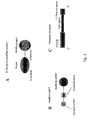
- the modified protein of the present invention is a protein of which C-terminal is modified, and it has a constitution in which a modifying agent binds to the C-terminal of protein, as shown in Fig. 1, A. That is, the modified protein of the present invention is constituted by a protein and a modifying agent.
- the "protein” constituting the modified protein of the present invention means a protein used as an object of analysis of interaction, of which functions are known or unknown.
- the C-terminal modified protein of the present invention can be used for measurement of presence or absence of an interaction of the protein with a target molecule described later.
- This protein may be any of natural proteins and mutants thereof as well as artificial proteins and mutants thereof.
- Natural proteins include a library of various proteins obtained by transcription and translation from a cDNA library derived from organs, tissues or cells of various organisms.
- Artificial proteins contain a sequence of a combination of total or partial sequences of natural proteins or a random amino acid sequence.
- the protein constituting the modified protein of the present invention is preferably a full-length protein.
- the "full-length protein” means a protein of which C-terminal is completely translated, i.e., a protein obtained by translation of codons of the nucleotide sequence coding for the protein up to the codon immediately before the stop codon.
- the N-terminal of the full-length protein may undergo a certain processing such as digestion of signal peptide.
- the protein constituting the modified protein of the present invention may be a protein fused to an affinity tag.
- affinity tag include polyhistidine peptides, epitope peptides, glutathione-S-transferase, protein A, maltose binding protein, calmodulin binding peptide and so forth.
- the modifying agent of the present invention has a constitution where an acceptor portion having a group (including a residue) capable of binding to a protein through a transpeptidation reaction in a protein translation system, i.e., a transpeptidation reaction on a ribosome, links to a modifying portion via a nucleotide linker, as shown in Fig. 1, B. If protein synthesis is performed in the presence of this modifying agent, and the obtained C-terminal modified protein is purified and used in a detection system of intermolecular interaction, it becomes possible to detect a protein interaction.
- nonradioactive modifying substance contained in the modifying portion examples include fluorescent or non-fluorescent modifying substances and so forth.
- fluorescent substances include fluorescent dyes such as those of fluorescein type, those of rhodamine type, Cy3, Cy5, those of eosine type and those of NBD type, fluorescent proteins such as green fluorescent proteins (GFP).
- the non-fluorescent substances may be any substances that can serve as a certain marker, for example, coenzymes such as biotin, proteins, peptides, saccharides, lipids, dyes, polyethylene glycols and so forth.
- the modifying portion preferably has a fluorescent group, a group that binds to a protein (e.g., biotinyl group, iminobiotinyl group), or both of these.
- the modifying portion preferably has biotinyl group or iminobiotinyl group, since the modification efficiency by the modifying agent of the present invention is improved.
- the acceptor portion has a group capable of binding to a protein through a transpeptidation reaction in a protein translation system, preferably a residue of puromycin or a derivative thereof.
- Puromycin has a structure similar to that of an aminoacyl-tRNA, and it is known as an antibiotic inhibiting protein synthesis and to bind to a C-terminal of protein at a low concentration (Miyamoto-Sato E. et al., Nucleic Acids Res., 28: 1176-1182, 2000).
- Any puromycin derivatives can be used for the present invention, so long as they are substances having a structure similar to that of puromycin and capable of binding to a C-terminal of protein. Specific examples thereof include 3'-N-aminoacylpuromycin aminonucleoside, 3'-N-aminoacyladenosine aminonucleoside and so forth.
- the nucleotide linker linking the modifying portion and the acceptor portion is specifically a nucleic acid or a nucleic acid derivative composed of one or more ribonucleotides or deoxyribonucleotides linked together, and particularly preferred examples thereof include compounds composed of one or more ribonucleotides (-rC-) or deoxyribonucleotide (-dC-) containing cytosine bases linked together.
- any substances that can increase yield of modified protein when inserted between the modifying portion and the acceptor portion may also be used.
- the nucleotide linker is preferably 2'-deoxycytidylic acid, 2'-deoxycytidyl-(3',5')-2'-deoxycytidylic acid, ribocytidylic acid or ribocytidyl-(3',5')-ribocytidylic acid.
- the modifying agent can be produced by ligating the aforementioned modifying portion and acceptor portion via a desired nucleotide linker using a chemical bonding method known per se. Specifically, it can be produced by, for example, binding the aforementioned acceptor portion protected with a suitable protective group to a solid phase carrier, successively binding nucleotide phosphoramidite or deoxynucleotide phosphoramidite as a nucleotide linker and nucleotide phosphoramidite bound to a fluorescent substance, biotin or the like as a modifying substance using a nucleic acid synthesizer or the like and then performing deprotection.
- a chelating reagent to which a metal ion can coordinate such as nitrilotriacetic acid or iminodiacetic acid, can be bound, and then a metal ion can be coordinated.
- the translation template of the present invention is a translation template that can be used in manufacture of the modified protein of the present invention, and it is constituted by a 3' end region containing a poly-A sequence, a 5' untranslated region (5' UTR) containing a transcription promoter, and an ORF region coding for a protein, as shown in Fig. 1, C.
- the translation template may be DNA or RNA.
- the translation template of the present invention is constituted by an ORF region coding for a protein, a 5' UTR containing a transcription promoter and a translation enhancer and locating on the 5' side of the ORF region, and a 3' end region containing a poly-A sequence (poly-A) and locating on the 3' side of the ORF region.
- the translation template more preferably contains the promoter sequence of SP6 RNA polymerase as the transcription promoter of 5' UTR and a part of omega sequence (029) of tobacco mosaic virus (TMV) as the translation enhancer.
- the ORF region contains an affinity tag sequence in the downstream portion thereof.
- the affinity tag sequence is a sequence coding for the aforementioned affinity tag, and it preferably contains a His-tag (polyhistidine tag) sequence.
- a longer polyhistidine tag is preferred, since the recovery yield obtained by using a nickel chelate resin is improved.
- the preferred range of the length of the polyhistidine tag may vary depending on type of protein to be modified or type of label, it is usually 8 to 12 residues.
- upstream and downstream are used in this specification according to the transcription or translation direction.
- the translation template of the present invention is DNA
- it may be a DNA vector or plasmid obtained by introducing the aforementioned regions into a suitable DNA vector or plasmid.
- the translation template of the present invention is RNA, it may or may not have a Cap structure at the 5' end.
- the production method of the present invention comprises expressing the translation template of the present invention in a translation system in the presence of the modifying agent of the present invention to cause protein synthesis and purifying the synthesized protein.
- Examples of the translation system used in the present invention include cell-free protein synthesis systems and cell expression systems.
- Examples of the cell-free protein synthesis systems include wheat germ extract, rabbit reticulocyte lysate, Escherichia coli S30 lysate and so forth.
- a C-terminal modified protein is synthesized.
- the synthesized modified protein can be used as it is in the following purification process or detection process.
- any of cells for which gene transfer is possible may be used, including those of microorganisms such as Escherichia coli, Bacillus subtilis, thermophilic bacteria and yeast, insect cells, cultured cells of mammals etc., those of nematodes, drosophila, zebra fish, mouse and so forth.
- a modified protein is synthesized.
- the synthesized modified protein can be collected by disrupting the cells and used for the following purification process or detection process. Further, the synthesized modified protein can also be used as it is in the cells for the detection process.
- the translation template is suitably selected depending on the translation system used.
- any of techniques usually used for purification of proteins for example, chromatography techniques such as affinity chromatography, gel filtration chromatography and ion exchange chromatography, electrophoresis, precipitation, dialysis and so forth, can be used.
- chromatography techniques such as affinity chromatography, gel filtration chromatography and ion exchange chromatography, electrophoresis, precipitation, dialysis and so forth.
- Preferred examples include affinity chromatography, gel filtration, ion chromatography, electrophoresis, precipitation, dialysis and arbitrary combinations of these.
- Particularly preferred example is a method comprising purifying the modified protein fused with an affinity tag such as polyhistidine peptide, epitope peptide, glutathione-S-transferase, protein A, maltose binding protein or calmodulin binding peptide using an affinity resin and further applying the purified protein several times to a gel filtration column in order to completely remove unreacted modifying agent.
- an affinity tag such as polyhistidine peptide, epitope peptide, glutathione-S-transferase, protein A, maltose binding protein or calmodulin binding peptide
- a method comprising purifying beforehand the modified protein fused with the aforementioned affinity tag using an affinity resin and completely removing unmodified protein using affinity between biotinyl group or iminobiotinyl group of the modifying portion and avidin or streptavidin to obtain the modified protein of 100% purity.
- the present invention provide a method for analyzing an interaction between a protein and a target molecule using the modified protein of the present invention, i.e., a method for analyzing an interaction between a protein and a target molecule, wherein the modified protein of the present invention comprising the protein is used.
- the interaction is usually analyzed by bringing the modified protein of the present invention obtained above and a target molecule suitably combined depending on the type of modifying substance and type of the reaction system, into contact with each other, and measuring change in a signal generated by the modified protein of the present invention or the target molecule due to the interaction between the both molecules.
- the analysis of the interaction is performed by, for example, fluorescence correlation spectroscopy, fluorescence imaging analysis method, fluorescence resonance energy transfer method, evanescent field molecular imaging method, fluorescence depolarization method, surface plasmon resonance method or enzyme linked immunosorbent assay. The details of these methods will be explained in (3) below.
- target molecule means a molecule that interacts with the modified protein of the present invention, and it may be specifically a protein, nucleic acid, sugar chain, low molecular weight compound or the like, preferably a protein or DNA.
- the protein is not particularly limited so long as it has an ability to interact with the modified protein of the present invention, and it may be a protein of full length or a partial peptide containing an activity site for binding. Further, it may be a protein of which amino acid sequence or function is known or unknown. It may be a synthesized peptide chain, a protein purified from an organism, a protein obtained by translation from a cDNA library using a suitable translation system and purification, or the like, and they can be used as the target molecule.
- the synthesized peptide chain may be a glycoprotein composed a synthesized peptide chain with a sugar chain attached. Among these, a purified protein of which amino acid sequence is known or a protein obtained by translation from a cDNA library and purification using suitable methods can be preferably used.
- the nucleic acid is not particularly limited so long as it has an ability to interact with the modified protein of the present invention, and either DNA or RNA may be used. Further, it may be a nucleic acid of which nucleotide sequence or function is known or unknown. Preferably, a nucleic acid of which function as a nucleic acid having an ability to bind to a protein or of which nucleotide sequence is known or a nucleic acid obtained by cleavage with a restriction enzyme or the like and isolation from a genomic library or the like can be used.
- the sugar chain is not particularly limited so long as it has an ability to interact with the modified protein of the present invention, and it may be a sugar chain of which saccharide sequence or function is known or unknown. Preferably, an already isolated and analyzed sugar chain of which saccharide sequence or function is known is used.
- the low molecular weight compound is not particularly limited so long as it has an ability to interact with the modified protein of the present invention.
- a compound of which function is unknown or a compound of which ability to bind to a protein is already known may also be used.
- the "interaction" caused by these targets molecules with the modified protein of the present invention usually means an action caused by an intermolecular force generated by at least one of covalent bond, hydrophobic bond, hydrogen bond, van der Waals binding and binding caused by electrostatic force between a protein and a target molecule.
- covalent bond includes a coordinate bond and dipole bond.
- the binding caused by electrostatic force includes, besides electrostatic bond, electric repulsion. Further, a bonding reaction, synthetic reaction and decomposition reaction caused as a result of the aforementioned action are also included in the interaction.
- interaction examples include association and dissociation of an antigen and an antibody, association and dissociation of a protein receptor and a ligand, association and dissociation of an adhesion molecule and a partner molecule, association and dissociation of an enzyme and a substrate, association and dissociation of a nucleic acid and a protein binding to it, association and dissociation of proteins in a signal transduction system, association and dissociation of a glycoprotein and a protein and association and dissociation of a sugar chain and a protein.
- the target molecule used may be modified with a modifying substance and used depending on embodiments.
- the modifying substance is usually selected from nonradioactive modifying substances such as fluorescent substances.
- the fluorescent substances may be any of various fluorescent dyes of, for example, fluorescein type, rhodamine type, Cy3, Cy5, eosine type, NBD type and so forth, which can bind to the aforementioned target substance such as proteins and nucleic acids and have a free functional group (e.g., carboxyl group, hydroxyl group, amino group etc.).
- other compounds such as dyes may be used, and type and size of the compounds are not critical so long as they can be modified.
- modifying substances a substance suitable for the method of measurement or analysis of change in signal generated due to an interaction between the target molecule and the modified protein of the present invention is used.
- the aforementioned modifying substance can be bound to the target molecule by a suitable method known per se. Specifically, when the target molecule is a protein, the method of modifying the C-terminal described above in (1-4) or the like may be used. Further, when the target molecule is a nucleic acid, it can by easily modified by a method of performing PCR using an oligo DNA primer bound with a modifying substance beforehand via a covalent bond or the like.
- modified protein of the present invention or the target molecule used for present invention may be bound to a solid phase (i.e., immobilized) depending on the embodiment.
- a solid phase i.e., immobilized
- the method for binding to a solid phase there are a method of binding it via the modified substance and a method of binding it via another portion.
- the modifying substance used in binding via the modifying substance is usually a molecule specifically binding to a particular polypeptide (henceforth also referred to as a "ligand"), and the particular polypeptide binding to the ligand (henceforth also referred to as an "adapter protein”) is bound to the solid phase.
- the adapter protein also includes binding proteins, acceptor proteins constituting acceptors, antibodies and so forth.
- Examples of combinations of the adapter protein and the ligand include any of various acceptor proteins and a ligand thereof, for example, a biotin- or iminobiotin-binding protein such as avidin and streptavidin and biotin or iminobiotin, maltose-binding protein and maltose, G protein and guanine nucleotide, polyhistidine peptide and metal ion such as nickel or cobalt ion, glutathione-S-transferase and glutathione, DNA-binding protein and DNA, antibody and antigen molecule (epitope), calmodulin and calmodulin-binding peptide, adenosine triphosphate (ATP)-binding protein and ATP, estradiol acceptor protein and estradiol and so forth.
- a biotin- or iminobiotin-binding protein such as avidin and streptavidin and biotin or iminobiotin
- the adapter protein and the ligand are biotin- or iminobiotin-binding protein such as avidin and streptavidin and biotin or iminobiotin, maltose-binding protein and maltose, polyhistidine peptide and metal ion such as nickel or cobalt ion, glutathione-S-transferase and glutathione, antibody and antigen molecule (epitope) and so forth, and a combination of streptavidin and biotin or iminobiotin is the most preferred.
- biotin- or iminobiotin-binding protein such as avidin and streptavidin and biotin or iminobiotin
- maltose-binding protein and maltose polyhistidine peptide and metal ion such as nickel or cobalt ion
- glutathione-S-transferase and glutathione antibody and antigen molecule (epitope) and so forth
- the adaptor protein can be bound to a solid phase surface by using a method known per se. Specifically, for example, there can be used a method of utilizing tannic acid, formalin, glutaraldehyde, pyruvic aldehyde, bis-diazotized benzizone, toluene-2,4-diisocyanate, amino group, carboxyl group that can be converted into an active ester group, hydroxyl group or amino group that can be converted into phosphoramidite group, or the like.
- a known method usually used for binding a protein, nucleic acid, sugar chain or low molecular weight compound to a solid phase there can be used, for example, a method of utilizing tannic acid, formalin, glutaraldehyde, pyruvic aldehyde, bis-diazotized benzizone, toluene-2,4-diisocyanate, amino group, carboxyl group that can be converted into an active ester group, hydroxyl group or amino group that can be converted into phosphoramidite group, or the like.
- the solid phase may be one usually used for immobilizing a protein, nucleic acid or the like, and material and shape thereof are not particularly limited.
- material and shape thereof are not particularly limited.
- glass plates, nitrocellulose membranes, nylon membranes, polyvinylidene fluoride membranes, microplates made of plastics and so forth can be used.
- the "measurement” is means for collecting change in a signal used for analysis, and it should not be construed in any limitative way.
- any of methods that can detect an intermolecular interaction can be used, including fluorescence correlation spectroscopy, fluorescence resonance energy transfer method, evanescent field molecular imaging method, fluorescence depolarization method, fluorescence imaging analysis method, surface plasmon resonance method, enzyme linked immunosorbent assay and so forth.
- the measurement method include a method comprising adding the modified protein of the present invention onto an array on which a target molecule is immobilized and detecting the modified protein of the present invention specifically binding to the target molecule.
- the array on which the target molecule is immobilized means a solid phase on which the target molecule is immobilized in an arrangement enabling identification thereof.
- the method for detecting the modified protein of the present invention specifically binding to the target molecule is not particularly limited, so long as the method enables detection of the modified protein of the present invention specifically binding to the target molecule. However, there is usually used, for example, a method of removing the modified protein of the present invention not binding to the target molecule by washing from the array to which the modified protein of the present invention is added and detecting the remaining modified protein of the present invention.
- the fluorescence correlation spectroscopy (FCS, Eigen, M., et al., Proc. Natl. Acad. Sci., USA, 91, 5740-5747, 1994) is a method of measuring flow rate, diffusion coefficient, volume shrinkage or the like of particles under a confocal laser microscope or the like.
- interacting molecules can be measured by measuring change in translational Brownian movement of one original modified molecule of the present invention (C-terminal modified protein) caused by an interaction between the modified protein and a target molecule.
- fluorescence emitted from sample particles in a partial volume of a sample solution due to excitation of the sample particles by an excitation light is measured to obtain a photon ratio. This value changes with the number of the particles existing in a space volume observed during a specific period of time.
- Apparatuses for carrying out FCS are also marketed from Carl Zeiss and so forth, and analysis can be performed by using these apparatuses also in the present invention.
- fluorescence cross-correlation spectroscopy using two kinds of fluorescent dyes can detect even an interaction between proteins having molecular weights of similar order, of which detection is difficult by FCS using one kind of fluorescent dye.
- FRET fluorescence resonance energy transfer
- two kinds of fluorescent dyes need to approach each other at a distance within 40 to 50 ⁇ in order to cause FRET, and there is a risk in this method that FRET may not be observed depending on sizes of proteins, locations at which the fluorescent dyes are attached or the like, even though an interaction occurs.
- the FCCS method since the detection of cross-correlation does not depend on the distance between the fluorescent dyes in the FCCS method, it does not suffer from such a problem. Further, comparing with the fluorescence depolarization method as another detection system, the FCCS method has advantages of a smaller amount of required sample, shorter detection time, easier automatization for HTS and so forth. Further, since the FCCS method provides extremely fundamental information such as size and number of fluorescence-labeled molecules, it may be used for general purpose like the surface plasmon resonance method. The difference between the both is that, in the surface plasmon resonance method, an interaction is detected in the state that proteins are immobilized, whereas the FCCS method enables observation of interaction in a solution, which is closer to a natural state. In the FCCS method, although proteins do not need to be immobilized, the proteins must be labeled with fluorescent dyes instead. However, it has been made possible by the present invention to overcome this problem.
- FCCS method enables investigation of a protein-protein interaction or protein-nucleic acid interaction in a state of solution, which is closer to the intracellular environment and enables convenient calculation of dissociation constant (binding constant) by one measurement.
- the method for bringing a target molecule into contact with the C-terminal modified protein in this method may be any method that allows the contact in a sufficient degree such that they can interact with each other. However, it is preferably attained by a method of introducing a solution obtained by dissolving the C-terminal modified protein in a buffer usually used for biochemical purpose or the like at an appropriate concentration, into a well for measurement in a commercially available FCS apparatus, and further introducing a solution obtained by dissolving the target molecule in the same buffer at an appropriate concentration, into the well.
- a method of performing multiple analyses for example, there is used a method of introducing multiple kinds of different C-terminal modified proteins into wells for measurement in the FCS apparatus, respectively, and further introducing a solution of a particular target molecule into the wells, or introducing a particular C-terminal modified protein into wells, and further introducing solutions of multiple kinds of different target molecules into the wells, respectively.
- the fluorescence imaging analysis method is a method of bringing a modifying molecule into contact with an immobilized molecule and measuring or analyzing fluorescence emitted by the immobilized modifying molecule remained on the immobilized molecule due to an interaction between the both molecules using a commercially available fluorescence imaging analyzer.
- one of the C-terminal modified protein or the target molecule must be immobilized by the aforementioned method.
- an immobilized target molecule either a modified or unmodified target molecule can be used. Further, when it is used without immobilization, it must be modified with the aforementioned modifying substance. Either a C-terminal modified protein immobilized at the modified portion or a C-terminal modifying protein immobilized at a portion other than the modifying portion may be used.
- a substrate (solid phase) for immobilizing a C-terminal modified protein or target molecule there can be used glass plates, nitrocellulose membranes, nylon membranes, microplates made of plastics and so forth, which are usually used for immobilizing a protein, nucleic acid or the like.
- substrates as mentioned above, on which various functional groups (amino group, carboxyl group, thiol group, hydroxyl group etc.) or various ligands (biotin, iminobiotin, metal ions such as nickel or cobalt ion, glutathione, saccharides, nucleotides, DNA, RNA, antibody, calmodulin, acceptor protein etc.) are bound, can also be used.
- the method for bringing a modified target molecule or a C-terminal modified protein into contact with an immobilized molecule in this method may be any method that allows the contact in a sufficient degree such that the both molecules can interact with each other. However, it is preferably attained by a method of preparing a solution by dissolving the modified target molecule or the C-terminal modified protein in a buffer usually used for biochemical purpose at an appropriate concentration, and bringing the solution into contact with the solid phase surface.
- a step of washing off excessively existing modified target molecule or C-terminal modified protein with the same buffer or the like is preferably performed, and fluorescence signal emitted from the modifying substance of the target molecule or C-terminal modified protein which remained on the solid phase, or a mixed signal of fluorescence emitted from the immobilized modifying molecule and fluorescence emitted from the modifying molecule remained on the solid phase can be measured or analyzed by using a commercially available imaging analyzer to identify the molecule that interacts with the immobilized molecule.
- a method of immobilizing multiple kinds of C-terminal modified proteins or modified or unmodified target molecules on the aforementioned solid phase surface with positioning addresses a method of bringing multiple kinds of non-immobilized C-terminal modified proteins or modified target molecules into contact with one kind of C-terminal modified protein or modified or unmodified target molecule, or the like.
- the molecules remained on the solid phase can be obtained by dissociating them using difference of buffer concentration or the like and analyzed by a known method to identify them.
- FRET fluorescence resonance energy transfer
- the evanescent field molecular imaging method is a method described in Funatsu, T., et al., Nature, 374,555-559, 1995 or the like, and it is a method of bringing a second molecule as a solution into contact with a molecule immobilized on a transparent material such as glass, irradiating it with a light source such as a laser light at such an angle that an evanescent field should be generated and measuring or analyzing the generated evanescent light using a detector.
- a light source such as a laser light
- one of the C-terminal modified protein or the target molecule must be immobilized by the aforementioned method.
- an immobilized target molecule it does not need to be modified. However, when it is used without immobilization, it must be modified with the aforementioned modifying substance.
- a substrate made of a material of glass or the like is used, and quartz glass is preferably used. Further, a substrate of which surface is cleaned by ultrasonication is preferred in order to prevent scatter of laser light or the like.
- the method for bringing a non-immobilized C-terminal modified protein or target molecule into contact with an immobilized molecule in this method may be any method that allows the contact in a sufficient degree such that the both molecules can interact with each other.
- a method of preparing a solution by dissolving the non-immobilized C-terminal modified protein or modified target molecule in a buffer usually used for biochemical purpose at an appropriate concentration, and adding the solution dropwise to the solid phase surface is preferred.
- fluorescence generated through excitation by the evanescent field illumination can be measured by using a detector such as a CCD camera to identify the molecule that interacts with the immobilized molecule.
- this method as a method of simultaneously performing multiple analyses, for example, there is used a method of immobilizing multiple kinds of C-terminal modified proteins or modified target molecules on the aforementioned substrate with positioning addresses, or the like.
- the fluorescence polarization method (Perran, J., et al., J. Phys. Rad., 1, 390-401, 1926) is a method utilizing the fact that a fluorescent molecule excited with a polarized fluorescent light emits fluorescence in the same plane of polarization when it maintains a stationary state during the excited state, whereas the emitted fluorescence has a plane different from that of the excitation light when the excited molecule undergoes rotational Brownian movement or the like during the excited state.
- the movement of molecule is affected by the size thereof, and when a fluorescent molecule is a macromolecule, the molecule scarcely shows movement during the excited state, and emitted light is maintained to be a polarized light.
- Apparatuses for carrying out this method such as BECON (produced by Panyera) are marketed, and this method can be carried out by using these apparatuses.
- the method for bringing a target molecule into contact with the C-terminal modified protein in this method may be any method that allows the contact in sufficient degree such that they should interact with each other. However, it is preferably attained by a method of introducing a solution obtained by dissolving the C-terminal modified protein in a buffer usually used for biochemical purpose at an appropriate concentration, into a well for measurement in a commercially available fluorescence depolarization apparatus, and further introducing a solution obtained by dissolving the target molecule in the same buffer at an appropriate concentration, into the well.
- degree of interaction should be numerically defined.
- an index representing degree of interaction for example, a value of the minimum target substance concentration providing the maximum fluorescence polarization degree for a C-terminal modified protein of a fixed concentration or the like can be used.
- this method as a method of simultaneously performing multiple analyses, for example, there is used a method of introducing multiple kinds of different C-terminal modified proteins into wells for measurement in the aforementioned fluorescence depolarization apparatus, respectively, and further introducing a solution of a particular target molecule into the wells, or introducing a particular C-terminal modified protein into wells and further introducing solutions of multiple kinds of different target molecules into the wells, respectively.
- the surface plasmon resonance method is a method of measuring surface plasmon excited by a molecule interacting at a metal/liquid interface as change in intensity of reflected light (Cullen, D.C., et al., Biosensors, 3 (4), 211-225, 1987-88).
- the C-terminal modified protein must be immobilized by the aforementioned method, but the target molecule does not need to be modified.
- a transparent substrate made of glass or the like on which a thin film of metal such as gold, silver or platinum is formed is used as a substrate for immobilizing the C-terminal modified protein.
- the transparent substrate may be any of those usually used for surface plasmon resonance apparatuses. It is generally made of glass as a substrate made of a material transparent to a laser light, and such a substrate having a thickness of about 0.1-5 mm is generally used. Further, thickness of the metal thin film is suitably about 100 to 2000 ⁇ . Those marketed as such immobilization substrates for surface plasmon resonance apparatuses can also be used.
- the C-terminal modified protein can be immobilized on the substrate by the method described above.
- the method for bringing a target molecule into contact with the C-terminal modified protein in this method may be any method that allows the contact in a sufficient degree such that the both molecules can interact with each other.
- a method of bringing the immobilized C-terminal modified protein into contact with a solution obtained by dissolving the target molecule in a buffer usually used for biochemical purpose at an appropriate concentration can be preferably used.
- steps may also be performed by using a commercially available surface plasmon resonance apparatus, for example, BIAcore 2000 (produced by Pharmacia Biosensor).
- BIAcore 2000 produced by Pharmacia Biosensor.
- change with time in relative intensity of each reflected light can be measured by using a surface plasmon resonance apparatus known per se to analyze or measure an interaction of the immobilized C-terminal modified protein and the target molecule.
- this method as a method of simultaneously performing multiple analyses, for example, there is used a method of immobilizing multiple kinds of C-terminal modified proteins on a substrate used for the surface plasmon resonance apparatus with positioning addresses, a method of bringing multiple kinds of target molecules into contact with one kind of immobilized C-terminal modified protein, or the like.
- the enzyme linked immunosorbent assay (ELISA, Crowther, J.R., Methods in Molecular Biology, 42, 1995) is a method of bringing a solution containing an antibody into contact with an antigen immobilized on a solid phase and measuring or analyzing the antibody remaining on the immobilized antigen due to the interaction between the both molecules (antigen-antibody reaction), based on fluorescence emitted from a modifying molecule (IgG etc.) specifically binding to the antibody or a signal emitted by a dye formed from the modifying molecule as a substrate using a commercially available detector (ELISA reader).
- ELISA enzyme linked immunosorbent assay
- the C-terminal modified protein serving as the antigen must be immobilized by the aforementioned method. Further; the target molecule serving as the antibody must be modified with the aforementioned modifying substance.
- microplates made of plastics usually used for ELISA and so forth can also be used.
- the method for bringing the modified target molecule serving as the antibody into contact with an immobilized molecule in this method may be any method that allows the contact in a sufficient degree such that the both molecules can interact with each other.
- a method of preparing a solution by dissolving the modified target molecule in a buffer usually used for biochemical purpose at an appropriate concentration, and introducing the solution into a microplate is preferred.
- a step of washing off excessively existing the modified molecule not binding to the immobilized molecule is preferably performed, and fluorescence emitted from the modified molecule remained on the solid phase can be measured or analyzed by using a commercially available ELISA reader or the like to identify the molecule that interacts with the immobilized antigen molecule.
- the primary structure can be analyzed by a suitable method known per se. Specifically, when the target molecule for which an interaction is recognized is a protein, its amino acid sequence can be analyzed by using an amino acid analyzer etc. to identify the primary structure. Further, when the target molecule is a nucleic acid, nucleotide sequence can be determined by a nucleotide sequence determination method using an automatic DNA sequencer or the like.
- Example 1 Increase of efficiency of fluorescence modification of protein and detection of protein interaction by fluorescence cross-correlation spectroscopy
- the c-Fos and c-Jun proteins which are cancer gene products, form a dimer, and recognize and bind to DNA having a particular nucleotide sequence to function as a transcription factor.
- the c-Fos and c-Jun proteins were modified with several types of fluorescent dyes to detect interactions between proteins and between a protein and DNA by fluorescence cross-correlation spectroscopy.
- Modifying agents containing a puromycin residue were synthesized by using a method outlined in Fig. 2 (Solid Phase Method 1) or Fig. 3 (Solid Phase Method 2).
- Compound 1 was synthesized by the method reported by Ikeda et al. (Ikeda, S. et al., Tetrahedron Lett. 39, 5975-5978, 1998).
- Nucleotide phosphoramidites, modifying substance-phosphoramidites and Compound 2 were purchased from Glen Research (Virginia, USA).
- Modifying substance-succinimides were purchased from Molecular Probes (Oregon, USA).
- UV absorption was measured by using Backman DU 640 Spectrophotometer. The mass spectra were measured by using Lasermat 2000 produced by Finnigan MAT.
- the solid phase carrier (CPG) was removed by filtration, and the filtrate was lyophilized.
- Y in Fig. 1 was tert-butyldimethylsilyloxy group
- 400 ⁇ L of 1 M solution of tetrabutylammonium fluoride in tetrahydrofuran was added to the residue, and the mixture was left at room temperature for 14 hours and concentrated under reduced pressure.
- Modifying agent 19 yield: 50%
- Modifying agent 20 yield: 44%
- mice c- fos and c -jun genes were cloned from a mouse testis cDNA library (Takara Shuzo) as follows. First, a gene region corresponding to the 216th to 318th amino acid residues in the amino acid sequence of c-Jun, which is required to bind to c-Fos and DNA (Ryder, K. and Nathans, D., Proc. Natl. Acad. Sci. U.S.A. 85, 8464-8467, 1988), was amplified by PCR (the nucleotide sequences of the used primers are shown in SEQ ID NOS: 1 and 2), and cloned into a plasmid provided from Dr.
- a linear DNA fragment as a template used for transcription of the mouse c-fos and c-jun genes was prepared as follows. PCR amplification was performed by using the plasmid pSP6-jun as a template, a primer upstream from the SP6 promoter (the nucleotide sequence is shown in SEQ ID NO: 5) and a primer for adding a sequence coding for 6 histidine residues (His tag) at the C-terminal of c-Jun (the nucleotide sequence is shown in SEQ ID NO: 6).
- PCR amplification was performed by using the plasmid pSP6-fos as a template, the aforementioned primer (the nucleotide sequence thereof is shown in SEQ ID NO: 5) and a primer for adding a His tag to the C-terminal of c-Fos (the nucleotide sequence is shown in SEQ ID NO: 7).
- QIAquick PCR purification kit QIAGEN
- a fluorescence-modified DNA fragment specifically binding to the mouse c-Fos/c-Jun dimer was prepared as follows. Single-stranded DNAs (the nucleotide sequences are shown in SEQ ID NOS: 8 and 9) of which 5' ends were modified with a fluorescent dye, Cy5, and which are complementary to each other were mixed in equimolar amounts, heated at 95°C in the presence of 0.1 M NaCl and then gradually cooled to room temperature so that the DNAs should anneal to obtain a double-stranded DNA. This DNA was used as it was for measurement by fluorescence cross-correlation spectroscopy.
- RNA cap analogue (Life Technologies Oriental) was added to the reaction mixture to modify the 5' end of RNA.
- the synthesized RNA was treated with phenol/chloroform and then purified by ethanol precipitation.
- RNA was added to wheat germ extract (Promega) and allowed to react at 25°C for 60 minutes.
- modifying agents Modifying agents 1 to 18 in which various linkers are inserted between a fluorescent dye (fluorescein, Rhodamine green, Cy5) and puromycin were added at various concentrations to modify the C-terminal of the protein with fluorescence.
- fluorescent dye fluorescein, Rhodamine green, Cy5
- puromycin were added at various concentrations to modify the C-terminal of the protein with fluorescence.
- translation products were subjected to SDS polyacrylamide electrophoresis, and bands of the fluorescence-modified proteins were detected and quantified by using a fluorescence imaging apparatus (Molecular Imager FX, Bio-Rad).
- modification efficiency of the C-terminal of the c-Fos protein significantly varied depending on the nucleotide linker structure of the modifying agent.
- the linker showing the highest yield was -dC- (Modifying agents 1, 9, 12) irrespective of the fluorescent dyes, and the yield of the modified protein increased 100 times at most in comparison with the cases using no nucleotide linker.
- the similar results were also observed in the c-Jun protein.
- the nucleotide linker was -dCdC- (Modifying agents 2, 10, 13)
- the modification efficiency was lower than that obtained with -dC-.
- the modification efficiency obtained with -rC- was significantly lower than that obtained with -dC-(Modifying agent 1), whereas that obtained with -rCrC-(Modifying agent 4) was higher than that obtained with -rC-(Modifying agent 3).
- the nucleotide linker was -dT-(Modifying agent 5), -dTdT- (Modifying agent 6), -dA-(Modifying agent 7) or -dG- (Modifying agent 8)
- the effect was not significant, a marked effect was observed for deoxycytidylic acid or ribocytidylic acid.
- the optimum concentrations of the modifying agents varied depending on the types of the fluorescent groups.
- the optimum concentration for labeling efficiency with Cy5 was as low as about half of that obtained with fluorescein (Modifying agents 1 and 2).
- the fluorescent group was rhodamine green (RG) (Modifying agents 12 and 13)
- the tendency of the optimum concentration was very similar to that observed for Cy5.
- Fluorescence-modified proteins used for measurement by fluorescence correlation spectroscopy were purified as follows to remove unreacted fluorescent dyes.
- the translation reaction mixture was mixed with an equilibrated nickel NTA agarose resin (QIAGEN), adsorbed on the resin by a specific bond between the His tag at the C-terminal of the fluorescence-modified protein and a nickel ion, washed and then eluted with imidazole.
- an eluted fraction containing the protein was applied to a gel filtration column (PD-10, Pharmacia) twice, and the eluted solution was concentrated by centrifugation using Centricon (Millipore).
- Measurement by fluorescence cross-correlation spectroscopy was performed by using the c-Fos and c-Jun proteins modified with a modifying agent having rhodamine green (RG) as a fluorescent group (Modifying agent 12) and DNA modified with Cy5.
- RG rhodamine green
- DNA modified with Cy5 DNA modified with Cy5.
- the purified proteins and DNA were mixed at a final concentration of 10 nM each and incubated at 37°C for 60 minutes. 10 ⁇ L of this sample was placed on an 8-well glass chamber (Nunc), and fluorescence cross-correlations were measured by using a fluorescence correlation spectrometer, ConfoCor2 (Carl Zeiss).
- Example 2 Translation template and fluorescence modification efficiency in wheat germ cell-free translation system
- a sequence of a vector or plasmid to which mouse-derived c-jun or c-fos was incorporated, or a DNA template containing such a sequence was amplified by PCR using TaKaRa Ex Taq (Takara Shuzo) and purified by using QIAquick PCR Purification Kit (QIAGEN).
- QIAGEN QIAquick PCR Purification Kit
- c-jun[pSPAM], c-fos[pSPAM] and c-jun[F] the nucleotide sequences are shown in SEQ ID NOS: 10-12; respectively
- SP6F and 5' SP6-O29 (the nucleotide sequences are shown in SEQ ID NOS: 13 and 14, respectively) were used as Primer 1 (forward primer), and JunHis, JunHisA, FosHis, FosHisA, JunFlagA, 3'HisA and 3'FlagA (the nucleotide sequences are shown in SEQ ID NOS: 15-21, respectively) were used as Primer 2 (reverse primer).
- the translation templates (DNA templates) were obtained by the above method.
- RNA template Each template was transcribed (37°C, 2 hours) by using RiboMAXTM Large Scale RNA Production Systems (Promega) and purified by using RNeasy Mini Kit (QIAGEN) to obtain a translation template (RNA template).
- RNA template The basic structure of a translation template and DNA nucleotide sequences of components are shown in Fig. 6.
- the translation templates are referred to in the forms of "name of Primer 1 - name of Primer 2" according to the primers used for amplification.
- the translation efficiency of the translation template of SP6-O29Jun-HisA used in this example was 5 to 6 times higher than that obtained with the translation template using AMV in Example 1. Therefore, it was found that when SP6-O29Jun-HisA as the translation template and a modifying agent having a nucleotide linker were used in combination, modification efficiency of C-terminals of proteins became about 500 times as high as the modification efficiency obtained by the conventional techniques (Japanese Patent Laid-open Publication Nos. 11-322781 and 2000-139468).
- a DNA fragment including a region for Fos/Jun dimer binding region was amplified by PCR using a primer modified with Cy5.
- QIAquick PCR purification kit QIAGEN was used for purification of DNA.
- a spotting solution 150 mM sodium phosphate in 0.01% SDS, pH 8.5
- containing the modified DNA fragment at a concentration of 200 ⁇ g/ml was prepared.
- a slide (DNA-ReadyTM TypeII Slide, CLONTECH) was set on a microarrayer (Micro Grid, BioRobotics), and spotting was performed (spotting conditions; temperature in chamber: about 25-28°C, humidity: 38-42%, solid pins were used, at 0.7 mm intervals).
- the slide was incubated at 80°C for 2 hours, and water vapor was applied to the spotted surface to hydrate the spot. Moisture was evaporated on a hot plate at 100°C, and the spots were irradiated with UV to immobilize the DNA fragments.
- the slide was immersed in a blocking solution (4 g of succinic acid, 252 ml of 1-methyl-2-pyrrolidinone, 28 ml of 1 M boric acid (pH 8)), vigorously shaken for first 1 minute and then shaken for 20 to 30 minutes. The slide was washed with distilled water at 90°C and then with 99.5% ethanol and dried.
- a hybridization solution was placed on the DNA-immobilized slide and the slide was covered with Parafilm to spread the solution over the whole spotted surface.
- Light was shielded with an aluminium foil, and the slide was incubated at 37°C for 30 minutes (water was filled under the table on which slide was arrayed to prevent dryness).
- the slide was shaken in 1 x buffer for 5 minutes, the solution was replaced, and this procedure was repeated.
- the slide was centrifuged at 5000 rpm and 4°C and then dried, and the fluorescence of the spots on the slide was detected at 532 nm and 635 nm by using a microarray scanner (Gene Pix 4000A, Axon Instruments).
- the lower left image for the same sample shows that DNA modified with Cy5 was immobilized and existed (existence of DNA can be confirmed by detecting fluorescence of Cy5 at 635 nm).
- the upper right image shows that p53 was not bound to DNA in the presence of Fos, since the fluorescence of rhodamine green was not detected.
- the lower right image for the same sample confirmed the existence of DNA, since the fluorescence of Cy5 was detected.
- the fluorescence-modified Jun was specifically bound to DNA for the binding region in the presence of Fos.
- interactions were examined without purification. Nevertheless, a distinct difference in interactions to such an extent as shown in Fig. 9 was observed. This indicates that the translation efficiency and the modification efficiency increased and that C-terminal modified proteins were produced in an amount sufficient for interactions.
- Example 5 Purification of fluorescence-modified protein to high purity and analysis of interactions between proteins by fluorescence cross-correlation spectroscopy
- Modification of modified proteins to high purity was aimed to enable kinetic analysis of intermolecular interaction between C-terminal modified proteins.
- Different affinity tags were introduced into the translation templates and the modifying compounds.
- Affinity purification of the translation product in two stages enabled purification of a protein of which C-terminal was modified with a fluorescent dye to high purity.
- the cancer gene products, the c-Fos and c-Jun proteins, fluorescence-modified with rhodamine green or Cy5 were purified in two stages to obtain samples purified to high purity.
- Modifying agents 21 to 25 were synthesized by the method outlined in Figs. 10 and 11. Compound 1' mentioned in Fig. 10 was synthesized by using the same method as Compound 1. Phosphoramidites were purchased from Glen Research (USA, Virginia). Modifying substance 1-succinimide was purchased from Pierce Biotechnology (Illinois, USA). Modifying substance 2-succinimide was purchased from Molecular Probes (USA, Oregon) and Amersham Pharmacia Biotech (Upsala, Sweden).
- the solid phase carrier (CPG) was removed by filtration, and the filtrate was concentrated under reduced pressure.
- the residue was purified by HPLC (column: YMC Pack ODS-A SH-343-5 produced by YMC (Kyoto), eluent: a linear concentration gradient of 10 to 60% acetonitrile in 0.1 M aqueous triethylammonium acetate (pH 7.0) over 30 minutes, flow rate: 10 mL/min) and then lyophilized.
- Modifying agent 26 was synthesized from Compound 1 by Solid phase method 1.
- the phosphoramidite containing Modifying substance 1 or Modifying substance 2 was purchased from Glen Research (Virginia, USA).
- plasmids to which the mouse c-fos and c-jun genes were introduced prepared in the preparation of DNA in Example 1 were used. Regions in the c- jun and c- fos including a domain required to bind to DNA were amplified by PCR using a primer containing SP6 promoter, ⁇ sequence and T7 tag (SEQ ID NO: 22), and a reverse primer containing a histidine tag, a stop codon and polyA sequence (SEQ ID NO: 23 or 24).
- the PCR product was subcloned into the pCR2.1TOPO ,vector by using Topo TA cloning kit (Invitrogen). The detail of the method was according to the manufacturer's protocol.
- the plasmid DNA of which nucleotide sequence was confirmed was purified by using Wizard Plus SV Minipreps DNA Purification System (Promega).
- the linear template DNA for RNA synthesis was obtained by PCR using a primer utilizing a part of a vector sequence upstream from the insertion site (SEQ ID NO: 25) and a reverse primer in the histidine tag portion.
- reverse primers SEQ ID NOS: 26-29
- the template DNA was purified by using QIAquick PCR Purification Kit (QIAGEN).
- the template DNA was transcribed in the presence of a cap analogue (Life Technologies Oriental Yeast) by using SP6 RiboMAX Large Scale RNA Production System (Promega) (37°C, 3 hours). The detail of procedure was according to the manufacturer's manual. After the transcription, the template DNA was removed by using deoxyribonuclease attached to the kit, and purified RNA was obtained by using SV Total RNA Isolation System (Promega).
- Fluorescent modifying compounds (Modifying agents 22 to 26) in which biotin or iminobiotin and a fluorescent dye (rhodamine Green, Cy5) were introduced into the same molecule were added in the same reaction system to synthesize proteins modified with fluorescence at their C-terminals (25°C, 1 hour).
- the optimum concentration of the fluorescent dyes with biotin introduced was about 125 ⁇ M for both of Cy5 (Modifying agents 24 and 26) and rhodamine green (Modifying agent 22).
- the optimum concentrations of the fluorescent dyes with iminobiotin introduced was 30 ⁇ M for Cy5 (modifying agent 25) and 12.5 ⁇ M for rhodamine green (Modifying agent 23).
- Ni-NTA Superflow Purification using a nickel chelate resin, Ni-NTA Superflow (QIAGEN), was performed according to the manufacturer's manual.
- a protease inhibitor (cocktail for use in histidine-tagged protein purification, Sigma) and 5-fold volume of a binding buffer were added, and the mixture was calmly mixed with 20 ⁇ l of a nickel chelate resin suspension (4°C, 1 hour). The resin was sufficiently washed with the binding buffer, and the histidine-tagged protein was eluted with 50 ⁇ l of buffer containing 0.5 M imidazole.
- An ordinary histidine-tagged protein is sufficiently recovered with a polyhistidine tag having a length of 6 residues using a nickel chelate resin (Abate, C. et al., Proc. Natl. Acad. Sci. USA., 87, 1032-1036, 1990).
- a nickel chelate resin Abate, C. et al., Proc. Natl. Acad. Sci. USA., 87, 1032-1036, 1990.
- the number of histidine residues was increased to examine the amount of recovery by the nickel chelate resin, since the recovery rate was slightly low when the polyhistidine tag having a length of 6 residues was used.
- the labeled protein containing iminobiotin was further purified with a streptavidin-immobilized resin, Streptavidin Sepharose High Performance (Amersham Pharmacia).
- the purification with this resin was performed according to the manufacturer's manual.
- the labeled protein containing biotin was purified with an avidin monomer-immobilized resin, UltraLink Immobilized Monomeric Avidin (Pierce Biotechnology).
- the purification using the resin was performed according to the manufacturer's manual.
- 9-fold volume of a binding buffer was added, and the mixture was calmly mixed with 10 ⁇ l vof the resin equilibrated beforehand (4°C, 30 minutes).
- the resin was sufficiently washed with the binding buffer, and the protein was eluted with 50 ⁇ l of buffer containing 50 mM biotin.
- the protein labeled with Cy5 containing iminobiotin was separated by SDS-PAGE, and the purification was confirmed by fluorescence image analysis and immunoblotting (Fig. 14, A and B).
- the gel after the electrophoresis was electrically transferred to a polyvinylidene fluoride membrane (Pall Gelman Science), a mouse monoclonal antibody (Novagen) directed to the T7 tag provided at the N-terminal and a horseradish peroxidase-labeled goat anti-mouse antibody (Transduction) were reacted, and chemiluminescence was obtained by using ECL kit (Amersham Pharmacia).
- This immunoblotting method was performed according to the manufacturer's manual attached to the aforementioned kit.
- the proteins passing through the streptavidin-immobilized resin were not detected based on fluorescence, whereas the biotin-eluted fraction was detected with both of the antibodies and fluorescence (Fig. 14, A and B, Lanes 2 and 3).
- the concentrations of c-Fos and c-Jun contained in the purified fractions were quantified by dot blotting assay using a T7-tag recombinant protein (Novagen) as a standard substance. 1 ⁇ l of sample was spotted on a nitrocellulose membrane (Schreier-Schulz), and a mouse anti-T7 tag antibody and a horseradish peroxidase-labeled goat anti-mouse antibody were reacted. Chemiluminescence obtained by using ECL was detected by a chemiluminescence analyzer (Molecular Imager ChemiDoc, Bio-Rad).
- the ratio of molar concentrations of the T7-tagged protein and the fluorescence of the purified fraction was 90% or higher (Table 5).
- the purified protein was separated by SDS-PAGE and stained, it was confirmed that it was a substantially single component (Fig. 15).
- N ac,r Na c,g and G c (0) can be obtained from the cross-correlation analysis.
- N cc 1 G c (0)-1
- the number of particles modified with Cy5 and rhodamine fluorescence: N gr N acg N ac.r N cc
- the number of particles modified only with Cy5: N r N ac.r - N gr
- the number of particles modified only with rhodamine: N g' N ac.r - N gr
- the dissociation constant is represented as follows.
- K D [ r ][ g ] [ gr ] ⁇ 1 ⁇ 10 -8 M
- Actinomycetes, Streptomyces avldinii was purchased from Riken.
- An oligo DNA (primer) was synthesized by Espec Oligo Service.
- Escherichia coli, plasmids, various enzymes, reagents etc. were used, including Escherichia coli JM109, plasmid pUC18 (Toyobo), pET20b (Novagen), restriction enzymes Bam HI, Bg III, Eco RI and Hind III (Toyobo), Ligation High (Toyobo), Ex Taq DNA polymerase, Recochip (Takara Shuzo), and QIAquick PCR Purification Kit (QIAGEN).
- the basic procedures of genetic engineering (cloning, transformation and culture of Escherichia coli, recovery of plasmids etc.) were performed according to Sambrook et al., Molecular Cloning, 1989, CSH Press.
- Plasmids pSP6-STA-Jun and pSP6-STA-Fos were constructed by the following procedure.
- the streptavidin gene was amplified by PCR using the Actinomycetes Streptomyces avidinii genome as a template and primers (SEQ ID NOS: 30 and 31) complementary to the upstream and downstream regions of the streptavidin gene, digested with Bam HI and Eco RI and cloned at the Bam HI- Eco RI site in pUC18 to obtain pUC-STA.
- PCR was performed by using this plasmid as a template, a primer for adding a T7 tag to the N-terminal of the streptavidin gene (SEQ ID NO: 32) and a primer complementary to the downstream region of the streptavidin gene (SEQ ID NO: 33) to obtain a streptavidin gene having the T7 tag sequence at the N-terminal.
- an untranslated region including the SP6 promoter and an enhancer sequence derived from tobacco mosaic virus
- a fragment amplified by PCR using primers complementary to the upstream and downstream regions of the streptavidin gene having the T7 tag sequence was digested with BamHI and cloned at the Bg III- Bam HI site of pET20b.
- the plasmid in which the Bg III side corresponded to the upstream of the gene was designated as pSP6-STA.
- the jun and fos genes prepared in Example 1 were amplified by PCR using two sets of primers (SEQ ID NOS: 35 and 36 and SEQ ID NOS: 3 and 37), digested with Bam HI and Hind III and cloned at the Bam HI- Hind III site of pSP6-STA to obtain pSP6-STA-Jun and pSP6-STA-Fos.
- the genes with a polyhistidine tag having a length of 12 residues at the C-terminal prepared in Example 5 were used.
- the mouse c-fos and c-jun gene DNAs were transcribed with the SP6 DNA Polymerase by using Ribomax RNA synthesis system (Promega) (37°C, 120 minutes).
- 20 ⁇ l of the reaction mixture contained 6 ⁇ l of DNA, 1 ⁇ l each of 100 mM rUTP, rCTP and rATP, 1 ⁇ l of 30 mM rGTP and 2 ⁇ l of SP6 polymerase, and it was further supplemented with 4 ⁇ l of RNA cap analogue (Life Technologies Oriental) prepared at 40 mM to modify the 5' end of RNA.
- the synthesized RNA was purified by using RNeasy Mini Kit (QIAGEN).
- the obtained mRNA was added to a cell-free translation system using wheat germ extract of ProteiosTM(TOYOBO) and allowed to react at 37°C for 5 hours.
- 4 ⁇ l of creatine kinase, 10 ⁇ l of RNase inhibitor, 20 ⁇ l of wheat germ, 10 ⁇ l of mRNA and a fluorescent modifying agent were added to 100 ⁇ l of the translation system.
- fluorescent modifying agent fluorescent dye (TAMRA)-dC-puromycin (Modifying agent 19) was used.
- the translation product was subjected to SDS polyacrylamide gel electrophoresis, and the band of the fluorescence-modified protein was detected by using a fluorescence imaging apparatus (Molecular Imager FX, Bio-Rad).
- jun and fos genes on the bait side fused with streptavidin for immobilization on a microarray were transcribed in the same manner as described above using pSP6-STA-Jun and pSP6-STA-Fos as templates and used for protein synthesis by a multilayer method using ProteiosTM (TOYOBO).
- the fluorescence-modified protein used as a protein on the prey side in the immobilization method was purified as follows to remove unreacted fluorescent dyes.
- a reaction mixture for translation of a His-tagged protein modified with fluorescence at the C-terminal was mixed with an equilibrated nickel NTA agarose resin (QIAGEN) so that the protein should be adsorbed to the resin by a specific binding between His tag at the C-terminal of the fluorescence-modified protein and a nickel ion.
- the resin was washed, and then the protein was eluted with 500 mM imidazole.
- a biotin-coated slide (Xenopore) was set on a DNA microarray (MicroGridII, BioRobotics), spotted with a translation solution of streptavidin-fused Jun and Fos proteins dissolved in 60% PBS (10 mM phosphate buffer, 150 mM NaCl (pH 7.4))/40% glycerol, and left in the humidity-controlled environment for 1 hour so that the protein should be immobilized on the slide surface by binding between streptavidin and biotin (Fig. 17, uppermost figure). After the immobilization, the slide was rinsed by shaking in 1% BSA/PBS solution for 1 minute and then further shaken in the replaced 1% BSA/PBS solution for 1 hour for blocking.
- the mixture was allowed to react at room temperature for 1 hour in the humidity-controlled environment.
- the slide was shaken in 1 x PBST for 5 minutes 5 times with replacing the buffer, further shaken in 1 x PBS for 3 minutes 3 times with replacing the buffer, centrifuged at 2000 rpm at 4°C for 1 minute and dried.
- a frame-like silicon sticker for in situ hybridization (EasiSeal, Hybaid) was stuck again around the spotted region on the slide, and the frame was filled with a reaction mixture containing 0.5 mg/ml of Cy3-labeled anti-mouse antibody (Chemicon) and 0.5 mg/ml of Cy5-labeled anti-rabbit antibody (Chemicon) dissolved in 1 x PBST and sealed with a sticker from the above. In this state, the mixture was allowed to react at room temperature for 1 hour in the humidity-controlled environment.
- the slide was shaken in 1 x PBST for 5 minutes 5 times with replacing the buffer, further shaken in 1 x PBS for 3 minutes 3 times also with replacing the buffer, centrifuged at 2000 rpm at 4°C for 1 minute and dried.
- a DNA chip scanner GenePix 4000B, Axon Instruments
- fluorescence of Cy3 was confirmed at the spotted site of the streptavidin-fused FOS
- fluorescence of Cy5 was confirmed at the spotted site of the streptavidin-fused Jun (Fig. 17, A). Based on these results, the immobilization of the streptavidin-fused proteins onto the biotin-coated slide was confirmed.
- a biotin-coated slide (Xenopore) was set on a DNA microarrayer (MicroGridII, BioRobotics), spotted with a translation solution of the streptavidin-fused Jun protein dissolved in 60% PBS/40% glycerol and left in the humidity-controlled environment for 1 hour so that the protein should be immobilized on the slide surface by binding between streptavidin and biotin.
- the slide was rinsed by shaking in 1% BSA/PBS solution for 1 minute and then further shaken in the replaced 1% BSA/PBS solution for 1 hour for blocking.
- the slide was washed with 1 x PBS and centrifuged at 2000 rpm at 4°C for 1 minute to remove the solution on the slide. Subsequently, a frame-like silicon sticker for in situ hybridization (EasiSeal, Hybaid) was stuck around the spotted region on the slide, the C-terminal was modified with TAMRA-dC-puromycin (Modifying agent 19), and then the frame was filled with a reaction mixture (1 x PBST/200 mM NaCl/20% glycerol) containing the Fos protein having 12 His tags and purified with Nickel NT agarose resin (QIAGEN), and sealed with a sticker from the above.
- a reaction mixture (1 x PBST/200 mM NaCl/20% glycerol
- the method of the present invention for modifying C-terminal of protein using a modifying agent containing a nucleotide linker is effective for detection of various protein interactions and provides extremely effective means for large scale and quick screening of interactions between proteins and interactions between a protein and a nucleic acid in function analyses of genes, of which information has been accumulated with the advance of the genome project.
Landscapes
- Chemical & Material Sciences (AREA)
- Organic Chemistry (AREA)
- Life Sciences & Earth Sciences (AREA)
- Health & Medical Sciences (AREA)
- Engineering & Computer Science (AREA)
- Wood Science & Technology (AREA)
- General Health & Medical Sciences (AREA)
- Genetics & Genomics (AREA)
- Zoology (AREA)
- Molecular Biology (AREA)
- Proteomics, Peptides & Aminoacids (AREA)
- Biochemistry (AREA)
- General Chemical & Material Sciences (AREA)
- Bioinformatics & Cheminformatics (AREA)
- Chemical Kinetics & Catalysis (AREA)
- Biophysics (AREA)
- Microbiology (AREA)
- Analytical Chemistry (AREA)
- Medicinal Chemistry (AREA)
- Biotechnology (AREA)
- General Engineering & Computer Science (AREA)
- Investigating, Analyzing Materials By Fluorescence Or Luminescence (AREA)
- Investigating Or Analysing Materials By The Use Of Chemical Reactions (AREA)
- Peptides Or Proteins (AREA)
- Measuring Or Testing Involving Enzymes Or Micro-Organisms (AREA)
- Preparation Of Compounds By Using Micro-Organisms (AREA)
Abstract
A translation template comprising an ORF region
coding for a protein, a 5' untranslated region comprising a
transcription promoter and a translation enhancer and
locating on the 5' side of the ORF region, and a 3' end
region comprising a poly-A sequence and locating on the 3'
side of the ORF region, is expressed in a translation
system in the presence of an agent for modifying a C-terminal
of a protein, which comprises an acceptor portion
having a group capable of binding to a protein through a
transpeptidation reaction in a protein translation system
and a modifying portion comprising a nonradioactive.
modifying substance linked to the acceptor portion via a
nucleotide linker, to cause protein synthesis and the
synthesized protein is purified. Thus, the yield of
modified protein in a method of modifying C-terminal of
protein is improved and detection of protein interaction
based on various intermolecular interaction detection
methods is realized at an improved level.
Description
The present invention relates to a method for
modifying a protein and a method for detecting a protein
interaction utilizing a modified protein. A lot of gene
information is being accumulated in connection with the
development of the genome project, and it is an important
object from now on to comprehensively analyze interactions
among these genes. In order to perform high throughput
screening (HTS) for interactions among a lot of genes at a
high speed, a system is required which can detect protein
interactions more quickly and more conveniently. The
present invention provides an effective means for
conveniently detecting interactions of proteins and other
biological molecules (protein, nucleic acid etc.) in
functional analysis of genome and proteome analyses.
As methods for detecting intermolecular interactions,
the surface plasmon resonance method, fluorescence
resonance energy transfer method, fluorescence
depolarization method, evanescent field imaging method,
fluorescence correlation spectroscopy, fluorescence imaging
method, enzyme linked immunosorbent assay etc. are known so
far. Especially, the fluorescence correlation spectroscopy
(FCS) has advantages of small amount of sample required for
the measurement (about femtoliter), short measurement time
(about 10 seconds), easiness of its automatization for HTS
(in fact, an apparatus aiming at ultra HTS for performing
screening of 100,000 or more test substances per day is
being developed by EVOTEC) etc., and therefore it is a
superior detection system (Kaneshiro M., Tanpakushitsu,
Kakusan, Koso (Protein, Nucleic acid and Enzyme), 44:1431-1438,
1999). Further, the fluorescence cross-correlation
spectroscopy (FCCS) utilizing two kinds of fluorescent dyes
enables detection of an interaction between molecules of
similar sizes, which is difficult by FCS utilizing one kind
of fluorescent dye, and its application to HTS of protein
interaction is expected. However, no example of successful
detection of protein interaction using FCCS has not been
known so far.
In general, in a detection system for protein
interaction, a protein needs to be modified with a tag for
immobilization and a probe such as fluorescent dye. The
inventors of the present invention previously proposed a
method of modifying a C-terminal of protein in a
translation system using a nucleic acid derivative such as
puromycin (Japanese Patent Laid-open Application (Kokai)
Nos. 11-322781 and 2000-139468). Although this method has
advantages, that is, it hardly degrades functions of
protein etc., compared with conventional chemical
modification methods or fluorescent protein fusing methods,
it still has aspects to be improved, for example, the small
yield of the modified protein, high cost for the
preparation of a large amount of the modified protein
mainly due to use of cell-free translation system etc. In
particular, when it is used for FCCS, which is most
excellent as a detection system of HTS, it is indispensable
to improve the yield for practical use in analysis of
genomic functions and so forth, because purification degree
of fluorescence-modified protein is critical.
An object of the present invention is to markedly
improve yield of modified protein in a method of modifying
C-terminal of protein and realize detection of protein
interaction based on various intermolecular interaction
detection methods such as the fluorescence cross-correlation
spectroscopy at an improved level by applying
the improved modification method to fluorescence
modification of protein and investigating suitable
purification methods for fluorescence-modified protein and
so forth.
The inventors of the present invention studied in
order to achieve the aforementioned object, and as a result,
they found that, if a modifying agent in which a nucleotide
linker is inserted between a non-radioactive modifying
substance such as fluorescent dye and puromycin was used,
the yield of modified protein was increased by about 100
times compared with the case where a conventional modifying
agent not containing a nucleotide linker was used.
Furthermore, they also found that the translation
efficiency was increased 5 to 6 times compared with the
conventional techniques by improving a translation template.
It was also found that, if a C-terminal of protein is
modified by using a modifying agent composed of a
fluorescent substance such as fluorescein, rhodamine green
and Cy5 and puromycin bonded to each other via the linker,
and the improved translation template, a C-terminal
fluorescence modified protein could be obtained at a yield
500 times as high as the yield obtained by the conventional
techniques, and if the C-terminal fluorescence modified
protein is purified by an appropriate method and measured
by the fluorescence cross-correlation spectroscopy or
fluorescence imaging analysis method, an interaction
between proteins and interaction between a protein and a
nucleic acid could be quickly and conveniently detected at
a practical level.
The present invention was achieved based on these
findings.
The present invention first provides an agent for
modifying a C-terminal of a protein, which comprises an
acceptor portion having a group capable of binding to a
protein through a transpeptidation reaction in a protein
translation system and a modifying portion comprising a
nonradioactive modifying substance linked to the acceptor
portion via a nucleotide linker (henceforth also referred
to as "modifying agent of the present invention").
In the modifying agent of the present invention, the
acceptor portion preferably has a residue of puromycin or a
derivative thereof.
In the modifying agent of the present invention, the
nucleotide linker is preferably composed of 2'-deoxycytidylic
acid, 2'-deoxycytidyl-(3',5')-2'-deoxycytidylic
acid, ribocytidylic acid or ribocytidyl-(3',5')-ribocytidylic
acid.
In the modifying agent of the present invention, the
modifying portion preferably has a fluorescent group, a
group which binds to a protein, or both of them.
The present invention second provides a C-terminal
modified protein, which is a protein to which the modifying
agent of the present invention binds at the C-terminal of
the protein (henceforth also referred to as the "modified
protein of the present invention").
In the modified protein of the present invention, the
protein to which the modifying agent of the present
invention binds at the C-terminal of the protein is
preferably a full-length protein.
The present invention third provides a translation
template comprising an ORF region coding for a protein, a
5' untranslated region comprising a transcription promoter
and a translation enhancer and locating on the 5' side of
the ORF region, and a 3' end region comprising a poly-A
sequence and locating on the 3' side of the ORF region
(henceforth also referred to as the "translation template
of the present invention").
In the translation template of the present invention,
the transcription promoter preferably comprises a promoter
sequence of SP6 RNA polymerase, and the translation
enhancer preferably comprises a part of omega sequence of
tobacco mosaic virus.
In the translation template of the present invention,
the ORF region preferably comprises an affinity tag
sequence in a downstream portion thereof. The affinity tag
sequence preferably comprises a His-tag sequence.
The present invention fourth provides a method for
producing a C-terminal modified protein, which comprises
expressing the translation template of the present
invention in a translation system in the presence of the
modifying agent of the present invention to cause protein
synthesis and purifying the synthesized protein (henceforth
also referred to as the "the production method of the
present invention"), and a C-terminal modified protein
obtained by the production method.
In the production method of the present invention,
the purification is preferably performed by affinity
chromatography, gel filtration, ion chromatography,
electrophoresis, precipitation, dialysis or an arbitrary
combination thereof.
The present invention fifth provides a method for
analyzing an interaction between a protein and a target
molecule utilizing the modified protein of the present
invention, that is, a method for analyzing an interaction
between a protein and a target molecule, which utilizes the
modified protein of the present invention comprising the
protein. The interaction is analyzed by, for example,
fluorescence correlation spectroscopy, fluorescence imaging
analysis method, fluorescence resonance energy transfer
method, evanescent field molecular imaging method,
fluorescence depolarization method, surface plasmon
resonance method or enzyme linked immunosorbent assay.
Further, in this analysis method, the modified protein of
the present invention may be immobilized. Alternatively,
the modified protein of the present invention may be added
to an array on which a target molecule is immobilized, and
the modified protein of the present invention specifically
binding with this target molecule may be detected.
Hereafter, the present invention will be explained in
more detail.
The modified protein of the present invention is a
protein of which C-terminal is modified, and it has a
constitution in which a modifying agent binds to the C-terminal
of protein, as shown in Fig. 1, A. That is, the
modified protein of the present invention is constituted by
a protein and a modifying agent.
The "protein" constituting the modified protein of
the present invention means a protein used as an object of
analysis of interaction, of which functions are known or
unknown. The C-terminal modified protein of the present
invention can be used for measurement of presence or
absence of an interaction of the protein with a target
molecule described later.
This protein may be any of natural proteins and
mutants thereof as well as artificial proteins and mutants
thereof. Natural proteins include a library of various
proteins obtained by transcription and translation from a
cDNA library derived from organs, tissues or cells of
various organisms. Artificial proteins contain a sequence
of a combination of total or partial sequences of natural
proteins or a random amino acid sequence.
The protein constituting the modified protein of the
present invention is preferably a full-length protein. In
the present specification, the "full-length protein" means
a protein of which C-terminal is completely translated,
i.e., a protein obtained by translation of codons of the
nucleotide sequence coding for the protein up to the codon
immediately before the stop codon. The N-terminal of the
full-length protein may undergo a certain processing such
as digestion of signal peptide.
Further, the protein constituting the modified
protein of the present invention may be a protein fused to
an affinity tag. Examples of the affinity tag include
polyhistidine peptides, epitope peptides, glutathione-S-transferase,
protein A, maltose binding protein, calmodulin
binding peptide and so forth.
The modifying agent of the present invention has a
constitution where an acceptor portion having a group
(including a residue) capable of binding to a protein
through a transpeptidation reaction in a protein
translation system, i.e., a transpeptidation reaction on a
ribosome, links to a modifying portion via a nucleotide
linker, as shown in Fig. 1, B. If protein synthesis is
performed in the presence of this modifying agent, and the
obtained C-terminal modified protein is purified and used
in a detection system of intermolecular interaction, it
becomes possible to detect a protein interaction.
Examples of the nonradioactive modifying substance
contained in the modifying portion include fluorescent or
non-fluorescent modifying substances and so forth.
Specific examples of the fluorescent substances include fluorescent dyes such as those of fluorescein type, those of rhodamine type, Cy3, Cy5, those of eosine type and those of NBD type, fluorescent proteins such as green fluorescent proteins (GFP). Further, the non-fluorescent substances may be any substances that can serve as a certain marker, for example, coenzymes such as biotin, proteins, peptides, saccharides, lipids, dyes, polyethylene glycols and so forth.
Specific examples of the fluorescent substances include fluorescent dyes such as those of fluorescein type, those of rhodamine type, Cy3, Cy5, those of eosine type and those of NBD type, fluorescent proteins such as green fluorescent proteins (GFP). Further, the non-fluorescent substances may be any substances that can serve as a certain marker, for example, coenzymes such as biotin, proteins, peptides, saccharides, lipids, dyes, polyethylene glycols and so forth.
In the modifying agent of the present invention, the
modifying portion preferably has a fluorescent group, a
group that binds to a protein (e.g., biotinyl group,
iminobiotinyl group), or both of these. In particular, the
modifying portion preferably has biotinyl group or
iminobiotinyl group, since the modification efficiency by
the modifying agent of the present invention is improved.
The acceptor portion has a group capable of binding
to a protein through a transpeptidation reaction in a
protein translation system, preferably a residue of
puromycin or a derivative thereof.
Puromycin has a structure similar to that of an
aminoacyl-tRNA, and it is known as an antibiotic inhibiting
protein synthesis and to bind to a C-terminal of protein at
a low concentration (Miyamoto-Sato E. et al., Nucleic Acids
Res., 28: 1176-1182, 2000). Any puromycin derivatives can
be used for the present invention, so long as they are
substances having a structure similar to that of puromycin
and capable of binding to a C-terminal of protein.
Specific examples thereof include 3'-N-aminoacylpuromycin
aminonucleoside, 3'-N-aminoacyladenosine aminonucleoside
and so forth.
The nucleotide linker linking the modifying portion
and the acceptor portion is specifically a nucleic acid or
a nucleic acid derivative composed of one or more
ribonucleotides or deoxyribonucleotides linked together,
and particularly preferred examples thereof include
compounds composed of one or more ribonucleotides (-rC-) or
deoxyribonucleotide (-dC-) containing cytosine bases linked
together. In addition, any substances that can increase
yield of modified protein when inserted between the
modifying portion and the acceptor portion may also be used.
In the modifying agent of the present invention, the
nucleotide linker is preferably 2'-deoxycytidylic acid, 2'-deoxycytidyl-(3',5')-2'-deoxycytidylic
acid, ribocytidylic
acid or ribocytidyl-(3',5')-ribocytidylic acid.
The modifying agent can be produced by ligating the
aforementioned modifying portion and acceptor portion via a
desired nucleotide linker using a chemical bonding method
known per se. Specifically, it can be produced by, for
example, binding the aforementioned acceptor portion
protected with a suitable protective group to a solid phase
carrier, successively binding nucleotide phosphoramidite or
deoxynucleotide phosphoramidite as a nucleotide linker and
nucleotide phosphoramidite bound to a fluorescent substance,
biotin or the like as a modifying substance using a nucleic
acid synthesizer or the like and then performing
deprotection. Depending on types of the aforementioned
portions and types of bindings, they can also be linked by
the liquid phase synthesis method, or the solid phase
synthesis method and the liquid phase synthesis method may
also be used in combination. Further, when metal ions such
as nickel ions are used as the modifying substance, a
chelating reagent to which a metal ion can coordinate, such
as nitrilotriacetic acid or iminodiacetic acid, can be
bound, and then a metal ion can be coordinated.
The translation template of the present invention is
a translation template that can be used in manufacture of
the modified protein of the present invention, and it is
constituted by a 3' end region containing a poly-A sequence,
a 5' untranslated region (5' UTR) containing a
transcription promoter, and an ORF region coding for a
protein, as shown in Fig. 1, C. The translation template
may be DNA or RNA.
More precisely, the translation template of the
present invention is constituted by an ORF region coding
for a protein, a 5' UTR containing a transcription promoter
and a translation enhancer and locating on the 5' side of
the ORF region, and a 3' end region containing a poly-A
sequence (poly-A) and locating on the 3' side of the ORF
region.
The translation template more preferably contains the
promoter sequence of SP6 RNA polymerase as the
transcription promoter of 5' UTR and a part of omega
sequence (029) of tobacco mosaic virus (TMV) as the
translation enhancer. Further, it is preferred that the
ORF region contains an affinity tag sequence in the
downstream portion thereof. The affinity tag sequence is a
sequence coding for the aforementioned affinity tag, and it
preferably contains a His-tag (polyhistidine tag) sequence.
When the modified protein of the present invention, which
is produced by using the translation template of the
present invention, is produced by using a polyhistidine tag,
a longer polyhistidine tag is preferred, since the recovery
yield obtained by using a nickel chelate resin is improved.
Although the preferred range of the length of the
polyhistidine tag may vary depending on type of protein to
be modified or type of label, it is usually 8 to 12
residues.
The terms "upstream" and "downstream" are used in
this specification according to the transcription or
translation direction.
When the translation template of the present
invention is DNA, it may be a DNA vector or plasmid
obtained by introducing the aforementioned regions into a
suitable DNA vector or plasmid.
Further, when the translation template of the present
invention is RNA, it may or may not have a Cap structure at
the 5' end.
The production method of the present invention
comprises expressing the translation template of the
present invention in a translation system in the presence
of the modifying agent of the present invention to cause
protein synthesis and purifying the synthesized protein.
Examples of the translation system used in the
present invention include cell-free protein synthesis
systems and cell expression systems. Examples of the cell-free
protein synthesis systems include wheat germ extract,
rabbit reticulocyte lysate, Escherichia coli S30 lysate and
so forth. When the aforementioned translation template and
1 to 100 µM of the modifying agent are simultaneously added
to any of these cell-free protein synthesis systems, and
the system is incubated at 25°C to 37°C for 1 to several
hours, a C-terminal modified protein is synthesized. The
synthesized modified protein can be used as it is in the
following purification process or detection process. On
the other hand, as for specific examples of the cell
expression systems, any of cells for which gene transfer is
possible may be used, including those of microorganisms
such as Escherichia coli, Bacillus subtilis, thermophilic
bacteria and yeast, insect cells, cultured cells of mammals
etc., those of nematodes, drosophila, zebra fish, mouse and
so forth. If the aforementioned translation template of
the present invention and 1 to 100 µM of the modifying
agent of the present invention are simultaneously
introduced into any of these cells by electroporation,
microinjection or the like, and the cells are incubated at
an optimum growth temperature for the cells for several
hours, a modified protein is synthesized. The synthesized
modified protein can be collected by disrupting the cells
and used for the following purification process or
detection process. Further, the synthesized modified
protein can also be used as it is in the cells for the
detection process. The translation template is suitably
selected depending on the translation system used.
As the method of purifying the modified protein of
the present invention, any of techniques usually used for
purification of proteins, for example, chromatography
techniques such as affinity chromatography, gel filtration
chromatography and ion exchange chromatography,
electrophoresis, precipitation, dialysis and so forth, can
be used. Preferred examples include affinity
chromatography, gel filtration, ion chromatography,
electrophoresis, precipitation, dialysis and arbitrary
combinations of these. Particularly preferred example is a
method comprising purifying the modified protein fused with
an affinity tag such as polyhistidine peptide, epitope
peptide, glutathione-S-transferase, protein A, maltose
binding protein or calmodulin binding peptide using an
affinity resin and further applying the purified protein
several times to a gel filtration column in order to
completely remove unreacted modifying agent.
Further, there can also be used a method comprising
purifying beforehand the modified protein fused with the
aforementioned affinity tag using an affinity resin and
completely removing unmodified protein using affinity
between biotinyl group or iminobiotinyl group of the
modifying portion and avidin or streptavidin to obtain the
modified protein of 100% purity.
The present invention provide a method for analyzing
an interaction between a protein and a target molecule
using the modified protein of the present invention, i.e.,
a method for analyzing an interaction between a protein and
a target molecule, wherein the modified protein of the
present invention comprising the protein is used.
In the analysis method of the present invention, the
interaction is usually analyzed by bringing the modified
protein of the present invention obtained above and a
target molecule suitably combined depending on the type of
modifying substance and type of the reaction system, into
contact with each other, and measuring change in a signal
generated by the modified protein of the present invention
or the target molecule due to the interaction between the
both molecules. The analysis of the interaction is
performed by, for example, fluorescence correlation
spectroscopy, fluorescence imaging analysis method,
fluorescence resonance energy transfer method, evanescent
field molecular imaging method, fluorescence depolarization
method, surface plasmon resonance method or enzyme linked
immunosorbent assay. The details of these methods will be
explained in (3) below.
The "target molecule" means a molecule that interacts
with the modified protein of the present invention, and it
may be specifically a protein, nucleic acid, sugar chain,
low molecular weight compound or the like, preferably a
protein or DNA.
The protein is not particularly limited so long as it
has an ability to interact with the modified protein of the
present invention, and it may be a protein of full length
or a partial peptide containing an activity site for
binding. Further, it may be a protein of which amino acid
sequence or function is known or unknown. It may be a
synthesized peptide chain, a protein purified from an
organism, a protein obtained by translation from a cDNA
library using a suitable translation system and
purification, or the like, and they can be used as the
target molecule. The synthesized peptide chain may be a
glycoprotein composed a synthesized peptide chain with a
sugar chain attached. Among these, a purified protein of
which amino acid sequence is known or a protein obtained by
translation from a cDNA library and purification using
suitable methods can be preferably used.
The nucleic acid is not particularly limited so long
as it has an ability to interact with the modified protein
of the present invention, and either DNA or RNA may be used.
Further, it may be a nucleic acid of which nucleotide
sequence or function is known or unknown. Preferably, a
nucleic acid of which function as a nucleic acid having an
ability to bind to a protein or of which nucleotide
sequence is known or a nucleic acid obtained by cleavage
with a restriction enzyme or the like and isolation from a
genomic library or the like can be used.
The sugar chain is not particularly limited so long
as it has an ability to interact with the modified protein
of the present invention, and it may be a sugar chain of
which saccharide sequence or function is known or unknown.
Preferably, an already isolated and analyzed sugar chain of
which saccharide sequence or function is known is used.
The low molecular weight compound is not particularly
limited so long as it has an ability to interact with the
modified protein of the present invention. A compound of
which function is unknown or a compound of which ability to
bind to a protein is already known may also be used.
The "interaction" caused by these targets molecules
with the modified protein of the present invention usually
means an action caused by an intermolecular force generated
by at least one of covalent bond, hydrophobic bond,
hydrogen bond, van der Waals binding and binding caused by
electrostatic force between a protein and a target molecule.
However, this term should be construed in its broadest
sense, and it should not be construed in any limitative way.
The covalent bond includes a coordinate bond and dipole
bond. The binding caused by electrostatic force includes,
besides electrostatic bond, electric repulsion. Further, a
bonding reaction, synthetic reaction and decomposition
reaction caused as a result of the aforementioned action
are also included in the interaction.
Specific examples of the interaction include
association and dissociation of an antigen and an antibody,
association and dissociation of a protein receptor and a
ligand, association and dissociation of an adhesion
molecule and a partner molecule, association and
dissociation of an enzyme and a substrate, association and
dissociation of a nucleic acid and a protein binding to it,
association and dissociation of proteins in a signal
transduction system, association and dissociation of a
glycoprotein and a protein and association and dissociation
of a sugar chain and a protein.
The target molecule used may be modified with a
modifying substance and used depending on embodiments. The
modifying substance is usually selected from nonradioactive
modifying substances such as fluorescent substances. The
fluorescent substances may be any of various fluorescent
dyes of, for example, fluorescein type, rhodamine type, Cy3,
Cy5, eosine type, NBD type and so forth, which can bind to
the aforementioned target substance such as proteins and
nucleic acids and have a free functional group (e.g.,
carboxyl group, hydroxyl group, amino group etc.). In
addition, other compounds such as dyes may be used, and
type and size of the compounds are not critical so long as
they can be modified.
Among these modifying substances, a substance
suitable for the method of measurement or analysis of
change in signal generated due to an interaction between
the target molecule and the modified protein of the present
invention is used.
The aforementioned modifying substance can be bound
to the target molecule by a suitable method known per se.
Specifically, when the target molecule is a protein, the
method of modifying the C-terminal described above in (1-4)
or the like may be used. Further, when the target molecule
is a nucleic acid, it can by easily modified by a method of
performing PCR using an oligo DNA primer bound with a
modifying substance beforehand via a covalent bond or the
like.
Further, the modified protein of the present
invention or the target molecule used for present invention
may be bound to a solid phase (i.e., immobilized) depending
on the embodiment. As the method for binding to a solid
phase, there are a method of binding it via the modified
substance and a method of binding it via another portion.
The modifying substance used in binding via the
modifying substance is usually a molecule specifically
binding to a particular polypeptide (henceforth also
referred to as a "ligand"), and the particular polypeptide
binding to the ligand (henceforth also referred to as an
"adapter protein") is bound to the solid phase. The
adapter protein also includes binding proteins, acceptor
proteins constituting acceptors, antibodies and so forth.
Examples of combinations of the adapter protein and
the ligand include any of various acceptor proteins and a
ligand thereof, for example, a biotin- or iminobiotin-binding
protein such as avidin and streptavidin and biotin
or iminobiotin, maltose-binding protein and maltose, G
protein and guanine nucleotide, polyhistidine peptide and
metal ion such as nickel or cobalt ion, glutathione-S-transferase
and glutathione, DNA-binding protein and DNA,
antibody and antigen molecule (epitope), calmodulin and
calmodulin-binding peptide, adenosine triphosphate (ATP)-binding
protein and ATP, estradiol acceptor protein and
estradiol and so forth.
Among these, preferred combinations of the adapter
protein and the ligand are biotin- or iminobiotin-binding
protein such as avidin and streptavidin and biotin or
iminobiotin, maltose-binding protein and maltose,
polyhistidine peptide and metal ion such as nickel or
cobalt ion, glutathione-S-transferase and glutathione,
antibody and antigen molecule (epitope) and so forth, and a
combination of streptavidin and biotin or iminobiotin is
the most preferred. These binding proteins per se are
known, and DNAs coding these proteins have already been
cloned.
The adaptor protein can be bound to a solid phase
surface by using a method known per se. Specifically, for
example, there can be used a method of utilizing tannic
acid, formalin, glutaraldehyde, pyruvic aldehyde, bis-diazotized
benzizone, toluene-2,4-diisocyanate, amino group,
carboxyl group that can be converted into an active ester
group, hydroxyl group or amino group that can be converted
into phosphoramidite group, or the like.
When the binding is attained via a portion other than
the modifying substance, there can be used a known method
usually used for binding a protein, nucleic acid, sugar
chain or low molecular weight compound to a solid phase.
Specifically, there can be used, for example, a method of
utilizing tannic acid, formalin, glutaraldehyde, pyruvic
aldehyde, bis-diazotized benzizone, toluene-2,4-diisocyanate,
amino group, carboxyl group that can be
converted into an active ester group, hydroxyl group or
amino group that can be converted into phosphoramidite
group, or the like.
The solid phase may be one usually used for
immobilizing a protein, nucleic acid or the like, and
material and shape thereof are not particularly limited.
For example, glass plates, nitrocellulose membranes, nylon
membranes, polyvinylidene fluoride membranes, microplates
made of plastics and so forth can be used.
The "measurement" is means for collecting change in a
signal used for analysis, and it should not be construed in
any limitative way. As the measurement method used, any of
methods that can detect an intermolecular interaction can
be used, including fluorescence correlation spectroscopy,
fluorescence resonance energy transfer method, evanescent
field molecular imaging method, fluorescence depolarization
method, fluorescence imaging analysis method, surface
plasmon resonance method, enzyme linked immunosorbent assay
and so forth.
The measurement method include a method comprising
adding the modified protein of the present invention onto
an array on which a target molecule is immobilized and
detecting the modified protein of the present invention
specifically binding to the target molecule. The array on
which the target molecule is immobilized means a solid
phase on which the target molecule is immobilized in an
arrangement enabling identification thereof. The method
for detecting the modified protein of the present invention
specifically binding to the target molecule is not
particularly limited, so long as the method enables
detection of the modified protein of the present invention
specifically binding to the target molecule. However,
there is usually used, for example, a method of removing
the modified protein of the present invention not binding
to the target molecule by washing from the array to which
the modified protein of the present invention is added and
detecting the remaining modified protein of the present
invention.
Hereafter, examples of the measurement method will be
explained.
The fluorescence correlation spectroscopy (FCS, Eigen,
M., et al., Proc. Natl. Acad. Sci., USA, 91, 5740-5747,
1994) is a method of measuring flow rate, diffusion
coefficient, volume shrinkage or the like of particles
under a confocal laser microscope or the like. In the
present invention, interacting molecules can be measured by
measuring change in translational Brownian movement of one
original modified molecule of the present invention (C-terminal
modified protein) caused by an interaction between
the modified protein and a target molecule.
Specifically, fluorescence emitted from sample
particles in a partial volume of a sample solution due to
excitation of the sample particles by an excitation light
is measured to obtain a photon ratio. This value changes
with the number of the particles existing in a space volume
observed during a specific period of time. The
aforementioned various parameters can be calculated from
the change in signals using an autocorrelation function.
Apparatuses for carrying out FCS are also marketed from
Carl Zeiss and so forth, and analysis can be performed by
using these apparatuses also in the present invention.
When a protein-target molecule interaction is
measured or analyzed by using this method, it is required
to provide both of the C-terminal modified protein and the
target molecule as solutions (liquid phase method). The
target molecule does not need to be labeled. Further, a
molecule having a molecular weight extremely smaller than
that of the C-terminal modified protein of which
interaction should be investigated is not suitable for this
method, since such a molecule does not affect the Brownian
movement of the C-terminal modified protein.
However, fluorescence cross-correlation spectroscopy
(FCCS) using two kinds of fluorescent dyes can detect even
an interaction between proteins having molecular weights of
similar order, of which detection is difficult by FCS using
one kind of fluorescent dye. Although the fluorescence
resonance energy transfer (FRET) method is known as another
method of using two kinds of fluorescent dyes, two kinds of
fluorescent dyes need to approach each other at a distance
within 40 to 50Å in order to cause FRET, and there is a
risk in this method that FRET may not be observed depending
on sizes of proteins, locations at which the fluorescent
dyes are attached or the like, even though an interaction
occurs. On the other hand, since the detection of cross-correlation
does not depend on the distance between the
fluorescent dyes in the FCCS method, it does not suffer
from such a problem. Further, comparing with the
fluorescence depolarization method as another detection
system, the FCCS method has advantages of a smaller amount
of required sample, shorter detection time, easier
automatization for HTS and so forth. Further, since the
FCCS method provides extremely fundamental information such
as size and number of fluorescence-labeled molecules, it
may be used for general purpose like the surface plasmon
resonance method. The difference between the both is that,
in the surface plasmon resonance method, an interaction is
detected in the state that proteins are immobilized,
whereas the FCCS method enables observation of interaction
in a solution, which is closer to a natural state. In the
FCCS method, although proteins do not need to be
immobilized, the proteins must be labeled with fluorescent
dyes instead. However, it has been made possible by the
present invention to overcome this problem.
Further, the FCCS method enables investigation of a
protein-protein interaction or protein-nucleic acid
interaction in a state of solution, which is closer to the
intracellular environment and enables convenient
calculation of dissociation constant (binding constant) by
one measurement.
The method for bringing a target molecule into
contact with the C-terminal modified protein in this method
may be any method that allows the contact in a sufficient
degree such that they can interact with each other.
However, it is preferably attained by a method of
introducing a solution obtained by dissolving the C-terminal
modified protein in a buffer usually used for
biochemical purpose or the like at an appropriate
concentration, into a well for measurement in a
commercially available FCS apparatus, and further
introducing a solution obtained by dissolving the target
molecule in the same buffer at an appropriate concentration,
into the well.
In this method, as a method of performing multiple
analyses, for example, there is used a method of
introducing multiple kinds of different C-terminal modified
proteins into wells for measurement in the FCS apparatus,
respectively, and further introducing a solution of a
particular target molecule into the wells, or introducing a
particular C-terminal modified protein into wells, and
further introducing solutions of multiple kinds of
different target molecules into the wells, respectively.
The fluorescence imaging analysis method is a method
of bringing a modifying molecule into contact with an
immobilized molecule and measuring or analyzing
fluorescence emitted by the immobilized modifying molecule
remained on the immobilized molecule due to an interaction
between the both molecules using a commercially available
fluorescence imaging analyzer.
When a protein-target molecule interaction is
measured or analyzed by using this method, one of the C-terminal
modified protein or the target molecule must be
immobilized by the aforementioned method. When an
immobilized target molecule is used, either a modified or
unmodified target molecule can be used. Further, when it
is used without immobilization, it must be modified with
the aforementioned modifying substance. Either a C-terminal
modified protein immobilized at the modified
portion or a C-terminal modifying protein immobilized at a
portion other than the modifying portion may be used.
As a substrate (solid phase) for immobilizing a C-terminal
modified protein or target molecule, there can be
used glass plates, nitrocellulose membranes, nylon
membranes, microplates made of plastics and so forth, which
are usually used for immobilizing a protein, nucleic acid
or the like. Further, such substrates as mentioned above,
on which various functional groups (amino group, carboxyl
group, thiol group, hydroxyl group etc.) or various ligands
(biotin, iminobiotin, metal ions such as nickel or cobalt
ion, glutathione, saccharides, nucleotides, DNA, RNA,
antibody, calmodulin, acceptor protein etc.) are bound, can
also be used.
The method for bringing a modified target molecule or
a C-terminal modified protein into contact with an
immobilized molecule in this method may be any method that
allows the contact in a sufficient degree such that the
both molecules can interact with each other. However, it
is preferably attained by a method of preparing a solution
by dissolving the modified target molecule or the C-terminal
modified protein in a buffer usually used for
biochemical purpose at an appropriate concentration, and
bringing the solution into contact with the solid phase
surface.
After bringing the both molecules into contact with
each other, a step of washing off excessively existing
modified target molecule or C-terminal modified protein
with the same buffer or the like is preferably performed,
and fluorescence signal emitted from the modifying
substance of the target molecule or C-terminal modified
protein which remained on the solid phase, or a mixed
signal of fluorescence emitted from the immobilized
modifying molecule and fluorescence emitted from the
modifying molecule remained on the solid phase can be
measured or analyzed by using a commercially available
imaging analyzer to identify the molecule that interacts
with the immobilized molecule.
In this method, as a method of simultaneously
performing multiple analyses, for example, there is used a
method of immobilizing multiple kinds of C-terminal
modified proteins or modified or unmodified target
molecules on the aforementioned solid phase surface with
positioning addresses, a method of bringing multiple kinds
of non-immobilized C-terminal modified proteins or modified
target molecules into contact with one kind of C-terminal
modified protein or modified or unmodified target molecule,
or the like. When multiple kinds of C-terminal modified
proteins or modified target molecules are brought into
contact, the molecules remained on the solid phase can be
obtained by dissociating them using difference of buffer
concentration or the like and analyzed by a known method to
identify them.
As another intermolecular interaction detection
method using two kinds of fluorescent dyes, the
fluorescence resonance energy transfer (FRET) method is
well known. FRET means a phenomenon that, if a distance
between two kinds of fluorescent dyes of an energy donor
and an energy acceptor and showing overlap of the
fluorescence spectrum and the absorption spectrum is
sufficiently small, it becomes likely that excitation
energy of the donor excites the acceptor before the donor
emits fluorescence. Therefore, when two kinds of proteins
of which interaction is desired to be detected are labeled
with fluorescent dyes serving as the donor and the acceptor,
respectively, and the donor is excited, presence or absence
of an interaction between proteins can be determined based
on difference in wavelengths of fluorescence spectra as
follows. When the two kinds of proteins do not interact
with each other, FRET is not caused because the distance
between the fluorescence dyes is large, and thus
fluorescence spectrum of the donor is observed. However,
if the two kinds of protein interact with each other, and
hence the distance between the fluorescent dyes becomes
smaller, fluorescence spectrum of the acceptor is observed
due to FRET. As for the fluorescent dyes, a combination of
fluorescein as the donor and rhodamine as the acceptor is
frequently used. Further, it is recently attempted to
observe FRET in a cell to detect an interaction by using
combination of mutant green fluorescence proteins (GFP)
emitting fluorescence of different wavelengths. As a
drawback of this method, it is mentioned that since two
kinds of fluorescent dyes need to approach to each other at
a distance within 40 to 50Å in order to cause FRET, there
is a risk that FRET may not be observed depending on sizes
of proteins, locations at which the fluorescent dyes are
attached or the like, even if an interaction occurs.
The evanescent field molecular imaging method is a
method described in Funatsu, T., et al., Nature, 374,555-559,
1995 or the like, and it is a method of bringing a
second molecule as a solution into contact with a molecule
immobilized on a transparent material such as glass,
irradiating it with a light source such as a laser light at
such an angle that an evanescent field should be generated
and measuring or analyzing the generated evanescent light
using a detector. These operations can be performed by
using an evanescent field fluorescence microscope known per
se.
When a protein-target molecule interaction is
measured or analyzed by using this method, one of the C-terminal
modified protein or the target molecule must be
immobilized by the aforementioned method. When an
immobilized target molecule is used, it does not need to be
modified. However, when it is used without immobilization,
it must be modified with the aforementioned modifying
substance.
As the substrate for immobilizing the C-terminal
modified protein or target molecule, a substrate made of a
material of glass or the like is used, and quartz glass is
preferably used. Further, a substrate of which surface is
cleaned by ultrasonication is preferred in order to prevent
scatter of laser light or the like.
The method for bringing a non-immobilized C-terminal
modified protein or target molecule into contact with an
immobilized molecule in this method may be any method that
allows the contact in a sufficient degree such that the
both molecules can interact with each other. However, a
method of preparing a solution by dissolving the non-immobilized
C-terminal modified protein or modified target
molecule in a buffer usually used for biochemical purpose
at an appropriate concentration, and adding the solution
dropwise to the solid phase surface is preferred.
After bringing the both molecules into contact with
each other, fluorescence generated through excitation by
the evanescent field illumination can be measured by using
a detector such as a CCD camera to identify the molecule
that interacts with the immobilized molecule.
In this method, as a method of simultaneously
performing multiple analyses, for example, there is used a
method of immobilizing multiple kinds of C-terminal
modified proteins or modified target molecules on the
aforementioned substrate with positioning addresses, or the
like.
The fluorescence polarization method (Perran, J., et
al., J. Phys. Rad., 1, 390-401, 1926) is a method utilizing
the fact that a fluorescent molecule excited with a
polarized fluorescent light emits fluorescence in the same
plane of polarization when it maintains a stationary state
during the excited state, whereas the emitted fluorescence
has a plane different from that of the excitation light
when the excited molecule undergoes rotational Brownian
movement or the like during the excited state. The
movement of molecule is affected by the size thereof, and
when a fluorescent molecule is a macromolecule, the
molecule scarcely shows movement during the excited state,
and emitted light is maintained to be a polarized light.
However, in the case of a low molecular weight fluorescence
molecule, since it shows high moving velocity, the emitted
light is depolarized. Therefore, if intensity of the
fluorescence emitted from a fluorescent molecule excited by
a plane polarized light is measured along the original
plane and a plane perpendicular thereto, information of
motility and existing state of the molecule can be obtained
from a ratio of the fluorescence intensities for the both
planes. According to this method, behavior of a target
molecule that interacts with a fluorescence-modified
molecule can be traced without being affected by
contaminants, if any. This is because change in
polarization degree is measured only when the fluorescence-modified
molecule and the target molecule interact with
each other.
Apparatuses for carrying out this method such as
BECON (produced by Panyera) are marketed, and this method
can be carried out by using these apparatuses.
When a protein-target molecule interaction is
measured or analyzed by using this method, it is required
to provide both of the C-terminal modified protein and the
target molecule as solutions. The target molecule does not
need to be modified. Further, a molecule having a
molecular weight extremely smaller than that of the C-terminal
modified protein of which interaction should be
investigated is not suitable for this method, since such a
molecule does not affect the Brownian movement of the C-terminal
modified protein.
The method for bringing a target molecule into
contact with the C-terminal modified protein in this method
may be any method that allows the contact in sufficient
degree such that they should interact with each other.
However, it is preferably attained by a method of
introducing a solution obtained by dissolving the C-terminal
modified protein in a buffer usually used for
biochemical purpose at an appropriate concentration, into a
well for measurement in a commercially available
fluorescence depolarization apparatus, and further
introducing a solution obtained by dissolving the target
molecule in the same buffer at an appropriate concentration,
into the well.
It is expected that specificity of interaction
between the C-terminal modified protein and the target
molecules to be measured in this method is not necessarily
so high as that of an antigen-antibody reaction. Therefore,
in order to identify an optimum combination, it is
effective that degree of interaction should be numerically
defined. As an index representing degree of interaction,
for example, a value of the minimum target substance
concentration providing the maximum fluorescence
polarization degree for a C-terminal modified protein of a
fixed concentration or the like can be used.
In this method, as a method of simultaneously
performing multiple analyses, for example, there is used a
method of introducing multiple kinds of different C-terminal
modified proteins into wells for measurement in
the aforementioned fluorescence depolarization apparatus,
respectively, and further introducing a solution of a
particular target molecule into the wells, or introducing a
particular C-terminal modified protein into wells and
further introducing solutions of multiple kinds of
different target molecules into the wells, respectively.
The surface plasmon resonance method is a method of
measuring surface plasmon excited by a molecule interacting
at a metal/liquid interface as change in intensity of
reflected light (Cullen, D.C., et al., Biosensors, 3 (4),
211-225, 1987-88). When a protein-target molecule
interaction is measured or analyzed by using this method,
the C-terminal modified protein must be immobilized by the
aforementioned method, but the target molecule does not
need to be modified.
As a substrate for immobilizing the C-terminal
modified protein, a transparent substrate made of glass or
the like on which a thin film of metal such as gold, silver
or platinum is formed is used. The transparent substrate
may be any of those usually used for surface plasmon
resonance apparatuses. It is generally made of glass as a
substrate made of a material transparent to a laser light,
and such a substrate having a thickness of about 0.1-5 mm
is generally used. Further, thickness of the metal thin
film is suitably about 100 to 2000 Å. Those marketed as
such immobilization substrates for surface plasmon
resonance apparatuses can also be used. The C-terminal
modified protein can be immobilized on the substrate by the
method described above.
The method for bringing a target molecule into
contact with the C-terminal modified protein in this method
may be any method that allows the contact in a sufficient
degree such that the both molecules can interact with each
other. However, a method of bringing the immobilized C-terminal
modified protein into contact with a solution
obtained by dissolving the target molecule in a buffer
usually used for biochemical purpose at an appropriate
concentration can be preferably used.
These steps may also be performed by using a
commercially available surface plasmon resonance apparatus,
for example, BIAcore 2000 (produced by Pharmacia Biosensor).
After bringing the both molecules into contact with each
other, change with time in relative intensity of each
reflected light can be measured by using a surface plasmon
resonance apparatus known per se to analyze or measure an
interaction of the immobilized C-terminal modified protein
and the target molecule.
In this method, as a method of simultaneously
performing multiple analyses, for example, there is used a
method of immobilizing multiple kinds of C-terminal
modified proteins on a substrate used for the surface
plasmon resonance apparatus with positioning addresses, a
method of bringing multiple kinds of target molecules into
contact with one kind of immobilized C-terminal modified
protein, or the like.
The enzyme linked immunosorbent assay (ELISA,
Crowther, J.R., Methods in Molecular Biology, 42, 1995) is
a method of bringing a solution containing an antibody into
contact with an antigen immobilized on a solid phase and
measuring or analyzing the antibody remaining on the
immobilized antigen due to the interaction between the both
molecules (antigen-antibody reaction), based on
fluorescence emitted from a modifying molecule (IgG etc.)
specifically binding to the antibody or a signal emitted by
a dye formed from the modifying molecule as a substrate
using a commercially available detector (ELISA reader).
When a protein-target molecule interaction is
measured or analyzed by using this method, the C-terminal
modified protein serving as the antigen must be immobilized
by the aforementioned method. Further; the target molecule
serving as the antibody must be modified with the
aforementioned modifying substance.
As a substrate for immobilizing the C-terminal
modified protein serving as the antigen, microplates made
of plastics usually used for ELISA and so forth can also be
used.
The method for bringing the modified target molecule
serving as the antibody into contact with an immobilized
molecule in this method may be any method that allows the
contact in a sufficient degree such that the both molecules
can interact with each other. However, a method of
preparing a solution by dissolving the modified target
molecule in a buffer usually used for biochemical purpose
at an appropriate concentration, and introducing the
solution into a microplate is preferred.
After bringing the both molecules into contact with
each other, a step of washing off excessively existing the
modified molecule not binding to the immobilized molecule
is preferably performed, and fluorescence emitted from the
modified molecule remained on the solid phase can be
measured or analyzed by using a commercially available
ELISA reader or the like to identify the molecule that
interacts with the immobilized antigen molecule.
In this method, as a method of simultaneously
performing multiple analyses, for example, there is used a
method of immobilizing multiple kinds of different modified
target molecules in each well of the aforementioned
microplate.
When primary structure of the molecule for which an
interaction with a C-terminal modified protein is
recognized based on measurement according to any of the
methods described in (3) mentioned above is unknown, the
primary structure can be analyzed by a suitable method
known per se. Specifically, when the target molecule for
which an interaction is recognized is a protein, its amino
acid sequence can be analyzed by using an amino acid
analyzer etc. to identify the primary structure. Further,
when the target molecule is a nucleic acid, nucleotide
sequence can be determined by a nucleotide sequence
determination method using an automatic DNA sequencer or
the like.
In order to carry out a method for binding
(immobilizing) the C-terminal modified protein to a solid
phase via a modified portion described in (2) mentioned
above, it is also possible to construct an apparatus by
combining appropriate known means. Each means in such an
apparatus per se is known, and operations in such means
such as retention of substrate, addition of C-terminal
modified protein solution and washing may be performed by
methods known per se. By combining these operations, a
full-automatic or semi-automatic apparatus for immobilizing
C-terminal modified protein can be constructed.
In order to perform the measurement of protein-target
molecule interaction described in (3) mentioned above, it
is also possible to construct an apparatus by combining
appropriate known means. Each means in such an apparatus
per se is known, and operations in such means such as
retention of substrate, addition of target molecule,
washing and detection of signal may be each performed by
methods known per se. By combining these operations, a
full-automatic or semi-automatic apparatus for measurement
of protein-target molecule interaction can be constructed.
Hereafter, the present invention will be described
more specifically with reference to examples. However, the
following examples should be construed as a mere aid for
specifically understanding the present invention, and the
scope of the present invention is no way limited by the
following examples.
The c-Fos and c-Jun proteins, which are cancer gene
products, form a dimer, and recognize and bind to DNA
having a particular nucleotide sequence to function as a
transcription factor. By using this system as a model, the
c-Fos and c-Jun proteins were modified with several types
of fluorescent dyes to detect interactions between proteins
and between a protein and DNA by fluorescence cross-correlation
spectroscopy.
Modifying agents containing a puromycin residue were
synthesized by using a method outlined in Fig. 2 (Solid
Phase Method 1) or Fig. 3 (Solid Phase Method 2). In the
synthesis, Compound 1 was synthesized by the method
reported by Ikeda et al. (Ikeda, S. et al., Tetrahedron
Lett. 39, 5975-5978, 1998). Nucleotide phosphoramidites,
modifying substance-phosphoramidites and Compound 2 were
purchased from Glen Research (Virginia, USA). Modifying
substance-succinimides were purchased from Molecular Probes
(Oregon, USA). UV absorption was measured by using Backman
DU 640 Spectrophotometer. The mass spectra were measured
by using Lasermat 2000 produced by Finnigan MAT.
The following treatments of A to D were repeatedly
performed for Compound 1 (400 mg, containing 10 µmol of
puromycin residue) until a predetermined number of
nucleotides were introduced.
For Compound 1 treated as above whereby the
predetermined number of nucleotides were introduced, the
treatment of A was performed, and then the treatment of B
was performed by using 30 µmol of a modifying substance-phosphoramidite,
instead of nucleotide phosphoramidite, and
then the treatment of C was performed. To thus obtained
Compound 1 into which the modifying substance and the
predetermined number of nucleotides were introduced, 1.5 mL
of concentrated aqueous ammonia and 0.5 mL of ethanol were
added and the mixture was shaken at room temperature for 14
hours. The solid phase carrier (CPG) was removed by
filtration, and the filtrate was lyophilized. When Y in
Fig. 2 was tert-butyldimethylsilyloxy group, 400 µL of 1 M
solution of tetrabutylammonium fluoride in tetrahydrofuran
was added to the residue, and the mixture was left at room
temperature for 14 hours and concentrated under reduced
pressure. The residue was purified by HPLC (column: YMC
Pack ODS-A SH-343-5 produced by YMC (Kyoto), eluent: a
linear concentration gradient of 10 to 60% acetonitrile in
0.1 M aqueous triethylammonium acetate (pH 7.0) over 30
minutes, flow rate: 10 mL/min) and lyophilized to obtain
each of Modifying agents 1 to 11.
Physicochemical properties of the modifying agents
were as follows.
Modifying agent 1: yield: 31%, UV (0.1 M aqueous Tris-hydrochloric acid, pH 9.0) λmax: 500 nm, MS m/z 1298 [M-H]-
Modifying agent 2: yield: 28%, UV (0.1 M aqueous Tris-hydrochloric acid, pH 9.0) λmax: 498 nm, MS m/z 1586 [M-H]-
Modifying agent 3: yield: 13%, UV (0.1 M aqueous Tris-hydrochloric acid, pH 9.0) λmax: 500 nm, MS m/z 1314 [M-H]-
Modifying agent 4: yield: 7%, UV (0.1 M aqueous Tris-hydrochloric acid, pH 9.0) λmax: 499 nm, MS m/z 1619 [M-H]-
Modifying agent 5: yield: 48%, UV (0.1 M aqueous Tris-hydrochloric acid, pH 9.0) λmax: 500 nm, MS m/z 1312 [M-H]-
Modifying agent 6: yield: 17%, UV (0.1 M aqueous Tris-hydrochloric acid, pH 9.0) λmax: 499 nm, MS m/z 1617 [M-H]-
Modifying agent 7: yield: 79%, UV (0.1 M aqueous Tris-hydrochloric acid, pH 9.0) λmax: 500 nm, MS m/z 1320 [M-H]-
Modifying agent 8: yield: 71%, UV (0.1 M aqueous Tris-hydrochloric acid, pH 9.0) λmax: 499 nm, MS m/z 1336 [M-H]-
Modifying agent 9: yield: 11%, UV (MeOH) λmax: 643 nm, MS m/z 1293 [M-H]-
Modifying agent 10: yield: 8%, UV (MeOH) λmax: 645 nm, MS m/z 1582 [M-H]-
Modifying agent 11: yield: 81%, UV (water) λmax: 273 nm, MS m/z 1164 [M-H]-
Modifying agent 1: yield: 31%, UV (0.1 M aqueous Tris-hydrochloric acid, pH 9.0) λmax: 500 nm, MS m/z 1298 [M-H]-
Modifying agent 2: yield: 28%, UV (0.1 M aqueous Tris-hydrochloric acid, pH 9.0) λmax: 498 nm, MS m/z 1586 [M-H]-
Modifying agent 3: yield: 13%, UV (0.1 M aqueous Tris-hydrochloric acid, pH 9.0) λmax: 500 nm, MS m/z 1314 [M-H]-
Modifying agent 4: yield: 7%, UV (0.1 M aqueous Tris-hydrochloric acid, pH 9.0) λmax: 499 nm, MS m/z 1619 [M-H]-
Modifying agent 5: yield: 48%, UV (0.1 M aqueous Tris-hydrochloric acid, pH 9.0) λmax: 500 nm, MS m/z 1312 [M-H]-
Modifying agent 6: yield: 17%, UV (0.1 M aqueous Tris-hydrochloric acid, pH 9.0) λmax: 499 nm, MS m/z 1617 [M-H]-
Modifying agent 7: yield: 79%, UV (0.1 M aqueous Tris-hydrochloric acid, pH 9.0) λmax: 500 nm, MS m/z 1320 [M-H]-
Modifying agent 8: yield: 71%, UV (0.1 M aqueous Tris-hydrochloric acid, pH 9.0) λmax: 499 nm, MS m/z 1336 [M-H]-
Modifying agent 9: yield: 11%, UV (MeOH) λmax: 643 nm, MS m/z 1293 [M-H]-
Modifying agent 10: yield: 8%, UV (MeOH) λmax: 645 nm, MS m/z 1582 [M-H]-
Modifying agent 11: yield: 81%, UV (water) λmax: 273 nm, MS m/z 1164 [M-H]-
Chemical structures of the synthesized modifying
agents are shown in Table 1.

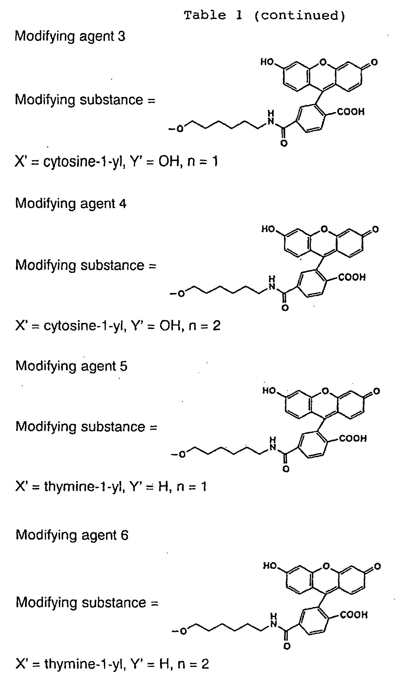

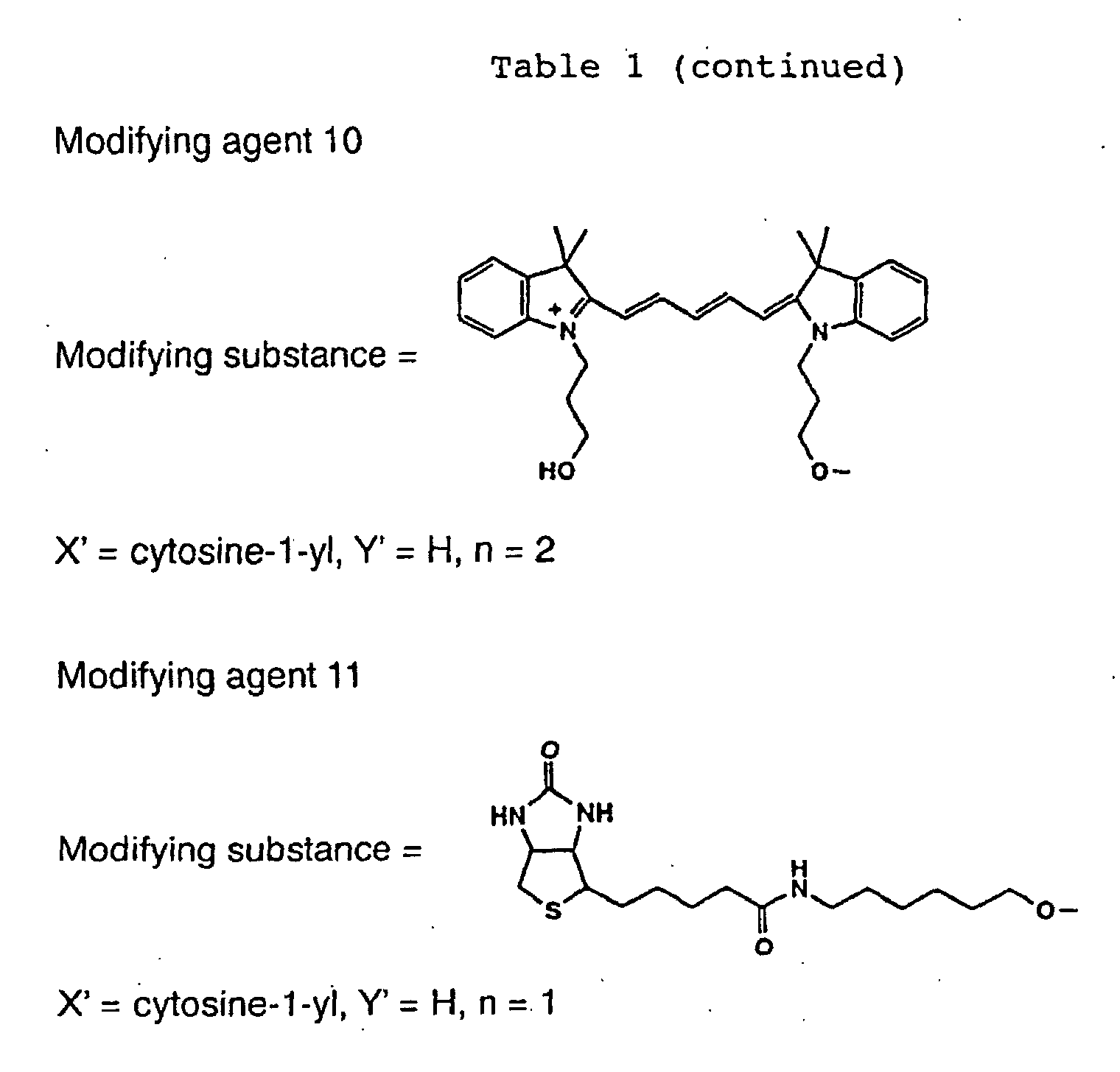
The treatments of A to D mentioned above were
repeatedly performed for Compound 1 (400 mg, containing 10
µmol of puromycin residue) until a predetermined number of
nucleotides were introduced.
For Compound 1 treated as described above, whereby
the predetermined number of nucleotides were introduced,
the treatment of A was performed, and then the treatment of
B was performed by using Compound 2 (30 µmol) instead of
the nucleotide phosphoramidite, and then the treatment of C
was performed. To thus obtained Compound 2 and Compound 1
into which the predetermined number of nucleotides were
introduced, 1 mL of 3% solution of trichloroacetic acid in
methylene chloride was added, and the mixture was left at
room temperature for 10 minutes and washed with 5 mL of
methylene chloride 3 times. The same procedure was
repeated, and then the mixture was washed with 5 mL of 10%
solution of diisopropylethylamine in methylene chloride 3
times and with 5 mL of methylene chloride 5 times and dried
under reduced pressure. To the obtained solid, 16 mmol of
a modifying substance-succinimide, 16 mL of
diisopropylethylamine and 1 mL of dimethylformamide were
added, and the mixture was shaken at room temperature for
48 hours. The solid was washed with 5 mL of
dimethylformamide 5 times and with 5 mL of ethanol 5 times,
then added with 1.5 mL of concentrated aqueous ammonia and
0.5 mL of ethanol and shaken at room temperature for 4
hours. The solid phase carrier (CPG) was removed by
filtration, and the filtrate was lyophilized. When Y in
Fig. 1 was tert-butyldimethylsilyloxy group, 400 µL of 1 M
solution of tetrabutylammonium fluoride in tetrahydrofuran
was added to the residue, and the mixture was left at room
temperature for 14 hours and concentrated under reduced
pressure. The residue was purified by HPLC (column: YMC
Pack ODS-A SH-343-5 produced by YMC (Kyoto), eluent: a
linear concentration gradient of 10 to 60% acetonitrile in
0.1 M aqueous triethylammonium acetate (pH 7.0) over 30
minutes, flow rate: 10 mL/min) and lyophilized to obtain
Modifying agents 12 to 18.
Physicochemical properties of the modifying agents
were as follows.
Modifying agent 12: yield: 6%, UV (MeOH) λmax: 503 nm, MS m/z 1295 [M-H]-
Modifying agent 13: yield: 6%, UV (MeOH) λmax: 504 nm, MS m/z 1585 [M-H]-
Modifying agent 14: yield: 3%, UV (MeOH) λmax: 503 nm, MS m/z 1313 [M-H]-
Modifying agent 15: yield: 2%, UV (MeOH) Xmax: 504 nm, MS m/z 1618 [M-H]-
Modifying agent 16: yield: 2%, UV (MeOH) λmax: 625 nm, MS m/z 1484 [M-H]-
Modifying agent 17: yield: 4%, UV (MeOH) λmax: 646 nm, MS m/z 1467 [M-H]-
Modifying agent 18: yield: 4%, UV (MeOH) λmax: 590 nm, MS m/z 1639 [M-H]-
Modifying agent 12: yield: 6%, UV (MeOH) λmax: 503 nm, MS m/z 1295 [M-H]-
Modifying agent 13: yield: 6%, UV (MeOH) λmax: 504 nm, MS m/z 1585 [M-H]-
Modifying agent 14: yield: 3%, UV (MeOH) λmax: 503 nm, MS m/z 1313 [M-H]-
Modifying agent 15: yield: 2%, UV (MeOH) Xmax: 504 nm, MS m/z 1618 [M-H]-
Modifying agent 16: yield: 2%, UV (MeOH) λmax: 625 nm, MS m/z 1484 [M-H]-
Modifying agent 17: yield: 4%, UV (MeOH) λmax: 646 nm, MS m/z 1467 [M-H]-
Modifying agent 18: yield: 4%, UV (MeOH) λmax: 590 nm, MS m/z 1639 [M-H]-
Chemical structures of the modifying agents
synthesized by the method shown in Fig. 2 (Modifying agents
12 to 18) are mentioned in Table 2.
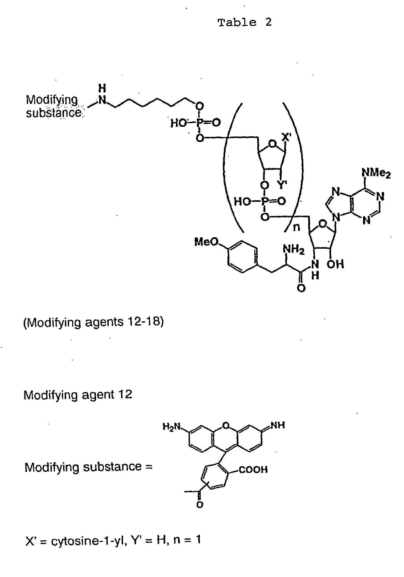
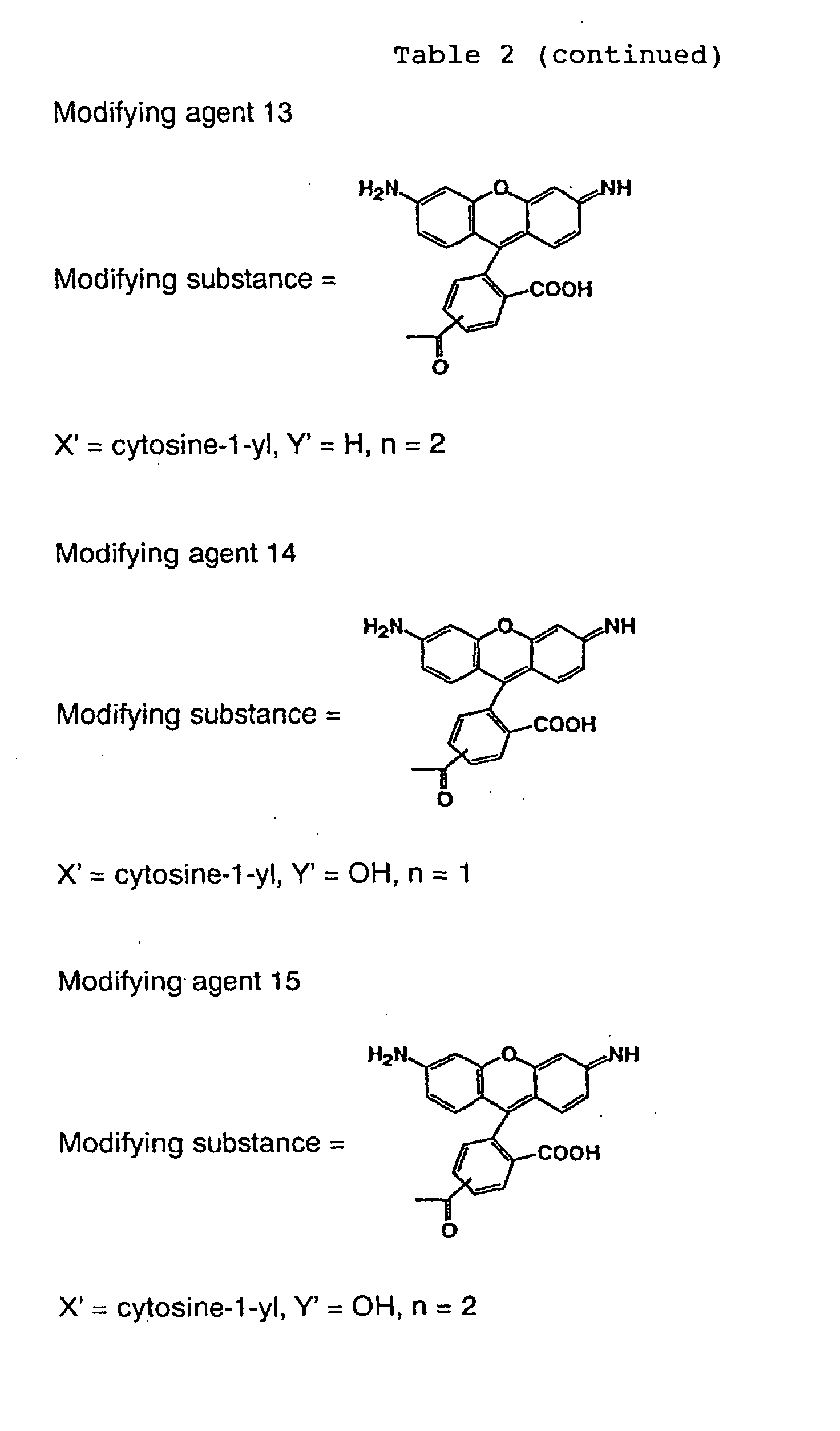

According to the Solid phase method 1 (Fig. 2), a
predetermined number of nucleotides were introduced into
Compound 1 and then a predetermined number of modifying
substances were introduced thereto by using modifying
substance-phosphoramidites. Subsequently, deprotection and
purification were performed to obtain Modifying agents 19
and 20.
Physicochemical properties of the modifying agents
were as follows.
Modifying agent 19: yield: 50%, UV (50% MeOH-H2O) λmax: 558 nm, MS m/z 1631 [M-H]-
Modifying agent 20: yield: 44%, UV (50% MeOH-H2O) λmax: 558 nm, MS m/z 2037 [M-H]-
Modifying agent 19: yield: 50%, UV (50% MeOH-H2O) λmax: 558 nm, MS m/z 1631 [M-H]-
Modifying agent 20: yield: 44%, UV (50% MeOH-H2O) λmax: 558 nm, MS m/z 2037 [M-H]-
Chemical structures of the chemically synthesized
Modifying agents 19 and 20 are shown in Table 3.

The mouse c-fos and c-jun genes were cloned from a
mouse testis cDNA library (Takara Shuzo) as follows. First,
a gene region corresponding to the 216th to 318th amino
acid residues in the amino acid sequence of c-Jun, which is
required to bind to c-Fos and DNA (Ryder, K. and Nathans,
D., Proc. Natl. Acad. Sci. U.S.A. 85, 8464-8467, 1988), was
amplified by PCR (the nucleotide sequences of the used
primers are shown in SEQ ID NOS: 1 and 2), and cloned into
a plasmid provided from Dr. Endo, Ehime University
downstream from the SP6 promoter sequence thereof to obtain
a plasmid, pSP6-jun. Similarly, a region corresponding to
the 118th to 211th amino acid residues of c-Fos (Van
Beveren, C., et al., Cell, 32, 1241-1255, 1983) was
amplified from the cDNA library by using primers (the
nucleotide sequences thereof are shown in SEQ ID NOS: 3 and
4) and substituted for the region coding for c-Jun in the
plasmid pSP6-jun to obtain a plasmid, pSP6-fos. The basic
procedures for the cloning (gene manipulation,
transformation and culture of Escherichia coli, recovery of
plasmids etc.) were performed according to Molecular
Cloning (Sambrook et al., 1989, CSH Press).
A linear DNA fragment as a template used for
transcription of the mouse c-fos and c-jun genes was
prepared as follows. PCR amplification was performed by
using the plasmid pSP6-jun as a template, a primer upstream
from the SP6 promoter (the nucleotide sequence is shown in
SEQ ID NO: 5) and a primer for adding a sequence coding for
6 histidine residues (His tag) at the C-terminal of c-Jun
(the nucleotide sequence is shown in SEQ ID NO: 6).
Similarly, PCR amplification was performed by using the
plasmid pSP6-fos as a template, the aforementioned primer
(the nucleotide sequence thereof is shown in SEQ ID NO: 5)
and a primer for adding a His tag to the C-terminal of c-Fos
(the nucleotide sequence is shown in SEQ ID NO: 7).
These two kinds of DNA were purified by using QIAquick PCR
purification kit (QIAGEN) and used for a transcription
reaction.
A fluorescence-modified DNA fragment specifically
binding to the mouse c-Fos/c-Jun dimer was prepared as
follows. Single-stranded DNAs (the nucleotide sequences
are shown in SEQ ID NOS: 8 and 9) of which 5' ends were
modified with a fluorescent dye, Cy5, and which are
complementary to each other were mixed in equimolar amounts,
heated at 95°C in the presence of 0.1 M NaCl and then
gradually cooled to room temperature so that the DNAs
should anneal to obtain a double-stranded DNA. This DNA
was used as it was for measurement by fluorescence cross-correlation
spectroscopy.
The mouse c-fos and c-jun gene DNAs were transcribed
by using Ribomax RNA synthesis system (Promega) and SP6 DNA
polymerase (37°C, 60 minutes). In this procedure, RNA cap
analogue (Life Technologies Oriental) was added to the
reaction mixture to modify the 5' end of RNA. The
synthesized RNA was treated with phenol/chloroform and then
purified by ethanol precipitation.
In order to further translate each RNA into a protein,
the obtained RNA was added to wheat germ extract (Promega)
and allowed to react at 25°C for 60 minutes. In this
reaction, modifying agents (Modifying agents 1 to 18) in
which various linkers are inserted between a fluorescent
dye (fluorescein, Rhodamine green, Cy5) and puromycin were
added at various concentrations to modify the C-terminal of
the protein with fluorescence. In order to compare yields
of the modified proteins, translation products were
subjected to SDS polyacrylamide electrophoresis, and bands
of the fluorescence-modified proteins were detected and
quantified by using a fluorescence imaging apparatus
(Molecular Imager FX, Bio-Rad).
As shown in Fig. 4, modification efficiency of the C-terminal
of the c-Fos protein significantly varied
depending on the nucleotide linker structure of the
modifying agent. The linker showing the highest yield was
-dC- (Modifying agents 1, 9, 12) irrespective of the
fluorescent dyes, and the yield of the modified protein
increased 100 times at most in comparison with the cases
using no nucleotide linker. The similar results were also
observed in the c-Jun protein. When the nucleotide linker
was -dCdC- (Modifying agents 2, 10, 13), the modification
efficiency was lower than that obtained with -dC-. Further,
when the fluorescent group was fluorescein, the
modification efficiency obtained with -rC- (Modifying agent
3) was significantly lower than that obtained with -dC-(Modifying
agent 1), whereas that obtained with -rCrC-(Modifying
agent 4) was higher than that obtained with -rC-(Modifying
agent 3). When the nucleotide linker was -dT-(Modifying
agent 5), -dTdT- (Modifying agent 6), -dA-(Modifying
agent 7) or -dG- (Modifying agent 8), although
the effect was not significant, a marked effect was
observed for deoxycytidylic acid or ribocytidylic acid.
Further, the optimum concentrations of the modifying agents
varied depending on the types of the fluorescent groups.
The optimum concentration for labeling efficiency with Cy5
(Modifying agents 9 and 10) was as low as about half of
that obtained with fluorescein (Modifying agents 1 and 2).
When the fluorescent group was rhodamine green (RG)
(Modifying agents 12 and 13), the tendency of the optimum
concentration was very similar to that observed for Cy5.
Fluorescence-modified proteins used for measurement
by fluorescence correlation spectroscopy were purified as
follows to remove unreacted fluorescent dyes. First, the
translation reaction mixture was mixed with an equilibrated
nickel NTA agarose resin (QIAGEN), adsorbed on the resin by
a specific bond between the His tag at the C-terminal of
the fluorescence-modified protein and a nickel ion, washed
and then eluted with imidazole. Furthermore, an eluted
fraction containing the protein was applied to a gel
filtration column (PD-10, Pharmacia) twice, and the eluted
solution was concentrated by centrifugation using Centricon
(Millipore).
Measurement by fluorescence cross-correlation
spectroscopy was performed by using the c-Fos and c-Jun
proteins modified with a modifying agent having rhodamine
green (RG) as a fluorescent group (Modifying agent 12) and
DNA modified with Cy5. First, the purified proteins and
DNA were mixed at a final concentration of 10 nM each and
incubated at 37°C for 60 minutes. 10 µL of this sample was
placed on an 8-well glass chamber (Nunc), and fluorescence
cross-correlations were measured by using a fluorescence
correlation spectrometer, ConfoCor2 (Carl Zeiss). As a
result, cross-correlations were observed in the samples to
which all the three types of substances, Fos, Jun and DNA
were added (Fig. 5, 1 and 2), whereas no cross-correlation
was observed in the control samples lacking either Fos or
Jun (Fig. 5, 3 and 4). As a result of numerical analysis
of the observed cross-correlations, it was found that about
30% of all the molecules bound to form complexes. Further,
the dissociation constant directly calculated from this
result was 10-8 M, which well matched the known value
obtained by another method.
From the above results, it was found that a C-terminal
of protein could be modified with fluorescence by
using a modifying agent having a linker of deoxycytidylic
acid inserted between the fluorescent dye and puromycin
with a yield about 100 times as high as that obtained by a
conventional method. Further, a protein interaction could
be detected by fluorescence cross-correlation spectroscopy
for the first time by using proteins modified by the method
of the present invention, and prospects of practical use
were offered.
A sequence of a vector or plasmid to which mouse-derived
c-jun or c-fos was incorporated, or a DNA template
containing such a sequence was amplified by PCR using
TaKaRa Ex Taq (Takara Shuzo) and purified by using QIAquick
PCR Purification Kit (QIAGEN). As the template for PCR, c-jun[pSPAM],
c-fos[pSPAM] and c-jun[F] (the nucleotide
sequences are shown in SEQ ID NOS: 10-12; respectively)
were used. As the primers, SP6F and 5' SP6-O29 (the
nucleotide sequences are shown in SEQ ID NOS: 13 and 14,
respectively) were used as Primer 1 (forward primer), and
JunHis, JunHisA, FosHis, FosHisA, JunFlagA, 3'HisA and
3'FlagA (the nucleotide sequences are shown in SEQ ID NOS:
15-21, respectively) were used as Primer 2 (reverse primer).
The translation templates (DNA templates) were obtained by
the above method. Each template was transcribed (37°C, 2
hours) by using RiboMAX™ Large Scale RNA Production
Systems (Promega) and purified by using RNeasy Mini Kit
(QIAGEN) to obtain a translation template (RNA template).
The basic structure of a translation template and DNA
nucleotide sequences of components are shown in Fig. 6.
Hereinafter, the translation templates are referred to in
the forms of "name of Primer 1 - name of Primer 2"
according to the primers used for amplification.
Two kinds of experiments were performed to examine
the effect of polyA and the effect of a translation
enhancer, 029, in the translation templates. In both of
the experiments, translation was performed (26°C, 60
minutes) by using wheat germ extract (Promega) in the
presence of a fluorescent modifying agent (Modifying agent
1) to modify proteins at the same time as the translation
and subjected to 17.5% SDS-PAGE, and the modification was
confirmed based on fluorescence (fluorescein) by using a
multiple image analyzer, Molecular Imager FX (Bio-Rad).
The Jun or Fos protein having a molecular weight of about
20-25 kDa was obtained. The results for relative ratios of
the modifying efficiencies of the following templates are
summarized in Fig. 7. In the experiment referred to in Fig.
7, as the translation templates, SP6F-JunHis, SP6F-JunHisA,
SP6F-FosHis and SP6F-FosHisA were used in the experiment
for the polyA sequence effect, and SP6F-JunFlagA, SP6-029
Jun-FlagA, SP6F-JunHisA and SP6-O29Jun-His were used in
the experiment for the 029 effect. Conversion was made
based on the result obtained with SP6F-FosHisA in the
experiment for the polyA effect and the result obtained
with 5'SP6-029-JunHisA in the experiment for the 029 effect,
both of which were taken as 1.0.
In the experiment for the polyA sequence effect, for
both of Jun and Fos, one containing the polyA sequence
showed modification efficiency about 3 times higher than
that obtained with one not containing the polyA sequence
irrespective of the proteins (Fig. 7). Further, as for the
translation enhancer, there was observed a tendency that
the effect of the 029 sequence was higher than that of the
AMV sequence. In particular, when the affinity tag was
His-tag, the modification efficiency doubly increased. The
effects of the polyA sequence, the 029 sequence and the
His-tag sequence in modification of the proteins were
confirmed. In Example 1, AMV was used as the translation
enhancer of the translation template, and the polyA
sequence was not used. The translation efficiency of the
translation template of SP6-O29Jun-HisA used in this
example was 5 to 6 times higher than that obtained with the
translation template using AMV in Example 1. Therefore, it
was found that when SP6-O29Jun-HisA as the translation
template and a modifying agent having a nucleotide linker
were used in combination, modification efficiency of C-terminals
of proteins became about 500 times as high as the
modification efficiency obtained by the conventional
techniques (Japanese Patent Laid-open Publication Nos. 11-322781
and 2000-139468).
Preparation, transcription and translation of DNA
coding for the mouse c-Jun protein and purification of the
c-Jun protein were performed in the same manner as in
Example 1.
5 µl of the c-Jun protein of which C-terminal was
modified with a modifying agent having biotin and a
fluorescent dye (TAMRA) in the same molecule (Modifying
agent 20) or a modifying agent having only a fluorescent
dye (TAMRA) (Modifying agent 19) was spotted on a
streptavidin membrane (SAM Biotin Capture Membrane,
Promega), and after 1 minute, the membrane was washed with
50 mL of 2 M NaCl solution 4 times and with distilled water
twice. Then, fluorescence of TAMRA was detected at 532 nm
by using an image analyzer (Molecular Imager FX, Bio-Rad).
As a result, as shown in Fig. 8, it was found that the c-Jun
protein modified at its C-terminal with the modifying
agent having biotin and fluorescent dye in the same
molecule (Modifying agent 20) was immobilized on the
streptavidin membrane (Fig. 8, 1). However, the protein
modified with the modifying agent having only a fluorescent
dye without biotin (Modifying agent 19) was not immobilized
on the streptavidin membrane (Fig. 8, 2). When free biotin
in an amount of 1000 times in molar ratio was added at the
same time as a control experiment, binding of the
biotinylated c-Jun protein to the membrane was
competitively inhibited (Fig. 8, 3). Therefore, it can be
seen that adsorption of c-Jun to the membrane is caused by
a biotin-specific binding.
A DNA fragment including a region for Fos/Jun dimer
binding region was amplified by PCR using a primer modified
with Cy5. QIAquick PCR purification kit (QIAGEN) was used
for purification of DNA. A spotting solution (150 mM
sodium phosphate in 0.01% SDS, pH 8.5) containing the
modified DNA fragment at a concentration of 200 µg/ml was
prepared.
A slide (DNA-Ready™ TypeII Slide, CLONTECH) was set
on a microarrayer (Micro Grid, BioRobotics), and spotting
was performed (spotting conditions; temperature in chamber:
about 25-28°C, humidity: 38-42%, solid pins were used, at
0.7 mm intervals).
After the spotting, the slide was incubated at 80°C
for 2 hours, and water vapor was applied to the spotted
surface to hydrate the spot. Moisture was evaporated on a
hot plate at 100°C, and the spots were irradiated with UV
to immobilize the DNA fragments. The slide was immersed in
a blocking solution (4 g of succinic acid, 252 ml of 1-methyl-2-pyrrolidinone,
28 ml of 1 M boric acid (pH 8)),
vigorously shaken for first 1 minute and then shaken for 20
to 30 minutes. The slide was washed with distilled water
at 90°C and then with 99.5% ethanol and dried.
A hybridization solution was placed on the DNA-immobilized
slide and the slide was covered with Parafilm
to spread the solution over the whole spotted surface.
Light was shielded with an aluminium foil, and the slide
was incubated at 37°C for 30 minutes (water was filled
under the table on which slide was arrayed to prevent
dryness). The slide was shaken in 1 x buffer for 5 minutes,
the solution was replaced, and this procedure was repeated.
The slide was centrifuged at 5000 rpm and 4°C and then
dried, and the fluorescence of the spots on the slide was
detected at 532 nm and 635 nm by using a microarray scanner
(Gene Pix 4000A, Axon Instruments).
That is, DNA for the Fos and Jun binding region
modified with Cy5 was immobilized on the slide
(fluorescence was measured at 635 nm) and sprinkled with
Jun and the p53 protein of which C-terminal was modified
with a modifying agent having rhodamine green as a
fluorescent group (Modifying agent 12, fluorescence was
measured at 532 nm) in the presence of Fos to examine
interactions between DNA and Fos-Jun and between DNA and
Fos-p53. The results are shown in Fig. 9. The upper left
image shows that Jun modified with rhodamine green at its
C-terminal is bound to DNA in the presence of Fos
(existence of Jun or p53 can be confirmed by detecting the
fluorescence of rhodamine green at 532 nm). Further, the
lower left image for the same sample shows that DNA
modified with Cy5 was immobilized and existed (existence of
DNA can be confirmed by detecting fluorescence of Cy5 at
635 nm). On the other hand, the upper right image shows
that p53 was not bound to DNA in the presence of Fos, since
the fluorescence of rhodamine green was not detected. The
lower right image for the same sample confirmed the
existence of DNA, since the fluorescence of Cy5 was
detected. As a result, it can be seen that the
fluorescence-modified Jun was specifically bound to DNA for
the binding region in the presence of Fos. In this example,
after the proteins were modified, interactions were
examined without purification. Nevertheless, a distinct
difference in interactions to such an extent as shown in
Fig. 9 was observed. This indicates that the translation
efficiency and the modification efficiency increased and
that C-terminal modified proteins were produced in an
amount sufficient for interactions.
Purification of modified proteins to high purity was
aimed to enable kinetic analysis of intermolecular
interaction between C-terminal modified proteins.
Different affinity tags were introduced into the
translation templates and the modifying compounds.
Affinity purification of the translation product in two
stages enabled purification of a protein of which C-terminal
was modified with a fluorescent dye to high purity.
The cancer gene products, the c-Fos and c-Jun proteins,
fluorescence-modified with rhodamine green or Cy5 were
purified in two stages to obtain samples purified to high
purity. The formation of a complex of AP-1 (DNA of the
binding region for c-Fos and c-Jun dimer)/rhodamine green-modified
c-fos/Cy5-modified c-Jun was detected by
fluorescence cross-correlation spectroscopy, and the
dissociation constant (Kd) was calculated from the
analytical values of the intermolecular interactions.
Modifying agents 21 to 25 were synthesized by the
method outlined in Figs. 10 and 11. Compound 1' mentioned
in Fig. 10 was synthesized by using the same method as
Compound 1. Phosphoramidites were purchased from Glen
Research (USA, Virginia). Modifying substance 1-succinimide
was purchased from Pierce Biotechnology
(Illinois, USA). Modifying substance 2-succinimide was
purchased from Molecular Probes (USA, Oregon) and Amersham
Pharmacia Biotech (Upsala, Sweden).
The treatments of A to D shown in the Solid phase
method 1 were repeatedly performed for Compound 1' (400 mg,
containing 10 µmol of puromycin) until a predetermined
number of nucleotides were introduced.
For Compound 1' treated as above whereby a
predetermined number of nucleotides were introduced, the
treatment of A was performed, the treatment of B was
performed by using 30 µmol of Compound 2 instead of the
nucleotide phosphoramidites, and then the treatment of C
was performed. To thus obtained Compound 1' to which
Compound 2 and the predetermined number of nucleotides were
introduced, 2 mL of 50 mM sodium carbonate in methanol when
Modifying agent 21 was used, or 1.5 mL of concentrated
aqueous ammonia and 0.5 mL of ethanol when Modifying agents
22 to 25 were used, was added and the mixture was shaken at
room temperature for 14 hours. The solid phase carrier
(CPG) was removed by filtration, and the filtrate was
concentrated under reduced pressure. The residue was
purified by HPLC (column: YMC Pack ODS-A SH-343-5 produced
by YMC (Kyoto), eluent: a linear concentration gradient of
10 to 60% acetonitrile in 0.1 M aqueous triethylammonium
acetate (pH 7.0) over 30 minutes, flow rate: 10 mL/min) and
then lyophilized.
When Modifying agent 21 was used, the above residue
was dissolved in 2 mL of 80% acetic acid in water, left at
room temperature for 4 hours and then concentrated under
reduced pressure. The residue was dissolved in 1 mL of 30%
acetonitrile in water, and a solution obtained by
dissolving 0.1 mL of 1 M sodium hydrogencarbonate in water
(pH 8.3) and 0.1 mmol of Modifying substance 1-succinimide
in 0.5 mL of N,N'-dimethylformamide was added thereto. The
mixture was left at room temperature for 2 hours. Then,
the mixture was desalted by using Poly-PakII (Glen
Research) and concentrated under reduced pressure.
When Modifying agents 22 to 25 were used, the above
residue was dissolved in 1 mL of 30% acetonitrile in water,
and 0.1 mL of 1 M sodium hydrogencarbonate in water (pH
8.3) and a solution obtained by dissolving 0.1 mmol of
Modifying substance 2-succinimide in 0.5 mL of N,N'-dimethylformamide
were added thereto. The mixture was left
at room temperature for 2 hours. Then, the mixture was
desalted by using Poly-PakII and concentrated under reduced
pressure. The residue was dissolved in 2 mL of 80% acetic
acid in water, and the solution was left at room
temperature for 4 hours and concentrated under reduced
pressure. The residue was dissolved in 1 mL of 30%
acetonitrile in water, and 0.1 mL of 1 M sodium
hydrogencarbonate in water (pH 8.3) and a solution obtained
by dissolving 0.1 mmol of Modifying substance 1-succinimide
in 0.5 mL of N,N'-dimethylformamide were added thereto.
The mixture was left at room temperature for 2 hours. Then,
the mixture was desalted by using Poly-PakII (Glen
Research) and concentrated under reduced pressure.
In the both cases of using Modifying agent 21 and
Modifying agents 22 to 25, to the above residue, 2 mL of
60% trifluoroacetic acid in water was added and the mixture
was left at room temperature for 30 minutes and
concentrated under reduced pressure. The residue was
purified by HPLC (column: YMC pack ODS-A SH-343-5, YMC
(Kyoto), eluent: a linear concentration gradient of 10 to
60% acetonitrile in 0.1 M aqueous triethylammonium acetate
(pH 7.0) over 30 minutes; flow rate, 10 mL/min) and then
lyophilized to obtain Modifying agents 21 to 25.
Modifying agent 26 was synthesized from Compound 1 by
Solid phase method 1. The phosphoramidite containing
Modifying substance 1 or Modifying substance 2 was
purchased from Glen Research (Virginia, USA).
Physicochemical properties of the modifying agents
were as follows.
Modifying agent 21; yield: 32%, UV (H2O) λmax: 558 nm, MS m/z 2035 [M-H]-
Modifying agent 22; yield: 8%, UV (H2O) λmax: 506 nm, MS m/z 2093 [M-H]-
Modifying agent 23; yield: 8%, UV (H2O) λmax: 506 nm, MS m/z 1979 [M-H]-
Modifying agent 24; yield: 13%, UV (H2O) λmax: 649 nm, MS m/z 2375 [M-H]-
Modifying agent 25; yield: 13%, UV (H2O) λmax: 649 nm, MS m/z 2261 [M-H]-
Modifying agent 26; yield: 22%, UV (H2O) λmax: 646 nm, MS m/z 1977 [M-H]-
Modifying agent 21; yield: 32%, UV (H2O) λmax: 558 nm, MS m/z 2035 [M-H]-
Modifying agent 22; yield: 8%, UV (H2O) λmax: 506 nm, MS m/z 2093 [M-H]-
Modifying agent 23; yield: 8%, UV (H2O) λmax: 506 nm, MS m/z 1979 [M-H]-
Modifying agent 24; yield: 13%, UV (H2O) λmax: 649 nm, MS m/z 2375 [M-H]-
Modifying agent 25; yield: 13%, UV (H2O) λmax: 649 nm, MS m/z 2261 [M-H]-
Modifying agent 26; yield: 22%, UV (H2O) λmax: 646 nm, MS m/z 1977 [M-H]-
Chemical structures of the synthesized modifying
agents are shown in Table 4.

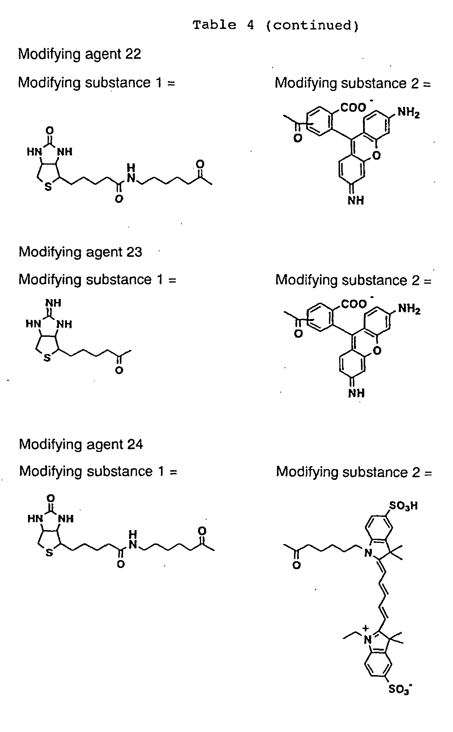
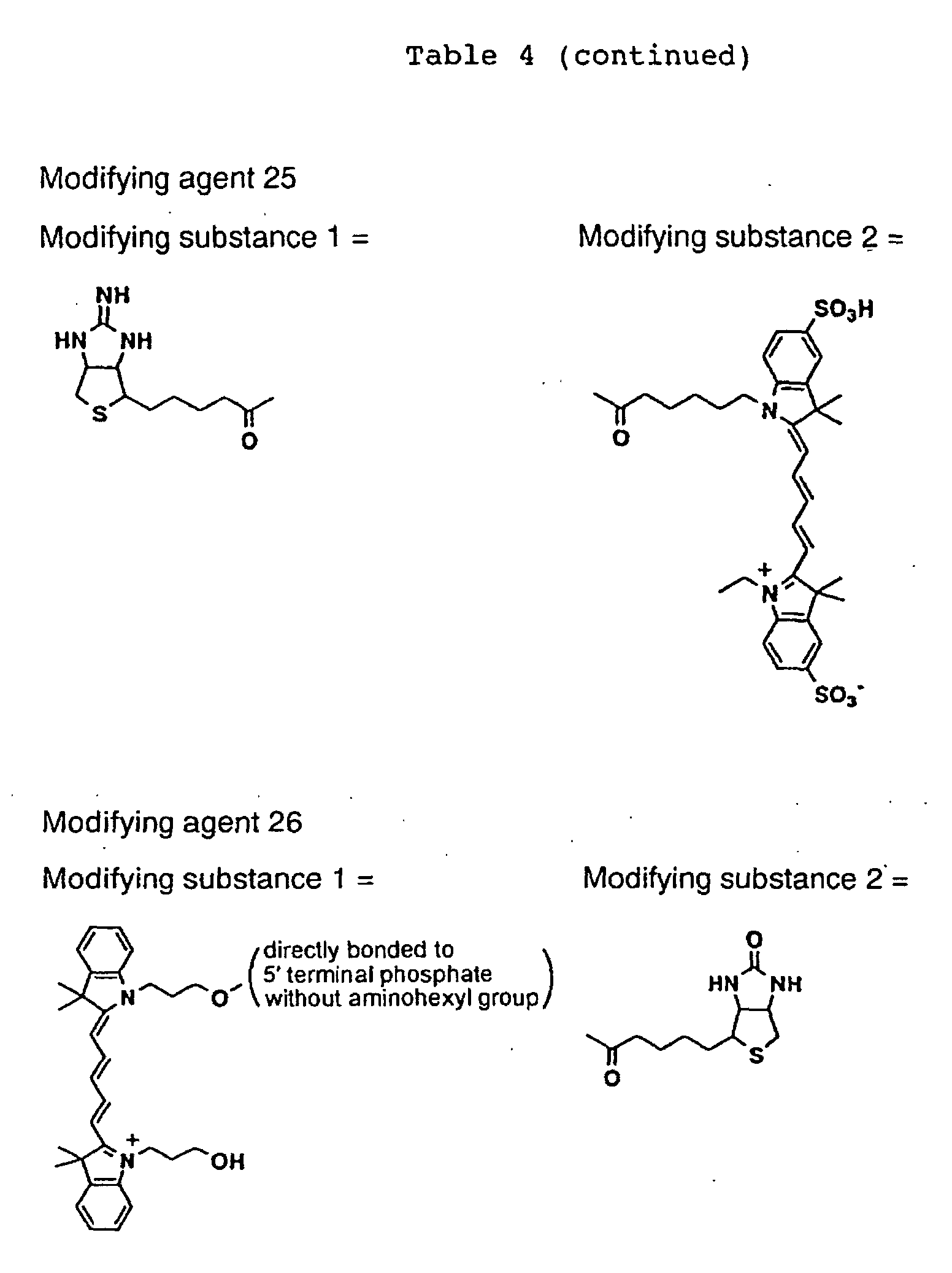
As the template DNA, plasmids to which the mouse c-fos
and c-jun genes were introduced, prepared in the
preparation of DNA in Example 1 were used. Regions in the
c-jun and c-fos including a domain required to bind to DNA
were amplified by PCR using a primer containing SP6
promoter, Ω sequence and T7 tag (SEQ ID NO: 22), and a
reverse primer containing a histidine tag, a stop codon and
polyA sequence (SEQ ID NO: 23 or 24). The PCR product was
subcloned into the pCR2.1TOPO ,vector by using Topo TA
cloning kit (Invitrogen). The detail of the method was
according to the manufacturer's protocol. The plasmid DNA
of which nucleotide sequence was confirmed was purified by
using Wizard Plus SV Minipreps DNA Purification System
(Promega).
The linear template DNA for RNA synthesis was
obtained by PCR using a primer utilizing a part of a vector
sequence upstream from the insertion site (SEQ ID NO: 25)
and a reverse primer in the histidine tag portion. In
order to examine the recovery of the translated protein,
reverse primers (SEQ ID NOS: 26-29) corresponding to
polyhistidines (length: 6 to 12 residues) were produced.
The template DNA was purified by using QIAquick PCR
Purification Kit (QIAGEN).
The template DNA was transcribed in the presence of a
cap analogue (Life Technologies Oriental Yeast) by using
SP6 RiboMAX Large Scale RNA Production System (Promega)
(37°C, 3 hours). The detail of procedure was according to
the manufacturer's manual. After the transcription, the
template DNA was removed by using deoxyribonuclease
attached to the kit, and purified RNA was obtained by using
SV Total RNA Isolation System (Promega).
Translation was performed according to the
manufacturer's manual, and 5 mg of the purified RNA and 100
µl of wheat germ extract (Promega) were used. Fluorescent
modifying compounds (Modifying agents 22 to 26) in which
biotin or iminobiotin and a fluorescent dye (rhodamine
Green, Cy5) were introduced into the same molecule were
added in the same reaction system to synthesize proteins
modified with fluorescence at their C-terminals (25°C, 1
hour). The optimum concentration of the fluorescent dyes
with biotin introduced was about 125 µM for both of Cy5
(Modifying agents 24 and 26) and rhodamine green (Modifying
agent 22). The optimum concentrations of the fluorescent
dyes with iminobiotin introduced was 30 µM for Cy5
(modifying agent 25) and 12.5 µM for rhodamine green
(Modifying agent 23).
Purification using a nickel chelate resin, Ni-NTA
Superflow (QIAGEN), was performed according to the
manufacturer's manual. To a reaction mixture, 0.1 µl of a
protease inhibitor (cocktail for use in histidine-tagged
protein purification, Sigma) and 5-fold volume of a binding
buffer were added, and the mixture was calmly mixed with 20
µl of a nickel chelate resin suspension (4°C, 1 hour). The
resin was sufficiently washed with the binding buffer, and
the histidine-tagged protein was eluted with 50 µl of
buffer containing 0.5 M imidazole.
An ordinary histidine-tagged protein is sufficiently
recovered with a polyhistidine tag having a length of 6
residues using a nickel chelate resin (Abate, C. et al.,
Proc. Natl. Acad. Sci. USA., 87, 1032-1036, 1990). In the
case of this labeling method, however, the number of
histidine residues was increased to examine the amount of
recovery by the nickel chelate resin, since the recovery
rate was slightly low when the polyhistidine tag having a
length of 6 residues was used. After the translation, the
supernatant, the fraction passed through the nickel chelate
resin and the fraction eluted with imidazole were separated
by SDS polyacrylamide electrophoresis (SDS-PAGE), and
detection was performed by using a fluorescence image
analyzer (Molecular Imager FX, Bio-Rad). As the length of
the polyhistidine increased, the amounts of the recovered
fluorescence-labeled proteins increased (Fig. 12). Similar
results were also obtained for c-Fos. Further, when a
labeling compound containing iminobiotin (Modifying agent
25) was used for fluorescence labeling, the labeling was
attained with efficiency twice as high as that obtained
with a labeling compound not containing iminobiotin
(Modifying agent 9, Fig. 13).
The labeled protein containing iminobiotin was
further purified with a streptavidin-immobilized resin,
Streptavidin Sepharose High Performance (Amersham
Pharmacia). The purification with this resin was performed
according to the manufacturer's manual. To the
aforementioned fraction eluted with imidazole, 5-fold
volume of a binding buffer was added, and the mixture was
calmly mixed with 10 µl of the resin equilibrated
beforehand (4°C, 30 minutes). The resin was sufficiently
washed with the binding buffer, and the protein was eluted
with 50 µl of a buffer containing 50 mM biotin.
The labeled protein containing biotin was purified
with an avidin monomer-immobilized resin, UltraLink
Immobilized Monomeric Avidin (Pierce Biotechnology). The
purification using the resin was performed according to the
manufacturer's manual. To the aforementioned fraction
eluted with imidazole, 9-fold volume of a binding buffer
was added, and the mixture was calmly mixed with 10 µl vof
the resin equilibrated beforehand (4°C, 30 minutes). The
resin was sufficiently washed with the binding buffer, and
the protein was eluted with 50 µl of buffer containing 50
mM biotin.
The protein labeled with Cy5 containing iminobiotin
was separated by SDS-PAGE, and the purification was
confirmed by fluorescence image analysis and immunoblotting
(Fig. 14, A and B). In the immunoblotting, the gel after
the electrophoresis was electrically transferred to a
polyvinylidene fluoride membrane (Pall Gelman Science), a
mouse monoclonal antibody (Novagen) directed to the T7 tag
provided at the N-terminal and a horseradish peroxidase-labeled
goat anti-mouse antibody (Transduction) were
reacted, and chemiluminescence was obtained by using ECL
kit (Amersham Pharmacia). This immunoblotting method was
performed according to the manufacturer's manual attached
to the aforementioned kit. Among the histidine-tagged
proteins bound to the nickel chelate resin, the proteins
passing through the streptavidin-immobilized resin were not
detected based on fluorescence, whereas the biotin-eluted
fraction was detected with both of the antibodies and
fluorescence (Fig. 14, A and B, Lanes 2 and 3).
Purity of the purified labeled proteins was examined.
As the fluorescence standard solutions, Cy5 dye quantified
based on the molecular extinction coefficient of 25,000 at
650 nm and rhodamine green dye quantified based on the
molecular extinction coefficient of 68,000 at 505 nm were
used. 100 µl of a sample dissolved in 0.1 M Tris-hydrochloric
acid buffer (pH 8) was placed on a black 384-well
polystyrene plate (Nunc) to quantify the proteins by
using a fluorescence image analyzer. The concentrations of
c-Fos and c-Jun contained in the purified fractions were
quantified by dot blotting assay using a T7-tag recombinant
protein (Novagen) as a standard substance. 1 µl of sample
was spotted on a nitrocellulose membrane (Schreier-Schulz),
and a mouse anti-T7 tag antibody and a horseradish
peroxidase-labeled goat anti-mouse antibody were reacted.
Chemiluminescence obtained by using ECL was detected by a
chemiluminescence analyzer (Molecular Imager ChemiDoc, Bio-Rad).
The ratio of molar concentrations of the T7-tagged
protein and the fluorescence of the purified fraction was
90% or higher (Table 5). When the purified protein was
separated by SDS-PAGE and stained, it was confirmed that it
was a substantially single component (Fig. 15).
| Purified protein | Ratio of amounts quantified by dot blotting and fluorescence |
| Cy5-labeled c-Fos | 0.99 |
| Cy5-labeled c-Jun | 0.92 |
| Rhodamine green-labeled c-Fos | 1.02 |
| Rhodamine green-labeled c-Jun | 0.93 |
Intermolecular interactions were analyzed by
fluorescence cross-correlation spectroscopy using
fluorescence-labeled c-Fos and c-Jun as well as AP-1
sequence double-stranded DNA. DNA and the modified
proteins (at a final concentration of 10 nM each) were
mixed. 10 µl of this sample was applied to an 8-well glass
chamber (Nunc), and fluorescence cross-correlations were
measured by using a fluorescence correlation spectrometer,
ConfoCor2 (Carl Zeiss). As a result, cross-correlation was
confirmed in the sample to which all the three of c-Fos, c-Jun
and the DNA were added, and no cross-correlation was
observed in the control sample not containing c-Jun (Fig.
16). The dissociation constant (Kd) calculated from the
analytical results of the cross-correlations according to
the following equations was about 1 x 10⁻⁸ M. The
dissociation constant obtained by other techniques was
reported to be about 1 x 10⁻⁹ M to about 1.1 x 10⁻⁷ M (Heuer,
K., et. al., Biochemistry 35, 9069-9075, 1996; Pernelle, C.,
et. al., Biochemistry 32, 11682-11687, 1993), and the
result obtained by using the fluorescence-labeled protein
of the present invention does not contradict these values.
Based on the above, it was confirmed that proteins labeled
with fluorescence according to the present invention could
be used to detect protein interactions by using a
fluorescence cross-correlation spectroscopy and further
could be utilized for convenient and quick measurement of
dissociation constant.
Nac,r Nac,g and Gc(0) can be obtained from the cross-correlation
analysis.
Ncc =1 Gc (0)-1
The number of particles modified with Cy5 and rhodamine
fluorescence:
Ngr = Nacg Nac.r Ncc
The number of particles modified only with Cy5:
Nr =Nac.r -Ngr
The number of particles modified only with rhodamine:
Ng' =Nac.r -Ngr
From the above, the dissociation constant is represented as
follows.
KD =[r ][g ][gr ] ≅1×10⁻⁸ M
Modifying agent 19 synthesized in Example 1 was used.
Actinomycetes, Streptomyces avldinii, was purchased
from Riken. An oligo DNA (primer) was synthesized by Espec
Oligo Service. Commercially available Escherichia coli,
plasmids, various enzymes, reagents etc. were used,
including Escherichia coli JM109, plasmid pUC18 (Toyobo),
pET20b (Novagen), restriction enzymes BamHI, BgIII, EcoRI
and HindIII (Toyobo), Ligation High (Toyobo), Ex Taq DNA
polymerase, Recochip (Takara Shuzo), and QIAquick PCR
Purification Kit (QIAGEN). The basic procedures of genetic
engineering (cloning, transformation and culture of
Escherichia coli, recovery of plasmids etc.) were performed
according to Sambrook et al., Molecular Cloning, 1989, CSH
Press.
Plasmids pSP6-STA-Jun and pSP6-STA-Fos were
constructed by the following procedure. First, the
streptavidin gene was amplified by PCR using the
Actinomycetes Streptomyces avidinii genome as a template
and primers (SEQ ID NOS: 30 and 31) complementary to the
upstream and downstream regions of the streptavidin gene,
digested with BamHI and EcoRI and cloned at the BamHI-EcoRI
site in pUC18 to obtain pUC-STA. PCR was performed by
using this plasmid as a template, a primer for adding a T7
tag to the N-terminal of the streptavidin gene (SEQ ID NO:
32) and a primer complementary to the downstream region of
the streptavidin gene (SEQ ID NO: 33) to obtain a
streptavidin gene having the T7 tag sequence at the N-terminal.
Further, in order to add an untranslated region
(5'-UTR) including the SP6 promoter and an enhancer
sequence derived from tobacco mosaic virus to the upstream
thereof, a fragment amplified by PCR using primers
complementary to the upstream and downstream regions of the
streptavidin gene having the T7 tag sequence (SEQ ID NOS:
34 and 33) was digested with BamHI and cloned at the BgIII-BamHI
site of pET20b. As for the insertion direction of
the streptavidin gene, the plasmid in which the BgIII side
corresponded to the upstream of the gene was designated as
pSP6-STA. Subsequently, the jun and fos genes prepared in
Example 1 were amplified by PCR using two sets of primers
(SEQ ID NOS: 35 and 36 and SEQ ID NOS: 3 and 37), digested
with BamHI and HindIII and cloned at the BamHI-HindIII site
of pSP6-STA to obtain pSP6-STA-Jun and pSP6-STA-Fos.
As the mouse c-fos and c-jun gene DNAs, the genes
with a polyhistidine tag having a length of 12 residues at
the C-terminal prepared in Example 5 were used.
The mouse c-fos and c-jun gene DNAs were transcribed
with the SP6 DNA Polymerase by using Ribomax RNA synthesis
system (Promega) (37°C, 120 minutes). In this reaction, 20
µl of the reaction mixture contained 6 µl of DNA, 1 µl each
of 100 mM rUTP, rCTP and rATP, 1 µl of 30 mM rGTP and 2 µl
of SP6 polymerase, and it was further supplemented with 4
µl of RNA cap analogue (Life Technologies Oriental)
prepared at 40 mM to modify the 5' end of RNA. The
synthesized RNA was purified by using RNeasy Mini Kit
(QIAGEN).
In order to translate the RNA into a protein, the
obtained mRNA was added to a cell-free translation system
using wheat germ extract of Proteios™(TOYOBO) and allowed
to react at 37°C for 5 hours. In addition to two types of
buffers, 4 µl of creatine kinase, 10 µl of RNase inhibitor,
20 µl of wheat germ, 10 µl of mRNA and a fluorescent
modifying agent were added to 100 µl of the translation
system. As the fluorescent modifying agent, fluorescent
dye (TAMRA)-dC-puromycin (Modifying agent 19) was used. In
order to obtain the yield of the modified protein, the
translation product was subjected to SDS polyacrylamide gel
electrophoresis, and the band of the fluorescence-modified
protein was detected by using a fluorescence imaging
apparatus (Molecular Imager FX, Bio-Rad).
The jun and fos genes on the bait side fused with
streptavidin for immobilization on a microarray were
transcribed in the same manner as described above using
pSP6-STA-Jun and pSP6-STA-Fos as templates and used for
protein synthesis by a multilayer method using Proteios™
(TOYOBO).
The fluorescence-modified protein used as a protein
on the prey side in the immobilization method was purified
as follows to remove unreacted fluorescent dyes.
A reaction mixture for translation of a His-tagged
protein modified with fluorescence at the C-terminal was
mixed with an equilibrated nickel NTA agarose resin
(QIAGEN) so that the protein should be adsorbed to the
resin by a specific binding between His tag at the C-terminal
of the fluorescence-modified protein and a nickel
ion. The resin was washed, and then the protein was eluted
with 500 mM imidazole.
A biotin-coated slide (Xenopore) was set on a DNA
microarray (MicroGridII, BioRobotics), spotted with a
translation solution of streptavidin-fused Jun and Fos
proteins dissolved in 60% PBS (10 mM phosphate buffer, 150
mM NaCl (pH 7.4))/40% glycerol, and left in the humidity-controlled
environment for 1 hour so that the protein
should be immobilized on the slide surface by binding
between streptavidin and biotin (Fig. 17, uppermost figure).
After the immobilization, the slide was rinsed by shaking
in 1% BSA/PBS solution for 1 minute and then further shaken
in the replaced 1% BSA/PBS solution for 1 hour for blocking.
After the blocking, the slide was washed with 1 x PBS and
dried. A frame-like silicon sticker for in situ
hybridization (EasiSeal, Hybaid) was stuck around the
spotted region of the slide, and the frame was filled with
a reaction mixture containing 0.5 mg/ml of mouse-derived
anti-Fos primary antibody (c-Fos(6-2H):sc-447, Santa Cruz)
and 0.5 mg/ml of rabbit-derived anti-Jun primary antibody
(c-Jun/AP-1, Oncogene) dissolved in 1 x PBST (10 mM
phosphate buffer, 150 mM NaCl, 0.1% Tween 20 (pH 7.4)) and
sealed with a sticker from the above. In this state, the
mixture was allowed to react at room temperature for 1 hour
in the humidity-controlled environment. After the reaction,
the slide was shaken in 1 x PBST for 5 minutes 5 times with
replacing the buffer, further shaken in 1 x PBS for 3
minutes 3 times with replacing the buffer, centrifuged at
2000 rpm at 4°C for 1 minute and dried. Then, a frame-like
silicon sticker for in situ hybridization (EasiSeal,
Hybaid) was stuck again around the spotted region on the
slide, and the frame was filled with a reaction mixture
containing 0.5 mg/ml of Cy3-labeled anti-mouse antibody
(Chemicon) and 0.5 mg/ml of Cy5-labeled anti-rabbit
antibody (Chemicon) dissolved in 1 x PBST and sealed with a
sticker from the above. In this state, the mixture was
allowed to react at room temperature for 1 hour in the
humidity-controlled environment. After the reaction, the
slide was shaken in 1 x PBST for 5 minutes 5 times with
replacing the buffer, further shaken in 1 x PBS for 3
minutes 3 times also with replacing the buffer, centrifuged
at 2000 rpm at 4°C for 1 minute and dried. When this slide
was scanned by using a DNA chip scanner (GenePix 4000B,
Axon Instruments) to detect the Jun and Fos proteins
spotted on the slide, fluorescence of Cy3 was confirmed at
the spotted site of the streptavidin-fused FOS and
fluorescence of Cy5 was confirmed at the spotted site of
the streptavidin-fused Jun (Fig. 17, A). Based on these
results, the immobilization of the streptavidin-fused
proteins onto the biotin-coated slide was confirmed.
A biotin-coated slide (Xenopore) was set on a DNA
microarrayer (MicroGridII, BioRobotics), spotted with a
translation solution of the streptavidin-fused Jun protein
dissolved in 60% PBS/40% glycerol and left in the humidity-controlled
environment for 1 hour so that the protein
should be immobilized on the slide surface by binding
between streptavidin and biotin. After the immobilization,
the slide was rinsed by shaking in 1% BSA/PBS solution for
1 minute and then further shaken in the replaced 1% BSA/PBS
solution for 1 hour for blocking. After the blocking with
BSA, the slide was washed with 1 x PBS and centrifuged at
2000 rpm at 4°C for 1 minute to remove the solution on the
slide. Subsequently, a frame-like silicon sticker for in
situ hybridization (EasiSeal, Hybaid) was stuck around the
spotted region on the slide, the C-terminal was modified
with TAMRA-dC-puromycin (Modifying agent 19), and then the
frame was filled with a reaction mixture (1 x PBST/200 mM
NaCl/20% glycerol) containing the Fos protein having 12 His
tags and purified with Nickel NT agarose resin (QIAGEN),
and sealed with a sticker from the above. In this state, a
reaction was allowed at room temperature for 1 hour in the
humidity-controlled environment. After the reaction, the
slide was shaken in 1 x PBST for 5 minutes and, after
replacing the buffer, for 10 minutes. After replacing the
buffer again, the slide was washed with shaking for 30
minutes. The slide was further shaken in 1 x PBS for 3
minutes 3 times with replacing the buffer, centrifuged at
2000 rpm at 4°C for 1 minute and then dried. When this
slide was scanned by using a DNA chip scanner (GenePix
4000B, Axon Instruments), binding of Fos modified with
TAMRA-dC-puromycin (Modifying agent 19) to Jun immobilized
on the slide was detected (Fig. 17, B).
The method of the present invention for modifying C-terminal
of protein using a modifying agent containing a
nucleotide linker is effective for detection of various
protein interactions and provides extremely effective means
for large scale and quick screening of interactions between
proteins and interactions between a protein and a nucleic
acid in function analyses of genes, of which information
has been accumulated with the advance of the genome project.
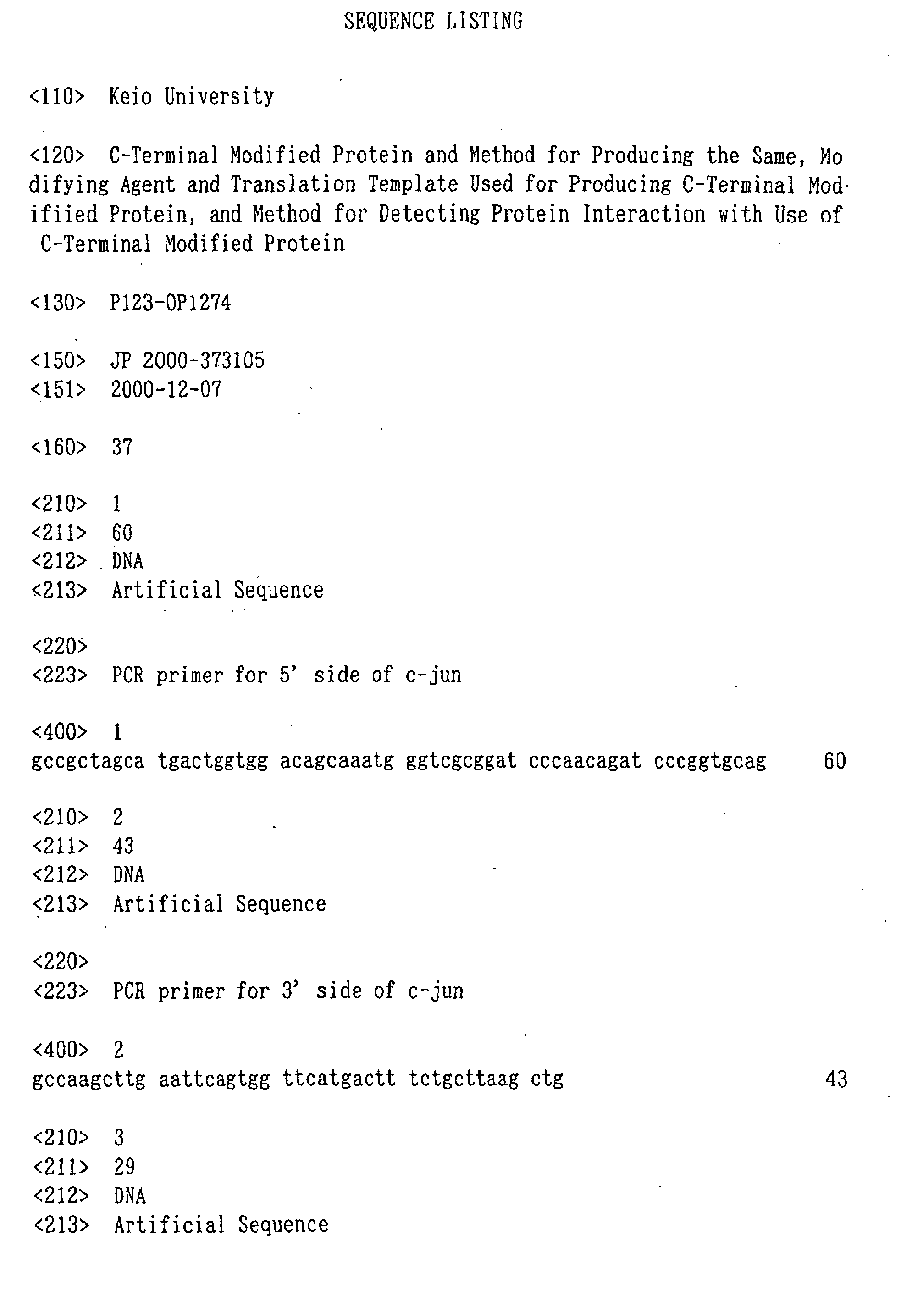
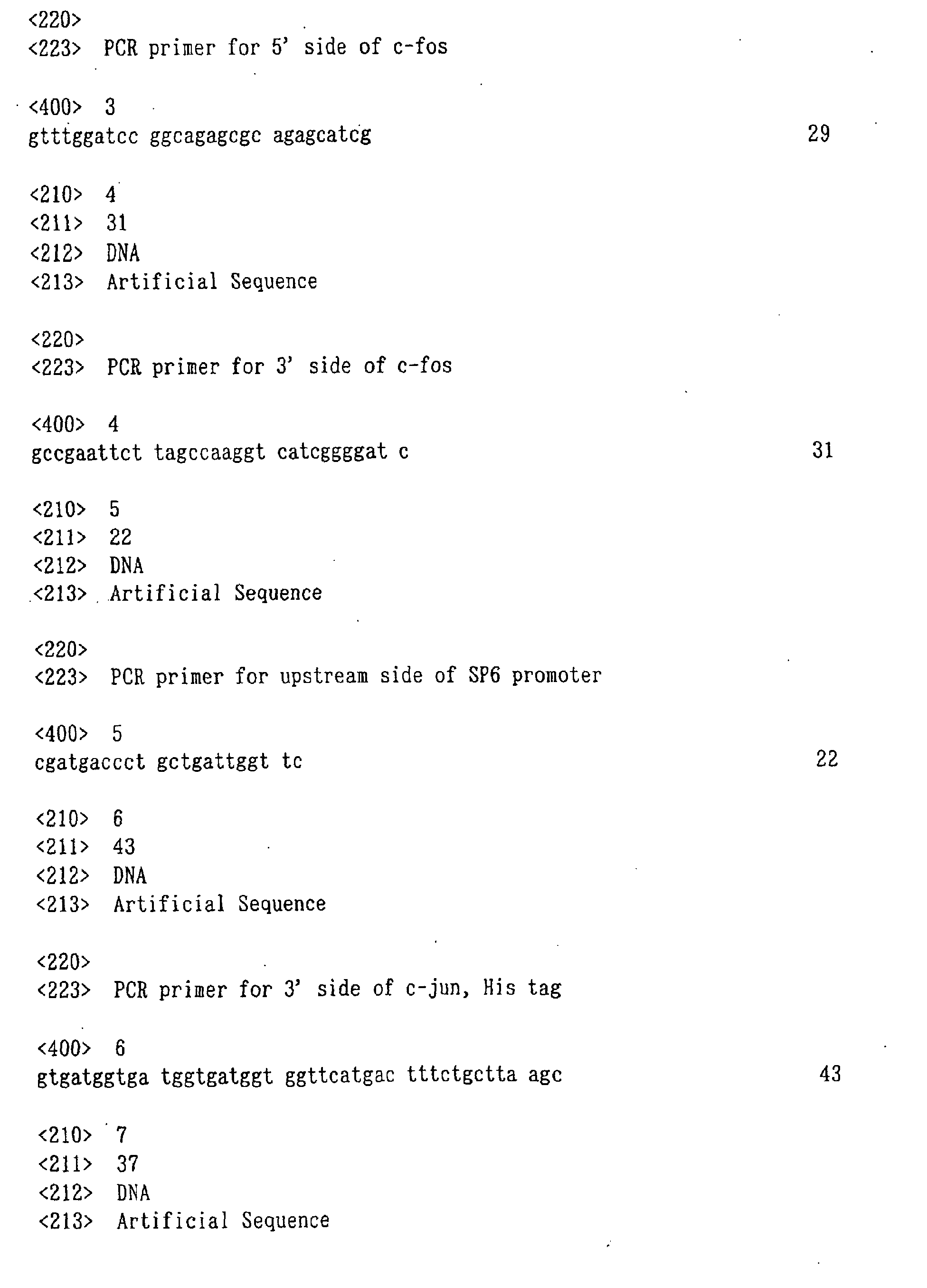
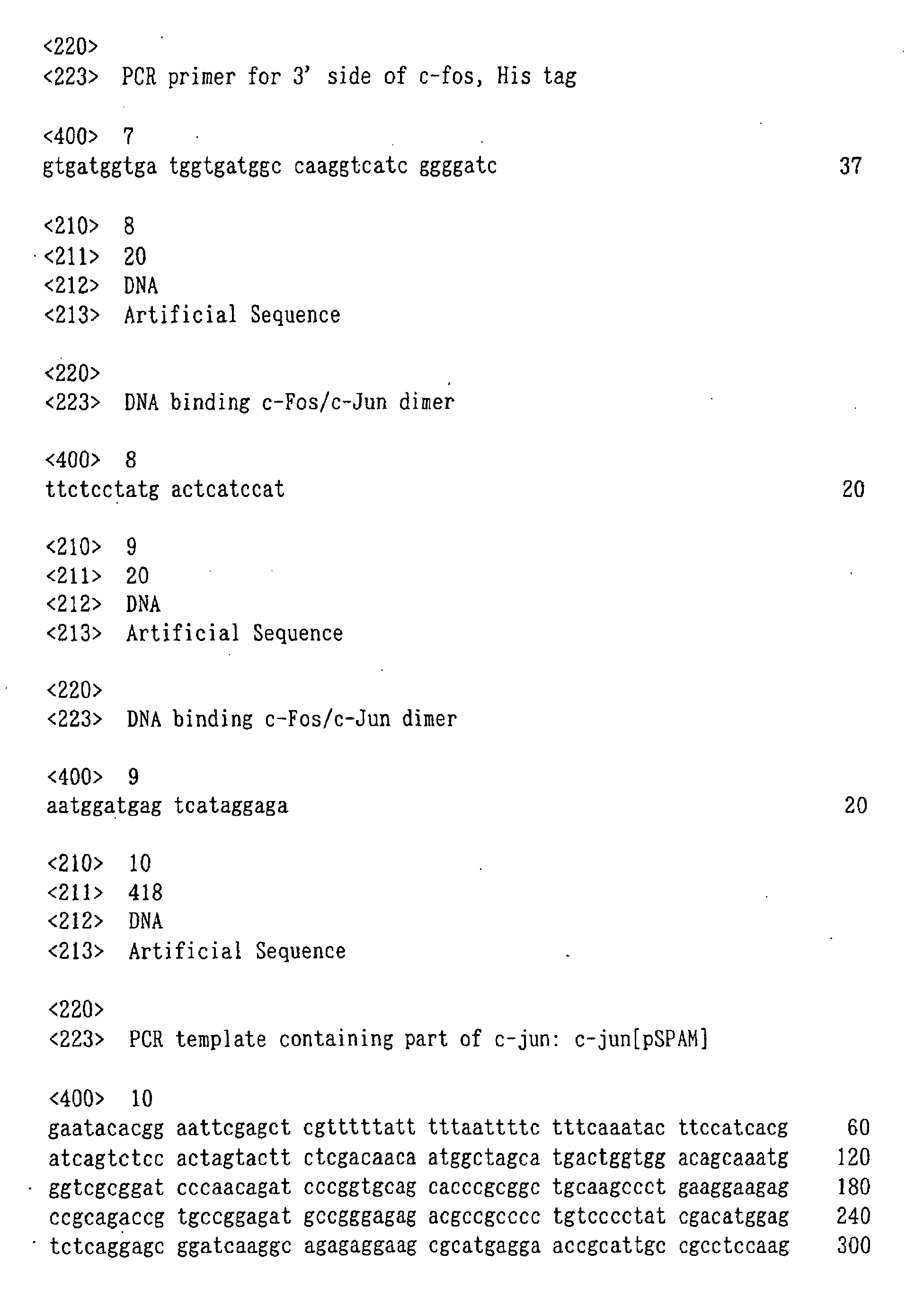


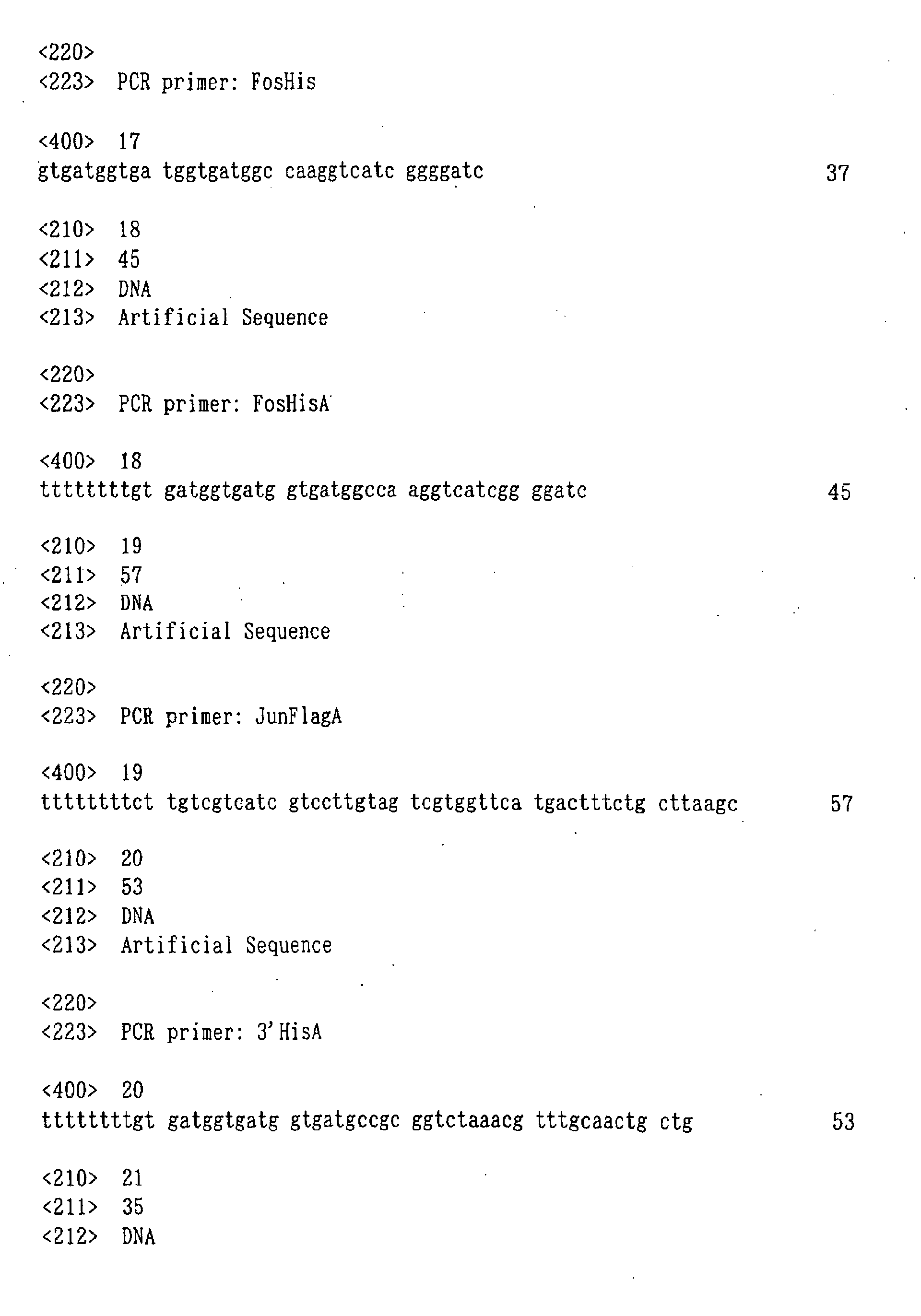
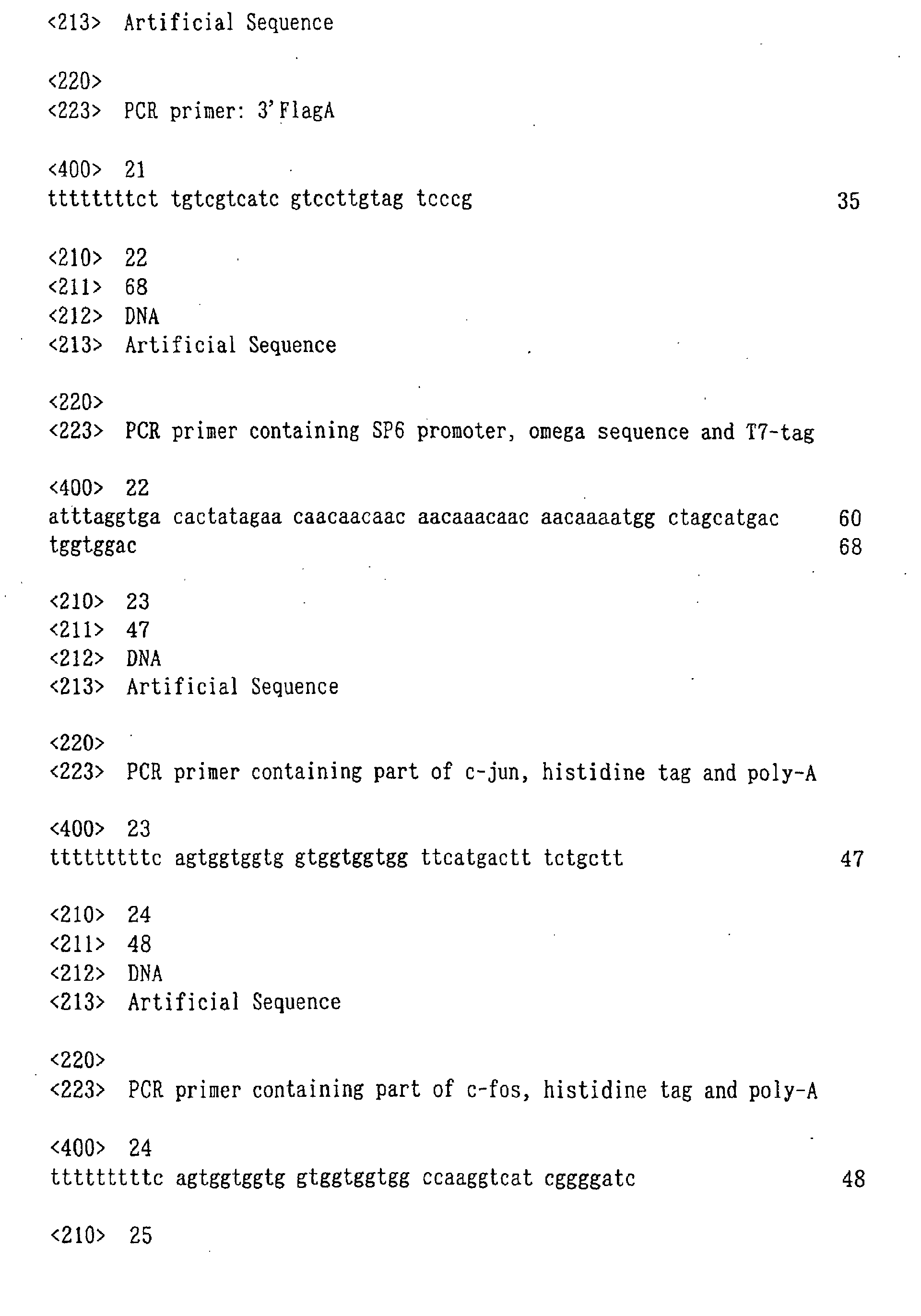


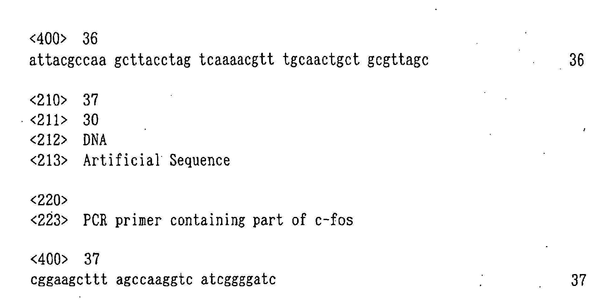
Claims (22)
- An agent for modifying a C-terminal of a protein, which comprises an acceptor portion having a group capable of binding to a protein through a transpeptidation reaction in a protein translation system and a modifying portion comprising a nonradioactive modifying substance linked to the acceptor portion via a nucleotide linker.
- The agent for modifying the C-terminal of the protein according to Claim 1, wherein the acceptor portion has a residue of puromycin or a derivative thereof.
- The agent for modifying the C-terminal of the protein according to Claim 1 or 2, wherein the nucleotide linker is 2'-deoxycytidylic acid.
- The agent for modifying the C-terminal of the protein according to Claim 1 or 2, wherein the nucleotide linker is 2'-deoxycytidyl-(3',5')-2'-deoxycytidylic acid.
- The agent for modifying the C-terminal of the protein according to Claim 1 or 2, wherein the nucleotide linker is ribocytidylic acid.
- The agent for modifying the C-terminal of the protein according to Claim 1 or 2, wherein the nucleotide linker is ribocytidyl-(3',5')-ribocytidylic acid.
- The agent for modifying the C-terminal of the protein according to any one of Claims 1 to 6, wherein the modifying portion has a fluorescent group.
- The agent for modifying the C-terminal of the protein according to any one of Claims 1 to 7, wherein the modifying portion has a group which binds to a protein.
- The agent for modifying the C-terminal of the protein according to any one of Claims 1 to 7, wherein the modifying portion has both of a fluorescent group and a group which binds to a protein.
- A C-terminal modified protein, which is a protein to which the modifying agent as defined in any one of Claims 1 to 9 binds at the C-terminal of the protein.
- The C-terminal modified protein according to Claim 10, wherein the protein to which the modifying agent at the C-terminal of the protein is a full-length protein.
- A translation template comprising an ORF region coding for a protein, a 5' untranslated region comprising a transcription promoter and a translation enhancer and locating on the 5' side of the ORF region, and a 3' end region comprising a poly-A sequence and locating on the 3' side of the ORF region.
- The translation template according to Claim 12, wherein the transcription promoter comprises a promoter sequence of SP6 RNA polymerase, and the translation enhancer comprises a part of omega sequence of tobacco mosaic virus.
- The translation template according to Claim 12 or 13, wherein the ORF region comprises an affinity tag sequence in a downstream portion thereof.
- The translation template according to Claim 14, wherein the affinity tag sequence comprises a His-tag sequence.
- A C-terminal modified protein, which can be obtained by expressing the translation template according to any one of Claims 12 to 15 in a translation system in the presence of the modifying agent according to any one of Claims 1 to 9 to cause protein synthesis.
- A method for producing a C-terminal modified protein, which comprises expressing the translation template as defined in any one of Claims 12 to 15 in a translation system in the presence of the modifying agent as defined in any one of Claims 1 to 9 to cause protein synthesis and purifying the synthesized protein.
- The method according to Claim 17, wherein the purification is performed by affinity chromatography, gel filtration, ion chromatography, electrophoresis, precipitation, dialysis or an arbitrary combination thereof.
- A method for analyzing an interaction between a protein and a target molecule, which utilizes the C-terminal modified protein as defined in Claim 10 or 11, comprising the protein.
- The method according to Claim 19, wherein the interaction is analyzed by fluorescence correlation spectroscopy, fluorescence imaging analysis method, fluorescence resonance energy transfer method, evanescent field molecular imaging method, fluorescence depolarization method, surface plasmon resonance method or enzyme linked immunosorbent assay.
- The method according to Claim 19, wherein the C-terminal modified protein as defined in Claim 10 or 11 is immobilized
- The method according to Claim 19, which comprises adding the C-terminal modified protein as defined in Claim 10 or 11 to an array on which a target molecule is immobilized and detecting the C-terminal modified protein specifically binding with the target molecule.
Applications Claiming Priority (3)
| Application Number | Priority Date | Filing Date | Title |
|---|---|---|---|
| JP2000373105 | 2000-12-07 | ||
| JP2000373105 | 2000-12-07 | ||
| PCT/JP2001/010731 WO2002046395A1 (en) | 2000-12-07 | 2001-12-07 | C-terminal modified protein and process for producing the same, modifying agent and translation template to be used in produfing c-terminal modified protein, and method of detecting protein interaction with the use of c-termial modified protein |
Publications (2)
| Publication Number | Publication Date |
|---|---|
| EP1350846A1 true EP1350846A1 (en) | 2003-10-08 |
| EP1350846A4 EP1350846A4 (en) | 2005-01-26 |
Family
ID=18842546
Family Applications (1)
| Application Number | Title | Priority Date | Filing Date |
|---|---|---|---|
| EP01999645A Withdrawn EP1350846A4 (en) | 2000-12-07 | 2001-12-07 | MODIFIED C-TERMINATION PROTEIN AND PROCESS FOR OBTAINING THE PROTEIN, MODIFICATION AGENT AND TRANSLATION MATRIX NECESSARY FOR THE PRODUCTION OF A MODIFIED C-TERMINATION PROTEIN, AND METHOD FOR DETECTING PROTEIN INTERACTION WITH THE USE OF THE PROTEIN |
Country Status (4)
| Country | Link |
|---|---|
| US (1) | US7150978B2 (en) |
| EP (1) | EP1350846A4 (en) |
| JP (1) | JP3750020B2 (en) |
| WO (1) | WO2002046395A1 (en) |
Cited By (5)
| Publication number | Priority date | Publication date | Assignee | Title |
|---|---|---|---|---|
| WO2006082058A3 (en) * | 2005-02-02 | 2006-11-09 | Univ Bayreuth | Cell-free translation system for the production of (chemically) modified proteins |
| FR2913424A1 (en) * | 2007-03-09 | 2008-09-12 | Cis Bio Internat Sa | CYANINE DERIVATIVES, FLUORESCENT CONJUGATES CONTAINING SAME AND USE THEREOF |
| US7842476B2 (en) | 2002-09-06 | 2010-11-30 | Isogenica Limited | In vitro peptide expression library |
| US8445413B2 (en) | 2004-10-15 | 2013-05-21 | National Institute Of Advanced Industrial Science And Technology | Linker for constructing mRNA-puromycin-protein conjugate |
| US11708573B2 (en) | 2017-02-21 | 2023-07-25 | Illumina, Inc. | Tagmentation using immobilized transposomes with linkers |
Families Citing this family (16)
| Publication number | Priority date | Publication date | Assignee | Title |
|---|---|---|---|---|
| ATE442589T1 (en) * | 2002-07-25 | 2009-09-15 | Aclara Biosciences Inc | DETECTION OF RECEPTOR OLIGOMERIZATION |
| JP4441623B2 (en) * | 2002-12-11 | 2010-03-31 | 学校法人慶應義塾 | Method for producing and using library of mapping molecule and component thereof |
| GB0307559D0 (en) * | 2003-04-02 | 2003-05-07 | Univ Nottingham | Fluorescently tagged ligands |
| DE112004002217B4 (en) | 2003-11-19 | 2017-04-06 | Tokyo University Of Science Foundation | A protein-forming complex with a c-Jun protein, nucleic acid encoding the same and methods using the same |
| DE102005031429A1 (en) * | 2005-07-04 | 2007-01-11 | Heinrich-Heine-Universität Düsseldorf | Method for the selective determination of pathological protein deposits |
| US8416417B2 (en) * | 2008-04-25 | 2013-04-09 | Arizona Board Of Regents For And On Behalf Of Arizona State University | Surface impedance imaging methods and apparatuses |
| KR20110036638A (en) | 2008-07-25 | 2011-04-07 | 리차드 더블유. 와그너 | Protein Screening Method |
| WO2010101157A1 (en) | 2009-03-02 | 2010-09-10 | 日本たばこ産業株式会社 | Method for detecting substance in biological sample |
| JP5686098B2 (en) * | 2009-09-17 | 2015-03-18 | Jsr株式会社 | Dissociation method and dissociator for avidin and biotin derivative |
| US9481882B2 (en) | 2010-03-31 | 2016-11-01 | Tokyo University Of Science Foundation | Constitution of tool for analyzing biomolecular interaction and analysis method using same |
| US9772305B2 (en) | 2011-09-15 | 2017-09-26 | Arizona Board Of Regents On Behalf Of Arizona State University | System and method for small molecule detection |
| JP5896511B2 (en) | 2011-09-28 | 2016-03-30 | 学校法人東京理科大学 | Method for detecting a protein that interacts with a target substance |
| EP2821504A4 (en) * | 2012-02-28 | 2016-02-17 | Univ Tokyo Nat Univ Corp | METHOD FOR DESIGNING DISEASE IN RELATION TO THE QUANTITY OF TDP-43 PRESENT IN CELLS |
| CN106928313B (en) * | 2015-12-31 | 2020-12-11 | 深圳翰宇药业股份有限公司 | Synthesis method of C-terminal modified peptide |
| US12656342B2 (en) | 2018-06-12 | 2026-06-16 | Arizona Board Of Regents On Behalf Of Arizona State University | Adaptation of NAPPA for surface plasmon resonance imaging analyses |
| US20240092820A1 (en) * | 2020-11-25 | 2024-03-21 | Sony Group Corporation | Polymer dyes |
Family Cites Families (12)
| Publication number | Priority date | Publication date | Assignee | Title |
|---|---|---|---|---|
| GB8613481D0 (en) * | 1986-06-04 | 1986-07-09 | Diatech Ltd | Translation of mrna |
| WO1991002076A1 (en) * | 1989-07-31 | 1991-02-21 | Institut Belka Akademii Nauk Sssr | Method for obtaining polypeptides in a cell-free system |
| WO1995003321A1 (en) * | 1993-07-20 | 1995-02-02 | Bionebraska, Inc. | Method for endomodification of proteins |
| EP0769552A4 (en) | 1994-06-27 | 1997-06-18 | Toagosei Co Ltd | ANTISENSE NUCLEIC ACID COMPOUND |
| EP1477561B1 (en) | 1996-10-17 | 2008-12-10 | Mitsubishi Chemical Corporation | Molecule assigning genotype to phenotype and use thereof |
| IL131016A (en) * | 1997-01-21 | 2004-12-15 | Gen Hospital Corp | Selection of proteins using rna-protein fusions |
| JP4240574B2 (en) * | 1998-05-15 | 2009-03-18 | 三菱化学株式会社 | Protein labeling composition and protein labeling method |
| JP4193249B2 (en) * | 1998-11-11 | 2008-12-10 | 三菱化学株式会社 | Method for producing C-terminally labeled protein |
| JP4493125B2 (en) | 1999-05-07 | 2010-06-30 | 独立行政法人理化学研究所 | Method for detecting interacting proteins |
| IL146451A0 (en) * | 1999-07-12 | 2002-07-25 | Phylos Inc | C-terminal protein tagging |
| EP1211514A4 (en) | 1999-08-31 | 2005-03-09 | Mitsubishi Chem Corp | METHOD OF ANALYZING A MUTUAL INTERACTION BETWEEN A PROTEIN AND A MOLECULE |
| JP4963142B2 (en) * | 2000-12-14 | 2012-06-27 | 学校法人慶應義塾 | Genotype-phenotype mapping molecule and its constituents, and method for producing and using mapping molecule |
-
2001
- 2001-12-07 WO PCT/JP2001/010731 patent/WO2002046395A1/en not_active Ceased
- 2001-12-07 EP EP01999645A patent/EP1350846A4/en not_active Withdrawn
- 2001-12-07 JP JP2002548112A patent/JP3750020B2/en not_active Expired - Fee Related
-
2003
- 2003-06-06 US US10/455,453 patent/US7150978B2/en not_active Expired - Lifetime
Cited By (7)
| Publication number | Priority date | Publication date | Assignee | Title |
|---|---|---|---|---|
| US7842476B2 (en) | 2002-09-06 | 2010-11-30 | Isogenica Limited | In vitro peptide expression library |
| US8679781B2 (en) | 2002-09-06 | 2014-03-25 | Isogenica Limited | In vitro peptide expression library |
| US8445413B2 (en) | 2004-10-15 | 2013-05-21 | National Institute Of Advanced Industrial Science And Technology | Linker for constructing mRNA-puromycin-protein conjugate |
| WO2006082058A3 (en) * | 2005-02-02 | 2006-11-09 | Univ Bayreuth | Cell-free translation system for the production of (chemically) modified proteins |
| FR2913424A1 (en) * | 2007-03-09 | 2008-09-12 | Cis Bio Internat Sa | CYANINE DERIVATIVES, FLUORESCENT CONJUGATES CONTAINING SAME AND USE THEREOF |
| US11708573B2 (en) | 2017-02-21 | 2023-07-25 | Illumina, Inc. | Tagmentation using immobilized transposomes with linkers |
| US12234451B2 (en) | 2017-02-21 | 2025-02-25 | Illumina, Inc. | Tagmentation using immobilized transposomes with linkers |
Also Published As
| Publication number | Publication date |
|---|---|
| US7150978B2 (en) | 2006-12-19 |
| WO2002046395A1 (en) | 2002-06-13 |
| JPWO2002046395A1 (en) | 2004-04-08 |
| US20050010028A1 (en) | 2005-01-13 |
| JP3750020B2 (en) | 2006-03-01 |
| EP1350846A4 (en) | 2005-01-26 |
Similar Documents
| Publication | Publication Date | Title |
|---|---|---|
| US7150978B2 (en) | Recombinant template used for producing a carboxy-terminal modified protien and a method of producing a carboxy-terminal modified protein | |
| EP1816192B1 (en) | LINKER FOR CONSTRUCTING mRNA-PUROMYCIN-PROTEIN CONJUGATE | |
| JP5647113B2 (en) | Multi-ligand capture agents and related compositions, methods and systems | |
| US8906700B2 (en) | Methods and compositions for phototransfer | |
| US9334530B2 (en) | Methods for making and imaging arrays that comprise a plurality of different biomolecules | |
| US20090240030A1 (en) | Biomolecular coupling methods using 1,3-dipolar cycloaddition chemistry | |
| KR100927886B1 (en) | Protein shock-oligonucleotide conjugates | |
| JP3942431B2 (en) | Protein-molecule interaction analysis method | |
| JPWO2001016600A1 (en) | Protein-molecular interaction analysis method | |
| JPWO2005062046A1 (en) | Biomolecule detection method and labeling dye and labeling kit used therefor | |
| WO2023049177A1 (en) | Single-molecule protein and peptide sequencing | |
| WO2002074950A1 (en) | Method of analyzing intermolecular interaction | |
| JP2010122071A (en) | Material-immobilized carrier having material immobilized thereon, and method for preparing material-immobilized carrier | |
| JPWO2003048363A1 (en) | Complex of mapping molecule and C-terminal labeled protein, complex of mapping molecule, and protein-protein interaction analysis method using the complex | |
| JPWO2005001086A1 (en) | Immobilized mRNA-puromycin conjugate and uses thereof | |
| JP2002257832A (en) | Protein labeling reagents | |
| JP2004053416A (en) | Method for analyzing protein-molecule interaction using C-terminal labeled protein | |
| JP4747292B2 (en) | Translation templates and libraries thereof, proteins and protein libraries synthesized from them, elements constituting them, and methods for producing and using them | |
| JPWO2007046520A1 (en) | Protein screening method using immobilized puromycin linker | |
| JPWO2005050518A1 (en) | Method of creating interaction map using gene and / or protein database, and software and apparatus for realizing the method | |
| US20060183885A1 (en) | Protein forming complex with c-fos protein, nucleic acid encoding the same and method of using the same | |
| JP6194894B2 (en) | Nucleic acid linker | |
| KR20060003494A (en) | High-throughput activity assay of anthrax lethal element using immunofluorescence |
Legal Events
| Date | Code | Title | Description |
|---|---|---|---|
| PUAI | Public reference made under article 153(3) epc to a published international application that has entered the european phase |
Free format text: ORIGINAL CODE: 0009012 |
|
| 17P | Request for examination filed |
Effective date: 20030702 |
|
| AK | Designated contracting states |
Kind code of ref document: A1 Designated state(s): AT BE CH CY DE DK ES FI FR GB GR IE IT LI LU MC NL PT SE TR |
|
| RBV | Designated contracting states (corrected) |
Designated state(s): CH DE FR GB LI SE |
|
| A4 | Supplementary search report drawn up and despatched |
Effective date: 20041214 |
|
| STAA | Information on the status of an ep patent application or granted ep patent |
Free format text: STATUS: THE APPLICATION HAS BEEN WITHDRAWN |
|
| 18W | Application withdrawn |
Effective date: 20061106 |