TECHNICAL FIELD
-
The present invention relates to a disease- (e.g. heart disease-) associated gene,
and use thereof. More specifically, the present invention relates to a method of
screening for a pharmaceutical, which comprises using the disease-associated gene; an
antisence nucleotide to the disease-associated gene, which is useful as a diagnostic
marker for heart disease such as cardiac myopathy, cardiac infarction, cardiac failure,
angina pectoris; an antibody to the product of the disease-associated gene.
TECHNICAL BACKGROUND
-
Heart failure is considered as insufficient myocardial contraction. Possible
onset mechanisms of heart failure is as follows: disturbance of a heart muscle,
mechanical and functional abnormality of a heart pump, an excessive pressure loaded
by hypertension and pulmonary hypertension, a voluminal overload due to anemia or
acute nephritis to cause a condition of an impossible cardiac output of a blood volume
sufficient to a living body demand. For this condition, compensation mechanisms by a
sympathetic nerve system, nerves - body fluid - endocrine system, and the like start to
work for maintaining the physiological homoeostasis. In the compensation
mechanisms of heart failure, 1) an increase in the aforementioned loads causes an
increase in cardiac contraction force, resulting in cardiac dilatation along with an
extension of a sarcomere, 2) a myocardial contraction unit extends to cause myocardial
hypertrophy, and 3) a neurohumoral factor is activated to compensate for the condition
of the insufficient cardiac output of blood systemically required to develop local
myocardial fibrosis. Fundamentally, these are mechanisms for adaptation to the given
loads; however, there is a case of heart failure enhanced by an insufficient action of the
mechanisms and another case of heart failure become worse by the excessive action.
The action of the compensation mechanisms results in myocardial cell hypertrophy to
cause hypercardia. However, when the aforementioned disturbances or loads continue
chronically, the compensation mechanisms will fail. In this case, the enlarged
myocardial cell cannot receive a sufficient volume of blood to cause ischemia, resulting
in myocardial disturbances such as insufficient myocardial contraction and finally
bringing about heart failure syndrome accompanied by such as lowering of the cardiac
output, cardiovascular disorder, venous stasis, retention of fluid. For treatment of the
syndrome, it is necessary to improve the myocardial cell disturbance, reinforce the
cardiac protecting action, recover from the cardiac hypofunction caused by insufficient
myocardial contraction, and prevent the underlying failure of the biological
compensation or improve an excessive compensation. Drugs currently used for
treatment of the heart failure syndromes are clinically, cardiotonic drugs such as 1) a
cardiotonic glycoside such as digoxin, 2) a sympathetic agent such as dobutamine, 3) a
phosphodiesterase inhibiter such as amrinone, and vasodilatator drugs such as
hydralazine, calcium antagonist, angiotensin I-converting enzyme inhibitor, and
angiotensin II receptor antagonist. For treatment of dilated cardiomyopathy, β-blocker
is used.
-
However, there is no remedy capable of quickly starting the compensation
mechanisms and suppressing the excessive expression of compensation mechanisms or
suppressing compensation failure including apoptosis. It is desired to develop the
remedy for the quick start of the compensation mechanisms and suppression of the
excessive compensation or the compensation failure.
DISCLOSURE OF THE INVENTION
-
The inventors intensively studied to solve the aforementioned problem. As
the result of the study, they found that an mRNA increased its expression at the onset of
heart failure in a rat model of cardiac infarction prepared by a surgery to ligate the
coronary artery. A detailed examination of an expression profile of the mRNA having
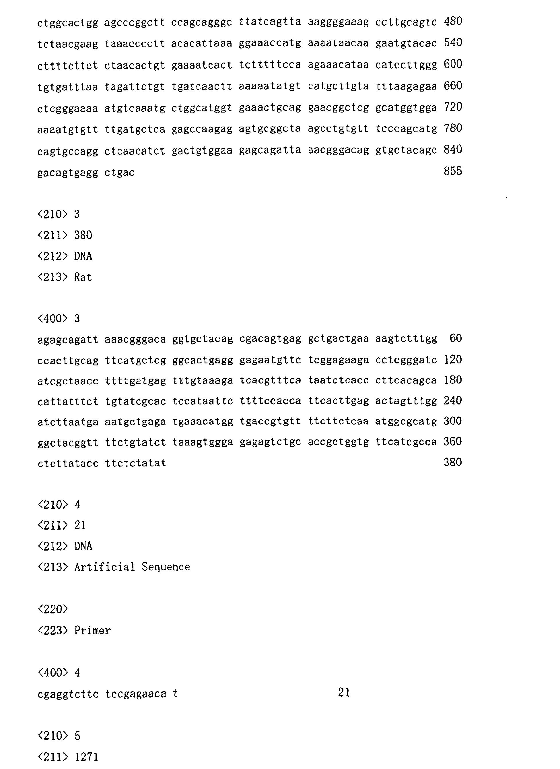
the base sequence represented by SEQ ID NO: 2 showed that this mRNA was increased
slightly 1 week after the surgery, reduced 8 weeks after the surgery, and also increased
distinctly 20 and 30 weeks after the surgery. Homology test of this base sequence to
that of a known gene showed that this is a sheer novel gene. A further study based on
these findings made us achieve the invention.
-
- (1) A protein comprising the same or substantially the same amino acid
sequence as the amino acid sequence shown by SEQ ID NO: 19, or a salt thereof;
- (2) The protein according to (1) or its salt, wherein the amino acid sequence
which is substantially the same as the amino acid sequence shown by SEQ ID NO: 19 is
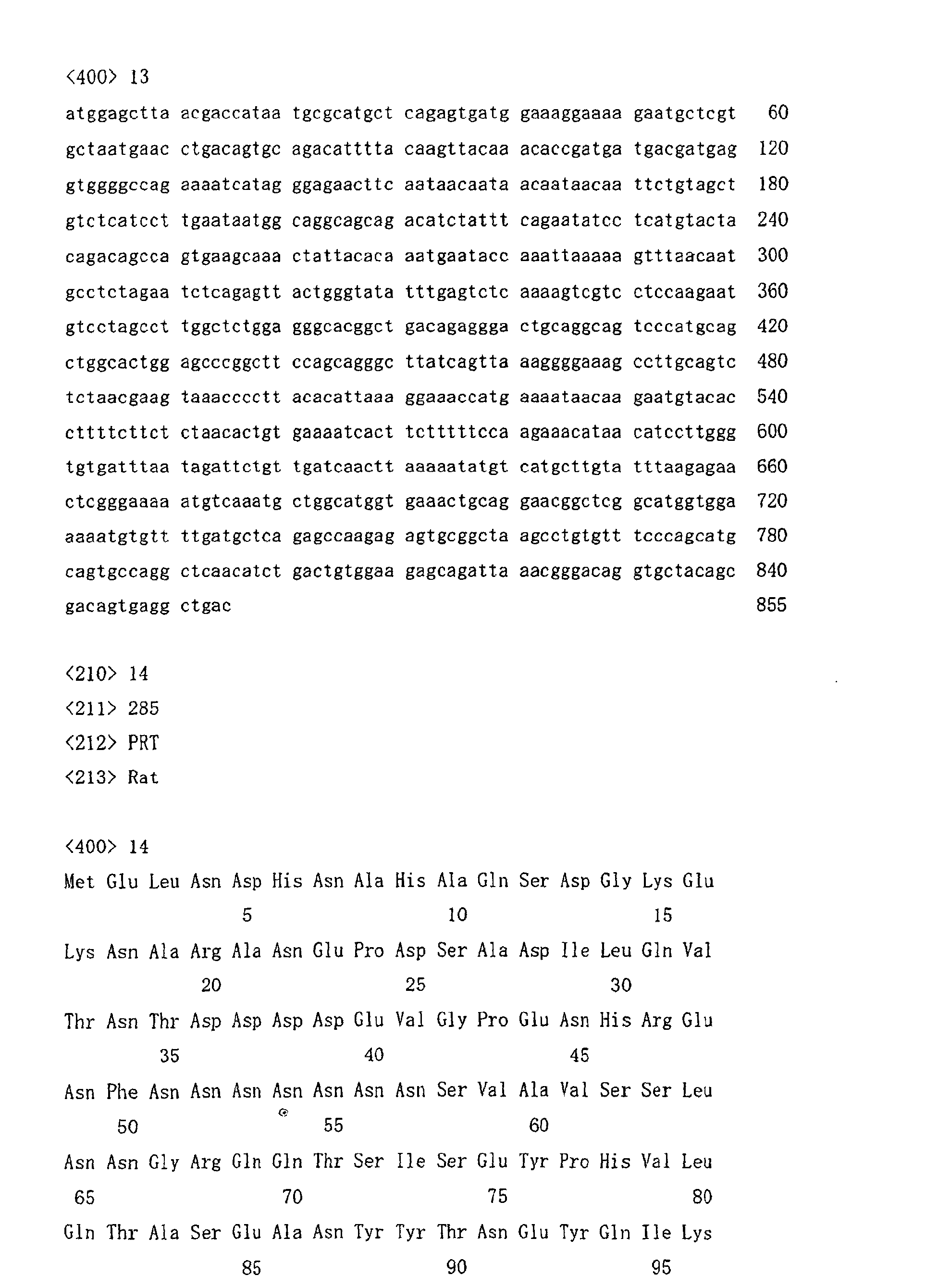
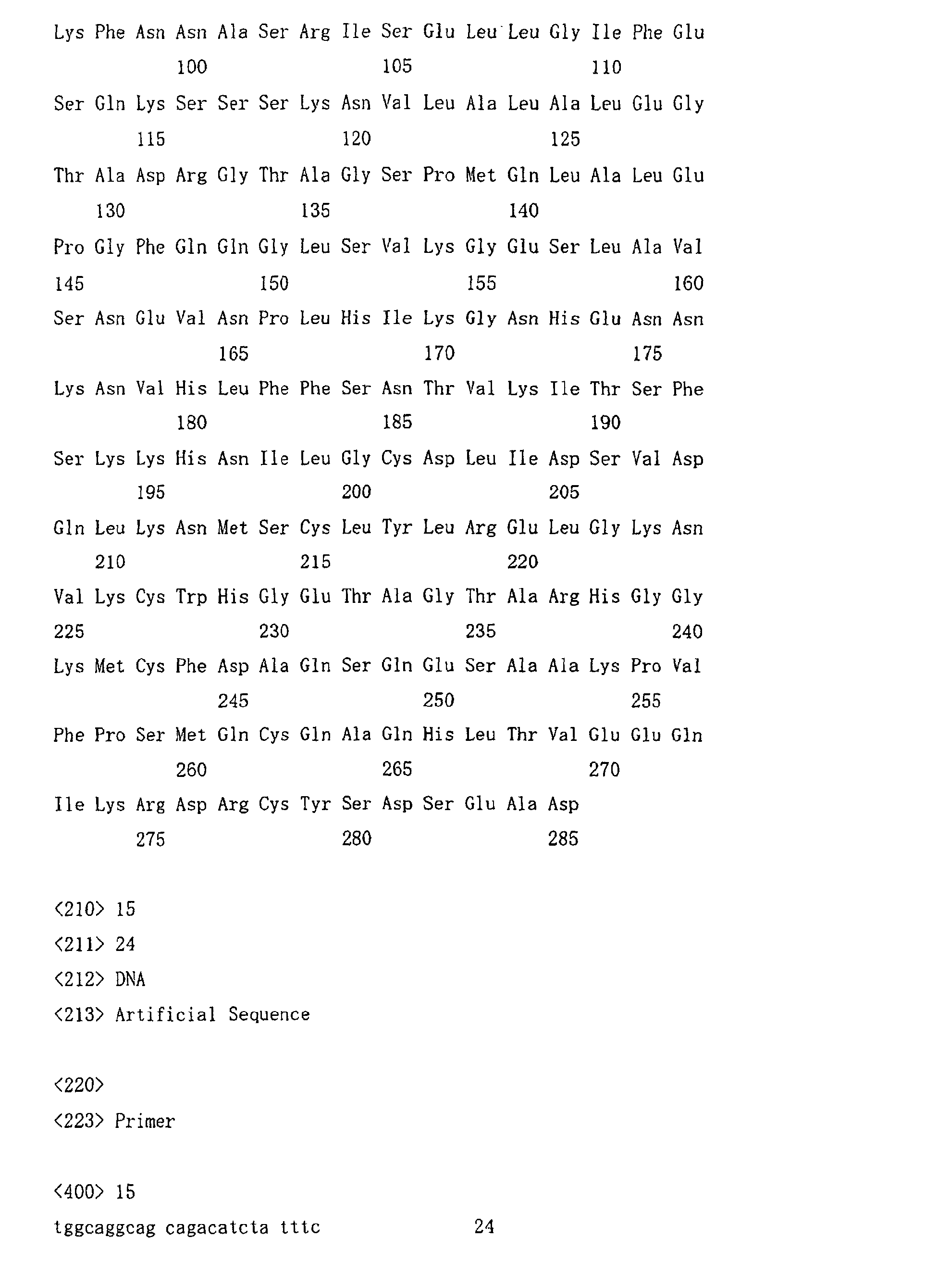
the amino acid sequence represented by SEQ ID NO: 1, NO: 14 or NO: 24;
- (3) The protein according to (1) or its salt, which comprises the amino acid
sequence shown by SEQ ID NO: 19;
- (4) The protein according to (1) or its salt, which comprises the amino acid
sequence shown by SEQ ID NO: 1;
- (5) The protein according to (1) or its salt, which comprises the amino acid
sequence shown by SEQ ID NO: 14;
- (6) The protein according to (1) or its salt, which comprises the amino acid
sequence shown by SEQ ID NO: 24;
- (7) A partial peptide of the protein according to (1), or a salt thereof;
- (8) A polynucleotide comprising a polynucleotide encoding the protein
according to (1) or the partial peptide according to (7);
- (9) The polynucleotide according to (8), which is a DNA;
- (10) The DNA according to (9), which comprises the base sequence
represented by SEQ ID NO: 2, NO: 13, NO: 18 or NO: 25;
- (11) A recombinant vector comprising the polynucleotide according to (8);
- (12) A transformant transformed by the recombinant vector according to (11);
- (13) A method of manufacturing the protein according to (1) or its salt or the
partial peptide according to (7) or its salt, which comprises culturing the transformant
according to (11) to produce and accumulate the protein according to (1) or its salt or
the partial peptide according to (7) or its salt; and collecting it;
- (14) A pharmaceutical composition comprising the protein according to (1) or
its salt, or the partial peptide according to (7) or its salt;
- (15) An antibody to the protein according to (1) or its salt, or the partial peptide
according to (7) or its salt;
- (16) A diagnostic agent comprising the antibody according to (15);
- (17) A method of screening for a compound or its salt regulating an activity of
the protein according to (1) or its salt, or the partial peptide according to (7) or its salt,
which comprises using the protein according to (1) or its salt, or the partial peptide
according to (7) or its salt;
- (18) The screening method according to (17), wherein the protein according to
(1) or its salt, or the partial peptide according to (7) or its salt is expressed in a
cytoplasm of a transformant transformed by a DNA comprising a DNA encoding the
protein according to (1) or the partial peptide according to (7);
- (19) The screening method according to (17), which comprises culturing cells
having a capability of producing the protein according to (1) or its salt in the presence
and the absence of a test compound, and measuring an amount of the expression of the
protein or its salt in the presence and the absence of the test compound, respectively;
- (20) A method of screening for a compound or its salt regulating the expression
of the gene of the protein according to (1), which comprises using a DNA encoding the
protein according to (1);
- (21) A kit for screening for a compound or its salt regulating the activity of the
protein according to (1) or its salt, or the partial peptide according to (7) or its salt,
which comprises the protein according to (1) or its salt, or the partial peptide according
to (7) or its salt;
- (22) A compound or its salt regulating the activity of the protein according to
(1) or its salt, or the partial peptide according to (7) or its salt, which is obtained by the
screening method according to (17) or the screening kit according to (21);
- (23) A compound or its salt regulating the expression of the gene of the protein
according to (1), which is obtained by the screening method according to (20);
- (24) A pharmaceutical composition comprising the compound according to
(22) or (23) or its salt;
- (25) The pharmaceutical composition according to (24), which is a preventive
and/or therapeutic agent for a heart disease;
- (26) An antisense nucleotide having a base sequence complementary or
substantially complementary to a DNA encoding the protein according to (1) or the
partial peptide according to (7);
- (27) A pharmaceutical composition comprising the antisense nucleotide
according to (26);
- (28) A pharmaceutical composition comprising the polynucleotide according to
(8);
- (29) A diagnostic agent comprising the polynucleotide according to (8);
- (30) A preventive and/or therapeutic method for a heart disease in a mammal,
which comprises administering an effective amount of the compound according to (22)
or (23) or its salt to the mammal;
- (31) A use of the compound according to (22) or (23) or its salt for
manufacturing a preventive and/or therapeutic agent for a heart disease.
-
-
Furthermore, the invention provides:
- (32) A non-human, DNA-transfected animal having the DNA encoding the
protein according to (1) or its mutant DNA;
- (33) The animal according to (32), wherein the non-human animal is a rodent;
- (34) The animal according to (33), wherein the rodent is a mouse or a rat;
- (35) An embryonic stem cell of a non-human mammal, in which the DNA of
the invention is inactivated;
- (36) The embryonic stem cell according to (35), in which the DNA is
inactivated by introducing a reporter gene;
- (37) The embryonic stem cell according to (35), which is resistant to
neomycin;
- (38) The embryonic stem cell according to (35), wherein the non-human
mammal is a rodent;
- (39) The embryonic stem cell according to (38), wherein the rodent is a mouse;
- (40) A non-human mammal deficient in expressing the DNA of the invention,
in which the DNA of the invention is inactivated;
- (41) The non-human mammal according to (40), in which the DNA is
inactivated by introducing a reporter gene therein and the reporter gene can be
expressed under the control of a promoter for the DNA of the invention;
- (42) The non-human mammal according to (41), which is a rodent;
- (43) The non-human mammal according to (42), wherein the rodent is a
mouse; and
- (44) A method of screening for a compound or its salt enhancing or inhibiting
the promoter activity for the DNA of the invention, which comprises administering a
test compound to the animal according to (43) and detecting the expression of the
reporter gene.
-
BRIEF DESCRIPTION OF THE DRAWINGS
-
- Fig. 1 shows a tissue distribution of the rat 187-2 gene in a healthy rat by
northern blot analysis, as described in Example 2. In this figure, Nos. 1, 2, 3, 4, 5, 6, 7
and 8 indicate heart, brain, spleen, lung, liver, skeletal muscle, kidney and testis,
respectively.
- Fig. 2 shows changes in the expression of the rat 187-2 cDNA with time in a
rat model of cardiac infarction, which is expressed as a change ratio in "fold-increase",
calculated through division by the expression measured for the sham surgery group.
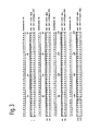
- Fig. 3 shows the amino acid sequence homology between rat 187-2 and a
variant of rat 187-2 (continued to Fig. 4). In this figure, "rat 187-2(T) pro.PRO"
indicates the amino acid sequence of rat 187-2, and "rat 187-2(K).pro" indicates the
amino acid sequence of the variant of rat 187-2.
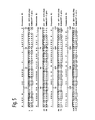
- Fig. 4 shows the amino acid sequence homology between rat 187-2 and a
variant of rat 187-2 (continued from Fig. 3). In this figure, "rat 187-2(T) pro.PRO"
indicates the amino acid sequence of rat 187-2, and "rat 187-2(K).pro" indicates the
amino acid sequence of the variant of rat 187-2.
- Fig. 5 shows the amino acid sequence homology between human 187-2 and rat
187-2 (continued to Fig. 6). In this figure, "rat 187-2(K).PRO" indicates the amino
acid sequence of rat 187-2, and "human 187-2.pro" indicates the amino acid sequence
of human 187-2.
- Fig. 6 shows the amino acid sequence homology between human 187-2 and rat
187-2 (continued from Fig. 5). In this figure, "rat 187-2(K).PRO" indicates the amino
acid sequence of rat 187-2, and "human 187-2.pro" indicates the amino acid sequence
of human 187-2.
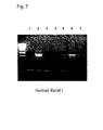
- Fig. 7 shows a tissue distribution of the human 187-2 gene in a healthy human
by RT-PCR analysis, as described in Example 6. In this figure, Nos. 1, 2, 3, 4, 5, 6, 7
and 8 indicate brain, heart, kidney, liver, lung, bronchia and chromosomes, respectively.
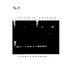
- Fig. 8 shows a tissue distribution of the human 187-2 gene in a healthy human
by RT-PCR analysis, as described in Example 6. In this figure, Nos. 1 to 28 indicate
the following tissues. 1: colon; 2: ovary; 3: peripheral lymphocytes; 4: prostate; 5:
small intestine; 6: spleen; 7: testtis; 8: thymus; 9: fetal brain; 10: fetal heart; 11: fetal
kidney; 12: fetal liver; 13: fetal lung; 14: fetal skeletal muscle; 15: fetal spleen; 16: fetal
thymus; 17: adult heart; 18: fetal heart; 19: aorta; 20: tricuspid valve; 21: left atrium; 22:
right atrium; 23: left auricle; 24: right auricle; 25: left ventricle; 26: right ventricle; 27:
interventricular septum; 28: atrioventricular node.
-
BEST MODE FOR CARRYING OUT THE INVENTION
-
The protein (hereinafter may be referred to the protein of the invention)
comprising the same or substantially the same amino acid sequence as the amino acid
sequence represented by SEQ ID NO: 19 may be any protein derived from any cells of
human or any other warm-blooded animals (e.g., guinea pig, rat, mouse, chicken, rabbit,
swine, sheep, bovine, monkey, etc.) (e.g., hepatocytes, splenocytes, nerve cells, glial
cells, cells of pancreas, bone marrow cells, mesangial cells, Langerhans' cells,
epidermic cells, epithelial cells, beaker cell, endothelial cells, smooth muscle cells,
fibroblasts, fibrocytes, myocytes, fat cells, immune cells (e.g., macrophages, T cells, B
cells, natural killer cells, mast cells, neutrophils, basophils, eosinophils, monocytes),
megakaryocytes, synovial cells, chondrocytes, bone cells, osteoblasts, osteoclasts,
mammary gland cells, hepatocytes or interstitial cells; or the corresponding precursor
cells, stem cells, cancer cells, etc.); or any tissues where such cells are present, such as
brain or any of brain regions (e.g., olfactory bulb, amygdaloid nucleus, basal ganglia,
hippocampus, thalamus, hypothalamus, cerebral cortex, medulla oblongata, cerebellum),
spinal cord, hypophysis, stomach, pancreas, kidney, liver, gonad, thyroid, gall-bladder,
bone marrow, adrenal gland, skin, muscle, lung, gastrointestinal tract (e.g., large
intestine and small intestine), blood vessel, heart, thymus, spleen, submandibular gland,
peripheral blood, prostate, testis, ovary, placenta, uterus, bone, joint, skeletal muscle,
etc.; the proteins may also be synthetic proteins.
-
The amino acid sequence which is substantially the same as the amino acid
sequence represented by SEQ ID NO: 19 includes amino acid sequences having at least
about 50 % homology, preferably at least about 60 % homology, and more preferably at
least about 70 % homology, even more preferably at least about 80 % homology, and
particularly preferably at least about 90 % homology to the amino acid sequence
represented by SEQ ID NO: 19.
-
Specifically, the amino acid sequence which is substantially the same as the
amino acid sequence represented by SEQ ID NO: 19 includes the amino acid sequences
represented by SEQ ID NO: 1, NO: 14, and NO: 24.
-
As a protein comprising substantially the same amino acid sequence as the
amino acid sequence represented by SEQ ID NO: 19, for example, a protein comprising
substantially the same amino acid sequence as the amino acid sequence represented by
SEQ ID NO: 19 and having substantially the same activity in quality as that of a protein
comprising the amino acid sequence represented by SEQ ID NO: 19 is preferred.
-
Such an activity having substantially the same quality is exemplified by the
activity of enhancing the cardiac hypofunction. "Substantially the same quality"
means that the activity is identical in quality (e.g., physiologically or
pharmacologically). Hence, it is preferable that the activity of enhancing the cardiac
hypofunction is the same level (e.g., about 0.01 to 100 folds, preferably about 0.1 to 10
folds, more preferably about 0.5 to 2 folds), however, quantitative factors such as a level
of this activity and a molecular weight of a protein may be different.
-
The activity of enhancing the cardiac hypofunction can be measured by an
echocardiographic device (Cell, Vol. 97: 189-198, 1999) or cardiac function
measurement using a cardiac catheter (Circulation Research 69: 370-377, 1991).
Moreover, the said activity can be determined by, for example, measurement of
activation of renin-angiotensin system (RAS) such as angiotensin I-converting enzyme
(ACE) using a commercial kit (made by e.g. Peninsula Corp., Phoenix Corp., etc.) or
measurement of an increase in blood catecholamine (full automatic catecholamine
analyzer, Toso) as an index.
-
The proteins of the invention also include so-called muteins such as proteins
comprising (i) an amino acid sequence represented by SEQ ID NO: 19, of which one or
more (preferably approximately 1 to 30, more preferably approximately 1 to 10, and
even more preferably 1 to 5) amino acids are deleted; (ii) an amino acid sequence
represented by SEQ ID NO: 19, to which one or more (preferably approximately 1 to 30,
more preferably approximately 1 to 10, and even more preferably 1 to 5) amino acids
are added; (iii) an amino acid sequence represented by SEQ ID NO: 19, into which one
or more (preferably approximately 1 to 30, more preferably approximately 1 to 10, and
even more preferably 1 to 5) amino acids are inserted, (iv) an amino acid sequence
represented by SEQ ID NO: 19, in which one or more (preferably approximately 1 to 30,
more preferably approximately 1 to 10, and even more preferably 1 to 5) amino acids
are substituted by other amino acids; and (v) a combination of the above amino acid
sequences.
-
When such an insertion, deletion or substitution is made in the amino acid
sequence as described above, there is no special limitation to positions for the insertion,
deletion or substitution. For example, these positions include positions other than
those in the consensus sequence of the amino acid sequences represented by SEQ ID
NO: 19, NO: 1, NO: 14 and NO: 24.
-
Throughout the present specification, the proteins are represented in
accordance with the conventional way of describing peptides, that is, with the
N-terminus (amino terminus) on the left side and the C-terminus (carboxyl terminus) on
the right side. In the proteins of the invention including the protein comprising the
amino acid sequence shown by SEQ ID NO: 19, the C-terminus may be in the form of a
carboxyl group (-COOH), a carboxylate (-COO-), an amide (-CONH2) or an ester
(-COOR).
-
Examples of the ester group shown by R include a C1-6 alkyl group such as
methyl, ethyl, n-propyl, isopropyl, n-butyl, etc.; a C3-8 cycloalkyl group such as
cyclopentyl, cyclohexyl, etc.; a C6-12 aryl group such as phenyl, α-naphthyl, etc.; a C7-14
aralkyl such as a phenyl-C1-2 alkyl group, e.g., benzyl, phenethyl, etc., and an
α-naphthyl-C1-2 alkyl group, e.g., α-naphthylmethyl, etc.; pivaloyloxymethyl, and the
like.
-
When the protein of the invention contains a carboxyl group (or a carboxylate)
at a position other than the C-terminus, it may be amidated or esterified and such an
amide or ester is also included within the protein of the invention. The ester group
may be the same group as that described with respect to the above C-terminal group.
-
Furthermore, examples of the protein of the invention include variants of the
above proteins, wherein the amino group at the N-terminus (e.g., methionine residue) of
the protein is protected with a protecting group (e.g., a C1-6 acyl group such as a C1-6
alkanoyl group, e.g., formyl group, acetyl group, etc.); those wherein the N-terminal
region is cleaved in vivo and the glutamyl group thus formed is pyroglutaminated; those
wherein a substituent (e.g., -OH, -SH, amino group, imidazole group, indole group,
guanidino group, etc.) on the side chain of an amino acid in the molecule is protected
with a suitable protecting group (e.g., a C1-6 acyl group such as a C1-6 alkanoyl group,
e.g., formyl group, acetyl group, etc.), or conjugated proteins such as glycoproteins
having sugar chains.
-
The protein of the invention specifically includes the human protein having the
amino acid sequence represented by SEQ ID NO: 19, the rat proteins having the amino
acid sequence represented by SEQ ID NO: 1, NO: 14 and NO: 24.
-
The partial peptides of the protein of the invention (hereinafter occasionally
referred to as the partial peptides of the invention) may be any partial peptides of the
protein of the invention described above, preferably those having a similar activity as
that of the protein of the invention described above.
-
For example, there are employed peptides having sequences of at least 20,
preferably at least 50, more preferably at least 70, much more preferably at least about
100 and most preferably at least about 150 amino acids in the amino acid sequence
which constitutes the protein of the invention.
-
In the partial peptide of the invention, (i) one or more (preferably
approximately 1 to 30, more preferably approximately 1 to 10, and even more
preferably several 1 to 5) amino acids in the amino acid sequence of the partial peptide
may be deleted; (ii) one or more (preferably approximately 1 to 30, more preferably
approximately 1 to 10, and even more preferably several 1 to 5) amino acids may be
added to the amino acid sequence; (iii) one or more (preferably approximately 1 to 30,
more preferably approximately 1 to 10, and even more preferably several 1 to 5) amino
acids may be inserted into the amino acid sequence; or (iv) one or more (preferably
approximately 1 to 30, more preferably approximately 1 to 10, and even more
preferably several 1 to 5) amino acids in the amino acid sequence may be substituted by
other amino acids.
-
A C-terminal of the partial peptides of the invention may be any one of
carboxyl (-COOH,) carboxylate (-COO-,) amide (-CONH2,) or ester (-COOR).
-
The partial peptides of the invention include, as in the aforementioned protein
of the invention, those in which the amino group of N-terminal amino acid residue (e.g.,
methionine residue) has been protected by a protecting group, those in which glutamine
residue, produced by cleavage of the N-terminal region in vivo, has been
pyroglutaminated, those in which a substituent in a side chain of the amino acid in the
molecule has been protected by a suitable protecting group, or a conjugated peptide
such as so-called glycopeptide which has a sugar chain.
-
The partial peptide of the invention can be used as an antigen for producing
antibodies thereto.
-
The salt of the protein or the partial peptide of the invention may be a salt with
physiologically acceptable acids (e.g., inorganic acids, organic acids) or bases (e.g.,
alkaline metal salts), and physiologically acceptable acid addition salts are particularly
preferred. Examples of such salts are salts with inorganic acids (e.g., hydrochloric
acid, phosphoric acid, hydrobromic acid or sulfuric acid), salts with organic acids (e.g.,
acetic acid, formic acid, propionic acid, fumaric acid, maleic acid, succinic acid, tartaric
acid, citric acid, malic acid, oxalic acid, benzoic acid, methanesulfonic acid,
benzenesulfonic acid) and the like.
-
The protein or its partial peptide or its salt of the invention may be
manufactured by publicly known methods used to purify proteins from human or any
other warm-blooded animal tissues or cells described above, or may be manufactured by
culturing transformants containing DNA encoding the protein. The protein or its salt
may also be manufactured by the peptide synthesis methods later described.
-
When the protein or its partial peptide is manufactured from human or any
other warm-blooded animal tissues or cells, these tissues or cells are homogenized, then
the protein or its partial peptide is extracted with an acid or the like, and is isolated and
purified from the obtained extract by a combination of chromatography techniques such
as reversed phase chromatography, ion exchange chromatography, and the like.
-
To synthesize the protein of the invention or the partial peptide, or salts thereof,
or amides thereof, commercially available resins that are used for protein synthesis may
be used. Examples of such resins include chloromethyl resin, hydroxymethyl resin,
benzhydrylamine resin, aminomethyl resin, 4-benzyloxybenzyl alcohol resin,
4-methylbenzhydrylamine resin, PAM resin, 4-hydroxymethylmethylphenyl
acetamidomethyl resin, polyacrylamide resin,
4-(2',4'-dimethoxyphenyl-hydroxymethyl)phenoxy resin,
4-(2',4'-dimethoxyphenyl-Fmoc-aminoethyl) phenoxy resin, etc. Using these resins,
amino acids in which α-amino groups and functional groups on the side chains are
appropriately protected are condensed on the resin in the order of the sequence of the
objective protein according to various condensation methods publicly known. At the
end of the reaction, the protein is excised from the resin and at the same time, the
protecting groups are removed. Then, intramolecular disulfide bond-forming reaction
is performed in a highly diluted solution to obtain the objective protein or amides
thereof.
-
For condensation of the protected amino acids described above, a variety of
activation reagents for protein synthesis may be used, but carbodiimides are particularly
preferably employed. Examples of such carbodiimides include DCC,
N,N'-diisopropylcarbodiimide, N-ethyl-N'-(3-dimethylaminopropyl)carbodiimide, etc.
For activation by these reagents, the protected amino acids in combination with a
racemization inhibitor (e.g., HOBt, HOOBt) are added directly to the resin, or the
protected amino acids are previously activated in the form of symmetric acid anhydrides,
HOBt esters or HOOBt esters, followed by adding the thus activated protected amino
acids to the resin.
-
Solvents used to activate the protected amino acids or condense with the resin
may be chosen from solvents that are known to be usable for protein condensation
reactions. Acid amides such as N,N-dimethylformamide, N,N-dimethylacetamide,
N-methylpyrrolidone, etc.; halogenated hydrocarbons such as methylene chloride,
chloroform, etc.; alcohols such as trifluoroethanol, etc.; sulfoxides such as
dimethylsulfoxide, etc.; ethers such as pyridine, dioxane, tetrahydrofuran, etc.; nitriles
such as acetonitrile, propionitrile, etc.; esters such as methyl acetate, ethyl acetate, etc.;
and appropriate mixtures of these solvents are usable. The reaction temperature is
appropriately chosen from the range known to be applicable to protein binding reactions
and is usually selected in the range of approximately -20°C to 50°C. The activated
amino acid derivatives are used generally in an excess of 1.5 to 4 times. The
condensation is examined using the ninhydrin reaction; when the condensation is
insufficient, the condensation can be completed by repeating the condensation reaction
without removal of the protecting groups. When the condensation is yet insufficient
even after repeating the reaction, unreacted amino acids are acetylated with acetic
anhydride or acetylimidazole to cancel any possible adverse affect on the subsequent
reaction.
-
Examples of the protecting groups used to protect the starting amino groups
include Z, Boc, t-pentyloxycarbonyl, isobornyloxycarbonyl,
4-methoxybenzyloxycarbonyl, Cl-Z, Br-Z, adamantyloxycarbonyl, trifluoroacetyl,
phthaloyl, formyl, 2-nitrophenylsulphenyl, diphenylphosphinothioyl, Fmoc, etc.
-
A carboxyl group can be protected by, e.g., alkyl esterification (in the form of
linear, branched or cyclic alkyl esters of the alkyl moiety such as methyl, ethyl, propyl,
butyl, t-butyl, cyclopentyl, cyclohexyl, cycloheptyl, cyclooctyl, 2-adamantyl, etc.),
aralkyl esterification (e.g., esterification in the form of benzyl ester, 4-nitrobenzyl ester,
4-methoxybenzyl ester, 4-chlorobenzyl ester, benzhydryl ester, etc.), phenacyl
esterification, benzyloxycarbonyl hydrazidation, t-butoxycarbonyl hydrazidation, trityl
hydrazidation, or the like.
-
The hydroxyl group of serine can be protected through, for example, its
esterification or etherification. Examples of groups appropriately used for the
esterification include a lower (C1-6) alkanoyl group, such as acetyl group, an aroyl group
such as benzoyl group, and a group derived from carbonic acid such as
benzyloxycarbonyl group and ethoxycarbonyl group. Examples of a group
appropriately used for the etherification include benzyl group, tetrahydropyranyl group,
t-butyl group, etc.
-
Examples of groups for protecting the phenolic hydroxyl group of tyrosine
include Bzl, Cl2-Bzl, 2-nitrobenzyl, Br-Z, t-butyl, etc.
-
Examples of groups used to protect the imidazole moiety of histidine include
Tos, 4-methoxy-2,3,6-trimethylbenzenesulfonyl, DNP, benzyloxymethyl, Bum, Boc, Trt,
Fmoc, etc.
-
Examples of the activated carboxyl groups in the starting amino acids include
the corresponding acid anhydrides, azides, activated esters [esters with alcohols (e.g.,
pentachlorophenol, 2,4,5-trichlorophenol, 2,4-dinitrophenol, cyanomethyl alcohol,
p-nitrophenol, HONB, N-hydroxysuccimide, N-hydroxyphthalimide, HOBt)]. As the
activated form of amino groups in the starting material, the corresponding phosphoric
amide is employed.
-
To eliminate (split off) the protecting groups, there are used catalytic reduction
under hydrogen gas flow in the presence of a catalyst such as Pd-black or Pd-carbon; an
acid treatment with anhydrous hydrogen fluoride, methanesulfonic acid,
trifluoromethanesulfonic acid or trifluoroacetic acid, or a mixture solution of these
acids; a treatment with a base such as diisopropylethylamine, triethylamine, piperidine
or piperazine; and reduction with sodium in liquid ammonia. The elimination of the
protecting group by the acid treatment described above is carried out generally at a
temperature of approximately -20°C to 40°C. In the acid treatment, it is efficient to
add a cation scavenger such as anisole, phenol, thioanisole, m-cresol, p-cresol,
dimethylsulfide, 1,4-butanedithiol or 1,2-ethanedithiol. Furthermore,
2,4-dinitrophenyl group known as the protecting group for the imidazole of histidine is
removed by a treatment with thiophenol. Formyl group used as the protecting group of
the indole of tryptophan is eliminated by the aforesaid acid treatment in the presence of
1,2-ethanedithiol or 1,4-butanedithiol, as well as by a treatment with an alkali such as a
dilute sodium hydroxide solution and dilute ammonia.
-
Protection of functional groups that should not be involved in the reaction of
the starting materials, protecting groups, elimination of the protecting groups and
activation of functional groups involved in the reaction may be appropriately selected
from publicly known groups and publicly known means.
-
In another method for obtaining the amides of the protein or the partial peptide,
for example, the α-carboxyl group of the carboxy terminal amino acid is first protected
by amidation; the peptide (protein) chain is then extended to amino group for a desired
length. Thereafter, the protein or the partial peptide in which only the protecting group
of the N-terminal α-amino group has been eliminated from the protein or the partial
peptide in which only the protecting group of the C-terminal carboxyl group has been
eliminated are manufactured. The protein or the peptide is condensed in a mixture of
the solvents described above. The details of the condensation reaction are the same as
described above. After the protected protein or the peptide obtained by the
condensation is purified, all the protecting groups are eliminated by the method
described above to give the desired crude protein or peptide. This crude protein or
peptide is purified by various known purification means. Lyophilization of the major
fraction gives the desired amide of the protein or peptide.
-
To prepare the esterified protein or peptide, for example, the α-carboxyl group
of the carboxy terminal amino acid is condensed with a desired alcohol to prepare the
amino acid ester, which is then processed by procedure similar to the preparation of the
amidated protein or peptide above to give the desired esterified protein or peptide.
-
The partial peptide of the invention or a salt thereof can be manufactured by
publicly known methods for peptide synthesis, or by cleaving the protein of the
invention with an appropriate peptidase. For the methods for peptide synthesis, for
example, either solid phase synthesis or liquid phase synthesis may be used. That is, a
partial peptide or amino acids that can construct the partial peptide of the invention can
be condensed with the remaining part of the partial peptide of the invention. When the
product contains protecting groups, these protecting groups are removed to give the
desired peptide. Publicly known methods for condensation and elimination of the
protecting groups are described in the following 1) to 5.)
- 1) M. Bodanszky & M.A. Ondetti: Peptide Synthesis, Interscience Publishers,
New York (1966)
- 2) Schroeder & Luebke: The Peptide, Academic Press, New York (1965)
- 3) Nobuo Izumiya, et al.: Peptide Gosei-no-Kiso to Jikken (Basics and
experiments of peptide synthesis), published by Maruzen Co. (1975)
- 4) Haruaki Yajima & Shunpei Sakakibara: Seikagaku Jikken Koza
(Biochemical Experiment) 1, Tanpakushitsu no Kagaku (Chemistry of Proteins) IV, 205
(1977)
- 5) Haruaki Yajima ed.: Zoku Iyakuhin no Kaihatsu (A sequel to Development
of Pharmaceuticals), Vol. 14, Peptide Synthesis, published by Hirokawa Shoten
-
-
After completion of the reaction, the partial peptide of the invention can be
purified and isolated by a combination of conventional purification methods such as
solvent extraction, distillation, column chromatography, liquid chromatography and
recrystallization to give the partial peptide of the invention. When the partial peptide
obtained by the above methods is in a free form, it can be converted into an appropriate
salt by a publicly known method; when the partial peptide is obtained in a salt form, it
can be converted into a free form or a different salt form by the publicly known method.
-
The polynucleotide encoding the protein of the invention may be any
polynucleotide which comprises the base sequence (DNA or RNA, preferably DNA)
encoding the protein of the invention described above. The polynucleotide may be
DNA or RNA such as mRNA encoding the protein of the invention and may be
double-stranded or single-stranded. The double-stranded polynucleotide may be a
double-stranded DNA, a double-stranded RNA, or a DNA:RNA hybrid. The
single-stranded polynucleotide may be a sense strand (i.e. coding strand) or an antisense
strand (i.e. non-coding strand).
-
Using the polynucleotide encoding the protein of the invention allows
quantification of the mRNA of the protein of the invention by a known method such as
the method described in "Zikken Igaku, Zoukan "New PCR and its application" 15(7),
1997" or a similar method.
-
The DNA encoding the protein of the invention may be any DNA containing
the base sequence encoding the above-described protein of the invention. The DNA
may be derived from a genome DNA, a genome DNA library, cDNAs derived from the
aforementioned tissues and cells, a cDNA library derived from the aforementioned
tissues and cells, or synthetic DNAs.
-
The vector to be used for the library may be any one of bacteriophage, plasmid,
cosmid, phagemid, and the like. In addition, the DNA can be amplified by reverse
transcriptase polymerase chain reaction (hereinafter abbreviated as RT-PCR) from total
RNA or mRNA fraction prepared from the above-described cells or tissues.
-
The DNA encoding the protein of the invention may be, for example, any DNA
comprising the base sequence represented by SEQ ID NO: 18, or any DNA hybridizable
to the DNA having the base sequence represented by SEQ ID NO: 18 under high
stringent conditions and encoding a protein which has a property substantially
equivalent to the property of the protein of the invention.
-
Examples of the DNA that is hybridizable to the DNA comprising the base
sequence represented by SEQ ID NO: 18 under high stringent conditions include DNAs
comprising a base sequence with at least about 70% homology, preferably at least about
80% homology, more preferably at least about 90% homology, and even more
preferably at least about 95% homology to the base sequence represented by SEQ ID
NO: 18.
-
The hybridization can be carried out by publicly known methods or by a
modification thereof, for example, according to the method described in Molecular
Cloning, 2nd Ed., J. Sambrook et al., Cold Spring Harbor Lab. Press, (1989). A
commercially available library may also be used according to the instructions of the
attached manufacturer's protocol. The hybridization can be carried out preferably
under high stringent conditions.
-
The high stringent conditions used herein refer to, for example, a sodium
concentration of about 19 to 40 mM, preferably about 19 to 20 mM and a temperature
of about 50 to 70°C, preferably about 60 to 65°C. In particular, the hybridization
condition in a sodium concentration of about 19 mM at a temperature of about 65°C is
most preferred.
-
More specifically, the DNA encoding the protein comprising the amino acid
sequence represented by SEQ ID NO: 19 may be the DNA comprising the base
sequence represented by SEQ ID NO: 18; the DNA encoding the protein comprising the
amino acid sequence represented by SEQ ID NO: 1 may be the DNA comprising the
base sequence represented by SEQ ID NO: 2; the DNA encoding the protein comprising
the amino acid sequence represented by SEQ ID NO: 14 may be the DNA comprising
the base sequence represented by SEQ ID NO: 13; the DNA encoding the protein
comprising the amino acid sequence represented by SEQ ID NO: 24 may be the DNA
comprising the base sequence represented by SEQ ID NO: 25.
-
The DNA encoding the partial peptide of the invention may be any DNA which
comprises the base sequence encoding the partial peptide of the invention described
above. The DNA may be derived from any of genomic DNAs, genomic DNA library,
cDNAs derived from the cells and tissues described above, cDNA library derived from
the cells and tissues described above and synthetic DNAs.
-
The DNA encoding the partial peptide of the invention may be, for example, a
DNA having a partial base sequence of the DNA having the base sequence represented
by SEQ ID NO: 18, or a DNA having a partial base sequence of a DNA hybridizable to
the DNA having the base sequence represented by SEQ ID NO: 18 under high stringent
conditions and encoding the protein which has the activity substantially equivalent to
the activity of the protein of the invention.
-
Examples of the DNA that is hybridizable to the DNA having the base
sequence represented by SEQ ID NO: 18 under high stringent conditions include DNAs
having a base sequence with at least about 70% homology, preferably at least about
80% homology, more preferably at least about 90% homology, and even more
preferably at least about 95% homology to the base sequence represented by SEQ ID
NO: 18.
-
Methods for the hybridization and the high stringent conditions that can be
used are the same as described above.
-
For cloning of the DNA that completely encodes the protein of the invention or
the partial peptide of the invention (hereinafter sometimes collectively referred to as the
protein of the invention in the following description of cloning and expression of the
DNA encoding these proteins or the like), the DNA may be either amplified by publicly
known PCR using synthetic DNA primers containing a part of the base sequence
encoding the protein of the invention, or the DNA inserted into an appropriate vector
can be selected by hybridization with a labeled DNA fragment or synthetic DNA that
encodes a part or the entire region of the protein of the invention. The hybridization
can be carried out, for example, according to the method described in Molecular
Cloning, 2nd (J. Sambrook et al., Cold Spring Harbor Lab. Press, 1989). The
hybridization may also be performed using commercially available library in
accordance with the protocol described in the attached instructions.
-
Conversion of the base sequence of DNA can be effected by publicly known
methods such as the ODA-LAPCR method, the Gapped duplex method or the Kunkel
method, or modifications thereof, by using publicly known kits available as
Mutan™-super Express Km (TaKaRa Shuzo Co., Ltd.) or Mutan™-K (TaKaRa Shuzo
Co., Ltd.), etc.
-
The cloned DNA encoding the protein of the invention can be used as it is,
depending upon purpose or, if desired, after digestion with a restriction enzyme or after
addition of a linker thereto. The DNA may contain ATG as a translation initiation
codon at the 5' end thereof and TAA, TGA or TAG as a translation termination codon at
the 3' end thereof. These translation initiation and termination codons may also be
added by using an appropriate synthetic DNA adapter.
-
The expression vector for the protein of the invention can be manufactured, for
example, by (a) excising the desired DNA fragment from the DNA encoding the protein
of the invention, (b) and then ligating the DNA fragment with an appropriate expression
vector downstream a promoter in the vector.
-
Examples of the vector include plasmids derived form E. coli (e.g., pBR322,
pBR325, pUC12, pUC13), plasmids derived from Bacillus subtilis (e.g., pUB110, pTP5,
pC194), plasmids derived from yeast (e.g., pSH19, pSH15), bacteriophages such as λ
phage, etc., animal viruses such as retrovirus, vaccinia virus, baculovirus, etc. as well as
pA1-11, pXTl, pRc/CMV, pRc/RSV, pcDNAI/Neo, etc.
-
The promoter used in the present invention may be any promoter if it matches
well with a host to be used for gene expression. In the case of using animal cells as the
host, examples of the promoter include SRα promoter, SV40 promoter, HIV/LTR
promoter, CMV promoter, HSV-TK promoter, etc.
-
Among them, CMV (cytomegalovirus) promoter or SRα promoter is preferably
used. When bacteria of the genus Escherichia are used as the host, preferred examples
of the promoter include trp promoter, lac promoter, recA promoter, λPL promoter, lpp
promoter, T7 promoter, etc. When bacteria of the genus Bacillus are used as the host,
preferred example of the promoter are SPO1 promoter, SPO2 promoter and penP
promoter. When yeast is used as the host, preferred examples of the promoter are
PHO5 promoter, PGK promoter, GAP promoter and ADH promoter. When insect cells
are used as the host, preferred examples of the promoter include polyhedrin promoter,
P10 promoter, etc.
-
In addition to the foregoing examples, the expression vector may further
optionally contain an enhancer, a splicing signal, a poly A addition signal, a selection
marker, SV40 replication origin (hereinafter sometimes abbreviated as SV40ori) etc.
Examples of the selection marker include dihydrofolate reductase (hereinafter
sometimes abbreviated as dhfr) gene [methotrexate (MTX) resistance], ampicillin
resistant gene (hereinafter sometimes abbreviated as Ampr), neomycin resistant gene
(hereinafter sometimes abbreviated as Neo, G418 resistance), etc. In particular, when
dhfr gene is used as the selection marker together with dhfr gene-deficient Chinese
hamster cells, selection can also be made on thymidine-free media.
-
If necessary, a signal sequence that matches with a host is added to the
N-terminus of the protein of the invention. Examples of the signal sequence that can
be used are Pho A signal sequence, OmpA signal sequence, etc. in case of using bacteria
of the genus Escherichia as the host; α-amylase signal sequence, subtilisin signal
sequence, etc. in case of using bacteria of the genus Bacillus as the host; MFα signal
sequence, SUC2 signal sequence, etc. in case of using yeast as the host; and insulin
signal sequence, α-interferon signal sequence, antibody molecule signal sequence, etc.
in case of using animal cells as the host, respectively.
-
Using the vector comprising the DNA encoding the protein of the invention
thus constructed, transformants can be manufactured.
-
Examples of the host, which may be employed, are bacteria belonging to the
genus Escherichia, bacteria belonging to the genus Bacillus, yeast, insect cells, insects
and animal cells, etc.
-
Specific examples of the bacteria belonging to the genus Escherichia include
Escherichia coli K12 DH1 [Proc. Natl. Acad. Sci. U.S.A., 60, 160 (1968)], JM103
[Nucleic Acids Research, 9, 309 (1981)), JA221 (Journal of Molecular Biology, 120,
517 (1978)], HB101 [Journal of Molecular Biology, 41, 459 (1969)), C600 (Genetics,
39, 440 (1954)], etc.
-
Examples of the bacteria belonging to the genus Bacillus include Bacillus
subtilis MI114 [Gene, 24, 255 (1983)], 207-21 (Journal of Biochemistry, 95, 87 (1984)],
etc.
-
Examples of yeast include Saccharomyces cereviseae AH22, AH22R-,
NA87-11A, DKD-5D, 20B-12, Schizosaccharomyces pombe NCYC1913, NCYC2036,
Pichia pastoris KM71, etc.
-
Examples of insect cells include, for the virus AcNPV, Spodoptera frugiperda
cell (Sf cell), MG1 cell derived from mid-intestine of Trichoplusia ni, High Five™ cell
derived from egg of Trichoplusia ni, cells derived from Mamestra brassicae, cells
derived from Estigmena acrea, etc.; and for the virus BmNPV, Bombyx mori N cell
(BmN cell), etc. is used. Examples of the Sf cell which can be used are Sf9 cell
(ATCC CRL1711) and Sf21 cell (both cells are described in Vaughn, J. L. et al., In Vivo,
13, 213-217 (1977).
-
As the insect, for example, a larva of Bombyx mori can be used (Maeda et al.,
Nature, 315, 592, 1985).
-
Examples of animal cells include monkey cell COS-7, Vero, Chinese hamster
cell CHO (hereinafter referred to as CHO cell), dhfr gene-deficient Chinese hamster cell
CHO (hereinafter simply referred to as CHO(dhfr-) cell), mouse L cell, mouse AtT-20,
mouse myeloma cell, rat GH 3, human FL cell, H9c2 cell, etc.
-
Bacteria belonging to the genus Escherichia can be transformed, for example,
by the method described in Proc. Natl. Acad. Sci. U.S.A., 69, 2110 (1972), Gene, 17,
107 (1982), etc.
-
Bacteria belonging to the genus Bacillus can be transformed, for example, by
the method described in Molecular & General Genetics, 168, 111 (1979).
-
Yeast can be transformed, for example, by the method described in Methods in
Enzymology, 194, 182-187 (1991) or Proc. Natl. Acad. Sci. U.S.A., 75, 1929 (1978),
etc.
-
Insect cells or insects can be transformed, for example, according to the
method described in Bio/Technology, 6, 47-55(1988), etc.
-
Animal cells can be transformed, for example, according to the method
described in Saibo Kogaku (Cell Engineering), extra issue 8, Shin Saibo Kogaku Jikken
Protocol (New Cell Engineering Experimental Protocol), 263-267 (1995), published by
Shujunsha, or Virology, 52, 456 (1973).
-
Thus, the transformant, which is transformed with the expression vector
comprising the DNA encoding the protein, can be obtained.
-
When the host is a bacterium belonging to the genus Escherichia or the genus
Bacillus, the transformant can be appropriately cultured in a liquid medium which
contains materials required for growth of the transformant such as carbon sources,
nitrogen sources, inorganic materials, etc. Examples of the carbon sources include
glucose, dextrin, soluble starch, sucrose, etc. Examples of the nitrogen sources include
inorganic or organic materials such as ammonium salts, nitrate salts, corn steep liquor,
peptone, casein, meat extract, soybean cake, potato extract, etc. Examples of the
inorganic materials are calcium chloride, sodium dihydrogenphosphate, magnesium
chloride, etc. In addition, yeast extracts, vitamins, growth-stimulating factors etc. may
also be added to the medium. Preferably, pH of the medium is adjusted to about 5 to
about 8.
-
A preferred example of the medium for culturing bacteria belonging to the
genus Escherichia is M9 medium supplemented with glucose and casamino acids
(Miller, Journal of Experiments in Molecular Genetics, 431-433, Cold Spring Harbor
Laboratory, New York, 1972). If necessary, a chemical such as 3β-indolylacrylic acid
can be added to the medium thereby to activate the promoter efficiently.
-
When a bacterium belonging to the genus Escherichia is used as the host, the
transformant is usually cultivated at about 15 to 43°C for about 3 to 24 hours. If
necessary, the culture may be aerated or agitated.
-
When a bacterium belonging to the genus Bacillus is used as the host, the
transformant is cultivated generally at about 30 to 40°C for about 6 to 24 hours. If
necessary, the culture can be aerated or agitated.
-
When yeast is used as the host, the transformant is cultivated, for example, in
Burkholder's minimal medium (Bostian, K. L. et al., Proc. Natl. Acad. Sci. U.S.A., 77,
4505, 1980) or in SD medium supplemented with 0.5% Casamino acids (Bitter, G. A. et
al., Proc. Natl. Acad. Sci. U.S.A., 81, 5330, 1984). Preferably, pH of the medium is
adjusted to about 5 to about 8. In general, the transformant is cultivated at about 20 to
35°C for about 24 to 72 hours. If necessary, the culture can be aerated or agitated.
-
When an insect cell or insect is used as the host, the transformant is cultivated
in, for example, Grace's Insect Medium (Grace, T. C. C., Nature, 195, 788 (1962)) to
which an appropriate additive such as 10 % inactivated bovine serum is added.
Preferably, pH of the medium is adjusted to about 6.2 to about 6.4. Generally, the
transformant is cultivated at about 27°C for about 3 days to about 5 days and, if
necessary, the culture can be aerated or agitated.
-
When an animal cell is employed as the host, the transformant is cultivated in,
for example, MEM medium (Science, 122, 501 (1952)], DMEM medium (Virology, 8,
396 (1959)], RPMI 1640 medium (The Journal of the American Medical Association,
199, 519 (1967)), or 199 medium (Proceeding of the Society for the Biological
Medicine, 73, 1 (1950)), which contains about 5% to about 20% fetal bovine serum.
Preferably, pH of the medium is adjusted to about 6 to about 8. The transformant is
usually cultivated at about 30 to 40°C for about 15 to 60 hours and, if necessary, the
culture can be aerated or agitated.
-
As described above, the protein of the invention can be produced inside or
outside of the transformant cell.
-
The protein of the invention can be separated and purified from the culture
described above by the following procedures.
-
When the protein of the invention is extracted from the cultured bacteria or
cells, after cultivation the bacteria or cells are collected by a publicly known method
and suspended in an appropriate buffer. The bacteria or cells are then disrupted by
publicly known methods such as ultrasonication, a treatment with lysozyme and/or
freeze-thaw cycling, followed by centrifugation, filtration, etc. Thus, the crude
proteinous extract can be obtained. The buffer used for the procedures may contain a
protein modifier such as urea or guanidine hydrochloride, or a surfactant such as Triton
X-100™, etc. When the protein of the invention is secreted into the culture broth,
after completion of the cultivation the supernatant can be separated from the bacteria or
cells to collect the supernatant by publicly known methods.
-
The protein of the invention contained in the supernatant or the extract thus
obtained can be purified by an appropriate combination of the publicly known methods
for separation and purification. Such publicly known methods for separation and
purification include a method utilizing difference in solubility such as salting out,
solvent precipitation, etc.; a method mainly utilizing difference in molecular weight
such as dialysis, ultrafiltration, gel filtration, SDS-polyacrylamide gel electrophoresis,
etc.; a method utilizing difference in electric charge such as ion exchange
chromatography, etc.; a method utilizing difference in specific affinity such as affinity
chromatography, etc.; a method utilizing difference in hydrophobicity such as reversed
phase high performance liquid chromatography, etc.; a method utilizing difference in
isoelectric point such as isoelectrofocusing electrophoresis; and the like.
-
When the protein of the invention thus obtained is in a free form, it can be
converted into a salt form by publicly known methods or modifications thereof. On
the other hand, when the protein is obtained in a salt form, it can be converted into the
free form or a different salt by publicly known methods or modifications thereof.
-
The protein of the invention produced by the recombinant can be treated,
before or after the purification, with an appropriate protein-modifying enzyme so that
the protein can be appropriately modified or partially deleted. Examples of the
protein-modifying enzyme include trypsin, chymotrypsin, arginyl endopeptidase,
protein kinase, glycosidase and the like.
-
The presence of the thus produced protein of the invention can be determined
by an enzyme immunoassay using a specific antibody, western blotting method, or the
like.
-
Antibodies to the protein of the invention, its partial peptide, or salts thereof,
may be any polyclonal antibodies or monoclonal antibodies, as long as they are capable
of recognizing the protein of the invention, its partial peptide, or salts thereof.
-
The antibodies to the protein of the invention, its partial peptide, or salts
thereof (hereinafter in the description of antibodies, these terms are occasionally
referred to simply as the protein of the invention) may be manufactured by publicly
known methods for manufacturing antibodies or antisera, using as an antigen the protein
of the invention.
[Preparation of monoclonal antibody]
(a) Preparation of monoclonal antibody-producing cells
-
The protein of the invention is administered to warm-blooded animals either
solely or together with carriers or diluents to the site where the production of antibody is
possible by the administration. In order to potentiate the antibody productivity upon
the administration, complete Freund's adjuvants or incomplete Freund's adjuvants may
be administered. The administration is usually carried out once every two to six weeks
and two to ten times in total. Examples of the applicable warm-blooded animals are
monkeys, rabbits, dogs, guinea pigs, mice, rats, sheep, goats and chickens, with the use
of mice and rats being preferred.
-
In the preparation of monoclonal antibody-producing cells, a warm-blooded
animal, e.g., mouse, immunized with an antigen wherein the antibody titer is noted is
selected, then spleen or lymph node is collected after two to five days from the final
immunization and antibody-producing cells contained therein are fused with myeloma
cells from homozoic or heterozoic animal to give monoclonal antibody-producing
hybridomas. Measurement of the antibody titer in antisera may be carried out, for
example, by reacting a labeled protein, which will be described later, with the antiserum
followed by assaying the binding activity of the labeling agent bound to the antibody.
The fusion may be carried out, for example, by the known method by Koehler and
Milstein [Nature, 256, 495 (1975)]. Examples of the fusion promoter are polyethylene
glycol (PEG), Sendai virus, etc., of which PEG is preferably employed.
-
Examples of the myeloma cells are those collected from warm-blooded animals
such as NS-1, P3U1, SP2/0, AP-1, etc. In particular, P3U1 is preferably employed.
A preferred ratio of the count of the antibody-producing cells used (spleen cells) to the
count of myeloma cells is within a range of approximately 1:1 to 20:1. When PEG
(preferably, PEG 1000 to PEG 6000) is added in a concentration of approximately 10 to
80% followed by culturing at 20 to 40°C, preferably at 30 to 37°C for 1 to 10 minutes,
an efficient cell fusion can be carried out.
-
Various methods can be used for screening of a monoclonal
antibody-producing hybridoma. Examples of such methods include a method which
comprises adding the supernatant of hybridoma to a solid phase (e.g., microplate)
adsorbed with the protein (protein) as an antigen directly or together with a carrier,
adding an anti-immunoglobulin antibody (where mouse cells are used for the cell fusion,
anti-mouse immunoglobulin antibody is used) labeled with a radioactive substance or an
enzyme or Protein A and detecting the monoclonal antibody bound to the solid phase,
and a method which comprises adding the supernatant of hybridoma to a solid phase
adsorbed with an anti-immunoglobulin antibody or Protein A, adding the protein labeled
with a radioactive substance or an enzyme and detecting the monoclonal antibody
bound to the solid phase; etc.
-
The monoclonal antibody can be selected according to publicly known
methods or their modifications. In general, the selection can be effected in a medium
for animal cells supplemented with HAT (hypoxanthine, aminopterin and thymidine).
Any selection and growth medium can be employed as far as the hybridoma can grow
there. For example, RPMI 1640 medium containing 1% to 20%, preferably 10% to
20% fetal bovine serum, GIT medium (Wako Pure Chemical Industries, Ltd.) containing
1% to 10% fetal bovine serum, a serum free medium for cultivation of a hybridoma
(SFM-101, Nissui Seiyaku Co., Ltd.) and the like can be used for the selection and
growth medium. The cultivation is carried out generally at 20°C to 40°C, preferably at
37°C, for about 5 days to about 3 weeks, preferably 1 to 2 weeks, normally in 5% CO2.
The antibody titer of the culture supernatant of a hybridoma can be determined as in the
assay for the antibody titer in antisera described above.
(b) Purification of monoclonal antibody
-
Separation and purification of a monoclonal antibody can be carried out by
publicly known methods, such as separation and purification of immunoglobulins (for
example, salting-out, alcohol precipitation, isoelectric point precipitation,
electrophoresis, adsorption and desorption with ion exchangers (e.g., DEAE),
ultracentrifugation, gel filtration, or a specific purification method which comprises
collecting only an antibody with an activated adsorbent such as an antigen-binding solid
phase, Protein A or Protein G, and dissociating the binding to obtain the antibody].
[Preparation of polyclonal antibody]
-
The polyclonal antibody of the invention can be manufactured by publicly
known methods or modifications thereof. For example, a warm-blooded animal is
immunized with an immunogen (protein of the invention as an antigen) per se, or a
complex of immunogen and a carrier protein is formed and a warm-blooded animal is
immunized with the complex in a manner similar to the method described above for the
manufacture of monoclonal antibodies. The product containing the antibody to the
protein of the invention is collected from the immunized animal followed by separation
and purification of the antibody.
-
In the complex of immunogen and carrier protein used to immunize a
warm-blooded animal, the type of carrier protein and the mixing ratio of carrier to
hapten may be any type and in any ratio, as long as the antibody is efficiently produced
to the hapten immunized by crosslinking to the carrier. For example, bovine serum
albumin, bovine thyroglobulin or hemocyanin is coupled to hapten in a carrier-to-hapten
weight ratio of approximately 0.1 to 20, preferably about 1 to about 5.
-
A variety of condensation agents can be used for the coupling of carrier to
hapten. Glutaraldehyde, carbodiimide, maleimide-activated ester, activated ester
reagents containing thiol group or dithiopyridyl group, and others are used for the
coupling.
-
The condensation product is administered to warm-blooded animals either
solely or together with carriers or diluents to the site that can produce the antibody by
the administration. In order to potentiate the antibody productivity upon the
administration, complete Freund's adjuvant or incomplete Freund's adjuvant may be
administered. The administration is usually made once every about 2 to 6 weeks about
3 to 10 times in total.
-
The polyclonal antibody can be collected from the blood, ascites, etc.,
preferably from the blood of warm-blooded animals immunized by the method
described above.
-
The polyclonal antibody titer in antiserum can be assayed by the same
procedure as that for the determination of serum antibody titer described above. The
separation and purification of the polyclonal antibody can be carried out, following the
method for the separation and purification of immunoglobulins performed as in the
separation and purification of monoclonal antibodies described above.
-
An antisense nucleotide having a base sequence complementary or
substantially complementary to the DNA encoding the protein or the partial peptide of
the invention (hereinafter, the DNA may be referred to as the DNA of the invention)
may be any antisense nucleotide having a base sequence complementary or substantially
complementary to the DNA of the invention, or a partial base sequence thereof, and
having an activity to inhibit the expression of the DNA; an antisense DNA is preferred.
-
The base sequence substantially complementary to the DNA of the invention
includes base sequences having at least about 70%, preferably at least about 80%, more
preferably at least about 90%, and most preferably at least about 95% homology to the
whole or a part of the base sequence complementary to the DNA of the invention (i.e. a
complementary strand of the DNA of the invention). Particularly, a preferable
antisense nucleotide is one having at least about 70%, preferably at least about 80%,
more preferably at least about 90%, and most preferably at least about 95% homology
to the complementary strand of the partial base sequence (e.g. the base sequence around
an initiation codon) encoding the N-terminal region of the protein of the invention
within the whole base sequence of the complementary strand of the DNA of the
invention.
-
The antisense nucleotide may consist of usually about 10 to 40 and preferably
15 to 30 bases.
-
To prevent the degradation by hydrolase such as nuclease, phosphate residues
(phosphates) of respective nucleotides constituting the antisense nucleotide may be
substituted with chemically modified phosphate residues such as phosphorothioate,
methyl phosphonate, phosphorodithionate, etc. These antisense nucleotides can be
manufactured by using a publicly known DNA synthesizer.
-
According to the invention, an antisense polynucleotide (nucleic acid) capable
of inhibiting the replication or expression of the gene of the protein of the invention can
be designed and synthesized on the basis of a base sequence information on the DNA
encoding the protein, which is cloned or determined. Such polynucleotide (nucleic
acid) can be hybridized with the RNA of the gene of the protein of the invention, and
can inhibit synthesis or a function of the RNA or can regulate and control the expression
of the gene of the protein of the invention through an interaction with an RNA
associated with the protein of the invention. A polynucleotide complementary to a
selected sequence of the RNA associated with the protein of the invention and a
polynucleotide hybridizable specifically with the RNA associated with the protein of the
invention are useful for regulating and controlling in vivo and in vitro expression of the
gene of the protein of the invention and also useful for therapy and diagnosis of diseases.
A term "corresponding" means "having homology" or "being complementary" to a
given nucleotide sequence, a base sequence, or a nucleic acid sequence, including the
gene. "Correspondence" between a peptide (protein) and a nucleotide sequence, base
sequence, or nucleic acid sequence means usually that the nucleotide (nucleic acid)
sequence or the complementary sequence thereto defines the amino acid sequence of the
peptide (protein). Preferred target regions include a 5'-end hairpin loop, 5'-end
6-base-pair repeats, 5'-end untranslated region, polypeptide translation initiation codon,
protein coding region, ORF translation termination codon, 3'-end untranslated region,
3'-end palindrome region, and 3'-end hairpin loop in the gene of the protein, but any
region within the gene of the protein can be selected as the target.
-
The relationship between the target nucleic acid and the polynucleotide
complementary to at least a part of the target region, specifically the relationship
between the target and the polynucleotide hybridizable to the target, is designated to be
"antisense". The antisense polynucleotide may be a polynucleotide containing
2-deoxy-D-ribose, a polynucleotide containing D-ribose, any other types of
polynucleotides, including N-glycosides of purine or pyrimidine bases, or other
polymers containing non-nucleotide backbones (e.g., proteinous nucleic acids and
synthetic sequence-specific nucleic acid polymers commercially available) or other
polymers containing non-standard linkages (wherein the polymers contain nucleotides
having such a configuration that allows base pairing or base sticking, as is found in
DNA and RNA). The antisense polynucleotide may be a double-stranded DNA, a
single-stranded DNA, a single-stranded RNA or a DNA:RNA hybrid, and further
includes unmodified polynucleotides (or unmodified oligonucleotides), and
polynucleotides having publicly known types of modifications, for example, those
having labels known in the art, those having caps, methylated polynucleotides, those
having substitution of one or more naturally occurring nucleotides having their analogue,
those having intramolecular modifications of nucleotides such as those having
uncharged linkages (e.g., methyl phosphonates, phosphotriesters, phosphoramidates,
carbamates, etc.) and those having charged linkages or sulfur-containing linkages (e.g.,
phosphorothioates, phosphorodithioates, etc.), those having side chain groups such as
proteins (including nucleases, nuclease inhibitors, toxins, antibodies, signal peptides,
poly-L-lysine, etc.) or saccharides (e.g., monosaccharides, etc.), those having
intercalators (e.g., acridine, psoralen, etc.), those containing chelators (e.g., metals,
radioactive metals, boron, oxidative metals, etc.) and those containing alkylating agents,
those having modified linkages (e.g., α anomeric nucleic acids, etc.). As used herein,
the terms "nucleoside," "nucleotide" and "nucleic acid" may include those containing
not only purine and pyrimidine bases, but also other heterocyclic bases, which are
modified. Such modifications may include methylated purines and pyrimidines,
acylated purines and pyrimidines or other heterocyclic rings. Modified nucleotides
and modified nucleotides may contain modifications on the sugar moiety, for example,
wherein one or more hydroxyl groups may optionally be replaced with a halogen,
aliphatic groups, etc., or may be converted into the corresponding functional groups
such as ethers, amines, or the like.
-
The antisense polynucleotide (nucleic acid) of the invention is an RNA, a DNA
or a modified nucleic acid (RNA, DNA). Examples of the modified nucleic acid are,
but not limited to, a sulfurized derivative and a thiophosphate derivative of the nucleic
acid, and those resistant to degradation of polynucleoside or oligonucleoside amides.
The antisense nucleic acid of the invention can be preferably designed in such a way to
increase the intracellular stability of the antisense nucleic acid, to increase the
permeability of the antisense nucleic acid into cells, to increase the affinity of the
antisense nucleic acid to the target sense strand, and to reduce the toxicity, if any, of the
antisense nucleic acid.
-
Many such modifications are known in the art, as disclosed in J. Kawakami et
al., Pharm. Tech. Japan, Vol. 8, pp. 247, 1992; Vol. 8, pp. 395, 1992; S. T. Crooke et al.
ed., Antisense Research and Applications, CRC Press, 1993, etc.
-
The antisense nucleic acid of the invention may be modified or contain
modified sugars, bases or linkages. The antisense nucleic acid may be provided or
applied to gene therapy in a special form such as liposomes or microspheres, or may be
provided in a form having attached moieties. Such attached moieties include
polycations such as polylysine, which act to neutralize the charge of the phosphate
backbone, or hydrophobic moieties such as lipids (e.g., phospholipids, cholesterols, etc.),
which enhance the interaction with cell membranes or increase the uptake of the nucleic
acid. Preferred examples of the lipids to be attached are cholesterols or derivatives
thereof (e.g., cholesteryl chloroformate, cholic acid, etc.). These moieties may be
attached to the nucleic acid at the 3'- or 5'-end thereof, or may be attached thereto
through a base, sugar, or intramolecular nucleoside linkage. Other moieties may be
capping groups specifically placed at the 3'- or 5'-end of the nucleic acid to prevent
degradation by nucleases such as exonuclease, RNase, etc. Such capping groups
include, but are not limited to, groups for protecting hydroxyl known in the art,
including glycols such as polyethylene glycol, tetraethylene glycol, etc.
-
The inhibitory activity of the antisense nucleic acid can be examined using the
transformant of the invention, the in vivo or in vitro gene expression system of the
invention, or the in vivo or in vitro translation system of the protein of the invention.
These nucleic acids can be applied to cells by a variety of publicly known methods.
-
Hereinafter the utilities of the protein of the invention or its partial peptide, or
salts thereof (hereinafter sometimes collectively referred to as the protein of the
invention); the DNA encoding the protein of the invention or its partial peptide
(hereinafter sometimes collectively referred to as the DNA of the invention), and the
antibody to the protein of the invention or its partial peptide, or salts thereof (hereinafter
sometimes collectively referred to as the antibody of the invention); and the antisense
nucleotide to the DNA of the invention (hereinafter occasionally referred to as the
antisense nucleotide of the invention).
-
The protein of the invention expresses in a heart increasingly during a
transition period to heart failure (the acute phase of heart failure) after cardiac infarction,
and hence can be used as a disease marker. It is useful as a marker for making an early
diagnosis of diseases characterized by the cardiac hypofunction (e.g. heart diseases such
as heart failure after cardiac infarction; angina pectoris; myocardosis; heart failure
derived from diseases such as angina pectoris, myocardosis, etc.), for determining
severity of the diseases, and for predicting progress of the diseases.
-
Pharmaceuticals containing an antisense nucleotide to the gene encoding the
protein of the invention, a compound or a salt thereof regulating the activity of the
protein of the invention, or an antibody to the protein of the invention are useful as
remedies and/or preventives for diseases characterized by the cardiac hypofunction (e.g.
heart diseases such as heart failure after cardiac infarction; angina pectoris;
myocardosis; heart failure derived from diseases such as angina pectoris, myocardosis,
etc.).
[1] Screening of compounds as candidates of pharmaceuticals for diseases
-
The protein of the invention expresses increasingly in accordance with
lowering of the cardiac function after cardiac infarction (the acute phase of heart failure)
and hence, the compound regulating the activity of the protein of the invention or a salt
thereof can be used as the remedies and/or preventives of diseases characterized by the
cardiac hypofunction (e.g. heart diseases such as heart failure after cardiac infarction;
angina pectoris; myocardosis; heart failure derived from diseases such as angina
pectoris, myocardosis, etc.).
-
Therefore, the protein of the invention is useful as a reagent for screening of
the compound or its salt regulating the activity of the protein of the invention.
-
Thus, the present invention provides:
- (1) a method of screening for a compound or a salt thereof regulating an
activity of the protein of the invention, which comprises using the protein of the
invention; and
- (2) a method of screening for a compound or its salt regulating the expression
of the gene of the protein of the invention, which comprises using a DNA encoding the
protein of the invention.
Specifically, the present invention provides, for example,
- (3) the screening method for a compound or a salt thereof regulating the
activity of the protein of the invention, which comprises comparing (i) the case where a
stretching stimulation is imposed on a cell having an ability of producing the protein of
the invention preferably under a low oxygen condition, with (ii) the case where a
stretching stimulation is imposed on a cell having an ability of producing the protein of
the invention in the presence of a test compound preferably under a low oxygen
condition.
More specifically, the above screening method is characterized by measuring,
for example, a cell-protecting effect of the protein of the invention, an amount of the
protein of the invention, an expression amount of the gene of the protein of the
invention (e.g. an amount of the mRNA) as an index for an expression amount of the
protein of the invention, and the like; and comparing the measurements in cases of (i)
and (ii).The aforementioned low oxygen condition means the condition of a 20% or
lower oxygen concentration such as 2% (Nature 394: 485-490, 1998). The stretching
stimulation means a mechanical stimulation to a myocardial cell, generated by
stretching the stretchable silicon film, on which a myocardial cell is cultured (J.B.C.
271: 33592-33597, 1996; Circulation 89: 2204-2211, 1994; J.B.C. 271: 3221-3228,
1996).In addition, the invention provides:
- (4) the screening method for a compound or a salt thereof regulating the
activity of the protein of the invention, which comprises comparing (iii) the case where
a cell having an ability of producing the protein of the invention or a cell, into which the
cDNA encoding the protein of the invention is transfected, is cultured, for example,
under the lethal condition (for example, cultured without serum or with an added
anticancer agent such as adriamycin having a relatively strong toxicity to a myocardial
cell), with (iv) the case where a cell having an ability of producing the protein of the
invention or a cell, into which the cDNA encoding the protein of the invention is
transfected, is cultured in the presence of a test compound, for example, under the lethal
condition (for example, cultured without serum or with an added anticancer agent such
as adriamycin having a relatively strong toxicity to a myocardial cell).
The above-described screening method comprises measuring, for example, a
cell-protecting effect of the protein of the invention, an amount of the protein of the
invention, an expression amount of the gene of the protein of the invention (e.g. an
amount of the mRNA) as an index for an expression amount of the protein of the
invention, and the like by a publicly known method; and comparing the measurements
in cases of (iii) and (iv).The cell-protecting effect can be expressed by a ratio of activated or surviving
myocardial cells. Specifically, the ratio can be determined by such methods well used
in general as the MTT (3-(4,5-Dimethyl-2-thiazolyl)-2,5-diphenyl-2H-tetrazolium)
method that can measure respiratory activity, trypan blue staining method, or the
TUNNEL Staining method (Terminal deoxytransferase-mediated d UTP-X nick end
labeling, Cell 97: 189-198, 1999).The amount of gene expression can be measured by publicly known methods
such as the northern blotting method, the reverse transcription-polymerase chain
reaction (RT-PCR,) or the real time PCR analysis system (ABI, TaqMan polymerase
chain reaction) or similar methods.Further, the invention provides:
- (5) the screening method for a compound or a salt thereof regulating the
activity of the protein of the invention, which comprises measuring an enzyme activity
of the reporter gene in the reporter gene assay using the promoter for the gene encoding
the protein of the invention.
-
-
This method is based on the fact that the enzyme activity of the reporter gene
increases in such a manner dependent on the amount and activity of the protein of the
invention. More particularly, the reporter gene assay is carried out using as a host cell,
a primary culture of myocardial cells or a culture of cell line H9c2 (ATCC No.
CRL-1446), or those transfected with the gene encoding the protein of the invention.
In the reporter gene assay, for example, a plasmid is constructed, in which a reporter
gene (e.g. β-galactosidase, chloramphenichol acetyltransferase, luciferase, etc.) is
ligated downstream of the promoter region for the gene encoding the protein of the
invention, and the plasmid is transfected into cells such as myocardial cells by a
well-known method. The construction of the plasmid, the transfection of the plasmid,
and the like can be carried out according to a well-known method, for example, a
method similar to that for the preparation of a recombinant vector comprising the DNA
encoding the protein of the invention and a transformant bearing the recombinant
vector.
-
Specifically, the invention provides:
- the screening method for a compound or a salt thereof regulating the activity of
the protein of the invention, which comprises comparing the enzyme activity of the
reporter gene in case (v) of culturing a cell transfected with the plasmid, in which the
reporter gene is ligated downstream of the promoter region for the gene encoding the
protein of the invention, with that in case (vi) of culturing a cell transfected with the
plasmid, in which the reporter gene is ligated downstream of the promoter region for the
gene encoding the protein of the invention, in the presence of a test compound.
-
-
The enzyme activity of the reporter gene is measured by a well-known method.
-
The compound or its salt obtained by the screening methods is a substance
having an influence on the expression amount and the activation of the DNA (gene)
encoding the protein of the invention. Accordingly, either of the compound enhancing
or inhibiting the activity of the protein of the invention can be selected by the screening
methods.
-
The test compound includes, e.g., peptides, proteins, peptide-like compounds
(saccharides, lipids, etc.) derived from an organism, synthetic compounds,
microorganism cultures, cell extracts, plant extracts, animal tissue extracts, etc. and
these compounds may be novel or publicly known.
-
For carrying out the above-described screening methods, the cell having the
ability of producing the protein of the invention is cultured in a culture medium suitable
for the screening. Any medium can be used unless it has any influence on the gene
expression for the protein of the invention.
-
The cell having the ability of producing the protein of the invention may be, for
example, the primary cultured myocardial cell having intrinsically the ability of
producing the protein of the invention or a host cell transformed by a vector comprising
the DNA encoding the protein of the invention (transformant) as described above.
Preferably usable hosts are such animal cells as H9c2 cells (ATCC No. CRL-1446).
For the screening, a preferably usable transformant is one in which the protein of the
invention has been expressed in its cytoplasm by culturing according to the
above-described method.
-
For example, a test compound inhibiting or enhancing the amount of the gene
expression in the case (ii) by about 20% or more, preferably about 30% or more, and
more preferably about 50% or more, as compared with the case (i) can be selected as the
compound inhibiting or enhancing the activity of the protein of the invention.
-
A recovery effect on the cardiac function can be expected when administering
the inhibitor compound thus selected in an acute stage and an end stage of heart failure
(a stage without the compensation for heart failure), where the expression of the DNA
of the invention is enhanced. On the other hand, a heart protective effect can be
expected when administering the enhancer compound in a chronic stage of heart failure,
where the expression is lowered, because the enhancer can suppress the excessive
expression of compensation mechanisms to protect myocardial cells.
-
The screening kit of the invention comprises the protein used in the invention,
its partial peptide or a salt thereof, or the cell having the ability of producing the protein
used in the invention or its partial peptide.
-
The compound or its salt, which is obtained by the screening method or the
screening kit of the invention, is one selected from the above described test compounds
such as peptides, proteins, non-peptide compounds (e.g. saccharides, lipids, etc.)
derived from an organism, synthetic compounds, microorganism cultures, fermentation
products, cell extracts, plant extracts, animal tissue extracts, blood plasma, and is one
which regulates (enhances or inhibits) the activity of the protein of the invention (the
activity of enhancing cardiac hypofunction and the like).
-
The salts of the compound used are the same kinds as the above-described salts
of the protein of the invention.
-
The compounds regulating (enhancing or inhibiting) the activity of the protein
of the invention, or salts thereof are useful as pharmaceuticals such as remedies or
preventives for diseases characterized by the cardiac hypofunction (e.g. heart diseases
such as heart failure after cardiac infarction; angina pectoris; myocardosis; heart failure
derived from diseases such as angina pectoris, myocardosis, etc.).
-
When using the compounds, which are obtained by the screening method or the
screening kit of the invention, or salts thereof as the above-described remedies or
preventives, they can be formulated in a conventional manner. For example, forms of
tablets, capsules, elixir, microcapsules, sterile solution, suspension, etc. are possible.
-
Such preparations obtained are safe and low toxic and thus can be administered
orally or parenterally to, for example, a human or other warm-blooded animals (e.g.
mouse, rat, rabbit, sheep, swine, bovine, horse, fowl, cat, dog, monkey, chimpanzee,
etc.).
-
The dose of the compound or its salt varies depending on its effect, a target
disease, a subject to be administered, a route for administration, etc. For example,
when administering orally the compound or its salt regulating the activity of the protein
of the invention to an adult (60 kg body weight) for the treatment of heart failure, the
daily dose is normally about 0.1 to 100 mg, preferably about 1.0 to 50 mg, and more
preferably about 1.0 to 20 mg of the compound. In parenteral administration, the
single dose of the compound or its salt also varies depending on a subject to be
administered, a target disease, etc. For example, when administering the compound or
its salt regulating the activity of the protein of the invention to an adult (60 kg body
weight) for the treatment of heart failure in an injectable form, it is advantageous to
inject intravenously the compound or its salt at a daily dose of about 0.01 to 30 mg,
preferably about 0.1 to 20 mg, and more preferably about 0.1 to 10 mg. For other
animal species, the corresponding dose as converted per 60 kg body weight can be
administered.
[2] Quantification of the protein of the invention, its partial peptide or a salt thereof
-
The antibody to the protein of the invention (hereinafter sometimes simply
referred to as the antibody of the invention) is capable of specifically recognizing the
protein of the invention and thus, can be used for a quantification of the protein of the
invention in a test liquid sample, in particular, for the quantification by sandwich
immunoassay.
-
Thus, the present invention provides:
- (i) a method for quantification of the protein of the invention in a test liquid
sample, which comprises competitively reacting the antibody of the invention with the
test liquid sample and a labeled form of the protein of the invention, and measuring the
ratio of the labeled protein of the invention; and,
- (ii) a method for quantification of the protein of the invention in a test liquid
sample, which comprises reacting the test liquid sample simultaneously or sequentially
with the antibody of the invention immobilized on a carrier and a labeled form of
another antibody of the invention, and then measuring the activity of the label on the
immobilizing carrier.
-
-
In the quantification method (ii) described above, it is preferred that one
antibody is capable of recognizing the N-terminal region of the protein of the invention,
while another antibody is capable of reacting the C-terminal region of the protein of the
invention.
-
The monoclonal antibody to the protein of the invention (hereinafter sometimes
referred to as the monoclonal antibody of the invention) can be used to quantify the
protein of the invention. Moreover, the protein of the invention can be detected by a
tissue staining method as well. For these purposes, the antibody molecule per se may
be used or F(ab')2, Fab' or Fab fractions of the antibody molecule may also be used.
-
There is no particular limitation for the type of quantification method using the
antibody to the protein of the invention, and any assay methods can be used whereby
the amount of antibody, antigen, or antibody-antigen complex corresponding to the
amount of antigen (e.g., the amount of the protein) in the test liquid can be detected by
chemical or physical means and the amount of the antigen can be calculated from a
standard curve prepared from standard solutions containing known amounts of the
antigen. Advantageously used are, for example, nephrometry, competitive method,
immunometric method and sandwich method. In terms of sensitivity and specificity,
the sandwich method, which will be described later, is particularly preferred.
-
Examples of the labeling agent used in the assay method using the labeled
substance are radioisotopes, enzymes, fluorescent substances and luminescent
substances, etc. Examples of the radioisotope are [125I], [131I], [3H], [14C], etc.
Preferred examples of the enzyme are ones that are stable and have a high specific
activity, and include β-galactosidase, β-glucosidase, alkaline phosphatase, peroxidase,
malate dehydrogenase, etc. Examples of the fluorescent substance are fluorescamine,
fluorescein isothiocyanate, etc. Examples of the luminescent substance are luminol,
luminol derivatives, luciferin, lucigenin, etc. Furthermore, the biotin-avidin system
may also be used for binding of an antibody or antigen to a labeling agent.
-
For the immobilizatio of antigens or antibodies, physical adsorption may be
used. Alternatively, chemical binding that is conventionally used for immobilization
of proteins or enzymes may be used as well. Examples of the carrier include insoluble
polysaccharides such as agarose, dextran and cellulose; synthetic resins such as
polystyrene, polyacrylamide, silicone; or glass, etc.
-
In the sandwich method, a test liquid sample is reacted with an immobilized
monoclonal antibody of the invention (primary reaction), then reacted with another
labeled monoclonal antibody of the invention (secondary reaction) and the activity of
the label on the immobilizing carrier is assayed, whereby the amount of the protein of
the invention in the test liquid sample can be quantified. The primary and secondary
reactions may be carried out in a reversed order, simultaneously or sequentially with an
interval. The type of the labeling agent and the method for immobilization may be the
same as those described above. In the immunoassay by the sandwich method, it is not
always necessary that one type of the antibody is used for the immobilized or labeled
antibody, but a mixture of two or more antibodies may also be used for the purpose of
improving the measurement sensitivity, etc.
-
In the quantification of the protein of the invention by the sandwich method, it
is preferred that the monoclonal antibodies of the invention used for the primary and
secondary reactions have different binding sites on the protein of the invention,
respectively. Thus, in respect to the antibodies used in the primary and secondary
reactions, for example, when the antibody used in the secondary reaction recognizes the
C-terminal region of the protein of the invention, the antibody used in the primary
reaction preferably recognize a region other than the C-terminal region, for example, the
N-terminal region.
-
The monoclonal antibody of the invention may be used in an assay system
other than the sandwich method, such as a competitive method, an immunometric
method, a nephrometry, etc.
-
In the competitive method, an antigen in a test liquid sample and a labeled
antigen are competitively reacted with an antibody, then the unreacted labeled antigen
(F) and the labeled antigen bound to the antibody (B) are separated (i.e. B/F separation),
and the amount of the label present in either B or F is measured to determine the amount
of the antigen in the test liquid sample. In the reactions for such a method, there are a
liquid phase method in which a soluble antibody is used as the antibody and the B/F
separation is effected by polyethylene glycol and a second antibody to the antibody, and
a solid phase method in which an immobilized antibody is used as the first antibody or a
soluble antibody is used as the first antibody while an immobilized antibody is used as
the second antibody.
-
In the immunometric method, an antigen in a test liquid sample and an
immobilized antigen are competitively reacted with a given amount of the labeled
antibody, followed by separating the solid phase from the liquid phase; or an antigen in
a test liquid sample and an excess amount of the labeled antibody are reacted, then an
immobilized antigen is added to bind the unreacted labeled antibody to the solid phase
and the solid phase is separated from the liquid phase. Thereafter, the amount of the
label in either of the phases is measured to determine the antigen amount in the test
liquid sample.
-
In the nephrometry, the amount of insoluble sediment, which is produced as a
result of the antigen-antibody reaction in a gel or in a solution, is measured. Even
when the amount of an antigen in a test liquid sample is small and only a small amount
of the sediment is obtained, a laser nephrometry utilizing laser scattering can be suitably
used.
-
For applying these immunological methods to the quantification method of the
invention, any special conditions or procedures are not required. A system for
quantifying the protein of the invention may be constructed according to the
combination of the usual technical consideration in the art and the conventional
conditions and procedures. For the details of these general technical means, reference
can be made to any reviews and textbooks.
-
For example, see Hiroshi Irie (ed.): "Radioimmunoassay" (published by
Kodansha, 1974); Hiroshi Irie (ed.): "Radioimmunoassay; Second Series" (published by
Kodansha, 1979); Eiji Ishikawa, et al. (ed.): "Enzyme Immunoassay" (published by
Igaku Shoin, 1978); Eiji Ishikawa, et al. (ed.): "Enzyme Immunoassay" (Second
Edition) (published by Igaku Shoin, 1982); Eiji Ishikawa, et al. (ed.): "Enzyme
Immunoassay" (Third Edition) (published by Igaku Shoin, 1987); "Methods in
Enzymology" Vol. 70 (Immunochemical Techniques (Part A)); ibid., Vol. 73
(Immunochemical Techniques (Part B)); ibid., Vol. 74 (Immunochemical Techniques
(Part C)); ibid., Vol. 84 (Immunochemical Techniques (Part D: Selected
Immunoassays)); ibid., Vol. 92 (Immunochemical Techniques (Part E: Monoclonal
Antibodies and General Immunoassay Methods)); ibid., Vol. 121 (Immunochemical
Techniques (Part I: Hybridoma Technology and Monoclonal Antibodies))(all published
by Academic Press).
-
As described above, the protein of the invention can be quantified with high
sensitivity, using the antibody of the invention.
-
Furthermore, (1) when an increased level of the protein of the invention is
detected in a subject by quantifying the protein level using the antibody of the invention,
the subject can be diagnosed as highly likely to suffer from, at that time or in the future,
diseases characterized by the cardiac hypofunction (e.g. heart diseases such as heart
failure after cardiac infarction; angina pectoris; myocardosis; heart failure derived from
diseases such as angina pectoris, myocardosis, etc.).
-
The antibody of the invention can be employed for detecting the protein of the
invention that may be present in a test sample such as a body fluid, a tissue, etc. The
antibody can also be used for preparation of an antibody column for purification of the
protein of the invention, detection of the protein of the invention in fractions upon
purification, and analysis of the behavior of the protein of the invention in the cells
under investigation.
[3] Genetic diagnosis agents
-
By using the DNA of the invention, e.g., as a probe, an abnormality of the
DNA or mRNA encoding the protein of the invention or its partial peptide in human or
any other warm-blooded animals (e.g., rat, mouse, guinea pig, rabbit, fowl, sheep, swine,
bovine, horse, cat, dog, monkey, chimpanzee, etc.) (gene abnormality) can be detected.
Therefore, the DNA of the invention is useful as a genetic diagnosis agent for detecting
the damage, mutation, decreased expression, or increased expression or overexpression
of said DNA or mRNA.
-
The genetic diagnosis described above using the DNA of the invention can be
performed by, for example, the publicly known northern hybridization assay or the
PCR-SSCP assay (Genomics, 5, 874-879 (1989); Proceedings of the National Academy
of Sciences of the United States of America, 86, 2766-2770 (1989)), etc.
-
For example, when an increased expression of the DNA is detected in a subject
by the northern hybridization assay or when a mutation of the DNA is detected by the
PCR-SSCP assay, the subject can be diagnosed as highly likely to suffer from diseases
such as diseases accompanied with cardiac hypofunction, etc.
[4] Pharmaceuticals containing the antisense nucleotide
-
The antisense nucleotide of the invention capable of binding complementally to
the DNA of the invention and of suppressing the expression of the DNA shows low
toxicity and can regulate (inhibit) the in vivo functions and activities of the protein or
the DNA of the invention (e.g. the activity of enhancing cardiac hypofunction) and
hence, can be used as, for example, the prophylactic and therapeutic agent for diseases
such as diseases characterized by the cardiac hypofunction (e.g. heart diseases such as
heart failure after cardiac infarction; angina pectoris; myocardosis; heart failure derived
from diseases such as angina pectoris, myocardosis, etc.).
-
When used as the prophylactic and therapeutic agent as described above, the
aforementioned antisense nucleotide can be formulated for administration according to
the publicly known method.
-
For example, when used, the antisense nucleotide can be orally or parenterally
administered to a human or any other warm-blooded animals (e.g., rat, mouse, rabbit,
sheep, swine, bovine, horse, fowl, feline, canine, monkey, chimpanzee, etc.) in a
conventional manner, alone or after inserted into a proper vector such as retrovirus
vector, adenovirus vector, adenovirus-associated virus vector, etc. The antisense DNA
can be administered using a gene gun or such a catheter as a hydrogel catheter, as it is,
or as formulated with a physiologically acceptable carrier such as an auxiliary material
for uptake enhancement.
-
The dose of the antisense nucleotide varies depending on target disease, subject
to be administered, route for administration, etc. For example, when the antisense
nucleotide of the invention is orally administered for the treatment of heart failure, it is
administered to the adult (60 kg body weight) in a daily dose of about 0.1 to 100 mg.
-
Further, the antisense nucleotide can be used as a diagnostic oligonucleotide
probe to examine the presence or expression profile of the DNA of the invention in a
tissue or a cell.
-
The present invention further provides:
- (1) a double stranded RNA comprising a part of the RNA encoding the protein
of the invention;
- (2) a pharmaceutical comprising the double stranded RNA;
- (3) a ribozyme comprising a part of the RNA encoding the protein of the
invention; and
- (4) a pharmaceutical comprising the ribozyme.
-
-
The double stranded RNA and ribozyme, like the antisense polynucleotide, can
suppress the expression of the polynucleotide (e.g., DNA) of the invention can inhibit
the in vivo functions and activities (e.g., activity of enhancing cardiac hypofunction) of
the peptide or the polynucleotide of the invention and hence can be used as, for example,
the prophylactic and therapeutic agent for diseases characterized by the cardiac
hypofunction (e.g. heart diseases such as heart failure after cardiac infarction; angina
pectoris; myocardosis; heart failure derived from diseases such as angina pectoris,
myocardosis, etc.).
-
The double stranded RNA can be designed on the basis of the sequence of the
polynucleotide of the invention according to the publicly known method (e.g., Nature
411: 494,2001) and then manufactured.
-
The ribozyme can be designed on the basis of the sequence of the
polynucleotide of the invention according to the publicly known method (e.g.,Trends in
Mol. Med. 7: 221. 2001) and then manufactured. For example, it can be manufactured
by connecting a part of the RNA encoding the peptide of the invention to a publicly
known ribozyme. Such a part of the RNA encoding the peptide of the invention
includes the part (RNA fragment) near a cleavage site on the RNA of the invention,
which is cleavable by the publicly known ribozyme.
-
When used as the above-described preventives or remedies, the aforementioned
double stranded RNA or ribozyme can be formulated for administration in a similar
manner to that for the antisense polynucleotide.
[5] Pharmaceuticals containing the antibody of the invention
-
The antibody of the invention having an effect to neutralize the activity of the
protein of the invention can be used as the prophylactic and therapeutic agent for
diseases characterized by the cardiac hypofunction (e.g. heart diseases such as heart
failure after cardiac infarction; angina pectoris; myocardosis; heart failure derived from
diseases such as angina pectoris, myocardosis, etc.).
-
The prophylactic and therapeutic agent containing the antibody of the invention
for the aforementioned diseases are low toxic and can be orally or parenterally
administered to a human and other warm-blooded animals (e.g., mouse, rat, rabbit,
sheep, swine, bovine, horses, fowl, feline, canine, monkey, chimpanzee, etc.) as an
intact solution or a pharmaceutical composition in a proper form.
-
The dose varies depending on a subject to be administered, a target disease, a
symptom, a route for administration, etc. When used for the purpose of prevention or
treatment for, e.g., heart failure of an adult, it is preferable that the antibody of the
invention is intravenously administered normally in a daily dose of about 0.01 mg to
about 20 mg/ kg body weight, preferably about 0.1 to about 10 mg/ kg body weight, and
more preferably about 0.1 to about 5 mg/ kg body weight, once to 5 times a day and
preferably once to 3 times a day. A dose similar to those given above can be
administered in another parenteral administration and an oral administration. When a
symptom is very severe, the dose may be increased depending on the symptom.
-
The antibody of the invention can be administered as it is or as a proper
pharmaceutical composition. The pharmaceutical composition used for the
above-described administration contains the antibody of the invention or its salt with a
physiologically acceptable carrier, a diluting agent, or a vehicle. Such a composition is
provided in an orally or parentally suitable drug form.
-
Thus, for example, the composition for oral administration is exemplified by a
solid or liquid drug form, specifically a tablet (including sugar-coated and film-coated
tables,) pill, granule, powder, capsule (including soft capsule,) syrup, emulsion,
suspension, etc. Such a composition is manufactured by a publicly known method and
contains a carrier, diluting agent, or vehicle generally used in pharmaceutical field.
For example, as the carrier and vehicle for the tablet, lactose, starch, sucrose,
magnesium stearate, etc. are used.
-
The composition used for parenteral administration includes, e.g., an injection,
a suppository, etc. and the injection includes forms such as intravenous injection,
subcutaneous injection, endodermic injection, muscular injection, drop injection, etc.
Such injections may be prepared by dissolving, suspending, or emulsifying the antibody
or its salt as described above in sterile aqueous or oily solution generally used for an
injection following the publicly known method. The aqueous solution used for an
injection includes, for example, a physiological saline, an isotonic solution containing
glucose or any other auxiliary agent and may be used in combination with a proper
dissolving aid such as alcohol (e.g. ethanol), polyalcohol (e.g. propylene glycol,
polyethylene glycol), nonionic surfactant (e.g. polysorbate 80, HCO-50
(polyoxyethylene (50 mol) adduct of hydrogenated castor oil)). The oily solution
includes, for example, sesame oil, soybean oil, etc., and may be used in combination
with benzyl benzoate, benzyl alcohol, etc. as the dissolving aid. As a rule, the
injection prepared is filled in a proper ampoule. The suppository used for
administration into rectum is prepared by compounding the antibody or its salt as
described above with a normal base material for a suppository.
-
It is advantageous to prepare the above-described pharmaceutical composition
for an oral or parenteral use in a dosage unit form suitable for the dosage of the active
component. The dosage unit form is exemplified by the table, pill, capsule, injection
(ampoule,) suppository, etc. It is preferable that the dosage unit form normally
contains 5 to 500 mg; particularly 5 to 10 mg for the injection, and 10 to 250 mg in
other forms.
-
These compositions as described above may contain other active components
unless there generates any undesirable interaction thereof with the aforementioned
antibody in a mixture.
[6] Pharmaceuticals containing the protein of the invention
-
The pharmaceutical containing the protein of the invention can be used as a
vaccine for production of the antibody of the invention.
[7] Pharmaceuticals containing the DNA of the invention
-
The pharmaceutical containing the DNA of the invention can be used for gene
therapy of heart failure.
[8] Preparation of animals having the DNA of the invention
-
Using the DNA of the invention, transgenic animals that express the protein of
the invention can be prepared. Examples of the animals include mammals (e.g., rats,
mice, rabbits, sheep, swine, bovine, cats, dogs, monkeys, etc.) or the like (hereinafter
sometimes referred to as animals), with particularly preferred being mice, rabbits, etc.
-
When transfecting the DNA of the invention into a target animal, it is generally
advantageous to use a gene construct in which the DNA is ligated downstream of a
promoter capable of expressing the DNA in an animal cell. For example, when
transfecting the rabbit-derived DNA of the invention, the gene construct, in which the
DNA of the invention, derived from animals, with high homology with the
aforementioned DNA is ligated downstream of various promoters that can express the
DNA of the invention in an animal cell, is microinjected to, e.g., a rabbit fertilized egg.
Thus, the DNA-transfected animal capable of producing a high level of the protein of
the invention can be prepared. Examples of the promoter that can be used are a
virus-derived promoter and a ubiquitous expression promoter such as metallothionein
promoter, but an NGF gene promoter, an enolase gene promoter, etc. that are expressed
specifically in the brain are preferably employed.
-
The DNA of the invention is transfected into a fertilized egg in a manner such
that the DNA is certainly present in all the germinal cells and somatic cells of the target
animal. The fact that the protein of the invention is present in the germinal cells of the
animal prepared by the DNA transfection means that all offsprings of the prepared
animal will have the protein of the invention in all of the germinal cells and somatic
cells thereof. The offspring of the animal that inherits the gene has the protein of the
invention in all of the germinal cells and somatic cells thereof.
-
The animal, in which the DNA of the invention has been transfected, can be
bred as the DNA-bearing animal under ordinary rearing environment, after confirming
that the gene is stably retained through the mating. In addition, mating male with
female animals, which have the target DNA, produces homozygote animals having the
transfected gene in both homologous chromosomes, which can be bred by mating its
male and female such that all the offsprings have the DNA.
-
The animal, in which the DNA of the invention is transfected, has the protein
of the invention expressed in a high degree and hence, useful as an animal suitable for
screening for an agonist or an antagonist of the protein of the invention.
-
The DNA-transfected animal of the invention can be used as a cell source of
tissue culture. For example, the protein of the invention can be analyzed through a
direct analysis of the DNA or RNA in the tissue of a mouse in which the DNA of the
invention is transfected or through the analysis of a tissue in which the receptor protein
of the invention is expressed by the gene. The cells from a tissue having the protein of
the invention, cultured by the standard tissue culture technique, can be used for study of
functions of cells derived from tissues such as a brain or a peripheral tissue, which are
generally difficult to culture. Moreover, using the cells, it is possible to select a
pharmaceutical to increase functions of various tissues. Further, it is also possible to
isolate and purify the protein of the invention from a cell line with a high expression
level, if any.
-
A test compound is added to the DNA-transfected animal of the invention for
measurement of a heart performance, an electrocardiogram, a heart weight, etc. of the
animal. The heart weight is a parameter of megalocardia. Specifically, the
calculation of the heart weight per body weight, a left ventricle weight per body weight,
and the left ventricle weight per right ventricle weight allows elucidation of the heart
structure. Megalocardia occurred increases the aforementioned parameter, and
therefore the test compound can be evaluated using a suppression of this increase as an
index. After a test compound is administered to the DNA-transfected animal of the
invention, cardiac infarction is induced by an surgery to measure the cardiac
performance, electrocardiogram, heart weight, etc. of the animal. Moreover, by
weighing an infarction layer after the cardiac infarction-inducing surgery, an activity of
the test compound to suppress infarction development can be examined. A test
compound is administered after the cardiac infarction-inducing surgery. Meanwhile,
mating the animal with a genetic hypertension rat model, such as SHR rat, makes it
possible to produce a new heart failure model. A test compound is administered to the
heart failure model prepared in such way to examine the cardiac performance,
electrocardiogram, heart weight, the infarction development-suppressing activity, etc. of
the animal.
[9] Knockout animals
-
The present invention provides a non-human mammal embryonic stem cell
bearing the DNA of the invention inactivated and a non-human mammal deficient in
expressing the DNA of the invention.
-
Thus, the present invention provides:
- (1) a non-human embryonic stem cell in which the DNA of the invention is
inactivated;
- (2) the embryonic stem cell according to (1), in which the DNA is inactivated
by introducing a reporter gene (e.g. β-galactosidase gene derived from Escherichia
coli);
- (3) the embryonic stem cell according to (1), which is resistant to neomycin;
- (4) the embryonic stem cell according to (1), wherein the non-human mammal
is a rodent;
- (5) the embryonic stem cell according to (4), wherein the rodent is mouse;
- (6) a non-human mammal deficient in expressing the DNA of the invention, in
which the DNA of the invention is inactivated;
- (7) the non-human mammal according to (6), in which the DNA is inactivated
by introducing a reporter gene (e.g. β-galactosidase derived from Escherichia coli)
therein and the reporter gene can be expressed under the control of a promoter for the
DNA of the invention;
- (8) the non-human mammal according to (6), which is a rodent;
- (9) the non-human mammal according to (8), wherein the rodent is a mouse;
and,
- (10) a method of screening for a compound that enhances or inhibits the
promoter activity for the DNA of the invention, which comprises administering a test
compound to the mammal of (7) and detecting expression of the reporter gene.
-
-
The non-human mammal embryonic stem cell in which the DNA of the
invention is inactivated refers to the embryonic stem cells (abbreviated hereinafter as
"ES cells") of a non-human mammal either in which the DNA expression ability is
suppressed by the artificial mutation of the DNA of the invention present in the
non-human mammal, or in which the activity of the protein of the invention encoded by
said DNA has substantially been eliminated so that the DNA is not substantially capable
of expressing the protein of the invention (sometimes referred to hereinafter as the
knockout DNA of the invention).
-
The non-human mammals used are similar to those as described above.
-
Techniques for artificially mutating the DNA of the invention include deletion
of a part or all of the DNA sequence and insertion of or substitution with other DNA, by
genetic engineering. The knockout DNA of the invention may be prepared by these
mutations, for example, by shifting the reading frame of a codon or by disrupting the
function of a promoter or exon.
-
Specifically, the non-human mammal embryonic stem cell in which the DNA
of the invention is inactivated (abbreviated hereinafter as the ES cells with the DNA of
the invention inactivated or the knocked-out ES cells of the invention) can be produced
as follows. For example, the DNA of the invention that the target non-human mammal
has is isolated, a drug-resistant gene, of which typical examples are neomycin-resistant,
hygromycin-resistant or other drug-resistant genes, or a reporter gene or the like, of
which typical examples are lacZ (β-galactosidase gene) or cat (chloramphenicol
acetyltransferase gene), is inserted into the exon to disrupt the function of the exon, or
else a DNA sequence (such as polyA addition signal) which terminates the gene
transcription is inserted into the intron between the exons to prevent synthesis of the
complete mRNA. A DNA strand having the thus constructed DNA sequence to disrupt
the gene (abbreviated hereinafter as "the targeting vector") is introduced into the
chromosomes of the animal by homologous recombination. The knocked-out ES cell
of the invention can be selected by analyzing the thus obtained ES cells either by the
southern hybridization analysis using a DNA sequence on or near the DNA of the
invention as a probe, or by the PCR analysis using as primers a DNA sequence on the
targeting vector and a DNA sequence of a nearby region of the DNA of the invention
used in producing the targeting vector.
-
The parent ES cells to inactivate the DNA of the invention by homologous
recombination, etc. may be of a strain already established as described above, or may be
originally established in accordance with a modification of the publicly known method
by Evans and Kaufman. For example, in the case of mouse ES cells, currently it is
common practice to use ES cells of the 129 strain, but their immunological genetic
background is obscure. Accordingly, to establish another pure ES cell line, of which
the immunological genetic background is clear, the C57BL/6 mouse or the BDF1 mouse
(F1 hybrid between C57BL/6 and DBA/2), in which the low egg availability in the
C57BL/6 mouse has been improved by crossing with DBA/2, may be preferably used.
The BDF1 mouse is advantageous in that, when a pathologic model mouse is generated
using the ES cells obtained therefrom, the genetic background can be changed to that of
the C57BL/6 mouse by back-crossing with the C57BL/6 mouse, since its background is
of the C57BL/6 mouse, as well as being advantageous in that egg availability is high
and eggs are robust.
-
In establishing ES cells, blastocytes at 3.5 days after fertilization are commonly
used. As well, embryos can be collected at the 8-cell stage, and cultured until the
blastocyte stage to efficiently obtain a large number of early stage embryos.
-
Although the ES cells used may be of either sex, male ES cells are generally
more convenient for generation of a germ chimera cell. It is also desirable that sex of
the ES cells is determined as soon as possible to save painstaking culture time.
-
Methods for sex determination of the ES cell include the method in which a
gene in the sex-determining region on the Y-chromosome is amplified by the PCR
process and detected. When this method is used, one colony of ES cells (about 50
cells) is sufficient for sex-determination, while karyotype analysis requires about 106
cells. Therefore, the first selection of ES cells at the early stage of culture can be
based on sex determination, and male cells can be selected early, which saves a
significant amount of time at the early stage of culture.
-
Second selection can be achieved by, for example, confirmation of the number
of chromosomes by the G-banding method. It is usually desirable that the
chromosome number of the obtained ES cells be 100% of the normal number.
However, when it is difficult to obtain the cells having the normal number of
chromosomes due to physical procedures, etc. in the cell establishment, it is desirable
that the ES cell is again cloned to a normal cell (e.g., in a mouse cell having the number
of chromosomes being 2n = 40) after knockout of the gene of the ES cells.
-
Although the embryonic stem cell line thus obtained shows a very high growth
potential, it must be subcultured with great care, since it tends to lose its ontogenic
capability. For example, the embryonic stem cell line is cultured at about 37°C in a
carbon dioxide incubator (preferably about 5% carbon dioxide and about 95% air, or
about 5% oxygen, about 5% carbon dioxide and 90% air) in the presence of LIF (1 to
10000 U/ml) on appropriate feeder cells such as STO fibroblasts, and treated with a
trypsin/EDTA solution (normally about 0.001 to about 0.5% trypsin/about 0.1 to about 5
mM EDTA, preferably about 0.1% trypsin/1 mM EDTA) at the time of passage to
obtain separate single cells, which are then seeded on freshly prepared feeder cells.
This passage is normally conducted every 1 to 3 days; it is desirable that cells be
observed at passage and cells found to be morphologically abnormal in culture, if any,
be abandoned.
-
By culturing ES cells to reach a high density in mono-layers or to form cell
aggregates in suspension under appropriate conditions, they can differentiate to various
cell types, for example, pariental and visceral muscles, cardiac muscle or the like [M. J.
Evans and M. H. Kaufman, Nature, 292, 154, 1981; G. R. Martin, Proc. Natl. Acad. Sci.
U.S.A., 78, 7634, 1981; T. C. Doetschman et al., Journal of Embryology Experimental
Morphology, 87, 27, 1985]. The cells deficient in expression of the DNA of the
invention, which are obtained from the differentiated ES cells of the invention, are
useful for an in vitro cell biological study of the function of the protein of the invention.
-
The non-human mammal deficient in expression of the DNA of the invention
can be distinguished from a normal animal by measuring the mRNA amount in the
subject animal by a publicly known method, and indirectly comparing the levels of
expression.
-
The non-human mammals used are similar to those as described above.
-
With respect to the non-human mammal deficient in expression of the DNA of
the invention, the DNA of the invention can be knocked out by transfecting a targeting
vector, prepared as described above, into mouse embryonic stem cells or egg cells
thereof, and conducting homologous recombination in which the DNA sequence in the
transfected targeting vector, wherein the DNA of the invention is inactivated, replaces
the DNA of the invention on a chromosome of mouse embryonic stem cells or egg cells
thereof.
-
The cells in which the DNA of the invention is knocked out can be identified
either by the southern hybridization analysis using a DNA sequence on or near the DNA
of the invention as a probe, or by the PCR analysis using as primers a DNA sequence on
the targeting vector and another DNA sequence of a nearby region of the mouse-derived
DNA of the invention used in creating the targeting vector. When using non-human
mammal embryonic stem cells, a cell line in which the DNA of the invention is
inactivated by homologous recombination can be cloned, and the cloned cells are
injected into an embryo or blastocyst of a non-human mammal at an appropriate stage
such as the 8-cell stage. The resulting chimera embryo is then transplanted to the
uterus of the pseudopregnant non-human mammal. The resulting animal is a chimera
animal comprising both cells having the normal locus of the DNA of the invention and
the artificially mutated locus of the DNA of the invention.
-
When some germ cells of the chimera animal have a mutation on the locus of
the DNA of the invention, an individual, whose entire tissue is composed of cells having
a mutation on the locus of the DNA of the invention can be selected from a series of
offsprings obtained by crossing such a chimera animal and a normal animal, e.g., by
coat color identification, etc. The individuals thus obtained are normally deficient in
heterozygous expression of the protein of the invention. The individuals are deficient
in homozygous expression of the protein of the invention and can be obtained from
offsprings of the intercross of the individuals deficient in heterozygous expression of the
protein of the invention.
-
When using egg cells, it is possible to obtain a transgenic non-human mammal
having the targeting vector inserted into the chromosomes by microinjection of the
DNA solution into an egg cell nucleus. From such transgenic non-human mammals,
selected is one having the mutation on the DNA locus of the invention due to
homologous recombination.
-
The animal in which the DNA of the invention has been knocked out in this
way can be successively reared in a normal environment after confirmation that the
DNA is knocked out in its offsprings obtained by breeding.
-
Reproductive lineages can also be obtained and maintained by ordinary
methods. Thus, female and male animals having the inactivated DNA can be bred to
obtain homozygote animals having the inactivated DNA in both loci of homologous
chromosomes. The resulting homozygote animals can be efficiently reproduced by
rearing under the condition of one normal individual and multiple homozygote
individuals to a mother animal. By crossing male and female heterozygotes,
homozygotes and heterozygotes having the inactivated DNA are successively
reproduced.
-
The non-human mammal embryonic stem cell, in which the DNA of the
invention is inactivated, is very useful for preparing a non-human mammal deficient in
expression of the DNA of the invention.
-
Since the non-human mammal, in which the DNA of the invention is
inactivated, lacks various biological activities derived from the protein of the invention,
such an animal can be a model for a disease resulted from inactivated biological
activities of the protein of the invention and thus, offers an effective study to investigate
the causes for and therapy for these diseases.
[9a] Method of screening a compound having a therapeutic/prophylactic effect on
diseases caused by deficiency, damages, etc. of the DNA of the invention
-
The non-human mammal deficient in expression of the DNA of the invention
can be employed for screening of a compound having a therapeutic/prophylactic effect
on diseases caused by deficiency, damages, etc. of the DNA of the invention.
-
That is, the present invention provides a method of screening a compound
having a therapeutic/prophylactic effect on diseases caused by deficiency, damages, etc.
of the DNA of the invention, which comprises administering a test compound to the
non-human mammal deficient in expression of the DNA of the invention and observing
and measuring a change occurred in the animal.
-
As the non-human mammal deficient in expression of the DNA of the
invention that can be employed for the screening method, the same examples as given
hereinabove apply.
-
Examples of the test compound include peptides, proteins, non-peptide
compounds, synthetic compounds, fermentation products, cell extracts, plant extracts,
animal tissue extracts, blood plasma and the like and these compounds may be novel
compounds or publicly known compounds.
-
Specifically, after treating the non-human mammal deficient in expression of
the DNA of the invention with a test compound, and making a comparison with an
intact control animal, a change in each organ, tissue, disease conditions, etc. of the
animal is used as an index to assess the therapeutic/prophylactic effects of the test
compound.
-
The method of treating an test animal with a test compound includes oral
administration, intravenous injection, etc., and it is appropriately selected depending
upon conditions of the test animal, properties of the test compound, etc. In addition,
the dose of the test compound can be appropriately selected depending on the
administration route, nature of the test compound and the like.
-
For example, when the screening of the compound having preventive or
therapeutic effect on diseases characterized by the cardiac hypofunction (e.g. heart
diseases such as heart failure after cardiac infarction; angina pectoris; myocardosis;
heart failure derived from diseases such as angina pectoris, myocardosis, etc.) is
conducted, a test compound is added to the non-human mammal deficient in expression
of the DNA of the invention to measure a heart performance, an electrocardiogram, a
heart weight, etc. of the animal. The heart weight is a parameter of megalocardia.
Specifically, the calculation of the heart weight per body weight, a left ventricle weight
per body weight, and the left ventricle weight per right ventricle weight allows
elucidation of the heart structure. Megalocardia occurred increases the aforementioned
parameter, and therefore the test compound can be evaluated using a suppression of this
increase as an index. After a test compound is administered to the non-human
mammal deficient in expression of the DNA of the invention, cardiac infarction is
induced by an surgery to measure the cardiac performance, electrocardiogram, heart
weight, etc. of the animal. Moreover, by weighing an infarction layer after the cardiac
infarction-inducing surgery, an activity of the test compound to suppress infarction
development can be examined. A test compound is administered after the cardiac
infarction-inducing surgery. Meanwhile, mating the animal with a genetic
hypertension rat model, such as SHR rat, makes it possible to produce a new heart
failure model. A test compound is administered to the heart failure model prepared in
such way to examine the cardiac performance, electrocardiogram, heart weight, the
infarction development-suppressing activity, etc. of the animal.
-
The compound obtained using the above screening method is a compound
selected from the test compounds described above and exhibits a therapeutic and
prophylactic effect on a disease caused by lack, damage, etc. of the protein of the
invention. Therefore, the compound can be employed as a safe and low toxic drug for
the treatment and prevention of the disease. Furthermore, compounds derived from
the compound obtained by the screening supra can be likewise employed.
-
The compound obtained by the screening method above may form a salt, and
may be used in the form of a salt with physiologically acceptable acids (e.g., inorganic
acids or organic acids) or bases (e.g., alkali metal salts), preferably in the form of
physiologically acceptable acid addition salts. Examples of such salts are salts with
inorganic acids (e.g., hydrochloric acid, phosphoric acid, hydrobromic acid, sulfuric
acid), salts with organic acids (e.g., acetic acid, formic acid, propionic acid, fumaric
acid, maleic acid, succinic acid, tartaric acid, citric acid, malic acid, oxalic acid, benzoic
acid, methanesulfonic acid, benzenesulfonic acid) and the like.
-
A pharmaceutical composition comprising the compound obtained by the
above screening method or a salt thereof may be manufactured in a manner similar to
the method for preparing the composition comprising the protein of the invention as
described above.
-
Since the pharmaceutical composition thus obtained is safe and low toxic, it
can be administered to a human and other mammals (e.g. rat, mouse, guinea pig, rabbit,
sheep, swine, bovine, horse, cat, dog, monkey).
-
A dose of the compound or its salt to be administered varies depending upon
target disease, subject to be administered, route of administration, etc. For example,
when the compound is orally administered, the compound is administered to an adult
heart disease patient (as 60 kg body weight) in a daily dose of about 0.1 to 100 mg,
preferably about 1.0 to 50 mg, more preferably about 1.0 to 20 mg. For parenteral
administration, a single dose may vary depending upon subject to be administered,
target disease, etc. For example, when the compound is administered in the form of an
injectable preparation, it is advantageous to administer the compound intravenously to
an adult heart disease patient (as 60 kg body weight) in a daily dose of about 0.01 to 30
mg, preferably about 0.1 to 20 mg, more preferably about 0.1 to 10 mg. For other
animals, the corresponding dose as converted per 60 kg body weight can be
administered.
[9b] Method for screening a compound that can enhance or inhibit the activity of the
promoter for the DNA of the invention
-
The present invention provides a method for screening a compound
that can enhance or inhibit the activity of the promoter for the DNA of the invention or a
salt thereof, which comprises administering a test compound to the non-human mammal
deficient in expression of the DNA of the invention and detecting expression of the
reporter gene.
-
In the screening method supra, used is the non-human mammal deficient in
expression of the DNA of the invention in which the DNA of the invention is
inactivated by introducing a reporter gene and the reporter gene is expressed under
control of the promoter for the DNA of the invention.
-
Examples of the test compound are as described above.
-
Examples of the reporter gene are as described above. Preferably employed
are β-galactosidase gene (lacZ), soluble alkaline phosphatase gene, luciferase gene and
the like.
-
In the non-human mammal deficient in expression of the DNA of the invention
wherein the DNA is substituted with the reporter gene, since the reporter gene is present
under control of the promoter for the DNA of the invention, the activity of the promoter
can be detected by monitoring the expression of the substance encoded by the reporter
gene.
-
For example, when a part of the DNA region encoding the protein of the
invention is substituted with β-galactosidase gene (lacZ) derived from Escherichia coli,
β-galactosidase is expressed in place of the protein of the invention in a tissue where the
protein of the invention should originally be expressed. Thus, the expression state of
the protein of the invention can be readily observed in an animal body by staining with
a reagent, e.g., 5-bromo-4-chloro-3-indolyl-β-galactopyranoside (X-gal) which is
substrate for β-galactosidase. Specifically, a mouse deficient in the protein of the
invention, or its tissue section is fixed with glutaraldehyde, washed with phosphate
buffered saline (PBS), and then incubated with a staining solution containing X-gal at
room temperature or about 37°C for about 30 minutes to an hour. After the
β-galactosidase reaction is terminated by washing the tissue preparation with 1 mM
EDTA/PBS solution, the color change is observed. Alternatively, the mRNA encoding
lacZ may be detected in a conventional manner.
-
The compound or a salt thereof obtained using the screening method supra is
selected from the test compounds described above and can enhance or inhibit the
promoter activity for the DNA of the invention.
-
The compound obtained by the screening method above may form a salt, and
may be used in the form of a salt with physiologically acceptable acids (e.g., inorganic
acids or organic acids) or bases (e.g., alkali metal salts), preferably in the form of
physiologically acceptable acid addition salts. Examples of such salts are salts with
inorganic acids (e.g., hydrochloric acid, phosphoric acid, hydrobromic acid, sulfuric
acid), salts with organic acids (e.g., acetic acid, formic acid, propionic acid, fumaric
acid, maleic acid, succinic acid, tartaric acid, citric acid, malic acid, oxalic acid, benzoic
acid, methanesulfonic acid, benzenesulfonic acid) and the like.
-
The compound or the salt thereof that can enhance the promoter activity for the
DNA of the invention can enhance the expression of the protein of the invention, and
finally enhance the function of the protein of the invention. Accordingly, it is useful as
a therapeutic and/or prophylactic agent for diseases characterized by the cardiac
hypofunction (e.g. heart diseases such as heart failure after cardiac infarction; angina
pectoris; myocardosis; heart failure derived from diseases such as angina pectoris,
myocardosis, etc.).
-
On the other hand, since the compound or the salt thereof that can inhibit the
promoter activity for the DNA of the invention can inhibit the expression of the protein
of the invention, and finally inhibit the function of the protein of the invention.
Accordingly, it is useful as a therapeutic and/or prophylactic agent for diseases
characterized by the cardiac hypofunction (e.g. heart diseases such as heart failure after
cardiac infarction; angina pectoris; myocardosis; heart failure derived from diseases
such as angina pectoris, myocardosis, etc.).
-
Furthermore, compounds derived from the compound obtained by the
screening supra can be likewise employed.
-
A pharmaceutical composition comprising the compound or a salt thereof
obtained by the screening method supra may be manufactured in a manner similar to
the method for preparing the composition comprising the protein of the invention as
described above.
-
Since the pharmaceutical composition obtained is safe and low toxic, it can be
administered to, for example, a human or other mammals (e.g., rat, mouse, guinea pig,
rabbit, sheep, swine, bovine, horse, cat, dog, monkey, etc.)
-
A dose of the compound or its salt to be administered varies depending upon
target disease, subject to be administered, route of administration, etc. For example,
when the compound enhancing the promoter activity for the DNA of the invention is
orally administered, the compound is administered to an adult heart disease patient (as
60 kg body weight) in a daily dose of about 0.1 to 100 mg, preferably about 1.0 to 50
mg, more preferably about 1.0 to 20 mg. For parenteral administration, a single dose
varies depending upon subject to be administered, target disease, etc. For example,
when the compound enhancing the promoter activity for the DNA of the invention is
administered in the form of an injectable preparation, it is advantageous to administer
the compound intravenously to an adult heart disease patient (as 60 kg body weight) in a
daily dose of about 0.01 to 30 mg, preferably about 0.1 to 20 mg, more preferably about
0.1 to 10 mg. For other animals, the corresponding dose as converted per 60 kg body
weight can be administered.
-
On the other hand, for example, when the compound inhibiting the promoter
activity for the DNA of the invention is orally administered, the compound is
administered to an adult heart disease patient (as 60 kg body weight) in a daily dose of
about 0.1 to 100 mg, preferably about 1.0 to 50 mg, more preferably about 1.0 to 20 mg.
For parenteral administration, a single dose varies depending upon subject to be
administered, target disease, etc. For example, when the compound inhibiting the
promoter activity for the DNA of the invention is administered in the form of an
injectable preparation, it is advantageous to administer the compound intravenously to
an adult heart disease patient (as 60 kg body weight) in a daily dose of about 0.01 to 30
mg, preferably about 0.1 to 20 mg, more preferably about 0.1 to 10 mg. For other
animals, the corresponding dose as converted per 60 kg body weight can be
administered.
-
As stated above, the non-human mammal deficient in expression of the DNA
of the invention is extremely useful for screening the compound or its salt that enhances
or inhibits the promoter activity to the DNA of the invention and can greatly contribute
to elucidation of causes for various diseases related to deficiency in expression of the
DNA of the invention and for the development of prophylactic/therapeutic drug for
these diseases.
-
Furthermore, in case that a so-called transgenic animal (gene-transfected
animal) is prepared by ligating various protein-coding genes downstream to a DNA
sequence containing the promoter region for the protein of the invention and injecting
the same into an animal egg, it can be used to study the in vivo functions of such protein,
which can be expressed in a specific manner. As well, in case that a cell line is
established in which an appropriate reporter gene is ligated to the said promoter site, it
can be used as a research system for a low-molecular weigh compound capable of
enhancing or inhibiting specifically the in vivo production of the protein of the
invention.
-
In the specification and drawings, the codes of bases, amino acids, etc. are
denoted in accordance with the IUPAC-IUB Commission on Biochemical Nomenclature
or by the common codes in the art, examples of which are shown below. For amino
acids that may have the optical isomer, L form is selected unless otherwise indicated.
- DNA :
- deoxyribonucleic acid
- cDNA :
- complementary deoxyribonucleic acid
- A :
- adenine
- T :
- thymine
- G :
- guanine
- C :
- cytosine
- RNA :
- ribonucleic acid
- mRNA :
- messenger ribonucleic acid
- dATP :
- deoxyadenosine triphosphate
- dTTP :
- deoxythymidine triphosphate
- dGTP :
- deoxyguanosine triphosphate
- dCTP :
- deoxycytidine triphosphate
- ATP :
- adenosine triphosphate
- EDTA :
- ethylenediaminetetraacetic acid
- SDS :
- sodium dodecyl sulfate
- Gly :
- glycine
- Ala :
- alanine
- Val :
- valine
- Leu :
- leucine
- Ile :
- isoleucine
- Ser :
- serine
- Thr :
- threonine
- Cys :
- cysteine
- Met :
- methionine
- Glu :
- glutamic acid
- Asp :
- aspartic acid
- Lys :
- lysine
- Arg :
- arginine
- His :
- histidine
- Phe :
- phenylalanine
- Tyr :
- tyrosine
- Trp :
- tryptophan
- Pro :
- proline
- Asn :
- asparagine
- Gln :
- glutamine
- pGlu :
- pyroglutamic acid
-
Substituents, protecting groups and reagents generally used in this specification
are presented as the codes below.
- Me :
- memyl
- Et :
- ethyl
- Bu :
- butyl
- Ph :
- phenyl
- TC :
- thiazolidine-4(R)-carboxamido
- Tos :
- p-toluenesulfonyl
- CHO :
- formyl
- Bzl :
- benzyl
- Cl2-Bzl :
- 2,6-dichlorobenzyl
- Bom :
- benzyloxymethyl
- Z :
- benzyloxycarbonyl
- Cl-Z :
- 2-chlorobenzyloxycarbonyl
- Br-Z :
- 2-bromobenzyl oxycarbonyl
- Boc :
- t-butoxycarbonyl
- DNP :
- dinitrophenol
- Trt :
- trityl
- Bum :
- t-butoxymethyl
- Fmoc :
- N-9-fluorenyl methoxycarbonyl
- HOBt :
- 1-hydroxybenztriazole
- HOOBt :
- 3,4-dihydro-3-hydroxy-4-oxo-1,2,3-benzotriazine
- HONB :
- 1-hydroxy-5-norbornene-2,3-dicarboxyimide
- DCC :
- N,N'-dichlorohexylcarbodiimide
-
The sequence identification numbers in the sequence listing of the specification
indicates the following sequence, respectively.
[SEQ ID NO: 1]
-
This shows the amino acid sequence encoded by a rat-derived novel gene of
the invention (rat 187-2).
[SEQ ID NO: 2]
-
This shows the base sequence of the DNA encoding rat 187-2 having the amino
acid sequence represented by SEQ ID NO: 1.
[SEQ ID NO: 3]
-
This shows the base sequence of the fragment of rat 187-2 gene obtained in
Example 1.
[SEQ ID NO: 4]
-
This shows the base sequence of the primer used in Examples 1 and 3.
[SEQ ID NO: 5]
-
This shows the whole base sequence of the rat 187-2 gene obtained in Example
1.
[SEQ ID NO: 6]
-
This shows the base sequence of the primer used in Example 1.
[SEQ ID NO: 7]
-
This shows the base sequence of the primer used in Examples 1 and 2.
[SEQ ID NO: 8]
-
This shows the base sequence of T7 primer used in Examples 1, 4 and 5.
[SEQ ID NO: 9]
-
This shows the base sequence of SP6 primer used in Examples 1, 4 and 5.
[SEQ ID NO: 10]
-
This shows the base sequence of the primer used in Example 2.
[SEQ ID NO: 11]
-
This shows the base sequence of the primer used in Example 3.
[SEQ ID NO: 12]
-
This shows the base sequence of the fluorescent probe used in Example 3.
[SEQ ID NO: 13]
-
This shows the base sequence of the DNA encoding a variant of rat 187-2
obtained in Example 4.
[SEQ ID NO: 14]
-
This shows the amino acid sequence encoded by a rat-derived novel gene of
the invention (a variant of rat 187-2) obtained in Example 4.
[SEQ ID NO: 15]
-
This shows the base sequence of the primer used in Example 4.
[SEQ ID NO: 16]
-
This shows the base sequence of the primer used in Example 4.
[SEQ ID NO: 17]
-
This shows the base sequence of the gene fragment obtained in Example 4.
[SEQ ID NO: 18]
-
This shows the base sequence of the DNA encoding human 187-2 having the
amino acid sequence represented by SEQ ID NO: 19.
[SEQ ID NO: 19]
-
This shows the amino acid sequence encoded by a human-derived novel gene
of the invention (human 187-2).
[SEQ ID NO: 20]
-
This shows the base sequence of the primer used in Examples 5 and 6.
[SEQ ID NO: 21]
-
This shows the base sequence of the primer used in Examples 5 and 6.
[SEQ ID NO: 22]
-
This shows the base sequence of the gene fragment obtained in Example 5.
[SEQ ID NO: 23]
-
This shows the base sequence of AP 1 primer used in Example 1.
[SEQ ID NO: 24]
-
This shows the amino acid sequence encoded by a rat-derived novel gene of
the invention (rat 187-2) obtained in Example 1.
[SEQ ID NO: 25]
-
This shows the base sequence of the DNA encoding rat 187-2 having the amino
acid sequence represented by SEQ ID NO: 24.
-
The transformant Escherichia coli DH5α/pTB2166, obtained in Example 1
described later, has been deposited in the Institute for Fermentation, Osaka (IFO),
located at 2-17-85, Jyuso-Honmati, Yodogawa-ku, Osaka-shi, Osaka, 532-8686, Japan,
under Accession Number IFO 16477 since September 26, 2000; and in the International
Patent Organism Depositary, the National Institute of Advanced Industrial Science and
Technology (now-defunct National Institute of Bioscience and Human Technology
(NIBH), Agency of Industrial Science and Technology, Ministry of International Trade
and Industry), located at Center No. 6, 1-1-1 Higasi, Tukuba-shi, Ibaraki, 305-8566,
Japan, under Accession Number FERM BP-7328 since October 19, 2000.
-
The transformant Escherichia coli DH5α/pTB2167, obtained in Example 4
described later, has been deposited in the Institute for Fermentation, Osaka (IFO),
located at 2-17-85, Jyuso-Honmati, Yodogawa-ku, Osaka-shi, Osaka, 532-8686, Japan,
under Accession Number IFO 16478 since September 26, 2000; and in the International
Patent Organism Depositary, the National Institute of Advanced Industrial Science and
Technology (now-defunct National Institute of Bioscience and Human Technology
(NIBH), Agency of Industrial Science and Technology, Ministry of International Trade
and Industry), located at Center No. 6, 1-1-1 Higasi, Tukuba-shi, Ibaraki, 305-8566,
Japan, under Accession Number FERM BP-7329 since October 19, 2000.
-
The transformant Escherichia coli DH5α/pTB2168, obtained in Example 5
described later, has been deposited in the Institute for Fermentation, Osaka (IFO),
located at 2-17-85, Jyuso-Honmati, Yodogawa-ku, Osaka-shi, Osaka, 532-8686, Japan,
under Accession Number IFO 16479 since September 26, 2000; and in the International
Patent Organism Depositary, the National Institute of Advanced Industrial Science and
Technology (now-defunct National Institute of Bioscience and Human Technology
(NIBH), Agency of Industrial Science and Technology, Ministry of International Trade
and Industry), located at Center No. 6, 1-1-1 Higasi, Tukuba-shi, Ibaraki, 305-8566,
Japan, under Accession Number FERM BP-7330 since October 19, 2000.
EXAMPLES
-
The present invention is described in detail below with reference to Examples,
but not intended to limit the scope of the present invention thereto. The gene
manipulation procedures using Escherichia coli were performed according to the
methods described in "Molecular Cloning", 2nd ed., J. Sambrook et al., Cold Spring
Harbor Lab. Press, 1989.
Example 1:
(1) Preparation of a rat model of cardiac infarction
-
Male Wistar rats (11-week-old; body weight 300 to 400 g) were anesthetized
with pentobarbital (50 mg/kg, i.p.) according to the report by Watanabe et al.
(Circulation Res. 69: 370-377, 1991) and subjected to the median sternotomy under
artificial respiration. After incision of a pleuropericardial membrane, a heart was
exposed. The coronary artery together with cardiac muscles was ligated in the origin
of a left anterior descending branch of the coronary artery using a surgical needle with
silk suture (Elp, 5-0 silk) and then the incision of the chest was closed. For a sham
surgery group, the chest was closed without the suture ligature of the coronary artery.
After recovery from anesthesia, all rats were kept in a normal manner.
(2) Extraction of total RNA
-
At 1 week, 8 weeks, 20 weeks, and 30 weeks passed after the surgery, these
rats were subjected to thoracotomy under pentobarbital anesthesia, and hearts were
isolated. Blood was washed out from the coronary artery by retrograde perfusion with
saline from an aorta. Tissues other than the left ventricle were removed from the
excised heart using scissors and then formation of infarction was observed followed by
removal of the infarction region (region where a scar has been formed) to leave a
non-infarction region. This region was minced with scissors and the total RNA was
extracted using Isogen (Wako Pure Chemicals) therefrom.
(3) Cloning of the rat 187-2 gene
-
A DNA degradation was carried out to remove a genome DNA from the total
RNA by using "Enzyme Set for DD" (Takara) and then differential display (DD)
analysis was conducted using "Fluorescence Differential Display Kit: Fluorescein
Version" (Takara). The total RNA derived from the left ventricle 8 weeks after the
sham surgery as a control tissue was used.
-
As a result, in comparison with the control tissue, a band was found, which was
remarkably increased in the tissue at 1 week, 20 weeks, and 30 weeks after the surgery,
and decreased in the tissue at 8 weeks after the surgery. This band was cut out from
the acrylamide gel with a cutter knife, suspended in sterilized distilled water, and heated
at 95 °C for 10 minutes to extract the gene fragment from the gel. Then, the gene
fragment was amplified again by PCR to determine its DNA sequence. On the basis of
the base sequence (SEQ ID No: 3) obtained in this way, homology search was carried
out by BlastN program on a public database or the Geneble Database, indicating that
there is no gene identical nor homologous to the sequence.
-
Subsequently, on the basis of SEQ ID No: 3, full-length cloning of the gene
fragment was carried out by 5'-RACE method.
-
A PCR was carried out using "Marathon Ready Heart cDNA Library"
(Clontech) as a template, and using two primer DNAs, i.e. AP1 primer (SEQ ID No: 23)
and a primer represented by SEQ ID No: 4 to clone the 5'-upstream region of the gene
fragment. Determination of the base sequence of the PCR product showed the
acquisition of the base sequence (SEQ ID No: 5) containing an open reading frame
(ORF). On the basis of this sequence, a PCR was carried out using 2 primer DNAs,
i.e. a 5'-upstream primer (SEQ ID No: 6) and a 3'-downstream primer (SEQ ID No: 7)
to obtain the ORF (the full length cDNA of rat 187-2)(SEQ ID No: 25). The PCR
reaction was conducted using Pfu DNA polymerase (Toyobo) on Thermal Cycler Gene
Amp PCR System 9700 (Perkin Elmer) with 33 cycles each consisting of reactions at
95 °C for 10 seconds, at 60 °C for 30 seconds, and at 72 °C for 3 minutes.
-
The thus obtained full length cDNA of rat 187-2 was subcloned into pCR
II-Blunt TOPO vector by using Zero Blunt TOPO PCR Cloning Kit (Invitrogen) and the
resulting plasmid was named pTB2166. In addition, the cDNA was analyzed on
Fluorescence DNA Sequencer (ABI Prism 377, Perkin Elmer) after the reaction using
"Cycle Sequence Kit" (PE Applied BioSystyems) and the publicly known synthetic
primers [T7 primer (SEQ ID No: 8) and SP6 primer (SEQ ID No: 9)], confirming that
the gene fragment encodes a novel protein (rat 187-2)(SEQ ID NO. 24). The
aforementioned pTB2166 was transfected into Escherichia coli to prepare a
transformant, named E. coli DH5α/pTB2166.
Example 2:
Analysis of tissue distribution of the rat 187-2 gene
-
In order to obtain a probe for the northern blotting analysis, a PCR was carried
out using the rat 187-2 cDNA obtained in Example 1 as the template, and using a primer
represented by SEQ ID No: 7 and another primer represented by SEQ ID No: 10 in the
same way as described in Example 1. The "Rat MTN Blot" membrane (Clontech) was
used for the northern blotting analysis. Prehybridization was conducted at 68 °C in
"Express Hyb Hybridization Solution" (Clontech) as a hybridization solution.
Meanwhile, the rat 187-2 gene fragment prepared above as the probe was labeled with
[α-32P] dCTP using "BcaBEST Labeling Kit" (Takara). Hybridization was conducted
in "Express Hyb Hybridization Solution" (Clontech) containing the labeled probe at
68 °C for 1 hour. The membrane was finally washed with a solution of 0.1x SSC and
0.1% SDS at 50 °C, and BAS-2000 (Fuji Film) was used for detection. The result is
shown in Fig. 1.
-
The result shows that the rat 187-2 mRNA is mainly expressed in heart and
skeletal muscle.
Example 3:
Analysis of changes of the rat 187-2 gene expression with time in a rat model of cardiac
infarction
-
cDNAs were synthesized using TaqMan Reverse Transcription Reagents (PE
Applied BioSystyems) from the total RNAs derived from the non-infarction regions of
the rat left ventricles at 1 week, 8 weeks, 20 weeks, and 30 weeks after the surgery
inducing cardiac infarction, which is described in Example 1, and from the total RNA
derived from the left ventricles at 8 weeks after the sham surgery, used as the control.
-
Subsequently, the copy number of the rat 187-2 gene was determined on ABI
Prism 7700 Sequence Detection System, by PCR using the DNAs represented by SEQ
ID No: 4 and SEQ ID No: 11 as primers and using a fluorescent-labeled form of the
DNA represented by SEQ ID No: 12 (PE Applied BioSystyems) as the probe. This
reaction was conducted by using "TaqMan PCR Core Reagents Kit" (PE Applied
BioSystyems) and following an instruction manual attached. A method for preparation
of a standard used for the quantification is described below.
-
The cDNA was synthesized from the total RNA derived from the
non-infarction region of the rat left ventricle at 1 week after the surgery inducing
cardiac infarction, by using TaqMan Reverse Transcription Reagents (PE Applied
BioSystyems). A PCR was then conducted using the DNAs represented by SEQ ID
No: 4 and SEQ ID No: 11 as primers, and the thus obtained gene fragment having a
partial sequence of the rat 187-2 gene was referred to the standard. Further, the
determined copy number was corrected using as an internal control, the copy number of
glycerol-3-phosphate dehydrogenase determined in the same way as the determination
of the copy number of the rat 187-2 gene, and then was expressed as the change ratio
over the copy number of the control tissue.
-
The results were shown in Fig. 2. The ordinate axis represents the change
ratio in "folds increase", and the abscissa axis represents samples collected with time
(week) from the heart failure rat model. "Sham 8w" shows the sample analyzed for
the heart of the sham surgery group; "MI 1w" shows the sample analyzed for the heart
at 1 week after the surgery; "MI 8w" shows the sample analyzed for the heart at 8
weeks after the surgery; "MI 20w" shows the sample analyzed for the heart at 20 weeks
after the surgery; and "MI 30w" shows the sample analyzed for the heart at 30 weeks
after the surgery.
-
This figure makes clear that the expression of the rat 187-2 gene was increased
(16 folds) at 1 week after the surgery, then decreased (0.44 folds) at 8 weeks after the
surgery, and increased again (25.53 folds and 22.48 folds) at 20 weeks and 30 weeks
after the surgery, respectively.
-
The time of 1 week just after the surgery is considered as the time when
infarction is being formed and it is presumed that myocardial cells rapidly die and are
stripped off at a downstream region of the ligated coronary artery, where lymphocytes
are infiltrating and inflammation is developed. In view of that the time from 20 to 30
weeks after the surgery is just before the time when death cases are found, it is
presumed that the time of 8 weeks after the surgery is the time when the compensation
mechanisms are working and that the time following 20 week after the surgery is the
time when the compensation mechanisms are insufficiently working or excessively
working to result in the compensation failure. Therefore, the time following 1 week
after the surgery is regarded as an acute stage (an acute stage of heart failure), the time
following 8 weeks after the surgery as a chronic stage (a compensation stage for heart
failure), and the time following 20 weeks after the surgery as an end stage (a
non-compensation stage for heart failure).
-
The compensation mechanisms related to transition from cardiac infarction to
heart failure are presumed as follows. Loss (necrosis or apoptosis) of some of the
myocardial cells induces hypertrophy of the remaining myocardial cells such that the
whole heart can compensate for decreased functions due to the loss of myocardial cells,
finally resulting in reconstruction of the heart (cardiac remodeling) accompanying
cardiac dilatation and fibrosis. In this way, the cardiac performance is functionally
compensated. On the other hand, it is indicated that the cardiac remodeling or the
excessive compensation by itself has a risk of inducing the heart failure (Naika (Internal
Medicine) 79: 2-20, 1997). However, no molecule relating to the compensation failure
per se has been identified and thus its mechanism is not still evident.
-
The expression of the rat 187-2 gene obtained in Example 1 is decreased at the
chronic stage (the compensation stage for heart failure) and remarkably increased at the
end stage (the non-compensation stage for heart failure). Therefore, it is expected that
the gene may be involved in the cardiac compensation mechanisms and the
compensation failure and thus that such pathological conditions may be improved by
adjusting the expression of the gene. Suitable controlling of expression of this gene
allows suppression of the excessive compensation and the compensation failure (the
decreased expression of the gene induces in excess the compensation mechanisms and
the increased expression of the gene accelerates the compensation failure), and thus this
may be used as a preventive and a remedy for a heart disease, and also the compound
regulating functions of the gene product is useful as a preventive and a remedy for a
heart disease.
Example 4:
Cloning of the mutant rat 187-2 gene
-
When the cloning of the rat 187-2 cDNA was conducted in Example 1, a
mutant having replacement of A at the 386-position in SEQ ID NO:25 by C was found
(SEQ ID NO:13). To confirm that this mutant is derived from the chromosome, the
chromosome was prepared from peripheral blood of male Wistar rats (10-week-old, 8
rats) and male SD rats (10-week-old, 4 rats) by using NucleoSpin Nucleic Acid
Purification Kit (Clontech) and then subjected to a PCR using, as primers, two
sequences (SEQ ID NOS:15 and 16) present in the sequence of SEQ ID NO:25, to
prepare a gene fragment (SEQ ID NO:17) having the partial sequence thereof. The
fragment was subjected to the reaction with a cycle sequence kit of PE Applied
Biosystems and the subsequent analysis by a fluorescence DNA sequencer (ABI PRISM
377, manufactured by Perkin-Elmer), to reveal that the Wistar rats are homozygotes
having only A, while the SD rats (4 rats) are heterozygotes having A and C.
-
The cDNA (mutant rat 187-2) represented by SEQ ID NO:13 was subcloned
into pCR II-Blunt TOPO vector by using Zero Blunt TOPO PCR Cloning Kit
(Invitrogen), and the resulting plasmid was named pTB2167. Further, it was subjected
to the reaction using a cycle sequence kit of PE Applied Biosystems with known
synthetic primers [T7 primer (SEQ ID NO:8) and SP6 primer (SEQ ID NO:9)] and then
analyzed by a fluorescence DNA sequencer (ABI PRISM 377, Perkin-Elmer), to
confirm that the gene is a mutant gene wherein A in the 386-position in SEQ ID NO:25
was replaced with C. A transformant was produced by introducing the above plasmid
pTB2167 into Escherichia coli, and named Escherichia coli DH5α/pTB2167.
-
In the amino acid sequence (SEQ ID NO:14) encoded by the mutant rat 187-2
gene, Lys at the 129-position in the amino acid sequence (SEQ ID NO:24) encoded by
the rat 187-2 gene is replaced with Thr [Figs. 3 and 4].
-
SD rats are often more sensitive to myocardial infarction (worse prognosis)
than Wistar rats, and thus the relation of the prognosis of myocardial infarction or heart
failure to the present gene SNP is expected.
Example 5:
Cloning of the human 187-2 gene
-
A PCR was carried out using Marathon Ready Heart cDNA Library (Clontech)
as a template, with two primers (SEQ ID NOS:20 and 21) synthesized on the basis of
the nucleotide sequence of SEQ ID NO:18, to give a gene fragment (SEQ ID NO:22)
having a partial sequence of SEQ ID NO:18. The PCR reaction was conducted using
Pfu DNA polymerase (Toyobo) on Thermal Cycler Gene Amp PCR System 9700
(Perkin-Elmer) with 33 cycles each consisting of reactions at 95°C for 10 seconds, at
60°C for 30 seconds, and at 72°C for 3 minutes.
-
The resulting gene was subcloned into pCR II-Blunt TOPO vector by using
Zero Blunt TOPO PCR Cloning Kit (Invitrogen) and the resulting plasmid was named
pTB2168. In addition, the gene was analyzed on a fluorescence DNA sequencer (ABI
Prism 377, Perkin-Elmer) after the reaction using a cycle sequence kit (PE Applied
Biosystems) and the known synthetic primers [T7 primer (SEQ ID NO:8) and SP6
primer (SEQ ID NO:9)], confirming that the gene fragment is a fragment containing a
part of SEQ ID NO:18, and also is a gene encoding a novel protein (human 187-2)
(SEQ ID NO:18).
-
A transformant was obtained by transfecting the aforementioned pTB2168 into
Escherichia coli, and named Escherichia coli DH5α/pTB2168.
-
The amino acid sequence (SEQ ID NO:19) encoded by the human 187-2 gene
has high homology to the amino acid sequence (SEQ ID NO:24) encoded by the rat
187-2 gene [Figs. 5 and 6].
Example 6:
Analysis of the tissue distribution of the human 187-2 gene
-
A PCR was carried out as follows. Using the human total RNA panel (Human
Panel I; Clontech), or the MTC panel (Human II, Human Fetal and Human
Cardiovascular; Clontech) as a template, and 2 primers (SEQ ID NOS:20 and 21)
synthesized on the basis of SEQ ID NO:18, the PCR was conducted with Ex-Taq DNA
polymerase (Takara Shuzo) on Thermal Cycler Gene Amp PCR System 9700
(Perkin-Elmer) with 40 cycles each consisting of reactions at 95°C for 10 seconds, at
60°C for 30 seconds, and at 72°C for 3 minutes. Thereafter, the sample was analyzed
by 2% agarose gel electrophoresis.
-
The results are shown in Figs. 7 and 8.
-
As can be seen from these results, an organ in which the human 187-2 gene is
mainly expressed is the heart. Further, the expression was also recognized in the
bronchus and skeletal muscle. The expression, though at very low levels, was
recognized in the prostate and testis (Figs. 7 and 8). In the heart, broad expression was
recognized (Fig. 8).
Example 7:
Expression profiling
(1) Preparation of a model rat of heart failure undergoing stricture of coronary
artery + stricture of aorta origin [a model rat of MIB (myocardial infarction binding)]
-
Male JCL-Wistar rats (13- to 15-week-old) were anesthetized with
pentobarbital (50 mg/kg, i.p.) and the chest of the rat under artificial respiration was
opened along the middle line. After incision of a pleuropericardial membrane, a heart
was exposed, and further the origin of ascending aorta was stripped off. The origin of
a left coronary artery together with cardiac muscles was ligated with suture for stricture
of the coronary artery. Immediately after the stricture of the coronary artery (within 5
minutes), the origin of ascending aorta was subjected to 40 to 50% stricture by fitting a
polyethylene tube of 3 mm in length (outer diameter, 2.80 mm; inner diameter, 1.77
mm) thereto. Thereafter, the incision of the chest was closed, and after the
spontaneous respiration of the rats was confirmed, the rats were returned to a keeping
cage and kept for 1 week in a usual manner.
(2) Expression analysis in the MIB model rats
-
At 1 week, 3 weeks and 6 weeks after the surgery, left ventricles were excised
from the rats and minced with surgical scissors, and the total RNA was extracted with
ISOGEN (Wako Pure Chemical Industries, Ltd.). cDNA was synthesized from the
total RNA by using TaqMan Reverse Transcription Reagents (PE Applied Biosystems).
Then, the expression level of the rat 187-2 gene was quantified by the method described
in Example 3, and then compared with that of a sham surgery group.
-
Examination of the 5 subjects indicated that 1 week, 3 weeks and 6 weeks after
the surgery, the expression was increased 6-fold, 3-fold and 8-fold, respectively.
(3) Histological examination
-
At 6 weeks after the surgery, left ventricles were excised from the rats, and the
expression of the rat 187-2 gene were examined by hematoxylin staining and In Situ
Hybridization (ISH), and as a result, the expression was decreased in non-ischemic
regions but increased to a high level in ischemic regions, as compared with the sham
surgery group.
-
This is described in more detail.
-
The PCR analysis showed the rat 187-2 gene was expressed specifically in the
heart, and thus the relation thereof to diseases such as myocardial infarction was
expected. Then, to reveal a certain function of the rat 187-2 gene, the expression of
the rat 187-2 gene in hearts of a normal rat and an MIB (myocardial infarction binding)
model rat as a model rat of myocardial infarction was examined by ISH (In Situ
Hybridization) using a cRNA probe of the rat 187-2 gene.
-
Normal 12-week-old male Wistar rats, and MIB rats in the sixth week after the
surgery, were anesthetized with ether, and after their chests were opened, the hearts
were excised and washed with phosphate-buffered saline (PBS), and the hearts were
fixed by perfusion through aortas with p-formaldehyde PBS. Thereafter, the heart
tissues were further fixed at 4°C overnight with 4% p-formaldehyde in PBS. Then, the
tissues were washed with water and used to prepare a paraffin-embedded block in a
usual manner ("Soshikigaku Kenkyuho" (Method for Histological Study), Yutaka Sano,
Nanzando, 1985). The prepared block was cut into slices of a thickness of 4 µm as
cross sections by a microtome. After cutting, the section was stretched in a hot bath,
mounted on a slide glass and dried sufficiently in an dryer at 37°C. The prepared
section was deparaffined and then stained in a usual manner with hematoxylin-eosin
(HE) and covered ("Soshikigaku Kenkyuho" (Method for Histological Study), Yutaka
Sano, Nanzando, 1985). A digoxigenin-labeled cRNA probe of about 150 bases was
prepared through alkali treatment of the sequence of the rat 187-2 gene (1.2 kbp).
After the mounted slide glass was deparaffinized, ISH was carried out using the labeled
cRNA probe. The procedure until the hybridization was carried out under RNase-free
condition. For coloration, alkali phosphatase-conjugated anti-digoxigenin antibody
and a substrate 5-bromo-4-chloro-3-indolyl phosphate/nitro blue tetrazolium
(BCIP/NBT) were used. Specific experimental procedures for preparation of the
cRNA probe and for ISH were based on the experimental protocols: Immune Staining/In
Situ Hybridization (in Japanese), Yodosha, 1997.
-
A myocardial structure in the MIB rat in the sixth week after the surgery was
observed by HE staining. The heart was divided into a non-infraction region, a region
of transition into infarction and an infarction region. The non-infarction region was
stained with eosin in a decreased level, indicating a reduction in eosinphilic cytosome.
In a longitudinal section of myocardial fibers, linear myocardial fibers were observed in
heart muscles of the non-infarction region, while the myocardial fibers in the vicinity of
the infarction region were deformed in a waved form, and there were also sites not
stained with eosin. Therefore, the myocardial cells even in the non-infarction region,
as in the infarction region, are considered to be in a morbid state.
-
Expression of the rat 187-2 in the heart of the normal rat is observed mainly in
the left ventricle but hardly observed in the right ventricle. Further, the expression site
in the heart of the normal rat was the inner wall layer and middle layer of the left
ventricle. It was further found that the rat 187-2 gene is not distributed uniformly in
all myocardial cells, and sites expressing the gene are intermingled with sites not
expressing the gene. In the heart of the MIB model rat, the expression of the rat 187-2
gene was not in a layered form, and the expression was not observed in the
non-infarction region but observed mainly in the wavy deformed region of transition
into infarction.
-
In summary, the function of the rat 187-2 gene is expressed in myocardial cells
on the verge of collapse, and thus it was estimated that the gene is involved in the
survival of myocardial cells rather than in contraction of the heart. Further, the
expression sites are intermingled with one another in the myocardial tissues, and
therefore it was also estimated that the function of the rat 187-2 gene is involved in
induction of growth and differentiation of myocardial cells.
(4) Expression analysis using H9c2 cells and construction of a screening
system
-
H9c2 cells are a cell strain derived from rat myocardial cells, and were
purchased from ATCC. The cells were cultured in DMEM medium containing 10%
bovine serum, and the cells were recovered using trypsin, inoculated at a density of
100000 cells/well of a 12-well plate and cultured for 12 hours in DMEM medium
containing 10% bovine serum. Thereafter, the medium was exchanged with
serum-free DMEM, and 10 nM norepinephrine was added at a concentration of 10 nM
or 100 nM to the medium, and after 4 and 8 hours, the amount of the rat 187-2 gene
expressed was quantified by TaqMan PCR. The copy number of glycerol triphosphate
dehydrogenase gene (G3PDH) was quantified in the same manner, and the calculated
copy number of the rat 187-2 gene was corrected on the basis of that of G3PDH, and as
a result, the amount of the rat 187-2 gene expressed was increased in a manner
depending on the amount of norepinephrine, to reach the maximum level which was
about twice that in the absence of norepinephrine. Accordingly, it was revealed that
the expression of the rat 187-2 gene is enhanced in response to hypertrophic simulation
even in the cell strain.
-
Accordingly, the above method using rat myocardial cell lines, e.g. H9c2 cells
can allow quantitative measurement of the expression of the 187-2 gene upon
hypertrophic stimulation or upon induction of cellular death, and this method can be
used in screening of a compound regulating the expression of the 187-2 gene or a salt
thereof. Further, by conducting a reporter gene assay using the promoter of the 187-2
gene, thus changing a low signal ratio to a high signal ratio to achieve higher sensitivity,
high-through-put screening can also be realized.
Example 8:
Functional analysis
(1) Preparation of a recombinant adenovirus
-
The rat 187-2 cDNA cloned in Example 3 was subcloned into a virus vector by
Adeno Custom Service in Takara Shuzo Co., Ltd. The subcloning was carried out
using a Takara Shuzo adenovirus expression vector kit (6150) according to its attached
instructions, to prepare rat 187-2 sense chain- and antisense chain-expressing
adenoviruses. Preparation of high-purity viruses and measurement of their titer were
carried out by known methods ("Jikken Igaku Bessatsu Shin-Idenshi Kogaku
Handbook" (Experimental Medicine, Extra Issue, New Genetic Engineering Handbook,
pp. 238-244)). The densities of the crude viruses were 2.2 × 10⁹ pfu/ml for the sense
chain and 0.8 × 10⁹ pfu/ml for the antisense chain. Stock solutions of the purified,
sense chain- and antisense chain-expressing viruses at densities of 1.3 × 10¹¹ pfu/ml and
1.0 × 10¹¹ pfu/ml respectively were finally prepared. Null virus (virus not containing
the insert) was also prepared in the same manner as above, and finally the purified virus
stock solution was prepared at a density of 1.3 × 10⁹ pfu/ml.
(2) Introduction of the gene into H9c2 cells
-
According to the method described in "Expression analysis using H9c2 cells
and construction of a screening system" above, the cells were cultured. The cells were
inoculated at densities of 3 × 10⁴, 1.5 × 10⁴, 0.75 × 10⁴ and 0.375 × 10⁴ cells/well of a
96-well plate respectively and cultured for 12 hours in DMEM medium containing 10%
bovine serum and used in an infection experiment.
-
In the infection experiment, the null virus not containing an insert, the sense
chain-expressing virus containing the ORF region, and the antisense chain-expressing
virus containing the ORF region were added in amounts of 1.5 × 10⁶ pfu, 0.5 × 10⁶ pfu
and 0.16 × 10⁶ pfu respectively to the H9c2 cells previously cultured at each
predetermined density, or as a control, serum-free DMEM was added to the cells.
Under these experimental conditions, therefore, the multiplicity of infection (M.O.I.,
indicative of the number of viruses per cell) was 0, 6, 11, 17, 22, 33, 44, 50, 67, 100,
133, 200 or 400. For infection of the cells with the virus, 10 µl of the virus solution
was added in the above amount (pfu) to each well and then cultured for 1 hour at 37°C,
and 90 µl DMEM containing 10% serum or 90 µl serum-free DMEM was added thereto,
and the cells were cultured at 37°C in a CO2 incubator. On 3, 4, 5 and 6 days after the
infection, 10 µl WST-8 (MTT forming soluble formazan, Dojin) was added thereto, then
the cells were further cultured for 5 hours, the reaction was terminated by adding 10 µl
of 10% SDS solution, and the respiratory activity of the cells was determined by
measuring the absorbance at 450 nM by a plate reader. Similarly, 4 and 5 days after
the infection, 50 µl culture supernatant was removed from the plate and then added to
50 µl DMEM to prepare a sample, 100 µl in total, which was then measured for cell
injury by an LDH measurement kit. The result indicated that in the cells infected with
the antisense chain-expressing virus, removal of serum led to a decrease in the
respiratory activity and an increase in the leakage of LDH into the cell supernatant,
depending on the increase in M.O.I. Further, the cell injury activity, measured in the
two measurement systems, was more strongly on culturing in the absence of serum than
in the presence of serum. This activity was increased with time and with the increase
in M.O.I., to reach the highest activity on the sixth day of culture. It was recognized
that after 5 days of culture (serum-free culture), the respiratory activities of the null
virus-infected cells and the sense chain-expressing cells, at M.O.I. of 133, were about
90% and 80% respectively relative to the respiratory activity (= 100%) of the
non-infected cells, while the respiratory activity of the antisense chain-expressing cells
was reduced to 50%. The linkage of LDH was 95% for the null, 110% for the sense
and 140% for the antisense, indicating evidently higher linkage in the antisense
chain-expressing cells.
-
From these results, it was revealed that this gene is a gene essential for the
survival of the cells, a reduction in the expression level of this gene inactivates the cells,
and an extreme reduction in the expression induces cell death.
(3) Introduction of the gene into primary myocardial cells
(3-1) Preparation of primary myocardial cells derived from newborn rats
-
Pregnant Wistar Imamichi rats purchased from Dobutsu Hanshoku Kenkyusho
were raised, and newborn rats (from the 5 pregnant rats) were anesthetized under ether
and disinfected with 80% ethanol, and their hearts were excised with scissors and
dipped in a PBS solution. Blood components were washed away by exchanging the
PBS solution 5 times, and finally the hearts were minced with scissors in a PBS solution.
This sample was transferred to a beaker and then treated with enzymes at 37°C for 15
minutes by adding 50 ml enzyme solution [solution prepared by dissolving 0.5 g trypsin
(DIFCO) and 0.1 g collagenase type I (DIFCO) in 200 ml PBS, then sterilized by
filtration and stored at -20°C]. Thereafter, the cells were triturated by pipeting and
simultaneously treated with the enzymes at 37°C for 15 minutes by adding 25 ml
enzyme solution. This treatment was carried out once more, to finally give 100 ml
digested solution. An equal volume of DMEM/F12 (Liftec Oriental) containing 10%
serum was added thereto, and then the cells were filtered through a 100 µm filter and
collected by centrifugation at 1500 rpm for 5 minutes. The cells were suspended again
in 250 ml DMEM/F12 containing 10% serum, then pipetted into 90 cm Petri dishes (10
ml suspension/dish) and cultured at 37°C for 1 hour, and highly adherent fibroblasts
were removed to a certain degree. This culture was recovered and filtered through a
100 µm filter, and the number of cells was counted. In this method, 1 × 10⁸ cells were
obtained from newborn rats from the 5 pregnant rats.
(3-2) Introduction of the gene
-
The primary myocardial cells prepared above were measured for their
respiratory activity and LDH linkage by WST-8 assay in the same manner as for the
H9c2 cells (with the same number of cells and M.O.I., in the presence or absence of
serum). On 1, 2, 3 and 4 days after infection, the measurement was carried out. As a
result, LDH leakage together with a reduction in the respiratory activity was recognized
in the antisense chain-expressing cells in a similar way to the H9c2 cells. In the sense
chain-expressing cells at M.O.I. of 50 or more, a slight reduction in the respiratory
activity was recognized in a similar manner to the H9c2 cells. In the cells at lower
M.O.I. (10 or less), on the other hand, an increase in the respiratory activity was
recognized, and the cells during subculture were confirmed to form protrusions,
indicating the activation of the cells.
-
From the above results, it can be said that the function of 187-2 is essential for
the survival of at least myocardial cells. In the heart with heart failure, expression of
the 187-2 gene was increased, and particularly in the region of transition into infarction,
and thus this increase in the expression can be considered as a compensation for the
survival of myocardial cells, and the excessive expression may adversely affect the cells
to cause a reduction in the survival rate. Further, in myocardial cells in the
non-infarction regions, there is no expression of the 187-2 gene, which may result in
induction of cell death to reduce the contraction force of heart muscle in the
non-infarction regions.
-
Therefore, it is estimated that the abnormal expression (increased or decreased
expression) of the 187-2 gene disturbs the survival mechanism of myocardial cells,
promoting loss of myocardial cells to reduce the cardiac functions.
-
Accordingly, a compound normalizing the expression level of 187-2 is desired
as a compound regulating (increasing or decreasing) the expression of the 187-2 gene or
a compound regulating (increasing or decreasing) the activity of the 187-2 protein.
INDUSTRIAL APPLICABILITY
-
The protein or its salt having the same or substantially the same amino acid
sequence as the amino acid sequence represented by SEQ ID NO: 19 is novel. The
compound regulating the activity of the protein or its salt and the antibody regulating
the activity of the protein or its salt can be used, for example, as a preventive and
therapeutic drug for heart diseases. The antisense nucleotide having a base sequence
complementary or substantially complementary to the DNA encoding the protein or its
salt can suppress the expression of the protein or its salt and can be used, for example,
as a preventive and therapeutic drug for heart diseases.