EP1336660A1 - Method of testing anticancer agent-sensitivity of tumor cells - Google Patents
Method of testing anticancer agent-sensitivity of tumor cells Download PDFInfo
- Publication number
- EP1336660A1 EP1336660A1 EP01997200A EP01997200A EP1336660A1 EP 1336660 A1 EP1336660 A1 EP 1336660A1 EP 01997200 A EP01997200 A EP 01997200A EP 01997200 A EP01997200 A EP 01997200A EP 1336660 A1 EP1336660 A1 EP 1336660A1
- Authority
- EP
- European Patent Office
- Prior art keywords
- gene
- genes
- levels
- tumor cell
- expression level
- Prior art date
- Legal status (The legal status is an assumption and is not a legal conclusion. Google has not performed a legal analysis and makes no representation as to the accuracy of the status listed.)
- Withdrawn
Links
- 210000004881 tumor cell Anatomy 0.000 title claims abstract description 69
- 238000010998 test method Methods 0.000 title description 8
- 230000001093 anti-cancer Effects 0.000 title 1
- 108090000623 proteins and genes Proteins 0.000 claims abstract description 187
- 230000014509 gene expression Effects 0.000 claims abstract description 76
- 239000002246 antineoplastic agent Substances 0.000 claims abstract description 57
- 206010028980 Neoplasm Diseases 0.000 claims abstract description 56
- 201000011510 cancer Diseases 0.000 claims abstract description 45
- 108091032973 (ribonucleotides)n+m Proteins 0.000 claims description 57
- 102000004169 proteins and genes Human genes 0.000 claims description 48
- 238000000034 method Methods 0.000 claims description 41
- 125000000217 alkyl group Chemical group 0.000 claims description 26
- UHOVQNZJYSORNB-UHFFFAOYSA-N Benzene Chemical compound C1=CC=CC=C1 UHOVQNZJYSORNB-UHFFFAOYSA-N 0.000 claims description 18
- 125000004433 nitrogen atom Chemical group N* 0.000 claims description 18
- 239000003153 chemical reaction reagent Substances 0.000 claims description 17
- 238000011282 treatment Methods 0.000 claims description 15
- 229910052757 nitrogen Inorganic materials 0.000 claims description 14
- 238000000018 DNA microarray Methods 0.000 claims description 13
- 238000003753 real-time PCR Methods 0.000 claims description 12
- 125000004435 hydrogen atom Chemical group [H]* 0.000 claims description 11
- 238000013518 transcription Methods 0.000 claims description 9
- 230000035897 transcription Effects 0.000 claims description 9
- JUJWROOIHBZHMG-UHFFFAOYSA-N Pyridine Chemical compound C1=CC=NC=C1 JUJWROOIHBZHMG-UHFFFAOYSA-N 0.000 claims description 8
- KAESVJOAVNADME-UHFFFAOYSA-N Pyrrole Chemical compound C=1C=CNC=1 KAESVJOAVNADME-UHFFFAOYSA-N 0.000 claims description 8
- 238000003018 immunoassay Methods 0.000 claims description 7
- 108091034117 Oligonucleotide Proteins 0.000 claims description 6
- 125000003118 aryl group Chemical group 0.000 claims description 6
- 125000000623 heterocyclic group Chemical group 0.000 claims description 6
- 125000002373 5 membered heterocyclic group Chemical group 0.000 claims description 5
- 238000002965 ELISA Methods 0.000 claims description 5
- 125000002619 bicyclic group Chemical group 0.000 claims description 5
- 125000005842 heteroatom Chemical group 0.000 claims description 5
- 150000002430 hydrocarbons Chemical group 0.000 claims description 5
- 125000002950 monocyclic group Chemical group 0.000 claims description 5
- 238000012360 testing method Methods 0.000 claims description 5
- 229930195735 unsaturated hydrocarbon Natural products 0.000 claims description 5
- 102000040650 (ribonucleotides)n+m Human genes 0.000 claims description 4
- OKTJSMMVPCPJKN-UHFFFAOYSA-N Carbon Chemical compound [C] OKTJSMMVPCPJKN-UHFFFAOYSA-N 0.000 claims description 4
- 229910052799 carbon Inorganic materials 0.000 claims description 4
- 125000004430 oxygen atom Chemical group O* 0.000 claims description 4
- UMJSCPRVCHMLSP-UHFFFAOYSA-N pyridine Natural products COC1=CC=CN=C1 UMJSCPRVCHMLSP-UHFFFAOYSA-N 0.000 claims description 4
- 230000000295 complement effect Effects 0.000 claims description 3
- 230000000984 immunochemical effect Effects 0.000 claims description 3
- 238000001262 western blot Methods 0.000 claims description 2
- SETFNECMODOHTO-UHFFFAOYSA-N indisulam Chemical compound C1=CC(S(=O)(=O)N)=CC=C1S(=O)(=O)NC1=CC=CC2=C1NC=C2Cl SETFNECMODOHTO-UHFFFAOYSA-N 0.000 abstract description 33
- 150000001875 compounds Chemical class 0.000 abstract description 17
- 210000004027 cell Anatomy 0.000 description 34
- 229940079593 drug Drugs 0.000 description 25
- 239000003814 drug Substances 0.000 description 25
- 238000006243 chemical reaction Methods 0.000 description 23
- 239000000047 product Substances 0.000 description 15
- 210000001519 tissue Anatomy 0.000 description 14
- 238000004458 analytical method Methods 0.000 description 13
- 239000002299 complementary DNA Substances 0.000 description 13
- 125000001424 substituent group Chemical group 0.000 description 13
- 239000000203 mixture Substances 0.000 description 12
- 238000000605 extraction Methods 0.000 description 11
- -1 nitro, mercapto Chemical class 0.000 description 11
- 230000000259 anti-tumor effect Effects 0.000 description 10
- 125000003545 alkoxy group Chemical group 0.000 description 9
- 230000008859 change Effects 0.000 description 9
- 238000011002 quantification Methods 0.000 description 8
- 150000003839 salts Chemical class 0.000 description 8
- 125000003277 amino group Chemical group 0.000 description 7
- 102000014914 Carrier Proteins Human genes 0.000 description 6
- 102000004594 DNA Polymerase I Human genes 0.000 description 6
- 108010017826 DNA Polymerase I Proteins 0.000 description 6
- TWRXJAOTZQYOKJ-UHFFFAOYSA-L Magnesium chloride Chemical compound [Mg+2].[Cl-].[Cl-] TWRXJAOTZQYOKJ-UHFFFAOYSA-L 0.000 description 6
- 125000002887 hydroxy group Chemical group [H]O* 0.000 description 6
- RAXXELZNTBOGNW-UHFFFAOYSA-N imidazole Natural products C1=CNC=N1 RAXXELZNTBOGNW-UHFFFAOYSA-N 0.000 description 6
- 102100034343 Integrase Human genes 0.000 description 5
- 238000001574 biopsy Methods 0.000 description 5
- 239000000872 buffer Substances 0.000 description 5
- 238000005516 engineering process Methods 0.000 description 5
- 238000010195 expression analysis Methods 0.000 description 5
- 238000009396 hybridization Methods 0.000 description 5
- 238000002372 labelling Methods 0.000 description 5
- 239000003550 marker Substances 0.000 description 5
- 239000002609 medium Substances 0.000 description 5
- 108020004999 messenger RNA Proteins 0.000 description 5
- 108010001058 Acyl-CoA Dehydrogenase Proteins 0.000 description 4
- 102000002735 Acyl-CoA Dehydrogenase Human genes 0.000 description 4
- 102100027047 Cell division control protein 6 homolog Human genes 0.000 description 4
- HEDRZPFGACZZDS-UHFFFAOYSA-N Chloroform Chemical compound ClC(Cl)Cl HEDRZPFGACZZDS-UHFFFAOYSA-N 0.000 description 4
- 108010036164 Glutathione synthase Proteins 0.000 description 4
- 102100034294 Glutathione synthetase Human genes 0.000 description 4
- 102000018779 Replication Protein C Human genes 0.000 description 4
- 108010027647 Replication Protein C Proteins 0.000 description 4
- 201000000582 Retinoblastoma Diseases 0.000 description 4
- 108091023040 Transcription factor Proteins 0.000 description 4
- 102000040945 Transcription factor Human genes 0.000 description 4
- 125000004414 alkyl thio group Chemical group 0.000 description 4
- 239000000427 antigen Substances 0.000 description 4
- 108091007433 antigens Proteins 0.000 description 4
- 102000036639 antigens Human genes 0.000 description 4
- 108091008324 binding proteins Proteins 0.000 description 4
- 230000003247 decreasing effect Effects 0.000 description 4
- 229910052736 halogen Inorganic materials 0.000 description 4
- 150000002367 halogens Chemical class 0.000 description 4
- 238000010438 heat treatment Methods 0.000 description 4
- 230000005764 inhibitory process Effects 0.000 description 4
- 230000007246 mechanism Effects 0.000 description 4
- SCVFZCLFOSHCOH-UHFFFAOYSA-M potassium acetate Chemical compound [K+].CC([O-])=O SCVFZCLFOSHCOH-UHFFFAOYSA-M 0.000 description 4
- 239000002243 precursor Substances 0.000 description 4
- 108091008598 receptor tyrosine kinases Proteins 0.000 description 4
- 102000027426 receptor tyrosine kinases Human genes 0.000 description 4
- 230000009467 reduction Effects 0.000 description 4
- 238000006722 reduction reaction Methods 0.000 description 4
- 238000010839 reverse transcription Methods 0.000 description 4
- 239000000523 sample Substances 0.000 description 4
- QTBSBXVTEAMEQO-UHFFFAOYSA-N Acetic acid Chemical compound CC(O)=O QTBSBXVTEAMEQO-UHFFFAOYSA-N 0.000 description 3
- 101710137189 Amyloid-beta A4 protein Proteins 0.000 description 3
- 102100022704 Amyloid-beta precursor protein Human genes 0.000 description 3
- 101710151993 Amyloid-beta precursor protein Proteins 0.000 description 3
- 206010009944 Colon cancer Diseases 0.000 description 3
- 108020004414 DNA Proteins 0.000 description 3
- KCXVZYZYPLLWCC-UHFFFAOYSA-N EDTA Chemical compound OC(=O)CN(CC(O)=O)CCN(CC(O)=O)CC(O)=O KCXVZYZYPLLWCC-UHFFFAOYSA-N 0.000 description 3
- LFQSCWFLJHTTHZ-UHFFFAOYSA-N Ethanol Chemical compound CCO LFQSCWFLJHTTHZ-UHFFFAOYSA-N 0.000 description 3
- 102100031181 Glyceraldehyde-3-phosphate dehydrogenase Human genes 0.000 description 3
- KFZMGEQAYNKOFK-UHFFFAOYSA-N Isopropanol Chemical compound CC(C)O KFZMGEQAYNKOFK-UHFFFAOYSA-N 0.000 description 3
- 208000031671 Large B-Cell Diffuse Lymphoma Diseases 0.000 description 3
- 102000005877 Peptide Initiation Factors Human genes 0.000 description 3
- 108010044843 Peptide Initiation Factors Proteins 0.000 description 3
- 108010022181 Phosphopyruvate Hydratase Proteins 0.000 description 3
- 102000012288 Phosphopyruvate Hydratase Human genes 0.000 description 3
- 108090000708 Proteasome Endopeptidase Complex Proteins 0.000 description 3
- 102000004245 Proteasome Endopeptidase Complex Human genes 0.000 description 3
- 102000015097 RNA Splicing Factors Human genes 0.000 description 3
- 108010039259 RNA Splicing Factors Proteins 0.000 description 3
- 108010092799 RNA-directed DNA polymerase Proteins 0.000 description 3
- ZMANZCXQSJIPKH-UHFFFAOYSA-N Triethylamine Chemical compound CCN(CC)CC ZMANZCXQSJIPKH-UHFFFAOYSA-N 0.000 description 3
- 102000007537 Type II DNA Topoisomerases Human genes 0.000 description 3
- 239000002253 acid Substances 0.000 description 3
- DZHSAHHDTRWUTF-SIQRNXPUSA-N amyloid-beta polypeptide 42 Chemical compound C([C@@H](C(=O)N[C@@H](C)C(=O)N[C@@H](CCC(O)=O)C(=O)N[C@@H](CC(O)=O)C(=O)N[C@H](C(=O)NCC(=O)N[C@@H](CO)C(=O)N[C@@H](CC(N)=O)C(=O)N[C@@H](CCCCN)C(=O)NCC(=O)N[C@@H](C)C(=O)N[C@H](C(=O)N[C@@H]([C@@H](C)CC)C(=O)NCC(=O)N[C@@H](CC(C)C)C(=O)N[C@@H](CCSC)C(=O)N[C@@H](C(C)C)C(=O)NCC(=O)NCC(=O)N[C@@H](C(C)C)C(=O)N[C@@H](C(C)C)C(=O)N[C@@H]([C@@H](C)CC)C(=O)N[C@@H](C)C(O)=O)[C@@H](C)CC)C(C)C)NC(=O)[C@H](CC=1C=CC=CC=1)NC(=O)[C@@H](NC(=O)[C@H](CC(C)C)NC(=O)[C@H](CCCCN)NC(=O)[C@H](CCC(N)=O)NC(=O)[C@H](CC=1N=CNC=1)NC(=O)[C@H](CC=1N=CNC=1)NC(=O)[C@@H](NC(=O)[C@H](CCC(O)=O)NC(=O)[C@H](CC=1C=CC(O)=CC=1)NC(=O)CNC(=O)[C@H](CO)NC(=O)[C@H](CC(O)=O)NC(=O)[C@H](CC=1N=CNC=1)NC(=O)[C@H](CCCNC(N)=N)NC(=O)[C@H](CC=1C=CC=CC=1)NC(=O)[C@H](CCC(O)=O)NC(=O)[C@H](C)NC(=O)[C@@H](N)CC(O)=O)C(C)C)C(C)C)C1=CC=CC=C1 DZHSAHHDTRWUTF-SIQRNXPUSA-N 0.000 description 3
- XSCHRSMBECNVNS-UHFFFAOYSA-N benzopyrazine Natural products N1=CC=NC2=CC=CC=C21 XSCHRSMBECNVNS-UHFFFAOYSA-N 0.000 description 3
- KRKNYBCHXYNGOX-UHFFFAOYSA-N citric acid Chemical compound OC(=O)CC(O)(C(O)=O)CC(O)=O KRKNYBCHXYNGOX-UHFFFAOYSA-N 0.000 description 3
- 208000029742 colonic neoplasm Diseases 0.000 description 3
- 125000004093 cyano group Chemical group *C#N 0.000 description 3
- 125000000753 cycloalkyl group Chemical group 0.000 description 3
- 206010012818 diffuse large B-cell lymphoma Diseases 0.000 description 3
- 125000001495 ethyl group Chemical group [H]C([H])([H])C([H])([H])* 0.000 description 3
- 108020004445 glyceraldehyde-3-phosphate dehydrogenase Proteins 0.000 description 3
- 238000001727 in vivo Methods 0.000 description 3
- 125000001449 isopropyl group Chemical group [H]C([H])([H])C([H])(*)C([H])([H])[H] 0.000 description 3
- 229910001629 magnesium chloride Inorganic materials 0.000 description 3
- 125000002496 methyl group Chemical group [H]C([H])([H])* 0.000 description 3
- 125000004123 n-propyl group Chemical group [H]C([H])([H])C([H])([H])C([H])([H])* 0.000 description 3
- 210000002569 neuron Anatomy 0.000 description 3
- 159000000000 sodium salts Chemical class 0.000 description 3
- YBJHBAHKTGYVGT-ZKWXMUAHSA-N (+)-Biotin Chemical compound N1C(=O)N[C@@H]2[C@H](CCCCC(=O)O)SC[C@@H]21 YBJHBAHKTGYVGT-ZKWXMUAHSA-N 0.000 description 2
- FCEHBMOGCRZNNI-UHFFFAOYSA-N 1-benzothiophene Chemical compound C1=CC=C2SC=CC2=C1 FCEHBMOGCRZNNI-UHFFFAOYSA-N 0.000 description 2
- BAXOFTOLAUCFNW-UHFFFAOYSA-N 1H-indazole Chemical compound C1=CC=C2C=NNC2=C1 BAXOFTOLAUCFNW-UHFFFAOYSA-N 0.000 description 2
- 108010034457 5'-methylthioadenosine phosphorylase Proteins 0.000 description 2
- 102000005758 Adenosylmethionine decarboxylase Human genes 0.000 description 2
- 108010070753 Adenosylmethionine decarboxylase Proteins 0.000 description 2
- 102100039075 Aldehyde dehydrogenase family 1 member A3 Human genes 0.000 description 2
- 101710192173 Aldehyde dehydrogenase family 1 member A3 Proteins 0.000 description 2
- 108010058065 Aminomethyltransferase Proteins 0.000 description 2
- 108010014885 Arginine-tRNA ligase Proteins 0.000 description 2
- IJGRMHOSHXDMSA-UHFFFAOYSA-N Atomic nitrogen Chemical compound N#N IJGRMHOSHXDMSA-UHFFFAOYSA-N 0.000 description 2
- 206010006187 Breast cancer Diseases 0.000 description 2
- 208000026310 Breast neoplasm Diseases 0.000 description 2
- 101100328957 Caenorhabditis elegans clk-1 gene Proteins 0.000 description 2
- 102000004657 Calcium-Calmodulin-Dependent Protein Kinase Type 2 Human genes 0.000 description 2
- 108010003721 Calcium-Calmodulin-Dependent Protein Kinase Type 2 Proteins 0.000 description 2
- 241000283707 Capra Species 0.000 description 2
- 108010018424 Carnitine O-palmitoyltransferase Proteins 0.000 description 2
- 102000002666 Carnitine O-palmitoyltransferase Human genes 0.000 description 2
- 108010078791 Carrier Proteins Proteins 0.000 description 2
- 101710086743 Cell division control protein 6 homolog Proteins 0.000 description 2
- 102100038602 Chromatin assembly factor 1 subunit A Human genes 0.000 description 2
- 101710169390 Chromatin assembly factor 1 subunit A Proteins 0.000 description 2
- 102000005221 Cleavage Stimulation Factor Human genes 0.000 description 2
- 108010081236 Cleavage Stimulation Factor Proteins 0.000 description 2
- 108010067499 Clk dual-specificity kinases Proteins 0.000 description 2
- 102000057710 Coatomer Human genes 0.000 description 2
- 108700022408 Coatomer Proteins 0.000 description 2
- 102100033843 Condensin complex subunit 1 Human genes 0.000 description 2
- 102100032951 Condensin complex subunit 2 Human genes 0.000 description 2
- 102000002495 Cyclin H Human genes 0.000 description 2
- 108010068237 Cyclin H Proteins 0.000 description 2
- 102000053602 DNA Human genes 0.000 description 2
- 102000012410 DNA Ligases Human genes 0.000 description 2
- 108010061982 DNA Ligases Proteins 0.000 description 2
- 102100022286 DNA repair-scaffolding protein Human genes 0.000 description 2
- 102000052510 DNA-Binding Proteins Human genes 0.000 description 2
- 230000004568 DNA-binding Effects 0.000 description 2
- 101710096438 DNA-binding protein Proteins 0.000 description 2
- 108010014303 DNA-directed DNA polymerase Proteins 0.000 description 2
- 102000016928 DNA-directed DNA polymerase Human genes 0.000 description 2
- 102100034690 Delta(14)-sterol reductase LBR Human genes 0.000 description 2
- 101100134627 Drosophila melanogaster mbo gene Proteins 0.000 description 2
- 102100040862 Dual specificity protein kinase CLK1 Human genes 0.000 description 2
- 102100031534 E3 ubiquitin-protein ligase RNF144A Human genes 0.000 description 2
- 102100037527 ER membrane protein complex subunit 2 Human genes 0.000 description 2
- 102100030013 Endoribonuclease Human genes 0.000 description 2
- 101710199605 Endoribonuclease Proteins 0.000 description 2
- 102000004190 Enzymes Human genes 0.000 description 2
- 108090000790 Enzymes Proteins 0.000 description 2
- 101000736086 Felis catus PC4 and SFRS1-interacting protein Proteins 0.000 description 2
- VZCYOOQTPOCHFL-OWOJBTEDSA-N Fumaric acid Chemical compound OC(=O)\C=C\C(O)=O VZCYOOQTPOCHFL-OWOJBTEDSA-N 0.000 description 2
- YLQBMQCUIZJEEH-UHFFFAOYSA-N Furan Chemical compound C=1C=COC=1 YLQBMQCUIZJEEH-UHFFFAOYSA-N 0.000 description 2
- 101710169327 G-rich sequence factor 1 Proteins 0.000 description 2
- 101001120785 Giardia intestinalis (strain ATCC 50803 / WB clone C6) UDP-N-acetylglucosamine-peptide N-acetylglucosaminyltransferase Proteins 0.000 description 2
- 102100038970 Histone-lysine N-methyltransferase EZH2 Human genes 0.000 description 2
- 101710196274 Histone-lysine N-methyltransferase EZH2 Proteins 0.000 description 2
- 101000914465 Homo sapiens Cell division control protein 6 homolog Proteins 0.000 description 2
- 101000710846 Homo sapiens Condensin complex subunit 1 Proteins 0.000 description 2
- 101000825159 Homo sapiens DNA repair-scaffolding protein Proteins 0.000 description 2
- 101001130270 Homo sapiens E3 ubiquitin-protein ligase RNF144A Proteins 0.000 description 2
- 101000880998 Homo sapiens ER membrane protein complex subunit 2 Proteins 0.000 description 2
- 101001010513 Homo sapiens Leukocyte elastase inhibitor Proteins 0.000 description 2
- 101000575041 Homo sapiens Male-enhanced antigen 1 Proteins 0.000 description 2
- 101000614990 Homo sapiens Mediator of RNA polymerase II transcription subunit 21 Proteins 0.000 description 2
- 101100440245 Homo sapiens NCAPH gene Proteins 0.000 description 2
- 101001108926 Homo sapiens Nuclear pore complex protein Nup160 Proteins 0.000 description 2
- 101001108862 Homo sapiens Nucleoporin NUP188 Proteins 0.000 description 2
- 101000736088 Homo sapiens PC4 and SFRS1-interacting protein Proteins 0.000 description 2
- 101000606741 Homo sapiens Phosphoribosylglycinamide formyltransferase Proteins 0.000 description 2
- 101000611939 Homo sapiens Programmed cell death protein 2 Proteins 0.000 description 2
- 101000705770 Homo sapiens Proteasome activator complex subunit 4 Proteins 0.000 description 2
- 101000757232 Homo sapiens Protein arginine N-methyltransferase 2 Proteins 0.000 description 2
- 101000743768 Homo sapiens R3H domain-containing protein 1 Proteins 0.000 description 2
- 101000743272 Homo sapiens RNA-binding protein 5 Proteins 0.000 description 2
- 101001104307 Homo sapiens Ribonuclease 7 Proteins 0.000 description 2
- 101000904787 Homo sapiens Serine/threonine-protein kinase ATR Proteins 0.000 description 2
- 101100368708 Homo sapiens TACSTD2 gene Proteins 0.000 description 2
- 101001125582 Homo sapiens Transcriptional repressor NF-X1 Proteins 0.000 description 2
- 101000801109 Homo sapiens Transmembrane protein 131 Proteins 0.000 description 2
- 101001104102 Homo sapiens X-linked retinitis pigmentosa GTPase regulator Proteins 0.000 description 2
- 101000620751 Homo sapiens mRNA export factor RAE1 Proteins 0.000 description 2
- UFHFLCQGNIYNRP-UHFFFAOYSA-N Hydrogen Chemical compound [H][H] UFHFLCQGNIYNRP-UHFFFAOYSA-N 0.000 description 2
- SIKJAQJRHWYJAI-UHFFFAOYSA-N Indole Chemical compound C1=CC=C2NC=CC2=C1 SIKJAQJRHWYJAI-UHFFFAOYSA-N 0.000 description 2
- 108010032354 Inositol 1,4,5-Trisphosphate Receptors Proteins 0.000 description 2
- 102000007640 Inositol 1,4,5-Trisphosphate Receptors Human genes 0.000 description 2
- 101710203526 Integrase Proteins 0.000 description 2
- 102100020944 Integrin-linked protein kinase Human genes 0.000 description 2
- 108010064052 Interferon-Stimulated Gene Factor 3 Proteins 0.000 description 2
- 102000014746 Interferon-Stimulated Gene Factor 3 Human genes 0.000 description 2
- 102100020788 Interleukin-10 receptor subunit beta Human genes 0.000 description 2
- 101710199214 Interleukin-10 receptor subunit beta Proteins 0.000 description 2
- 101710015718 KIAA0100 Proteins 0.000 description 2
- 102100023424 Kinesin-like protein KIF2C Human genes 0.000 description 2
- 101710134369 Kinesin-like protein KIF2C Proteins 0.000 description 2
- 101710201625 Leucine-rich protein Proteins 0.000 description 2
- 102100030635 Leukocyte elastase inhibitor Human genes 0.000 description 2
- 206010058467 Lung neoplasm malignant Diseases 0.000 description 2
- 102100026299 MAP kinase-interacting serine/threonine-protein kinase 1 Human genes 0.000 description 2
- 101710139011 MAP kinase-interacting serine/threonine-protein kinase 1 Proteins 0.000 description 2
- 102100025532 Male-enhanced antigen 1 Human genes 0.000 description 2
- 102100021072 Mediator of RNA polymerase II transcription subunit 21 Human genes 0.000 description 2
- AFVFQIVMOAPDHO-UHFFFAOYSA-N Methanesulfonic acid Chemical compound CS(O)(=O)=O AFVFQIVMOAPDHO-UHFFFAOYSA-N 0.000 description 2
- 108010007784 Methionine adenosyltransferase Proteins 0.000 description 2
- 108010091751 Mitochondrial Aspartate Aminotransferase Proteins 0.000 description 2
- 102100026934 Mitochondrial intermediate peptidase Human genes 0.000 description 2
- 108010047660 Mitochondrial intermediate peptidase Proteins 0.000 description 2
- BAWFJGJZGIEFAR-NNYOXOHSSA-O NAD(+) Chemical compound NC(=O)C1=CC=C[N+]([C@H]2[C@@H]([C@H](O)[C@@H](COP(O)(=O)OP(O)(=O)OC[C@@H]3[C@H]([C@@H](O)[C@@H](O3)N3C4=NC=NC(N)=C4N=C3)O)O2)O)=C1 BAWFJGJZGIEFAR-NNYOXOHSSA-O 0.000 description 2
- 102000006746 NADH Dehydrogenase Human genes 0.000 description 2
- 108010086428 NADH Dehydrogenase Proteins 0.000 description 2
- 102000002587 NFX1 Human genes 0.000 description 2
- UFWIBTONFRDIAS-UHFFFAOYSA-N Naphthalene Chemical compound C1=CC=CC2=CC=CC=C21 UFWIBTONFRDIAS-UHFFFAOYSA-N 0.000 description 2
- 102100021510 Nuclear pore complex protein Nup160 Human genes 0.000 description 2
- 102100021530 Nucleoporin NUP188 Human genes 0.000 description 2
- 101150021079 Nup88 gene Proteins 0.000 description 2
- SUHOOTKUPISOBE-UHFFFAOYSA-N O-phosphoethanolamine Chemical compound NCCOP(O)(O)=O SUHOOTKUPISOBE-UHFFFAOYSA-N 0.000 description 2
- 108010027968 PAP39 Proteins 0.000 description 2
- 102100036220 PC4 and SFRS1-interacting protein Human genes 0.000 description 2
- 102000008880 Peptidase C12, ubiquitin carboxyl-terminal hydrolases Human genes 0.000 description 2
- 108050000823 Peptidase C12, ubiquitin carboxyl-terminal hydrolases Proteins 0.000 description 2
- ISWSIDIOOBJBQZ-UHFFFAOYSA-N Phenol Chemical compound OC1=CC=CC=C1 ISWSIDIOOBJBQZ-UHFFFAOYSA-N 0.000 description 2
- 108090000216 Phospholipid Transfer Proteins Proteins 0.000 description 2
- 102000003867 Phospholipid Transfer Proteins Human genes 0.000 description 2
- 102100022063 Phosphoribosyl pyrophosphate synthase-associated protein 1 Human genes 0.000 description 2
- 102100039654 Phosphoribosylglycinamide formyltransferase Human genes 0.000 description 2
- 229920001213 Polysorbate 20 Polymers 0.000 description 2
- 102100036134 Probable arginine-tRNA ligase, mitochondrial Human genes 0.000 description 2
- 102100040676 Programmed cell death protein 2 Human genes 0.000 description 2
- 102100031297 Proteasome activator complex subunit 4 Human genes 0.000 description 2
- 108010029485 Protein Isoforms Proteins 0.000 description 2
- 102000001708 Protein Isoforms Human genes 0.000 description 2
- 102100037163 Protein KIAA0100 Human genes 0.000 description 2
- 102100033954 Protein PRRC2A Human genes 0.000 description 2
- 101710129491 Protein disulfide-thiol oxidoreductase Proteins 0.000 description 2
- 102100030728 Protein disulfide-thiol oxidoreductase Human genes 0.000 description 2
- 101710104378 Putative malate oxidoreductase [NAD] Proteins 0.000 description 2
- 101710119496 Putative serine/threonine-protein kinase Proteins 0.000 description 2
- KYQCOXFCLRTKLS-UHFFFAOYSA-N Pyrazine Chemical compound C1=CN=CC=N1 KYQCOXFCLRTKLS-UHFFFAOYSA-N 0.000 description 2
- WTKZEGDFNFYCGP-UHFFFAOYSA-N Pyrazole Chemical compound C=1C=NNC=1 WTKZEGDFNFYCGP-UHFFFAOYSA-N 0.000 description 2
- 108010070648 Pyridoxal Kinase Proteins 0.000 description 2
- 102100038517 Pyridoxal kinase Human genes 0.000 description 2
- SMWDFEZZVXVKRB-UHFFFAOYSA-N Quinoline Chemical compound N1=CC=CC2=CC=CC=C21 SMWDFEZZVXVKRB-UHFFFAOYSA-N 0.000 description 2
- 102100038382 R3H domain-containing protein 1 Human genes 0.000 description 2
- 102000009572 RNA Polymerase II Human genes 0.000 description 2
- 108010009460 RNA Polymerase II Proteins 0.000 description 2
- 102000028391 RNA cap binding Human genes 0.000 description 2
- 108091000106 RNA cap binding Proteins 0.000 description 2
- 238000002123 RNA extraction Methods 0.000 description 2
- 102100027514 RNA-binding protein 10 Human genes 0.000 description 2
- 101710203305 RNA-binding protein 10 Proteins 0.000 description 2
- 102100038152 RNA-binding protein 5 Human genes 0.000 description 2
- 239000012980 RPMI-1640 medium Substances 0.000 description 2
- 101710100969 Receptor tyrosine-protein kinase erbB-3 Proteins 0.000 description 2
- 102100029986 Receptor tyrosine-protein kinase erbB-3 Human genes 0.000 description 2
- 108010034782 Ribosomal Protein S6 Kinases Proteins 0.000 description 2
- 102000009738 Ribosomal Protein S6 Kinases Human genes 0.000 description 2
- 102100026115 S-adenosylmethionine synthase isoform type-1 Human genes 0.000 description 2
- 102100034187 S-methyl-5'-thioadenosine phosphorylase Human genes 0.000 description 2
- 102100021225 Serine hydroxymethyltransferase, cytosolic Human genes 0.000 description 2
- 101710099809 Serine hydroxymethyltransferase, cytosolic Proteins 0.000 description 2
- 101710113029 Serine/threonine-protein kinase Proteins 0.000 description 2
- 102100023921 Serine/threonine-protein kinase ATR Human genes 0.000 description 2
- 102100028234 Serine/threonine-protein kinase VRK2 Human genes 0.000 description 2
- 101710169730 Serine/threonine-protein phosphatase 2B catalytic subunit A1 Proteins 0.000 description 2
- 108020004682 Single-Stranded DNA Proteins 0.000 description 2
- 102100031710 Splicing factor 3A subunit 3 Human genes 0.000 description 2
- 102100023155 Succinate dehydrogenase [ubiquinone] flavoprotein subunit, mitochondrial Human genes 0.000 description 2
- 101710152097 Succinate dehydrogenase flavoprotein subunit Proteins 0.000 description 2
- 101150104012 TOP2 gene Proteins 0.000 description 2
- 102000018679 Tacrolimus Binding Proteins Human genes 0.000 description 2
- 108010027179 Tacrolimus Binding Proteins Proteins 0.000 description 2
- YTPLMLYBLZKORZ-UHFFFAOYSA-N Thiophene Chemical compound C=1C=CSC=1 YTPLMLYBLZKORZ-UHFFFAOYSA-N 0.000 description 2
- 101710183280 Topoisomerase Proteins 0.000 description 2
- 102100033700 Transmembrane protein 131 Human genes 0.000 description 2
- 108010030743 Tropomyosin Proteins 0.000 description 2
- 102000005937 Tropomyosin Human genes 0.000 description 2
- 102000004243 Tubulin Human genes 0.000 description 2
- 108090000704 Tubulin Proteins 0.000 description 2
- 102100027212 Tumor-associated calcium signal transducer 2 Human genes 0.000 description 2
- 108010046308 Type II DNA Topoisomerases Proteins 0.000 description 2
- 108010072724 U2 Small Nuclear Ribonucleoprotein Proteins 0.000 description 2
- 102000006986 U2 Small Nuclear Ribonucleoprotein Human genes 0.000 description 2
- 108060008747 Ubiquitin-Conjugating Enzyme Proteins 0.000 description 2
- 102000003431 Ubiquitin-Conjugating Enzyme Human genes 0.000 description 2
- 102100040092 X-linked retinitis pigmentosa GTPase regulator Human genes 0.000 description 2
- 230000009471 action Effects 0.000 description 2
- XMQFTWRPUQYINF-UHFFFAOYSA-N bensulfuron-methyl Chemical compound COC(=O)C1=CC=CC=C1CS(=O)(=O)NC(=O)NC1=NC(OC)=CC(OC)=N1 XMQFTWRPUQYINF-UHFFFAOYSA-N 0.000 description 2
- WPYMKLBDIGXBTP-UHFFFAOYSA-N benzoic acid Chemical compound OC(=O)C1=CC=CC=C1 WPYMKLBDIGXBTP-UHFFFAOYSA-N 0.000 description 2
- IOJUPLGTWVMSFF-UHFFFAOYSA-N benzothiazole Chemical compound C1=CC=C2SC=NC2=C1 IOJUPLGTWVMSFF-UHFFFAOYSA-N 0.000 description 2
- 230000015572 biosynthetic process Effects 0.000 description 2
- 125000004432 carbon atom Chemical group C* 0.000 description 2
- 230000010261 cell growth Effects 0.000 description 2
- 239000007795 chemical reaction product Substances 0.000 description 2
- 238000012258 culturing Methods 0.000 description 2
- SUYVUBYJARFZHO-RRKCRQDMSA-N dATP Chemical compound C1=NC=2C(N)=NC=NC=2N1[C@H]1C[C@H](O)[C@@H](COP(O)(=O)OP(O)(=O)OP(O)(O)=O)O1 SUYVUBYJARFZHO-RRKCRQDMSA-N 0.000 description 2
- SUYVUBYJARFZHO-UHFFFAOYSA-N dATP Natural products C1=NC=2C(N)=NC=NC=2N1C1CC(O)C(COP(O)(=O)OP(O)(=O)OP(O)(O)=O)O1 SUYVUBYJARFZHO-UHFFFAOYSA-N 0.000 description 2
- RGWHQCVHVJXOKC-SHYZEUOFSA-J dCTP(4-) Chemical compound O=C1N=C(N)C=CN1[C@@H]1O[C@H](COP([O-])(=O)OP([O-])(=O)OP([O-])([O-])=O)[C@@H](O)C1 RGWHQCVHVJXOKC-SHYZEUOFSA-J 0.000 description 2
- HAAZLUGHYHWQIW-KVQBGUIXSA-N dGTP Chemical compound C1=NC=2C(=O)NC(N)=NC=2N1[C@H]1C[C@H](O)[C@@H](COP(O)(=O)OP(O)(=O)OP(O)(O)=O)O1 HAAZLUGHYHWQIW-KVQBGUIXSA-N 0.000 description 2
- 238000007405 data analysis Methods 0.000 description 2
- 230000002950 deficient Effects 0.000 description 2
- 230000001419 dependent effect Effects 0.000 description 2
- 238000001514 detection method Methods 0.000 description 2
- FFYPMLJYZAEMQB-UHFFFAOYSA-N diethyl pyrocarbonate Chemical compound CCOC(=O)OC(=O)OCC FFYPMLJYZAEMQB-UHFFFAOYSA-N 0.000 description 2
- 108010079167 dihydrolipoamide succinyltransferase Proteins 0.000 description 2
- 230000000694 effects Effects 0.000 description 2
- 230000005284 excitation Effects 0.000 description 2
- 238000002376 fluorescence recovery after photobleaching Methods 0.000 description 2
- 108020001507 fusion proteins Proteins 0.000 description 2
- 102000037865 fusion proteins Human genes 0.000 description 2
- RWSXRVCMGQZWBV-WDSKDSINSA-N glutathione Chemical compound OC(=O)[C@@H](N)CCC(=O)N[C@@H](CS)C(=O)NCC(O)=O RWSXRVCMGQZWBV-WDSKDSINSA-N 0.000 description 2
- 102000053174 human GRSF1 Human genes 0.000 description 2
- 102000046485 human PRMT2 Human genes 0.000 description 2
- 239000001257 hydrogen Substances 0.000 description 2
- 229910052739 hydrogen Inorganic materials 0.000 description 2
- 230000007062 hydrolysis Effects 0.000 description 2
- 238000006460 hydrolysis reaction Methods 0.000 description 2
- 230000006872 improvement Effects 0.000 description 2
- 230000002401 inhibitory effect Effects 0.000 description 2
- 108010059517 integrin-linked kinase Proteins 0.000 description 2
- 150000002500 ions Chemical class 0.000 description 2
- 125000000959 isobutyl group Chemical group [H]C([H])([H])C([H])(C([H])([H])[H])C([H])([H])* 0.000 description 2
- AWJUIBRHMBBTKR-UHFFFAOYSA-N isoquinoline Chemical compound C1=NC=CC2=CC=CC=C21 AWJUIBRHMBBTKR-UHFFFAOYSA-N 0.000 description 2
- JVTAAEKCZFNVCJ-UHFFFAOYSA-N lactic acid Chemical compound CC(O)C(O)=O JVTAAEKCZFNVCJ-UHFFFAOYSA-N 0.000 description 2
- 108010022838 lamin B receptor Proteins 0.000 description 2
- 201000005202 lung cancer Diseases 0.000 description 2
- 208000020816 lung neoplasm Diseases 0.000 description 2
- 102100022885 mRNA export factor RAE1 Human genes 0.000 description 2
- UEGPKNKPLBYCNK-UHFFFAOYSA-L magnesium acetate Chemical compound [Mg+2].CC([O-])=O.CC([O-])=O UEGPKNKPLBYCNK-UHFFFAOYSA-L 0.000 description 2
- 239000011654 magnesium acetate Substances 0.000 description 2
- 229940069446 magnesium acetate Drugs 0.000 description 2
- 235000011285 magnesium acetate Nutrition 0.000 description 2
- 238000005259 measurement Methods 0.000 description 2
- 230000004060 metabolic process Effects 0.000 description 2
- 230000002438 mitochondrial effect Effects 0.000 description 2
- 125000004108 n-butyl group Chemical group [H]C([H])([H])C([H])([H])C([H])([H])C([H])([H])* 0.000 description 2
- 125000000740 n-pentyl group Chemical group [H]C([H])([H])C([H])([H])C([H])([H])C([H])([H])C([H])([H])* 0.000 description 2
- 230000003647 oxidation Effects 0.000 description 2
- 238000007254 oxidation reaction Methods 0.000 description 2
- 125000004043 oxo group Chemical class O=* 0.000 description 2
- 229910052760 oxygen Inorganic materials 0.000 description 2
- 239000001301 oxygen Substances 0.000 description 2
- 239000000256 polyoxyethylene sorbitan monolaurate Substances 0.000 description 2
- 235000010486 polyoxyethylene sorbitan monolaurate Nutrition 0.000 description 2
- 235000011056 potassium acetate Nutrition 0.000 description 2
- 108010011137 protein IEF SSP 9502 Proteins 0.000 description 2
- 238000011160 research Methods 0.000 description 2
- 239000003161 ribonuclease inhibitor Substances 0.000 description 2
- 210000003705 ribosome Anatomy 0.000 description 2
- 108010022412 splicing factor 3a Proteins 0.000 description 2
- 238000010186 staining Methods 0.000 description 2
- UCSJYZPVAKXKNQ-HZYVHMACSA-N streptomycin Chemical compound CN[C@H]1[C@H](O)[C@@H](O)[C@H](CO)O[C@H]1O[C@@H]1[C@](C=O)(O)[C@H](C)O[C@H]1O[C@@H]1[C@@H](NC(N)=N)[C@H](O)[C@@H](NC(N)=N)[C@H](O)[C@H]1O UCSJYZPVAKXKNQ-HZYVHMACSA-N 0.000 description 2
- 125000004434 sulfur atom Chemical group 0.000 description 2
- 238000003786 synthesis reaction Methods 0.000 description 2
- JOXIMZWYDAKGHI-UHFFFAOYSA-N toluene-4-sulfonic acid Chemical compound CC1=CC=C(S(O)(=O)=O)C=C1 JOXIMZWYDAKGHI-UHFFFAOYSA-N 0.000 description 2
- VZCYOOQTPOCHFL-UHFFFAOYSA-N trans-butenedioic acid Natural products OC(=O)C=CC(O)=O VZCYOOQTPOCHFL-UHFFFAOYSA-N 0.000 description 2
- 125000002023 trifluoromethyl group Chemical group FC(F)(F)* 0.000 description 2
- PIEPQKCYPFFYMG-UHFFFAOYSA-N tris acetate Chemical compound CC(O)=O.OCC(N)(CO)CO PIEPQKCYPFFYMG-UHFFFAOYSA-N 0.000 description 2
- 230000004614 tumor growth Effects 0.000 description 2
- 125000005919 1,2,2-trimethylpropyl group Chemical group 0.000 description 1
- 125000005918 1,2-dimethylbutyl group Chemical group 0.000 description 1
- BCMCBBGGLRIHSE-UHFFFAOYSA-N 1,3-benzoxazole Chemical compound C1=CC=C2OC=NC2=C1 BCMCBBGGLRIHSE-UHFFFAOYSA-N 0.000 description 1
- FLBAYUMRQUHISI-UHFFFAOYSA-N 1,8-naphthyridine Chemical compound N1=CC=CC2=CC=CN=C21 FLBAYUMRQUHISI-UHFFFAOYSA-N 0.000 description 1
- 125000006218 1-ethylbutyl group Chemical group [H]C([H])([H])C([H])([H])C([H])([H])C([H])(*)C([H])([H])C([H])([H])[H] 0.000 description 1
- HYZJCKYKOHLVJF-UHFFFAOYSA-N 1H-benzimidazole Chemical compound C1=CC=C2NC=NC2=C1 HYZJCKYKOHLVJF-UHFFFAOYSA-N 0.000 description 1
- 101150028074 2 gene Proteins 0.000 description 1
- HFFXLYHRNRKAPM-UHFFFAOYSA-N 2,4,5-trichloro-n-(5-methyl-1,2-oxazol-3-yl)benzenesulfonamide Chemical compound O1C(C)=CC(NS(=O)(=O)C=2C(=CC(Cl)=C(Cl)C=2)Cl)=N1 HFFXLYHRNRKAPM-UHFFFAOYSA-N 0.000 description 1
- 125000006176 2-ethylbutyl group Chemical group [H]C([H])([H])C([H])([H])C([H])(C([H])([H])*)C([H])([H])C([H])([H])[H] 0.000 description 1
- 102100027328 2-hydroxyacyl-CoA lyase 2 Human genes 0.000 description 1
- 125000004493 2-methylbut-1-yl group Chemical group CC(C*)CC 0.000 description 1
- 125000005916 2-methylpentyl group Chemical group 0.000 description 1
- 102100024419 28S ribosomal protein S31, mitochondrial Human genes 0.000 description 1
- 101710119973 28S ribosomal protein S31, mitochondrial Proteins 0.000 description 1
- VHMICKWLTGFITH-UHFFFAOYSA-N 2H-isoindole Chemical compound C1=CC=CC2=CNC=C21 VHMICKWLTGFITH-UHFFFAOYSA-N 0.000 description 1
- 101150090724 3 gene Proteins 0.000 description 1
- 125000003542 3-methylbutan-2-yl group Chemical group [H]C([H])([H])C([H])(*)C([H])(C([H])([H])[H])C([H])([H])[H] 0.000 description 1
- 125000005917 3-methylpentyl group Chemical group 0.000 description 1
- 102100027278 4-trimethylaminobutyraldehyde dehydrogenase Human genes 0.000 description 1
- 101150096316 5 gene Proteins 0.000 description 1
- 102100026423 Adhesion G protein-coupled receptor E5 Human genes 0.000 description 1
- 229920001817 Agar Polymers 0.000 description 1
- 108010065763 Aminobutyraldehyde dehydrogenase Proteins 0.000 description 1
- 102000004149 Annexin A2 Human genes 0.000 description 1
- 108090000668 Annexin A2 Proteins 0.000 description 1
- 102100034273 Annexin A7 Human genes 0.000 description 1
- 108010039940 Annexin A7 Proteins 0.000 description 1
- 239000004475 Arginine Substances 0.000 description 1
- 108010003415 Aspartate Aminotransferases Proteins 0.000 description 1
- 102000004625 Aspartate Aminotransferases Human genes 0.000 description 1
- 102100035656 BCL2/adenovirus E1B 19 kDa protein-interacting protein 3 Human genes 0.000 description 1
- 239000005711 Benzoic acid Substances 0.000 description 1
- 108090000732 Bis(5'-nucleosyl)-tetraphosphatase (asymmetrical) Proteins 0.000 description 1
- 102100036461 Bis(5'-nucleosyl)-tetraphosphatase [asymmetrical] Human genes 0.000 description 1
- 108091003079 Bovine Serum Albumin Proteins 0.000 description 1
- WKBOTKDWSSQWDR-UHFFFAOYSA-N Bromine atom Chemical compound [Br] WKBOTKDWSSQWDR-UHFFFAOYSA-N 0.000 description 1
- 102100022002 CD59 glycoprotein Human genes 0.000 description 1
- 102000006378 Catechol O-methyltransferase Human genes 0.000 description 1
- 108020002739 Catechol O-methyltransferase Proteins 0.000 description 1
- 102000009193 Caveolin Human genes 0.000 description 1
- 108050000084 Caveolin Proteins 0.000 description 1
- ZAMOUSCENKQFHK-UHFFFAOYSA-N Chlorine atom Chemical compound [Cl] ZAMOUSCENKQFHK-UHFFFAOYSA-N 0.000 description 1
- 208000010200 Cockayne syndrome Diseases 0.000 description 1
- 108050006400 Cyclin Proteins 0.000 description 1
- 102100026805 Cyclin-dependent-like kinase 5 Human genes 0.000 description 1
- 101710133745 Cyclin-dependent-like kinase 5 Proteins 0.000 description 1
- 102100031620 Cysteine and glycine-rich protein 3 Human genes 0.000 description 1
- 102000004403 Cysteine-tRNA ligases Human genes 0.000 description 1
- 108090000918 Cysteine-tRNA ligases Proteins 0.000 description 1
- 102100028624 Cytoskeleton-associated protein 5 Human genes 0.000 description 1
- 102000008158 DNA Ligase ATP Human genes 0.000 description 1
- 108010060248 DNA Ligase ATP Proteins 0.000 description 1
- AHCYMLUZIRLXAA-SHYZEUOFSA-N Deoxyuridine 5'-triphosphate Chemical compound O1[C@H](COP(O)(=O)OP(O)(=O)OP(O)(O)=O)[C@@H](O)C[C@@H]1N1C(=O)NC(=O)C=C1 AHCYMLUZIRLXAA-SHYZEUOFSA-N 0.000 description 1
- 102000013444 Diacylglycerol Cholinephosphotransferase Human genes 0.000 description 1
- MYMOFIZGZYHOMD-UHFFFAOYSA-N Dioxygen Chemical compound O=O MYMOFIZGZYHOMD-UHFFFAOYSA-N 0.000 description 1
- 101001081251 Drosophila melanogaster Protein held out wings Proteins 0.000 description 1
- 206010059866 Drug resistance Diseases 0.000 description 1
- 102100024749 Dynein light chain Tctex-type 1 Human genes 0.000 description 1
- 102100038744 E3 ubiquitin-protein ligase PPP1R11 Human genes 0.000 description 1
- 238000008157 ELISA kit Methods 0.000 description 1
- 108700039887 Essential Genes Proteins 0.000 description 1
- 108091008794 FGF receptors Proteins 0.000 description 1
- 102000044168 Fibroblast Growth Factor Receptor Human genes 0.000 description 1
- 102000013366 Filamin Human genes 0.000 description 1
- 108060002900 Filamin Proteins 0.000 description 1
- PXGOKWXKJXAPGV-UHFFFAOYSA-N Fluorine Chemical compound FF PXGOKWXKJXAPGV-UHFFFAOYSA-N 0.000 description 1
- 230000010190 G1 phase Effects 0.000 description 1
- 102000018898 GTPase-Activating Proteins Human genes 0.000 description 1
- 108091006094 GTPase-accelerating proteins Proteins 0.000 description 1
- 101710191797 Gamma-enolase Proteins 0.000 description 1
- 102100028652 Gamma-enolase Human genes 0.000 description 1
- 108010024636 Glutathione Proteins 0.000 description 1
- 108010001483 Glycogen Synthase Proteins 0.000 description 1
- 108010016306 Glycylpeptide N-tetradecanoyltransferase Proteins 0.000 description 1
- 102100028085 Glycylpeptide N-tetradecanoyltransferase 1 Human genes 0.000 description 1
- 102100033325 Golgi-specific brefeldin A-resistance guanine nucleotide exchange factor 1 Human genes 0.000 description 1
- 108010067218 Guanine Nucleotide Exchange Factors Proteins 0.000 description 1
- 102000016285 Guanine Nucleotide Exchange Factors Human genes 0.000 description 1
- 102000005976 HMG-CoA lyase Human genes 0.000 description 1
- 108020003145 HMG-CoA lyase Proteins 0.000 description 1
- 102000029746 Histidine-tRNA Ligase Human genes 0.000 description 1
- 101710177011 Histidine-tRNA ligase, cytoplasmic Proteins 0.000 description 1
- 101001009238 Homo sapiens 2-hydroxyacyl-CoA lyase 2 Proteins 0.000 description 1
- 101000718243 Homo sapiens Adhesion G protein-coupled receptor E5 Proteins 0.000 description 1
- 101000803294 Homo sapiens BCL2/adenovirus E1B 19 kDa protein-interacting protein 3 Proteins 0.000 description 1
- 101000897400 Homo sapiens CD59 glycoprotein Proteins 0.000 description 1
- 101000766864 Homo sapiens Cytoskeleton-associated protein 5 Proteins 0.000 description 1
- 101000908688 Homo sapiens Dynein light chain Tctex-type 1 Proteins 0.000 description 1
- 101000741914 Homo sapiens E3 ubiquitin-protein ligase PPP1R11 Proteins 0.000 description 1
- 101100279706 Homo sapiens ENO2 gene Proteins 0.000 description 1
- 101000926793 Homo sapiens Golgi-specific brefeldin A-resistance guanine nucleotide exchange factor 1 Proteins 0.000 description 1
- 101001003102 Homo sapiens Hypoxia up-regulated protein 1 Proteins 0.000 description 1
- 101001042036 Homo sapiens Isocitrate dehydrogenase [NAD] subunit alpha, mitochondrial Proteins 0.000 description 1
- 101001006909 Homo sapiens Kinetochore-associated protein 1 Proteins 0.000 description 1
- 101001109508 Homo sapiens NKG2-A/NKG2-B type II integral membrane protein Proteins 0.000 description 1
- 101001024605 Homo sapiens Next to BRCA1 gene 1 protein Proteins 0.000 description 1
- 101000589482 Homo sapiens Nuclear cap-binding protein subunit 2 Proteins 0.000 description 1
- 101001069727 Homo sapiens Paired mesoderm homeobox protein 1 Proteins 0.000 description 1
- 101001091544 Homo sapiens Peptidylprolyl isomerase domain and WD repeat-containing protein 1 Proteins 0.000 description 1
- 101000702559 Homo sapiens Probable global transcription activator SNF2L2 Proteins 0.000 description 1
- 101001083769 Homo sapiens Probable helicase with zinc finger domain Proteins 0.000 description 1
- 101001068634 Homo sapiens Protein PRRC2A Proteins 0.000 description 1
- 101000582404 Homo sapiens Replication factor C subunit 4 Proteins 0.000 description 1
- 101000851593 Homo sapiens Separin Proteins 0.000 description 1
- 101000684994 Homo sapiens Stromal cell-derived factor 2 Proteins 0.000 description 1
- 101000697810 Homo sapiens Syntaxin-5 Proteins 0.000 description 1
- 101000642191 Homo sapiens Terminal uridylyltransferase 4 Proteins 0.000 description 1
- 101000702545 Homo sapiens Transcription activator BRG1 Proteins 0.000 description 1
- 101000711846 Homo sapiens Transcription factor SOX-9 Proteins 0.000 description 1
- 101000841466 Homo sapiens Ubiquitin carboxyl-terminal hydrolase 8 Proteins 0.000 description 1
- 101000808654 Homo sapiens Ubiquitin conjugation factor E4 A Proteins 0.000 description 1
- CPELXLSAUQHCOX-UHFFFAOYSA-N Hydrogen bromide Chemical class Br CPELXLSAUQHCOX-UHFFFAOYSA-N 0.000 description 1
- 102100020755 Hypoxia up-regulated protein 1 Human genes 0.000 description 1
- 102000000510 Integrin alpha3 Human genes 0.000 description 1
- 108010041357 Integrin alpha3 Proteins 0.000 description 1
- 102000012334 Integrin beta4 Human genes 0.000 description 1
- 108010022238 Integrin beta4 Proteins 0.000 description 1
- 102100021332 Isocitrate dehydrogenase [NAD] subunit alpha, mitochondrial Human genes 0.000 description 1
- 208000008839 Kidney Neoplasms Diseases 0.000 description 1
- 102100028394 Kinetochore-associated protein 1 Human genes 0.000 description 1
- ODKSFYDXXFIFQN-BYPYZUCNSA-P L-argininium(2+) Chemical compound NC(=[NH2+])NCCC[C@H]([NH3+])C(O)=O ODKSFYDXXFIFQN-BYPYZUCNSA-P 0.000 description 1
- KDXKERNSBIXSRK-YFKPBYRVSA-N L-lysine Chemical compound NCCCC[C@H](N)C(O)=O KDXKERNSBIXSRK-YFKPBYRVSA-N 0.000 description 1
- 206010025323 Lymphomas Diseases 0.000 description 1
- 239000004472 Lysine Substances 0.000 description 1
- KDXKERNSBIXSRK-UHFFFAOYSA-N Lysine Natural products NCCCCC(N)C(O)=O KDXKERNSBIXSRK-UHFFFAOYSA-N 0.000 description 1
- 102100026001 Lysosomal acid lipase/cholesteryl ester hydrolase Human genes 0.000 description 1
- 108010003060 Methionine-tRNA ligase Proteins 0.000 description 1
- 102000000362 Methionyl-tRNA synthetases Human genes 0.000 description 1
- 108060004795 Methyltransferase Proteins 0.000 description 1
- 102000002151 Microfilament Proteins Human genes 0.000 description 1
- 108010040897 Microfilament Proteins Proteins 0.000 description 1
- BRHGXWLSYBXUHW-UHFFFAOYSA-N N#Cc(c1ccc2)c[nH]c1c2NS(c(cc1)ccc1C#N)(=O)=O Chemical compound N#Cc(c1ccc2)c[nH]c1c2NS(c(cc1)ccc1C#N)(=O)=O BRHGXWLSYBXUHW-UHFFFAOYSA-N 0.000 description 1
- 102000002023 NADH:ubiquinone oxidoreductases Human genes 0.000 description 1
- 108050009313 NADH:ubiquinone oxidoreductases Proteins 0.000 description 1
- 102100022682 NKG2-A/NKG2-B type II integral membrane protein Human genes 0.000 description 1
- 102100037001 Next to BRCA1 gene 1 protein Human genes 0.000 description 1
- 238000000636 Northern blotting Methods 0.000 description 1
- 102000015863 Nuclear Factor 90 Proteins Human genes 0.000 description 1
- 108010010424 Nuclear Factor 90 Proteins Proteins 0.000 description 1
- 108020003217 Nuclear RNA Proteins 0.000 description 1
- 102000043141 Nuclear RNA Human genes 0.000 description 1
- 102100032342 Nuclear cap-binding protein subunit 2 Human genes 0.000 description 1
- 108091028043 Nucleic acid sequence Proteins 0.000 description 1
- 239000004677 Nylon Substances 0.000 description 1
- 102000016304 Origin Recognition Complex Human genes 0.000 description 1
- 108010067244 Origin Recognition Complex Proteins 0.000 description 1
- 241000283973 Oryctolagus cuniculus Species 0.000 description 1
- ZCQWOFVYLHDMMC-UHFFFAOYSA-N Oxazole Chemical compound C1=COC=N1 ZCQWOFVYLHDMMC-UHFFFAOYSA-N 0.000 description 1
- 102100033786 Paired mesoderm homeobox protein 1 Human genes 0.000 description 1
- 206010061902 Pancreatic neoplasm Diseases 0.000 description 1
- 229930182555 Penicillin Natural products 0.000 description 1
- JGSARLDLIJGVTE-MBNYWOFBSA-N Penicillin G Chemical compound N([C@H]1[C@H]2SC([C@@H](N2C1=O)C(O)=O)(C)C)C(=O)CC1=CC=CC=C1 JGSARLDLIJGVTE-MBNYWOFBSA-N 0.000 description 1
- 102100034852 Peptidylprolyl isomerase domain and WD repeat-containing protein 1 Human genes 0.000 description 1
- PCNDJXKNXGMECE-UHFFFAOYSA-N Phenazine Natural products C1=CC=CC2=NC3=CC=CC=C3N=C21 PCNDJXKNXGMECE-UHFFFAOYSA-N 0.000 description 1
- 208000006664 Precursor Cell Lymphoblastic Leukemia-Lymphoma Diseases 0.000 description 1
- 102100031018 Probable helicase with zinc finger domain Human genes 0.000 description 1
- 101710096715 Probable histidine-tRNA ligase, cytoplasmic Proteins 0.000 description 1
- 102100036691 Proliferating cell nuclear antigen Human genes 0.000 description 1
- 102000004079 Prolyl Hydroxylases Human genes 0.000 description 1
- 108010043005 Prolyl Hydroxylases Proteins 0.000 description 1
- OFOBLEOULBTSOW-UHFFFAOYSA-N Propanedioic acid Natural products OC(=O)CC(O)=O OFOBLEOULBTSOW-UHFFFAOYSA-N 0.000 description 1
- 101710130886 Protein PRRC2A Proteins 0.000 description 1
- 101710167959 Putative UTP-glucose-1-phosphate uridylyltransferase Proteins 0.000 description 1
- CZPWVGJYEJSRLH-UHFFFAOYSA-N Pyrimidine Chemical compound C1=CN=CN=C1 CZPWVGJYEJSRLH-UHFFFAOYSA-N 0.000 description 1
- 102100034419 Ras GTPase-activating-like protein IQGAP1 Human genes 0.000 description 1
- 101710145390 Ras GTPase-activating-like protein IQGAP1 Proteins 0.000 description 1
- 206010038389 Renal cancer Diseases 0.000 description 1
- 102100030542 Replication factor C subunit 4 Human genes 0.000 description 1
- 102100033193 Rho guanine nucleotide exchange factor 12 Human genes 0.000 description 1
- 101710085368 Rho guanine nucleotide exchange factor 12 Proteins 0.000 description 1
- 102000005041 SLC6A8 Human genes 0.000 description 1
- 108091006647 SLC9A1 Proteins 0.000 description 1
- 240000004808 Saccharomyces cerevisiae Species 0.000 description 1
- 102100036750 Separin Human genes 0.000 description 1
- 102100030267 Serine/threonine-protein kinase PLK4 Human genes 0.000 description 1
- 101710183229 Serine/threonine-protein kinase PLK4 Proteins 0.000 description 1
- 102100032889 Sortilin Human genes 0.000 description 1
- 108010055297 Sterol Esterase Proteins 0.000 description 1
- 208000005718 Stomach Neoplasms Diseases 0.000 description 1
- 102100023184 Stromal cell-derived factor 2 Human genes 0.000 description 1
- KDYFGRWQOYBRFD-UHFFFAOYSA-N Succinic acid Natural products OC(=O)CCC(O)=O KDYFGRWQOYBRFD-UHFFFAOYSA-N 0.000 description 1
- 102100027973 Syntaxin-5 Human genes 0.000 description 1
- 102100021679 Syntaxin-binding protein 3 Human genes 0.000 description 1
- 101710096024 Syntaxin-binding protein 3 Proteins 0.000 description 1
- 102000008685 T Cell Transcription Factor 1 Human genes 0.000 description 1
- 108010088184 T Cell Transcription Factor 1 Proteins 0.000 description 1
- 239000004809 Teflon Substances 0.000 description 1
- 229920006362 Teflon® Polymers 0.000 description 1
- 102100033225 Terminal uridylyltransferase 4 Human genes 0.000 description 1
- FZWLAAWBMGSTSO-UHFFFAOYSA-N Thiazole Chemical compound C1=CSC=N1 FZWLAAWBMGSTSO-UHFFFAOYSA-N 0.000 description 1
- 102100031027 Transcription activator BRG1 Human genes 0.000 description 1
- 102100034204 Transcription factor SOX-9 Human genes 0.000 description 1
- 102100029088 Ubiquitin carboxyl-terminal hydrolase 8 Human genes 0.000 description 1
- 102100038532 Ubiquitin conjugation factor E4 A Human genes 0.000 description 1
- JLCPHMBAVCMARE-UHFFFAOYSA-N [3-[[3-[[3-[[3-[[3-[[3-[[3-[[3-[[3-[[3-[[3-[[5-(2-amino-6-oxo-1H-purin-9-yl)-3-[[3-[[3-[[3-[[3-[[3-[[5-(2-amino-6-oxo-1H-purin-9-yl)-3-[[5-(2-amino-6-oxo-1H-purin-9-yl)-3-hydroxyoxolan-2-yl]methoxy-hydroxyphosphoryl]oxyoxolan-2-yl]methoxy-hydroxyphosphoryl]oxy-5-(5-methyl-2,4-dioxopyrimidin-1-yl)oxolan-2-yl]methoxy-hydroxyphosphoryl]oxy-5-(6-aminopurin-9-yl)oxolan-2-yl]methoxy-hydroxyphosphoryl]oxy-5-(6-aminopurin-9-yl)oxolan-2-yl]methoxy-hydroxyphosphoryl]oxy-5-(6-aminopurin-9-yl)oxolan-2-yl]methoxy-hydroxyphosphoryl]oxy-5-(6-aminopurin-9-yl)oxolan-2-yl]methoxy-hydroxyphosphoryl]oxyoxolan-2-yl]methoxy-hydroxyphosphoryl]oxy-5-(5-methyl-2,4-dioxopyrimidin-1-yl)oxolan-2-yl]methoxy-hydroxyphosphoryl]oxy-5-(4-amino-2-oxopyrimidin-1-yl)oxolan-2-yl]methoxy-hydroxyphosphoryl]oxy-5-(5-methyl-2,4-dioxopyrimidin-1-yl)oxolan-2-yl]methoxy-hydroxyphosphoryl]oxy-5-(5-methyl-2,4-dioxopyrimidin-1-yl)oxolan-2-yl]methoxy-hydroxyphosphoryl]oxy-5-(6-aminopurin-9-yl)oxolan-2-yl]methoxy-hydroxyphosphoryl]oxy-5-(6-aminopurin-9-yl)oxolan-2-yl]methoxy-hydroxyphosphoryl]oxy-5-(4-amino-2-oxopyrimidin-1-yl)oxolan-2-yl]methoxy-hydroxyphosphoryl]oxy-5-(4-amino-2-oxopyrimidin-1-yl)oxolan-2-yl]methoxy-hydroxyphosphoryl]oxy-5-(4-amino-2-oxopyrimidin-1-yl)oxolan-2-yl]methoxy-hydroxyphosphoryl]oxy-5-(6-aminopurin-9-yl)oxolan-2-yl]methoxy-hydroxyphosphoryl]oxy-5-(4-amino-2-oxopyrimidin-1-yl)oxolan-2-yl]methyl [5-(6-aminopurin-9-yl)-2-(hydroxymethyl)oxolan-3-yl] hydrogen phosphate Polymers Cc1cn(C2CC(OP(O)(=O)OCC3OC(CC3OP(O)(=O)OCC3OC(CC3O)n3cnc4c3nc(N)[nH]c4=O)n3cnc4c3nc(N)[nH]c4=O)C(COP(O)(=O)OC3CC(OC3COP(O)(=O)OC3CC(OC3COP(O)(=O)OC3CC(OC3COP(O)(=O)OC3CC(OC3COP(O)(=O)OC3CC(OC3COP(O)(=O)OC3CC(OC3COP(O)(=O)OC3CC(OC3COP(O)(=O)OC3CC(OC3COP(O)(=O)OC3CC(OC3COP(O)(=O)OC3CC(OC3COP(O)(=O)OC3CC(OC3COP(O)(=O)OC3CC(OC3COP(O)(=O)OC3CC(OC3COP(O)(=O)OC3CC(OC3COP(O)(=O)OC3CC(OC3COP(O)(=O)OC3CC(OC3COP(O)(=O)OC3CC(OC3CO)n3cnc4c(N)ncnc34)n3ccc(N)nc3=O)n3cnc4c(N)ncnc34)n3ccc(N)nc3=O)n3ccc(N)nc3=O)n3ccc(N)nc3=O)n3cnc4c(N)ncnc34)n3cnc4c(N)ncnc34)n3cc(C)c(=O)[nH]c3=O)n3cc(C)c(=O)[nH]c3=O)n3ccc(N)nc3=O)n3cc(C)c(=O)[nH]c3=O)n3cnc4c3nc(N)[nH]c4=O)n3cnc4c(N)ncnc34)n3cnc4c(N)ncnc34)n3cnc4c(N)ncnc34)n3cnc4c(N)ncnc34)O2)c(=O)[nH]c1=O JLCPHMBAVCMARE-UHFFFAOYSA-N 0.000 description 1
- 239000008351 acetate buffer Substances 0.000 description 1
- 239000012190 activator Substances 0.000 description 1
- 239000008272 agar Substances 0.000 description 1
- 150000001413 amino acids Chemical group 0.000 description 1
- 230000033115 angiogenesis Effects 0.000 description 1
- 238000011319 anticancer therapy Methods 0.000 description 1
- ODKSFYDXXFIFQN-UHFFFAOYSA-N arginine Natural products OC(=O)C(N)CCCNC(N)=N ODKSFYDXXFIFQN-UHFFFAOYSA-N 0.000 description 1
- 125000006615 aromatic heterocyclic group Chemical group 0.000 description 1
- 150000004945 aromatic hydrocarbons Chemical group 0.000 description 1
- QVGXLLKOCUKJST-UHFFFAOYSA-N atomic oxygen Chemical compound [O] QVGXLLKOCUKJST-UHFFFAOYSA-N 0.000 description 1
- RFRXIWQYSOIBDI-UHFFFAOYSA-N benzarone Chemical compound CCC=1OC2=CC=CC=C2C=1C(=O)C1=CC=C(O)C=C1 RFRXIWQYSOIBDI-UHFFFAOYSA-N 0.000 description 1
- 235000010233 benzoic acid Nutrition 0.000 description 1
- 239000000090 biomarker Substances 0.000 description 1
- 229960002685 biotin Drugs 0.000 description 1
- 235000020958 biotin Nutrition 0.000 description 1
- 239000011616 biotin Substances 0.000 description 1
- GDTBXPJZTBHREO-UHFFFAOYSA-N bromine Substances BrBr GDTBXPJZTBHREO-UHFFFAOYSA-N 0.000 description 1
- 229910052794 bromium Inorganic materials 0.000 description 1
- KDYFGRWQOYBRFD-NUQCWPJISA-N butanedioic acid Chemical compound O[14C](=O)CC[14C](O)=O KDYFGRWQOYBRFD-NUQCWPJISA-N 0.000 description 1
- 159000000007 calcium salts Chemical class 0.000 description 1
- 230000022131 cell cycle Effects 0.000 description 1
- 238000001516 cell proliferation assay Methods 0.000 description 1
- 239000006285 cell suspension Substances 0.000 description 1
- 238000005119 centrifugation Methods 0.000 description 1
- 239000003795 chemical substances by application Substances 0.000 description 1
- 239000000460 chlorine Substances 0.000 description 1
- 229910052801 chlorine Inorganic materials 0.000 description 1
- WCZVZNOTHYJIEI-UHFFFAOYSA-N cinnoline Chemical compound N1=NC=CC2=CC=CC=C21 WCZVZNOTHYJIEI-UHFFFAOYSA-N 0.000 description 1
- 230000021615 conjugation Effects 0.000 description 1
- 230000002596 correlated effect Effects 0.000 description 1
- 108010007169 creatine transporter Proteins 0.000 description 1
- 238000012136 culture method Methods 0.000 description 1
- 125000000113 cyclohexyl group Chemical group [H]C1([H])C([H])([H])C([H])([H])C([H])(*)C([H])([H])C1([H])[H] 0.000 description 1
- 125000001511 cyclopentyl group Chemical group [H]C1([H])C([H])([H])C([H])([H])C([H])(*)C1([H])[H] 0.000 description 1
- 125000001559 cyclopropyl group Chemical group [H]C1([H])C([H])([H])C1([H])* 0.000 description 1
- XUJNEKJLAYXESH-UHFFFAOYSA-N cysteine Natural products SCC(N)C(O)=O XUJNEKJLAYXESH-UHFFFAOYSA-N 0.000 description 1
- 108010023942 cysteine and glycine-rich protein 3 Proteins 0.000 description 1
- 230000001086 cytosolic effect Effects 0.000 description 1
- 238000011161 development Methods 0.000 description 1
- 201000010099 disease Diseases 0.000 description 1
- 208000037265 diseases, disorders, signs and symptoms Diseases 0.000 description 1
- BFMYDTVEBKDAKJ-UHFFFAOYSA-L disodium;(2',7'-dibromo-3',6'-dioxido-3-oxospiro[2-benzofuran-1,9'-xanthene]-4'-yl)mercury;hydrate Chemical class O.[Na+].[Na+].O1C(=O)C2=CC=CC=C2C21C1=CC(Br)=C([O-])C([Hg])=C1OC1=C2C=C(Br)C([O-])=C1 BFMYDTVEBKDAKJ-UHFFFAOYSA-L 0.000 description 1
- 102000013035 dynein heavy chain Human genes 0.000 description 1
- 108060002430 dynein heavy chain Proteins 0.000 description 1
- 150000002148 esters Chemical class 0.000 description 1
- 125000001301 ethoxy group Chemical group [H]C([H])([H])C([H])([H])O* 0.000 description 1
- 238000002474 experimental method Methods 0.000 description 1
- 239000012091 fetal bovine serum Substances 0.000 description 1
- 229910052731 fluorine Inorganic materials 0.000 description 1
- 239000011737 fluorine Substances 0.000 description 1
- 239000012634 fragment Substances 0.000 description 1
- 239000001530 fumaric acid Substances 0.000 description 1
- 206010017758 gastric cancer Diseases 0.000 description 1
- 230000002068 genetic effect Effects 0.000 description 1
- 229960003180 glutathione Drugs 0.000 description 1
- 230000012010 growth Effects 0.000 description 1
- 230000009422 growth inhibiting effect Effects 0.000 description 1
- 201000010536 head and neck cancer Diseases 0.000 description 1
- 208000014829 head and neck neoplasm Diseases 0.000 description 1
- 150000004677 hydrates Chemical class 0.000 description 1
- 150000003840 hydrochlorides Chemical class 0.000 description 1
- 238000000338 in vitro Methods 0.000 description 1
- PZOUSPYUWWUPPK-UHFFFAOYSA-N indole Natural products CC1=CC=CC2=C1C=CN2 PZOUSPYUWWUPPK-UHFFFAOYSA-N 0.000 description 1
- RKJUIXBNRJVNHR-UHFFFAOYSA-N indolenine Natural products C1=CC=C2CC=NC2=C1 RKJUIXBNRJVNHR-UHFFFAOYSA-N 0.000 description 1
- HOBCFUWDNJPFHB-UHFFFAOYSA-N indolizine Chemical compound C1=CC=CN2C=CC=C21 HOBCFUWDNJPFHB-UHFFFAOYSA-N 0.000 description 1
- 208000000509 infertility Diseases 0.000 description 1
- 230000036512 infertility Effects 0.000 description 1
- 231100000535 infertility Toxicity 0.000 description 1
- 238000002347 injection Methods 0.000 description 1
- 239000007924 injection Substances 0.000 description 1
- 150000007529 inorganic bases Chemical class 0.000 description 1
- 230000003993 interaction Effects 0.000 description 1
- 230000031146 intracellular signal transduction Effects 0.000 description 1
- 125000004491 isohexyl group Chemical group C(CCC(C)C)* 0.000 description 1
- 125000001972 isopentyl group Chemical group [H]C([H])([H])C([H])(C([H])([H])[H])C([H])([H])C([H])([H])* 0.000 description 1
- 201000010982 kidney cancer Diseases 0.000 description 1
- 239000004310 lactic acid Substances 0.000 description 1
- 235000014655 lactic acid Nutrition 0.000 description 1
- 208000032839 leukemia Diseases 0.000 description 1
- 208000003747 lymphoid leukemia Diseases 0.000 description 1
- VZCYOOQTPOCHFL-UPHRSURJSA-N maleic acid Chemical compound OC(=O)\C=C/C(O)=O VZCYOOQTPOCHFL-UPHRSURJSA-N 0.000 description 1
- 239000011976 maleic acid Substances 0.000 description 1
- 208000015486 malignant pancreatic neoplasm Diseases 0.000 description 1
- 201000001441 melanoma Diseases 0.000 description 1
- 239000012528 membrane Substances 0.000 description 1
- 229940098779 methanesulfonic acid Drugs 0.000 description 1
- 125000000956 methoxy group Chemical group [H]C([H])([H])O* 0.000 description 1
- 150000007522 mineralic acids Chemical class 0.000 description 1
- 210000003205 muscle Anatomy 0.000 description 1
- 125000001280 n-hexyl group Chemical group C(CCCCC)* 0.000 description 1
- 125000001971 neopentyl group Chemical group [H]C([*])([H])C(C([H])([H])[H])(C([H])([H])[H])C([H])([H])[H] 0.000 description 1
- QJGQUHMNIGDVPM-UHFFFAOYSA-N nitrogen group Chemical group [N] QJGQUHMNIGDVPM-UHFFFAOYSA-N 0.000 description 1
- 229920001778 nylon Polymers 0.000 description 1
- 230000003287 optical effect Effects 0.000 description 1
- 150000007524 organic acids Chemical class 0.000 description 1
- 235000005985 organic acids Nutrition 0.000 description 1
- 150000007530 organic bases Chemical class 0.000 description 1
- 108010052258 oxygen-regulated proteins Proteins 0.000 description 1
- 208000008443 pancreatic carcinoma Diseases 0.000 description 1
- 229940049954 penicillin Drugs 0.000 description 1
- 108091022886 phosphatidate cytidylyltransferase Proteins 0.000 description 1
- LFSXCDWNBUNEEM-UHFFFAOYSA-N phthalazine Chemical compound C1=NN=CC2=CC=CC=C21 LFSXCDWNBUNEEM-UHFFFAOYSA-N 0.000 description 1
- 108010036962 polypeptide 3 90kDa ribosomal protein S6 kinase Proteins 0.000 description 1
- 159000000001 potassium salts Chemical class 0.000 description 1
- 239000002244 precipitate Substances 0.000 description 1
- 108090000765 processed proteins & peptides Proteins 0.000 description 1
- 238000004393 prognosis Methods 0.000 description 1
- 108010008359 protein kinase C lambda Proteins 0.000 description 1
- 238000001814 protein method Methods 0.000 description 1
- PBMFSQRYOILNGV-UHFFFAOYSA-N pyridazine Chemical compound C1=CC=NN=C1 PBMFSQRYOILNGV-UHFFFAOYSA-N 0.000 description 1
- JWVCLYRUEFBMGU-UHFFFAOYSA-N quinazoline Chemical compound N1=CN=CC2=CC=CC=C21 JWVCLYRUEFBMGU-UHFFFAOYSA-N 0.000 description 1
- 230000009257 reactivity Effects 0.000 description 1
- 102000005962 receptors Human genes 0.000 description 1
- 108020003175 receptors Proteins 0.000 description 1
- 230000002829 reductive effect Effects 0.000 description 1
- 238000012827 research and development Methods 0.000 description 1
- 230000004044 response Effects 0.000 description 1
- 238000003757 reverse transcription PCR Methods 0.000 description 1
- 125000002914 sec-butyl group Chemical group [H]C([H])([H])C([H])([H])C([H])(*)C([H])([H])[H] 0.000 description 1
- 125000003548 sec-pentyl group Chemical group [H]C([H])([H])C([H])([H])C([H])([H])C([H])(*)C([H])([H])[H] 0.000 description 1
- 230000035945 sensitivity Effects 0.000 description 1
- 210000002460 smooth muscle Anatomy 0.000 description 1
- 239000000243 solution Substances 0.000 description 1
- 108010014657 sortilin Proteins 0.000 description 1
- 238000001228 spectrum Methods 0.000 description 1
- 201000011549 stomach cancer Diseases 0.000 description 1
- 229960005322 streptomycin Drugs 0.000 description 1
- 239000000126 substance Substances 0.000 description 1
- PXQLVRUNWNTZOS-UHFFFAOYSA-N sulfanyl Chemical class [SH] PXQLVRUNWNTZOS-UHFFFAOYSA-N 0.000 description 1
- 229940124530 sulfonamide Drugs 0.000 description 1
- 150000003456 sulfonamides Chemical class 0.000 description 1
- 150000003459 sulfonic acid esters Chemical class 0.000 description 1
- 229910052717 sulfur Inorganic materials 0.000 description 1
- 150000003467 sulfuric acid derivatives Chemical class 0.000 description 1
- 239000006228 supernatant Substances 0.000 description 1
- 230000008685 targeting Effects 0.000 description 1
- 125000004213 tert-butoxy group Chemical group [H]C([H])([H])C(O*)(C([H])([H])[H])C([H])([H])[H] 0.000 description 1
- 125000000999 tert-butyl group Chemical group [H]C([H])([H])C(*)(C([H])([H])[H])C([H])([H])[H] 0.000 description 1
- 125000001973 tert-pentyl group Chemical group [H]C([H])([H])C([H])([H])C(*)(C([H])([H])[H])C([H])([H])[H] 0.000 description 1
- 229930192474 thiophene Natural products 0.000 description 1
- 125000000464 thioxo group Chemical class S=* 0.000 description 1
- 231100000816 toxic dose Toxicity 0.000 description 1
- 231100000440 toxicity profile Toxicity 0.000 description 1
- 102000035160 transmembrane proteins Human genes 0.000 description 1
- 108091005703 transmembrane proteins Proteins 0.000 description 1
- 102000027257 transmembrane receptors Human genes 0.000 description 1
- 108091008578 transmembrane receptors Proteins 0.000 description 1
- XLYOFNOQVPJJNP-UHFFFAOYSA-N water Substances O XLYOFNOQVPJJNP-UHFFFAOYSA-N 0.000 description 1
Images
Classifications
-
- G—PHYSICS
- G01—MEASURING; TESTING
- G01N—INVESTIGATING OR ANALYSING MATERIALS BY DETERMINING THEIR CHEMICAL OR PHYSICAL PROPERTIES
- G01N33/00—Investigating or analysing materials by specific methods not covered by groups G01N1/00 - G01N31/00
- G01N33/48—Biological material, e.g. blood, urine; Haemocytometers
- G01N33/50—Chemical analysis of biological material, e.g. blood, urine; Testing involving biospecific ligand binding methods; Immunological testing
- G01N33/5005—Chemical analysis of biological material, e.g. blood, urine; Testing involving biospecific ligand binding methods; Immunological testing involving human or animal cells
- G01N33/5008—Chemical analysis of biological material, e.g. blood, urine; Testing involving biospecific ligand binding methods; Immunological testing involving human or animal cells for testing or evaluating the effect of chemical or biological compounds, e.g. drugs, cosmetics
- G01N33/5011—Chemical analysis of biological material, e.g. blood, urine; Testing involving biospecific ligand binding methods; Immunological testing involving human or animal cells for testing or evaluating the effect of chemical or biological compounds, e.g. drugs, cosmetics for testing antineoplastic activity
-
- A—HUMAN NECESSITIES
- A61—MEDICAL OR VETERINARY SCIENCE; HYGIENE
- A61P—SPECIFIC THERAPEUTIC ACTIVITY OF CHEMICAL COMPOUNDS OR MEDICINAL PREPARATIONS
- A61P35/00—Antineoplastic agents
-
- C—CHEMISTRY; METALLURGY
- C12—BIOCHEMISTRY; BEER; SPIRITS; WINE; VINEGAR; MICROBIOLOGY; ENZYMOLOGY; MUTATION OR GENETIC ENGINEERING
- C12Q—MEASURING OR TESTING PROCESSES INVOLVING ENZYMES, NUCLEIC ACIDS OR MICROORGANISMS; COMPOSITIONS OR TEST PAPERS THEREFOR; PROCESSES OF PREPARING SUCH COMPOSITIONS; CONDITION-RESPONSIVE CONTROL IN MICROBIOLOGICAL OR ENZYMOLOGICAL PROCESSES
- C12Q1/00—Measuring or testing processes involving enzymes, nucleic acids or microorganisms; Compositions therefor; Processes of preparing such compositions
- C12Q1/68—Measuring or testing processes involving enzymes, nucleic acids or microorganisms; Compositions therefor; Processes of preparing such compositions involving nucleic acids
- C12Q1/6813—Hybridisation assays
- C12Q1/6834—Enzymatic or biochemical coupling of nucleic acids to a solid phase
- C12Q1/6837—Enzymatic or biochemical coupling of nucleic acids to a solid phase using probe arrays or probe chips
-
- C—CHEMISTRY; METALLURGY
- C12—BIOCHEMISTRY; BEER; SPIRITS; WINE; VINEGAR; MICROBIOLOGY; ENZYMOLOGY; MUTATION OR GENETIC ENGINEERING
- C12Q—MEASURING OR TESTING PROCESSES INVOLVING ENZYMES, NUCLEIC ACIDS OR MICROORGANISMS; COMPOSITIONS OR TEST PAPERS THEREFOR; PROCESSES OF PREPARING SUCH COMPOSITIONS; CONDITION-RESPONSIVE CONTROL IN MICROBIOLOGICAL OR ENZYMOLOGICAL PROCESSES
- C12Q1/00—Measuring or testing processes involving enzymes, nucleic acids or microorganisms; Compositions therefor; Processes of preparing such compositions
- C12Q1/68—Measuring or testing processes involving enzymes, nucleic acids or microorganisms; Compositions therefor; Processes of preparing such compositions involving nucleic acids
- C12Q1/6876—Nucleic acid products used in the analysis of nucleic acids, e.g. primers or probes
- C12Q1/6883—Nucleic acid products used in the analysis of nucleic acids, e.g. primers or probes for diseases caused by alterations of genetic material
- C12Q1/6886—Nucleic acid products used in the analysis of nucleic acids, e.g. primers or probes for diseases caused by alterations of genetic material for cancer
-
- C—CHEMISTRY; METALLURGY
- C12—BIOCHEMISTRY; BEER; SPIRITS; WINE; VINEGAR; MICROBIOLOGY; ENZYMOLOGY; MUTATION OR GENETIC ENGINEERING
- C12Q—MEASURING OR TESTING PROCESSES INVOLVING ENZYMES, NUCLEIC ACIDS OR MICROORGANISMS; COMPOSITIONS OR TEST PAPERS THEREFOR; PROCESSES OF PREPARING SUCH COMPOSITIONS; CONDITION-RESPONSIVE CONTROL IN MICROBIOLOGICAL OR ENZYMOLOGICAL PROCESSES
- C12Q2600/00—Oligonucleotides characterized by their use
- C12Q2600/106—Pharmacogenomics, i.e. genetic variability in individual responses to drugs and drug metabolism
-
- C—CHEMISTRY; METALLURGY
- C12—BIOCHEMISTRY; BEER; SPIRITS; WINE; VINEGAR; MICROBIOLOGY; ENZYMOLOGY; MUTATION OR GENETIC ENGINEERING
- C12Q—MEASURING OR TESTING PROCESSES INVOLVING ENZYMES, NUCLEIC ACIDS OR MICROORGANISMS; COMPOSITIONS OR TEST PAPERS THEREFOR; PROCESSES OF PREPARING SUCH COMPOSITIONS; CONDITION-RESPONSIVE CONTROL IN MICROBIOLOGICAL OR ENZYMOLOGICAL PROCESSES
- C12Q2600/00—Oligonucleotides characterized by their use
- C12Q2600/158—Expression markers
Definitions
- the present invention relates to a method for testing whether a tumor cell is susceptible to an anticancer agent or not.
- reactivity of a living body when an anticancer agent is administered is largely depends on susceptibility of a tumor cell, which is a target of the drug, to the drug.
- this susceptibility of tumor cell to the drug greatly varies in every type of tumor cell.
- Such differences in susceptibility are attributable to quantitative or qualitative differences of target molecules of the drug or factors relating to the molecules, acquisition of drug resistance and so forth.
- this surrogate marker should be specific to antitumor effect, and it is sufficient that the change can be measured with high sensitivity. Specifically, quantification of variations in the gene expression specific to the antitumor effect of the drug, analysis of quantitative variations of a protein along with the changes in gene expression, analysis of functional changes associated with these changes and so forth can be used as the surrogate markers.
- E7070 N-(3-chloro-7-indolyl)-1,4-benzenedisulfonamide
- E7070 is a compound that has an antitumor effect targeting the G1 phase of the cell cycle, and is being clinically developed (Takashi Owa, Hiroshi Yoshino, Tatsuo Okauchi, Kentaro Yoshimatsu, Yoichi Ozawa, Naoko Hata Sugi, Takeshi Nagasu, Nozomu Koyanagi and Kyosuke Kitoh, J. Med. Chem, 1999, 42, 3789-3799).
- Spectra of intensity of the growth inhibitory action of this compound on various tumor cells are different from those of any of existing anticancer agents, and this compound is expected to have an effect as an anticancer agent having a novel action mechanism.
- DLBCL diffuse large B-cell lymphoma
- An object of the present invention is to provide surrogate markers for antitumor effect of E7070 and its related compounds in cases that the compounds are caused to act on tumor cells.
- the inventors of the present invention analyzed changes in gene expression caused when E7070 and its related compounds were caused to act on tumor cells susceptible to these anticancer agents by the DNA microarray method and found changes in gene expression caused by these anticancer agents in common.
- the present invention provides the followings.
- the test method of the present invention is characterized in that a change in expression level of a gene in a tumor cell caused when the tumor cell is exposed to an anticancer agent in vivo or in vitro is used as an index of susceptibility of the tumor cell to the anticancer agent.
- the test method of the first embodiment of the present invention comprises the steps of 1) measuring an expression level or levels of a gene or genes selected from the group consisting of the genes listed in Tables 3 and 4 of a tumor cell collected from a cancer patient treated with an anticancer agent represented by the general formula (I), and 2) determining that the tumor cell is susceptible to the anticancer agent when the expression level or levels of the gene or genes listed in Table 3 increase, or the expression level or levels of the gene or genes listed in Table 4 decrease in comparison with those in a tumor cell collected from the cancer patient before the treatment with the anticancer agent.
- test method of the second embodiment of the present invention comprises the steps of 1) causing an anticancer agent represented by the general formula (I) to act on a tumor cell collected from a cancer patient, 2) measuring an expression level or levels of a gene or genes selected from the group consisting of the genes listed in Tables 3 and 4 in the tumor cell, and 3) determining that the tumor cell is susceptible to the anticancer agent when the expression level or levels of the gene or genes listed in Table 3 increase, or the expression level or levels of the gene or genes listed in Table 4 decrease in comparison with those in an untreated tumor cell.
- an anticancer agent represented by the general formula (I) to act on a tumor cell collected from a cancer patient
- measuring an expression level or levels of a gene or genes selected from the group consisting of the genes listed in Tables 3 and 4 in the tumor cell and 3) determining that the tumor cell is susceptible to the anticancer agent when the expression level or levels of the gene or genes listed in Table 3 increase, or the expression level or levels of the gene or genes listed in Table 4 decrease in comparison with
- the anticancer agent used in the present invention is a sulfonamide derivative or a sulfonnic acid ester derivative represented by the general formula (I).
- the "monocyclic or bicyclic aromatic ring, which may be substituted" represented by A is an aromatic hydrocarbon ring or an aromatic heterocyclic ring containing at least one of nitrogen, oxygen and sulfur atoms, each of which may have one to three substituents thereon.
- aromatic rings included in the ring A include pyrrole, pyrazole, imidazole, thiophene, furan, thiazole, oxazole, benzene, pyridine, pyrimidine, pyrazine, pyridazine, naphthalene, quinoline, isoquinoline, phthalazine, naphthyridine, quinoxaline, quinazoline, cinnoline, indole, isoindole, indolizine, indazole, benzofuran, benzothiophene, benzoxazole, benzimidazole, benzopyrazole, benzothiazole and so forth.
- substituents may have one to three substituents. When two or more substituents are present, they may be the same or different.
- substituents include an amino group which may be substituted with a lower alkyl or lower cycloalkyl group, a lower alkyl group, a lower alkoxy group, hydroxyl, nitro, mercapto, cyano, a lower alkylthio group, halogen, a group represented by the formula -a-b [wherein a represents a single bond, -(CH 2 ) k -, -O-(CH 2 ) k -, -S-(CH 2 ) k - or - N(R 3 )-(CH 2 ) k - (k is an integer of 1 to 5, R 3 represents a hydrogen atom or a lower alkyl group), and b represents -CH 2 - d (wherein d represents an amino group which may be substituted with a lower alkyl group, halogen,
- both of the alkyl groups may bond to form a 5- or 6-membered ring.
- A is a nitrogen-containing heterocyclic ring having hydroxyl or mercapto, these groups may present in the form of an oxo or thioxo group by resonance.
- the "6-membered unsaturated hydrocarbon ring or 6-membered unsaturated heterocyclic ring containing one nitrogen atom as a heteroatom, which may be substituted" represented by B means benzene or pyridine which may be partially hydrogenated. It may have one or two of substituents on the ring, and when two of substituents are present, they may be the same or different.
- the "5-membered heterocyclic ring containing one or two nitrogen atoms, which may be substituted" represented by C means pyrrole, pyrazole or imidazole which may be partially hydrogenated. It may have one or two of substituents on the ring, and when two of substituents are present, they may be the same or different.
- Examples of the substituents that the rings of B and C may have include halogen, cyano, a lower alkyl group, a lower alkoxy group, hydroxyl, oxo, a group represented by the formula -C(O)-r (wherein r represents a hydrogen atom, an amino group which may be substituted with a lower alkyl group, a lower alkyl group, a lower alkoxy group or hydroxyl), an amino group substituted with a lower alkyl group, trifluoromethyl and so forth.
- the lower alkyl group in the definitions of R 1 and R 2 as well as the substituents that the rings of A, B and C may have means a linear or branched alkyl group having 1 to 6 carbon atoms, and examples thereof include methyl, ethyl, n-propyl, isopropyl, n-butyl, isobutyl, sec-butyl, tert-butyl, n-pentyl (amyl), isopentyl, neopentyl, tert-pentyl, 1-methylbutyl, 2-methylbutyl, 1,2-dimethylpropyl, n-hexyl, isohexyl, 1-methylpentyl, 2-methylpentyl, 3-methylpentyl, 1,1-dimethylbutyl, 1,2-dimethylbutyl, 2,2-dimethylbutyl, 1,3-dimethylbutyl, 2,3-dimethylbutyl, 2,3-dimethyl
- methyl, ethyl, n-propyl, isopropyl, n-butyl and isobutyl are preferred, and methyl, ethyl, n-propyl and isopropyl are most preferred.
- the lower cycloalkyl group mentioned in the definitions of the substituents that the ring of A may have means a cycloalkyl group having 3 to 8 carbon atoms, and examples thereof include cyclopropyl, cyclopentyl, cyclohexyl and so forth.
- the lower alkoxy mentioned in the definitions of the substituents that the rings of A, B and C may have means an alkoxyl group derived from the aforementioned lower alkyl group, such as methoxy, ethoxy, n-propoxy, isopropoxy, n-butoxy, isobutoxy and tert-butoxy. Among these, methoxy and ethoxy are most preferred.
- the lower alkylthio group means an alkylthio group derived from the aforementioned lower alkyl group.
- examples of the halogen include fluorine, chlorine, bromine and so forth.
- the sulfonamide derivatives or sulfonic acid ester derivatives represented by the general formula (I) may form a salt with an acid or a base.
- the anticancer agent used in the present invention also includes salts of the compounds represented by the general formula (I).
- Examples of the salt with an acid include salts with inorganic acids, such as hydrochlorides, hydrobromides and sulfates, and salts with organic acids such as acetic acid, lactic acid, succinic acid, fumaric acid, maleic acid, citric acid, benzoic acid, methanesulfonic acid and p-toluenesulfonic acid, and examples of the salt with a base include salts with inorganic bases, such as sodium salts, potassium salts and calcium salts and salts with organic bases such as triethylamine, arginine and lysine.
- inorganic acids such as hydrochlorides, hydrobromides and sulfates
- organic acids such as acetic acid, lactic acid, succinic acid, fumaric acid, maleic acid, citric acid, benzoic acid, methanesulfonic acid and p-toluenesulfonic acid
- examples of the salt with a base include salts with inorganic bases
- the compounds include hydrates and optical isomers of these compounds if they are present.
- the anticancer agent used in the present invention exhibits a high antitumor activity, and the anticancer agents also includes compounds which undergo metabolism such as oxidation, reduction, hydrolysis and conjugation in vivo to exhibit the antitumor activity. Further, the anticancer agent used in the present invention includes compounds which undergo metabolism in vivo such as oxidation, reduction and hydrolysis to form compounds represented by the general formula (I).
- Tumor cells of which susceptibility is tested are not particularly limited so long as they are susceptible to the anticancer agent represented by the general formula (I). Examples thereof include tumor cells derived from colon cancer, lung cancer, breast cancer, leukemia, pancreas cancer, kidney cancer, melanoma, malignant lymphoma, head and neck cancer, gastric cancer and so forth.
- the tumor cells collected from a cancer patient include tumor cells contained in a cancer tissue isolated from the cancer patient.
- the tumor cell collected from a cancer patient may be a tumor cell collected from a cancer patient treated with an anticancer agent represented by the general formula (I) in such an amount that susceptibility of the tumor cell can be measured.
- tumor cells collected from a cancer patient treated with a dose of 100 to 1500 mg for 1 to 14 days are used.
- conditions for causing the anticancer agent represented by the general formula (I) to act on a tumor cell collected from a cancer patient are not limited so long as susceptibility of the tumor cells can be measured, and culture is usually performed in a medium containing the anticancer agent at a concentration of 0.01 to 10 ⁇ M for 6 to 72 hours.
- the gene expression level can be measured by quantifying RNA as a gene transcription product or a protein as a gene product.
- the RNA or protein can be quantified usually by extracting the RNA or protein from tumor cells and quantifying the RNA or protein in the extract.
- examples of 1. extraction of RNA or protein, 2. quantification of RNA, and 3. quantification of protein will be explained in detail in this order.
- RNA or protein is extracted from a cancer tissue collected by biopsy or the like from a patient treated with an anticancer agent represented by the general formula (I) by the method described below.
- RNA can be extracted according to a usual RNA extraction method.
- extraction can be carried out by using TRIZOL Reagent (Life Technologies Oriental) or the like according to the attached operation manual.
- TRIZOL Reagent Life Technologies Oriental
- a cancer tissue with 1 ml of TRIZOL reagent per 50 to 100 mg of the cancer tissue is homogenized by using a Teflon homogenizer.
- the mixture is centrifuged (12,000 x g, 10 minutes, 4°C), and the obtained supernatant is left at room temperature for 5 minutes and 0.2 ml of chloroform is added thereto per 1 ml of the used TRIZOL Reagent.
- the resulting solution is vigorously stirred by shaking for 15 seconds, left at room temperature for 2 to 3 minutes and then centrifuged (12,000 x g, 15 minutes, 4°C). After the centrifugation, the aqueous layer is transferred to a new tube, and 0.5 ml of isopropyl alcohol is added per 1 ml of the used TRIZOL reagent. The mixture is left at room temperature for 10 minutes and centrifuged (12,000 x g, 10 minutes, 4°C). The obtained precipitates are washed with 75% ethanol, air-died and used in subsequent operations as total RNA.
- Protein can be extracted from a cancer tissue according to the methods described in Bollag, D.M., Rozycki M.D., Edelstein S.J., Protein Methods, 1996, Wiley-Liss, Inc., New York, U.S.A.; Walker, J.M., The protein handbook, 1996, Humana Press, New Jersey, U.S.A. and so forth.
- Cancer cells are isolated in a conventional manner from a cancer tissue obtained from a patient by biopsy or the like.
- a cancer tissue obtained from a patient by biopsy or the like.
- the obtained tissue is aseptically chopped, and then a cell suspension is prepared by using a stainless mesh, injection needle, nylon mesh or the like.
- the obtained cells are cultured in a suitable medium (for example, RPMI-1640, MEM, McCoy medium etc. containing 10-15% FCS).
- the obtained cancer cells are cultured at 37°C in the presence of the anticancer agent represented by the general formula (I) under 5% CO 2 condition for a suitable period, preferably, for 3, 6, 12 or 24 hours, more preferably for 12 hours, and RNA or protein is extracted by the method described below. Further, it is also possible to specifically isolate only cancer cells from a tissue obtained by biopsy or the like by using the soft agar culture method (Hamburger A., and Salmon S. E., Science, 197, 461-463, 1977; Hamburger A., and Salmon S. E., J. Clin. Invest., 60, 846-854, 1977; Von Hoff D. D., and Johnson, G. E., Proc. Am. Assoc. Cancer Res., 20, 51, 1979) and use them.
- the soft agar culture method Haamburger A., and Salmon S. E., Science, 197, 461-463, 1977; Hamburger A., and Salmon S. E., J. Clin. Invest., 60, 846-854
- RNA can be extracted from a cancer cell according to a usual RNA extraction method as the extraction of RNA from a cancer tissue.
- a usual RNA extraction method as the extraction of RNA from a cancer tissue.
- extraction can be carried out according to the attached operation manual. Specifically, 1 ml of TRIZOL Reagent is added per 5 to 10 x 10 6 cancer cells, and the same operations as in the extraction of RNA from a cancer tissue can be performed.
- Extraction of protein from cancer cells can also be carried out according to methods described in publications as in the extraction from cancer tissue.
- RNA can be quantified by techniques of Northern blotting analysis, DNA microarray analysis, RT-PCR, quantitative PCR and so forth. It is preferable to use a DNA microarray or quantitative PCR. These techniques will be explained below, but the present invention is not limited to them.
- Quantification by using a DNA microarray is performed as follows. First, double-stranded cDNA is synthesized by using the obtained RNA as a template, Superscript Choice System (Life Technologies Oriental) and T7-d(T) 24 primer. Subsequently, biotinylated cRNA is synthesized by using the cDNA as a template.
- single-stranded DNA is first synthesized from the obtained RNA by using the T7-d(T) 24 primer. Then, dNTP, DNA ligase, DNA polymerase I and RNase H are added thereto to react them and T4 DNA polymerase I is further added thereto to complete the synthesis of double-stranded cDNA.
- the obtained cDNA is purified, and then biotinylated UTP and CTP are added to allow a labeling reaction by using RNA Transcript Labeling Kit (Enzo Diagnostics). The reaction product is purified and heated at 94°C in 200 mM Tris/acetate (pH 8.1), 150 mM magnesium acetate, 50 mM potassium acetate for 35 minutes to obtain fragmented cRNA.
- the fragmented cRNA is hybridized with GeneChip Hu6800 (Affymetrix) or an update product, for example, in 100 mM MES, 1 M sodium salt, 20 mM EDTA, 0.01% Tween 20 at 45°C for 16 hours.
- GeneChip Hu6800 Affymetrix
- the GeneChip is washed and stained according to the protocol EukGE-WS2 attached to the Affymetrix Fluidics Station. For the staining, streptavidin-phycoerythrin and biotinylated goat anti-streptavidin antibody are used.
- the stained GeneChip is scanned by using HP Argon Ion Laser Confocal Microscope (Hewlett Packard) to measure its fluorescence intensity. When this fluorochrome is used, fluorescence is measured at 570 nm by using an excitation light at 488 nm.
- the quantitative data analysis is preferably conducted by using GeneChip Software (Affymetrix). To quantify RNA, an average of “differences [(Perfect match hybridization signal) - (Mismatch signal)]" is obtained for each probe family. When this value is 50 or higher, and quantitative values of RNA obtained under two kinds of conditions diverge, preferably when they diverge 1.8 times or more, it is determined that the gene expression has significantly "increased” or "decreased”.
- RNA level or levels of the gene or genes listed in Table 3 increase, or the RNA level or levels of the gene or genes listed in Table 4 decrease, it is determined that the tumor cells are susceptible to the anticancer agent.
- the operation is performed in two steps of a reverse transcription reaction and PCR reaction.
- the reverse transcription reaction as the first step is carried out by adding dNTP, oligo d(T) 16 primer, RNase inhibitor mixture and Multiscribe Reverse Transcriptase (Perkin-Elmer Applied Biosystems) to the obtained RNA, keeping the mixture at 25°C for 10 minutes and then heating the mixture at 48°C for 30 minutes. The reaction is terminated by heating at 95°C for 5 minutes.
- the obtained cDNA is subjected to the PCR reaction as the second step.
- the PCR reaction is carried out, for example, in a reaction system composed of 4 ng of cDNA, 1 x SYBR PCR buffer, 3 mM MgCl 2 , 200 ⁇ M each of dATP, dCTP and dGTP, 400 ⁇ M dUTP, 200 nM primer pair, 0.01 U/ ⁇ l AmpErase UNG and 0.025 U/ ⁇ l AmpliTaq Gold DNA Polymerase (Perkin-Elmer Applied Biosystems).
- the PCR reaction is performed with, for example, reactions at 50°C for 2 minutes and at 95°C for 10 minutes followed by a cycle of reactions at 95°C for 20 seconds, at 55°C for 20 seconds and at 72°C for 30 seconds, which are repeated 40 times.
- the primers and the probe are designed, for example, by using Primer Expression (Perkin-Elmer Applied Biosystems).
- a quantified value is corrected into transcription amount in each specimen by using mRNA level of a house keeping gene, which shows little variation in transcription amount, preferably mRNA level of GAPDH, and used.
- RNA level or levels of the gene or genes listed in Table 3 increase, or the RNA level or levels of the gene or genes listed in Table 4 decrease, it is determined that the tumor cells are susceptible to the anticancer agent.
- the present invention also provides a reagent for quantifying RNA for use in the test method of the present invention, which comprises an oligonucleotide complementary to RNA as a transcription product of a gene of which expression level is to be measured, as a component.
- a reagent for quantifying RNA for use in the test method of the present invention, which comprises an oligonucleotide complementary to RNA as a transcription product of a gene of which expression level is to be measured, as a component.
- the oligonucleotide included as a component is a primer and/or probe used in quantitative PCR and can be designed as described above.
- the RNA-quantifying reagent of the present invention may contain components conventionally used in an ordinary quantifying reagent in addition to the above oligonucleotide.
- protein is quantified based on its activity or antigenicity
- quantification based on antigenicity i.e., immunochemical quantification, which can be easily and generally applied to proteins, is preferred.
- an antigen determinant is predicted from the amino acid sequence based on the report of Parker et al. (Parker J. M. R., Guo D., Hodges R. S., Biochemistry, 25,5425, 1986) or the report of Karplus et al. (Karplus P. A., Schulz G. E., Naturwissenschaften, 72, 212, 1985) to synthesize a peptide.
- a fusion protein for example, a fusion protein with glutathione synthetase (GST) is expressed and purified by using a glutathione column to produce an antigen, and a rabbit, a mouse or the like is immunized with the obtained antigen to produce polyclonal or preferably monoclonal antibodies (Harlow E., Lane D., Antibodies: A Laboratory Manual, 1988, Cold Spring Harbor Laboratory Press, New York). Further, when commercially available antibodies are available, they can also be used after their specificity is confirmed.
- GST glutathione synthetase
- ELISA or RIA is performed to quantify the protein, for example, by the enzyme immunoassay method (Ishikawa E. et al., Igakushoin, 1982) or the method described in Ishikawa E., Kawai T., Miyai, K., Enzyme Immunoassay, Igakushoin, Tokyo, New York, 1981.
- the protein in a medium can be quantified without extracting the protein from the tumor cell.
- the present invention also provides an immunoassay reagent for use in the test method of the present invention, which comprises an antibody directed to the protein as a component.
- the antibody used as a component can be obtained as described above.
- the immunoassay reagent of the present invention may contain components conventionally used in ordinary immunoassay reagents in addition to the aforementioned antibody.
- All cells were cultured in RPMI-1640 medium supplemented with 10% of fetal bovine serum, 100 units/ml of penicillin and 100 mg/ml of streptomycin at 37°C under 5% CO 2 .
- E7070, or ER-35748 or ER-68487 represented by the structural formula shown below was added to the media of an E7070-susceptible strain HCT116-C9 and an E7070-resistant strain HCT116-C9-C1 at a concentration of 8 ⁇ M and the cells were cultured. Then, cells were recovered after 0, 3, 6 and 12 hours. Further, the cells were also cultured for 12 hours without adding the drug and recovered.
- the RNA extracted from the cells cultured for 12 hours with the drug and the RNA from extracted the cells cultured for 12 hours without the drug were used for the gene expression analysis using a DNA microarray described in Example 2.
- HCT116-C9 was a substrain isolated from HCT116 derived from human colon cancer (American Type Culture Collection, Manassas, VA, U.S.A.), and HCT116-C9-C1 was an E7070-resistant substrain obtained by culturing this HCT116-C9 in the presence of E7070 with gradually increasing the E7070 concentration.
- E7070 was added to the media of an E7070-susceptible strain LX-1 and an E7070-resistant strain LX-1-E2 at a concentration of 8 ⁇ M and the cells were cultured. Cells were recovered after 0, 3, 6 and 12 hours. Total RNA was extracted from these cells and subjected to the subsequent analyses.
- LX-1 Cancer Chemotherapy Center, Japan Foundation for Cancer Research, Tokyo, Japan
- LX-1-E2 is an E7070-resistant substrain obtained by culturing this LX-1 in the presence of E7070 with gradually increasing the E7070 concentration.
- the used cell strains HCT116-C9, HCT116-C9-C1, LX-1 and LX-1-E2 were cultured for 72 hours with addition of E7070.
- Cell growth inhibition curves measured by the MTT method (Mosmann T., J. Immunol. Methods, 65, 55 (1983)) are shown in Fig. 1.
- the actual operation was performed by using CellTiter 96 Non-Radioactive Cell Proliferation Assay (Promega, Madison, WI) according to the attached operation manual.
- RNA obtained in Example 1 was dissolved in 100 ⁇ l of sterilized water treated with diethyl pyrocarbonate (DEPC) and further purified by using RNeasy Column (QIAGEN), and double-stranded cDNA was synthesized by using Superscript Choice System (Life Technologies Oriental) and a T7-d(T) 24 primer.
- DEPC diethyl pyrocarbonate
- QIAGEN RNeasy Column
- the obtained cDNA was purified with phenol/chloroform, and a labeling reaction with biotinylated UTP and CTP was carried out by using RNA Transcript Labeling Kit (Enzo Diagnostics) according to the attached operation manual.
- the reaction product was purified with Rneasy Column and heated in 200 mM Tris/acetate buffer (pH 8.1), 150 mM magnesium acetate, 50 mM potassium acetate at 94°C for 35 minutes to obtain fragment cRNA.
- the fragmented cRNA was hybridized with GeneChip Hu6800 (Affymetrix) in 100 mM MES, 1 M sodium salt, 20 mM EDTA and 0.01% Tween 20 at 45°C for 16 hours. After the hybridization, the GeneChip was washed and stained according to the protocol EukGE-WS2 attached to Affymetrix Fluidics Station. For the staining, streptavidin-phycoerythrin and biotinylated goat anti-streptavidin antibody were used. The stained GeneChip was scanned by using HP Argon Ion Laser Confocal Scanner (Hewlett Packard) to measure its fluorescence intensity. The fluorescence was measured at 570 nm by using an excitation light at 488 nm.
- RNA was measured by using GeneChip Software (Affymetrix). To quantify RNA, an average of “differences [(Perfect match hybridization signal) - (Mismatch signal)]" was obtained for each probe family. When this value was 50 or higher, and quantitative values of RNA measured under two kinds of conditions diverge, preferably they diverge 1.8 times or more, it was determined that the gene expression had significantly "increased” or "decreased”.
- E7070, or ER-35748 or ER-68487 was added to a medium for HCT116-C9 at a concentration of 8 ⁇ and culture was performed.
- the results of analysis of RNA extracted from the cells at 12 hours by using GeneChip were compared with the results of RNA analysis for HCT116-C9 cells obtained in the same manner except for not adding the drug.
- Table 1 a list of genes of which expression levels were increased in common by the treatments with three types of drugs, E7070, ER-35748 and ER-68487 (GenBank registration numbers are also shown (the same shall apply in other tables shown below)) is shown in Table 1.
- Table 2 a list of genes of which expression levels were decreased in common by the treatments with the three types of drugs is shown in Table 2.
- C9-C1 and another resistant strain (C9-C4) obtained in the same method as C9-C1) were treated with 8 ⁇ M of E7070 for 12 hours in the same manner as described above, and variations of the gene expression were analyzed by using GeneChip for the genes listed in Tables 1 and 2. As a result, no gene that increased or decreased 1.8 times or more was observed in the both strains.
- genes of which expression levels are changed in common by the treatments with these three types of anticancer agents can be used as surrogate markers of antitumor effect of E7070 and its related compounds by measuring each of their changes or changes of two or more of them in combination.
- Example 2 In order to confirm that variations in gene expression obtained in Example 2 reflected the susceptibility of a tumor cell to E7070, changes of gene expression in E7070-susceptible cells and E7070-resistant cells were examined by quantitative PCR using RNA shown in Example 1, SYBR Green and ABI Prism 7700 Sequence Detection System (Perkin-Elmer Applied Biosystems).
- the operation was performed in two steps of a reverse transcription reaction and PCR reaction.
- the reverse transcription reaction as the first step was carried out by adding 1 x TaqMan RT buffer, 5.5 mM MgCl 2 , 500 ⁇ M dNTP mix, 2.5 ⁇ M oligo d(T) 16 primer, 0.4 U/ ⁇ l of RNase inhibitor and 1.25 U/ ⁇ l of Multiscribe Reverse Transcriptase (Perkin-Elmer Applied Biosystems) to 1 ⁇ g of the total RNA, keeping the mixture at 25°C for 10 minutes and then heating it at 48°C for 30 minutes. The reaction was terminated by heating at 95°C for 5 minutes.
- the obtained cDNA was subjected to the PCR reaction as the second step.
- the PCR reaction was carried out in a reaction system composed of 4 ng of cDNA, 1 x SYBR PCR buffer, 3 mM MgCl 2 , 200 ⁇ M each of dATP, dCTP and dGTP, 400 ⁇ MdUTP, 200 nM primer pair, 0.01 U/ ⁇ l AmpErase UNG and 0.025 U/ ⁇ l AmpliTaq Gold DNA Polymerase (Perkin-Elmer Applied Biosystems).
- oligonucleotides having the nucleotide sequences of SEQ ID NOS: 1 and 2 were used for GAPDH, those of SEQ ID NOS: 3 and 4 for S80343, those of SEQ ID NOS: 5 and 6 for U07919, those of SEQ ID NOS: 7 and 8 for U11791, those of SEQ ID NOS: 9 and 10 for M95809, those of SEQ ID NOS: 11 and 12 for U18291, those of SEQ ID NOS: 13 and 14 for U63743, and those of SEQ ID NOS: 15 and 16 for M61764.
- the mRNA level in each specimen was quantified by measuring fluorescence intensity. In comparison of two or more specimens, quantitative values were corrected based on the mRNA level of GAPDH in each specimen.
- the relationships between the drug treatment time and each mRNA level are shown in Figs. 2 to 5, where the quantified value at 0 hour after the drug treatment was used as a control (100%).
- the genes showed the largest expression variation in HCT116-C9 with the highest susceptibility (Fig. 2), and in the susceptible strain LX-1, all the genes except for S80343 showed expression variations although the variation magnitudes were smaller than those in HCT116-C9 (Fig. 3).
- no expression variation was observed for the resistant strain HCT116-C9-C1 (Fig. 4), and in the resistant strain LX-1-E2, which showed some growth inhibitory effect at the drug concentration of 8 ⁇ used in the experiment, U11791 showed an expression change and M95809 and U18291 showed a slight expression change (Fig. 5).
- Example 2 The variations in expression of the gene retrieved in Example 2 correlated to the susceptibility to E7070, and it was confirmed that the genes retrieved in Example 2 could be used solely or in combination as surrogate markers of antitumor effect of E7070 and its related compounds.
- HCT116-C9 human colon cancer cell strain
- MDA-MB-435 human breast cancer cell strain
- MOLT-4 human T lymphoblastic leukemia cell strain
- a list of the genes of which expression was enhanced 1.8 times or more in common in the three kinds of cancers is shown in Table 3. Further, a list of the genes of which expression was inhibited 1.8 times or more in common in the three kinds of cancers is shown in Table 4. Since the genes of which expression varies (enhanced or reduced) in common in these strains of the three kinds of cancers established from cancer patients with totally different genetic backgrounds are very likely to be more closely associated with the E7070 antitumor action mechanism common to the tumor cells, it is considered that they can be used as valid markers when susceptibility of a tumor cell to E7070 and its related compounds is tested.
- the present invention by measuring an expression level or levels of a gene or genes listed in Tables 3 and 4 in a tumor cell collected from a cancer patient treated with an anticancer agent represented by the general formula (I) or causing the anticancer agent represented by the general formula (I) to act on a tumor cell collected from a cancer patient and measuring an expression level or levels of a gene or genes listed in Tables 3 and 4, susceptibility of the tumor cell to the anticancer agent can be examined.
Landscapes
- Health & Medical Sciences (AREA)
- Life Sciences & Earth Sciences (AREA)
- Chemical & Material Sciences (AREA)
- Engineering & Computer Science (AREA)
- Immunology (AREA)
- Organic Chemistry (AREA)
- Proteomics, Peptides & Aminoacids (AREA)
- Molecular Biology (AREA)
- Analytical Chemistry (AREA)
- Biomedical Technology (AREA)
- Pathology (AREA)
- Zoology (AREA)
- General Health & Medical Sciences (AREA)
- Wood Science & Technology (AREA)
- Bioinformatics & Cheminformatics (AREA)
- Physics & Mathematics (AREA)
- Genetics & Genomics (AREA)
- Biochemistry (AREA)
- Biotechnology (AREA)
- Microbiology (AREA)
- Hematology (AREA)
- Urology & Nephrology (AREA)
- General Engineering & Computer Science (AREA)
- Medicinal Chemistry (AREA)
- Biophysics (AREA)
- Tropical Medicine & Parasitology (AREA)
- Oncology (AREA)
- Hospice & Palliative Care (AREA)
- Toxicology (AREA)
- General Physics & Mathematics (AREA)
- Cell Biology (AREA)
- Food Science & Technology (AREA)
- Nuclear Medicine, Radiotherapy & Molecular Imaging (AREA)
- General Chemical & Material Sciences (AREA)
- Chemical Kinetics & Catalysis (AREA)
- Pharmacology & Pharmacy (AREA)
- Animal Behavior & Ethology (AREA)
- Public Health (AREA)
- Veterinary Medicine (AREA)
- Measuring Or Testing Involving Enzymes Or Micro-Organisms (AREA)
Abstract
Description
- The present invention relates to a method for testing whether a tumor cell is susceptible to an anticancer agent or not.
- In a conventional clinical study of an anticancer agent, its toxicity profile and maximum recommended dose are first determined in the Phase I clinical study, and then the agent is evaluated as a drug based on the response rate using the tumor reduction rate as a criterion for efficacy in the Phase II clinical study. Meanwhile, with the advancement in cancer biology in recent years, drugs having a novel action mechanism for inhibiting the intracellular signal transduction system, angiogenesis and so forth are in the course of active research and development. For these novel anticancer agents, it may be possible that a maximum recommended dose close to a toxic dose does not necessarily need to be administered. Further, it is estimated that drug efficacy could be more appropriately judged by using improvement of QOL (Quality of Life) or prolongation of life associated with tumor growth inhibition as an index rather than tumor reduction. In this case, to more logically and specifically confirm the drug efficacy, it is desirable to utilize change of a biological marker closely relating to a tumor growth inhibition mechanism as a surrogate marker.
- In the anticancer therapy, in general, reactivity of a living body when an anticancer agent is administered is largely depends on susceptibility of a tumor cell, which is a target of the drug, to the drug. Generally, this susceptibility of tumor cell to the drug greatly varies in every type of tumor cell. Such differences in susceptibility are attributable to quantitative or qualitative differences of target molecules of the drug or factors relating to the molecules, acquisition of drug resistance and so forth. Considering such a background, it would be very useful if change in a tumor cell specifically caused when the tumor cell as a target exhibits susceptibility to a drug can be measured by using tumor tissue obtained by biopsy etc., because early determination of drug efficacy, establishment of a treatment method, selection of a new treatment method and so forth become possible by using the change as a surrogate marker. Further, if a tumor cell is isolated from tumor tissue obtained by biopsy or the like in a conventional manner prior to a treatment and then treated with a drug, and whether this tumor cell is susceptible to the drug is determined based on change of the aforementioned surrogate marker, it becomes possible to preliminarily predict whether the treatment using the drug is effective or not, and this would be extremely useful in clinical practice. It is important that the change of this surrogate marker should be specific to antitumor effect, and it is sufficient that the change can be measured with high sensitivity. Specifically, quantification of variations in the gene expression specific to the antitumor effect of the drug, analysis of quantitative variations of a protein along with the changes in gene expression, analysis of functional changes associated with these changes and so forth can be used as the surrogate markers.
- E7070 (N-(3-chloro-7-indolyl)-1,4-benzenedisulfonamide) is a compound that has an antitumor effect targeting the G1 phase of the cell cycle, and is being clinically developed (Takashi Owa, Hiroshi Yoshino, Tatsuo Okauchi, Kentaro Yoshimatsu, Yoichi Ozawa, Naoko Hata Sugi, Takeshi Nagasu, Nozomu Koyanagi and Kyosuke Kitoh, J. Med. Chem, 1999, 42, 3789-3799).
- Spectra of intensity of the growth inhibitory action of this compound on various tumor cells are different from those of any of existing anticancer agents, and this compound is expected to have an effect as an anticancer agent having a novel action mechanism. In order to accelerate the clinical development of this drug and early establish a clinical treatment method, and contribute to improvement of QOL of patients by efficiently advancing treatment based on established treatment methods, it is desirable to discover and apply a surrogate marker that can be specifically used upon the administration of the drug.
- In recent years, methods of using various DNA microarrays to detect expression levels of a large number of genes at the same time have been established and widely used (Schena M, Shalon D, Davis RW, Brown PO, Science 1995, 270, 467-70; Lockhart, D.J., Dong, H., Byrne, M.C., Follettie, M.T., Gallo, M.V., Chee, M.S., Mittmann, M., Wang C., Kobayashi, M., Horton, H. Brown, E.L., Nature Biotechnology, 1996, 14, 1675-1680).
- Also in the field of cancer studies, researches using such DNA microarrays are actively being conducted. For example, in a study in which diffuse large B-cell lymphoma (DLBCL) was investigated by expression analysis using a DNA microarray, DLBCL has been classified into two of different types according to differences in gene expression profiles, and it has been shown that this classification leads to prediction of prognosis (Alizadeh AA, Eisen MB, Davis RE, Ma C, Lossos IS, Rosenwald A, Boldrick JC, Sabet H, Tran T, Yu X, Powell JI, Yang L, Marti GE, Moore T, Hudson J Jr, Lu L, Lewis DB, Tibshirani R, Sherlock G, Chan WC, Greiner TC, Weisenburger DD, Armitage JO, Warnke R, Staudt LM, et al, Nature, 2000, 403, 503-11). Further, there have been a report in which, by analyzing gene expression profiles of a panel of 60 types of cancer cell lines from the National Cancer Institute in the United States, these cell lines were reclassified and their characteristics were examined (Ross DT, Scherf U, Eisen MB, Perou CM, Rees C, Spellman P, Iyer V, Jeffrey SS, Van de Rijn M, Waltham M, Pergamenschikov A, Lee JC, Lashkari D, Shalon D, Myers TG, Weinstein JN, Botstein D, Brown PO, Nat Genet, 2000, 24, 227-35), a report in which relationships of the gene expression profiles of the panel of 60 types of cancer cell lines and susceptibility to various anticancer agents of each cell line were discussed (Scherf U, Ross DT, Waltham M, Smith LH, Lee JK, Tanabe L, Kohn KW, Reinhold WC, Myers TG, Andrews DT, Scudiero DA, Eisen MB, Sausville EA, Pommier Y, Botstein D, Brown PO, Weinstein JN, Nat Genet, 2000, 24, 236-44) and so forth.
- Further, there have also been several reports in which changes in gene expression that occurred when anticancer agents were caused to act on tumor cells were examined by similarly using a DNA microarray (partly macroarray using a membrane filter) (Rhee CH, Ruan S, Chen S, Chenchik A, Levin VA, Yung AW, Fuller GN, Zhang W , Oncol Rep, 1999, 6, 393-401. Zimmermann J, Erdmann D, Lalande I, Grossenbacher R, Noorani M, Furst P, Oncogene, 2000, 19, 2913-20. Kudoh K, Ramanna M, Ravatn R, Elkahloun AG, Bittner ML, Meltzer PS, Trent JM, Dalton WS, Chin KV, Cancer Res, 2000, 4161-6). These reports show that the analysis of variations in gene expression is extremely useful in comparison of characteristics of two or more cell populations and comprehensive studies of biological changes of cells caused by drug treatment or the like at a molecular level.
- An object of the present invention is to provide surrogate markers for antitumor effect of E7070 and its related compounds in cases that the compounds are caused to act on tumor cells.
- The inventors of the present invention analyzed changes in gene expression caused when E7070 and its related compounds were caused to act on tumor cells susceptible to these anticancer agents by the DNA microarray method and found changes in gene expression caused by these anticancer agents in common.
- Further, they also found that, among these genes, there were genes that exhibited changes of expression common to three kinds of cancers and that expression changes of these genes can be used as surrogate markers for the antitumor effect of E7070 and its related compounds, and thus accomplished the present invention.
- The present invention provides the followings.
- 1. A method for testing susceptibility of a tumor cell
to an anticancer agent, which comprises:
- 1) measuring an expression level or levels of a gene or
genes selected from the group consisting of the genes listed
in Tables 3 and 4 in a tumor cell collected from a cancer
patient treated with an anticancer agent represented by the
following general formula (I):
wherein A represents a monocyclic or bicyclic aromatic ring, which may be substituted,

B represents a 6-membered unsaturated hydrocarbon ring or a 6-membered unsaturated heterocyclic ring containing one nitrogen atom as a heteroatom, each of which may be substituted,
C represents a 5-membered heterocyclic ring containing one or two nitrogen atoms, which may be substituted,
W represents a single bond or -CH=CH-,
X represents -N(R1)- or an oxygen atom,
Y represents a carbon or nitrogen atom,
Z represents -N(R2)- or a nitrogen atom, and
R1 and R2 may be identical or different and each represent a hydrogen atom or a lower alkyl group, and - 2) determining that the tumor cell is susceptible to the anticancer agent when the expression level or levels of the gene or genes listed in Table 3 increase, or the expression level or levels of the gene or genes listed in Table 4 decrease in comparison with those in a tumor cell collected from the cancer patient before the treatment with the anticancer agent.
- 2. A method for testing susceptibility of a tumor cell
to an anticancer agent, which comprises:
- 1) causing an anticancer agent to act on a tumor cell
collected from a cancer patient, the said anticancer agent
being represented by the following general formula (I):
wherein A represents a monocyclic or bicyclic aromatic ring, which may be substituted,

B represents a 6-membered unsaturated hydrocarbon ring or a 6-membered unsaturated heterocyclic ring containing one nitrogen atom as a heteroatom, each of which may be substituted,
C represents a 5-membered heterocyclic ring containing one or two nitrogen atoms, which may be substituted,
W represents a single bond or -CH=CH-,
X represents -N(R1)- or an oxygen atom,
Y represents a carbon or nitrogen atom,
Z represents -N(R2)- or a nitrogen atom, and
R1 and R2 may be identical or different and each represent a hydrogen atom or a lower alkyl group, - 2) measuring an expression level or levels of a gene or genes selected from the group consisting of the genes listed in Tables 3 and 4 in the tumor cell, and
- 3) determining that the tumor cell is susceptible to the anticancer agent when the expression level or levels of the gene or genes listed in Table 3 increase, or the expression level or levels of the gene or genes listed in Table 4 decrease in comparison with those in an untreated tumor cell.
- 3. The method according to 1 or 2, wherein the expression level or levels of the gene or genes are measured by quantifying an RNA or RNAS which are a transcription product or products of the gene or genes by using a DNA microarray.
- 4. The method according to 1 or 2, wherein the expression level or levels of the gene or genes are measured by quantifying an RNA or RNAs which are a transcription product or products of the gene or genes by quantitative PCR.
- 5. A reagent for quantifying an RNA for use in the method as defined in 4, which comprises an oligonucleotide complementary to the RNA as a component.
- 6. The method according to 1 or 2, wherein the expression level or levels of the gene or genes are measured by quantifying a protein or proteins which are a gene product products of the gene or genes by an immunochemical method.
- 7. The method according to 6, wherein the expression level or levels of the gene or genes are measured by quantifying a protein or proteins which are a gene product or products of the gene or genes by ELISA.
- 8. The method according to 6, wherein the expression level or levels of the gene or genes are measured by quantifying a protein or proteins which are a gene product or products of the gene or genes by Western blotting.
- 9. An immunoassay reagent used in the method as defined in 6, which comprises an antibody directed to the protein as a component.
- 10. The method according to 1 or 2, wherein A represents benzene or pyridine which may be substituted, B represents benzene which may be substituted, C represents pyrrole which may be substituted, W represents a single bond, and X and Z both represent -NH-.
-
-
- Fig. 1 shows cell growth inhibition curves obtained with E7070, of cell strains HCT116-C9, HCT116-C9-C1, LX-1 and LX-1-E2.
- Fig. 2 shows results of analysis of variations in gene expression in the E7070-susceptible strain HCT116-C9 by quantitative PCR.
- Fig. 3 shows results of analysis of variations in gene expression in the E7070-susceptible strain LX-1 by quantitative PCR.
- Fig. 4 shows results of analysis of variations in gene expression in the E7070-resistant strain HCT116-C9-C1 by quantitative PCR.
- Fig. 5 shows results of analysis of variations in gene expression in the E7070-resistant strain LX-1-E2 by quantitative PCR.
-
- Hereafter, embodiments of the present invention will be described in detail.
- The test method of the present invention is characterized in that a change in expression level of a gene in a tumor cell caused when the tumor cell is exposed to an anticancer agent in vivo or in vitro is used as an index of susceptibility of the tumor cell to the anticancer agent.
- Therefore, the test method of the first embodiment of the present invention comprises the steps of 1) measuring an expression level or levels of a gene or genes selected from the group consisting of the genes listed in Tables 3 and 4 of a tumor cell collected from a cancer patient treated with an anticancer agent represented by the general formula (I), and 2) determining that the tumor cell is susceptible to the anticancer agent when the expression level or levels of the gene or genes listed in Table 3 increase, or the expression level or levels of the gene or genes listed in Table 4 decrease in comparison with those in a tumor cell collected from the cancer patient before the treatment with the anticancer agent.
- Further, the test method of the second embodiment of the present invention comprises the steps of 1) causing an anticancer agent represented by the general formula (I) to act on a tumor cell collected from a cancer patient,
2) measuring an expression level or levels of a gene or genes selected from the group consisting of the genes listed in Tables 3 and 4 in the tumor cell, and
3) determining that the tumor cell is susceptible to the anticancer agent when the expression level or levels of the gene or genes listed in Table 3 increase, or the expression level or levels of the gene or genes listed in Table 4 decrease in comparison with those in an untreated tumor cell. - The anticancer agent used in the present invention is a sulfonamide derivative or a sulfonnic acid ester derivative represented by the general formula (I).
- In the general formula (I), the "monocyclic or bicyclic aromatic ring, which may be substituted" represented by A is an aromatic hydrocarbon ring or an aromatic heterocyclic ring containing at least one of nitrogen, oxygen and sulfur atoms, each of which may have one to three substituents thereon. Examples of such aromatic rings included in the ring A include pyrrole, pyrazole, imidazole, thiophene, furan, thiazole, oxazole, benzene, pyridine, pyrimidine, pyrazine, pyridazine, naphthalene, quinoline, isoquinoline, phthalazine, naphthyridine, quinoxaline, quinazoline, cinnoline, indole, isoindole, indolizine, indazole, benzofuran, benzothiophene, benzoxazole, benzimidazole, benzopyrazole, benzothiazole and so forth. They may have one to three substituents. When two or more substituents are present, they may be the same or different. Examples of the substituents include an amino group which may be substituted with a lower alkyl or lower cycloalkyl group, a lower alkyl group, a lower alkoxy group, hydroxyl, nitro, mercapto, cyano, a lower alkylthio group, halogen, a group represented by the formula -a-b [wherein a represents a single bond, -(CH2)k-, -O-(CH2)k-, -S-(CH2)k- or - N(R3)-(CH2)k- (k is an integer of 1 to 5, R3 represents a hydrogen atom or a lower alkyl group), and b represents -CH2- d (wherein d represents an amino group which may be substituted with a lower alkyl group, halogen, hydroxyl, a lower alkylthio group, cyano or a lower alkoxy group)], a group represented by the formula -a-e-f [wherein a has the same meaning as defined above, e represents -S(O) or -S(O)2-, f represents an amino group which may be substituted with a lower alkyl or lower alkoxy group, a lower alkyl group, trifluoromethyl, -(CH2)m-b or -N(R4)-(CH2)m-b (wherein b has the same meaning as defined above, R4 represents a hydrogen atom or a lower alkyl group, and m is an integer of 1 to 5)], a group represented by the formula -a-g-h [wherein a has the same meaning as defined above, g represents -C(O)- or -C(S)-, h represents an amino group which may be substituted with a lower alkyl group, hydroxyl, a lower alkyl group, a lower alkoxy group, -(CH2)n-b or -N(R5)-(CH2)n-b (wherein b has the same meaning as defined above, R5 represents a hydrogen atom or a lower alkyl group, and n is an integer of 1 to 5)], a group represented by the formula -a-N(R6)-g-i [wherein a and g have the same meanings as defined above, R6 represents a hydrogen atom or a lower alkyl group, i represents a hydrogen atom or a lower alkoxy group or f (f has the same meaning as defined above)], a group represented by the formula -a-N(R7)-e-f (wherein a, e and f have the same meanings as defined above, and R7 represents a hydrogen atom or a lower alkyl group) and group represented by the formula -(CH2)p-j-(CH2)q-b (wherein j represents an oxygen or sulfur atom, b has the same meaning as defined above, and p and q may be the same or different and each represent an integer of 1 to 5) and so forth.
- When the substituent is an amino group substituted with two of alkyl groups, both of the alkyl groups may bond to form a 5- or 6-membered ring. Further, when A is a nitrogen-containing heterocyclic ring having hydroxyl or mercapto, these groups may present in the form of an oxo or thioxo group by resonance.
- The "6-membered unsaturated hydrocarbon ring or 6-membered unsaturated heterocyclic ring containing one nitrogen atom as a heteroatom, which may be substituted" represented by B means benzene or pyridine which may be partially hydrogenated. It may have one or two of substituents on the ring, and when two of substituents are present, they may be the same or different.
- The "5-membered heterocyclic ring containing one or two nitrogen atoms, which may be substituted" represented by C means pyrrole, pyrazole or imidazole which may be partially hydrogenated. It may have one or two of substituents on the ring, and when two of substituents are present, they may be the same or different.
- Examples of the substituents that the rings of B and C may have include halogen, cyano, a lower alkyl group, a lower alkoxy group, hydroxyl, oxo, a group represented by the formula -C(O)-r (wherein r represents a hydrogen atom, an amino group which may be substituted with a lower alkyl group, a lower alkyl group, a lower alkoxy group or hydroxyl), an amino group substituted with a lower alkyl group, trifluoromethyl and so forth.
- In the general formula (I), the lower alkyl group in the definitions of R1 and R2 as well as the substituents that the rings of A, B and C may have means a linear or branched alkyl group having 1 to 6 carbon atoms, and examples thereof include methyl, ethyl, n-propyl, isopropyl, n-butyl, isobutyl, sec-butyl, tert-butyl, n-pentyl (amyl), isopentyl, neopentyl, tert-pentyl, 1-methylbutyl, 2-methylbutyl, 1,2-dimethylpropyl, n-hexyl, isohexyl, 1-methylpentyl, 2-methylpentyl, 3-methylpentyl, 1,1-dimethylbutyl, 1,2-dimethylbutyl, 2,2-dimethylbutyl, 1,3-dimethylbutyl, 2,3-dimethylbutyl, 3,3-dimethylbutyl, 1-ethylbutyl, 2-ethylbutyl, 1,1,2-trimethylpropyl, 1,2,2-trimethylpropyl, 1-ethyl-1-methylpropyl, 1-ethyl-2-methylpropyl and so forth. Among these, methyl, ethyl, n-propyl, isopropyl, n-butyl and isobutyl are preferred, and methyl, ethyl, n-propyl and isopropyl are most preferred.
- The lower cycloalkyl group mentioned in the definitions of the substituents that the ring of A may have means a cycloalkyl group having 3 to 8 carbon atoms, and examples thereof include cyclopropyl, cyclopentyl, cyclohexyl and so forth.
- The lower alkoxy mentioned in the definitions of the substituents that the rings of A, B and C may have means an alkoxyl group derived from the aforementioned lower alkyl group, such as methoxy, ethoxy, n-propoxy, isopropoxy, n-butoxy, isobutoxy and tert-butoxy. Among these, methoxy and ethoxy are most preferred. The lower alkylthio group means an alkylthio group derived from the aforementioned lower alkyl group. Further, examples of the halogen include fluorine, chlorine, bromine and so forth.
- The sulfonamide derivatives or sulfonic acid ester derivatives represented by the general formula (I) may form a salt with an acid or a base. The anticancer agent used in the present invention also includes salts of the compounds represented by the general formula (I). Examples of the salt with an acid include salts with inorganic acids, such as hydrochlorides, hydrobromides and sulfates, and salts with organic acids such as acetic acid, lactic acid, succinic acid, fumaric acid, maleic acid, citric acid, benzoic acid, methanesulfonic acid and p-toluenesulfonic acid, and examples of the salt with a base include salts with inorganic bases, such as sodium salts, potassium salts and calcium salts and salts with organic bases such as triethylamine, arginine and lysine.
- It is needless to say that the compounds include hydrates and optical isomers of these compounds if they are present. The anticancer agent used in the present invention exhibits a high antitumor activity, and the anticancer agents also includes compounds which undergo metabolism such as oxidation, reduction, hydrolysis and conjugation in vivo to exhibit the antitumor activity. Further, the anticancer agent used in the present invention includes compounds which undergo metabolism in vivo such as oxidation, reduction and hydrolysis to form compounds represented by the general formula (I).
- Tumor cells of which susceptibility is tested are not particularly limited so long as they are susceptible to the anticancer agent represented by the general formula (I). Examples thereof include tumor cells derived from colon cancer, lung cancer, breast cancer, leukemia, pancreas cancer, kidney cancer, melanoma, malignant lymphoma, head and neck cancer, gastric cancer and so forth.
- The tumor cells collected from a cancer patient include tumor cells contained in a cancer tissue isolated from the cancer patient.
- In the test method of the first embodiment, the tumor cell collected from a cancer patient may be a tumor cell collected from a cancer patient treated with an anticancer agent represented by the general formula (I) in such an amount that susceptibility of the tumor cell can be measured. Usually, tumor cells collected from a cancer patient treated with a dose of 100 to 1500 mg for 1 to 14 days are used.
- In the test method of the second embodiment, conditions for causing the anticancer agent represented by the general formula (I) to act on a tumor cell collected from a cancer patient are not limited so long as susceptibility of the tumor cells can be measured, and culture is usually performed in a medium containing the anticancer agent at a concentration of 0.01 to 10 µM for 6 to 72 hours.
- The gene expression level can be measured by quantifying RNA as a gene transcription product or a protein as a gene product. The RNA or protein can be quantified usually by extracting the RNA or protein from tumor cells and quantifying the RNA or protein in the extract. Hereafter, examples of 1. extraction of RNA or protein, 2. quantification of RNA, and 3. quantification of protein will be explained in detail in this order.
- RNA or protein is extracted from a cancer tissue collected by biopsy or the like from a patient treated with an anticancer agent represented by the general formula (I) by the method described below.
- RNA can be extracted according to a usual RNA extraction method. For example, extraction can be carried out by using TRIZOL Reagent (Life Technologies Oriental) or the like according to the attached operation manual. Specifically, a cancer tissue with 1 ml of TRIZOL reagent per 50 to 100 mg of the cancer tissue is homogenized by using a Teflon homogenizer. The mixture is centrifuged (12,000 x g, 10 minutes, 4°C), and the obtained supernatant is left at room temperature for 5 minutes and 0.2 ml of chloroform is added thereto per 1 ml of the used TRIZOL Reagent. The resulting solution is vigorously stirred by shaking for 15 seconds, left at room temperature for 2 to 3 minutes and then centrifuged (12,000 x g, 15 minutes, 4°C). After the centrifugation, the aqueous layer is transferred to a new tube, and 0.5 ml of isopropyl alcohol is added per 1 ml of the used TRIZOL reagent. The mixture is left at room temperature for 10 minutes and centrifuged (12,000 x g, 10 minutes, 4°C). The obtained precipitates are washed with 75% ethanol, air-died and used in subsequent operations as total RNA.
- Protein can be extracted from a cancer tissue according to the methods described in Bollag, D.M., Rozycki M.D., Edelstein S.J., Protein Methods, 1996, Wiley-Liss, Inc., New York, U.S.A.; Walker, J.M., The protein handbook, 1996, Humana Press, New Jersey, U.S.A. and so forth.
- Cancer cells (tumor cells) are isolated in a conventional manner from a cancer tissue obtained from a patient by biopsy or the like. For example, according to the method of Hamburger et al. (Hamburger A., Salmon S.E., Kim M.B., Trent J.M., Soehnlen B.J., Alberts D.S. and Schmidt H.T., Cancer Res., 38, 3438-3443, 1978), the obtained tissue is aseptically chopped, and then a cell suspension is prepared by using a stainless mesh, injection needle, nylon mesh or the like. The obtained cells are cultured in a suitable medium (for example, RPMI-1640, MEM, McCoy medium etc. containing 10-15% FCS). The obtained cancer cells are cultured at 37°C in the presence of the anticancer agent represented by the general formula (I) under 5% CO2 condition for a suitable period, preferably, for 3, 6, 12 or 24 hours, more preferably for 12 hours, and RNA or protein is extracted by the method described below. Further, it is also possible to specifically isolate only cancer cells from a tissue obtained by biopsy or the like by using the soft agar culture method (Hamburger A., and Salmon S. E., Science, 197, 461-463, 1977; Hamburger A., and Salmon S. E., J. Clin. Invest., 60, 846-854, 1977; Von Hoff D. D., and Johnson, G. E., Proc. Am. Assoc. Cancer Res., 20, 51, 1979) and use them.
- RNA can be extracted from a cancer cell according to a usual RNA extraction method as the extraction of RNA from a cancer tissue. For example, when TRIZOL Reagent (Life Technologies Oriental) is used, extraction can be carried out according to the attached operation manual. Specifically, 1 ml of TRIZOL Reagent is added per 5 to 10 x 106 cancer cells, and the same operations as in the extraction of RNA from a cancer tissue can be performed.
- Extraction of protein from cancer cells can also be carried out according to methods described in publications as in the extraction from cancer tissue.
- RNA can be quantified by techniques of Northern blotting analysis, DNA microarray analysis, RT-PCR, quantitative PCR and so forth. It is preferable to use a DNA microarray or quantitative PCR. These techniques will be explained below, but the present invention is not limited to them.
- Quantification by using a DNA microarray is performed as follows. First, double-stranded cDNA is synthesized by using the obtained RNA as a template, Superscript Choice System (Life Technologies Oriental) and T7-d(T)24 primer. Subsequently, biotinylated cRNA is synthesized by using the cDNA as a template.
- Specifically, single-stranded DNA is first synthesized from the obtained RNA by using the T7-d(T)24 primer. Then, dNTP, DNA ligase, DNA polymerase I and RNase H are added thereto to react them and T4 DNA polymerase I is further added thereto to complete the synthesis of double-stranded cDNA. The obtained cDNA is purified, and then biotinylated UTP and CTP are added to allow a labeling reaction by using RNA Transcript Labeling Kit (Enzo Diagnostics). The reaction product is purified and heated at 94°C in 200 mM Tris/acetate (pH 8.1), 150 mM magnesium acetate, 50 mM potassium acetate for 35 minutes to obtain fragmented cRNA.
- The fragmented cRNA is hybridized with GeneChip Hu6800 (Affymetrix) or an update product, for example, in 100 mM MES, 1 M sodium salt, 20 mM EDTA, 0.01
% Tween 20 at 45°C for 16 hours. After the hybridization, the GeneChip is washed and stained according to the protocol EukGE-WS2 attached to the Affymetrix Fluidics Station. For the staining, streptavidin-phycoerythrin and biotinylated goat anti-streptavidin antibody are used. The stained GeneChip is scanned by using HP Argon Ion Laser Confocal Microscope (Hewlett Packard) to measure its fluorescence intensity. When this fluorochrome is used, fluorescence is measured at 570 nm by using an excitation light at 488 nm. - The quantitative data analysis is preferably conducted by using GeneChip Software (Affymetrix). To quantify RNA, an average of "differences [(Perfect match hybridization signal) - (Mismatch signal)]" is obtained for each probe family. When this value is 50 or higher, and quantitative values of RNA obtained under two kinds of conditions diverge, preferably when they diverge 1.8 times or more, it is determined that the gene expression has significantly "increased" or "decreased".
- When the RNA level or levels of the gene or genes listed in Table 3 increase, or the RNA level or levels of the gene or genes listed in Table 4 decrease, it is determined that the tumor cells are susceptible to the anticancer agent.
- Further, quantitative PCR is performed as follows by using SYBR Green and ABI Prism 7700 Sequence Detection System (Perkin-Elmer Applied Biosystems).
- The operation is performed in two steps of a reverse transcription reaction and PCR reaction. The reverse transcription reaction as the first step is carried out by adding dNTP, oligo d(T)16 primer, RNase inhibitor mixture and Multiscribe Reverse Transcriptase (Perkin-Elmer Applied Biosystems) to the obtained RNA, keeping the mixture at 25°C for 10 minutes and then heating the mixture at 48°C for 30 minutes. The reaction is terminated by heating at 95°C for 5 minutes.
- The obtained cDNA is subjected to the PCR reaction as the second step. The PCR reaction is carried out, for example, in a reaction system composed of 4 ng of cDNA, 1 x SYBR PCR buffer, 3 mM MgCl2, 200 µM each of dATP, dCTP and dGTP, 400 µM dUTP, 200 nM primer pair, 0.01 U/µl AmpErase UNG and 0.025 U/µl AmpliTaq Gold DNA Polymerase (Perkin-Elmer Applied Biosystems). The PCR reaction is performed with, for example, reactions at 50°C for 2 minutes and at 95°C for 10 minutes followed by a cycle of reactions at 95°C for 20 seconds, at 55°C for 20 seconds and at 72°C for 30 seconds, which are repeated 40 times. The primers and the probe are designed, for example, by using Primer Expression (Perkin-Elmer Applied Biosystems). For comparison of two or more specimens, a quantified value is corrected into transcription amount in each specimen by using mRNA level of a house keeping gene, which shows little variation in transcription amount, preferably mRNA level of GAPDH, and used.
- When the RNA level or levels of the gene or genes listed in Table 3 increase, or the RNA level or levels of the gene or genes listed in Table 4 decrease, it is determined that the tumor cells are susceptible to the anticancer agent.
- The present invention also provides a reagent for quantifying RNA for use in the test method of the present invention, which comprises an oligonucleotide complementary to RNA as a transcription product of a gene of which expression level is to be measured, as a component. The oligonucleotide included as a component is a primer and/or probe used in quantitative PCR and can be designed as described above. The RNA-quantifying reagent of the present invention may contain components conventionally used in an ordinary quantifying reagent in addition to the above oligonucleotide.
- Although protein is quantified based on its activity or antigenicity, quantification based on antigenicity, i.e., immunochemical quantification, which can be easily and generally applied to proteins, is preferred.
- As for antibody directed to a protein, an antigen determinant is predicted from the amino acid sequence based on the report of Parker et al. (Parker J. M. R., Guo D., Hodges R. S., Biochemistry, 25,5425, 1986) or the report of Karplus et al. (Karplus P. A., Schulz G. E., Naturwissenschaften, 72, 212, 1985) to synthesize a peptide. Alternatively, a fusion protein, for example, a fusion protein with glutathione synthetase (GST) is expressed and purified by using a glutathione column to produce an antigen, and a rabbit, a mouse or the like is immunized with the obtained antigen to produce polyclonal or preferably monoclonal antibodies (Harlow E., Lane D., Antibodies: A Laboratory Manual, 1988, Cold Spring Harbor Laboratory Press, New York). Further, when commercially available antibodies are available, they can also be used after their specificity is confirmed.
- By using the obtained antibodies, ELISA or RIA is performed to quantify the protein, for example, by the enzyme immunoassay method (Ishikawa E. et al., Igakushoin, 1982) or the method described in Ishikawa E., Kawai T., Miyai, K., Enzyme Immunoassay, Igakushoin, Tokyo, New York, 1981. When the protein is secreted out of the tumor cell, the protein in a medium can be quantified without extracting the protein from the tumor cell.
- When the protein level or levels of the gene or genes listed in Table 3 increase, or the protein level or levels of the gene or genes listed in Table 4 decrease, it is determined that the tumor cells are susceptible to the anticancer agent.
- The present invention also provides an immunoassay reagent for use in the test method of the present invention, which comprises an antibody directed to the protein as a component. The antibody used as a component can be obtained as described above. The immunoassay reagent of the present invention may contain components conventionally used in ordinary immunoassay reagents in addition to the aforementioned antibody.
- Hereafter, the present invention will be described more specifically with reference to the following specific examples. However, the present invention is not limited to them.
- All cells were cultured in RPMI-1640 medium supplemented with 10% of fetal bovine serum, 100 units/ml of penicillin and 100 mg/ml of streptomycin at 37°C under 5% CO2.
- E7070, or ER-35748 or ER-68487 represented by the structural formula shown below was added to the media of an E7070-susceptible strain HCT116-C9 and an E7070-resistant strain HCT116-C9-C1 at a concentration of 8 µM and the cells were cultured. Then, cells were recovered after 0, 3, 6 and 12 hours. Further, the cells were also cultured for 12 hours without adding the drug and recovered.
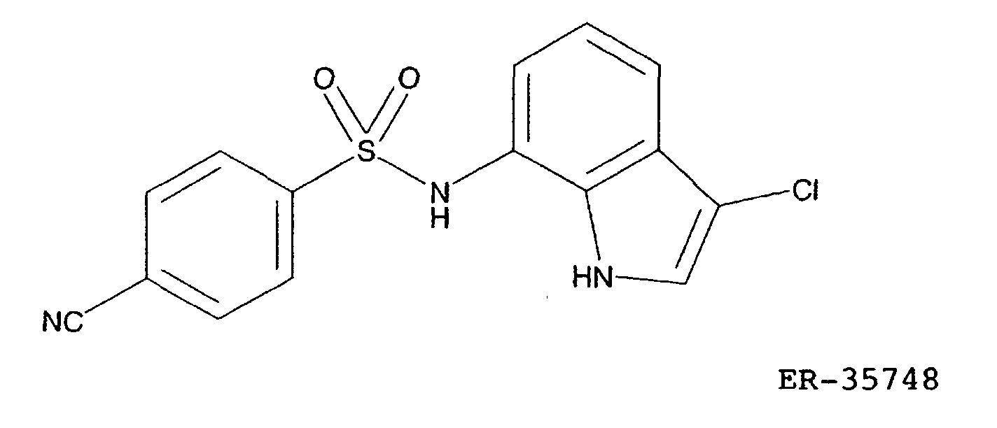
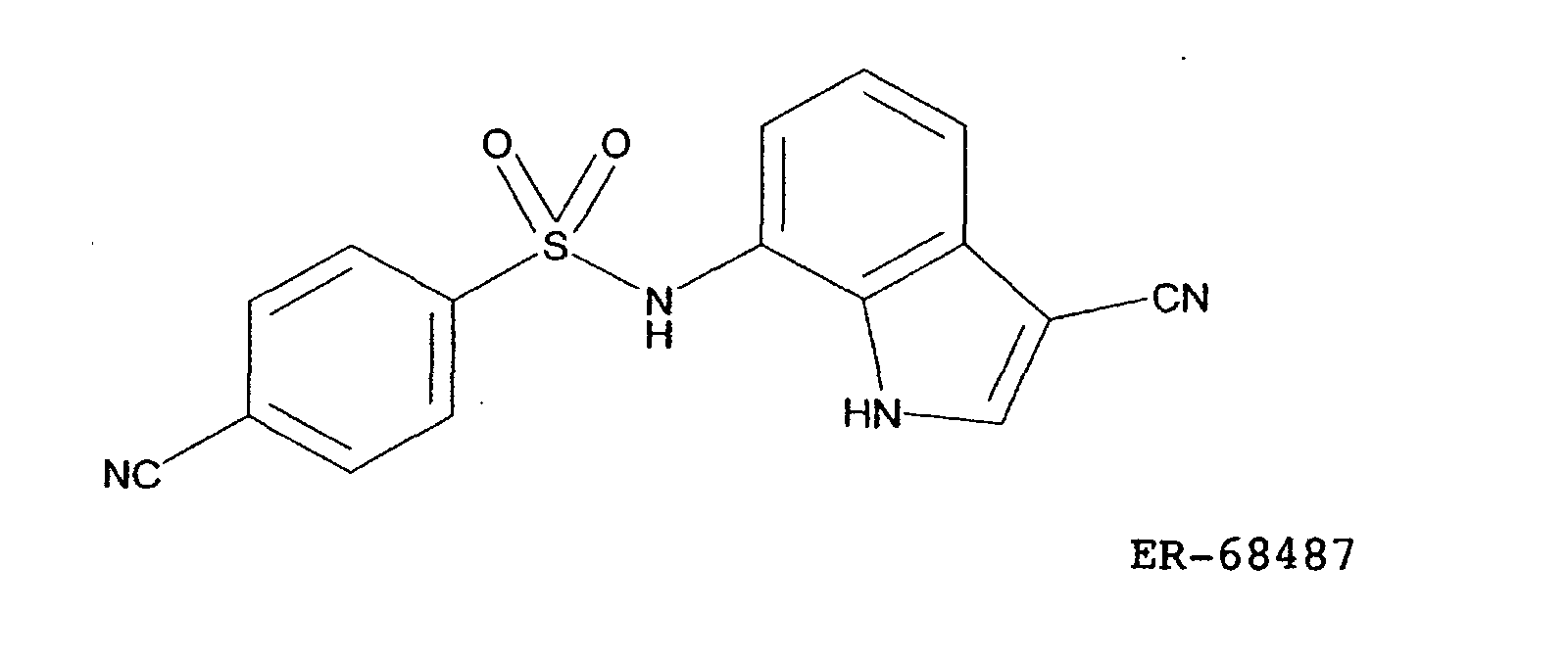
- Total RNA was extracted from these cells and subjected to the subsequent analyses. The RNA extracted from the cells cultured for 12 hours with the drug and the RNA from extracted the cells cultured for 12 hours without the drug were used for the gene expression analysis using a DNA microarray described in Example 2. HCT116-C9 was a substrain isolated from HCT116 derived from human colon cancer (American Type Culture Collection, Manassas, VA, U.S.A.), and HCT116-C9-C1 was an E7070-resistant substrain obtained by culturing this HCT116-C9 in the presence of E7070 with gradually increasing the E7070 concentration.
- Similarly, E7070 was added to the media of an E7070-susceptible strain LX-1 and an E7070-resistant strain LX-1-E2 at a concentration of 8 µM and the cells were cultured. Cells were recovered after 0, 3, 6 and 12 hours. Total RNA was extracted from these cells and subjected to the subsequent analyses. LX-1 (Cancer Chemotherapy Center, Japan Foundation for Cancer Research, Tokyo, Japan) was a cell strain derived from human parvicellular lung cancer, and LX-1-E2 is an E7070-resistant substrain obtained by culturing this LX-1 in the presence of E7070 with gradually increasing the E7070 concentration.
- The used cell strains HCT116-C9, HCT116-C9-C1, LX-1 and LX-1-E2 were cultured for 72 hours with addition of E7070. Cell growth inhibition curves measured by the MTT method (Mosmann T., J. Immunol. Methods, 65, 55 (1983)) are shown in Fig. 1. The actual operation was performed by using CellTiter 96 Non-Radioactive Cell Proliferation Assay (Promega, Madison, WI) according to the attached operation manual.
- Total RNA was extracted from the recovered cells by using TRIZOL Reagent (Life Technologies Oriental) according to the attached operation manual.
- Each RNA obtained in Example 1 was dissolved in 100 µl of sterilized water treated with diethyl pyrocarbonate (DEPC) and further purified by using RNeasy Column (QIAGEN), and double-stranded cDNA was synthesized by using Superscript Choice System (Life Technologies Oriental) and a T7-d(T)24 primer.
- First, to 10 µg of RNA, 5 µM T7-d(T)24 primer, 1 x First strand buffer, 10 mM DTT, 500 µM dNTP mix and 20 units/µl of Superscript II Reverse Transcriptase were added and the mixture was allowed to react at 42°C for 1 hour to synthesize single-stranded DNA. Subsequently, to the DNA, 1 x Second strand buffer, 200 µM dNTP mix, 67 U/ml of DNA ligase, 270 U/ml of DNA polymerase I and 13 U/ml of RNase H were added and the mixture was allowed to react at 16°C for 2 hours to synthesize double-stranded cDNA. Further, to the cDNA, 67 U/ml of T4 DNA polymerase I was added and the mixture was allowed to react at 16°C for 5 minutes and then 10 µl of 0.5 M EDTA was added to terminate the reaction.
- The obtained cDNA was purified with phenol/chloroform, and a labeling reaction with biotinylated UTP and CTP was carried out by using RNA Transcript Labeling Kit (Enzo Diagnostics) according to the attached operation manual. The reaction product was purified with Rneasy Column and heated in 200 mM Tris/acetate buffer (pH 8.1), 150 mM magnesium acetate, 50 mM potassium acetate at 94°C for 35 minutes to obtain fragment cRNA.
- The fragmented cRNA was hybridized with GeneChip Hu6800 (Affymetrix) in 100 mM MES, 1 M sodium salt, 20 mM EDTA and 0.01
% Tween 20 at 45°C for 16 hours. After the hybridization, the GeneChip was washed and stained according to the protocol EukGE-WS2 attached to Affymetrix Fluidics Station. For the staining, streptavidin-phycoerythrin and biotinylated goat anti-streptavidin antibody were used. The stained GeneChip was scanned by using HP Argon Ion Laser Confocal Scanner (Hewlett Packard) to measure its fluorescence intensity. The fluorescence was measured at 570 nm by using an excitation light at 488 nm. - The quantitative data analyses were all conducted by using GeneChip Software (Affymetrix). To quantify RNA, an average of "differences [(Perfect match hybridization signal) - (Mismatch signal)]" was obtained for each probe family. When this value was 50 or higher, and quantitative values of RNA measured under two kinds of conditions diverge, preferably they diverge 1.8 times or more, it was determined that the gene expression had significantly "increased" or "decreased".
- E7070, or ER-35748 or ER-68487 was added to a medium for HCT116-C9 at a concentration of 8 µ and culture was performed. The results of analysis of RNA extracted from the cells at 12 hours by using GeneChip were compared with the results of RNA analysis for HCT116-C9 cells obtained in the same manner except for not adding the drug. As a result, a list of genes of which expression levels were increased in common by the treatments with three types of drugs, E7070, ER-35748 and ER-68487 (GenBank registration numbers are also shown (the same shall apply in other tables shown below)) is shown in Table 1. Similarly, a list of genes of which expression levels were decreased in common by the treatments with the three types of drugs is shown in Table 2.
Registration number Gene name D59253 NCBP interacting protein 1 D78514 Ubiquitin-conjugating enzyme D83767 Rep-8 HG1139-HT4910 Fk506-Binding protein HG3484-HT3678 Cdc-like kinase 1 (Clk1) J04152 Tumor-associated antigen GA733-1, M1S1 M21154 S-adenosylmethionine decarboxylase M60724 p70 Ribosomal S6 kinase alpha-1 M84349 Transmembrane protein CD59 S61953 c-ErbB3 receptor tyrosine kinase U52960 RNA polymerase II complex component SRB7 U84720 Export protein RAE1 U92014 Defective mariner transposon Hsmar2 Z18951 Caveolin Registration number Gene name AB000409 Serine/threonine protein kinase MNK1 AB000450 Putative serine/threonine protein kinase VRK2 AB002380 Leukemia-associated Rho GEF AB003102 Proteasome 26S subunit p44.5 AC002045 CIT987SK-A-589H1 AF002020 Niemann-Pick C disease protein (NPC1) AF008445 Phospholipid scramblase D00723 Hydrogen carrier protein, glycine synthase D14659 KIAA0103 D21852 KIAA0029 D26535 Dihydrolipoamide succinyltransferase D28364 Annexin II D29677 KIAA0054 D29810 Unknown product D29956 Ubiquitin specific protease 8 D30756 KIAA0049 D31883 Actin-binding LIM protein D32002 Nuclear cap binding protein D38521 KIAA0077 D38552 KIAA0073 D38553 KIAA0074 D43947 KIAA0100 D43948 ch-TOG D50645 SDF2 D50663 Dynein (TCTEL1) D50912 RNA-binding motif protein 10 D50916 Ubiquitination factor E4A D61391 PAP39 D63480 KIAA0146 D63506 Syntaxin-binding protein 3 D63875 TPR-containing/SH2-binding protein D63880 KIAA0159 D78586 Multifunctional protein CAD D79983 KIAA0161 D79987 KIAA0165 D79988 KIAA0166 D79991 KIAA0169 D83776 KIAA0191 D83781 KIAA0197 D84307 Phosphoethanolamine cytidylyltransferase D86981 Amyloid precursor protein-binding protein 2 D87435 KIAA0248 D87446 KIAA0257 D87448 DNA topoisomerase II-binding protein D87743 Solute carrier family 9 HG1869-HT1904 Male enhanced antigen HG2379-HT3996 Serine hydroxymethyltransferase, cytosolic HG4094-HT4364 Transcription factor Lsf-Id J04088 DNA topoisomerase II (top2) J04501 Muscle glycogen synthase J04543 Synexin L06845 Cysteinyl-tRNA synthetase L07033 Hydroxymethylglutaryl-CoA lyase L07540 Replication factor C, 36-kDa subunit L07597 Ribosomal protein S6 kinase 2 L07758 IEF SSP 9502 L21936 Succinate dehydrogenase flavoprotein subunit L25444 TAFII70-alpha L25931 Lamin B receptor L33075 Ras GTPase-activating-like protein IQGAP1 L33881 Protein kinase C iota isoform L38810 Proteasome 26S subunit p45 L40395 Eukaryotic initiation factor 2B-beta L41870 Retinoblastoma susceptibility protein (RB1) L47276 Alpha topoisomerase truncated-form M15796 PCNA M19267 Tropomyosin M22632 Mitochondrial aspartate aminotransferase M23379 GTPase-activating protein ras p21 M24486 Prolyl 4-hydroxylase alpha subunit M29204 DNA-binding factor M29550 Calcineurin A1 M30496 Ubiquitin carboxyl-terminal hydrolase M33518 HLA-B-associated transcript 2 (BAT2) M34309 Epidermal growth factor receptor HER3 M37400 Cytosolic aspartate aminotransferase M55905 Mitochondrial NAD(P) + dependent malic enzyme M58525 Catechol-O-methyltransferase M59911 Integrin alpha-3 M61764 Gamma-tubulin M62994 Filamin B M74089 TB1 M85085 Cleavage stimulation factor M86707 Myristoyl CoA: protein N-myristoyl transferase M87338 Replication factor C, 40-kDa subunit (A1) M87339 Replication factor C, 37-kDa subunit (RFC4) M88163 Global transcription activator (hSNF2/SWI2) M91432 Medium-chain acyl-CoA dehydrogenase (MCAD) M92439 Leucine-rich protein M93056 Monocyte/neutrophil elastase inhibitor M95809 Basic transcription factor 62kD subunit (BTF2) M95929 Homeobox protein PHOX1 M97935 Transcription factor ISGF-3 S58544 75kDa infertility-related sperm protein S59184 RYK = related to receptor tyrosine kinase S72904 APK1 antigen = MAb KI recognized S78085 PDCD2, Rp8 homolog S80343 Arginyl-tRNA synthetase (ArgRS) U01062 Inositol 1,4,5-triphosphate receptor type 3 U02566 Receptor tyrosine kinase tif U04285 Lysosomal acid lipase U06631 H326 U07231 G-rich sequence factor-1 U07681 Isocitrate dehydrogenase 3 (NAD+) alpha U079l9 Aldehyde dehydrogenase 6 (ALDH6) U08815 Splicesomal protein SAP61 U10324 Nuclear factor NF90 U11791 Cyclin H U15174 Nip3 U15306 DNA-binding protein NFX1 U18291 CDC16HS U18934 Receptor tyrosine kinase DTK U20979 Chromatin assembly factor-I p150 subunit U22233 Methylthioadenosine phosphorylase U23028 Eukaryotic initiation factor 2B-epsilon U23946 LUCA15 U26648 Syntaxin 5A U27459 Origin recognition complex protein 2 homolog U27460 Uridine diphosphoglucose pyrophosphorylase U28413 Cockayne syndrome complementation group A U28811 Cystein-rich fibroblast growth factor receptor U28831 Immuno-reactive with anti-PTH Ab U28963 Gps2 U30313 Diadenosine tetraphosphatase U30521 P311 HUM -3.1 U30827 Splicing factor SRp40-3 (SRp40) U30828 Splicing factor SRp55-2 (SRp55) U34252 Gamma-aminobutyraldehyde dehydrogenase U34683 Glutathione synthetase U36341 SLC6A8 U40282 Integrin-linked kinase U46006 Smooth muscle LIM protein (h-SmLIM) U49844 FRAP-related protein (FRP1/ATR) U50078 Guanine nucleotide exchange factor p532 U50939 Amyloid precursor protein-binding protein 1 U53468 NADH: ubiquinone oxidoreductase subunit B13 U57629 Retinitis pigmentosa GTPase regulator U60808 CDP-diacylglycerol synthase U61145 Enhancer of zeste homolog 2 U61263 Acetolactate synthase homolog U63743 Mitotic centromere-associated kinesin (MCAK) U65785 Oxygen-regulated protein ORP150 U72514 C2f U72515 C3f U76764 CD97 U77413 O-linked GlcNAc transferase U77949 Cdc6-related protein (HsCDC6) U79241 Clone 23759 U80034 Mitochondrial intermediate peptidase precursor U81554 CaM kinase II isoform U85611 DNA-PK interaction protein (KIP) U89606 Pyridoxal kinase U90426 Nuclear RNA helicase U94319 Transcription coactivator p75 (DFS70) U95740 362G6.1 U97188 Putative RNA binding protein KOC X06745 DNA polymerase alpha subunit X13482 U2 snRNP-specific A protein X51956 Neuron specific (gamma) enolase X53587 Integrin beta-4 X54199 GARS-AIRS-GART X54867 NKG2-A X59871 T cell factor 1 X61100 75 kDa subunit NADH dehydrogenase precursor X66364 Serine/threonine protein kinase PSSALRE X68836 S-adenosylmethionine synthetase X70476 Subunit of coatomer complex X75535 PxF X81003 HCG V X84740 DNA ligase III X94754 Yeast methionyl-tRNA synthetase homologue X98248 Sortilin X99209 Arginine methyltransferase Y08612 Nup88 Y08682 Carnitine palmitoyltransferase I type I Y13115 Serine/threonine protein kinase SAK Z11518 Histidyl-tRNA synthetase Z17227 Transmenbrane receptor protein CRF2-4 Z46629 SOX9 Z68747 Imogen 38 - Two kinds of E7070-resistant strains (C9-C1 and another resistant strain (C9-C4) obtained in the same method as C9-C1) were treated with 8 µM of E7070 for 12 hours in the same manner as described above, and variations of the gene expression were analyzed by using GeneChip for the genes listed in Tables 1 and 2. As a result, no gene that increased or decreased 1.8 times or more was observed in the both strains.
- From the above results, it is apparent that genes of which expression levels are changed in common by the treatments with these three types of anticancer agents can be used as surrogate markers of antitumor effect of E7070 and its related compounds by measuring each of their changes or changes of two or more of them in combination.
- In order to confirm that variations in gene expression obtained in Example 2 reflected the susceptibility of a tumor cell to E7070, changes of gene expression in E7070-susceptible cells and E7070-resistant cells were examined by quantitative PCR using RNA shown in Example 1, SYBR Green and ABI Prism 7700 Sequence Detection System (Perkin-Elmer Applied Biosystems).
- The operation was performed in two steps of a reverse transcription reaction and PCR reaction. The reverse transcription reaction as the first step was carried out by adding 1 x TaqMan RT buffer, 5.5 mM MgCl2, 500 µM dNTP mix, 2.5 µM oligo d(T)16 primer, 0.4 U/µl of RNase inhibitor and 1.25 U/µl of Multiscribe Reverse Transcriptase (Perkin-Elmer Applied Biosystems) to 1 µg of the total RNA, keeping the mixture at 25°C for 10 minutes and then heating it at 48°C for 30 minutes. The reaction was terminated by heating at 95°C for 5 minutes.
- The obtained cDNA was subjected to the PCR reaction as the second step. The PCR reaction was carried out in a reaction system composed of 4 ng of cDNA, 1 x SYBR PCR buffer, 3 mM MgCl2, 200 µM each of dATP, dCTP and dGTP, 400 µMdUTP, 200 nM primer pair, 0.01 U/µl AmpErase UNG and 0.025 U/µl AmpliTaq Gold DNA Polymerase (Perkin-Elmer Applied Biosystems). The PCR reaction was performed with reactions at 50°C for 2 minutes and at 95°C for 10 minutes followed by a cycle of reactions at 95°C for 20 seconds, at 55°C for 20 seconds and at 72°C for 30 seconds, which are repeated 40 times. As the primers, oligonucleotides having the nucleotide sequences of SEQ ID NOS: 1 and 2 were used for GAPDH, those of SEQ ID NOS: 3 and 4 for S80343, those of SEQ ID NOS: 5 and 6 for U07919, those of SEQ ID NOS: 7 and 8 for U11791, those of SEQ ID NOS: 9 and 10 for M95809, those of SEQ ID NOS: 11 and 12 for U18291, those of SEQ ID NOS: 13 and 14 for U63743, and those of SEQ ID NOS: 15 and 16 for M61764.
- The mRNA level in each specimen was quantified by measuring fluorescence intensity. In comparison of two or more specimens, quantitative values were corrected based on the mRNA level of GAPDH in each specimen. The relationships between the drug treatment time and each mRNA level are shown in Figs. 2 to 5, where the quantified value at 0 hour after the drug treatment was used as a control (100%). The genes showed the largest expression variation in HCT116-C9 with the highest susceptibility (Fig. 2), and in the susceptible strain LX-1, all the genes except for S80343 showed expression variations although the variation magnitudes were smaller than those in HCT116-C9 (Fig. 3). On the other hand, no expression variation was observed for the resistant strain HCT116-C9-C1 (Fig. 4), and in the resistant strain LX-1-E2, which showed some growth inhibitory effect at the drug concentration of 8 µ used in the experiment, U11791 showed an expression change and M95809 and U18291 showed a slight expression change (Fig. 5).
- The variations in expression of the gene retrieved in Example 2 correlated to the susceptibility to E7070, and it was confirmed that the genes retrieved in Example 2 could be used solely or in combination as surrogate markers of antitumor effect of E7070 and its related compounds.
- Among the genes listed in Table 2 in Example 2, the expression level of X51956 (Human ENO2 gene for neuron specific (gamma) enolase), which has been reported to be secreted out of the cell, was examined by the already reported ELISA method (Duncan M. E., McAleese S. M., Booth N. A., Melvin W. T., and Fothergill J. E., J. of Immunol. Methods, 151, 227-236, 1992, Yamaguchi K., Aoyagi K., Urakami K., Fukutani T., Maki N., Yamamoto S., Otsubo K., Miyake Y., and Kodama T., Jpn. J. Cancer Res., 86, 698-705, 1995). The actual measurement was carried out by using NSE ELISA Kit produced by Eiken Chemical (Tokyo) according to the attached reference.
- Gene expression changes in three kinds of cancers of HCT116-C9 (human colon cancer cell strain), MDA-MB-435 (human breast cancer cell strain) and MOLT-4 (human T lymphoblastic leukemia cell strain) caused by E7070 (treated with 8 µM for 12 hours) were examined by using GeneChip in the same manner as in Example 2.
- A list of the genes of which expression was enhanced 1.8 times or more in common in the three kinds of cancers is shown in Table 3. Further, a list of the genes of which expression was inhibited 1.8 times or more in common in the three kinds of cancers is shown in Table 4. Since the genes of which expression varies (enhanced or reduced) in common in these strains of the three kinds of cancers established from cancer patients with totally different genetic backgrounds are very likely to be more closely associated with the E7070 antitumor action mechanism common to the tumor cells, it is considered that they can be used as valid markers when susceptibility of a tumor cell to E7070 and its related compounds is tested.
Registration number Gene name D78514 Ubiquitin-conjugating enzyme D83767 Rep-8 HG1139-HT4910 Fk506-Binding protein HG3484-HT3678 Cdc-like kinase 1 (Clk1) M21154 S-adenosylmethionine decarboxylase M60724 p70 Ribosomal S6 kinase alpha-1 S61953 c-ErbB3 receptor tyrosine kinase U52960 RNA polymerase II complex component SRB7 U84720 Export protein RAE1 U92014 Defective mariner transposon Hsmar2 Registration number Gene name AB000409 Serine/threonine protein kinase MNK1 AB000450 Putative serine/threonine protein kinase VRK2 AC002045 CIT987SK-A-589H1 AF008445 Phospholipid scramblase D00723 Hydrogen carrier protein, glycine synthase D14659 KIAA0103 D21852 KIAA0029 D26535 Dihydrolipoamide succinyltransferase D32002 Nuclear cap binding protein D38521 KIAA0077 D38553 KIAA0074 D43947 KIAA0100 D50912 RNA-binding motif protein 10 D61391 PAP39 D63480 KIAA0146 D63880 KIAA0159 D78586 Multifunctional protein CAD D79983 KIAA0161 D79991 KIAA0169 D83781 KIAA0197 D84307 Phosphoethanolamine cytidylyltransferase D87446 KIAA0257 HG1869-HT1904 Male enhanced antigen HG2379-HT3996 Serine hydroxymethyltransferase, cytosolic HG4094-HT4364 Transcription factor Lsf-Id J04088 DNA topoisomerase II (top2) L07758 IEF SSP 9502 L21936 Succinate dehydrogenase flavoprotein subunit L25931 Lamin B receptor L38810 Proteasome 26S subunit p45 L41870 Retinoblastoma susceptibility protein (RB1) L47276 Alpha topoisomerase truncated-form M19267 Tropomyosin M22632 Mitochondrial aspartate aminotransferase M29204 DNA-binding factor M29550 Calcineurin A1 M30496 Ubiquitin carboxyl-terminal hydrolase M34309 Epidermal growth factor receptor HER3 M55905 Mitochondrial NAD(P) + dependent malic enzyme M61764 Gamma-tubulin M85085 Cleavage stimulation factor M87338 Replication factor C, 40-kDa subunit (A1) M91432 Medium-chain acyl-CoA dehydrogenase (MCAD) M92439 Leucine-rich protein M93056 Monocyte/neutrophil elastase inhibitor M95809 Basic transcription factor 62kD subunit (BTF2) M97935 Transcription factor ISGF-3 S72904 APK1 antigen = MAb KI recognized S78085 PDCD2, Rp8 homolog U01062 Inositol 1,4,5-triphosphate receptor type 3 U07231 G-rich sequence factor-1 U08815 Splicesomal protein SAP61 U11791 Cyclin H U15306 DNA-binding protein NFX1 U18291 CDC16HS U18934 Receptor tyrosine kinase DTK U20979 Chromatin assembly factor-I p150 subunit U22233 Methylthioadenosine phosphorylase U23028 Eukaryotic initiation factor 2B-epsilon U23946 LUCA15 U28831 Immuno-reactive with anti-PTH Ab U28963 Gps2 U30828 Splicing factor SRp55-2 (SRp55) U34683 Glutathione synthetase U40282 Integrin-linked kinase U49844 FRAP-related protein (FRP1/ATR) U50939 Amyloid precursor protein-binding protein 1 U57629 Retinitis pigmentosa GTPase regulator U61145 Enhancer of zeste homolog 2 U72514 C2f U77413 O-linked GlcNAc transferase U77949 Cdc6-related protein (HsCDC6) U79241 Clone 23759 U80034 Mitochondrial intermediate peptidase precursor U81554 CaM kinase II isoform U89606 Pyridoxal kinase U94319 Transcription coactivator p75 (DFS70) X06745 DNA polymerase alpha subunit X13482 U2 snRNP-specific A protein X51956 Neuron specific (gamma) enolase X54199 GARS-AIRS-GART X61100 75 kDa subunit NADH dehydrogenase precursor X68836 S-adenosylmethionine synthetase X70476 Subunit of coatomer complex X75535 PxF X99209 Arginine methyltransferase Y08612 Nup88 Y08682 Carnitine palmitoyltransferase I type I Z17227 Transmembrane receptor protein CRF2-4 - According to the present invention, by measuring an expression level or levels of a gene or genes listed in Tables 3 and 4 in a tumor cell collected from a cancer patient treated with an anticancer agent represented by the general formula (I) or causing the anticancer agent represented by the general formula (I) to act on a tumor cell collected from a cancer patient and measuring an expression level or levels of a gene or genes listed in Tables 3 and 4, susceptibility of the tumor cell to the anticancer agent can be examined.


Claims (10)
- A method for testing susceptibility of a tumor cell to an anticancer agent, which comprises:1) measuring an expression level or levels of a gene or genes selected from the group consisting of the genes listed in Tables 3 and 4 in a tumor cell collected from a cancer patient treated with an anticancer agent represented by the following general formula (I) :wherein A represents a monocyclic or bicyclic aromatic ring, which may be substituted,

B represents a 6-membered unsaturated hydrocarbon ring or a 6-membered unsaturated heterocyclic ring containing one nitrogen atom as a heteroatom, each of which may be substituted,
C represents a 5-membered heterocyclic ring containing one or two nitrogen atoms, which may be substituted,
W represents a single bond or -CH=CH-,
X represents -N(R1)- or an oxygen atom,
Y represents a carbon or nitrogen atom,
Z represents -N(R2)- or a nitrogen atom, and
R1 and R2 may be identical or different and each represent a hydrogen atom or a lower alkyl group, and2) determining that the tumor cell is susceptible to the anticancer agent when the expression level or levels of the gene or genes listed in Table 3 increase, or the expression level or levels of the gene or genes listed in Table 4 decrease in comparison with those in a tumor cell collected from the cancer patient before the treatment with the anticancer agent. - A method for testing susceptibility of a tumor cell to an anticancer agent, which comprises:1) causing an anticancer agent to act on a tumor cell collected from a cancer patient, said anticancer agent being represented by the following general formula (I):wherein A represents a monocyclic or bicyclic aromatic ring, which may be substituted,
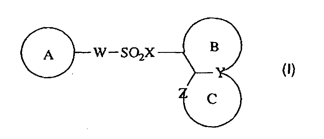
B represents a 6-membered unsaturated hydrocarbon ring or a 6-membered unsaturated heterocyclic ring containing one nitrogen atom as a heteroatom, each of which may be substituted,
C represents a 5-membered heterocyclic ring containing one or two nitrogen atoms, which may be substituted,
W represents a single bond or -CH=CH-,
X represents -N(R1)- or an oxygen atom,
Y represents a carbon or nitrogen atom,
Z represents -N(R2)- or a nitrogen atom, and
R1 and R2 may be identical or different and each represent a hydrogen atom or a lower alkyl group,2) measuring an expression level or levels of a gene or genes selected from the group consisting of the genes listed in Tables 3 and 4 in the tumor cell, and3) determining that the tumor cell is susceptible to the anticancer agent when the expression level or levels of the gene or genes listed in Table 3 increase, or the expression level or levels of the gene or genes listed in Table 4 decrease in comparison with those in an untreated tumor cell. - The method according to claim 1 or 2, wherein the expression level or levels of gene or genes are measured by quantifying an RNA or RNAs which are a transcription product or products of the gene or genes by using a DNA microarray.
- The method according to claim 1 or 2, wherein the expression level or levels of the gene or genes are measured by quantifying an RNA or RNAs which are a transcription product or products of the gene or genes by quantitative PCR.
- A reagent for quantifying an RNA for use in the method as defined in claim 4, which comprises an oligonucleotide complementary to the RNA as a component.
- The method according to claim 1 or 2, wherein the expression level or levels of the gene or genes are measured by quantifying a protein or proteins which are a gene product or products of the gene or genes by an immunochemical method.
- The method according to claim 6, wherein the expression level or levels of the gene or genes are measured by quantifying a protein or proteins which are a gene product or products of the gene or genes by ELISA.
- The method according to claim 6, wherein the expression level or levels of the gene or genes are measured by quantifying a protein or proteins which are a gene product or products of the gene or genes by Western blotting.
- An immunoassay reagent used in the method as defined in claim 6, which comprises an antibody directed to the protein as a component.
- The method according to claim 1 or 2, wherein A represents benzene or pyridine which may be substituted, B represents benzene which may be substituted, C represents pyrrole which may be substituted, W represents a single bond, and X and Z both represent -NH-.
Applications Claiming Priority (3)
| Application Number | Priority Date | Filing Date | Title |
|---|---|---|---|
| JP2000357398 | 2000-11-24 | ||
| JP2000357398 | 2000-11-24 | ||
| PCT/JP2001/010282 WO2002042493A1 (en) | 2000-11-24 | 2001-11-26 | Method of testing anticancer agent-sensitivity of tumor cells |
Publications (2)
| Publication Number | Publication Date |
|---|---|
| EP1336660A1 true EP1336660A1 (en) | 2003-08-20 |
| EP1336660A4 EP1336660A4 (en) | 2006-03-15 |
Family
ID=18829473
Family Applications (1)
| Application Number | Title | Priority Date | Filing Date |
|---|---|---|---|
| EP01997200A Withdrawn EP1336660A4 (en) | 2000-11-24 | 2001-11-26 | Method of testing anticancer agent-sensitivity of tumor cells |
Country Status (7)
| Country | Link |
|---|---|
| US (1) | US7445892B2 (en) |
| EP (1) | EP1336660A4 (en) |
| KR (2) | KR100858644B1 (en) |
| CN (3) | CN1721554B (en) |
| AU (2) | AU2409602A (en) |
| CA (1) | CA2429688A1 (en) |
| WO (1) | WO2002042493A1 (en) |
Cited By (1)
| Publication number | Priority date | Publication date | Assignee | Title |
|---|---|---|---|---|
| EP2015070A4 (en) * | 2006-04-20 | 2010-04-21 | Eisai R&D Man Co Ltd | NEW MARKER AGAINST SENSITIVITY TO A SULFONAMIDE COMPOUND |
Families Citing this family (8)
| Publication number | Priority date | Publication date | Assignee | Title |
|---|---|---|---|---|
| ATE469985T1 (en) * | 2004-02-06 | 2010-06-15 | Eisai R&D Man Co Ltd | METHOD FOR STUDYING THE SENSITIVITY OF A CANCER CELL TO AN ANTI-CANCER DRUG |
| JP4848355B2 (en) * | 2004-02-27 | 2011-12-28 | アンスティテュ・キュリ | Novel growth markers in clinical practice and their use for cancer prognosis or diagnosis |
| US20100093767A1 (en) * | 2004-12-03 | 2010-04-15 | Takeda San Diego, Inc. | Mitotic Kinase Inhibitors |
| WO2007086605A1 (en) * | 2006-01-27 | 2007-08-02 | Eisai R & D Management Co., Ltd. | Method for prediction of the effect of sulfonamide compound |
| JP2012136435A (en) * | 2009-03-30 | 2012-07-19 | Eisai R & D Management Co Ltd | Method for examining sensitivity of tumor tissue |
| CN103088102B (en) * | 2013-01-09 | 2015-05-20 | 苏州麦克威尔生物医药科技有限公司 | Sterile instant type medicine film and tumor drug sensitivity testing application thereof |
| CN109781985B (en) * | 2019-02-27 | 2021-10-29 | 中山大学肿瘤防治中心 | Kit for detecting radiotherapy sensitivity of cancer and its application |
| CN113549696B (en) * | 2021-09-23 | 2022-01-04 | 广州医科大学附属肿瘤医院 | Tumor marker SORCS2 and application thereof |
Family Cites Families (3)
| Publication number | Priority date | Publication date | Assignee | Title |
|---|---|---|---|---|
| RU2121997C1 (en) * | 1993-09-10 | 1998-11-20 | Эйсай Ко., ЛТД | 7-amino-1h-indole derivatives |
| AU3395900A (en) * | 1999-03-12 | 2000-10-04 | Human Genome Sciences, Inc. | Human lung cancer associated gene sequences and polypeptides |
| WO2006036025A1 (en) | 2004-09-30 | 2006-04-06 | Eisai R & D Management Co., Ltd. | Method of evaluating sensitivity of tumor cell to sulfonamide-containing compound |
-
2001
- 2001-11-26 AU AU2409602A patent/AU2409602A/en active Pending
- 2001-11-26 US US10/432,661 patent/US7445892B2/en not_active Expired - Fee Related
- 2001-11-26 WO PCT/JP2001/010282 patent/WO2002042493A1/en not_active Ceased
- 2001-11-26 KR KR1020037006971A patent/KR100858644B1/en not_active Expired - Fee Related
- 2001-11-26 AU AU2002224096A patent/AU2002224096B2/en not_active Ceased
- 2001-11-26 KR KR1020087004808A patent/KR100857735B1/en not_active Expired - Fee Related
- 2001-11-26 CA CA 2429688 patent/CA2429688A1/en not_active Abandoned
- 2001-11-26 CN CN2005100860155A patent/CN1721554B/en not_active Expired - Fee Related
- 2001-11-26 CN CNB018222404A patent/CN100342030C/en not_active Expired - Fee Related
- 2001-11-26 CN CN2007100966962A patent/CN101054604B/en not_active Expired - Fee Related
- 2001-11-26 EP EP01997200A patent/EP1336660A4/en not_active Withdrawn
Non-Patent Citations (4)
| Title |
|---|
| OWA TAKASHI ET AL: "Microarray-based expression profiling of sulfonamide antitumor agents" PROCEEDINGS OF THE AMERICAN ASSOCIATION FOR CANCER RESEARCH ANNUAL MEETING, vol. 42, March 2001 (2001-03), page 371, XP001246420 & 92ND ANNUAL MEETING OF THE AMERICAN ASSOCIATION FOR CANCER RESEARCH; NEW ORLEANS, LA, USA; MARCH 24-28, 2001 ISSN: 0197-016X * |
| See also references of WO0242493A1 * |
| WATANABE T ET AL: "A novel antitumor agent ER-35744, targeting G1 phase. III: Studies of mechanism of action" PROCEEDINGS OF THE AMERICAN ASSOCIATION FOR CANCER RESEARCH ANNUAL MEETING, vol. 37, no. 0, 1996, page 391, XP001208114 & 87TH ANNUAL MEETING OF THE AMERICAN ASSOCIATION FOR CANCER RESEARCH; WASHINGTON, D.C., USA; APRIL 20-24, 1996 ISSN: 0197-016X * |
| WU L ET AL: "MULTIDRUG-RESISTANT PHENOTYPE OF DISEASE-ORIENTED PANELS OF HUMAN TUMOR CELL LINES USED FOR ANTICANCER DRUG SCREENING" CANCER RESEARCH, vol. 52, no. 11, 1992, pages 3029-3034, XP001208236 ISSN: 0008-5472 * |
Cited By (1)
| Publication number | Priority date | Publication date | Assignee | Title |
|---|---|---|---|---|
| EP2015070A4 (en) * | 2006-04-20 | 2010-04-21 | Eisai R&D Man Co Ltd | NEW MARKER AGAINST SENSITIVITY TO A SULFONAMIDE COMPOUND |
Also Published As
| Publication number | Publication date |
|---|---|
| US20040115664A1 (en) | 2004-06-17 |
| KR100857735B1 (en) | 2008-09-12 |
| EP1336660A4 (en) | 2006-03-15 |
| KR20080026224A (en) | 2008-03-24 |
| CN1721554B (en) | 2010-05-26 |
| KR100858644B1 (en) | 2008-09-17 |
| US7445892B2 (en) | 2008-11-04 |
| AU2002224096B2 (en) | 2006-07-27 |
| KR20030051871A (en) | 2003-06-25 |
| AU2409602A (en) | 2002-06-03 |
| CA2429688A1 (en) | 2003-05-23 |
| CN100342030C (en) | 2007-10-10 |
| CN1488001A (en) | 2004-04-07 |
| CN101054604B (en) | 2010-12-15 |
| CN1721554A (en) | 2006-01-18 |
| CN101054604A (en) | 2007-10-17 |
| WO2002042493A1 (en) | 2002-05-30 |
Similar Documents
| Publication | Publication Date | Title |
|---|---|---|
| KR101620642B1 (en) | Methods and compositions for assessing responsiveness of b-cell lymphoma to treatment with anti-cd40 antibodies | |
| KR101828290B1 (en) | Markers for endometrial cancer | |
| US20090098538A1 (en) | Prognostic and diagnostic method for disease therapy | |
| CA2500482A1 (en) | Method for diagnosing pancreatic cancer | |
| EP2744917A2 (en) | Methods and compositions for the treatment and diagnosis of breast cancer | |
| EP2059615A2 (en) | Prognostic and diagnostic method for disease therapy | |
| BRPI0618597A2 (en) | cancer prediction and prognosis methods and cancer therapy monitoring method | |
| KR20180028524A (en) | Sensitivity to FGFR expression and FGFR inhibitors | |
| US7445892B2 (en) | Method of testing anticancer agent-sensitivity of tumor cells | |
| CN114096256A (en) | Methods and compositions for predicting the efficacy of a BCL-2/BCL-XL inhibitor on cancer | |
| RU2449016C2 (en) | Gene involved in immortalisation of human cancer cell and use thereof | |
| KR102549771B1 (en) | Method for screening an inhibitor for metastasis of colorectal cancer | |
| EP1575417A2 (en) | Methods for prediction and prognosis of cancer, and monitoring cancer therapy | |
| WO2008025968A2 (en) | Method of prognosis | |
| JP2003038200A (en) | Method for assaying sensitivity of tumor cells to anticancer drugs | |
| KR20230107484A (en) | Inhibitor for metastasis of colorectal cancer | |
| KR101683961B1 (en) | Recurrence Marker for Diagnosis of Bladder Cancer | |
| KR20180137049A (en) | The biomarker for triple negative breast cancer comprising deubiquitinating enzymes genes and information providing a method for triple negative breast cancer diagnosis using thereof | |
| JP2003284596A (en) | How to detect breast cancer cells | |
| CN108085383B (en) | Gene detection composition and application thereof | |
| JP2003284594A (en) | Ovarian cancer cell detection method | |
| AU2010202867A1 (en) | Novel compositions and methods in cancer associated with altered expression of KCNJ9 | |
| JP2003284563A (en) | How to detect uterine cancer cells | |
| IT201900000130A1 (en) | METHOD FOR IDENTIFYING SUBJECTS RESISTANT TO TREATMENT WITH PLATINUM-BASED DRUGS AND RELATIVE KIT | |
| CN108060223A (en) | The new opplication of ATF2 genes |
Legal Events
| Date | Code | Title | Description |
|---|---|---|---|
| PUAI | Public reference made under article 153(3) epc to a published international application that has entered the european phase |
Free format text: ORIGINAL CODE: 0009012 |
|
| 17P | Request for examination filed |
Effective date: 20030610 |
|
| AK | Designated contracting states |
Designated state(s): AT BE CH CY DE DK ES FI FR GB GR IE IT LI LU MC NL PT SE TR |
|
| AX | Request for extension of the european patent |
Extension state: AL LT LV MK RO SI |
|
| A4 | Supplementary search report drawn up and despatched |
Effective date: 20060127 |
|
| 17Q | First examination report despatched |
Effective date: 20061010 |
|
| RAP1 | Party data changed (applicant data changed or rights of an application transferred) |
Owner name: EISAI R&D MANAGEMENT CO., LTD. |
|
| GRAP | Despatch of communication of intention to grant a patent |
Free format text: ORIGINAL CODE: EPIDOSNIGR1 |
|
| STAA | Information on the status of an ep patent application or granted ep patent |
Free format text: STATUS: THE APPLICATION IS DEEMED TO BE WITHDRAWN |
|
| 18D | Application deemed to be withdrawn |
Effective date: 20111125 |