Field of the Invention
The present invention relates to a primer pool for amplifying a human HNF-1α
gene, and more particularly, to a primer pool for amplifying a target DNA sequence in
a human HNF-1α gene by multiplex PCR.
Background of the Invention
One type of process utilized for the detection of hybridized nucleic acids
involves polymerase chain reaction (PCR). The PCR process is well known in the art
(U.S. Pat. Nos. 4,683,195, 4,683,202 and 4,800,159). In PCR, nucleic acid primers
that are complementary to opposite strands of a nucleic acid amplification target
sequence are permitted to anneal to the denatured sample. Next, DNA polymerase
(typically heat stable) extends the DNA duplex from the hybridized primer. The
process is then repeated to amplify the nucleic acid target. If the nucleic acid primers
do not hybridize to the sample, then there is no corresponding amplified PCR product.
In this case, the PCR primer acts as a hybridization probe.
In a PCR method, the amplified nucleic acid product may be detected in a
number of ways, e.g., by incorporation of a labeled nucleotide into the amplified
strand by using labeled primers. Primers used in PCR include, but are not limited to
radioactive substances, fluorescent dyes, digoxygenin, horseradish peroxidase,
alkaline phosphatase, acridinium esters, biotin and jack bean urease. PCR products
made with unlabeled primers may be detected in other ways, such as electrophoretic
gel separation followed by dye-based visualization.
PCR-based methods are of limited use for the detection of nucleic acids of
unknown sequence. The human genome is composed of about 3x10⁹ nucleotides;
thus, it is difficult to isolate and analyze a specific human gene. Yet, PCR can
amplify a target sequence with high speed, specificity and sensitivity by using a set of
primers including primers complementary to both ends of the target sequence (Saiki
et al. Science 239: 487, 1988).
PCR may be widely used in analyzing a disease-associated gene. Specifically,
gene amplification by PCR may be useful for analyzing genetic variations of a
disease-associated gene in the medical field. A specific disease-associated gene
may be amplified using PCR, and analyzed by using a sequencing, hybridization or
single strand conformational polymorphism. In analyzing genetic variations of a gene
a single PCR may be enough to amplify the entire gene if the size of a target gene is
small. However, if the size of a target gene is large, e.g., 1kb or more, a single PCR
may have difficulty in amplifying the entire gene. Thus, PCR may be conducted
several times on several portions of the entire gene to amplify the entire gene of a
large target gene. In analyzing a genetic variation of a disease-associated gene, a
multiple PCR is more frequently used than a single PCR since most disease-associated
genes may be 1.5kb or larger in size.
A multiple PCR process requires a large amount of a sample, for example, a
patient's DNA or blood. A multiple PCR also costs more and requires more effort and
time. Thus, multiplex PCR assays have been developed to solve the above
problems. A multiplex PCR simultaneously amplifies a plurality of target sequences
of a gene in one reaction. Therefore, a plurality of target sequences are amplified by
a single PCR using a primer pool for amplifying each target sequence.
Multiplex PCR assays are well known in the art. For example, U.S. Pat. No.
5,582,989 discloses the simultaneous detection of multiple known DNA sequence
deletions. The technique disclosed therein uses a first set of probes to hybridize to
the targets. Those probes are extended if the targets are present. The extension
products are amplified using PCR.
A set of primers for a multiplex PCR may be able to specifically bind to a target
sequence and should not interfere with each other in order to amplify the target
sequence by a sufficient amount. A multiplex PCR using such a set of primers may
be able to save time, effort and cost for amplifying a target sequence in comparison
with a single PCR. In analyzing a genetic variation of a gene by using a DNA chip, a
multiplex PCR may be useful in amplifying more than one kind of DNA sample in a
reaction. Such a DNA chip may be useful in analyzing genetic variations in a gene.
It is known that a genetic variation of a human HNF-1α (hepatocyte nuclear
factor-1α) gene, including a point mutation, causes maturity-onset diabetes in the
young (MODY 3) (Matschinsky & Magnuson, in 'Molecular Pathogenesis of MODYs',
Karger, 1998; US patent 5,541,060; WO9321343). MODY 3, a kind of MODY disease
( MODY 1, 2, 3, 4 and 5) accounts for about 10-30 % of all type II diabetes mellitus
cases. Thus, in analyzing a genetic variation of a human HNF-1α gene, it is possible
to anticipate a person's propensity to a diabetes mellitus. Therefore, in order to
rapidly analyze a human HNF-1α gene using, for example, a DNA chip, a set of
primers for amplifying a human HNF-1α gene by a multiplex PCR needs to be
developed.
Summary of the Invention
One of the embodiments of the present invention provides a primer pool
comprising at least one set of primers for amplifying at least one target sequence of a
human HNF-1α gene, the at least one set of primers being selected from the group
consisting of:
wherein said variant oligonucleotide is an oligonucleotide having 1 to 3
additional nucleotides joined or deleted from the 3' end, the 5' end, or both the 3' end
and the 5' end of the corresponding oligonucleotide .
Another embodiment of the present invention provides a method for
sequencing a human HNF-1α gene by using the primer pool.
Other embodiments of the present invention provide kits for amplifying a target
sequence of a human HNF-1α gene comprising the primer pool according to the
present invention.
The present invention also provides a method for amplifying a target sequence
of a human HNF-1α gene comprising subjecting a target sequence of a human HNF-1α
gene to a PCR which uses the primer pool according to the present invention.
Brief Description of the Drawings
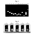
FIG. 1 illustrates an electrophoresis result of a single PCR product and a
multiplex PCR product of exons 1 to 10 of a human HNF-1α gene.
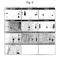
FIGs. 2A through 2E depict an electrophoresis result of a multiplex PCR
product obtained using a set of variant primers.
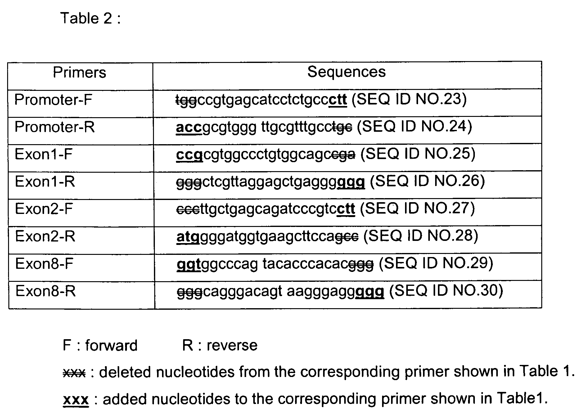
FIG. 3 is a photograph for illustrating a Southern blotting result of a single
PCR and a multiplex PCR obtained using each primer set for amplifying each exon
as a probe.
FIG. 4 illustrates a comparison of the nucleotide sequence of a single PCR
product (SEQ ID. 31) obtained using a set of primers for amplifying a promoter with a
known nucleotide sequence of a human HNF-1α gene.
FIG. 5 illustrates the result of a stability test of a reaction solution for a
multiplex PCR.
FIG. 6 is a photograph for illustrating electrophoresis results of multiplex PCR
products obtained using three different amplification devices.
Detailed Description of the Preferred Embodiments
To facilitate understanding of the invention, a number of terms are defined
below. A "nucleic acid," as used herein, is a covalently linked sequence of
nucleotides in which the 3' position of the pentose of one nucleotide is joined by a
phosphodiester group to the 5' position of the pentose of the next, and in which the
nucleotide residues (bases) are linked in specific sequence; i.e., a linear order of
nucleotides.
A "polynucleotide," as used herein, is a nucleic acid containing a sequence
that is greater than about 100 nucleotides in length.
An "oligonucleotide," as used herein, is a short polynucleotide or a portion of a
polynucleotide. An oligonucleotide typically contains a sequence of about two to
about one hundred bases. The word "oligo" is sometimes used in place of the word
"oligonucleotide".
A base "position" as used herein refers to the location of a given base or
nucleotide residue within a nucleic acid.
A "nucleic acid of interest," as used herein, is any particular nucleic acid one
desires to study in a sample.
The term "isolated", when used in relation to a nucleic acid or protein, refers to
a nucleic acid sequence or protein that is identified and separated from at least one
contaminant (nucleic acid or protein, respectively) with which it is ordinarily
associated in its natural source. As isolated nucleic acid is different from that which it
is found in nature. In contrast, non-isolated nucleic acids or proteins are found in the
state in which they exist in nature.
As used herein, the term "purified" or "to purify" means the result of any
process which removes some contaminants from the component of interest, such as
a protein or nucleic acid. The percent of a purified component is thereby increased in
the sample.
The term "wild-type," as used herein, refers to a gene or gene product that has
the characteristics of that gene or gene product that is most frequently observed in a
population and is, thus, arbitrarily designated the "normal" or "wild-type" form of the
gene. In contrast, the term "modified" or "mutant", as used herein, refers to a gene or
gene product that displays modifications in sequence and/or functional properties
(i.e., altered characteristics) when compared to the wild-type gene or gene product.
Nucleic acids are known to contain different types of mutations. As used
herein, a "point" mutation refers to an alteration in the sequence of a nucleotide at a
single base position. A "lesion", as used herein, refers to a site within a nucleic acid
where one or more bases are mutated by deletion or insertion, so that the nucleic
acid sequence differs from the wild-type sequence. "Insertions" or "deletions" are
typically in the range of about 1 to 5 nucleic acids. The variation allowed may be
experimentally determined by systematically making insertions, deletions, or
substitutions of nucleic acids in a molecule using recombinant DNA techniques and
assaying the resulting recombinant variants for activity.
A "single nucleotide polymorphism" or SNP, as used herein, is a variation from
the most frequently occurring base at a particular nucleic acid position.
Homologous genes or alleles from different species are also known to vary in
sequence. Regions of homologous genes or alleles from different species can be
essentially identical in sequence. Such regions are referred to herein as "regions of
identity." It is contemplated herein that a "region of substantial identity" can contain
some "mismatches," where bases at the same position in the region of identity are
different. This base position is referred to herein as "mismatch position." DNA
molecules are said to have a "5'-terminus" (5' end) and a "3'-terminus" (3' end)
because nucleic acid phosphodiester linkages occur to the 5' carbon and 3' carbon of
the pentose ring of the substituent mononucleotides. The end of a polynucleotide at
which a new linkage would be to a 5' carbon is its 5' terminal nucleotide. The end of a
polynucleotide at which a new linkage would be to a 3' carbon is its 3' terminal
nucleotide. A terminal nucleotide, as used herein, is the nucleotide at the end
position of the 3'- or 5'-terminus. As used herein, a nucleic acid sequence, even if
internal to a larger oligonucleotide or polynucleotide, also can be said to have 5'- and
3'- ends. For example, a gene sequence located within a larger chromosome
sequence can still be said to have a 5'- and 3'-end.
As used herein, the 3'-terminal region of the nucleic acid probe refers to the
region of the probe including nucleotides within about 10 residues from the 3'-terminal
position.
In either a linear or circular DNA molecule, discrete elements are referred to
as being "upstream" or "5"' relative to an element if they are bonded or would be
bonded to the 5'-end of that element. Similarly, discrete elements are "downstream"
or "3"' relative to an element if they are or would be bonded to the 3'-end of that
element. Transcription proceeds in a 5' to 3' manner along the DNA strand. This
means that RNA is made by the sequential addition of ribonucleotide-5'-triphosphates
to the 3'-terminus of the growing chain (with the elimination of pyrophosphate).
As used herein, the term "target nucleic acid" or "nucleic acid target" refers to
a particular nucleic acid sequence of interest. Thus, the "target" can exist in the
presence of other nucleic acid molecules or within a larger nucleic acid molecule.
As used herein, the term "nucleic acid probe" refers to an oligonucleotide or
polynucleotide that is capable of hybridizing to another nucleic acid of interest. A
nucleic acid probe may occur naturally as in a purified restriction digest or be
produced synthetically, recombinantly or by PCR amplification. As used herein, the
term "nucleic acid probe" refers to the oligonucleotide or polynucleotide used in a
method of the present invention. That same oligonucleotide may also be used, for
example, in a PCR method as a primer for polymerization, but as used herein, that
oligonucleotide would then be referred to as a "primer". Herein, oligonucleotides or
polynucleotides may contain some modified linkages such as a phosphorothioate
bond.
As used herein, the term "complementary" and derivatives therof are used in
reference to nucleic acids (i.e., a sequence of nucleotides) related by the well-known
base-pairing rules that A pairs with T and C pairs with G. For example, the sequence
5'-A-G-T-3', is complementary to the sequence 3'-T-C-A-5'. Complementarity can be
"partial," in which only some of the nucleic acid bases are matched according to the
base pairing rules. On the other hand, there may be "complete" or "total"
complementarity between the nucleic acid strands when all of the bases are matched
according to base pairing rules. The degree of complementarity between nucleic acid
strands may have significant effects on the efficiency and strength of hybridization
between nucleic acid strands as known well in the art. This may be of particular
importance in detection methods that depend upon binding between nucleic acids,
such as those of the invention. The term "substantially complementary" refers to any
probe that can hybridize to either or both strands of the target nucleic acid sequence
under conditions of low stringency as described below or, preferably, in polymerase
reaction buffer (Promega, M195A) heated to 95° C. and then cooled to room
temperature. As used herein, when the nucleic acid probe is referred to as partially or
totally complementary to the target nucleic acid, that refers to the 3'-terminal region of
the probe (i.e. within about 10 nucleotides of the 3'-terminal nucleotide position).
The term "homology", as used herein, refers to a degree of complementarity.
There may be partial homology or complete homology (i.e., identity). A partially
complementary sequence that at least partially inhibits an identical sequence from
hybridizing to a target nucleic acid is referred to using the functional term
"substantially homologous." The inhibition of hybridization of the completely
complementary sequence to the target sequence may be examined using a
hybridization assay (Southern or northern blot, solution hybridization and the like)
under conditions of low stringency. A substantially homologous sequence or
hybridization probe will compete for and inhibit the binding of a completely
homologous sequence to the target sequence under conditions of low stringency.
This is not to say that conditions of low stringency are such that non-specific binding
is permitted; low stringency conditions require that the binding of two sequences to
one another be a specific (i.e., selective) interaction. The absence of non-specific
binding may be tested by the use of a second target sequence which lacks even a
partial degree of complementarity (e.g., less than about 30% identity). In the absence
of non-specific binding, the probe will not hybridize to the second non-complementary
target sequence. Hybridization of such sequences as disclosed in the present
invention may be carried out under conditions of reduced stringency or even stringent
conditions (e.g., conditions represented by a wash stringency of 0.3 M NaCI, 0.03 M
sodium citrate, 0.1% SDS at 60°C or even 70°C to DNA herein in a standard in situ
hybridization assay. See J. Sambrook et al., Molecular Cloning, A Laboratory Manual
(2d Ed. 1989)(Cold Spring Harbor Laboratory)). In general, such sequences will be at
least 65% similar, 75% similar, 80% similar, 85% similar, 90% similar, or even 95%
similar, or more, with the sequences given herein.
A multiplex method of this invention may be used to determine the presence
or absence of a plurality of predetermined (known) nucleic acid target sequences in a
nucleic acid sample. A nucleic acid target is "predetermined" in that its sequence
must be known to design a probe that hybridizes with that target. However, it should
be noted that a nucleic acid target sequence, as used with respect to a process of
this invention, may be able to act as a reporter to signal the presence of a different
nucleic acid whose presence is desired to be determined. The other nucleic acid of
interest does not necessarily have to have a predetermined sequence. Furthermore,
the invention may be useful in determining the identity of a base within a target in
which only enough of the sequence is known to design a probe that hybridizes to that
target with partial complementarity at the 3'-terminal region of the probe. Such a
method utilizes an enzyme that can depolymerize the 3'-terminus of an
oligonucleotide probe hybridized to the nucleic acid target sequence to release one
or more identifier nucleotides whose presence or absence can then be determined as
an analytical output that indicates the presence or absence of the target sequence.
A nucleic acid target sequence may be predetermined in that a nucleic acid
probe is provided to be partially or totally complementary to that nucleic acid target
sequence. Such nucleic acid target sequence is a portion of nucleic acid sample with
which the probe hybridizes if that target sequence is present in the sample.
The present invention includes methods of admixing a sample to be assayed
with a plurality of nucleic acid probes. The admixing of the first step is typically
carried out under low stringency hybridizing conditions to form a hybridization
composition. In such a hybridization composition, the 3'-terminal region of the nucleic
acid probes (i) hybridize with partial or total complementarity to a nucleic acid target
sequence that may be present in the sample; and (ii) include an identifier nucleotide
in the 3'-terminal region.
The nucleic acid probe may be designed so as not hybridize with itself to form
a hairpin structure in such a way as to interfere with hybridization of the 3'-terminal
region of the probe to the target nucleic acid. Parameters guiding probe design are
well known in the art.
The hybridization composition may be maintained under hybridizing conditions
for a time period sufficient to form a treated sample that may contain a plurality of
predetermined nucleic acid target sequences hybridized with their respective nucleic
acid probes.
In the event that the sample to be assayed does not contain a target sequence
to which a probe hybridizes, no hybridization takes place for that probe. When a
method of the present invention is used to determine whether a particular target
sequence is present or absent in a sample to be assayed, the resulting treated
sample may not contain a substrate for the enzymes of the present invention. As a
result, a 3' terminal region identifier nucleotide is not released and the analytical
output is at or near background levels.
In developing a primer pool for amplifying a human HNF-1 α gene by a
multiplex PCR, the following factors may be taken into consideration:
A primer of the primer pool should be able to bind to a target sequence of a
human HNF-1α gene and should not interfere with each other in order to amplify a
target sequence in a sufficient amount. Each primer of a primer pool will likely have
a similar melting temperature and preferably should not form a primer pair-dimer. In
addition, each primer of a primer pool should not form a hairpin or primer self-dimer.
A microsatellite region and a repetitive region may be excluded from a primer
sequence.
A primer pool for amplifying target sequences of a human HNF-1α gene
includes at least one set of primers for amplifying target sequences of a human HNF-1α
gene selected from the group consisting of:
wherein said variant oligonucleotide is an oligonucleotide having 1 to 3
additional nucleotides joined or deleted from the 3' end, the 5' end, or both the 3' end
and the 5' end of the corresponding oligonucleotide.
Thus, the variant oligonucleotide has the same nucleotide sequence as that of
the corresponding oligonucleotide except for at least one terminus portion thereof.
The present invention also includes kits for amplifying a target sequence of a
human HNF-1α gene according to the present invention comprises a primer pool.
The kit may comprise of a conventional reagent for PCR such as a dNTP solution,
DNA polymerase and buffers and the like
Additionally, the present invention may be used in determining the genetic
variation of a human HNF-1α gene. One such variation includes MODY 3, a kind of
MODY disease ( MODY 1, 2, 3, 4 and 5), which accounts for about 10-30 % of all
type II diabetes mellitus cases. Thus, in analyzing a genetic variation of a human
HNF-1α gene, it is possible to anticipate a person's propensity to a diabetes mellitus.
Therefore, in order to rapidly analyze a human HNF-1α gene using, for example, a
DNA chip, a set of primers for amplifying a human HNF-1α gene by a multiplex PCR
may be developed.
The present invention will be described in further detail with reference to the
following examples. The following examples are for illustrative purposes and are not
intended to limit the scope of the invention.
Example 1: Preparation of primers for amplifying 10 target sequences of
a human HNF-1α gene
Primers were designed so that the size of each PCR product differs by at least
5-10bp. In designing a set of primers for a multiplex PCR, the above described
factors were taken into consideration. Furthermore, each primer was designed so as
not to include four or more identical consecutive nucleotides.
HYBsimulator™ (Advanced Gene Computing Technologies, Inc) was used to
analyze the primer.
To improve an amplification efficiency of a PCR product, a T7 promoter
sequence (SEQ ID NO.1) was added at 5' end of a forward primer and a T3 promoter
sequence (SEQ ID NO.2) was added at 5' end of a reverse primer.
The sequence number and characteristics for each primer are listed in Table 1.
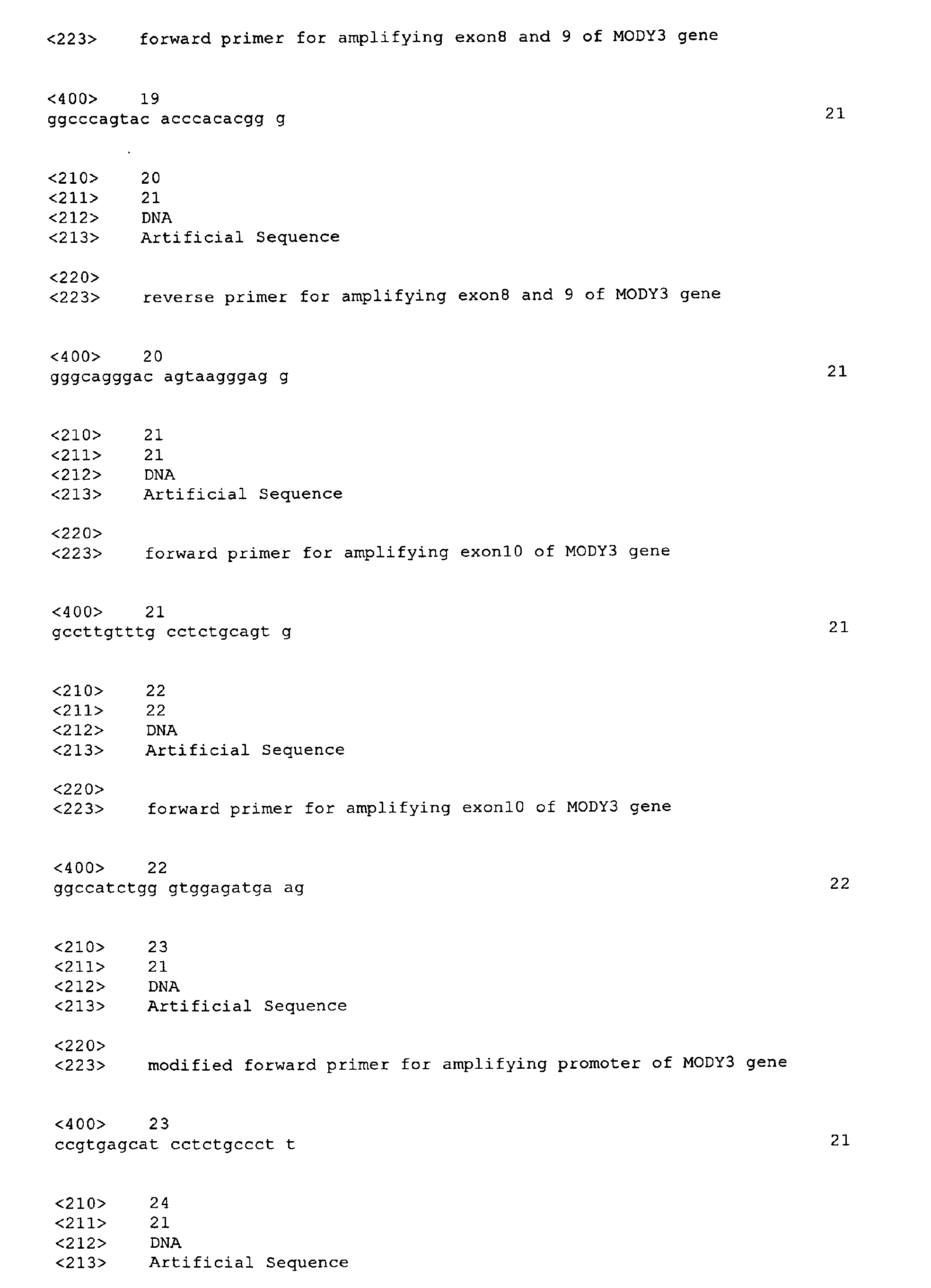
| Primers | sequence | size | Tm (°C) | PCR product (bp) |
| Set | Promoter-F | SEQ ID NO.3 | 21 | 74.1 | 540 |
| Promoter-R | SEQ ID NO.4 | 21 | 73.6 |
| Set | Exon1-F | SEQ ID NO.5 | 20 | 77.5 | 420 |
| Exon1-R | SEQ ID NO.6 | 21 | 72.3 |
| Set | Exon2-F | SEQ ID NO.7 | 22 | 72.7 | 326 |
| Exon2-R | SEQ ID NO.8 | 21 | 71 |
| Set | Exon3-F | SEQ ID NO.9 | 20 | 71.1 | 269 |
| Exon3-R | SEQ ID NO.10 | 23 | 69.6 |
| Set | Exon4-F | SEQ ID NO.11 | 23 | 70 | 293 |
| Exon4-R | SEQ ID NO.12 | 23 | 76.3 |
| Set | Exon5-F | SEQ ID NO.13 | 22 | 73.6 | 202 |
| Exon5-R | SEQ ID NO.14 | 23 | 70 |
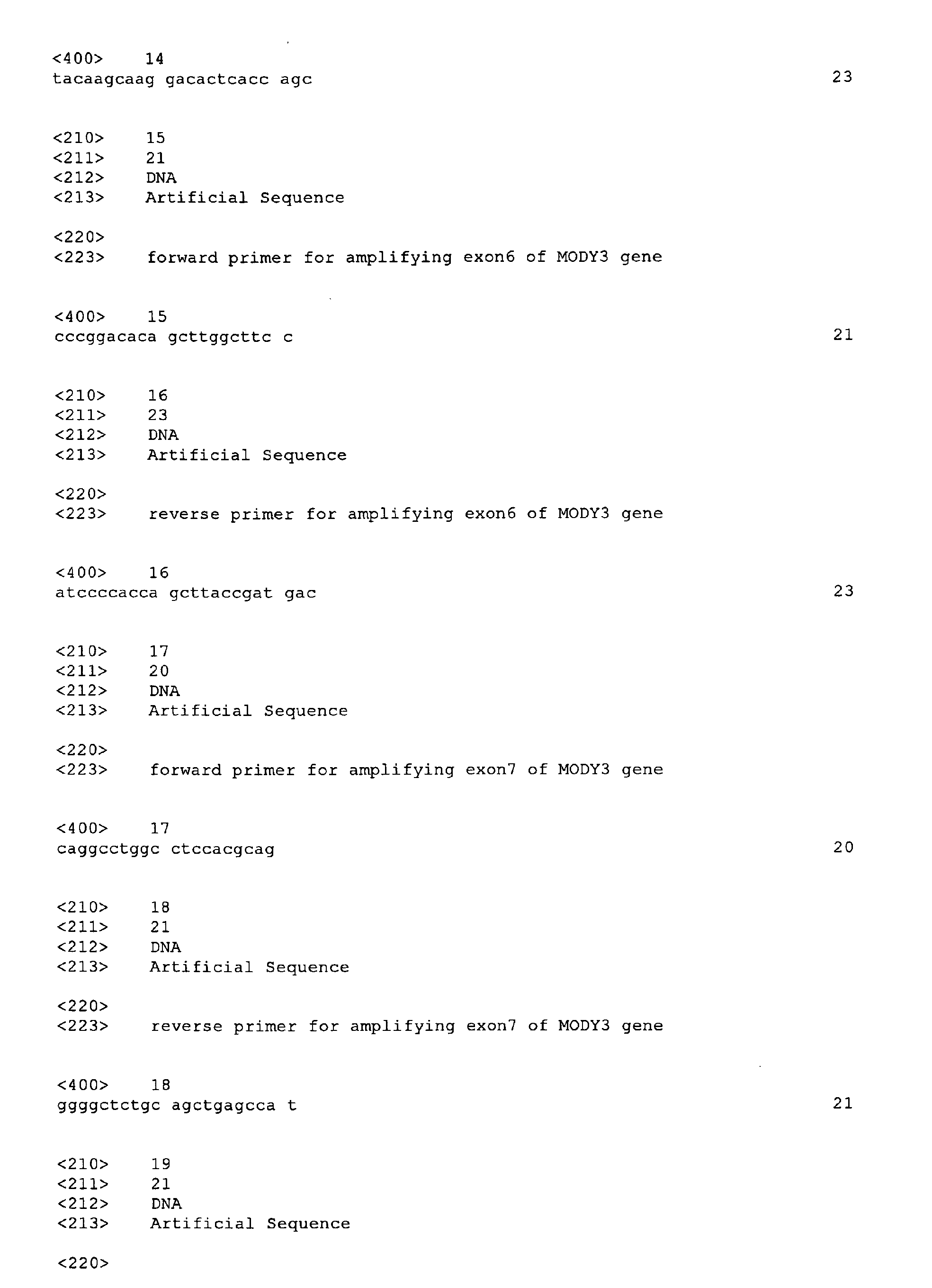
| Set | Exon6-F | SEQ ID NO.15 | 21 | 71.25 | 247 |
| Exon6-R | SEQ ID NO.16 | 23 | 72 |
| Set | Exon7-F | SEQ ID NO.17 | 20 | 75.99 | 191 |
| Exon7-R | SEQ ID NO.18 | 21 | 74.17 |
| Set | Exon8&9-F | SEQ ID NO.19 | 21 | 76.16 | 375 |
| Exon8&9-R | SEQ ID NO.20 | 21 | 73.14 |
| Set | Exon10-F | SEQ ID NO.21 | 21 | 68.05 | 132 |
| Exon10-R | SEQ ID NO.22 | 22 | 70.53 |
| F : forward R : reverse |
Each target sequence of a human HNF-1α gene was amplified by a single
PCR using each set of primers according to Example 1. The PCR was completed
through initial denaturation (5 mins at 95°C), 30 cycles of denaturation (30 secs at
95°C), annealing (15 secs at 64°C) and polymerization (30 secs at 72°C), and final
extension (3 mins at 72°C). The composition of a reaction solution for the PCR was
as follows:
| Sterilized DNase and RNase free water | 12.8 µl |
| dNTP mix (each nucleotide 2.5 mM) | 2 µℓ |
| 10x Taq polymerase buffer | 2 µℓ |
| a set of primers (each primer 10 pmol) | 2 µℓ |
| genomic DNA (200-1.0 µg) | 1 µℓ |
| Taq polymerase (5 unit/µℓ) | 0.2 µℓ |
The PCR product was analyzed by electrophoresis with 1.8 % agarose gel
(FIG.1). In FIG.1, lanes 1 and 13 represent molecular markers, 50 bp DNA ladders.
Lane 2 represents a PCR products corresponding to promoter, and lanes 3-11
represent PCR products corresponding exons 1, 2, 3, 4, 5, 6, 7, 8&9 and 10,
repectively. Lane 12 represents a multiplex PCR product according to the following
Example 3.
As shown in FIG.1, a target sequence of a human HNF-1α gene was amplified
by a single PCR using a set of primers according to Example 1.
Example 3: Amplification of a human HNF-1α gene by a multiplex PCR
A multiple PCR was conducted by using a set of primers according to Example
1. The reaction was conducted through initial denaturation (5 mins at 95°C), 30
cycles of denaturation (30 secs at 95°C), annealing (15 secs at 64°C) and
polymerization (30 secs at 72°C), and final extension (3 mins at 72°C). All the
primers were added in a single reaction tube. The composition of the reaction
solution was as follows:
| Sterilized DNase and RNase free water | 18.4 µℓ |
| dNTP mix (each nucleotide 2.5 mM) | 5 µℓ |
| 10x Taq polymerase buffer | 5 µℓ |
| a set of primers (each primer 10 pmol) | 20 µℓ |
| genomic DNA (200-1.0 µg) | 1 µℓ |
| Taq polymerase (5 unit/µℓ) | 0.6 µℓ |
The PCR product was analyzed by an electrophoresis with 1.8 % agarose gel
(FIG.1 lane 12). As shown in FIG.1, as a result of a multiplex PCR using the set of
primers according to Example 1, all of the 10 target sequences (579, 459, 365, 308,
332, 241, 286, 230, 414, and 171 bp) of a human HNF-1α gene were amplified.
Example 4 : Amplification of a human HNF-1α qene by a multiplex PCR
using a set of variant primers
A set of variant primers was synthesized by adding 3 nucleotides
complementary to the template at one end of 4 sets of primers (primers sets for
amplifying promoter,
exons 1, 2 and 8) and by deleting 3 nucleotides at the other end
of each primer (Table 2). A multiplex PCR was conducted by the same method as
described in the Example 3 except that one set of primers among above 4 sets of
primers and the other 3 corresponding sets of primers shown in Table 1 were used
(FIG. 2).
Fig. 2A through 2E are photographs for illustrating electrophoresis results of a
PCR product. Fig. 2A, 2B, 2C and 2D show electrophoresis results of PCR products
obtained using a primer pool comprising a set of variant primers for amplifying
promoter, exon 1, 2 or 8 shown in Table 2. Fig. 2E illustrates an electrophoresis
result of a multiplex PCR product obtained using a primer pool comprising all said
sets of variant primers for promoter, exons 1, 2 and 8. In Fig. 2A through 2E, the
lane 1 represents a molecular marker, a 50 bp DNA ladder. Lane 2 represents a
multiplex PCR result obtained by using a set of primers listed in Table 1, lane 3
represents a multiplex PCR result obtained by using a primer pool comprising a set
of variant primers for promoter, exons 1, 2 or 8 listed in Table 2 and a set of primers
listed in Table 1 without the corresponding set of variant primers, and lane 4
represents a multiplex PCR result obtained by using a primer pool comprising a set
of primers listed in Table 1 without the set of primers for amplifying promoter, exon 1,
2 or 8 (Fig. 2A, B, C and D) or without the set of primers for amplifying promoter,
exon 1, 2 and 8 (Fig. 2E).
As shown in Fig.2, a corresponding target sequence of a gene was amplified
by a multiplex PCR using a set of variant primers.
Example 5 : Identification of a multiplex PCR product through a southern
blotting analysis
PCR products amplified according to Examples 2 and 3 were electrophoresed
with 1.8 % agarose gel (FIG.1). The gel was added into a denaturation solution
(0.5N NaOH + 1.5M NaCI) with stirring for 15 minutes to denature the doublestranded
DNA. After performing the denaturation process twice, repeating two times,
the gel was washed with distilled water, then the gel was added into a neutralization
solution (3M NaCI containing 0.5M Tris-HCI, pH 7.5) for 15 minutes with gentle
stirring to neutralize the gel. After performing the neutralization procedure twice, a
DNA within the gel was transferred to a nylon membrane by reacting the gel with the
nylon membrane in 20XSSC solution for 12.5 hours. The DNA was cross-linked to
the nylon membrane by reacting for 30 minutes at 120°C, and the membrane was
washed for 1-2 minutes and dried.
The obtained DNA attached-membrane was then put into 20ml of a
hybridization solution for about 2 hours at 62°C for prehybridization and the solution
was discarded. The DNA attached-membrane and 5pmol/ml of DIG (digoxigenin)
labeled probe were added into 10ml of a fresh hybridization solution bag and reacted
for about 12 hours at 62°C. During the reaction, the DIG labeled probe hybridized to
a complementary region of a gene. After the reaction, the membrane was washed
twice with 2X washing solution (2XSSC + 0.1 % SDS) for 5 minutes at room
temperature, and washed twice with 0.5X washing solution (0.5XSSC + 0.1 % SDS)
for 15 minutes at 62°C.
The membrane was introduced into 20ml of a blocking solution for about 30-60
minutes, then the membrane was reacted with anti-DIG antibody in 20 ml of the
blocking solution containing 1 µl of an alkaline phosphatase-conjugated sheep anti-DIG
antibody (Roche) for about 30 minutes. The membrane was then washed twice
with a washing solution (100 mM maleic acid, 150 mM NaCI, pH 7.5 at room
temperature, 0.3 % Tween 20) for about 15 minutes. To confirm the hybridization
between DNA and a probe on the membrane, an alkaline phosphatase substrate,
CDP-Star was mixed with a detection buffer solution with a ratio of 1:100 and the
membrane was treated with this solution for about 5 minutes, and an X-ray film was
exposed to this membrane. The film was developed to confirm the position of a
probe (FIG.3). As shown in FIG.3, lane 13 represents a molecular marker labeled
with DIG, and each of 2 through 11 represents a single In Fig.3, lane 13 represents a
molecular marker labeled with DIG, and each of lanes 2 through 11 represents a
single PCR product of promoter, or exons 1, 2, 3, 4, 5, 6, 7, 8&9, or 10 respectively.
Lane 12 represents a multiplex PCR product.
As shown in Fig.3, the probe hybridized into a single PCR product and a
multiplex PCR product at the same position, which means that the amplified product
is substantially identical.
Example 6 : Identification of a multiplex PCR product through a
sequencing analysis
A multiplex PCR product amplified according to Examples 3 was
electrophoresed with 1.8 % agarose gel. An Individual DNA band was isolated, and
purified with a gel extraction kit. The sequence of the purified DNA was identified by
ABI3700, and compared with a corresponding known sequence of a human HNF-1α
gene (Fig.4).
Fig.4 illustrates a PCR product corresponding to a PCR product obtained by
using a set of primers for amplifying promoter. The PCR product corresponding to a
PCR product obtained by using a set of primers SEQ ID NOS. 3 and 4 in a multiplex
PCR was isolated and sequenced by using an automatic sequencing method. The
resultant nucleotide sequence (SEQ ID NO. 31) (referred to as "Query") was
compared with a known sequence of a human HNF-1α gene promoter (referred to as
"Promoter") by a program called Lagergene (DNAstar Inc.) (Fig.4). As shown in Fig.4,
the PCR product showed 100 % sequence homology with the known sequence of a
human HNF-1α promoter. The gap frequency used in the alignment was 0 %. In
addition to the promoter, single PCR products of the other exons were also
sequenced and analyzed in the same way as described above, and each PCR
product showed 95 % or more of homologies with a known corresponding exon
sequence.
Example 7 : Stability test of a reaction solution for the multiplex PCR
The reaction solution for the multiplex PCR used in Example 3 was prepared
in large quantities, excluding genomic DNA, to test the stability thereof. An amount
of 49 µℓ of the reaction solution, which was required for one PCR, was alliquoted into
each of a plurality of 0.2ml-PCR tubes. Some of the tubes including the reaction
solution were kept at -20°C, and others were kept at -70°C, for three months.
Multiplex PCR was performed using the reaction solution in the tube at 1 day, 3 days,
4 days, 1 week, 2 weeks, 3 weeks, and 12 weeks, respectively. Fig.5 shows the
electrophoresis results of the multiplex PCR products. In Fig.5, the first lane from the
left is a molecular marker, lane 2 is a multiplex PCR result obtained using a reaction
solution kept at -20°C, and lane 3 is a multiplex PCR result obtained using a reaction
solution kept at -70°C. As shown in Fig.5, the reaction solution for the multiplex PCR
used in the present invention was stable at -20°C or -70°C, and it did not degenerate
PCR product therein even after a period of three months.
Example 8 : Gene amplifications when using various PCR devices
Multiplex PCRs were conducted according to the method of Example 3 using
various PCR devices. The amplified multiplex PCR products were electrophoresed
with 1.8 % agarose gel to examine the dependency of the PCR products upon PCR
device (Fig.6A through C).
Fig.6A represents the multiplex PCR product amplified using PTC-100 (MJ
Research Co.), Fig.6B represents the multiplex PCR product amplified using
GeneAmp 9700 (Applied Biosystems Co.), and Fig.6C represents the multiplex PCR
product amplified using Multi-Block System (ThermoHybaid Co.).
In Fig.6A to 6C, the lane 1 is a molecular marker, a 50 bp DNA ladder, and
lane 2 is a multiplex PCR result obtained by using a set of primers shown in Table 1.
Arrows in the figure represent 350 bp and 50bp, respectively.
As shown in Fig.6, similar PCR product-patterns were obtained independently
of PCR devices, which indicates that the PCR product obtained by using a set of
primers of the present invention is stable and economical.
The primer pool for amplifying a human HNF-1α gene of the present invention
is effective in amplifying a corresponding gene by a multiplex PCR. Particularly, the
primer pool for amplifying a gene by a multiplex PCR is useful for an analysis of a
disease-associated gene by using a DNA chip since the multiplex PCR requires
relatively a reduced number of samples.
The primer pool for amplifying a human HNF-1α gene of the present invention
may then be applied to a kit for amplifying or sequencing a human HNF-1α gene.
In the specification, there have been disclosed typical preferred embodiments
of the invention and, although specific terms are employed, they are used in a
generic and descriptive sense only and not for purposes of limitation of the scope of
the invention, that being set forth in the following claims.