EP1316560A1 - Novel human cancer/testis antigen and gene thereof - Google Patents
Novel human cancer/testis antigen and gene thereof Download PDFInfo
- Publication number
- EP1316560A1 EP1316560A1 EP01963516A EP01963516A EP1316560A1 EP 1316560 A1 EP1316560 A1 EP 1316560A1 EP 01963516 A EP01963516 A EP 01963516A EP 01963516 A EP01963516 A EP 01963516A EP 1316560 A1 EP1316560 A1 EP 1316560A1
- Authority
- EP
- European Patent Office
- Prior art keywords
- protein
- cancer
- peptide
- cell
- amino acid
- Prior art date
- Legal status (The legal status is an assumption and is not a legal conclusion. Google has not performed a legal analysis and makes no representation as to the accuracy of the status listed.)
- Withdrawn
Links
Images




Classifications
-
- C—CHEMISTRY; METALLURGY
- C07—ORGANIC CHEMISTRY
- C07K—PEPTIDES
- C07K14/00—Peptides having more than 20 amino acids; Gastrins; Somatostatins; Melanotropins; Derivatives thereof
- C07K14/82—Translation products from oncogenes
-
- A—HUMAN NECESSITIES
- A61—MEDICAL OR VETERINARY SCIENCE; HYGIENE
- A61P—SPECIFIC THERAPEUTIC ACTIVITY OF CHEMICAL COMPOUNDS OR MEDICINAL PREPARATIONS
- A61P35/00—Antineoplastic agents
-
- A—HUMAN NECESSITIES
- A61—MEDICAL OR VETERINARY SCIENCE; HYGIENE
- A61P—SPECIFIC THERAPEUTIC ACTIVITY OF CHEMICAL COMPOUNDS OR MEDICINAL PREPARATIONS
- A61P37/00—Drugs for immunological or allergic disorders
- A61P37/02—Immunomodulators
- A61P37/04—Immunostimulants
-
- A—HUMAN NECESSITIES
- A61—MEDICAL OR VETERINARY SCIENCE; HYGIENE
- A61P—SPECIFIC THERAPEUTIC ACTIVITY OF CHEMICAL COMPOUNDS OR MEDICINAL PREPARATIONS
- A61P37/00—Drugs for immunological or allergic disorders
- A61P37/02—Immunomodulators
- A61P37/06—Immunosuppressants, e.g. drugs for graft rejection
-
- C—CHEMISTRY; METALLURGY
- C07—ORGANIC CHEMISTRY
- C07K—PEPTIDES
- C07K14/00—Peptides having more than 20 amino acids; Gastrins; Somatostatins; Melanotropins; Derivatives thereof
- C07K14/435—Peptides having more than 20 amino acids; Gastrins; Somatostatins; Melanotropins; Derivatives thereof from animals; from humans
- C07K14/46—Peptides having more than 20 amino acids; Gastrins; Somatostatins; Melanotropins; Derivatives thereof from animals; from humans from vertebrates
- C07K14/47—Peptides having more than 20 amino acids; Gastrins; Somatostatins; Melanotropins; Derivatives thereof from animals; from humans from vertebrates from mammals
- C07K14/4701—Peptides having more than 20 amino acids; Gastrins; Somatostatins; Melanotropins; Derivatives thereof from animals; from humans from vertebrates from mammals not used
-
- C—CHEMISTRY; METALLURGY
- C07—ORGANIC CHEMISTRY
- C07K—PEPTIDES
- C07K14/00—Peptides having more than 20 amino acids; Gastrins; Somatostatins; Melanotropins; Derivatives thereof
- C07K14/435—Peptides having more than 20 amino acids; Gastrins; Somatostatins; Melanotropins; Derivatives thereof from animals; from humans
- C07K14/46—Peptides having more than 20 amino acids; Gastrins; Somatostatins; Melanotropins; Derivatives thereof from animals; from humans from vertebrates
- C07K14/47—Peptides having more than 20 amino acids; Gastrins; Somatostatins; Melanotropins; Derivatives thereof from animals; from humans from vertebrates from mammals
- C07K14/4701—Peptides having more than 20 amino acids; Gastrins; Somatostatins; Melanotropins; Derivatives thereof from animals; from humans from vertebrates from mammals not used
- C07K14/4748—Tumour specific antigens; Tumour rejection antigen precursors [TRAP], e.g. MAGE
-
- G—PHYSICS
- G01—MEASURING; TESTING
- G01N—INVESTIGATING OR ANALYSING MATERIALS BY DETERMINING THEIR CHEMICAL OR PHYSICAL PROPERTIES
- G01N33/00—Investigating or analysing materials by specific methods not covered by groups G01N1/00 - G01N31/00
- G01N33/48—Biological material, e.g. blood, urine; Haemocytometers
- G01N33/50—Chemical analysis of biological material, e.g. blood, urine; Testing involving biospecific ligand binding methods; Immunological testing
- G01N33/5005—Chemical analysis of biological material, e.g. blood, urine; Testing involving biospecific ligand binding methods; Immunological testing involving human or animal cells
- G01N33/5008—Chemical analysis of biological material, e.g. blood, urine; Testing involving biospecific ligand binding methods; Immunological testing involving human or animal cells for testing or evaluating the effect of chemical or biological compounds, e.g. drugs, cosmetics
- G01N33/5044—Chemical analysis of biological material, e.g. blood, urine; Testing involving biospecific ligand binding methods; Immunological testing involving human or animal cells for testing or evaluating the effect of chemical or biological compounds, e.g. drugs, cosmetics involving specific cell types
- G01N33/5047—Cells of the immune system
- G01N33/505—Cells of the immune system involving T-cells
-
- A—HUMAN NECESSITIES
- A61—MEDICAL OR VETERINARY SCIENCE; HYGIENE
- A61K—PREPARATIONS FOR MEDICAL, DENTAL OR TOILETRY PURPOSES
- A61K39/00—Medicinal preparations containing antigens or antibodies
-
- G—PHYSICS
- G01—MEASURING; TESTING
- G01N—INVESTIGATING OR ANALYSING MATERIALS BY DETERMINING THEIR CHEMICAL OR PHYSICAL PROPERTIES
- G01N2500/00—Screening for compounds of potential therapeutic value
Definitions
- the present invention relates to: a cancer/testis antigen protein or peptide; a cancer/testis gene encoding the cancer/testis antigen protein or the peptide; a screening method for a promoter or a suppressor for the immunity-inducing activity using the cancer/testis antigen protein/peptide; an anti-tumor agent with the cancer/testis antigen protein or a partial peptide of the antigen protein or the like as an effective ingredient; a diagnostic probe for cancer using the cancer/testis gene; etc.
- Tumor cells often express various genes such as a particular kind of protooncogenes which are not usually expressed in normal cells or at a considerably low level even if expressed (Science 235, 305-11, 1987). Gene populations expressed in such an abnormal manner include populations that are involved in tissue-specific differentiation (Immunol. Today 18, 267-8, 1997), and the expression of these genes in tumor cells are thought to be responsible for the malignant phenotype of the tumor cells. Many testis-specific genes are expressed in the testis (Reprod. Fertility & Develop. 7, 695-704, 1995, Int. J. Develop. Biol. 40, 379-83, 1996), however, most of these genes are reported not to be activated at all in ordinary tumors or very scarcely if any (Biochem.
- genes mentioned above including genes encoding cancer/testis antigens are called cancer/testis-related genes and are classified as shown in Table 1 below.
- genes such as MAGE, BAGE, GAGE, LAGE, SSX, etc. are known (Science 254, 1643-7, 1991, Immunity 2, 167-75, 1995, J. Exp. Med. 182, 689-98, 1995, Int. J. Cancer 76, 903-8, 1998, Int. J. Cancer 72, 965-71, 1997, Cancer Res. 56, 4766-72, 1996).
- These antigens are identified according to their production of antibodies or responsiveness to cytotoxic T lymphocytes in cancer patients (Proc. Natl.
- cancer/testis-related genes include some genes whose antigenicity is not yet elucidated (The Cancer J. from Scientific American, 16-7, 1999, Proc. Natl. Acad. Sci. USA 92, 11810-3, 1995, Cancer Res. 59, 3215-21, 1999. Proc. Natl. Acad. Sci. USA 95, 10757-62, 1998, Cancer Res. 59, 1445-8, 1999). There is a possibility that the in vivo expression of the former is frequent enough to raise immune response while the expression of the latter is not enough.
- MAGE subfamily exists in four regions on X chromosome, i.e. Xq28, Xq21.3, Xq26 and Xq11.23 (Immunogenet. 40, 360-9, 1994, Cancer Res. 58, 743-52, 1998, Genomics 59, 161-7, 1999).
- SSX genes exist on Xq11.2 (Nature Genet.
- SCP1 chromosome 1
- rejection of a living body to cancer is known to be caused by cytotoxic T lymphocytes which have recognized the complex of a cancer antigen peptide and a histocompatibility antigen molecule (HLA) that are bound in a tissue or in a cell. Because a cancer antigen binds to HLA which is expressed in the tumor cell, it is known that a cytotoxic lymphocyte recognizes the complex as an antigen and thus causes rejection which results in the regression of a cancer cell. Marie Marchand et al.
- the transcription factor GCF is known as a factor which binds to the transcription regulatory region of a EGF (epidermal growth factor) receptor gene (Cell 59, 815-25, 1989).
- EGF epidermal growth factor
- GCFcDNA has recently been proved as being an artificial fusion molecule (Biochem. Biophys. Acta 1447, 125-31, 1999). That is to say, its sequence on the amino-terminal is derived from GCF2 which is a recently and newly identified transcription factor (J Biol. Chem. 273, 21594-602, 1998), and the most of the remaining cDNA is comprised of a part tentatively named GCF1 by the present inventors.
- GCF1 is devoid of the DNA-binding activity (Biochem. Biophys. Acta 1447, 125-31, 1999).
- a gene encoding a protein which specifically binds to GCF1 is a cancer/testis-related gene.
- yeast Two-hybrid System (Nature 340, 245-6, 1989, Proc. Natl. Acad. Sci. USA 88, 9578-82, 1991) is known as a system for detecting the in vivo protein-protein interactions using saccaromyces yeast.
- a usually used protein in the yeast Two-hybrid System is Gal4, a transcription activator for yeast which has a DNA-binding domain and a transcription-activating domain that can be separated from one another.
- a plasmid vector carrying a gene encoding a fusion protein of the Gal4 DNA-binding domain and protein X and a plasmid vector carrying a gene encoding a fusion protein of the Gal4 transcription-activating domain and protein Y are introduced into yeast containing a reporter gene such as LacZ gene, HIS3 gene, etc. and then the X-Y interaction can be detected by observing whether the function of Gal4 recovers and the transcription of a reporter gene is activated.
- a known protein is used as X and cDNA library is used as a gene encoding Y in this yeast Two-hybrid System
- a gene encoding a protein which interacts with X can be screened from the aforementioned cDNA library.
- Cancer antigens are classified as in Table 2 based on the recent molecular biological studies. Since no class I molecule of a human histocompatibility antigen molecule (HLA) is expressed in the testis, a protein on a testis cell membrane is not recognized by a T lymphocyte. This gives us the view that a cancer/testis antigen is a cancer-specific antigen in a narrow sense because immunologically it is highly specific to cancer in spite of the fact that it is expressed in the testis.
- HLA human histocompatibility antigen molecule
- tissue-specific differentiated antigens are also expressed, though weakly, in normal tissues, adverse reaction on normal cells is causing drawbacks in performing cancer immunotherapy, and mutation-derived cancer antigens have drawbacks as well in that such mutation is limited only to the individual cancers, and therefore these antigens are hardly regarded as appropriate for the use in cancer immunotherapy.
- cancer/testis antigens are not expressed in normal tissues other than in the testis and are expressed in various cancers over a broad range, so that their use in immunotherapy agents for cancer is highly expected.
- the object of the present invention is to a provide a cancer/testis antigen applicable to diagnosis and immunotherapy for cancer, that is to say, a cancer/testis antigen which is not expressed in normal tissues other than the testis and which is expressed in various cancers over a broad range and induces immune response in a host, and a cancer/testis gene encoding the cancer/testis antigen and the like.
- the present inventors have made a keen study to attain the objects mentioned above and by using the yeast Two-hybrid System, the present inventors screened a protein which interacts with GCF (GC element binding Factor), a previously reported transcription factor, as a gene controlling the proliferation of mammalian cells, cloned a gene for a protein which specifically binds to the transcription factor GCF, confirmed that the protein D40, the resulting gene product, is a cancer/testis antigen which is expressed substantially only in the testis in normal human tissues but is expressed in human primary cancers over a broad range deriving from various tissues and cells, and found that the D40 shows high affinity with the human histocompatibility antigen HLA and has a sequence comprising 9 or 10 amino acid residues which binds to a plurality of HLAs.
- GCF GC element binding Factor
- the present invention relates to: a gene encoding the following protein (a) or (b), (a) a protein comprising the amino acid sequence shown by SEQ ID NO:2, (b) a protein which comprises an amino acid sequence wherein one or a few amino acids are deleted, substituted or added in the amino acid sequence shown by SEQ ID NO:2 and which has the immunity-inducing activity (claim 1); a gene encoding the following protein (a) or (b), (a) a protein comprising the amino acid sequence shown by SEQ ID NO:3, (b) a protein which comprises an amino acid sequence wherein one or a few amino acids are deleted, substituted or added in the amino acid sequence shown by SEQ ID NO:3 and which has the immunity-inducing activity (claim 2); DNA containing the base sequence shown by SEQ ID NO:1 or its complementary sequence and part of whole of these sequences (claim 3); and DNA which hybridizes under a stringent condition with DNA of claim 3 that forms a gene, and which encodes a protein having the immunity-in
- the present invention further relates to: a protein comprising the amino acid sequence shown by SEQ ID NO:2 (claim 5); a protein which comprises an amino acid sequence wherein one or a few amino acids are deleted, substituted or added in the amino acid sequence shown by SEQ ID NO:2 and which has the immunity-inducing activity (claim 6); a protein comprising the amino acid sequence shown by SEQ ID NO:3 (claim 7); and a protein which comprises an amino acid sequence wherein one or a few amino acids are deleted, substituted or added in the amino acid sequence shown by SEQ ID NO:3 and which has the immunity-inducing activity (claim 8).
- the present invention still further relate to: a peptide which comprises part of the protein of any of claims 5 to 8 and which binds to a histocompatibility antigen molecule Class I (claim 9); the peptide according to claim 9, wherein said peptide comprises part of the protein of claim 5 or 6 and binds to a histocompatibility antigen molecule Class I (claim 10); the peptide according to claim 10, wherein said peptide comprises an amino acid sequence shown by any of SEQ ID NOs:4-111 (claim 11); a peptide comprising the amino acid sequence of Ser-Tyr-Thr-Ile-Glu-Ile-Asn-His-Arg-Leu (claim 12); the peptide according to claim 9, wherein said peptide comprises part of the protein of claim 7 or 8 and binds to the histocompatibility antigen molecule Class I (claim 13); and the peptide according to claim 13, wherein said peptide comprises an amino acid sequence shown by any of SEQ ID NOs: 112-221 (claim
- the present invention also relates to: a fusion protein or a fusion peptide wherein the protein of claim 5 or 6 or the peptide of any of claims 10 to 12, and a marker protein and/or a peptide tag, are bound (claim 15); a fusion protein or a fusion peptide wherein the protein of claim 7 or 8 or the peptide of claim 13 or 14, and a marker protein and/or a peptide tag, are bound (claim 16); an antibody which specifically binds to the protein of claim 5 or 6 or to the peptide of any of claims 10 to 12 (claim 17); an antibody which specifically binds to the protein of claim 7 or 8 or to the peptide of claim 13 or 14 (claim 18); the antibody according to claim 17 or 18, wherein said antibody is a monoclonal antibody (claim 19); and a recombinant protein or peptide wherein the antibody of any of claims 17 to 19 specifically binds to said recombinant protein or peptide (claim 20).
- the present invention further relates to: a host cell comprising an expression system capable of expressing the protein according to claim 5 or 6 (claim 21); a host cell comprising an expression system capable of expressing the protein according to claim 7 or 8 (claim 22); a non-human animal whose gene function to encode the protein of claim 5 or 6 is deficient on its chromosome (claim 23); a non-human animal whose gene function to encode the protein of claim 7 or 8 is deficient on its chromosome (claim 24); the non-human animal according to claim 23 or 24, wherein said non-human animal is a mouse or a rat (claim 25); a non-human animal which over-expresses the protein of claim 5 or 6 (claim 26); a non-human animal which over-expresses the protein of claim 7 or 8 (claim 27); and the non-human animal according to claim 26 or 27, wherein said non-human animal is a mouse or a rat (claim 28).
- the present invention still further relates to: a method of screening a promoter or a suppressor for the immunity-inducing activity wherein the protein of claim 5 or 6, the peptide of any of claims 10 to 12 or a cell membrane expressing the protein of claim 5 or 6, and a test substance are used (claim 29); a method of screening a promoter or a suppressor for the immunity-inducing activity wherein the protein of claim 7 or 8, the peptide of claim 13 or 14, or a cell membrane expressing the protein of claim 7 or 8, and a test substance are used (claim 30); a method of screening a promoter or a suppressor for the immunity-inducing activity or for the expression of the protein of claim 5 or 6, wherein a cell expressing the protein and a test substance are used (claim 31); the method of screening a promoter or a suppressor for the immunity-inducing activity or for the expression of the protein of claim 5 or 6 according to claim 31, wherein the cell expressing the protein of claim 5 or 6 is the host cell of claim 21 (claim
- the present invention further relates to: a method for detecting a cytotoxic T cell or its precursor cell wherein a HLA molecule, and the peptide of any of claims 9 to 16 or the peptide of claim 20 are used (claim 43); a method for detecting a cytotoxic T cell or its precursor cell wherein a complex of a HLA Class I molecule, and the peptide of any of claims 9 to 16 or the peptide of claim 20 is formed on the surface of fluorescent microparticles (claim 44); a detection reagent for a cytotoxic T cell or its precursor cell, wherein said detection reagent comprises a HLA molecule, and the peptide of any of claims 9 to 16 or the peptide of claim 20 (claim 45); a detection reagent for a cytotoxic T cell or its precursor cell wherein said detection reagent comprises: a complex of a HLA Class I molecule, and the peptide of any of claims 9 to 16 or the peptide of claim 20; and a fluorescent microparticle (
- a cancer/testis-related gene of the present invention can be screened by a yeast Two-hybrid System as a gene encoding a protein which interacts with the transcription factor GCF, from a cDNA library or a genomic DNA library. Further, a cancer/testis antigen protein of the present invention can be obtained by expressing the cancer/testis-related gene obtained by such screening.
- a cancer/testis-related gene is defined as follows: its expression is observed specifically in the normal testis and is not substantially observed in other normal tissues; it activates in a malignant tumor, as a result of which high level mRNA is detected; and it is expressed in a tissue-non-specific manner in a malignant tumor.
- Fig. 1 schematizes a yeast Two-hybrid System using the transcription factor GCF.
- a transcription factor GCF is expressed as a fusion protein with a DNA-binding domain of Gal4 (GalDBD-GCF), whereas a protein derived from a cDNA library is expressed as a fusion protein with a transcriptional activation domain of Gal4.
- the interaction (binding) of GalDBD-GCF and a cDNA library-derived protein activates transcription of a reporter gene and thereby the expression of His3 gene and LacZ is raised.
- cDNA encoding a protein which interacts with GCF therefore, can be cloned by isolating yeast in which a reporter gene is expressed.
- MATCHMAKER Clontech
- HybriZAP HybriZAP
- the cancer/testis antigen protein D40 is exemplified.
- a specific example of a cancer/testis-related gene encoding this D40 is a gene having the base sequence shown by SEQ ID NO: 1 in the sequence listing.
- proteins as objects of the present invention specifically include: the D40 (ORF1) protein shown by SEQ ID NO:2 or the D40 (ORF2) protein shown by SEQ ID NO:3; a protein comprising an amino acid sequence in which one or a few amino acids are deleted, substituted or added in the amino acid sequence shown by SEQ ID NO:2 or 3 and which has the immunity-inducing activity; and the recombinant proteins of these.
- the immunity-inducing activity here means an activity inducing immune response such as antibody production, cellular immunity, immune tolerance, etc. in a host.
- the T cell-inducing activity by which the frequency of cytotoxic T lymphocyte (CTL) precursor cells is increased, is particularly preferable for a protein to possess.
- a peptide as an object of the present invention is a peptide which comprises a part of the protein of the present invention, for instance, a peptide comprising an amino acid sequence of five or more consecutive amino acids, and which binds to a histocompatibility antigen molecule (HLA) Class I.
- HLA histocompatibility antigen molecule
- the specific examples are: a peptide comprising an amino acid sequence shown by any of SEQ ID NOs:4-111 that are derived from the aforementioned D40 (ORF1) protein or the like, e.g.
- Ser-Tyr-Thr-Ile-Glu-Ile-Asn-His-Arg-Leu SEQ ID NO:36
- a peptide comprising an amino acid sequence shown by any of SEQ ID NOs:112-221 that are derived from the aforementioned D40 (ORF2) protein or the like.
- HLA Class I the specific examples include: HLA-A3, HLA-68, HLA-A1, HLA-A24, HLA-A0201, HLA-A0205, HLA-B7, HLA-B8, HLA-B14, HLA-B40. B1501 (B62), etc.
- Proteins and peptides as objects of the present invention together with the recombinant proteins and peptides to which antibodies specifically bind while such antibodies specifically bind to the said proteins and peptides, may collectively be referred to as "the present proteins/peptides".
- the present proteins/peptides can be prepared in accordance with the known methods based on their DNA sequence information or the like and these antigens will not be limited to those derived from human.
- Genes or DNAs as objects of the present invention are specifically exemplified by: a gene encoding the D40 (ORF1) protein which comprises the amino acid sequence shown by SEQ ID NO:2; DNA encoding a protein which comprises an amino acid sequence wherein one or a few amino acids are deleted, substituted or added in the amino acid sequence shown by SEQ ID NO:2 and which has the immunity-inducing activity; a gene encoding the D40 (ORF2) protein comprising the amino acid sequence shown by SEQ ID NO:3; DNA encoding a protein which comprises an amino acid sequence wherein one or a few amino acids are deleted, substituted or added in the amino acid sequence shown by SEQ ID NO:3 and which has the immunity-inducing activity; and DNA comprising the base sequence shown by SEQ ID NO:1 or its complementary sequence and part or whole of these sequences.
- genes or DNAs can be prepared by the known methods from, for instance, a human gene library or a human cDNA library based on their DNA sequence information or the like. Besides, there is no limitation to the preparation methods for cDNA which encodes the D40 protein and it can be obtained, for example, from a mRNA-derived cDNA library which is derived from HL60 or the T cell leukemia cell line Jurkat.
- DNA encoding a protein of the interest which has the same effect as the D40 gene, having the immunity-inducing activity can be obtained by the hybridization with a DNA library derived from various cancer cells under a stringent condition by using the base sequence shown by SEQ ID NO:1 or its complementary sequence and part or whole of these sequences as a probe, and then by the isolation of DNA which hybridizes with the probe.
- a hybridization condition for obtaining the DNA is exemplified by hybridization at 42. degree. C. and washing at 42.degree. C. in a buffer solution containing 1 ⁇ SSC, 0.1% SDS, and more preferable exemplification is hybridization at 65.degree. C. and washing at 65.degree. C.
- Any fusion protein and fusion peptide may be used as a fusion protein and a fusion peptide for the present invention as long as the present proteins/peptides are bound to marker proteins and/or peptide tags.
- a marker protein there is no limitation as long as it is a conventionally known marker protein and the specific examples include alkaline phosphatase, the Fc region of an antibody, HRP, GFP, etc.
- Conventionally known peptide tags including Myc tag, His tag, FLAG tag, GST tag, etc. are the specific examples of the peptide tags for the use in the present invention.
- fusion proteins can be generated according to the ordinary protocols and are useful for the following: purification of the D40 protein or the like using affinity of Ni-NTA and a His tag; detection of a protein having the T cell-inducing activity; quantification of an antibody against the D40 protein or the like; use as a diagnostic marker for uterine cervix cancer, epithelial-like cancer, T cell tumor, promyelocytic leukemia, esophageal cancer, pancreatic cancer, malignant melanoma, lung cancer, oral cancer, breast cancer, bladder cancer, uterine cancer, ovarian cancer, neuroblastoma, colon cancer, etc.; and use as a laboratory reagent in this field of art.
- Antibodies that specifically bind to the aforementioned proteins and peptides of the present invention can be particularly exemplified by immune-specific antibodies such as monoclonal antibodies, polyclonal antibodies, chimeric antibodies, single-stranded antibodies, humanized antibodies, etc. These antibodies can be generated according to the ordinary protocols using a protein, such as the above-mentioned D40 etc., or part of the protein as an antigen. However, monoclonal antibodies are more preferable than the other sorts of antibodies mentioned because of their specificity.
- Antibodies such as the monoclonal antibodies or the like are useful not only for diagnosis and therapy, such as missile therapy, for uterine cervix cancer, epithelial-like cancer, T cell tumor, promyelocytic leukemia, esophageal cancer, pancreatic cancer, malignant melanoma, lung cancer, oral cancer, breast cancer, bladder cancer, uterine cancer, ovarian cancer, neuroblastoma, colon cancer, etc., for instance, but for elucidating the development mechanism of malignant tumors such as oral cancer and the like.
- missile therapy for uterine cervix cancer, epithelial-like cancer, T cell tumor, promyelocytic leukemia, esophageal cancer, pancreatic cancer, malignant melanoma, lung cancer, oral cancer, breast cancer, bladder cancer, uterine cancer, ovarian cancer, neuroblastoma, colon cancer, etc.
- Antibodies of the present invention described above can be created by administering to an animal (preferably non-human) the present proteins/peptides, their fragments containing epitopes, or the cells expressing the said proteins on the membrane surface, according to the conventional protocols.
- the monoclonal antibodies can be prepared, for instance, by any optional method such as a hybridoma method that brings antibodies produced by cultured materials of continuous cell line (Nature 256, 495-497, 1975), a trioma method, a human B-cell hybridoma method (Immunology Today 4, 72, 1983), an EBV-hybridoma method (MONOCLONAL ANTIBODIES AND CANCER THERAPY, pp.77-96, Alan R. Liss, Inc., 1985), etc.
- the preparation method for a single chain antibody (US PAT.No. 4,946,778) can be adopted to prepare single-stranded antibodies to the present proteins/peptides of the present invention mentioned above.
- transgenic mice, other mammals, etc. can be used for expressing humanized antibodies
- clones expressing the present proteins/peptides can be isolated/identified using the antibodies mentioned above, and their polypeptides can be purified by affinity chromatography.
- Antibodies to the present proteins/peptides or to their peptides containing antigenic epitopes can possibly be used for diagnosis and therapy for various cancers as described earlier.
- recombinant proteins or peptides to which these antibodies specifically bind are also covered by the present proteins/peptides of the present invention as described earlier.
- the functions of the present proteins/peptides can be analyzed by using, for example, antibodies such as the aforementioned monoclonal antibodies and the like that are labeled with fluorescent materials such as FITC (Fluorescein isothiocyanate), tetramethylrhodamine isothiocyanate, etc., radioisotopes such as 125 I, 32 p, 14 C, 35 S, 3 H, etc., or enzymes such as alkaline phosphatase, peroxidase, â-galactosidase, phycoerythrin, etc. and by using fusion proteins fused with fluorescent proteins such as Green Fluorescent Protein (GFP), etc.
- RIA method, ELISA method, fluorescent-antibody method, plaque method, spot method, haemagglutination, Ouchterlony method, etc. are exemplified.
- the present invention also relates to a host cell comprising an expression system capable of expressing the present proteins/peptides of the above.
- Genes encoding the present proteins/peptides can be introduced into a host cell by the methods described in many standard laboratory manuals such as manuals of Davis et al. (BASIC METHODS IN MOLECULAR BIOLOGY, 1986) and of Sambrook et al.
- the examples of host cells include bacterial prokaryotic cells such as E.
- coli Streptomyces, Bacillus Subtilis, Streptococcus, Staphylococcus, etc.
- eukaryotic cells such as yeast, aspergillus, etc., insect cells such as Drosophila S2, Spodoptera Sf9, etc.
- animal cells such as L cell, CHO cell, COS cell, HeLa cell, C127 cell, BALB/c3T3 cell (including mutants deficient in dihydrofolate reductase, tymidine kinase, etc.), BHK21 cell, HEK293 cell, Bowes malignant melanoma cell, etc. and plant cells or the like.
- an expression system there is no limitation to an expression system as long as the expression system is capable of expressing the present proteins/peptides described above in a host cell and the examples include chromosome-, episome- and virus-derived expression systems, for instance, vectors derived from bacterial plasmid, vectors derived from yeast plasmid, vectors derived from papovavirus such as SV40, vaccinia virus, adenovirus, varicella virus, pseudorabies virus, retrovirus, and vectors derived from bacteriophage or transposon and vectors derived from the combination of these two, e.g. vectors derived from genetic factors of plasmid and bacteriophage, such as cosmid and phagemid.
- the expression systems may comprise a control sequence for regulating the expression in addition to a sequence for raising the expression.
- Host cells comprising the above-mentioned expression systems and the cell membranes of the cells, or the present proteins/peptides obtained by culturing the cells can be used in the screening methods of the present invention as described below.
- the method of F. Pietri-Rouxel et al. (Eur. J. Biochem., 247, 1174-1179, 1997) or the like can be used to obtain cell membranes.
- the known methods can be adopted including ammonium sulfate- or ethanol-precipitation, acid extraction, anion- or cation-exchange chromatography, phosphocellulose chromatography, hydrophobic interaction chromatography, affinity chromatography, hydroxyapatite chromatography and lectin chromatography, where the high performance liquid chromatography is preferably used.
- affinity chromatography columns to which antibodies to the present proteins/peptides are bound, for instance, are used, and when ordinary peptide tags are added to the present proteins/peptides mentioned above, columns to which substances having affinity with the peptide tags are bound are used in order to obtain the present proteins/peptides.
- the purification methods for the present proteins/peptides mentioned above may also be employed for peptide synthesis.
- a non-human animal whose gene function to encode the present proteins/peptides mentioned above is deficient on its chromosome means a non-human animal part or whole of whose gene on its chromosome encoding the present proteins/peptides is inactivated by gene mutations such as destruction, deletion, substitution, etc. so that whose function to express the present proteins/peptides is lost.
- a non-human animal which over-expresses the present proteins/peptides is specifically exemplified by a non-human animal which produces larger amount of the present proteins/peptides than a wild-type non-human animal does.
- rodents or the like such as mice, rats, etc. are particularly exemplified for non-human animals of the present invention, the examples will not be limited to these animals only.
- Homozygous non-human animals that are born according to Mendel's Law include the deficient type or the over-expressing type for the present proteins/peptides as well as their wild type littermates.
- the deficient type animals or the over-expressing type animals of these homozygous non-human animals together with their wild-type littermates at the same time, accurate comparative experiments can be carried out on the individual level.
- the method of producing a non-human animal whose gene function to encode the present proteins/peptides is deficient or over-expressing on its chromosome is now explained in the following with a D40 knockout mouse and a D40 transgenic mouse as examples.
- a mouse for instance, whose gene function to encode the D40 protein is deficient on its chromosome, i.e. a D40 knockout mouse is generated by the following steps.
- a gene encoding mouse D40 which is homologous to human D40, is screened by using a gene fragment obtained by a method such as PCR or the like from the mouse gene library.
- a screened gene which encodes mouse D40 is subcloned with a viral vector or the like and is identified by DNA sequencing. Then whole or part of a gene of this clone which encodes mouse D40 is substituted with a pMC1 neo gene cassette or the like.
- a gene such as a diphtheria toxin A fragment (DT-A) gene, a herpes simplex virus tymidine kinase (HSV-tk) gene, etc. is introduced onto the 3'-end, and thus a targeting vector is constructed.
- DT-A diphtheria toxin A fragment
- HSV-tk herpes simplex virus tymidine kinase
- the targeting vectors thus constructed are linearlized and introduced into ES cells by electroporation or the like to cause homologous recombination.
- ES cells in which homologous recombination have occurred are selected by the use of antibiotics such as G418, ganciclovir (GANC), etc. It is preferable to confirm whether the ES cells selected are the recombinants of the interest by Southern blotting or the like. A clone of the ES cells confirmed is microinjected into a mouse blastocyst and which blastocyst is placed back to the recipient mouse to generate a chimeric mouse. A heterozygous mouse can be obtained by intercrossing the chimeric mouse and a wild type mouse.
- the D40 knockout mice of the present invention can be generated. Whether the ability of expressing D40 is lost in a D40 knockout mouse is examined by Northern blotting upon isolating RNA from the mouse obtained by the above-described method and by Western blotting or the like with which the D40 expression in the mouse can be directly examined.
- a D40 transgenic mouse is created by the following steps.
- a promoter such as chicken â-actin, mouse neurofilament, SV40, etc. and poly (A) such as rabbit â-globin, SV40, etc. or introns are fused with cDNA encoding D40 derived from human, mouse, rat, rabbit, etc., to construct a transgene.
- This transgene is microinjected into the pronucleus of a mouse fertilized egg. After the obtained egg cell is cultured, it is transplanted to the oviduct of the recipient mouse which was fed thereafter.
- Neonetal mice that have the aforementioned cDNA were selected from among all the mice born and thus the transgenic mice are created.
- Neonatal mice having the cDNA can be selected by extracting crude DNA from the mice tails or the like and then by a dot hybridization method using a gene encoding the introduced D40 as a probe and by PCR method or the like using a specific primer.
- Genes or DNA encoding the present proteins/peptides mentioned above, the present proteins/peptides, fusion proteins in which the present proteins/peptides and marker proteins and/or peptide tags are bound, antibodies to the present proteins/peptides, host cells comprising expression systems capable of expressing the present proteins/peptides, etc. are useful, as specifically explained below, for therapy and diagnosis for uterine cervix cancer, epithelial-like cancer, T cell tumor, promyelocytic leukemia, esophageal cancer, pancreatic cancer, malignant melanoma, lung cancer, oral cancer, breast cancer, bladder cancer, uterine cancer, ovarian cancer, neuroblastoma, colon cancer, etc.
- cytotoxic T cells including all the T cells such as CD4 antigen-positive T cells, CD8 antigen-positive T cells, helper T cells, killer T cells, suppressor T cells, etc.
- a screening method for a promoter or a suppressor for the immunity-inducing activity of the present invention methods are exemplified that use the following: the foregoing present proteins/peptides of the present invention or a cell membrane which expresses the present proteins/peptides, and a test substance; a cell which expresses the present proteins/peptides and a test substance; a non-human animal such as a knockout mouse or a transgenic mouse for the present proteins/peptides and a test substance; and etc.
- a method using a cell which expresses the present proteins/peptides mentioned above and a test substance a method using a non-human animal such as a knockout mouse and a transgenic mouse for the present proteins/peptides and a test substance, etc. can be employed for a method of screening a promoter or a suppressor for expression of the present proteins/peptides.
- a screening method wherein the above-mentioned present proteins/peptides or a cell membrane that expresses the present proteins/peptides are used along with a test substance is specifically exemplified by a method wherein the present proteins/peptides or the present proteins/peptides expressed on the surface of a cell membrane is made to contact a test substance and wherein the immunity-inducing activity of the present proteins/peptides are measured and assessed.
- a method is specifically exemplified in which a cell expressing the present proteins/peptides is made to contact a test substance and in which the immunity-inducing activity of the present proteins/peptides and the variation in the expression amount of the present proteins/peptides are measured and assessed.
- a screening method wherein a non-human animal whose gene function to encode the aforementioned present proteins/peptides is deficient on its chromosome, or a non-human animal which over-expresses the present proteins/peptides is used along with a test substance
- the following methods are specifically exemplified: a method wherein a cell or a tissue from these non-human animals is made to contact a test substance in vitro to measure and assess the immunity-inducing activity of the present proteins/peptides and the variation in the expression amount of the present proteins/peptides; a method wherein a non-human animal whose gene function to encode the aforementioned present proteins/peptides is deficient on its chromosome or a non-human animal which over-expresses the present proteins/peptides is administered with a test substance in advance and wherein the immunity-inducing activity of the present proteins/peptides and the variation in the expression amount of the present proteins/peptides in a cell or a tissue obtained from the
- test substance mentioned above and the present proteins/peptides
- the screening methods of the present invention will not be limited to these examples.
- a method for measuring and assessing the immunity-inducing activity of the present proteins/peptides by contacting the above-mentioned test substance with the present proteins/peptides a method is specifically exemplified wherein the test substance and the present proteins/peptides are made to contact in the presence of tumor-infiltrating T cells which are largely proliferated in vitro from peripheral lymphocytes by using ordinary interleukin-2, and wherein the increase and decrease of the inducing activity of T cells such as CTL is measured and then the results are compared for the assessment with that of the control in which the test substance is absent.
- a method of measuring and assessing the variation in the expression of the present proteins/peptides by contacting a test substance with a cell membrane or a cell which expresses the present proteins/peptides as mentioned above, a method is specifically exemplified wherein a cell expressing the present proteins/peptides is cultured in the presence of a test substance and wherein the increase or decrease of the present proteins/peptides that are expressed on the surface of the cell membrane after culturing for a certain period of time is assessed either by the immunochemical detection by ELISA or the like with the use of antibodies that specifically bind to the present proteins/peptides of the present invention, or by using the suppression or promotion of the mRNA expression as an index.
- Detection of mRNA can be carried out by such as DNA chip method, Northern hybridization method, etc.
- a cell introduced with a gene to which a reporter gene such as luciferase, etc. is linked in the downstream of the promoter of a gene encoding the present proteins/peptides is used, suppression or promotion, caused by a test substance, of the expression of the gene encoding the present proteins/peptides can be detected with the activity of the reporter gene as an index.
- a promoter for the immunity-inducing activity or an expression promoter of the present invention obtained by the aforementioned screening methods can be applied to the therapy and the like for patients in need of the promotion of the immunity-inducing activity or promotion of expression of the present proteins/peptides.
- a suppressor for the immunity-inducing activity or an expression suppressor of the present invention obtained by the aforementioned screening methods can be applied to the therapy and the like for patients in need of the suppression of the immunity-inducing activity or the expression of the present proteins/peptides.
- present proteins/peptides of the present invention and the antibodies against these can be employed for an active ingredient in anti-tumor agents for uterine cervix cancer, epithelial-like cancer, T cell tumor, promyelocytic leukemia, esophageal cancer, pancreatic cancer, malignant melanoma, lung cancer, oral cancer, breast cancer, bladder cancer, uterine cancer, ovarian cancer, neuroblastoma, colon cancer, etc.
- the present proteins/peptides are injected orally, intravenously, intradermally, subcutaneously, etc., an anti-tumor effect can be expected due to the increase in the in vivo T cell-inducing activity.
- the above-mentioned antibodies can be used in a missile therapy.
- a detection method of the present invention for cytotoxic T cells (activated T cells) or their precursor cells uses a HLA molecule and a present peptide and the examples include a method wherein a complex of a HLA molecule, such as a HLA Class I molecule or the like, and a present peptide is used, and a method wherein a complex of a HLA molecule, such as a HLA Class I molecule or the like, and a present peptide is formed on the cell surface or on the surface of a carrier such as a fluorescent microparticle or the like.
- a method is exemplified, for instance, according to the method described in the literature (Science, 274, 94-96, 1996), wherein a complex of a HLA Class I molecule and a present peptide is formed on the surface of a fluorescent microparticle, a T lymphocyte cell from peripheral blood of a cancer patient is made to contact and bind to the complex, and then T cells bound to HLA-peptide complexes that are formed on the surface of the microparticles are detected.
- a detection reagent of the present invention for cytotoxic T cells (activated T cells) or their precursor cells contains HLA molecules and the present peptides
- the examples are a reagent containing a complex of a HLA molecule such as a HLA Class I molecule or the like and a present peptide and a reagent containing a complex of a HLA molecule such as a HLA Class I molecule or the like and a present peptide along with a carrier such as a fluorescent microparticle or the like.
- activated T cells can be induced upon the in vitro stimuli with the present proteins/peptides of the present invention.
- the tumor-responsive activated T cells are induced when peripheral lylmphcytes or tumor-infiltrating lymphocytes are stimulated with the present proteins/peptides and IL-2.
- These activated T cells can be used effectively in an adoptive immunotherapy.
- by expressing the present proteins/peptides either in vivo or in vitro in dendritic cells that are strong antigen-presenting cells immunity can be induced by injecting these antigen-expressing dendritic cells.
- whole or part of the antisense chain of DNA or RNA encoding the present proteins/peptides of the present invention can be made a diagnostic probe for cancer.
- cancers such as uterine cervix cancer, epithelial-like cancer, T cell tumor, promyelocytic leukemia, esophageal cancer, pancreatic cancer, malignant melanoma, lung cancer, oral cancer, breast cancer, bladder cancer, uterine cancer, ovarian cancer, neuroblastoma, colon cancer, etc. can be diagnosed by the use of this diagnostic probe for cancer and a diagnostic drug of the present invention containing an antibody which specifically binds to the present proteins/peptides.
- a probe which comprises whole or part of the antisense chain of DNA (cDNA) or RNA (cRNA) encoding the present proteins/peptides and which has enough length to be established as a probe (at least equal to or more than 20 bases) is preferable.
- the specific examples of test samples used for detection are genomic DNA, RNA or cDNA from the cells of subjects that are obtained from, for example, biopsy of such as blood, urine, saliva, tissues, etc.
- the test samples will not be limited to these examples and amplified test samples by the means of PCR or the like may also be used.
- A-1 Cosmetic cell lines and tumor samples
- Culture cell lines used are shown in Table 3. These cell lines are maintained in RPMI 1640 containing 10% bovine fetal serum and 0.3% glutamine or in DMEM (Dulbecco Modified Eagle Medium) and are cultured at 37.degree. C. under 5% CO 2 .
- the primary tumor samples are obtained from operations performed at Department of Oral Surgery, School of Dental Medicine, Hokkaido University and Department of Obstetrics and Gynecology, Second Department of surgery, and First Department of Internal medicine of School of Medicine, Hokkaido University. They were immediately frozen in liquid nitrogen and kept at ⁇ 80. degree. C. until used.
- pGAL-GCF was transformed to the yeast Y153 line (Genes Dev.7, 555-69, 1993), with which a cDNA library of an immortalized human B cell line was screened. Taking a clone which was positive to histidine- and LacZ-phenotype, its binding specificity with GCF was examined and its base sequence was identified.
- a cDNA library derived from the human promyelocytic leukemia cell line HL60 was screened. Further, in accordance with the protocol attached to the 5'/3' RACE kit (Beohringer/Roche), 5' RACE (Rapid Amplification of cDNA ends) reaction was carried out using the directed primer (Proc. Natl. Acad. Sci.
- RNA derived from HL60 and the T cell leukemia cell line Jurkat were used along with the D40-specific reverse primer MT 194 (5'-GAGTCCTCGGTGTGAAGCTTTA-3': SEQ ID NO:222) for cDNA synthesis of the first strand.
- Deoxyadenosine was added to 3' end of the cDNA and an oligo dT anchor primer and a D40-specific reverse primer MT195 (5'-ACAGTGTGACTTGATGTCAGGT-3'; SEQ ID NO:223) were used for PCR.
- Plasmid DNA was purified in a Qiagen column (Qiagen) and used as a template for the cycle sequencing reaction in performing Dye terminator method (Proc. Natl. Acad. Sci. USA 74, 5463-5, 1977), then the base sequence was determined on a fluorescent DNA sequencer (373 genetic analyzer, ABI). Sequence homology was studied with the disclosed data bases.
- Fluorescence in situ hybridization was performed in accordance with a previously reported procedure (Sambrook J, Fritsch EF, Maniatis T (1989): Molecular Cloning, Cold Spring Harber Press, New York, p7.39). Metaphase spreads were prepared by incorporating bromodeoxyuridine (BrdU) into the concanavalin-A stimulated lymphocytes of a human male which were transformed with EB virus. A mixture of plural cDNA clones over the full length of D40cDNA (200 ng) was labeled with biotin. This cDNA as a probe was reacted with samples of R-banded metaphase chromosomes for 48 hours at 37.degree. C.
- PrdU bromodeoxyuridine
- the chromosome samples were sequentially washed in 50% formamide, first at 0.5 ⁇ SSC and then at 2 ⁇ SSC. Then, FISH signals were amplified with an anti-biotin antibody (3 ⁇ g/ml, Vector) and a FITC anti-goat antibody (40 ⁇ g/ml, American Qualex). Further, FISH signals were amplified with the anti-FITC-Alexa488 (40 ⁇ g/ml, Molecular Probes).
- a reaction was occurred in the presence of 35S-methionine using a plasmid pBS-D40 having all coding regions of D40cDNA in pBluescript KS( ), a T7 RNA polymerase and the TNT rabbit reticulocyte lysate (Promega).
- the translation products were analyzed on SDS-PAGE and detected by the image analyzer BAS2000 (Fuji Film).
- Cell lysate was prepared using TNE buffer (10 mM Tris-Cl, pH 7.8, 1% NP40, 150 mM NaCl, 1 mM EDTA, 0.5 mM PMSF, 10 mg/ml Aprotinin, 10 mg/ml Leupeptin), which was then analyzed on a 8% SDS-PAGE and fixed on a nitrocellulose filter.
- TNE buffer 10 mM Tris-Cl, pH 7.8, 1% NP40, 150 mM NaCl, 1 mM EDTA, 0.5 mM PMSF, 10 mg/ml Aprotinin, 10 mg/ml Leupeptin
- An anti-Flag monoclonal antibody M2: Kodak
- a peroxydase-conjugated goat anti-mouse immunoglobulin Jackson ImmunoRes.
- Northern blot analysis was performed in accordance with a previously described method (Genomics 32, 483-484, 1996). The hybridization took place overnight at 37.degree. C. in a solution containing 50% formamide, 5 ⁇ Denhardt solution and 0.5% SDS. The filter membrane was sequentially washed under the gradually increasing stringency. The final washing was done under 0.2 ⁇ SSC and 0.1% SDS at 48. degree. C., and then the signals were detected by the BAS2000.
- RNA 1 ig of the total RNA was reversely transcribed to cDNA using Superscript II RNase H-reverse transcriptase (Life Technology).
- the PCR reaction was occurred in a pH8.3 buffer containing 10 mM Tris-HCl, 1.5 mM MgCl 2 and 50 mM KCl using: 0.1 ⁇ g of cDNA; 10 pmol each of specific oligonucleotide primers (forward: MT1149; 5'-CACATCCAGTGAGACCA-3'; SEQ ID NO:224, reverse: MT152; 5'-TCCCATCTTCTGATGTG-3'; SEQ ID NO:225, or forward: MT227; 5'-CAGGAATCCAATGCTTGG-3'; SEQ ID NO;226, reverse: MT188; 5'-CTGTTGCAGGTAAGATC-3'; SEQ ID NO:227); and 0.5 unit of Taq polymerase.
- the RT-PCR was carried out using a
- Peripheral blood of a patient whose D40 gene expression in the tumor tissue was observed by RT-PCR method was multi-layered in Ficoll-Conray solution with specific gravity of 1.082 and the blood was separated to plasma, lymphocyte phase, Ficoll-Conray phase and erythrocyte phase from the top by centrifugation of 1500 rpm for 30 min.
- the lymphocyte phase was collected and cultured for a day in AIM-V, a serum-free medium, after which the cell population adhered to the bottom of the culture flask and the suspended cell population were separated by gently shaking the flask.
- the adhered cell population is a cell population called PBMC (peripheral blood mononuclear cells) which includes B cells, macrophages and dendritic cells that are so-called antigen-presenting cells (APC).
- PBMC peripheral blood mononuclear cells
- APC antigen-presenting cells
- the APC were cultured for four days in AIM-V+GM-CSF, a culture condition for dendritic cells which have particularly high antigen-presenting ability among the APC mentioned above. Thereafter, a test peptide was added to this APC culture solution at the concentration of 1 ⁇ M, which was then cultured for two days. When this culture period was over, the culture solution was replaced with AIM-V+TNF- ⁇ +INF- ⁇ , and the APC were cultured for another two days. The APC after the culture were treated with radioactivity in order to get rid of their proliferation ability.
- the suspended cell population mainly contains T cells and these suspended cells were cultured in AIM-V+IL-2 for four days.
- CD8-positive T cells were selected using magnetic beads in which anti-CD8 antibodies were solid-layered and the CD8-positive T cells obtained were cultured for four days.
- These CD8-positive T cells were added to APC, which have been treated with radioactivity as mentioned above, and cultured for two days.
- This operation named MLPC (Mix lymphocyte peptide culture) by the present inventors, was repeated for two or three times and the CD8-positive T cells obtained were used for the cytotoxity test.
- the CD8-positive T cells which had undergone MLPC twice as described above were used as effecter cells and C1R-A24 cells incorporated with 51 Cr were used as target cells in the cytotoxity test.
- 5 ⁇ 10 2 target C1R-A24 cells added with 1 ⁇ M of a test peptide were co-cultured with 2.5 ⁇ 10 3 effecter T cells for six hours. Then the radioactivity of 51 Cr released from the target C1R-A24 cells were measured and the cytotoxity activity (%) was calculated. The case when a test peptide was not added was made the control.
- Proteins that specifically bind to the transcription factor GCF were screened by the yeast Two-hybrid System. As described in Example A-2 above, a plasmid pGAL-GCF which expresses a fusion protein of a DNA-binding domain of Gal4 and GCF was introduced into the yeast Y153 line, which was then used to screen the human B cell-derived cDNA library.
- yeast Y153 line Nine among 12 ⁇ 10 5 yeast clones that underwent search were positive to histidine- and LacZ-phenotypes.
- D40 One among the nine clones was named D40 and its binding specificity with GCF was examined. The result showed that a protein encoded by this clone did not bind to proteins such as lamin, p53, etc., thus revealing that the binding of D40 and GCF was a specific one.
- Fig. 2 Part of the base sequence of D40 (ORF1) cDNA and the ORF1 amino acid sequence coded by that base sequence part are shown in Fig. 2.
- the amino acid sequence is shown by single letters under the base sequence, the mark * indicates the termination codon and the numbers on left and right hand sides of the sequences indicate positions in the amino acid sequence and the base sequence, respectively.
- the leucine zipper-like sequence (Science 240, 1759-64, 1988) are shown in bold letters and the examples of peptides having high affinity with various human histocompatibility antigens (HLA) Class I molecules (see Table 4) are underlined, respectively.
- HLA human histocompatibility antigens
- amino acid sequence shown by any of SEQ ID NOs:4-111 in the D40 (ORF1) protein and an amino acid sequence shown by any of SEQ ID NOs:112-221 in the D40 (ORF2) protein also scored high marks in the above-mentioned scoring so that these sequences are very likely to possess antigenicity.
- HLA Class I D40-derived peptide Score A1 HTEDSRMKK 225 A0201 WVLKILPYL 402 A3 LLDNPISEK 45 A24 IYGNDFMDL 240 A0205 WVLKILPYL 252 A68 DVTTGYGTK 360 A68 SVGGPKIDK 240 B7 PPRSPLQDL 120 B8 WNKDKDWVL 120 B14 ERPVRRRHS 300 B40 CEITGMNTL 80
- ORF1 in D40cDNA encodes 887 amino acids.
- in vitro transcription and translation reactions were occurred in accordance with the method described in the Example A-5 by using reticulocyte lysate which is well known as an in vitro synthesis system for proteins.
- cDNA in the whole coding region of the D40 (ORF1) protein was incorporated into a plasmid having a T7 RNA promoter. The reaction was occurred using this plasmid in a manner described in the Example A-5.
- the result is shown in Fig. 3.
- (+) and (-) indicate presence or absence of a plasmid having D40cDNA and the numbers on the right of the gel indicate molecular weight markers.
- the result of the gel analysis for the translated protein exhibited two bands between the molecular weights of 110-130 KD.
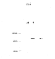
- Fig. 4 shows the result.
- S and AS indicate that D40cDNA was cloned into a plasmid in a sense and antisense way respectively, and the numbers on the gel left indicate the molecular weight markers. As shown in Fig. 4, a band with the molecular weight of 120 KD was detected.
- the D40 protein is a protein which, having associated with a fiber protein, contributes to the construction of the cell structure which is only observed in a testis cell or of the cell structure which is observed in the ectopic expression in a cancer cell.
- SCP1 is similar to D40 also in that it exists in a human autosome. Further, homology search was similarly performed, but no protein having high homology with the D40 (ORF2) protein was found.
- D40 genes on chromosomes were determined by fluorescent in situ hybridization. As shown in Fig. 5, signals were evident near the centromere region of human chromosome 15, i.e. in the long arm q14-15. Out of 49 cells examined, fluorescence signals in this region were observed on all four chromatids in 1 cell, on three chromatids in 3 cells, on two chromatids in 30 cells and on one chromatid only in 15 cells. No reproducible fluorescence signal was detected on other chromosomes. Localization of D40 genes on this chromosome was also confirmed in the simultaneous hybridization experiment using an a-satellite DNA probe which is specific to chromosome 15. These results suggest that D40 genes localize on human chromosome 15q14-15.
- Fig. 6 shows the result of Northern blotting for a human multiple tissue mRNA blot membrane (Clontech). As is shown in Fig. 6, frequent expression of D40mRNA was observed in the testis. Besides, about 5 Kb of a major transcript and about 3.5 Kb of a minor transcript were detected in the testis. Although the expression was equal to or less than 1/50 of that observed in the testis, a small amount of D40mRNA expression was observed in the placenta other than in the testis. However, expression of D40 was not at all detected in other normal human adult tissues other than those two (Fig. 6, top). But when the probe was taken out from the same membrane which was then hybridized with a probe for â-actin, D40 was confirmed to be expressed in all the tissues (Fig. 6, bottom).
- Fig. 7 shows the result of RT-PCR method performed with a human multiple tissue cDNA (Clontech). The arrow in Fig. 7 indicates specific PCR products. Frequent expression of D40 was observed in the testis in the RT-PCR result. D40 expression was not detected in other normal human tissues except that a faint expression was observed in the placenta. These results suggest the selective expression of D40mRNA in the testis in normal human tissues.
- D40 expression was examined in cancer cells. First, D40 expression in various cancer cell lines was studied by RT-PCR method. The results are shown in Table 3. As is shown in Table 3, D40 was observed to be expressed in all the cancer cell lines examined including malignant melanoma, lung cancer and digestive system-derived cancers. However, the expression was not detected in the normal fetal pulmonary fibroblast line. Expression frequency of D40 in human primary cancers was then examined. Table 5 shows the results. As is clear from Table 5, expression of D40mRNA was observed in most of samples for primary cancers such as oral cancer, uterine cancer, lung cancer, etc. Oral cancer among these cancers expressed D40 in about 63% of the incidents examined. This was the highest frequency in the primary cancers examined.
- D40 expression is observed in several cancer cell lines and normal testis, which expression pattern reveals that D40 is one of the gene populations selectively expressed in cancer/testis (Immunol. Today 18, 267-8, 1997, The Cancer J. from Scientific American, 16-7, 1999).
- a peptide comprising the amino acid sequence shown by SEQ ID NO:36 (SYTIEINHRL)
- the immunity-inducing activity of the peptide was examined by the cytotoxic test described in the Example A-10.
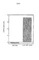
- Fig. 8 shows the result of cytotoxic activity of T cells derived from peripheral blood of a cancer patient.
- T cells derived from peripheral blood of a cancer patient (effecter cells) specifically injured target C1R-A24 cells which were added with 1 ⁇ M of the peptide (SYTIEINHRL) and did not injure the control target C1R-A24 cells which were not added with the peptide (SYTIEINHRL).
- SYTIEINHRL T cells derived from peripheral blood of a cancer patient
- effecter cells specifically injured target C1R-A24 cells which were added with 1 ⁇ M of the peptide (SYTIEINHRL) and did not injure the control target C1R-A24 cells which were not added with the peptide (
- HLA Class I molecules are not expressed in the testis, a D40-derived peptide and a HLA Class I molecule do not form a complex even when D40 is expressed so that D40 is not recognized by cytotoxic T lymphocytes. HLA Class I molecules are expressed whereas D40 is not in normal cells other than in the testis, so that it is again not recognized by cytotoxic T lymphocytes. However, since both D40 and HLA Class I molecules are expressed in cancer cells, the complex of these two are recognized by cytotoxic T lymphocytes and the cancer cells are injured.
- the present invention makes it possible to provide a cancer/testis antigen, with D40 as a primary example, which is applicable to diagnosis and immunotherapy for cancer, i.e. a cancer/testis antigen which is not expressed in normal tissues other than the testis, which is expressed in various cancers over a broad range and which induces immune response in a host, and the cancer/testis gene encoding the antigen or the like.
Landscapes
- Health & Medical Sciences (AREA)
- Life Sciences & Earth Sciences (AREA)
- Chemical & Material Sciences (AREA)
- Immunology (AREA)
- Organic Chemistry (AREA)
- Engineering & Computer Science (AREA)
- Medicinal Chemistry (AREA)
- General Health & Medical Sciences (AREA)
- Molecular Biology (AREA)
- Biochemistry (AREA)
- Genetics & Genomics (AREA)
- Biophysics (AREA)
- Gastroenterology & Hepatology (AREA)
- Proteomics, Peptides & Aminoacids (AREA)
- Biomedical Technology (AREA)
- Cell Biology (AREA)
- Toxicology (AREA)
- Bioinformatics & Cheminformatics (AREA)
- Zoology (AREA)
- Urology & Nephrology (AREA)
- Hematology (AREA)
- Veterinary Medicine (AREA)
- Chemical Kinetics & Catalysis (AREA)
- Animal Behavior & Ethology (AREA)
- Pharmacology & Pharmacy (AREA)
- Public Health (AREA)
- Nuclear Medicine, Radiotherapy & Molecular Imaging (AREA)
- General Chemical & Material Sciences (AREA)
- Oncology (AREA)
- Pathology (AREA)
- Tropical Medicine & Parasitology (AREA)
- Microbiology (AREA)
- Biotechnology (AREA)
- Food Science & Technology (AREA)
- Physics & Mathematics (AREA)
- Analytical Chemistry (AREA)
- General Physics & Mathematics (AREA)
- Transplantation (AREA)
- Micro-Organisms Or Cultivation Processes Thereof (AREA)
- Peptides Or Proteins (AREA)
Abstract
The present invention provides a cancer/testis antigen
which is applicable to diagnosis and immunotherapy for cancer,
that is to say, to provide a cancer/testis antigen which is not
expressed in normal tissues other than the testis, which is
expressed in various cancers over a broad range and which
induces immune response in a host, and to provide a caner/testis
gene encoding the cancer/testis antigen, etc. By using the
yeast Two-hybrid System, the present inventors screened a
protein which specifically binds to GCF, a previously reported
transcription factor, cloned a gene of a protein specifically
binding to the GCF, confirmed that the D40 protein as a resulting
gene product is a cancer/testis antigen which is expressed
substantially only in the testis in normal human tissues whereas
in cancers it is expressed in human primary cancers over a broad
range derived from various tissues and cells, and found that
the D40 shows high affinity with HLA and has a plurality of
sequences comprising 9 or 10 amino acid residues and binding
to HLA.
Description
The present invention relates to: a cancer/testis antigen
protein or peptide; a cancer/testis gene encoding the
cancer/testis antigen protein or the peptide; a screening
method for a promoter or a suppressor for the immunity-inducing
activity using the cancer/testis antigen protein/peptide; an
anti-tumor agent with the cancer/testis antigen protein or a
partial peptide of the antigen protein or the like as an
effective ingredient; a diagnostic probe for cancer using the
cancer/testis gene; etc.
Tumor cells often express various genes such as a
particular kind of protooncogenes which are not usually
expressed in normal cells or at a considerably low level even
if expressed (Science 235, 305-11, 1987). Gene populations
expressed in such an abnormal manner include populations that
are involved in tissue-specific differentiation (Immunol.
Today 18, 267-8, 1997), and the expression of these genes in
tumor cells are thought to be responsible for the malignant
phenotype of the tumor cells. Many testis-specific genes are
expressed in the testis (Reprod. Fertility & Develop. 7, 695-704,
1995, Int. J. Develop. Biol. 40, 379-83, 1996), however, most
of these genes are reported not to be activated at all in ordinary
tumors or very scarcely if any (Biochem. Biophys. Res. Comm.
241, 653-657, 1997). Recent studies, however, identified a
population of genes which are expressed both in cancer and
normal testis and some of which induce immune response in a host.
These genes are called cancer-testis antigens (CT antigen).
Besides, genes whose base sequences are homologous to
cancer/testis antigen genes but which are expressed not only
in testis but in genital organs of both sexes are also known
(Proc. Natl. Acad. Sci. USA 95, 10757-62, 1998, Cancer Res. 59,
1445-8, 1999, J. Biol. Chem. 273, 17618-25, 1998).
The genes mentioned above including genes encoding
cancer/testis antigens are called cancer/testis-related genes
and are classified as shown in Table 1 below. As for
cancer/testis-related genes encoding antigens which induce
immune response in cancer patients, genes such as MAGE, BAGE,
GAGE, LAGE, SSX, etc. are known (Science 254, 1643-7, 1991,
Immunity 2, 167-75, 1995, J. Exp. Med. 182, 689-98, 1995, Int.
J. Cancer 76, 903-8, 1998, Int. J. Cancer 72, 965-71, 1997,
Cancer Res. 56, 4766-72, 1996). These antigens are identified
according to their production of antibodies or responsiveness
to cytotoxic T lymphocytes in cancer patients (Proc. Natl. Acad.
Sci. USA 94, 1914-8, 1997, Science 254, 1643-7, 1991). On the
other hand, known cancer/testis-related genes include some
genes whose antigenicity is not yet elucidated (The Cancer J.
from Scientific American, 16-7, 1999, Proc. Natl. Acad. Sci.
USA 92, 11810-3, 1995, Cancer Res. 59, 3215-21, 1999. Proc. Natl.
Acad. Sci. USA 95, 10757-62, 1998, Cancer Res. 59, 1445-8, 1999).
There is a possibility that the in vivo expression of the former
is frequent enough to raise immune response while the expression
of the latter is not enough. Although original physiological
functions of the cancer/testis-related genes largely remain
unknown, the genes have been studied for analyzing of the
specific gene expression mechanism in a tumor and the testis
and for searching a possibility of application to diagnosis and
the therapy for cancer (Cancer Res. 55, 3478-82, 1995, Int. J.
Cancer 80, 219-30, 1999).
| 1) Genes where their gene products have antigenicity (cancer/testis antigens). | MAGE, GAGE, BAGE, LAGE, SSX, SCP1 |
| 2) Genes where their gene products have no antigenicity | BRS-3, TSP-50 |
| 3) Genes having 00.sequence homology with cancer/testis antigens but being expressed in plural genitourinary organs other than the testis. | PAGE |
Most of the previously known cancer/testis-related genes
exist in human X chromosomes. For instance, MAGE subfamily
exists in four regions on X chromosome, i.e. Xq28, Xq21.3, Xq26
and Xq11.23 (Immunogenet. 40, 360-9, 1994, Cancer Res. 58,
743-52, 1998, Genomics 59, 161-7, 1999). Further, SSX genes
exist on Xq11.2 (Nature Genet. 7, 502-8, 1994), LAGE1 and
NY-ESO-1 genes exist on Xq28 (Cancer J From Scientific American
5: 16-17, 1999, Intl J Cancer 76, 903-908, 1998) and GAGE genes
exist between Xp11.2-Xp11.4 (Cancer Res. 59, 3157-3165, 1999).
Recently, however, the synaptonemal complex protein, which is
known to exist on chromosome 1 (SCP1), has been shown as a member
of the cancer/testis-related gene population (EMBO J. 11,
5091-5100, 1992), which, at the same time, has been elucidated
to be identical with the HOM-TES-14 gene (Cytogen Cell Genet
78, 103-104, 1997, Proc Natl Acad Sci USA 95, 5211-5216, 1998).
On the other hand, rejection of a living body to cancer
is known to be caused by cytotoxic T lymphocytes which have
recognized the complex of a cancer antigen peptide and a
histocompatibility antigen molecule (HLA) that are bound in a
tissue or in a cell. Because a cancer antigen binds to HLA which
is expressed in the tumor cell, it is known that a cytotoxic
lymphocyte recognizes the complex as an antigen and thus causes
rejection which results in the regression of a cancer cell.
Marie Marchand et al. reported that they found a sequence
comprising 9 amino acid residues in the MAGE 3 gene product
(protein) has a property to bind to HLA-A1, one of HLA class
I molecules, that they synthesized the peptide containing this
amino acid sequence and immunized malignant melanoma patients
with the peptide, and that tumor regression was observed in 7
out of 25 patients (Int. J. Cancer 80, 219-30, 1999).
Further, the transcription factor GCF is known as a factor
which binds to the transcription regulatory region of a EGF
(epidermal growth factor) receptor gene (Cell 59, 815-25, 1989).
However, the previously reported GCFcDNA has recently been
proved as being an artificial fusion molecule (Biochem. Biophys.
Acta 1447, 125-31, 1999). That is to say, its sequence on the
amino-terminal is derived from GCF2 which is a recently and
newly identified transcription factor (J Biol. Chem. 273,
21594-602, 1998), and the most of the remaining cDNA is
comprised of a part tentatively named GCF1 by the present
inventors. It has been made clear that the base sequence-specific
DNA-binding activity is found in GCF2, whereas GCF1
is devoid of the DNA-binding activity (Biochem. Biophys. Acta
1447, 125-31, 1999). However, it has not yet been known that
a gene encoding a protein which specifically binds to GCF1 is
a cancer/testis-related gene.
Other than the above, the yeast Two-hybrid System (Nature
340, 245-6, 1989, Proc. Natl. Acad. Sci. USA 88, 9578-82, 1991)
is known as a system for detecting the in vivo protein-protein
interactions using saccaromyces yeast. A usually used protein
in the yeast Two-hybrid System is Gal4, a transcription
activator for yeast which has a DNA-binding domain and a
transcription-activating domain that can be separated from one
another. For example, a plasmid vector carrying a gene encoding
a fusion protein of the Gal4 DNA-binding domain and protein X,
and a plasmid vector carrying a gene encoding a fusion protein
of the Gal4 transcription-activating domain and protein Y are
introduced into yeast containing a reporter gene such as LacZ
gene, HIS3 gene, etc. and then the X-Y interaction can be
detected by observing whether the function of Gal4 recovers and
the transcription of a reporter gene is activated. When a known
protein is used as X and cDNA library is used as a gene encoding
Y in this yeast Two-hybrid System, a gene encoding a protein
which interacts with X can be screened from the aforementioned
cDNA library.
Cancer antigens are classified as in Table 2 based on the
recent molecular biological studies. Since no class I molecule
of a human histocompatibility antigen molecule (HLA) is
expressed in the testis, a protein on a testis cell membrane
is not recognized by a T lymphocyte. This gives us the view
that a cancer/testis antigen is a cancer-specific antigen in
a narrow sense because immunologically it is highly specific
to cancer in spite of the fact that it is expressed in the testis.
Since tissue-specific differentiated antigens are also
expressed, though weakly, in normal tissues, adverse reaction
on normal cells is causing drawbacks in performing cancer
immunotherapy, and mutation-derived cancer antigens have
drawbacks as well in that such mutation is limited only to the
individual cancers, and therefore these antigens are hardly
regarded as appropriate for the use in cancer immunotherapy.
On the other hand, cancer/testis antigens are not expressed in
normal tissues other than in the testis and are expressed in
various cancers over a broad range, so that their use in
immunotherapy agents for cancer is highly expected. The object
of the present invention is to a provide a cancer/testis antigen
applicable to diagnosis and immunotherapy for cancer, that is
to say, a cancer/testis antigen which is not expressed in normal
tissues other than the testis and which is expressed in various
cancers over a broad range and induces immune response in a host,
and a cancer/testis gene encoding the cancer/testis antigen and
the like.
| 1) Cancer specific antigen | Cancer/testis antigen |
| 2) Tissue specific differentiation antigen | tyrosinase, Melan/Mart-1, Pmel-17/gp100, TRP-1/gp75 |
| 3) Mutation-derived antigen | cdk4, caspase8 |
| 4) Overexpression-derived antigen | HER-2/neu, SART-1 |
| 5) Mutin | |
| 6) Viral antigen | E7 of HPV16 |
The present inventors have made a keen study to attain
the objects mentioned above and by using the yeast Two-hybrid
System, the present inventors screened a protein which
interacts with GCF (GC element binding Factor), a previously
reported transcription factor, as a gene controlling the
proliferation of mammalian cells, cloned a gene for a protein
which specifically binds to the transcription factor GCF,
confirmed that the protein D40, the resulting gene product, is
a cancer/testis antigen which is expressed substantially only
in the testis in normal human tissues but is expressed in human
primary cancers over a broad range deriving from various tissues
and cells, and found that the D40 shows high affinity with the
human histocompatibility antigen HLA and has a sequence
comprising 9 or 10 amino acid residues which binds to a plurality
of HLAs. Here, the present invention is completed.
The present invention relates to: a gene encoding the
following protein (a) or (b), (a) a protein comprising the amino
acid sequence shown by SEQ ID NO:2, (b) a protein which comprises
an amino acid sequence wherein one or a few amino acids are
deleted, substituted or added in the amino acid sequence shown
by SEQ ID NO:2 and which has the immunity-inducing activity
(claim 1); a gene encoding the following protein (a) or (b),
(a) a protein comprising the amino acid sequence shown by SEQ
ID NO:3, (b) a protein which comprises an amino acid sequence
wherein one or a few amino acids are deleted, substituted or
added in the amino acid sequence shown by SEQ ID NO:3 and which
has the immunity-inducing activity (claim 2); DNA containing
the base sequence shown by SEQ ID NO:1 or its complementary
sequence and part of whole of these sequences (claim 3); and
DNA which hybridizes under a stringent condition with DNA of
claim 3 that forms a gene, and which encodes a protein having
the immunity-inducing activity (claim 4).
The present invention further relates to: a protein
comprising the amino acid sequence shown by SEQ ID NO:2 (claim
5); a protein which comprises an amino acid sequence wherein
one or a few amino acids are deleted, substituted or added in
the amino acid sequence shown by SEQ ID NO:2 and which has the
immunity-inducing activity (claim 6); a protein comprising the
amino acid sequence shown by SEQ ID NO:3 (claim 7); and a protein
which comprises an amino acid sequence wherein one or a few amino
acids are deleted, substituted or added in the amino acid
sequence shown by SEQ ID NO:3 and which has the immunity-inducing
activity (claim 8).
The present invention still further relate to: a peptide
which comprises part of the protein of any of claims 5 to 8 and
which binds to a histocompatibility antigen molecule Class I
(claim 9); the peptide according to claim 9, wherein said
peptide comprises part of the protein of claim 5 or 6 and binds
to a histocompatibility antigen molecule Class I (claim 10);
the peptide according to claim 10, wherein said peptide
comprises an amino acid sequence shown by any of SEQ ID NOs:4-111
(claim 11); a peptide comprising the amino acid sequence of
Ser-Tyr-Thr-Ile-Glu-Ile-Asn-His-Arg-Leu (claim 12); the
peptide according to claim 9, wherein said peptide comprises
part of the protein of claim 7 or 8 and binds to the
histocompatibility antigen molecule Class I (claim 13); and the
peptide according to claim 13, wherein said peptide comprises
an amino acid sequence shown by any of SEQ ID NOs: 112-221 (claim
14).
The present invention also relates to: a fusion protein
or a fusion peptide wherein the protein of claim 5 or 6 or the
peptide of any of claims 10 to 12, and a marker protein and/or
a peptide tag, are bound (claim 15); a fusion protein or a fusion
peptide wherein the protein of claim 7 or 8 or the peptide of
claim 13 or 14, and a marker protein and/or a peptide tag, are
bound (claim 16); an antibody which specifically binds to the
protein of claim 5 or 6 or to the peptide of any of claims 10
to 12 (claim 17); an antibody which specifically binds to the
protein of claim 7 or 8 or to the peptide of claim 13 or 14 (claim
18); the antibody according to claim 17 or 18, wherein said
antibody is a monoclonal antibody (claim 19); and a recombinant
protein or peptide wherein the antibody of any of claims 17 to
19 specifically binds to said recombinant protein or peptide
(claim 20).
The present invention further relates to: a host cell
comprising an expression system capable of expressing the
protein according to claim 5 or 6 (claim 21); a host cell
comprising an expression system capable of expressing the
protein according to claim 7 or 8 (claim 22); a non-human animal
whose gene function to encode the protein of claim 5 or 6 is
deficient on its chromosome (claim 23); a non-human animal whose
gene function to encode the protein of claim 7 or 8 is deficient
on its chromosome (claim 24); the non-human animal according
to claim 23 or 24, wherein said non-human animal is a mouse or
a rat (claim 25); a non-human animal which over-expresses the
protein of claim 5 or 6 (claim 26); a non-human animal which
over-expresses the protein of claim 7 or 8 (claim 27); and the
non-human animal according to claim 26 or 27, wherein said
non-human animal is a mouse or a rat (claim 28).
The present invention still further relates to: a method
of screening a promoter or a suppressor for the immunity-inducing
activity wherein the protein of claim 5 or 6, the
peptide of any of claims 10 to 12 or a cell membrane expressing
the protein of claim 5 or 6, and a test substance are used (claim
29); a method of screening a promoter or a suppressor for the
immunity-inducing activity wherein the protein of claim 7 or
8, the peptide of claim 13 or 14, or a cell membrane expressing
the protein of claim 7 or 8, and a test substance are used (claim
30); a method of screening a promoter or a suppressor for the
immunity-inducing activity or for the expression of the protein
of claim 5 or 6, wherein a cell expressing the protein and a
test substance are used (claim 31); the method of screening a
promoter or a suppressor for the immunity-inducing activity or
for the expression of the protein of claim 5 or 6 according to
claim 31, wherein the cell expressing the protein of claim 5
or 6 is the host cell of claim 21 (claim 32); a method of screening
a promoter or a suppressor for the immunity-inducing activity
or for the expression of the protein of claim 7 or 8, wherein
a cell expressing the protein and a test substance are used
(claim 33); the method of screening a promoter or a suppressor
for the immunity-inducing activity or for the expression of the
protein of claim 7 or 8 according to claim 33, wherein the cell
expressing the protein of claim 7 or 8 is the host cell of claim
22 (claim 34); a method of screening a promoter or a suppressor
for the immunity-inducing activity or a promoter or a suppressor
for expression of the protein of any of claims 5 to 8, wherein
the non-human animal of any of claims 23 to 25 and a test
substance are used (claim 35); a method of screening a promoter
or a suppressor for the immunity-inducing activity or a promoter
or a suppressor for expression of the protein of any of claims
5 to 8, wherein the non-human animal of any of claims 26 to 28
and a test substance are used (claim 36); a promoter for the
immunity-inducing activity obtained by the screening method
according to any of claims 29 to 36 (claim 37); a suppressor
for the immunity-inducing activity obtained by the screening
method according to any of claims 29 to 36 (claim 38); an
expression promoter for the protein of claim 5 or 6, wherein
said expression promoter is obtained by the screening method
according to any of claims 29 to 36 (claim 39); an expression
suppressor for the protein of claim 7 or 8, wherein said
expression suppressor is obtained by the screening method
according to any of claims 29 to 36 (claim 40); an anti-tumor
agent comprising the protein of any of claims 5 to 8, the peptide
of any of claims 9 to 16, the protein or peptide of claim 20
or the antibody of any of claims 17 to 19, as an active ingredient
(claim 41); and the anti-tumor agent according to claim 41,
wherein the cancer is one or more cancers selected from uterine
cervix cancer, epithelial-like cancer, T cell tumor,
promyelocytic leukemia, esophageal cancer, pancreatic cancer,
malignant melanoma, lung cancer, oral cancer, breast cancer,
bladder cancer, uterine cancer, ovarian cancer, neuroblastoma
and colon cancer (claim 42).
The present invention further relates to: a method for
detecting a cytotoxic T cell or its precursor cell wherein a
HLA molecule, and the peptide of any of claims 9 to 16 or the
peptide of claim 20 are used (claim 43); a method for detecting
a cytotoxic T cell or its precursor cell wherein a complex of
a HLA Class I molecule, and the peptide of any of claims 9 to
16 or the peptide of claim 20 is formed on the surface of
fluorescent microparticles (claim 44); a detection reagent for
a cytotoxic T cell or its precursor cell, wherein said detection
reagent comprises a HLA molecule, and the peptide of any of
claims 9 to 16 or the peptide of claim 20 (claim 45); a detection
reagent for a cytotoxic T cell or its precursor cell wherein
said detection reagent comprises: a complex of a HLA Class I
molecule, and the peptide of any of claims 9 to 16 or the peptide
of claim 20; and a fluorescent microparticle (claim 46); a
cytotoxic T cell induced by in vitro stimuli wherein the protein
of any of claims 5 to 8, the peptide of any of claims 9 to 16,
or the protein or peptide of claim 20 is used (claim 47); a
diagnostic probe for cancer comprising whole or part of the
antisense strand of DNA or RNA encoding the protein of claim
5 or 6 (claim 48); a diagnostic probe for cancer comprising whole
or part of the antisense strand of DNA or RNA encoding the protein
of claim 7 or 8 (claim 49); a diagnostic drug for cancer
comprising the diagnostic probe for cancer according to claim
48 or 49 and/or the antibody according to any of claims 17 to
19 (claim 50); and the diagnostic drug for cancer according to
claim 50, wherein the cancer is one or more cancers selected
from uterine cervix cancer, epithelial-like cancer, T cell
tumor, promyelocytic leukemia, esophageal cancer, pancreatic
cancer, malignant melanoma, lung cancer, oral cancer, breast
cancer, bladder cancer, uterine cancer, ovarian cancer,
neuroblastoma and colon cancer (claim 51).
A cancer/testis-related gene of the present invention can
be screened by a yeast Two-hybrid System as a gene encoding a
protein which interacts with the transcription factor GCF, from
a cDNA library or a genomic DNA library. Further, a
cancer/testis antigen protein of the present invention can be
obtained by expressing the cancer/testis-related gene obtained
by such screening. Here, a cancer/testis-related gene is
defined as follows: its expression is observed specifically in
the normal testis and is not substantially observed in other
normal tissues; it activates in a malignant tumor, as a result
of which high level mRNA is detected; and it is expressed in
a tissue-non-specific manner in a malignant tumor.
Fig. 1 schematizes a yeast Two-hybrid System using the
transcription factor GCF. A transcription factor GCF is
expressed as a fusion protein with a DNA-binding domain of Gal4
(GalDBD-GCF), whereas a protein derived from a cDNA library is
expressed as a fusion protein with a transcriptional activation
domain of Gal4. The interaction (binding) of GalDBD-GCF and
a cDNA library-derived protein activates transcription of a
reporter gene and thereby the expression of His3 gene and LacZ
is raised. cDNA encoding a protein which interacts with GCF,
therefore, can be cloned by isolating yeast in which a reporter
gene is expressed. Commercially available MATCHMAKER
(Clontech) and HybriZAP (Stratagene) can be adopted for
performing the yeast Two-hybrid System.
As for a protein which is obtained by the above-mentioned
yeast Two-hybrid System and which interacts with the
transcription factor GCF, the cancer/testis antigen protein D40
is exemplified. A specific example of a cancer/testis-related
gene encoding this D40 is a gene having the base sequence shown
by SEQ ID NO: 1 in the sequence listing. There are two ORFs (open
reading frames) in the D40 cancer/testis-related gene.
Therefore, proteins as objects of the present invention
specifically include: the D40 (ORF1) protein shown by SEQ ID
NO:2 or the D40 (ORF2) protein shown by SEQ ID NO:3; a protein
comprising an amino acid sequence in which one or a few amino
acids are deleted, substituted or added in the amino acid
sequence shown by SEQ ID NO:2 or 3 and which has the
immunity-inducing activity; and the recombinant proteins of
these. The immunity-inducing activity here means an activity
inducing immune response such as antibody production, cellular
immunity, immune tolerance, etc. in a host. Among these
immunity-inducing activities, the T cell-inducing activity, by
which the frequency of cytotoxic T lymphocyte (CTL) precursor
cells is increased, is particularly preferable for a protein
to possess.
There is no particular limitation to a peptide as an
object of the present invention as long as it is a peptide which
comprises a part of the protein of the present invention, for
instance, a peptide comprising an amino acid sequence of five
or more consecutive amino acids, and which binds to a
histocompatibility antigen molecule (HLA) Class I. The
specific examples are: a peptide comprising an amino acid
sequence shown by any of SEQ ID NOs:4-111 that are derived from
the aforementioned D40 (ORF1) protein or the like, e.g.
Ser-Tyr-Thr-Ile-Glu-Ile-Asn-His-Arg-Leu (SEQ ID NO:36); and a
peptide comprising an amino acid sequence shown by any of SEQ
ID NOs:112-221 that are derived from the aforementioned D40
(ORF2) protein or the like. As for HLA Class I mentioned above,
the specific examples include: HLA-A3, HLA-68, HLA-A1, HLA-A24,
HLA-A0201, HLA-A0205, HLA-B7, HLA-B8, HLA-B14, HLA-B40.
B1501 (B62), etc. Proteins and peptides as objects of the
present invention together with the recombinant proteins and
peptides to which antibodies specifically bind while such
antibodies specifically bind to the said proteins and peptides,
may collectively be referred to as "the present
proteins/peptides". The present proteins/peptides can be
prepared in accordance with the known methods based on their
DNA sequence information or the like and these antigens will
not be limited to those derived from human.
Genes or DNAs as objects of the present invention are
specifically exemplified by: a gene encoding the D40 (ORF1)
protein which comprises the amino acid sequence shown by SEQ
ID NO:2; DNA encoding a protein which comprises an amino acid
sequence wherein one or a few amino acids are deleted,
substituted or added in the amino acid sequence shown by SEQ
ID NO:2 and which has the immunity-inducing activity; a gene
encoding the D40 (ORF2) protein comprising the amino acid
sequence shown by SEQ ID NO:3; DNA encoding a protein which
comprises an amino acid sequence wherein one or a few amino acids
are deleted, substituted or added in the amino acid sequence
shown by SEQ ID NO:3 and which has the immunity-inducing
activity; and DNA comprising the base sequence shown by SEQ ID
NO:1 or its complementary sequence and part or whole of these
sequences. These genes or DNAs can be prepared by the known
methods from, for instance, a human gene library or a human cDNA
library based on their DNA sequence information or the like.
Besides, there is no limitation to the preparation methods for
cDNA which encodes the D40 protein and it can be obtained, for
example, from a mRNA-derived cDNA library which is derived from
HL60 or the T cell leukemia cell line Jurkat.
Further, DNA encoding a protein of the interest, which
has the same effect as the D40 gene, having the immunity-inducing
activity can be obtained by the hybridization with a
DNA library derived from various cancer cells under a stringent
condition by using the base sequence shown by SEQ ID NO:1 or
its complementary sequence and part or whole of these sequences
as a probe, and then by the isolation of DNA which hybridizes
with the probe. A hybridization condition for obtaining the
DNA is exemplified by hybridization at 42. degree. C. and washing
at 42.degree. C. in a buffer solution containing 1×SSC, 0.1%
SDS, and more preferable exemplification is hybridization at
65.degree. C. and washing at 65.degree. C. in a buffer solution
containing 0.1×SSC 0.1% SDS. There are number of factors other
than the temperature condition mentioned above that affect the
hybridization stringency and those skilled in the art can
actualize the same stringency as that of the hybridization
referred to in the above by appropriately combining various
factors.
Any fusion protein and fusion peptide may be used as a
fusion protein and a fusion peptide for the present invention
as long as the present proteins/peptides are bound to marker
proteins and/or peptide tags. As for a marker protein, there
is no limitation as long as it is a conventionally known marker
protein and the specific examples include alkaline phosphatase,
the Fc region of an antibody, HRP, GFP, etc. Conventionally
known peptide tags including Myc tag, His tag, FLAG tag, GST
tag, etc. are the specific examples of the peptide tags for the
use in the present invention. These fusion proteins can be
generated according to the ordinary protocols and are useful
for the following: purification of the D40 protein or the like
using affinity of Ni-NTA and a His tag; detection of a protein
having the T cell-inducing activity; quantification of an
antibody against the D40 protein or the like; use as a diagnostic
marker for uterine cervix cancer, epithelial-like cancer, T
cell tumor, promyelocytic leukemia, esophageal cancer,
pancreatic cancer, malignant melanoma, lung cancer, oral cancer,
breast cancer, bladder cancer, uterine cancer, ovarian cancer,
neuroblastoma, colon cancer, etc.; and use as a laboratory
reagent in this field of art.
Antibodies that specifically bind to the aforementioned
proteins and peptides of the present invention can be
particularly exemplified by immune-specific antibodies such as
monoclonal antibodies, polyclonal antibodies, chimeric
antibodies, single-stranded antibodies, humanized antibodies,
etc. These antibodies can be generated according to the
ordinary protocols using a protein, such as the above-mentioned
D40 etc., or part of the protein as an antigen. However,
monoclonal antibodies are more preferable than the other sorts
of antibodies mentioned because of their specificity.
Antibodies such as the monoclonal antibodies or the like are
useful not only for diagnosis and therapy, such as missile
therapy, for uterine cervix cancer, epithelial-like cancer, T
cell tumor, promyelocytic leukemia, esophageal cancer,
pancreatic cancer, malignant melanoma, lung cancer, oral cancer,
breast cancer, bladder cancer, uterine cancer, ovarian cancer,
neuroblastoma, colon cancer, etc., for instance, but for
elucidating the development mechanism of malignant tumors such
as oral cancer and the like.
Antibodies of the present invention described above can
be created by administering to an animal (preferably non-human)
the present proteins/peptides, their fragments containing
epitopes, or the cells expressing the said proteins on the
membrane surface, according to the conventional protocols.
The monoclonal antibodies can be prepared, for instance, by any
optional method such as a hybridoma method that brings
antibodies produced by cultured materials of continuous cell
line (Nature 256, 495-497, 1975), a trioma method, a human
B-cell hybridoma method ( Immunology Today 4, 72, 1983), an
EBV-hybridoma method (MONOCLONAL ANTIBODIES AND CANCER THERAPY,
pp.77-96, Alan R. Liss, Inc., 1985), etc.
The preparation method for a single chain antibody (US
PAT.No. 4,946,778) can be adopted to prepare single-stranded
antibodies to the present proteins/peptides of the present
invention mentioned above. Besides, transgenic mice, other
mammals, etc. can be used for expressing humanized antibodies,
clones expressing the present proteins/peptides can be
isolated/identified using the antibodies mentioned above, and
their polypeptides can be purified by affinity chromatography.
Antibodies to the present proteins/peptides or to their
peptides containing antigenic epitopes can possibly be used for
diagnosis and therapy for various cancers as described earlier.
Furthermore, recombinant proteins or peptides to which these
antibodies specifically bind are also covered by the present
proteins/peptides of the present invention as described
earlier.
The functions of the present proteins/peptides can be
analyzed by using, for example, antibodies such as the
aforementioned monoclonal antibodies and the like that are
labeled with fluorescent materials such as FITC (Fluorescein
isothiocyanate), tetramethylrhodamine isothiocyanate, etc.,
radioisotopes such as 125I, 32p, 14C, 35S, 3H, etc., or enzymes such
as alkaline phosphatase, peroxidase, â-galactosidase,
phycoerythrin, etc. and by using fusion proteins fused with
fluorescent proteins such as Green Fluorescent Protein (GFP),
etc. As for immunological detection methods using the
antibodies of the present invention, RIA method, ELISA method,
fluorescent-antibody method, plaque method, spot method,
haemagglutination, Ouchterlony method, etc. are exemplified.
The present invention also relates to a host cell
comprising an expression system capable of expressing the
present proteins/peptides of the above. Genes encoding the
present proteins/peptides can be introduced into a host cell
by the methods described in many standard laboratory manuals
such as manuals of Davis et al. (BASIC METHODS IN MOLECULAR
BIOLOGY, 1986) and of Sambrook et al. (MOLECULAR CLONING: A
LABORATORY MANUAL, 2nd Ed., Cold Spring Harbor Laboratory Press,
Cold Spring Harbor, N.Y., 1989), and the examples include
calcium-phosphate transfection, DEAE-dextran-mediated
transfection, transvection, microinjection, cationic lipid-mediated
transfection, electroporation, transduction, scrape
loading, ballistic introduction, infection, etc. The examples
of host cells include bacterial prokaryotic cells such as E. coli,
Streptomyces, Bacillus Subtilis, Streptococcus,
Staphylococcus, etc., eukaryotic cells such as yeast,
aspergillus, etc., insect cells such as Drosophila S2,
Spodoptera Sf9, etc., animal cells such as L cell, CHO cell,
COS cell, HeLa cell, C127 cell, BALB/c3T3 cell (including
mutants deficient in dihydrofolate reductase, tymidine kinase,
etc.), BHK21 cell, HEK293 cell, Bowes malignant melanoma cell,
etc. and plant cells or the like.
There is no limitation to an expression system as long
as the expression system is capable of expressing the present
proteins/peptides described above in a host cell and the
examples include chromosome-, episome- and virus-derived
expression systems, for instance, vectors derived from
bacterial plasmid, vectors derived from yeast plasmid, vectors
derived from papovavirus such as SV40, vaccinia virus,
adenovirus, varicella virus, pseudorabies virus, retrovirus,
and vectors derived from bacteriophage or transposon and
vectors derived from the combination of these two, e.g. vectors
derived from genetic factors of plasmid and bacteriophage, such
as cosmid and phagemid. The expression systems may comprise
a control sequence for regulating the expression in addition
to a sequence for raising the expression.
Host cells comprising the above-mentioned expression
systems and the cell membranes of the cells, or the present
proteins/peptides obtained by culturing the cells can be used
in the screening methods of the present invention as described
below. For example, the method of F. Pietri-Rouxel et al. (Eur.
J. Biochem., 247, 1174-1179, 1997) or the like can be used to
obtain cell membranes. Further, to collect and purify the
present proteins/peptides from the cell culture, the known
methods can be adopted including ammonium sulfate- or
ethanol-precipitation, acid extraction, anion- or cation-exchange
chromatography, phosphocellulose chromatography,
hydrophobic interaction chromatography, affinity
chromatography, hydroxyapatite chromatography and lectin
chromatography, where the high performance liquid
chromatography is preferably used. As a column especially used
for affinity chromatography, columns to which antibodies to the
present proteins/peptides are bound, for instance, are used,
and when ordinary peptide tags are added to the present
proteins/peptides mentioned above, columns to which substances
having affinity with the peptide tags are bound are used in order
to obtain the present proteins/peptides. The purification
methods for the present proteins/peptides mentioned above may
also be employed for peptide synthesis.
In the present invention, a non-human animal whose gene
function to encode the present proteins/peptides mentioned
above is deficient on its chromosome means a non-human animal
part or whole of whose gene on its chromosome encoding the
present proteins/peptides is inactivated by gene mutations such
as destruction, deletion, substitution, etc. so that whose
function to express the present proteins/peptides is lost.
Further, a non-human animal which over-expresses the present
proteins/peptides is specifically exemplified by a non-human
animal which produces larger amount of the present
proteins/peptides than a wild-type non-human animal does.
Although rodents or the like such as mice, rats, etc. are
particularly exemplified for non-human animals of the present
invention, the examples will not be limited to these animals
only.
Homozygous non-human animals that are born according to
Mendel's Law include the deficient type or the over-expressing
type for the present proteins/peptides as well as their wild
type littermates. By using the deficient type animals or the
over-expressing type animals of these homozygous non-human
animals together with their wild-type littermates at the same
time, accurate comparative experiments can be carried out on
the individual level. In performing screening of the present
invention described below, it is, therefore, preferable to use
the wild type non-human animals, i.e. animals of the same
species as, or even better the littermates of, non-human animals
whose gene function to encode the present proteins/peptides is
deficient or over-expressing on their chromosomes in parallel
with the deficient or over-expressing type animals. The method
of producing a non-human animal whose gene function to encode
the present proteins/peptides is deficient or over-expressing
on its chromosome is now explained in the following with a D40
knockout mouse and a D40 transgenic mouse as examples.
A mouse, for instance, whose gene function to encode the
D40 protein is deficient on its chromosome, i.e. a D40 knockout
mouse is generated by the following steps. A gene encoding
mouse D40, which is homologous to human D40, is screened by using
a gene fragment obtained by a method such as PCR or the like
from the mouse gene library. A screened gene which encodes
mouse D40 is subcloned with a viral vector or the like and is
identified by DNA sequencing. Then whole or part of a gene of
this clone which encodes mouse D40 is substituted with a pMC1
neo gene cassette or the like. A gene such as a diphtheria toxin
A fragment (DT-A) gene, a herpes simplex virus tymidine kinase
(HSV-tk) gene, etc. is introduced onto the 3'-end, and thus a
targeting vector is constructed.
The targeting vectors thus constructed are linearlized and
introduced into ES cells by electroporation or the like to cause
homologous recombination. Among the homologous recombinants,
ES cells in which homologous recombination have occurred are
selected by the use of antibiotics such as G418, ganciclovir
(GANC), etc. It is preferable to confirm whether the ES cells
selected are the recombinants of the interest by Southern
blotting or the like. A clone of the ES cells confirmed is
microinjected into a mouse blastocyst and which blastocyst is
placed back to the recipient mouse to generate a chimeric mouse.
A heterozygous mouse can be obtained by intercrossing the
chimeric mouse and a wild type mouse. By further intercrossing
the heterozygous mice, the D40 knockout mice of the present
invention can be generated. Whether the ability of expressing
D40 is lost in a D40 knockout mouse is examined by Northern
blotting upon isolating RNA from the mouse obtained by the
above-described method and by Western blotting or the like with
which the D40 expression in the mouse can be directly examined.
A D40 transgenic mouse is created by the following steps.
A promoter such as chicken â-actin, mouse neurofilament, SV40,
etc. and poly (A) such as rabbit â-globin, SV40, etc. or introns
are fused with cDNA encoding D40 derived from human, mouse, rat,
rabbit, etc., to construct a transgene. This transgene is
microinjected into the pronucleus of a mouse fertilized egg.
After the obtained egg cell is cultured, it is transplanted to
the oviduct of the recipient mouse which was fed thereafter.
Neonetal mice that have the aforementioned cDNA were selected
from among all the mice born and thus the transgenic mice are
created. Neonatal mice having the cDNA can be selected by
extracting crude DNA from the mice tails or the like and then
by a dot hybridization method using a gene encoding the
introduced D40 as a probe and by PCR method or the like using
a specific primer.
Genes or DNA encoding the present proteins/peptides
mentioned above, the present proteins/peptides, fusion
proteins in which the present proteins/peptides and marker
proteins and/or peptide tags are bound, antibodies to the
present proteins/peptides, host cells comprising expression
systems capable of expressing the present proteins/peptides,
etc. are useful, as specifically explained below, for therapy
and diagnosis for uterine cervix cancer, epithelial-like cancer,
T cell tumor, promyelocytic leukemia, esophageal cancer,
pancreatic cancer, malignant melanoma, lung cancer, oral cancer,
breast cancer, bladder cancer, uterine cancer, ovarian cancer,
neuroblastoma, colon cancer, etc. These can be used for not
only screening a promoter or a suppressor for the immunity-inducing
activity or a promoter or a suppressor for expression
of the present proteins/peptides. but elucidating the mechanism
of immune response such as induction of cytotoxic T cells
(including all the T cells such as CD4 antigen-positive T cells,
CD8 antigen-positive T cells, helper T cells, killer T cells,
suppressor T cells, etc.), or the like.
As for a screening method for a promoter or a suppressor
for the immunity-inducing activity of the present invention,
methods are exemplified that use the following: the foregoing
present proteins/peptides of the present invention or a cell
membrane which expresses the present proteins/peptides, and a
test substance; a cell which expresses the present
proteins/peptides and a test substance; a non-human animal such
as a knockout mouse or a transgenic mouse for the present
proteins/peptides and a test substance; and etc. Further, a
method using a cell which expresses the present
proteins/peptides mentioned above and a test substance, a
method using a non-human animal such as a knockout mouse and
a transgenic mouse for the present proteins/peptides and a test
substance, etc. can be employed for a method of screening a
promoter or a suppressor for expression of the present
proteins/peptides.
A screening method wherein the above-mentioned present
proteins/peptides or a cell membrane that expresses the present
proteins/peptides are used along with a test substance is
specifically exemplified by a method wherein the present
proteins/peptides or the present proteins/peptides expressed
on the surface of a cell membrane is made to contact a test
substance and wherein the immunity-inducing activity of the
present proteins/peptides are measured and assessed. Further,
as for a screening method wherein the cells expressing the
present proteins/peptides and a test substance is used, a method
is specifically exemplified in which a cell expressing the
present proteins/peptides is made to contact a test substance
and in which the immunity-inducing activity of the present
proteins/peptides and the variation in the expression amount
of the present proteins/peptides are measured and assessed.
As for a screening method wherein a non-human animal whose
gene function to encode the aforementioned present
proteins/peptides is deficient on its chromosome, or a non-human
animal which over-expresses the present
proteins/peptides is used along with a test substance, the
following methods are specifically exemplified: a method
wherein a cell or a tissue from these non-human animals is made
to contact a test substance in vitro to measure and assess the
immunity-inducing activity of the present proteins/peptides
and the variation in the expression amount of the present
proteins/peptides; a method wherein a non-human animal whose
gene function to encode the aforementioned present
proteins/peptides is deficient on its chromosome or a non-human
animal which over-expresses the present proteins/peptides is
administered with a test substance in advance and wherein the
immunity-inducing activity of the present proteins/peptides
and the variation in the expression amount of the present
proteins/peptides in a cell or a tissue obtained from the
non-human animal are measured and assessed; or a method wherein
a non-human animal whose gene function to encode the
aforementioned present proteins/peptides is deficient on its
chromosome or a non-human animal which over-expresses the
present proteins/peptides is administered with a test substance
in advance and wherein the immunity-inducing activity of the
present proteins/peptides and the variation in the expression
amount of the present proteins/peptides in the non-human animal
are measured and assessed.
The screening methods using a test substance mentioned
above and the present proteins/peptides will be explained below
with the specific examples, however, the screening methods of
the present invention will not be limited to these examples.
As for a method for measuring and assessing the immunity-inducing
activity of the present proteins/peptides by
contacting the above-mentioned test substance with the present
proteins/peptides, a method is specifically exemplified
wherein the test substance and the present proteins/peptides
are made to contact in the presence of tumor-infiltrating T
cells which are largely proliferated in vitro from peripheral
lymphocytes by using ordinary interleukin-2, and wherein the
increase and decrease of the inducing activity of T cells such
as CTL is measured and then the results are compared for the
assessment with that of the control in which the test substance
is absent. Further, as for a method of measuring and assessing
the variation in the expression of the present
proteins/peptides by contacting a test substance with a cell
membrane or a cell which expresses the present
proteins/peptides as mentioned above, a method is specifically
exemplified wherein a cell expressing the present
proteins/peptides is cultured in the presence of a test
substance and wherein the increase or decrease of the present
proteins/peptides that are expressed on the surface of the cell
membrane after culturing for a certain period of time is
assessed either by the immunochemical detection by ELISA or the
like with the use of antibodies that specifically bind to the
present proteins/peptides of the present invention, or by using
the suppression or promotion of the mRNA expression as an index.
Detection of mRNA, mentioned above, can be carried out by such
as DNA chip method, Northern hybridization method, etc.
Alternatively, when a cell introduced with a gene to which a
reporter gene such as luciferase, etc. is linked in the
downstream of the promoter of a gene encoding the present
proteins/peptides is used, suppression or promotion, caused by
a test substance, of the expression of the gene encoding the
present proteins/peptides can be detected with the activity of
the reporter gene as an index.
A promoter for the immunity-inducing activity or an
expression promoter of the present invention obtained by the
aforementioned screening methods can be applied to the therapy
and the like for patients in need of the promotion of the
immunity-inducing activity or promotion of expression of the
present proteins/peptides. Meanwhile, a suppressor for the
immunity-inducing activity or an expression suppressor of the
present invention obtained by the aforementioned screening
methods can be applied to the therapy and the like for patients
in need of the suppression of the immunity-inducing activity
or the expression of the present proteins/peptides. The
present proteins/peptides of the present invention and the
antibodies against these can be employed for an active
ingredient in anti-tumor agents for uterine cervix cancer,
epithelial-like cancer, T cell tumor, promyelocytic leukemia,
esophageal cancer, pancreatic cancer, malignant melanoma, lung
cancer, oral cancer, breast cancer, bladder cancer, uterine
cancer, ovarian cancer, neuroblastoma, colon cancer, etc. For
example, when the present proteins/peptides are injected orally,
intravenously, intradermally, subcutaneously, etc., an
anti-tumor effect can be expected due to the increase in the
in vivo T cell-inducing activity. Furthermore, the above-mentioned
antibodies can be used in a missile therapy.
There is no limitation to a detection method of the
present invention for cytotoxic T cells (activated T cells) or
their precursor cells as long as the method uses a HLA molecule
and a present peptide and the examples include a method wherein
a complex of a HLA molecule, such as a HLA Class I molecule or
the like, and a present peptide is used, and a method wherein
a complex of a HLA molecule, such as a HLA Class I molecule or
the like, and a present peptide is formed on the cell surface
or on the surface of a carrier such as a fluorescent
microparticle or the like. A method is exemplified, for
instance, according to the method described in the literature
(Science, 274, 94-96, 1996), wherein a complex of a HLA Class
I molecule and a present peptide is formed on the surface of
a fluorescent microparticle, a T lymphocyte cell from
peripheral blood of a cancer patient is made to contact and bind
to the complex, and then T cells bound to HLA-peptide complexes
that are formed on the surface of the microparticles are
detected. Further, there is no limitation to a detection
reagent of the present invention for cytotoxic T cells
(activated T cells) or their precursor cells as long as a reagent
contains HLA molecules and the present peptides where the
examples are a reagent containing a complex of a HLA molecule
such as a HLA Class I molecule or the like and a present peptide
and a reagent containing a complex of a HLA molecule such as
a HLA Class I molecule or the like and a present peptide along
with a carrier such as a fluorescent microparticle or the like.
Further, activated T cells can be induced upon the in
vitro stimuli with the present proteins/peptides of the present
invention. For instance, the tumor-responsive activated T
cells are induced when peripheral lylmphcytes or tumor-infiltrating
lymphocytes are stimulated with the present
proteins/peptides and IL-2. These activated T cells can be used
effectively in an adoptive immunotherapy. Besides, by
expressing the present proteins/peptides either in vivo or in
vitro in dendritic cells that are strong antigen-presenting
cells, immunity can be induced by injecting these antigen-expressing
dendritic cells.
Further, whole or part of the antisense chain of DNA or
RNA encoding the present proteins/peptides of the present
invention can be made a diagnostic probe for cancer.
Furthermore, cancers such as uterine cervix cancer,
epithelial-like cancer, T cell tumor, promyelocytic leukemia,
esophageal cancer, pancreatic cancer, malignant melanoma, lung
cancer, oral cancer, breast cancer, bladder cancer, uterine
cancer, ovarian cancer, neuroblastoma, colon cancer, etc. can
be diagnosed by the use of this diagnostic probe for cancer and
a diagnostic drug of the present invention containing an
antibody which specifically binds to the present
proteins/peptides. As for the above-mentioned diagnostic
probe, a probe which comprises whole or part of the antisense
chain of DNA (cDNA) or RNA (cRNA) encoding the present
proteins/peptides and which has enough length to be established
as a probe (at least equal to or more than 20 bases) is preferable.
The specific examples of test samples used for detection are
genomic DNA, RNA or cDNA from the cells of subjects that are
obtained from, for example, biopsy of such as blood, urine,
saliva, tissues, etc. However, the test samples will not be
limited to these examples and amplified test samples by the
means of PCR or the like may also be used.
The present invention will be specifically explained in
the following with reference to the examples, but the technical
scope of the invention will not be limited to these examples.
Culture cell lines used are shown in Table 3. These cell
lines are maintained in RPMI 1640 containing 10% bovine fetal
serum and 0.3% glutamine or in DMEM (Dulbecco Modified Eagle
Medium) and are cultured at 37.degree. C. under 5% CO2. The
primary tumor samples are obtained from operations performed
at Department of Oral Surgery, School of Dental Medicine,
Hokkaido University and Department of Obstetrics and Gynecology,
Second Department of surgery, and First Department of Internal
medicine of School of Medicine, Hokkaido University. They were
immediately frozen in liquid nitrogen and kept at □80. degree.
C. until used.
| Cell type | Culture cell line | Expression |
| Normal fetal pulmonary fibroblast | HFL | - |
| Uterine cervix cancer | HeLa | + |
| Epithelial-like cancer | A431 | + |
| T cell tumor | Jurkat | + |
| Promyelocytic leukemia | HL60 | + |
| Esophageal cancer | TE-8 | + |
| Pancreatic cancer | PC143c | + |
| Malignant melanoma | SkMEL | + |
| AKI | + | |
| G361 | + | |
| Lung cancer | PI10 | + |
| NCIH226 | + | |
| RERF-LC-MS | + | |
| RERF-LC-OK | + | |
| Oral cancer | HSC-2 | + |
| Breast cancer | MCF-7 | + |
| Bladder cancer | DAB-1 | + |
| T-24 | + | |
| UMUC-2 | + |
Two thirds of carboxyl terminal of the coding region of
transcription factor GCF was incorporated into pGBT-9
(Clontech) and the resulting plasmid pGAL-GCF was used as a bait
for screening by the yeast Two-hybrid System (see Fig. 1).
First, pGAL-GCF was transformed to the yeast Y153 line (Genes
Dev.7, 555-69, 1993), with which a cDNA library of an
immortalized human B cell line was screened. Taking a clone
which was positive to histidine- and LacZ-phenotype, its
binding specificity with GCF was examined and its base sequence
was identified. In order to obtain a longer clone by using this
clone, a cDNA library derived from the human promyelocytic
leukemia cell line HL60 was screened. Further, in accordance
with the protocol attached to the 5'/3' RACE kit
(Beohringer/Roche), 5' RACE (Rapid Amplification of cDNA ends)
reaction was carried out using the directed primer (Proc. Natl.
Acad. Sci. USA 85, 8998-9002, 1988), that is, total RNA derived
from HL60 and the T cell leukemia cell line Jurkat were used
along with the D40-specific reverse primer MT 194 (5'-GAGTCCTCGGTGTGAAGCTTTA-3':
SEQ ID NO:222) for cDNA synthesis
of the first strand. Deoxyadenosine was added to 3' end of the
cDNA and an oligo dT anchor primer and a D40-specific reverse
primer MT195 (5'-ACAGTGTGACTTGATGTCAGGT-3'; SEQ ID NO:223)
were used for PCR.
Plasmid DNA was purified in a Qiagen column (Qiagen) and
used as a template for the cycle sequencing reaction in
performing Dye terminator method (Proc. Natl. Acad. Sci. USA
74, 5463-5, 1977), then the base sequence was determined on a
fluorescent DNA sequencer (373 genetic analyzer, ABI).
Sequence homology was studied with the disclosed data bases.
Fluorescence in situ hybridization (FISH) was performed
in accordance with a previously reported procedure (Sambrook
J, Fritsch EF, Maniatis T (1989): Molecular Cloning, Cold Spring
Harber Press, New York, p7.39). Metaphase spreads were
prepared by incorporating bromodeoxyuridine (BrdU) into the
concanavalin-A stimulated lymphocytes of a human male which
were transformed with EB virus. A mixture of plural cDNA clones
over the full length of D40cDNA (200 ng) was labeled with biotin.
This cDNA as a probe was reacted with samples of R-banded
metaphase chromosomes for 48 hours at 37.degree. C.
Subsequently, the chromosome samples were sequentially washed
in 50% formamide, first at 0.5×SSC and then at 2×SSC. Then,
FISH signals were amplified with an anti-biotin antibody (3
ìg/ml, Vector) and a FITC anti-goat antibody (40 ìg/ml, American
Qualex). Further, FISH signals were amplified with the
anti-FITC-Alexa488 (40 ìg/ml, Molecular Probes).
A reaction was occurred in the presence of 35S-methionine
using a plasmid pBS-D40 having all coding regions of D40cDNA
in pBluescript KS( ), a T7 RNA polymerase and the TNT rabbit
reticulocyte lysate (Promega). The translation products were
analyzed on SDS-PAGE and detected by the image analyzer BAS2000
(Fuji Film).
Cell lysate was prepared using TNE buffer (10 mM Tris-Cl,
pH 7.8, 1% NP40, 150 mM NaCl, 1 mM EDTA, 0.5 mM PMSF, 10 mg/ml
Aprotinin, 10 mg/ml Leupeptin), which was then analyzed on a
8% SDS-PAGE and fixed on a nitrocellulose filter. An anti-Flag
monoclonal antibody (M2: Kodak) and a peroxydase-conjugated
goat anti-mouse immunoglobulin (Jackson
ImmunoRes.) were used as the first and the second antibodies,
respectively. Signals were detected by ECL system (Amersham).
Total RNA was separated and purified from the cell lines
and tumor samples by the method of acid guanidinium
thiocyanate-phenol-chloroform extraction (Anal. Biochem. 162,
156-159, 1987). RNA was purified from approximately 1×10⁷
cells and 80 mg of a tumor tissue using 2 ml of Trizol reagent
(Life Technology).
Northern blot analysis was performed in accordance with
a previously described method (Genomics 32, 483-484, 1996).
The hybridization took place overnight at 37.degree. C. in a
solution containing 50% formamide, 5×Denhardt solution and 0.5%
SDS. The filter membrane was sequentially washed under the
gradually increasing stringency. The final washing was done
under 0.2×SSC and 0.1% SDS at 48. degree. C., and then the signals
were detected by the BAS2000.
1 ig of the total RNA was reversely transcribed to cDNA
using Superscript II RNase H-reverse transcriptase (Life
Technology). The PCR reaction was occurred in a pH8.3 buffer
containing 10 mM Tris-HCl, 1.5 mM MgCl2 and 50 mM KCl using:
0.1 ìg of cDNA; 10 pmol each of specific oligonucleotide primers
(forward: MT1149; 5'-CACATCCAGTGAGACCA-3'; SEQ ID NO:224,
reverse: MT152; 5'-TCCCATCTTCTGATGTG-3'; SEQ ID NO:225, or
forward: MT227; 5'-CAGGAATCCAATGCTTGG-3'; SEQ ID NO;226,
reverse: MT188; 5'-CTGTTGCAGGTAAGATC-3'; SEQ ID NO:227); and
0.5 unit of Taq polymerase. The RT-PCR was carried out using
a â-actin primer in order to check the integrity of RNA.
Peripheral blood of a patient whose D40 gene expression
in the tumor tissue was observed by RT-PCR method was
multi-layered in Ficoll-Conray solution with specific gravity
of 1.082 and the blood was separated to plasma, lymphocyte phase,
Ficoll-Conray phase and erythrocyte phase from the top by
centrifugation of 1500 rpm for 30 min. The lymphocyte phase
was collected and cultured for a day in AIM-V, a serum-free
medium, after which the cell population adhered to the bottom
of the culture flask and the suspended cell population were
separated by gently shaking the flask. The adhered cell
population is a cell population called PBMC (peripheral blood
mononuclear cells) which includes B cells, macrophages and
dendritic cells that are so-called antigen-presenting cells
(APC). The APC were cultured for four days in AIM-V+GM-CSF,
a culture condition for dendritic cells which have particularly
high antigen-presenting ability among the APC mentioned above.
Thereafter, a test peptide was added to this APC culture
solution at the concentration of 1 ìM, which was then cultured
for two days. When this culture period was over, the culture
solution was replaced with AIM-V+TNF-α+INF-α, and the APC were
cultured for another two days. The APC after the culture were
treated with radioactivity in order to get rid of their
proliferation ability.
On the other hand, the suspended cell population mainly
contains T cells and these suspended cells were cultured in
AIM-V+IL-2 for four days. Then CD8-positive T cells were
selected using magnetic beads in which anti-CD8 antibodies were
solid-layered and the CD8-positive T cells obtained were
cultured for four days. These CD8-positive T cells were added
to APC, which have been treated with radioactivity as mentioned
above, and cultured for two days. This operation, named MLPC
(Mix lymphocyte peptide culture) by the present inventors, was
repeated for two or three times and the CD8-positive T cells
obtained were used for the cytotoxity test. The CD8-positive
T cells which had undergone MLPC twice as described above were
used as effecter cells and C1R-A24 cells incorporated with 51Cr
were used as target cells in the cytotoxity test. 5×10² target
C1R-A24 cells added with 1 ìM of a test peptide were co-cultured
with 2.5×10³ effecter T cells for six hours. Then the
radioactivity of 51Cr released from the target C1R-A24 cells
were measured and the cytotoxity activity (%) was calculated.
The case when a test peptide was not added was made the control.
Proteins that specifically bind to the transcription
factor GCF were screened by the yeast Two-hybrid System. As
described in Example A-2 above, a plasmid pGAL-GCF which
expresses a fusion protein of a DNA-binding domain of Gal4 and
GCF was introduced into the yeast Y153 line, which was then used
to screen the human B cell-derived cDNA library. Nine among
12×105 yeast clones that underwent search were positive to
histidine- and LacZ-phenotypes. One among the nine clones was
named D40 and its binding specificity with GCF was examined.
The result showed that a protein encoded by this clone did not
bind to proteins such as lamin, p53, etc., thus revealing that
the binding of D40 and GCF was a specific one.
Upon determination of the DNA base sequence of this clone,
ORF was found which, however, was devoid of a termination codon
on 5' side of the codon corresponding to methionine on the amino
end, although it had a termination codon on 3' side. In order
to obtain longer clones, the cDNA library of the human
promyelocytic leukemia cell line HL60 was screened with the
D40cDNA clone as a probe. Some clones were isolated as a result,
however, none of them were shown to have a termination codon
on 5' side. Hence, the 5'RACE (Rapid amplification of cDNA
ends) method was carried out using total RNA derived from HL60
and the T cell leukemia cell line Jurkat. With this method,
a clone having a termination codon for a reading frame of the
5' end was successfully obtained (ORF1) . In addition to this,
a clone containing the 3' end region was obtained and its base
sequence was determined, which proved the existence of a
sequence encoding another protein (ORF2) on the 3' side of ORF1.
The total cDNA sequence of D40 is shown as SEQ ID NO: 1, the amino
acid sequence of the D40 (ORF1) protein is shown as SEQ ID NO:2
and the amino acid sequence of the D40 (ORF2) protein is shown
as SEQ ID NO:3, respectively.
Part of the base sequence of D40 (ORF1) cDNA and the ORF1
amino acid sequence coded by that base sequence part are shown
in Fig. 2. In Fig. 2, the amino acid sequence is shown by single
letters under the base sequence, the mark * indicates the
termination codon and the numbers on left and right hand sides
of the sequences indicate positions in the amino acid sequence
and the base sequence, respectively. Besides, the leucine
zipper-like sequence (Science 240, 1759-64, 1988) are shown in
bold letters and the examples of peptides having high affinity
with various human histocompatibility antigens (HLA) Class I
molecules (see Table 4) are underlined, respectively. There
are some sequences that very closely resemble a peptide motif
which binds to a HLA Class I molecule (Immunogenet. 41, 178-228,
1995), in the amino acid sequence of the D40 (ORF1) protein as
shown in Fig. 2 or the D40 (ORF2) protein mentioned above.
According to the program which predicts amino acid sequences
binding to HLA Class I molecules and which scores such
prediction (J. Immunol. 152:163. 1994. Web site; HLA Peptide
Binding Predictions;
http://bimas.dcrt.nih.gov/molbio/hla bind/). scores of the
underlined amino acid sequences in D40 (ORF1) in Fig. 2 were
calculated. The results are shown in Table 4. Some peptides
having high affinity with HLA Class I molecules as mentioned
above are shown in Table 4. The scores for the amino acid
sequences of these peptides are shown to be equal to or higher
than that of the MAGE3 sequence which have already been shown
to bind to a HLA-A1 molecule and proved to be recognized by a
cytotoxic T lymphocyte. Therefore, it is understood that the
underlined amino acid sequences in D40 (ORF1) in Fig. 2 have
antigenicity. Further, an amino acid sequence shown by any of
SEQ ID NOs:4-111 in the D40 (ORF1) protein and an amino acid
sequence shown by any of SEQ ID NOs:112-221 in the D40 (ORF2)
protein also scored high marks in the above-mentioned scoring
so that these sequences are very likely to possess antigenicity.
| HLA Class I | D40-derived peptide | Score | |
| A1 | HTEDSRMKK | 225 | |
| A0201 | WVLKILPYL | 402 | |
| A3 | LLDNPISEK | 45 | |
| A24 | IYGNDFMDL | 240 | |
| | WVLKILPYL | 252 | |
| A68 | DVTTGYGTK | 360 | |
| A68 | SVGGPKIDK | 240 | |
| B7 | PPRSPLQDL | 120 | |
| B8 | WNKDKDWVL | 120 | |
| B14 | ERPVRRRHS | 300 | |
| B40 | CEITGMNTL | 80 |
As shown in Fig. 2, ORF1 in D40cDNA encodes 887 amino acids.
To confirm the accuracy of ORF1, in vitro transcription and
translation reactions were occurred in accordance with the
method described in the Example A-5 by using reticulocyte lysate
which is well known as an in vitro synthesis system for proteins.
cDNA in the whole coding region of the D40 (ORF1) protein was
incorporated into a plasmid having a T7 RNA promoter. The
reaction was occurred using this plasmid in a manner described
in the Example A-5. The result is shown in Fig. 3. In Fig.
3, (+) and (-) indicate presence or absence of a plasmid having
D40cDNA and the numbers on the right of the gel indicate
molecular weight markers. The result of the gel analysis for
the translated protein exhibited two bands between the
molecular weights of 110-130 KD.
Next, in vivo expression of the protein encoded by D40
(ORF1) was examined. A plasmid, which expresses the D40 (ORF1)
protein added with a Gal4 DNA-binding domain to the amino end
of D40 (ORF1) protein as a tag (FLAG), was introduced into a
Cos7 cell using lipofectamine. 48 hours later, the Cos7 cell
extract was prepared which underwent Western blotting using an
anti-flag antibody as described in the Example A-6. Fig. 4
shows the result. In Fig. 4, S and AS indicate that D40cDNA
was cloned into a plasmid in a sense and antisense way
respectively, and the numbers on the gel left indicate the
molecular weight markers. As shown in Fig. 4, a band with the
molecular weight of 120 KD was detected.
Homology was searched with each data base of
GenBank/EMBL/DDBJ as mentioned in the Example A-3, and no
protein having high homology with the D40 (ORF1) protein was
found. Although it did not show high homology, the D40 (ORF1)
protein had a faint homology with some proteins such as
Synaptonemal complex protein 1 (SCP1), Paramyosin, etc. These
proteins rich in α-helix are thought to be fiber proteins which
bind intrecellular and extracellular structures together (EMBO
J 11, 5091-5100, 1992). Therefore, there is a possibility that
the D40 protein is a protein which, having associated with a
fiber protein, contributes to the construction of the cell
structure which is only observed in a testis cell or of the cell
structure which is observed in the ectopic expression in a
cancer cell. As mentioned earlier, SCP1 is similar to D40 also
in that it exists in a human autosome. Further, homology search
was similarly performed, but no protein having high homology
with the D40 (ORF2) protein was found.
Localization of D40 genes on chromosomes was determined
by fluorescent in situ hybridization. As shown in Fig. 5,
signals were evident near the centromere region of human
chromosome 15, i.e. in the long arm q14-15. Out of 49 cells
examined, fluorescence signals in this region were observed on
all four chromatids in 1 cell, on three chromatids in 3 cells,
on two chromatids in 30 cells and on one chromatid only in 15
cells. No reproducible fluorescence signal was detected on
other chromosomes. Localization of D40 genes on this
chromosome was also confirmed in the simultaneous hybridization
experiment using an a-satellite DNA probe which is specific to
chromosome 15. These results suggest that D40 genes localize
on human chromosome 15q14-15.
The expression of D40 genes in normal human tissues was
first examined by Northern blot method. Fig. 6 shows the result
of Northern blotting for a human multiple tissue mRNA blot
membrane (Clontech). As is shown in Fig. 6, frequent expression
of D40mRNA was observed in the testis. Besides, about 5 Kb of
a major transcript and about 3.5 Kb of a minor transcript were
detected in the testis. Although the expression was equal to
or less than 1/50 of that observed in the testis, a small amount
of D40mRNA expression was observed in the placenta other than
in the testis. However, expression of D40 was not at all
detected in other normal human adult tissues other than those
two (Fig. 6, top). But when the probe was taken out from the
same membrane which was then hybridized with a probe for â-actin,
D40 was confirmed to be expressed in all the tissues (Fig.
6, bottom).
The result of the Northern blotting was next confirmed
by RT-PCR method which exhibits higher sensitivity than
Northern blot method does. Fig. 7 shows the result of RT-PCR
method performed with a human multiple tissue cDNA (Clontech).
The arrow in Fig. 7 indicates specific PCR products. Frequent
expression of D40 was observed in the testis in the RT-PCR result.
D40 expression was not detected in other normal human tissues
except that a faint expression was observed in the placenta.
These results suggest the selective expression of D40mRNA in
the testis in normal human tissues.
D40 expression was examined in cancer cells. First, D40
expression in various cancer cell lines was studied by RT-PCR
method. The results are shown in Table 3. As is shown in Table
3, D40 was observed to be expressed in all the cancer cell lines
examined including malignant melanoma, lung cancer and
digestive system-derived cancers. However, the expression was
not detected in the normal fetal pulmonary fibroblast line.
Expression frequency of D40 in human primary cancers was then
examined. Table 5 shows the results. As is clear from Table
5, expression of D40mRNA was observed in most of samples for
primary cancers such as oral cancer, uterine cancer, lung cancer,
etc. Oral cancer among these cancers expressed D40 in about
63% of the incidents examined. This was the highest frequency
in the primary cancers examined. The expression was also
observed in uterine cancer (44%), lung cancer (40%), ovarian
cancer (36%), pancreatic cancer (27%), neuroblastoma (20%) and
colon cancer (13%), but not in cholangiocarcinoma, gastric
cancer and seminoma. D40 expression is observed in several
cancer cell lines and normal testis, which expression pattern
reveals that D40 is one of the gene populations selectively
expressed in cancer/testis (Immunol. Today 18, 267-8, 1997, The
Cancer J. from Scientific American, 16-7, 1999).
| Cancers | Positive incidents/total incidents tested |
| Oral cancer | 10/16(63%) |
| | 8/18(44%) |
| | 8/20(40%) |
| | 4/11(36%) |
| Pancreatic cancer | 3/11(27%) |
| Neuroblastoma | 1/5(20%) |
| | 1/8(13%) |
| Cholangiocarcinoma | 0/7(0%) |
| Gastric cancer | 0/4(0%) |
| Seminoma | 0/3(0%) |
With the use of a peptide comprising the amino acid
sequence shown by SEQ ID NO:36 (SYTIEINHRL) as a test peptide,
the immunity-inducing activity of the peptide was examined by
the cytotoxic test described in the Example A-10. Fig. 8 shows
the result of cytotoxic activity of T cells derived from
peripheral blood of a cancer patient. As is clear from Fig.
8, T cells derived from peripheral blood of a cancer patient
(effecter cells) specifically injured target C1R-A24 cells
which were added with 1 ìM of the peptide (SYTIEINHRL) and did
not injure the control target C1R-A24 cells which were not added
with the peptide (SYTIEINHRL). These indicate that the peptide
(SYTIEINHRL) possesses the immunity-inducing activity.
Since HLA Class I molecules are not expressed in the
testis, a D40-derived peptide and a HLA Class I molecule do not
form a complex even when D40 is expressed so that D40 is not
recognized by cytotoxic T lymphocytes. HLA Class I molecules
are expressed whereas D40 is not in normal cells other than in
the testis, so that it is again not recognized by cytotoxic T
lymphocytes. However, since both D40 and HLA Class I molecules
are expressed in cancer cells, the complex of these two are
recognized by cytotoxic T lymphocytes and the cancer cells are
injured. The present invention makes it possible to provide
a cancer/testis antigen, with D40 as a primary example, which
is applicable to diagnosis and immunotherapy for cancer, i.e.
a cancer/testis antigen which is not expressed in normal tissues
other than the testis, which is expressed in various cancers
over a broad range and which induces immune response in a host,
and the cancer/testis gene encoding the antigen or the like.


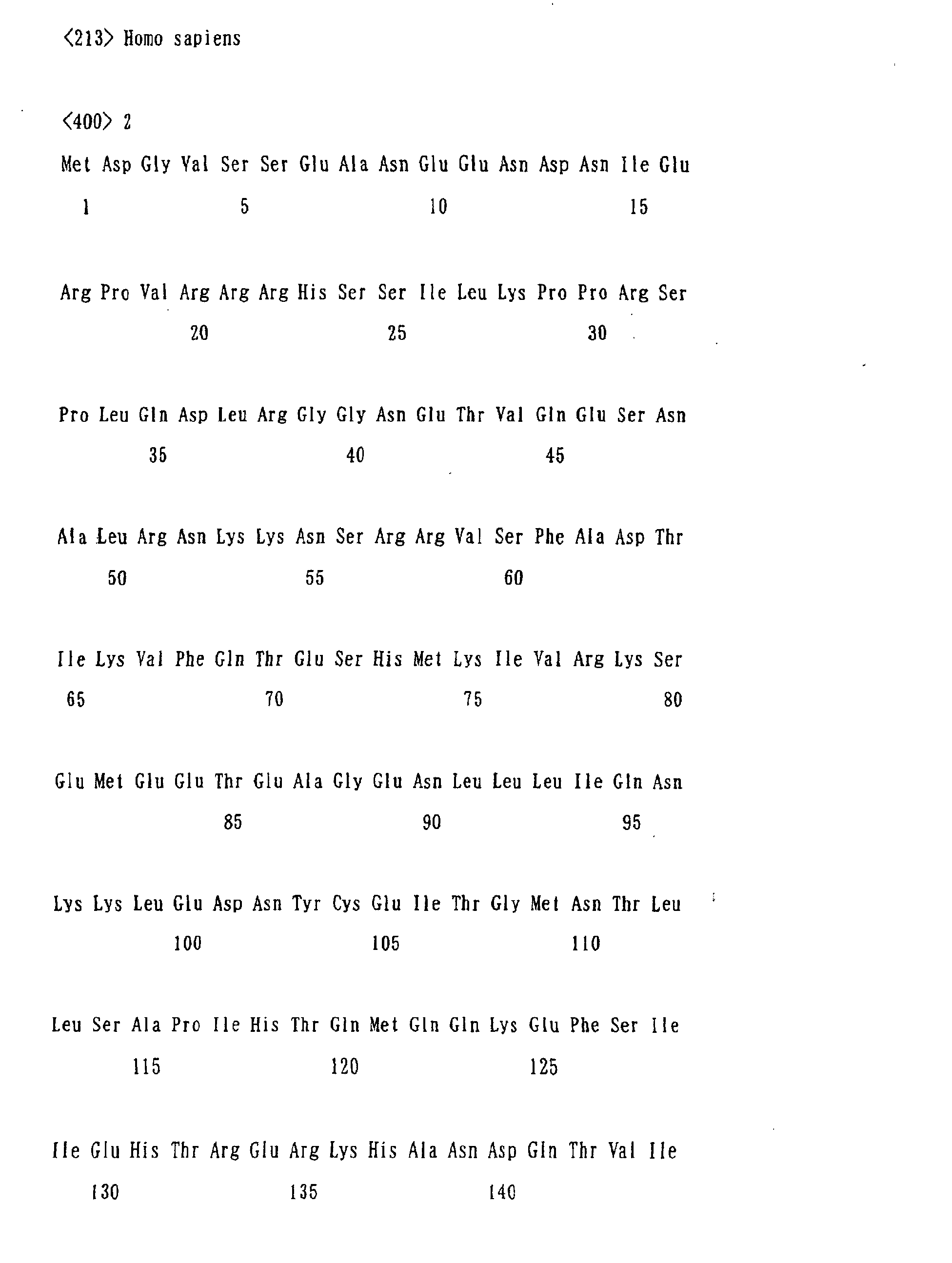
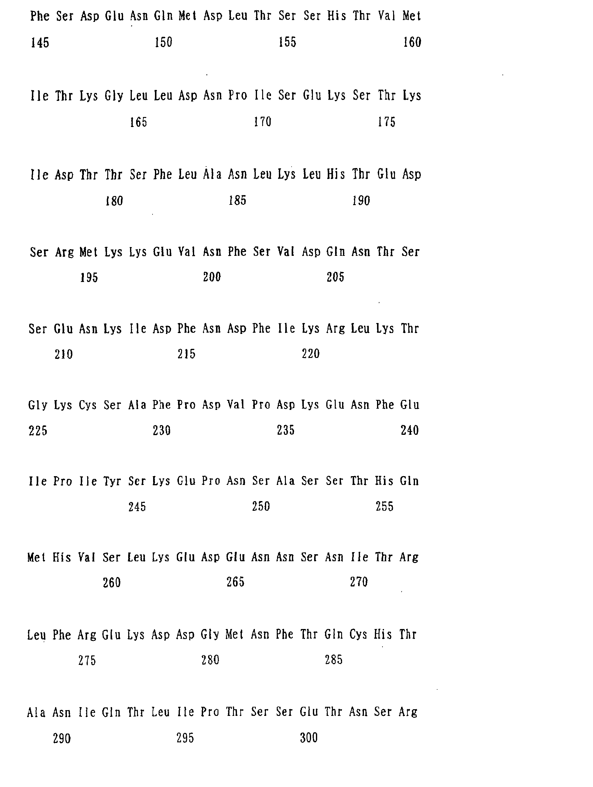
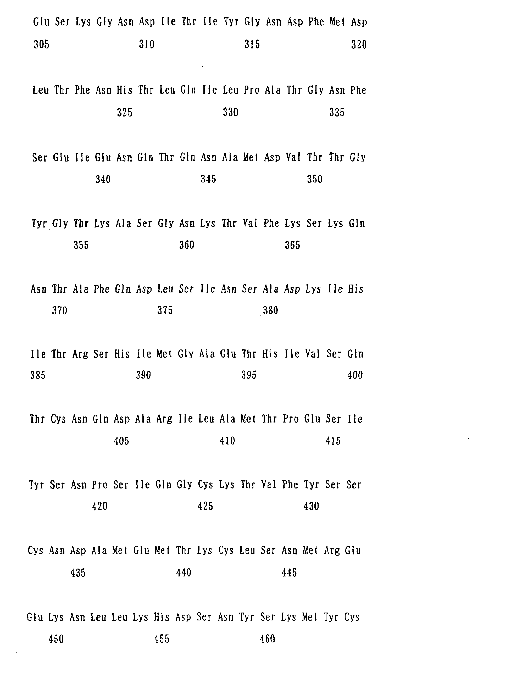
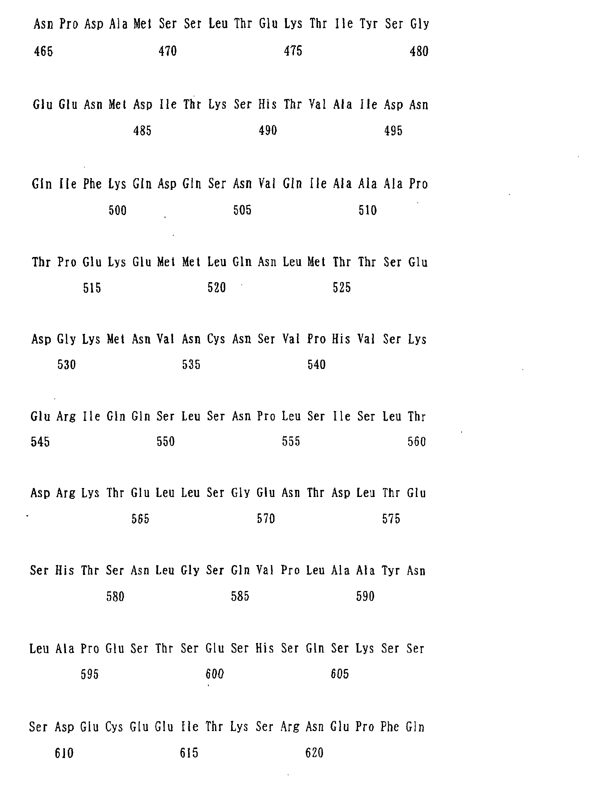


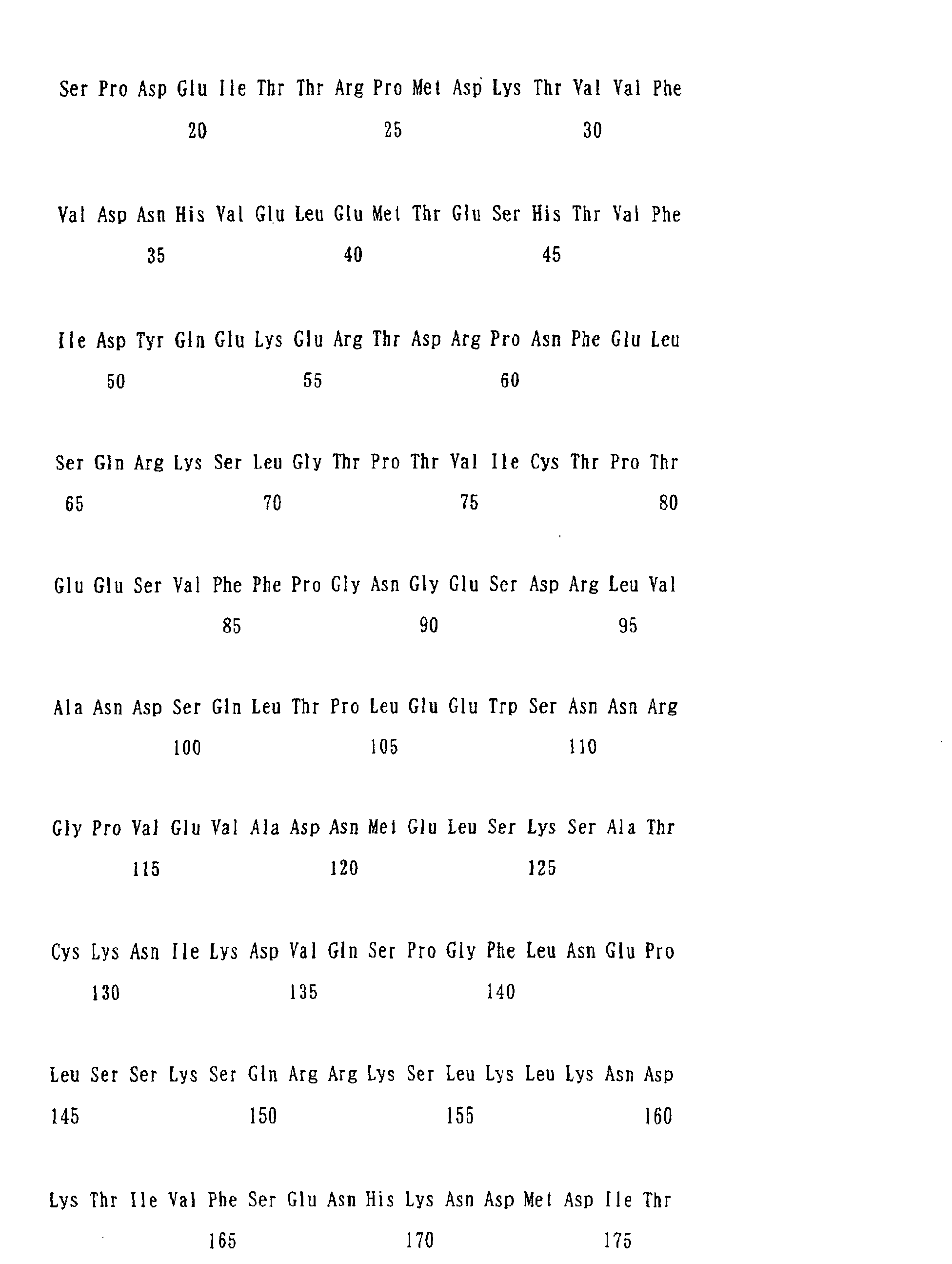


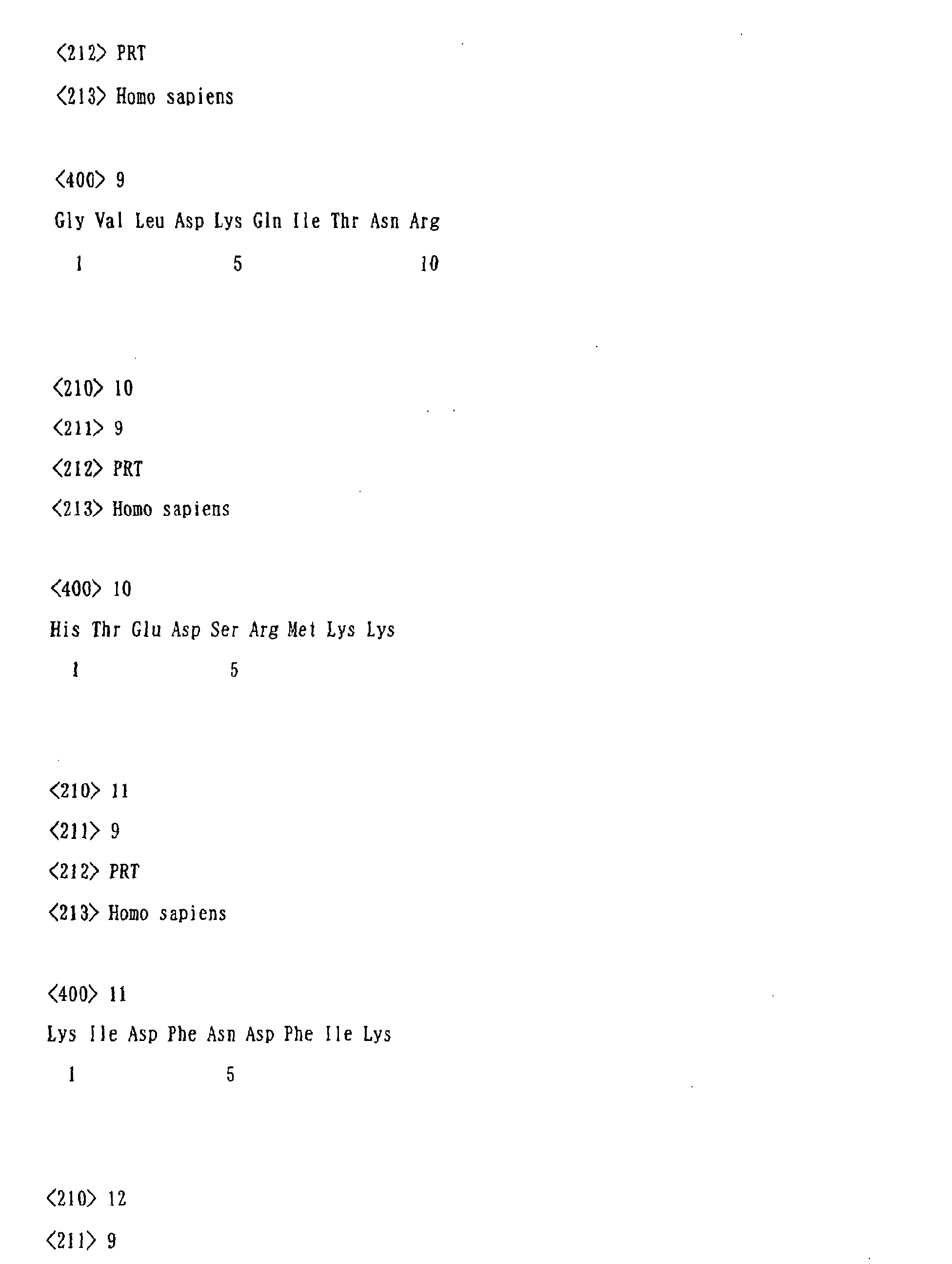

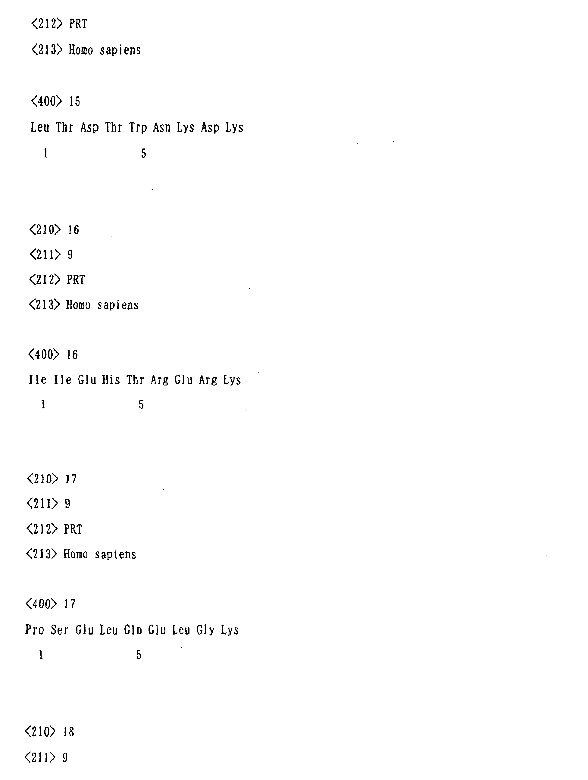


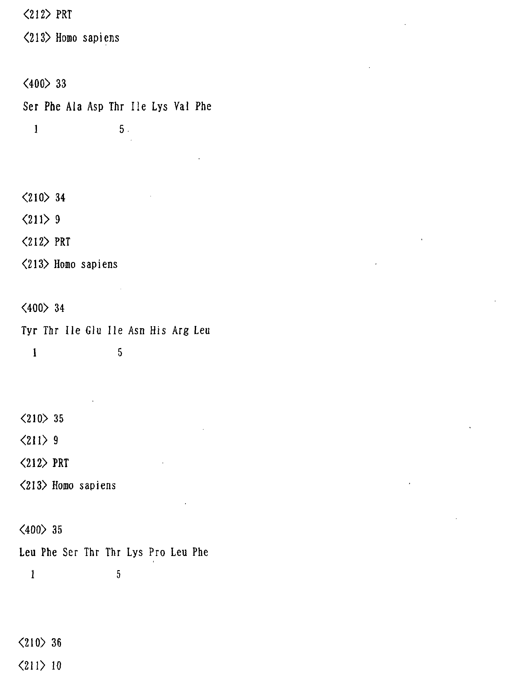
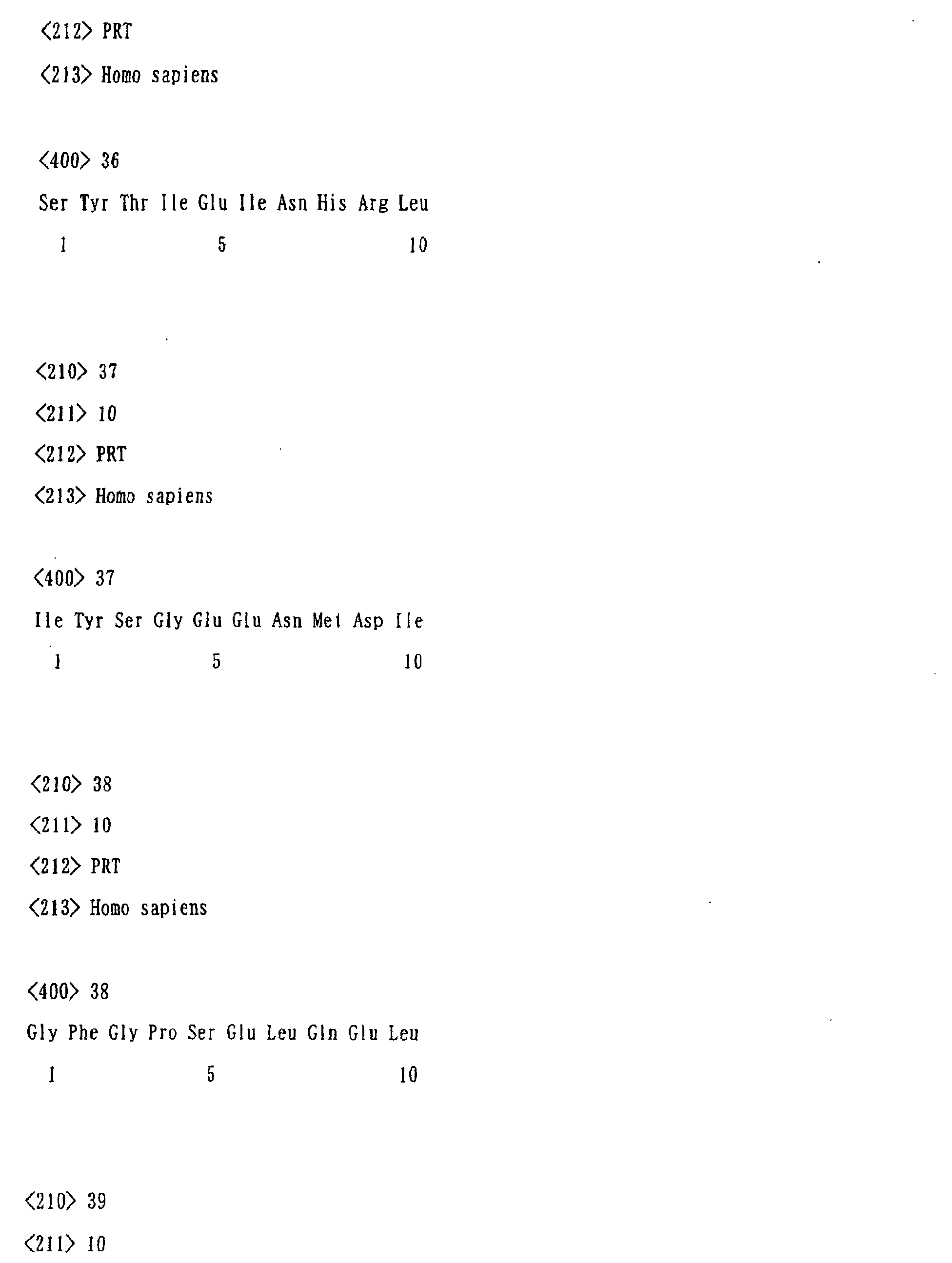
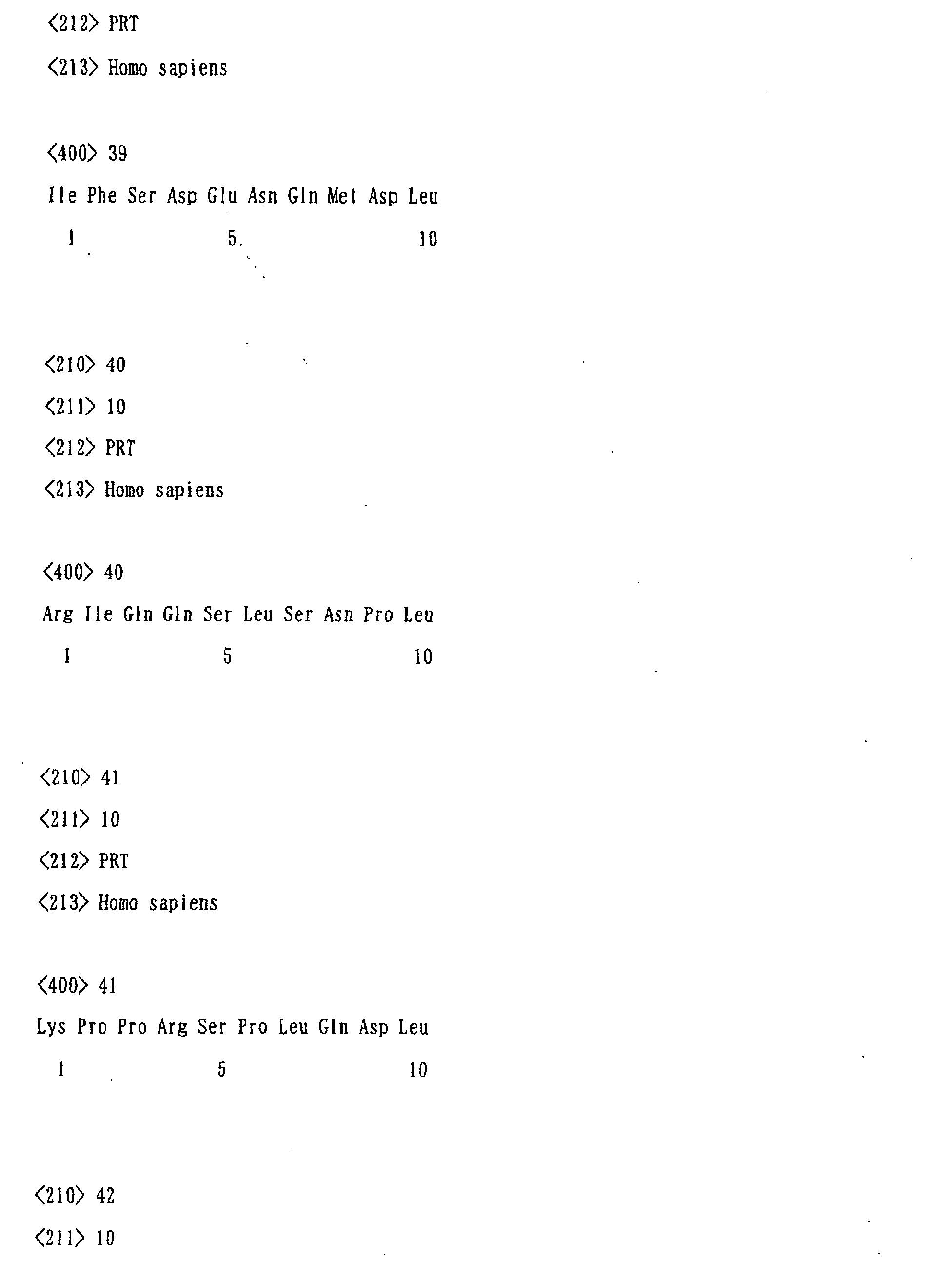


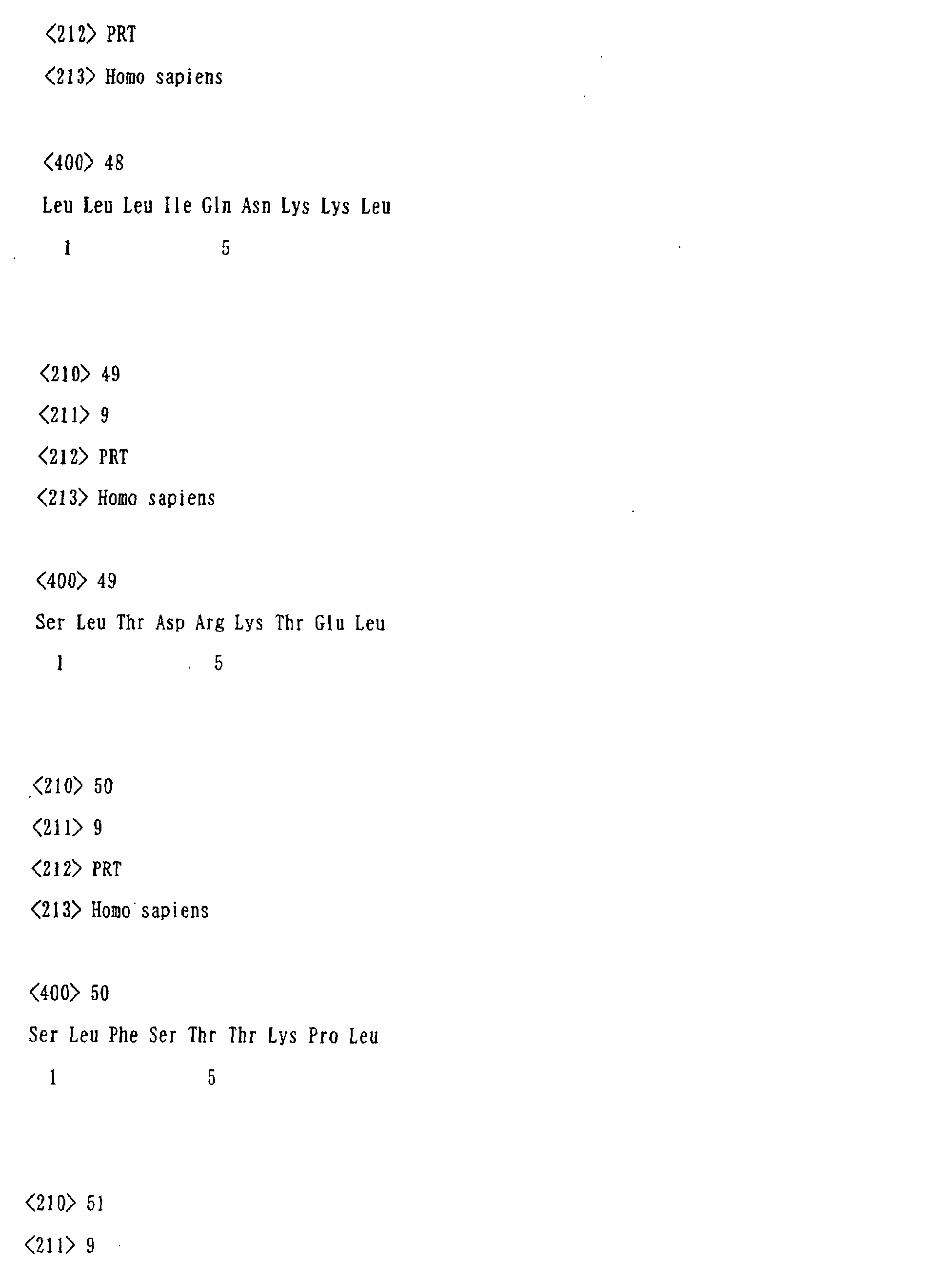

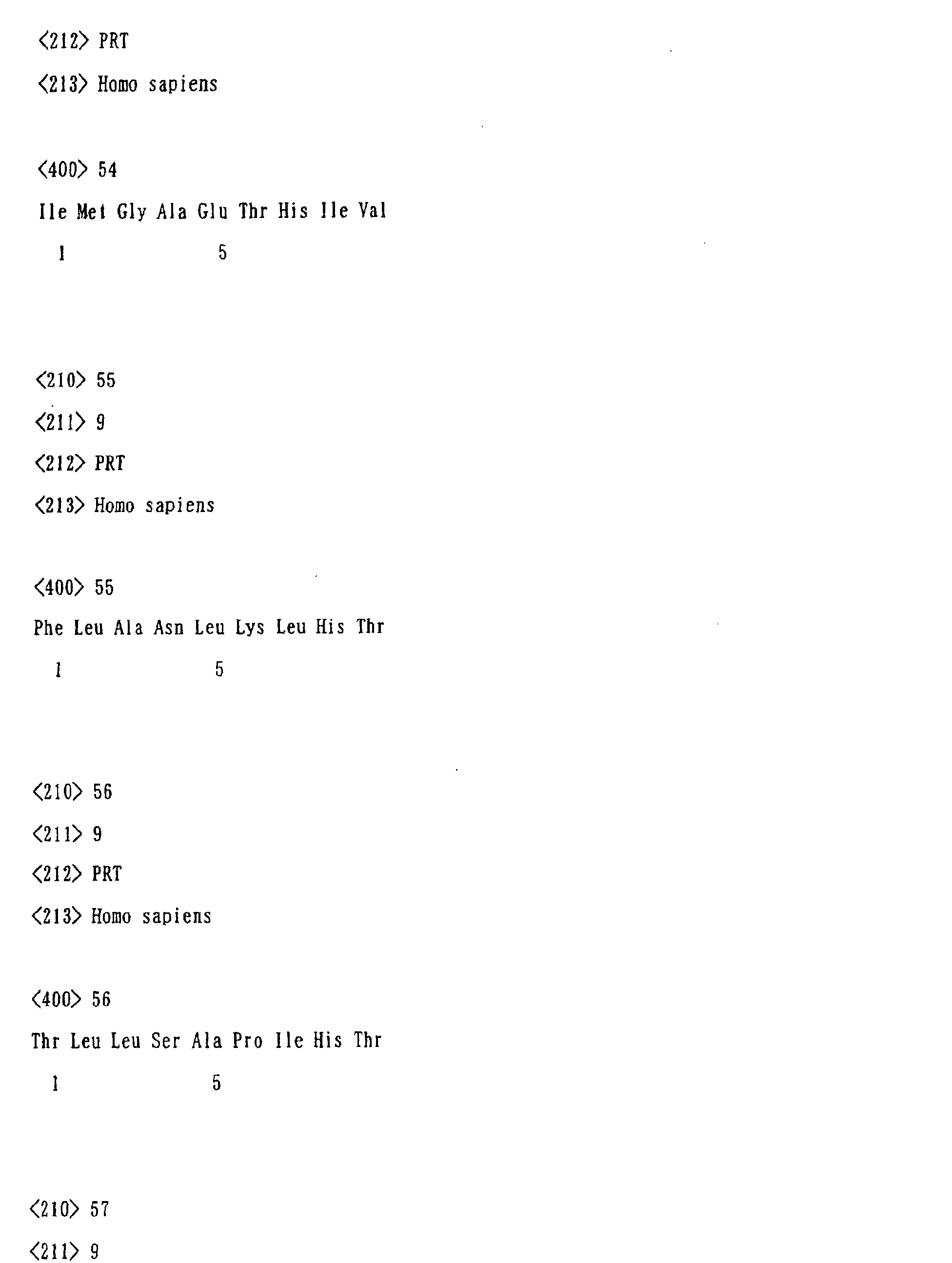

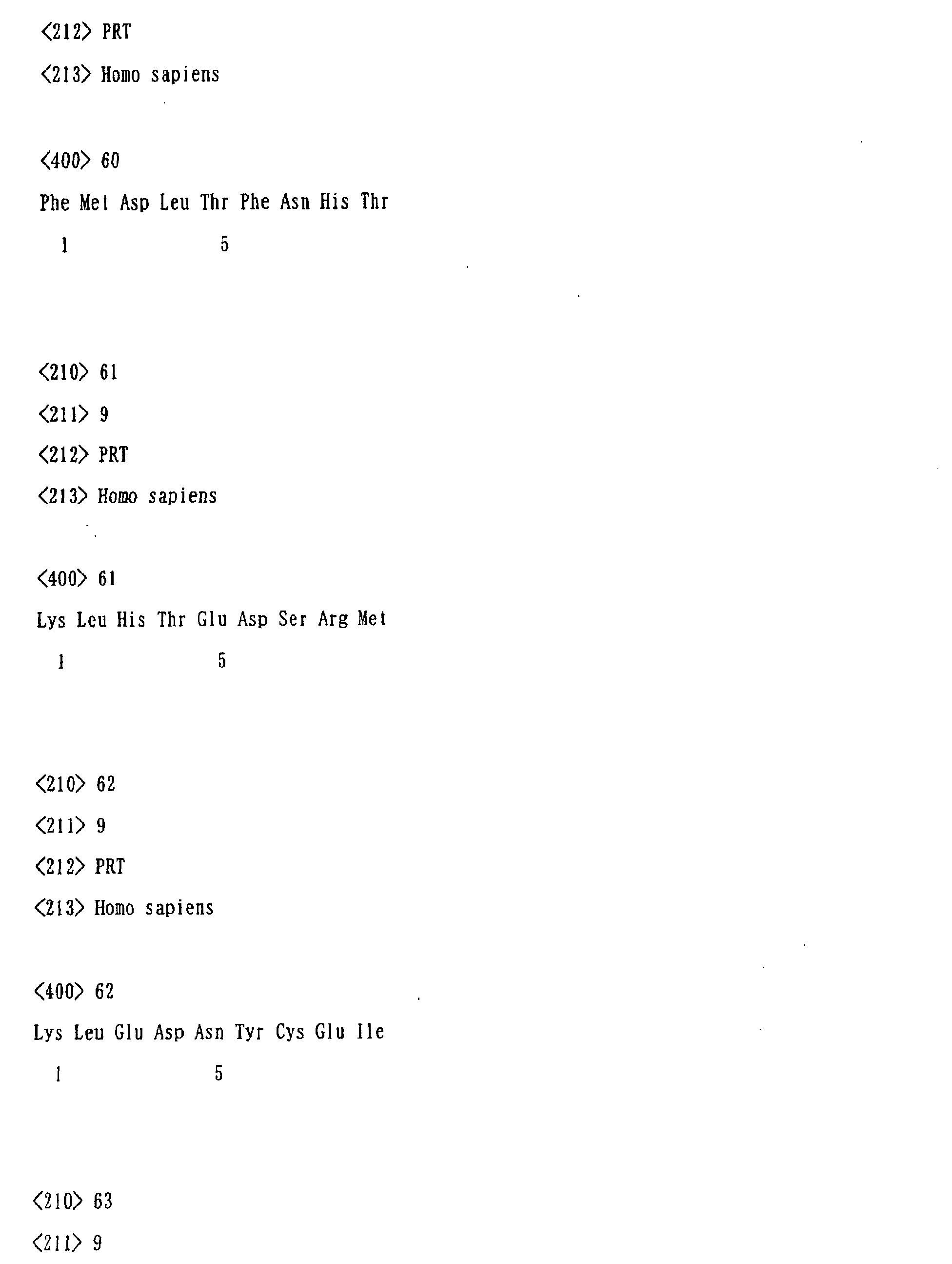
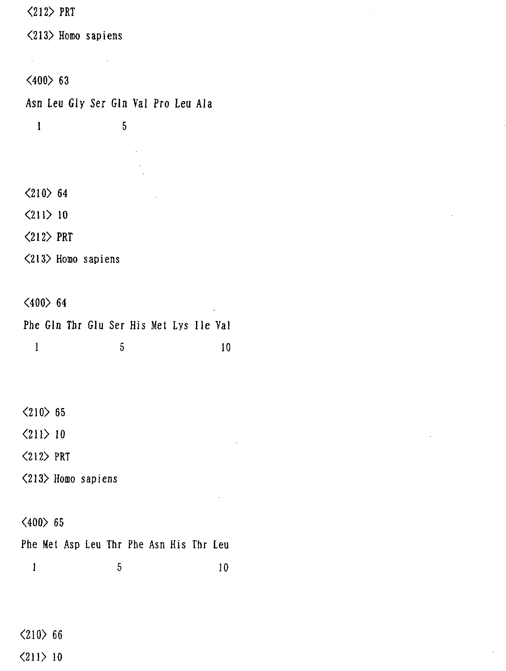

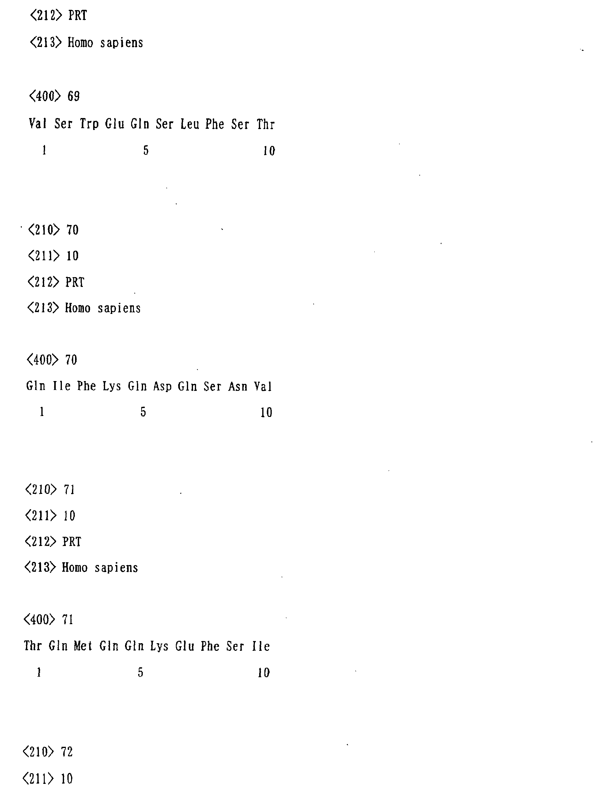


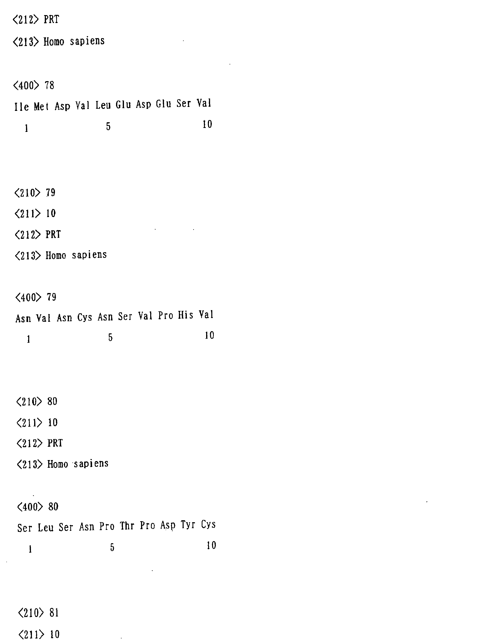
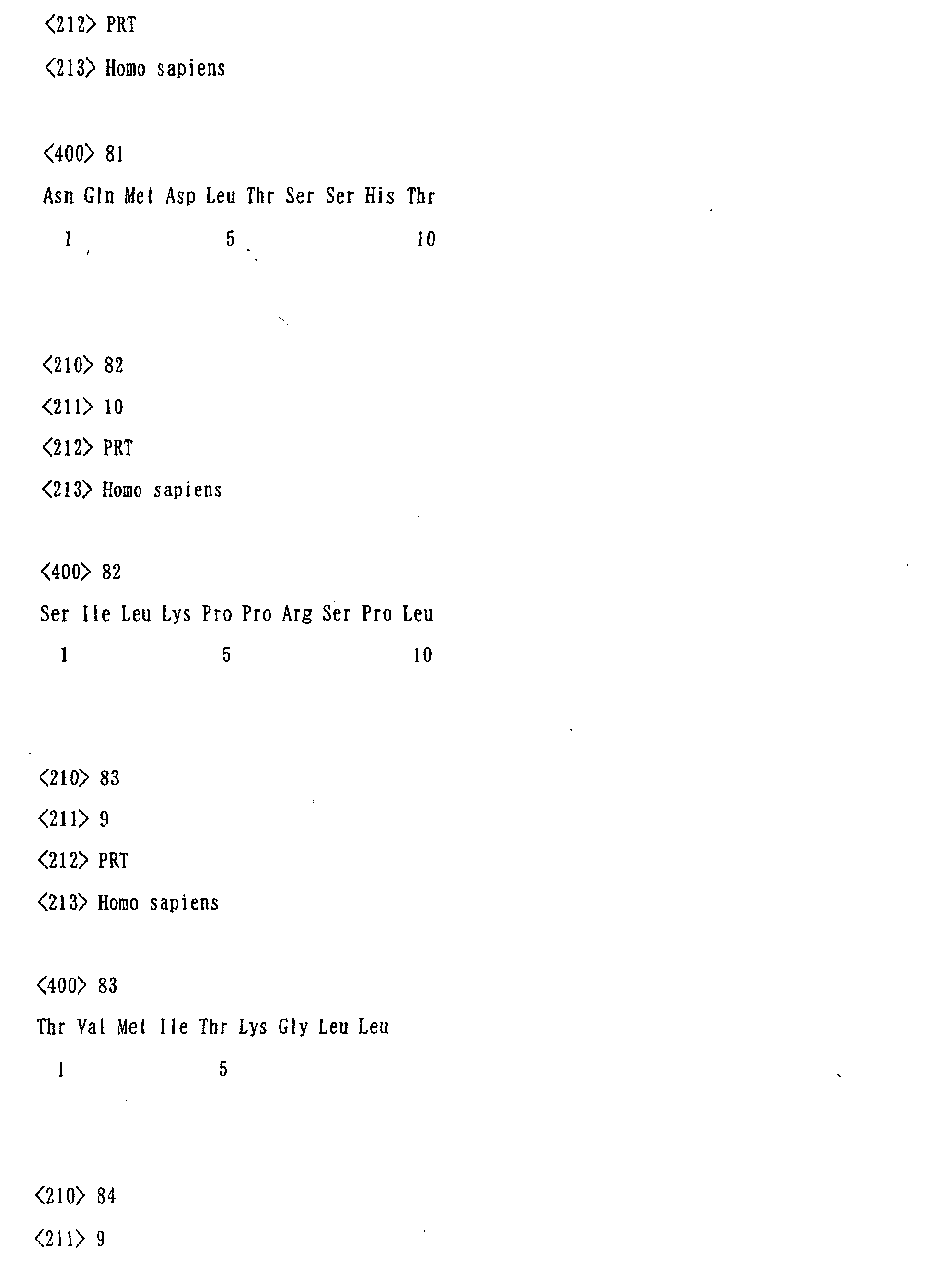
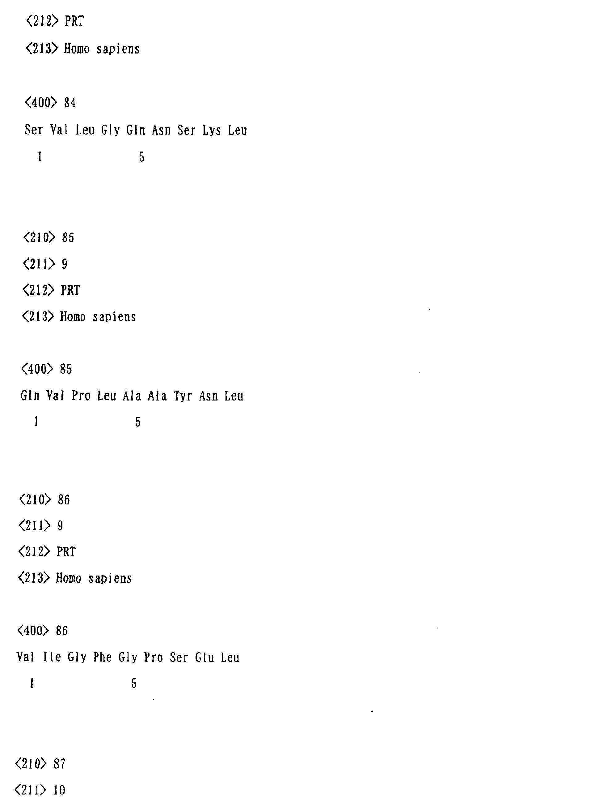
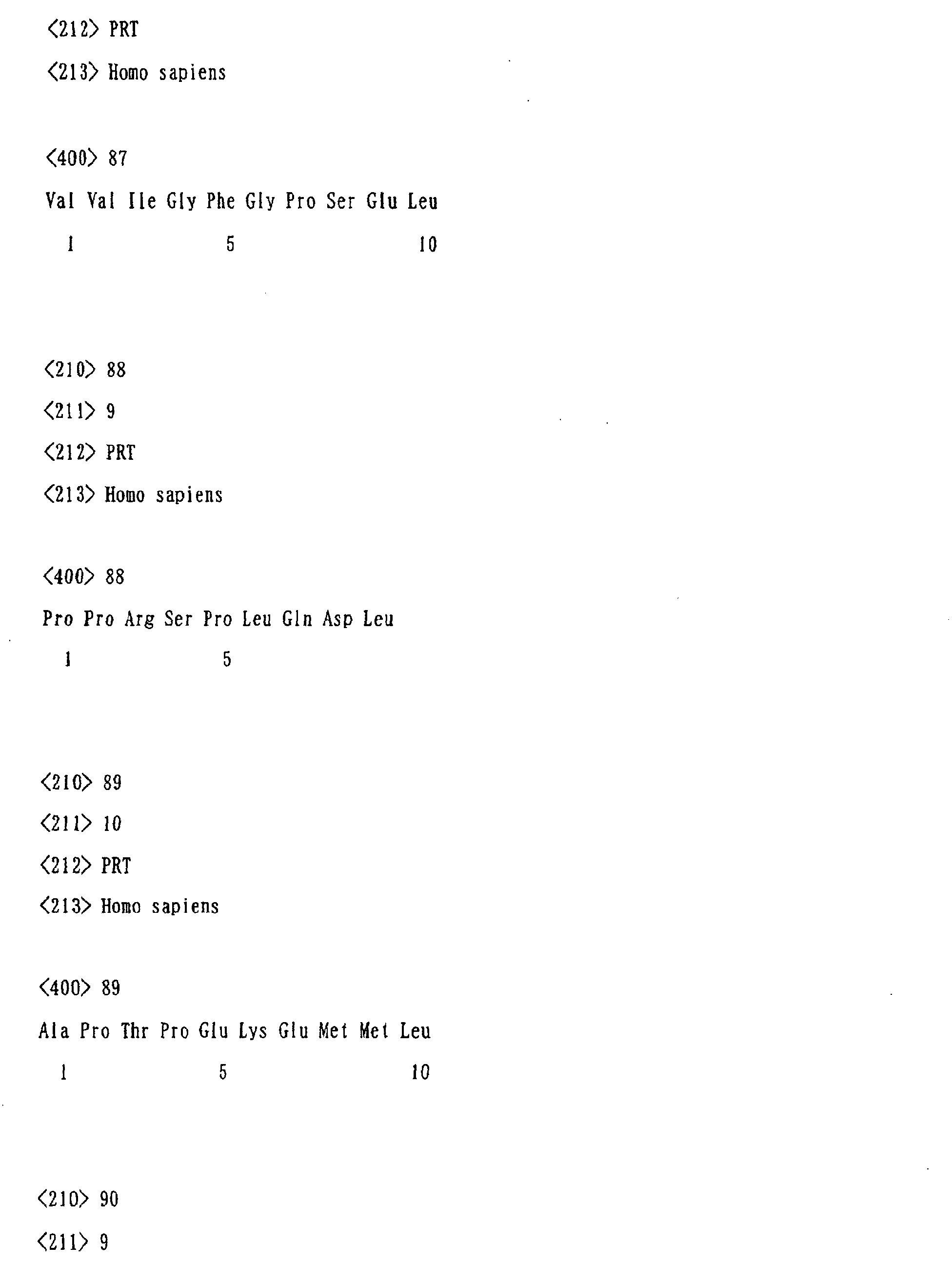
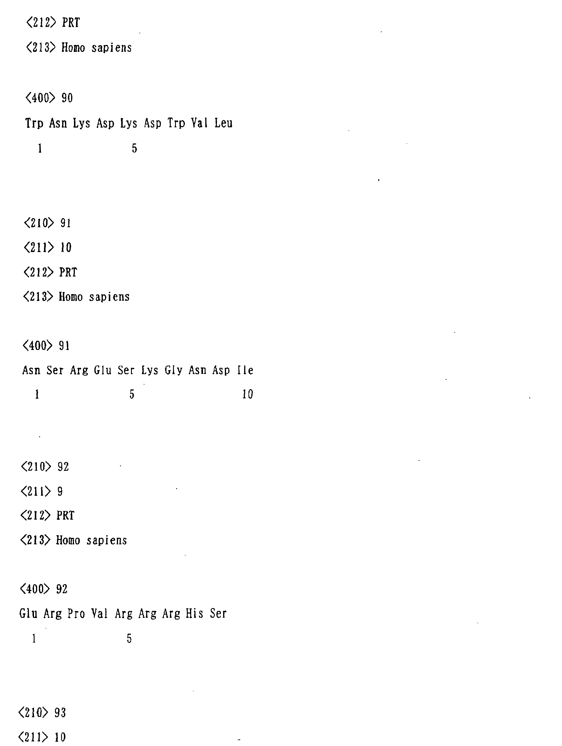

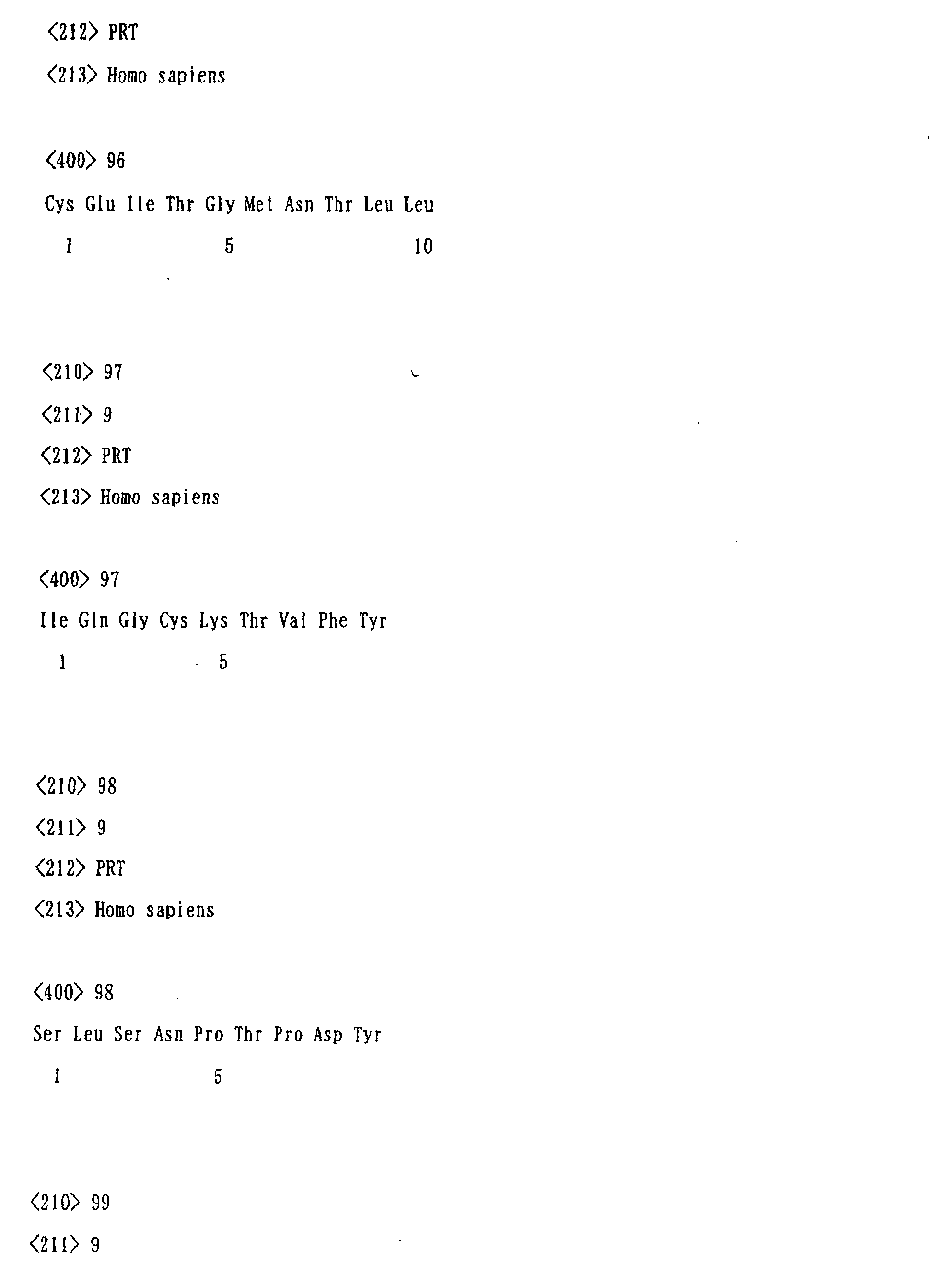


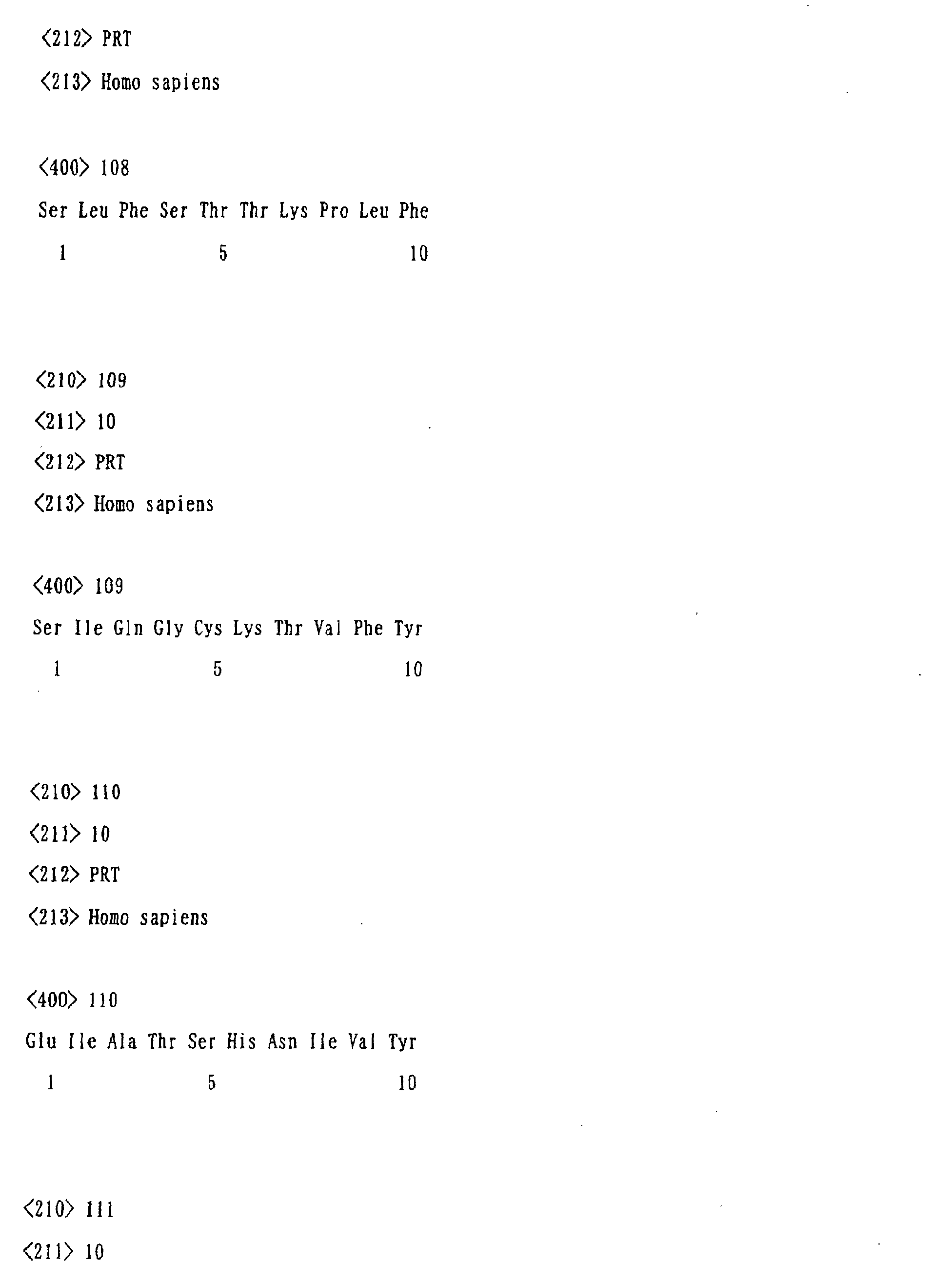


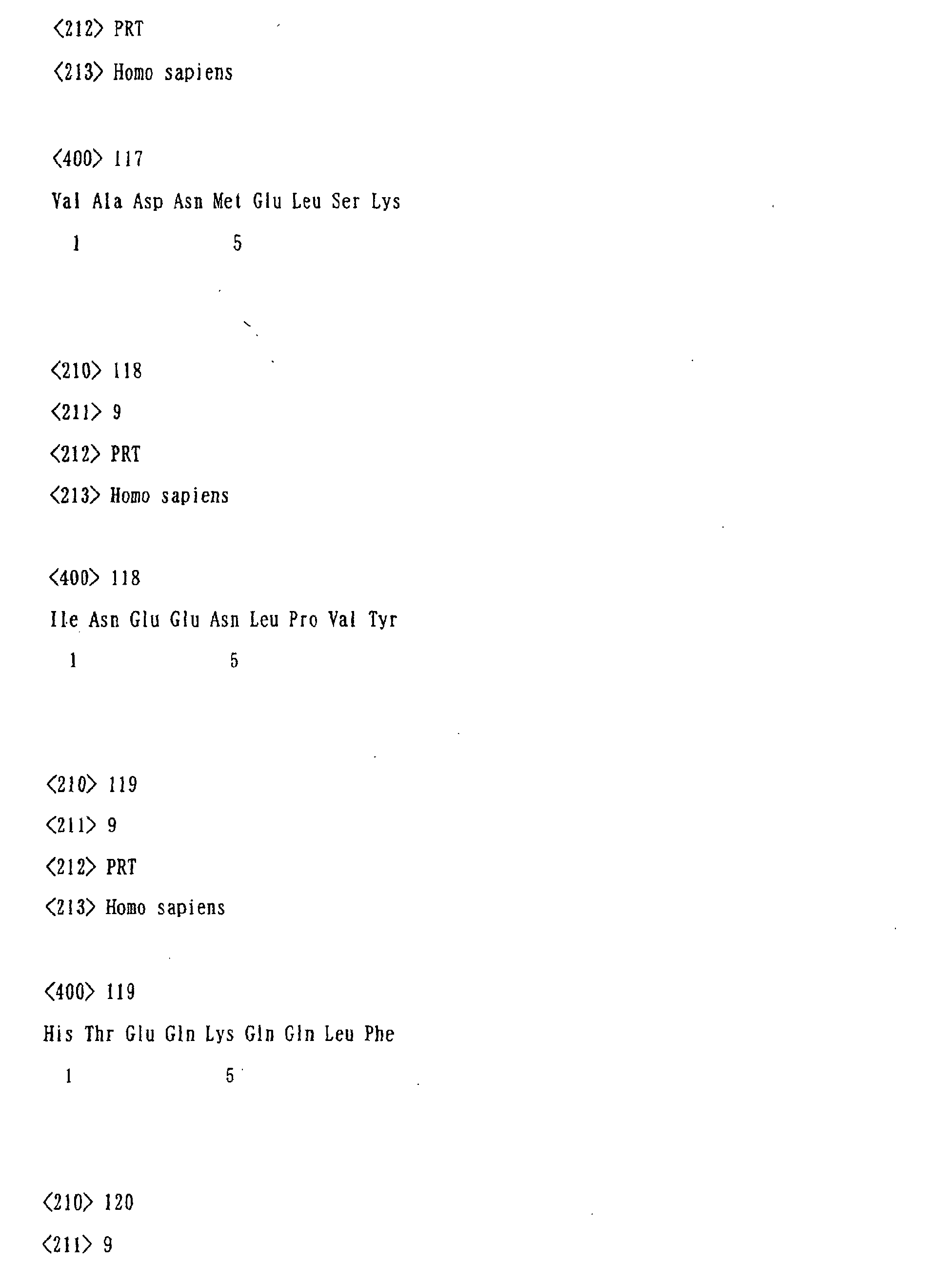


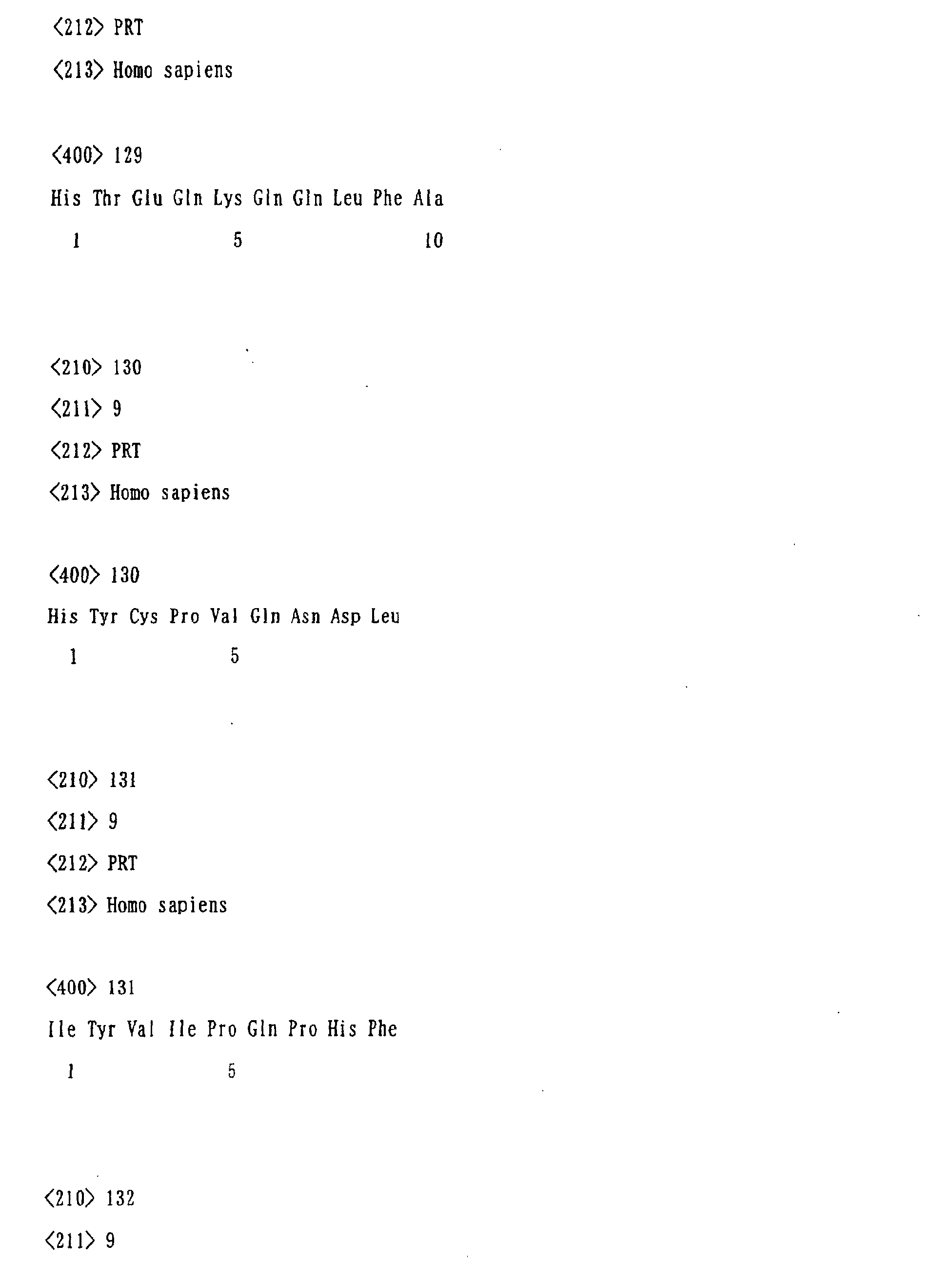
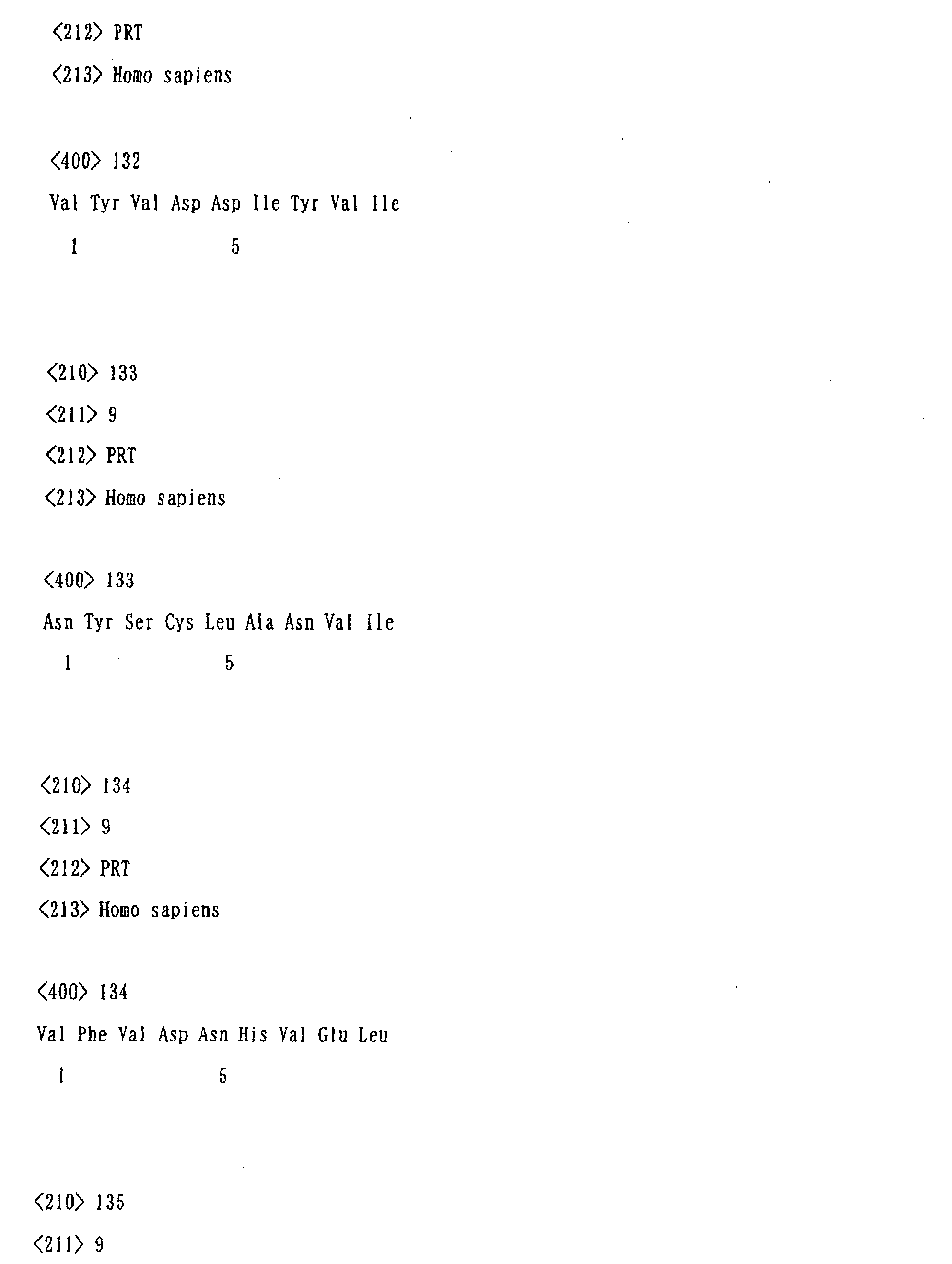
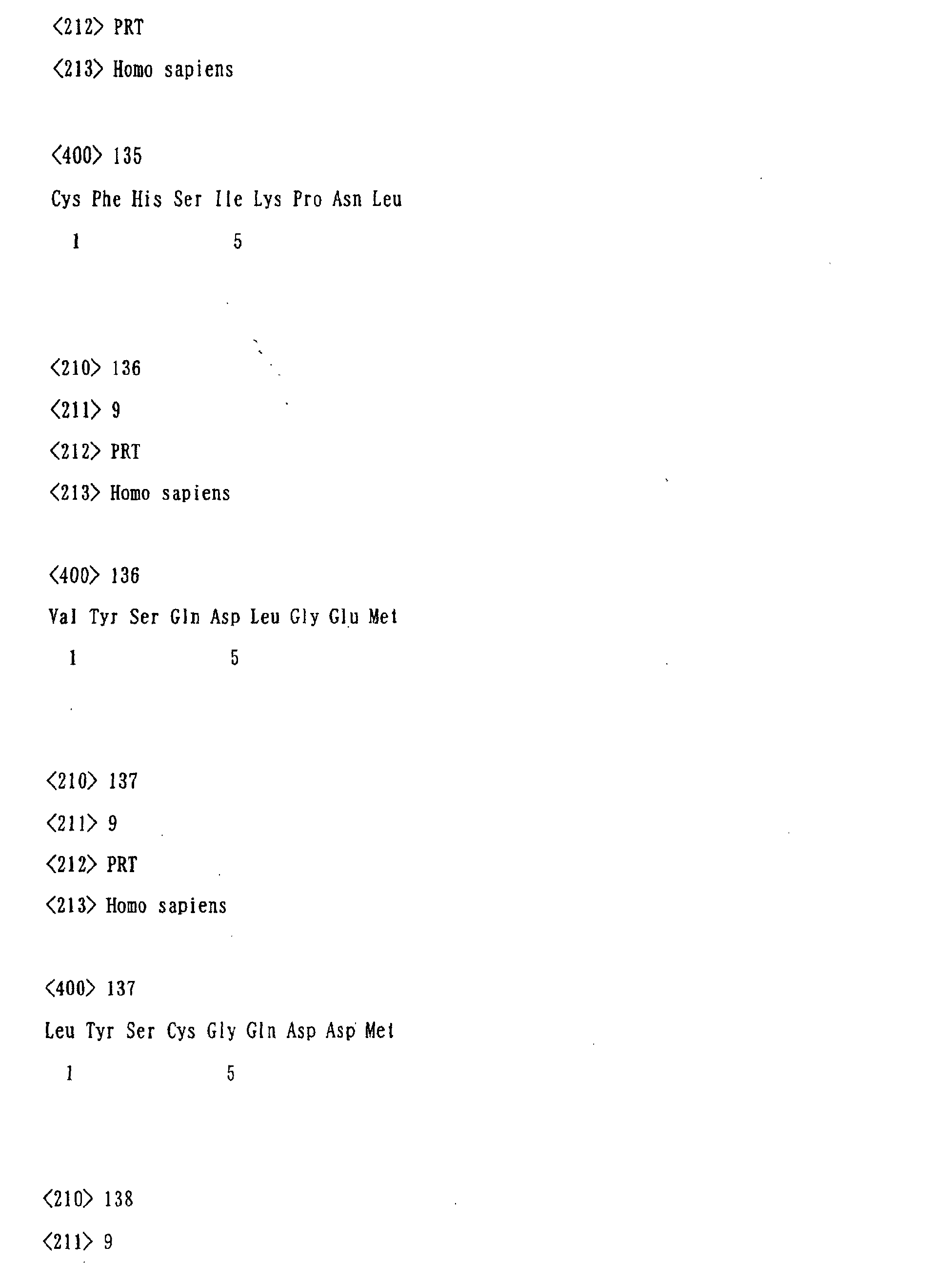
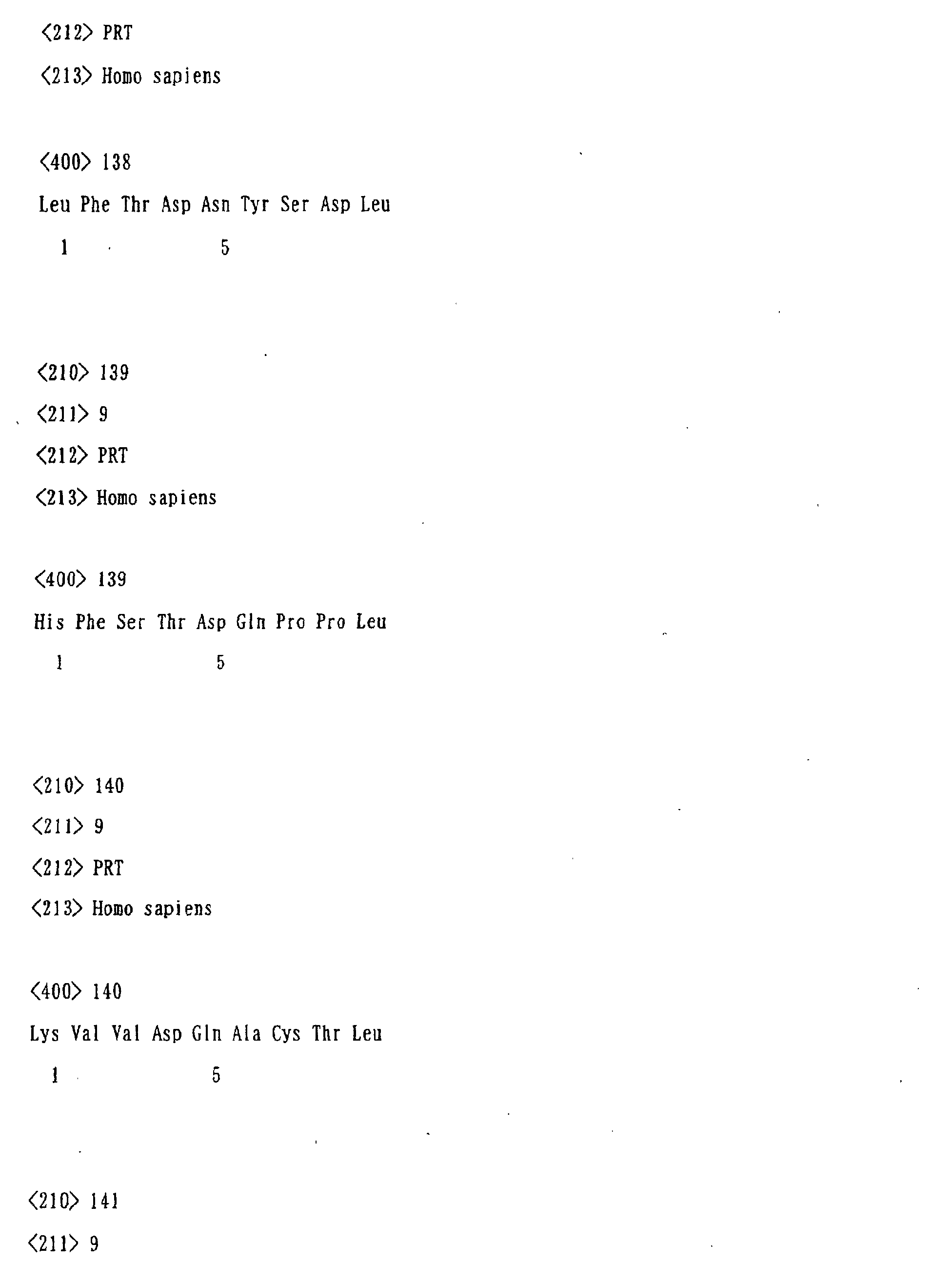

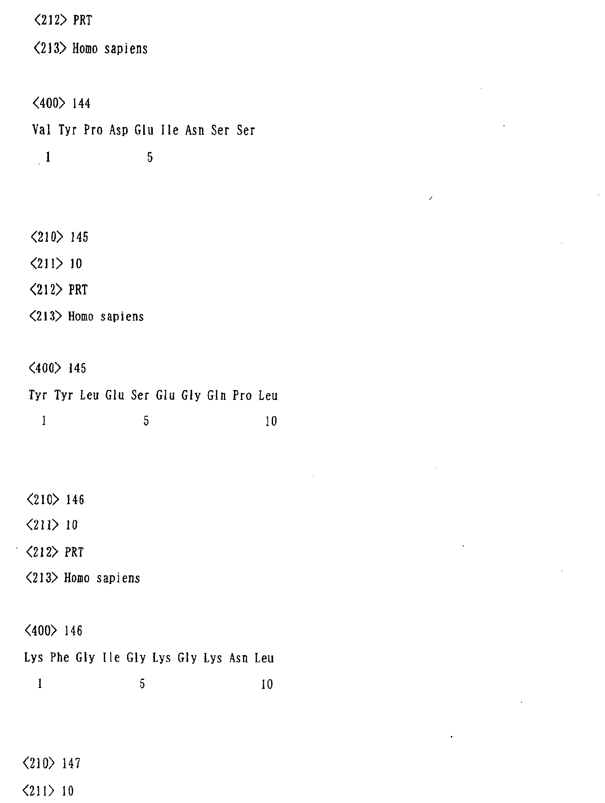
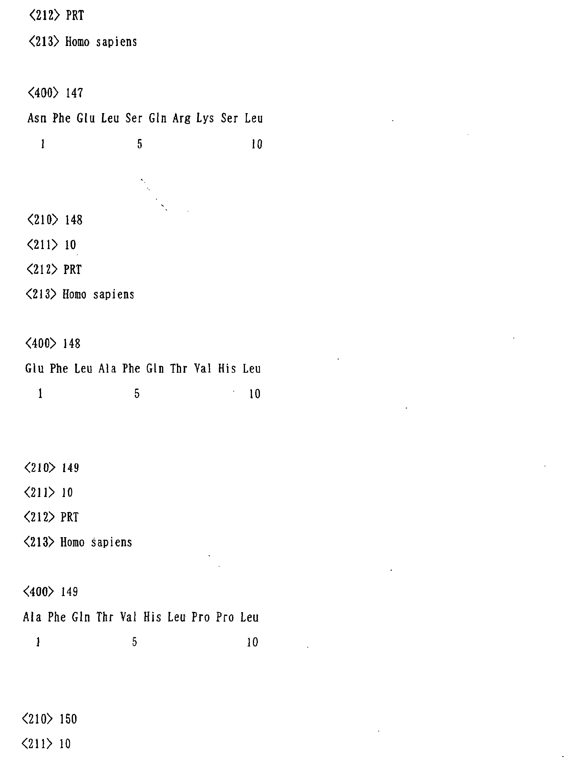


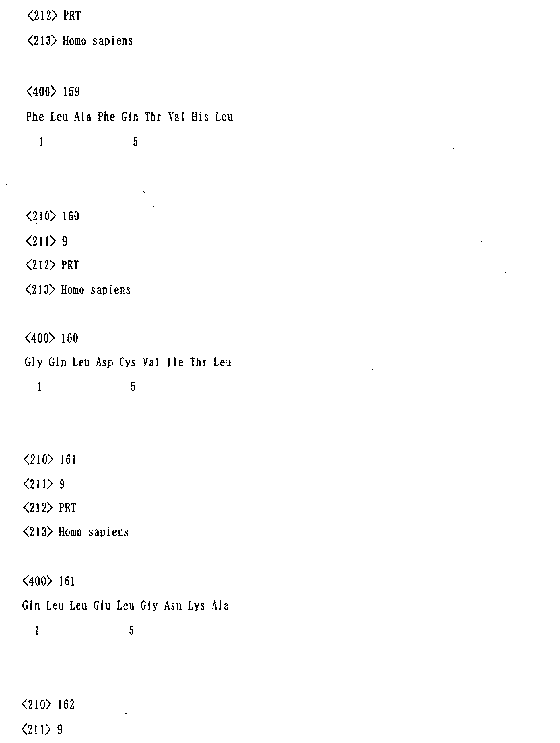


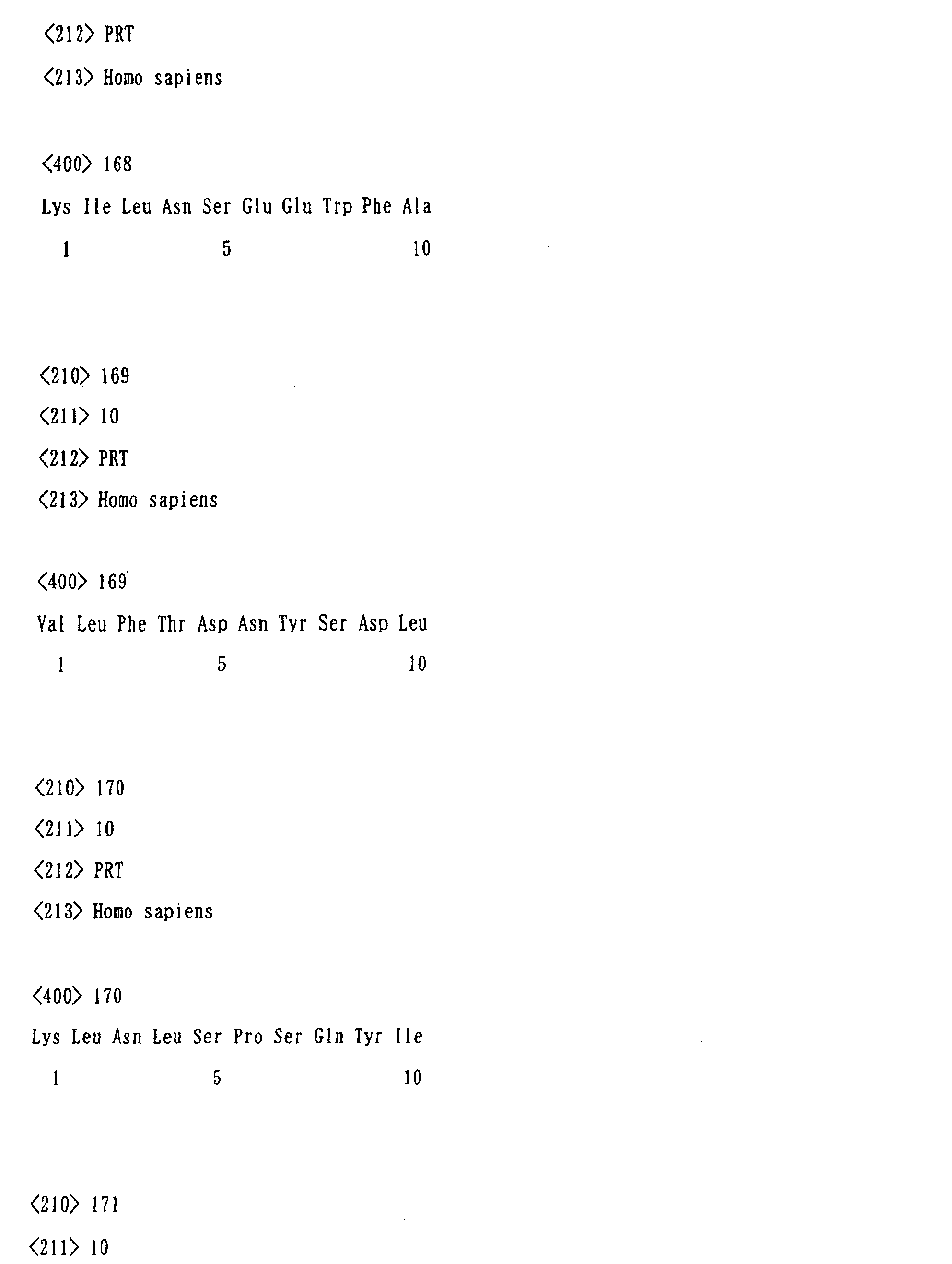
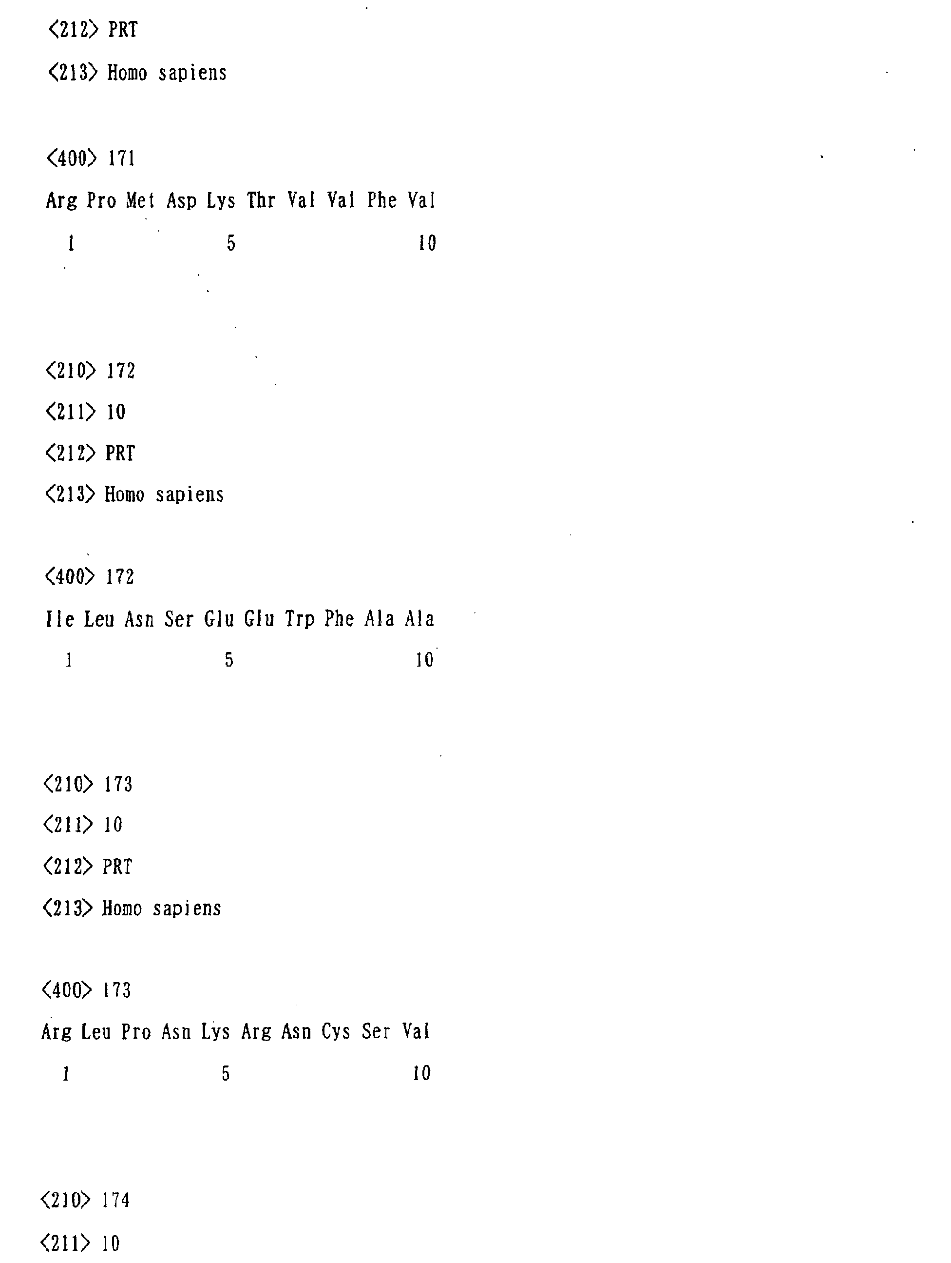
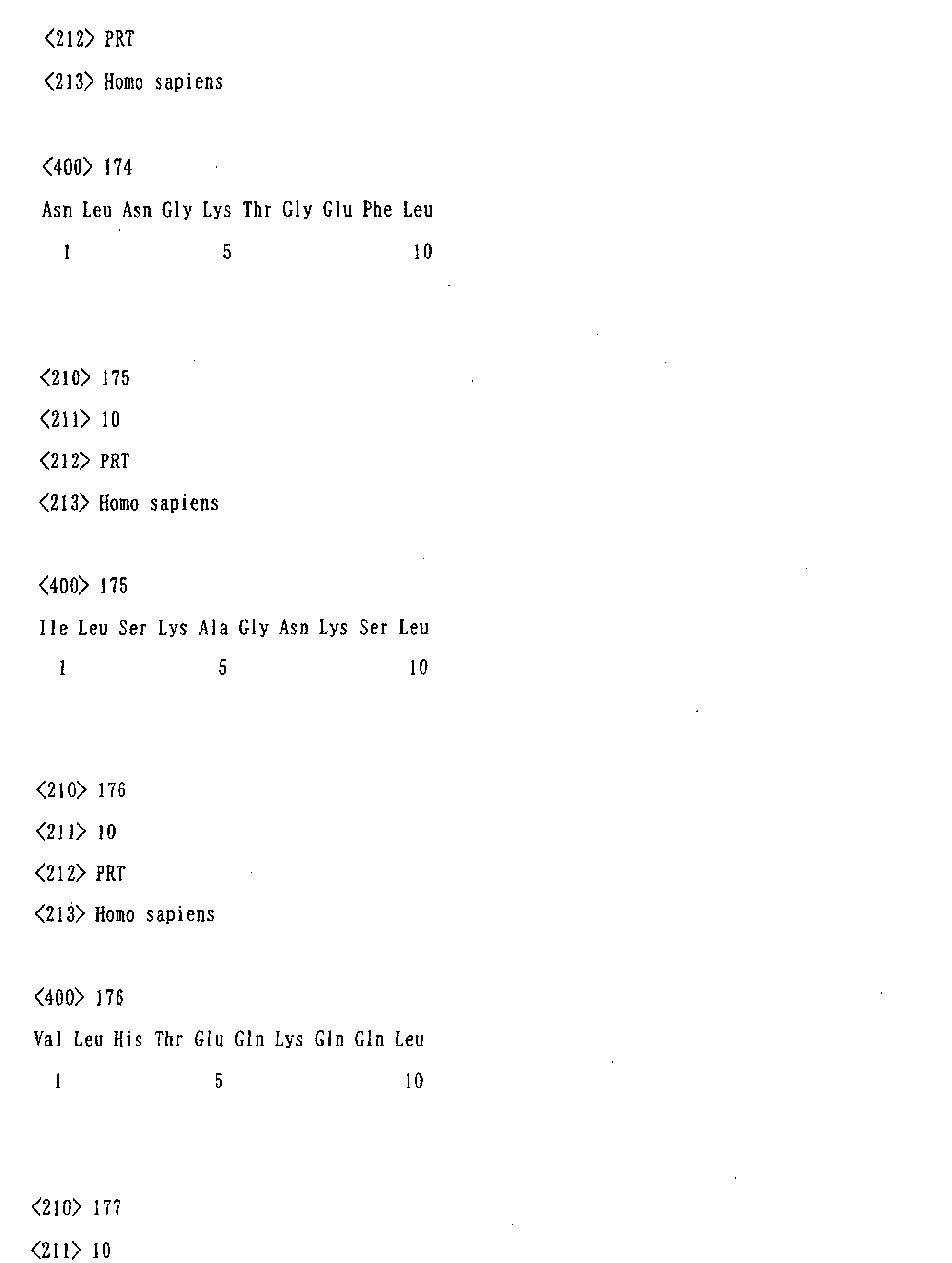
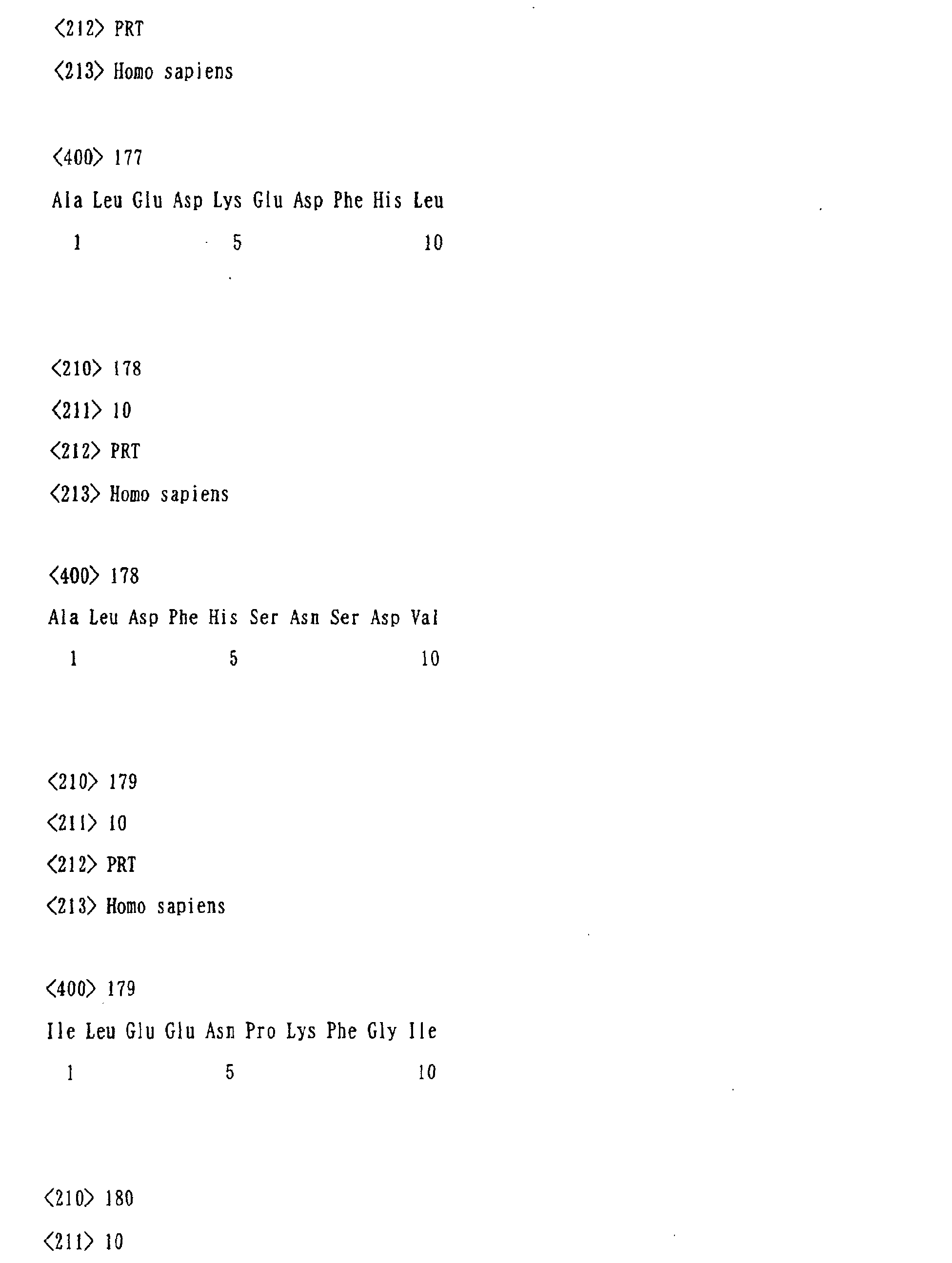
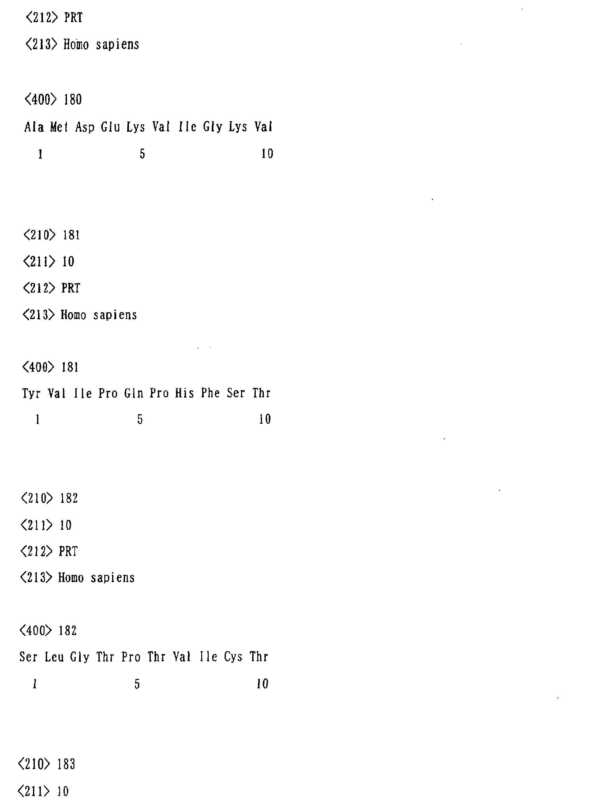
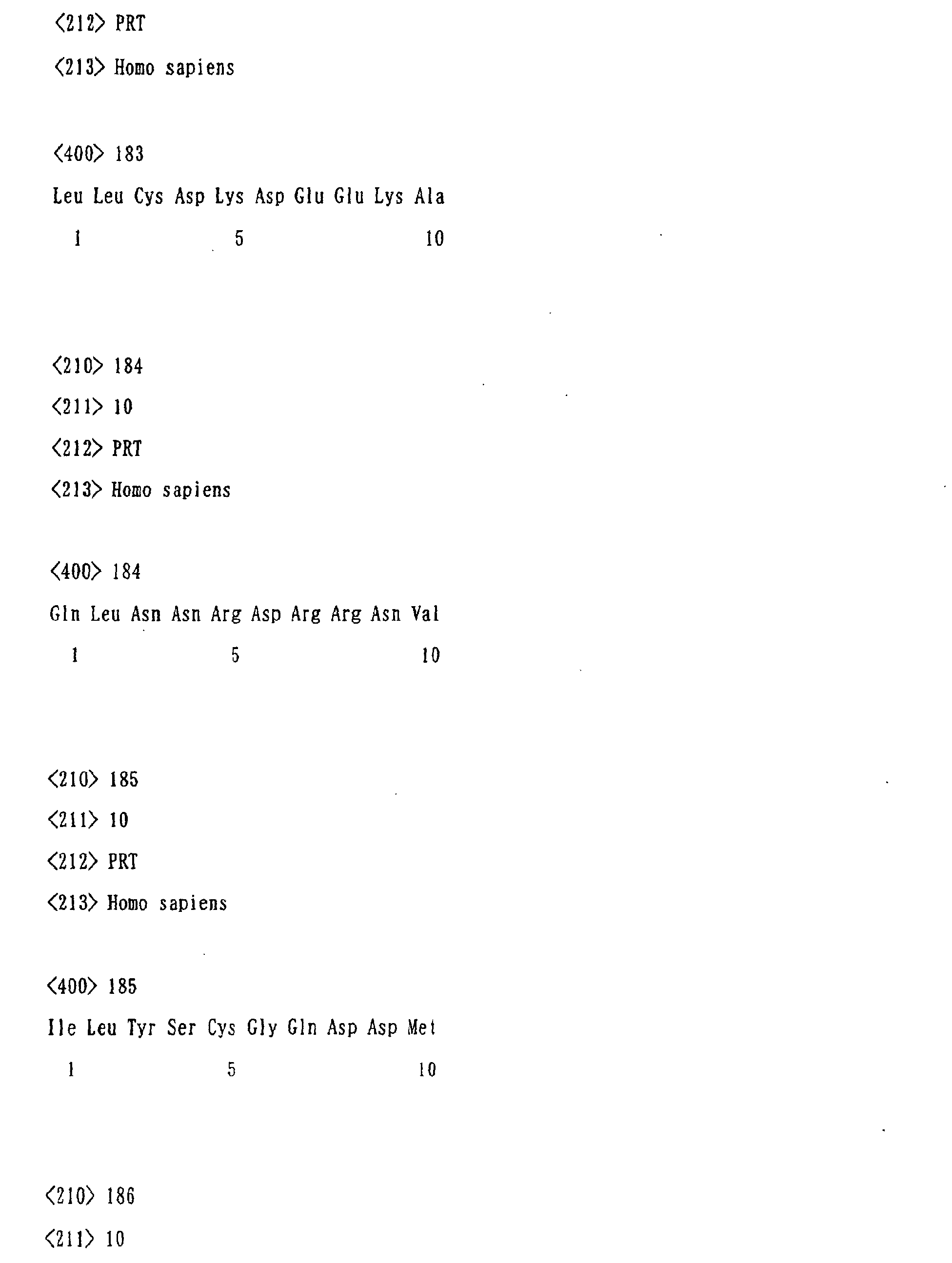

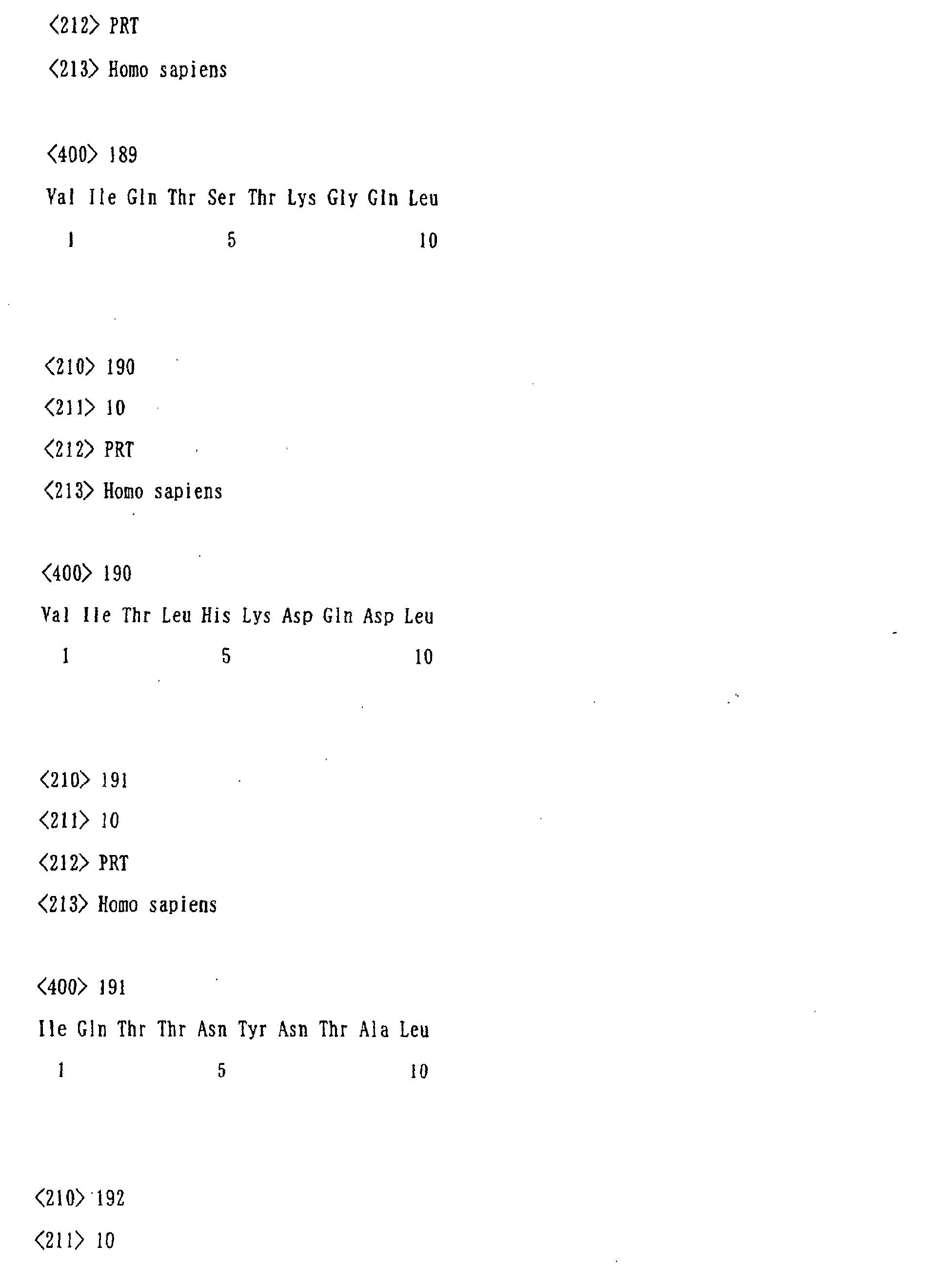


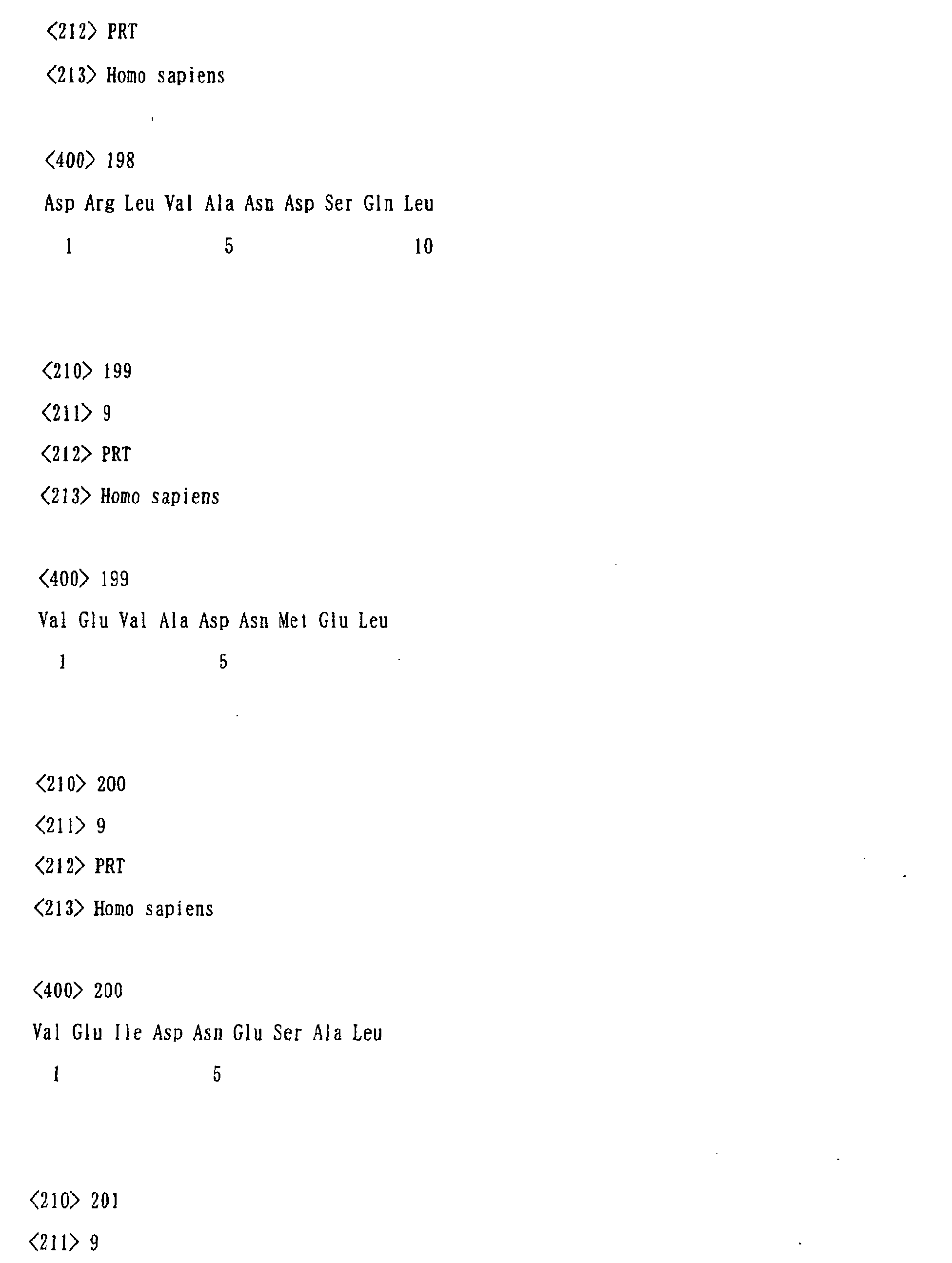




Claims (51)
- A gene encoding the following protein (a) or (b).(a) A protein comprising the amino acid sequence shown by SEQ ID NO:2.(b) A protein which comprises an amino acid sequence wherein one or a few amino acids are deleted, substituted or added in the amino acid sequence shown by SEQ ID NO:2 and which has the immunity-inducing activity.
- A gene encoding the following protein (a) or (b).(a) A protein comprising the amino acid sequence shown by SEQ ID NO:3.(b) A protein which comprises an amino acid sequence wherein one or a few amino acids are deleted, substituted or added in the amino acid sequence shown by SEQ ID NO:3 and which has the immunity-inducing activity.
- DNA containing the base sequence shown by SEQ ID NO:1 or its complementary sequence and part of whole of these sequences.
- DNA which hybridizes under a stringent condition with DNA of claim 3 that forms a gene, and which encodes a protein having the immunity-inducing activity.
- A protein comprising the amino acid sequence shown by SEQ ID NO:2.
- A protein which comprises an amino acid sequence wherein one or a few amino acids are deleted, substituted or added in the amino acid sequence shown by SEQ ID NO:2 and which has the immunity-inducing activity.
- A protein comprising the amino acid sequence shown by SEQ ID NO:3.
- A protein which comprises an amino acid sequence wherein one or a few amino acids are deleted, substituted or added in the amino acid sequence shown by SEQ ID NO:3 and which has the immunity-inducing activity.
- A peptide which comprises part of the protein of any of claims 5 to 8 and which binds to a histocompatibility antigen molecule Class I.
- The peptide according to claim 9, wherein said peptide comprises part of the protein of claim 5 or 6 and binds to a histocompatibility antigen molecule Class I.
- The peptide according to claim 10, wherein said peptide comprises an amino acid sequence shown by any of SEQ ID NOs:4-111.
- A peptide comprising the amino acid sequence of Ser-Tyr-Thr-Ile-Glu-Ile-Asn-His-Arg-Leu.
- The peptide according to claim 9, wherein said peptide comprises part of the protein of claim 7 or 8 and binds to the histocompatibility antigen molecule Class I.
- The peptide according to claim 13, wherein said peptide comprises an amino acid sequence shown by any of SEQ ID NOs:112-221.
- A fusion protein or a fusion peptide wherein the protein of claim 5 or 6 or the peptide of any of claims 10 to 12, and a marker protein and/or a peptide tag, are bound.
- A fusion protein or a fusion peptide wherein the protein of claim 7 or 8 or the peptide of claim 13 or 14, and a marker protein and/or a peptide tag, are bound.
- An antibody which specifically binds to the protein of claim 5 or 6 or to the peptide of any of claims 10 to 12.
- An antibody which specifically binds to the protein of claim 7 or 8 or to the peptide of claim 13 or 14.
- The antibody according to claim 17 or 18, wherein said antibody is a monoclonal antibody.
- A recombinant protein or peptide wherein the antibody of any of claims 17 to 19 specifically binds to said recombinant protein or peptide.
- A host cell comprising an expression system capable of expressing the protein according to claim 5 or 6.
- A host cell comprising an expression system capable of expressing the protein according to claim 7 or 8.
- A non-human animal whose gene function to encode the protein of claim 5 or 6 is deficient on its chromosome.
- A non-human animal whose gene function to encode the protein of claim 7 or 8 is deficient on its chromosome.
- The non-human animal according to claim 23 or 24, wherein said non-human animal is a mouse or a rat.
- A non-human animal which over-expresses the protein of claim 5 or 6.
- A non-human animal which over-expresses the protein of claim 7 or 8.
- The non-human animal according to claim 26 or 27, wherein said non-human animal is a mouse or a rat.
- A method of screening a promoter or a suppressor for the immunity-inducing activity wherein the protein of claim 5 or 6, the peptide of any of claims 10 to 12 or a cell membrane expressing the protein of claim 5 or 6, and a test substance is used.
- A method of screening a promoter or a suppressor for the immunity-inducing activity wherein the protein of claim 7 or 8, the peptide of claim 13 or 14, or a cell membrane expressing the protein of claim 7 or 8, and a test substance is used.
- A method of screening a promoter or a suppressor for the immunity-inducing activity or for the expression of the protein of claim 5 or 6, wherein a cell expressing the protein and a test substance are used.
- The method of screening a promoter or a suppressor for the immunity-inducing activity or for the expression of the protein of claim 5 or 6 according to claim 31, wherein the cell expressing the protein of claim 5 or 6 is the host cell of claim 21.
- A method of screening a promoter or a suppressor for the immunity-inducing activity or for the expression of the protein of claim 7 or 8, wherein a cell expressing the protein and a test substance are used.
- The method of screening a promoter or a suppressor for the immunity-inducing activity or for the expression of the protein of claim 7 or 8 according to claim 33, wherein the cell expressing the protein of claim 7 or 8 is the host cell of claim 22.
- A method of screening a promoter or a suppressor for the immunity-inducing activity or a promoter or a suppressor for expression of the protein of any of claims 5 to 8, wherein the non-human animal of any of claims 23 to 25 and a test substance are used.
- A method of screening a promoter or a suppressor for the immunity-inducing activity or a promoter or a suppressor for expression of the protein of any of claims 5 to 8, wherein the non-human animal of any of claims 26 to 28 and a test substance are used.
- A promoter for the immunity-inducing activity obtained by the screening method according to any of claims 29 to 36.
- A suppressor for the immunity-inducing activity obtained by the screening method according to any of claims 29 to 36.
- An expression promoter for the protein of claim 5 or 6, wherein said expression promoter is obtained by the screening method according to any of claims 29 to 36.
- An expression suppressor for the protein of claim 7 or 8, wherein said expression suppressor is obtained by the screening method according to any of claims 29 to 36.
- An anti-tumor agent comprising the protein of any of claims 5 to 8, the peptide of any of claims 9 to 16, the protein or peptide of claim 20 or the antibody of any of claims 17 to 19, as an active ingredient.
- The anti-tumor agent according to claim 41, wherein the cancer is one or more cancers selected from uterine cervix cancer, epithelial-like cancer, T cell tumor, promyelocytic leukemia, esophageal cancer, pancreatic cancer, malignant melanoma, lung cancer, oral cancer, breast cancer, bladder cancer, uterine cancer, ovarian cancer, neuroblastoma and colon cancer.
- A method for detecting a cytotoxic T cell or its precursor cell wherein a HLA molecule, and the peptide of any of claims 9 to 16 or the peptide of claim 20 are used.
- A method for detecting a cytotoxic T cell or its precursor cell wherein a complex of a HLA Class I molecule, and the peptide of any of claims 9 to 16 or the peptide of claim 20 is formed on the surface of fluorescent microparticles.
- A detection reagent for a cytotoxic T cell or its precursor cell, wherein said detection reagent comprises a HLA molecule, and the peptide of any of claims 9 to 16 or the peptide of claim 20.
- A detection reagent for a cytotoxic T cell or its precursor cell wherein said detection reagent comprises: a complex of a HLA Class I molecule, and the peptide of any of claims 9 to 16 or the peptide of claim 20; and a fluorescent microparticle.
- A cytotoxic T cell induced by in vitro stimuli wherein the protein of any of claims 5 to 8, the peptide of any of claims 9 to 16, or the protein or peptide of claim 20 is used.
- A diagnostic probe for cancer comprising whole or part of the antisense strand of DNA or RNA encoding the protein of claim 5 or 6.
- A diagnostic probe for cancer comprising whole or part of the antisense strand of DNA or RNA encoding the protein of claim 7 or 8.
- A diagnostic drug for cancer comprising the diagnostic probe for cancer according to claim 48 or 49 and/or the antibody according to any of claims 17 to 19.
- The diagnostic drug for cancer according to claim 50, wherein the cancer is one or more cancers selected from uterine cervix cancer, epithelial-like cancer, T cell tumor, promyelocytic leukemia, esophageal cancer, pancreatic cancer, malignant melanoma, lung cancer, oral cancer, breast cancer, bladder cancer, uterine cancer, ovarian cancer, neuroblastoma and colon cancer.
Applications Claiming Priority (3)
| Application Number | Priority Date | Filing Date | Title |
|---|---|---|---|
| JP2000274218A JP4503801B2 (en) | 2000-09-08 | 2000-09-08 | Novel human cancer / testis antigen and its gene |
| JP2000274218 | 2000-09-08 | ||
| PCT/JP2001/007784 WO2002020560A1 (en) | 2000-09-08 | 2001-09-07 | Novel human cancer/testis antigen and gene thereof |
Publications (2)
| Publication Number | Publication Date |
|---|---|
| EP1316560A1 true EP1316560A1 (en) | 2003-06-04 |
| EP1316560A4 EP1316560A4 (en) | 2005-01-05 |
Family
ID=18760012
Family Applications (1)
| Application Number | Title | Priority Date | Filing Date |
|---|---|---|---|
| EP01963516A Withdrawn EP1316560A4 (en) | 2000-09-08 | 2001-09-07 | NOVEL ANTIGEN OF HUMAN CANCER-TESTIS TYPE AND GENE OF THE SAME |
Country Status (5)
| Country | Link |
|---|---|
| US (1) | US20040029197A1 (en) |
| EP (1) | EP1316560A4 (en) |
| JP (1) | JP4503801B2 (en) |
| CA (1) | CA2422114A1 (en) |
| WO (1) | WO2002020560A1 (en) |
Cited By (1)
| Publication number | Priority date | Publication date | Assignee | Title |
|---|---|---|---|---|
| EP2107127A1 (en) * | 2008-03-31 | 2009-10-07 | Université Joseph Fourier | In vitro diagnostic method for the diagnosis of somatic and ovarian cancers |
Families Citing this family (8)
| Publication number | Priority date | Publication date | Assignee | Title |
|---|---|---|---|---|
| WO2006028429A2 (en) * | 2002-08-05 | 2006-03-16 | The Johns Hopkins University | Peptides for targeting the prostate specific membrane antigen |
| US7560537B2 (en) * | 2002-09-30 | 2009-07-14 | Ludwig Institute For Cancer Research | Human sarcoma-associated antigens |
| US7662917B2 (en) | 2002-09-30 | 2010-02-16 | Ludwig Institute For Cancer Research | Human sarcoma-associated antigens |
| JP4848512B2 (en) * | 2005-10-18 | 2011-12-28 | 国立大学法人北海道大学 | D40 or CASC5, or a gene encoding the splicing isoform protein of the cancer / testis antigen protein, a method of inducing cancer cell proliferation / mitosis inhibition and cell death targeting the gene product, and Screening method for substances that induce cell division and cell death |
| WO2007123591A1 (en) | 2006-01-30 | 2007-11-01 | Ludwig Institute For Cancer Research | Ctsp cancer-testis antigens |
| GB0803352D0 (en) * | 2008-02-22 | 2008-04-02 | Ntnu Technology Transfer As | Oligopeptidic compounds and uses thereof |
| CN102507937A (en) * | 2011-09-27 | 2012-06-20 | 中国人民解放军第四军医大学 | Method for screening and identifying tumor/testis antigens based on monoclonal antibodies with spermatogenic cell specificities |
| US20260085330A1 (en) * | 2022-09-09 | 2026-03-26 | New York University | Karyocreate (karyotype crispr engineered aneuploidy technology) |
Family Cites Families (6)
| Publication number | Priority date | Publication date | Assignee | Title |
|---|---|---|---|---|
| DE69020143T2 (en) * | 1989-02-17 | 1996-01-25 | Chiron Mimotopes Pty Ltd | METHOD FOR USE AND PRODUCTION OF PEPTIDES. |
| US5807978A (en) * | 1995-06-07 | 1998-09-15 | Kokolus; William J. | Immunogenic peptides of prostate specific antigen |
| US5840839A (en) * | 1996-02-09 | 1998-11-24 | The United States Of America As Represented By The Secretary Of The Department Of Health And Human Services | Alternative open reading frame DNA of a normal gene and a novel human cancer antigen encoded therein |
| AU2001265009A1 (en) * | 2000-05-24 | 2001-12-03 | Curagen Corporation | Human polynucleotides and polypeptides encoded thereby |
| AU2002255478A1 (en) * | 2001-01-10 | 2002-09-12 | Pe Corporation (Ny) | Kits, such as nucleic acid arrays, comprising a majority of human exons or transcripts, for detecting expression and other uses thereof |
| CA2443770A1 (en) * | 2001-04-23 | 2002-10-31 | Curagen Corporation | Proteins and nucleic acids encoding same |
-
2000
- 2000-09-08 JP JP2000274218A patent/JP4503801B2/en not_active Expired - Fee Related
-
2001
- 2001-09-07 WO PCT/JP2001/007784 patent/WO2002020560A1/en not_active Ceased
- 2001-09-07 US US10/363,791 patent/US20040029197A1/en not_active Abandoned
- 2001-09-07 CA CA002422114A patent/CA2422114A1/en not_active Abandoned
- 2001-09-07 EP EP01963516A patent/EP1316560A4/en not_active Withdrawn
Cited By (2)
| Publication number | Priority date | Publication date | Assignee | Title |
|---|---|---|---|---|
| EP2107127A1 (en) * | 2008-03-31 | 2009-10-07 | Université Joseph Fourier | In vitro diagnostic method for the diagnosis of somatic and ovarian cancers |
| WO2009121878A3 (en) * | 2008-03-31 | 2010-01-21 | Universite Joseph Fourier | In vitro diagnostic method for the diagnosis of somatic and ovarian cancers |
Also Published As
| Publication number | Publication date |
|---|---|
| JP2002085071A (en) | 2002-03-26 |
| JP4503801B2 (en) | 2010-07-14 |
| CA2422114A1 (en) | 2002-03-14 |
| WO2002020560A1 (en) | 2002-03-14 |
| US20040029197A1 (en) | 2004-02-12 |
| EP1316560A4 (en) | 2005-01-05 |
Similar Documents
| Publication | Publication Date | Title |
|---|---|---|
| JP4458551B2 (en) | Mammalian apoptosis inhibitory protein gene family, primers, probes and detection methods | |
| Suzuki et al. | HAX-1, a novel intracellular protein, localized on mitochondria, directly associates with HS1, a substrate of Src family tyrosine kinases | |
| US5863795A (en) | Nucleic acids that encode peptides which modulate apoptosis | |
| Geffers et al. | Divergent functions and distinct localization of the Notch ligands DLL1 and DLL3 in vivo | |
| JP4486815B2 (en) | DNA sequence for human angiogenic genes | |
| KR101713586B1 (en) | Foxm1 peptides and vaccines containing the same | |
| US6946544B2 (en) | XAF genes and polypeptides: methods and reagents for modulating apoptosis | |
| US5843648A (en) | P15 and tyrosinase melanoma antigens and their use in diagnostic and therapeutic methods | |
| JP4503801B2 (en) | Novel human cancer / testis antigen and its gene | |
| JP4005135B2 (en) | Use of neuronal apoptosis inhibitory protein (NAIP) | |
| US8975371B2 (en) | Melanoma antigen peptide and uses thereof | |
| US6221615B1 (en) | Peptides and compositions which modulate apoptosis | |
| US6607880B1 (en) | Methods and reagents for modulating apoptosis | |
| TW201100090A (en) | C6orf167 peptides and vaccines containing the same | |
| US6683168B1 (en) | Genes and methods that modulate apoptosis | |
| US6740751B2 (en) | Methods and compositions for stabilizing microtubules and intermediate filaments in striated muscle cells | |
| US7070957B2 (en) | STARS—a muscle-specific actin-binding protein | |
| US20040180339A1 (en) | Isolation and characterization of ECA1, a gene overexpressed in endometrioid carconomas of ovary and endometrium | |
| JP2002223765A (en) | Human melanoma antigen | |
| JP2005500049A (en) | Organic compounds | |
| US20040072265A1 (en) | Human glioma antigen and process for preparing the same | |
| JPH1132780A (en) | Xaf gene and polypeptide: regulation of apoptosis and reagent therefor | |
| JP2001333782A (en) | Human melanoma antigen | |
| JP2002065276A (en) | Human melanoma antigen |
Legal Events
| Date | Code | Title | Description |
|---|---|---|---|
| PUAI | Public reference made under article 153(3) epc to a published international application that has entered the european phase |
Free format text: ORIGINAL CODE: 0009012 |
|
| 17P | Request for examination filed |
Effective date: 20030305 |
|
| AK | Designated contracting states |
Designated state(s): AT BE CH CY DE DK ES FI FR GB GR IE IT LI LU MC NL PT SE TR |
|
| RBV | Designated contracting states (corrected) |
Designated state(s): CH DE FR GB LI |
|
| RAP1 | Party data changed (applicant data changed or rights of an application transferred) |
Owner name: JAPAN SCIENCE AND TECHNOLOGY AGENCY |
|
| A4 | Supplementary search report drawn up and despatched |
Effective date: 20041124 |
|
| 17Q | First examination report despatched |
Effective date: 20050204 |
|
| STAA | Information on the status of an ep patent application or granted ep patent |
Free format text: STATUS: THE APPLICATION IS DEEMED TO BE WITHDRAWN |
|
| 18D | Application deemed to be withdrawn |
Effective date: 20080401 |