EP1304072B1 - Verfahren und Vorrichtung zum seriellen Vergleich von Elektrokardiogrammen - Google Patents
Verfahren und Vorrichtung zum seriellen Vergleich von Elektrokardiogrammen Download PDFInfo
- Publication number
- EP1304072B1 EP1304072B1 EP02256909A EP02256909A EP1304072B1 EP 1304072 B1 EP1304072 B1 EP 1304072B1 EP 02256909 A EP02256909 A EP 02256909A EP 02256909 A EP02256909 A EP 02256909A EP 1304072 B1 EP1304072 B1 EP 1304072B1
- Authority
- EP
- European Patent Office
- Prior art keywords
- electrocardiogram
- changes
- ecg
- severity
- severity value
- Prior art date
- Legal status (The legal status is an assumption and is not a legal conclusion. Google has not performed a legal analysis and makes no representation as to the accuracy of the status listed.)
- Expired - Lifetime
Links
- 238000000034 method Methods 0.000 title claims description 18
- 238000000718 qrs complex Methods 0.000 claims description 19
- 238000004458 analytical method Methods 0.000 claims description 15
- 230000000747 cardiac effect Effects 0.000 claims description 12
- 238000007726 management method Methods 0.000 description 61
- 208000004476 Acute Coronary Syndrome Diseases 0.000 description 19
- 206010000891 acute myocardial infarction Diseases 0.000 description 15
- 206010006578 Bundle-Branch Block Diseases 0.000 description 14
- 230000001154 acute effect Effects 0.000 description 12
- 208000020401 Depressive disease Diseases 0.000 description 11
- 208000011818 severe chest pain Diseases 0.000 description 11
- 206010006580 Bundle branch block left Diseases 0.000 description 9
- 201000001715 left bundle branch hemiblock Diseases 0.000 description 9
- 208000028867 ischemia Diseases 0.000 description 8
- 208000031225 myocardial ischemia Diseases 0.000 description 3
- 206010006582 Bundle branch block right Diseases 0.000 description 2
- 208000029078 coronary artery disease Diseases 0.000 description 2
- 238000001514 detection method Methods 0.000 description 2
- 208000028659 discharge Diseases 0.000 description 2
- 238000005516 engineering process Methods 0.000 description 2
- 230000006870 function Effects 0.000 description 2
- 238000012806 monitoring device Methods 0.000 description 2
- 201000007916 right bundle branch block Diseases 0.000 description 2
- 238000012360 testing method Methods 0.000 description 2
- 206010008479 Chest Pain Diseases 0.000 description 1
- 206010003119 arrhythmia Diseases 0.000 description 1
- 210000005242 cardiac chamber Anatomy 0.000 description 1
- 238000004891 communication Methods 0.000 description 1
- 238000010276 construction Methods 0.000 description 1
- 238000003745 diagnosis Methods 0.000 description 1
- 230000000694 effects Effects 0.000 description 1
- 235000013601 eggs Nutrition 0.000 description 1
- 238000002513 implantation Methods 0.000 description 1
- 230000000302 ischemic effect Effects 0.000 description 1
- 230000007774 longterm Effects 0.000 description 1
- 238000012544 monitoring process Methods 0.000 description 1
- 229940028444 muse Drugs 0.000 description 1
- 208000010125 myocardial infarction Diseases 0.000 description 1
- 210000004165 myocardium Anatomy 0.000 description 1
- 238000012545 processing Methods 0.000 description 1
- GMVPRGQOIOIIMI-DWKJAMRDSA-N prostaglandin E1 Chemical compound CCCCC[C@H](O)\C=C\[C@H]1[C@H](O)CC(=O)[C@@H]1CCCCCCC(O)=O GMVPRGQOIOIIMI-DWKJAMRDSA-N 0.000 description 1
- 238000010223 real-time analysis Methods 0.000 description 1
- 230000035945 sensitivity Effects 0.000 description 1
- 230000001052 transient effect Effects 0.000 description 1
Images


Classifications
-
- A—HUMAN NECESSITIES
- A61—MEDICAL OR VETERINARY SCIENCE; HYGIENE
- A61B—DIAGNOSIS; SURGERY; IDENTIFICATION
- A61B5/00—Measuring for diagnostic purposes; Identification of persons
- A61B5/24—Detecting, measuring or recording bioelectric or biomagnetic signals of the body or parts thereof
- A61B5/316—Modalities, i.e. specific diagnostic methods
- A61B5/318—Heart-related electrical modalities, e.g. electrocardiography [ECG]
- A61B5/346—Analysis of electrocardiograms
- A61B5/349—Detecting specific parameters of the electrocardiograph cycle
- A61B5/366—Detecting abnormal QRS complex, e.g. widening
Definitions
- the invention relates generally to a method and apparatus for the serial comparison of electrocardiograms (ECGs), and more specifically to a method and apparatus for the serial comparison of ECGs using an ECG acquisition device.
- ECGs electrocardiograms
- the serial comparison of multiple ECGs can reveal acute coronary syndromes more accurately than the analysis of a single ECG. For example, if a clinician performs a serial comparison between an ECG acquired while the patient is in a stable cardiac period and a subsequent ECG indicating a left bundle branch block, the clinician can determine whether the left bundle branch block is new in order to predict acute myocardial infarction. If the serial comparison is performed by the clinician while the patient is suffering from severe chest pain, the clinician can detect and treat the myocardial infarction appropriately in a timely basis.
- the clinician can more accurately detect changes in the ST segment and changes in the amplitude of the T wave in order to predict acute cardiac ischemia and evolving acute myocardial infarction.
- ECG analysis programs for performing serial comparisons are generally implemented in ECG management systems, such as the GE Medical Systems Information Technologies, Inc. MUSE system, or in specially-designed ST segment monitoring devices, such as the GE Medical Systems Information Technologies, Inc. ST-Guard device.
- ECG management systems such as the GE Medical Systems Information Technologies, Inc. MUSE system
- ST segment monitoring devices such as the GE Medical Systems Information Technologies, Inc. ST-Guard device.
- few emergency departments have access to ECG management systems or are equipped with specially-designed ST segment monitoring devices in order to perform serial comparisons quickly and accurately enough to detect acute coronary syndromes in patients suffering from severe chest pain.
- a medical interventional device is structured for implantation in a human patient, to respond to detection of cardiac activity of the patient indicative of cardiac dysrhythmias.
- a fuzzy logic subsystem is used for diagnosis including identification of the originating heart chamber.
- US-B-5 906 583 discloses a device and method for user's ECG self monitoring and real time analysis.
- the device comprises: a sensing element and a processing element for recording and storing a first ECG signal from the user during a first time interval and recording a second ECG signal from the same user during a second time interval; a comparing element for comparing the second ECG signal to the first ECG signal; a logical decision element for making a logical decision based on the comparison and providing a recommendation to the user of a step he should take.
- the invention provides a method and apparatus for acquiring ECGs from a patient with an ECG acquisition device and using the ECG acquisition device to perform a serial comparison between two or more of the ECGs acquired from the patient in order to accurately detect acute coronary syndromes according to claims 1 and 3.
- the apparatus is an acquisition device for acquiring ECGs from a patient.
- the acquisition device includes an acquisition module for acquiring a plurality of electrocardiograms, a signal processor coupled to the acquisition module for performing a serial comparison between at least two of the plurality of electrocardiograms, wherein the plurality of electrocardiograms includes a baseline electrocardiogram and an index electrocardiogram and at least one subsequent electrocardiogram, and wherein the signal processor performs a serial comparison between the baseline electrocardiogram and the index electrocardiogram and the at least one subsequent electrocardiogram.
- the signal processor further includes means for performing the serial comparisons in real-time and for identifying changes between the index electrocardiogram and the at least one subsequent electrocardiogram in QRS complex, ST elevation, ST depression, and T wave; and means for assigning severity values to the identified changes.
- the signal processor also includes means for implementing a fuzzy logic algorithm to analyze the severity values.
- Means are also included for providing a first indication if the severity value of the changes in the QRS complex is high, if the severity value of the changes in the ST elevation is high, if the severity value of the changes in the ST elevation and the ST depression are equal to or greater than moderate, or if the severity value of the changes in the T wave is equal to or greater than moderate; and for providing a second indication if the severity value of the changes in the ST depression is high or if the severity level of the changes in the ST depression and the T wave are equal to or greater than moderate.
- the method of performing comparison between acquired electrocardiograms comprises acquiring a plurality of electrocardiograms with an acquisition device, using the acquisition device to perform a serial comparison between at least two of the plurality of electrocardiograms including acquiring a baseline electrocardiogram and an index electrocardiogram and at least one subsequent electrocardiogram, and performing a serial comparison in real-time using a fuzzy logic algorithm between the baseline electrocardiogram and the index electrocardiogram and the at least one subsequent electrocardiogram.
- the method further includes performing the serial comparisons between electrocardiograms in real time, identifying changes between the index electrocardiogram and the at least one subsequent electrocardiogram in QRS complex, ST elevation, ST depression, and T wave, assigning severity values to the identified changes and analyzing the severity values according to a fuzzy logic algorithm.
- a first indication is provided if the severity value of the changes in the QRS complex is high, if the severity value of the changes in the ST elevation is high, if the severity value of the changes in the ST elevation and the ST depression are equal to or greater than moderate, or if the severity value of the changes in the T wave is equal to or greater than moderate; and a second indication is provided if the severity value of the changes in the ST depression is high or if the severity level of the changes in the ST depression and the T wave are equal to or greater than moderate.
- the acquisition device may perform the serial comparison in real-time as ECGs are acquired from the patient in order to detect acute coronary syndrome.
- the acquisition device may be used to perform the serial comparison between the first ECG acquired from the patient during the cardiac episode, i.e., an index ECG, and subsequent ECGs.
- the acquisition device may be physically or wirelessly coupled to an ECG management system in order to access an ECG acquired from the patient during a stable cardiac period, i.e., a baseline ECG. If the acquisition device is wirelessly coupled to the ECG management system, the acquisition device may include a receiver for wirelessly communicating with an ECG management system located in a hospital remote from the acquisition device in order to access the patient's baseline ECG stored in the ECG management system.
- the acquisition device is used to perform serial comparisons between the baseline ECG, the index ECG, and the subsequent ECGs.
- ECGs are acquired from a patient with an ECG acquisition device and the ECG acquisition device is used to perform a serial comparison between two or more of the acquired ECGs.
- the serial comparison may be performed in real-time as ECGs are acquired from the patient in order to detect acute coronary syndrome.
- the serial comparison is performed between an index ECG and subsequent ECGs.
- the method may also include accessing a baseline ECG from an ECG management system coupled to the acquisition device or an ECG management system located in a hospital remote from the acquisition device.
- the serial comparisons may be performed between the baseline ECG, the index ECG, and the subsequent ECGs.
- the invention also provides a method of performing a serial comparison between ECGs in order to accurately detect acute coronary syndrome.
- the method includes acquiring an index ECG and subsequent ECGs and identifying changes between the index ECG and the subsequent ECGs in QRS complex, ST elevation, ST depression and T wave.
- the method also includes assigning severity values to the identified changes and analyzing the severity values according to a fuzzy logic algorithm.
- Acute myocardial infarction is indicated to a clinician if the severity value of the changes in the QRS complex is high, if the severity value of the changes in the ST elevation is high, if the severity value of the changes in the ST elevation and the ST depression are moderate, or if the severity value of the changes in the T wave inversion is equal to or greater than moderate.
- Acute ischemia is indicated to a clinician if the severity value of the changes in the ST depression is high, or if the severity level of the changes in the ST depression and the T wave inversion are moderate.
- the invention further provides a software program for implementation in an ECG acquisition device and for performing a serial comparison between two or more EGGs acquired from a patient.
- the software program includes a management module for analyzing corresponding leads of the ECGs, for determining which corresponding leads indicate differences between the ECGs, for sorting the corresponding leads into groups according to the indicated differences, and for assigning a severity value to each one of the groups.
- the software program also includes a decision logic module for implementing a fuzzy logic algorithm to analyze the severity value assigned to each one of the groups and for outputting an indication of an acute coronary syndrome based on the analysis.
- FIG. 1 illustrates an ECG device 10 embodying the invention.
- the ECG device 10 includes electrodes 12 attached to a patient 14. Ten electrodes 12 may be attached to the patient 14 in order to acquire a standard, twelve-lead ECG. However, any number of electrodes 12 may be attached to the patient 14 in any manner suitable for acquiring one or more leads of ECG data.
- the electrodes 12 are coupled via leadwires 13 to an ECG acquisition device 16.
- the ECG acquisition device 16 may be any device capable of acquiring ECGs, such as a cardiograph, a Holter monitor, an event recorder, or a stress ECG machine.
- the ECG acquisition device 16 includes an acquisition module 18 for coordinating the acquisition of ECGs from the patient 14.
- the acquisition module 18 acquires a first ECG from the patient 14 and designates the first ECG as the index ECG for the patient 14.
- the acquisition module 18 also acquires subsequent ECGs from the patient 14 for any time period desired by a clinician, such as for the extent of a cardiac episode.
- the ECG acquisition device 16 may be physically or wirelessly coupled to an ECG management system 22.
- the ECG acquisition device 16 preferably includes a transmitter/receiver device 20 coupled to the acquisition module 18 for wirelessly communicating with an ECG management system 22 at a hospital in a location remote from the ECG acquisition device 16.
- the acquisition module 18 wirelessly communicates with the ECG management system 22 via the transmitter/receiver device 20 in order to acquire a baseline ECG, i.e., an ECG acquired from the patient 14 during a stable cardiac period, from the ECG management system 22.
- the transmitter/receiver device 20 is coupled to memory 32 in order to store the baseline ECG after the baseline ECG is acquired so that the baseline ECG can be used for later serial comparisons.
- the index ECG and the subsequent ECGs acquired with the ECG acquisition device 16 may also be stored in the memory 32.
- the acquisition module 18 also wirelessly communicates with the ECG management system 22 via the transmitter/receiver device 20 in order to transmit the index ECG and the subsequent ECGs stored in the memory 32 to the ECG management system 22 for further analysis or for long-term storage.
- the acquisition module 18 is coupled to a signal processor 24.
- the signal processor 24 performs serial comparisons between the baseline ECG, the index ECG, and the subsequent ECGs.
- the signal processor 24 includes a management module 26 which receives the baseline ECG, the index ECG, and the subsequent ECGs from the acquisition module 18.
- the management module 26 also coordinates and performs serial comparisons between several sets of ECGs at once.
- the management module 26 performs serial comparisons between any two ECGs, such as between the baseline ECG and the index ECG, between the baseline ECG and one of the subsequent ECGs, between the index ECG and one of the subsequent ECGs, or between two of the subsequent ECGs.
- the management module 26 preferably performs the serial comparisons in real-time as subsequent ECGs are acquired from the patient 14.
- the management module 26 is coupled to a decision logic module 28.
- the decision logic module 28 is used to implement a fuzzy logic algorithm that analyzes the results of the serial comparisons performed by the management module 26.
- the decision logic module 28 uses the fuzzy logic algorithm to analyze the results of the serial comparisons and then sends the results of the analysis to a display 30 coupled to the signal processor 24 in order to indicate to a clinician whether the patient 14 is suffering from an acute coronary syndrome.
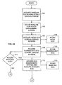
- FIGS. 2A , 2B , 2C , and 2D illustrate the method of the invention.
- a baseline ECG is acquired (at 100) from the patient 14 during a stable cardiac period.
- the baseline ECG may be acquired (at 100) several days or even several years prior to the patient 14 being treated for severe chest pain during a cardiac episode.
- the baseline ECG is generally acquired (at 100) during a standard, twelve-lead ECG test, such as an ECG stress test, when the patient 14 is in a clinic or hospital.
- the baseline ECG is stored (at 102) in the ECG management system 22.
- the electrodes 12 and leadwires 13 are attached to the patient 14 and an initial ECG, i.e., the index ECG, is acquired (at 104).
- the index ECG is the first ECG acquired (at 104) from the patient 14 during the cardiac episode.
- the index ECG is stored (at 106) in the memory 32 of the ECG acquisition device 16 and designated as such, so that the index ECG can be differentiated from subsequent ECGs acquired from the patient 14.
- Subsequent ECGs are then acquired (at 108) from the patient 14 for any period of time desired by the clinician, such as for the extent of a cardiac episode.
- the subsequent ECGs are stored (at 110) in the memory 32 of the ECG acquisition device 16.
- serial comparisons may be performed between the index ECG and the subsequent ECGs.
- the time period between each serial comparison may be from twenty seconds to several hours.
- a baseline ECG for the patient 14 is acquired from the ECG management system 22.
- the ECG acquisition device 16 is used to determine (at 111) whether a baseline ECG should be acquired.
- the management module 26 outputs to the display 30 a request for the clinician to decide whether the baseline ECG should be acquired.
- the clinician may press a button on a housing of the ECG acquisition device 16 or the clinician may press a touchscreen button on the display 30 of the ECG acquisition device 16 in order to indicate whether the baseline ECG for the patient 14 should be acquired.
- the acquisition module 18 automatically attempts to communicate with the ECG management system 22 in order to acquire the baseline ECG for the patient 14. For example, the acquisition module 18 first automatically attempts to access the ECG management system 22 via a physical connection. If the ECG acquisition device 16 is not physically connected to the ECG management system 22, the acquisition module 18 automatically attempts to access the ECG management system 22 via the transmitter/receiver 20.
- the acquisition module 18 establishes communication with the ECG management system 22 and acquires (at 112) the baseline ECG
- the baseline ECG is stored (at 114) in the memory 32 of the ECG acquisition device 16, so that the baseline ECG can be used for later serial comparisons.
- the management module 26 can perform serial comparisons between the baseline ECG, the index ECG, and the subsequent ECGs.
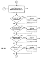
- the acquired ECGs from the acquisition module 18 are input into (at 116) the management module 26 of the signal processor 24.
- the management module 26 coordinates multiple serial comparisons between several sets of ECGs at once.
- the management module 26 first determines which leads of the pair of ECGs correspond to one another. For example, for a serial comparison between the baseline ECG and the index ECG for the patient 14, the management module 26 determines which leads of the baseline ECG correspond to the leads of the index ECG, e.g., lead V1 of the baseline ECG corresponds to lead V1 of the index ECG and lead V2 of the baseline ECG corresponds to lead V2 of the index ECG.
- the management module 26 will determine that twelve leads of the baseline ECG correspond to twelve leads of the index ECG.
- the ECG acquisition device 16 may not acquire all twelve leads for a standard, twelve-lead ECG, while the baseline ECG is preferably a standard, twelve-lead ECG.
- the management module 26 will determine which leads are acquired by the ECG acquisition device 16 and determine which leads of the baseline ECG correspond to the leads acquired by the acquisition device 16.
- the management module 26 analyzes the sets of corresponding leads according to several functional blocks.
- the functional blocks represent various portions of a typical ECG waveform, as illustrated in FIG. 3 .
- the functional blocks represent the P wave, the Q wave, the QRS complex, the ST elevation, the ST depression, the T wave, and the QT interval.
- the management module 26 determines which sets of corresponding leads exhibit changes in a portion of the ECG waveform represented by one of the functional blocks. If a set of corresponding leads exhibits changes in a portion of the ECG waveform represented by one of the functional blocks, the set of corresponding leads is divided into a group associated with that functional block.
- each of the groups associated with the functional blocks are assigned a severity value.
- a preferred embodiment of the analysis performed by the management module 26 is described below with respect to FIGS. 2B and 2C .
- the management module 26 first analyzes each of the corresponding leads to determine (at 118) if there is a change in the QRS complex in order to detect a new bundle branch block, i.e., either left bundle branch block or right bundle branch block. The management module 26 determines if there are changes in the duration and the amplitude of the QRS complex. The sets of corresponding leads indicating a change in the QRS complex are divided into the QRS functional block. Once all the sets of corresponding leads are analyzed and divided, the management module 26 assigns (at 120) a severity value to the QRS functional block.
- a "high" severity value (e.g., a severity value of 2) is assigned to the QRS functional block if all of the following are true: (1) if the QRS complex duration for the second ECG of any of the corresponding leads is greater than 120 milliseconds, while the QRS complex duration for the first ECG of any of the corresponding leads is less than 120 milliseconds; (2) if the QRS complex in any of the corresponding V1 and V2 leads is negative and either one of the Q wave or the S wave has a duration greater than 80 milliseconds; or (3) in any two of the corresponding I, V5, and V6 leads, if the sum of the durations of the R wave (i.e., the first positive deflection in the QRS complex) and the R' wave (i.e., a second positive deflection that sometimes occurs in the QRS complex) is greater than 100 milliseconds.
- a severity value e.g., a severity value of 2
- the management module 26 analyzes each of the sets of corresponding leads to determine (at 122) if there is a change in ST depression in order to detect acute ischemia and acute myocardial infarction.
- the management module 26 determines if there are amplitude changes that indicate ST depression between the sets of corresponding leads at the following points in the ECG waveform: STJ (the beginning of the ST segment), STM (the beginning of the ST segment plus the average interval between R wave peaks divided by 16), STE (the beginning of the ST segment plus the average interval between R wave peaks divided by 8), STJ+40 (the beginning of the ST segment plus 40 milliseconds), STJ+80 (the beginning of the ST segment plus 80 milliseconds), and ST slope (whether up, down, or flat).
- the corresponding leads indicating ST depression are divided into the ST depression functional block.
- the management module 26 assigns (at 124) a severity value to the ST depression functional block.
- a severity value is assigned to the ST depression functional block.
- a "high” severity value is assigned to the ST depression functional block.
- a "moderate” severity value e.g., a severity value of 1
- the management module 26 then analyzes each of the corresponding leads to determine (at 126) if there is a change in ST elevation in order to detect acute myocardial infarction.
- the management module 26 determines if there are amplitude changes that indicate ST elevation between the sets of corresponding leads at the following points in the ECG waveform: STJ (the beginning of the ST segment), STM (the beginning of the ST segment plus the average interval between R wave peaks divided by 16), STE (the beginning of the ST segment plus the average interval between R wave peaks divided by 8), STJ+40 (the beginning of the ST segment plus 40 milliseconds), STJ+80 (the beginning of the ST segment plus 80 milliseconds), and ST slope (whether up, down, or flat).
- the sets of corresponding leads indicating ST elevation are divided into the ST elevation functional block. Once all the sets of corresponding leads are analyzed and divided, the management module 26 assigns (at 128) a severity value to the change of ST elevation functional block.
- a severity value is assigned to the ST elevation block.
- the ST elevation between any of the corresponding precordial leads i.e., leads V1, V2, V3, V4, V5, and V6
- leads I, II, III, AVR, AVL, and AVF is greater than 100 microvolts
- a "high" severity value is assigned to the ST elevation block.
- the management module 26 then analyzes each of the sets of corresponding leads to determine (at 130) if there is a change in the T wave in order to detect acute ischemia and acute myocardial infarction.
- the management module 26 determines if there are changes in the amplitude and morphology of the T wave.
- the sets of corresponding leads indicating a change in the T wave are divided into the T wave functional block.
- the management module 26 assigns (at 132) a severity value to the T wave functional block.
- a "high" severity value is assigned to the T wave functional block.
- the management module 26 then analyzes each of the sets of corresponding leads to determine (at 134) if there is a change in the Q wave in order to detect acute and non-acute myocardial infarction.
- the management module 26 determines if there are changes in the amplitude and duration of the Q wave.
- the sets of corresponding leads indicating a change in the Q wave are divided into the Q wave functional block. Once all the sets of corresponding leads are analyzed and divided, the management module 26 assigns (at 136) a severity value to the Q wave functional block.
- the duration of the Q wave of the first lead of the sets of corresponding leads is less than 40 milliseconds and the duration of the Q wave of the second lead of the sets of corresponding leads is greater than 40 milliseconds, a "moderate” severity level is assigned to the Q wave functional block.
- a "high" severity level is assigned to the Q wave functional block.
- the management module 26 analyzes each of the corresponding leads to determine (at 138) if there is a change in the QT interval in order to detect acute ischemia.
- the sets of corresponding leads indicating a change in the QT interval are divided into the QT interval functional block. Once all the sets of corresponding leads are analyzed and divided, the management module 26 assigns (at 140) a severity value to the QT interval functional block.
- the corrected QT interval (QTC) is analyzed by the management module 26.
- a "moderate” severity value is assigned to the QT interval functional block. If the QTC of the first lead of the sets of corresponding leads is less than 450 milliseconds and the QTC of the second lead of the sets of corresponding leads is greater than 480 milliseconds, a "high" severity value is assigned to the QT interval functional block.
- the analysis performed by the management module 26 is described above and shown in the drawings in a particular order, the analysis may be performed in any order and still be within the scope of the invention. Moreover, each and every step in the analysis described above and shown in the drawings does not have to be performed to be within the scope of the invention.
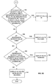
- the decision logic module 28 is used to implement a fuzzy logic algorithm. Fuzzy logic algorithms differ from conventional, fixed-value logic algorithms in that fuzzy logic algorithms use smoothed, membership functions to define the boundaries between groups. A set of fuzzy logic rules defines how the membership functions are combined. The fuzzy logic algorithm then makes decisions based on the output of the entire set of fuzzy logic rules.
- the decision logic module 28 first determines (at 144) whether the severity value assigned to the QRS functional block is "high,” indicating a new bundle branch block, i.e., either left bundle branch block or right bundle branch block. If the QRS functional block does indicate a new bundle branch block, the display 30 indicates (at 146) acute myocardial infarction to a clinician.
- the decision logic module 28 determines (at 148) whether the ST elevation functional block has been assigned a "high” severity value. If the ST elevation functional block has been assigned a "high” severity value, the display 30 indicates (at 150) acute myocardial infarction to a clinician.
- the decision logic module 28 next determines (at 152) whether the ST elevation functional block has been assigned a "moderate” severity value. The decision logic module 28 also determines whether the ST depression functional block has been assigned a "moderate” severity value. The decision logic module 28 also determines whether the T wave functional block has been assigned a "moderate” severity value. If the decision logic module 28 determines (at 152) that the ST elevation functional block has been assigned a "moderate” severity value and, at the same time, the ST depression functional block has been assigned a severity value equal to or greater than "moderate,” the display 30 indicates (at 154) an acute myocardial infarction to a clinician. In addition, if the decision logic module 28 determines (at 152) that the T wave functional block has been assigned a severity value equal to or greater than "moderate,” the display 30 indicates (at 154) an acute myocardial infarction to a clinician.
- the decision logic module 28 next determines (at 156) whether the ST depression functional block has been assigned a "high” severity value. If the decision logic module 28 determines (at 156) that the ST depression functional block has been assigned a "high” severity value, the display 30 indicates (at 158) acute ischemia to a clinician.
- the decision logic module 28 next determines (at 160) whether the ST depression functional block has been assigned a "moderate” severity value. The decision logic module 28 also determines (at 160) whether the T wave functional block has been assigned a "moderate” severity value. If the decision logic module 28 determines (at 160) that the ST depression functional block has been assigned a "moderate” severity value and the T wave functional block has been assigned a severity value equal to or greater than "moderate,” the display 30 indicates (at 162) acute ischemia to a clinician.
- the display 30 indicates (at 164) to a clinician that the patient 14 is not suffering from either acute myocardial infarction or acute ischemia. If the patient 14 is not suffering from either acute myocardial infarction or acute ischemia; the clinician may determine and treat the cause of the patient's chest pain or the clinician may discharge the patient 14 from the emergency room.
Landscapes
- Health & Medical Sciences (AREA)
- Life Sciences & Earth Sciences (AREA)
- Cardiology (AREA)
- Heart & Thoracic Surgery (AREA)
- Molecular Biology (AREA)
- Pathology (AREA)
- Engineering & Computer Science (AREA)
- Biomedical Technology (AREA)
- Physics & Mathematics (AREA)
- Medical Informatics (AREA)
- Biophysics (AREA)
- Surgery (AREA)
- Animal Behavior & Ethology (AREA)
- General Health & Medical Sciences (AREA)
- Public Health (AREA)
- Veterinary Medicine (AREA)
- Measurement And Recording Of Electrical Phenomena And Electrical Characteristics Of The Living Body (AREA)
Claims (4)
- Erfassungsvorrichtung (16) zum Erfassen von Elektrokardiogrammen, aufweisend:ein Erfassungsmodul (18) zum Erfassen mehrerer Elektrokardiogramme;einen mit dem Erfassungsmodul (18) gekoppelten Signalprozessor (24) zum Durchführen eines seriellen Vergleichs zwischen wenigstens zwei von den mehreren Elektrokardiogrammen;wobei die mehreren Elektrokardiogramme ein Ausgangs-Elektrokardiogramm und ein Index-Elektrokardiogramm und wenigstens ein nachfolgendes Elektrokardiogramm enthalten; undwobei der Signalprozessor (24) einen seriellen Vergleich zwischen dem Ausgangs-Elektrokardiogramm und dem Index-Elektrokardiogramm und dem wenigstens einen anschließenden Elektrokardiogramm durchführt;wobei der Signalprozessor (24) ferner enthält:eine Einrichtung (26) zum Durchführen der seriellen Vergleiche in Echtzeit und zum Identifizieren von Änderungen (118), (122), (126), (130) zwischen dem Index-Elektrokardiogramm und dem wenigstens einem anschließenden Elektrokardiogramm im QRS-Komplex, in der ST-Erhebung, ST-Absenkung, und T-Welle; undeine Einrichtung (120), (124), (128), (132) zum Zuweisen von Schweregraden zu den identifizierten Änderungen;gekennzeichnet durch:eine Einrichtung (28) zum Implementieren eines Fuzzy-Logik-Algorithmus zum Analysieren der Schweregrade;eine Einrichtung (144), (148), (152) zum Erzeugen einer ersten Anzeige, wenn der Schweregrad der Änderungen in dem QRS-Komplex hoch ist, wenn der Schweregrad der Änderungen in der ST-Erhebung hoch ist, wenn der Schweregrad der Änderungen in der ST-Erhebung und der ST-Absenkung gleich oder größer als moderat ist, oder wenn der Schweregrad der Änderungen in der T-Welle gleich oder größer als moderat ist; undeine Einrichtung (156), (160), (152) zum Erzeugen einer zweiten Anzeige, wenn der Schweregrad der Änderungen in der ST-Absenkung hoch ist oder wenn der Schweregrad der Änderungen in der ST-Absenkung und des T-Intervalls gleich oder größer als moderat ist.
- Vorrichtung nach Anspruch 1, wobei das Ausgangs-Elektrokardiogramm ein während einer stabilen Herzperiode erfasstes Elektrokardiogramm ist.
- Verfahren zum Durchführen eines Vergleichs zwischen erfassten Elektrokardiogrammen, mit den Schritten:Erfassen mehrerer Elektrokardiogramme mit einer Erfassungsvorrichtung (16);Nutzen der Erfassungsvorrichtung (16) zum Durchführen eines seriellen Vergleichs zwischen wenigstens zwei von den mehreren Elektrokardiogrammen, was der Erfassung eines Ausgangs-Elektrokardiogramms und eines Index-Elektrokardiogramms und wenigstens eines nachfolgenden Elektrokardiogramms einschließt; undDurchführen eines seriellen Vergleichs in Echtzeit zwischen dem Ausgangs-Elektrokardiogramm und dem Index-Elektrokardiogramm und dem wenigstens einen anschließenden Elektrokardiogramm;Durchführen der seriellen Vergleiche zwischen Elektrokardiogrammen in Echtzeit;Identifizieren von Änderungen (118), (122), (126), (130) zwischen dem Index-Elektrokardiogramm und dem wenigstens einen anschließenden Elektrokardiogramm im QRS-Komplex, in der ST-Erhebung, ST-Absenkung, und T-Welle;Zuweisen von Schweregraden (120), (124), (128), (132) zu den identifizierten Änderungen;gekennzeichnet durch:Analysieren der Schweregrade gemäß einem Fuzzy-Logik-Algorithmus;Erzeugen einer ersten Anzeige, wenn der Schweregrad der Änderungen in dem QRS-Komplex hoch ist, wenn der Schweregrad der Änderungen in der ST-Erhebung hoch ist, wenn der Schweregrad der Änderungen in der ST-Erhebung und der ST-Absenkung gleich oder größer als moderat ist, oder wenn der Schweregrad der Änderungen in der T-Welle gleich oder größer als moderat ist; undErzeugen einer zweiten Anzeige, wenn der Schweregrad der Änderungen in der ST-Absenkung hoch ist oder wenn der Schweregrad der Änderungen in der ST-Absenkung und des T-Intervalls gleich oder größer als moderat sind.
- Softwareprogramm zur Implementation des Verfahrens nach Anspruch 3, aufweisend:ein Verwaltungsmodul (26) zum Analysieren entsprechender Ableitungen der wenigstens zwei Elektrokardiogramme, um zu ermitteln, welche entsprechenden Ableitungen Unterschiede zwischen den wenigstens zwei Elektrokardiogrammen anzeigen, um die entsprechenden Ableitungen in mehrere Gruppen gemäß den angezeigten Unterschieden zu sortieren, und um jeder Einzelnen von den mehreren Gruppen einen Schweregrad zuzuordnen; undein Entscheidungslogikmodul (28) zum Implementieren des Fuzzy-Logik-Algorithmus, um in Echtzeit den jeder Einzelnen von den mehreren Gruppen zugeordneten Schweregrad zu analysieren und eine Anzeige auf der Basis der Analyse auszugeben.
Applications Claiming Priority (2)
| Application Number | Priority Date | Filing Date | Title |
|---|---|---|---|
| US682733 | 2001-10-11 | ||
| US09/682,733 US6564090B2 (en) | 2001-10-11 | 2001-10-11 | Method and apparatus for the serial comparison of electrocardiograms |
Publications (3)
| Publication Number | Publication Date |
|---|---|
| EP1304072A2 EP1304072A2 (de) | 2003-04-23 |
| EP1304072A3 EP1304072A3 (de) | 2004-01-02 |
| EP1304072B1 true EP1304072B1 (de) | 2011-12-14 |
Family
ID=24740906
Family Applications (1)
| Application Number | Title | Priority Date | Filing Date |
|---|---|---|---|
| EP02256909A Expired - Lifetime EP1304072B1 (de) | 2001-10-11 | 2002-10-04 | Verfahren und Vorrichtung zum seriellen Vergleich von Elektrokardiogrammen |
Country Status (3)
| Country | Link |
|---|---|
| US (1) | US6564090B2 (de) |
| EP (1) | EP1304072B1 (de) |
| JP (1) | JP4386235B2 (de) |
Families Citing this family (33)
| Publication number | Priority date | Publication date | Assignee | Title |
|---|---|---|---|---|
| US7076287B2 (en) * | 2000-12-29 | 2006-07-11 | Ge Medical Systems Information Technologies, Inc. | System and method for detecting new left bundle branch block for accelerating treatment of acute myocardial infarction |
| US7274959B1 (en) * | 2003-06-24 | 2007-09-25 | Pacesetter, Inc. | System and method for detecting cardiac ischemia using an implantable medical device |
| US7225015B1 (en) * | 2003-06-24 | 2007-05-29 | Pacesetter, Inc. | System and method for detecting cardiac ischemia based on T-waves using an implantable medical device |
| US7218960B1 (en) | 2003-06-24 | 2007-05-15 | Pacesetter, Inc. | System and method for detecting cardiac ischemia based on T-waves using an implantable medical device |
| US20050085736A1 (en) * | 2003-10-17 | 2005-04-21 | Ambrose John A. | Portable ECG detector |
| WO2005077267A1 (en) * | 2004-02-11 | 2005-08-25 | Henry Ford Health System | Detecting prolonged myocardial repolarization indicative of cardiac condition |
| US7058444B2 (en) * | 2004-04-05 | 2006-06-06 | Hewlett-Packard Development Company, L.P. | Computer method and system for reading and analyzing ECG signals |
| US7996073B2 (en) * | 2004-10-13 | 2011-08-09 | International Business Machines Corporation | System and method for interpreting electrocardiograms |
| JP2008539988A (ja) * | 2005-05-13 | 2008-11-20 | カーディオコア ラブ、インコーポレイテッド | 心電図波形の解析を高速に行う方法と装置 |
| US7702382B2 (en) * | 2006-04-17 | 2010-04-20 | General Electric Company | Multi-tier system for cardiology and patient monitoring data analysis |
| US8116859B2 (en) * | 2007-10-24 | 2012-02-14 | Ela Medical S.A.S. | Electrocardiologic device for the assisted diagnosis of brugada syndrome or early repolarization syndrome |
| CN101836211A (zh) * | 2007-10-24 | 2010-09-15 | 皇家飞利浦电子股份有限公司 | 用于将连续ecg分析和ecg医嘱相组合的系统和方法 |
| US9462955B2 (en) * | 2007-12-18 | 2016-10-11 | Koninklijke Philips N.V. | Automated identification of culprit coronary artery using anatomically oriented ECG data display |
| US20090228298A1 (en) * | 2008-03-04 | 2009-09-10 | The General Electric Company | System and method of morphology feature analysis of physiological data |
| US9622669B2 (en) | 2008-06-13 | 2017-04-18 | Parkinson's Institute | Diagnosis of neurodegenerative disorders |
| US8082027B2 (en) | 2010-05-07 | 2011-12-20 | General Electric Company | Portable USB electrocardiograph system and method |
| JP5559425B2 (ja) | 2010-05-12 | 2014-07-23 | イリズム・テクノロジーズ・インコーポレイテッド | 長期粘着用の装置機構及び構成要素 |
| WO2012061518A1 (en) | 2010-11-02 | 2012-05-10 | Cardionet, Inc. | Medical data collection apparatus |
| US20140296714A1 (en) * | 2011-06-01 | 2014-10-02 | Shigehiro Kuroki | Ultrasonic probe, bioinformation measurement device, and bioinformation measurement method |
| US8620418B1 (en) | 2013-01-04 | 2013-12-31 | Infobionic, Inc. | Systems and methods for processing and displaying patient electrocardiograph data |
| EP3753483A1 (de) | 2013-01-24 | 2020-12-23 | Irhythm Technologies, Inc. | Physiologische überwachungsvorrichtung |
| JP5933138B2 (ja) * | 2013-12-20 | 2016-06-08 | コーニンクレッカ フィリップス エヌ ヴェKoninklijke Philips N.V. | Ecgデータにおけるqrs群の発生を決定する装置及び方法 |
| WO2016035000A1 (en) * | 2014-09-02 | 2016-03-10 | Koninklijke Philips N.V. | User feedback to controls ischemia monitoring ecg algorithm |
| JP2018504148A (ja) | 2014-10-31 | 2018-02-15 | アイリズム・テクノロジーズ・インコーポレイテッドiRhythm Technologies,Inc. | 無線式生体モニタリングデバイス及びシステム |
| US10818393B2 (en) | 2015-10-06 | 2020-10-27 | General Electric Company | System and method for clinical decision support |
| KR101777583B1 (ko) | 2015-12-02 | 2017-09-13 | 한양대학교 에리카산학협력단 | 심전도 신호 처리 방법 및 그 장치 |
| US10930392B2 (en) * | 2018-02-19 | 2021-02-23 | General Electric Company | System and method for processing ECG recordings from multiple patients for clinician overreading |
| AU2021218704B2 (en) | 2020-02-12 | 2023-11-02 | Irhythm Technologies, Inc. | Non-invasive cardiac monitor and methods of using recorded cardiac data to infer a physiological characteristic of a patient |
| US12318208B2 (en) * | 2020-06-26 | 2025-06-03 | Alivecor, Inc. | Two-lead QT interval prediction |
| AU2021320404B2 (en) | 2020-08-06 | 2025-03-20 | Irhythm Technologies, Inc. | Electrical components for physiological monitoring device |
| KR20230047455A (ko) | 2020-08-06 | 2023-04-07 | 아이리듬 테크놀로지스, 아이엔씨 | 점착성 생리학적 모니터링 장치 |
| USD1063079S1 (en) | 2021-08-06 | 2025-02-18 | Irhythm Technologies, Inc. | Physiological monitoring device |
| US20260011434A1 (en) * | 2022-07-14 | 2026-01-08 | Medical Ai Co., Ltd. | Method, program, and device for providing transport services for emergency patient and monitoring of hospitalized patient on basis of electrocardiograms |
Family Cites Families (9)
| Publication number | Priority date | Publication date | Assignee | Title |
|---|---|---|---|---|
| US5253650A (en) | 1989-05-16 | 1993-10-19 | Sharp Kabushiki Kaisha | Apparatus for recording an electrocardiogram |
| JP3154425B2 (ja) | 1992-01-07 | 2001-04-09 | フクダ電子株式会社 | 心電図情報記録方法及び装置 |
| US5724983A (en) * | 1994-08-01 | 1998-03-10 | New England Center Hospitals, Inc. | Continuous monitoring using a predictive instrument |
| US6038469A (en) * | 1994-10-07 | 2000-03-14 | Ortivus Ab | Myocardial ischemia and infarction analysis and monitoring method and apparatus |
| IT1278679B1 (it) * | 1995-05-22 | 1997-11-27 | Paolo Alcidi | Metodo ed apparecchiatura per l'acquisizione ed il trattamento di segnali elettrocardiografici |
| SE9503019D0 (sv) * | 1995-09-01 | 1995-09-01 | Siemens Elema Ab | Förfarande och anordning för att korrigera för icke fysiologiska variationer i EKG-signaler |
| US5906583A (en) * | 1997-08-20 | 1999-05-25 | R.Z. Comparative Diagnostics Ltd. | Automatic cardiometer |
| US6171256B1 (en) | 1998-04-30 | 2001-01-09 | Physio-Control Manufacturing Corporation | Method and apparatus for detecting a condition associated with acute cardiac ischemia |
| WO2000015098A2 (en) * | 1998-09-17 | 2000-03-23 | Pangea Medical, Inc. | Pictorial-display electrocardiographic interpretation system and method |
-
2001
- 2001-10-11 US US09/682,733 patent/US6564090B2/en not_active Expired - Lifetime
-
2002
- 2002-10-04 EP EP02256909A patent/EP1304072B1/de not_active Expired - Lifetime
- 2002-10-11 JP JP2002298127A patent/JP4386235B2/ja not_active Expired - Lifetime
Also Published As
| Publication number | Publication date |
|---|---|
| US20030073914A1 (en) | 2003-04-17 |
| US6564090B2 (en) | 2003-05-13 |
| EP1304072A3 (de) | 2004-01-02 |
| JP2003159226A (ja) | 2003-06-03 |
| EP1304072A2 (de) | 2003-04-23 |
| JP4386235B2 (ja) | 2009-12-16 |
Similar Documents
| Publication | Publication Date | Title |
|---|---|---|
| EP1304072B1 (de) | Verfahren und Vorrichtung zum seriellen Vergleich von Elektrokardiogrammen | |
| US7412395B2 (en) | Automated scheduling of emergency procedure based on identification of high-risk patient | |
| US8897863B2 (en) | Arrhythmia detection using hidden regularity to improve specificity | |
| US7330750B2 (en) | Estimation of cardiac death risk | |
| US6665559B2 (en) | Method and apparatus for perioperative assessment of cardiovascular risk | |
| EP1179319B1 (de) | Vorrichtung zur Erfassung akuter kardiologischer Syndrome in spezifischen Gruppen von Patienten mittels EKG | |
| US8417326B2 (en) | RR interval monitoring method and blood pressure cuff utilizing same | |
| US6607480B1 (en) | Evaluation system for obtaining diagnostic information from the signals and data of medical sensor systems | |
| US7076287B2 (en) | System and method for detecting new left bundle branch block for accelerating treatment of acute myocardial infarction | |
| EP1284645A2 (de) | System und gerät zur multiskalaren analyse und darstellung elektrokardiographischer daten | |
| US11013448B2 (en) | Monitoring of biosignals, in particular electrocardiograms | |
| US20020138012A1 (en) | Multiple parameter electrocardiograph system | |
| US6491629B1 (en) | Method for determining at least one diagnostic piece of information from signal patterns of medical sensor systems | |
| RU2598049C2 (ru) | Автоматизированная идентификация местоположения окклюзии в инфаркт-зависимой коронарной артерии | |
| KR20240096098A (ko) | 휴대용 심전도계를 이용한 심전도 측정 서비스 제공방법 및 시스템 | |
| Yanowitz et al. | Accuracy of a continuous real-time ECG dysrhythmia monitoring system | |
| WO2020075973A1 (ko) | 동시 동작 분석이 가능한 심전도 모니터링 시스템 | |
| Jan et al. | Long-term follow-up case study of atrial fibrillation after treatment | |
| Adams et al. | A new method for electrocardiographic monitoring | |
| Zong et al. | Reduction of false blood pressure alarms by use of electrocardiogram blood pressure relationships | |
| Nasser et al. | Lessons learned during the pandemic: Correlation of QT intervals between telemetry and 12-lead electrocardiogram | |
| DK202201081A1 (en) | A method for identifying morphological abnormalities in heart rhythm data | |
| Quinn et al. | Evaluation of computer diagnosis of ectopic beats encountered in routine patient monitoring | |
| JPS59120135A (ja) | 心機能診断装置 | |
| De Koning et al. | Average heart rate, atrial fibrillation and R-on-T ventricular ectopy in 24h Holter recordings predict all-cause mortality in healthy middle-aged men |
Legal Events
| Date | Code | Title | Description |
|---|---|---|---|
| PUAI | Public reference made under article 153(3) epc to a published international application that has entered the european phase |
Free format text: ORIGINAL CODE: 0009012 |
|
| AK | Designated contracting states |
Designated state(s): AT BE BG CH CY CZ DE DK EE ES FI FR GB GR IE IT LI LU MC NL PT SE SK TR |
|
| AX | Request for extension of the european patent |
Extension state: AL LT LV MK RO SI |
|
| PUAL | Search report despatched |
Free format text: ORIGINAL CODE: 0009013 |
|
| AK | Designated contracting states |
Kind code of ref document: A3 Designated state(s): AT BE BG CH CY CZ DE DK EE ES FI FR GB GR IE IT LI LU MC NL PT SE SK TR |
|
| AX | Request for extension of the european patent |
Extension state: AL LT LV MK RO SI |
|
| RIC1 | Information provided on ipc code assigned before grant |
Ipc: 7G 06F 17/00 B Ipc: 7A 61B 5/0452 B Ipc: 7A 61B 5/0402 B Ipc: 7A 61B 5/04 A |
|
| 17P | Request for examination filed |
Effective date: 20040702 |
|
| AKX | Designation fees paid |
Designated state(s): DE GB SE |
|
| 17Q | First examination report despatched |
Effective date: 20040910 |
|
| 17Q | First examination report despatched |
Effective date: 20040910 |
|
| GRAP | Despatch of communication of intention to grant a patent |
Free format text: ORIGINAL CODE: EPIDOSNIGR1 |
|
| GRAS | Grant fee paid |
Free format text: ORIGINAL CODE: EPIDOSNIGR3 |
|
| GRAA | (expected) grant |
Free format text: ORIGINAL CODE: 0009210 |
|
| AK | Designated contracting states |
Kind code of ref document: B1 Designated state(s): DE GB SE |
|
| REG | Reference to a national code |
Ref country code: GB Ref legal event code: FG4D |
|
| REG | Reference to a national code |
Ref country code: DE Ref legal event code: R096 Ref document number: 60241731 Country of ref document: DE Effective date: 20120209 |
|
| REG | Reference to a national code |
Ref country code: SE Ref legal event code: TRGR |
|
| PLBE | No opposition filed within time limit |
Free format text: ORIGINAL CODE: 0009261 |
|
| STAA | Information on the status of an ep patent application or granted ep patent |
Free format text: STATUS: NO OPPOSITION FILED WITHIN TIME LIMIT |
|
| 26N | No opposition filed |
Effective date: 20120917 |
|
| REG | Reference to a national code |
Ref country code: DE Ref legal event code: R097 Ref document number: 60241731 Country of ref document: DE Effective date: 20120917 |
|
| REG | Reference to a national code |
Ref country code: DE Ref legal event code: R079 Ref document number: 60241731 Country of ref document: DE Free format text: PREVIOUS MAIN CLASS: A61B0005040000 Ipc: A61B0005240000 |
|
| PGFP | Annual fee paid to national office [announced via postgrant information from national office to epo] |
Ref country code: SE Payment date: 20210921 Year of fee payment: 20 Ref country code: GB Payment date: 20210922 Year of fee payment: 20 |
|
| PGFP | Annual fee paid to national office [announced via postgrant information from national office to epo] |
Ref country code: DE Payment date: 20210921 Year of fee payment: 20 |
|
| REG | Reference to a national code |
Ref country code: DE Ref legal event code: R071 Ref document number: 60241731 Country of ref document: DE |
|
| REG | Reference to a national code |
Ref country code: GB Ref legal event code: PE20 Expiry date: 20221003 |
|
| PG25 | Lapsed in a contracting state [announced via postgrant information from national office to epo] |
Ref country code: GB Free format text: LAPSE BECAUSE OF EXPIRATION OF PROTECTION Effective date: 20221003 |
|
| REG | Reference to a national code |
Ref country code: SE Ref legal event code: EUG |