EP1296140A1 - Method of evaluating binding activity of ligand to ligand-binding protein - Google Patents
Method of evaluating binding activity of ligand to ligand-binding protein Download PDFInfo
- Publication number
- EP1296140A1 EP1296140A1 EP01943834A EP01943834A EP1296140A1 EP 1296140 A1 EP1296140 A1 EP 1296140A1 EP 01943834 A EP01943834 A EP 01943834A EP 01943834 A EP01943834 A EP 01943834A EP 1296140 A1 EP1296140 A1 EP 1296140A1
- Authority
- EP
- European Patent Office
- Prior art keywords
- ligand
- binding
- antibody
- protein
- substance
- Prior art date
- Legal status (The legal status is an assumption and is not a legal conclusion. Google has not performed a legal analysis and makes no representation as to the accuracy of the status listed.)
- Withdrawn
Links
Images



Classifications
-
- G—PHYSICS
- G01—MEASURING; TESTING
- G01N—INVESTIGATING OR ANALYSING MATERIALS BY DETERMINING THEIR CHEMICAL OR PHYSICAL PROPERTIES
- G01N33/00—Investigating or analysing materials by specific methods not covered by groups G01N1/00 - G01N31/00
- G01N33/48—Biological material, e.g. blood, urine; Haemocytometers
- G01N33/50—Chemical analysis of biological material, e.g. blood, urine; Testing involving biospecific ligand binding methods; Immunological testing
- G01N33/68—Chemical analysis of biological material, e.g. blood, urine; Testing involving biospecific ligand binding methods; Immunological testing involving proteins, peptides or amino acids
- G01N33/6854—Immunoglobulins
- G01N33/6857—Antibody fragments
-
- G—PHYSICS
- G01—MEASURING; TESTING
- G01N—INVESTIGATING OR ANALYSING MATERIALS BY DETERMINING THEIR CHEMICAL OR PHYSICAL PROPERTIES
- G01N33/00—Investigating or analysing materials by specific methods not covered by groups G01N1/00 - G01N31/00
- G01N33/48—Biological material, e.g. blood, urine; Haemocytometers
- G01N33/50—Chemical analysis of biological material, e.g. blood, urine; Testing involving biospecific ligand binding methods; Immunological testing
- G01N33/53—Immunoassay; Biospecific binding assay; Materials therefor
- G01N33/543—Immunoassay; Biospecific binding assay; Materials therefor with an insoluble carrier for immobilising immunochemicals
Definitions
- the present invention relates to a method of evaluating the binding activity of a ligand to a ligand-binding protein.
- RIA radioimmunoassay
- ELISA enzyme-linked immunosorbent assay
- SPR optical phenomenon
- BIACORETM a instrument for analyzing antibody-antigen interaction using a SPR sensor
- the basic system of BIACORETM consists of a light source, a prism, a detector and microfluidic system.
- ligands are immobilized on a cassette-type sensor chip, into which an analyte is injected. If the analyte has affinity for the ligands, the amount of analyte-bound to ligands is detected optically.
- the detection principle is a phenomenon referred to as SPR. Specifically, incident light is totally reflected into the interface between a glass and a thin metal film. The incident light at a certain angle excites surface plasmons, thus reducing the reflected light intensity at that angle. This angle is varied depending on changes in the concentration of the solvent that is in contact with the thin metal film (sensor). BIACORETM detects these changes.
- BIACORETM In the BIACORETM system, this change is referred to as a SPR signal, and a shift in resonance angle of 0.1 degree represents 1,000 RU (resonance units).
- a response of 1,000 RU corresponds to a change when approximately 1 ng of protein is bound onto a thin metal sensor with a surface area of 1 mm 2 .
- BIACORETM can sufficiently detect changes of about 50 RU (50 pg) for protein.
- a computer provided with BIACORETM converts the detected signals into a binding curve, referred to as a sensorgram, which is drawn on the computer display in real-time (Toru Natsume et al., 1995, Experimental medicine, 13, p563-569) (Karlsson, R. et al., (1991) J. Immunol. Methods 145, p229-240).
- the kinetics parameters of antibodies specifically, the dissociation constant (KD), dissociation rate constant (Kdiss) and association rate constant (Kass) can be measured.
- KD dissociation constant
- Kdiss dissociation rate constant
- Kass association rate constant
- An object of the present invention is to provide a convenient and highly accurate method of measuring the biological activity of a ligand (in particular, an antibody).
- the present inventors have studied a method of measuring the binding activity by quantitatively determining the amount of antibodies bound to antigens in order to conveniently evaluate in vitro the biological activity of an antibody without kinetics analysis. As a result, the present inventors have completed the present invention by succeeding in establishing a highly accurate and convenient test method of evaluating the binding activity of antibodies by correcting the antibody concentration by means of a system that quantitatively determines the amount of antibodies bound to Protein A.
- the present invention provides a method of evaluating the binding activity of a ligand to a ligand-binding protein, which comprises; measuring the binding activity value of a ligand to a ligand-binding protein, and measuring the binding activity value of the ligand to a second ligand-binding substance which binds to a ligand site other than the binding site where the ligand binds to the ligand-binding protein.
- Either the ligand or the ligand-binding protein may be immobilized.
- the ligand may be preferably an antigen or the antibody thereof, an enzyme or the substrate protein thereof or a ligand for various receptors.
- the ligand may be an antibody, and the ligand-binding protein may be an antigen peptide.
- the second ligand-binding substance may be selected from substances which can specifically bind to sites other than the antigen-binding site, such as the constant regions (an Fc portion, an L chain ⁇ region, an L chain ⁇ region, or the like) and the sugar chain of an antibody.
- the second ligand-binding substance may be a substance which specifically binds to the Fc portion of an antibody.
- a substance which specifically binds to the Fc portion of an antibody is preferably Protein A, protein G, protein L, Fc receptor or lectin.
- FLAG protein is fused to a ligand, and a substance which specifically binds to the FLAG protein may be used as a second ligand-binding protein.
- the ligand-binding protein and the second ligand-binding substance may be separately immobilized, and then allowed to react respectively with the same ligand-containing samples, so that each of the binding activities may be measured.
- the ligand is an antibody
- the ligand-binding protein is an antigen peptide
- the second ligand-binding substance is Protein A.
- the binding activity value of the antibody to the antigen peptide is preferably corrected with the binding activity value of the antibody to the second ligand-binding substance.
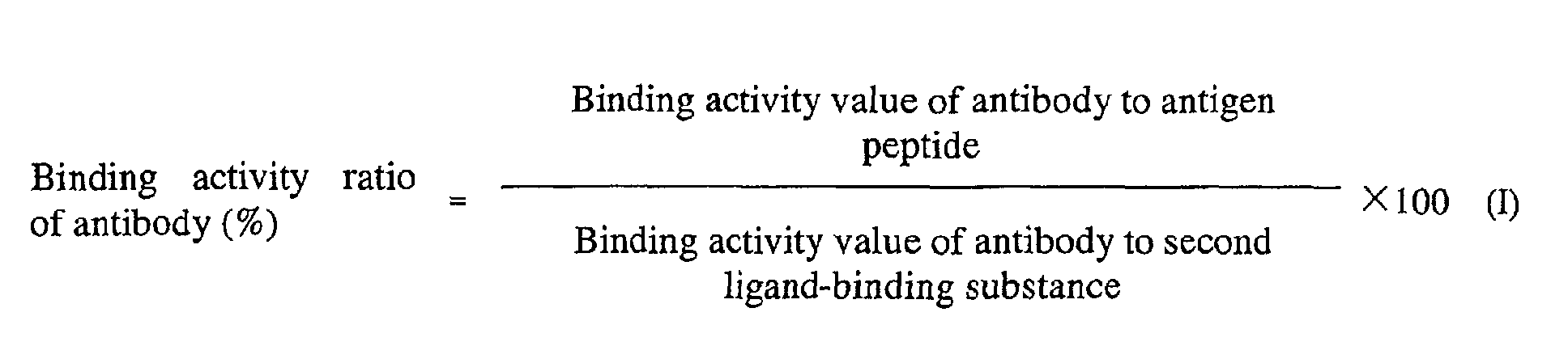
- the binding activity of an antibody to an antigen peptide can be evaluated using the binding activity ratio of the antibody calculated by the following formula (I):
- the binding activity value of an antibody to an antigen peptide, and the binding activity value of the antibody to the second ligand-binding substance can be calculated by the following formulae (II) and (III), respectively:
- the binding concentration of an antibody to an antigen peptide, and that of the antibody to a second ligand-binding substance can be measured by SPR, a fluorescence polarization measurement method, a radioimmunoassay (RIA), an enzyme-linked immunosorbent assay (ELISA), HPLC, an analytical ultracentrifugation method or a titration calorimetry.
- SPR a fluorescence polarization measurement method
- RIA radioimmunoassay
- ELISA enzyme-linked immunosorbent assay
- HPLC an analytical ultracentrifugation method or a titration calorimetry.
- the binding activity of an antibody of an unknown sample to an antigen can be evaluated by the binding activity ratio of the antibody calculated by the following formula (I'):
- binding activity value of an antibody to an antigen and that of an antibody to Protein A can be calculated by the following formulae (II) and (III'), respectively:
- the binding concentration of an antibody to an antigen peptide, and that to Protein A can be measured by SPR, a fluorescence polarization measurement method, a radioimmunoassay (RIA), an enzyme-linked immunosorbent assay (ELISA), HPLC, an analytical ultracentrifugation method, a titration calorimetry or the like.
- SPR a fluorescence polarization measurement method
- RIA radioimmunoassay
- ELISA enzyme-linked immunosorbent assay
- HPLC an analytical ultracentrifugation method
- titration calorimetry or the like.
- Target antibodies of the method of the present invention may be antibodies for any antigens.
- an antibody hereinafter, referred to as "anti-PTHrP antibody” for parathyroid hormone-related peptide (hereinafter, referred to as "PTHrP")
- PTHrP parathyroid hormone-related peptide
- IL-6 receptor an antibody for IL-6 receptor
- HM1.24 antigen an antibody for tissue factor (TF).
- TF tissue factor
- the antibody may be either a monoclonal antibody or a polyclonal antibody, and a monoclonal antibody is preferable.
- the antibody class may be any of IgM, IgD, IgG, IgA and IgE, and IgG is desirable.
- the subclass of H chain may be any of IgG1, IgG2, IgG3, and IgG4, and IgG1 and IgG4 are desirable, and the subclass of L chain may be either ⁇ or ⁇ .
- a monoclonal antibody may be a human antibody, humanized antibody or chimeric antibody.
- target substances that can be measured by the measurement method of the present invention also include antibody fragments, such as Fab, (Fab') 2 and re-constituted antibody fragments, such as a single-chain Fv.
- antibody fragments such as Fab, (Fab') 2
- re-constituted antibody fragments such as a single-chain Fv.
- a preferred antibody forms few (preferably, 5% or less) or no oligomer, such as dimmers.
- second ligand-binding substance indicates a substance which binds to a ligand at a site other than the binding site of the ligand with a ligand-binding protein (also referred to as "first ligand-binding substance”).
- a second ligand-binding substance specifically binds to a portion other than the antigen binding site, such as the constant region (Fc portion, L chain ⁇ region, an L chain ⁇ region, or the like) and the sugar chain of the antibody.
- a substance which binds to Fc portion include Protein A, protein G and Fc receptor.
- Examples of a substance which binds to L chain ⁇ region include protein L and anti- ⁇ antibody.
- An example of a substance which binds to L chain ⁇ region is anti- ⁇ antibody.
- a substance which binds to sugar chains include lectins, such as RCA, LCA and ConA.
- Preferred examples of a substance which binds to a portion other than the antigen binding site of an antibody include Protein A, protein G, Fc receptor, protein L and lectins, such as RCA, LCA and ConA. More preferred examples include Protein A, protein G and protein L, and most preferably, Protein A.
- the present invention provides a kit for evaluating the binding activity of a ligand to a ligand-binding protein.
- This kit contains both a ligand-binding protein and a second ligand-binding substance.
- This ligand-binding protein may be an antigen peptide.
- the kit of the present invention may contain other reagents.
- the kit of the present invention may further contain a reagent for immobilizing onto a sensor chip an antigen and a substance which binds to a portion other than the antigen-binding site of an antibody.
- the reagent may be any reagent as long as it activates the functional group of an antigen, for example, an amino group, thiol group, or aldehyde group to bind onto the chip.
- a reagent include N-ethyl-N'-(3-dimethylaminopropyl) carbodiimidehydrochloride, N-hydroxysuccinimide, ethanolamine hydrochloride, 2-(2-pyridinyldithio) ethaneamine hydrochloride, cysteine, biotinhydrazide.
- the method of evaluating ligand (for example, antibody) activity and the kit of the present invention do not require additional purification procedures to evaluate the binding activity ratio of the ligand (for example, antibody) molecule. Therefore, the evaluation method and the kit can be used for studying quality control, in-process control and formulation design of antibodies in the production process or purified antibodies as an drug substance.
- the binding activity ratio which is finally obtained by the method of evaluating ligand (for example, antibody) activity and the kit of the present invention does not reflect the concentration of an active ingredient in a sample solution, but the activity of the binding activity of a ligand (for example, antibody) to a second ligand-binding activity. Therefore, the activity ratio of 100% or more may suggest that some decomposition occurs at a second portion (for example, Fc portion) other than the antigen-binding site of an antibody; and the activity ratio of 100% or less may suggest some decomposition at the antigen-binding site of an antibody. Accordingly, the method and the kit of the present invention in combination with another method of evaluating physical properties can be used for stability testing and shelf life setting.
- the method of evaluating ligand (for example, antibody) activity and the kit of the present invention can also be used for selecting a highly active protein (in particular, a mutant protein, various monoclonal antibodies or the like).
- a highly active protein in particular, a mutant protein, various monoclonal antibodies or the like.
- the method and the kit of the present invention are effective in selecting a mutant protein with high biological activity or stability resulting from point mutation, or the like.
- the method and the kit of the present invention are particularly useful in screening antibodies, which are reconstituted from natural antibodies, such as chimeric antibodies and humanized antibodies.
- the method and the kit of the present invention are also effective of evaluating whether or not various electrically charged hetero components (the plural minor peaks, which are detected when the bulk protein, is applied to ion exchange chromatography) detected in the bulk proteins, are "Product-Related Substances," as is required for New Drug Application of a protein drug product, such as an antibody.
- the method of evaluating ligand (for example, antibody) binding activity, and the kit of the present invention can provide a convenient and highly accurate measurement of the binding activity of, and the quantitative determination of highly active proteins within body fluids, such as blood, serum or the like (in particular, a blood factor, a mutant protein, various monoclonal antibodies, antibody fragments, such as Fab and (Fab') 2 , a single-chain Fv and the like).
- body fluids such as blood, serum or the like
- a blood factor, a mutant protein in particular, various monoclonal antibodies, antibody fragments, such as Fab and (Fab') 2 , a single-chain Fv and the like.
- a method of evaluating the binding activity of an anti-PTHrP antibody to PTHrP is described as an example of the method of the present invention.
- This method comprises the steps of measuring, using BIACORETM, the amount of anti-PTHrP antibody bound to PTHrP and to Protein A respectively, and of correcting the measured amount of the anti-PTHrP antibody bound to PTHrP with the measured amount of the anti-PTHrP antibody bound to Protein A.
- anti-PTHrP antibodies are produced according to the method described in WO98/13388.
- Anti-PTHrP antibodies are diluted with water, and then a solution of the anti-PTHrP antibody is prepared at an appropriate concentration (for example, 100-500 ⁇ g/mL).
- the solution is quantitatively determined by the absorbance method in the general test of the Japanese Pharmacopoeia.
- the solution is diluted with HBS buffer solution (HBS-EP Buffer (BIACORE)) at an appropriate concentration (for example, 10-50 ⁇ g/mL). This preparation is repeated three times.
- HBS buffer solution is added to each solution, so that solutions each having a certain concentration (for example, 1-5 ⁇ g/mL) are prepared as unknown sample solutions 1, 2 and 3.
- anti-PTHrP antibodies are diluted with water, and then a solution of anti-PTHrP antibody at an appropriate concentration (for example, 100-500 ⁇ g/mL) is prepared.
- the solution is diluted with HBS buffer solution, and then solutions of anti-PTHrP antibody having various concentrations (for example, 0 to 5 ⁇ g/mL) are prepared as standard solutions A, B, C, D, E and F for a calibration curve.
- a sensor chip CM 5 (BIACORE, Code # BR-1000-14) is docked in BIACORETM, and then HBS buffer solution is run at a flow rate of 5-20 ⁇ L/min.
- PTHrP(1-34+Cys) immobilize on the chip: for example, 150 ⁇ L of a mixed solution containing equal amounts of EDC (N-ethyl-N'-(3-dimethyl aminopropyl)-carbodiimide hydrochloride) solution and NHS (N-hydroxysuccinimide) solution; for example, 150 ⁇ L of borate buffer solution of PDEA (2-(2-pyridinyldithio)ethaneaminehydrochloride); for example, 40 ⁇ L of acetate buffer solution of PTHrP(1-34+Cys) (a synthetic peptide having Cys added to the C terminus of 1-34 peptides of PTHrP); for example, 150 ⁇ L of formate buffer solution of cysteine-sodium chloride; for example, 10 ⁇ L of glycine hydrochloride buffer solution; and for example, 10 ⁇ L of 10 mM hydrochloric acid aqueous solution.
- EDC N-ethyl
- HBS buffer solution is run similarly at a flow rate of 5 to 20 ⁇ L/min.
- different solutions are run successively to immobilize Protein A onto a chip: for example, 100 ⁇ L of the mixed solution of equal amounts of EDC solution and NHS solution; for example, 40 ⁇ L of acetate buffer solution of Protein A; for example, 100 ⁇ L of ethanolamine hydrochloride buffer solution; for example, 10 ⁇ L of glycine hydrochloride buffer solution; and for example, 10 ⁇ L of 10 mM hydrochloric acid aqueous solution.
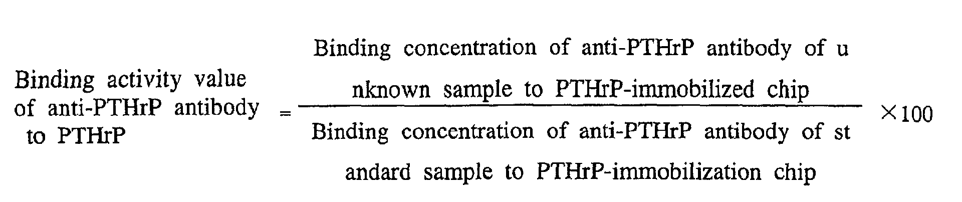
- the standard solutions for a calibration curve A, B, C, D, E and F, the standard sample solutions 1, 2 and 3, and the unknown sample solutions 1, 2 and 3 are applied to the PTHrP-immobilized chip and the Protein A-immobilized chip respectively. Two measurements are performed for each batch. Binding phase was defined two minutes period, 1 to 100 ⁇ L, preferably, 10 ⁇ L each of the standard sample solutions and the unknown sample solutions are injected. Here, the bound amount is a difference between the resonance unit (RU), provided 20 to 60 seconds after the solution is switched to the HBS buffer solution, and baseline. Subsequently, 15 mM hydrochloric acid solution is injected for washing and regenerating the sensor chips. With 1 cycle of analysis consisting of the binding, washing and regeneration, a sensorgram is obtained, a calibration curve is produced using the attached software, and then each binding concentration is calculated. From the binding concentration, the binding activity ratio (%) is calculated by the following formulae:
- the respective amounts of anti-PTHrP antibodies bound to PTHrP for 4 lots ( a , b , c and d ) of anti-PTHrP antibody were measured by the following procedures. Then, differences among the lots were studied.
- Original solutions of samples a , b, c and d were diluted with purified water to prepare about 500 ⁇ g/mL solutions, and then the concentrations were determined by the absorbance method.
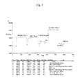
- Figure 1 shows the result of immobilization of PTHrP.
- the amount of antibodies bound to PTHrP was measured for 4 lots of anti-PTHrP antibody certified to have approximately equal quality.
- a representative example of the obtained sensorgram is shown in Fig 2.
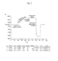
- a calibration curve was produced from the resulting bound amount using the calculation software attached to the BIACORETM system for quantitatively determining concentrations (Fig. 3).
- the binding phase on the sensorgram (Fig. 2) was almost a straight line, no dissociation was observed, and the calibration curve (Fig. 3) almost maintained its linearity within the concentration range. Taking these results together, it was concluded that the amount of immobilized ligands was sufficient for use in the quantification system. Therefore, the concentrations of samples to quantitatively determine the amount of antibodies bound to a PTHrP(1-34+Cys)-immobilized chip were set according to the conditions employed for this experiment. Specifically, standard samples for a calibration curve were set at six concentrations: 0, 1, 2, 3, 4 and 5 ⁇ g/mL, and unknown samples were set at a concentration of 2 ⁇ g/mL, which was about middle point of the calibration curve.
- Concentration of the sample anti-PTHrP antibody e was prepared based on the absorbance method, and then the binding concentrations of the sample e to the PTHrP(1-34-Cys)-immobilized chip were measured (I-(2)-i)). As a result, the binding activity value of sample e to PTHrP was 46%, compared to that of the standard sample a (Table 4). Possible reasons for these results include lowered biological activity, and contamination with foreign substances having absorbance at 280 nm. Another possible reason is that the protein content was lowered because the concentrations obtained by the absorbance method did not reflect the antibody concentration.
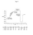
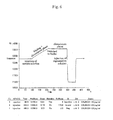
- Figure 5 shows the results of the immobilization of Protein A. Based on the linearity of the sensorgram and the calibration curve (Figs. 6 and 7) resulting from the prepared Protein A-immobilized chip, it was determined that the amount of immobilized ligands was sufficient for use in the quantification system. Further, it was also confirmed that the calibration curve showed sufficient linearity with the concentration range of 0 to 5 ⁇ g/mL employed for measurement using a PTHrP(1-34-Cys)-immobilized chip. Therefore, the sample concentrations of both samples for a calibration curve and unknown samples for quantitatively determining the bound amount to the Protein A-immobilized chip were set under the same conditions with a PTHrP(1-34-Cys)-immobilized chip.
- f was measured using standard product a.
- dispersions resulting from one experiment repeated 7 times were less than 1% (0.95%), suggesting that the experiment system was extremely precise (Tables 7 and 8).
- CAL binding activity was calculated using eq. 3 and summarized in Table 8. With the binding activity of a at 100%, the biological activity of f was at 97.0%, while the coefficient of variation of the repeatability (when repeated 7 times) was at 0.95%, these suggest that the difference between the two was significant and f had a slightly decreased activity with antigen.
- Binding concentration Binding activity ratio PTHrP Protein A % 1 a 2.04 2.04 100 f 2.13 2.21 96.4 2 a 2.05 2.06 100 f 2.18 2.24 97.8 3 a 2.18 2.00 100 f 2.35 2.21 97.6 4 a 2.02 2.04 100 f 2.27 2.40 95.5 5 a 2.04 2.02 100 f 2.41 2.43 98.2 6 a 2.20 2.01 100 f 2.90 2.74 96.7 7 a 2.03 1.98 100 f 2.40 2.42 96.7 Average f - - 97.0 0.95 (%cv)
- the bound amount observed at a certain time after injection of antibody followed by switching to a buffer is simply defined as the amount of PTHrP binding activity. Since the effect of dissociation is ignored in the system, the result obtained herein cannot be biological activity in the strictest sense. However, this method is good enough as a convenient method of evaluating activity without requiring kinetics analysis for samples observed from the form of the sensorgrams to have the dissociation rates sufficiently smaller than the binding rates.
- the present invention provides a convenient and highly accurate method of measuring the biological activity of a ligand.
Landscapes
- Health & Medical Sciences (AREA)
- Life Sciences & Earth Sciences (AREA)
- Immunology (AREA)
- Engineering & Computer Science (AREA)
- Molecular Biology (AREA)
- Biomedical Technology (AREA)
- Chemical & Material Sciences (AREA)
- Hematology (AREA)
- Urology & Nephrology (AREA)
- Biotechnology (AREA)
- Biochemistry (AREA)
- Cell Biology (AREA)
- Food Science & Technology (AREA)
- Medicinal Chemistry (AREA)
- Physics & Mathematics (AREA)
- Analytical Chemistry (AREA)
- Microbiology (AREA)
- General Health & Medical Sciences (AREA)
- General Physics & Mathematics (AREA)
- Pathology (AREA)
- Proteomics, Peptides & Aminoacids (AREA)
- Peptides Or Proteins (AREA)
- Investigating Or Analysing Biological Materials (AREA)
Abstract
Description
- The present invention relates to a method of evaluating the binding activity of a ligand to a ligand-binding protein.
- To measure antibody-antigen interaction, radioimmunoassay (RIA), enzyme-linked immunosorbent assay (ELISA) and the like have been used. Recently, the use of an optical phenomenon, surface plasmon resonance (hereinafter, referred to as "SPR") has enabled real time monitoring of antibody-antigen interaction without labeling. As an example, a instrument for analyzing antibody-antigen interaction using a SPR sensor, referred to as BIACORE™ is being marketed by BIACORE (formerly Pharmacia Biotech).
- The basic system of BIACORE™ consists of a light source, a prism, a detector and microfluidic system. In practice, ligands are immobilized on a cassette-type sensor chip, into which an analyte is injected. If the analyte has affinity for the ligands, the amount of analyte-bound to ligands is detected optically.
- The detection principle is a phenomenon referred to as SPR. Specifically, incident light is totally reflected into the interface between a glass and a thin metal film. The incident light at a certain angle excites surface plasmons, thus reducing the reflected light intensity at that angle. This angle is varied depending on changes in the concentration of the solvent that is in contact with the thin metal film (sensor). BIACORE™ detects these changes.
- In the BIACORE™ system, this change is referred to as a SPR signal, and a shift in resonance angle of 0.1 degree represents 1,000 RU (resonance units). A response of 1,000 RU corresponds to a change when approximately 1 ng of protein is bound onto a thin metal sensor with a surface area of 1 mm2. BIACORE™ can sufficiently detect changes of about 50 RU (50 pg) for protein.
- A computer provided with BIACORE™ converts the detected signals into a binding curve, referred to as a sensorgram, which is drawn on the computer display in real-time (Toru Natsume et al., 1995, Experimental medicine, 13, p563-569) (Karlsson, R. et al., (1991) J. Immunol. Methods 145, p229-240).
- Using BIACORE™, the kinetics parameters of antibodies, specifically, the dissociation constant (KD), dissociation rate constant (Kdiss) and association rate constant (Kass) can be measured.
- However, kinetics analysis of antigen-antibody reaction using the above-mentioned BIACORE™ is inappropriate for treatment with many analytes, because it is time consuming to study the conditions. Therefore, there is a need for a convenient and highly accurate method capable of measuring the biological activity of antibodies while maintaining the specificity of BIACORE™ and is appropriate for studying the quality design and prescription design of antibodies.
- An object of the present invention is to provide a convenient and highly accurate method of measuring the biological activity of a ligand (in particular, an antibody).
- The present inventors have studied a method of measuring the binding activity by quantitatively determining the amount of antibodies bound to antigens in order to conveniently evaluate in vitro the biological activity of an antibody without kinetics analysis. As a result, the present inventors have completed the present invention by succeeding in establishing a highly accurate and convenient test method of evaluating the binding activity of antibodies by correcting the antibody concentration by means of a system that quantitatively determines the amount of antibodies bound to Protein A.
- Specifically, the present invention provides a method of evaluating the binding activity of a ligand to a ligand-binding protein, which comprises; measuring the binding activity value of a ligand to a ligand-binding protein, and measuring the binding activity value of the ligand to a second ligand-binding substance which binds to a ligand site other than the binding site where the ligand binds to the ligand-binding protein.
- Either the ligand or the ligand-binding protein may be immobilized.
- The ligand may be preferably an antigen or the antibody thereof, an enzyme or the substrate protein thereof or a ligand for various receptors.
- In the above method, the ligand may be an antibody, and the ligand-binding protein may be an antigen peptide.
- The second ligand-binding substance may be selected from substances which can specifically bind to sites other than the antigen-binding site, such as the constant regions (an Fc portion, an L chain κ region, an L chain λ region, or the like) and the sugar chain of an antibody. In particular, the second ligand-binding substance may be a substance which specifically binds to the Fc portion of an antibody.
- A substance which specifically binds to the Fc portion of an antibody is preferably Protein A, protein G, protein L, Fc receptor or lectin.
- In the method of the present invention, FLAG protein is fused to a ligand, and a substance which specifically binds to the FLAG protein may be used as a second ligand-binding protein.
- The ligand-binding protein and the second ligand-binding substance may be separately immobilized, and then allowed to react respectively with the same ligand-containing samples, so that each of the binding activities may be measured.
- In a preferred aspect of the present invention, the ligand is an antibody, and the ligand-binding protein is an antigen peptide, and the second ligand-binding substance is Protein A.
- When the activity of an antibody to an antigen peptide is evaluated by the method of the present invention, the binding activity value of the antibody to the antigen peptide is preferably corrected with the binding activity value of the antibody to the second ligand-binding substance.
- For example, the binding activity of an antibody to an antigen peptide can be evaluated using the binding activity ratio of the antibody calculated by the following formula (I):
- The binding activity value of an antibody to an antigen peptide, and the binding activity value of the antibody to the second ligand-binding substance can be calculated by the following formulae (II) and (III), respectively:

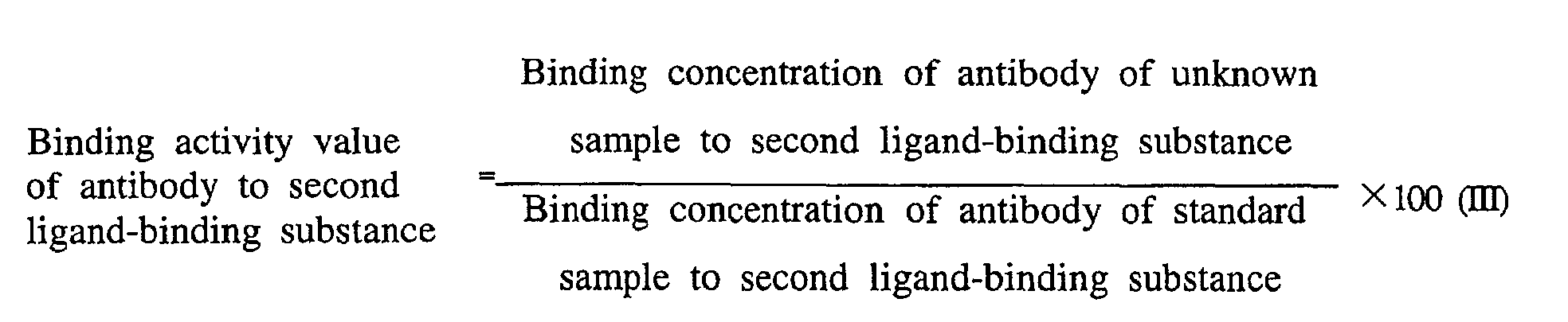
- The binding concentration of an antibody to an antigen peptide, and that of the antibody to a second ligand-binding substance can be measured by SPR, a fluorescence polarization measurement method, a radioimmunoassay (RIA), an enzyme-linked immunosorbent assay (ELISA), HPLC, an analytical ultracentrifugation method or a titration calorimetry. In particular, it is preferable to measure by SPR.
- As an example of the above methods, the binding activity of an antibody of an unknown sample to an antigen can be evaluated by the binding activity ratio of the antibody calculated by the following formula (I'):

- The binding activity value of an antibody to an antigen, and that of an antibody to Protein A can be calculated by the following formulae (II) and (III'), respectively:
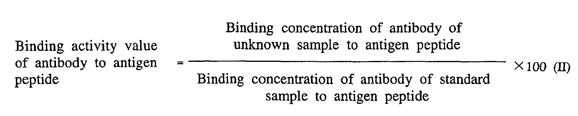
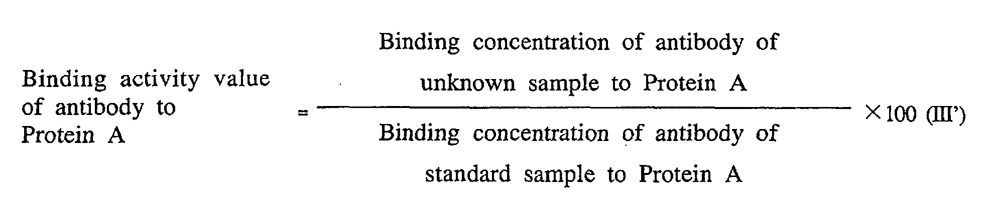
- The binding concentration of an antibody to an antigen peptide, and that to Protein A can be measured by SPR, a fluorescence polarization measurement method, a radioimmunoassay (RIA), an enzyme-linked immunosorbent assay (ELISA), HPLC, an analytical ultracentrifugation method, a titration calorimetry or the like. Of these, the measurement by SPR is convenient, because it can monitor in real-time the antibody-antigen interaction without labeling.
- Target antibodies of the method of the present invention may be antibodies for any antigens. Examples of such an antibody include an antibody (hereinafter, referred to as "anti-PTHrP antibody") for parathyroid hormone-related peptide (hereinafter, referred to as "PTHrP"), an antibody for IL-6 receptor, an antibody for HM1.24 antigen, and an antibody for tissue factor (TF).
- The antibody may be either a monoclonal antibody or a polyclonal antibody, and a monoclonal antibody is preferable.
- The antibody class may be any of IgM, IgD, IgG, IgA and IgE, and IgG is desirable. In addition, the subclass of H chain may be any of IgG1, IgG2, IgG3, and IgG4, and IgG1 and IgG4 are desirable, and the subclass of L chain may be either κ or λ.
- A monoclonal antibody may be a human antibody, humanized antibody or chimeric antibody.
- In addition, examples of target substances that can be measured by the measurement method of the present invention also include antibody fragments, such as Fab, (Fab')2 and re-constituted antibody fragments, such as a single-chain Fv.
- A preferred antibody forms few (preferably, 5% or less) or no oligomer, such as dimmers.
- In the present specification, the term "second ligand-binding substance" indicates a substance which binds to a ligand at a site other than the binding site of the ligand with a ligand-binding protein (also referred to as "first ligand-binding substance"). When a ligand is an antibody, a second ligand-binding substance specifically binds to a portion other than the antigen binding site, such as the constant region (Fc portion, L chain κ region, an L chain λ region, or the like) and the sugar chain of the antibody. Examples of a substance which binds to Fc portion include Protein A, protein G and Fc receptor. Examples of a substance which binds to L chain κ region include protein L and anti-κ antibody. An example of a substance which binds to L chain λ region is anti-λ antibody. Examples of a substance which binds to sugar chains include lectins, such as RCA, LCA and ConA.
- Preferred examples of a substance which binds to a portion other than the antigen binding site of an antibody include Protein A, protein G, Fc receptor, protein L and lectins, such as RCA, LCA and ConA. More preferred examples include Protein A, protein G and protein L, and most preferably, Protein A.
- In addition, the present invention provides a kit for evaluating the binding activity of a ligand to a ligand-binding protein. This kit contains both a ligand-binding protein and a second ligand-binding substance. This ligand-binding protein may be an antigen peptide. Furthermore, the kit of the present invention may contain other reagents. For example, when SPR is used for measuring the binding activity of an antibody to an antigen, the kit of the present invention may further contain a reagent for immobilizing onto a sensor chip an antigen and a substance which binds to a portion other than the antigen-binding site of an antibody. The reagent may be any reagent as long as it activates the functional group of an antigen, for example, an amino group, thiol group, or aldehyde group to bind onto the chip. Examples of such a reagent include N-ethyl-N'-(3-dimethylaminopropyl) carbodiimidehydrochloride, N-hydroxysuccinimide, ethanolamine hydrochloride, 2-(2-pyridinyldithio) ethaneamine hydrochloride, cysteine, biotinhydrazide.
- Even if other protein is present as a contaminant, or a medium, a stabilizer or the like in a sample solution, the method of evaluating ligand (for example, antibody) activity and the kit of the present invention do not require additional purification procedures to evaluate the binding activity ratio of the ligand (for example, antibody) molecule. Therefore, the evaluation method and the kit can be used for studying quality control, in-process control and formulation design of antibodies in the production process or purified antibodies as an drug substance.
- In addition, the binding activity ratio which is finally obtained by the method of evaluating ligand (for example, antibody) activity and the kit of the present invention does not reflect the concentration of an active ingredient in a sample solution, but the activity of the binding activity of a ligand (for example, antibody) to a second ligand-binding activity. Therefore, the activity ratio of 100% or more may suggest that some decomposition occurs at a second portion (for example, Fc portion) other than the antigen-binding site of an antibody; and the activity ratio of 100% or less may suggest some decomposition at the antigen-binding site of an antibody. Accordingly, the method and the kit of the present invention in combination with another method of evaluating physical properties can be used for stability testing and shelf life setting.
- Furthermore, the method of evaluating ligand (for example, antibody) activity and the kit of the present invention can also be used for selecting a highly active protein (in particular, a mutant protein, various monoclonal antibodies or the like). Specifically, the method and the kit of the present invention are effective in selecting a mutant protein with high biological activity or stability resulting from point mutation, or the like. The method and the kit of the present invention are particularly useful in screening antibodies, which are reconstituted from natural antibodies, such as chimeric antibodies and humanized antibodies.
- Further, the method and the kit of the present invention are also effective of evaluating whether or not various electrically charged hetero components (the plural minor peaks, which are detected when the bulk protein, is applied to ion exchange chromatography) detected in the bulk proteins, are "Product-Related Substances," as is required for New Drug Application of a protein drug product, such as an antibody.
- Furthermore, the method of evaluating ligand (for example, antibody) binding activity, and the kit of the present invention can provide a convenient and highly accurate measurement of the binding activity of, and the quantitative determination of highly active proteins within body fluids, such as blood, serum or the like (in particular, a blood factor, a mutant protein, various monoclonal antibodies, antibody fragments, such as Fab and (Fab')2, a single-chain Fv and the like).
-
- Figure 1 shows a sensorgram when PTHrP(1-34+Cys) was immobilized.
- Figure 2 shows a sensorgram (1Cycle) when anti-PTHrP antibody a (2.0 µg/mL) was measured.
- Figure 3 shows the calibration curve (Duplicate) when the amount of anti-PTHrP antibody bound to PTHrP was quantitatively determined.
- Figure 4 shows a cycle ignored from data because of a disturbance in the sensorgram.
- Figure 5 shows a sensorgram when Protein A was immobilized.
- Figure 6 shows a sensorgram (1Cycle, Protein A) when anti-PTHrP antibody a (2.0 µg/mL) was measured.
- Figure 7 shows the calibration curve (Duplicate) when the amount of anti-PTHrP antibody bound to Protein A was quantitatively determined.
-
- This specification includes part or all of the contents as disclosed in the specification of Japanese Patent Application No. 2000-192829, which is a priority document of the present application.
- A method of evaluating the binding activity of an anti-PTHrP antibody to PTHrP is described as an example of the method of the present invention. This method comprises the steps of measuring, using BIACORE™, the amount of anti-PTHrP antibody bound to PTHrP and to Protein A respectively, and of correcting the measured amount of the anti-PTHrP antibody bound to PTHrP with the measured amount of the anti-PTHrP antibody bound to Protein A.
- First, anti-PTHrP antibodies are produced according to the method described in WO98/13388.
- Anti-PTHrP antibodies are diluted with water, and then a solution of the anti-PTHrP antibody is prepared at an appropriate concentration (for example, 100-500 µg/mL). The solution is quantitatively determined by the absorbance method in the general test of the Japanese Pharmacopoeia. The solution is diluted with HBS buffer solution (HBS-EP Buffer (BIACORE)) at an appropriate concentration (for example, 10-50 µg/mL). This preparation is repeated three times. HBS buffer solution is added to each solution, so that solutions each having a certain concentration (for example, 1-5 µg/mL) are prepared as
unknown sample solutions - The above procedure is performed for a standard product of anti-PTHrP antibody, thereby preparing
standard sample solutions - Separately, anti-PTHrP antibodies are diluted with water, and then a solution of anti-PTHrP antibody at an appropriate concentration (for example, 100-500 µg/mL) is prepared. The solution is diluted with HBS buffer solution, and then solutions of anti-PTHrP antibody having various concentrations (for example, 0 to 5 µg/mL) are prepared as standard solutions A, B, C, D, E and F for a calibration curve.
- Next, PTHrP- and Protein A-immobilized chips are prepared. A sensor chip CM 5 (BIACORE, Code # BR-1000-14) is docked in BIACORE™, and then HBS buffer solution is run at a flow rate of 5-20 µL/min. Then, different solutions are run successively, to immobilize PTHrP(1-34+Cys) on the chip: for example, 150 µL of a mixed solution containing equal amounts of EDC (N-ethyl-N'-(3-dimethyl aminopropyl)-carbodiimide hydrochloride) solution and NHS (N-hydroxysuccinimide) solution; for example, 150 µL of borate buffer solution of PDEA (2-(2-pyridinyldithio)ethaneaminehydrochloride); for example, 40 µL of acetate buffer solution of PTHrP(1-34+Cys) (a synthetic peptide having Cys added to the C terminus of 1-34 peptides of PTHrP); for example, 150 µL of formate buffer solution of cysteine-sodium chloride; for example, 10 µL of glycine hydrochloride buffer solution; and for example, 10 µL of 10 mM hydrochloric acid aqueous solution. Using another flow cell, HBS buffer solution is run similarly at a flow rate of 5 to 20 µL/min. Then, different solutions are run successively to immobilize Protein A onto a chip: for example, 100 µL of the mixed solution of equal amounts of EDC solution and NHS solution; for example, 40 µL of acetate buffer solution of Protein A; for example, 100 µL of ethanolamine hydrochloride buffer solution; for example, 10 µL of glycine hydrochloride buffer solution; and for example, 10 µL of 10 mM hydrochloric acid aqueous solution.
- The standard solutions for a calibration curve A, B, C, D, E and F, the
standard sample solutions unknown sample solutions 

- The present invention will be described more specifically by the following examples. These examples are given only for explanation, and are not intended to limit the scope of the present invention.
- Instruments, reagents and samples employed in the examples are as follows:
- (1) Instrument
- BIACORE™ upgrade: (BIACORE)
- Sensor Chip : Sensor Chip CM5 : (BIACORE)
- Spectrophotometer: DU-640 (BECKMAN)
- (2) Reagent
- Running Buffer : HBS buffer BIA Certified (0.01 M HEPES pH 7.4, 0.15 M NaCl, 3 mM EDTA, 0.005% (v/v) Surfactant P20) (BIACORE) Code # BR-1001-88
- NHS : 0.05 M N-hydroxysuccinimide, Amine coupling kit (BIACORE) Code # BR-1000-50
- EDC: 0.2 M N-ethyl-N'-(3-dimethyl aminopropyl)-carbodiimide hydrochloride, Amine coupling kit (BIACORE) Code # BR-1000-50
- Ethanolamine-HCl (pH8.5): Amine coupling kit (BIACORE) Code # BR-1000-50
- PDEA: Thiol coupling kit (BIACORE) Code # BR-1000-58
- PTHrP(1-34+Cys): product synthesized by SAWADY Technology Co., Ltd. (custom peptide JH 365, 21/11/94, B-CAL 35a.a.)
- Protein A: (CAPPEL) Cat # 55832, Lot # 40690
- L-Cystein, NaCl, HCl
- 0.1 M Borate-Na Buffer (pH8.5)
- 10 mM Acetate-Na Buffer (pH4.0, 5.0)
- 0.1 M Na-Formate Buffer (pH4.3)
- 0.1 M Gly-HCl Buffer (pH2.5)
- (3) Sample
- Anti-PTHrP antibodies a, b, c, d, e and f : in house (all of them are lots for research)
-
- The flow rate was set at 5 µL/min, the reagents were injected according to Table 1, and then antigen PTHrP(1-34+Cys) was immobilized by the thiol coupling method.
Conditions for immobilizing PTHrP(1-34+Cys) Reagent Injection volume NHS/ EDC 100 µL PDEA (5.4 mg/250 µL 0.1 M Borate-Na Buffer (pH 8.5)) 100 µL PTHrP(1-34+Cys) (10 µg/ mL 10 mM Na-Acetate Buffer(pH 5.0))20 µL 50 mM Cys / 1 M NaCl (0.1 M Na-Formate Buffer(pH 4.3) 100 µL 0.1 M Gly-HCl (pH 2.5) 10 µL 10 mM HCl 10 µL - Using the chips prepared under the conditions of i), the respective amounts of anti-PTHrP antibodies bound to PTHrP for 4 lots ( a , b , c and d ) of anti-PTHrP antibody were measured by the following procedures. Then, differences among the lots were studied. Original solutions of samples a, b, c and d , were diluted with purified water to prepare about 500 µg/mL solutions, and then the concentrations were determined by the absorbance method. Based on the concentrations, 0, 1, 2, 3, 4 and 5 µg/mL (n=1) solution in HBS for a calibration curve and a 2 µg/mL (n=3) solution in HBS as a control were prepared for the standard sample a , and 2 µg/mL (n=3) solution in HBS were prepared for the unknown samples b, c and d respectively. First, the flow rate was set at 5 µL/min, and binding phase was defined two minutes period while 10 µL of the sample solution was injected. After the sample solution was switched to a running buffer, a difference between resonance unit (RU) after 30 seconds and a baseline immediately before injection was determined as the bound amount. Subsequently, the sensor chips were washed and regenerated by injecting 15 mM HCl. With 1 cycle of analysis consisting of the binding and regeneration, measurement was performed twice for each sample solution, so that the sensorgram and the bound amount were obtained.
- Original solutions of the samples ( a and e ) of anti-PTHrP antibody were diluted with purified water to prepare about 100 µg/mL solutions, and then the concentrations were determined by the absorbance method. Based on the concentrations, 0, 1, 2, 3, 4 and 5 µg/mL (n=1) solution in HBS for a calibration curve and a 2 µg/mL (n=3) solution in HBS as a control were prepared for standard samples a , and 2 µg/mL (n=3) solution in HBS were prepared for unknown samples e . In a manner similar to procedure (1), the amount of bound PTHrP was measured.
- The flow rate was set at 5 µL/min, the reagents were injected according to Table 2, and then Protein A was immobilized by the amine coupling method.
Immobilization conditions for Protein A Reagent Injection volume NHS/ EDC 150 µL Protein A (50 µg/mL 0.1 M Borate-Na Buffer (pH 4.0)) 50 µL Ethanolamine 150 µL 0.1 M Gly-HCl (pH 2.5) 10 µL 10 mM HCl 10 µL - Based on the concentrations obtained by the absorbance method, 0, 1, 2, 3, 4 and 5 µg/mL (n=1) solution in HBS for a calibration curve and a 2 µg/mL (n=3) solution in HBS as a control were prepared for the standard sample. Diluted solutions (n=3) were prepared by diluting 1000-fold the original sample solution for unknown sample e . In a manner similar to procedure (1), the amount of anti-PTHrP antibody bound to PTHrP was measured. Next, similar measurements were performed for Protein A-immobilized chip, thereby obtaining the sensorgram and the amount of anti-PTHrP antibody bound to Protein A.
- Based on the concentrations obtained by the absorbance method, 0, 1, 2, 3, 4 and 5 µg/mL (n=1) solution in HBS for a calibration curve and a 2 µg/mL (n=3) solution in HBS as a control] were prepared for the standard sample a . 2 µg/mL (n=3) solution in HBS were prepared for unknown sample f . In a manner similar to (2)-iii), 0 to 5 µg/mL samples for a calibration curve (n=1) and 2 µg/mL unknown sample (n=3) were injected to PTHrP-immobilized chip and Protein A-immobilized chip, respectively. Two measurements of the bound amount were considered as one measurement. The series of procedures starting from the preparation of samples were performed 7-times in total, so that the repeatability was studied.
- Figure 1 shows the result of immobilization of PTHrP. To evaluate the PTHrP(1-34+Cys)-immobilized chip prepared and to set an appropriate concentration range for a calibration curve and the concentration of a sample, the amount of antibodies bound to PTHrP was measured for 4 lots of anti-PTHrP antibody certified to have approximately equal quality. A representative example of the obtained sensorgram is shown in Fig 2.
- A calibration curve was produced from the resulting bound amount using the calculation software attached to the BIACORE™ system for quantitatively determining concentrations (Fig. 3). As a result, the binding phase on the sensorgram (Fig. 2) was almost a straight line, no dissociation was observed, and the calibration curve (Fig. 3) almost maintained its linearity within the concentration range. Taking these results together, it was concluded that the amount of immobilized ligands was sufficient for use in the quantification system. Therefore, the concentrations of samples to quantitatively determine the amount of antibodies bound to a PTHrP(1-34+Cys)-immobilized chip were set according to the conditions employed for this experiment. Specifically, standard samples for a calibration curve were set at six concentrations: 0, 1, 2, 3, 4 and 5 µg/mL, and unknown samples were set at a concentration of 2 µg/mL, which was about middle point of the calibration curve.
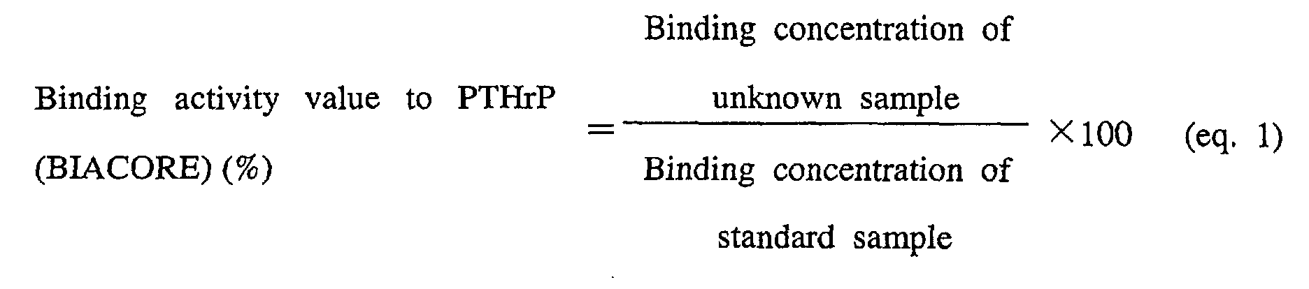
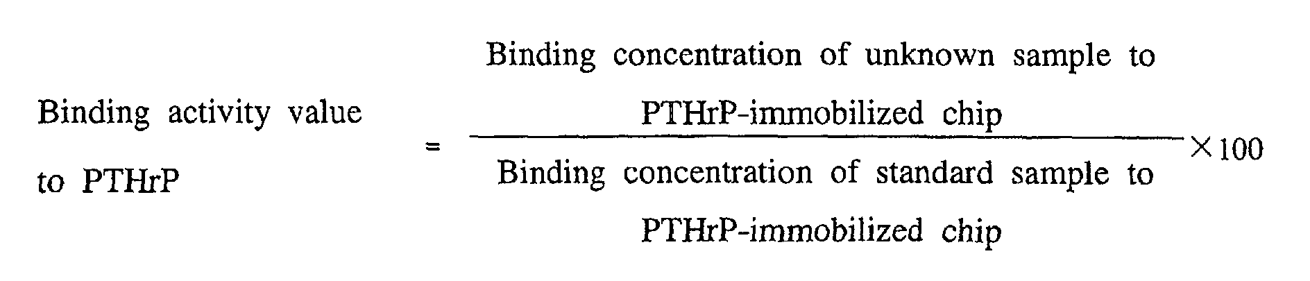
- Using the calibration curve, the binding concentrations of 4 lots that were actual samples were calculated with the attached software using the bound amount provided from the sensorgram. At this time, a value was ignored for a cycle for which no appropriate bound amount could be detected due to a disturbance in the sensorgram (Fig. 4). Based on the binding concentrations, PTHrP binding activity value was defined as eq. 1, and then the results of the 4 lots were summarized in Table 3. Thus, the measurement system was shown to be extremely precise such that coefficient of variation was within 3% when n=3. From this experiment, it was concluded that there was no significant difference in the PTHrP binding activity among 4 lots.
Measurement results of binding activity value to PTHrP (lot to lot consistency) Sample n Bound amount (Resonance unit, RU) Binding concentration calculated from calibration curve
(µg/mL)Measurement (measured three times) Binding activity value (%) 1st.
cycle2nd.
cycle1st.
cycle2nd.
cycleAverage %cv.
dupliAverage %cv b 1 966.4 979.7 1.99 2.01 2.00 0.71 2.01 0.90 99.2 2 979.0 957.8 2.01 1.97 1.99 1.42 3 981.4 987.2 2.02 2.03 2.03 0.35 c 1 951.7 1008.7 1.96 2.07 2.02 3.86 2.05 1.73 101 2 987.5 985.5 2.06 2.02 2.04 1.39 3 1014.7 1019.2 2.08 2.09 2.09 0.34 d 1 967.8 971.1 1.99 2.00 2.00 0.35 1.99 2.06 98.2 2 974.2 913.9 2.00 1.88 1.94 4.37 3 983.4 981.7 2.02 2.02 2.02 0.00 a 1 985.6 985.2 2.02 2.02 2.02 0.00 2.02 0.14 100 2 982.4 982.1 2.02 2.02 2.02 0.00 3 984.4 990.2 2.02 2.03 2.03 0.35 - Concentration of the sample anti-PTHrP antibody e was prepared based on the absorbance method, and then the binding concentrations of the sample e to the PTHrP(1-34-Cys)-immobilized chip were measured (I-(2)-i)). As a result, the binding activity value of sample e to PTHrP was 46%, compared to that of the standard sample a (Table 4). Possible reasons for these results include lowered biological activity, and contamination with foreign substances having absorbance at 280 nm. Another possible reason is that the protein content was lowered because the concentrations obtained by the absorbance method did not reflect the antibody concentration. Hence, attempts to improve the accuracy and the precision were undertaken by taking the system to quantitatively determine the bound amount to Protein A-immobilized chip as a system to quantitatively determine antibody concentration using BIACORE™, and correcting the antibody concentration.
Measurement results of binding activity value of anti-PTHrP antibody e to PTHrP Sample n Bound amount (Resonance unit, RU) Binding concentration calculated from calibration curve
(µg/mL)Measurement (measured three times) Binding activity value (%) 1st.
cycle2nd.
cycle1st.
cycle2nd.
cycleAverage %cv.
dupliAverage %cv 1 986.7 1011.0 2.01 2.06 2.04 1.74 2.05 0.49 100 a 2 996.1 1020.6 2.03 2.08 2.06 1.72 3 992.5 1009.6 2.03 2.06 2.05 1.04 1 438.2 446.0 0.95 0.97 0.96 1.47 0.95 1.59 46.2 e 2 428.6 428.6 0.93 0.93 0.93 0.00 3 429.4 435.0 0.94 0.95 0.95 0.75 - Figure 5 shows the results of the immobilization of Protein A. Based on the linearity of the sensorgram and the calibration curve (Figs. 6 and 7) resulting from the prepared Protein A-immobilized chip, it was determined that the amount of immobilized ligands was sufficient for use in the quantification system. Further, it was also confirmed that the calibration curve showed sufficient linearity with the concentration range of 0 to 5 µg/mL employed for measurement using a PTHrP(1-34-Cys)-immobilized chip. Therefore, the sample concentrations of both samples for a calibration curve and unknown samples for quantitatively determining the bound amount to the Protein A-immobilized chip were set under the same conditions with a PTHrP(1-34-Cys)-immobilized chip.
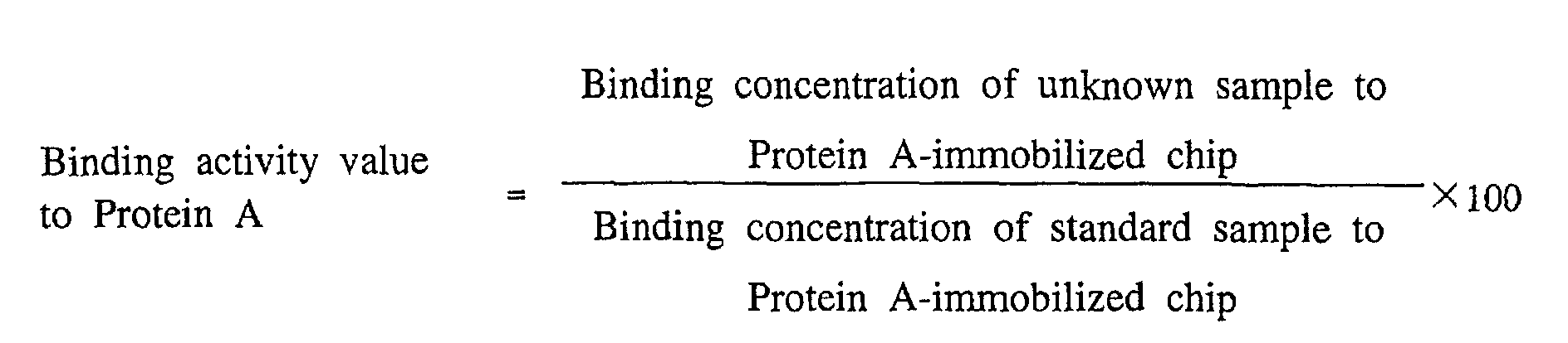
- Using the calibration curves for each PTHrP and Protein A, the binding concentration was calculated by means of the attached software using the bound amount provided from the sensorgram. The results were summarized in Table 5. The binding activity value to PTHrP was corrected with the concentration calculated from the bound amount to Protein A, anti-PTHrP antibody binding activity ratio was newly defined as eq. 2, and these results were summarized in Table 6. As a result, e had a binding activity of 94% when compared to a, suggesting a slightly lowered activity.
- As described above, based on the quantitatively determined amount of antibodies bound to Protein A, more precise measurement of anti-PTHrP antibody binding activity became possible by the confirmation of antibody concentration using the sample concentrations, which are same as those employed for the measurement of the bound amount to PTHrP.

Measurement results of binding activity values of anti-PTHrP antibody e to PTHrP and Protein A Ligand: PTHrP Sample n Bound amount (Resonance unit, RU) Binding concentration calculated from calibration curve
(µg/mL)Measurement (measured three times) 1st.
cycle2nd.
cycle1st.
cycle2nd.
cycleAverage %cv.
dupliAverage %cv a 1 760.9 779.2 1.98 2.03 2.01 1.76 2.02 0.50 2 768.8 788.3 2.00 2.05 2.03 1.75 3 763.2 783.2 1.99 2.04 2.02 1.75 e 1 824.4 852.5 2.14 2.21 2.18 2.28 2.17 0.70 2 819.0 838.5 2.13 2.18 2.16 1.64 3 834.0 849.7 2.17 2.20 2.19 0.97 Ligand: Protein A Sample n Bound amount (Resonance unit, RU) Binding concentration calculated from calibration curve (µg/mL) Measurement (measured three times) 1st.
cycle2nd.
cycle1st.
cycle2nd.
cycleAverage %cv.
dupliAverage %cv a 1 770.5 786.7 2.00 2.04 2.02 1.40 2.03 0.43 2 770.9 785.2 2.00 2.04 2.02 1.40 3 776.4 790.4 2.02 2.05 2.04 1.04 e 1 875.9 886.0 2.27 2.30 2.29 0.93 2.31 0.99 2 876.5 894.0 2.28 2.32 2.30 1.23 3 887.6 909.6 2.30 2.36 2.33 1.82 Measurement results of binding activity ratio of anti-PTHrP antibody e Sample Binding concentration Binding activity ratio % PTHrP Protein A a 2.02 2.03 100 e 2.17 2.31 94.7 - To evaluate the measurement method for binding activity to PTHrP, which had been set according to the obtained results including concentration correction using Protein A, f was measured using standard product a. In one measurement, dispersions when each batch was measured twice (Dupli) were all less than 4% (83% of all measurement cycles were less than 2% dispersion), and dispersions provided when n=3 were all less than 3% (90% of all measurement cycles were less than 2% dispersion). However, dispersions resulting from one experiment repeated 7 times were less than 1% (0.95%), suggesting that the experiment system was extremely precise (Tables 7 and 8).
- CAL binding activity was calculated using eq. 3 and summarized in Table 8. With the binding activity of a at 100%, the biological activity of f was at 97.0%, while the coefficient of variation of the repeatability (when repeated 7 times) was at 0.95%, these suggest that the difference between the two was significant and f had a slightly decreased activity with antigen.
-

Summary of 7-times repeated measurement results of binding activity of anti-PTHrP antibody f N Ligand Sample Mean value of binding concentration (Dupli, measured twice) (µg/mL) Mean value of measurement (measured three times) %cv n=1 n=2 n=3 1 Protein A a 2.02 2.06 2.05 2.04 1.02 f 2.20 2.22 2.21 2.21 0.35 PTHrP a 2.04 2.05 2.02 2.04 0.89 f 2.14 2.14 2.12 2.13 0.41 2 Protein A a 2.04 2.03 2.12 2.06 2.25 f 2.24 2.26 2.24 2.24 0.46 PTHrP a 2.05 2.05 2.05 2.05 0.14 f 2.17 2.17 2.20 2.18 0.80 3 Protein A a 1.96 2.01 2.02 2.00 1.52 f 2.19 2.20 2.24 2.21 1.25 PTHrP a 2.16 2.19 2.19 2.18 0.87 f 2.33 2.34 2.39 2.35 1.29 4 Protein A a 2.03 2.04 2.05 2.04 0.62 f 2.38 2.37 2.44 2.40 1.58 PTHrP a 1.99 2.05 2.02 2.02 1.49 ef 2.27 2.26 2.29 2.27 0.79 5 Protein A a 2.03 2.02 2.02 2.02 0.38 f 2.44 2.45 2.39 2.43 1.29 PTHrP a 2.04 2.04 2.05 2.04 0.28 f 2.41 2.42 2.42 2.41 0.32 6 Protein A a 1.97 2.02 2.05 2.01 2.01 f 2.73 2.73 2.78 2.74 1.00 PTHrP a 2.15 2.23 2.21 2.20 2.02 f 2.86 2.89 2.96 2.90 1.67 7 Protein A a 1.98 1.97 1.98 0.36 f 2.41 2.42 2.44 2.42 0.62 PTHrP a 2.03 2.05 2.01 2.03 0.87 f 2.38 2.39 2.42 2.40 0.75 Summary of 7-times repeated measurement results of binding activity of f N Lot No. Binding concentration Binding activity ratio PTHrP Protein A % 1 a 2.04 2.04 100 f 2.13 2.21 96.4 2 a 2.05 2.06 100 f 2.18 2.24 97.8 3 a 2.18 2.00 100 f 2.35 2.21 97.6 4 a 2.02 2.04 100 f 2.27 2.40 95.5 5 a 2.04 2.02 100 f 2.41 2.43 98.2 6 a 2.20 2.01 100 f 2.90 2.74 96.7 7 a 2.03 1.98 100 f 2.40 2.42 96.7 Average f - - 97.0 0.95 (%cv) 


- In the measurement system, the bound amount observed at a certain time after injection of antibody followed by switching to a buffer is simply defined as the amount of PTHrP binding activity. Since the effect of dissociation is ignored in the system, the result obtained herein cannot be biological activity in the strictest sense. However, this method is good enough as a convenient method of evaluating activity without requiring kinetics analysis for samples observed from the form of the sensorgrams to have the dissociation rates sufficiently smaller than the binding rates.
- As described above, we have invented a convenient and highly accurate testing method, with which in vitro CAL biological activity can be evaluated without kinetics analysis using BIACORE™. In appropriate combination with other testing methods for testing biological activity, this method can be used as a testing method which reflects in vitro environment more accurately by solving the problems inherent from the principle of BIACORE™.
- All publications, patents and patent applications cited herein are incorporated herein by reference in their entirety.
- The present invention provides a convenient and highly accurate method of measuring the biological activity of a ligand.
Claims (21)
- A method of evaluating the binding activity of a ligand to a ligand-binding protein, which comprises; measuring the binding activity value of the ligand to a ligand-binding protein, and measuring the binding activity value of the ligand to a second ligand-binding substance which binds to a site of the ligand other than the binding site where the ligand binds to the ligand-binding protein.
- The method of claim 1, wherein either the ligand or the ligand-binding protein is immobilized.
- The method of claim 1 or 2, wherein the ligand is an antigen or an antibody thereof, an enzyme or the substrate protein thereof or a ligand for various receptors.
- The method of claim 1 or 2, wherein the ligand is an antibody, and the ligand-binding protein is an antigen peptide.
- The method of claim 4, wherein the second ligand-binding substance specifically binds to the constant region or the sugar chain of an antibody.
- The method of claim 5, wherein the constant region of an antibody is an Fc portion, an L chain κ region, or an L chain λ region.
- The method of claim 5, wherein the second ligand-binding substance specifically binds to an Fc portion of an antibody.
- The method of claim 6 or 7, wherein the substance which specifically binds to an Fc portion of an antibody is Protein A, protein G, protein L, Fc receptor or lectin.
- The method of any one of claims 1 to 4, wherein FLAG protein is fused to a ligand, and a substance which specifically binds to the FLAG protein is used as the second ligand-binding protein.
- The method of any one of claims 1 to 9, which comprises; separately immobilizing the ligand-binding protein and the second ligand-binding substance, allowing them to react respectively with the same ligand-containing samples, and then measuring each for binding activities.
- The method of claim 4, wherein the second ligand-binding substance is Protein A.
- The method of any one of claims 4 to 11, wherein the binding activity value of an antibody to an antigen peptide is corrected with the binding activity value of the antibody to the second ligand-binding substance.
- The method of any one of claims 4 to 12, wherein the binding activity of an antibody to an antigen peptide is measured using the binding activity ratio of the antibody which is calculated by the following formula (I):

- The method of claim 13, wherein the binding activity value of an antibody to an antigen peptide, and the binding activity value of the antibody to the second ligand-binding substance are calculated by the following formulae (II) and (III), respectively:


- The method of any one of claims 1 to 14, wherein the binding concentration of the ligand to the ligand-binding protein, and the binding concentration of the ligand to the second ligand-binding substance are measured by either SPR, a fluorescence polarization measurement method, a radioimmunoassay (RIA), an enzyme-linked immunosorbent assay (ELISA), HPLC, an analytical ultracentrifugation method, or a titration calorimetry.
- The method of claim 15, wherein the binding concentration of the ligand to the ligand-binding protein, and the binding concentration of the ligand to the second ligand-binding substance are measured by SPR.
- The method of any one of claims 1 to 16, wherein the ligand is a monoclonal antibody.
- The method of claim 17, wherein the monoclonal antibody is a humanized antibody.
- The method of claim 17 or 18, wherein the monoclonal antibody is an antibody against parathyroid hormone-related peptide.
- A kit for evaluating the binding activity of the ligand to the ligand-binding protein, which contains the ligand-binding protein and the second ligand-binding substance.
- The kit of claim 20, wherein the ligand-binding protein is an antigen peptide.
Applications Claiming Priority (3)
| Application Number | Priority Date | Filing Date | Title |
|---|---|---|---|
| JP2000192829 | 2000-06-27 | ||
| JP2000192829 | 2000-06-27 | ||
| PCT/JP2001/005501 WO2002001224A1 (en) | 2000-06-27 | 2001-06-27 | Method of evaluating binding activity of ligand to ligand-binding protein |
Publications (2)
| Publication Number | Publication Date |
|---|---|
| EP1296140A1 true EP1296140A1 (en) | 2003-03-26 |
| EP1296140A4 EP1296140A4 (en) | 2005-06-29 |
Family
ID=18691907
Family Applications (1)
| Application Number | Title | Priority Date | Filing Date |
|---|---|---|---|
| EP01943834A Withdrawn EP1296140A4 (en) | 2000-06-27 | 2001-06-27 | METHOD FOR EVALUATING THE BINDING ACTIVITY OF A LIGAND WITH A LIGAND BINDING PROTEIN |
Country Status (5)
| Country | Link |
|---|---|
| US (1) | US20030138870A1 (en) |
| EP (1) | EP1296140A4 (en) |
| JP (1) | JP3515101B2 (en) |
| AU (1) | AU2001267833A1 (en) |
| WO (1) | WO2002001224A1 (en) |
Cited By (1)
| Publication number | Priority date | Publication date | Assignee | Title |
|---|---|---|---|---|
| EP1347300A4 (en) * | 2000-11-30 | 2005-06-29 | Chugai Pharmaceutical Co Ltd | METHOD FOR MEASURING THE BINDING ACTIVITY OF A LIGAND-BINDING PROTEIN HAVING LOW CHEMICAL STABILITY WITH A FIRST LIGAND |
Families Citing this family (8)
| Publication number | Priority date | Publication date | Assignee | Title |
|---|---|---|---|---|
| JP2005209795A (en) * | 2004-01-21 | 2005-08-04 | Koito Mfg Co Ltd | Light emitting module and lamp |
| JPWO2006109533A1 (en) * | 2005-03-31 | 2008-10-23 | 国立大学法人大阪大学 | Methods for producing and assaying antibodies against cell membrane surface antigen epitopes |
| JP2009184981A (en) * | 2008-02-07 | 2009-08-20 | Shiseido Co Ltd | Anti-D-amino acid monoclonal antibody and immunological analysis method of D-amino acid using anti-D-amino acid monoclonal antibody |
| AU2009255777A1 (en) * | 2008-06-02 | 2009-12-10 | Ge Healthcare Bio-Sciences Ab | Concentration assay |
| JP2011007664A (en) * | 2009-06-26 | 2011-01-13 | Fujifilm Corp | Method for screening antibody |
| US20160003815A1 (en) * | 2013-03-15 | 2016-01-07 | Inanovate, Inc. | Method, system, and device for analyte detection and measurement using longitudinal assay |
| JP6625519B2 (en) * | 2013-03-15 | 2019-12-25 | イナノベイト, インコーポレイテッド | Assay systems and cartridge devices |
| CN110398590B (en) * | 2019-06-21 | 2022-11-15 | 深圳大学 | Sensing chip and application |
Family Cites Families (5)
| Publication number | Priority date | Publication date | Assignee | Title |
|---|---|---|---|---|
| SE504507C2 (en) * | 1993-05-24 | 1997-02-24 | Pharmacia Biosensor Ab | Methods to determine the binding properties of low molecular weight ligands |
| US5602041A (en) * | 1994-08-26 | 1997-02-11 | Sea Run Holdings, Inc. | Fish serum as a blocking reagent |
| JP2000512131A (en) | 1996-05-29 | 2000-09-19 | マクギル ユニバーシティー | Prion protein binding protein and its use |
| US5922845A (en) * | 1996-07-11 | 1999-07-13 | Medarex, Inc. | Therapeutic multispecific compounds comprised of anti-Fcα receptor antibodies |
| US6737056B1 (en) * | 1999-01-15 | 2004-05-18 | Genentech, Inc. | Polypeptide variants with altered effector function |
-
2001
- 2001-06-27 JP JP2002506106A patent/JP3515101B2/en not_active Expired - Fee Related
- 2001-06-27 US US10/312,346 patent/US20030138870A1/en not_active Abandoned
- 2001-06-27 AU AU2001267833A patent/AU2001267833A1/en not_active Abandoned
- 2001-06-27 EP EP01943834A patent/EP1296140A4/en not_active Withdrawn
- 2001-06-27 WO PCT/JP2001/005501 patent/WO2002001224A1/en not_active Ceased
Non-Patent Citations (8)
| Title |
|---|
| CATIMEL B ET AL: "Kinetic analysis of the interaction between the monoclonal antibody A33 and its colonic epithelial antigen by the use of an optical biosensor - A comparison of immobilisation strategies" JOURNAL OF CHROMATOGRAPHY A, ELSEVIER, AMSTERDAM, NL, vol. 776, no. 1, 25 July 1997 (1997-07-25), pages 15-30, XP004125534 ISSN: 0021-9673 * |
| EIRY KOBATAKE: "BIOLUMINESCENT IMMUNOASSAY WITH A PROTEIN A-LUCIFERASE FUSION PROTEIN" ANALYTICAL BIOCHEMISTRY, ACADEMIC PRESS, SAN DIEGO, CA, US, vol. 208, no. 2, 1 February 1993 (1993-02-01), pages 300-305, XP000335006 ISSN: 0003-2697 * |
| NGAI P K M ET AL: "PROTEIN A ANTIBODY-CAPTURE ELISA (PACE): AN ELISA FORMAT TO AVOID DENATURATION OF SURFACE-ADSORBED ANTIGENS" JOURNAL OF IMMUNOLOGICAL METHODS, ELSEVIER SCIENCE PUBLISHERS B.V.,AMSTERDAM, NL, vol. 158, no. 2, 1993, pages 267-276, XP000916753 ISSN: 0022-1759 * |
| PIEHLER J ET AL: "Assessment of affinity constants by rapid solid phase detection of equilibrium binding in a flow system" JOURNAL OF IMMUNOLOGICAL METHODS, ELSEVIER SCIENCE PUBLISHERS B.V.,AMSTERDAM, NL, vol. 201, no. 2, 28 February 1997 (1997-02-28), pages 189-206, XP004117417 ISSN: 0022-1759 * |
| QIN Q ET AL: "Dual-label time-resolved immunofluorometric assay for simultaneous determination of pregnancy-associated plasma protein A and free beta-subunit of human chorionic gonadotrophin" JOURNAL OF IMMUNOLOGICAL METHODS, ELSEVIER SCIENCE PUBLISHERS B.V.,AMSTERDAM, NL, vol. 205, no. 2, 14 July 1997 (1997-07-14), pages 169-175, XP004126290 ISSN: 0022-1759 * |
| QUINN JOHN G ET AL: "Development and application of surface plasmon resonance-based biosensors for the detection of cell-ligand interactions" ANALYTICAL BIOCHEMISTRY, vol. 281, no. 2, 1 June 2000 (2000-06-01), pages 135-143, XP002327218 ISSN: 0003-2697 * |
| See also references of WO0201224A1 * |
| VISHWANATH S ET AL: "KINETIC STUDIES OF SITE-SPECIFICALLY AND RANDOMLY IMMOBILIZED ALKALINE PHOSPHATASE ON FUNCTIONALIZED MEMBRANES" JOURNAL OF CHEMICAL TECHNOLOGY AND BIOTECHNOLOGY. (INTERNATIONAL JOURNAL OF BIOTECHNICAL AND CHEMICAL PROCESSES), ELSEVIER APPLIED SCIENCE PUBLISHERS. BARKING, GB, vol. 68, no. 3, March 1997 (1997-03), pages 294-302, XP000700648 ISSN: 0268-2575 * |
Cited By (1)
| Publication number | Priority date | Publication date | Assignee | Title |
|---|---|---|---|---|
| EP1347300A4 (en) * | 2000-11-30 | 2005-06-29 | Chugai Pharmaceutical Co Ltd | METHOD FOR MEASURING THE BINDING ACTIVITY OF A LIGAND-BINDING PROTEIN HAVING LOW CHEMICAL STABILITY WITH A FIRST LIGAND |
Also Published As
| Publication number | Publication date |
|---|---|
| AU2001267833A1 (en) | 2002-01-08 |
| EP1296140A4 (en) | 2005-06-29 |
| US20030138870A1 (en) | 2003-07-24 |
| JP3515101B2 (en) | 2004-04-05 |
| WO2002001224A1 (en) | 2002-01-03 |
Similar Documents
| Publication | Publication Date | Title |
|---|---|---|
| JP6761085B2 (en) | Interference suppression immunoassay to detect anti-drug antibodies in serum samples | |
| Drake et al. | Characterizing high-affinity antigen/antibody complexes by kinetic-and equilibrium-based methods | |
| Swanson et al. | Evaluation of methods to detect and characterize antibodies against recombinant human erythropoietin | |
| TWI434855B (en) | Conjugate and its use as a standard in an immunoassay | |
| EP2294227B1 (en) | Concentration assay | |
| JPH07507865A (en) | Improvements in or relating to the detection of analytes | |
| CN105358986A (en) | For rapid detection of urine albumin and creatinine | |
| Pei et al. | Real-time immunoassay of antibody activity in serum by surface plasmon resonance biosensor | |
| JP7456653B2 (en) | Method for measuring analyte-ligand binding on sensor surfaces | |
| WO2009022001A1 (en) | Improvement of drug tolerance in immunogenicity testing | |
| US20030138870A1 (en) | Method of evaluating binding activity of ligand to lignad-binding protein | |
| Zeder‐Lutz et al. | Active concentration measurements of recombinant biomolecules using biosensor technology | |
| JPWO2002001224A1 (en) | Method for evaluating binding activity between a ligand and a ligand-binding protein | |
| US20240410881A1 (en) | Methods and systems for detecting antibodies by surface plasmon resonance | |
| Catimel et al. | Recent applications of instrumental biosensors for protein and peptide structure-function studies | |
| Rohskopf et al. | Continuous online titer monitoring in CHO cell culture supernatant using a herringbone nanofluidic filter array | |
| Wu et al. | Biosensor analysis of receptor-ligand interactions | |
| US20220404350A1 (en) | Method for determination of aggregates | |
| EP2313777B1 (en) | A method of characterizing antibodies | |
| Hauck et al. | 15 Analysis of Protein Interactions Using Microbalance Biosensor | |
| Winheim et al. | Development and qualification of a generic IgG quantification assay using surface plasmon resonance | |
| JPH11281646A (en) | Immunoassay | |
| Carroll | Development of bead injection methodology for immunoassays | |
| Erdem | Detecting binding activity of a therapeutic monoclonal antibody targeting vascular endothelial growth factor using surface plasmon resonance | |
| Roder et al. | Biomolecular Interaction Analysis |
Legal Events
| Date | Code | Title | Description |
|---|---|---|---|
| PUAI | Public reference made under article 153(3) epc to a published international application that has entered the european phase |
Free format text: ORIGINAL CODE: 0009012 |
|
| 17P | Request for examination filed |
Effective date: 20030113 |
|
| AK | Designated contracting states |
Kind code of ref document: A1 Designated state(s): AT BE CH CY DE DK ES FI FR GB GR IE IT LI LU MC NL PT SE TR Designated state(s): AT BE CH CY DE DK ES FI FR GB GR IE IT LI LU MC NL PT SE TR |
|
| AX | Request for extension of the european patent |
Extension state: AL LT LV MK RO SI |
|
| A4 | Supplementary search report drawn up and despatched |
Effective date: 20050518 |
|
| RIC1 | Information provided on ipc code assigned before grant |
Ipc: 7G 01N 33/557 B Ipc: 7G 01N 33/566 B Ipc: 7G 01N 33/543 B Ipc: 7G 01N 33/53 A |
|
| 17Q | First examination report despatched |
Effective date: 20050805 |
|
| STAA | Information on the status of an ep patent application or granted ep patent |
Free format text: STATUS: THE APPLICATION IS DEEMED TO BE WITHDRAWN |
|
| 18D | Application deemed to be withdrawn |
Effective date: 20070905 |