EP1290139B1 - Testverfahren zur bestimmung von gen-imprinting und methylierte cpg inseln - Google Patents
Testverfahren zur bestimmung von gen-imprinting und methylierte cpg inseln Download PDFInfo
- Publication number
- EP1290139B1 EP1290139B1 EP01941519A EP01941519A EP1290139B1 EP 1290139 B1 EP1290139 B1 EP 1290139B1 EP 01941519 A EP01941519 A EP 01941519A EP 01941519 A EP01941519 A EP 01941519A EP 1290139 B1 EP1290139 B1 EP 1290139B1
- Authority
- EP
- European Patent Office
- Prior art keywords
- methylated
- dna
- cells
- sequences
- methylation
- Prior art date
- Legal status (The legal status is an assumption and is not a legal conclusion. Google has not performed a legal analysis and makes no representation as to the accuracy of the status listed.)
- Expired - Lifetime
Links
- 108091029523 CpG island Proteins 0.000 title claims abstract description 122
- 238000000034 method Methods 0.000 title claims abstract description 52
- 108090000623 proteins and genes Proteins 0.000 title abstract description 74
- 108020004414 DNA Proteins 0.000 claims abstract description 133
- 201000010099 disease Diseases 0.000 claims abstract description 7
- 208000037265 diseases, disorders, signs and symptoms Diseases 0.000 claims abstract description 7
- 241000282414 Homo sapiens Species 0.000 claims description 108
- 238000007069 methylation reaction Methods 0.000 claims description 86
- 230000011987 methylation Effects 0.000 claims description 85
- 210000001519 tissue Anatomy 0.000 claims description 65
- 206010028980 Neoplasm Diseases 0.000 claims description 34
- 238000000338 in vitro Methods 0.000 claims description 29
- 239000012634 fragment Substances 0.000 claims description 26
- 108091008146 restriction endonucleases Proteins 0.000 claims description 19
- 208000006937 Hydatidiform mole Diseases 0.000 claims description 17
- 239000013598 vector Substances 0.000 claims description 17
- 230000008774 maternal effect Effects 0.000 claims description 15
- 230000008775 paternal effect Effects 0.000 claims description 15
- 201000011510 cancer Diseases 0.000 claims description 14
- 239000000523 sample Substances 0.000 claims description 13
- 210000000349 chromosome Anatomy 0.000 claims description 10
- 208000008383 Wilms tumor Diseases 0.000 claims description 8
- 238000012360 testing method Methods 0.000 claims description 8
- 206010043276 Teratoma Diseases 0.000 claims description 7
- 230000001580 bacterial effect Effects 0.000 claims description 7
- 239000013068 control sample Substances 0.000 claims description 5
- 230000003252 repetitive effect Effects 0.000 claims description 4
- 241000894006 Bacteria Species 0.000 claims description 2
- 238000013507 mapping Methods 0.000 claims description 2
- 230000001613 neoplastic effect Effects 0.000 claims description 2
- 230000001035 methylating effect Effects 0.000 claims 1
- 230000001131 transforming effect Effects 0.000 claims 1
- 210000004027 cell Anatomy 0.000 description 169
- 108700028369 Alleles Proteins 0.000 description 82
- 230000004069 differentiation Effects 0.000 description 82
- 230000014509 gene expression Effects 0.000 description 78
- 101150002416 Igf2 gene Proteins 0.000 description 23
- 238000004458 analytical method Methods 0.000 description 23
- 210000004602 germ cell Anatomy 0.000 description 22
- 239000002609 medium Substances 0.000 description 22
- 241000699670 Mus sp. Species 0.000 description 21
- 239000010410 layer Substances 0.000 description 19
- IAZDPXIOMUYVGZ-UHFFFAOYSA-N Dimethylsulphoxide Chemical compound CS(C)=O IAZDPXIOMUYVGZ-UHFFFAOYSA-N 0.000 description 18
- 238000001727 in vivo Methods 0.000 description 17
- 101150061256 KCNQ1 gene Proteins 0.000 description 16
- 230000029087 digestion Effects 0.000 description 16
- 230000001605 fetal effect Effects 0.000 description 15
- 241000699666 Mus <mouse, genus> Species 0.000 description 14
- SHGAZHPCJJPHSC-YCNIQYBTSA-N all-trans-retinoic acid Chemical compound OC(=O)\C=C(/C)\C=C\C=C(/C)\C=C\C1=C(C)CCCC1(C)C SHGAZHPCJJPHSC-YCNIQYBTSA-N 0.000 description 12
- 210000002459 blastocyst Anatomy 0.000 description 12
- 229930002330 retinoic acid Natural products 0.000 description 12
- 230000007067 DNA methylation Effects 0.000 description 11
- 101150069235 Snrpn gene Proteins 0.000 description 11
- 230000008859 change Effects 0.000 description 11
- OPTASPLRGRRNAP-UHFFFAOYSA-N cytosine Chemical compound NC=1C=CNC(=O)N=1 OPTASPLRGRRNAP-UHFFFAOYSA-N 0.000 description 11
- 201000002893 dermoid cyst of ovary Diseases 0.000 description 11
- 210000002257 embryonic structure Anatomy 0.000 description 11
- 210000001161 mammalian embryo Anatomy 0.000 description 11
- 208000004971 ovarian teratoma Diseases 0.000 description 11
- 230000000392 somatic effect Effects 0.000 description 11
- 238000003556 assay Methods 0.000 description 10
- 230000030279 gene silencing Effects 0.000 description 10
- 101150021490 mrpl23 gene Proteins 0.000 description 10
- 238000012163 sequencing technique Methods 0.000 description 10
- 108020005351 Isochores Proteins 0.000 description 9
- 238000013459 approach Methods 0.000 description 9
- 229960001727 tretinoin Drugs 0.000 description 9
- 239000006144 Dulbecco’s modified Eagle's medium Substances 0.000 description 8
- 241000124008 Mammalia Species 0.000 description 8
- 239000000499 gel Substances 0.000 description 8
- 210000003734 kidney Anatomy 0.000 description 8
- 239000004033 plastic Substances 0.000 description 8
- 229920003023 plastic Polymers 0.000 description 8
- 239000000047 product Substances 0.000 description 8
- 238000012546 transfer Methods 0.000 description 8
- LNAZSHAWQACDHT-XIYTZBAFSA-N (2r,3r,4s,5r,6s)-4,5-dimethoxy-2-(methoxymethyl)-3-[(2s,3r,4s,5r,6r)-3,4,5-trimethoxy-6-(methoxymethyl)oxan-2-yl]oxy-6-[(2r,3r,4s,5r,6r)-4,5,6-trimethoxy-2-(methoxymethyl)oxan-3-yl]oxyoxane Chemical compound CO[C@@H]1[C@@H](OC)[C@H](OC)[C@@H](COC)O[C@H]1O[C@H]1[C@H](OC)[C@@H](OC)[C@H](O[C@H]2[C@@H]([C@@H](OC)[C@H](OC)O[C@@H]2COC)OC)O[C@@H]1COC LNAZSHAWQACDHT-XIYTZBAFSA-N 0.000 description 7
- 108020001027 Ribosomal DNA Proteins 0.000 description 7
- 238000010367 cloning Methods 0.000 description 7
- 210000002242 embryoid body Anatomy 0.000 description 7
- 230000012010 growth Effects 0.000 description 7
- 210000004185 liver Anatomy 0.000 description 7
- 230000007246 mechanism Effects 0.000 description 7
- 229920000609 methyl cellulose Polymers 0.000 description 7
- 239000001923 methylcellulose Substances 0.000 description 7
- 235000010981 methylcellulose Nutrition 0.000 description 7
- 229960002900 methylcellulose Drugs 0.000 description 7
- 239000000203 mixture Substances 0.000 description 7
- 210000001082 somatic cell Anatomy 0.000 description 7
- KCXVZYZYPLLWCC-UHFFFAOYSA-N EDTA Chemical compound OC(=O)CN(CC(O)=O)CCN(CC(O)=O)CC(O)=O KCXVZYZYPLLWCC-UHFFFAOYSA-N 0.000 description 6
- LFQSCWFLJHTTHZ-UHFFFAOYSA-N Ethanol Chemical compound CCO LFQSCWFLJHTTHZ-UHFFFAOYSA-N 0.000 description 6
- ZDXPYRJPNDTMRX-VKHMYHEASA-N L-glutamine Chemical compound OC(=O)[C@@H](N)CCC(N)=O ZDXPYRJPNDTMRX-VKHMYHEASA-N 0.000 description 6
- 229930182816 L-glutamine Natural products 0.000 description 6
- 210000004369 blood Anatomy 0.000 description 6
- 239000008280 blood Substances 0.000 description 6
- 210000004072 lung Anatomy 0.000 description 6
- 201000008026 nephroblastoma Diseases 0.000 description 6
- 208000001608 teratocarcinoma Diseases 0.000 description 6
- 102000002260 Alkaline Phosphatase Human genes 0.000 description 5
- 108020004774 Alkaline Phosphatase Proteins 0.000 description 5
- 238000011740 C57BL/6 mouse Methods 0.000 description 5
- -1 H19 Proteins 0.000 description 5
- 102100032352 Leukemia inhibitory factor Human genes 0.000 description 5
- 108090000581 Leukemia inhibitory factor Proteins 0.000 description 5
- 102000004142 Trypsin Human genes 0.000 description 5
- 108090000631 Trypsin Proteins 0.000 description 5
- 238000006243 chemical reaction Methods 0.000 description 5
- 239000002299 complementary DNA Substances 0.000 description 5
- 229940104302 cytosine Drugs 0.000 description 5
- 230000002354 daily effect Effects 0.000 description 5
- 239000002773 nucleotide Substances 0.000 description 5
- 125000003729 nucleotide group Chemical group 0.000 description 5
- 239000008188 pellet Substances 0.000 description 5
- 102000054765 polymorphisms of proteins Human genes 0.000 description 5
- 230000008569 process Effects 0.000 description 5
- 238000010186 staining Methods 0.000 description 5
- 239000000126 substance Substances 0.000 description 5
- 239000012588 trypsin Substances 0.000 description 5
- 208000013440 Complete hydatidiform mole Diseases 0.000 description 4
- 108010033040 Histones Proteins 0.000 description 4
- 101001076292 Homo sapiens Insulin-like growth factor II Proteins 0.000 description 4
- 102100025947 Insulin-like growth factor II Human genes 0.000 description 4
- 238000002105 Southern blotting Methods 0.000 description 4
- 239000007795 chemical reaction product Substances 0.000 description 4
- 238000002474 experimental method Methods 0.000 description 4
- 230000004807 localization Effects 0.000 description 4
- 239000003550 marker Substances 0.000 description 4
- 238000011580 nude mouse model Methods 0.000 description 4
- 238000002360 preparation method Methods 0.000 description 4
- 238000000746 purification Methods 0.000 description 4
- 239000000243 solution Substances 0.000 description 4
- 230000002269 spontaneous effect Effects 0.000 description 4
- 229960005322 streptomycin Drugs 0.000 description 4
- RWQNBRDOKXIBIV-UHFFFAOYSA-N thymine Chemical compound CC1=CNC(=O)NC1=O RWQNBRDOKXIBIV-UHFFFAOYSA-N 0.000 description 4
- DGVVWUTYPXICAM-UHFFFAOYSA-N β‐Mercaptoethanol Chemical compound OCCS DGVVWUTYPXICAM-UHFFFAOYSA-N 0.000 description 4
- 102000004190 Enzymes Human genes 0.000 description 3
- 108090000790 Enzymes Proteins 0.000 description 3
- 108010010803 Gelatin Proteins 0.000 description 3
- PEDCQBHIVMGVHV-UHFFFAOYSA-N Glycerine Chemical compound OCC(O)CO PEDCQBHIVMGVHV-UHFFFAOYSA-N 0.000 description 3
- 108091092214 HYMAI Proteins 0.000 description 3
- 101000980673 Homo sapiens Multicilin Proteins 0.000 description 3
- 101000829171 Hypocrea virens (strain Gv29-8 / FGSC 10586) Effector TSP1 Proteins 0.000 description 3
- 241000699660 Mus musculus Species 0.000 description 3
- 230000001594 aberrant effect Effects 0.000 description 3
- 239000011543 agarose gel Substances 0.000 description 3
- 230000008901 benefit Effects 0.000 description 3
- 238000004113 cell culture Methods 0.000 description 3
- 239000006285 cell suspension Substances 0.000 description 3
- 238000012512 characterization method Methods 0.000 description 3
- 230000002759 chromosomal effect Effects 0.000 description 3
- 150000001875 compounds Chemical class 0.000 description 3
- 238000009795 derivation Methods 0.000 description 3
- 239000003814 drug Substances 0.000 description 3
- 210000001671 embryonic stem cell Anatomy 0.000 description 3
- 229940088598 enzyme Drugs 0.000 description 3
- 238000001943 fluorescence-activated cell sorting Methods 0.000 description 3
- 239000008273 gelatin Substances 0.000 description 3
- 229920000159 gelatin Polymers 0.000 description 3
- 235000019322 gelatine Nutrition 0.000 description 3
- 235000011852 gelatine desserts Nutrition 0.000 description 3
- 238000012226 gene silencing method Methods 0.000 description 3
- 201000007116 gestational trophoblastic neoplasm Diseases 0.000 description 3
- 210000002149 gonad Anatomy 0.000 description 3
- 230000006195 histone acetylation Effects 0.000 description 3
- 238000012405 in silico analysis Methods 0.000 description 3
- 230000004048 modification Effects 0.000 description 3
- 238000012986 modification Methods 0.000 description 3
- 230000035772 mutation Effects 0.000 description 3
- 238000002135 phase contrast microscopy Methods 0.000 description 3
- 230000009897 systematic effect Effects 0.000 description 3
- 210000001550 testis Anatomy 0.000 description 3
- HWPZZUQOWRWFDB-UHFFFAOYSA-N 1-methylcytosine Chemical compound CN1C=CC(N)=NC1=O HWPZZUQOWRWFDB-UHFFFAOYSA-N 0.000 description 2
- 101710145634 Antigen 1 Proteins 0.000 description 2
- 206010004272 Benign hydatidiform mole Diseases 0.000 description 2
- 241000283707 Capra Species 0.000 description 2
- 102100037182 Cation-independent mannose-6-phosphate receptor Human genes 0.000 description 2
- 208000032170 Congenital Abnormalities Diseases 0.000 description 2
- 206010010356 Congenital anomaly Diseases 0.000 description 2
- 238000001712 DNA sequencing Methods 0.000 description 2
- 108700039887 Essential Genes Proteins 0.000 description 2
- 108090000379 Fibroblast growth factor 2 Proteins 0.000 description 2
- 102100024785 Fibroblast growth factor 2 Human genes 0.000 description 2
- 238000000729 Fisher's exact test Methods 0.000 description 2
- 238000012413 Fluorescence activated cell sorting analysis Methods 0.000 description 2
- WSFSSNUMVMOOMR-UHFFFAOYSA-N Formaldehyde Chemical compound O=C WSFSSNUMVMOOMR-UHFFFAOYSA-N 0.000 description 2
- WZUVPPKBWHMQCE-UHFFFAOYSA-N Haematoxylin Chemical compound C12=CC(O)=C(O)C=C2CC2(O)C1C1=CC=C(O)C(O)=C1OC2 WZUVPPKBWHMQCE-UHFFFAOYSA-N 0.000 description 2
- 101001028831 Homo sapiens Cation-independent mannose-6-phosphate receptor Proteins 0.000 description 2
- 101000629400 Homo sapiens Mesoderm-specific transcript homolog protein Proteins 0.000 description 2
- 108091023242 Internal transcribed spacer Proteins 0.000 description 2
- 102100026821 Mesoderm-specific transcript homolog protein Human genes 0.000 description 2
- 241001465754 Metazoa Species 0.000 description 2
- 241001529936 Murinae Species 0.000 description 2
- 108010006140 N-sulfoglucosamine sulfohydrolase Proteins 0.000 description 2
- 239000004677 Nylon Substances 0.000 description 2
- PXIPVTKHYLBLMZ-UHFFFAOYSA-N Sodium azide Chemical compound [Na+].[N-]=[N+]=[N-] PXIPVTKHYLBLMZ-UHFFFAOYSA-N 0.000 description 2
- UIIMBOGNXHQVGW-UHFFFAOYSA-M Sodium bicarbonate Chemical compound [Na+].OC([O-])=O UIIMBOGNXHQVGW-UHFFFAOYSA-M 0.000 description 2
- 108050007567 Wolframin Proteins 0.000 description 2
- 210000001766 X chromosome Anatomy 0.000 description 2
- 230000000721 bacterilogical effect Effects 0.000 description 2
- 230000004888 barrier function Effects 0.000 description 2
- 230000004071 biological effect Effects 0.000 description 2
- 230000015572 biosynthetic process Effects 0.000 description 2
- 230000007698 birth defect Effects 0.000 description 2
- 210000004556 brain Anatomy 0.000 description 2
- 210000004413 cardiac myocyte Anatomy 0.000 description 2
- 230000024245 cell differentiation Effects 0.000 description 2
- 210000003169 central nervous system Anatomy 0.000 description 2
- 230000014107 chromosome localization Effects 0.000 description 2
- 238000003776 cleavage reaction Methods 0.000 description 2
- 210000001072 colon Anatomy 0.000 description 2
- 238000010276 construction Methods 0.000 description 2
- 230000002950 deficient Effects 0.000 description 2
- 238000011161 development Methods 0.000 description 2
- 230000018109 developmental process Effects 0.000 description 2
- 238000010790 dilution Methods 0.000 description 2
- 239000012895 dilution Substances 0.000 description 2
- 125000000118 dimethyl group Chemical group [H]C([H])([H])* 0.000 description 2
- 238000002224 dissection Methods 0.000 description 2
- 229940079593 drug Drugs 0.000 description 2
- 230000013020 embryo development Effects 0.000 description 2
- 238000013401 experimental design Methods 0.000 description 2
- 230000006870 function Effects 0.000 description 2
- 210000004392 genitalia Anatomy 0.000 description 2
- UYTPUPDQBNUYGX-UHFFFAOYSA-N guanine Chemical compound O=C1NC(N)=NC2=C1N=CN2 UYTPUPDQBNUYGX-UHFFFAOYSA-N 0.000 description 2
- 238000002347 injection Methods 0.000 description 2
- 239000007924 injection Substances 0.000 description 2
- 230000013011 mating Effects 0.000 description 2
- 239000012528 membrane Substances 0.000 description 2
- 238000004264 monolayer culture Methods 0.000 description 2
- 208000005340 mucopolysaccharidosis III Diseases 0.000 description 2
- 239000013642 negative control Substances 0.000 description 2
- 229920001778 nylon Polymers 0.000 description 2
- 210000001672 ovary Anatomy 0.000 description 2
- 230000036961 partial effect Effects 0.000 description 2
- 230000026731 phosphorylation Effects 0.000 description 2
- 238000006366 phosphorylation reaction Methods 0.000 description 2
- 230000005855 radiation Effects 0.000 description 2
- 230000009467 reduction Effects 0.000 description 2
- 150000003839 salts Chemical class 0.000 description 2
- 230000007017 scission Effects 0.000 description 2
- 238000005204 segregation Methods 0.000 description 2
- 125000006850 spacer group Chemical group 0.000 description 2
- 210000004989 spleen cell Anatomy 0.000 description 2
- 210000000130 stem cell Anatomy 0.000 description 2
- 238000011476 stem cell transplantation Methods 0.000 description 2
- 230000004083 survival effect Effects 0.000 description 2
- 230000001225 therapeutic effect Effects 0.000 description 2
- 229940113082 thymine Drugs 0.000 description 2
- 230000002103 transcriptional effect Effects 0.000 description 2
- 238000001890 transfection Methods 0.000 description 2
- 230000007704 transition Effects 0.000 description 2
- 238000011144 upstream manufacturing Methods 0.000 description 2
- XLYOFNOQVPJJNP-UHFFFAOYSA-N water Substances O XLYOFNOQVPJJNP-UHFFFAOYSA-N 0.000 description 2
- 108091032973 (ribonucleotides)n+m Proteins 0.000 description 1
- KXSKAZFMTGADIV-UHFFFAOYSA-N 2-[3-(2-hydroxyethoxy)propoxy]ethanol Chemical compound OCCOCCCOCCO KXSKAZFMTGADIV-UHFFFAOYSA-N 0.000 description 1
- OSBLTNPMIGYQGY-UHFFFAOYSA-N 2-amino-2-(hydroxymethyl)propane-1,3-diol;2-[2-[bis(carboxymethyl)amino]ethyl-(carboxymethyl)amino]acetic acid;boric acid Chemical compound OB(O)O.OCC(N)(CO)CO.OC(=O)CN(CC(O)=O)CCN(CC(O)=O)CC(O)=O OSBLTNPMIGYQGY-UHFFFAOYSA-N 0.000 description 1
- 108700012277 ALL-VI protocol Proteins 0.000 description 1
- 229920001817 Agar Polymers 0.000 description 1
- 241000283690 Bos taurus Species 0.000 description 1
- 241000282472 Canis lupus familiaris Species 0.000 description 1
- 208000005623 Carcinogenesis Diseases 0.000 description 1
- 241000700198 Cavia Species 0.000 description 1
- 108010077544 Chromatin Proteins 0.000 description 1
- 108091062157 Cis-regulatory element Proteins 0.000 description 1
- 108091026890 Coding region Proteins 0.000 description 1
- 241000699800 Cricetinae Species 0.000 description 1
- 230000030933 DNA methylation on cytosine Effects 0.000 description 1
- 238000000018 DNA microarray Methods 0.000 description 1
- 208000007033 Dysgerminoma Diseases 0.000 description 1
- 102100031780 Endonuclease Human genes 0.000 description 1
- 241000283086 Equidae Species 0.000 description 1
- 108700024394 Exon Proteins 0.000 description 1
- 241000282326 Felis catus Species 0.000 description 1
- 101150066002 GFP gene Proteins 0.000 description 1
- WQZGKKKJIJFFOK-GASJEMHNSA-N Glucose Natural products OC[C@H]1OC(O)[C@H](O)[C@@H](O)[C@@H]1O WQZGKKKJIJFFOK-GASJEMHNSA-N 0.000 description 1
- 102100032610 Guanine nucleotide-binding protein G(s) subunit alpha isoforms XLas Human genes 0.000 description 1
- 101001014590 Homo sapiens Guanine nucleotide-binding protein G(s) subunit alpha isoforms XLas Proteins 0.000 description 1
- 101001014594 Homo sapiens Guanine nucleotide-binding protein G(s) subunit alpha isoforms short Proteins 0.000 description 1
- 101001014610 Homo sapiens Neuroendocrine secretory protein 55 Proteins 0.000 description 1
- 101000693243 Homo sapiens Paternally-expressed gene 3 protein Proteins 0.000 description 1
- 101000797903 Homo sapiens Protein ALEX Proteins 0.000 description 1
- 101000628562 Homo sapiens Serine/threonine-protein kinase STK11 Proteins 0.000 description 1
- 101000782147 Homo sapiens WD repeat-containing protein 20 Proteins 0.000 description 1
- 101000730643 Homo sapiens Zinc finger protein PLAGL1 Proteins 0.000 description 1
- 101150091556 Igf2r gene Proteins 0.000 description 1
- 108091092195 Intron Proteins 0.000 description 1
- OUYCCCASQSFEME-QMMMGPOBSA-N L-tyrosine Chemical compound OC(=O)[C@@H](N)CC1=CC=C(O)C=C1 OUYCCCASQSFEME-QMMMGPOBSA-N 0.000 description 1
- 108060004795 Methyltransferase Proteins 0.000 description 1
- 102000016397 Methyltransferase Human genes 0.000 description 1
- 241000699661 Mus musculus castaneus Species 0.000 description 1
- 241001531264 Mus musculus musculus Species 0.000 description 1
- 108010084498 Myosin Heavy Chains Proteins 0.000 description 1
- 102100027661 N-sulphoglucosamine sulphohydrolase Human genes 0.000 description 1
- 108091034117 Oligonucleotide Proteins 0.000 description 1
- 241000283973 Oryctolagus cuniculus Species 0.000 description 1
- 229930040373 Paraformaldehyde Natural products 0.000 description 1
- 102100025757 Paternally-expressed gene 3 protein Human genes 0.000 description 1
- 241001494479 Pecora Species 0.000 description 1
- 206010034764 Peutz-Jeghers syndrome Diseases 0.000 description 1
- 108010092799 RNA-directed DNA polymerase Proteins 0.000 description 1
- 241000700159 Rattus Species 0.000 description 1
- 101000931106 Rattus norvegicus DNA (cytosine-5)-methyltransferase 1 Proteins 0.000 description 1
- 101000702488 Rattus norvegicus High affinity cationic amino acid transporter 1 Proteins 0.000 description 1
- 108091081062 Repeated sequence (DNA) Proteins 0.000 description 1
- MEFKEPWMEQBLKI-AIRLBKTGSA-N S-adenosyl-L-methioninate Chemical compound O[C@@H]1[C@H](O)[C@@H](C[S+](CC[C@H](N)C([O-])=O)C)O[C@H]1N1C2=NC=NC(N)=C2N=C1 MEFKEPWMEQBLKI-AIRLBKTGSA-N 0.000 description 1
- 102100034803 Small nuclear ribonucleoprotein-associated protein N Human genes 0.000 description 1
- 238000000692 Student's t-test Methods 0.000 description 1
- 241000282887 Suidae Species 0.000 description 1
- 239000008051 TBE buffer Substances 0.000 description 1
- 102000007000 Tenascin Human genes 0.000 description 1
- 108010008125 Tenascin Proteins 0.000 description 1
- 102000044209 Tumor Suppressor Genes Human genes 0.000 description 1
- 108700025716 Tumor Suppressor Genes Proteins 0.000 description 1
- 102100036561 WD repeat-containing protein 20 Human genes 0.000 description 1
- 102100032570 Zinc finger protein PLAGL1 Human genes 0.000 description 1
- 230000002159 abnormal effect Effects 0.000 description 1
- 239000002253 acid Substances 0.000 description 1
- 229960001570 ademetionine Drugs 0.000 description 1
- 230000001919 adrenal effect Effects 0.000 description 1
- 239000008272 agar Substances 0.000 description 1
- 210000001643 allantois Anatomy 0.000 description 1
- 150000001413 amino acids Chemical class 0.000 description 1
- 229960000723 ampicillin Drugs 0.000 description 1
- 230000003321 amplification Effects 0.000 description 1
- 239000007864 aqueous solution Substances 0.000 description 1
- 238000011717 athymic nude mouse Methods 0.000 description 1
- 230000009286 beneficial effect Effects 0.000 description 1
- 230000033228 biological regulation Effects 0.000 description 1
- 230000005540 biological transmission Effects 0.000 description 1
- 238000009835 boiling Methods 0.000 description 1
- 244000309466 calf Species 0.000 description 1
- 230000036952 cancer formation Effects 0.000 description 1
- 231100000504 carcinogenesis Toxicity 0.000 description 1
- 230000001413 cellular effect Effects 0.000 description 1
- 239000003795 chemical substances by application Substances 0.000 description 1
- 210000003483 chromatin Anatomy 0.000 description 1
- 230000008711 chromosomal rearrangement Effects 0.000 description 1
- 208000003611 congenital autoimmune diabetes mellitus Diseases 0.000 description 1
- 210000004748 cultured cell Anatomy 0.000 description 1
- 230000009615 deamination Effects 0.000 description 1
- 238000006481 deamination reaction Methods 0.000 description 1
- 230000007123 defense Effects 0.000 description 1
- 230000009547 development abnormality Effects 0.000 description 1
- 206010012601 diabetes mellitus Diseases 0.000 description 1
- 210000001705 ectoderm cell Anatomy 0.000 description 1
- 230000000694 effects Effects 0.000 description 1
- 238000004520 electroporation Methods 0.000 description 1
- 239000003623 enhancer Substances 0.000 description 1
- 230000002255 enzymatic effect Effects 0.000 description 1
- YQGOJNYOYNNSMM-UHFFFAOYSA-N eosin Chemical compound [Na+].OC(=O)C1=CC=CC=C1C1=C2C=C(Br)C(=O)C(Br)=C2OC2=C(Br)C(O)=C(Br)C=C21 YQGOJNYOYNNSMM-UHFFFAOYSA-N 0.000 description 1
- 210000003743 erythrocyte Anatomy 0.000 description 1
- 239000003797 essential amino acid Substances 0.000 description 1
- 235000020776 essential amino acid Nutrition 0.000 description 1
- 230000003203 everyday effect Effects 0.000 description 1
- 210000003414 extremity Anatomy 0.000 description 1
- 210000002458 fetal heart Anatomy 0.000 description 1
- 210000003754 fetus Anatomy 0.000 description 1
- MHMNJMPURVTYEJ-UHFFFAOYSA-N fluorescein-5-isothiocyanate Chemical compound O1C(=O)C2=CC(N=C=S)=CC=C2C21C1=CC=C(O)C=C1OC1=CC(O)=CC=C21 MHMNJMPURVTYEJ-UHFFFAOYSA-N 0.000 description 1
- 210000001035 gastrointestinal tract Anatomy 0.000 description 1
- 238000012248 genetic selection Methods 0.000 description 1
- 238000011331 genomic analysis Methods 0.000 description 1
- 239000008103 glucose Substances 0.000 description 1
- 239000001963 growth medium Substances 0.000 description 1
- 238000003306 harvesting Methods 0.000 description 1
- 238000012165 high-throughput sequencing Methods 0.000 description 1
- 230000009215 host defense mechanism Effects 0.000 description 1
- 210000003917 human chromosome Anatomy 0.000 description 1
- 238000010874 in vitro model Methods 0.000 description 1
- 230000001939 inductive effect Effects 0.000 description 1
- 238000003780 insertion Methods 0.000 description 1
- 230000037431 insertion Effects 0.000 description 1
- 230000010354 integration Effects 0.000 description 1
- 230000006799 invasive growth in response to glucose limitation Effects 0.000 description 1
- 238000011835 investigation Methods 0.000 description 1
- 238000002955 isolation Methods 0.000 description 1
- 210000000265 leukocyte Anatomy 0.000 description 1
- 230000007774 longterm Effects 0.000 description 1
- 230000002132 lysosomal effect Effects 0.000 description 1
- 210000004962 mammalian cell Anatomy 0.000 description 1
- 239000000463 material Substances 0.000 description 1
- 238000001840 matrix-assisted laser desorption--ionisation time-of-flight mass spectrometry Methods 0.000 description 1
- 230000001404 mediated effect Effects 0.000 description 1
- 125000002496 methyl group Chemical group [H]C([H])([H])* 0.000 description 1
- 102000031635 methyl-CpG binding proteins Human genes 0.000 description 1
- 108091009877 methyl-CpG binding proteins Proteins 0.000 description 1
- 238000012544 monitoring process Methods 0.000 description 1
- 108700039855 mouse a Proteins 0.000 description 1
- 229940126619 mouse monoclonal antibody Drugs 0.000 description 1
- 208000011045 mucopolysaccharidosis type 3 Diseases 0.000 description 1
- 210000003205 muscle Anatomy 0.000 description 1
- 210000000107 myocyte Anatomy 0.000 description 1
- ZIUHHBKFKCYYJD-UHFFFAOYSA-N n,n'-methylenebisacrylamide Chemical compound C=CC(=O)NCNC(=O)C=C ZIUHHBKFKCYYJD-UHFFFAOYSA-N 0.000 description 1
- 230000007935 neutral effect Effects 0.000 description 1
- 238000003199 nucleic acid amplification method Methods 0.000 description 1
- 229920002866 paraformaldehyde Polymers 0.000 description 1
- 244000045947 parasite Species 0.000 description 1
- 230000001776 parthenogenetic effect Effects 0.000 description 1
- 230000037361 pathway Effects 0.000 description 1
- 210000005105 peripheral blood lymphocyte Anatomy 0.000 description 1
- 230000002688 persistence Effects 0.000 description 1
- 230000001766 physiological effect Effects 0.000 description 1
- 239000013612 plasmid Substances 0.000 description 1
- 229920002401 polyacrylamide Polymers 0.000 description 1
- 230000035755 proliferation Effects 0.000 description 1
- 230000000644 propagated effect Effects 0.000 description 1
- 102000004169 proteins and genes Human genes 0.000 description 1
- 125000000714 pyrimidinyl group Chemical group 0.000 description 1
- 238000012175 pyrosequencing Methods 0.000 description 1
- 238000011536 re-plating Methods 0.000 description 1
- 230000006798 recombination Effects 0.000 description 1
- 230000002829 reductive effect Effects 0.000 description 1
- 230000014493 regulation of gene expression Effects 0.000 description 1
- 230000022532 regulation of transcription, DNA-dependent Effects 0.000 description 1
- 230000001105 regulatory effect Effects 0.000 description 1
- 238000009877 rendering Methods 0.000 description 1
- 238000011160 research Methods 0.000 description 1
- 230000000717 retained effect Effects 0.000 description 1
- 238000003757 reverse transcription PCR Methods 0.000 description 1
- 230000002441 reversible effect Effects 0.000 description 1
- 239000012266 salt solution Substances 0.000 description 1
- 230000035945 sensitivity Effects 0.000 description 1
- 210000002966 serum Anatomy 0.000 description 1
- 108010039827 snRNP Core Proteins Proteins 0.000 description 1
- 229910000030 sodium bicarbonate Inorganic materials 0.000 description 1
- 210000002023 somite Anatomy 0.000 description 1
- 241000894007 species Species 0.000 description 1
- 210000000952 spleen Anatomy 0.000 description 1
- 238000003756 stirring Methods 0.000 description 1
- 210000003699 striated muscle Anatomy 0.000 description 1
- 239000000725 suspension Substances 0.000 description 1
- 238000004114 suspension culture Methods 0.000 description 1
- 238000012353 t test Methods 0.000 description 1
- 108091035539 telomere Proteins 0.000 description 1
- 102000055501 telomere Human genes 0.000 description 1
- 210000003411 telomere Anatomy 0.000 description 1
- 238000002560 therapeutic procedure Methods 0.000 description 1
- 238000013518 transcription Methods 0.000 description 1
- 230000035897 transcription Effects 0.000 description 1
- 108091005703 transmembrane proteins Proteins 0.000 description 1
- 102000035160 transmembrane proteins Human genes 0.000 description 1
- 238000002054 transplantation Methods 0.000 description 1
- 210000004881 tumor cell Anatomy 0.000 description 1
- OUYCCCASQSFEME-UHFFFAOYSA-N tyrosine Natural products OC(=O)C(N)CC1=CC=C(O)C=C1 OUYCCCASQSFEME-UHFFFAOYSA-N 0.000 description 1
- 108700026220 vif Genes Proteins 0.000 description 1
- 102000018441 wolframin Human genes 0.000 description 1
Images


















Classifications
-
- C—CHEMISTRY; METALLURGY
- C12—BIOCHEMISTRY; BEER; SPIRITS; WINE; VINEGAR; MICROBIOLOGY; ENZYMOLOGY; MUTATION OR GENETIC ENGINEERING
- C12N—MICROORGANISMS OR ENZYMES; COMPOSITIONS THEREOF; PROPAGATING, PRESERVING, OR MAINTAINING MICROORGANISMS; MUTATION OR GENETIC ENGINEERING; CULTURE MEDIA
- C12N5/00—Undifferentiated human, animal or plant cells, e.g. cell lines; Tissues; Cultivation or maintenance thereof; Culture media therefor
- C12N5/06—Animal cells or tissues; Human cells or tissues
- C12N5/0602—Vertebrate cells
- C12N5/0608—Germ cells
- C12N5/0611—Primordial germ cells, e.g. embryonic germ cells [EG]
-
- A—HUMAN NECESSITIES
- A61—MEDICAL OR VETERINARY SCIENCE; HYGIENE
- A61P—SPECIFIC THERAPEUTIC ACTIVITY OF CHEMICAL COMPOUNDS OR MEDICINAL PREPARATIONS
- A61P35/00—Antineoplastic agents
-
- C—CHEMISTRY; METALLURGY
- C12—BIOCHEMISTRY; BEER; SPIRITS; WINE; VINEGAR; MICROBIOLOGY; ENZYMOLOGY; MUTATION OR GENETIC ENGINEERING
- C12Q—MEASURING OR TESTING PROCESSES INVOLVING ENZYMES, NUCLEIC ACIDS OR MICROORGANISMS; COMPOSITIONS OR TEST PAPERS THEREFOR; PROCESSES OF PREPARING SUCH COMPOSITIONS; CONDITION-RESPONSIVE CONTROL IN MICROBIOLOGICAL OR ENZYMOLOGICAL PROCESSES
- C12Q1/00—Measuring or testing processes involving enzymes, nucleic acids or microorganisms; Compositions therefor; Processes of preparing such compositions
- C12Q1/68—Measuring or testing processes involving enzymes, nucleic acids or microorganisms; Compositions therefor; Processes of preparing such compositions involving nucleic acids
- C12Q1/6813—Hybridisation assays
- C12Q1/6827—Hybridisation assays for detection of mutation or polymorphism
-
- C—CHEMISTRY; METALLURGY
- C12—BIOCHEMISTRY; BEER; SPIRITS; WINE; VINEGAR; MICROBIOLOGY; ENZYMOLOGY; MUTATION OR GENETIC ENGINEERING
- C12Q—MEASURING OR TESTING PROCESSES INVOLVING ENZYMES, NUCLEIC ACIDS OR MICROORGANISMS; COMPOSITIONS OR TEST PAPERS THEREFOR; PROCESSES OF PREPARING SUCH COMPOSITIONS; CONDITION-RESPONSIVE CONTROL IN MICROBIOLOGICAL OR ENZYMOLOGICAL PROCESSES
- C12Q1/00—Measuring or testing processes involving enzymes, nucleic acids or microorganisms; Compositions therefor; Processes of preparing such compositions
- C12Q1/68—Measuring or testing processes involving enzymes, nucleic acids or microorganisms; Compositions therefor; Processes of preparing such compositions involving nucleic acids
- C12Q1/6876—Nucleic acid products used in the analysis of nucleic acids, e.g. primers or probes
- C12Q1/6883—Nucleic acid products used in the analysis of nucleic acids, e.g. primers or probes for diseases caused by alterations of genetic material
- C12Q1/6886—Nucleic acid products used in the analysis of nucleic acids, e.g. primers or probes for diseases caused by alterations of genetic material for cancer
-
- C—CHEMISTRY; METALLURGY
- C12—BIOCHEMISTRY; BEER; SPIRITS; WINE; VINEGAR; MICROBIOLOGY; ENZYMOLOGY; MUTATION OR GENETIC ENGINEERING
- C12N—MICROORGANISMS OR ENZYMES; COMPOSITIONS THEREOF; PROPAGATING, PRESERVING, OR MAINTAINING MICROORGANISMS; MUTATION OR GENETIC ENGINEERING; CULTURE MEDIA
- C12N2501/00—Active agents used in cell culture processes, e.g. differentation
- C12N2501/10—Growth factors
- C12N2501/115—Basic fibroblast growth factor (bFGF, FGF-2)
-
- C—CHEMISTRY; METALLURGY
- C12—BIOCHEMISTRY; BEER; SPIRITS; WINE; VINEGAR; MICROBIOLOGY; ENZYMOLOGY; MUTATION OR GENETIC ENGINEERING
- C12N—MICROORGANISMS OR ENZYMES; COMPOSITIONS THEREOF; PROPAGATING, PRESERVING, OR MAINTAINING MICROORGANISMS; MUTATION OR GENETIC ENGINEERING; CULTURE MEDIA
- C12N2501/00—Active agents used in cell culture processes, e.g. differentation
- C12N2501/10—Growth factors
- C12N2501/125—Stem cell factor [SCF], c-kit ligand [KL]
-
- C—CHEMISTRY; METALLURGY
- C12—BIOCHEMISTRY; BEER; SPIRITS; WINE; VINEGAR; MICROBIOLOGY; ENZYMOLOGY; MUTATION OR GENETIC ENGINEERING
- C12N—MICROORGANISMS OR ENZYMES; COMPOSITIONS THEREOF; PROPAGATING, PRESERVING, OR MAINTAINING MICROORGANISMS; MUTATION OR GENETIC ENGINEERING; CULTURE MEDIA
- C12N2501/00—Active agents used in cell culture processes, e.g. differentation
- C12N2501/20—Cytokines; Chemokines
- C12N2501/23—Interleukins [IL]
- C12N2501/235—Leukemia inhibitory factor [LIF]
-
- C—CHEMISTRY; METALLURGY
- C12—BIOCHEMISTRY; BEER; SPIRITS; WINE; VINEGAR; MICROBIOLOGY; ENZYMOLOGY; MUTATION OR GENETIC ENGINEERING
- C12N—MICROORGANISMS OR ENZYMES; COMPOSITIONS THEREOF; PROPAGATING, PRESERVING, OR MAINTAINING MICROORGANISMS; MUTATION OR GENETIC ENGINEERING; CULTURE MEDIA
- C12N2501/00—Active agents used in cell culture processes, e.g. differentation
- C12N2501/30—Hormones
- C12N2501/38—Hormones with nuclear receptors
- C12N2501/385—Hormones with nuclear receptors of the family of the retinoic acid recptor, e.g. RAR, RXR; Peroxisome proliferator-activated receptor [PPAR]
-
- C—CHEMISTRY; METALLURGY
- C12—BIOCHEMISTRY; BEER; SPIRITS; WINE; VINEGAR; MICROBIOLOGY; ENZYMOLOGY; MUTATION OR GENETIC ENGINEERING
- C12N—MICROORGANISMS OR ENZYMES; COMPOSITIONS THEREOF; PROPAGATING, PRESERVING, OR MAINTAINING MICROORGANISMS; MUTATION OR GENETIC ENGINEERING; CULTURE MEDIA
- C12N2502/00—Coculture with; Conditioned medium produced by
- C12N2502/13—Coculture with; Conditioned medium produced by connective tissue cells; generic mesenchyme cells, e.g. so-called "embryonic fibroblasts"
-
- C—CHEMISTRY; METALLURGY
- C12—BIOCHEMISTRY; BEER; SPIRITS; WINE; VINEGAR; MICROBIOLOGY; ENZYMOLOGY; MUTATION OR GENETIC ENGINEERING
- C12N—MICROORGANISMS OR ENZYMES; COMPOSITIONS THEREOF; PROPAGATING, PRESERVING, OR MAINTAINING MICROORGANISMS; MUTATION OR GENETIC ENGINEERING; CULTURE MEDIA
- C12N2503/00—Use of cells in diagnostics
-
- C—CHEMISTRY; METALLURGY
- C12—BIOCHEMISTRY; BEER; SPIRITS; WINE; VINEGAR; MICROBIOLOGY; ENZYMOLOGY; MUTATION OR GENETIC ENGINEERING
- C12N—MICROORGANISMS OR ENZYMES; COMPOSITIONS THEREOF; PROPAGATING, PRESERVING, OR MAINTAINING MICROORGANISMS; MUTATION OR GENETIC ENGINEERING; CULTURE MEDIA
- C12N2503/00—Use of cells in diagnostics
- C12N2503/02—Drug screening
-
- C—CHEMISTRY; METALLURGY
- C12—BIOCHEMISTRY; BEER; SPIRITS; WINE; VINEGAR; MICROBIOLOGY; ENZYMOLOGY; MUTATION OR GENETIC ENGINEERING
- C12N—MICROORGANISMS OR ENZYMES; COMPOSITIONS THEREOF; PROPAGATING, PRESERVING, OR MAINTAINING MICROORGANISMS; MUTATION OR GENETIC ENGINEERING; CULTURE MEDIA
- C12N2510/00—Genetically modified cells
-
- C—CHEMISTRY; METALLURGY
- C12—BIOCHEMISTRY; BEER; SPIRITS; WINE; VINEGAR; MICROBIOLOGY; ENZYMOLOGY; MUTATION OR GENETIC ENGINEERING
- C12N—MICROORGANISMS OR ENZYMES; COMPOSITIONS THEREOF; PROPAGATING, PRESERVING, OR MAINTAINING MICROORGANISMS; MUTATION OR GENETIC ENGINEERING; CULTURE MEDIA
- C12N2517/00—Cells related to new breeds of animals
-
- C—CHEMISTRY; METALLURGY
- C12—BIOCHEMISTRY; BEER; SPIRITS; WINE; VINEGAR; MICROBIOLOGY; ENZYMOLOGY; MUTATION OR GENETIC ENGINEERING
- C12Q—MEASURING OR TESTING PROCESSES INVOLVING ENZYMES, NUCLEIC ACIDS OR MICROORGANISMS; COMPOSITIONS OR TEST PAPERS THEREFOR; PROCESSES OF PREPARING SUCH COMPOSITIONS; CONDITION-RESPONSIVE CONTROL IN MICROBIOLOGICAL OR ENZYMOLOGICAL PROCESSES
- C12Q2600/00—Oligonucleotides characterized by their use
- C12Q2600/118—Prognosis of disease development
-
- C—CHEMISTRY; METALLURGY
- C12—BIOCHEMISTRY; BEER; SPIRITS; WINE; VINEGAR; MICROBIOLOGY; ENZYMOLOGY; MUTATION OR GENETIC ENGINEERING
- C12Q—MEASURING OR TESTING PROCESSES INVOLVING ENZYMES, NUCLEIC ACIDS OR MICROORGANISMS; COMPOSITIONS OR TEST PAPERS THEREFOR; PROCESSES OF PREPARING SUCH COMPOSITIONS; CONDITION-RESPONSIVE CONTROL IN MICROBIOLOGICAL OR ENZYMOLOGICAL PROCESSES
- C12Q2600/00—Oligonucleotides characterized by their use
- C12Q2600/136—Screening for pharmacological compounds
-
- C—CHEMISTRY; METALLURGY
- C12—BIOCHEMISTRY; BEER; SPIRITS; WINE; VINEGAR; MICROBIOLOGY; ENZYMOLOGY; MUTATION OR GENETIC ENGINEERING
- C12Q—MEASURING OR TESTING PROCESSES INVOLVING ENZYMES, NUCLEIC ACIDS OR MICROORGANISMS; COMPOSITIONS OR TEST PAPERS THEREFOR; PROCESSES OF PREPARING SUCH COMPOSITIONS; CONDITION-RESPONSIVE CONTROL IN MICROBIOLOGICAL OR ENZYMOLOGICAL PROCESSES
- C12Q2600/00—Oligonucleotides characterized by their use
- C12Q2600/154—Methylation markers
Definitions
- Genomic imprinting is a parental origin-specific gene silencing that leads to differential expression of the two alleles of a gene in mammalian cells. Imprinting has attracted intense interest for several reasons: (i) Imprinting is by definition reversible and may be regulated over a large genomic domain ( 1 ). (ii) Imprinted genes and the imprinting mechanism itself are important in human birth defects and cancer ( 2 ). (iii) It has been suggested that imprinting cannot be reprogrammed without passage through the germline and thus constitutes a barrier to human embryonic stem cell transplantation ( 3 ).
- DNA methylation is central to many mammalian processes including embryonal development, X-inactivation, genomic imprinting, regulation of gene expression, and host defense against parasites, as well as abnormal processes such as carcinogenesis, fragile site expression, and cytosine to thymine transition mutations.
- DNA methylation in mammals is achieved by the transfer of a methyl group from S-adenosyl-methionine to the C5 position of cytosine. This reaction is catalyzed by DNA methyltransferases and is specific to cytosines in CpG dinucleotides.
- CpG islands have been estimated to constitute 1-2% of the mammalian genome (38), and are found around the promoters of all housekeeping genes, as well as in a less conserved position in 40% of tissue specific genes (39).
- the persistence of CpG dinucleotides in CpG islands is largely attributed to a general lack of methylation, regardless of expression status (reviewed in ref. 40).
- CpG islands are on the inactive X chromosome (41) and in association with imprinted genes (42,43).
- Genomic imprinting is the differential expression of the two parental alleles of a gene, and most imprinted genes are associated with at least one CpG island methylated uniquely on a specific parental chromosome (42).
- aberrant methylation of CpG islands has been observed in tumors and cultured cells, and it is thought to be a mechanism to silence tumor suppressor genes (44,45).
- the invention provides a method for isolating methylated CpG islands.
- Eukaryotic genomic DNA is digested with a first restriction endonuclease which recognizes a recognition sequence found in A/T rich regions of DNA or found in CpG island-poor regions of DNA.
- the eukaryotic genomic DNA is digested with a second restriction endonuclease which recognizes a 4 base-pair sequence in unmethylated C/G rich regions.
- Fragments of at least I kb formed by the step of digesting are isolated and the fragments are inserted into bacterial vectors.
- Non-methylating, non-restricting bacteria are transformed with the bacterial vectors to propagate the vectors and render the fragments' progeny unmethylated.
- the unmethylated fragments are digested with a third restriction endonuclease which recognizes a sequence of at least 6 base pair in G/C rich regions.
- the resulting fragments are isolated and inserted into bacterial vectors to form a library of sequences which are enriched for sequences derived from methylated CpG islands in the eukaryotic genome.
- Also provided by the present invention are a library of fragments isolated by the method of the present invention which are enriched at least 100-fold in methylated CpG islands relative to total genomic DNA.
- the present invention also provides an in vitro method of providing an assessment of risk of developing cancer.
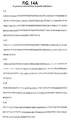
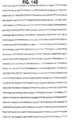
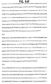
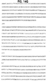
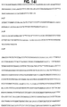
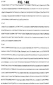
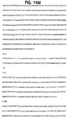
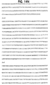
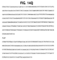
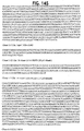
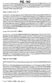
- Methylation status is determined in a sample of a patient for a CpG island selected from the group identified in the sequences shown in SEQ ID NO. 18, 22, 24, 38, 39, 46, 52 53, 54, 55, 61 and 66 (below).
- the methylation status of the CpG island is compared to that found in a control group of healthy individuals.
- the patient is identified as having an increased risk of developing cancer if methylation status of the CpG island is perturbed relative to the methylation status in the control group.
- Methylation status of a CpG island selected from the group identified in the sequences shown in SEQ ID NO. 18, 22, 24, 38, 39, 46, 52 53, 54, 55, 61 and 66 is determined in a sample of a tissue of a patient suspected of being neoplastic.
- the methylation status of the CpG island is compared to that found in a control sample of said tissue which is apparently normal.
- the patient is identified as having an increased risk of developing cancer if methylation status of the CpG island is perturbed relative to the methylation status in the control sample.
- an isolated and purified methylated CpG island is provided which is selected from the sequences shown in SEQ ID NO. 18, 22, 24, 38, 39, 46, 52 53, 54, 55, 61 and 66.
- the present invention thus provides the art with tools and methods for accessing imprinted genes and using them for detecting birth defects, diabetes, and cancers associated with aberrant imprinting.
- EG cell lines represent the first in vitro model system in which genomic imprinting can be followed dynamically and the two alleles can be distinguished. This system significantly enhances the identification and characterization of trans and cis-acting elements that modify imprinting, and it also confers the advantages of extending such investigations into an in vivo setting.
- methylated CpG islands present in normal tissues (termed MCI). There have been systematic efforts to identify unique CpG islands differentially methylated in tumors (46-48) but no such successful efforts have been described for normally methylated CpG islands. While such sequences may have been suspected, this study represents their first systematic identification in normal tissues, and as such represents a first step toward defining a "methylome", i.e. the distribution of methylation patterns layered on the distribution of genes in the genome.
- MCI sequences appear to fall within distinct biological subgroups.
- the first group, MCI-R is clearly the most abundant, and comprises high copy number sequences such as the SVA element, and the intergenic and internal spacer sequences of ribosomal genes. Methylation of one of these sequences, the rDNA nontranscribed spacer, was previously found after genomic purification from a methyl-CpG binding protein column (51), and one wonders whether the large number of these sequences obscured the identification of unique MCI's.
- MCI-S and MCI-D The two largest classes of these unique methylated CpG islands, MCI-S and MCI-D, appear to have different properties, suggesting that they may serve distinct potential functional roles. Specifically, the MCI-S sequences were localized to high isochore regions near the ends of chromosomes, and the MCI-D sequences generally showed a more centromeric localization within low isochore regions.
- MCI-S which are ubiquitously methylated, even in sperm, retain their high CpG content, which also suggests that they may serve an important role. That role, however, would not appear to be gene silencing, since most of the MCI-S were within the body of transcriptionally active genes.
- the MCI-D sequences are particularly interesting for further study, because of their apparent differential methylation in the germline.
- these sequences may mark imprinted gene regions, as at least two of these sequences in the Eag I library were found within imprinted genes, namely IGF2R and HYMAI.
- imprinted genes appear to lie within low isochore regions ( PLAGL1, IGF2R, PEG1 / MEST, SNRPN, PEG3, GNAS, unpublished data), like the MCI-D sequences.
- An interesting possibility is that a subset of low isochore domains, marked with MCI-D sequences, harbor such genes.
- MCI-T tumor-specific
- MCI-T tumor-specific
- MCI-T may represent a comparatively small fraction of the total number of unique methylated CpG islands.
- MCI-T may include sequences that are variably methylated in the population, such as MCI-T/2-d10. This is an interesting idea because it suggests that the methylome might contribute to polymorphic variation in the population, which is consistent with the idea that methylation mutations may be more common in outbred populations than in laboratory strains (64).
- Imprinting as used herein is the preferential expression of a specific parental allele, maternal or paternal. Typically it is associated with the modification of a specific parental allele, such as by DNA methylation, histone acetylation, histone phosphorylation, or histone methylation. Imprinting can be assessed using any method known in the art for determining expression from a particular allele. Such techniques include without limitation pyrosequencing for high throughput assaying, MALDI-TOF mass spectrometry, allele specific oligonucleotide DNA microarray, Hot-stop PCR (Uejima et al., Nat. Genet.
- Unimprinted genes are typically expressed in an approximately equal biallelic fashion, whereas imprinted genes display preferential expression of a specific parental allele. Approximately equal biallelic expression may be as disparate as about 40 % : 60 %, preferably from about 45 %: 55 %, more preferably from about 47.5 % : 52.5 %. Expression differences greater than this, such as 30 %:70 %, 20 %:80 %, 10 %:90 %, and 5 %: 95 % are considered preferential expression of a specific parental allele.
- Methylated CpG islands which are repetitive can be used as portable sites of genetic recombination, as indications of past chromosomal rearrangements or as indications of past insertion element-created mutations.
- Most CpG dinucleotides within a methylated CpG island contain a methylated 5-position on the pyrimidine ring of cytosine.
- the methylation level within a CpG island is believed to be quite hight, with at least 75 %, 80 %, 90 %, 95 %, or even 98% of the cytosine residues being methylated.
- Methylated CpG islands which are differentially methylated among maternal-derived and paternal-derived tissues (MCI-D) can be used as markers of the locations of imprinted genes.
- MCI-D are located within imprinted genes are adjacent to imprinted genes. Adjacency is within 2 x 10 6 base pairs, preferably within 1 x 10 6 base pairs, more preferably within 0.5 x 10 6 base pairs.
- MCI-S and MCI-T methylated CpG islands which are expressed similarly in uniparental tissues and those which are differentially expressed in tumors and normal tissues, can be used as methylation polymorphism markers in the population. Thus they can be used as sequence polymorphisms, forensically, diagnostically, and predictively as risk factors for disease traits.
- Embryonic germ cells are useful as a dynamic model system for studying imprinting.
- the ability to induce imprinting permits the analysis of factors which stimulate or inhibit the process.
- the factors can be endogenous or exogenously applied.
- parental animals which are of the same species yet which are sufficiently genetically divergent such that at least 50% of genes in resulting offspring have at least one sequence difference between alleles of said genes. More preferably at least 60 %, 70 %, 75 %, 80 %, 90 %, or 95 % of the maternal and paternal genes in the offspring will be detectably different. This greatly facilitates analysis of imprinting by rendering most genes amenable to analysis of differential allelic expression.
- Suitable mammals which can be used include without limitation mice, rats, hamsters, guinea pigs, rabbits, goats, cows, sheep, pigs, horses, dogs, and cats.
- Embryos are desirably removed from the pregnant female mammal at a stage of embryonic development between when 2-3 somites become visualizable and when gonads are recognizable. In mice, this stage is between day 7 and 10 post conception. Obtaining embryos at such an early stage is believed to be beneficial in obtaining cells which have many genes which are not yet imprinted. Embryos are dissected and cultured, preferably on feeder cell layers. The posterior third of the emybryo can be dissected and used to form dissociated cells. Alternatively, the genital ridge of the embryo is dissected out and used to form dissociated cells. Still another alternative method dissects out gonads of the embryo to form dissociated cells.
- the cell lines express one or more imprintable genes in an approximately equal biparental fashion, form cells which express one or more imprintable genes in an approximately equal biparental manner, and differentiate to form cells which express said one or more imprintable genes in a preferentially uniparental fashion.
- the assays for imprinting can be done in vitro or in vivo as is desired by the practicioner.
- the mammalian embryonic germ cells are grown in suspension culture under conditions in which the embryonic germ cells differentiate.
- the differentiated cells may or may not form an embryoid body.
- Upon differentiation expression of one or more imprintable genes changes from approximately equal biallelic to preferentially uniparental.
- Differentiation can be induced by growth on plastic in the absence of feeder cells, by growth in the presence of dimethylsulfoxide, by growth in the presence ofretinoic acid, by growth on a methyl-cellulose containing medium, or any other method known in the art.
- the germ cells contain a selectable marker under transcriptional control of a tissue-specific promoter, and the germ cells are subjected to selection conditions to select for germ cells which have differentiated into a lineage which activates the tissue-specific promoter.
- the mammalian embryonic germ cells can be injected into a nude mouse in which it will form a teratocarcinoma.
- One or more imprintable genes change from approximately equal biallelic to preferentially uniparental expression upon formation of the teratocarcinoma.
- Another way to achieve imprinting in an in vivo model is to inject a mammalian embryonic germ cell into a blastocyst of a mammal. The blastocyst is then implanted into a pseudopregnant mammal so that the blastocyst develops into a chimeric mammal, i.e.
- somatic cells are not genetically identical. Expression of one or more imprintable genes in somatic cells derived from the embryonic germ cell becomes preferentially uniparental.
- the germ cells used for formation of teratocarcinomas or chimeric blastocysts can optionally be transfected with a vector which expresses a detectable marker protein. This makes distinguishing among the cells of the mammal a simpler exercise.
- Imprinting can be assayed directly in any of the models of the invention by detecting parental allele specific expression.
- a surrogate for such expression can be used such as cytosine methylation, histone acetylation, histone phosphorylation, histone methylation. Methods for detecting such modifications are known in the art.
- Test substances used to contact with the cell lines or chimeric mammals of the present invention can be any natural, synthetic, or semisynthetic substance, whether a pure compound or a mixture of compounds.
- the test substances can be compounds or drugs which are known to have one mor more biological effects, or substances which are not known to have any biological or physiological effects. If the test animal contains a teratoma, one can identify a test substance as a candidate drug if it inhibits the growth of the teratoma or causes regression of the teratoma. Techniques for assessing the growth of a teratoma or regression of a teratoma are well known in the art.
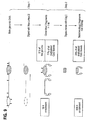
- Methylated CpG islands can be isolated using a scheme as outlined in Figure 9. Any restriction endonucleases can be used which have the desired properties specified. The properties are based on the frequency of cleavage sites, and the preference of the cleavage sites for being in G/C or A/T rich regions.
- the CpG islands can be isolated from genomic DNA from males or females, from tumor or normal cells. Any type of tumor or normal tissue can be used as a source of cells. Once such methylated CpG islands are isolated, they can be used for a number of different techniques. In one, they are tested to identify sequences which are differentially methylated between maternal and paternal chromosomes.
- CpG islands can be used to identify an imprinted gene adjacent to the methylated CpG island, as methylated CpG islands are markers for such genes. If a CpG island is found to map to the same region as a disease which is preferentially transmitted by one parent, an imprinted gene in the region can be identified as a candidate gene involved in transmitting the disease.
- the CpG islands can be used to screen populations of individuals for methylation.
- a sequence which is differentially methylated between individuals is a methylation polymorphism which can be used to identify individuals.
- each library of fragements will contain at least 25, at least 50 , or at least 75 distinct members.
- CpG islands which have been found using the method of the present invention are disclosed in Table 2. These particular CpG islands can be used to assess risk of developing cancer. Perturbed methylation of CpG islands relative to sequences in a control group of healthy individuals suggests that the individual being tested are at increased risk of developing cancer. Any number of CpG islands can be tested in such a method, but preferably at least 2, 5, 10, or 15 such islands will be tested. An increased risk of developing cancer is determined if at least 1 of 2, 3 of 5, 6 of 10, or 8 of 15 of the CpG islands have perturbed methylation status relative to control group. Similarly aberrant methylation of CpG islands can be determined where the methylation in a suspect tissue sample of a patient is compared to the methylation in an ostensibly healthy tissue sample of the patient.
- CpG islands can be used to identify genes which are within about 2 million base pairs of a CpG island identified in Table 2 in the human genome.
- the genes are preferably within I million base pairs, and more preferably within 500,000 base pairs. If the gene is preferrentially uniparentally expressed, then it is identified as an imprinted gene.
- mice We used 129/SvEv mice as the mothers in the cross
- CAST/Ei Mus musculus castaneus mice, separated from 129/SvEv by 5 million years in evolution, as the father in the cross, providing an average of one polymorphic marker per 400 bp of transcribed sequence.
- the experimental strategy is summarized in Figure 1, and it allows differentiation in vitro by a variety of mechanisms, including targeted differentiation using a selectable construct, and differentiation in vivo using chimeric mice.
- EG cell lines were derived from primordial germ cells (PGCs) of 8.5 day embryos ( 4 ), as determined by colony morphology and positive alkaline phosphatase staining (Fig. 2A,B), and four of these lines were characterized in detail (termed SJEG-1, 2, 7, and 15). These EG cell lines formed embryoid bodies after in vitro differentiation (Fig. 2C,D), teratocarcinomas in nude mice (Fig. 2E, F), and generated chimeric mice when injected into the blastocyst of C57BL/6 mice ( 5 ). One male line was also used for subsequent germline transmission ( 5 ). Most of the imprinting studies were done on lines SJEG-1, 2, and 7.
- 129/SvEv sequence was identical to known Mus musculus musculus sequence in GenBank, except that Kv1qt1 sequence was unavailable and done here. 2 From first nucleotide of cDNA 3 Single strand conformation polymorphism ( 27 ). 4 Quantitative sequencing ( 28 ). 5 Single nucleotide primer extension ( 29 ).
- Kvlqt1 shows preferential expression of the maternal allele throughout development in this strain background ( 6 ).
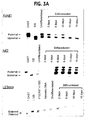
- Kvlqt1 Prior to somatic differentiation of EG cells in vitro , Kvlqt1 showed approximately equal expression of the two alleles (Fig. 3A).
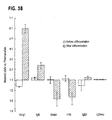
- Kvlqt1 After differentiation by replating on plastic in the absence of a feeder cell layer, Kvlqt1 showed clear preferential expression of the maternal allele, which increased to a 6:1 ratio by day 16 (Fig. 3A), and this result was seen in all three cell lines tested (Fig. 3B).
- Igf2 Like Kvlqt1, Igf2 showed approximately equal biallelic expression of the two parental alleles prior to differentiation (Fig. 3A).
- Igf2 showed allele-specific expression but in opposite direction to the F1 offspring.
- differentiated EG cells showed preferential expression of the maternal allele of Igf2 (Fig. 3A). While this was a surprising observation, it was consistent among different cell lines (Fig. 3B).
- the expression of the maternal allele of IGF2 is also consistent with an observation of allele reversal in embryonic stem (ES) cells ( 7 ). This may be a property of pluripotent embryonic stem cells (although note that in contrast to EG cells, imprinting shows little or no change in ES cells ( 7 )).
- H19 normally shows reciprocal allele-specific expression to IGF2, perhaps due to competition for a shared enhancer ( 8 ). Consistent with this pattern, H19 exhibited approximately equal expression of the two parental alleles before differentiation, and preferential expression of the paternal allele after differentiation, changing from a ratio of 1:1 to 3:1 after differentiation (Fig. 3B). Snrpn, which is preferentially expressed from the paternal allele in somatic cells ( 9 ), also showed equal biallelic expression in undifferentiated EG cells (Fig. 3B). After differentiation, Snrpn showed preferential expression of the normally expressed paternal allele, at a ratio of 3:1 (Fig. 3B). In contrast, Igf2r showed approximately equal biallelic expression both before and after differentiation, suggesting that for this gene, the gametic mark had been completely erased in EG cells (Fig. 3B).
- L23mrp As a negative control, we analyzed the nonimprinted gene L23mrp, which is just outside of a contiguous imprinted gene domain that includes Igf2, H19, and Kvlqt1 ( 10 ). In contrast to Igf2, H19, and Kvlqt1, L23mrp showed equal biallelic expression of the two parental alleles both before and after in vitro differentiation (Fig. 3A,B). Furthermore, the ratio of allele-specific expression of the imprinted genes after differentiation differed significantly from that of L23mrp (p ⁇ 0.01, two-tailed t-test). In summary, in vitro differentiation partially restored imprinting to EG cells.
- Embryoid bodies that result from in vitro differentiation of EG cells show considerable cellular heterogeneity, and not all of the cells are differentiated.
- Clones of transfected EG cells remained undifferentiated, and showed equal biallelic expression of Kvlqt1, Igf2, H19, Snrpn, Igf2r and L23mrp (Fig. 4B and data not shown).
- EG-derived GFP(+) cells were purified by fluorescence-activated cell sorting (FACS) to 99% homogeneity (Fig. 5B). Purity of EG-derived cells isolated from the chimeric mice was confirmed by measuring the allele ratio in genomic DNA for polymorphisms that distinguish the two strains (data not shown).
- H19 also showed monoallelic expression of the paternal allele, the same allele preferentially expressed after in vitro differentiation (data not shown).
- Snrpn exhibited predominant expression of the paternal allele (4:1 ratio) after in vivo differentiation.
- L23mrp showed equal biallelic expression after in vivo differentiation (data not shown).
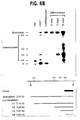
- H19 acquired a typical half-methylated pattern, similar to that seen in the parental and F1 mice, with a 1:1 ratio of unmethylated to methylated bands (Fig. 6A). This change in methylation reflected well the change in expression from approximately biallelic to predominantly monoallelic in these cells after differentiation.
- Fig. 6A we analyzed the allele composition of methylated H19 DMR using a previously described method ( 13 ).
- Our analysis of differentiated EG cells revealed that the half-methylation pattern described above (Fig. 6A) was due to methylation of the non-expressed allele (data not shown).
- the methylation was allele-specific and related to silencing of the H19 gene during differentiation.
- Igf2 DMR2 within exon 6, is known to be the more closely linked DMR to Igf2 imprinting ( 15 ).
- Igf2 acquired a half-methylated pattern, with a 1:1 ratio of methylated to unmethylated bands (Fig.
- DNA methylation reflected the pattern of gene expression of both Igf2 and H19, with a nonimprinted pattern of DNA methylation before differentiation, and an imprinted pattern after differentiation.
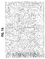
- genomic imprinting in a differentiated monolayer culture of lineage-restricted cell types ( 20 ) (Fig. 7A), derived from a human EG culture reported previously ( 17 ).
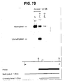
- IGF2 was examined using an Apa I polymorphism in exon 9 ( 21 ). While Apa I digestion revealed two alleles in genomic DNA, analysis of cDNA showed a nearly complete monoallelic expression pattern (Fig. 7B), indicating a nearly complete establishment of imprinting of IGF2 gene after in vitro differentiation of a human EG culture.
- H19 was then examined using an Alu I polymorphism in exon 5 ( 22 ). While Alu I digestion revealed two alleles in genomic DNA, analysis of cDNA showed a complete monoallelic expression pattern (Fig. 7C), indicating complete establishment of imprinting of H19 after in vitro differentiation of human EG culture.
- the first step involves digestion with Mse I and Hpa II, followed by gel purification of fragments ⁇ 1 kb in length. This step was predicted to enrich approximately 10-fold for CpG islands (enrichment was confirmed by a Southern blot, data not shown), while eliminating all unmethylated CpG islands because of the methylcytosine sensitivity of Hpa II.
- This "Mse I library” was cloned into the restriction-negative strain XL2-Blue MRF' to avoid bacterial digestion of methylated genomic DNA. CpG islands were further selected by digesting Mse I library DNA with Eag I and subcloning, providing a total expected 800-fold enrichment for CpG islands in this "Eag I” library (see Fig. 9 brief description for details).
- DNA from a male Wilms' tumor sample was isolated as described (52). 200 ⁇ g of DNA were digested overnight with 1000 units of Hpa II (LTI) followed by a five hour digest with 600 units of Mse I (NEB), according to the manufacturer's conditions, and the volume was reduced using a SpeedVac concentrator (Savant). In order to select for fragments ⁇ 1 kb, the digest was passed through a size selection CHROMA-SPIN+TE-400 column (Clontech). Fragments between 1-9 kb were purified from a 0.8% gel by electroelution and passed through an Elutip-D column (S&S).
- the eluate was ethanol precipitated, cloned into the compatible Nde I site of pGEM-4Z, which was first modified to abolish the Sma I site, transformed into the competent cells of the restriction-deficient strain XL2-Blue MRF' (Stratagene), and plated onto LB-Ampicillin agar plates.
- Library DNA was prepared directly from plates using a plasmid Maxi kit (Qiagen).
- Eag I libraries 100 ⁇ g of the Mse I library DNA were digested with 1,000 u of Eag I (NEB) according to the manufacturer's conditions. The digest was ethanol precipitated, and 100 to 1500 bp fragments were size-selected by purification from a 1.5% agarose gel, cloned into the Eag I site of pBC (Stratagene), and transformed into XL1-Blue MRF' (Stratagene). DNA from individual colonies was prepared using a Perfect Prep kit (Eppendorf).
- MCI-R sequences M ethylated C pG I sland- R epetitive, see results
- 3.5 ⁇ g of the Mse I library was purified, and half was digested with Acc I and half with Tth III1, pooled and digested with Dra III, Sal I, and Asc I, then re-transformed into XL2-Blue MRF'.
- This step eliminated >90% of the MCI-R sequences, while retaining approximately 30% of the MCI-S and MCI-D sequences (MCI- s ame in uniparental tissues, MCI- d ifferent in uniparental tissues, respectively, see results).
- Eag I libraries were prepared as described above, after gel purification from three overlapping fractions, 100-700 bp, 400-1000 bp, 700-1500 bp, termed ES-1,2, and 3, respectively.
- DNA Sequencing was performed using an ABI 377 automated sequencer following protocols recommended by the manufacturer (Perkin-Elmer). The sequences were analyzed by a BLAST search (53) of the NR, dbEST, dbGSS, dbHTGS, and dbSTS databases, and by GRAIL analysis. Chromosomal localization was performed by electronic PCR (ePCR, NCBI), or in some cases without matches using the GeneBridge 4 radiation hybrids panel (Research Genetics).
- Genomic DNA was digested with Mse I alone or Mse I together with a methylcytosine-sensitive (Hpa II, LTI, or Sma I, NEB) or methyl-insensitive (Msp I or Xma I, NEB) restriction endonuclease according to the manufacturer's conditions. Southern hybridization was performed as described (54).
- SVA-U a probe unique to the SVA and present in all of the SVA elements, to analyze copy number and methylation of this sequence in genomic DNA.
- the copy number was estimated to be 5000 per haploid genome (data not shown, L.S.-A. and A.P.F., in preparation).
- the SVA elements were found to be completely methylated in all adult somatic tissues examined, including peripheral blood lymphocytes, kidney, adrenal, liver and lung, as well as fetal tissues including kidney, limb, and lung (Fig. 10). However, in germinal tissues SVA elements were hypomethylated but not completely unmethylated.
- This methylation pattern was consistent with a retroposon methylation pattern, where a group of active elements is unmethylated in the germ line and maintains a high GC content, whereas in somatic tissues the element is methylated and silenced.
- a somewhat less abundant high copy repeat, representing an additional 20% of the Eag I library corresponded to the nontranscribed intergenic spacer of ribosomal DNA, which was a known methylated repetitive sequence (58).
- a third high copy methylated sequence was the ribosomal DNA internal transcribed spacer and the 28S gene, comprising an estimated 5% of the Eag I library , suggesting that ribosomal gene methylation may be more extensive than was previously suspected.
- CpG islands were then characterized by detailed genomic analysis, including radiation hybrid mapping of clones not within the known database, and analysis of methylation in somatic and germline tissues and in ovarian teratomas (OT) and complete hydatidiform moles (CHM), which are of uniparental maternal and paternal origin, respectively.
- OT ovarian teratomas
- CHM complete hydatidiform moles
- sequences recovered in this manner were predicted to be methylated, we confirmed this assumption by direct examination of genomic DNA. Furthermore, as the original source of material was a Wilms tumor DNA sample, we had no a priori knowledge about the methylation of these sequences in normal tissue. Surprisingly, most were methylated normally. More specifically, this analysis revealed that all of the sequences represented methylated CpG islands, and they could be divided into 3 major groups. The largest group consisted of sequences methylated in all tissues examined, including fetal and adult somatic tissues, ovarian teratomas (OT), complete hydatidiform moles (CHM), and sperm.
- OT ovarian teratomas
- CHM complete hydatidiform moles
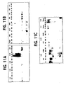
- clone 1-41 showed in blood an identical pattern after Mse I + Hpa II digestion, as after Mse I digestion alone, compared to Mse I + Msp I digestion which cut regardless of methylation (Fig. 11A). This was true for other somatic tissues, as well as for ovarian teratoma, hydatidiform mole, and sperm (Fig. 11B,C). Altogether, half of the unique methylated CpG islands fell within this category, which we term MCI-S ( M ethylated C pG I sland- S imilar in uniparental tissues).
- the second largest group approximately 30% of the unique clones, were methylated in normal somatic tissues, and unmethylated in complete hydatidiform mole (CHM), which are uniparentally derived from the male germline, as well as in sperm.
- CHM complete hydatidiform mole
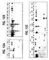
- clone 2-78 showed an identical pattern after Mse I + Hpa II digestion, as after Mse I digestion alone, in blood and other somatic tissues (Fig. 12A,B).
- clone 2-78 showed complete digestion after Hpa II treatment of sperm and hydatidiform mole DNA, similar to the pattern seen after Msp I digestion (Fig. 12C).
- MCI-D M ethylated C pG I sland- D ifferent in uniparental tissues. All of the MCI-D sequences were methylated in OT and not CHM.
- clone 2-d10 showed an identical methylation pattern in blood DNA after Mse I + Hpa II digestion as was seen after Mse I + Msp I digestion.
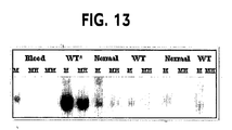
- Wilms tumor DNA from which the Mse I library had been constructed, was fully methylated (Fig. 13). Consistent with our nomenclature, this category is termed MCI-T ( M ethylated C pG I sland- T umors).
- MCI-T sequences were identified by virtue of their being methylated in tumor tissue, they may represent sequences of polymorphic methylation in the population, as a second individual showed methylation of 2-d10 in both tumor and normal tissues and a third showed methylation in neither tumor nor normal tissues (Fig. 13).
- MCI-D and MCI-S sequences were localized within or near the coding sequence of known genes or of anonymous ESTs within the GenBank database. These genes serve a wide variety of functions, including the wolframin gene, a transmembrane protein involved in congenital diabetes; sulphamidase, a lysosomal enzyme involved in Sanfilippo syndrome (MPS-IIIA); a cDNA similar to the gene for the extracellular matrix protein tenascin; and an EST adjacent to the Peutz-Jeghers syndrome gene STK11 (Table 2).
- VNTR variable number tandem repeat
- MCI-S sequences were found to include the start site of transcription.
- MCI-S/1-19 was within the last exon of the wolframin gene
- MCI-S/2-h1 was within the 5-6 exons of the sulphamidase gene.
- some of the MCI-D sequences may lie within or near imprinted genes, consistent with their differential methylation in uniparental tissues.
- the IGF2R gene which contains an Eag I site, was identified in the Eag I library (data not shown), consistent with the observation that one allele is methylated in normal cells.
- MSI-D/3-d4 which like other MSI-D sequences was methylated differentially in ovarian teratomas and hydatidiform moles, differed from most other MSI-D sequences in that it was only partially methylated in somatic tissues.
- this sequence was found to lie within the promoter and first exon of the HYMAI gene, which has recently also been demonstrated to be imprinted (60).
- a subset of MCI-D sequences may mark the location of imprinted genes.
- DMEM with high glucose (4.5 g/liter) supplemented with 15% FBS (performance tested), non-essential amino acid (0.01 mM), L-glutamine (2 mM), Pen-Strep, and 2-mercaptoethanol (0.1 mM).
- STO cells are used as feeder layers for EG derivation and long term culture. Normally STO culture is maintained in 10 cm dish in STO media. Culture must be split before reaching 85% confluence. Irradiation resistance of the maintained culture needs to be tested after a certain period of time. Should cells surviving irradiation found, throw away the culture and thaw a new vial of cells.
- Natural mating is setup for 129/SvEv female and mus. Castanious male. Male must be older than 7 weeks and female must be between 8-18 weeks.
- leukocyte alkaline phosphatase kit catalog No. 85L-3R
- EG cultures are trypsinized (0.25% trypsin/EDTA) and single cell suspension is created. Plate cells into 10 cm tissue culture dish at 37°C for 1.5 hr to allow feeder layer cells attach the bottom. Replate the media into another plate for an additional 1.5 hr. Then collect media and pellet cells.
- Protocol III (RA induced differentiation as aggregates)
- Protocol IV (Differentiation in methylcellulaose medium)
- Preparation of 2.2% aqueous methylcellulaose Add 11g of methylcellulaose power into bottle and add water to 500 ml. Stir the solution in cold room overnight. Put bottle in microwave and boil the solution three times (be careful not to spill the content). Tighten the cap right after the last boiling and leave the bottle in cold room overnight. Store in refregirator.
- Protocol V (DMSO induced differentiation as single cell culture)
- Protocol VI (RA induced differentiation as single cell culture)
Landscapes
- Life Sciences & Earth Sciences (AREA)
- Chemical & Material Sciences (AREA)
- Health & Medical Sciences (AREA)
- Organic Chemistry (AREA)
- Engineering & Computer Science (AREA)
- Zoology (AREA)
- Wood Science & Technology (AREA)
- Proteomics, Peptides & Aminoacids (AREA)
- Genetics & Genomics (AREA)
- Biotechnology (AREA)
- Bioinformatics & Cheminformatics (AREA)
- General Health & Medical Sciences (AREA)
- Biochemistry (AREA)
- Biomedical Technology (AREA)
- General Engineering & Computer Science (AREA)
- Microbiology (AREA)
- Analytical Chemistry (AREA)
- Immunology (AREA)
- Molecular Biology (AREA)
- Pathology (AREA)
- Biophysics (AREA)
- Developmental Biology & Embryology (AREA)
- Physics & Mathematics (AREA)
- Hospice & Palliative Care (AREA)
- Oncology (AREA)
- Cell Biology (AREA)
- General Chemical & Material Sciences (AREA)
- Chemical Kinetics & Catalysis (AREA)
- Medicinal Chemistry (AREA)
- Nuclear Medicine, Radiotherapy & Molecular Imaging (AREA)
- Pharmacology & Pharmacy (AREA)
- Animal Behavior & Ethology (AREA)
- Public Health (AREA)
- Veterinary Medicine (AREA)
- Measuring Or Testing Involving Enzymes Or Micro-Organisms (AREA)
- Micro-Organisms Or Cultivation Processes Thereof (AREA)
- Eye Examination Apparatus (AREA)
- Nitrogen And Oxygen Or Sulfur-Condensed Heterocyclic Ring Systems (AREA)
Claims (25)
- Verfahren zum Isolieren von methylierten CpG-Inseln, umfassend:a) Verdauen von eukaryotischer genomisher DNA mit einer ersten Restriktionsendonuclease, welche eine Erkennungssequenz erkennt, die in A/T-reichen Bereichen der DNA oder in CpG-Insel-armen Bereichen der DNA gefunden wird;b) Verdauen der eukaryotischen genomischen DNA mit einer zweiten Restriktionsendonuclease, welche eine 4-Basen-Paar-Sequenz in unmethylierten C/G-reichen Bereichen erkennt;c) Isolieren von Fragmenten mit einer Größe von wenigstens 1 kb, gebildet durch den Schritt der Verdauung, und Einfügen der Fragmente in bakterielle Vektoren;d) Transformieren von nichtmethylierenden, nichtschneidenden Bakterien mit den bakteriellen Vektoren, um die Vektoren zu propagieren und um die Nachkommenschaft der Fragmente nicht zu methylieren;e) Verdauen der nichtmethylierten Fragmente mit einer dritten Restriktionsendonuclease, welche eine Sequenz mit einer Länge von wenigstens 6 Basen-Paaren in C/G-reichen Bereichen erkennt;f) Isolieren der resultierenden Fragmente und Einfügen der Fragmente in bakterielle Vektoren, um eine Bibliothek aus Sequenzen zu bilden, welche hinsichtlich der Sequenzen angereichert sind, die von methylierten CpG-Inseln in dem eukaryotischen Genom abstammen.
- Verfahren gemäß Anspruch 1, weiterhin umfassend den Schritt des Eliminierens von nichtgewünschten repetitiven Elementen durch Verdauen der resultierenden Fragmente, auf die in Schritt (f) Bezug genommen wird, mit einer vierten Restriktionsendonuclease, welche einen einzigartigen Ort in den repetitiven Elementen erkennt.
- Verfahren gemäß Anspruch 1, wobei die erste Restriktionsendonuclease Mse I ist.
- Verfahren gemäß Anspruch 1, wobei die zweite Restriktionsendonuclease Hpa II ist.
- Verfahren gemäß Anspruch 1, wobei die dritte Restriktionsendonuclease Eag I ist.
- Verfahren gemäß Anspruch 1, wobei die vierte Restriktionsendonuclease einen Ort in dem SVA Element erkennt.
- verfahren gemäß Anspruch 1, wobei die eukaryotische genomische DNA aus einem männlichen Individuum isoliert wird.
- Verfahren gemäß Anspruch 1, wobei die eukaryotische genomische DNA aus einem Tumor isoliert wird.
- Verfahren gemäß Anspruch 1, wobei die eukaryotische genomische DNA aus einem Wilms-Tumor isoliert wird.
- Verfahren gemäß Anspruch 1, weiterhin umfassend den Schritt:Testen von einem oder mehreren Mitgliedern der Sequenzbibliothek, welche hinsichtlich Sequenzen angereichert sind, die von methylierten CpG-Inseln abgeleitet sind, um Sequenzen zu identifizieren, welche zwischen maternalen und paternalen Chromosomen differenziell methyliert sind.
- Verfahren gemäß Anspruch 1, weiterhin umfassend den Schritt:Testen von einem oder mehreren Mitgliedern der Sequenzbibliothek, welche hinsichtlich Sequenzen angereichert sind, die von methylierten CpG-Inseln abgeleitet sind, um Sequenzen zu identifizieren, welche zwischen Blasenmolen und Teratomas differenziell methyliert sind.
- Verfahren gemäß Anspruch 1, weiterhin umfassend den Schritt:Kartieren von einem oder mehreren Mitgliedern der Sequenzbibliothek auf einen genamischen Bereich, wobei die Anordnung einer methylierten CpG-Insel bestimmt wird.
- Verfahren gemäß Anspruch 1, weiterhin umfassend den Schritt:Testen einer Population von Individuen hinsichtlich der Methylierung eines Mitglieds der Sequenzbibliothek, wobei eine Sequenz, welche differenziell unter den Individuen methyliert ist, ein Methylierungspolymorphismus ist, welcher verwendet werden kann, um Individuen zu identifizieren.
- Fragmentbibliothek isoliert nach dem Verfahren gemäß Anspruch 1, wobei die Fragmente wenigstens 100-fach bezüglich der methylierten CpG-Inseln relativ zu der gesamtgenomischen DNA angereichert sind.
- Fragmentbibliothek gemäß Anspruch 14, welche wenigstens 50 einzelne Mitglieder umfasst.
- In-vitro-Verfahren zum Zur-Verfügung-stellen von einer Herangehensweise zur Bestimmung des Risikos, Krebs zu entwickeln, umfassend die folgenden Schritte:Bestimmen des Methylierungsstatus einer CpG-Insel ausgewählt aus der Gruppe, die in den Sequenzen identifiziert ist, die in Seq. ID Nr. 18, 22, 24, 25, 27, 29, 36, 38, 39, 42, 46, 47, 49, 52, 53, 54, 55, 56, 57, 59, 61 und 66 gezeigt sind, in einer Probe eines Patienten;Vergleichen des Methylierungsstatus der CpG-Insel mit einem, der in einer Kontrollgruppe aus gesunden Individuen gefunden wird;Identifizieren des Patienten als ein erhöhtes Risiko zur Entwicklung von Krebs habend, wenn der Methylierungsstatus der CpG-Insel relativ zu dem Methylierungsstatus der Kontrollgruppe verändert ist.
- In-vitro-Verfahren zum Zur-Verfugung-stellen von diagnostischen Informationen relativ zu Krebs, umfassend die folgenden Schritte:Bestimmen des Mathylierungsstatus einer CpG-Insel ausgewählt aus der Gruppe, identifiziert in den Sequenzen, die in Seq. ID Nr. 18, 22, 24, 25, 27, 29, 36, 38, 39, 42, 46, 47, 49, 52, 53, 54, 55, 56, 57, 59, 61 und 66 gezeigt sind, in einer Gewebeprobe des Patienten, von dem vermutet wird, dass er neoplastisch ist;Vergleichen des Methylierungsstatus der cpG-Insel mit demjenigen, der in einer Kontrollprobe des Gewebes gefunden wird, das anscheinend normal ist;Identifizieren des Patienten als Krebs habend, wenn der Methylierungsstatus der CpG-Insel relativ zu dem Methylierungsstatus der Kontrollprobe verändert ist.
- Verfahren gemäß Anspruch 16, wobei der Status von wenigstens 5 CpG-Inseln bestimmt wird, und der Patient als ein erhöhtes Risiko habend identifiziert wird, wenn wenigstens 3 der CpG-Inseln einen veränderten Methylierungsstatus verglichen mit der Kontrollprobe haben.
- Isolierte und aufgereinigte methylierte CpG-Insel, welche ausgewählt ist aus den Sequenzen, die in Seq. ID Nr. 18, 22, 24, 25, 27, 29, 36, 38, 39, 42, 46, 47, 49, 52, 53, 54, 55, 56, 57, 59, 61 und 66 gezeigt sind.
- CpG-Insel gemäß Anspruch 19, welche ihr Methylierungsmuster, das in einem Menschen gefunden wird, beibehält.
- CpG-Insel gemäß Anspruch 20, wobei das Methylierungsmuster, das in einem Menschen gefunden wird, in normalen Individuen methyliert ist, aber nicht in erkrankten oder der Erkrankung zugängigen Individuen.
- CpG-Insel gemäß Anspruch 20, wobei das Methylierungsmuster, das in einem Menschen gefunden wird, in normalen Individuen unmethyliert ist, aber in erkrankten und der Erkrankung zugängigen Individuen methyliert ist.
- CpG-Insel gemäß Anspruch 20, wobei das Methylierungsmuster, das in einem Menschen gefunden wird, in normalen Geweben methyliert ist, aber nicht in Erkrankten oder erkrankten Geweben.
- CpG-Insel gemäß Anspruch 20, wobei das Methylierungsmuster, das in Menschen gefunden wird, in normalen Geweben unmethyliert ist, aber erkrankten Geweben methyliert ist.
- CpG-Insel gemäß Anspruch 19, welche das Methylierungsmuster, das in Menschen gefunden wird, verloren hat.
Applications Claiming Priority (5)
| Application Number | Priority Date | Filing Date | Title |
|---|---|---|---|
| US20615800P | 2000-05-22 | 2000-05-22 | |
| US20616100P | 2000-05-22 | 2000-05-22 | |
| US206158P | 2000-05-22 | ||
| US206161P | 2000-05-22 | ||
| PCT/US2001/016253 WO2001090313A2 (en) | 2000-05-22 | 2001-05-22 | METHODS FOR ASSAYING GENE IMPRINTING AND METHYLATED CpG ISLANDS |
Publications (2)
| Publication Number | Publication Date |
|---|---|
| EP1290139A2 EP1290139A2 (de) | 2003-03-12 |
| EP1290139B1 true EP1290139B1 (de) | 2006-03-01 |
Family
ID=26901093
Family Applications (1)
| Application Number | Title | Priority Date | Filing Date |
|---|---|---|---|
| EP01941519A Expired - Lifetime EP1290139B1 (de) | 2000-05-22 | 2001-05-22 | Testverfahren zur bestimmung von gen-imprinting und methylierte cpg inseln |
Country Status (8)
| Country | Link |
|---|---|
| US (4) | US6960434B2 (de) |
| EP (1) | EP1290139B1 (de) |
| JP (1) | JP2003534005A (de) |
| AT (1) | ATE318891T1 (de) |
| AU (2) | AU7486601A (de) |
| CA (1) | CA2410317A1 (de) |
| DE (1) | DE60117542T2 (de) |
| WO (1) | WO2001090313A2 (de) |
Cited By (1)
| Publication number | Priority date | Publication date | Assignee | Title |
|---|---|---|---|---|
| DE102025101936A1 (de) | 2025-01-21 | 2026-07-23 | Schaeffler Technologies AG & Co. KG | Verfahren und Abgassensor zum Ermitteln eines korrigierten Stickoxidgehalts in einem Abgas einer Brennkraftmaschine |
Families Citing this family (18)
| Publication number | Priority date | Publication date | Assignee | Title |
|---|---|---|---|---|
| EP1315817A2 (de) * | 2000-07-28 | 2003-06-04 | Millennium Pharmaceuticals, Inc. | 18431 und 32374, mitglieder der humanen proteinkinasefamilie und ihre verwendungen |
| AU2002353043B2 (en) * | 2001-11-30 | 2008-07-03 | The Johns Hopkins University | Methods for analyzing methylated CpG islands and GC rich regions |
| DK1342794T3 (da) * | 2002-03-05 | 2006-04-24 | Epigenomics Ag | Fremgangsmåde og anordning til at bestemme vævsspecificitet af fritflydende DNA i legemsvæsker |
| US20060024676A1 (en) * | 2003-04-14 | 2006-02-02 | Karen Uhlmann | Method of detecting epigenetic biomarkers by quantitative methyISNP analysis |
| FR2854903A1 (fr) * | 2003-05-13 | 2004-11-19 | Centre Nat Rech Scient | Utilisation des loci bage (b melanoma antigens) comme marqueurs tumoraux |
| US20080102452A1 (en) * | 2006-10-31 | 2008-05-01 | Roberts Douglas N | Control nucleic acid constructs for use in analysis of methylation status |
| WO2009059327A2 (en) * | 2007-11-02 | 2009-05-07 | University Of Southern California (Usc) | Compositions and methods comprising biomarkers of sperm quality, semen quality and fertility |
| US20090137024A1 (en) * | 2007-11-28 | 2009-05-28 | Canon U.S. Life Sciences, Inc. | Method of Separating Target DNA from Mixed DNA |
| US20110256538A1 (en) * | 2010-04-15 | 2011-10-20 | Philadelphia Health & Education Corporation D/B/A Drexel University College Of Medicine | Epigenomic dna modifications for tissue typing, early cancer detection, and disease management |
| CN103459611B (zh) | 2010-09-17 | 2016-11-02 | 哈佛大学校长及研究员协会 | 对多能干细胞的效用和安全性进行表征的功能基因组学研究 |
| WO2012078321A1 (en) | 2010-12-10 | 2012-06-14 | The Board Of Trustees Of The Leland Stanford Junior University | Phased genome sequencing |
| EP4528275A3 (de) | 2016-08-10 | 2025-05-14 | Grail, Inc. | Verfahren zur herstellung von dual-indexierten dna-bibliotheken für bisulfitumwandlungssequenzierung |
| WO2018214249A1 (zh) * | 2017-05-22 | 2018-11-29 | 立森印迹诊断技术(无锡)有限公司 | 一种印记基因分级模型及其组成的系统和应用 |
| CN110714075B (zh) * | 2018-07-13 | 2024-05-03 | 立森印迹诊断技术(无锡)有限公司 | 一种用于检测肺肿瘤良恶性程度的分级模型及其应用 |
| CN110791563B (zh) * | 2018-08-01 | 2024-02-09 | 立森印迹诊断技术(无锡)有限公司 | 一种用于检测甲状腺肿瘤良恶性程度的分级模型及其应用 |
| WO2020210754A1 (en) * | 2019-04-10 | 2020-10-15 | University Of Pittsburgh - Of The Commonwealth System Of Higher Education | Computational filtering of methylated sequence data for predictive modeling |
| CN111613271B (zh) * | 2020-04-26 | 2023-02-14 | 西南大学 | 一种预测畜禽数量性状显性遗传效应的方法及应用 |
| JP2025514547A (ja) | 2022-05-11 | 2025-05-02 | プロバンシャル ヘルス サービシズ オーソリティ | 遺伝子疾患の診断及び管理のための片親起源疾患対立遺伝子検出のための方法及び装置 |
Family Cites Families (5)
| Publication number | Priority date | Publication date | Assignee | Title |
|---|---|---|---|---|
| US6617434B1 (en) * | 1996-05-31 | 2003-09-09 | North Shore Long Island Jewish Research Institute | Identificiaton of differentially methylated and mutated nucleic acids |
| US6235474B1 (en) * | 1996-12-30 | 2001-05-22 | The Johns Hopkins University | Methods and kits for diagnosing and determination of the predisposition for diseases |
| AU7829398A (en) * | 1997-06-09 | 1998-12-30 | University Of Southern California | A cancer diagnostic method based upon dna methylation differences |
| EP0976835B1 (de) * | 1998-07-29 | 2003-07-09 | Keygene N.V. | Verfahren zur Erkennung von Nukleinsäuremethylierungen durch AFLP |
| US7700324B1 (en) * | 1998-11-03 | 2010-04-20 | The Johns Hopkins University School Of Medicine | Methylated CpG island amplification (MCA) |
-
2001
- 2001-05-22 CA CA002410317A patent/CA2410317A1/en not_active Abandoned
- 2001-05-22 EP EP01941519A patent/EP1290139B1/de not_active Expired - Lifetime
- 2001-05-22 AT AT01941519T patent/ATE318891T1/de not_active IP Right Cessation
- 2001-05-22 WO PCT/US2001/016253 patent/WO2001090313A2/en not_active Ceased
- 2001-05-22 JP JP2001587109A patent/JP2003534005A/ja active Pending
- 2001-05-22 AU AU7486601A patent/AU7486601A/xx active Pending
- 2001-05-22 US US09/861,893 patent/US6960434B2/en not_active Expired - Fee Related
- 2001-05-22 AU AU2001274866A patent/AU2001274866B2/en not_active Ceased
- 2001-05-22 DE DE60117542T patent/DE60117542T2/de not_active Expired - Lifetime
- 2001-05-22 US US10/296,263 patent/US20050153440A1/en not_active Abandoned
-
2005
- 2005-03-17 US US11/084,085 patent/US7488815B2/en not_active Expired - Fee Related
-
2009
- 2009-01-07 US US12/350,159 patent/US7985550B2/en not_active Expired - Fee Related
Cited By (1)
| Publication number | Priority date | Publication date | Assignee | Title |
|---|---|---|---|---|
| DE102025101936A1 (de) | 2025-01-21 | 2026-07-23 | Schaeffler Technologies AG & Co. KG | Verfahren und Abgassensor zum Ermitteln eines korrigierten Stickoxidgehalts in einem Abgas einer Brennkraftmaschine |
Also Published As
| Publication number | Publication date |
|---|---|
| EP1290139A2 (de) | 2003-03-12 |
| AU2001274866B2 (en) | 2006-11-09 |
| JP2003534005A (ja) | 2003-11-18 |
| WO2001090313A2 (en) | 2001-11-29 |
| US20050153440A1 (en) | 2005-07-14 |
| US7488815B2 (en) | 2009-02-10 |
| DE60117542T2 (de) | 2006-12-07 |
| AU7486601A (en) | 2001-12-03 |
| CA2410317A1 (en) | 2001-11-29 |
| WO2001090313A3 (en) | 2002-05-16 |
| US20100273658A1 (en) | 2010-10-28 |
| US7985550B2 (en) | 2011-07-26 |
| WO2001090313A8 (en) | 2003-11-13 |
| US20020045257A1 (en) | 2002-04-18 |
| US6960434B2 (en) | 2005-11-01 |
| WO2001090313A9 (en) | 2003-03-06 |
| US20050282147A1 (en) | 2005-12-22 |
| ATE318891T1 (de) | 2006-03-15 |
| DE60117542D1 (de) | 2006-04-27 |
Similar Documents
| Publication | Publication Date | Title |
|---|---|---|
| US7985550B2 (en) | Methods of isolating methylated CpG islands | |
| AU2001274866A1 (en) | Methods for assaying gene imprinting and methylated CpG islands | |
| Romanenko et al. | Segmental paleotetraploidy revealed in sterlet (Acipenser ruthenus) genome by chromosome painting | |
| Migeon et al. | Incomplete X chromosome dosage compensation in chorionic villi of human placenta. | |
| Norris et al. | Evidence that random and imprinted Xist expression is controlled by preemptive methylation | |
| O’Neill et al. | Allelic expression of IGF2 in marsupials and birds | |
| Dighe et al. | Heterozygous embryonic stem cell lines derived from nonhuman primate parthenotes | |
| Hori et al. | Aberrant CpG methylation of the imprinting control region KvDMR1 detected in assisted reproductive technology-produced calves and pathogenesis of large offspring syndrome | |
| WO2008033469A1 (en) | Methods for producing embryonic stem cells from parthenogenetic embryos | |
| Gebert et al. | DNA methylation in the IGF2 intragenic DMR is re-established in a sex-specific manner in bovine blastocysts after somatic cloning | |
| Monk et al. | Expression of imprinted genes in human preimplantation development | |
| AU2007200578B2 (en) | Methods for assaying gene imprinting and methylated CpG islands | |
| Migeon et al. | Programmed demethylation in CpG islands during human fetal development | |
| Sturm et al. | Unrestricted lineage differentiation of parthenogenetic ES cells | |
| Ratnam et al. | Ssm1b expression and function in germ cells of adult mice and in early embryos | |
| Riggs | X inactivation, DNA methylation, and differentiation revisited | |
| Obata | Study on the mechanism of maternal imprinting during oocyte growth | |
| Kimura et al. | Testicular teratomas: back to pluripotent stem cells | |
| Grybel | Epigenetic changes in preimplantation embryos from a diabetic pregnancies | |
| Smith | Disruption of epigenetic regulatory elements and chromosomal alterations in patients with Beckwith-Wiedemann syndrome | |
| Capriglione et al. | Mechanisms of differentiation in the sex chromosomes of some Lacertidae | |
| Maatouk | Regulatory mechanisms controlling primordial germ cell differentiation within the murine fetal gonad | |
| Jiang | Establishing genomic imprinting by cell differentiation: A model system using embryonic germ cells | |
| Hargan Calvopina | Mechanisms of demethylation in primordial germ cells and the importance of stage-specific demethylation in safeguarding against precocious differentiation | |
| Alhazmi | Assessing the relationship between DNA methylation and gene expression in germ cell tumours |
Legal Events
| Date | Code | Title | Description |
|---|---|---|---|
| PUAI | Public reference made under article 153(3) epc to a published international application that has entered the european phase |
Free format text: ORIGINAL CODE: 0009012 |
|
| 17P | Request for examination filed |
Effective date: 20021220 |
|
| AK | Designated contracting states |
Kind code of ref document: A2 Designated state(s): AT BE CH CY DE DK ES FI FR GB GR IE IT LI LU MC NL PT SE TR |
|
| AX | Request for extension of the european patent |
Extension state: AL LT LV MK RO SI |
|
| 17Q | First examination report despatched |
Effective date: 20030704 |
|
| GRAP | Despatch of communication of intention to grant a patent |
Free format text: ORIGINAL CODE: EPIDOSNIGR1 |
|
| GRAS | Grant fee paid |
Free format text: ORIGINAL CODE: EPIDOSNIGR3 |
|
| GRAA | (expected) grant |
Free format text: ORIGINAL CODE: 0009210 |
|
| AK | Designated contracting states |
Kind code of ref document: B1 Designated state(s): AT BE CH CY DE DK ES FI FR GB GR IE IT LI LU MC NL PT SE TR |
|
| PG25 | Lapsed in a contracting state [announced via postgrant information from national office to epo] |
Ref country code: IT Free format text: LAPSE BECAUSE OF FAILURE TO SUBMIT A TRANSLATION OF THE DESCRIPTION OR TO PAY THE FEE WITHIN THE PRESCRIBED TIME-LIMIT;WARNING: LAPSES OF ITALIAN PATENTS WITH EFFECTIVE DATE BEFORE 2007 MAY HAVE OCCURRED AT ANY TIME BEFORE 2007. THE CORRECT EFFECTIVE DATE MAY BE DIFFERENT FROM THE ONE RECORDED. Effective date: 20060301 Ref country code: FI Free format text: LAPSE BECAUSE OF FAILURE TO SUBMIT A TRANSLATION OF THE DESCRIPTION OR TO PAY THE FEE WITHIN THE PRESCRIBED TIME-LIMIT Effective date: 20060301 Ref country code: LI Free format text: LAPSE BECAUSE OF FAILURE TO SUBMIT A TRANSLATION OF THE DESCRIPTION OR TO PAY THE FEE WITHIN THE PRESCRIBED TIME-LIMIT Effective date: 20060301 Ref country code: NL Free format text: LAPSE BECAUSE OF FAILURE TO SUBMIT A TRANSLATION OF THE DESCRIPTION OR TO PAY THE FEE WITHIN THE PRESCRIBED TIME-LIMIT Effective date: 20060301 Ref country code: BE Free format text: LAPSE BECAUSE OF FAILURE TO SUBMIT A TRANSLATION OF THE DESCRIPTION OR TO PAY THE FEE WITHIN THE PRESCRIBED TIME-LIMIT Effective date: 20060301 Ref country code: AT Free format text: LAPSE BECAUSE OF FAILURE TO SUBMIT A TRANSLATION OF THE DESCRIPTION OR TO PAY THE FEE WITHIN THE PRESCRIBED TIME-LIMIT Effective date: 20060301 Ref country code: CH Free format text: LAPSE BECAUSE OF FAILURE TO SUBMIT A TRANSLATION OF THE DESCRIPTION OR TO PAY THE FEE WITHIN THE PRESCRIBED TIME-LIMIT Effective date: 20060301 |
|
| REG | Reference to a national code |
Ref country code: GB Ref legal event code: FG4D |
|
| REG | Reference to a national code |
Ref country code: CH Ref legal event code: EP |
|
| REG | Reference to a national code |
Ref country code: IE Ref legal event code: FG4D |
|
| REF | Corresponds to: |
Ref document number: 60117542 Country of ref document: DE Date of ref document: 20060427 Kind code of ref document: P |
|
| PG25 | Lapsed in a contracting state [announced via postgrant information from national office to epo] |
Ref country code: IE Free format text: LAPSE BECAUSE OF NON-PAYMENT OF DUE FEES Effective date: 20060522 |
|
| PG25 | Lapsed in a contracting state [announced via postgrant information from national office to epo] |
Ref country code: MC Free format text: LAPSE BECAUSE OF NON-PAYMENT OF DUE FEES Effective date: 20060531 |
|
| PG25 | Lapsed in a contracting state [announced via postgrant information from national office to epo] |
Ref country code: DK Free format text: LAPSE BECAUSE OF FAILURE TO SUBMIT A TRANSLATION OF THE DESCRIPTION OR TO PAY THE FEE WITHIN THE PRESCRIBED TIME-LIMIT Effective date: 20060601 Ref country code: SE Free format text: LAPSE BECAUSE OF FAILURE TO SUBMIT A TRANSLATION OF THE DESCRIPTION OR TO PAY THE FEE WITHIN THE PRESCRIBED TIME-LIMIT Effective date: 20060601 |
|
| PG25 | Lapsed in a contracting state [announced via postgrant information from national office to epo] |
Ref country code: ES Free format text: LAPSE BECAUSE OF FAILURE TO SUBMIT A TRANSLATION OF THE DESCRIPTION OR TO PAY THE FEE WITHIN THE PRESCRIBED TIME-LIMIT Effective date: 20060612 |
|
| PG25 | Lapsed in a contracting state [announced via postgrant information from national office to epo] |
Ref country code: PT Free format text: LAPSE BECAUSE OF FAILURE TO SUBMIT A TRANSLATION OF THE DESCRIPTION OR TO PAY THE FEE WITHIN THE PRESCRIBED TIME-LIMIT Effective date: 20060801 |
|
| NLV1 | Nl: lapsed or annulled due to failure to fulfill the requirements of art. 29p and 29m of the patents act | ||
| REG | Reference to a national code |
Ref country code: CH Ref legal event code: PL |
|
| ET | Fr: translation filed | ||
| PLBE | No opposition filed within time limit |
Free format text: ORIGINAL CODE: 0009261 |
|
| STAA | Information on the status of an ep patent application or granted ep patent |
Free format text: STATUS: NO OPPOSITION FILED WITHIN TIME LIMIT |
|
| 26N | No opposition filed |
Effective date: 20061204 |
|
| PG25 | Lapsed in a contracting state [announced via postgrant information from national office to epo] |
Ref country code: GR Free format text: LAPSE BECAUSE OF FAILURE TO SUBMIT A TRANSLATION OF THE DESCRIPTION OR TO PAY THE FEE WITHIN THE PRESCRIBED TIME-LIMIT Effective date: 20060602 |
|
| PG25 | Lapsed in a contracting state [announced via postgrant information from national office to epo] |
Ref country code: LU Free format text: LAPSE BECAUSE OF NON-PAYMENT OF DUE FEES Effective date: 20060522 Ref country code: TR Free format text: LAPSE BECAUSE OF FAILURE TO SUBMIT A TRANSLATION OF THE DESCRIPTION OR TO PAY THE FEE WITHIN THE PRESCRIBED TIME-LIMIT Effective date: 20060301 |
|
| PG25 | Lapsed in a contracting state [announced via postgrant information from national office to epo] |
Ref country code: CY Free format text: LAPSE BECAUSE OF FAILURE TO SUBMIT A TRANSLATION OF THE DESCRIPTION OR TO PAY THE FEE WITHIN THE PRESCRIBED TIME-LIMIT Effective date: 20060301 |
|
| PGFP | Annual fee paid to national office [announced via postgrant information from national office to epo] |
Ref country code: DE Payment date: 20120529 Year of fee payment: 12 |
|
| PGFP | Annual fee paid to national office [announced via postgrant information from national office to epo] |
Ref country code: FR Payment date: 20120607 Year of fee payment: 12 Ref country code: GB Payment date: 20120525 Year of fee payment: 12 |
|
| GBPC | Gb: european patent ceased through non-payment of renewal fee |
Effective date: 20130522 |
|
| PG25 | Lapsed in a contracting state [announced via postgrant information from national office to epo] |
Ref country code: DE Free format text: LAPSE BECAUSE OF NON-PAYMENT OF DUE FEES Effective date: 20131203 |
|
| REG | Reference to a national code |
Ref country code: DE Ref legal event code: R119 Ref document number: 60117542 Country of ref document: DE Effective date: 20131203 |
|
| REG | Reference to a national code |
Ref country code: FR Ref legal event code: ST Effective date: 20140131 |
|
| PG25 | Lapsed in a contracting state [announced via postgrant information from national office to epo] |
Ref country code: GB Free format text: LAPSE BECAUSE OF NON-PAYMENT OF DUE FEES Effective date: 20130522 |
|
| PG25 | Lapsed in a contracting state [announced via postgrant information from national office to epo] |
Ref country code: FR Free format text: LAPSE BECAUSE OF NON-PAYMENT OF DUE FEES Effective date: 20130531 |