-
The present invention relates to a method for the detection of intermolecular interactions between probe(s) and protein targets, said protein targets including at least one protein or polypeptide, which, when expressed in vivo into host cells, is toxic for said host cells or cannot be purified, or does not fold correctly. In particular, the invention results from the combination of the preparation of protein arrays with cell-free synthesized proteins and the detection of specific interactions using near-infra-red fluorescent dyes. The invention also relates to protein arrays and methods for their preparation.
-
The systematic sequencing of genomes has recently provided to the biologists, a high number of sequences of putative proteins of unknown functions. In parallel, by the use of functional genomics technologies, systematic screening for abnormal expression of sequences correlated to a specific pathology enabled to identify potential targets for new drugs, consisting of proteins which, for a part thereof, have unknown functions.
-
In order to develop and/or screen the potential drugs which could target these proteins whose abnormal expression is correlated to specific pathologies, there is a need in high-throughput methods for the detection of molecules, such as nucleic acids, proteins, small ligands and other compounds which bind to these protein targets.
-
Several methods have already been proposed to identify protein-DNA or protein-protein interactions both in vivo and in vitro. These methods are characterized in that they allow the analysis of a high number of interactions in parallel. Especially, improved versions of a two-hybrid system, originally based on a selective activation of reporter genes transcribed by RNA polymerase II (Fields & Song, 1989), are widely used in proteomic researches (Vidal & Legrain, 1999; Joung et al., 2000; Walhout et al., 2000; Uetz et al., 2000). Also, phage-display, enabling the screening of short peptides interacting with other molecules, has been used for studying protein-protein and protein-DNA interactions (Rebar and Pabo, 1994).
-
More recently, protein array technology has become a powerful approach for the identification of intermolecular interactions (Büssow et al., 1998, Emili & Cagney, 2000; MacBeath & Schreiber, 2000; Zhu et al., 2000; Holt et al., 2000; Mahlknecht et al., 2001). Several methods for preparing protein arrays have been described. For example, one method is based on the binding of proteins on a nitrocellulose membrane at defined positions and the incubation of the membrane with 32P-labelled probe to identify protein-nucleic acid, protein-protein and protein-small ligand interactions (Ge, 2000, WO 0054046). After several washings, specific interactions between probes and targets are detected by autoradiography or by Phosphorlmager.
-
In the above-mentioned methods, the proteins which are spotted as protein arrays are generally synthesized using in vivo cellular expression.
-
However, in vivo intracellular synthesis of proteins requires several steps, including insertion of the corresponding DNA into a vector molecule, its introduction into a given host, its expression and further purification of the expression product. Moreover, some genes may be toxic for host cells, or their expression product cannot be purified easily, for example, due to their hydrophobic structure, or do not fold correctly in such a way that they do not recover a natural conformation after in vivo synthesis and purification. This raises the question of providing methods for the preparation of protein arrays, which comprise a protein which cannot be synthesized in vivo, namely by intracellular expression. One solution is provided by in vitro chemical synthesis. However, the chemical synthesis is expensive and is not suitable for the synthesis of long polypeptides or proteins. This approach is thus limited for the synthesis of small peptides, generally up to 50 amino acids. Besides, this method does not provide possible maturation of proteins that normally occurs in cells.
-
Contrary to in vivo protein production, cell-free systems allow to synthesize proteins harmful for cell hosts (Zubay, 1973; Pavlov et al., 1996). Cell-free synthesis has been used as an essential component for translation-based screening in polysome display (Mattheakis et al., 1996), truncation test (Van Essen et al., 1997), scanning saturation mutagenesis (Chen et al., 1999), site-specific incorporation of unnatural amino acids into proteins (Thorson et al., 1998), stable-isotope labeling of proteins (Kigawa et al., 1999). Besides, cell-free systems have been used for identification of modified proteins by analysis of their electrophoretic migration (King et al., 1997) or for identification of DNA-binding proteins by mobility shift assay (Lustig et al., 1997). Both circular and linear exogenous DNAs can be used as templates for protein synthesis and a good protein yield can be reached from strong phage promoters in bacterial cell-free system (Kawarasaki et al., 1995; Kim et al., 1996; Kigawa et al., 1999). However, many factors affect the yield and the homogeneity of full-length synthesized proteins in a cell-free system and, therefore, its wider application is limited. Thus, considering the lower yield of protein synthesis using a cell-free protein synthesis system, in vivo cellular systems are generally preferred.
-
Indeed, the deposit of a high amount of stable proteins is considered as essential for the preparation of a protein array suitable to detect specific probe-protein interactions with a good sensitivity using the commonly used labeling methodology.
-
Till now, no protein array based on cell-free synthesized proteins has been described comprising serially produced proteins which are not producible in vivo either due to a toxicity for their host cells or for the difficulty to obtain a purified or correctly folded product.
-
Thus, there is still a need to develop new methodological approaches able to perform serial, rapid and sensitive detection of intermolecular interactions between probe(s) and protein targets, said targets including at least one protein or polypeptide which is not producible in vivo in host cells, and is toxic for said host cells or cannot be purified, or does not fold correctly.
-
The use of the term "serial detection" means that several distinct targets are tested in parallel for their interaction with at least one specific probe.
-
Preferably, more than ten targets are tested simultaneously.
-
Fluorescence methodology, as an alternative to radioactive and chemiluminescence's ones, is widely used for direct detection of labelled macromolecules and for screening of macromolecules targeted by dye-labelled probes. Recently, a detection method of nucleic acids and proteins by infrared fluorescence imaging has been developed by LI-COR Inc. Reacting macromolecules labelled with near infrared (IR) fluorescent dyes with emission at approximately 700 nm and 800 nm can be detected by means of the LI-COR Odyssey IR Imager equipped with lasers that excite at 680 and 780 nm. The infrared region has a lower background, as compared to the lower wave-length dyes used in other fluorescent detection methods, thus providing a higher signal-to-noise ratio and allowing to detect target molecules with a higher sensitivity. Validity of the technology has been shown for the detection of nucleic acids and proteins bound to membranes as well as for the detection of protein targets with IRD-labelled antibodies (Esser et al., 2000; Schutz et al., 2000).
-
The invention results from the finding that in vitro synthesized proteins can be used for the preparation of protein array. Indeed, the inventors have shown that use of arrays based on in vitro synthesized proteins, in combination with the IRD-technology, provide a highly sensitive and rapid method for detection of intermolecular interactions between probes and protein targets.
-
Especially, the invention provides a method for the preparation of a protein array comprising protein targets including at least one protein or polypeptide unproducible in vivo which, when expressed in host cells, is toxic for said host cells or cannot be purified, or does not fold correctly, said method comprising the steps of:
- a) synthesizing in parallel said protein targets using a cell-free protein synthesis method;
- b) recovering the synthesized protein targets ; and,
- c) preparing a protein array by binding each synthesized protein target to a support at a defined position.
-
As used herein, the term "protein array" refers to a support comprising a set of proteins or polypeptides bound at defined positions, each position corresponding to one specific sequence of polypeptide or protein. One or more positions can correspond to the same sequence of polypeptide or protein. A protein array can comprise between tens to thousands different polypeptides or proteins.
-
According to the invention, the protein targets refer, interchangeably, to the proteins or polypeptides which are bound to the support.
-
The one skilled in the art can choose any protein or polypeptide, for which intermolecular interaction with specific probe(s) is questioned. These targets can be for example all the proteins encoded by the genome of an organism. These targets can also be a specific family of proteins, according to their function in the cell, such as protein kinases, transcription factors, receptors and the like. These targets can also be proteins involved in diseases, and especially proteins from pathogenic organisms or proteins with abnormal expression in cancer, cardio-vascular, neuro-degenerative or genetic pathologie. These targets can also be proteins exhibiting various catalytic activities and/or enabling the binding of small ligands or cofactors or short peptides.
-
Preferably, the protein targets include proteins or polypeptides which are not obtainable using an in vivo cellular expression system and which have at least an amino acid sequence longer than 30 amino acids, preferably longer than 50 amino acids.
-
The protein targets comprise at least one protein target which, when expressed in vivo into host cells, is toxic for said host cells or cannot be purified, or does not fold correctly.
-
As used herein, the term "cannot be purified", means that conventional purification procedures cannot provide a purified protein containing less than 10% of impurities, or cannot be purified without affecting its correct folding such as membrane proteins.
-
According to the invention, a protein which does not fold correctly is a recombinant protein which has not substantially the same binding properties as a corresponding native protein purified from homologous host-organism.
-
More particularly, proteins or polypeptides that are toxic or cannot be purified or do not fold correctly comprise mis-folded wild-type or mutant oligomeric or monomeric proteins, belonging to regulatory, membrane-attached, secretory and other proteins, because of their expression in vivo, in particular, in heterologous cytoplasmic environment. In particular, in vivo multiplication of a given gene under selective pressure, said given gene encoding a protein harmful for the host cell, may generate mutations in the gene and the corresponding mutated protein which do not reflect real binding properties of the wild-type protein.
-
Thus, according to the method of the invention, the target proteins are synthesized in vitro using a cell free protein synthesis method, allowing rapid parallel synthesis of number of proteins, including those which, when expressed in vivo into host cells, are toxic for said host cells or cannot be purified, or do not fold correctly.
-
As used herein, a cell-free protein synthesis method refers to any method for cell-free synthesis of a desired protein from a ribonucleic acid (RNA) template or a deoxyribonucleic acid (DNA) template. Hence, it can refer either to in vitro transcription-translation or in vitro translation methods. These methods are opposed to in vivo protein synthesis methods involving the culture of recombinant cells expressing the desired proteins, and to chemical synthesis methods. Examples of eukaryotic in vitro translation methods are the methods involving the rabbit reticulocytes (Pelham and Jackson, 1976) or wheat germ lysate (Roberts and Paterson, 1973). The E. coli S30 extract method described by Zubay, (1973) is an example of a widely used prokaryotic in vitro transcription-translation method.
-
Lesley et al., (1991) optimised the Zubay (1973) E. coli extract for use with PCR fragments and other linear DNA templates by preparing a bacterial extract from a nuclease-deficient strain of E. coli. Also, improvement of the method has been described by Kigawa et al., (1999).
-
In a preferred embodiment of the invention, said cell-free protein synthesis method comprises the use of a cell-free extract, preferentially a bacterial cell-free extract, more preferably E. coli cell-free extract. The term cell-free extract as used herein defines any preparation comprising the components of protein synthesis machinery wherein such components are sufficient to enable translation of a messenger ribonucleic acid encoding a desired protein. Optionally, the cell-free extract comprises components which further allow transcription of the DNA encoding the desired protein.
-
When the template is a DNA, it comprises at least the sequence of an Open Reading Frame (ORF) encoding the desired protein and the sequences appropriate for transcription and translation initiation. Optionally, the DNA template may comprise short sequences encoding tags for facilitating further purification of the synthesized protein.
-
In a preferred embodiment, the DNA template also comprises a strong bacterial promoter with at least one UP element, preferentially the promoter of the argCo gene from Bacillus stearothermophilus.
-
The linear DNA template can also be a PCR-produced DNA template comprising the ORF encoding said protein or polypeptide, and an additional sequence which is in a preferred embodiment, at least 3 bp, preferably longer than 100 bp and more preferably longer than 200 bp. This elongation of PCR-produced DNA template has been shown to improve the yield and the homogeneity of in vitro protein synthesis using a bacterial cell free extract.
-
Each protein target can thus be synthesized in parallel by, first, amplifying the gene encoding the desired protein or polypeptide and second, carrying out cell-free protein synthesis using the amplification product as a DNA template.
-
It is known in the art that adding purified RNA polymerase holoenzyme may improve the yield of protein synthesis. For example, purified T7 RNA polymerase can be added to the reaction mixture when carrying out cell-free protein synthesis using a T7 phage promoter. However, it has also been shown that adding purified thermostable RNA polymerase, preferably T. thermophilus, enables similar yield, when corresponding promoter sequences are applied.
-
Thus, in a preferred embodiment of the method, wherein a bacterial cell-free extract is used, a purified thermostable polymerase, preferably from T. thermophilus, is added into the bacterial cell-free extract.
-
It has also been shown that, surprisingly, adding specifically purified α subunit of RNA polymerase in the cell-free extract in excess to the host RNA polymerase holoenzyme of the like provides better yield of protein synthesis, than with T7 RNA polymerase holoenzyme, when corresponding promoter sequences are applied.
-
In one preferred embodiment wherein a bacterial cell-free extract is used, the method of the invention is characterized in that purified α subunit of RNA polymerase is added in the cell-free extract in excess to other subunits of the like.
-
The protein targets are then recovered after cell-free synthesis. They can be purified prior to the deposition on the support or they can be partially purified or directly deposited without any purification. In particular, they can be purified using column affinity chromatography methods.
-
In order to form a protein array, the protein targets are bound to a support at defined positions. Depending on the type of the support, the target might be either tightly bound to the support or needs to be chemically linked to the support. An example of a support requiring no chemical linking is an organic membrane, preferably a nitrocellulose membrane. In other examples, a linking or binding method must be performed to ensure the binding of the polypeptides. Examples of linking methods are described in the prior Art, as well as the nature of supports appropriate for the binding of the protein targets.
-
As an example for the preparation of a protein array, protein targets ranging from 0.1 pmole to 400 pmole can be deposited on nitrocellulose membrane by vacuum without using any specific linker. One advantage of the vacuum blotting of cell-free synthesized proteins as compared with spotting approaches is that both addition and binding proteins to the support can be done on a relatively large surface, easily controlled even for diluted samples, allowing the binding of equimolar quantities of cell-free synthesized proteins to the support.
-
One advantage of the method of the invention is that protein targets can be directly deposited after their synthesis without purification.
-
Accordingly, the invention provides a protein array, characterized in that it comprises at least one non-purified protein or polypeptide not obtainable by in vivo synthesis method but obtainable with a cell-free protein synthesis method.
-
The invention also relates to a protein array, characterized in that said protein or polypeptide bound to the support, when expressed in vivo into host cells, is toxic for said host cells or cannot be purified, or does not fold correctly.
-
In a specific embodiment of the invention, the protein array is characterized in that said protein or polypeptide which is not obtainable using an in vivo cellular expression system, has an amino acid sequence longer than 30 amino acids, preferably longer than 50 amino acids.
-
In another specific embodiment, a protein array of the invention is characterized in that it is obtainable by the method for the preparation of a protein array as defined above.
-
Indeed, the present invention relates to a simple, rapid and sensitive method for the serial detection of intermolecular interactions between protein or polypeptide targets and probes.
-
The present invention encompasses also a method for the detection of intermolecular interactions between probe(s) and protein targets, said protein targets including at least one protein or polypeptide, unobtainable by in vivo synthesis and which, when expressed in vivo into host cells, is toxic for said host cells or cannot be purified, or does not fold correctly, said method combining the steps of
- a) providing protein targets serially synthesized in vitro using a cell-free protein synthesis method ;
- b) binding said protein targets to a support at defined positions, to prepare a protein array ;
- c) providing probe(s) labelled with a near-infrared fluorescent dye;
- d) contacting said protein array with the labelled probe(s) in a medium suitable to allow a specific binding;
- e) detecting on the protein array, the protein target(s) to which the labelled probe(s) specifically bind using an Infrared imager device.
-
Thus, the method allows to detect any intermolecular interactions between the protein targets and the probe. As used herein, "intermolecular interactions" or "specific binding" between a probe and a protein target refer to a physical interaction between two molecules which cannot be dissociated using usual washing conditions comprising incubation in a low stringency buffer, for example buffer A100 as described in Ge, 2000, at least 1 hour at room temperature.
-
According to the method of the invention, the protein targets are synthesized using cell-free protein synthesis systems, inducing especially bacterial cell-free extract, preferably E. coli cell-free extract, as described above. Similarly, the preparation of the protein array in step (b) is carried out as described above.
-
The term "probe" refers to a molecule which might bind with one or more polypeptides or proteins of the array in order to allow the detection of bound or reacting complexes. This probe can be any type of molecule. According to a preferred embodiment of the invention, said probe molecule is a nucleic acid, a polypeptide or a protein, bound directly or indirectly with a near-infrared fluorescent dye. Preferentially, a probe molecule is bound directly or indirectly with near infrared fluorescent dyes with emission at approximately 700 nm and 800 nm. These dyes can be detected, by means for example, of the Li-COR Odyssey Infrared Imager equipped with lasers that excite at 680 and 780 nm. Optionally, different probes labelled with different dyes can be pooled simultaneously to detect intermolecular interactions between the targets and said different probes.
-
The protein array is incubated in an incubation buffer comprising the probe(s), said incubation buffer consisting of a medium suitable to allow specific binding. After a period of time for incubation which can vary depending on the amount of targets and probes, the protein array is washed to avoid non specific interactions and very weak interactions.
-
According to the invention, the binding is considered to be specific when said binding is observed in an incubation buffer containing a non-specific competitor, i.e., a molecule which binds weakly to any protein or polypeptide. An example of an incubation buffer for the detection of protein-DNA interactions is the following: 20 mM Tris-HCI pH 7.9, 50 mM NaCI, 50 mM KCI, 0.1 mM DTT, 0.005% surfactant P20 (Biacore) and 25 µg/ml of a sonicated salmon DNA used as competitor DNA. An example of an incubation buffer for the detection of protein-protein interactions is the PBS containing 1% BSA and 0.1% Tween 20 (LI-COR, INC.) or the A100 buffer as described by Ge (2000) containing 1 % non-fat milk.
-
The probes labelled with infrared fluorescent dyes remain bound to the proteins to which they interact specifically and generate, on the protein array a signal which can be detected using a device allowing infrared detection. As an example, a device allowing infrared detection is the IRD Imager developed by LI-COR ®.
-
Cell-free protein synthesis enables to provide normalized amounts of protein target before binding to the support.
-
Thus, in another specific embodiment, the method of the invention is characterized in that it further comprises steps for normalization of the amount of synthesized protein prior to binding to a support, said normalization steps consisting of:
- a) labelling each synthesized protein, preferably by supplying a radioactive amino acid in the cell-free system,
- b) quantifying each labelled protein,
- c) providing equivalent amount of each synthesized protein for normalization prior to binding to a support.
-
Another advantage of the method of the invention is that multiple probes can be used simultaneously, the probes being labelled with different specific IRD-dyes of different wave-length. Accordingly, in one specific embodiment of the method of the invention, at least two different probes are used, said probes being labelled with IRD-dyes of different wave-lengths.
-
The present invention also relates to the use of in vitro synthesized proteins or polypeptides for the preparation of protein arrays wherein, at least one of the protein or polypeptide, when expressed in vivo in host cells, is toxic for said host cells or cannot be purified, or does not fold correctly.
-
The proteins are synthesized in vitro as defined above for their use in the preparation of protein arrays.
-
Especially, proteins or polypeptides for use in the preparation of protein arrays as above-mentioned, are synthesized from a PCR-produced DNA template, said PCR produced DNA template comprising an ORF encoding said protein or polypeptide, and an additional sequence which is at least 3 bp long, preferably longer than 100 bp, and more preferably longer than 200 bp, located immediately downstream the stop codon of the gene encoding said protein or polypeptide.
-
In a preferred embodiment, purified α subunit of RNA polymerase is added in the cell-free extract in excess to the RNA polymerase holoenzyme for the preparation of the proteins or polypeptides for use in the protein arrays.
-
The examples below illustrate some preferred embodiments of the invention without limiting the scope of the invention. Especially, the results presented in the examples show that the method of the invention is particularly sensitive and can detect weak intermolecular interactions, for example with KD less than 10-6M for DNA-protein interactions and protein-protein interactions.
LEGENDS OF THE FIGURES
-
-
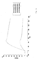
Figure 1:
- A drawing showing the transcriptional and translational signals identified for the B. stearothermophilus argC gene.
-
An UP DNA element is shown in italic. -35 and -10 promoter sites as well as a Shine-Dalgarno site are underlined. Position of the oligonucleotide primers used for the PCR amplification of the 89-bp, 59-bp and 39-bp promoters are shown by arrow. argCo operator sequence is shown in thick line.
- Figure 2:
- An image of a protein array prepared with cell-free synthesized 35S-labelled proteins immobilized on a nitrocellulose membrane.
-
T. maritima GntRTm0439 (lines 1 - 6) and T. maritima LaclTm1856 (7 - 12) cell-free synthesized His-tagged proteins were purified by affinity chromatography, diluted consecutively 2-fold (from A to H), blotted by vacuum on a nitrocellulose membrane and cut out into strips. The strips were directly used for autoradiography (lines 1 and 7) or consecutively rinsed in binding buffer (lines 2 and 8) and washed once or twice with A100 ( lines 3 and 9 or 4 and 10), A500 (lines 5 and 11) and A1000 (lines 6 and 12). Autoradiography was performed with BioMax MS film for 30 min.
- Figure 3:
- An image of immobilized protein showing ArgR protein interaction with a biotin-labelled T. neapolitana argRo operator DNA.
-
His-tagged ArgR proteins of B. stearothermophilus (lines 1 and 6) or T. neapolitana (lines 3 and 8) were purified from recombinant E. coli BL21 (DE3) cells. A His-tagged ArgR of T. neapolitana was cell-free synthesized and purified by affinity-chromatography (slots 5A and 10A). Slots 5B and 10B - purified S30 extracts without synthesized ArgR, and slots 5C and 10C - non-purified S30 extracts without cell-free synthesized ArgR. Slots 1A, 3A, 6A and 8A - contain 100 pmol protein; slots 1B, 3B, 6B and 8B - contain 20 pmol protein; slots 1C, 3C, 6C and 8C - contain 2 pmol protein. A biotin-labelled T. neapolitana argRo operator DNA (final concentration 10 ng/ml) was incubated with membranes at 4°C (lines 1 - 5) or 60°C (lines 6 - 10) overnight.
- Figure 4:
- An image of a protein array showing DNA-protein interactions with IRD-800 labelled 59-bp DNA (a) or a 39-bp DNA (b) of B. stearothermophilus argCo.
-
1 - ArgR of T. neapolitana; 2 - ArgR of B. stearothermophilus; 3 - a chimeric ArgR-EbE; 4 - GroES of T. neapolitana; 5 -α subunit of E. coli RNA polymerase; 6 - CRP of E. coli. The serially 2-fold diluted protein samples were blotted on the nitrocellulose membrane, the membrane was divided into two halves and probed with respect to one of the two IRD-labelled probes.
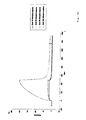
- Figure 5:
- Sensorgrams showing binding of wild-type ArgR proteins of T. neapolitana, B. stearothermophilus, E. coli and a chimeric ArgR-EbE with respect to the B. stearothermophilus argCo operator DNA.
-
Purified His-tagged proteins were injected at 10 mM, 25 mM, 50 mM and 100 mM in presence of 5 mM L-arginine.
- Figure 6:
- Sensorgram showing E. coli CRP interaction with respect to the B. stearothermophilus argCo operator DNA.
-
Purified His-tagged CRP was injected in absence or presence of 5 mM cAMP.
- Figure 7:
- An image of a protein array showing subsequently protein-protein and DNA-protein interactions.
-
Proteins were serially diluted and blotted to the nitrocellulose membrane: 1 - ArgR of T. neapolitana; 2 - ArgR of B. stearothermophilus; 3 - ArgR of E. coli; 4 - GroES of T. neapolitana; 5 - α subunit of E. coli RNA polymerase; 6 - CRP of E. coli. The membrane was first used to probe with Cy5.5-labelled α subunit of E. coli RNA polymerase (a), then washed and reprobed with a IRD-800-labelled 39-bp DNA carrying a shortened argCo operator (b). 5 µg of deposited His-tagged proteins in the slots A correspond to 290 pmol of ArgR proteins, 500 pmol of GroES, 140 pmol of α subunit or 208 pmol of CRP.
- Figure 8:
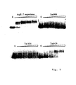
- An image of cell-free synthesized protein array showing DNA-protein interactions.
-
Equimolar quantities (for monomer) of cell-free synthesized T. maritima putative proteins belonging to Gnt (a), Lac (b) and Xyl (c) family of regulators were serially 2-fold diluted and blotted on a nitrocellulose membrane. Incubation was carried out with respect to IRD-800 labelled operator DNA probes of E. coli gntKo, laclo and xylFo, respectively at 60°C, overnight.
- Figure 9:
- Non-denaturant agarose gel showing interaction of cell-free synthesized proteins with respect to operator DNAs.
-
His-tagged purified proteins were synthesized in a cell-free system and probed with respect to Dig-labelled operator DNA probes in DNA-binding buffer at 37°C for 30 min. ArgR binding was performed in presence of 10 mM L-arginine in binding and running buffers.
- Figure 10:
- An image of cell-free synthesized protein array showing protein-protein interactions.
-
Equimolar quantities of cell-free synthesized T. maritima putative proteins belonging to Gnt (a), Lac (b) and Xyl (c) family of regulators were serially diluted and blotted on a nitrocellulose membrane. Incubation was performed with the Cy5.5-labelled α subunit of E. coli RNA polymerase at room temperature for 1 h. Non-diluted samples of CRP and α subunit of E. coli RNA purified from cells contain 200 pmol of proteins.
- Figure 11:
- An image of a protein array showing ligand-protein interactions in rat cell-membrane homogenates using IRD-800 labelled rat secondary antibodies.
-
The immobilized homogenates were first bound to HNK-1 carbohydrate (a) or to 8 amino acid mimic protein (c), then these membranes were incubated with corresponding anti-HNK-1 or anti-mimic primary rat antibodies and then probed with IRD-800 labelled secondary antibodies. The membrane (b) was incubated with anti-GABAR primary antibodies and then probed with IRD-800 labelled secondary antibodies.
- 1, 5 and 8 - homogenates with concomitant expressed GABABR1a and GABABR2a receptors;
- 2, 7 and 9 - homogenates with concomitant expressed GABABR1b and GABABR2a receptors;
- 3 and 10 - laminin (the initial amount is 100 µg);
- 4, 6 and 11 - homogenates without expressed GABA receptors.
- Figure 12:
- An image of a protein array showing the detection of DNA-protein interactions using extracts of E. coli BL21 (DE3) cells.
-
Concentration of a total or purified proteins added in A slots was 63 µg, except A2 in which 41 µg total protein was added because of a slower growth of E. coli (pET-ArgRBs-His). Incubation was carried out with a IRD-800 labelled 39-bp DNA of B. stearothermophilus argCo at room temperature overnight.
-
Vertical lines 1, 2 and 3, respectively 1 h IPTG-induced samples of E. coli (pET-ArgREc-HIS), E. coli (pET-ArgRBs-His) and E. coli (pET-ArgRTn-HIS); 4, 5, 6 and 7, respectively 5 h IPTG-induced samples of E. coli, E. coli (pET-ArgREc-HIS), E. coli (pET-ArgRBs-His) and E. coli (pET-ArgRTn-HIS); 8, 9 and 10- respectively, pure His-tagged ArgR of E. coli, B. stearothermophilus and T. neapolitana; 11 - pure His-tagged E. coli CRP.
- Figure 13:
- An image of a glass-slide microarray showing near-infrared detection of protein-protein interactions.
-
Spots 1, 2, 3, 4, 5 and 6 - respectively, correspond to T. neapolitana ArgR synthesized in a cell-free system or to, B. stearothermophilus ArgR, E. coli ArgR, T. neapolitana GroES, E. coli α subunit of RNA polymerase and CRP synthesized in E. coli cells. The amount of deposited proteins synthesized in vivo is shown in pg. Samples were diluted 4-fold and binding was carried out with Cy5.5-labelled T. neapolitana GroEL at room temperature for 1 h.
EXAMPLES
A. Materials and Methods
A.1 Bacterial strains, plasmids and growth conditions.
-
Bacterial strains and plasmids are described in Table 1.
-
E. coli cells were grown by shaking (180 rpm) at 28°C or 37°C on LB media (Miller, 1992) supplemented with appropriate antibiotics: ampicillin 100 µg/ml, chloramphenicol 25 µg/ml and kanamycin 25 µg/ml. For preparation of cell-free extracts, E. coli cells were grown at 37°C in a medium with 5.6 g/L, KH2PO4, 28.9 g/L K2HPO4, 10 g/L yeast extract (Difco), 1 mM Mg-acetate, 10g/L glucose, 15 µg/ml thiamine described by Zubay (Zubay, 1973). B. stearothermophilus cells were grown in LB-broth (pH 7.3) at 56°C with vigorous shaking. For in vivo expression of His-tagged proteins, recombinant E. coli BL21 strains were grown in LB with 100 µg/ml ampicillin at 28° C until an OD600 0.8-1.0 and after induction by IPTG (1 mM), incubation was continued for 5 h.
A.2 DNA isolation
-
Bacterial biomass of Thermotoga maritima MSB8 or Bacillus stearothermophilus NCIB8224 was harvested, washed with 10 mM Tris-HCI pH 8.0, 1 mM EDTA, 0.15 M NaCI and thoroughly resuspended in the same solution containing 2 mg/ml lysozyme. After 20 min of incubation at 37°C, a lysis buffer (0.1 M Tris pH 8.0, 1% SDS) was added by gentle mixing and incubation was continued for 30 min at 60°C. The lysis was completed by adding proteinase K (0.6 mg/ml) and incubating the reaction mixture for 30 min at 60°C. After four consecutive extractions by phenol/chloroform/isoamylic alcohol (25:24:1, v/v) DNA was precipitated with 0.6 volume of isopropanol by centrifugation at 15000 rpm for 30 minutes at 4°C, rinsed with 70% ethanol and then diluted in 10 mM Tris-HCI pH 8.0, 0.1 mM EDTA.
-
Chromosomal and plasmid DNA from E. coli strains were isolated according to Ausubel et al. (1993).
A.3 PCR amplification and construction of recombinant DNAs
-
The B. stearothermophilus PargC promoter of the argCJBD oferon (Sakanyan et al., 1990; Savchenko et al., 1998; Fig. 1) was used as a strong promoter for driving protein synthesis in a cell-free system. Two oligonucleotide primers were used for amplification of the PargCo promoter-operator region and corresponding to the upstream and downstream extremities of said promoter-operator region (5'-CATAGACTTAGGGAGGGGC and 5'-ATGATGATGATGATGATGCATATGTTCCCCCTCACCCGTATG); the latter contains 6 histidine codons to create a N-terminal tag. Two other oligonucleotides, 5'- CCTCGAAAATTATTAAATATAC and 5'-ACATTTGATTTTATTTTTATAC, were also used to create upstream shortened fragments of promoter sequence, i.e., a 59-bp and a 39-bp fragment of the PargC promoter-operator DNA (see also the figure 1).
-
A DNA sequence coding for a protein of interest was amplified by PCR and fused to the B. stearothermophilus PargC promoter by the overlap extension method (Ho et al., 1989). Basically, the overlap extension method comprises the following steps:
- At the first step, two separate DNA fragments corresponding to N-terminal and C-terminal parts of a given protein, were amplified from chromosomal DNA templates from Bacillus stearothermophilus NCIB 8224, Escherichia coli K12 XA4, Thermotoga maritima MSB8 or Thermotoga neapolitana DSM 5068, with specific primers designed in such a way that the amplified products have overlapping sequences with the sequence downstream the PargC promoter region.
- At a second step, the obtained PCR products were then combined in a subsequent fusion PCR product using only two flanking primers by annealing of the overlapped ends for providing the full-length recombinant DNA template.
-
All putative genes were fused to a his-tag sequence in frame to N-terminal extremity of the open reading frame to enable further purification of the protein by Ni-affinity chromatography as described in A6 hereinafter.
-
Oligonucleotide primers used for amplification of putative ORFs from
T. maritima genome are described in the Tables 2A and 2B below.
-
Oligonucleotide primers used for amplification of operator sequences are described in Table 3.
If necessary, PCR-produced templates were extracted from agarose gel, treated with phenol-chloroform-isoamyl (25 : 24 : 1, v/v), precipitated with ethanol and resuspended. Their DNA concentration was determined by spectrophotometry and confirmed by comparing band intensity with a Smart Ladder reference DNA (Eurogentec) after SDS-PAGE or agarose electrophoresis.
-
Otherwise, the quantified PCR-produced DNA templates were directly added to a cell-free extract to drive protein synthesis.
-
The E. coli XA4 rpoA gene coding for the a subunit of RNA polymerase was amplified by PCR using oligonucleotide primers 5'-GACACCATGGAGGGTTCTGTGACAGAG (the Ncol site is underlined) and 5'-CCGCTCGAGCTCGTCAGCGATGCTTGC (the Xhol site is underlined) and cloned into pET21d(+).
-
The E. coli XA4 crp gene coding for CRP was amplified by PCR using oligonucleotide primers 5'-CATGCCATGGTGCTTGGCAAACC and 5'-CCGCTCGAGACGAGTGCCGTAAACGAC.
-
The T. neapolitana groES-groEL genes coding for chaperonines were amplified by PCR using degenerate oligonucleotide primers 5'-GGAGGGATGGATGATGAARGTNA and 5'-GAYTTYATHGAYCARCAYCCNGA, in which R = A+G, Y = C+T, H = A+C+T, N = all bases. The obtained 2.4 kb DNA fragment was cloned into pCR4-TOPO (Invitrogen). The T. neapolitana groES and groEL sequences were assigned by the EMBL accession number AF275319.
-
A chimeric ArgR-EbE protein was constructed by fusing sequences corresponging to DNA-binding and oligomerization domains of E. coli ArgR repressor to a linker from B. stearothermophilus ArgR. This chimeric sequence displays a better affinity with the heterologous B. stearothermophilus argCo operator than with a wild-type E. coli ArgR.
-
The amplified rpoA, crp, and wild-type or chimeric argR genes were cloned in pET21d(+)vector (Novagen) which allowed expression in frame to a his-tag sequence at the 5'extremity of corresponding proteins. Amplified T.neapolitana groES and groEL genes were cloned separately in pET19b vector (Novagen) in frame to a His-tag at the N-extremity of chaperonine proteins.
-
Non-labelled and biotine-labelled oligonucleotide primers were purchased from Life Technologies. Dig-labelled and IRD-labelled oligonucleotide primers were purchased from MWG-Biotech.
A.4 Preparation of cell-free extracts
-
E. coli strains were used for the preparation of cell-free extracts by the method of Zubay (1973) with modifications as follow:
Cells were grown at 37°C, harvested in mid-log phase by centrifugation and washed twice thoroughly in ice-cold buffer containing 10 mM Tris-acetate pH 8.2, 14 mM Mg-acetate, 60 mM KCI, 6 mM β-mercaptoethanol. Then, cells were resuspended in a buffer containing 10 mM Tris-acetate pH 8.2, 14 mM Mg-acetate, 60 mM KCI, 1 mM dithiotreitol and disrupted by French press (Carver, ICN) at 9 tonnes (≈ 20.000 psi). The disrupted cells were centrifuged at 30.000 g at 4°C for 30 min, the pellet was discarded and the supernatant was centrifuged again. The clear lysate was added in a ratio 1 : 0.3 to the preincubation mixture containing 300 mM Tris-acetate at pH 8.2, 9.2 mM Mg-acetate, 26 mM ATP, 3.2 mM dithiotreitol, 3.2 mM L-amino acids, 3 U/ml pyruvate kinase (Sigma) and incubated at 37°C for 80 min. The mixed extract solution was centrifuged at 6000 g at 4°C for 10 min, dialysed against a buffer containing 10 mM Tris-acetate pH 8.2, 14 mM Mg-acetate, 60 mM K-acetate, 1 mM dithiotreitol at 4°C for 45 min with 2 changes of buffer, concentrated 2-4 times by dialysis against the same buffer with 50% PEG-20.000, followed by additional dialysis without PEG for 1 hour. The obtained cell-free extract was aliquoted and stored at -80°C.
A.5 Coupled transcription-translation reaction
-
The expression system was based on the use of transcriptional and translational signals of the B. stearothermophilus argC gene (see Fig. 1). The argC gene is transcribed from a strong PargC promoter which overlaps a 42 bp argCo operator (Savchenko et al., 1996) recognized by the B. stearothermophilus ArgR repressor (Dion et al., 1997). However, the E. coli ArgR repressor has a very weak repression effect with respect to this operator. Therefore, heterologous genes can be overexpressed from B. stearothermophilus PargC promoter in E. coli cells (Savchenko et al., 1998).
-
T7 promoter based cell-free synthesis was carried out from pET vectors carrying corresponding genes. T7 RNA polymerase was purchased from Promega.
-
Using the E. coli S30 extract system, the transcription-translation coupled reaction was carried out as described by Zubay (1973) with some modifications. The standard pre-mix contained 50 mM Tris-acetate pH 8.2, 46.2 mM K-acetate, 0.8 mM dithiotreitol, 33.7 mM NH4-acetate, 12.5 mM Mg-acetate, 125 µg/ml tRNA from E. coli (Sigma), 6 mM mixture of CTP, GTP and TTP, 5.5 mM ATP, 26.2 mM phosphoenol pyruvate, 8.7 mM CaCl2, 1.9 % PEG-8000, 0.32 mM L-amino acids, 5.4 µg/ml folic acid, 5.4 µg/ml FAD, 10.8 µg/ml NADP, 5.4 µg/ml pyridoxin, 5.4 µg/ml para-aminobenzoic acid. In the experiments, wherein pyruvate is used as the energy regenerating compound (Kim and Swartz, 1999), phosphoenol pyruvate is replaced in the standard reaction mixture by 32 mM pyruvate and 6.7 mM K-phosphate pH 7.5, 3.3 mM thiamine pyrophosphate, 0.3 mM FAD and 6 U/ml pyruvate oxidase (Sigma) are added. Typically, to 25 µl of pre-mix containing all the amino acids except for methionine, 10 µCi of [α35S]-L-methionine (specific activity 1000 Ci/mmol, 37 TBq/mmol, Amersham-Pharmacia Biotech) and the E. coli S30 extracts, 100 - 750 ng of circular plasmid DNA or linear PCR-produced DNA, were added and the mixture was incubated at 37°C for 90-120 min.
-
If necessary, the protein samples were treated at 80°C for 10 min and then quickly centrifuged. The supernatant was precipitated with acetone and used for protein separation on SDS-PAGE. After coloration with Coomassie Brilliant Blue (Sigma), gels were treated with an amplifier solution (Amersham-Pharmacia Biotech), fixed on a 3 MM paper by vacuum drying and the radioactive bands were visualized by autoradiography using BioMax MR film (Kodak). Molecular weights of putative proteins were deduced from ORFs of the T. maritima genome (Nelson et al., 1999) and further confirmed by migration of the corresponding proteins on SDS-PAGE. Quantification of cell-free synthesized proteins (as monomer equivalent) was performed either, (i), by comparing non-radiolabeled protein bands with known reference proteins after coloration with EZBlue gel staining reagent (Sigma) or, (ii), by counting radioactivity of 35S-labeled protein bands with Phosphorlmager.
A.6 Purification of His-tagged proteins
-
Recombinant bacteria were suspended in a buffer (50 mM NaH2PO4 pH 8.0, 300 mM NaCI, 10 mM imidazole), broaken by sonication and His-tagged proteins were purified by affinity chromatography on a Ni-NTA column (Qiagen). The column was washed successively with 10 x bed volumes of the mentioned buffer and proteins were eluted by 6 x bed volumes of elution buffer [50mM Tris-HCI pH 7.9, 50 or 300 mM NaCI, 200 or 400 mM imidazole, 5% (vol/vol) glycerol)] and then dialysed against buffer containing 20 mM NaH2PO4, pH 8.0, 5 mM β-mercaptoethanol, 5% (vol/vol) glycerol. Protein concentration was measured by Bradford method (Bradford, 1976).
-
Proteins synthesized by cell-free synthesis were purified using Ni-NTA-magnetic agarose beads (Quiagen).
A.7 DNA labelling with IRD dyes
-
Two approaches were used to introduce IRD-dyes into DNAs. A DNA probe was amplified by PCR using a pair of primers in which one or both primers was labelled by IRD-700 or IRD-800 (see Table 3 above). Alternatively, a DNA probe was subjected to a random incorporation of IRD-700 or IRD-800 using a corresponding kit (LI-COR, Inc.) purchased from ScienceTec.
A.8 Protein labelling with IRD dyes
-
The purified His-tagged α subunit of E. coli RNA polymerase and GroEL of T. Neapolitana were labelled by FluoroLink Mab Cy 5.5 labelling kit following the manufacturer's instructions (Amersham Pharmacia Biotech). Typically, 5 µl of a coupling buffer was added to 100 µl of 0.1 mg/ml protein solution and incubated in the dark for 30 min by shaking every 10 min. The conjugated dye-protein complex was separated from free dye by passing through a PD 10 column and eluted with PBS buffer pH 7.2. Fractions giving a final molar dye (A678) /protein (A280) ratio around 2 were used for detecting protein-protein interactions.
A.9 Preparation of protein array
-
Both, in vivo and in vitro synthesized protein were used for the preparation of protein array. To produce a sufficient amount of proteins for a small scale analysis of putative ORFs of T. maritima, cell-free synthesis was performed in a 200 or 400 µl reaction mixture for 2 h.
-
Different quantities of purified proteins (dilutions from 200 pmol to 0.1 pmol) in a A100 buffer (100 mM KCl, 10% glycerol, 20 mM HEPES Na pH 7.9, 0.2 mM EDTA, 10 mM 2-mercaptoethanol and 0.5 mM PMSF) described by Ge (2000) were deposited on a 12 x 8 cm nitrocellulose membrane using a 96-well dot blot apparatus (Bio-Rad) under vacuum and rinsed twice in A100 buffer. Radioactive signals on nitrocellulose membranes were detected by autoradiography using BioMax MS film (Kodak) or Phosphorlmager 445 SI (Molecular Dynamics).
A.10 Detection of intermolecular interactions with fluorescent probes
-
For protein-DNA interactions, the membranes were incubated in a DNA-binding buffer (DBB): 20 mM Tris-HCI pH 7.9, 50 mM NaCI, 50 mM KCI, 0.1 mM DTT 0.005% surfactant P20 (Biacore) and 25 µg/ml of a competitor DNA (sonicated salmon DNA) or a mixture of 12.5 µg/ml poly-dGdC and 12.5 µg/ml poly-dAdT (Sigma) at room temperature for 30 min. Then, an IRD-labelled DNA probe (10 ng/ml) was added to the prebinding solution and incubated for 12 hours with a slow rotation at different temperatures (4°C, 18°C or 60°C). When necessary, L-arginine (10 mM) or cAMP (5 mM) were included in the reaction mixture. The membranes were washed consecutively with A100 and A500 and then used for the detection of fluorescent signals.
-
For protein-protein interactions, the membranes were incubated in A100 buffer containing 1% non-fat milk at room temperature for 30 min as described (Ge, 2000). Then, a Cy 5.5-labelled protein probe (5 ng/ml) was added to the prebinding solution and incubated at room temperature for 12 hours.
-
Alternatively, the protein samples were dialysed against PBS buffer (4.3 mM Na2HPO4, 1.4 mM KH2PO4, 137 mM NaCI, 2.7 mM KCI, pH 7.4 with 20% glycerol) and then deposited on the nitrocellulose membrane under vacuum. After incubation in the PBS solution with 1% BSA for 1 h at room temperature, the membranes were incubated with corresponding probes in the same solution with 0.1% Tween 20 at room temperature for 1 h for detection of protein-protein interactions.
-
The protein macroarrays were reused after consecutive washing with buffer A containing 1 M (NH4)2SO4, 1 M urea (Ge, 2000) and then consecutively with PBS containing 0.1% Tween 20 and PBS, at room temperature. To prepare a protein microarray, the method described by MacBeath and Schreiber (MacBeath and Schreiber, 2000) was used with modifications as follow:
The protein samples were dialysed against PBS-buffer with 20% glycerol, and then 600 pl were delivered with a high-precision contact-printing robot SDDC-20 (Virtek) to glass slide covered by superaldehyde (Telechem International), yielding spots of 150 µm in diameter. The spotted samples were incubated in a humid chamber at room temperature for 3 h to quench the unreacted aldehydes on the slide. After rinsing in the PBS-solution with 1% BSA, the slides were incubated with Cy5.5 labelled probe (final concentration 1 µg/ml) for 1 h at room temperature.
-
Near-infrared fluorescence signals on membranes were detected by IR-Imager Odyssey (LI-COR, Inc.).
A11. Chemiluminescent detection
-
Diluted samples of proteins in A100 buffer (from 100 pmol to 0.2 pmol) were blotted on nitrocellulose membrane as described above. Incubation was carried out with respect to a biotine-labelled DNA probe (10 ng/ml) and detection of bound DNA-protein complexes was performed with streptavidin (Phototope-star detection kit, BioLabs). In this method, a moderately stable intermediate is formed which spontaneously decays by emitting light at 461 nm. The emission was detected by exposing the membrane to BioMax MS X-ray film (Kodak).
A.12 Mobility-shift assay (MSA)
-
DNA binding activity of proteins was also tested by mobility-shift assay using Dig-labelled operator DNAs from B. stearothermophilus, E. coli, T. maritima or T. neapolitana. DNA binding was performed in DNA-binding buffer in presence of a 100-fold excess of unlabelled sonicated herring sperm DNA and 3 ng Dig-labelled operator DNA at 37°C or 70°C for 30 minutes. If necessary, L-arginine (10 mM) was included in the reaction mixture. Samples were loaded on a 2% agarose gel prepared in TAE buffer (40 mM Tris-base 10 mM sodium acetate, 1 mM EDTA pH 8.0) and migrated by electrophoresis at room temperature at 12 Vcm-1 for 1 h. The DNA-protein complexes were transferred onto nylon membranes (Schleicher and Schuell) by capillary method (Ausubel et al., 1993). Immunological detection was carried out with CSPD-mediated luminescence (Boehringer Mannheim). Quantification of free and retarded DNA in gels was done by scanning densitometry of chemioluminograms (Molecular Analyst, Bio-Rad).
A.13 Surface plasmon resonance (SPR)
-
The SPR technique (for a review, see Malmqvist, 1993) was used to study a real-time interaction of wild type and chimeric ArgR proteins with operator DNAs. End-biotinylated DNA fragments corresponding to different operators were prepared by PCR using the above mentioned primers for MSA, except that digoxigenin was replaced by biotin. The labelled DNA fragments were purified from excess of primers and dNTPs by passage through a spin cartridge containing a silica-based membrane (Life Technologies Gibco BRL). Immobilization of biotinylated DNA was carried out on streptavidine-captured biosensor chips SA (Bioacore AB). Biotinylated operator DNA (5 mg/ml) in DNA-binding buffer was injected over the sensor chip at a flow rate of 5 µl/min at 25°C and its immobilization was controlled manually by achieving nearly 150 RU, a relatively low amount of DNA coupled to the biosensor in order to keep mass transport effects at a minimum. Binding assays were carried out in the same DNA-binding buffer by injection of 10 - 100 nM of a protein (monomer equivalent) at a flow rate 20 µl/min at 25°C. When necessary, L-arginine (5 mM) or cAMP (5 mM) were included in the reaction mixture. SPR measurements were conducted in parallel channels using a Biacore 3000 (Biacore AB). The sensor chip was regenerated by washing with 1% NaCI at a flow around 10 µl/min. The channel with no immobilized DNA was used as a reference signal for each cycle. Data for protein-DNA interactions were evaluated from sensorgrams using the 1:1 binding model (BlAevaluation Software Handbook, 1999). The SPR signal is directly proportional to the mass changes at the sensor chip surface and is expressed in resonance units; 1000 RU correspond to a surface concentration of approximately 1 ng /mm-2.
B. Experimental results
B.1 Example 1: Preparation of a protein array comprising the binding of cell-free synthesized proteins to nitrocellulose membrane
-
In order to prepare an array with chemically non-modified proteins, the method using nitrocellulose membranes as described by Ge (Ge, 2000) was used. However, since the author has not presented data on a possible washing off of membrane-attached proteins, the efficiency of the binding of proteins to nitrocellulose membrane after incubation in binding and washing solutions has been tested.
-
Two thermostable His-tagged proteins of different size, namely a 25 kDa GntRTm0439 and a 37 kDa LaclTm1856, corresponding to putative proteins of T. maritima (Nelson et al., 1999) were cell-free synthesized using PCR-produced DNA templates and E. coli BL21Z cell-free extracts in 150 µl reaction mixture for 2 hours. Samples were treated at 80°C for 10 min, centrifuged at 15000 g for 10 min. The pellets were discarded and the proteins were purified from supernatant fractions by affinity chromatography with Ni-NTA-magnetic agarose beads. The incorporated radioactive methionine allowed to quantify the SDS-PAGE separated bands of cell-free synthesized proteins with Phosphorlmager. The data were confirmed by the staining of gels with EZBlue.
-
Then, both GntR Tm0439 and LaclTm1856 purified proteins were serially diluted and slowly blotted on a nitrocellulose membrane under vacuum for 5 min. Membrane strips corresponding to each line were cut out, incubated in different solutions and then submitted to autoradiography. Practically no diminution of radioactivity was detected in corresponding slots after consecutive thorough washings with A100, A500 and A1000 buffers (as defined in Ge et al., 2000) (Fig. 2; compare samples in horizontal lines for each protein). More than 98% of the two tested proteins, GntR Tm0439 of T. maritima and Lacl TM1856 of T. maritima remained bound on the membranes after the steps of washing. Thus, once a protein is immobilized on the nitrocellulose membrane and rinsed in a binding buffer, it remains perfectly bound to the support.
-
The binding of cell-free synthesized proteins on a solid phase (here nitrocellulose membrane) that does not involve a covalent chemical linkage appears to be uniform. Moreover, vacuum-blotting cell-free synthesized proteins is advantageous as compared with spotting approaches since both addition and binding of proteins can be done on a relatively large surface, easily controlled even for diluted samples and thereby, quantities of protein patterns in different wells can be normalized precisely.
B.2 Example 2: Failure of usual chemiluminescent methods for the detection of ArgR protein-DNA interactions
-
A 17 kDa ArgR repressor governs arginine biosynthesis in E. coli cells (for reviews see Maas, 1994; Glansdorff, 1996). ArgR proteins of thermophilic bacteria B. stearothermophilus (Dion et al., 1997; Ni et al., 1999; Karaivanova et al., 1999) and T. neapolitana (Dimova et al., 2000) were studied previoulsy. ArgR repressor consists of two structural domains connected by a short linker peptide. The N-terminal domain contains a winged helix-turn-helix module which recognizes and binds arg-specific operators (Sunnerhagen et al., 1997; Ni et al., 1999). The C-terminal domain is involved in the formation of trimers which dimerize into hexameric molecules in the presence of L-arginine. Arginine is used as an allosteric co-repressor for improving DNA binding cognate arg operators. Sequence and structure similarity between ArgR proteins from various bacteria suggest a common DNA-recognition mechanism. Nevertheless, major differences exist between the different ArgR repressors. In particular, E. coli ArgR displays low DNA-binding affinity for the argCo operator from B. stearothermophilus (Savchenko et al., 1996), whereas both B. stearothermophilus and T. neapolitana repressors bind E. coli operators efficiently (Wang, 1998; Karaivanova et al., 1999; Dimova, 2000).
-
Taking into consideration this valuable information, interactions of ArgR proteins synthesized in vivo or in vitro, and then bound to nitrocellulose membrane, were first tested using a chemiluminescent detection method. Recombinant His-tagged ArgR proteins of B. stearothermophilus and T. neapolitana were synthesized in E. coli BL21 (DE3) cells, purified by affinity chromatography and blotted on a membrane at different concentrations. Besides, the T. neapolitana ArgR protein was in vitro synthesized, purified with Ni-NTA-magnetic agarose beads and blotted on the same membrane. Control samples of S30 extracts were also blotted on the membrane. Biotine or digoxigenin labelled operator DNAs of B. stearothermophilus argCo or T. neapolitana argRo were used as probes and DNA binding was carried out at 4°C or 60°C in DNA-binding buffer solution overnight. Prewashing, prebinding and binding were carried out in presence of 10 mM L-arginine.
-
It has been found that a digoxygenine-labelled probe gave a rather weak signal with respect to tested proteins on nitrocellulose membrane, whereas biotine labelled probes gave a strong signal. However, the intensity of the signal was nearly the same for both cell-free extract samples irrespective whether T. neapolitana ArgR was synthesized or not (Fig. 3). Moreover, a signal was still strong when binding had been carried out at 60°C.
-
Although it was possible to distinguish stronger positive signal between samples with in vitro synthesized and in vivo synthesized ArgR repressor by prolongation of the radioautography exposition, the interpretation of results was always ambiguous. It appeared, that traces of a E. coli biotin-binding protein(s), like naturally biotinylated pyruvate carboxylase, were present in samples after purification and these protein traces exhibited high affinity towards streptavidin that mimicked positive signal.
-
Therefore, these results indicate that the chemiluminescent method is not sensitive enough for testing protein-DNA interactions using the protein arrays comprising cell-free synthesized proteins.
B.3 Example 3: Detection of ArgR protein-arg operator DNA interactions using near-infrared fluorescent dyes
-
Searching for other dyes that could provide a higher signal-to-noise ratio and thereby low background on nitrocellulose membrane, longer-wavelength, near-infrared fluorescent dyes have been tested. A probe consisting of the 59-bp argCo operator DNA of B. stearothermophilus (see Fig. 1) was labelled by IRD-800. This probe has been used in binding experiments with ArgR repressors blotted on nitrocellulose membrane. Serially 2-fold diluted (from 100 pmole to 0.8 pmole) wild-type ArgR proteins of T. neapolitana, B. stearothermophilus as well as a chimeric ArgR-EbE were used to prepare a protein array of a 48-well format. Binding reaction was carried out at 4°C, 18°C or 60°C in presence of 10 mM L-arginine overnight.
-
As it can be observed in the figure 4a, a weak signal was detected from a chimeric ArgR-EbE with any of the IRD-probes. On the contrary, a strong signal was detected for bound T. neapolitana or B. stearothermophilus ArgR-DNA complexes. The signal was gradually decreased as a function of the bound protein concentration and the membrane displayed a very low background. A positive signal was observed up to 0.8 pmol and 3.12 pmol, respectively for T. neapolitana and B. stearothermophilus ArgR. Such a low protein detection level demonstrates that the method is highly sensitive.
-
Similar results were monitored when binding was performed at three different temperatures and when operator DNA was labelled by IRD-700. It appeared, that the formed complexes between synthesized proteins and IRD-labelled probes were rather stable and that equilibrated state of ArgR-argCo interactions was not affected by temperature.
-
Next a 39-bp argCo operator DNA lacking upstream operator sequence (see Fig. 1) was used as a probe to detect interactions with the ArgR proteins blotted on the membrane. As shown in figure 4b, a binding was detected for higher concentrations of thermostable ArgR proteins, 6.25 pmol and 25 pmol, respectively for T. neapolitana and B. stearothermophilus repressors.
-
The affinity of ArgR for arg-specific operator DNA depends on the number of Arg boxes in the operator (Chen et al., 1997), therefore the difference in monitored signals appeared to reflect DNA-binding ability of thermostable repressors to the entire and shortened argCo operator sequences.
-
Data obtained with the protein arrays were confirmed by SPR analysis. Indeed, as shown in figure 5, wild-type ArgR proteins from T. neapolitana and B. stearothermophilus exhibited a high binding capacity towards the B. stearothermophilus argCo operator. In contrast, the ArgR-EbE chimera and especially the wild-type E. coli ArgR repressor exhibited a very low binding to this operator DNA. It is worth mentioning that all proteins exhibited a high DNA binding activity with respect to the E. coli carAB operator.
-
Therefore, the data presented in Examples 2 and 3 clearly demonstrate that the protein arrays of the invention can be used in combination with IRD technology
- (i) for screening equilibrated protein-DNA interactions; and
- (ii) for preliminary quantification of DNA binding properties of proteins.
B.4 Example 4 : Detection of interactions between α subunit of RNA polymerase and arg operator DNA
-
The transcription level is directed by the strength of binding of the C-terminal domain of α subunit of RNA polymerase to a UP-element in some E. coli promoters (Ross et al., 1993; Gourse et at., 2000). The data with a shortened 39-bp probe indicated that a 19 bp AT-rich sequence upstream a -35 site of the PargC promoter could make contacts with α subunit of E. coli RNA polymerase. To confirm this hypothesis, a α subunit was blotted on the same protein array and tested for interactions with the IRD-labelled PargC 59-bp and 39-bp promoter DNAs. Again, as shown in figure 4, a signal was detected for up to 50 pmol of bound α subunit, with respect to a 59-bp operator DNA probe, whereas no binding signal was detected with a 39-bp operator DNA. Thus, these results show that the method of the invention enables to detect binding of α subunit of E. coli RNA polymerase to the upstream sequence of the B. stearothermophilus PargC promoter.
B.5 Example 5 : Detection of cAMP receptor protein (CRP)-arg operator DNA interactions
-
E. coli CRP is involved in the transcriptional activation of some promoters (Busby and Ebright, 1999). Its binding to DNA requires an allosteric interaction with cAMP (Valentin-Hansen et al., 1996). The sequence upstream the -35 site in B. stearothermophilus PargCo promoter-operator shares a weak similarity with CRP binding-sequence of E. coli DNA. Therefore, E. coli CRP was blotted on the same protein array and tested for binding with PargCo DNA probes. As it can be seen in the figure 4, positive signal was detected with a 59-bp but not with a 39-bp operator DNA in presence of 5 mM cAMP. Thus, taken together with the examples 3 and 4, these results demonstrate that the method of the invention is able to detect distinct and specific protein-DNA interactions.
-
Indeed, these data were confirmed by SPR analysts. As shown in figure 6, CRP displayed a binding to the B. stearothermophilus PargCo promoter-operator region in the presence of cAMP, whereas no binding was monitored in the absence of the ligand.
-
Thus, by using different length IRD-labelled probes and different DNA-binding proteins, it has been shown that the detected protein-DNA interactions by the protein arrays of the invention are specific. The method of the invention permitted to detect relatively low-affinity interactions. This emphasizes the high potential of the method for screening intermolecular interactions at a large scale.
B.6 Example 6 : Detection of protein-protein interactions on a protein array using a probe labelled with near-infrared dyes.
-
The transcription efficiency of RNA polymerase holoenzyme can be modulated by interaction of its α subunit with other proteins, like CRP (Gourse et al., 2000). The data presented above showed that B. stearothermophilus or T. neapolitana ArgR proteins, as well as α subunit of E. coli RNA polymerase and CRP have affinity for the same DNA-target, namely the B. stearothermophilus PargCo promoter-operator region. Therefore, it was presumed that these proteins might contact each other. To verify this hypothesis, a Cy 5.5 labelled α subunit was tested for its binding to other proteins bound to nitrocellulose in a 48-well substractive array format.
-
Positive signals were detected for T. neapolitana and B. stearothermophilus ArgR proteins, E. coli CRP, but not for E. coli ArgR, α subunit of E. coli RNA polymerase or T. neapolitana GroES and GroEL. As shown in figure 7, protein-protein interactions were detectable for deposited 1.7 pmol (or 0.04 µg) E. coli CRP, 9.1 pmol (or 0.16 µg) T. neapolitana and B. stearothermophilus ArgR proteins. The absence of binding signal between α subunits themselves appeared to indicate that dimeric molecules of the protein do not contact together. The negative result with respect to E. coli ArgR suggest that this repressor does not bind significantly to the homologous α subunit.
-
The membrane was washed with a urea-containing buffer A (it was noticed that even thorough washing in this solution does not remove completely bound proteins as judged from residual fluorescence) and reused in a binding reaction with a 39-bp long PargC DNA labelled by IRD-800. As expected, specific IRD-800 red signal (IRD-700 gives green colour) was monitored for both T. neapolitana and B. stearothermophilus ArgR proteins (with a higher affinity for the former), but not for E. coli ArgR (Fig. 7b). The specificity of intermolecular interactions was visible also from a weak signal towards E. coli CRP (compare Figures 4 and 7), whereas this protein exhibited stronger protein-protein interactions.
-
Similar data for protein-protein interactions were obtained when the α subunit of RNA polymerase is labelled by IRD-800.
-
Thus, binding of blotted thermostable ArgRs or E. coli CRP to the IRD labelled α subunit of E. coli RNA polymerase clearly reflects specific protein-protein interactions. Moreover, these interactions did not prohibit further binding to a IRD labelled 39-bp DNA probe. It appears that the detected protein-protein and protein-DNA interactions are provided by different regions of the blotted proteins.
-
These data showed that the protein arrays of the invention can be used several times with different probes.
B.7 Example 7: Screening DNA-protein interactions using a protein array based on cell-free synthesized proteins
-
The data presented above showed that both DNA-protein and protein-protein interactions can be detected with IRD-labelled probes of interest with respect to rather low quantities of membrane-blotted proteins purified from bacterial cells. Consequently, the use of IRD-labelled probes has been tested with a protein array prepared with cell-free synthesized proteins.
-
The optimal conditions were first determined for the different steps of the preparation of the array. These conditions were determined by probing T. neapolitana ArgR with respect to IRD-labelled DNA and protein probes.
-
A 35S-labelled T. neapolitana ArgR has been synthesized in a new PargC-mediated cell-free system using the extracts of the E. coli BL21 Star RecBCD (DE3) (pRIL) strain and the His-tagged protein was purified by affinity chromatography by several methods. Each purification step (binding to Ni+2, washing and elution with imidazol) was controlled by counting the radioactivity in protein bands after separation in SDS-PAGE. Two purification methods, based on using Ni-coated plates (Pierce) and Ni-NTA-magnetic beads (Quiagen) gave the best results. Since the second method was found more reproducible and with a slightly higher yield, it was used in further experiments for serial purification of His-tagged proteins synthesized in vitro.
-
The purified ArgR (cell-free synthesis was carried out in a 200 µl reaction mixture) was blotted on a nitrocellulose membrane as described in Example 1. Binding was performed with respect to IRD-800-labelled argRo of T. neapolitana or Cy5.5-labelled α subunit of E. coli RNA polymerase, respectively at 60°C and 18°C. In all cases, signals were monitored from up to 4-fold-diluted protein samples, whereas no signal was detected from non-diluted samples without synthesized ArgR. Thus, it was concluded that the thermostable ArgR from T. neapolitana is synthesized in cell-free system in a sufficient quantity and a folded protein exhibits specific DNA-protein and protein-protein intereactions as found for a native recombinant protein purified from cells.
-
Taking into consideration these data, a protein macroarray was prepared with cell-free synthesized proteins for screening DNA-protein and protein-protein interactions in a small scale. 14 ORFs of
T. maritima coding for putative proteins belonging to Gnt, Xyl and Lac families of regulators (Nelson
et al., 1999) were used to synthesize corresponding proteins in a P
argC-mediated cell-free system. The His-tagged
T. maritima proteins were purified by affinity chromatography with Ni-NTA-magnetic beads. Quantity of proteins was normalized with respect to each other by counting the radioactivity level in respective bands and taking into account the number of methionine residues in deduced protein sequences. Namely, the following equation has been used:
in which
- R is a radioactivity level detected in a protein band with Phosphorlmager,
- m is a number of methionine residues in a monomeric protein,
- V is a volume of a sample in µl,
- N is a arbitrary quantitity of a synthesized protein.
-
Practically, the sample with the lowest value N was taken as a reference (the maximal volume) and other protein sample volumes were adjusted (normalized) with respect to the reference (Table 4).
Table 4. | Characterization of T. maritima putative proteins. |
| Putative protein of T. maritima | Length of a gene | Deduced molecular weight of a protein | Number of methionine residues |
| XylR |
| 0393 | 1299 bp | 42.3 kDa | 10 |
| XylR 0032 | 1341 bp | 43.0 kDa | 5 |
| XylR 0110 | 1328 bp | 42.5 kDa | 9 |
| XylR 0411 | 1356 bp | 41.9 kDa | 5 |
| XylR 0808 | 1270 bp | 38.9 kDa | 4 |
| XylR 1224 | 1203 bp | 40.9 kDa | 9 |
| GntR 0275 | 1218 bp | 38.6 kDa | 8 |
| GntR 0766 | 569 bp | 14.0 kDa | 1 |
| GntR 0439 | 843 bp | 25.0 kDa | 4 |
| Lacl 0299 | 1235 bp | 38.9 kDa | 7 |
| Lacl 0949 | 1194 bp | 37.5 kDa | 8 |
| Lacl 1200 | 1109 bp | 38.0 kDa | 9 |
| Lacl 1218 | 1184 bp | 36.9 kDa | 5 |
| Lacl 1856 | 1186 bp | 37.0 kDa | 8 |
-
Thus, equimolar quantities of proteins (monomers) were blotted on the nitrocellulose membrane in order to prepare the protein macroarray.
-
A protein macroarray, prepared as defined above, was divided into three parts and used to detect DNA-protein interactions with respect to three IRD-labelled DNA probes.
-
Similar DNA-binding sequences are indicative of the resemblance between regulatory systems (for reviews, see Pabo & Sauer, 1992; Roy et al., 1998). Indeed, the presented above data with a ArgR-regulatory system indicated that operator sequences might be conserved in evolutionary distant bacteria T. neapolitana and E. coli. Therefore, well studied operator sequences from E. coli genome were used as probes. Namely, short DNA fragments carrying gntKo (Tong et al., 1996), xylFo (Song and Park, 1997) and laclo (Oehler et al., 1990) operator sequences were labelled by IRD-800 and probed with respect to the 14 putative regulatory proteins of T. maritima (see Table 4) at room temperature.
-
As shown in figure 8, strong signals were detected from up to 4-fold diluted samples for putative GntTm0766 and GntTm0275 proteins, whereas a signal was rather weak for GntTm0439. Among Lac protein family, a strong signal was detected for LacTm1200, whereas the signal was relatively weak for others. Finally, strong signals were detected for five of six XylTm proteins, except for XylTm0110.
-
The specificity of the detected signals was confirmed by cross-binding experiment. In particular, GntR proteins could not bind to the IRD-700 labelled xylFo operator DNA. Besides, protein array data were confirmed by MSA. In particular, cell-free synthesized GntTm0439, LacTm1856 and probably XylTm0808 bound to the Dig-labelled E. coli target operator DNAs by forming retarding bands in agarose gel, as observed also for cell-free synthesized T. neapolitana ArgR (Fig. 9).
-
These results show that protein arrays prepared with cell-free synthesized proteins according to the invention can be used for rapid and parallel screening and characterization of putative DNA-binding proteins, especially transcriptional regulators.
B.8 Example 8: Screening of protein-protein interactions using array based on cell-free synthesized proteins
-
A similar protein macroarray bearing cell-free synthesized proteins was used to detect protein-protein interactions using a Cy5.5-labelled α subunit of E. coli RNA polymerase as a probe. As shown in Fig. 10, all GntTm proteins exhibit binding with respect to the heterologous α subunit. Three Lac family proteins Tm1856, Tm1218 and Tm0299 also give clear signals from non-diluted samples. A signal was also detected for three Xyl family proteins Tm0393, Tm0808 and Tm0032. A strong response was detected from E. coli CRP and T. neapolitana ArgR synthesized in vitro or in vivo, but not from E. coli RNA polymerase α subunit or ArgR all used as controls. These data were confirmed with IRD-800 labelled α subunit of RNA polymerase used as a probe.
-
Thus, protein array prepared with cell-free synthesized proteins are convenient for serial detection of protein-protein interactions such as those involved in transcription regulation using IRD-labelled protein probes.
B.9 Example 9: Detection of ligand-protein interactions with non-purified proteins from cell homogenates bound to nitrocellulose membrane
-
To assess the sensitivity of method of the invention with respect to non-purified proteins bound to nitrocellulose membrane, the following example presents the detection of ligand-protein interactions by applying IRD-labelled secondary antibodies.
-
The neurotransmitter γ-aminobutyric acid (GABA) recognizes several receptors expressed on cell surface of mammalian cells (Bettler et al., 1998). GABAB receptor is composed from two subunits GABABR1 and GABABR2. GABABR1 can exist in 2 splice variants, GABABR1a and GABABR1b that differ by their extracellular part. It was postulated that GABAB receptor could bind to HNK-1 carbohydrate of adhesion molecules.
-
To test this hypothesis, the membrane homogenate of cells was used, in which a concomitant expression of rat GABABR1a and GABABR2a or GABABR1b and GABABR2a receptors occurred from cloned cDNAs. Membrane homogenates obtained from cells in which the receptors were not expressed (Novartis Pharma), were used as a negative control. Laminine (Sigma), the protein which binds to HNK-1 carbohydrate (Hall et al., 1995) was used as a positive control. Besides, a 8 amino acid peptide with a FLHTRLFV sequence mimicking HNK-1 carbohydrate epitope (Strekalova et al., 2001) was also used as a control.
-
Serially 2-fold diluted samples of homogenates were arrayed on nitrocellulose membrane under vacuum. A total protein was 6.7 µg in each starting material. The membranes were rinsed twice in A100 buffer solution, blocked with Odyssey blocking buffer for 30 min at 18°C and, after addition of a synthetic HNK-1 carbohydrate (Kornilov et al., 2000) at a final concentration of 12,5 µg/ml or of HNK-1 mimick peptide at a final concentration of 85 µg/ml, incubation was continued overnight. Then, membranes were rinsed in PBS buffer for 5 min, reincubated in blocking buffer for 1 h, incubation was continued for 1 h after addition of rat primary antibodies against HNK-1 carbohydrate (dilution 1:2000) in a blocking buffer with 0.1% Tween 20. The membranes were washed out in PBS + 0.1% Tween 20 for 5 min and incubated with IRD-800 labelled secondary anti-rat antibodies (dilution 1:2000 in blocking buffer) for 1 h. The membranes were consecutively washed with PBS + 0.1% Tween 20 for 5 min and then in PBS for another 5 min.
-
In a control experiment, the membrane with immobilized homogenates of cells with expressed GABAB R1a and GABAB R1b and GABAB R2a receptor subunits were directly incubated with rat GABAB antibodies (dilution 1:2000) and then with IRD-800-labelled secondary antibodies as described above.
-
Figure 11 reveals that a strong signal was detected up to 128-fold diluted homogenate samples with expressed GABA receptors, whereas only a weak signal was detected for control homogenate from non-diluted and 2-fold diluted samples. Thus, IRD-technology provides a rather high sensitivity sufficient to detect minor quantities of target proteins in cell membrane homogenates and can be used with non-purified protein samples.
B.10 Example 10: Detection of DNA-protein interactions in crude cell extracts blotted on a nitrocellulose membrane
-
Another experiment was also carried out to evaluate the sensitivity of IRD-detection method for DNA-protein interactions in crude cell extracts. The design of this experiment was based on the observation that a shortened 39-bp PargCo operator DNA of B. stearothermophilus exhibited a specific affinity with respect to B. stearothermophilus and T. neapolitana ArgR proteins, whereas no binding could be detected with respect to homologous E. coli ArgR and α subunit of RNA polymerase or a weak binding is detected for CRP (see Example 3). Therefore, such a short operator sequence could be used as a probe for screening a total protein pool in E. coli extracts in order to detect DNA-protein interactions with respect to B. stearothermophilus or T. neapolitana ArgR repressors synthesized in E. coli cells.
-
The E. coli BL21 (DE3) cells bearing a pETd-ArgR-His plasmid with corresponding E. coli, B. stearothermophilus or T. neapolitana argR gene were cultivated up to OD600 0.8, then 1 mM IPTG was added and cultivation was continued for 5 h. Samples were taken before and after 1 and 5 h IPTG-induction, thoroughly washed three times in A100, sonicated and centrifuged. After discarding the pellet, the supernatant was 2-fold serially diluted in A100 and blotted on the nitrocellulose membrane under vacuum. The membrane was probed with a IRD-800 labelled 39-bp PargCo DNA at room temperature overnight. As it is shown in Figure 12, it was found, that starting from 1 h IPTG-induction samples with expressed B. stearothermophilus or T. neapolitana ArgR provided strong signal but not those with E. coli repressor.
-
The three ArgR proteins were also synthesized in a cell-free system. The reaction mixture was blotted to a nitrocellulose membrane and probed with respect to a IRD-800 labelled 39-bp PargCo DNA probe at room temperature overnight. Signals were detected for samples with synthesized ArgR proteins of B. stearathermophilus and T. neapolitana, whereas no signal was detected for E. coli ArgR repressor.
-
Thus, the method of the invention is also sensitive for the detection of specific DNA-protein interactions with respect to non-purified proteins in crude cell extracts, especially when a high amount of protein targets is synthesized.
B. 11 Example 11: Detection of protein-protein interactions on a glass slide using near-infrared fluorescence detection
-
Similar experiments were performed with IRD-labeled proteins chemically linked to superaldehyde-covered glass slide.
-
It has been found that protein-protein interactions can be detected with samples of cell-free synthesized proteins spotted on slides in spite of relatively high background. In particular, signals were monitored from spotted several proteins, namely for T. neapolitana ArgR cell-free synthesized and for B. stearothermophilus ArgR, T. neapolitana GroEs and E. coli CRP synthesized in vivo with respect to the Cy5.5- labelled GroEL of T. neapolitana (Fig. 13). The highest sensitivity was detected for CRP protein, up to 2.3 pg of protein from 256-fold diluted sample. Signals were not monitored for E. coli ArgR and α subunit of RNA polymerase.
-
Thus, these preliminary data indicate that superaldehyde-covered glass slides (or preferably nitrocellulose membrane slides) can also be used as convenient supports for fabrication of protein microarrays from cell-free synthesized proteins and following detection of protein-protein interactions with IRD-dyes according to the invention.
-
The innovations introduced in this work allowed to exclude fastidious steps of gene cloning and protein expression in living cells in order to prepare protein arrays for detection of diverse intermolecular interactions, as well as to eliminate 32P-labelled radioactive probes. Another advantage of cell-free synthesized proteins application for manufacturing of protein arrays is a possibility to perform accurate normalization of samples to be immobilized on a solid phase. In this study, it was achieved an extremely high sensitivity of the detection of DNA-protein, protein-protein and ligand-protein interactions by applying IRD-technology. The new designed cell-free protein production system along with the used IRD-technology can be applied as a universal, sensitive, less time-consuming and low-expensive protein array method for identification of intermolecular interactions.
REFERENCES
-
Ausubel, F.M., Brent, R., Kingston, R. E., Moore, D. D., Seidman, J. G., Smith, J. A. & Struhl, K. (1993). Current Protocols in Molecular Biology. John Wiley & Sons, N. Y.
-
Bettler, B., Kaupmann, K. & Bowery, N. (1998). GABAB receptors: drugs meet clones. Cur. Opinion Neurobiol. 8, 345-350.
-
Bradford, M.M. (1976). A rapid sensitive method for the quantitation of microgram quantities of protein utilising the principle of protein-dye binding. Anal. Biochem. 72, 248-254.
-
Busby, S. & Ebright, R. E. (1999). Transcription activation by catabolite activator protein (CAP). J. Mol. Biol. 293, 199-213.
-
Büssow, K., Cahill, D., Nietfeld, W., Bancroft, D., Scherzinger, E., Lehrach, H. & Walter, G. (1998). A method for global protein expression and antibody screening on high-density filters of an arrayed cDNA library. Nucleic. Acids Res. 26, 5007-5008.
-
Chen, S.-H., Merican, A. F. & Sherratt, D. J. (1997). DNA binding of Escherichia coli arginine repressor mutants altered in oligomeric state. Mol. Microbiol. 24, 1143-1156.
-
Chen, G., Dubrawsky, I., Mendez, P., Georgiou, G. & Iverson, B. L. (1999). In vitro scanning saturation mutagenesis of all the specificity determining residues in an antibody binding site. Protein Eng. 12, 349-356.
-
Dimova, D. (2000). La protéine régulatrice ArgR de bactéries hyperthermophiles du genre Thermotoga: clonage, thermostabilité et interaction avec l'ADN. Ph. Thesis, Nantes.
-
Dimova, D., Weigel, P., Takahashi, M., Marc, F., Van Duyne, G. & Sakanyan, V. (2000). Thermostability, oligomerization and DNA binding properties of the regulatory protein ArgR from the hyperthermophilic bacterium Thermotoga neapolitana. Mol. Gen. Genet. 263, 119-130.
-
Dion, M., Charlier, D., Wang, H., Gigot, D., Savchenko, A., Hallet, J.-N., Glansdorff, N. & Sakanyan, V. (1997). The highly thermostable arginine repressor of Bacillus stearothermophilus: gene cloning and repressor-operator interactions. Mol. Microbiol. 25, 385-398.
-
Ebright, R. E. & Busby, S. (1995). The Escherichia coli RNA polymerase alpha subunit: structure and function. Curr. Opin. Genet. Dev. 5, 197-203.
-
Emili, A. Q. & Cagney, G. (2000). Large-scale functional analysis using peptide or protein arrays. Nat. Biotechnol. 18, 393-397.
-
Esser, M. M., Urlacher, T. M., Draney, D. & Olive, D. M. (2000). Novel applications for infrared fluorescence imaging. On-line posters, www.licor.com
-
Fields, S. & Song, O. (1989). A novel genetic system to detect protein-protein interactions. Nature 340, 245-246.
-
Ge, H. (2000). UPA, a universal protein assay system for quantitative detection of protein-protein, protein-DNA, protein-RNA and protein-ligand interactions. Nucleic Acids Res. 28, e3
-
Glansdorff, N. (1996). Biosynthesis of arginine and polyamines. In: Neidhardt, F. C. (Ed. in chief) Escherichia coli and Salmonella: cellular and molecular biology. American Society for Microbiology Press, Washington, DC, p. 408-433.
-
Gourse, R. L., Ross, W. & Gaal, T. (2000). Ups and downs in bacterial transcription initiation: the role of the alpha subunit of RNA polymerase in promoter recognition. Mol. Microbial. 37, 687-695.
-
Hall, H., Vorherr, T. & Schachner, M. (1995). Characterization of a 21 amino acid peptide sequence of the laminin G2 domain that is involved in HNK-1 carbohydrate bindning and cell adhesion. Glycobiology 5, 435-441.
-
Holt, L. J., Büssow, K., Walter, G. & Tomlinson, M. (2000). By-passing selection: direct screening for antibody-antigen interactions using protein arrays. Nuclic Acids Res. 28, e72.
-
Joung, J. K., Ramm, E. I. & Pabo, C. O. (2000). A bacterial two-hybrid selection system for studying protein-DNA and protein-protein interactions. Proc. Natl. Acad. Sci. USA 97, 7382-7387.
-
Karaivanova, I., Weigel, P., Takahashi, M., Fort, C., Versavaud, A., Van Duyne, G., Charlier, D., Hallet, J.-N., Glansdorff, N. & Sakanyan, V. (1999). Mutational analysis of the thermostable arginine repressor from Bacillus stearothermophilus: dissecting residues involved in DNA binding properties. J. Mol. Biol. 291, 843-855.
-
Kawarasaki, Y., Kawai, T., Nakano, H. & Yamane, T. (1995). A long-lived batch reaction system of cell-free protein synthesis. Anal. Biochem. 58, 631-634.
-
Kigawa, T., Yabuki, T., Yoshida, Y., Tsutsui, M., Ito, Y., Shibata, T. & Yokoyama, S. (1999). Cell-free production and stable-isotope labeling of milligram quantities of proteins. FEBS Letters 442, 15-19.
-
Kim, D.-M., Kigawa, T., Chen, C. Y. & Yokoyama, S. (1996). A highly efficient cell-free protein synthesis system from Escherichia coli. Eur. J. Biochem. 239, 881-886.
-
Kim, D.-M. & Swartz, J. R. (1999). Prolonging cell-free protein synthesis with a novel ATP regeneration system. Biotech. & Bioengin. 66, 180-188.
-
King, R. W., Lustig, K. D., Stukenberg, P. T., McGarry, T. J. & Kirschner, M. W. (1997). Expression cloning in the test tube. Science 277, 973-974.
-
Kornilov, A. V., Sherman, A. A., Kononov, L. O., Shashkov, A. S. & Nifant'ev, N. E. (2000). Synthesis of 3-O-sulfoglucuronyl lacto-N-neotetraose 2-aminoethyl glycoside and biotinylated neoglycoconjugates. Carbohydr Res. 329, 717-730.
-
Lesley, S. S., Brow, M. A. & Burgess, R. R. (1991). Use of in vitro protein synthesis from polymerase chain reaction-generated templates to study interaction of Escherichia coli transcription factors with core RNA polymerase and for epitope mapping of monoclonal antibodies. J. Biol. Chem. 266, 2632-2638.
-
Lustig, K. D., Stukenberg, P. T., McGarry, T. J., King, R. W., Cryns, V. L., Mead, P. E., Zon, L. I., Yuan, J. & Kirschner, M. W. (1997). Small pool expression screening: identification of genes involved in cell cycle control, apoptosis, and early development. Methods Enzymol. 283, 83-99.
-
Maas, W. (1994). The arginine repressor of Escherichia coli. Microbiol. Rev. 58, 631-640.
-
MacBeath, G. & Schreiber, S. L. (2000). Printing proteins as microarrays for high-throughout function determination. Science 289, 1760-1763.
-
Mahlknecht, U., Ottmann, O. G. & Hoelzer, D. (2001). Far-Western based protein-protein interaction screening of high-density protein filter arrays. J. Biotechnol. 88, 89-94.
-
Malmqvist, M. (1993). Biospecific interaction analysis using biosensor technology. Nature 361, 186-187.
-
Mattheakis, L. C., Dias, J. M. & Dower, W. J. (1996). Cell-free synthesis of peptide libraries displaied on polysomes. Meth. Enzymol. 267, 195-207.
-
Miller, J. H. (1992). A short course in bacterial genetics. Cold Spring Harbor, NY: Cold Spring Harbor Laboratory.
-
Nelson, K. E. et al. (1999). Evidence for lateral gene transfer between Archaea and Bacteria from genome sequence of Thermotoga maritima. Nature 399, 323-329.
-
Ni, J.-P., Sakanyan, V., Charlier, D., Glansdorff, N. & Van Duyne, G. D. (1999). Structure of the arginine repressor from Bacillus stearothermophilus. Nature Struct. Biol. 6, 427-432.
-
Oehler, S., Eisman, E., Krämer, H. & Müller-Hill, B. (1990). The three operators of the lac operon cooperate in repression. EMBO J. 9, 973-979.
-
Pabo, C. O. & Sauer, R. T. (1992). Transcription factors: structural families and principles of DNA recognition. Annu. Rev. Biochem. 61, 1053-1095.
-
Pavlov, M. Y. & Ehrenberg, M. (1996). Rate of translation of natural mRNAs in an optimized in vitro system. Arch. Biophys. 328, 9-16.
-
Pelham, H. R. & Jackson, R. J. (1976). An efficient mRNA-dependent translation system from reticulocyte lysates. Eur. J. Biochem. 677, 247-256.
-
Rebar, E. J. & Pabo. C. O. (1994). Zinc finger phage: affinity selection of fingers with new DNA-binding specificities. Science 263, 671-673.
-
Roberts, B. E. & Paterson, B. M. (1973). Efficient translation of tobacco mosaic virus RNA and rabbit globin 9S RNA in a cell-free system from commercial wheat germ. Proc. Natl. Acad. Sci. USA 70, 2330-2334.
-
Ross, W., Gosink, K. K., Salomon, J., Igarashi, K., Zou, C., Ishihama, A., Severinov, K. & Gourse, R. L. (1993). A third recognition element in bacterial promoters: DNA binding by the α subunit of RNA polymerase. Science 262, 1407-1413.
-
Roy, S., Garges, S. & Adhya, S. (1998). Activation and repression of transcription by differential contact: two sides of a coin. J. Biol. Chem. 273, 14059-14062.
-
Sakanyan, V. A., Hovsepyan, A. S., Mett, I. L., Kochikyan, A. V. & Petrosyan, P. K. (1990). Molecular cloning and structural-functional analysis of arginine biosynthesis genes of the thermophilic bacterium Bacillus stearothermophilus. Genetika (USSR) 26, 1915-1925.
-
Savchenko, A., Charlier, D., Dion, M., Weigel, P., Hallet, J.-N., Holtham, C., Baumberg, S., Glansdorff, N. & Sakanyan, V. (1996). The arginine operon of Bacillus stearothermophilus: characterization of the control region and its interaction with the heterologous B. subtilis arginine repressor. Mol. Gen. Genet. 252, 69-78.
-
Savchenko A., Weigel P., Dimova D., Lecocq M. & Sakanyan V. (1998). The Bacillus stearothermophilus argCJBD operon harbours a strong promoter as evaluated in Escherichia coli cells. Gene 212, 167-177.
-
Schutz, A. R., Altschuler, Y., Mostov, K. E. & Olive, D. M. (2000). Highly sensitive detection of proteins on membranes with near-infrared fluorescence. On-line posters, www.licor.com
-
Song, S. & Park, C. (1997). Organization and regulation of the D-xylose operons in Escherichia coli K-12: XylR acts as a transcriptional activator. J. Bacteriol. 179, 7025-7032.
-
Strekalova, T., Wotjak, C. T. & Schachner, M. (2001). Intrahippocampal Administration of an Antibody against the HNK-1 Carbohydrate Impairs Memory Consolidation in an Inhibitory Learning Task in Mice. Mol Cell Neurosci. 17, 1102-1113.
-
Sunnerhagen, M., Nilges, M., Otting, G. & Carey, J. (1997). Solution structure of the DNA binding domain and model for the complex multifunctional hexameric arginine repressor with DNA. Nature Struct. Biol. 4, 819-826.
-
Thorson, J. S., Cornish, V. W., Barrett, J. E., Cload, S. T., Yano, T. &Schultz, P. G. (1998). A biosynthetic approach for the incorporation of unnatural amino acids into proteins. In: Methods Mol. Biol. vol. 77, Protein Synthesis: methods and protocols. Ed. R. Martin, Humana Press Inc., Totowa, NJ, p. 43-73.
-
Tong, S., Porco, A., Isturiz T., & Conway T. (1996). Cloning and molecular genetic characterization of the Escherichia coli gntR, gntK, and gntU genes of Gntl, the main system for gluconate metabolism. J. Bacteriol. 178, 3260-3269.
-
Uetz, P., Giot, L., Cagney, G., Mansfield, T. A., Judson, R. S., Knight, J. R., Lockshon, D., Narayan, V. (2000). A comprehensive analysis of protein-protein interactions in Saccharomyces cerevisiae. Nature 403, 623-627.
-
Valentin-Hansen, P., Sogaard-Andersen, L. & Pedersen, H. (1996). A flexible partnership: the CytR anti-activator and the cAMP-CRP activator protein, comrades in transcriptional control. Mol. Microbiol. 20, 461-466.
-
van Essen, A. J., Kneppers, A. L., van der Hout, A. H., Scheffer, H., Ginjaar, I. B., ten Kate, L. P., van Ommen, G. J., Buys, C. H. & Bakker, E. (1997). The clinical and molecular genetic approach to Duchenne and Becker muscular distrophy: an updated protocol. J. Meth. Genet. 34, 805-812.
-
Vidal, M. & Legrain, P. (1999). Yeast forward and reverse "n"-hybrid systems. Nucleic Acids Res. 27, 919-929.
-
Walhout, A. J. M., Sordella R., Lu, X., Hartley J. L., Temple, G. F., Brasch, M. A., Thierry-Mieg, N., Vidal, M. (2000). Protein interaction mapping in C. elegans using proteins involved in vulval development. Science 287, 116-122.
-
Wang, H., Glansdorff, N. & Charlier, D. (1998). The arginine repressor of Escherichia coli K-12 makes direct contacts to minor and major groove determinants of the operators. J. Mol. Biol. 277, 805-824.
-
Zhu, H., Klemic, J.F., Chang, S., Bertone, P., Casamayor, A., Klemic, K.G., Smith, D., Gerstein, M., Reed, M.A. & Snyder, M. (2000). Analysis of yeast protein kinases using protein chips. Nat. Genet. 26, 283-289.
-
Zubay, G. (1973).
In vitro synthesis of protein in microbial systems.
Ann. Rev. Genet. 7, 267-287.