EP1268836B1 - Non-human sperm-dna complexes for genetically modifying non-human animals - Google Patents
Non-human sperm-dna complexes for genetically modifying non-human animals Download PDFInfo
- Publication number
- EP1268836B1 EP1268836B1 EP01918350A EP01918350A EP1268836B1 EP 1268836 B1 EP1268836 B1 EP 1268836B1 EP 01918350 A EP01918350 A EP 01918350A EP 01918350 A EP01918350 A EP 01918350A EP 1268836 B1 EP1268836 B1 EP 1268836B1
- Authority
- EP
- European Patent Office
- Prior art keywords
- sperm
- antibody
- dna
- linker
- vector
- Prior art date
- Legal status (The legal status is an assumption and is not a legal conclusion. Google has not performed a legal analysis and makes no representation as to the accuracy of the status listed.)
- Expired - Lifetime
Links
- UHOVQNZJYSORNB-UHFFFAOYSA-N c1ccccc1 Chemical compound c1ccccc1 UHOVQNZJYSORNB-UHFFFAOYSA-N 0.000 description 1
Images


Classifications
-
- A—HUMAN NECESSITIES
- A01—AGRICULTURE; FORESTRY; ANIMAL HUSBANDRY; HUNTING; TRAPPING; FISHING
- A01K—ANIMAL HUSBANDRY; CARE OF BIRDS, FISHES, INSECTS; FISHING; REARING OR BREEDING ANIMALS, NOT OTHERWISE PROVIDED FOR; NEW BREEDS OF ANIMALS
- A01K67/00—Rearing or breeding animals, not otherwise provided for; New breeds of animals
- A01K67/027—New breeds of vertebrates
- A01K67/0275—Genetically modified vertebrates, e.g. transgenic
-
- A—HUMAN NECESSITIES
- A61—MEDICAL OR VETERINARY SCIENCE; HYGIENE
- A61K—PREPARATIONS FOR MEDICAL, DENTAL OR TOILETRY PURPOSES
- A61K47/00—Medicinal preparations characterised by the non-active ingredients used, e.g. carriers or inert additives; Targeting or modifying agents chemically bound to the active ingredient
- A61K47/50—Medicinal preparations characterised by the non-active ingredients used, e.g. carriers or inert additives; Targeting or modifying agents chemically bound to the active ingredient the non-active ingredient being chemically bound to the active ingredient, e.g. polymer-drug conjugates
- A61K47/69—Medicinal preparations characterised by the non-active ingredients used, e.g. carriers or inert additives; Targeting or modifying agents chemically bound to the active ingredient the non-active ingredient being chemically bound to the active ingredient, e.g. polymer-drug conjugates the conjugate being characterised by physical or galenical forms, e.g. emulsion, particle, inclusion complex, stent or kit
- A61K47/6901—Conjugates being cells, cell fragments, viruses, ghosts, red blood cells or viral vectors
-
- C—CHEMISTRY; METALLURGY
- C07—ORGANIC CHEMISTRY
- C07K—PEPTIDES
- C07K16/00—Immunoglobulins [IGs], e.g. monoclonal or polyclonal antibodies
- C07K16/18—Immunoglobulins [IGs], e.g. monoclonal or polyclonal antibodies against material from animals or humans
- C07K16/28—Immunoglobulins [IGs], e.g. monoclonal or polyclonal antibodies against material from animals or humans against receptors, cell surface antigens or cell surface determinants
-
- C—CHEMISTRY; METALLURGY
- C12—BIOCHEMISTRY; BEER; SPIRITS; WINE; VINEGAR; MICROBIOLOGY; ENZYMOLOGY; MUTATION OR GENETIC ENGINEERING
- C12N—MICROORGANISMS OR ENZYMES; COMPOSITIONS THEREOF; PROPAGATING, PRESERVING, OR MAINTAINING MICROORGANISMS; MUTATION OR GENETIC ENGINEERING; CULTURE MEDIA
- C12N15/00—Mutation or genetic engineering; DNA or RNA concerning genetic engineering, vectors, e.g. plasmids, or their isolation, preparation or purification; Use of hosts therefor
- C12N15/09—Recombinant DNA-technology
- C12N15/87—Introduction of foreign genetic material using processes not otherwise provided for, e.g. co-transformation
-
- C—CHEMISTRY; METALLURGY
- C12—BIOCHEMISTRY; BEER; SPIRITS; WINE; VINEGAR; MICROBIOLOGY; ENZYMOLOGY; MUTATION OR GENETIC ENGINEERING
- C12N—MICROORGANISMS OR ENZYMES; COMPOSITIONS THEREOF; PROPAGATING, PRESERVING, OR MAINTAINING MICROORGANISMS; MUTATION OR GENETIC ENGINEERING; CULTURE MEDIA
- C12N5/00—Undifferentiated human, animal or plant cells, e.g. cell lines; Tissues; Cultivation or maintenance thereof; Culture media therefor
- C12N5/0006—Modification of the membrane of cells, e.g. cell decoration
-
- C—CHEMISTRY; METALLURGY
- C12—BIOCHEMISTRY; BEER; SPIRITS; WINE; VINEGAR; MICROBIOLOGY; ENZYMOLOGY; MUTATION OR GENETIC ENGINEERING
- C12N—MICROORGANISMS OR ENZYMES; COMPOSITIONS THEREOF; PROPAGATING, PRESERVING, OR MAINTAINING MICROORGANISMS; MUTATION OR GENETIC ENGINEERING; CULTURE MEDIA
- C12N5/00—Undifferentiated human, animal or plant cells, e.g. cell lines; Tissues; Cultivation or maintenance thereof; Culture media therefor
- C12N5/06—Animal cells or tissues; Human cells or tissues
- C12N5/0602—Vertebrate cells
- C12N5/0608—Germ cells
- C12N5/061—Sperm cells, spermatogonia
-
- A—HUMAN NECESSITIES
- A01—AGRICULTURE; FORESTRY; ANIMAL HUSBANDRY; HUNTING; TRAPPING; FISHING
- A01K—ANIMAL HUSBANDRY; CARE OF BIRDS, FISHES, INSECTS; FISHING; REARING OR BREEDING ANIMALS, NOT OTHERWISE PROVIDED FOR; NEW BREEDS OF ANIMALS
- A01K2217/00—Genetically modified animals
- A01K2217/05—Animals comprising random inserted nucleic acids (transgenic)
-
- A—HUMAN NECESSITIES
- A61—MEDICAL OR VETERINARY SCIENCE; HYGIENE
- A61K—PREPARATIONS FOR MEDICAL, DENTAL OR TOILETRY PURPOSES
- A61K39/00—Medicinal preparations containing antigens or antibodies
- A61K2039/505—Medicinal preparations containing antigens or antibodies comprising antibodies
-
- A—HUMAN NECESSITIES
- A61—MEDICAL OR VETERINARY SCIENCE; HYGIENE
- A61K—PREPARATIONS FOR MEDICAL, DENTAL OR TOILETRY PURPOSES
- A61K35/00—Medicinal preparations containing materials or reaction products thereof with undetermined constitution
- A61K35/12—Materials from mammals; Compositions comprising non-specified tissues or cells; Compositions comprising non-embryonic stem cells; Genetically modified cells
-
- C—CHEMISTRY; METALLURGY
- C12—BIOCHEMISTRY; BEER; SPIRITS; WINE; VINEGAR; MICROBIOLOGY; ENZYMOLOGY; MUTATION OR GENETIC ENGINEERING
- C12N—MICROORGANISMS OR ENZYMES; COMPOSITIONS THEREOF; PROPAGATING, PRESERVING, OR MAINTAINING MICROORGANISMS; MUTATION OR GENETIC ENGINEERING; CULTURE MEDIA
- C12N2510/00—Genetically modified cells
Definitions
- the present invention relates to the field of genetic modification in non-human animals.
- Lavitrano, M., et. al. reported that simply incubating foreign DNA with mice's sperm cells and effecting fertilization in vitro could lead to genetically modified mice.
- Lavitrano, M., et. al. (1989) sperm Cells as Vectors for introducing Foreign DNA into Eggs - Genetic Transformation of Mice, Cell. Vol. 57, pp. 717-723. Characterized as the "cold fusion" equivalent in biotechnology, this report generated much excitement in the field. Birnstiel. M., et. al. (1989) Dangerous Liaisons: Spermatozoa as Natural Vectors for Foreign DNA?, Cell. Vol. 57, pp. 701-702.
- sperm cells have the inherent ability to internalize foreign DNA.
- certain inhibitory factors present in seminal fluid may inhibit this ability to take up DNA.
- foreign DNA introduced into sperm cells may also suffer from extensive DNA rearrangement because in mature sperm cells, internalization of foreign DNA may activate certain endogenous nucleases in these cells.
- Maione, B. et. al. (1997) Activation of Endogenous Nucleases in Maturesperm Cells upon Interaction with Exogenous DNA, DNA and Cell Biology, Vol. 16, pp. 1087-1097. Such rearrangement could threaten the usefulness of genetically modified animals using this technique.
- Gandolfi in "Spermatozoa, DNA Binding and Transgenic Animals", Transgenic Research 7, 147-155 (1998) reports that the interaction between spermatozoa and exogenous DNA molecules could be accepted as a reliable experimental observation.
- Gandolfi F. much more severe obstacles to any easy exchange of genes at the time of fertilization were encountered after DNA had entered the oocyte since in most cases exogenous genes were completely or partially degraded and/or rearranged.
- EP 0 867 114 A1 describes a method for preparing transgenic non-human vertebrate animals, wherein an exogenous DNA is injected into a testis of a mature non-human vertebrate male after forming a complex with liposome for the purpose of transduction of DNA into mammalian cells.
- US 5,428,132 describes a DNA-antibody conjugate and the method for integration of foreign DNA into cells in a tissue-specific manner by conjugating the foreign DNA with a target specific antibody.
- lipid-based agents which are often toxic, and electroporation may require extensive experimentation to prevent the death or the loss of spenn cell motility.
- Other techniques have also focused on using recombinant virus infection, as disclosed in PCT Publications WO 99/38991, or on using a "gene gun" with micro-carriers, as disclosed in PCT Publication WO 93/24626, to introduce foreign DNA into sperm cells.
- Such techniques may be technically challenging and may also affect the viability and motility of the sperm cells. They may also suffer from the same problem of DNA internalization and exposure to nucleases that could cause rearrangement of the foreign DNA being introduced.
- the present invention is directed to a vector used to generate genetically modified non-human animals and cells.
- One aspect of this invention involves a vector that comprises a sperm cell and one or more polynucleotide molecules bound to a sperm cell through one or more non-liposome based linkers.
- the sperm cell is a non-human animal sperm cell.
- the one or more polynucleotide molecules encode for a gene product that confers desired characteristics in the cells or the animals.
- the linker of the present invention is an antibody, which binds with the external surface of the sperm cell.
- the linker interacts with one or more polynucleotide molecules preferably by ionic interaction. This interaction can also be carried out by different molecular interactions, including the use of another or secondary linker.
- the association of the sperm, linker, and the one or more polynucleotide can also occur in vitro or in vivo.
- Genetically modified cells or non-human animals can be derived from the fertilization of an animal egg cell with the vector described above. Fertilization can occur in vitro or in vivo. Genetic modification occurs with the polynucleotide molecule integrating, wholly or partially, into the cell or animal's genome. Further, cells, such as sperm cells or egg cells, and cell lines can be derived from these genetically modified non-human animals or their descendants.
- the genetically modified non-human animals derived from the use of the sperm vector described above possess certain desired characteristics. Examples of these characteristics include faster growth rates, disease or pathogen resistance, high production of certain proteins in milk, and organs suitable for animal to human xenotransplantation.
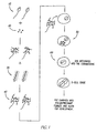
- figure 1 shows the basic steps involved in using one embodiment of the present invention to genetically modify cells or animals using a sperm vector.
- animal sperm cells 10 are collected by methods known in the art or purchased commercially from sources such as Birchwood Genetics in West Manchester, Ohio, and are bound together with linkers 20.
- linkers 20 are antibodies, preferably immunoglobulins of the types, IgG, IgA or IgM. These linkers bind or associate to the sperm cells' external surface through different molecular interactions such as ionic interaction, covalent bonds, Vander Waals forces, or ligand-receptor interaction.
- Circular or linear DNA molecules 30 then bind or attach to the linkers on the sperm-linker complex also through different molecular interactions such as ionic, covalent bonds, Vander Waals forces, or ligand-receptor interaction. These DNA molecules may encode for certain gene products, but they may also be disrupted genes, homologous with endogenous genes, that recombine into the chromosome to knockout a gene.
- the sperni-linker-DNA complex 40 formed can then be used to effectuate fertilization in vitro or in vivo. Upon fertilization, the DNA is introduced into the fertilized egg 50 and embryo 60 and can integrate into the chromosome, becoming a part of an animal or cell's genetic material.
- the binding, coupling, linking, attaching, or association of the sperm-linker-DNA complex can also be accomplished in vivo.
- the linker and the DNA can first be coupled or bound together in vitro. Afterwards, this linker-DNA complex can be injected directly or indirectly into a male animal's testicles.
- PCT Publications WO 99/40213 and WO 97/11597 disclose procedures for injecting DNA into the testicles.
- linker-DNA complex is an antibody attached with DNA molecules where the antibody specifically recognizes certain surface epitopes on sperm cells. Because of the acidic characteristic of naked DNA, it can ionically associate, bind or, couple with an antibody that has basic or positively charged properties. However, the DNA-linker interaction is not limited to ionic interaction.
- the complex can also be crosslinked by UV light to form covalent bonds by well known methods in the art. Both the DNA and the linker can also be modified by methods known in the art.
- the DNA can be biotinylated by adding biotinylated deoxynucleotides in a PCR reaction;
- the antibody can be modified or purchased with attached streptavidin, which binds tightly to the biotin on the DNA; or a secondary antibody, which is modified with streptavidin and recognizes the first antibody can also act as a secondary linker between the modified DNA and the first linker.
- the DNA-linker complex is injected into the testis of the non-human animal, this complex can seek out the sperm cells and bind to them. Fertilization can then occur in vivo via either natural copulation of the male and female non-human animals or by artificial insemination of the female with collected sperm cells. The collected sperm cells can also be used with in vitro fertilization techniques, which are well known in the art. On the other hand, if binding of the sperm-linker-DNA complex, as a whole, occurred in vitro, fertilization can be achieved by in vitro fertilization techniques. The fertilized eggs and resulting embryos can then be transplanted to surrogate non-human animal mothers for development. Alternatively, well known artificial insemination methods or injections of the sperm-linker-DNA complex directly into the oviduct of non-human female animals can also achieve fertilization in vivo.
- Livestock, poultry, or fish can be inserted with genes that encode for growth hormones to make them grow faster than normal or they can also be inserted with the somatotropin gene to increase muscle growth and decrease adipose tissue.
- Inserting genes such as interferon that boost the immune system or other genes, such as genes encoding for viral, prion, or bacterial proteins can also make these livestock, poultry, or fish disease or pathogen resistant.
- infectious pathogens include Salmonella, influenza virus, prion proteins for the Mad Cow Disease, etc.
- introducing DNA encoding for anti-sense RNA molecules, which are complementary to these viral, prion, or bacterial RNAs may also inhibit translation and production of proteins from these RNA. which limits growth and spread of these infectious pathogens.
- these genetically modified non-human animals can also produce therapeutic proteins, such as insulin, growth hormone, interferon, erythropoietin, colony stimulating factor (GM-CSF), t-PA, or factor VIII, in their milk by joining the genes for these proteins with promoters from mammary specific genes such as sheep's ⁇ -lactoglobulin, mouse whey acid protein, or bovine ⁇ SI-casein. Id.
- the non-human animal's milk can also be fortified with addition of humanized proteins, such as human lactoferrin that enhance the intestinal iron absorption in infants. Lonnerdal, B. (1996) Recombinant Human Milk Proteins -- An Opportunity and a Challenge, American Journal of Clinical Nutrition, Vol.
- This example illustrates the preparation of an antibody specific to sperm cells.
- sperm cells collected from male mice were injected back into mice as antigens to immunize and produce antibodies reactive to sperm-surface antigens.
- Monoclonal antibodies developed using common hybridoma techniques, were screened using flow cytometry to identify candidate antibodies that will bind to a series of different animals (mouse, pig, cow, sheep, goat, and chicken). Briefly, sperm cells were incubated with the different primary monoclonal antibodies, washed, and further incubated with a secondary antibody that specifically recognized mouse immunoglobulin.
- This secondary antibody which was commercially available and well known in the art, had fluorescent molecules such as fluorescein or rhodamine conjugated to it. Once the secondary antibody molecules were bound and washed, the flow-cytometry instrument or the FACS sorter counted the number of fluorescent sperm cells with bound primary and secondary antibodies from naked sperm cells.
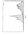
- Figure 2 - 7 show these flow-cytometry analyses for mAbC that bind to sperm cells of mouse, pig, cow, chicken, goat, and sheep, respectively.
- the Y-axis corresponds to the number of sperm cells detected while the X-axis is the relative intensity of fluorescence bound to the cell.
- Cross-lined peaks denote control reactions where the sperm cells were incubated only with the fluorescent anti-mouse immunoglobulin antibody.
- the shaded peaks denote the reactions where mAbC antibody and the secondary antibody were incubated with corresponding sperm cells in a mouse, pig, cow, chicken, goat, and sheep, respectively.
- Right shifts in the peaks denote positive binding of the mAbC antibody.
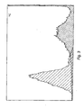
- fluorescence can be detected from goat sperm cells incubated with mAbC and the fluorescent secondary antibody as evidenced by the two right shaded peaks.
- the left shaded peak may suggest a population of the goat sperm cells that express the antigen recognized by mAbC at a lower level than the population in the right peak.
- the anti-mouse immunoglobulin fluorescent antibody seems to also bind to the goat sperm cells, but at a much reduced level than with mAbC acting as a linker.
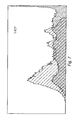
- fluorescence can be detected from sheep sperm cells incubated with mAbC and the fluorescent secondary antibody as evidenced by the right shaded peaks.
- the distribution of the peaks again suggests the possibility that different sperm cells have different levels of the antigen recognized by mAbC.
- mammalian sperm cells bind, at some lower level, to the fluorescent secondary antibody. Since the secondary antibody is directed to a mouse immunoglobulin, it may have cross reactivity to other mammalian proteins on the sperm cell surfaces, which are not present in the chicken sperm cells (figure 5). Nevertheless, the shifts in fluorescence peaks upon addition of mAbC clearly demonstrate the higher affinity of the mAbC antibody to different animal sperm cells.
- This example illustrates the ability of the monoclonal antibody mAbC to bind to DNA molecules through ionic interaction.
- lanes 1, 2, and 8 were controls with lane 1 being pure Sal I cut pCMV- ⁇ plasmid and lanes 2 and 8 being Sal I cut pCMV- ⁇ plasmid in Modified Tyrode's medium. Lanes 3, 4, 5, 6, and 7 corresponded to experimental reactions with the Sal I cut pCMV- ⁇ plasmid incubated with 0.2 ⁇ l, 1 ⁇ l, 2.5 ⁇ l, 6 ⁇ l, and 10 ⁇ l of mAbC at 0.5mg/ml.
- This example illustrates the binding or coupling of the DNA to the sperm via the linker or antibody.
- DNA molecules labeled with P 32 using standard end labeling techniques with T4 DNA polymerase, were incubated with mouse, pig, chicken, sheep, goat, and cow sperm cells together with either mAbC, mAbD, or a control antibody specific to a Drosophila protein. The amount of DNA binding was measured by scintillation counting. The ratio of sperm cells to antibody were as follows:
- Table 1 shows that with the presence of mAbC and mAbD, sperm cells significantly bound more labeled DNA compared with reactions with no antibody or with the Drosophila protein-specific antibody.
- Reactions 1 and 2 contained only sperm cells and labeled DNA, while reactions 3 and 4 contained the Drosophila-protein-specific antibody together with sperm cells and labeled DNA.
- Reactions 5 contained mAbD while reactions 6 and 7 contained mAbC together with sperm cells and labeled DNA.
- This example illustrates the procedures carried out to generate genetically modified mice.
- Sperm cells were collected from dissected epididymis of nine to twenty weeks old FVB male mice. Cut into small pieces, these epididymis tissues were incubated in 300 ⁇ l of Modified Tyrode's medium at pH 7 ⁇ 8 for one hour to allow the sperm cells to escape into the medium. Once the sperm cells were collected in 300 ⁇ l of medium, five micrograms of the linker antibody were added to one million sperm cells at 37°C for one hour. The sperm-linker complex was washed three times with 300 ⁇ l of Modified Tyrode's medium using a standard microcentrifuge set at 3000 rpm for one and a half minutes.
- the sperm-linker complex was finally resuspended in 300 ⁇ l of medium, and one microgram of linearized pCMV- ⁇ plasmid or a plasmid encoding for Hepatitis B surface antigen (HBsAg) was added and incubated for one hour.
- HBsAg Hepatitis B surface antigen
- FVB female mice To collect ovulated eggs, nine to twelve weeks FVB female mice each received an injection of 5 I.U. Pregnant Mares Serum (PMS) four days before the collection date and another 5 I.U. of human chorionic gonadotropin (hCG) two days before the collection date.
- PMS Pregnant Mares Serum
- hCG human chorionic gonadotropin
- Dissected ovulated eggs surrounded by cumulus cells were placed in a 35-mm petri dish containing a drop of Modified Tyrode's medium at room temperature. Afterwards, 300 ⁇ l of sperm-linker-DNA complex prepared as described above were added directly to the ovulated eggs. The whole mix was equilibrated with CO 2 at 37°C with mineral oil added on top to prevent evaporation.
- FIG. 11 shows the southern blot hybridization results with complementary probe sequences to HBsAg.
- Lanes 1-13 contained genomic DNA from mice born from pseudo-pregnant mice that received embryos fertilized with the sperm-linker-DNA complex described above; lanes C1-C7 contained genomic DNA from mice that were untreated or non-transgenic mice.
- Lanes 4, 5, and 8 show bands positive for HBsAg sequences integrated in the mice's genome, thus, demonstrating that three out the thirteen mice were genetically modified.
- This example illustrates the procedures carried out to generate genetically modified pigs.
- Ejaculated sperm cells from pigs were collected using methods generally known in the art of animal husbandry. Suspended in one milliliter of pig extender medium (purchased from Merck, Germany, Ref.N.R.13515/0001 - dilute mixture M3 for boar sperm), fifteen million sperm cells were incubated with five micrograms of the linker antibody for forty minutes at room temperature with intermittent shaking in between. After washing the sperm-linker mixture once with pig extender medium and finally resuspending the mixture in 1.5 ml of the same medium, five micrograms of the plasmid pSEAP2-control ( Figure 12, Clontech Laboratories, Inc., Cat.
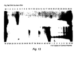
- Figure 13 shows the southern blot analysis of genomic DNA isolated from the tail tissues of these pigs. Briefly, genomic DNA isolated from these pigs were digested, run on a gel, and transferred to a nylon membrane according to methods well known in the art. The blot was then probed with labeled sequences from the Not I to BamH I region of the pSEAP2-control plasmid shown in Figure 12. In figure 13, M denotes the marker lanes, and 1-43 denotes the number of pigs analyzed.
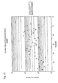
- eight lanes with increasing copies of pSEAP2-control plasmid molecules (1, 2, 2, 4, 4, 8, 16, and 32) were also loaded on the gel together with the DNA from the experimental pigs. These eight lanes were used to estimate the copy number of pSEAP2-control plasmid integrated into the pigs based on the densitometric intensities of the bands ( Figure 14). As can be seen in Figure 14, S5 had the highest intensity, which corresponds to lane 5 of figure 13.
- SEAP secreted alkaline phosphatase expressed from the pSEAP2-control plasmid were also detected in 70-day old genetically modified pigs. Serum from these pigs were collected and assayed for SEAP activity using Clontech's Great EscAPE TM SEAP Chemiluminescence Detection Kit (Cat. # K2041-1) and its protocol, which is incorporated herein by reference.
- the SEAP enzyme expressed from Clontech's pSEAP-2 vector is thermostable.
- the assay required the deactivation of the endogenous alkaline phosphatase enzyme by heating the samples at 65°C for thirty minutes before adding the chemiluminescence substrate.
- figure 15 shows the result of the assay without performing this heat deactivation step.
- the level of total alkaline phosphatase activity was not significantly different between the genetically modified pigs and non-transgenic control pigs.
- figure 16 shows the result including this heat deactivation step.
- SEAP activity was significantly higher in the genetically modified pigs than in the non-transgenic control pigs.
- the pSEAP2-control plasmid had integrated well in the pigs' genome and was actively expressing the SEAP enzyme.
Abstract
Description
- The present invention relates to the field of genetic modification in non-human animals.
- Efficient genetic modification of animals, especially in higher mammals, has been a major goal of researchers in the biotechnology field for the last two decades. Not only can genetic modification of animals advance our understanding of genes and genefunctions in multi-cell organisms, it can also serve useful applications in the bio-agricultural industry. Examples of these applications include raising livestock with desired characteristics such as faster growth rate, production of therapeutic proteins in milk, or even the generation of more "humanized" organs from animals for use in animal to human xenotransplantation.
- Current techniques to modify the genome include microinjection of foreign DNA into the pronuclei of fertilized eggs, delivery of foreign DNA into embryonic stem cells in vitro or blastomere cells in vivo through lipid-based agents, electroporation, or viral, infection. Aside from mice, however, current techniques have been reported to have had limited success in higher or larger animals. The microinjection technique, for example, has been reported to be technically very demanding and requires the use of highly sensitive and expensive equipment. The viability of embryos after microinjection has also been reported to be very poor. Wall, R.J., et. al. (1992) Making Transgenic Livestock, Genetic Engineering on a Large Scale, Journal of Cellular Biochemistry, Vol. 49, pp. 113-120. This has led researchers in the field to investigate alternative and easier ways of delivering genes into an animal.
- In 1989, Lavitrano, M., et. al. reported that simply incubating foreign DNA with mice's sperm cells and effecting fertilization in vitro could lead to genetically modified mice. Lavitrano, M., et. al. (1989) Sperm Cells as Vectors for introducing Foreign DNA into Eggs - Genetic Transformation of Mice, Cell. Vol. 57, pp. 717-723. Characterized as the "cold fusion" equivalent in biotechnology, this report generated much excitement in the field. Birnstiel. M., et. al. (1989) Dangerous Liaisons: Spermatozoa as Natural Vectors for Foreign DNA?, Cell. Vol. 57, pp. 701-702. Those skilled in the art, however, are reported to remain skeptical even to this day about the Lavitrano's report since a number of researchers in the field have reportedly failed to repeat the experiment. Brinster, R., et. al. (1989) No Simple Solution for Making Transgenic Mice, Cell, Vol. 59, pp. 239-241; Smith, K. (1999) Sperm Cell Mediated Transgenesis: A Review, Animal Biotechnology, Vol. 10(1&2), pp. 1-13.
- Over the last decade, efforts have continued to explore the use of sperm cells as a vector for mediating gene transfer in animals. Researchers have elucidated that sperm cells have the inherent ability to internalize foreign DNA. Francolini, M., et. al (1993) Evidence for Nuclear Internalization of Exogenous DNA into Mammalian Sperm Cells, Mol. Reprod. Devel., Vol. 34, pp. 133-139. Yet, certain inhibitory factors present in seminal fluid may inhibit this ability to take up DNA. Lavitrano, M., et. al. (1992) The Interaction Between Exogenous DNA and Sperm Cells, Mol. Reprod. Devel., Vol. 31, pp. 161-169. In addition, foreign DNA introduced into sperm cells may also suffer from extensive DNA rearrangement because in mature sperm cells, internalization of foreign DNA may activate certain endogenous nucleases in these cells. Maione, B. et. al. (1997) Activation of Endogenous Nucleases in Mature Sperm Cells upon Interaction with Exogenous DNA, DNA and Cell Biology, Vol. 16, pp. 1087-1097. Such rearrangement could threaten the usefulness of genetically modified animals using this technique.
- Gandolfi, F. in "Spermatozoa, DNA Binding and Transgenic Animals", Transgenic Research 7, 147-155 (1998) reports that the interaction between spermatozoa and exogenous DNA molecules could be accepted as a reliable experimental observation. However, according to Gandolfi F. much more severe obstacles to any easy exchange of genes at the time of fertilization were encountered after DNA had entered the oocyte since in most cases exogenous genes were completely or partially degraded and/or rearranged.
-
EP 0 867 114 A1 describes a method for preparing transgenic non-human vertebrate animals, wherein an exogenous DNA is injected into a testis of a mature non-human vertebrate male after forming a complex with liposome for the purpose of transduction of DNA into mammalian cells. - US 5,428,132 describes a DNA-antibody conjugate and the method for integration of foreign DNA into cells in a tissue-specific manner by conjugating the foreign DNA with a target specific antibody.
- Finally, Smith et al., "Sperm cell mediated transgenesis: A review", Animal Biotechnology, 10 (1 & 2), 1-13 (1999) reports about existing technologies using sperm cells as transgenic vectors published between 1989 and 1999.
- Other work with sperm cells as vector have focused on the use of either lipid-based agents or electroporation to deliver foreign DNA into the spenn cells. Smith, supra; Rottman R., et. al. (1996) Liposome-mediated Gene Transfer via Sperm Cells. High Transfer Efficiency and Persistence of Transgenes by Use of Liposomes and Sperm Cells and a Murine Amplification Element. Journal of Animal Breeding and Genetics. Vol. 113. pp. 401-411; PCT Publications WO 99/42569, WO 99/40213, and WO 97111597. Such methods may also suffer from the same problem of DNA internalization and exposure to nucleases that could cause rearrangement of the foreign DNA being introduced. In addition, lipid-based agents, which are often toxic, and electroporation may require extensive experimentation to prevent the death or the loss of spenn cell motility. Other techniques have also focused on using recombinant virus infection, as disclosed in PCT Publications WO 99/38991, or on using a "gene gun" with micro-carriers, as disclosed in PCT Publication WO 93/24626, to introduce foreign DNA into sperm cells. Such techniques may be technically challenging and may also affect the viability and motility of the sperm cells. They may also suffer from the same problem of DNA internalization and exposure to nucleases that could cause rearrangement of the foreign DNA being introduced.
- Since 1989, researchers have reported the use of sperm cells as vectors in different animals ranging from insects, marine animals, amphibians, birds, and mammals. Smith, supra. However, few reported that the genetic modification was observed in viable mature offspring. Smith, supra. More problematic is the fact that some reports used only PCR analysis to verify the existence of the foreign DNA in the cells. These reports are summarized in table one of Gandolfi, F. (1998) Spermatozoa, DNA Binding and Transgenic Animals, Transgenic Research, Vol. 7, pp. 147-155. Since PCR cannot distinguish between foreign DNA transmitted through episomes or Through the chromosomal DNA, Gandolfi has questioned the value of these reports stating that it "opens up an important argument relating, to appropriate evaluation of the results described in some reports." Gandolfi, supra. Episomal transmission is not as desirable as chromosomal transmission since the episome may be lost during subsequent cell division, and the desired effect of genetic modification may never be expressed in adult animals.
- Because an easy, non-toxic, and efficient way of genetically modifying animals, especially in higher mammals, can greatly advance this field, a new way of using sperm cells for delivering genes into animals is needed.
- The present invention is directed to a vector used to generate genetically modified non-human animals and cells. One aspect of this invention involves a vector that comprises a sperm cell and one or more polynucleotide molecules bound to a sperm cell through one or more non-liposome based linkers. The sperm cell is a non-human animal sperm cell. In one preferred embodiment of this invention, the one or more polynucleotide molecules encode for a gene product that confers desired characteristics in the cells or the animals. The linker of the present invention is an antibody, which binds with the external surface of the sperm cell. The linker interacts with one or more polynucleotide molecules preferably by ionic interaction. This interaction can also be carried out by different molecular interactions, including the use of another or secondary linker. The association of the sperm, linker, and the one or more polynucleotide can also occur in vitro or in vivo.
- Genetically modified cells or non-human animals can be derived from the fertilization of an animal egg cell with the vector described above. Fertilization can occur in vitro or in vivo. Genetic modification occurs with the polynucleotide molecule integrating, wholly or partially, into the cell or animal's genome. Further, cells, such as sperm cells or egg cells, and cell lines can be derived from these genetically modified non-human animals or their descendants.
- The genetically modified non-human animals derived from the use of the sperm vector described above possess certain desired characteristics. Examples of these characteristics include faster growth rates, disease or pathogen resistance, high production of certain proteins in milk, and organs suitable for animal to human xenotransplantation.
-
- Figure 1 is a pictorial representation of the basic steps involved in using one embodiment of the present invention.
- Figure 2 shows a flow-cytometry result of binding a sperm-specific antibody to mice's sperm cells as embodied in one aspect of the present invention.
- Figure 3 shows a flow-cytometry result of binding a sperm-specific antibody to pig's sperm cells as embodied in one aspect of the present invention.
- Figure 4 shows a flow-cytometry result of binding a sperm-specific antibody to cow's sperm cells as embodied in one aspect of the present invention.
- Figure 5 shows a flow-cytometry result of binding a sperm-specific antibody to chicken's sperm cells as embodied in one aspect of the present invention.
- Figure 6 shows a flow-cytometry result of binding a sperm-specific antibody to goat's sperm cells as embodied in one aspect of the present invention.
- Figure 7 shows a flow-cytometry result of binding a sperm-specific antibody to sheep's sperm cells as embodied in one aspect of the present invention.
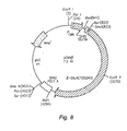
- Figure 8 shows a plasmid map of pCMV-β.
- Figure 9 shows an agarose-gel analysis of a sperm-specific antibody binding to pCMV-β plasmid.
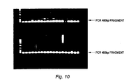
- Figure 10 show results of PCR analysis for the detection of pCMV-β sequences in genomic DNA isolated from mice's embryos genetically modified according to one embodiment of the present invention.
- Figure 11 shows results of southern-blot analysis for the detection of Hepatitis B surface-antigen gene-sequence in mice-tail-genomic DNA with this gene-sequence being integrated into the mice's chromosome according to one embodiment of the present invention.
- Figure 12 shows the plasmid map of pSEAP-2-control.
- Figure 13 shows the result of southem-blot analysis for the detection of pSEAP2-control plasmid sequence in the genomic DNA isolated from tail tissues of genetically modified pigs according to one embodiment of the present invention.
- Figure 14 shows the copy number of integrated pSJEAP2-control plasmid in four genetically modified pigs based on densitometric intensities of bands in Figure 13.
- Figure 15 and 16 show the results of enzyme assays for secreted alkaline phosphatase found in serum of pigs genetically modified according to one embodiment of the present invention.
- Generally, figure 1 shows the basic steps involved in using one embodiment of the present invention to genetically modify cells or animals using a sperm vector. Briefly,
animal sperm cells 10, are collected by methods known in the art or purchased commercially from sources such as Birchwood Genetics in West Manchester, Ohio, and are bound together withlinkers 20. These linkers are antibodies, preferably immunoglobulins of the types, IgG, IgA or IgM. These linkers bind or associate to the sperm cells' external surface through different molecular interactions such as ionic interaction, covalent bonds, Vander Waals forces, or ligand-receptor interaction. Circular orlinear DNA molecules 30 then bind or attach to the linkers on the sperm-linker complex also through different molecular interactions such as ionic, covalent bonds, Vander Waals forces, or ligand-receptor interaction. These DNA molecules may encode for certain gene products, but they may also be disrupted genes, homologous with endogenous genes, that recombine into the chromosome to knockout a gene. The sperni-linker-DNA complex 40 formed can then be used to effectuate fertilization in vitro or in vivo. Upon fertilization, the DNA is introduced into the fertilizedegg 50 andembryo 60 and can integrate into the chromosome, becoming a part of an animal or cell's genetic material. - Alternatively, the binding, coupling, linking, attaching, or association of the sperm-linker-DNA complex can also be accomplished in vivo. The linker and the DNA can first be coupled or bound together in vitro. Afterwards, this linker-DNA complex can be injected directly or indirectly into a male animal's testicles. PCT Publications WO 99/40213 and WO 97/11597 disclose procedures for injecting DNA into the testicles.
- An example of a linker-DNA complex is an antibody attached with DNA molecules where the antibody specifically recognizes certain surface epitopes on sperm cells. Because of the acidic characteristic of naked DNA, it can ionically associate, bind or, couple with an antibody that has basic or positively charged properties. However, the DNA-linker interaction is not limited to ionic interaction. The complex can also be crosslinked by UV light to form covalent bonds by well known methods in the art. Both the DNA and the linker can also be modified by methods known in the art. For example, the DNA can be biotinylated by adding biotinylated deoxynucleotides in a PCR reaction; the antibody can be modified or purchased with attached streptavidin, which binds tightly to the biotin on the DNA; or a secondary antibody, which is modified with streptavidin and recognizes the first antibody can also act as a secondary linker between the modified DNA and the first linker.
- If the DNA-linker complex is injected into the testis of the non-human animal, this complex can seek out the sperm cells and bind to them. Fertilization can then occur in vivo via either natural copulation of the male and female non-human animals or by artificial insemination of the female with collected sperm cells. The collected sperm cells can also be used with in vitro fertilization techniques, which are well known in the art. On the other hand, if binding of the sperm-linker-DNA complex, as a whole, occurred in vitro, fertilization can be achieved by in vitro fertilization techniques. The fertilized eggs and resulting embryos can then be transplanted to surrogate non-human animal mothers for development. Alternatively, well known artificial insemination methods or injections of the sperm-linker-DNA complex directly into the oviduct of non-human female animals can also achieve fertilization in vivo.
- Genetically modified non-human animals can serve a number of useful applications. Livestock, poultry, or fish can be inserted with genes that encode for growth hormones to make them grow faster than normal or they can also be inserted with the somatotropin gene to increase muscle growth and decrease adipose tissue. Pursel, V. G., et.al. (1989) Genetic Engineering of Livestock, Science, Vol. 244, pp. 1281-1288; Etherton, T.D., et. al. (1993) Mechanism by which Somatotropin Decreases Adipose Tissue Growth, American Journal of Clinical Nutrition, Vol. 58 (Supp.), pp. 287S-295S. Inserting genes such as interferon that boost the immune system or other genes, such as genes encoding for viral, prion, or bacterial proteins, can also make these livestock, poultry, or fish disease or pathogen resistant. Examples of these infectious pathogens include Salmonella, influenza virus, prion proteins for the Mad Cow Disease, etc. Alternatively, introducing DNA encoding for anti-sense RNA molecules, which are complementary to these viral, prion, or bacterial RNAs, may also inhibit translation and production of proteins from these RNA. which limits growth and spread of these infectious pathogens.
- Moreover, in non-human animals, including insects such as silkworms, that produce raw materials for clothing such as wool and silk, inserting genes for biochemical enzymes that produce the rate-limiting amino acid may increase production of these raw materials. In sheep, for example, the availability of the amino-acid cysteine limits the production of wool. Inserting bacterial genes that encode for serine transacetylase and O-acetylserine sulfhydrylase may increase the conversion of serine and acetyl-CoA into cysteine, which in turn may increase production of wool. Ward, K., (1991) The Application of Transgenic Techniques for the Improvement of Domestic Animal Productivity, Current Opinion in Biotechnology, Vol. 2, pp. 834-839.
- Furthermore, these genetically modified non-human animals can also produce therapeutic proteins, such as insulin, growth hormone, interferon, erythropoietin, colony stimulating factor (GM-CSF), t-PA, or factor VIII, in their milk by joining the genes for these proteins with promoters from mammary specific genes such as sheep's β-lactoglobulin, mouse whey acid protein, or bovine αSI-casein. Id. On the other hand; the non-human animal's milk can also be fortified with addition of humanized proteins, such as human lactoferrin that enhance the intestinal iron absorption in infants. Lonnerdal, B. (1996) Recombinant Human Milk Proteins -- An Opportunity and a Challenge, American Journal of Clinical Nutrition, Vol. 63, pp. 622-626. Genetically modified pigs can even be a source for more "humanized" organs in animal to human xenotransplantation using genes such as human decay accelerating factor. Cozzi, E., et. al. (1994) Expression of Human Decay Accelerating Factor in Transgenics Pigs, Transplantation Proceedings, Vol. 26, pp. 1402-1403.
- The following examples demonstrate that the inventor has produced a number of genetically modified animals using the sperm vector as described above. Methods in molecular genetics, flow cytometry, antibody production, hybridoma technology, in vitro fertilization, embryo manipulation, and artificial insemination used but not explicitly described in this disclosure had already been amply reported in the scientific literature. These methods are well within the ability of one skilled in the art.
- This example illustrates the preparation of an antibody specific to sperm cells.
- Sperm cells collected from male mice were injected back into mice as antigens to immunize and produce antibodies reactive to sperm-surface antigens. Monoclonal antibodies, developed using common hybridoma techniques, were screened using flow cytometry to identify candidate antibodies that will bind to a series of different animals (mouse, pig, cow, sheep, goat, and chicken). Briefly, sperm cells were incubated with the different primary monoclonal antibodies, washed, and further incubated with a secondary antibody that specifically recognized mouse immunoglobulin. This secondary antibody, which was commercially available and well known in the art, had fluorescent molecules such as fluorescein or rhodamine conjugated to it. Once the secondary antibody molecules were bound and washed, the flow-cytometry instrument or the FACS sorter counted the number of fluorescent sperm cells with bound primary and secondary antibodies from naked sperm cells.
- Figure 2 - 7 show these flow-cytometry analyses for mAbC that bind to sperm cells of mouse, pig, cow, chicken, goat, and sheep, respectively. The Y-axis corresponds to the number of sperm cells detected while the X-axis is the relative intensity of fluorescence bound to the cell. Cross-lined peaks denote control reactions where the sperm cells were incubated only with the fluorescent anti-mouse immunoglobulin antibody. On the other hand, the shaded peaks denote the reactions where mAbC antibody and the secondary antibody were incubated with corresponding sperm cells in a mouse, pig, cow, chicken, goat, and sheep, respectively. Right shifts in the peaks denote positive binding of the mAbC antibody.
- As can be seen in figure 2, greater fluorescence signals can be detected from mouse sperm cells incubated with mAbC and the fluorescent secondary antibody compared with sperm cells incubated with fluorescent secondary antibody alone. Similarly, in figure 3 and 4, greater fluorescence can be detected from pig and cow sperm cells, respectively, incubated with mAbC and the fluorescent secondary antibody as evidenced by the right shaded peaks.
- In figure 5, the incubation of the fluorescence antibody alone with the chicken sperm cells did not result in any fluorescence being detected in these sperm cells. In contrast, the right peak signified fluorescence in the chicken sperm cells that have attached mAbC antibodies. Figure 5 also shows that some population of chicken sperm cells may not express the antigen recognized by mAbC as evidence by the left shaded peak.
- In figure 6, fluorescence can be detected from goat sperm cells incubated with mAbC and the fluorescent secondary antibody as evidenced by the two right shaded peaks. The left shaded peak may suggest a population of the goat sperm cells that express the antigen recognized by mAbC at a lower level than the population in the right peak. In contrast with the, chicken sperm cells incubated with only the fluorescent secondary antibody in figure 5, the anti-mouse immunoglobulin fluorescent antibody seems to also bind to the goat sperm cells, but at a much reduced level than with mAbC acting as a linker.
- Similarly, in figure 7, fluorescence can be detected from sheep sperm cells incubated with mAbC and the fluorescent secondary antibody as evidenced by the right shaded peaks. The distribution of the peaks again suggests the possibility that different sperm cells have different levels of the antigen recognized by mAbC.
- As seen in figures 2, 3, 4, 6, and 7, mammalian sperm cells bind, at some lower level, to the fluorescent secondary antibody. Since the secondary antibody is directed to a mouse immunoglobulin, it may have cross reactivity to other mammalian proteins on the sperm cell surfaces, which are not present in the chicken sperm cells (figure 5). Nevertheless, the shifts in fluorescence peaks upon addition of mAbC clearly demonstrate the higher affinity of the mAbC antibody to different animal sperm cells.
- This example illustrates the ability of the monoclonal antibody mAbC to bind to DNA molecules through ionic interaction.
- Different volumes of purified solutions of mAbC at a concentration of 0.5 mg/ml were added to DNA solutions containing 300ng of Sal I cut pCMV-β plasmid (Figure 8, Clontech Laboratories, Inc., Cat. # 6177-1). After incubating the mixtures at room temperature for forty minutes, the mixtures were loaded on a regular one percent agarose gel and run at 20 milli-amps for one hour. Afterwards, the DNA was stained with Ethidium Bromide and visualized under UV light.
- In figure 9,
lanes lane 1 being pure Sal I cut pCMV-β plasmid andlanes Lanes lanes - This example illustrates the binding or coupling of the DNA to the sperm via the linker or antibody.
- DNA molecules, labeled with P32 using standard end labeling techniques with T4 DNA polymerase, were incubated with mouse, pig, chicken, sheep, goat, and cow sperm cells together with either mAbC, mAbD, or a control antibody specific to a Drosophila protein. The amount of DNA binding was measured by scintillation counting. The ratio of sperm cells to antibody were as follows:
- Mouse -- 400 thousand sperm cells to 600ng of labeled DNA;
- Pig -- 600 thousand sperm cells to 800ng labeled DNA;
- Chicken -- 300 thousand sperm cells to 500ng of labeled DNA;
- Sheep -- 1 million sperm cells to 500ng of labeled DNA;
- Goat -- 1 million sperm cells to 500ng of labeled DNA; and
- Cow -- 1 million sperm cells to 500ng of labeled DNA.
- Table 1 shows that with the presence of mAbC and mAbD, sperm cells significantly bound more labeled DNA compared with reactions with no antibody or with the Drosophila protein-specific antibody.
Reactions reactions Reactions 5 contained mAbD whilereactions Table 1 Reactions Mouse (cpm) Pig (cpm) Chicken (cpm) Sheep (cpm) Goat (cpm) Cow (cpm) 1 no antibody 12471 12971 5830 15367 17749 12766 2 no antibody 15814 13713 6383 13259 16574 14398 3 Control Antibody 11541 10531 N/D 14018 155347 15351 4 Control Antibody 13653 14038 N/D 12834 15997 13918 5 mAbD 18900 16220 10314 N/D N/D N/ D 6 mAbC 18139 16269 7294 19368 20385 20417 7 mAbC 19314 17343 9865 18437 19543 18643 N/D = Not determined - This example illustrates the procedures carried out to generate genetically modified mice.
- Sperm cells were collected from dissected epididymis of nine to twenty weeks old FVB male mice. Cut into small pieces, these epididymis tissues were incubated in 300µl of Modified Tyrode's medium at
pH 7~8 for one hour to allow the sperm cells to escape into the medium. Once the sperm cells were collected in 300µl of medium, five micrograms of the linker antibody were added to one million sperm cells at 37°C for one hour. The sperm-linker complex was washed three times with 300µl of Modified Tyrode's medium using a standard microcentrifuge set at 3000 rpm for one and a half minutes. The sperm-linker complex was finally resuspended in 300µl of medium, and one microgram of linearized pCMV-β plasmid or a plasmid encoding for Hepatitis B surface antigen (HBsAg) was added and incubated for one hour. - To collect ovulated eggs, nine to twelve weeks FVB female mice each received an injection of 5 I.U. Pregnant Mares Serum (PMS) four days before the collection date and another 5 I.U. of human chorionic gonadotropin (hCG) two days before the collection date. Dissected ovulated eggs surrounded by cumulus cells were placed in a 35-mm petri dish containing a drop of Modified Tyrode's medium at room temperature. Afterwards, 300µl of sperm-linker-DNA complex prepared as described above were added directly to the ovulated eggs. The whole mix was equilibrated with CO2 at 37°C with mineral oil added on top to prevent evaporation. After four hours of in vitro fertilization at 37°C, fertilized eggs were collected with capillary tubes and washed thrice with CZB medium. The embryos were further incubated in 300µl of CZB medium for 20-22 hrs before being transferred to oviducts of pseudo-pregnant female mice.
- To confirm the presence of the pCMV-β plasmid, genomic DNA isolated from embryos, ten days after transplantation into the pseudo-pregnant female mice, were analyzed by PCR using primers that detect a 480bp fragment corresponding to the CMV promoter region of the pCMV-β plasmid (Figure 8). In figure 10,
lanes Lanes - To confirm integration of the HBsAg plasmid into the mice genome, southern blot analysis were also performed. Genomic DNA isolated from mice's tails were digested, ran on a gel, transferred to a nylon membrane according to methods known in the art. Figure 11 shows the southern blot hybridization results with complementary probe sequences to HBsAg. Lanes 1-13 contained genomic DNA from mice born from pseudo-pregnant mice that received embryos fertilized with the sperm-linker-DNA complex described above; lanes C1-C7 contained genomic DNA from mice that were untreated or non-transgenic mice.
Lanes - This example illustrates the procedures carried out to generate genetically modified pigs.
- Ejaculated sperm cells from pigs were collected using methods generally known in the art of animal husbandry. Suspended in one milliliter of pig extender medium (purchased from Merck, Germany, Ref.N.R.13515/0001 - dilute mixture M3 for boar sperm), fifteen million sperm cells were incubated with five micrograms of the linker antibody for forty minutes at room temperature with intermittent shaking in between. After washing the sperm-linker mixture once with pig extender medium and finally resuspending the mixture in 1.5 ml of the same medium, five micrograms of the plasmid pSEAP2-control (Figure 12, Clontech Laboratories, Inc., Cat. # 6052-1) were added and incubated with the mixture for forty minutes at room temperature. Direct injections of 200µl of the resulting sperm-linker-DNA complex into the oviducts of anesthetized female pigs resulted in fertilization in vivo.
- After the pigs were born and grown to 70-day-old pigs, they were analyzed for the presence of the pSEAP2-control plasmid. Figure 13 shows the southern blot analysis of genomic DNA isolated from the tail tissues of these pigs. Briefly, genomic DNA isolated from these pigs were digested, run on a gel, and transferred to a nylon membrane according to methods well known in the art. The blot was then probed with labeled sequences from the Not I to BamH I region of the pSEAP2-control plasmid shown in Figure 12. In figure 13, M denotes the marker lanes, and 1-43 denotes the number of pigs analyzed. Hybridization signals in
lanes lane 5 of figure 13. - In another study, secreted alkaline phosphatase (SEAP) expressed from the pSEAP2-control plasmid were also detected in 70-day old genetically modified pigs. Serum from these pigs were collected and assayed for SEAP activity using Clontech's Great EscAPE™ SEAP Chemiluminescence Detection Kit (Cat. # K2041-1) and its protocol, which is incorporated herein by reference. The SEAP enzyme expressed from Clontech's pSEAP-2 vector is thermostable. Thus, to determine the level of SEAP activity as opposed to the pigs' endogenous alkaline phosphatase enzyme activity, the assay required the deactivation of the endogenous alkaline phosphatase enzyme by heating the samples at 65°C for thirty minutes before adding the chemiluminescence substrate. As a control, figure 15 shows the result of the assay without performing this heat deactivation step. The level of total alkaline phosphatase activity was not significantly different between the genetically modified pigs and non-transgenic control pigs. In contrast, figure 16 shows the result including this heat deactivation step. Without the endogenous alkaline phosphatase activity, SEAP activity was significantly higher in the genetically modified pigs than in the non-transgenic control pigs. Thus, the pSEAP2-control plasmid had integrated well in the pigs' genome and was actively expressing the SEAP enzyme.
- The preceding examples demonstrate that the inventor has produced a number of genetically modified animals using the sperm vector as described above. These data are intended only as examples and are not intended to limit the invention to these examples.
Claims (10)
- A vector for genetically modifying non-human animals or cells comprising:a non-human sperm cell,at least one non-liposome based linker bound to the external surface of the non-human sperm cell, wherein the linker is an antibody, andat least one polynucleotide molecule, wherein the polynucleotide molecule is bound to the non-human sperm cell through the one non-liposome based linker.
- The vector of claim 1, wherein the antibody preferentially binds to an external surface of the non-human sperm cell.
- The vector of claim 1 or 2, wherein the at least one polynucleotide molecule is a DNA molecule.
- The vector of claim 3, wherein the DNA molecule encodes for a gene product.
- The vector of claim 4, wherein the gene product is an RNA molecule.
- The vector of claim 4, wherein the gene product is a protein.
- The vector of any of claims 1 to 6, wherein the antibody interacts with the at least one polynucleotide molecule via molecular interactions selected from the group consisting of ionic interaction, covalent interaction, Van der Waals interaction, and ligand-receptor interaction.
- The vector of any of claims 1 to 6, wherein the antibody interacts with the at least one polynucleotide molecule through at least one secondary non-liposome based linker.
- The vector of any of claims 1 to 8, wherein the at least one polynucleotide molecule binds to the external surface of the non-human sperm cell through the antibody.
- A non-liposome based linker for attaching at least one polynucleotide molecule to an external surface of a non-human sperm cell, wherein the linker is an antibody, and wherein the antibody is bound to the external surface of the non-human sperm cell and to the polynucleotide molecule.
Priority Applications (1)
| Application Number | Priority Date | Filing Date | Title |
|---|---|---|---|
| EP06125927A EP1760155A3 (en) | 2000-03-28 | 2001-03-05 | Non-human sperm-DNA complexes for geneticallly modifying non-human animals |
Applications Claiming Priority (3)
| Application Number | Priority Date | Filing Date | Title |
|---|---|---|---|
| US09/537,861 US7067308B1 (en) | 2000-03-28 | 2000-03-28 | Vector for genetically modifying non-human animals |
| US537861 | 2000-03-28 | ||
| PCT/US2001/007018 WO2001073094A2 (en) | 2000-03-28 | 2001-03-05 | Novel sperm-dna complexes for genetically modifying non-human animals |
Related Child Applications (1)
| Application Number | Title | Priority Date | Filing Date |
|---|---|---|---|
| EP06125927A Division EP1760155A3 (en) | 2000-03-28 | 2001-03-05 | Non-human sperm-DNA complexes for geneticallly modifying non-human animals |
Publications (2)
| Publication Number | Publication Date |
|---|---|
| EP1268836A2 EP1268836A2 (en) | 2003-01-02 |
| EP1268836B1 true EP1268836B1 (en) | 2006-12-13 |
Family
ID=24144410
Family Applications (2)
| Application Number | Title | Priority Date | Filing Date |
|---|---|---|---|
| EP06125927A Withdrawn EP1760155A3 (en) | 2000-03-28 | 2001-03-05 | Non-human sperm-DNA complexes for geneticallly modifying non-human animals |
| EP01918350A Expired - Lifetime EP1268836B1 (en) | 2000-03-28 | 2001-03-05 | Non-human sperm-dna complexes for genetically modifying non-human animals |
Family Applications Before (1)
| Application Number | Title | Priority Date | Filing Date |
|---|---|---|---|
| EP06125927A Withdrawn EP1760155A3 (en) | 2000-03-28 | 2001-03-05 | Non-human sperm-DNA complexes for geneticallly modifying non-human animals |
Country Status (10)
| Country | Link |
|---|---|
| US (2) | US7067308B1 (en) |
| EP (2) | EP1760155A3 (en) |
| JP (1) | JP2003528617A (en) |
| CN (1) | CN1432067A (en) |
| AR (1) | AR028298A1 (en) |
| AT (1) | ATE348184T1 (en) |
| AU (1) | AU2001245438A1 (en) |
| DE (1) | DE60125156T2 (en) |
| PE (1) | PE20011214A1 (en) |
| WO (1) | WO2001073094A2 (en) |
Families Citing this family (10)
| Publication number | Priority date | Publication date | Assignee | Title |
|---|---|---|---|---|
| US7067308B1 (en) * | 2000-03-28 | 2006-06-27 | Bioagri Corporation | Vector for genetically modifying non-human animals |
| US7053187B2 (en) * | 2000-03-28 | 2006-05-30 | Gioagri Corporation | Sperm-specific monoclonal antibody, mAbC |
| MX2009012317A (en) | 2007-05-16 | 2010-04-01 | Mat Malta Advanced Technologie | Treatment and prevention of influenza. |
| US9157097B2 (en) | 2008-09-25 | 2015-10-13 | Proteovec Holding, L.L.C. | Vectors for production of growth hormone |
| WO2010036976A2 (en) | 2008-09-25 | 2010-04-01 | Transgenrx, Inc. | Novel vectors for production of antibodies |
| CN113186149A (en) * | 2009-04-08 | 2021-07-30 | 加利福尼亚大学董事会 | DNA-cell conjugates |
| US9150881B2 (en) | 2009-04-09 | 2015-10-06 | Proteovec Holding, L.L.C. | Production of proteins using transposon-based vectors |
| CN102453720B (en) * | 2010-10-28 | 2013-01-30 | 华中农业大学 | Fusion promoter capable of realizing high-efficiency expression in pig muscular tissue |
| RS59997B1 (en) * | 2011-10-28 | 2020-04-30 | Regeneron Pharma | Genetically modified mice expressing chimeric major histocompatibility complex (mhc) ii molecules |
| US20150024497A1 (en) * | 2013-07-19 | 2015-01-22 | Elwha Llc | Methods and Systems for Utilizing Sperm for Molecular Delivery |
Family Cites Families (16)
| Publication number | Priority date | Publication date | Assignee | Title |
|---|---|---|---|---|
| US5428132A (en) | 1987-10-11 | 1995-06-27 | United States Of America | Conjugate and method for integration of foreign DNA into cells |
| IT1228210B (en) | 1989-01-10 | 1991-06-05 | Consiglio Nazionale Ricerche | PROCEDURE FOR THE INTRODUCTION OF EXOGENOUS DNA INTO SOMATIC AND GERMINAL CELLS OF ANIMALS. |
| IL92529A0 (en) | 1989-12-03 | 1990-08-31 | Yissum Res Dev Co | Generation of transgenic vertebrates by employing transformed sperm cells via artificial insemination |
| US5521291A (en) | 1991-09-30 | 1996-05-28 | Boehringer Ingelheim International, Gmbh | Conjugates for introducing nucleic acid into higher eucaryotic cells |
| US6063630A (en) * | 1991-11-05 | 2000-05-16 | Transkaryotic Therapies, Inc. | Targeted introduction of DNA into primary or secondary cells and their use for gene therapy |
| WO1993024626A1 (en) | 1992-05-28 | 1993-12-09 | Scientific Dimensions Usa, Inc. | Transgenic animal production with biolistically transformed spermatozoa |
| RU2081914C1 (en) | 1994-07-21 | 1997-06-20 | Андрей Вадимович Кузнецов | Method of foreign dna incorporation in spermatozoons |
| US5744335A (en) | 1995-09-19 | 1998-04-28 | Mirus Corporation | Process of transfecting a cell with a polynucleotide mixed with an amphipathic compound and a DNA-binding protein |
| JPH10304790A (en) | 1995-09-29 | 1998-11-17 | Hoechst Japan Ltd | Formation of transgenic animal |
| CN1112165C (en) | 1997-08-15 | 2003-06-25 | 中国农业大学 | Method for producing transfer-gene animals by using sperm dielectric transfer DNA |
| CN1289370A (en) | 1998-01-28 | 2001-03-28 | 宝酒造株式会社 | Method for transferring gene into germ cell |
| US6686199B2 (en) | 1998-02-09 | 2004-02-03 | Tran Xenogen, Inc. | Genetic manipulation of spermatogonia |
| IL123411A0 (en) | 1998-02-22 | 1998-09-24 | Kimron Veterinary Inst | High efficiency methods and compositions for integrating exogenous dna into genomic dna of sperm |
| BR9913644A (en) | 1998-08-11 | 2001-11-20 | Anthony C F Perry | Method of carrying out transgenesis |
| US7067308B1 (en) * | 2000-03-28 | 2006-06-27 | Bioagri Corporation | Vector for genetically modifying non-human animals |
| US7053187B2 (en) * | 2000-03-28 | 2006-05-30 | Gioagri Corporation | Sperm-specific monoclonal antibody, mAbC |
-
2000
- 2000-03-28 US US09/537,861 patent/US7067308B1/en not_active Expired - Fee Related
-
2001
- 2001-03-05 AU AU2001245438A patent/AU2001245438A1/en not_active Abandoned
- 2001-03-05 AT AT01918350T patent/ATE348184T1/en not_active IP Right Cessation
- 2001-03-05 CN CN01810496A patent/CN1432067A/en active Pending
- 2001-03-05 JP JP2001570810A patent/JP2003528617A/en active Pending
- 2001-03-05 DE DE60125156T patent/DE60125156T2/en not_active Expired - Lifetime
- 2001-03-05 EP EP06125927A patent/EP1760155A3/en not_active Withdrawn
- 2001-03-05 EP EP01918350A patent/EP1268836B1/en not_active Expired - Lifetime
- 2001-03-05 WO PCT/US2001/007018 patent/WO2001073094A2/en active IP Right Grant
- 2001-03-27 PE PE2001000287A patent/PE20011214A1/en not_active Application Discontinuation
- 2001-03-28 AR ARP010101471A patent/AR028298A1/en unknown
-
2006
- 2006-04-20 US US11/407,851 patent/US20070214512A1/en not_active Abandoned
Non-Patent Citations (6)
Also Published As
| Publication number | Publication date |
|---|---|
| AU2001245438A1 (en) | 2001-10-08 |
| EP1760155A3 (en) | 2007-03-14 |
| EP1760155A2 (en) | 2007-03-07 |
| DE60125156D1 (en) | 2007-01-25 |
| US7067308B1 (en) | 2006-06-27 |
| US20070214512A1 (en) | 2007-09-13 |
| DE60125156T2 (en) | 2007-09-20 |
| JP2003528617A (en) | 2003-09-30 |
| WO2001073094A2 (en) | 2001-10-04 |
| AR028298A1 (en) | 2003-04-30 |
| PE20011214A1 (en) | 2001-11-29 |
| EP1268836A2 (en) | 2003-01-02 |
| WO2001073094A3 (en) | 2002-05-30 |
| ATE348184T1 (en) | 2007-01-15 |
| CN1432067A (en) | 2003-07-23 |
Similar Documents
| Publication | Publication Date | Title |
|---|---|---|
| US20070214512A1 (en) | Vector for genetically modifying non-human animals | |
| US11230697B2 (en) | Enhanced expression of human or humanized immunoglobulin in non-human transgenic animals | |
| Chang et al. | Effective generation of transgenic pigs and mice by linker based sperm-mediated gene transfer. | |
| US5523222A (en) | Polyelectrolyte DNA conjugation and genetic transformation of an animal | |
| KR102096731B1 (en) | Compositions and methods for inhibiting endogenous immunoglobulin genes and producing transgenic human idiotype antibodies | |
| US7803981B2 (en) | Transgenic ungulates capable of human antibody production | |
| US20020028488A1 (en) | Transgenic avian species for making human and chimeric antibodies | |
| JP2002510973A (en) | Transgenic animals expressing human Fc receptor | |
| EP0665883A1 (en) | Tetracycline repressor-mediated binary regulation system for control of gene expression in transgenic animals | |
| US10370641B2 (en) | Enhanced expression of human or humanized immunoglobulin in non-human transgenic animals | |
| RU2005117629A (en) | TRANSGENIC ANTIC ANIMALS HAVING A REDUCED PRIONAL PROTEIN ACTIVITY, AND THEIR APPLICATIONS | |
| KR20050000558A (en) | Transgenic Ungulates Capable of Human Antibody Production | |
| CA2233694A1 (en) | Method for preparing transgenic animal | |
| EP0350052A2 (en) | Transgenic animals transformed with autonomously replicating sequence-containing plasmid | |
| US20020194638A1 (en) | Vector for genetically modifying non-human animals | |
| RU2402211C2 (en) | Method for production of transgenic rabbits producing proteins into mammary gland | |
| CN100526460C (en) | Transgenic ungulates having reduced prion protein activity and uses thereof | |
| Velander | Polyelectrolyte DNA conjugation and genetic transformation of an animal | |
| US20100235929A1 (en) | Compositions and Methods for Producing Antibodies Having Human Idiotypes in Transgenic Birds | |
| Gordon | Modification of the germ line in animals | |
| Page | Evaluation of techniques for the production of transgenic animals | |
| Brem | Generation of recombinant antibody transgenic farm animals | |
| CYTOPLASMIC | Raymond L. Page", Christopher G. Russell", Rodolfo S. Canseco", Francis C. Gwazdauskas", John L. Johnson", and William H. Velander | |
| Wall et al. | Alteration of the genome as a method to increase diversity in animal germplasm resources |
Legal Events
| Date | Code | Title | Description |
|---|---|---|---|
| PUAI | Public reference made under article 153(3) epc to a published international application that has entered the european phase |
Free format text: ORIGINAL CODE: 0009012 |
|
| 17P | Request for examination filed |
Effective date: 20021028 |
|
| AK | Designated contracting states |
Kind code of ref document: A2 Designated state(s): AT BE CH CY DE DK ES FI FR GB GR IE IT LI LU MC NL PT SE TR |
|
| AX | Request for extension of the european patent |
Free format text: AL;LT;LV;MK;RO;SI |
|
| RIN1 | Information on inventor provided before grant (corrected) |
Inventor name: WANG, KANGSHENG |
|
| 17Q | First examination report despatched |
Effective date: 20030528 |
|
| RTI1 | Title (correction) |
Free format text: NON-HUMAN SPERM-DNA COMPLEXES FOR GENETICALLY MODIFYING NON-HUMAN ANIMALS |
|
| GRAP | Despatch of communication of intention to grant a patent |
Free format text: ORIGINAL CODE: EPIDOSNIGR1 |
|
| GRAS | Grant fee paid |
Free format text: ORIGINAL CODE: EPIDOSNIGR3 |
|
| GRAA | (expected) grant |
Free format text: ORIGINAL CODE: 0009210 |
|
| AK | Designated contracting states |
Kind code of ref document: B1 Designated state(s): AT BE CH CY DE DK ES FI FR GB GR IE IT LI LU MC NL PT SE TR |
|
| PG25 | Lapsed in a contracting state [announced via postgrant information from national office to epo] |
Ref country code: NL Free format text: LAPSE BECAUSE OF FAILURE TO SUBMIT A TRANSLATION OF THE DESCRIPTION OR TO PAY THE FEE WITHIN THE PRESCRIBED TIME-LIMIT Effective date: 20061213 Ref country code: BE Free format text: LAPSE BECAUSE OF FAILURE TO SUBMIT A TRANSLATION OF THE DESCRIPTION OR TO PAY THE FEE WITHIN THE PRESCRIBED TIME-LIMIT Effective date: 20061213 Ref country code: CH Free format text: LAPSE BECAUSE OF FAILURE TO SUBMIT A TRANSLATION OF THE DESCRIPTION OR TO PAY THE FEE WITHIN THE PRESCRIBED TIME-LIMIT Effective date: 20061213 Ref country code: FI Free format text: LAPSE BECAUSE OF FAILURE TO SUBMIT A TRANSLATION OF THE DESCRIPTION OR TO PAY THE FEE WITHIN THE PRESCRIBED TIME-LIMIT Effective date: 20061213 Ref country code: DK Free format text: LAPSE BECAUSE OF FAILURE TO SUBMIT A TRANSLATION OF THE DESCRIPTION OR TO PAY THE FEE WITHIN THE PRESCRIBED TIME-LIMIT Effective date: 20061213 Ref country code: LI Free format text: LAPSE BECAUSE OF FAILURE TO SUBMIT A TRANSLATION OF THE DESCRIPTION OR TO PAY THE FEE WITHIN THE PRESCRIBED TIME-LIMIT Effective date: 20061213 Ref country code: AT Free format text: LAPSE BECAUSE OF FAILURE TO SUBMIT A TRANSLATION OF THE DESCRIPTION OR TO PAY THE FEE WITHIN THE PRESCRIBED TIME-LIMIT Effective date: 20061213 |
|
| REG | Reference to a national code |
Ref country code: GB Ref legal event code: FG4D |
|
| REG | Reference to a national code |
Ref country code: CH Ref legal event code: EP |
|
| REG | Reference to a national code |
Ref country code: IE Ref legal event code: FG4D |
|
| REF | Corresponds to: |
Ref document number: 60125156 Country of ref document: DE Date of ref document: 20070125 Kind code of ref document: P |
|
| PG25 | Lapsed in a contracting state [announced via postgrant information from national office to epo] |
Ref country code: SE Free format text: LAPSE BECAUSE OF FAILURE TO SUBMIT A TRANSLATION OF THE DESCRIPTION OR TO PAY THE FEE WITHIN THE PRESCRIBED TIME-LIMIT Effective date: 20070313 |
|
| PG25 | Lapsed in a contracting state [announced via postgrant information from national office to epo] |
Ref country code: ES Free format text: LAPSE BECAUSE OF FAILURE TO SUBMIT A TRANSLATION OF THE DESCRIPTION OR TO PAY THE FEE WITHIN THE PRESCRIBED TIME-LIMIT Effective date: 20070324 |
|
| PG25 | Lapsed in a contracting state [announced via postgrant information from national office to epo] |
Ref country code: PT Free format text: LAPSE BECAUSE OF FAILURE TO SUBMIT A TRANSLATION OF THE DESCRIPTION OR TO PAY THE FEE WITHIN THE PRESCRIBED TIME-LIMIT Effective date: 20070514 |
|
| NLV1 | Nl: lapsed or annulled due to failure to fulfill the requirements of art. 29p and 29m of the patents act | ||
| REG | Reference to a national code |
Ref country code: CH Ref legal event code: PL |
|
| ET | Fr: translation filed | ||
| PLBE | No opposition filed within time limit |
Free format text: ORIGINAL CODE: 0009261 |
|
| STAA | Information on the status of an ep patent application or granted ep patent |
Free format text: STATUS: NO OPPOSITION FILED WITHIN TIME LIMIT |
|
| 26N | No opposition filed |
Effective date: 20070914 |
|
| PG25 | Lapsed in a contracting state [announced via postgrant information from national office to epo] |
Ref country code: MC Free format text: LAPSE BECAUSE OF NON-PAYMENT OF DUE FEES Effective date: 20070331 Ref country code: IE Free format text: LAPSE BECAUSE OF NON-PAYMENT OF DUE FEES Effective date: 20070305 |
|
| PG25 | Lapsed in a contracting state [announced via postgrant information from national office to epo] |
Ref country code: GR Free format text: LAPSE BECAUSE OF FAILURE TO SUBMIT A TRANSLATION OF THE DESCRIPTION OR TO PAY THE FEE WITHIN THE PRESCRIBED TIME-LIMIT Effective date: 20070314 |
|
| PG25 | Lapsed in a contracting state [announced via postgrant information from national office to epo] |
Ref country code: CY Free format text: LAPSE BECAUSE OF FAILURE TO SUBMIT A TRANSLATION OF THE DESCRIPTION OR TO PAY THE FEE WITHIN THE PRESCRIBED TIME-LIMIT Effective date: 20061213 Ref country code: LU Free format text: LAPSE BECAUSE OF NON-PAYMENT OF DUE FEES Effective date: 20070305 |
|
| PG25 | Lapsed in a contracting state [announced via postgrant information from national office to epo] |
Ref country code: TR Free format text: LAPSE BECAUSE OF FAILURE TO SUBMIT A TRANSLATION OF THE DESCRIPTION OR TO PAY THE FEE WITHIN THE PRESCRIBED TIME-LIMIT Effective date: 20061213 |
|
| REG | Reference to a national code |
Ref country code: GB Ref legal event code: 732E Free format text: REGISTERED BETWEEN 20101021 AND 20101027 |
|
| REG | Reference to a national code |
Ref country code: FR Ref legal event code: TP |
|
| REG | Reference to a national code |
Ref country code: FR Ref legal event code: PLFP Year of fee payment: 15 |
|
| PGFP | Annual fee paid to national office [announced via postgrant information from national office to epo] |
Ref country code: IT Payment date: 20150326 Year of fee payment: 15 |
|
| PGFP | Annual fee paid to national office [announced via postgrant information from national office to epo] |
Ref country code: GB Payment date: 20150324 Year of fee payment: 15 Ref country code: FR Payment date: 20150319 Year of fee payment: 15 |
|
| PGFP | Annual fee paid to national office [announced via postgrant information from national office to epo] |
Ref country code: DE Payment date: 20150529 Year of fee payment: 15 |
|
| REG | Reference to a national code |
Ref country code: DE Ref legal event code: R119 Ref document number: 60125156 Country of ref document: DE |
|
| GBPC | Gb: european patent ceased through non-payment of renewal fee |
Effective date: 20160305 |
|
| REG | Reference to a national code |
Ref country code: FR Ref legal event code: ST Effective date: 20161130 |
|
| PG25 | Lapsed in a contracting state [announced via postgrant information from national office to epo] |
Ref country code: DE Free format text: LAPSE BECAUSE OF NON-PAYMENT OF DUE FEES Effective date: 20161001 Ref country code: GB Free format text: LAPSE BECAUSE OF NON-PAYMENT OF DUE FEES Effective date: 20160305 Ref country code: FR Free format text: LAPSE BECAUSE OF NON-PAYMENT OF DUE FEES Effective date: 20160331 |
|
| PG25 | Lapsed in a contracting state [announced via postgrant information from national office to epo] |
Ref country code: IT Free format text: LAPSE BECAUSE OF NON-PAYMENT OF DUE FEES Effective date: 20160305 |