EP1209235A1 - Expression vector for fusion gene and method for producing immobilized enzyme - Google Patents
Expression vector for fusion gene and method for producing immobilized enzyme Download PDFInfo
- Publication number
- EP1209235A1 EP1209235A1 EP01309792A EP01309792A EP1209235A1 EP 1209235 A1 EP1209235 A1 EP 1209235A1 EP 01309792 A EP01309792 A EP 01309792A EP 01309792 A EP01309792 A EP 01309792A EP 1209235 A1 EP1209235 A1 EP 1209235A1
- Authority
- EP
- European Patent Office
- Prior art keywords
- protein
- yeast
- immobilized
- cell wall
- fusion gene
- Prior art date
- Legal status (The legal status is an assumption and is not a legal conclusion. Google has not performed a legal analysis and makes no representation as to the accuracy of the status listed.)
- Withdrawn
Links
- 108090000623 proteins and genes Proteins 0.000 title claims abstract description 156
- 230000004927 fusion Effects 0.000 title claims abstract description 58
- 239000013604 expression vector Substances 0.000 title claims abstract description 50
- 108010093096 Immobilized Enzymes Proteins 0.000 title claims abstract description 35
- 238000004519 manufacturing process Methods 0.000 title claims description 15
- 102000004169 proteins and genes Human genes 0.000 claims abstract description 85
- 240000004808 Saccharomyces cerevisiae Species 0.000 claims abstract description 70
- 230000014509 gene expression Effects 0.000 claims abstract description 48
- 210000005253 yeast cell Anatomy 0.000 claims abstract description 40
- 102000004190 Enzymes Human genes 0.000 claims abstract description 36
- 108090000790 Enzymes Proteins 0.000 claims abstract description 36
- 108700023372 Glycosyltransferases Proteins 0.000 claims abstract description 30
- 102000051366 Glycosyltransferases Human genes 0.000 claims abstract description 28
- 150000001413 amino acids Chemical class 0.000 claims description 41
- 238000000034 method Methods 0.000 claims description 34
- 210000002421 cell wall Anatomy 0.000 claims description 33
- 235000000346 sugar Nutrition 0.000 claims description 31
- 239000002344 surface layer Substances 0.000 claims description 23
- 238000012258 culturing Methods 0.000 claims description 11
- 150000008163 sugars Chemical class 0.000 claims description 11
- 238000012217 deletion Methods 0.000 claims description 9
- 230000037430 deletion Effects 0.000 claims description 9
- 108700014210 glycosyltransferase activity proteins Proteins 0.000 claims description 6
- FWMNVWWHGCHHJJ-SKKKGAJSSA-N 4-amino-1-[(2r)-6-amino-2-[[(2r)-2-[[(2r)-2-[[(2r)-2-amino-3-phenylpropanoyl]amino]-3-phenylpropanoyl]amino]-4-methylpentanoyl]amino]hexanoyl]piperidine-4-carboxylic acid Chemical compound C([C@H](C(=O)N[C@H](CC(C)C)C(=O)N[C@H](CCCCN)C(=O)N1CCC(N)(CC1)C(O)=O)NC(=O)[C@H](N)CC=1C=CC=CC=1)C1=CC=CC=C1 FWMNVWWHGCHHJJ-SKKKGAJSSA-N 0.000 claims description 4
- 125000003275 alpha amino acid group Chemical group 0.000 claims 6
- 102000045442 glycosyltransferase activity proteins Human genes 0.000 claims 1
- 230000000694 effects Effects 0.000 abstract description 23
- 230000002542 deteriorative effect Effects 0.000 abstract description 2
- 235000014680 Saccharomyces cerevisiae Nutrition 0.000 description 59
- 210000004027 cell Anatomy 0.000 description 44
- 239000000243 solution Substances 0.000 description 34
- 238000006243 chemical reaction Methods 0.000 description 25
- 108020001507 fusion proteins Proteins 0.000 description 24
- 102000037865 fusion proteins Human genes 0.000 description 24
- 239000000203 mixture Substances 0.000 description 18
- 101150038013 PIR gene Proteins 0.000 description 17
- 239000002585 base Substances 0.000 description 16
- 229930004094 glycosylphosphatidylinositol Natural products 0.000 description 16
- WEVYAHXRMPXWCK-UHFFFAOYSA-N Acetonitrile Chemical compound CC#N WEVYAHXRMPXWCK-UHFFFAOYSA-N 0.000 description 15
- YBYRMVIVWMBXKQ-UHFFFAOYSA-N phenylmethanesulfonyl fluoride Chemical compound FS(=O)(=O)CC1=CC=CC=C1 YBYRMVIVWMBXKQ-UHFFFAOYSA-N 0.000 description 14
- FAPWRFPIFSIZLT-UHFFFAOYSA-M Sodium chloride Chemical compound [Na+].[Cl-] FAPWRFPIFSIZLT-UHFFFAOYSA-M 0.000 description 12
- 210000004899 c-terminal region Anatomy 0.000 description 11
- 239000013612 plasmid Substances 0.000 description 10
- 101150009006 HIS3 gene Proteins 0.000 description 9
- 101100394989 Rhodopseudomonas palustris (strain ATCC BAA-98 / CGA009) hisI gene Proteins 0.000 description 9
- QKNYBSVHEMOAJP-UHFFFAOYSA-N 2-amino-2-(hydroxymethyl)propane-1,3-diol;hydron;chloride Chemical compound Cl.OCC(N)(CO)CO QKNYBSVHEMOAJP-UHFFFAOYSA-N 0.000 description 8
- 239000003513 alkali Substances 0.000 description 8
- 239000000872 buffer Substances 0.000 description 8
- 239000003550 marker Substances 0.000 description 8
- 239000000758 substrate Substances 0.000 description 8
- 239000011534 wash buffer Substances 0.000 description 8
- 241000235343 Saccharomycetales Species 0.000 description 7
- 108010019236 Fucosyltransferases Proteins 0.000 description 6
- 102000006471 Fucosyltransferases Human genes 0.000 description 6
- 108060003306 Galactosyltransferase Proteins 0.000 description 6
- 102000030902 Galactosyltransferase Human genes 0.000 description 6
- 239000007836 KH2PO4 Substances 0.000 description 6
- 230000003321 amplification Effects 0.000 description 6
- 239000006285 cell suspension Substances 0.000 description 6
- BNIILDVGGAEEIG-UHFFFAOYSA-L disodium hydrogen phosphate Chemical compound [Na+].[Na+].OP([O-])([O-])=O BNIILDVGGAEEIG-UHFFFAOYSA-L 0.000 description 6
- 229910000397 disodium phosphate Inorganic materials 0.000 description 6
- 239000012634 fragment Substances 0.000 description 6
- 238000004128 high performance liquid chromatography Methods 0.000 description 6
- 238000011534 incubation Methods 0.000 description 6
- 239000012528 membrane Substances 0.000 description 6
- 229910000402 monopotassium phosphate Inorganic materials 0.000 description 6
- 238000003199 nucleic acid amplification method Methods 0.000 description 6
- GNSKLFRGEWLPPA-UHFFFAOYSA-M potassium dihydrogen phosphate Chemical compound [K+].OP(O)([O-])=O GNSKLFRGEWLPPA-UHFFFAOYSA-M 0.000 description 6
- 239000011780 sodium chloride Substances 0.000 description 6
- 239000002904 solvent Substances 0.000 description 6
- 239000006228 supernatant Substances 0.000 description 6
- 238000005516 engineering process Methods 0.000 description 5
- 238000002360 preparation method Methods 0.000 description 5
- 238000010561 standard procedure Methods 0.000 description 5
- AVBGNFCMKJOFIN-UHFFFAOYSA-N triethylammonium acetate Chemical compound CC(O)=O.CCN(CC)CC AVBGNFCMKJOFIN-UHFFFAOYSA-N 0.000 description 5
- XLYOFNOQVPJJNP-UHFFFAOYSA-N water Substances O XLYOFNOQVPJJNP-UHFFFAOYSA-N 0.000 description 5
- IJGRMHOSHXDMSA-UHFFFAOYSA-N Atomic nitrogen Chemical compound N#N IJGRMHOSHXDMSA-UHFFFAOYSA-N 0.000 description 4
- 101100464170 Candida albicans (strain SC5314 / ATCC MYA-2876) PIR1 gene Proteins 0.000 description 4
- 108020004414 DNA Proteins 0.000 description 4
- TWRXJAOTZQYOKJ-UHFFFAOYSA-L Magnesium chloride Chemical compound [Mg+2].[Cl-].[Cl-] TWRXJAOTZQYOKJ-UHFFFAOYSA-L 0.000 description 4
- 101150049683 PIR1 gene Proteins 0.000 description 4
- 101100231811 Saccharomyces cerevisiae (strain ATCC 204508 / S288c) HSP150 gene Proteins 0.000 description 4
- 101100464174 Schizosaccharomyces pombe (strain 972 / ATCC 24843) pir2 gene Proteins 0.000 description 4
- 239000011324 bead Substances 0.000 description 4
- 238000003776 cleavage reaction Methods 0.000 description 4
- 238000010276 construction Methods 0.000 description 4
- 239000008188 pellet Substances 0.000 description 4
- 239000000047 product Substances 0.000 description 4
- 230000007017 scission Effects 0.000 description 4
- 229940035893 uracil Drugs 0.000 description 4
- GOJUJUVQIVIZAV-UHFFFAOYSA-N 2-amino-4,6-dichloropyrimidine-5-carbaldehyde Chemical group NC1=NC(Cl)=C(C=O)C(Cl)=N1 GOJUJUVQIVIZAV-UHFFFAOYSA-N 0.000 description 3
- QTBSBXVTEAMEQO-UHFFFAOYSA-N Acetic acid Chemical compound CC(O)=O QTBSBXVTEAMEQO-UHFFFAOYSA-N 0.000 description 3
- 101150000468 DUSP11 gene Proteins 0.000 description 3
- PEDCQBHIVMGVHV-UHFFFAOYSA-N Glycerine Chemical compound OCC(O)CO PEDCQBHIVMGVHV-UHFFFAOYSA-N 0.000 description 3
- -1 LEU2 Proteins 0.000 description 3
- 229910021380 Manganese Chloride Inorganic materials 0.000 description 3
- GLFNIEUTAYBVOC-UHFFFAOYSA-L Manganese chloride Chemical compound Cl[Mn]Cl GLFNIEUTAYBVOC-UHFFFAOYSA-L 0.000 description 3
- 241000235347 Schizosaccharomyces pombe Species 0.000 description 3
- HEMHJVSKTPXQMS-UHFFFAOYSA-M Sodium hydroxide Chemical compound [OH-].[Na+] HEMHJVSKTPXQMS-UHFFFAOYSA-M 0.000 description 3
- LUEWUZLMQUOBSB-UHFFFAOYSA-N UNPD55895 Natural products OC1C(O)C(O)C(CO)OC1OC1C(CO)OC(OC2C(OC(OC3C(OC(O)C(O)C3O)CO)C(O)C2O)CO)C(O)C1O LUEWUZLMQUOBSB-UHFFFAOYSA-N 0.000 description 3
- LHAOFBCHXGZGOR-NAVBLJQLSA-N alpha-D-Manp-(1->3)-alpha-D-Manp-(1->2)-alpha-D-Manp Chemical compound O[C@H]1[C@H](O)[C@@H](CO)O[C@H](O)[C@H]1O[C@@H]1[C@@H](O)[C@@H](O[C@@H]2[C@H]([C@@H](O)[C@H](O)[C@@H](CO)O2)O)[C@H](O)[C@@H](CO)O1 LHAOFBCHXGZGOR-NAVBLJQLSA-N 0.000 description 3
- LUEWUZLMQUOBSB-MHJOMNRISA-N beta-D-Manp-(1->4)-beta-D-Manp-(1->4)-beta-D-Manp-(1->4)-D-Manp Chemical compound O[C@H]1[C@@H](O)[C@H](O)[C@@H](CO)O[C@H]1O[C@@H]1[C@@H](CO)O[C@@H](O[C@@H]2[C@H](O[C@@H](O[C@@H]3[C@H](OC(O)[C@@H](O)[C@H]3O)CO)[C@@H](O)[C@H]2O)CO)[C@@H](O)[C@H]1O LUEWUZLMQUOBSB-MHJOMNRISA-N 0.000 description 3
- 238000005119 centrifugation Methods 0.000 description 3
- 238000010367 cloning Methods 0.000 description 3
- 238000004925 denaturation Methods 0.000 description 3
- 230000036425 denaturation Effects 0.000 description 3
- 239000011521 glass Substances 0.000 description 3
- 210000002288 golgi apparatus Anatomy 0.000 description 3
- 238000002347 injection Methods 0.000 description 3
- 239000007924 injection Substances 0.000 description 3
- 229940062780 lacto-n-neotetraose Drugs 0.000 description 3
- 239000007788 liquid Substances 0.000 description 3
- 239000011565 manganese chloride Substances 0.000 description 3
- FYGDTMLNYKFZSV-UHFFFAOYSA-N mannotriose Natural products OC1C(O)C(O)C(CO)OC1OC1C(CO)OC(OC2C(OC(O)C(O)C2O)CO)C(O)C1O FYGDTMLNYKFZSV-UHFFFAOYSA-N 0.000 description 3
- 238000005259 measurement Methods 0.000 description 3
- 229920001542 oligosaccharide Polymers 0.000 description 3
- 230000008569 process Effects 0.000 description 3
- 108091008146 restriction endonucleases Proteins 0.000 description 3
- 150000004043 trisaccharides Chemical class 0.000 description 3
- 239000013598 vector Substances 0.000 description 3
- 238000005406 washing Methods 0.000 description 3
- OWEGMIWEEQEYGQ-UHFFFAOYSA-N 100676-05-9 Natural products OC1C(O)C(O)C(CO)OC1OCC1C(O)C(O)C(O)C(OC2C(OC(O)C(O)C2O)CO)O1 OWEGMIWEEQEYGQ-UHFFFAOYSA-N 0.000 description 2
- MVMSCBBUIHUTGJ-UHFFFAOYSA-N 10108-97-1 Natural products C1=2NC(N)=NC(=O)C=2N=CN1C(C(C1O)O)OC1COP(O)(=O)OP(O)(=O)OC1OC(CO)C(O)C(O)C1O MVMSCBBUIHUTGJ-UHFFFAOYSA-N 0.000 description 2
- JKMHFZQWWAIEOD-UHFFFAOYSA-N 2-[4-(2-hydroxyethyl)piperazin-1-yl]ethanesulfonic acid Chemical compound OCC[NH+]1CCN(CCS([O-])(=O)=O)CC1 JKMHFZQWWAIEOD-UHFFFAOYSA-N 0.000 description 2
- GUBGYTABKSRVRQ-PZPXDAEZSA-N 4β-mannobiose Chemical compound O[C@H]1[C@@H](O)[C@H](O)[C@@H](CO)O[C@H]1O[C@@H]1[C@@H](CO)O[C@@H](O)[C@@H](O)[C@H]1O GUBGYTABKSRVRQ-PZPXDAEZSA-N 0.000 description 2
- QGZKDVFQNNGYKY-UHFFFAOYSA-N Ammonia Chemical compound N QGZKDVFQNNGYKY-UHFFFAOYSA-N 0.000 description 2
- 101100327917 Caenorhabditis elegans chup-1 gene Proteins 0.000 description 2
- VTYYLEPIZMXCLO-UHFFFAOYSA-L Calcium carbonate Chemical compound [Ca+2].[O-]C([O-])=O VTYYLEPIZMXCLO-UHFFFAOYSA-L 0.000 description 2
- 241000283707 Capra Species 0.000 description 2
- OKTJSMMVPCPJKN-UHFFFAOYSA-N Carbon Chemical compound [C] OKTJSMMVPCPJKN-UHFFFAOYSA-N 0.000 description 2
- WQZGKKKJIJFFOK-QTVWNMPRSA-N D-mannopyranose Chemical compound OC[C@H]1OC(O)[C@@H](O)[C@@H](O)[C@@H]1O WQZGKKKJIJFFOK-QTVWNMPRSA-N 0.000 description 2
- 241000588724 Escherichia coli Species 0.000 description 2
- LQEBEXMHBLQMDB-UHFFFAOYSA-N GDP-L-fucose Natural products OC1C(O)C(O)C(C)OC1OP(O)(=O)OP(O)(=O)OCC1C(O)C(O)C(N2C3=C(C(N=C(N)N3)=O)N=C2)O1 LQEBEXMHBLQMDB-UHFFFAOYSA-N 0.000 description 2
- MVMSCBBUIHUTGJ-GDJBGNAASA-N GDP-alpha-D-mannose Chemical compound C([C@H]1O[C@H]([C@@H]([C@@H]1O)O)N1C=2N=C(NC(=O)C=2N=C1)N)OP(O)(=O)OP(O)(=O)O[C@H]1O[C@H](CO)[C@@H](O)[C@H](O)[C@@H]1O MVMSCBBUIHUTGJ-GDJBGNAASA-N 0.000 description 2
- LQEBEXMHBLQMDB-JGQUBWHWSA-N GDP-beta-L-fucose Chemical compound O[C@H]1[C@H](O)[C@H](O)[C@H](C)O[C@@H]1OP(O)(=O)OP(O)(=O)OC[C@@H]1[C@@H](O)[C@@H](O)[C@H](N2C3=C(C(NC(N)=N3)=O)N=C2)O1 LQEBEXMHBLQMDB-JGQUBWHWSA-N 0.000 description 2
- 108010043685 GPI-Linked Proteins Proteins 0.000 description 2
- 102000002702 GPI-Linked Proteins Human genes 0.000 description 2
- 239000007995 HEPES buffer Substances 0.000 description 2
- 108010087568 Mannosyltransferases Proteins 0.000 description 2
- 102000006722 Mannosyltransferases Human genes 0.000 description 2
- 108010052285 Membrane Proteins Proteins 0.000 description 2
- DKXNBNKWCZZMJT-UHFFFAOYSA-N O4-alpha-D-Mannopyranosyl-D-mannose Natural products O=CC(O)C(O)C(C(O)CO)OC1OC(CO)C(O)C(O)C1O DKXNBNKWCZZMJT-UHFFFAOYSA-N 0.000 description 2
- 241000235070 Saccharomyces Species 0.000 description 2
- HSCJRCZFDFQWRP-ABVWGUQPSA-N UDP-alpha-D-galactose Chemical compound O[C@@H]1[C@@H](O)[C@@H](O)[C@@H](CO)O[C@@H]1OP(O)(=O)OP(O)(=O)OC[C@@H]1[C@@H](O)[C@@H](O)[C@H](N2C(NC(=O)C=C2)=O)O1 HSCJRCZFDFQWRP-ABVWGUQPSA-N 0.000 description 2
- XSQUKJJJFZCRTK-UHFFFAOYSA-N Urea Chemical compound NC(N)=O XSQUKJJJFZCRTK-UHFFFAOYSA-N 0.000 description 2
- HSCJRCZFDFQWRP-UHFFFAOYSA-N Uridindiphosphoglukose Natural products OC1C(O)C(O)C(CO)OC1OP(O)(=O)OP(O)(=O)OCC1C(O)C(O)C(N2C(NC(=O)C=C2)=O)O1 HSCJRCZFDFQWRP-UHFFFAOYSA-N 0.000 description 2
- 239000013543 active substance Substances 0.000 description 2
- 238000000137 annealing Methods 0.000 description 2
- 230000008275 binding mechanism Effects 0.000 description 2
- 239000004202 carbamide Substances 0.000 description 2
- 229910052799 carbon Inorganic materials 0.000 description 2
- 210000000170 cell membrane Anatomy 0.000 description 2
- 239000013611 chromosomal DNA Substances 0.000 description 2
- 210000000349 chromosome Anatomy 0.000 description 2
- 101150115879 gma12 gene Proteins 0.000 description 2
- 230000003100 immobilizing effect Effects 0.000 description 2
- 238000010166 immunofluorescence Methods 0.000 description 2
- 238000012744 immunostaining Methods 0.000 description 2
- 229930193965 lacto-N-fucopentaose Natural products 0.000 description 2
- 230000004807 localization Effects 0.000 description 2
- 229910001629 magnesium chloride Inorganic materials 0.000 description 2
- 230000004060 metabolic process Effects 0.000 description 2
- 238000002156 mixing Methods 0.000 description 2
- 239000003068 molecular probe Substances 0.000 description 2
- 229910052757 nitrogen Inorganic materials 0.000 description 2
- 238000000746 purification Methods 0.000 description 2
- 230000010076 replication Effects 0.000 description 2
- 239000012508 resin bead Substances 0.000 description 2
- 150000003839 salts Chemical class 0.000 description 2
- 238000012216 screening Methods 0.000 description 2
- 239000000126 substance Substances 0.000 description 2
- 238000012546 transfer Methods 0.000 description 2
- 238000006276 transfer reaction Methods 0.000 description 2
- 238000001262 western blot Methods 0.000 description 2
- FQVLRGLGWNWPSS-BXBUPLCLSA-N (4r,7s,10s,13s,16r)-16-acetamido-13-(1h-imidazol-5-ylmethyl)-10-methyl-6,9,12,15-tetraoxo-7-propan-2-yl-1,2-dithia-5,8,11,14-tetrazacycloheptadecane-4-carboxamide Chemical compound N1C(=O)[C@@H](NC(C)=O)CSSC[C@@H](C(N)=O)NC(=O)[C@H](C(C)C)NC(=O)[C@H](C)NC(=O)[C@@H]1CC1=CN=CN1 FQVLRGLGWNWPSS-BXBUPLCLSA-N 0.000 description 1
- HVCOBJNICQPDBP-UHFFFAOYSA-N 3-[3-[3,5-dihydroxy-6-methyl-4-(3,4,5-trihydroxy-6-methyloxan-2-yl)oxyoxan-2-yl]oxydecanoyloxy]decanoic acid;hydrate Chemical compound O.OC1C(OC(CC(=O)OC(CCCCCCC)CC(O)=O)CCCCCCC)OC(C)C(O)C1OC1C(O)C(O)C(O)C(C)O1 HVCOBJNICQPDBP-UHFFFAOYSA-N 0.000 description 1
- 102100035277 4-galactosyl-N-acetylglucosaminide 3-alpha-L-fucosyltransferase FUT6 Human genes 0.000 description 1
- 102100034035 Alcohol dehydrogenase 1A Human genes 0.000 description 1
- 102100034044 All-trans-retinol dehydrogenase [NAD(+)] ADH1B Human genes 0.000 description 1
- 101710193111 All-trans-retinol dehydrogenase [NAD(+)] ADH4 Proteins 0.000 description 1
- 101710204899 Alpha-agglutinin Proteins 0.000 description 1
- 101100288313 Arabidopsis thaliana KTI4 gene Proteins 0.000 description 1
- 102100030981 Beta-alanine-activating enzyme Human genes 0.000 description 1
- 101150008604 CAN1 gene Proteins 0.000 description 1
- 101100224748 Caenorhabditis elegans pir-1 gene Proteins 0.000 description 1
- 241000222120 Candida <Saccharomycetales> Species 0.000 description 1
- 108091026890 Coding region Proteins 0.000 description 1
- 206010059866 Drug resistance Diseases 0.000 description 1
- 101150006554 FUT6 gene Proteins 0.000 description 1
- 101150094690 GAL1 gene Proteins 0.000 description 1
- 101150038242 GAL10 gene Proteins 0.000 description 1
- 102100028501 Galanin peptides Human genes 0.000 description 1
- 102100024637 Galectin-10 Human genes 0.000 description 1
- 108700039691 Genetic Promoter Regions Proteins 0.000 description 1
- 108700007698 Genetic Terminator Regions Proteins 0.000 description 1
- 101000892220 Geobacillus thermodenitrificans (strain NG80-2) Long-chain-alcohol dehydrogenase 1 Proteins 0.000 description 1
- 229920001503 Glucan Polymers 0.000 description 1
- 108010055629 Glucosyltransferases Proteins 0.000 description 1
- 102000000340 Glucosyltransferases Human genes 0.000 description 1
- 229930186217 Glycolipid Natural products 0.000 description 1
- HVLSXIKZNLPZJJ-TXZCQADKSA-N HA peptide Chemical compound C([C@@H](C(=O)N[C@@H](CC(O)=O)C(=O)N[C@@H](C(C)C)C(=O)N1[C@@H](CCC1)C(=O)N[C@@H](CC(O)=O)C(=O)N[C@@H](CC=1C=CC(O)=CC=1)C(=O)N[C@@H](C)C(O)=O)NC(=O)[C@H]1N(CCC1)C(=O)[C@@H](N)CC=1C=CC(O)=CC=1)C1=CC=C(O)C=C1 HVLSXIKZNLPZJJ-TXZCQADKSA-N 0.000 description 1
- 101001022175 Homo sapiens 4-galactosyl-N-acetylglucosaminide 3-alpha-L-fucosyltransferase FUT6 Proteins 0.000 description 1
- 101000780443 Homo sapiens Alcohol dehydrogenase 1A Proteins 0.000 description 1
- 101000773364 Homo sapiens Beta-alanine-activating enzyme Proteins 0.000 description 1
- 101100121078 Homo sapiens GAL gene Proteins 0.000 description 1
- 108010058683 Immobilized Proteins Proteins 0.000 description 1
- 101150063995 KRE2 gene Proteins 0.000 description 1
- ROHFNLRQFUQHCH-YFKPBYRVSA-N L-leucine Chemical compound CC(C)C[C@H](N)C(O)=O ROHFNLRQFUQHCH-YFKPBYRVSA-N 0.000 description 1
- ROHFNLRQFUQHCH-UHFFFAOYSA-N Leucine Natural products CC(C)CC(N)C(O)=O ROHFNLRQFUQHCH-UHFFFAOYSA-N 0.000 description 1
- 101150053531 MNN1 gene Proteins 0.000 description 1
- 102100023315 N-acetyllactosaminide beta-1,6-N-acetylglucosaminyl-transferase Human genes 0.000 description 1
- 108010056664 N-acetyllactosaminide beta-1,6-N-acetylglucosaminyltransferase Proteins 0.000 description 1
- 101150014136 SUC2 gene Proteins 0.000 description 1
- 102000003838 Sialyltransferases Human genes 0.000 description 1
- 108090000141 Sialyltransferases Proteins 0.000 description 1
- 101150033985 TPI gene Proteins 0.000 description 1
- 101150032817 TPI1 gene Proteins 0.000 description 1
- 241000973887 Takayama Species 0.000 description 1
- 108700015934 Triose-phosphate isomerases Proteins 0.000 description 1
- 102100033598 Triosephosphate isomerase Human genes 0.000 description 1
- 239000008351 acetate buffer Substances 0.000 description 1
- 238000005273 aeration Methods 0.000 description 1
- 239000000910 agglutinin Substances 0.000 description 1
- 238000013019 agitation Methods 0.000 description 1
- 229910021529 ammonia Inorganic materials 0.000 description 1
- 230000008901 benefit Effects 0.000 description 1
- 239000013060 biological fluid Substances 0.000 description 1
- 230000015572 biosynthetic process Effects 0.000 description 1
- 239000007978 cacodylate buffer Substances 0.000 description 1
- 229910000019 calcium carbonate Inorganic materials 0.000 description 1
- 150000001720 carbohydrates Chemical class 0.000 description 1
- 235000014633 carbohydrates Nutrition 0.000 description 1
- 230000005754 cellular signaling Effects 0.000 description 1
- 230000007547 defect Effects 0.000 description 1
- 239000003599 detergent Substances 0.000 description 1
- 150000002016 disaccharides Chemical class 0.000 description 1
- 238000004520 electroporation Methods 0.000 description 1
- 210000002472 endoplasmic reticulum Anatomy 0.000 description 1
- 238000006911 enzymatic reaction Methods 0.000 description 1
- 239000013613 expression plasmid Substances 0.000 description 1
- GNBHRKFJIUUOQI-UHFFFAOYSA-N fluorescein Chemical compound O1C(=O)C2=CC=CC=C2C21C1=CC=C(O)C=C1OC1=CC(O)=CC=C21 GNBHRKFJIUUOQI-UHFFFAOYSA-N 0.000 description 1
- 238000007429 general method Methods 0.000 description 1
- 238000010353 genetic engineering Methods 0.000 description 1
- 150000004676 glycans Chemical class 0.000 description 1
- 239000005556 hormone Substances 0.000 description 1
- 229940088597 hormone Drugs 0.000 description 1
- 238000001727 in vivo Methods 0.000 description 1
- 230000002779 inactivation Effects 0.000 description 1
- 238000010348 incorporation Methods 0.000 description 1
- 238000003780 insertion Methods 0.000 description 1
- 230000037431 insertion Effects 0.000 description 1
- 230000010354 integration Effects 0.000 description 1
- IEQCXFNWPAHHQR-UHFFFAOYSA-N lacto-N-neotetraose Natural products OCC1OC(OC2C(C(OC3C(OC(O)C(O)C3O)CO)OC(CO)C2O)O)C(NC(=O)C)C(O)C1OC1OC(CO)C(O)C(O)C1O IEQCXFNWPAHHQR-UHFFFAOYSA-N 0.000 description 1
- 150000002632 lipids Chemical class 0.000 description 1
- XIXADJRWDQXREU-UHFFFAOYSA-M lithium acetate Chemical compound [Li+].CC([O-])=O XIXADJRWDQXREU-UHFFFAOYSA-M 0.000 description 1
- 238000012423 maintenance Methods 0.000 description 1
- 230000007246 mechanism Effects 0.000 description 1
- 239000012533 medium component Substances 0.000 description 1
- 244000005700 microbiome Species 0.000 description 1
- 150000007522 mineralic acids Chemical class 0.000 description 1
- 238000012986 modification Methods 0.000 description 1
- 230000004048 modification Effects 0.000 description 1
- 235000019796 monopotassium phosphate Nutrition 0.000 description 1
- AYOOGWWGECJQPI-NSHDSACASA-N n-[(1s)-1-(5-fluoropyrimidin-2-yl)ethyl]-3-(3-propan-2-yloxy-1h-pyrazol-5-yl)imidazo[4,5-b]pyridin-5-amine Chemical compound N1C(OC(C)C)=CC(N2C3=NC(N[C@@H](C)C=4N=CC(F)=CN=4)=CC=C3N=C2)=N1 AYOOGWWGECJQPI-NSHDSACASA-N 0.000 description 1
- RBMYDHMFFAVMMM-PLQWBNBWSA-N neolactotetraose Chemical compound O([C@H]1[C@H](O)[C@H]([C@@H](O[C@@H]1CO)O[C@@H]1[C@H]([C@H](O[C@H]([C@H](O)CO)[C@H](O)[C@@H](O)C=O)O[C@H](CO)[C@@H]1O)O)NC(=O)C)[C@@H]1O[C@H](CO)[C@H](O)[C@H](O)[C@H]1O RBMYDHMFFAVMMM-PLQWBNBWSA-N 0.000 description 1
- 108020004707 nucleic acids Proteins 0.000 description 1
- 102000039446 nucleic acids Human genes 0.000 description 1
- 150000007523 nucleic acids Chemical class 0.000 description 1
- 150000007524 organic acids Chemical class 0.000 description 1
- JTJMJGYZQZDUJJ-UHFFFAOYSA-N phencyclidine Chemical compound C1CCCCN1C1(C=2C=CC=CC=2)CCCCC1 JTJMJGYZQZDUJJ-UHFFFAOYSA-N 0.000 description 1
- 230000001766 physiological effect Effects 0.000 description 1
- 239000013600 plasmid vector Substances 0.000 description 1
- 230000002062 proliferating effect Effects 0.000 description 1
- 239000012460 protein solution Substances 0.000 description 1
- 230000006337 proteolytic cleavage Effects 0.000 description 1
- 230000003362 replicative effect Effects 0.000 description 1
- 239000000523 sample Substances 0.000 description 1
- 239000013605 shuttle vector Substances 0.000 description 1
- 238000002415 sodium dodecyl sulfate polyacrylamide gel electrophoresis Methods 0.000 description 1
- 239000000725 suspension Substances 0.000 description 1
- 230000002194 synthesizing effect Effects 0.000 description 1
- 238000013518 transcription Methods 0.000 description 1
- 230000035897 transcription Effects 0.000 description 1
- 230000009466 transformation Effects 0.000 description 1
- 230000001131 transforming effect Effects 0.000 description 1
- 230000014616 translation Effects 0.000 description 1
- 238000011144 upstream manufacturing Methods 0.000 description 1
- 108700026220 vif Genes Proteins 0.000 description 1
- 239000011782 vitamin Substances 0.000 description 1
- 229940088594 vitamin Drugs 0.000 description 1
- 229930003231 vitamin Natural products 0.000 description 1
- 235000013343 vitamin Nutrition 0.000 description 1
- DGVVWUTYPXICAM-UHFFFAOYSA-N β‐Mercaptoethanol Chemical compound OCCS DGVVWUTYPXICAM-UHFFFAOYSA-N 0.000 description 1
Images




Classifications
-
- C—CHEMISTRY; METALLURGY
- C07—ORGANIC CHEMISTRY
- C07K—PEPTIDES
- C07K14/00—Peptides having more than 20 amino acids; Gastrins; Somatostatins; Melanotropins; Derivatives thereof
- C07K14/37—Peptides having more than 20 amino acids; Gastrins; Somatostatins; Melanotropins; Derivatives thereof from fungi
- C07K14/39—Peptides having more than 20 amino acids; Gastrins; Somatostatins; Melanotropins; Derivatives thereof from fungi from yeasts
- C07K14/395—Peptides having more than 20 amino acids; Gastrins; Somatostatins; Melanotropins; Derivatives thereof from fungi from yeasts from Saccharomyces
-
- C—CHEMISTRY; METALLURGY
- C12—BIOCHEMISTRY; BEER; SPIRITS; WINE; VINEGAR; MICROBIOLOGY; ENZYMOLOGY; MUTATION OR GENETIC ENGINEERING
- C12N—MICROORGANISMS OR ENZYMES; COMPOSITIONS THEREOF; PROPAGATING, PRESERVING, OR MAINTAINING MICROORGANISMS; MUTATION OR GENETIC ENGINEERING; CULTURE MEDIA
- C12N11/00—Carrier-bound or immobilised enzymes; Carrier-bound or immobilised microbial cells; Preparation thereof
- C12N11/16—Enzymes or microbial cells immobilised on or in a biological cell
-
- C—CHEMISTRY; METALLURGY
- C12—BIOCHEMISTRY; BEER; SPIRITS; WINE; VINEGAR; MICROBIOLOGY; ENZYMOLOGY; MUTATION OR GENETIC ENGINEERING
- C12N—MICROORGANISMS OR ENZYMES; COMPOSITIONS THEREOF; PROPAGATING, PRESERVING, OR MAINTAINING MICROORGANISMS; MUTATION OR GENETIC ENGINEERING; CULTURE MEDIA
- C12N9/00—Enzymes; Proenzymes; Compositions thereof; Processes for preparing, activating, inhibiting, separating or purifying enzymes
- C12N9/10—Transferases (2.)
- C12N9/1048—Glycosyltransferases (2.4)
- C12N9/1051—Hexosyltransferases (2.4.1)
-
- C—CHEMISTRY; METALLURGY
- C12—BIOCHEMISTRY; BEER; SPIRITS; WINE; VINEGAR; MICROBIOLOGY; ENZYMOLOGY; MUTATION OR GENETIC ENGINEERING
- C12P—FERMENTATION OR ENZYME-USING PROCESSES TO SYNTHESISE A DESIRED CHEMICAL COMPOUND OR COMPOSITION OR TO SEPARATE OPTICAL ISOMERS FROM A RACEMIC MIXTURE
- C12P19/00—Preparation of compounds containing saccharide radicals
- C12P19/18—Preparation of compounds containing saccharide radicals produced by the action of a glycosyl transferase, e.g. alpha-, beta- or gamma-cyclodextrins
-
- C—CHEMISTRY; METALLURGY
- C07—ORGANIC CHEMISTRY
- C07K—PEPTIDES
- C07K2319/00—Fusion polypeptide
Definitions
- the present invention relates to a fusion gene expression vector which enables expression of a Pir (protein internal repeat) protein having ability to be localized and immobilized on a yeast cell wall in the form of a fusion protein comprising the Pir protein bound to the N-terminus of a desired, useful protein; yeast transformed with the expression vector; an immobilized enzyme comprising a useful protein immobilized on the yeast cell wall; and a method for producing a sugar chain or sugars using a glycosyltransferase immobilized on the above yeast cell wall, wherein the useful protein is the glycosyltransferase.
- a Pir protein internal repeat
- An enzyme protein used for producing substances with a bioreactor or the like is generally used as an immobilized enzyme which is immobilized to an insoluble carrier, for the sake of the convenience of procedures and from an economical standpoint.
- a general method for producing an immobilized enzyme involves purifying enzyme proteins and immobilizing the proteins to resin beads or the like.
- mass purification of enzyme proteins may not only involve complex procedures, but also cost a great deal, and cause inactivation of enzyme activity during a purification process.
- a process to immobilize to a carrier e.g., resin beads, may also inactivate enzyme activity.
- There is another example of a method which immobilizes an intact cell producing an enzyme protein and uses it as an immobilized enzyme.
- the efficacy of enzyme reaction cannot be said to be optimum since the enzyme protein is located within a cell.
- GPI glycolipid, GPI (glycosylphosphatidylinositol), to the C-terminus of a protein which is anchored to a membrane by the lipid portion of GPI (GPI anchor) embedded within the membrane (Conzelmann, EMBO J. 7, 2233-2240, (1988)).
- a GPI anchor is a useful means to immobilize a protein to the surface layer of a cell.
- an established technique employs ⁇ -agglutinin that is anchored by GPI to a cell wall to immobilize a foreign protein to a cell wall (Schreuder et al., Trends Biotechnol., 14, 115-120, (1996)).
- a defect of GPI anchors is that a GPI anchor functions for localization and immobilization to a cell wall only when a GPI attachment signal and the C-terminus of a protein are fused, and does not function when the signal and the C-terminus are not fused (Lipke, Mol. Cell Biol. 9, 3155-3165, (1989)).
- a fusion protein to which a target protein has been linked to the C-terminal side of AGA2p is bound to AGA1p (which has been immobilized on a cell wall by a GPI anchor) via S-S bonding on the cell wall so as to immobilize the target protein to the cell surface layer.
- this method can be applied only to a cell expressing AGA1p.
- glycosyltransferase is an essential enzyme in the formation of a sugar chain structure. It is thought that there are several hundred types of human glycosyltransferase. There must therefore be an enormous number of different types of glycosyltransferases when those of microorganisms and plants are included.
- a glycosyltransferase possesses an extremely high substrate specificity so that it adds a certain sugar to a receptor sugar chain with a certain structure by a certain binding pattern, thereby synthesizing a sugar chain with a certain structure (Qwens, Biochem. Biophys. Res. Commun., 109, 1075-1082, (1982); Betteridge, Eur. J. Biochem., 132, 29-35, (1983)). That is, the structure of a sugar chain is infinitely diversified such that a variety of sugars are associated by various binding patterns including branching. The diversity of the structure is determined by the combination of the substrate specificities of various glycosyltransferases. Accordingly, production and utilization of complex carbohydrates requires efficient expression and preparation of a glycosyltransferase that synthesizes a necessary sugar chain structure, because a substance functioning in vivo cannot be produced without precise control of the sugar chain structure.
- glycosyltransferases are type II membrane proteins, and are localized over the membrane of the Golgi apparatus with a topology in which an active region on the C-terminal side is oriented to the lumen of the Golgi apparatus (Paulson, J. Biol. Chem., 264, 17615-17618, 1989). Therefore, when the C-terminal side of a glycosyltransferase is genetically altered, the enzyme activity will often be deteriorated. It is very difficult to immobilize a glycosyltransferase onto a cell wall while maintaining its enzyme activity, using the above-described GPI anchor that cannot function unless it is bound to the C-terminal side.
- glycosyltransferase contains a transmembrane region, by which the enzymes are localized to ER or Golgi membrane.
- Soluble glycosyltransferase forms are also present in biological fluid. They arise from proteolytic cleavage in the stem region. That is, addition of a novel anchor protein (for immobilization to cell surface layer) to a glycosyltransferase lacking a transmembrane region would be effective in controlling localization of glycosyltransferases.
- a Pir (protein with internal repeat) protein is a cell wall protein covalently bound to a yeast cell wall, and Pir 1 to 4 genes, which are homologous to each other, compose a family (TOH-E, YEAST, 9, 481-494, (1993) ; Mrsa, YEAST, 13, 1145-1154, (1997)).
- a Pir protein has no GPI-anchor attachment signal.
- Pir proteins since Pir proteins are not eluted with a detergent, but break away from a cell wall under alkali conditions, they may be localized and bound to a cell wall by a binding mechanism different from those of GPI-anchored proteins and other non-covalently proteins, and their mechanism remains unknown (Mrsa, YEAST, 15, 813-820, (1999)).
- Pir as an anchor protein, in which a protein A gene is inserted between genes encoding Pir 4 proteins to be localized on a cell wall (Moukadiri et al., J. Bacteriology, 181, 4741-4740, (1999)).
- the binding pattern to a cell wall is so varied among Pir proteins that Pir proteins cannot be generalized.
- Pir4 can be freed from a cell wall by ⁇ -mercaptoethanol treatment, while Pir1 and Pir2 cannot be freed by the same treatment but can be eluted from a cell wall only under alkali conditions. Therefore, Pir 4 is thought to differ from other Pir1, Pir2 and Pir3, in the binding mechanism to a cell wall (Moukadiri et al., J.
- a protein fused to Pir4 is a protein A, which does not require a very precise structure compared to a protein, for example, an enzyme, and is not fused to the N-terminal side of a target protein.
- a protein to be expressed is, for example, an enzyme protein, has activity closely related to its structure, use of a gene encoding a Pir4 protein as an anchor protein does not always cause the resulting, expressed protein to retain enzyme activity.
- the purpose of the present invention is to localize and immobilize a protein represented by a glycosyltransferase, which is deteriorated by genetic manipulation of the C-terminus, on the surface layer of a yeast cell wall while maintaining its activity, and provide the protein as an immobilized enzyme.
- a fusion protein comprising a Pir protein bound to the N-terminus of a useful protein can be expressed on the surface layer of a yeast cell wall while maintaining the activity of the useful protein by transformation of yeast with a fusion gene expression vector containing the fusion gene that comprises a gene encoding the useful protein downstream of a gene encoding the Pir protein of a yeast cell wall.
- the present invention is as described in the following (1) to (16), by which the above problems are solved.
- a fusion gene expression vector of the present invention contains a fusion gene which comprises a gene of a desired enzyme protein downstream of a gene encoding a Pir protein that is present on a yeast cell wall having an amino acid sequence of SEQ ID NO: 1 or 2.
- PIR genes used in the present invention include a gene (PIR1) encoding a Pir1 protein represented by SEQ ID NO: 1, and a gene (PIR2) encoding a Pir2 protein represented by SEQ ID NO: 2, and a gene encoding a protein having an amino acid sequence derived from the amino acid sequence above by deletion, replacement or addition of one or more amino acids and having ability to be localized or immobilized to a yeast cell wall.
- PIR genes used in the present invention include a gene (PIR1) encoding a Pir1 protein represented by SEQ ID NO: 1, and a gene (PIR2) encoding a Pir2 protein represented by SEQ ID NO: 2, and a gene encoding a protein having an amino acid sequence derived from the amino acid sequence above by deletion, replacement or addition of one or more amino acids and having ability to be localized or immobilized to a yeast cell wall.
- Examples of an amino acid sequence derived from the amino acid sequence represented by SEQ ID NO: 1 or 2 by deletion, replacement or addition of one or more amino acids include: an amino acid sequence which is derived from the amino acid sequence of SEQ ID NO: 1 or 2 by deletion of one to 10 amino acids, preferably 1 to 5 amino acids, and more preferably 1 to 2 amino acids; an amino acid sequence which is derived from the amino acid sequence of SEQ ID NO: 1 or 2 by replacement of one to 10 amino acids, preferably 1 to 5 amino acids, and more preferably 1 to 2 amino acids by other amino acids; and an amino acid sequence which is derived from the amino acid sequence of SEQ ID NO: 1 or 2 by addition of one to 10 amino acids, preferably 1 to 5 amino acids, and more preferably 1 to 2 amino acids.
- Examples of such an amino acid sequence derived from the amino acid sequence represented by SEQ ID NO: 1 or 2 by deletion, replacement or addition of one or more amino acids have at least 80% or more, preferably 90% or more, more preferably 95% or more, and particularly preferably 97% or more homology with the amino acid sequence of SEQ ID NO: 2 when homology is calculated with BLAST.
- Such an amino acid sequence derived from the amino acid sequence represented by SEQ ID NO: 1 or 2 by deletion, replacement or addition of one or more amino acids is substantially identical to the amino acid sequence of SEQ ID NO: 1 or 2.
- these genes can be obtained by PCR using a genome obtained from budding yeast ( Saccharomyces cerevisiae ) strain W303-1A (ura3, leu2, his3, trp1, ade2) [Kainuma et al., Glycobiology, Vol. 9, 133-141 (1999)) as a template.
- a primer used herein contains a restriction enzyme cleavage portion, which is useful for easy cleavage of a portion encoding a Pir protein and simple insertion of a epitope tag sequence and the like.
- any desired, useful protein gene may be downstream of the above PIR gene.
- a gene of a protein such as a glycosyltransferase whose activity may be deteriorated by gene manipulation of the C-terminus, is preferred.
- examples of such a protein include a galactosyltransferase, fucosyltransferase, glucosyltransferase, mannosyltransferase, galactosamyltransferase, sialyltransferase and N-acetylglucosaminyltransferase.
- An expression cassette is constructed by inserting a promoter to upstream and a terminator to downstream of a fusion gene comprising a desired protein gene downstream of a PIR gene. Then, the expression cassette is introduced into an vector.
- a promoter and a terminator have already been present in an expression vector into which the fusion gene is introduced, only the fusion gene is introduced between the promoter and the terminator without constructing any expression cassette.
- any promoter which is generally used for a yeast expression system and allows expression of a fusion gene introduced in a transformed yeast cell can be used.
- examples of such a promoter include, but are not specifically limited to, include PGK, GAP, TPI, GAL1, GAL10, ADH2, PH05 and CUP1.
- a GAP promoter is preferred.
- any terminator which is generally used for a yeast expression system and allows termination of transcription when it is located downstream of a fusion gene introduced can be used.
- Examples of such a terminator include ADH1, TDH1, TFF and TRP5.
- Examples of an expression vector to which an expression cassette is introduced are not specifically limited as long as they are generally used in a yeast expression system, and allow expression of a fusion gene on the surface layer of the cell wall of transformant yeast that has been transformed with the expression vector.
- a yeast episome expression vector can be preferably used.
- a yeast episome plasmid vector contains a 2 ⁇ plasmid sequence, which is an original sequence of yeast and the vector is rendered capable of replication within a host yeast cell using the autonomously replicating sequence of the 2 ⁇ plasmid sequence.
- yeast episome expression vector used in the present invention are not specifically limited, as long as they contain at least an ARS sequence of the yeast 2 ⁇ plasmid sequence and they can replicate outside the chromosome within a host yeast cell.
- Such a yeast episome expression vector may be YEp51, pYES2, YEp351, YEp352 or the like.
- the above yeast episome expression vector is preferably a shuttle vector, which can proliferate within E.coli cells to perform subcloning in recombinant E.coli.
- a more preferred expression vector contains a selective marker gene, such as an ampicillin-resistantgene.
- the expression vector contains a marker gene with which a yeast clone can be selected based on auxotrophy and drug resistance when recombinant yeast is produced. Examples of a marker gene include HIS3, TRP1, LEU2, URA3, ADE2, CAN1, SUC2, LYS2, and CUP1 (Yasuji Oshima (writer-editor), Experimental Protocols in Biochemistry 39, Experimental Protocols in Yeast Molecular Genetics, 119-144 (1996)). These are merely examples, and selection should be made depending on a genotype of a yeast strain to be used as a host for gene transfer.
- examples of host yeast to be transformed with the above fusion gene expression vector include, but are not limited to, yeast belonging to the genus Saccharomyces and the genus candida .
- yeast belonging to the genus Saccharomyces include Saccharomyces cerevisiae strains KK4, Y334, Inv-Sc1 and W303.
- yeast may be transformed simultaneously using multiple expression vectors in which genes encoding different useful proteins are bound to the same type of PIR genes.
- Yeast may also be transformed simultaneously using multiple expression vectors in which genes encoding different useful proteins are bound respectively to the different types of PIR genes (e.g. PIR1 gene and PIR2 gene).
- PIR1 gene and PIR2 gene e.g. PIR1 gene and PIR2 gene.
- immobilized enzymes of the transformed yeast can perform multiple reactions sequentially.
- these cases have an advantage that greatly diverse sugar chains or sugars can be produced.
- immobilized enzyme means an enzyme immobilized to a yeast cell wall.
- An appropriate selective marker is used for screening for transformant yeast.
- a gene involved in the metabolism on a chromosomal DNA of a host cell is used. That is, when transforming a host cell having the above gene on chromosomal DNA which has been disabled function by appropriate techniques, e.g., mutant with an expression vector that contains a corresponding normal gene, a preferred selection marker is one which can be used to screen by proliferating the transformant cell that contains a normal metabolism gene. More specifically, a widely used selective marker gene, for example, URA3, and LEU2 as described above is integrated to an expression vector. For a chromosome incorporation type (YIp type), these genes are also used as markers for screening.
- Culturing of the transformed transformant yeast enables expression of a fusion protein comprising a Pir protein bound to the N-terminus of a desired protein onto the surface layer of the cell wall.
- the desired protein is immobilized via Pir on the surface layer of the cell wall of the transformant yeast, so that the transformant yeast can be used directly as an immobilized enzyme.
- Culturing of transformant yeast can be performed by standard techniques for culturing yeast.
- a medium used herein contains a yeast assimilable carbon source, a nitrogen source, inorganic salts and the like to enable efficient culturing of transformants.
- a synthetic medium that can be used herein contains various medium components (supplied from Difco) added thereto, except amino acid (required for replication and maintenance of plasmids, but can be supplied with a marker) which is removed from the medium (Sherman, Methods Enszymol., 194, 3-57 (1991)).
- pH for a medium is preferably adjusted to 6 to 8. Adjustment of pH is performed using inorganic or organic acid, alkali solution, urea, calcium carbonate, ammonia or the like.
- Culturing is performed at 28 to 32°C, preferably at 30°C, for 15 to 48 hours with aeration and agitation.
- PIR1 gene was isolated by PCR using the genome obtained from a budding yeast ( Saccharomyces cerevisiae ) strain W303-1A (ura3, leu2, his3, trp1, ade2) [Kainuma et al., Glycobiology, Vol. 9, 133 (1999)] as a template.
- a primer was designed based on a base sequence which has been registered on a database (DB name: GenBank; Accession NO: D13740). At this time, a primer previously containing an SacI site at the N-terminal portion and an NotI site at the C-terminal portion was so designed that a portion encoding a protein can be easily cleaved out with a restriction enzyme and a epitope tag sequence and the like can be simply inserted.
- Base sequences for each primer are as follows.
- the underlined portion in the base sequence of the forward primer indicates the SacI site; and that of the reverse primer indicates the NotI site.
- the designed primers were synthesized by standard techniques.
- Composition of the PCR solution is as shown in Table 1.
- Composition of PCR solution 10 x ExpandHF buffer (including 15mM MgCl 2 ) 10 ⁇ l dNTP Mixture (2.5mM each) 8 ⁇ l Forward primer (20pmpl / ⁇ l) 2 ⁇ l Reverse primer (20pmpl / ⁇ l) 2 ⁇ l Genome DNA 3 ⁇ l ExpandTM High Fidelity PCR System enzyme mix 0.75 ⁇ l Water 74.25 ⁇ l Total 100 ⁇ l
- the first PCR reaction consisted of 10 cycles.
- the temperature conditions for each cycle consisted of denaturation of template DNA at 94°C for 2 min, 94°C for 15 sec (denaturation), 50°C for 30 sec (annealing), and 72°C for 1 min (elongation).
- the next reaction consisted of 15 cycles, the temperature conditions for each cycle consisting of 94°C for 15 sec (denaturation), 50°C for 30 sec (annealing), and 72°C for 1 min (elongation) and each cycle having a prolongation of 5 sec.
- elongation reaction at 72°C was performed for 7 min.
- the amplified DNA fragment with a length of approximately 1kbp obtained by the PCR was cleaved with NotI-SacI, and then inserted to an NotI-SacI site of pBluescript II SK(-) (Stratagen), thereby constructing pBSII (PIR1) (pAB2).
- Example 2 Construction of fusion protein composed of fission yeast Gma12 and HA Tag
- a gma12 gene (DB name: GenBank; Accession No: Z30917) (Chappell, Mol. Biol. Cell, 5, 519-528 (1994)) of fission yeast ( Schizosaccharomyces pombe ) was also cloned by PCR. Composition of the PCR solution is shown in Table 2. Composition of PCR solution 10 x ExpandHF buffer (including 15mM MgCl 2 ) 10 ⁇ l dNTP Mixture (2.5mM each) 8 ⁇ l Forward primer (20pmpl / ⁇ l) 2 ⁇ l Reverse primer (20pmpl / ⁇ l) 2 ⁇ l Plasmid DNA 1 ⁇ l ExpandTM High Fidelity PCR System enzyme mix 0.75 ⁇ l Water 76.25 ⁇ l Total 100 ⁇ l
- the plasmid (pYD1-HA-gma12) that has been constructed from gma12 gene cloned into YEpU-GAP-gma12 (YOKO-O et al., FEBS, Vol.257, 1998) by Takayama who belonged to the inventors' laboratory was used as a template.
- a primer was so designed to enable amplification of HA-gma12 fusion gene that the amplified product previously contains an NotI site on the 5' side and an SmaI site on the 3' side. Primers having the following base sequences were used.
- the underlined portion in the base sequence of the forward primer indicates the NotI site; that of the reverse primer indicates the SmaI site.
- PCR was performed under temperature conditions the same as those employed for amplification of PIR1 by PCR.
- HA-gma12 fusion gene was inserted to an NotI-SmaI site of pBluescript II SK(-) (Stratagene), thereby constructing pBSII (HA-gma12) (pAB1).
- a PIR1 gene inserted in pBSII(PIR1) was cleaved with SacI-NotI, and then inserted into an SacI-NotI site of pBSII (HA-gma12), thereby constructing pBSII (PIR1-HA-gma12) (pAB3).
- a PIR1-HA-gma12 portion of the pBSII (PIR-HA-gma12) was cleaved with SacI-SmaI. Then the product was inserted into an SacI-SmaI site of a expression vector YEp352GAP-II (Nakayama) in which the multi-cloning site of a yeast expression vector YEp352GAP (Roy et al., J. Biol.
- the expression vector YEp352GAP-II (PIR1-HA-gma12) was transformed to a yeast strain W303-1A (ura3, leu2, his3, trp1, ade2) [Kainumaetal., Glycobiology, 9, 133-141 (1999)], thereby obtaining a strain W303- YEp352GAP-II(PIR1-HA-gma12).
- strain W303- YEp352GAP-II(PIR1-HA-gma12) was deposited under the Accession No. FERM BP-7794 at the International Patent Organism Depository, National Institute of Advanced Industrial Science and Technology on November 16, 2000.
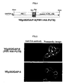
- Example 3 The transformant obtained in Example 3 was examined by the indirect immunofluorescence technique to ascertain whether a fusion gene was expressed within a cell and a fusion protein was presented on the surface layer of the yeast cell.
- the cells were collected, suspended in 250 ⁇ l of a PBS solution [8mg / ml NaCl, 0.2mg / ml KCl, 1.44mg / ml Na 2 HPO 4 , 0.24mg / ml KH2PO4 (pH7.4) , 1mg / ml BSA] containing 1 ⁇ g of HA antibody [Anti-HA High Affinity (Roche)], and then incubated on ice for 30 min. The cells were collected and washed once with a PBS solution.
- the cells were suspended in 250 ⁇ l of a PBS solution [8mg / ml NaCl, 0.2mg / ml KCl, 1.44mg / ml Na 2 HPO 4 , 0.24mg / ml KH 2 PO 4 (pH7.4), 1mg/ml BSA] containing 1 ⁇ g of a fluorescein secondary antibody [Alexa FluorTM 546 goat anti-rat IgG (H+L) conjugate (Molecular Probe)], and then incubated on ice for 30 min while shielding from light. During their respective incubation for 30 min, the cells and the antibody solution were occasionally mixed by a turning-over method for thorough mixing. The cells were collected, washed twice with PBS, suspended in 40 ⁇ l of PBS, and then observed with a fluorescence microscope (Fig. 2).
- a fluorescein secondary antibody Alexa FluorTM 546 goat anti-rat IgG (H+L) conjugate (Molecular Probe)
- Galactosyltransferase activity was measured by referring to Yoko-O et al's method (Yoko-O, Eur. J. Biochem., 257, 630-637 (1998)).
- Yoko-O Eur. J. Biochem., 257, 630-637
- the yeast intact cell itself [W303-YEp352GAP-II(PIR1-HA-gma12)] prepared in Example 3 was used.
- PA-mannobiose was used; as a donor substrate, UDP-galactose was used.
- a reaction solution of 50 ⁇ l [100mM HEPES (pH7.2) , 1mM MnCl 2 , 5mM UDP-galactose, 300pmol PA-mannobiose] was prepared to contain 11 ⁇ l of a cell suspension, followed by incubation at 37°C for 5 hours.
- the precipitated cells were removed by centrifugation at 3,000rpm for 3 min, the supernatant with a molecular weight of 10,000 or more was removed with an Ultra Free (0.22 ⁇ m), and then mannobiose and galactosylmannobiose were measured with HPLC.
- An Amide-80 column (TSK gel Amide-80, TOSOH, 0.46cm in diameter x 25cm in length) was used for HPLC.
- a mixture A containing 200mM acetic acid-triethylamine buffer (pH 7.0) and acetonitrile (10 : 90), and a mixture B containing 200mM acetic acid-triethylamine buffer (pH 7.0) and acetonitrile (60 : 40) were prepared.
- the column had been previously equilibrated by running the solvent A through the column at a flow rate of 1.0ml/min. Immediately after injection of samples, the proportion of the solvent B was raised linearly for 60 min up to 100%, so that PA-oligosaccharide was eluted.
- a plasmid pBS(SK-)/FT6H1.3 (provided by Dr. Narumatsu of Soka University) containing the amino acid coding region of a FUT6 gene, which is a human ⁇ -1,3-FucT (DB name: GenBank; AccessionNo:L01698) (Weston, J. Biol. Chem., 267, 24575-24585 (1992)) , was used as a template.
- a primer previously containing an SalI site on the N-terminal side and an XhoI site on the C-terminal side was designed to enable amplification except for a transmembrane region located on the N-terminal side of an FUT6 protein. Primers having the following base sequences were used.
- the underlined portion in the base sequence of the forward primer indicates the SalI site; that of the reverse primer indicates the XhoI site.
- the composition of a reaction solution and the reaction conditions employed for PCR followed Table 2 of Example 1 and the reaction conditions for Example 1, respectively.
- the amplified fragment of approximately 1kb was insertedin-frame to the SalI-XhoI site of pBSII(PIR1-HA-gma12), thereby constructing pBSII(PIR1-HA-FUT6)(pAB7).
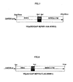
- the PIR1-HA-FUT6 portion was cleaved with SacI-XhoI from the pBSII(PIR1-HA-FUT6), blunt-ended with Blunting high (TOYOBO), and then inserted into an SmaI site of an expression vector YEp 352GAP-II (provided by Nakayama of the inventors' laboratory), thereby constructing YEp352GAP-II(PIR1-HA-FUT6)(pAB9) (Fig. 4).
- the expression vector YEp352GAP-II(PIR1-HA-FUT6) was transformed to a yeast strain W303-1A (ura3, leu2, his3, trp1, ade2) [Kainuma et al., Glycobiology, Vol. 9, 133-141 (1999)], thereby obtaining a strain W303- YEp352GAP-II(PIR1-HA-FUT6).
- strain W303- YEp352GAP-II(PIR1-HA-FUT6) was deposited under the Accession No. FERM BP-7797 at the International Patent Organism Depository, National Institute of Advanced Industrial Science and Technology on November 16, 2000.
- Example 6 The transformant obtained in Example 6 was examined by the indirect immunofluorescent technique to ascertain whether a fusion gene was expressed within a cell and a fusion protein was presented on the surface layer of the yeast cell.
- the cells were collected, suspended in 250 ⁇ l of a PBS solution [8mg / ml NaCl, 0.2mg / ml KCl, 1.44mg / ml Na 2 HPO 4 , 0.24mg / ml KH 2 PO 4 (pH7.4) , 1mg / ml BSA] containing 1 ⁇ g of HA antibody [Anti-HA High Affinity (Roche)], and then incubated on ice for 30 min. The cells of each strain were collected respectively and washed once with a PBS solution.
- a PBS solution [8mg / ml NaCl, 0.2mg / ml KCl, 1.44mg / ml Na 2 HPO 4 , 0.24mg / ml KH 2 PO 4 (pH7.4) , 1mg / ml BSA] containing 1 ⁇ g of HA antibody [Anti-HA High Affinity (Roche)], and then incubated
- the cells were suspended in 250 ⁇ l of a PBS solution [8mg / ml NaCl, 0.2mg / ml KCl, 1.44mg / ml Na 2 HPO 4 , 0.24mg / ml KH 2 PO 4 (pH7.4) , 1mg / ml BSA) containing 1 ⁇ g of a labeled secondary antibody [Alexa FluorTM 546 goat anti-rat IgG (H+L) conjugate (Molecular Probe)], and then incubated on ice for 30 min while shielding from light. During their respective incubation for 30 min, the cells and the antibody solution were mixed occasionally by a turning-over method for thorough mixing. The cells were collected, washed twice with PBS, suspended in 40 ⁇ l of PBS, and then observed with a fluorescence microscope.
- a PBS solution [8mg / ml NaCl, 0.2mg / ml KCl, 1.44mg / ml Na 2
- Fucosyltransferase activity was measured by referring to GLYCOBIOLOGY Experimental Protocol (ed. Taniguchi et al., 156-159 (1996)).
- As an enzyme source a solution was prepared by disrupting yeast cells [W303 ⁇ YEp352GAP-II(PIR1-HA-FUT6), prepared in Example 6] with glass beads in Wash Buffer [10mM Tris-HCl (pH8), 1mM PMSF]. PA-Lacto-N-neotetraose was used as an acceptor substrate; GDP-fucose was used as a donor substrate.
- 5.5 ⁇ l of the cell disruption solution was added to 4.5 ⁇ l of a reaction solution [50mM Cacodylate buffer (pH 6.8), 5mM ATP, 25mM MnCl 2 , 0.075mM GDP-fucose, 0.075mM PA-Lacto-N-neotetraose], followed by incubation at 37°C for 5 hours.
- a mixture A containing 200mM acetic acid-triethylamine buffer (pH 7.0) and acetonitrile (10 : 90), and a mixture B containing 200mM acetic acid -triethylaminebuffer (pH7.0) andacetonitrile (60 : 40) were prepared.
- the column had been previously equilibrated by running the solvent A through the column at a flow rate of 1.0ml/min. Immediately after injection of samples, the proportion of the solvent B was raised linearly for 60 min to 100%, so that PA-oligosaccharide was eluted.
- a KRE2 gene was isolated by PCR using the genome obtained from a budding yeast strain W303-1A (ura3, leu2, his3, trp1, ade2) [Kainuma et al., Glycobiology, Vol. 9, 133-141 (1999)] as a template.
- Primers were designed based on a base sequence registered on a database (DB name: GenBank; Accession No: X62647). At this time, the primers previously containing an SalI site on the N-terminal side and an XhoI site on the C-terminal side were designed to enable amplification without the transmembrane region located on the N-terminal side. Primers having the following base sequences were used.
- the underlined portion in the base sequence of the forward primer indicates the SalI site; that of the reverse primer indicates the XhoI site.
- the designed primers were synthesized by standard techniques.
- composition of a reaction solution and the reaction conditions employed for PCR followed Table 1 of Example 1 and the reaction conditions of Example 1, respectively.
- the amplified fragment of approximately 1kb was inserted in-frame to the SalI-XhoI site of pBSII(PIR1-HA-gma12), thereby constructing pBSII(PIR1-HA-KRE2)(pAB27).
- the PIR1-HA-KRE2 portion was cleaved with SacI-XhoI from the pBSII(PIR1-HA-KRE2), blunt-ended with Blunting high (TOYOBO), and then inserted into an SmaI site of an expression vector YEp352GAP-II (provided by Nakayama of the inventors laboratory), thereby constructing YEp352GAP-II(PIR1-HA-KRE2) (pAB30) (Fig. 7).
- Example 10 Construction of fusion protein composed of budding yeast Pir2 and FLAG
- a PIR2 gene was isolated by PCR using the genome obtained from a budding yeast strain W303-1A (ura3, leu2, his3, trp1, ade2) [Kainuma et al., Glycobiology, Vol. 9, 133-141 (1999)] as a template.
- Primers were designed based on a base sequence registered on a database (DB name: GenBank; Accession No: D13741). At this time, the primers previously containing an SacI site at the N-terminal portion and the sequence of FLAG epitope tag sequence containing an NotI site at the C-terminal portion were designed to enable easy cleavage of a portion encoding a protein with a restriction enzyme and to enable a epitope tag sequence to be added to the C-terminal portion of a Pir2 protein.
- the base sequences of each primer were as follows.
- the underlined portion in the base sequence of the forward primer indicates the SacI site; that of the reverse primer indicates the NotI site.
- the box portion indicates a sequence of a FLAG epitope tag.
- Example 1 The composition of a reaction solution and the reaction conditions employed for PCR followed Table 1 of Example 1 and the reaction conditions of Example 1, respectively.
- the amplified fragment of approximately 1kb was cleaved with SacI-NotI, and then inserted to the SacI-NotI site of pBluescript II SK(-) (Stratagene), thereby constructing pBSII(PIR2-FLAG)(pAB22).
- An MNN1 gene was isolated by PCR using the genome obtained from a budding yeast strain W303-1A (ura3, leu2, his3, trp1, ade2) [Kainuma et al., Glycobiology, Vol. 9, 133-141 (1999)] as a template.
- Primers were designed based on a base sequence registered on a database (DB name: GenBank; Accession No: L23753). At this time, the primers previously containing an NotI site on the N-terminal side and an SmaI site on the C-terminal side were designed to enable amplification without the transmembrane region located on the N-terminal side. Primers having the following base sequences were used.
- the underlined portion in the base sequence of the forward primer indicates the NotI site; that of the reverse primer indicates the SmaI site.
- the designed primers were synthesized by standard techniques.
- composition of a reaction solution and the reaction conditions employed for PCR followed Table 1 of Example 1 and the reaction conditions of Example 1, except that the time for elongation reaction was changed from 1 to 2 min.
- the amplified fragment of approximately 2kb was inserted in-frame to the NotI-SmaI site of pBSII(PIR2-FLAG) (pAB22), thereby constructing pBSII (PIR2-FLAG-MNN1) (pAB28).
- the PIR2-FLAG-MNN1 portion was cleaved with SacI-SmaI from the pBSII(PIR2-FLAG-MNN1), and then inserted into an SacI-SmaI site of an expression vector YEp352GAP-II (provided by Nakayama of the inventors laboratory), thereby constructing YEp352GAP-II(PIR2-FLAG-MNN1)(pAB29).
- a Bg1I fragment containing a promoter region, PIR2-FLAG-MNN1 and a terminator region was cleaved from Yep352GAP-II (PIR2-FLAG-MNN1), and then inserted into a Bg1I site of self amplification vector YEp351 [Hill et al., YEAST, 2, 163-167 (1986)], thereby constructing YEp351GAP-II(PIR2-FLAG-MNN1) (pAB31)(Fig. 8).
- Example 12 Construction of yeast transformant containing expression vector YEp352GAP-II(PIR1-HA-KRE2) and YEp351GAP-II(PIR2-FLAG-MNN1), and expression of both fusion proteins in yeast cells
- the expression vectors YEp352GAP-II (PIR1-HA-KRE2) and YEp351GAP-II(PIR2-FLAG-MNN1) were transformed simultaneously to a yeast strain W303-1A (ura3, leu2, his3, trp1, ade2) [Kainuma et al., Glycobiology, 9, 133-141(1999)) , thereby obtaining a strain W303-YEp352GAP-II(PIR1-HA-KRE2), YEp351GAP-II(PIR2-FLAG-MNN1).
- strain W303-YEp352GAP-II(PIR1-HA-KRE2), YEp351GAP-II(PIR2-FLAG-MNN1) was deposited under the Accession No. FERM BP-7789 at the International Patent Organism Depository, National Institute of Advanced Industrial Science and Technology on June 20, 2001.
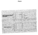
- the obtained transformant was subjected to Western blotting to examine if fusion genes were co-expressed in a cell and fusion proteins were co-localized on the yeast cell surface layer.
- PIR protein was covalently attached to a cell wall by an alkali-sensitive linkage, suggesting that a fusion protein with PIR would be freed by a mild alkali treatment of the cell wall fraction.
- the pellet fraction was washed three times with Na-acetate Buffer (pH 5.5) [0.1M Na-acetate], and then incubated with 100 ⁇ l of a mild alkali solution [30mM NaOH] at 4°C for 15 hours.
- the suspension with the mild alkali solution was centrifuged, so that supernatant was collected (Lane 3 of Fig. 9A and B).
- the collected samples were subjected to SDS-PAGE, and then Western blotting.
- primary antibodies used herein were HA antibody [MONOCLONAL ANTIBODY,HA.11 (CONVANCE)] and FLAG antibody [ANTI-FLAG M2 Monoclonal Antibody (SIGMA)] ; and a secondary antibody used herein was anti-mouse IgG-HRP [Anti-Mouse IgG (H&L) HRP-Linked Antibody ( Cell Signaling TECHNOLOGY)].
- HA antibody and FLAG antibody two membranes to which the same protein solution had been blotted were prepared, and then immuno-staining was performed separately with the 2 types of antibodies.
- Mannosyltransferase activity was measured by referring to Lussier et al's method (Lussier et al., JBC., 271, 11001-11008 (1996)).
- yeast intact cell itself [ W303- YEp352GAP-II(PIR1-HA-KRE2), YEp351GAP-II(PIR2-FLAG-MNN1)] prepared in Example 12 was used.
- a control strain used herein was W303-YEp352GAP-II.
- PA-mannobiose was used; as a donor substrate, GDP-mannose was used.
- a reaction solution 50 ⁇ l [100mM HEPES(pH7.2), 1mM MnCl 2 , 5mM GDP-mannose, 300pmol PA-mannobiose] was prepared to contain 20 ⁇ l of a cell suspension, followed by incubation at 37°C for 3 hours.
- the precipitated cells were removed by centrifugation at 3,000rpm for 3 min, and then the supernatant with a molecular weight of 10,000 or more was removed with an Ultra Free (0.22 ⁇ m). Then, mannobiose (disaccharide), mannotriose (trisaccharide) and mannotetraose (tetraose) were detected with HPLC.
- Amide-80 column (TSK gel An Amide-80, TOSOH, 0.46cm in diameter x 25cm in length) was used for HPLC.
- a mixture A containing 200mM acetic acid-triethylamine buffer (pH 7.0) and acetonitrile (10 : 90), and a mixture B containing 200mM acetic acid-triethylamine buffer (pH 7.0) and acetonitrile (60 : 40) were prepared.
- the column had been previously equilibrated by running the solvent A through the column at a flow rate of 1.0ml/min. Immediately after injection of samples, the proportion of the solvent B was raised linearly for 60 min to 100%, so that PA-oligosaccharide was eluted.
- the present invention enables immobilization of a useful protein, such as a glycosyltransferase, onto the surface of a yeast cell without deteriorating its enzyme activity, so that the invention can provide the immobilized protein as an immobilized enzyme. Since a process for purifying enzymes and a process for immobilizing enzymes to beads can be omitted, immobilized enzyme can be produced very easily and in large quantities.
- a useful protein such as a glycosyltransferase
Landscapes
- Life Sciences & Earth Sciences (AREA)
- Chemical & Material Sciences (AREA)
- Organic Chemistry (AREA)
- Health & Medical Sciences (AREA)
- Engineering & Computer Science (AREA)
- Genetics & Genomics (AREA)
- Zoology (AREA)
- Wood Science & Technology (AREA)
- Bioinformatics & Cheminformatics (AREA)
- Microbiology (AREA)
- Biochemistry (AREA)
- General Health & Medical Sciences (AREA)
- Biotechnology (AREA)
- Biomedical Technology (AREA)
- Molecular Biology (AREA)
- General Engineering & Computer Science (AREA)
- Mycology (AREA)
- Medicinal Chemistry (AREA)
- Gastroenterology & Hepatology (AREA)
- Biophysics (AREA)
- Proteomics, Peptides & Aminoacids (AREA)
- Chemical Kinetics & Catalysis (AREA)
- General Chemical & Material Sciences (AREA)
- Micro-Organisms Or Cultivation Processes Thereof (AREA)
- Immobilizing And Processing Of Enzymes And Microorganisms (AREA)
- Enzymes And Modification Thereof (AREA)
- Preparation Of Compounds By Using Micro-Organisms (AREA)
Abstract
The present invention enables immobilization of a useful
protein, for example, a glycosyltransferase, onto the surface
of a yeast cell without deteriorating its enzyme activity. The
present invention provides a fusion gene expression vector which
is characterized by containing a fusion gene that comprises a
gene encoding a useful protein bound downstream of a gene encoding
a yeast cell wall protein; a transformant yeast, which is
transformed with the fusion gene expression vector; and an
immobilized enzyme, which is characterized in that the expressed
useful protein is immobilized on a yeast cell wall.
Description
- The present invention relates to a fusion gene expression vector which enables expression of a Pir (protein internal repeat) protein having ability to be localized and immobilized on a yeast cell wall in the form of a fusion protein comprising the Pir protein bound to the N-terminus of a desired, useful protein; yeast transformed with the expression vector; an immobilized enzyme comprising a useful protein immobilized on the yeast cell wall; and a method for producing a sugar chain or sugars using a glycosyltransferase immobilized on the above yeast cell wall, wherein the useful protein is the glycosyltransferase.
- An enzyme protein used for producing substances with a bioreactor or the like is generally used as an immobilized enzyme which is immobilized to an insoluble carrier, for the sake of the convenience of procedures and from an economical standpoint. A general method for producing an immobilized enzyme involves purifying enzyme proteins and immobilizing the proteins to resin beads or the like. However, mass purification of enzyme proteins may not only involve complex procedures, but also cost a great deal, and cause inactivation of enzyme activity during a purification process. Further, a process to immobilize to a carrier, e.g., resin beads, may also inactivate enzyme activity. There is another example of a method which immobilizes an intact cell producing an enzyme protein, and uses it as an immobilized enzyme. However in this case, the efficacy of enzyme reaction cannot be said to be optimum since the enzyme protein is located within a cell. Hence, it is desired to localize an enzyme protein on the surface of a cell that is a carrier.
- One of the binding patterns of plasma membrane proteins to a membrane involves addition of the glycolipid, GPI (glycosylphosphatidylinositol), to the C-terminus of a protein which is anchored to a membrane by the lipid portion of GPI (GPI anchor) embedded within the membrane (Conzelmann, EMBO J. 7, 2233-2240, (1988)). It has been reported that immobilization of a cell wall protein (GPI-anchored protein) having the GPI anchor onto a cell wall comprises cleavage and removal of the C-terminal portion of a protein in the endoplasmic reticulum during the protein biosynthesis, addition of a GPI anchor to the protein, modification of a GPI core sugar chain in the Golgi apparatus, transportation to a cell membrane, transfer of the GPI glycan to a cell wall glucan, and covalent attachment to the cell wall (Lu, J. Cell, Biol. 128, 333-340, (1995)). Thus, a GPI anchor is a useful means to immobilize a protein to the surface layer of a cell. For example, an established technique employs α-agglutinin that is anchored by GPI to a cell wall to immobilize a foreign protein to a cell wall (Schreuder et al., Trends Biotechnol., 14, 115-120, (1996)). However, a defect of GPI anchors is that a GPI anchor functions for localization and immobilization to a cell wall only when a GPI attachment signal and the C-terminus of a protein are fused, and does not function when the signal and the C-terminus are not fused (Lipke, Mol. Cell Biol. 9, 3155-3165, (1989)). As a method to avoid such drawback, a modified method of immobilization to a cell surface layer with a GPI anchor has been reported which employs association of two subunits composing an a-agglutinin, AGA1p (Roy, Mol Cell Biol., 11, 4196-4206, (1991)) and AGA2p (Cappellaro, EMBO J., 10, 4081-4088, (1991)) on the surface layer of a cell (Boder and Wittrup, Nature Biotechnol. 15, 553-557, 1997) . In this method, a fusion protein (to which a target protein has been linked to the C-terminal side of AGA2p) is bound to AGA1p (which has been immobilized on a cell wall by a GPI anchor) via S-S bonding on the cell wall so as to immobilize the target protein to the cell surface layer. However, this method can be applied only to a cell expressing AGA1p.
- On the other hand, human hormones, most physiologically active substances and the receptors of these physiologically active substances consist of proteins and sugar chains. The sugar chain portion plays an important role in physiological activity, and a protein body alone cannot exhibit its original function (Akira Kobata, Protein, Nucleic Acid and Enzyme, 36, 775-788 (1991)). A glycosyltransferase is an essential enzyme in the formation of a sugar chain structure. It is thought that there are several hundred types of human glycosyltransferase. There must therefore be an enormous number of different types of glycosyltransferases when those of microorganisms and plants are included. A glycosyltransferase possesses an extremely high substrate specificity so that it adds a certain sugar to a receptor sugar chain with a certain structure by a certain binding pattern, thereby synthesizing a sugar chain with a certain structure (Qwens, Biochem. Biophys. Res. Commun., 109, 1075-1082, (1982); Betteridge, Eur. J. Biochem., 132, 29-35, (1983)). That is, the structure of a sugar chain is infinitely diversified such that a variety of sugars are associated by various binding patterns including branching. The diversity of the structure is determined by the combination of the substrate specificities of various glycosyltransferases. Accordingly, production and utilization of complex carbohydrates requires efficient expression and preparation of a glycosyltransferase that synthesizes a necessary sugar chain structure, because a substance functioning in vivo cannot be produced without precise control of the sugar chain structure.
- Most glycosyltransferases are type II membrane proteins, and are localized over the membrane of the Golgi apparatus with a topology in which an active region on the C-terminal side is oriented to the lumen of the Golgi apparatus (Paulson, J. Biol. Chem., 264, 17615-17618, 1989). Therefore, when the C-terminal side of a glycosyltransferase is genetically altered, the enzyme activity will often be deteriorated. It is very difficult to immobilize a glycosyltransferase onto a cell wall while maintaining its enzyme activity, using the above-described GPI anchor that cannot function unless it is bound to the C-terminal side. Meanwhile, the N-terminal side of a glycosyltransferase contains a transmembrane region, by which the enzymes are localized to ER or Golgi membrane. Soluble glycosyltransferase forms are also present in biological fluid. They arise from proteolytic cleavage in the stem region. That is, addition of a novel anchor protein (for immobilization to cell surface layer) to a glycosyltransferase lacking a transmembrane region would be effective in controlling localization of glycosyltransferases.
- A Pir (protein with internal repeat) protein is a cell wall protein covalently bound to a yeast cell wall, and Pir 1 to 4 genes, which are homologous to each other, compose a family (TOH-E, YEAST, 9, 481-494, (1993) ; Mrsa, YEAST, 13, 1145-1154, (1997)). However, a Pir protein has no GPI-anchor attachment signal. Further, since Pir proteins are not eluted with a detergent, but break away from a cell wall under alkali conditions, they may be localized and bound to a cell wall by a binding mechanism different from those of GPI-anchored proteins and other non-covalently proteins, and their mechanism remains unknown (Mrsa, YEAST, 15, 813-820, (1999)).
- There has been an attempt to use Pir as an anchor protein, in which a protein A gene is inserted between genes encoding Pir 4 proteins to be localized on a cell wall (Moukadiri et al., J. Bacteriology, 181, 4741-4740, (1999)). However the binding pattern to a cell wall is so varied among Pir proteins that Pir proteins cannot be generalized. For example, Pir4 can be freed from a cell wall by β-mercaptoethanol treatment, while Pir1 and Pir2 cannot be freed by the same treatment but can be eluted from a cell wall only under alkali conditions. Therefore, Pir 4 is thought to differ from other Pir1, Pir2 and Pir3, in the binding mechanism to a cell wall (Moukadiri et al., J. Bacteriology, 181, 4741-4740, (1999)). Further, a protein fused to Pir4 is a protein A, which does not require a very precise structure compared to a protein, for example, an enzyme, and is not fused to the N-terminal side of a target protein. Hence, when a protein to be expressed is, for example, an enzyme protein, has activity closely related to its structure, use of a gene encoding a Pir4 protein as an anchor protein does not always cause the resulting, expressed protein to retain enzyme activity.
- The purpose of the present invention is to localize and immobilize a protein represented by a glycosyltransferase, which is deteriorated by genetic manipulation of the C-terminus, on the surface layer of a yeast cell wall while maintaining its activity, and provide the protein as an immobilized enzyme.
- As a result of thorough studies to solve the above problems, the inventors have completed the present invention by finding that a fusion protein comprising a Pir protein bound to the N-terminus of a useful protein can be expressed on the surface layer of a yeast cell wall while maintaining the activity of the useful protein by transformation of yeast with a fusion gene expression vector containing the fusion gene that comprises a gene encoding the useful protein downstream of a gene encoding the Pir protein of a yeast cell wall.
- That is, the present invention is as described in the following (1) to (16), by which the above problems are solved.
- (1) A fusion gene expression vector which contains a fusion gene
comprising a gene encoding a useful protein downstream
of a gene encoding the following yeast cell wall protein (a)
or (b) :
- (a) a protein having an amino acid sequence represented by SEQ ID NO: 1;
- (b) a protein having an amino acid sequence derived from the amino acid sequence represented by SEQ NO: 1 by deletion, replacement, or addition of one or more amino acids, and having ability to be localized and immobilized on a yeast cell wall.
- (2) The fusion gene expression vector of (1) wherein the useful protein is a glycosyltransferase protein.
- (3) A transformant yeast which is transformed by the fusion gene expression vector of (1) or (2).
- (4) A method for producing an immobilized enzyme, which comprises culturing the transformant yeast of (3), expressing a fusion gene on the surface layer of a yeast cell wall, and obtaining yeast that contains a useful protein immobilized on the cell wall.
- (5) An immobilized enzyme, which is obtained by the method of (4).
- (6) The immobilized enzyme of (5) wherein the enzyme to be immobilized is a glycosyltransferase.
- (7) A method for producing a sugar chain or sugars which method employs the immobilized enzyme of (6).
- (8) A fusion gene expression vector which contains a fusion gene
comprising a gene encoding a useful protein bound downstream
of a gene encoding the following yeast cell wall protein (a)
or (b) :
- (a) a protein having an amino acid sequence represented by SEQ ID NO: 2;
- (b) a protein having an amino acid sequence derived from the amino acid sequence represented by SEQ NO: 2 by deletion, replacement, or addition of one or more amino acids, and having ability to be localized and immobilized on a yeast cell wall.
- (9) The fusion gene expression vector of (8) wherein the useful protein is a glycosyltransferase protein.
- (10) A transformant yeast, which is transformed with the fusion gene expression vector of (8) or (9).
- (11) A method for producing an immobilized enzyme which method comprises culturing the transformant yeast of (10), expressing a fusion gene on the surface layer of a yeast cell wall, and obtaining yeast that contains a useful protein immobilized on the cell wall.
- (12) An immobilized enzyme, which is obtained by the method of (11).
- (13) The immobilized enzyme of (12) wherein the enzyme to be immobilized is a glycosyltransferase.
- (14) A method for producing a sugar chain or sugars which method employs the immobilized enzyme of (13).
- (15) A transformant yeast which is transformed by allowing the yeast to contain at least two or more types of the fusion gene expression vector of (1) and (8).
- (16) A method for producing an immobilized enzyme which method comprises culturing the transformant yeast of (15), expressing simultaneously fusion genes on the surface layer of a yeast cell wall, and obtaining yeast that contains at least two or more types of useful proteins immobilized on the cell wall.
- (17) An immobilized enzyme, which is obtained by the method of (16).
- (18) The immobilized enzyme of (17) wherein the enzymes to be immobilized are two or more types of glycosyltransferases.
- (19) A method for producing a sugar chain or sugars which method sequentially converts sugar chains or sugars using the immobilized enzymes of (18).
-
-
- Figure 1 shows a structure of YEp352GAP-II (PIR1-HA-gma12)(pAB4).
- Figure 2 shows results of detecting expression of a fusion protein for a transformant strain W303-YEp352GAP-II(PIR1-HA-gma12) and a control strain W303-YEp352GAP-II by the indirect immunofluorescence technique.
- Figure 3 shows results of detecting galactosyltransferase activity for a transformant strain W303-YEp352GAP-II(PIR1-HA-gma12) and a control strain W303-YEp352GAP-II. The tip of an arrow in the figure indicates a peak of galactosyl mannobiose.
- Figure 4 shows the structure of YEp352GAP-II(PIR1-HA-FUT6)(pAB9).
- Figure 5 shows results of detecting expression of a fusion protein for a transformant strain W303-YEp352GAP-II (PIR1-HA-FUT6) and a control strain W303-YEp352GAP-II.
- Figure 6 shows results of detecting fucosyltransferase activity for a transformant strain W303-YEp352GAP-II (PIR1-HA-FUT6) and a control strain W303-YEp352GAP-II. The tip of an arrow in the figure indicates a peak of Lacto-N-fucopentaose.
- Figure 7 shows the structure of YEp352GAP-II(PIR1-HA-KRE2).
- Figure 8 shows the structure of YEp352GAP-II(PIR2-FLAG-MNN1).
- Figure 9 indicates simultaneous expression of YEp352GAP-II (PIR1-HA-KRE2) and YEp351GAP-II(PIR2-FLAG-MNN1) in transformant yeast.
- Figure 10 indicates that a yeast strain [ W303-YEp352GAP-II(PIR1-HA-KRE2), YEp351GAP-II(PIR2-FLAG-MNN1)] which has been transformed with Pir1-HA-Kre2 and Pir2-FLAG-Mnn1 at the same time performed sequential transfer reaction of mannose resulting from the two expression vectors above.
-
- A fusion gene expression vector of the present invention contains a fusion gene which comprises a gene of a desired enzyme protein downstream of a gene encoding a Pir protein that is present on a yeast cell wall having an amino acid sequence of SEQ ID NO: 1 or 2.
- PIR genes used in the present invention include a gene (PIR1) encoding a Pir1 protein represented by SEQ ID NO: 1, and a gene (PIR2) encoding a Pir2 protein represented by SEQ ID NO: 2, and a gene encoding a protein having an amino acid sequence derived from the amino acid sequence above by deletion, replacement or addition of one or more amino acids and having ability to be localized or immobilized to a yeast cell wall.
- Examples of an amino acid sequence derived from the amino acid sequence represented by SEQ ID NO: 1 or 2 by deletion, replacement or addition of one or more amino acids include: an amino acid sequence which is derived from the amino acid sequence of SEQ ID NO: 1 or 2 by deletion of one to 10 amino acids, preferably 1 to 5 amino acids, and more preferably 1 to 2 amino acids; an amino acid sequence which is derived from the amino acid sequence of SEQ ID NO: 1 or 2 by replacement of one to 10 amino acids, preferably 1 to 5 amino acids, and more preferably 1 to 2 amino acids by other amino acids; and an amino acid sequence which is derived from the amino acid sequence of SEQ ID NO: 1 or 2 by addition of one to 10 amino acids, preferably 1 to 5 amino acids, and more preferably 1 to 2 amino acids.
- Examples of such an amino acid sequence derived from the amino acid sequence represented by SEQ ID NO: 1 or 2 by deletion, replacement or addition of one or more amino acids have at least 80% or more, preferably 90% or more, more preferably 95% or more, and particularly preferably 97% or more homology with the amino acid sequence of SEQ ID NO: 2 when homology is calculated with BLAST.
- Such an amino acid sequence derived from the amino acid sequence represented by SEQ ID NO: 1 or 2 by deletion, replacement or addition of one or more amino acids is substantially identical to the amino acid sequence of SEQ ID NO: 1 or 2.
- More specifically, these genes can be obtained by PCR using a genome obtained from budding yeast (Saccharomyces cerevisiae) strain W303-1A (ura3, leu2, his3, trp1, ade2) [Kainuma et al., Glycobiology, Vol. 9, 133-141 (1999)) as a template. A primer used herein contains a restriction enzyme cleavage portion, which is useful for easy cleavage of a portion encoding a Pir protein and simple insertion of a epitope tag sequence and the like.
- Any desired, useful protein gene may be downstream of the above PIR gene. For example, a gene of a protein such as a glycosyltransferase whose activity may be deteriorated by gene manipulation of the C-terminus, is preferred. Examples of such a protein include a galactosyltransferase, fucosyltransferase, glucosyltransferase, mannosyltransferase, galactosamyltransferase, sialyltransferase and N-acetylglucosaminyltransferase.
- An expression cassette is constructed by inserting a promoter to upstream and a terminator to downstream of a fusion gene comprising a desired protein gene downstream of a PIR gene. Then, the expression cassette is introduced into an vector. Alternatively, when a promoter and a terminator have already been present in an expression vector into which the fusion gene is introduced, only the fusion gene is introduced between the promoter and the terminator without constructing any expression cassette.
- For a promoter in an expression cassette, any promoter which is generally used for a yeast expression system and allows expression of a fusion gene introduced in a transformed yeast cell can be used. Examples of such a promoter include, but are not specifically limited to, include PGK, GAP, TPI, GAL1, GAL10, ADH2, PH05 and CUP1. Particularly, a GAP promoter is preferred.
- For a terminator, any terminator which is generally used for a yeast expression system and allows termination of transcription when it is located downstream of a fusion gene introduced can be used. Examples of such a terminator include ADH1, TDH1, TFF and TRP5.
- Examples of an expression vector to which an expression cassette is introduced are not specifically limited as long as they are generally used in a yeast expression system, and allow expression of a fusion gene on the surface layer of the cell wall of transformant yeast that has been transformed with the expression vector. For example, a yeast episome expression vector can be preferably used.
- A yeast episome plasmid vector contains a 2µ plasmid sequence, which is an original sequence of yeast and the vector is rendered capable of replication within a host yeast cell using the autonomously replicating sequence of the 2µ plasmid sequence. Examples of a yeast episome expression vector used in the present invention are not specifically limited, as long as they contain at least an ARS sequence of the yeast 2µ plasmid sequence and they can replicate outside the chromosome within a host yeast cell. Such a yeast episome expression vector may be YEp51, pYES2, YEp351, YEp352 or the like.
- The above yeast episome expression vector is preferably a shuttle vector, which can proliferate within E.coli cells to perform subcloning in recombinant E.coli. A more preferred expression vector contains a selective marker gene, such as an ampicillin-resistantgene. In addition, the expression vector contains a marker gene with which a yeast clone can be selected based on auxotrophy and drug resistance when recombinant yeast is produced. Examples of a marker gene include HIS3, TRP1, LEU2, URA3, ADE2, CAN1, SUC2, LYS2, and CUP1 (Yasuji Oshima (writer-editor), Experimental Protocols in Biochemistry 39, Experimental Protocols in Yeast Molecular Genetics, 119-144 (1996)). These are merely examples, and selection should be made depending on a genotype of a yeast strain to be used as a host for gene transfer.
- The above series of techniques involved in construction of a fusion gene expression plasmid may be appropriately performed by persons skilled in the art by referring to descriptions given in examples described later or by standard technology.
- In this invention, examples of host yeast to be transformed with the above fusion gene expression vector include, but are not limited to, yeast belonging to the genus Saccharomyces and the genus candida. Examples of yeast belonging to the genus Saccharomyces include Saccharomyces cerevisiae strains KK4, Y334, Inv-Sc1 and W303.
- To transform yeast with a fusion gene expression vector, for example, known methods such as a lithium acetate method, electroporation, and the like can be used (Becker and Guarente, Methods Enzymol., 194, 182-187 (1991) ).
- In the present invention, yeast may be transformed simultaneously using multiple expression vectors in which genes encoding different useful proteins are bound to the same type of PIR genes. Yeast may also be transformed simultaneously using multiple expression vectors in which genes encoding different useful proteins are bound respectively to the different types of PIR genes (e.g. PIR1 gene and PIR2 gene). In these cases, for example, by using as genes encoding useful proteins multiple genes (different, but related to each other) encoding glycosyltransferase proteins, immobilized enzymes of the transformed yeast can perform multiple reactions sequentially. Hence, these cases have an advantage that greatly diverse sugar chains or sugars can be produced.
- Here, the term "immobilized enzyme" means an enzyme immobilized to a yeast cell wall.
- An appropriate selective marker is used for screening for transformant yeast. In a preferred example, a gene involved in the metabolism on a chromosomal DNA of a host cell is used. That is, when transforming a host cell having the above gene on chromosomal DNA which has been disabled function by appropriate techniques, e.g., mutant with an expression vector that contains a corresponding normal gene, a preferred selection marker is one which can be used to screen by proliferating the transformant cell that contains a normal metabolism gene. More specifically, a widely used selective marker gene, for example, URA3, and LEU2 as described above is integrated to an expression vector. For a chromosome incorporation type (YIp type), these genes are also used as markers for screening.
- Culturing of the transformed transformant yeast enables expression of a fusion protein comprising a Pir protein bound to the N-terminus of a desired protein onto the surface layer of the cell wall. The desired protein is immobilized via Pir on the surface layer of the cell wall of the transformant yeast, so that the transformant yeast can be used directly as an immobilized enzyme.
- Culturing of transformant yeast can be performed by standard techniques for culturing yeast.
A medium used herein contains a yeast assimilable carbon source, a nitrogen source, inorganic salts and the like to enable efficient culturing of transformants. For example, a synthetic medium that can be used herein (containing a carbon source, a nitrogen source, inorganic salts, amino acids, vitamins and the like) contains various medium components (supplied from Difco) added thereto, except amino acid (required for replication and maintenance of plasmids, but can be supplied with a marker) which is removed from the medium (Sherman, Methods Enszymol., 194, 3-57 (1991)). - pH for a medium is preferably adjusted to 6 to 8. Adjustment of pH is performed using inorganic or organic acid, alkali solution, urea, calcium carbonate, ammonia or the like.
- Culturing is performed at 28 to 32°C, preferably at 30°C, for 15 to 48 hours with aeration and agitation.
- Now, the present invention will be further described by the following examples, but is not limited thereto.
- PIR1 gene was isolated by PCR using the genome obtained from a budding yeast (Saccharomyces cerevisiae) strain W303-1A (ura3, leu2, his3, trp1, ade2) [Kainuma et al., Glycobiology, Vol. 9, 133 (1999)] as a template.
- A primer was designed based on a base sequence which has been registered on a database (DB name: GenBank; Accession NO: D13740). At this time, a primer previously containing an SacI site at the N-terminal portion and an NotI site at the C-terminal portion was so designed that a portion encoding a protein can be easily cleaved out with a restriction enzyme and a epitope tag sequence and the like can be simply inserted. Base sequences for each primer are as follows.
-

-

- The underlined portion in the base sequence of the forward primer indicates the SacI site; and that of the reverse primer indicates the NotI site. Thus, the designed primers were synthesized by standard techniques.
- Composition of the PCR solution is as shown in Table 1.
Composition of PCR solution 10 x ExpandHF buffer (including 15mM MgCl2) 10µl dNTP Mixture (2.5mM each) 8µl Forward primer (20pmpl / µl) 2µl Reverse primer (20pmpl / µl) 2µl Genome DNA 3µl ExpandTM High Fidelity PCR System enzyme mix 0.75µl Water 74.25µl Total 100µl - The first PCR reaction consisted of 10 cycles. The temperature conditions for each cycle consisted of denaturation of template DNA at 94°C for 2 min, 94°C for 15 sec (denaturation), 50°C for 30 sec (annealing), and 72°C for 1 min (elongation). The next reaction consisted of 15 cycles, the temperature conditions for each cycle consisting of 94°C for 15 sec (denaturation), 50°C for 30 sec (annealing), and 72°C for 1 min (elongation) and each cycle having a prolongation of 5 sec. Finally, elongation reaction at 72°C was performed for 7 min. The amplified DNA fragment with a length of approximately 1kbp obtained by the PCR was cleaved with NotI-SacI, and then inserted to an NotI-SacI site of pBluescript II SK(-) (Stratagen), thereby constructing pBSII (PIR1) (pAB2).
- A gma12 gene (DB name: GenBank; Accession No: Z30917) (Chappell, Mol. Biol. Cell, 5, 519-528 (1994)) of fission yeast (Schizosaccharomyces pombe) was also cloned by PCR. Composition of the PCR solution is shown in Table 2.
Composition of PCR solution 10 x ExpandHF buffer (including 15mM MgCl2) 10µl dNTP Mixture (2.5mM each) 8µl Forward primer (20pmpl / µl) 2µl Reverse primer (20pmpl / µl) 2µl Plasmid DNA 1µl ExpandTM High Fidelity PCR System enzyme mix 0.75µl Water 76.25µl Total 100µl - The plasmid (pYD1-HA-gma12) that has been constructed from gma12 gene cloned into YEpU-GAP-gma12 (YOKO-O et al., FEBS, Vol.257, 1998) by Takayama who belonged to the inventors' laboratory was used as a template. A primer was so designed to enable amplification of HA-gma12 fusion gene that the amplified product previously contains an NotI site on the 5' side and an SmaI site on the 3' side. Primers having the following base sequences were used.
-

-

- The underlined portion in the base sequence of the forward primer indicates the NotI site; that of the reverse primer indicates the SmaI site. PCR was performed under temperature conditions the same as those employed for amplification of PIR1 by PCR.
- Subsequently, the HA-gma12 fusion gene was inserted to an NotI-SmaI site of pBluescript II SK(-) (Stratagene), thereby constructing pBSII (HA-gma12) (pAB1).
- A PIR1 gene inserted in pBSII(PIR1) was cleaved with SacI-NotI, and then inserted into an SacI-NotI site of pBSII (HA-gma12), thereby constructing pBSII (PIR1-HA-gma12) (pAB3).
- A PIR1-HA-gma12 portion of the pBSII (PIR-HA-gma12) was cleaved with SacI-SmaI. Then the product was inserted into an SacI-SmaI site of a expression vector YEp352GAP-II (Nakayama) in which the multi-cloning site of a yeast expression vector YEp352GAP (Roy et al., J. Biol. Chem., Vol.237, 2538 (1998)) has been replaced by a portion from EcoRI to SalI of the multi-cloning site of pUC18, thereby constructing YEp352GAP-II (PIR1-HA-gma12) (pAB4) (Fig. 1).
- The expression vector YEp352GAP-II (PIR1-HA-gma12) was transformed to a yeast strain W303-1A (ura3, leu2, his3, trp1, ade2) [Kainumaetal., Glycobiology, 9, 133-141 (1999)], thereby obtaining a strain W303- YEp352GAP-II(PIR1-HA-gma12).
- In addition, the strain W303- YEp352GAP-II(PIR1-HA-gma12) was deposited under the Accession No. FERM BP-7794 at the International Patent Organism Depository, National Institute of Advanced Industrial Science and Technology on November 16, 2000.
- The transformant obtained in Example 3 was examined by the indirect immunofluorescence technique to ascertain whether a fusion gene was expressed within a cell and a fusion protein was presented on the surface layer of the yeast cell.
- First, the above transformant strain W303-YEp352GAP-II(PIR1-HA-gmal2) and a control strain W303-YEp352GAP-II, which had been transformed from a strain W303-1A using YEp352GAP-II, were cultured in 5ml of a SD (-uracil) liquid medium up to OD600=5 (for approximately 30 hours). Then, 1ml of the culture solution was collected, and then the cells were washed with PBS [8mg / ml NaCl, 0.2mg / ml KCl, 1.44mg / ml Na2HPO4, 0.24mg/ml KH2PO4 (pH7.4)]. The cells were collected, suspended in 250µl of a PBS solution [8mg / ml NaCl, 0.2mg / ml KCl, 1.44mg / ml Na2HPO4, 0.24mg / ml KH2PO4 (pH7.4) , 1mg / ml BSA] containing 1µg of HA antibody [Anti-HA High Affinity (Roche)], and then incubated on ice for 30 min. The cells were collected and washed once with a PBS solution. Subsequently, the cells were suspended in 250µl of a PBS solution [8mg / ml NaCl, 0.2mg / ml KCl, 1.44mg / ml Na2HPO4, 0.24mg / ml KH2PO4 (pH7.4), 1mg/ml BSA] containing 1µg of a fluorescein secondary antibody [Alexa FluorTM 546 goat anti-rat IgG (H+L) conjugate (Molecular Probe)], and then incubated on ice for 30 min while shielding from light. During their respective incubation for 30 min, the cells and the antibody solution were occasionally mixed by a turning-over method for thorough mixing. The cells were collected, washed twice with PBS, suspended in 40µl of PBS, and then observed with a fluorescence microscope (Fig. 2).
- As a result, expression of Pir1-HA-Gma12 fusion protein on the cell surface layer was confirmed for the strain W303-YEp352GAP-II(PIR1-HA-gma12).
- Galactosyltransferase activity was measured by referring to Yoko-O et al's method (Yoko-O, Eur. J. Biochem., 257, 630-637 (1998)). As an enzyme source, the yeast intact cell itself [W303-YEp352GAP-II(PIR1-HA-gma12)] prepared in Example 3 was used. As an acceptor substrate, PA-mannobiose was used; as a donor substrate, UDP-galactose was used. A reaction solution of 50µl [100mM HEPES (pH7.2) , 1mM MnCl2, 5mM UDP-galactose, 300pmol PA-mannobiose] was prepared to contain 11µl of a cell suspension, followed by incubation at 37°C for 5 hours. The cell suspension used herein was prepared by collecting 1ml of a culture solution with OD600=6, washing twice with Wash Buffer [10mM Tris-HCl (pH8) , 1mM PMSF], and suspending in 11µl of Wash Buffer [10mM Tris-HCl (pH8) , 1mM PMSF]. Then, 30µl of ice-cooled water was added to the reaction solution. The precipitated cells were removed by centrifugation at 3,000rpm for 3 min, the supernatant with a molecular weight of 10,000 or more was removed with an Ultra Free (0.22µm), and then mannobiose and galactosylmannobiose were measured with HPLC. An Amide-80 column (TSK gel Amide-80, TOSOH, 0.46cm in diameter x 25cm in length) was used for HPLC. A mixture A containing 200mM acetic acid-triethylamine buffer (pH 7.0) and acetonitrile (10 : 90), and a mixture B containing 200mM acetic acid-triethylamine buffer (pH 7.0) and acetonitrile (60 : 40) were prepared. The column had been previously equilibrated by running the solvent A through the column at a flow rate of 1.0ml/min. Immediately after injection of samples, the proportion of the solvent B was raised linearly for 60 min up to 100%, so that PA-oligosaccharide was eluted.
- As a result, a peak of the enzyme product was detected, that is, galactosyltransferase activity could be confirmed, only for the strain W303-YEp352GAP-II (PIR1-HA-gma12) that can express a fusion protein of Pir1-HA-gma12 (Fig. 3). No galactosyltransferase activity was detected for the strain W303-YEp352GAP-II that expresses no fusion gene.
- A plasmid pBS(SK-)/FT6H1.3 (provided by Dr. Narumatsu of Soka University) containing the amino acid coding region of a FUT6 gene, which is a human α-1,3-FucT (DB name: GenBank; AccessionNo:L01698) (Weston, J. Biol. Chem., 267, 24575-24585 (1992)) , was used as a template. A primer previously containing an SalI site on the N-terminal side and an XhoI site on the C-terminal side was designed to enable amplification except for a transmembrane region located on the N-terminal side of an FUT6 protein. Primers having the following base sequences were used.
-

-

- The underlined portion in the base sequence of the forward primer indicates the SalI site; that of the reverse primer indicates the XhoI site. The composition of a reaction solution and the reaction conditions employed for PCR followed Table 2 of Example 1 and the reaction conditions for Example 1, respectively. The amplified fragment of approximately 1kb was insertedin-frame to the SalI-XhoI site of pBSII(PIR1-HA-gma12), thereby constructing pBSII(PIR1-HA-FUT6)(pAB7). The PIR1-HA-FUT6 portion was cleaved with SacI-XhoI from the pBSII(PIR1-HA-FUT6), blunt-ended with Blunting high (TOYOBO), and then inserted into an SmaI site of an expression vector YEp 352GAP-II (provided by Nakayama of the inventors' laboratory), thereby constructing YEp352GAP-II(PIR1-HA-FUT6)(pAB9) (Fig. 4).
- The expression vector YEp352GAP-II(PIR1-HA-FUT6) was transformed to a yeast strain W303-1A (ura3, leu2, his3, trp1, ade2) [Kainuma et al., Glycobiology, Vol. 9, 133-141 (1999)], thereby obtaining a strain W303- YEp352GAP-II(PIR1-HA-FUT6).
- In addition, the strain W303- YEp352GAP-II(PIR1-HA-FUT6) was deposited under the Accession No. FERM BP-7797 at the International Patent Organism Depository, National Institute of Advanced Industrial Science and Technology on November 16, 2000.
- The transformant obtained in Example 6 was examined by the indirect immunofluorescent technique to ascertain whether a fusion gene was expressed within a cell and a fusion protein was presented on the surface layer of the yeast cell.
- First, the above transformant strain W303-YEp352GAP-II(PIR1-HA-FUT6) and a control strain W303-YEpGAP-II, which had been transformed from a strain W303-1A using YEp352GAP-II, were cultured in 5ml of an SD (-uracil) liquid medium to OD600=5 (for approximately 30 hours). Then, 1ml of the culture solution was collected, and then the cells were washed with PBS [8mg / ml NaCl, 0.2mg / ml KCl, 1.44mg / ml Na2HPO4, 0.24mg / ml KH2PO4 (pH7.4) ]. The cells were collected, suspended in 250µl of a PBS solution [8mg / ml NaCl, 0.2mg / ml KCl, 1.44mg / ml Na2HPO4, 0.24mg / ml KH2PO4 (pH7.4) , 1mg / ml BSA] containing 1µg of HA antibody [Anti-HA High Affinity (Roche)], and then incubated on ice for 30 min. The cells of each strain were collected respectively and washed once with a PBS solution. Subsequently, the cells were suspended in 250µl of a PBS solution [8mg / ml NaCl, 0.2mg / ml KCl, 1.44mg / ml Na2HPO4, 0.24mg / ml KH2PO4 (pH7.4) , 1mg / ml BSA) containing 1µg of a labeled secondary antibody [Alexa FluorTM 546 goat anti-rat IgG (H+L) conjugate (Molecular Probe)], and then incubated on ice for 30 min while shielding from light. During their respective incubation for 30 min, the cells and the antibody solution were mixed occasionally by a turning-over method for thorough mixing. The cells were collected, washed twice with PBS, suspended in 40µl of PBS, and then observed with a fluorescence microscope.
- As a result, expression of Pir1-HA-FUT6 fusion protein on the cell surface layer was confirmed for the strain W303-YEp352GAP-II(PIR1-HA-FUT6) (Fig. 5).
- Fucosyltransferase activity was measured by referring to GLYCOBIOLOGY Experimental Protocol (ed. Taniguchi et al., 156-159 (1996)). As an enzyme source, a solution was prepared by disrupting yeast cells [W303―YEp352GAP-II(PIR1-HA-FUT6), prepared in Example 6] with glass beads in Wash Buffer [10mM Tris-HCl (pH8), 1mM PMSF]. PA-Lacto-N-neotetraose was used as an acceptor substrate; GDP-fucose was used as a donor substrate. 5.5µl of the cell disruption solution was added to 4.5µl of a reaction solution [50mM Cacodylate buffer (pH 6.8), 5mM ATP, 25mM MnCl2, 0.075mM GDP-fucose, 0.075mM PA-Lacto-N-neotetraose], followed by incubation at 37°C for 5 hours. A cell suspension used herein was a solution containing disrupted cells which had been prepared by collecting 0.25ml of the culture solution with OD600=6, washing twice with Wash Buffer [10mM Tris-HCl (pH 8), 1mM PMSF] and then crushing with glass beads. Next, to stop reaction, incubation was performed at 98°C for 3 min, and then 40µl of ice-cooled water was added to the reaction solution. The precipitated cells were removed by centrifugation at 3,000rpm for 3 min, and then the supernatant with a molecular weight of 10,000 or more was removed with an Ultra Free (0.22µm). Subsequently, Lacto-N-neotetraose and Lacto-N-fucopentaose were measured with HPLC. An Amide-80 column (TSK gel Amide-80, TOSOH, 0.46cm in diameter x 25cm in length) was used for HPLC. A mixture A containing 200mM acetic acid-triethylamine buffer (pH 7.0) and acetonitrile (10 : 90), and a mixture B containing 200mM acetic acid -triethylaminebuffer (pH7.0) andacetonitrile (60 : 40) were prepared. The column had been previously equilibrated by running the solvent A through the column at a flow rate of 1.0ml/min. Immediately after injection of samples, the proportion of the solvent B was raised linearly for 60 min to 100%, so that PA-oligosaccharide was eluted.
- As a result, a peak of the enzyme product was detected, that is, fucosyltransferase activity could be confirmed, only for the strain W303-YEp352GAP-II (PIR1-HA-FUT6) that can express a fusion protein of Pir1-HA-FUT6 (Fig. 6). No fucosyltransferase activity was detected for the strain W303-YEp352GAP-II that expresses no fusion gene.
- A KRE2 gene was isolated by PCR using the genome obtained from a budding yeast strain W303-1A (ura3, leu2, his3, trp1, ade2) [Kainuma et al., Glycobiology, Vol. 9, 133-141 (1999)] as a template.
- Primers were designed based on a base sequence registered on a database (DB name: GenBank; Accession No: X62647). At this time, the primers previously containing an SalI site on the N-terminal side and an XhoI site on the C-terminal side were designed to enable amplification without the transmembrane region located on the N-terminal side. Primers having the following base sequences were used.
-

-

- Here, the underlined portion in the base sequence of the forward primer indicates the SalI site; that of the reverse primer indicates the XhoI site. Thus the designed primers were synthesized by standard techniques.
- The composition of a reaction solution and the reaction conditions employed for PCR followed Table 1 of Example 1 and the reaction conditions of Example 1, respectively. The amplified fragment of approximately 1kb was inserted in-frame to the SalI-XhoI site of pBSII(PIR1-HA-gma12), thereby constructing pBSII(PIR1-HA-KRE2)(pAB27). The PIR1-HA-KRE2 portion was cleaved with SacI-XhoI from the pBSII(PIR1-HA-KRE2), blunt-ended with Blunting high (TOYOBO), and then inserted into an SmaI site of an expression vector YEp352GAP-II (provided by Nakayama of the inventors laboratory), thereby constructing YEp352GAP-II(PIR1-HA-KRE2) (pAB30) (Fig. 7).
- A PIR2 gene was isolated by PCR using the genome obtained from a budding yeast strain W303-1A (ura3, leu2, his3, trp1, ade2) [Kainuma et al., Glycobiology, Vol. 9, 133-141 (1999)] as a template.
- Primers were designed based on a base sequence registered on a database (DB name: GenBank; Accession No: D13741). At this time, the primers previously containing an SacI site at the N-terminal portion and the sequence of FLAG epitope tag sequence containing an NotI site at the C-terminal portion were designed to enable easy cleavage of a portion encoding a protein with a restriction enzyme and to enable a epitope tag sequence to be added to the C-terminal portion of a Pir2 protein. The base sequences of each primer were as follows.
-

-

- The underlined portion in the base sequence of the forward primer indicates the SacI site; that of the reverse primer indicates the NotI site. The box portion indicates a sequence of a FLAG epitope tag. Thus the designed primers were synthesized by standard techniques.
- The composition of a reaction solution and the reaction conditions employed for PCR followed Table 1 of Example 1 and the reaction conditions of Example 1, respectively. The amplified fragment of approximately 1kb was cleaved with SacI-NotI, and then inserted to the SacI-NotI site of pBluescript II SK(-) (Stratagene), thereby constructing pBSII(PIR2-FLAG)(pAB22).
- An MNN1 gene was isolated by PCR using the genome obtained from a budding yeast strain W303-1A (ura3, leu2, his3, trp1, ade2) [Kainuma et al., Glycobiology, Vol. 9, 133-141 (1999)] as a template.
- Primers were designed based on a base sequence registered on a database (DB name: GenBank; Accession No: L23753). At this time, the primers previously containing an NotI site on the N-terminal side and an SmaI site on the C-terminal side were designed to enable amplification without the transmembrane region located on the N-terminal side. Primers having the following base sequences were used.
-

-

- The underlined portion in the base sequence of the forward primer indicates the NotI site; that of the reverse primer indicates the SmaI site. Thus, the designed primers were synthesized by standard techniques.
- The composition of a reaction solution and the reaction conditions employed for PCR followed Table 1 of Example 1 and the reaction conditions of Example 1, except that the time for elongation reaction was changed from 1 to 2 min. The amplified fragment of approximately 2kb was inserted in-frame to the NotI-SmaI site of pBSII(PIR2-FLAG) (pAB22), thereby constructing pBSII (PIR2-FLAG-MNN1) (pAB28). The PIR2-FLAG-MNN1 portion was cleaved with SacI-SmaI from the pBSII(PIR2-FLAG-MNN1), and then inserted into an SacI-SmaI site of an expression vector YEp352GAP-II (provided by Nakayama of the inventors laboratory), thereby constructing YEp352GAP-II(PIR2-FLAG-MNN1)(pAB29). Further, to construct a plasmid having a leucine marker, a Bg1I fragment containing a promoter region, PIR2-FLAG-MNN1 and a terminator region was cleaved from Yep352GAP-II (PIR2-FLAG-MNN1), and then inserted into a Bg1I site of self amplification vector YEp351 [Hill et al., YEAST, 2, 163-167 (1986)], thereby constructing YEp351GAP-II(PIR2-FLAG-MNN1) (pAB31)(Fig. 8).
- The expression vectors YEp352GAP-II (PIR1-HA-KRE2) and YEp351GAP-II(PIR2-FLAG-MNN1) were transformed simultaneously to a yeast strain W303-1A (ura3, leu2, his3, trp1, ade2) [Kainuma et al., Glycobiology, 9, 133-141(1999)) , thereby obtaining a strain W303-YEp352GAP-II(PIR1-HA-KRE2), YEp351GAP-II(PIR2-FLAG-MNN1). The strain W303-YEp352GAP-II(PIR1-HA-KRE2),
YEp351GAP-II(PIR2-FLAG-MNN1) was deposited under the Accession No. FERM BP-7789 at the International Patent Organism Depository, National Institute of Advanced Industrial Science and Technology on June 20, 2001. - The obtained transformant was subjected to Western blotting to examine if fusion genes were co-expressed in a cell and fusion proteins were co-localized on the yeast cell surface layer. PIR protein was covalently attached to a cell wall by an alkali-sensitive linkage, suggesting that a fusion protein with PIR would be freed by a mild alkali treatment of the cell wall fraction.
- First, the above transformant strain W303-YEp352GAP-II(PIR1-HA-KRE2),
YEp351GAP-II(PIR2-FLAG-MNN1) and a control strain W303-YEp352GAP-II, which had been transformed from a strain W303-1A using YEp352GAP-II, were cultured in 5ml each of an SD (-uracil, -leucine) and an SD(-uracil) liquid media to OD600=5 (for approximately 48 hours). Then, the cells were collected and washed with Wash Buffer [10mM Tris-HCl (pH8.0), 1mM PMSF]. Glass beads were added to the cell suspension and Voltex was applied at 4°C for 15 min, so that the cells were disrupted. The solution containing the disrupted cells was separated into supernatant (Lane 1 of Fig. 9A and B) and pellet. The pellet was washed three times with Wash Buffer [10mM Tris-HCl (pH8.0), 1mM PMSF], suspended in 100µl of Laemmli Buffer [4% SDS, 20% glycerol, 0.12M Tris-HCl (pH6.6), 8M urea, 2%β-ME], and then boiled at 100°C for 10 min. This sample was centrifuged and further separated into supernatant (Lane 2 of Fig. 9A and B) and pellet fraction. The pellet fraction was washed three times with Na-acetate Buffer (pH 5.5) [0.1M Na-acetate], and then incubated with 100µl of a mild alkali solution [30mM NaOH] at 4°C for 15 hours. The suspension with the mild alkali solution was centrifuged, so that supernatant was collected (Lane 3 of Fig. 9A and B). The collected samples were subjected to SDS-PAGE, and then Western blotting. At this time, primary antibodies used herein were HA antibody [MONOCLONAL ANTIBODY,HA.11 (CONVANCE)] and FLAG antibody [ANTI-FLAG M2 Monoclonal Antibody (SIGMA)] ; and a secondary antibody used herein was anti-mouse IgG-HRP [Anti-Mouse IgG (H&L) HRP-Linked Antibody ( Cell Signaling TECHNOLOGY)]. To perform immuno-staining with 2 types of antibodies, HA antibody and FLAG antibody, two membranes to which the same protein solution had been blotted were prepared, and then immuno-staining was performed separately with the 2 types of antibodies. Thus, a specific band was detected only for a strain expressing the fusion protein when the cell wall fraction was treated with mild alkali. This result reveals that Pir1-HA-Kre2 fusion protein and Pir2-FLAG-Mnn1 fusion protein were localized simultaneously on a cell wall with a binding pattern (to a cell wall) representing the characteristics of PIR. - Mannosyltransferase activity was measured by referring to Lussier et al's method (Lussier et al., JBC., 271, 11001-11008 (1996)). As an enzyme source, the yeast intact cell itself [ W303- YEp352GAP-II(PIR1-HA-KRE2), YEp351GAP-II(PIR2-FLAG-MNN1)] prepared in Example 12 was used. A control strain used herein was W303-YEp352GAP-II. As an acceptor substrate, PA-mannobiose was used; as a donor substrate, GDP-mannose was used. A reaction solution 50µl [100mM HEPES(pH7.2), 1mM MnCl2, 5mM GDP-mannose, 300pmol PA-mannobiose] was prepared to contain 20µl of a cell suspension, followed by incubation at 37°C for 3 hours. The cell suspension used herein was prepared by collecting 1ml of a culture solution with OD600=4, washing twice with Wash Buffer [ 10mM Tris-HCl(pH8), 1mM PMSF], and then suspending in 20µl of Wash Buffer. Then, 50µl of ice-cooled water was added to the reaction solution. The precipitated cells were removed by centrifugation at 3,000rpm for 3 min, and then the supernatant with a molecular weight of 10,000 or more was removed with an Ultra Free (0.22µm). Then, mannobiose (disaccharide), mannotriose (trisaccharide) and mannotetraose (tetraose) were detected with HPLC. Amide-80 column (TSK gel An Amide-80, TOSOH, 0.46cm in diameter x 25cm in length) was used for HPLC. A mixture A containing 200mM acetic acid-triethylamine buffer (pH 7.0) and acetonitrile (10 : 90), and a mixture B containing 200mM acetic acid-triethylamine buffer (pH 7.0) and acetonitrile (60 : 40) were prepared. The column had been previously equilibrated by running the solvent A through the column at a flow rate of 1.0ml/min. Immediately after injection of samples, the proportion of the solvent B was raised linearly for 60 min to 100%, so that PA-oligosaccharide was eluted.
- As a result, peaks indicating trisaccharide (mannotriose) and tetraose (mannotetraose) were detected only for the strain W303- YEp352GAP-II(PIR1-HA-KRE2), YEp351GAP-II (PIR2-FLAG-MNN1) that can simultaneously express both fusion proteins, Pir1-HA-Kre2 and Pir2-FLAG-Mnn1 (Fig. 10). Regarding the strain W303-YEP3S2GAP-II that expresses no fusion gene, almost no peak indicating trisaccharide (mannotriose) and tetraose (mannotetraose) was observed (Fig. 10).
- These results suggest that integration of PIR1 and PIR2 as anchor proteins to a cell wall onto the N-terminal side of a useful protein enables presentation of the useful protein on the yeast cell surface layer. Moreover, the results also suggest that simultaneous expression of the above fusion proteins can easily cause a complex sequential reaction of enzyme on a yeast cell surface layer.
- All publications, patents and patent applications cited herein are incorporated herein by reference in their entirety.
- The present invention enables immobilization of a useful protein, such as a glycosyltransferase, onto the surface of a yeast cell without deteriorating its enzyme activity, so that the invention can provide the immobilized protein as an immobilized enzyme. Since a process for purifying enzymes and a process for immobilizing enzymes to beads can be omitted, immobilized enzyme can be produced very easily and in large quantities.

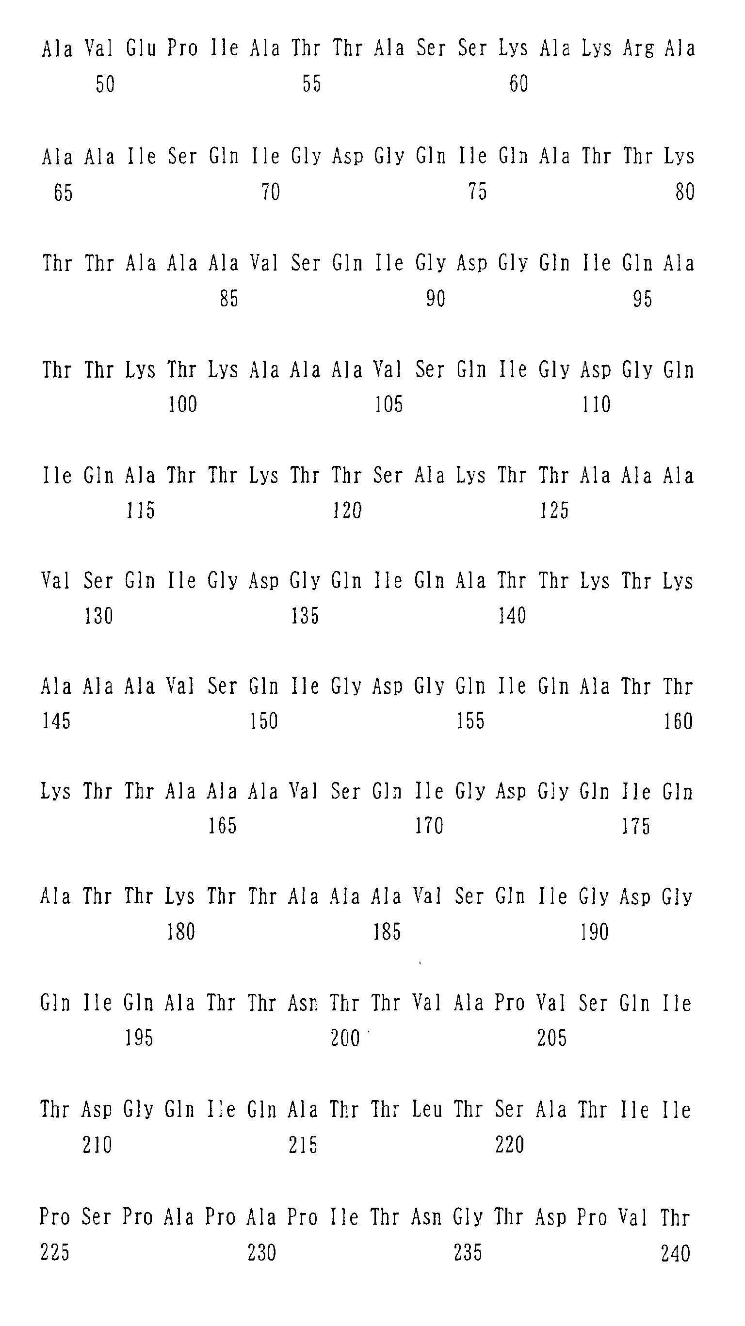
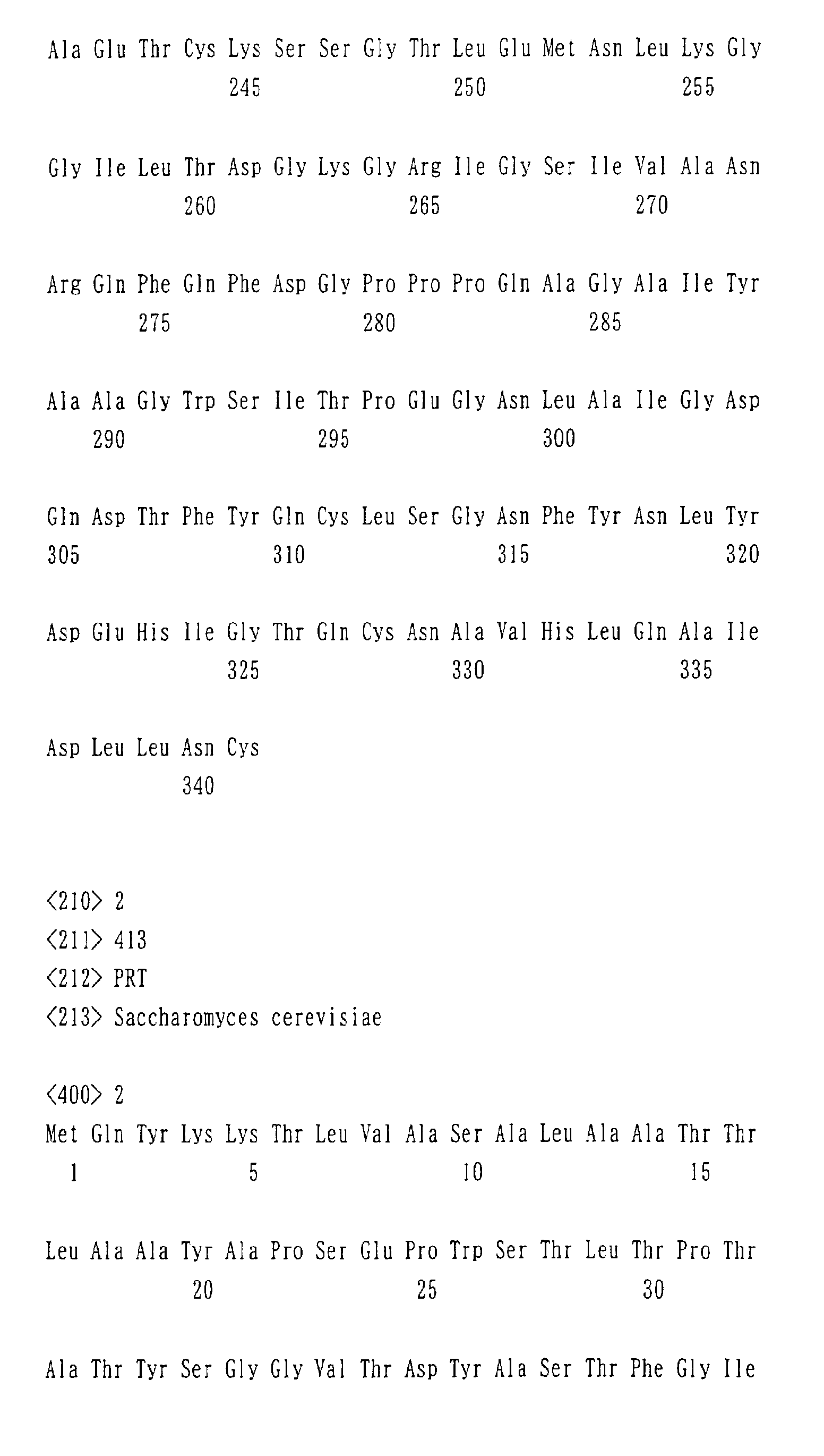
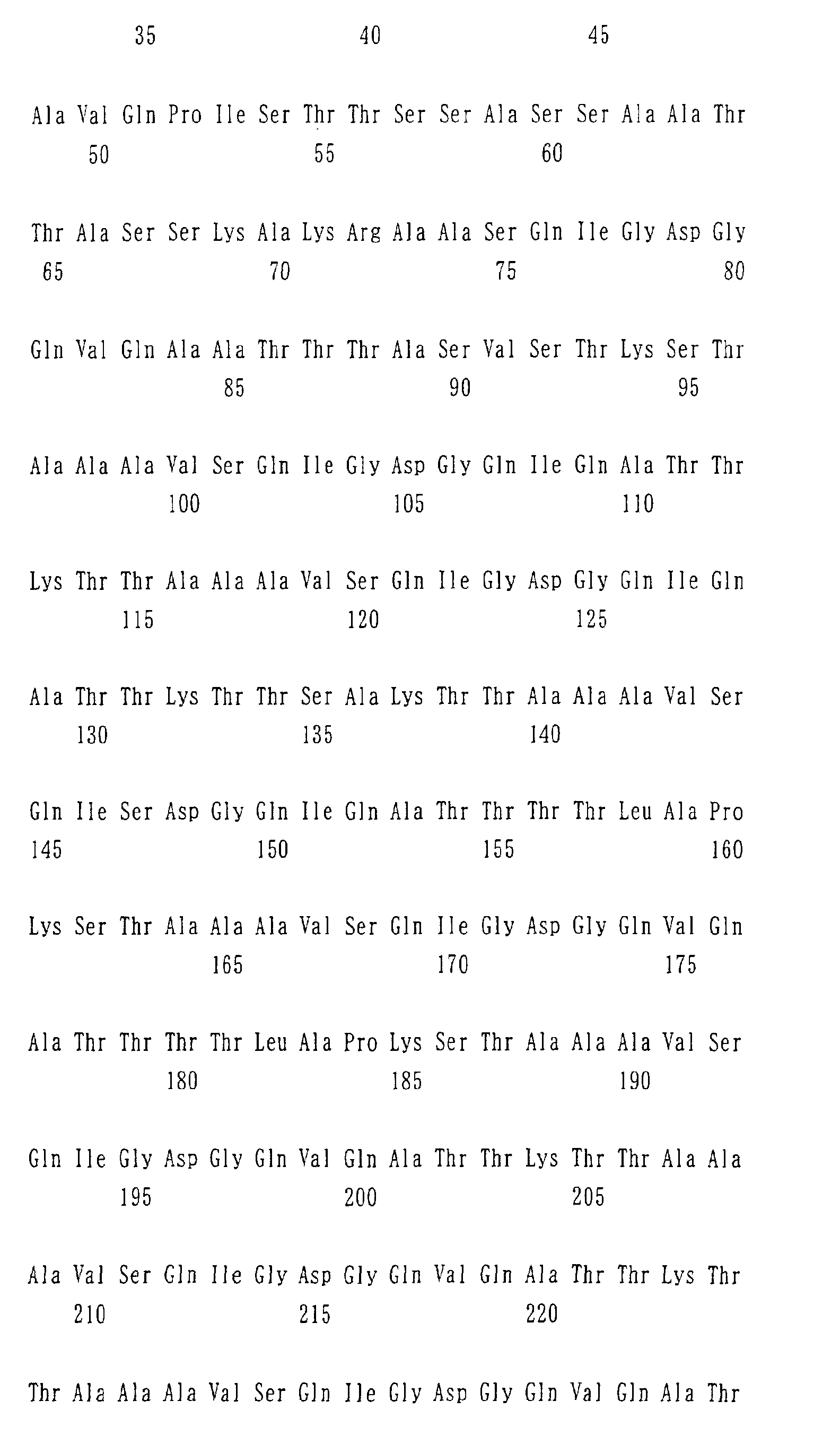

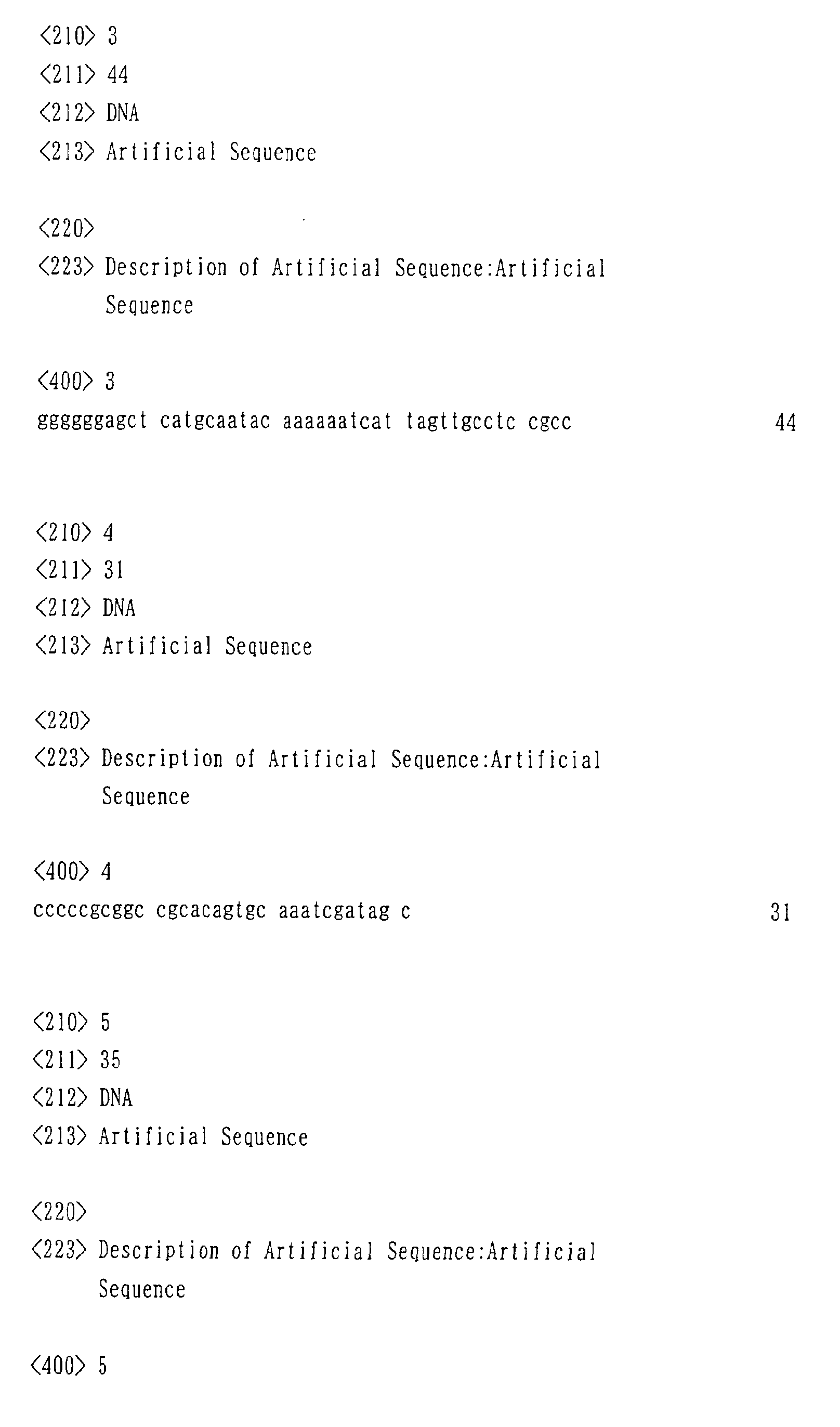


Claims (19)
- A fusion gene expression vector which contains a fusion gene comprising a gene encoding a useful protein downstream of a gene encoding the following yeast cell wall protein (a) or (b):(a) a protein having an amino acid sequence represented by SEQ ID NO: 1; or(b) a protein having an amino acid sequence derived from the amino acid sequence represented by SEQ NO: 1 by deletion, replacement, or addition of one or more amino acids, and having ability to be localized and immobilized on a yeast cell wall.
- The fusion gene expression vector of claim 1 wherein the useful protein is a glycosyltransferase protein.
- A transformant yeast which is transformed by the fusion gene expression vector of claim 1 or 2.
- A method for producing an immobilized enzyme which comprises culturing the transformant yeast of claim 3, expressing a fusion gene on the surface layer of a yeast cell wall, and obtaining yeast that contains a useful protein immobilized on the cell wall.
- An immobilized enzyme which is obtained by the method of claim 4.
- The immobilized enzyme of claim 5 wherein the enzyme to be immobilized is a glycosyltransferase.
- A method for producing a sugar chain or sugars which employs the immobilized enzyme of claim 6.
- A fusion gene expression vector which contains a fusion gene comprising a gene encoding a useful protein downstream of a gene encoding the following yeast cell wall protein (a) or (b) as follows:(a) a protein having an amino acid sequence represented by SEQ ID NO: 2;(b) a protein having an amino acid sequence derived from the amino acid sequence represented by SEQ NO: 2 by deletion, replacement, or addition of one or more amino acids, and having ability to be localized and immobilized on a yeast cell wall.
- The fusion gene expression vector of claim 8 wherein the useful protein is a glycosyltransferase protein.
- A transformant yeast which is transformed with the fusion gene expression vector of claim 8 or 9.
- A method for producing an immobilized enzyme which method comprises culturing the transformant yeast of claim 10, expressing a fusion gene on the surface layer of a yeast cell wall, and obtaining yeast that contains a useful protein immobilized on the cell wall.
- An immobilized enzyme which is obtained by the method of claim 11.
- The immobilized enzyme of claim 12 wherein the enzyme to be immobilized is a glycosyltransferase.
- A method for producing a sugar chain or sugars which employs the immobilized enzyme of claim 13.
- A transformant yeast which is transformed by allowing the yeast to contain at least two or more types of the fusion gene expression vectors of claims 1 and 8.
- A method for producing an immobilized enzyme which comprises culturing the transformant yeast of claim 15, expressing simultaneously fusion genes on the surface layer of a yeast cell wall, and obtaining yeast that contains at least two or more types of useful proteins immobilized on the cell wall.
- An immobilized enzyme which is obtained by the method of claim 16.
- The immobilized enzyme of claim 17 wherein the enzymes to be immobilized are two or more types of glycosyltransferases.
- A method for producing a sugar chain or sugars which sequentially converts sugar chains or sugars using the immobilized enzymes of claim 18.
Applications Claiming Priority (4)
| Application Number | Priority Date | Filing Date | Title |
|---|---|---|---|
| JP2000354396 | 2000-11-21 | ||
| JP2000354396 | 2000-11-21 | ||
| JP2001190524 | 2001-06-22 | ||
| JP2001190524A JP2002320485A (en) | 2000-11-21 | 2001-06-22 | Method for producing fusion gene expression vector and immobilized enzyme |
Publications (1)
| Publication Number | Publication Date |
|---|---|
| EP1209235A1 true EP1209235A1 (en) | 2002-05-29 |
Family
ID=26604366
Family Applications (1)
| Application Number | Title | Priority Date | Filing Date |
|---|---|---|---|
| EP01309792A Withdrawn EP1209235A1 (en) | 2000-11-21 | 2001-11-21 | Expression vector for fusion gene and method for producing immobilized enzyme |
Country Status (3)
| Country | Link |
|---|---|
| US (1) | US20030059872A1 (en) |
| EP (1) | EP1209235A1 (en) |
| JP (1) | JP2002320485A (en) |
Cited By (1)
| Publication number | Priority date | Publication date | Assignee | Title |
|---|---|---|---|---|
| WO2007099694A1 (en) * | 2006-02-24 | 2007-09-07 | Suntory Limited | Gene encoding protein responsible for flocculation property of yeast and use thereof |
Families Citing this family (1)
| Publication number | Priority date | Publication date | Assignee | Title |
|---|---|---|---|---|
| EP2497478A3 (en) | 2003-12-05 | 2012-11-07 | Children's Hospital Medical Center | Oligosaccaride compositions and use thereof in the treatment of infection |
Citations (3)
| Publication number | Priority date | Publication date | Assignee | Title |
|---|---|---|---|---|
| US5677172A (en) * | 1992-03-11 | 1997-10-14 | Makarow; Marja | Method for production of proteins in yeast |
| WO1999036569A1 (en) * | 1998-01-20 | 1999-07-22 | The Board Of Trustees Of The University Of Illinois | Yeast cell surface display of proteins and uses thereof |
| WO1999060152A1 (en) * | 1998-05-18 | 1999-11-25 | Nakarow Marja | Process for sialylating glycoproteins |
Family Cites Families (4)
| Publication number | Priority date | Publication date | Assignee | Title |
|---|---|---|---|---|
| JP3602972B2 (en) * | 1998-07-28 | 2004-12-15 | 富士通株式会社 | Communication performance measuring device and its measuring method |
| US6646987B1 (en) * | 1998-10-05 | 2003-11-11 | Nortel Networks Limited | Method and system for transmission control protocol (TCP) packet loss recovery over a wireless link |
| US6560243B1 (en) * | 1999-04-30 | 2003-05-06 | Hewlett-Packard Development Company | System and method for receiver based allocation of network bandwidth |
| US20020071388A1 (en) * | 2000-11-16 | 2002-06-13 | Einar Bergsson | Selectable network protocol |
-
2001
- 2001-06-22 JP JP2001190524A patent/JP2002320485A/en active Pending
- 2001-11-21 US US09/989,975 patent/US20030059872A1/en not_active Abandoned
- 2001-11-21 EP EP01309792A patent/EP1209235A1/en not_active Withdrawn
Patent Citations (3)
| Publication number | Priority date | Publication date | Assignee | Title |
|---|---|---|---|---|
| US5677172A (en) * | 1992-03-11 | 1997-10-14 | Makarow; Marja | Method for production of proteins in yeast |
| WO1999036569A1 (en) * | 1998-01-20 | 1999-07-22 | The Board Of Trustees Of The University Of Illinois | Yeast cell surface display of proteins and uses thereof |
| WO1999060152A1 (en) * | 1998-05-18 | 1999-11-25 | Nakarow Marja | Process for sialylating glycoproteins |
Non-Patent Citations (8)
| Title |
|---|
| DATABASE FASTA 4 February 1998 (1998-02-04), MAKAROW MARJA: "Method for production of proteins in yeast", XP002191644 * |
| DATABASE SWISS-PROT 1 October 1993 (1993-10-01), XP002191643 * |
| MATTILA P ET AL: "TARGETING OF ACTIVE RAT A2,3-SIALYLTRANSFERASE TO THE YEAST CELL WALL BY THE AID OF THE HSP 150D-CARRIER: TOWARD SYNTHESIS OF SLEX-DECORATED L-SELECTIN LIGANDS", GLYCOBIOLOGY, IRL PRESS,, GB, vol. 6, no. 8, 1996, pages 851 - 859, XP002919879, ISSN: 0959-6658 * |
| MOUKADIRI ISMAIL ET AL: "Identification of two mannoproteins released from cell walls of a Saccharomyces cerevisiae mnn1 mnn9 double mutant by reducing agents.", JOURNAL OF BACTERIOLOGY, vol. 181, no. 16, August 1999 (1999-08-01), pages 4741 - 4745, XP002191640, ISSN: 0021-9193 * |
| MRSA VLADIMIR ET AL: "Role of NaOH-extractable cell wall proteins Ccw5p, Ccw6p, Ccw7p and Ccw8p (members of the Pir protein family) in stability of the Saccharomyces cerevisiae cell wall.", YEAST, vol. 15, no. 10, July 1999 (1999-07-01), pages 813 - 820, XP002191641, ISSN: 0749-503X * |
| RUSSO P ET AL: "A HEAT SHOCK GENE FROM SACCHAROMYCES-CEREVISIAE ENCODING A SECRETORY GLYCOPROTEIN", PROCEEDINGS OF THE NATIONAL ACADEMY OF SCIENCES OF THE UNITED STATES, vol. 89, no. 9, 1992, 1992, pages 3671 - 3675, XP002191642, ISSN: 0027-8424 * |
| SCHREUDER M P ET AL: "Immobilizing proteins on the surface of yeast cells", TRENDS IN BIOTECHNOLOGY, ELSEVIER PUBLICATIONS, CAMBRIDGE, GB, vol. 14, no. 4, 1 April 1996 (1996-04-01), pages 115 - 120, XP004035795, ISSN: 0167-7799 * |
| TOH-E AKIO ET AL: "Three yeast genes, PIR1, PIR2 and PIR3, containing internal tandem repeats, are related to each other, and PIR1 and PIR2 are required for tolerance to heat shock.", YEAST, vol. 9, no. 5, 1993, ISSN: 0749-503X, pages 481 - 494, XP001057649 * |
Cited By (2)
| Publication number | Priority date | Publication date | Assignee | Title |
|---|---|---|---|---|
| WO2007099694A1 (en) * | 2006-02-24 | 2007-09-07 | Suntory Limited | Gene encoding protein responsible for flocculation property of yeast and use thereof |
| KR100929997B1 (en) * | 2006-02-24 | 2009-12-07 | 산토리 홀딩스 가부시키가이샤 | Genes encoding proteins responsible for the aggregation properties of yeast and uses thereof |
Also Published As
| Publication number | Publication date |
|---|---|
| JP2002320485A (en) | 2002-11-05 |
| US20030059872A1 (en) | 2003-03-27 |
Similar Documents
| Publication | Publication Date | Title |
|---|---|---|
| Abe et al. | In vitro oligosaccharide synthesis using intact yeast cells that display glycosyltransferases at the cell surface through cell wall–anchored protein Pir | |
| Murai et al. | Construction of a starch-utilizing yeast by cell surface engineering | |
| Tu et al. | Localization of Golgi-resident glycosyltransferases | |
| Breitling et al. | N-linked protein glycosylation in the endoplasmic reticulum | |
| US5879912A (en) | Method of synthesizing saccharide compositions | |
| AU2005233387B2 (en) | Production of galactosylated glycoproteins in lower eukaryotes | |
| US8815544B2 (en) | Production of galactosylated glycoproteins in lower eukaryotes | |
| EP2365089B1 (en) | Method of engineering a cytidine monophosphate-sialic acid synthetic pathway in yeast | |
| Tessier et al. | Cloning and characterization of mammalian UDP-glucose glycoprotein: glucosyltransferase and the development of a specific substrate for this enzyme | |
| WO2001014522A1 (en) | Novel yeast variants and process for producing glycoprotein containing mammalian type sugar chain | |
| JP2011167194A (en) | Method for producing modified glycoprotein | |
| US20100285563A1 (en) | Non-Ribosomal Peptide Synthetases | |
| EP2707482B1 (en) | Whole cell biocatalyst comprising a prenyltransferase | |
| Bencúrová et al. | Expression of eukaryotic glycosyltransferases in the yeast Pichia pastoris | |
| US8741602B2 (en) | Method for producing glucuronic acid conjugate using Saccharomyces cerevisiae | |
| KR20150146300A (en) | HELICOBACTER PYLORI α-1,2 FUCOSYLTRANSFERASE GENE AND PROTEIN WITH IMPROVED SOLUBLE PROTEIN EXPRESSION, AND THEREOF APPLICATION FOR SYNTHESIS OF α-1,2 FUCOSYLOLIGOSACCHARIDE | |
| JP2002320485A (en) | Method for producing fusion gene expression vector and immobilized enzyme | |
| Bettler et al. | Production of recombinant xenotransplantation antigen in Escherichia coli | |
| Kim et al. | Display of Bacillus macerans cyclodextrin glucanotransferase on cell surface of Saccharomyces cerevisiae | |
| KR102129279B1 (en) | Noble Carbon Monoxide:Formate Oxidoreductase Comprising Fe-S Fusion Protein, and Use thereof | |
| EP1231266B1 (en) | Arabidopsis-origin gdp-4-keto-6-deoxy-d-mannose-3,5-epimerase-4-reductase gene | |
| KR102192800B1 (en) | Noble Fe-S Fusion Protein Acting as an Electron Transfer Chain, and Use thereof | |
| KR102129282B1 (en) | Novel Strain from Thermococcus BCF12 and Method for Producing Formic Acid Using the Same | |
| JPH05199871A (en) | Improved method for producing glycosyltransferase | |
| KR100701209B1 (en) | Cell surface expression method of target protein using cell surface protein in yeast of Yroia spp. |
Legal Events
| Date | Code | Title | Description |
|---|---|---|---|
| PUAI | Public reference made under article 153(3) epc to a published international application that has entered the european phase |
Free format text: ORIGINAL CODE: 0009012 |
|
| AK | Designated contracting states |
Kind code of ref document: A1 Designated state(s): AT BE CH CY DE DK ES FI FR GB GR IE IT LI LU MC NL PT SE TR |
|
| AX | Request for extension of the european patent |
Free format text: AL;LT;LV;MK;RO;SI |
|
| 17P | Request for examination filed |
Effective date: 20021108 |
|
| AKX | Designation fees paid |
Designated state(s): DE FR GB |
|
| STAA | Information on the status of an ep patent application or granted ep patent |
Free format text: STATUS: THE APPLICATION IS DEEMED TO BE WITHDRAWN |
|
| 18D | Application deemed to be withdrawn |
Effective date: 20060411 |