EP1182253A2 - Method for improving the thermostability of proteins - Google Patents
Method for improving the thermostability of proteins Download PDFInfo
- Publication number
- EP1182253A2 EP1182253A2 EP01115642A EP01115642A EP1182253A2 EP 1182253 A2 EP1182253 A2 EP 1182253A2 EP 01115642 A EP01115642 A EP 01115642A EP 01115642 A EP01115642 A EP 01115642A EP 1182253 A2 EP1182253 A2 EP 1182253A2
- Authority
- EP
- European Patent Office
- Prior art keywords
- amino acid
- protein
- ancestral
- proteins
- thermostability
- Prior art date
- Legal status (The legal status is an assumption and is not a legal conclusion. Google has not performed a legal analysis and makes no representation as to the accuracy of the status listed.)
- Withdrawn
Links
- 108090000623 proteins and genes Proteins 0.000 title claims abstract description 167
- 102000004169 proteins and genes Human genes 0.000 title claims abstract description 156
- 238000000034 method Methods 0.000 title claims abstract description 78
- 125000003275 alpha amino acid group Chemical group 0.000 claims abstract description 80
- 125000000539 amino acid group Chemical group 0.000 claims abstract description 64
- 150000007523 nucleic acids Chemical class 0.000 claims abstract description 12
- 108020004707 nucleic acids Proteins 0.000 claims abstract description 10
- 102000039446 nucleic acids Human genes 0.000 claims abstract description 10
- 108010039636 3-isopropylmalate dehydrogenase Proteins 0.000 claims description 58
- 241000894007 species Species 0.000 claims description 48
- 108010075869 Isocitrate Dehydrogenase Proteins 0.000 claims description 39
- 102000012011 Isocitrate Dehydrogenase Human genes 0.000 claims description 39
- 241000894006 Bacteria Species 0.000 claims description 38
- 241000203069 Archaea Species 0.000 claims description 12
- 108020004511 Recombinant DNA Proteins 0.000 claims description 4
- 238000011392 neighbor-joining method Methods 0.000 claims description 4
- 238000012360 testing method Methods 0.000 claims description 3
- 102000053602 DNA Human genes 0.000 claims description 2
- 230000000694 effects Effects 0.000 description 42
- 230000035772 mutation Effects 0.000 description 30
- 210000004027 cell Anatomy 0.000 description 22
- 241000205088 Sulfolobus sp. Species 0.000 description 21
- 102000004190 Enzymes Human genes 0.000 description 19
- 108090000790 Enzymes Proteins 0.000 description 19
- 239000000243 solution Substances 0.000 description 14
- 239000000203 mixture Substances 0.000 description 13
- 239000002609 medium Substances 0.000 description 12
- 239000013612 plasmid Substances 0.000 description 12
- 108020004414 DNA Proteins 0.000 description 11
- 241000588724 Escherichia coli Species 0.000 description 11
- 108091008146 restriction endonucleases Proteins 0.000 description 11
- TWRXJAOTZQYOKJ-UHFFFAOYSA-L Magnesium chloride Chemical compound [Mg+2].[Cl-].[Cl-] TWRXJAOTZQYOKJ-UHFFFAOYSA-L 0.000 description 10
- 241000589499 Thermus thermophilus Species 0.000 description 10
- 102200115815 rs121918068 Human genes 0.000 description 9
- 239000006228 supernatant Substances 0.000 description 8
- 230000000813 microbial effect Effects 0.000 description 7
- ISAKRJDGNUQOIC-UHFFFAOYSA-N Uracil Chemical compound O=C1C=CNC(=O)N1 ISAKRJDGNUQOIC-UHFFFAOYSA-N 0.000 description 6
- AVKUERGKIZMTKX-NJBDSQKTSA-N ampicillin Chemical compound C1([C@@H](N)C(=O)N[C@H]2[C@H]3SC([C@@H](N3C2=O)C(O)=O)(C)C)=CC=CC=C1 AVKUERGKIZMTKX-NJBDSQKTSA-N 0.000 description 6
- 229960000723 ampicillin Drugs 0.000 description 6
- 239000012131 assay buffer Substances 0.000 description 6
- 238000005119 centrifugation Methods 0.000 description 6
- 238000010438 heat treatment Methods 0.000 description 6
- 101150025049 leuB gene Proteins 0.000 description 6
- 229930027945 nicotinamide-adenine dinucleotide Natural products 0.000 description 6
- 239000002773 nucleotide Substances 0.000 description 6
- 125000003729 nucleotide group Chemical group 0.000 description 6
- 239000013598 vector Substances 0.000 description 6
- 241000577831 Caldococcus noboribetus Species 0.000 description 5
- KCXVZYZYPLLWCC-UHFFFAOYSA-N EDTA Chemical compound OC(=O)CN(CC(O)=O)CCN(CC(O)=O)CC(O)=O KCXVZYZYPLLWCC-UHFFFAOYSA-N 0.000 description 5
- 240000004808 Saccharomyces cerevisiae Species 0.000 description 5
- 235000014680 Saccharomyces cerevisiae Nutrition 0.000 description 5
- XJLXINKUBYWONI-DQQFMEOOSA-N [[(2r,3r,4r,5r)-5-(6-aminopurin-9-yl)-3-hydroxy-4-phosphonooxyoxolan-2-yl]methoxy-hydroxyphosphoryl] [(2s,3r,4s,5s)-5-(3-carbamoylpyridin-1-ium-1-yl)-3,4-dihydroxyoxolan-2-yl]methyl phosphate Chemical compound NC(=O)C1=CC=C[N+]([C@@H]2[C@H]([C@@H](O)[C@H](COP([O-])(=O)OP(O)(=O)OC[C@@H]3[C@H]([C@@H](OP(O)(O)=O)[C@@H](O3)N3C4=NC=NC(N)=C4N=C3)O)O2)O)=C1 XJLXINKUBYWONI-DQQFMEOOSA-N 0.000 description 5
- 150000001413 amino acids Chemical class 0.000 description 5
- 229910001629 magnesium chloride Inorganic materials 0.000 description 5
- 238000002703 mutagenesis Methods 0.000 description 5
- 231100000350 mutagenesis Toxicity 0.000 description 5
- 238000002360 preparation method Methods 0.000 description 5
- 244000063299 Bacillus subtilis Species 0.000 description 4
- 235000014469 Bacillus subtilis Nutrition 0.000 description 4
- FAPWRFPIFSIZLT-UHFFFAOYSA-M Sodium chloride Chemical compound [Na+].[Cl-] FAPWRFPIFSIZLT-UHFFFAOYSA-M 0.000 description 4
- 241000589596 Thermus Species 0.000 description 4
- 238000002835 absorbance Methods 0.000 description 4
- BFNBIHQBYMNNAN-UHFFFAOYSA-N ammonium sulfate Chemical compound N.N.OS(O)(=O)=O BFNBIHQBYMNNAN-UHFFFAOYSA-N 0.000 description 4
- 229910052921 ammonium sulfate Inorganic materials 0.000 description 4
- 235000011130 ammonium sulphate Nutrition 0.000 description 4
- 239000000872 buffer Substances 0.000 description 4
- 238000004422 calculation algorithm Methods 0.000 description 4
- 230000008859 change Effects 0.000 description 4
- 238000006243 chemical reaction Methods 0.000 description 4
- 238000010276 construction Methods 0.000 description 4
- 230000003247 decreasing effect Effects 0.000 description 4
- 238000000746 purification Methods 0.000 description 4
- 102220212423 rs55907012 Human genes 0.000 description 4
- 102220252087 rs79952473 Human genes 0.000 description 4
- 238000002741 site-directed mutagenesis Methods 0.000 description 4
- 108020004465 16S ribosomal RNA Proteins 0.000 description 3
- 229920001817 Agar Polymers 0.000 description 3
- 239000008000 CHES buffer Substances 0.000 description 3
- 108010054576 Deoxyribonuclease EcoRI Proteins 0.000 description 3
- 102220519092 E3 ubiquitin-protein ligase TRIM22_A259S_mutation Human genes 0.000 description 3
- MKWKNSIESPFAQN-UHFFFAOYSA-N N-cyclohexyl-2-aminoethanesulfonic acid Chemical compound OS(=O)(=O)CCNC1CCCCC1 MKWKNSIESPFAQN-UHFFFAOYSA-N 0.000 description 3
- 239000008272 agar Substances 0.000 description 3
- 238000000137 annealing Methods 0.000 description 3
- 238000003776 cleavage reaction Methods 0.000 description 3
- 239000002131 composite material Substances 0.000 description 3
- 238000012258 culturing Methods 0.000 description 3
- 239000013613 expression plasmid Substances 0.000 description 3
- 239000013604 expression vector Substances 0.000 description 3
- 239000001963 growth medium Substances 0.000 description 3
- WQVJUBFKFCDYDQ-BBWFWOEESA-N leubethanol Natural products C1=C(C)C=C2[C@H]([C@H](CCC=C(C)C)C)CC[C@@H](C)C2=C1O WQVJUBFKFCDYDQ-BBWFWOEESA-N 0.000 description 3
- 238000009630 liquid culture Methods 0.000 description 3
- 244000005700 microbiome Species 0.000 description 3
- BOPGDPNILDQYTO-NNYOXOHSSA-N nicotinamide-adenine dinucleotide Chemical compound C1=CCC(C(=O)N)=CN1[C@H]1[C@H](O)[C@H](O)[C@@H](COP(O)(=O)OP(O)(=O)OC[C@@H]2[C@H]([C@@H](O)[C@@H](O2)N2C3=NC=NC(N)=C3N=C2)O)O1 BOPGDPNILDQYTO-NNYOXOHSSA-N 0.000 description 3
- 102220043361 rs73866065 Human genes 0.000 description 3
- 239000000523 sample Substances 0.000 description 3
- 230000007017 scission Effects 0.000 description 3
- 238000002415 sodium dodecyl sulfate polyacrylamide gel electrophoresis Methods 0.000 description 3
- 229940035893 uracil Drugs 0.000 description 3
- IHPYMWDTONKSCO-UHFFFAOYSA-N 2,2'-piperazine-1,4-diylbisethanesulfonic acid Chemical compound OS(=O)(=O)CCN1CCN(CCS(O)(=O)=O)CC1 IHPYMWDTONKSCO-UHFFFAOYSA-N 0.000 description 2
- 241000589155 Agrobacterium tumefaciens Species 0.000 description 2
- 241001362614 Crassa Species 0.000 description 2
- 241001288713 Escherichia coli MC1061 Species 0.000 description 2
- 238000012218 Kunkel's method Methods 0.000 description 2
- ODBLHEXUDAPZAU-FONMRSAGSA-N L-threo-isocitric acid Chemical compound OC(=O)[C@@H](O)[C@H](C(O)=O)CC(O)=O ODBLHEXUDAPZAU-FONMRSAGSA-N 0.000 description 2
- 239000006142 Luria-Bertani Agar Substances 0.000 description 2
- CSNNHWWHGAXBCP-UHFFFAOYSA-L Magnesium sulfate Chemical compound [Mg+2].[O-][S+2]([O-])([O-])[O-] CSNNHWWHGAXBCP-UHFFFAOYSA-L 0.000 description 2
- 241000221961 Neurospora crassa Species 0.000 description 2
- 108091028043 Nucleic acid sequence Proteins 0.000 description 2
- 239000007990 PIPES buffer Substances 0.000 description 2
- 101000702488 Rattus norvegicus High affinity cationic amino acid transporter 1 Proteins 0.000 description 2
- 238000005349 anion exchange Methods 0.000 description 2
- 210000000170 cell membrane Anatomy 0.000 description 2
- 238000002983 circular dichroism Methods 0.000 description 2
- 238000004590 computer program Methods 0.000 description 2
- 230000002950 deficient Effects 0.000 description 2
- 238000002474 experimental method Methods 0.000 description 2
- 239000000284 extract Substances 0.000 description 2
- 230000002209 hydrophobic effect Effects 0.000 description 2
- 239000007788 liquid Substances 0.000 description 2
- 238000005259 measurement Methods 0.000 description 2
- 230000007246 mechanism Effects 0.000 description 2
- 125000001360 methionine group Chemical group N[C@@H](CCSC)C(=O)* 0.000 description 2
- 238000012986 modification Methods 0.000 description 2
- 230000004048 modification Effects 0.000 description 2
- 238000011533 pre-incubation Methods 0.000 description 2
- 239000002244 precipitate Substances 0.000 description 2
- 125000002924 primary amino group Chemical group [H]N([H])* 0.000 description 2
- 239000012460 protein solution Substances 0.000 description 2
- 239000011780 sodium chloride Substances 0.000 description 2
- 230000007928 solubilization Effects 0.000 description 2
- 238000005063 solubilization Methods 0.000 description 2
- 238000000527 sonication Methods 0.000 description 2
- 241000589158 Agrobacterium Species 0.000 description 1
- FSHURBQASBLAPO-WDSKDSINSA-N Ala-Met Chemical compound CSCC[C@@H](C(O)=O)NC(=O)[C@H](C)N FSHURBQASBLAPO-WDSKDSINSA-N 0.000 description 1
- 238000009020 BCA Protein Assay Kit Methods 0.000 description 1
- 238000000035 BCA protein assay Methods 0.000 description 1
- 241000283690 Bos taurus Species 0.000 description 1
- 108091003079 Bovine Serum Albumin Proteins 0.000 description 1
- UXVMQQNJUSDDNG-UHFFFAOYSA-L Calcium chloride Chemical compound [Cl-].[Cl-].[Ca+2] UXVMQQNJUSDDNG-UHFFFAOYSA-L 0.000 description 1
- 241000306001 Cetartiodactyla Species 0.000 description 1
- 108020004705 Codon Proteins 0.000 description 1
- 108700010070 Codon Usage Proteins 0.000 description 1
- 102000012410 DNA Ligases Human genes 0.000 description 1
- 108010061982 DNA Ligases Proteins 0.000 description 1
- 102000016928 DNA-directed DNA polymerase Human genes 0.000 description 1
- 108010014303 DNA-directed DNA polymerase Proteins 0.000 description 1
- 102100023933 Deoxyuridine 5'-triphosphate nucleotidohydrolase, mitochondrial Human genes 0.000 description 1
- 241000206602 Eukaryota Species 0.000 description 1
- WQZGKKKJIJFFOK-GASJEMHNSA-N Glucose Natural products OC[C@H]1OC(O)[C@H](O)[C@@H](O)[C@@H]1O WQZGKKKJIJFFOK-GASJEMHNSA-N 0.000 description 1
- MIDZLCFIAINOQN-WPRPVWTQSA-N Phe-Ala Chemical compound OC(=O)[C@H](C)NC(=O)[C@@H](N)CC1=CC=CC=C1 MIDZLCFIAINOQN-WPRPVWTQSA-N 0.000 description 1
- GLUBLISJVJFHQS-VIFPVBQESA-N Phe-Gly Chemical compound OC(=O)CNC(=O)[C@@H](N)CC1=CC=CC=C1 GLUBLISJVJFHQS-VIFPVBQESA-N 0.000 description 1
- 108010021757 Polynucleotide 5'-Hydroxyl-Kinase Proteins 0.000 description 1
- 102000008422 Polynucleotide 5'-hydroxyl-kinase Human genes 0.000 description 1
- QAOWNCQODCNURD-UHFFFAOYSA-L Sulfate Chemical compound [O-]S([O-])(=O)=O QAOWNCQODCNURD-UHFFFAOYSA-L 0.000 description 1
- 241000205101 Sulfolobus Species 0.000 description 1
- 241000868182 Thermus thermophilus HB8 Species 0.000 description 1
- JZRWCGZRTZMZEH-UHFFFAOYSA-N Thiamine Natural products CC1=C(CCO)SC=[N+]1CC1=CN=C(C)N=C1N JZRWCGZRTZMZEH-UHFFFAOYSA-N 0.000 description 1
- NLKUJNGEGZDXGO-XVKPBYJWSA-N Tyr-Ala Chemical compound OC(=O)[C@H](C)NC(=O)[C@@H](N)CC1=CC=C(O)C=C1 NLKUJNGEGZDXGO-XVKPBYJWSA-N 0.000 description 1
- 230000004075 alteration Effects 0.000 description 1
- 238000012870 ammonium sulfate precipitation Methods 0.000 description 1
- 230000008901 benefit Effects 0.000 description 1
- 229940098773 bovine serum albumin Drugs 0.000 description 1
- 239000001110 calcium chloride Substances 0.000 description 1
- 229910001628 calcium chloride Inorganic materials 0.000 description 1
- 238000004364 calculation method Methods 0.000 description 1
- 230000003197 catalytic effect Effects 0.000 description 1
- 239000012295 chemical reaction liquid Substances 0.000 description 1
- 239000003153 chemical reaction reagent Substances 0.000 description 1
- 238000001142 circular dichroism spectrum Methods 0.000 description 1
- 238000010367 cloning Methods 0.000 description 1
- 230000000052 comparative effect Effects 0.000 description 1
- 238000012790 confirmation Methods 0.000 description 1
- 238000011109 contamination Methods 0.000 description 1
- 238000007796 conventional method Methods 0.000 description 1
- 239000000287 crude extract Substances 0.000 description 1
- 108010011219 dUTP pyrophosphatase Proteins 0.000 description 1
- 238000012217 deletion Methods 0.000 description 1
- 230000037430 deletion Effects 0.000 description 1
- 238000013461 design Methods 0.000 description 1
- 238000010586 diagram Methods 0.000 description 1
- 238000000502 dialysis Methods 0.000 description 1
- RTZKZFJDLAIYFH-UHFFFAOYSA-N ether Substances CCOCC RTZKZFJDLAIYFH-UHFFFAOYSA-N 0.000 description 1
- -1 ether lipid Chemical class 0.000 description 1
- 238000002523 gelfiltration Methods 0.000 description 1
- 239000008103 glucose Substances 0.000 description 1
- 229960000789 guanidine hydrochloride Drugs 0.000 description 1
- PJJJBBJSCAKJQF-UHFFFAOYSA-N guanidinium chloride Chemical compound [Cl-].NC(N)=[NH2+] PJJJBBJSCAKJQF-UHFFFAOYSA-N 0.000 description 1
- 238000009396 hybridization Methods 0.000 description 1
- 230000006872 improvement Effects 0.000 description 1
- 210000003000 inclusion body Anatomy 0.000 description 1
- 230000001939 inductive effect Effects 0.000 description 1
- 238000003780 insertion Methods 0.000 description 1
- 230000037431 insertion Effects 0.000 description 1
- 238000011835 investigation Methods 0.000 description 1
- 238000004255 ion exchange chromatography Methods 0.000 description 1
- BPHPUYQFMNQIOC-NXRLNHOXSA-N isopropyl beta-D-thiogalactopyranoside Chemical compound CC(C)S[C@@H]1O[C@H](CO)[C@H](O)[C@H](O)[C@H]1O BPHPUYQFMNQIOC-NXRLNHOXSA-N 0.000 description 1
- 125000001909 leucine group Chemical group [H]N(*)C(C(*)=O)C([H])([H])C(C([H])([H])[H])C([H])([H])[H] 0.000 description 1
- 150000002632 lipids Chemical class 0.000 description 1
- 229910052943 magnesium sulfate Inorganic materials 0.000 description 1
- 238000004519 manufacturing process Methods 0.000 description 1
- 102000035118 modified proteins Human genes 0.000 description 1
- 108091005573 modified proteins Proteins 0.000 description 1
- 238000010369 molecular cloning Methods 0.000 description 1
- 108010024607 phenylalanylalanine Proteins 0.000 description 1
- 229910052700 potassium Inorganic materials 0.000 description 1
- 239000008057 potassium phosphate buffer Substances 0.000 description 1
- 230000008569 process Effects 0.000 description 1
- 239000000047 product Substances 0.000 description 1
- 239000011541 reaction mixture Substances 0.000 description 1
- 230000001105 regulatory effect Effects 0.000 description 1
- 238000011160 research Methods 0.000 description 1
- 238000004366 reverse phase liquid chromatography Methods 0.000 description 1
- 102200089569 rs373068386 Human genes 0.000 description 1
- 150000003839 salts Chemical class 0.000 description 1
- 238000012163 sequencing technique Methods 0.000 description 1
- 238000001228 spectrum Methods 0.000 description 1
- 229910021653 sulphate ion Inorganic materials 0.000 description 1
- 239000004094 surface-active agent Substances 0.000 description 1
- 239000000725 suspension Substances 0.000 description 1
- KYMBYSLLVAOCFI-UHFFFAOYSA-N thiamine Chemical compound CC1=C(CCO)SCN1CC1=CN=C(C)N=C1N KYMBYSLLVAOCFI-UHFFFAOYSA-N 0.000 description 1
- 229960003495 thiamine Drugs 0.000 description 1
- 235000019157 thiamine Nutrition 0.000 description 1
- 239000011721 thiamine Substances 0.000 description 1
Images






Classifications
-
- C—CHEMISTRY; METALLURGY
- C12—BIOCHEMISTRY; BEER; SPIRITS; WINE; VINEGAR; MICROBIOLOGY; ENZYMOLOGY; MUTATION OR GENETIC ENGINEERING
- C12N—MICROORGANISMS OR ENZYMES; COMPOSITIONS THEREOF; PROPAGATING, PRESERVING, OR MAINTAINING MICROORGANISMS; MUTATION OR GENETIC ENGINEERING; CULTURE MEDIA
- C12N9/00—Enzymes; Proenzymes; Compositions thereof; Processes for preparing, activating, inhibiting, separating or purifying enzymes
- C12N9/0004—Oxidoreductases (1.)
- C12N9/0006—Oxidoreductases (1.) acting on CH-OH groups as donors (1.1)
-
- C—CHEMISTRY; METALLURGY
- C12—BIOCHEMISTRY; BEER; SPIRITS; WINE; VINEGAR; MICROBIOLOGY; ENZYMOLOGY; MUTATION OR GENETIC ENGINEERING
- C12N—MICROORGANISMS OR ENZYMES; COMPOSITIONS THEREOF; PROPAGATING, PRESERVING, OR MAINTAINING MICROORGANISMS; MUTATION OR GENETIC ENGINEERING; CULTURE MEDIA
- C12N9/00—Enzymes; Proenzymes; Compositions thereof; Processes for preparing, activating, inhibiting, separating or purifying enzymes
- C12N9/96—Stabilising an enzyme by forming an adduct or a composition; Forming enzyme conjugates
Definitions
- the present invention relates to a method for improving thermostability of a protein.
- the present invention also relates to a protein having an improved thermostability and a nucleic acid encoding the protein having improved thermostability.
- thermostable enzyme A protein active at a high temperature, particularly a thermostable enzyme, is more advantageous than another protein which is inactivated at a high temperature, for example, in that it can be used without being cooled.
- a protein is mostly produced by a bacterium called thermophilic bacterium, which can grow at a high temperature.
- thermophilic bacterium which can grow at a high temperature.
- amino acid sequence of a corresponding protein of such a group of thermophilic bacteria is analyzed and the characteristic feature of the amino acid sequence common to them is taken into account.
- the three-dimensional structure of a protein produced by the thermophilic bacterium is analyzed, the structure for imparting the thermostability is estimated from thus obtained information, and the structure of the heat-unstable protein is modified according to the estimated structure.
- IPMDH 3-isopropylmalate dehydrogenase encoded by leu B
- the three-dimensional structure of IPMDH of Thermus thermophilus HB8 has been elucidated (K. Imada et al., J. Mol. Biol. 222, 725-738, 1991).
- ICDH isocitrate dehydrogenase
- the object of the present invention is to provide a method for improving thermostability of protein, a protein having an improved thermostability and a nucleic acid encoding the protein, and host cells capable of producing a protein having improved thermostability.
- the object of the present invention is to provide a method for improving thermostability of a protein, taking advantage of only the information of the primary structure of the protein.
- thermophilic bacteria On the basis of the fact that many organisms which properly grow at a temperature of 80°C or above are located at the root of a phylogenetic tree by 16S rRNA (Fig. 1) shown by Woese et al., the inventors had an idea that the ancestors common to eubacteria, eukaryotes and archaebacteria might be ultra-thermophilic bacteria. On the basis of this supposition, the inventors have gotten an idea that although protein of many kinds of existing thermophilic bacteria are not always the protein of a true ancestral protein having an amino acid sequence of the ancestral or an amino acid sequence close to the ancestral sequence might have a further improved thermostability.
- thermostable protein it is more important that the amino acid sequence of ancestral protein is estimated and mimicked than that only the sequence and the higher-order structure of protein of a thermophilic bacterium are analyzed and mimicked.
- the present invention provides a method for improving thermostability of proteins, which comprises the steps of
- the present invention particularly includes the comparison of species evolutionarily close to thermophilic bacteria or archaebacteria in the phylogenetic tree with each other on the amino acid sequence of corresponding proteins.
- the present invention also provides an enzyme improved in heat resistance by the above-described method, a nucleic acid encoding the enzyme and host cells containing such a nucleic acid.
- Phylogenetic tree Molecular phylogenetic tree (hereinafter referred to as "phylogenetic tree") based on the molecular level information of species or an algorithm for the preparation of the phylogenetic tree is utilized in the present invention.
- Some algorithms for preparing phylogenetic trees such as the algorithm based on the maximum parsimony principle, are known.
- Computer programs for implementing the algorithms are utilizable or available. For example, various phylogenetic tree estimation programs such as CLUSTALW, PUZZLE, MOLPHY and PHYLIP are utilizable.
- phylogenetic trees can be produced by such programs, it is easier to utilize an already published phylogenetic tree (Fig. 1).
- a phylogenetic tree based on 16S rRNA data proposed by Woese et al. is also usable.
- species which are close to each other in the molecular evolution appear in positions close to each other.
- Species positioned closely to the root of the phylogenetic tree are considered to be close to the ancestors.
- thermophilic bacteria and archaebacteria are positioned close to the root, namely, evolutionarily close to the ancestors in the phylogenetic tree. Further, proteins produced by them are expected to be relatively close to ancestral super-thermostable protein.
- thermophilic bacteria is a generic name for bacteria capable of growing at a high temperature of usually above about 55°C. These bacteria are also called thermostable bacteria for the purpose of the present invention.
- thermostable bacteria indicates both highly thermophilic bacteria capable of growing at a temperature of higher than above 75°C and also moderately thermophilic bacteria capable of growing at about 55 to 74°C. They also include facultative thermophilic bacteria capable of growing at ambient temperature and obligate thermophilic bacteria capable of growing only at a temperature of above about 40°C.
- non-thermophilic bacteria indicates microorganisms other than the thermophilic bacteria.
- archaebacteria indicates those classified according to the above-described Woese's classification.
- proteins belonging to the same family indicates proteins which are similar to each other in at least one of the function, amino acid sequence, domain structure and steric structure. They include a group of proteins, at least amino acid sequences of which are partially homologous and the multiple alignment of which is possible. In particular, they include a group of proteins, at least amino acid sequences of which are homologous and can be multiple aligned. It is eagerly expected that two or more proteins belonging the same family are derived from the same ancestral protein.
- proteins to which the present invention can be applied are not particularly limited, they are preferably proteins present in various species. Particularly enzymes having a high value of industrial utilization is preferable. Preferred examples of them are proteins produced by thermophilic bacteria, particularly thermostable enzymes. Example of them is IPMDH and ICDH of Sulfolobus sp. stain 7. The gene encoding IPMDH of this strain was cloned by Suzuki et al. [T. Suzuki et al., J. Bacteriol. 179 (4), 1174-1179, 1997].
- Amino acid sequences of protein to be improved in thermostability can be also obtained from an already known data base.
- any method for determining amino acid sequence known in the art can be employed. It is also possible to estimate the amino acid sequence by obtaining a nucleic acid encoding the protein according to the information of partial amino acid sequence, determining the nucleic acid sequence by a well-known sequencing techniques and estimating the amino acid sequence from the nucleic acid sequence.
- the amino acid sequences obtained from the respective species are compared with each other.
- Some methods for the multiple alignment are known.
- One of the methods is based on the maximun parsimony principle for minimizing the change due to the insertion, deletion, replacement, etc.
- Computer programs for implementing this principle have been developed, which can be used or available. For example, TreeAlign is known among them. From DDBJ, "malign" which is the 1990 version of the program can be used.
- each of the species to be compared preferably contains one or more thermophilic bacteria or archaebacteria, based on the aforementioned reason. It is also preferred that it contains a family protein, namely another protein expected to be derived from the same ancestral protein.
- amino acid sequence of the ancestral protein can be estimated on the phylogenetic tree.
- the maximum parsimony method or maximal likelihood method is utilizable.
- the procedure of such a method is well known to those skilled in the art [see, for example, Young, Z., Kumar, S and Nei. M, Genetics 141, 1641-16510, 1995; Steward, C. -B. Active ancestral molecules, Nature 374, 12-13, 1995; and Molecuar Evolutinary Genetics, Columbia University Press, New York, USA, 1987].
- the maximal parsimony method which can be employed in the present invention is, in short, a method wherein an ancestral type having the minimal number of the mutation expected to occur after the estimation of the ancestral type is likely estimated to be the true ancestral type.
- the maximal likelihood method can be employed instead of the maximum parsimony method.
- a program PROTPARS (included in PHYLIP) for directly estimating the ancestral type from the amino acid sequence according to the maximum parsimony method can be also employed. Because the phylogenetic tree and ancestral amino acid are principally estimated at the same time in those methods, it is not always necessary to prepare the phylogenetic tree when such a method is employed.
- the preparation of the phylogenetic tree is preferred particularly when the ancestral amino acid is to be estimated by manual calculation.
- the ancestral amino acid sequence can be determined by the following maximum parsimony method or maximal likelihood method according to a phylogenetic tree produced by the above-described method or another already known method, particularly based on an already published phylogenetic tree.
- Ancestral amino acids in respective sites of the multiply aligned residues can be determined by means of a phylogenetic tree obtained by any method.
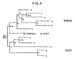
- Fig. 4 shows amino acid residues from various organisms corresponding to residue 152 of Sulfolobus sp. strain 7 of IPMDH. Amino acids at this position in the organisms shown in Fig. 4 are R, S, K or E.
- both residues in species close to each other in the phylogenetic tree are R, it can be estimated that in the ancestral species common to them (shown by the binding point connecting two species in the phylogenetic tree), the amino acid residue corresponding to residue 152 of Sulfolobus sp.
- strain 7 would be R for the following reasons: When R is the ancestral type, only one variation can elucidate the mechanism of the realization of the amino acid residue corresponding to residue 152 of Sulfolobus sp. strain 7 in the present species, while when S is the ancestral type, two or more times of variation must be taken into consideration.
- the common ancestor can be estimated to be R when another branch in one branch deeper position (i. e. junction on the left-hand side in the phylogenetic tree) is R.
- the amino acid sequence on the most left-hand side in the figure can be estimated to be the most ancestral amino acid sequence by evolutionarily tracing back (i. e. going back to the left in the figure).
- the ancestral amino acid residue corresponding to residue152 of Sulfolobus sp. strain 7 is estimated to be R.
- the ancestral amino acid sequence in a corresponding region can be estimated.
- the species used for the estimation of the ancestral amino acid sequence is changed, the shape of the phylogenetic tree is changed and, therefore, a different ancestral amino residue is obtained in some cases.
- the position and variety thereof are variable also depending on the protein used for the comparison. Therefore, for attaining the object of the present invention, it is preferred to alter an amino acid residue selected at a position of a relatively slight change.
- Such an amino acid residue can be determined by changing the species used for the preparation of the phylogenetic tree or by using only a part of amino acid sequence information used for the preparation of the phylogenetic tree without changing the species, and estimating the degree of the change in shape of the tree due to the change of the amino acid sequence information used for preparing the phylogenetic tree and selecting a residue which only slightly influence on the shape of the tree.
- each amino acid residue in thus determined amino acid sequence may correspond to amino acid residues in many positions in a protein of a present species of organism particularly when the organism is a thermophilic bacterium or archaebacteria. Accordingly, in the present invention, only amino acid residues having a sequence different from that of the ancestral protein amino acid sequence are to be modified in such a case.
- the ancestral type can be determined by the above-described procedure irrespective of the fact that a thermophilic bacterium or non-thermophilic bacterium is contained in the species to be compared or the fact that only the thermophilic bacterium has an amino residue different from that of other species to be compared.
- the ancestral type cannot be estimated only from the information or the degree of accuracy is considered to be low, data for the alignment can be further added.
- this amino acid residue can be employed as the ancestral one.
- positions and regions having such amino acid residues may present in the protein. These positions and regions might be either apart from one another or close to one another. All of these positions and amino acid residues are recorded for the modification which will be described below.
- the number and position of the amino acid residues to be replaced may vary depending on the protein to be modified, required thermostability and desired specific activity.
- the position and number of the amino acid residues to be replaced are selected so that both sufficient thermostability and high specific activity can be attained. For obtaining both sufficient thermostability and high specific activity at the same time, further information of the position of the active center and amino acid sequence around the active center is useful.
- the protein to be modified can be derived from any of the comparative species, it is preferred to select protein from species having the highest thermostability. It is particularly preferred to select a protein produced by the thermophilic bacterium as the protein to be modified for the following reasons: A protein from a species of organism having a high thermostability is generally expected to have a high thermostability. Further, by modifying a protein expected to already have certain thermostability to a more complete ancestral protein, a further improvement in the thermostability can be expected. The amino acid residues in a protein can be replaced by altering a nucleic acid encoding the protein.
- the site-specific mutagenesis by Kunkel method can be conducted by obtaining a gene encoding the protein in which the amino acid residue is to be replaced and using a primer capable of replacing an amino acid residue in an intended site. Further, the site-specific mutagenesis can be carried out by a PCR method.
- An intended gene can be obtained by a hybridization method or PCR after designing a suitable probe according to a known amino acid sequence information or a partial amino acid sequence information of the protein.
- DNA having an intended mutation can be efficiently replicated by previously preparing a template for the mutagenesis in ung - host. It is convenient for the confirmation of the mutation when a primer for the mutagenesis is designed to have a restriction enzyme site.
- the molecular biological techniques such as introduction of a gene into a host, cloning of genes and site-specific mutagenesis including ung - hosts, are well known by those skilled in the art. For these techniques, for example, Sambrook et al., 1989, Molecular Cloning: A Laboratory Manual, Second Edition (1989) Cold Spring Harbor Laboratory Press, Cold Spring Harbor, New York, and F. M. Ausubel et alo. (eds), Current Protocols in Molecular Biology, John Wiley & Sons, Inc. (1994) can be referred to. Further, kits for carrying out these molecular biological techniques are commercially available.
- the mutation thus introduced can be confirmed by determining the nucleotide sequence. When a restriction enzyme site has been introduced in the primer for the variation introduction, the introduction of the mutation can be more easily confirmed on the basis of the fact that it can be digested by a corresponding restriction enzyme.
- the modified gene thus obtained can be expressed with a suitable host-vector system.
- the hosts usable herein include both eucaryotic cells and procaryotic cells. Generally, microorganisms such as Escherichia coli are preferred.
- Recombinant DNA molecules prepared by introducing the modified gene into an expression vector having a regulatory sequence required for expressing the modified gene depending on the selected host can be prepared.
- Such an expression vector is well known in the art, and many host - vector systems are available on the market. Among those vectors, usually host - vector high expression systems are preferred. Inducible host - vector systems are particularly preferred. However, the selection of a suitable host - vector system will vary depending on the properties of protein because some proteins will harm the host upon the high expression. If necessary, the codon usage may be optimized depending on the selected host.
- the host containing such a recombinant DNA molecule may be cultured using a method well known in the art and then the produced protein may be recovered.
- the protein can be recovered from the host cells or culture medium by an ordinary method selected depending on the host and properties of the produced protein. For example, when the protein is recovered from the microbial cells, the cells are broken by, for example, sonication, the residue is removed by centrifugation and the intended protein is obtained by a proper combination of ammonium sulfate precipitation, reversed phase chromatography, ion exchange chromatography, gel filtration, etc. When the protein is in the form of an inclusion body, it can be solubilized with 6 M guanidine hydrochloride or the like and reconstituted. When the protein is recovered from the culture medium, the microbial cells are removed by centrifugation and then the intended protein is recovered in the same manner as that described above. When the intended protein has a property of being associating with the cell membrane, a suitable surfactant can be used for the solubilization.
- the solubilization methods are well known in the art, and they are suitably selected depending on the properties of the protein.
- the purity of the obtained protein can be confirmed by, for example, SDS-polyacrylamide gel electrophoresis.
- concentration of the obtained protein can be determined by a method well-known in the art, for example using BCA Protein Assay Kit from PIERCE Co., wherein bovine serum albumin is used as the standard protein, as will be described in Examples given below.
- the thermostability of the protein can be determined by examining the activity thereof after the heat treatment.
- thermostability of IPMDH can be determined by the following method: An assay buffer (50 mM CHES/KOH, pH 9.5, 200 mM KCI, 1 mM NAD, 0.4 mM IPM, 5 mM MgCl 2 ) was introduced into a cell and then incubated at an appropriate temperature, for example 50°C-99°C for 5 minutes. A suitable amount of an enzyme solution having a suitably prepared concentration is added to the assay buffer and the obtained mixture is lightly stirred. The mixture is kept at 50°C-75°C and the increase in NADH is determined by the ultraviolet absorbance at 340 nm. The specific activity of IPMDH is shown in terms of units (U) per mg of protein. The activity for producing 1 micromole of NADH per minute 75°C can be represented to be 1 U (unit).
- thermostability can be determined by the following method: An assay buffer (10 mM MgCl 2 ,0.4mM D,L-isocitrate, 0.8mM NADP, 100mM PIPES pH 7.0) was introduced into a cell and then incubated at a high temperature, for example 50°C-99°C for 5 minutes. A suitable amount of an enzyme solution having a suitably prepared concentration is added to the assay buffer and the obtained mixture is lightly stirred. The mixture is kept at 50°C-75°C and the increase in NADPH is determined by the ultraviolet absorbance at 340 nm. The activity for producing 1 micromole of NADPH per minute 70°C can be represented to be 1 U (unit).
- ancestral variants may be optionally tested for thermostability by determining their activity at high temperature with suitable methods to select more thermostable proteins.
- CJ236 This strain was used for preparing uracil single strand DNA (UssDNA). This strain is defective in uracil glycosylase and dUTPase.
- MC1061 and JM109 They were used as hosts in the gene operation.
- MA153 This strain was used as the host for large scale expression of IPMDH. This strain is defective in leuB.
- LB agar medium 1.0 % of bactotryptone, 0.5 % of bactoyeast extract, 1 % of NaCI, 1.5 % of agar and, if necessary, 100 ⁇ g/ml of ampicillin.
- M9 agar medium 1xM9 salt, 1 mM of MgSO 4 , 0.1 mM of CaCl 2 , 0.001 % of thiamine, 0.2 % of glucose and 1.5 % of agar. This medium was used for the selection of Escherichia coli JM109.
- 2xYT medium 1.6 % of bactotryptone, 1.0 % of bactoyeast extract and 0.5 % of NaCl. This medium was used for the liquid culture of Escherichia coli . If necessary, 100 ⁇ g/ml of ampicillin was added.
- an assay buffer (10 mM of MgCl 2 , 0.4mM D.L-isocitrate, 0.8mM NADP, 100mM PIPES pH7.0) was fed into a cell and then preincubated at 50°C-75°C for 5 minutes. Then 10 ⁇ I of an enzyme solution having a predetermined concentration was added thereto, and the obtained mixture was lightly stirred. Then keeping the mixture at the same temperature as the preincubation temperature, an increase in NADPH was determined according to the ultraviolet absorbance at 340 nm.
- leuB expression plasmid pE7-SB21 (Fig. 5) was introduced into competent cells of E . coli CJ236.
- the obtained transformed CJ236 was cultured in 2xYT medium to obtain 30 ml of a liquid culture.
- CJ236 in the liquid culture was infected with helper phage M13KO7. After shaking the culture in 2xYT medium at 37°C for 5 hours, the obtained culture was centrifuged at 5,000 rpm at 4°C for 10 minutes. The supernatant was further centrifuged at 6,000 rpm at 4°C for 10 minutes to obtain a supernatant.
- a phage was precipitated from 10 ml of the supernatant by PEG/NaCl. 10.9 ⁇ g of UssDNA was obtained from the phage by an ordinary method. The concentration was 363 ⁇ g/ml.
- a phylogenetic tree containing these species was prepared by the neighbor-joining method (Fig. 3). Then b regions of Saccharomyces cerevisiae and Neurospora crassa in the phylogenetic tree were compared with each other. The amino acid residues corresponding to residue 152 of Sulfolobus sp. strain 7 were R in these two species. Accordingly, amino acid residues at the corresponding positions of the two ancestral species were estimated to be R. Then Escherichia coli and Agrobacterium tumefaciens were compared with each other to find that the amino acid residues corresponding to residue 152 of Sulfolobus sp. strain 7 were R and S, respectively.
- amino acid residues at corresponding positions of the two ancestral species could not be estimated from only this fact.
- the amino acid residue was estimated to be R in another branch (i. e. branch which branches into Saccharomyces cerevisiae and Nuerospora crassa) as described above.
- the amino acid residue at this position in four common ancestral species i. e. Saccharomyces cerevisiae, Nuerospora crassa, Escherichia coli and Agrobacterium tumefaciens, was estimated to be R.
- amino acid residue of Bacillus subtilis corresponding to residue 152 of Sulfolobus sp.
- strain 7 was R, it was estimated that amino acid residue in the corresponding position in the ancestral species of 5 organisms (the above-described 4 organisms and Bacillus subtilis) was estimated to be R. By thus tracing back to the left in the phylogenetic tree in Fig. 5, it was estimated that the amino acid residue corresponding to position 152 of Sulfolobus sp. strain 7 would be R.
- the ancestral amino acid sequence for the amino acid sequences in the domains shown in Table 1 was finally determined. Then thus determined ancestral amino acid sequence was compared with the amino acid sequence of Sulfolobus sp. strain 7 to determine the amino acid residue and position thereof of Sulfolobus sp. strain 7 different from the ancestral sequence. As a result, it was found that the amino acid residue and position thereof of each of M91, 195, K152, G154, A259, F261 and Y282 were different from those of the ancestral type. As for these symbols, for example, M91 represents M (methionine) residue at position 91. The same shall apply to other symbols.
- amino acid residue at position 91 was L
- amino acid residue at position 95 was L
- amino acid residue at position 152 was R
- amino acid residue 154 at position was A
- amino acid residue at position 259 was S
- amino acid residue at position 261 was P
- amino acid residue at position 282 was L.
- sequence SEQ ID:1 to SEQ ID:48 are shown as sequence SEQ ID:1 to SEQ ID:48 in order in the sequence listing.
- ancestral variants were prepared by replacing amino acid residues in regions a, b, c and d and the combinations of them.
- the amino acid residue replacement in the ancestral variants was as follows: ancestral variation in a region (M91L and I95L), ancestral variation in b' region (K152R), ancestral variation in b" region (G154A), ancestral variation in b region (K152R and G154A), ancestral variation in c region (A259S and F261P), ancestral variation in d region (Y282L), and ancestral variation in a, b, c and d region (M91L, I95L, K152R, G154A, A259S, F2651P and Y282L).
- M91L represents the replacement of M (methionine) residue at position 91 with L (leucine) residue. The same shall apply to other symbols.
- Primers shown below were designed for preparing these ancestral variants using a site-specific mutagenesis method.
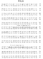
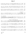
- the respective primers were designed with reference to the nucleotide sequence (SEQ ID:49) and amino acid sequence (SEQ ID:50) of IPMDH of Sulfolobus sp. strain 7 (Figs. 6 and 7).
- each of the primers having the sequence of SEQ ID:3 to SEQ ID:8 was dissolved in TE (10 mM Tris-HCI, 1 mM EDTA, pH 8.0) by an ordinary method to obtain 10 pmol/ ⁇ l solution.
- 1 ⁇ l of the primer solution (the total: 10 ⁇ l) was phosphorylated with polynucleotide kinase by an conventional method.
- the enzyme was inactivated by the treatment at 70°C for 10 minutes.
- 3 ⁇ l of the reaction liquid was taken and mixed with 1.5 ⁇ l of UssDNA obtained in step (1) and was allowed to anneal.
- the mixture contained all the primers of phosphatized sequence Nos. 3 to 8.
- the annealing step was conducted in the total amount of 20 ⁇ l containing 10 x annealing buffer (200 mM Tris-HCI, 20 mM MgCl 2 , 100 mM DTT, pH 8.0). The mixture was heated to 70°C and then left to stand at room temperature to cool it to about 30°C.
- 10 x annealing buffer 200 mM Tris-HCI, 20 mM MgCl 2 , 100 mM DTT, pH 8.0.
- the obtained mixture was left to stand at 0°C for 20 minutes, at 42°C for 1 minute and 0°C for 2 minutes.
- 450 ⁇ l of 2xYT medium was added thereto and they were left to stand at 37°C for 1 hour.
- 138.5 ⁇ l of of the culture liquid was poured into 5 ml of 2xYT liquid medium containing 100 ⁇ g /ml of ampicillin. After overnight culture, the plasmid DNA was recovered from the cells by alkali-SDS method.
- Escherichia coli MC1061 was again transformed by DNA thus obtained. Transformed colonies were selected on LB agar medium containing 100 ⁇ g/ml of ampicillin. The colonies were cultured and plasmid DNA was recovered therefrom to confirm whether the site of the restriction enzyme was found or not. When the mutation was introduced, DNA would be digested by the restriction enzyme in the primer corresponding to the mutation site.
- (M91L and I95L) ancestral variant, (K 152 R) ancestral variant, (G154A) ancestral variant, (K152R and G154A) ancestral variant, (A259S and F261P) ancestral variant and (Y282L) ancestral variant were named a variant, b' variant, b" variant, b variant, c variant and d variant, respectively, and also corresponding expression plasmids were named pE7-SB21a, pE7-SB21b', pE7-SB21b", pE7-SB21b, pE7-SB21c and pE7-SB21d, respectively.
- Ancestral bcd region variant plasmid pE7-SB21bcd DNA obtained as described above was digested with Sma I.
- a variant plasmid pE7-SB21a DNA was digested with Xba I and Eco RI, and DNA segment encoding the intended enzyme was subcloned into Xba I - Eco RI multicloning site of pUC118 to obtain plasmid pUC118-SB21a.
- pUC118-SB21a was digested with Sma I and ligated with the above-described bcd rgion ancestral variant plasmid DNA digested with Sma I to obtain pUC118 -SB21abcd. Then pUC118-SB21abcd and pE7-SB21 were digested with Xba I and Eco RI. They were mixed together to obtain expression plasmid pE7-SB21abcd for the ancestral variant in abcd region.
- Fig. 8 shows a schematic diagram of the construction of the plasmids.
- the obtained microbial cells were suspended in buffer I (20 mM KHPO 4 , 0.5 mM EDTA, pH 7.0) and cleaned by the centrifugation at 7,000 rpm at 4°C for 20 minutes. When the next step was not immediately started, the cells were kept at -80°C. 19.6 g of the microbial cells were obtained.
- the supernatant was treated with anion exchange column DE-52 equilibrated with Buffer I, and the passed fraction was recovered. 3 M ammonium sulfate (AS) solution was added to the obtained fraction to obtain the final concentration of 1 M. After leaving the mixture to stand at 4°C for about 1 hour, the precipitates thus formed were removed by the centrifugation at 30,000 rpm at 4°C for 20 minutes. The supernatant was passed through butyl-Toyopearl 650 s column (a hydrophobic column) equilibrated with Buffer I containing 1 M of AS. Protein was eluted by the linear inclination of AS concentration of 1 M to OM. The activity of each of the obtained fractions was determined. The active fractions were collected and dialyzed against Buffer II (20 mM CHES/KOH, 0.5 mM EDTA, pH 9.3).
- Buffer II 20 mM CHES/KOH, 0.5 mM EDTA, pH 9.3
- the protein solution obtained by the dialysis was treated with a Resource Q column (an anion exchange column) equilibrated with Buffer II and protein was eluted by the linear gradient of KCI concentration of 0 M to 0.1 M. Each fraction thus obtained was dialyzed against Buffer I and the purity was confirmed with SDS-PAGE. Fractions of a single band confirmed with SDS-PAGE were collected and concentrated to 1 mg/ml with Cetnriprep 30. The protein concentration was determined using BCA protein assay reagent kit of PIERCE Co. with BSA as the standard. The purification results are shown in Table 2.
- thermostability of Sulfolobus sp. IPMDH is very high at pH 7.0, the thermostability thereof at 99°C was determined. In particular, a time required for reducing the activity to 1/2 (half-life T 1/2 ) at 99°C was determined and utilized as the index of the thermostability.
- the half-lives of natural and variant (ancestral) enzymes at 99°C were determined as follows: Enzyme solutions having a protein concentration of 0.25 mg/ml (for b', b", b, c and d variants) or 1.0 mg/ml (for abcd variant) were prepared by using a potassium phosphate buffer (20mM KHPO 4 , 0.5 mM EDTA, 1 mM DTT, pH 7.0). Also for natural IPMDH, enzyme solutions having protein concentrations of 0.25 mg/ml and 1.0 mg/ml were prepared. These enzyme solutions were heat-treated at 99°C for 10, 20, 30, 60 or 120 minutes.
- the enzyme solutions were left to stand in ice for 5 minutes and then centrifuged at 12,000 rpm at 4°C for 20 minutes. The supernatant was recovered from each product. 10 ⁇ l of each supernatant was used to determine the activity at 75°C. The determination was repeatedly conducted 3 times for each sample, and the average of results was taken as the residual activity. The residual activity was plotted in a graph wherein the horizontal axis represent the time, and the ordinates represent the relative activity (time 0 was represented as 100). The time at which the relative activity was 50 % was taken as the half-life T 1/2 . At the same time, the specific activity was also determined. The results are shown in Tables 3 and 4.
- thermostability of all of b', b", b, c, d and abcd variants was improved as compared with that of natural IPMDH.
- the specific activity of each of b', b" and d variants was also increased.
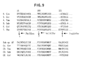
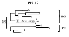
- Fig. 9 Amino acid sequence of IPMDH and ICDH from representative species which has been cloned were aligned (Fig. 9:Amino acid sequences in Fig. 9 were described in the sequence listing as SEQ ID:57 to SEQ ID:89, from top left to bottom right respectively). Among them, amino acids which are conserved among species and which are different in Thermus thermophilus were investigated. Also, considering the information together with the composite phylogenetic tree (Fig.3) of IPMDH and ICDH, the sites were estimated where the tree branches before Thermus and the amino acid residue before the branching can be clearly identified. Fig. 10 shows the amino acid residues in various species at the position corresponding to position 53 in Thermus.
- Wild type IPMDH from Thermus thermophilus and ancestral IPMDH were purified using the similar procedure as described in Example 2, making it a proviso that the third nucleotide of several codons of the gene were changed to A or T to lager production of the protein, because IPMDH gene from Thermus thermophilus is GC rich, which may decrease the expression of the gene.
- the final yields from 1L culture were 184mg/L for wild type, 11.3mg/L for ancestral variant F53L and 8.4mg/L for ancestral variant V181T.
- Wild type IPMDH and ancestral IPMDH were subjected to heat treatment and the residual activities were determined. For all the experiments, the measurement was conducted three times for each experiment and the residual activity was obtained as the average of the measurements.
- Wild type and ancestral IPMDH protein solution were prepared as a solution of 0.4mg/ml (20mM KHPO 4 , pH7.6, 0.5mM EDTA), respectively. 50 ⁇ l of each sample was taken in 0.5ml tube and the activity was determined at 50°C after heating at 80, 82, 84, 86, 88, and 90°C for 10 minutes. The temperature was determined where the residual activity reduces to 50%. The results were shown in Fig. 12. The results show that the temperature where the activity reduces to 50% was 85.5°C for wild type, 83.5°C for F53L variant and 86.8°C for V181T variant and 86.5°C for P324T variant. Thus determined temperature was increased by 1.3°C for V181T variant and 1.0°C for P324T variant, although it was decreased by about 2°C for F53L variant.
- ⁇ T 1/2 was increased by 12.7min. for V181T and 3.1min. for P324T, although it was decreased by 5.9min for F53L.
- thermostability of F53L variant was reduced to less than the thermostability of wild type may reside in the following factors: Investigation of the amino acid sequence around residue 53 revealed that the residue 58 in Thermus thermophius is Arg, while it is Leu or Val in many other species. From the fact, it is believed that the structure became unstable by changing the amino acid residue at position 53 to Leu which cannot fill the space between the residue 53 and Arg at position 58, unlike Phe, and the thermostability was reduced as a result.
- Wild type IPMDH and variants F53L, V181T and P324T were prepared as a solution of 0.1mg/ml (20mM KHPO 4 , pH7.6), respectively and their secondary structures were investigated using CD (Circular dichroism) spectra ranging 210nm-250nm . NO significant changes were found for each variant compared to wilt type. This indicates that these mutations did not significantly affect the secondary structure of the protein.
- Amino acid sequences of IPMDH from representative species and ICDH from various species were obtained from NCBI database and they were subjected to the multiple alignment using Clustal X, an software for alignment (Fig. 14). Also the composite phylogenetic tree was produced using Puzzle, the software for producing a phylogenetic tree, based on these sequences. From the result of alignment and the composite phylogenetic tree, six ancestral mutation, A336F, Y309I, I310L, I321L, A325P and G326S, were predicted using similar procedure as described in Example 1 and 4. The meaning of the notation such as A336F is identical to the meaning described in Example 1 and 4.
- Y309I and I310L, and also A325P and G326S are adjacently located and are located in the same secondary structure, they were considered as a double mutant, respectively. Therefore, Y309/I310L mutation, I312L mutation, A325P/G326S mutation and A336F mutation will be also hereinafter referred to as N1, N2, N3 and N4 mutation, respectively.
- N1, N2, N3 and N4 mutation were introduced by the similar methods in Example 1 and 4 using the plasmid where ICDH from Caldococcus noboribetus (NCBI accession No. BAA13177) had been cloned into pET21c, as the template
- Wild type ICDH from Caldococcus noboribetus and ancestral ICDH were produced in large scale using pET21c and mutant pET21c to which N1-N4 mutation was introduced and E . coli , as described in Example 2, and then the proteins were purified according to the conventional procedures.
- the final yields from 1L culture were 10mg/L, 15.4mg/L, 10.9mg/L, 14.2mg/L, 14.2mg/L and 4.39mg/L for wild type, N1 type variant, N2 type variant, N3 type variant and N4 type variant.
- thermostability of wild type ICDH from Caldococcus noboribetus and each variant are subjected to the heat treatment at various temperature (80, 82, 84, 86, 88, 90, 92 and 94°C) for 10 minute, before the residual activity was determined at 70°C.
- the relationship between the residual activity and temperature was similar to that in Example 5 (see Fig. 12).
- the temperature where the activity reduces to 50% (T 1/2 ) was 87.5, 88.8, 88.8, 91.3, 74.0°C for wild type, N1-N4 ICDH variants, respectively.
- the thermostability increased by 1°C for N1 and N2 type ICDH variant and 4°C for N3 type ICDH variant compared to wild type, although the thermostability of N4 type variant was decreased by 13°C.
- the specific activity was also determined at 80°C.
- the relative activities of ICDH variants were about 72, 62, 127 and 21% (based on the activity of wild type as 100%).
- the specific activities of N1, N2 and N3 type ICDH variants were not significantly changed but the specific activity of N4 type variant of which thermostability had been largely reduced was also significantly decreased.
- thermostability of N4 type ICDH variant was significantly reduced, the tertiary structure was additionally investigated. The results showed that Leu327, Tyr363 and Leu364 were located around Ala336 and they formed a hydrophobic pocket. The sites corresponding to Ala336 and Leu327 in other species varied such that they formed a pair in the manner where if one of these residues is a large residue, the other is a smaller residue, such as Phe-Ala, Phe-Gly, Tyr-Ala, Ala-Met. Considering these observations, the reason why the thermostability of N4 type ICDH variant was reduced was believed to be the steric hindrance caused by the alteration from Ala336 to Phe resulted from the compactness of this region.
- thermostability of protein can be improved by the information of only the primary structure without the information of the secondary and tertiary structures of protein.
- thermostability of thermostable proteins produced by thermophilic bacteria particularly the thermostable enzymes, can be further improved.
- thermostable enzyme when used, the reaction can be carried out at a high temperature without temperature control and, therefore, the reaction can be carried out at a high reaction rate at a high temperature. Accordingly, the contamination with unnecessary microorganisms can be minimized.
Landscapes
- Life Sciences & Earth Sciences (AREA)
- Chemical & Material Sciences (AREA)
- Health & Medical Sciences (AREA)
- Genetics & Genomics (AREA)
- Organic Chemistry (AREA)
- Zoology (AREA)
- Engineering & Computer Science (AREA)
- Bioinformatics & Cheminformatics (AREA)
- Wood Science & Technology (AREA)
- Microbiology (AREA)
- Biotechnology (AREA)
- Biomedical Technology (AREA)
- Molecular Biology (AREA)
- Biochemistry (AREA)
- General Engineering & Computer Science (AREA)
- General Health & Medical Sciences (AREA)
- Medicinal Chemistry (AREA)
- Enzymes And Modification Thereof (AREA)
- Micro-Organisms Or Cultivation Processes Thereof (AREA)
- Preparation Of Compounds By Using Micro-Organisms (AREA)
Abstract
Description
- The present invention relates to a method for improving thermostability of a protein. The present invention also relates to a protein having an improved thermostability and a nucleic acid encoding the protein having improved thermostability.
- A protein active at a high temperature, particularly a thermostable enzyme, is more advantageous than another protein which is inactivated at a high temperature, for example, in that it can be used without being cooled. Such a protein is mostly produced by a bacterium called thermophilic bacterium, which can grow at a high temperature. Accordingly, in designing a thermostable protein, amino acid sequence of a corresponding protein of such a group of thermophilic bacteria is analyzed and the characteristic feature of the amino acid sequence common to them is taken into account. Alternatively, the three-dimensional structure of a protein produced by the thermophilic bacterium is analyzed, the structure for imparting the thermostability is estimated from thus obtained information, and the structure of the heat-unstable protein is modified according to the estimated structure. As an example of proteins of thermophilic bacteria, 3-isopropylmalate dehydrogenase (IPMDH) encoded by leuB is known. The three-dimensional structure of IPMDH of Thermus thermophilus HB8 has been elucidated (K. Imada et al., J. Mol. Biol. 222, 725-738, 1991). Further, isocitrate dehydrogenase (ICDH) is known as a protein having a similar catalytic mechanism, amino acid sequence and three-dimensional structure as those of IPMDH, namely, a protein belonging to the same family as IPMDH.
- The object of the present invention is to provide a method for improving thermostability of protein, a protein having an improved thermostability and a nucleic acid encoding the protein, and host cells capable of producing a protein having improved thermostability.
- In particular, the object of the present invention is to provide a method for improving thermostability of a protein, taking advantage of only the information of the primary structure of the protein.
- On the basis of the fact that many organisms which properly grow at a temperature of 80°C or above are located at the root of a phylogenetic tree by 16S rRNA (Fig. 1) shown by Woese et al., the inventors had an idea that the ancestors common to eubacteria, eukaryotes and archaebacteria might be ultra-thermophilic bacteria. On the basis of this supposition, the inventors have gotten an idea that although protein of many kinds of existing thermophilic bacteria are not always the protein of a true ancestral protein having an amino acid sequence of the ancestral or an amino acid sequence close to the ancestral sequence might have a further improved thermostability. The inventors have completed the present invention on the basis of an idea that for designing and producing a thermostable protein, it is more important that the amino acid sequence of ancestral protein is estimated and mimicked than that only the sequence and the higher-order structure of protein of a thermophilic bacterium are analyzed and mimicked.
- Namely, the present invention provides a method for improving thermostability of proteins, which comprises the steps of
- (i) comparing amino acid sequences of proteins derived from two or more species which evolutionarily correspond to each other in a phylogenetic tree;
- (ii) estimating an amino acid sequence of an ancestral protein corresponding to the amino acid sequences compared in step (i); and,
- (iii) and comparing the amino acid residues in the amino acid sequence in one of the proteins compared in step (i) with amino acid residues at a corresponding position in the ancestral protein estimated in step (ii), and replacing one or more of the amino acid residues different from those of the ancestral protein with the same amino acid residues as those of the ancestral protein. The present invention may further comprise the setps of
- (iv) testing the proteins obtained in step (iii) for thermostability; and
- (v) selecting a protein having improved thermostability.
-
- The present invention particularly includes the comparison of species evolutionarily close to thermophilic bacteria or archaebacteria in the phylogenetic tree with each other on the amino acid sequence of corresponding proteins.
- The present invention also provides an enzyme improved in heat resistance by the above-described method, a nucleic acid encoding the enzyme and host cells containing such a nucleic acid.
-
- Fig. 1 shows a phylogenetic tree based on the comparison of 16S rRNA.
- Fig. 2 shows the multiple alignment of amino acid sequences of IPMDH and ICDH from various biological species.
- Fig. 3 shows a phylogenetic tree constructed by the simultaneous comparison of IPMDH and ICDH.
- Fig. 4 shows the evolution of residue 152 of Sulfolobus sp. 7 strain.
- Fig. 5 is a pE7-SB21 restriction enzyme map. pE7-SB21 was produced by inserting leuB gene into NdeI-EcoEI region of expression vector pET21c. Symbols in the figure represent the following restriction enzyme cleavage sites: N: Nde I, Sm: Sma I, E: EcoR I, E47: Eco47 III, B: Bgl II, Xb: Xba I, H: Hind III, Xh: Xho I, and M: Mro I.
- Fig. 6 shows the nucleotide sequence and amino acid sequence of Sulfolobus sp. leuB gene.
- Fig. 7 shows the nucleotide sequence and amino acid sequence of Sulfolobus sp. leuB gene (continuation of Fig. 6).
- Fig. 8 shows a rough variation introduction in abcd region. Symbols in the figure represent the following restriction enzyme cleavage sites: N: Nde I, Sm: Sma I, E: EcoR I, E47: Eco47 III, B: Bgl II, Xb: Xba I, H: Hind III, Xh: Xho I, M: Mro I, Na: Nae I and Sa: Sal I.
- Fig. 9 shows the multiple alignment of amino acid sequences of IPMDH and ICDH. The sequences with (ICDH) represent ICDH sequence and the sequences without the indication represent the IPMDH sequence. N. Cra: Neurospora crassa, S. Cer: Saccharomyces cerevisiae, A. tum: Agrobacterium tumefacience, B. sub: Bacillus subtilis, E. Col: Escherichia coli, T. The: Thermus thermophilus, Sub sp.#7: Sulfolobus stain #7, Cs. Cer: Saccharomyces cerevisiae (ICDH), CB. Tau: Bos taurus(ICDH), CB. Sub: Bacillus subtilis(ICDH), CE. Col: Escherichia coli (ICDH).
- Fig. 10 shows the evolution of residue 53 of Thermus thermophilus.
- Fig. 11 shows the scheme of mutagenesis using the plasmid containing cloned Thermus thermophilus IPMDH as a template.
- Fig. 12 shows the residual activity of wild type Thermus thermophilus IPMDH and ancestral variants.
- Fig. 13 shows the multiple alignment of IPMDH and ICDH.
-
- Molecular phylogenetic tree (hereinafter referred to as "phylogenetic tree") based on the molecular level information of species or an algorithm for the preparation of the phylogenetic tree is utilized in the present invention. Some algorithms for preparing phylogenetic trees, such as the algorithm based on the maximum parsimony principle, are known. Computer programs for implementing the algorithms are utilizable or available. For example, various phylogenetic tree estimation programs such as CLUSTALW, PUZZLE, MOLPHY and PHYLIP are utilizable. Although phylogenetic trees can be produced by such programs, it is easier to utilize an already published phylogenetic tree (Fig. 1). For example, a phylogenetic tree based on 16S rRNA data proposed by Woese et al. is also usable. In such a phylogenetic tree, species which are close to each other in the molecular evolution appear in positions close to each other. Species positioned closely to the root of the phylogenetic tree are considered to be close to the ancestors.
- For attaining the object of the present invention, it is preferred to use a part relatively close to the root of a phylogenetic tree, it is more preferred to use a part older than birds or even-toed ungulates, and it is particularly preferred to use a part of the phylogenetic tree which contains thermophilic bacteria or archaebacteria for the following reasons: The thermophilic bacteria and archaebacteria are positioned close to the root, namely, evolutionarily close to the ancestors in the phylogenetic tree. Further, proteins produced by them are expected to be relatively close to ancestral super-thermostable protein. It is also preferred to contain another protein belonging to the same family because ancestral amino acid residues (or sequence) at the root of the phylogenetic tree can be estimated, by a method which will be described below, by comparing the protein with a protein of archaebacteria or with another protein of the same family.
- The term "thermophilic bacteria" is a generic name for bacteria capable of growing at a high temperature of usually above about 55°C. These bacteria are also called thermostable bacteria for the purpose of the present invention. In the present invention, the term "thermophilic bacteria" indicates both highly thermophilic bacteria capable of growing at a temperature of higher than above 75°C and also moderately thermophilic bacteria capable of growing at about 55 to 74°C. They also include facultative thermophilic bacteria capable of growing at ambient temperature and obligate thermophilic bacteria capable of growing only at a temperature of above about 40°C. The term "non-thermophilic bacteria" indicates microorganisms other than the thermophilic bacteria. The term "archaebacteria" indicates those classified according to the above-described Woese's classification. They indicate bacteria of prokaryote group including methane-forming bacteria, hyperhalophilic bacteria and sulphate reducing archaebacteria. The archaebacteria are clearly differentiated from eubacteria in that the lipid of the cell membrane of the former is an ether lipid. The expression "proteins belonging to the same family" herein indicates proteins which are similar to each other in at least one of the function, amino acid sequence, domain structure and steric structure. They include a group of proteins, at least amino acid sequences of which are partially homologous and the multiple alignment of which is possible. In particular, they include a group of proteins, at least amino acid sequences of which are homologous and can be multiple aligned. It is eagerly expected that two or more proteins belonging the same family are derived from the same ancestral protein.
- Then information of amino acid sequences of proteins corresponding to each other, which are to be improved in thermostability, is obtained or determined from various species. Although proteins to which the present invention can be applied are not particularly limited, they are preferably proteins present in various species. Particularly enzymes having a high value of industrial utilization is preferable. Preferred examples of them are proteins produced by thermophilic bacteria, particularly thermostable enzymes. Example of them is IPMDH and ICDH of Sulfolobus sp.
stain 7. The gene encoding IPMDH of this strain was cloned by Suzuki et al. [T. Suzuki et al., J. Bacteriol. 179 (4), 1174-1179, 1997]. - Amino acid sequences of protein to be improved in thermostability can be also obtained from an already known data base. When an amino acid sequence is to be newly determined, any method for determining amino acid sequence known in the art can be employed. It is also possible to estimate the amino acid sequence by obtaining a nucleic acid encoding the protein according to the information of partial amino acid sequence, determining the nucleic acid sequence by a well-known sequencing techniques and estimating the amino acid sequence from the nucleic acid sequence.
- After the multiple alignment of the obtained amino acid sequences from the species, the amino acid sequences obtained from the respective species are compared with each other. Some methods for the multiple alignment are known. One of the methods is based on the maximun parsimony principle for minimizing the change due to the insertion, deletion, replacement, etc. Computer programs for implementing this principle have been developed, which can be used or available. For example, TreeAlign is known among them. From DDBJ, "malign" which is the 1990 version of the program can be used. Because species which are evolutionarily close to each other in the phylogenetic tree are selected in the present invention, phylogenetic information has already been utilized in the multiple alignment and, as a result, the alignment is more suitable than that in a case of no phylogenetic information can be conducted. Information from at least three species is utilized for the multiple alignment. The larger the number of origin of the data to be used for the alignment, the more suitable the information. Furthermore, each of the species to be compared preferably contains one or more thermophilic bacteria or archaebacteria, based on the aforementioned reason. It is also preferred that it contains a family protein, namely another protein expected to be derived from the same ancestral protein.
- After obtaining the results of the alignment, amino acid sequence of the ancestral protein can be estimated on the phylogenetic tree. For this purpose, the maximum parsimony method or maximal likelihood method is utilizable. The procedure of such a method is well known to those skilled in the art [see, for example, Young, Z., Kumar, S and Nei. M, Genetics 141, 1641-16510, 1995; Steward, C. -B. Active ancestral molecules, Nature 374, 12-13, 1995; and Molecuar Evolutinary Genetics, Columbia University Press, New York, USA, 1987]. For example, the maximal parsimony method which can be employed in the present invention is, in short, a method wherein an ancestral type having the minimal number of the mutation expected to occur after the estimation of the ancestral type is likely estimated to be the true ancestral type. The maximal likelihood method can be employed instead of the maximum parsimony method. Also, a program PROTPARS (included in PHYLIP) for directly estimating the ancestral type from the amino acid sequence according to the maximum parsimony method can be also employed. Because the phylogenetic tree and ancestral amino acid are principally estimated at the same time in those methods, it is not always necessary to prepare the phylogenetic tree when such a method is employed. However, the preparation of the phylogenetic tree is preferred particularly when the ancestral amino acid is to be estimated by manual calculation. The ancestral amino acid sequence can be determined by the following maximum parsimony method or maximal likelihood method according to a phylogenetic tree produced by the above-described method or another already known method, particularly based on an already published phylogenetic tree.
- A process according to the maximum parsimony method will be described in detail with reference to IPMDH which will be shown also in Examples given below.
- Amino acid sequences from some species of IPMDH and ICDH, which have already been cloned and of which sequences were determined, are multiply aligned (Fig. 2). Then a phylogenetic tree is prepared on the basis of the sequences by, for example, the maximum parsimony method or neighbor-joining method (Fig. 3). In this case, it is possible to directly estimate the ancestral amino acid sequence, without preparing the phylogenetic tree, by the maximum parsimony method as described above. However, a procedure wherein the phylogenetic tree is explicitly used will be described for easy understanding of the procedure. This procedure is also applicable to a case when an already prepared phylogenetic tree such as a published known phylogenetic tree is used.
- Ancestral amino acids in respective sites of the multiply aligned residues can be determined by means of a phylogenetic tree obtained by any method. For example, Fig. 4 shows amino acid residues from various organisms corresponding to residue 152 of Sulfolobus sp.
strain 7 of IPMDH. Amino acids at this position in the organisms shown in Fig. 4 are R, S, K or E. When both residues in species close to each other in the phylogenetic tree are R, it can be estimated that in the ancestral species common to them (shown by the binding point connecting two species in the phylogenetic tree), the amino acid residue corresponding to residue 152 of Sulfolobus sp.strain 7 would be R for the following reasons: When R is the ancestral type, only one variation can elucidate the mechanism of the realization of the amino acid residue corresponding to residue 152 of Sulfolobus sp.strain 7 in the present species, while when S is the ancestral type, two or more times of variation must be taken into consideration. - When two species have residues different from each other, such as residues R and S, the ancestor common to both of them cannot be immediately determined. However, even in such a case, the common ancestor can be estimated to be R when another branch in one branch deeper position (i. e. junction on the left-hand side in the phylogenetic tree) is R. Thus, the amino acid sequence on the most left-hand side in the figure can be estimated to be the most ancestral amino acid sequence by evolutionarily tracing back (i. e. going back to the left in the figure). In Fig. 4, the ancestral amino acid residue corresponding to residue152 of Sulfolobus sp.
strain 7 is estimated to be R. - By thus estimating the ancestral amino acid residue of each residue in the sequence in the multiple alignment, the ancestral amino acid sequence in a corresponding region can be estimated. When the species used for the estimation of the ancestral amino acid sequence is changed, the shape of the phylogenetic tree is changed and, therefore, a different ancestral amino residue is obtained in some cases. The position and variety thereof are variable also depending on the protein used for the comparison. Therefore, for attaining the object of the present invention, it is preferred to alter an amino acid residue selected at a position of a relatively slight change. Such an amino acid residue can be determined by changing the species used for the preparation of the phylogenetic tree or by using only a part of amino acid sequence information used for the preparation of the phylogenetic tree without changing the species, and estimating the degree of the change in shape of the tree due to the change of the amino acid sequence information used for preparing the phylogenetic tree and selecting a residue which only slightly influence on the shape of the tree.
- As far as various species have regions corresponding to each other, the ancestral amino acid sequence in the regions can be estimated in proteins to be improved in the thermostability by the above-described procedure. Each amino acid residue in thus determined amino acid sequence may correspond to amino acid residues in many positions in a protein of a present species of organism particularly when the organism is a thermophilic bacterium or archaebacteria. Accordingly, in the present invention, only amino acid residues having a sequence different from that of the ancestral protein amino acid sequence are to be modified in such a case.
- In the estimation of the amino acid sequence of protein of ancestral species according to the above-described procedure, the ancestral type can be determined by the above-described procedure irrespective of the fact that a thermophilic bacterium or non-thermophilic bacterium is contained in the species to be compared or the fact that only the thermophilic bacterium has an amino residue different from that of other species to be compared. When there are many species having proteins having amino acid sequences different from others and, therefore, the ancestral type cannot be estimated only from the information or the degree of accuracy is considered to be low, data for the alignment can be further added. When the ancestral amino acid residue can be thus determined, this amino acid residue can be employed as the ancestral one.
- Generally, two or more positions and regions having such amino acid residues may present in the protein. These positions and regions might be either apart from one another or close to one another. All of these positions and amino acid residues are recorded for the modification which will be described below.
- After the determination of the ancestral amino acid residue for the amino acid residue at each position, at least one of non-ancestral amino acid residues of the protein to be analyzed is replaced with the ancestral amino acid residue to modify the protein. In this case, the number and position of the amino acid residues to be replaced may vary depending on the protein to be modified, required thermostability and desired specific activity. Preferably, the position and number of the amino acid residues to be replaced are selected so that both sufficient thermostability and high specific activity can be attained. For obtaining both sufficient thermostability and high specific activity at the same time, further information of the position of the active center and amino acid sequence around the active center is useful.
- Although the protein to be modified can be derived from any of the comparative species, it is preferred to select protein from species having the highest thermostability. It is particularly preferred to select a protein produced by the thermophilic bacterium as the protein to be modified for the following reasons: A protein from a species of organism having a high thermostability is generally expected to have a high thermostability. Further, by modifying a protein expected to already have certain thermostability to a more complete ancestral protein, a further improvement in the thermostability can be expected. The amino acid residues in a protein can be replaced by altering a nucleic acid encoding the protein. In short, the site-specific mutagenesis by Kunkel method can be conducted by obtaining a gene encoding the protein in which the amino acid residue is to be replaced and using a primer capable of replacing an amino acid residue in an intended site. Further, the site-specific mutagenesis can be carried out by a PCR method.
- An intended gene can be obtained by a hybridization method or PCR after designing a suitable probe according to a known amino acid sequence information or a partial amino acid sequence information of the protein. DNA having an intended mutation can be efficiently replicated by previously preparing a template for the mutagenesis in ung- host. It is convenient for the confirmation of the mutation when a primer for the mutagenesis is designed to have a restriction enzyme site.
- The molecular biological techniques such as introduction of a gene into a host, cloning of genes and site-specific mutagenesis including ung- hosts, are well known by those skilled in the art. For these techniques, for example, Sambrook et al., 1989, Molecular Cloning: A Laboratory Manual, Second Edition (1989) Cold Spring Harbor Laboratory Press, Cold Spring Harbor, New York, and F. M. Ausubel et alo. (eds), Current Protocols in Molecular Biology, John Wiley & Sons, Inc. (1994) can be referred to. Further, kits for carrying out these molecular biological techniques are commercially available. The mutation thus introduced can be confirmed by determining the nucleotide sequence. When a restriction enzyme site has been introduced in the primer for the variation introduction, the introduction of the mutation can be more easily confirmed on the basis of the fact that it can be digested by a corresponding restriction enzyme.
- The modified gene thus obtained can be expressed with a suitable host-vector system. The hosts usable herein include both eucaryotic cells and procaryotic cells. Generally, microorganisms such as Escherichia coli are preferred. Recombinant DNA molecules prepared by introducing the modified gene into an expression vector having a regulatory sequence required for expressing the modified gene depending on the selected host can be prepared. Such an expression vector is well known in the art, and many host - vector systems are available on the market. Among those vectors, usually host - vector high expression systems are preferred. Inducible host - vector systems are particularly preferred. However, the selection of a suitable host - vector system will vary depending on the properties of protein because some proteins will harm the host upon the high expression. If necessary, the codon usage may be optimized depending on the selected host. The host containing such a recombinant DNA molecule may be cultured using a method well known in the art and then the produced protein may be recovered.
- The protein can be recovered from the host cells or culture medium by an ordinary method selected depending on the host and properties of the produced protein. For example, when the protein is recovered from the microbial cells, the cells are broken by, for example, sonication, the residue is removed by centrifugation and the intended protein is obtained by a proper combination of ammonium sulfate precipitation, reversed phase chromatography, ion exchange chromatography, gel filtration, etc. When the protein is in the form of an inclusion body, it can be solubilized with 6 M guanidine hydrochloride or the like and reconstituted. When the protein is recovered from the culture medium, the microbial cells are removed by centrifugation and then the intended protein is recovered in the same manner as that described above. When the intended protein has a property of being associating with the cell membrane, a suitable surfactant can be used for the solubilization. The solubilization methods are well known in the art, and they are suitably selected depending on the properties of the protein.
- The purity of the obtained protein can be confirmed by, for example, SDS-polyacrylamide gel electrophoresis. The concentration of the obtained protein can be determined by a method well-known in the art, for example using BCA Protein Assay Kit from PIERCE Co., wherein bovine serum albumin is used as the standard protein, as will be described in Examples given below. The thermostability of the protein can be determined by examining the activity thereof after the heat treatment. For example, the thermostability of IPMDH can be determined by the following method: An assay buffer (50 mM CHES/KOH, pH 9.5, 200 mM KCI, 1 mM NAD, 0.4 mM IPM, 5 mM MgCl2) was introduced into a cell and then incubated at an appropriate temperature, for example 50°C-99°C for 5 minutes. A suitable amount of an enzyme solution having a suitably prepared concentration is added to the assay buffer and the obtained mixture is lightly stirred. The mixture is kept at 50°C-75°C and the increase in NADH is determined by the ultraviolet absorbance at 340 nm. The specific activity of IPMDH is shown in terms of units (U) per mg of protein. The activity for producing 1 micromole of NADH per
minute 75°C can be represented to be 1 U (unit). - For ICDH the thermostability can be determined by the following method: An assay buffer (10 mM MgCl2,0.4mM D,L-isocitrate, 0.8mM NADP, 100mM PIPES pH 7.0) was introduced into a cell and then incubated at a high temperature, for example 50°C-99°C for 5 minutes. A suitable amount of an enzyme solution having a suitably prepared concentration is added to the assay buffer and the obtained mixture is lightly stirred. The mixture is kept at 50°C-75°C and the increase in NADPH is determined by the ultraviolet absorbance at 340 nm. The activity for producing 1 micromole of NADPH per minute 70°C can be represented to be 1 U (unit).
- Thus, ancestral variants may be optionally tested for thermostability by determining their activity at high temperature with suitable methods to select more thermostable proteins.
- Strains and culture media shown below were used.
- CJ236: This strain was used for preparing uracil single strand DNA (UssDNA). This strain is defective in uracil glycosylase and dUTPase.
- MC1061 and JM109: They were used as hosts in the gene operation.
- MA153: This strain was used as the host for large scale expression of IPMDH. This strain is defective in leuB.
- LB agar medium: 1.0 % of bactotryptone, 0.5 % of bactoyeast extract, 1 % of NaCI, 1.5 % of agar and, if necessary, 100µg/ml of ampicillin.
- M9 agar medium: 1xM9 salt, 1 mM of MgSO4, 0.1 mM of CaCl2, 0.001 % of thiamine, 0.2 % of glucose and 1.5 % of agar. This medium was used for the selection of Escherichia coli JM109.
- 2xYT medium: 1.6 % of bactotryptone, 1.0 % of bactoyeast extract and 0.5 % of NaCl. This medium was used for the liquid culture of Escherichia coli. If necessary, 100 µg/ml of ampicillin was added.
- 490 µI of an assay buffer (50 mM of CHES/KOH, pH 9.5, 200 mM of KCI, 1 mM of NAD, 0.4 mM of IPM and 5 mM of MgCl2) was fed into a cell and then preincubated at 50°C-75°C for 5 minutes. Then 10 µI of an enzyme solution having a predetermined concentration was added thereto, and the obtained mixture was lightly stirred. Then keeping the mixture at the same temperature as the preincubation temperature, an increase in NADH was determined according to the ultraviolet absorbance at 340 nm.
- 490 µl of an assay buffer (10 mM of MgCl2, 0.4mM D.L-isocitrate, 0.8mM NADP, 100mM PIPES pH7.0) was fed into a cell and then preincubated at 50°C-75°C for 5 minutes. Then 10 µI of an enzyme solution having a predetermined concentration was added thereto, and the obtained mixture was lightly stirred. Then keeping the mixture at the same temperature as the preincubation temperature, an increase in NADPH was determined according to the ultraviolet absorbance at 340 nm.
- leuB expression plasmid pE7-SB21 (Fig. 5) was introduced into competent cells of E. coli CJ236. The obtained transformed CJ236 was cultured in 2xYT medium to obtain 30 ml of a liquid culture. CJ236 in the liquid culture was infected with helper phage M13KO7. After shaking the culture in 2xYT medium at 37°C for 5 hours, the obtained culture was centrifuged at 5,000 rpm at 4°C for 10 minutes. The supernatant was further centrifuged at 6,000 rpm at 4°C for 10 minutes to obtain a supernatant. A phage was precipitated from 10 ml of the supernatant by PEG/NaCl. 10.9µg of UssDNA was obtained from the phage by an ordinary method. The concentration was 363 µg/ml.
- Amino acid sequences of IPMDH and ICDH which had been cloned and the amino acid sequences of which had been made clear were subjected to the multiple alignment. The results are shown in Table 1. Then, the ancestral amino acid sequences in respective regions (regions a, b or b' and b", c and d) shown in Table 1 were estimated. The estimation was conducted by the above-described procedure. For example, residue 152 was estimated as will be described below.
- At first, a phylogenetic tree containing these species was prepared by the neighbor-joining method (Fig. 3). Then b regions of Saccharomyces cerevisiae and Neurospora crassa in the phylogenetic tree were compared with each other. The amino acid residues corresponding to residue 152 of Sulfolobus sp.
strain 7 were R in these two species. Accordingly, amino acid residues at the corresponding positions of the two ancestral species were estimated to be R. Then Escherichia coli and Agrobacterium tumefaciens were compared with each other to find that the amino acid residues corresponding to residue 152 of Sulfolobus sp.strain 7 were R and S, respectively. Therefore, amino acid residues at corresponding positions of the two ancestral species could not be estimated from only this fact. However, at the junction in the left branch, the amino acid residue was estimated to be R in another branch (i. e. branch which branches into Saccharomyces cerevisiae and Nuerospora crassa) as described above. Accordingly, the amino acid residue at this position in four common ancestral species, i. e. Saccharomyces cerevisiae, Nuerospora crassa, Escherichia coli and Agrobacterium tumefaciens, was estimated to be R. Further, because amino acid residue of Bacillus subtilis corresponding to residue 152 of Sulfolobus sp.strain 7 was R, it was estimated that amino acid residue in the corresponding position in the ancestral species of 5 organisms (the above-described 4 organisms and Bacillus subtilis) was estimated to be R. By thus tracing back to the left in the phylogenetic tree in Fig. 5, it was estimated that the amino acid residue corresponding to position 152 of Sulfolobus sp.strain 7 would be R. - By repeating the procedure, the ancestral amino acid sequence for the amino acid sequences in the domains shown in Table 1 was finally determined. Then thus determined ancestral amino acid sequence was compared with the amino acid sequence of Sulfolobus sp.
strain 7 to determine the amino acid residue and position thereof of Sulfolobus sp.strain 7 different from the ancestral sequence. As a result, it was found that the amino acid residue and position thereof of each of M91, 195, K152, G154, A259, F261 and Y282 were different from those of the ancestral type. As for these symbols, for example, M91 represents M (methionine) residue at position 91. The same shall apply to other symbols. - In Table 1, these residues are underlined. The ancestral amino acid sequences determined by the above-described procedure and the positions and varieties of amino acid residues to be modified are also shown in Table 1. Residues shown by "x" in Table 1 are positions at which the ancestral type was not only one.
- From these results, it was determined that in the ancestral enzyme, amino acid residue at position 91 was L, amino acid residue at position 95 was L, amino acid residue at position 152 was R, amino acid residue 154 at position was A, amino acid residue at position 259 was S, amino acid residue at position 261 was P and amino acid residue at position 282 was L.
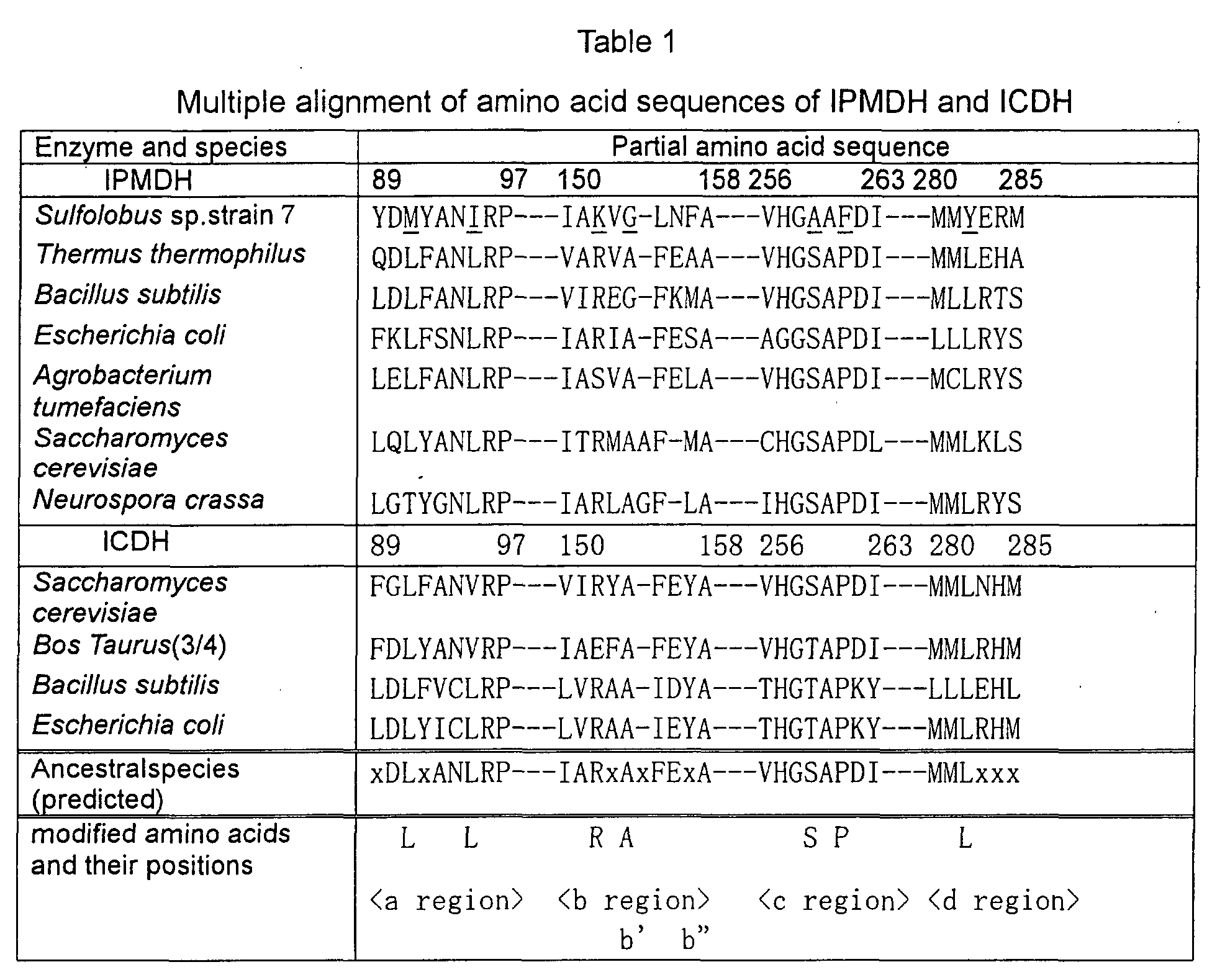
- The partial amino acid sequences in the above Table are shown as sequence SEQ ID:1 to SEQ ID:48 in order in the sequence listing.
- After the amino acid sequences of ancestral IPMDH and ICDH were determined, some ancestral variants were prepared by replacing amino acid residues in regions a, b, c and d and the combinations of them. The amino acid residue replacement in the ancestral variants was as follows: ancestral variation in a region (M91L and I95L), ancestral variation in b' region (K152R), ancestral variation in b" region (G154A), ancestral variation in b region (K152R and G154A), ancestral variation in c region (A259S and F261P), ancestral variation in d region (Y282L), and ancestral variation in a, b, c and d region (M91L, I95L, K152R, G154A, A259S, F2651P and Y282L). As for these symbols, for example, M91L represents the replacement of M (methionine) residue at position 91 with L (leucine) residue. The same shall apply to other symbols.
- Primers shown below were designed for preparing these ancestral variants using a site-specific mutagenesis method. The respective primers were designed with reference to the nucleotide sequence (SEQ ID:49) and amino acid sequence (SEQ ID:50) of IPMDH of Sulfolobus sp. strain 7 (Figs. 6 and 7).
- Primer P1 for introduction of ancestral mutation in a domain
(The underlined part is the site of recognition of restriction enzyme Af/II)

- Primer P2 for introduction of ancestral mutation in b' domain
(The underlined part is the site of recognition of restriction enzyme NruI)

- Primer P3 for introduction of ancestral mutation in b" domain
(The underlined part is the site of recognition of Eco47 III)

- Primer P4 for introduction of ancestral double mutation in b domain
(The underlined part is the site of recognition of Eco47 III)

- Primer P5 for introduction of ancestral mutation in c domain
(The underlined part is the site of recognition of Mro I)

- Primer P6 for introduction of ancestral mutation in d domain
(The underlined part is the site of recognition of Xho I)

-
- Because abcd ancestral mutation includes all the mutations introduced by the combination of the above-described primers, no primer was prepared.
- Each of the primers having the sequence of SEQ ID:3 to SEQ ID:8 was dissolved in TE (10 mM Tris-HCI, 1 mM EDTA, pH 8.0) by an ordinary method to obtain 10 pmol/µl solution. 1 µl of the primer solution (the total: 10 µl) was phosphorylated with polynucleotide kinase by an conventional method. After the completion of the reaction, the enzyme was inactivated by the treatment at 70°C for 10 minutes. 3 µl of the reaction liquid was taken and mixed with 1.5 µl of UssDNA obtained in step (1) and was allowed to anneal. Thus the mixture contained all the primers of phosphatized sequence Nos. 3 to 8. The annealing step was conducted in the total amount of 20µl containing 10 x annealing buffer (200 mM Tris-HCI, 20 mM MgCl2, 100 mM DTT, pH 8.0). The mixture was heated to 70°C and then left to stand at room temperature to cool it to about 30°C.
- After annealing, 2 µl of 10 x synthetic buffer (50 mM Tris-HCI, 20 mM MgCl2, 5 mM dNTPs, 10 mM ATP, 20 mM DTT, pH 7.9), 1µl of T4 DNA ligase and 1µl of T4 DNA polymerase were added to the annealed solution. The obtained mixture was kept in ice for 5 minutes and then at room temperature for 5 minutes, and then incubated at 37°C for 90 minutes. 4µl of the reaction mixture was taken and mixed with 100 µl of Escherichia coli MC 1061 competent cells. The obtained mixture was left to stand at 0°C for 20 minutes, at 42°C for 1 minute and 0°C for 2 minutes. 450µl of 2xYT medium was added thereto and they were left to stand at 37°C for 1 hour. 138.5µl of of the culture liquid was poured into 5 ml of 2xYT liquid medium containing 100µg /ml of ampicillin. After overnight culture, the plasmid DNA was recovered from the cells by alkali-SDS method.
- Escherichia coli MC1061 was again transformed by DNA thus obtained. Transformed colonies were selected on LB agar medium containing 100µg/ml of ampicillin. The colonies were cultured and plasmid DNA was recovered therefrom to confirm whether the site of the restriction enzyme was found or not. When the mutation was introduced, DNA would be digested by the restriction enzyme in the primer corresponding to the mutation site.
- As a result, several plasmids having ancestral variation introduced into the above-described regions a to d or a combination of them were obtained.
- In the variants thus obtained, (M91L and I95L) ancestral variant, (K 152 R) ancestral variant, (G154A) ancestral variant, (K152R and G154A) ancestral variant, (A259S and F261P) ancestral variant and (Y282L) ancestral variant were named a variant, b' variant, b" variant, b variant, c variant and d variant, respectively, and also corresponding expression plasmids were named pE7-SB21a, pE7-SB21b', pE7-SB21b", pE7-SB21b, pE7-SB21c and pE7-SB21d, respectively.
- Because ancestral variant in abcd region was not obtained, however, this variant was constructed from the ancectral a region variant and ancestral bcd region variant.
- Ancestral bcd region variant plasmid pE7-SB21bcd DNA obtained as described above was digested with Sma I. On the other hand, a variant plasmid pE7-SB21a DNA was digested with Xba I and Eco RI, and DNA segment encoding the intended enzyme was subcloned into Xba I - Eco RI multicloning site of pUC118 to obtain plasmid pUC118-SB21a. pUC118-SB21a was digested with Sma I and ligated with the above-described bcd rgion ancestral variant plasmid DNA digested with Sma I to obtain pUC118 -SB21abcd. Then pUC118-SB21abcd and pE7-SB21 were digested with Xba I and Eco RI. They were mixed together to obtain expression plasmid pE7-SB21abcd for the ancestral variant in abcd region.
- The fact that pE7-XB21a, pE7-SB21b', pE-7-SB21b", pE7-SB21b, pE7-SB21c, pE-SB21d and pE7-SB21abcd had the intended ancestral variants was confirmed by examining the presence or absence of a cleavage site of the corresponding restriction enzyme and determining the nucleotide sequence.
- Fig. 8 shows a schematic diagram of the construction of the plasmids.
- Colonies of Escherichia coli MA153 having plasmid of natural type or ancestral variant were taken in 100 ml of 2xYT medium containing 100µg/ml of ampicillin. After culturing overnight, they were each inoculated to 10 liters of 2 xYT medium containing 100µg/ml of ampicillin. After culturing by shaking at 37°C until OD600 = 0.6, IPTG was added so as to obtain a final concentration of 0.4 mM. After culturing by shaking for additional 2 hours, the microbial cells were recovered by the centrifugation at 7,000 rpm at 4°C for 10 minutes. The obtained microbial cells were suspended in buffer I (20 mM KHPO4, 0.5 mM EDTA, pH 7.0) and cleaned by the centrifugation at 7,000 rpm at 4°C for 20 minutes. When the next step was not immediately started, the cells were kept at -80°C. 19.6 g of the microbial cells were obtained.
- 2 parts of buffer I containing 1 mM DTT was added to 1 part of the microbial cells to obtain a suspension. The suspended cells were crushed by sonication, and the precipitate was removed by the centrifugation at 30,000 rpm at 4°C for 20 minutes. The supernatant was heat-treated at 75°C for 20 minutes and then centrifuged at 30,000 rpm at 4°C for 20 minutes. Modified protein thus precipitated was removed.
- The supernatant was treated with anion exchange column DE-52 equilibrated with Buffer I, and the passed fraction was recovered. 3 M ammonium sulfate (AS) solution was added to the obtained fraction to obtain the final concentration of 1 M. After leaving the mixture to stand at 4°C for about 1 hour, the precipitates thus formed were removed by the centrifugation at 30,000 rpm at 4°C for 20 minutes. The supernatant was passed through butyl-Toyopearl 650 s column (a hydrophobic column) equilibrated with Buffer I containing 1 M of AS. Protein was eluted by the linear inclination of AS concentration of 1 M to OM. The activity of each of the obtained fractions was determined. The active fractions were collected and dialyzed against Buffer II (20 mM CHES/KOH, 0.5 mM EDTA, pH 9.3).
- The protein solution obtained by the dialysis was treated with a Resource Q column (an anion exchange column) equilibrated with Buffer II and protein was eluted by the linear gradient of KCI concentration of 0 M to 0.1 M. Each fraction thus obtained was dialyzed against Buffer I and the purity was confirmed with SDS-PAGE. Fractions of a single band confirmed with SDS-PAGE were collected and concentrated to 1 mg/ml with Cetnriprep 30. The protein concentration was determined using BCA protein assay reagent kit of PIERCE Co. with BSA as the standard. The purification results are shown in Table 2.
19.67g of microbial cells Total activity (U) Yield (%) Protein (mg) Specific activity (U/mg) Relative Purity Crude extract - - 2278.3 - - After heating 34.74 100.0 230.5 0.15 1.00 DE-52 33.93 97.7 80.67 0.42 2.80 Butyl-Toyopearl 33.72 97.1 7.12 5.02 33.47 Resource Q 15.05 43.3 1.60 11.00 73.33 - Because thermostability of Sulfolobus sp. IPMDH is very high at pH 7.0, the thermostability thereof at 99°C was determined. In particular, a time required for reducing the activity to 1/2 (half-life T1/2) at 99°C was determined and utilized as the index of the thermostability.
- The half-lives of natural and variant (ancestral) enzymes at 99°C were determined as follows: Enzyme solutions having a protein concentration of 0.25 mg/ml (for b', b", b, c and d variants) or 1.0 mg/ml (for abcd variant) were prepared by using a potassium phosphate buffer (20mM KHPO4, 0.5 mM EDTA, 1 mM DTT, pH 7.0). Also for natural IPMDH, enzyme solutions having protein concentrations of 0.25 mg/ml and 1.0 mg/ml were prepared. These enzyme solutions were heat-treated at 99°C for 10, 20, 30, 60 or 120 minutes. After the completion of the treatment, the enzyme solutions were left to stand in ice for 5 minutes and then centrifuged at 12,000 rpm at 4°C for 20 minutes. The supernatant was recovered from each product. 10 µl of each supernatant was used to determine the activity at 75°C. The determination was repeatedly conducted 3 times for each sample, and the average of results was taken as the residual activity. The residual activity was plotted in a graph wherein the horizontal axis represent the time, and the ordinates represent the relative activity (
time 0 was represented as 100). The time at which the relative activity was 50 % was taken as the half-life T1/2. At the same time, the specific activity was also determined. The results are shown in Tables 3 and 4.Half-life and specific activity of natural IPMDH and b', b", b, c and d variants Type T1/2 (min) Specific activity (µ/mg) Natural IPMDH of Sulfolobus sp. 10.1 11.0 b' variant 15.8 11.0 b" variant 13.1 10.9 b variant 12.8 14.7 c variant 16.4 17.5 d variant 16.7 11.6 Half-life and specific activity of natural IPMDH and abcd variant Type T1/2 (min) Specific activity (u/mg) Natural IPMDH of Sulfolobus sp. 15.3 11.0 abcd variant 23.7 11.0 - It is apparent from these results that the thermostability of all of b', b", b, c, d and abcd variants was improved as compared with that of natural IPMDH. The specific activity of each of b', b" and d variants was also increased.
- Amino acid sequence of IPMDH and ICDH from representative species which has been cloned were aligned (Fig. 9:Amino acid sequences in Fig. 9 were described in the sequence listing as SEQ ID:57 to SEQ ID:89, from top left to bottom right respectively). Among them, amino acids which are conserved among species and which are different in Thermus thermophilus were investigated. Also, considering the information together with the composite phylogenetic tree (Fig.3) of IPMDH and ICDH, the sites were estimated where the tree branches before Thermus and the amino acid residue before the branching can be clearly identified. Fig. 10 shows the amino acid residues in various species at the position corresponding to position 53 in Thermus. From this, it was clearly suggested that Leu had branched to Phe for Thermus. Thus clearly estimated ancestral variants were 3 variants, F53L, V181T and P324T. The meaning of the notation such as F53L, V181T, P324T is identical to the meaning described in Example 1.
- Mutations were introduced in site-specific manner using PCR according to the method of Veronique Picard (Picard, VC. et. al., Nucleic Acid Research, 22, 2587-2591 (1994)). Briefly, the region from 5'-primer to mutant primer was amplified using the plasmid where Thermus thermophilus IPMDH (NCBI accession No. AAA16706) was cloned into pET21c (Fig. 11) as a template. Then, full length was amplified by adding 3'-primer. Next, additional 5'-primer was added and the full length was further amplified. P324T could not be amplified using this procedure because the mutation site was located on the 3' end region of IPMDH. Therefore, the reverse oligo 5P324T3 was produced to amplify P324T variant from 3'-end to introduce the mutation. The primers used for mutagenesis were as follows:



- Wild type IPMDH from Thermus thermophilus and ancestral IPMDH were purified using the similar procedure as described in Example 2, making it a proviso that the third nucleotide of several codons of the gene were changed to A or T to lager production of the protein, because IPMDH gene from Thermus thermophilus is GC rich, which may decrease the expression of the gene. The final yields from 1L culture were 184mg/L for wild type, 11.3mg/L for ancestral variant F53L and 8.4mg/L for ancestral variant V181T.
- Wild type IPMDH and ancestral IPMDH were subjected to heat treatment and the residual activities were determined. For all the experiments, the measurement was conducted three times for each experiment and the residual activity was obtained as the average of the measurements.
- Wild type and ancestral IPMDH protein solution were prepared as a solution of 0.4mg/ml (20mM KHPO4, pH7.6, 0.5mM EDTA), respectively. 50 µl of each sample was taken in 0.5ml tube and the activity was determined at 50°C after heating at 80, 82, 84, 86, 88, and 90°C for 10 minutes. The temperature was determined where the residual activity reduces to 50%. The results were shown in Fig. 12. The results show that the temperature where the activity reduces to 50% was 85.5°C for wild type, 83.5°C for F53L variant and 86.8°C for V181T variant and 86.5°C for P324T variant. Thus determined temperature was increased by 1.3°C for V181T variant and 1.0°C for P324T variant, although it was decreased by about 2°C for F53L variant.
- The time at which the activity reduces to 50% was determined by determining the residual activity at 50°C after the heat treatment for 0, 5, 10, 15 and 20 minutes at 86°C .The results were shown in Table 5.
Time where the residual activity reduces to 50% T1/2 (min.) ΔT1/2 (min.) Wild Type 9.4 F53L 3.5 -5.9 V181T 22.1 +12.7 P324T 12.5 +3.1 - As can be seen in Table 5, ΔT1/2 was increased by 12.7min. for V181T and 3.1min. for P324T, although it was decreased by 5.9min for F53L.
- The reason why the thermostability of F53L variant was reduced to less than the thermostability of wild type may reside in the following factors: Investigation of the amino acid sequence around residue 53 revealed that the residue 58 in Thermus thermophius is Arg, while it is Leu or Val in many other species. From the fact, it is believed that the structure became unstable by changing the amino acid residue at position 53 to Leu which cannot fill the space between the residue 53 and Arg at position 58, unlike Phe, and the thermostability was reduced as a result.
- Wild type IPMDH and variants F53L, V181T and P324T were prepared as a solution of 0.1mg/ml (20mM KHPO4, pH7.6), respectively and their secondary structures were investigated using CD (Circular dichroism) spectra ranging 210nm-250nm . NO significant changes were found for each variant compared to wilt type. This indicates that these mutations did not significantly affect the secondary structure of the protein.
- Amino acid sequences of IPMDH from representative species and ICDH from various species were obtained from NCBI database and they were subjected to the multiple alignment using Clustal X, an software for alignment (Fig. 14). Also the composite phylogenetic tree was produced using Puzzle, the software for producing a phylogenetic tree, based on these sequences. From the result of alignment and the composite phylogenetic tree, six ancestral mutation, A336F, Y309I, I310L, I321L, A325P and G326S, were predicted using similar procedure as described in Example 1 and 4. The meaning of the notation such as A336F is identical to the meaning described in Example 1 and 4. Among them, since Y309I and I310L, and also A325P and G326S are adjacently located and are located in the same secondary structure, they were considered as a double mutant, respectively. Therefore, Y309/I310L mutation, I312L mutation, A325P/G326S mutation and A336F mutation will be also hereinafter referred to as N1, N2, N3 and N4 mutation, respectively.
- N1, N2, N3 and N4 mutation were introduced by the similar methods in Example 1 and 4 using the plasmid where ICDH from Caldococcus noboribetus (NCBI accession No. BAA13177) had been cloned into pET21c, as the template
- Wild type ICDH from Caldococcus noboribetus and ancestral ICDH were produced in large scale using pET21c and mutant pET21c to which N1-N4 mutation was introduced and E. coli, as described in Example 2, and then the proteins were purified according to the conventional procedures. The final yields from 1L culture were 10mg/L, 15.4mg/L, 10.9mg/L, 14.2mg/L, 14.2mg/L and 4.39mg/L for wild type, N1 type variant, N2 type variant, N3 type variant and N4 type variant.
- To estimate the thermostability of wild type ICDH from Caldococcus noboribetus and each variant, they are subjected to the heat treatment at various temperature (80, 82, 84, 86, 88, 90, 92 and 94°C) for 10 minute, before the residual activity was determined at 70°C. The relationship between the residual activity and temperature was similar to that in Example 5 (see Fig. 12). The temperature where the activity reduces to 50% (T1/2) was 87.5, 88.8, 88.8, 91.3, 74.0°C for wild type, N1-N4 ICDH variants, respectively. The thermostability increased by 1°C for N1 and N2 type ICDH variant and 4°C for N3 type ICDH variant compared to wild type, although the thermostability of N4 type variant was decreased by 13°C.
- The specific activity was also determined at 80°C. The relative activities of ICDH variants were about 72, 62, 127 and 21% (based on the activity of wild type as 100%). The specific activities of N1, N2 and N3 type ICDH variants were not significantly changed but the specific activity of N4 type variant of which thermostability had been largely reduced was also significantly decreased.
- Since the thermostability of N4 type ICDH variant was significantly reduced, the tertiary structure was additionally investigated. The results showed that Leu327, Tyr363 and Leu364 were located around Ala336 and they formed a hydrophobic pocket. The sites corresponding to Ala336 and Leu327 in other species varied such that they formed a pair in the manner where if one of these residues is a large residue, the other is a smaller residue, such as Phe-Ala, Phe-Gly, Tyr-Ala, Ala-Met. Considering these observations, the reason why the thermostability of N4 type ICDH variant was reduced was believed to be the steric hindrance caused by the alteration from Ala336 to Phe resulted from the compactness of this region.
- According to the present invention, the thermostability of protein can be improved by the information of only the primary structure without the information of the secondary and tertiary structures of protein. In particular, the thermostability of thermostable proteins produced by thermophilic bacteria, particularly the thermostable enzymes, can be further improved. When such a thermostable enzyme is used, the reaction can be carried out at a high temperature without temperature control and, therefore, the reaction can be carried out at a high reaction rate at a high temperature. Accordingly, the contamination with unnecessary microorganisms can be minimized.
- It is also understood that the examples and embodiments described herein are only for illustrative purpose, and that various modifications will be suggested to those skilled in the art without departing from the spirit and the scope of the invention as hereinafter claimed.

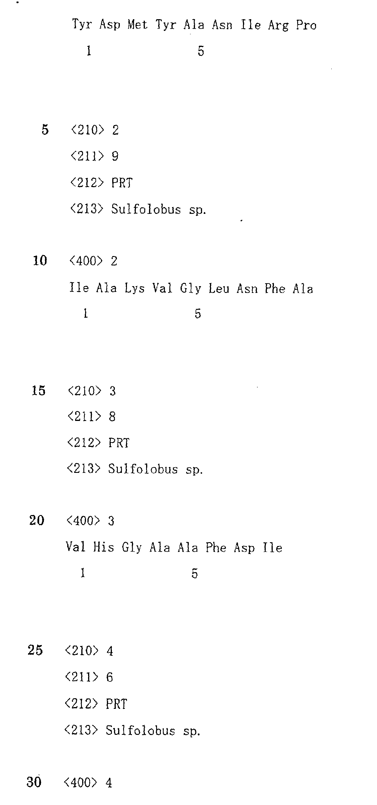
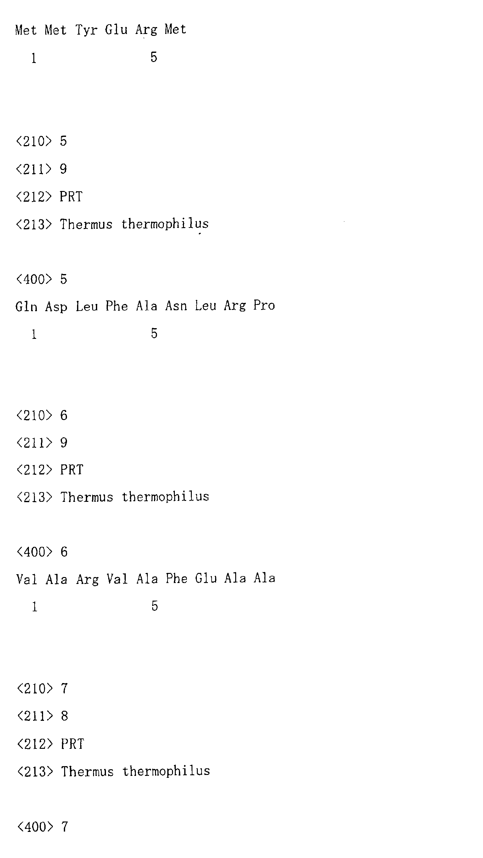


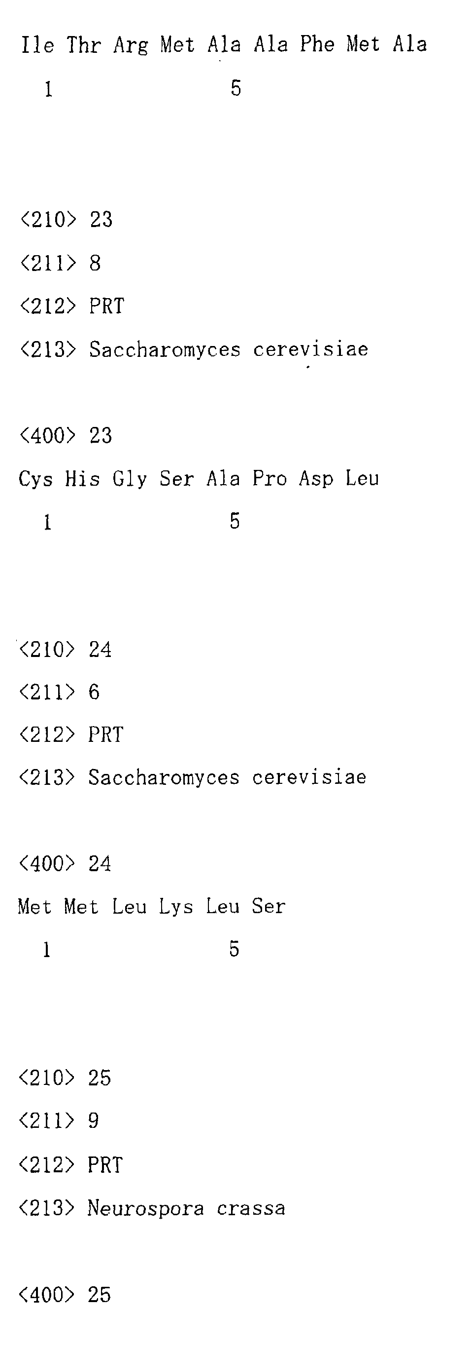
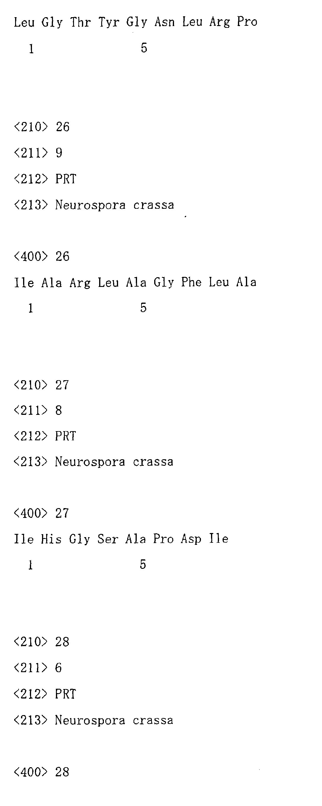

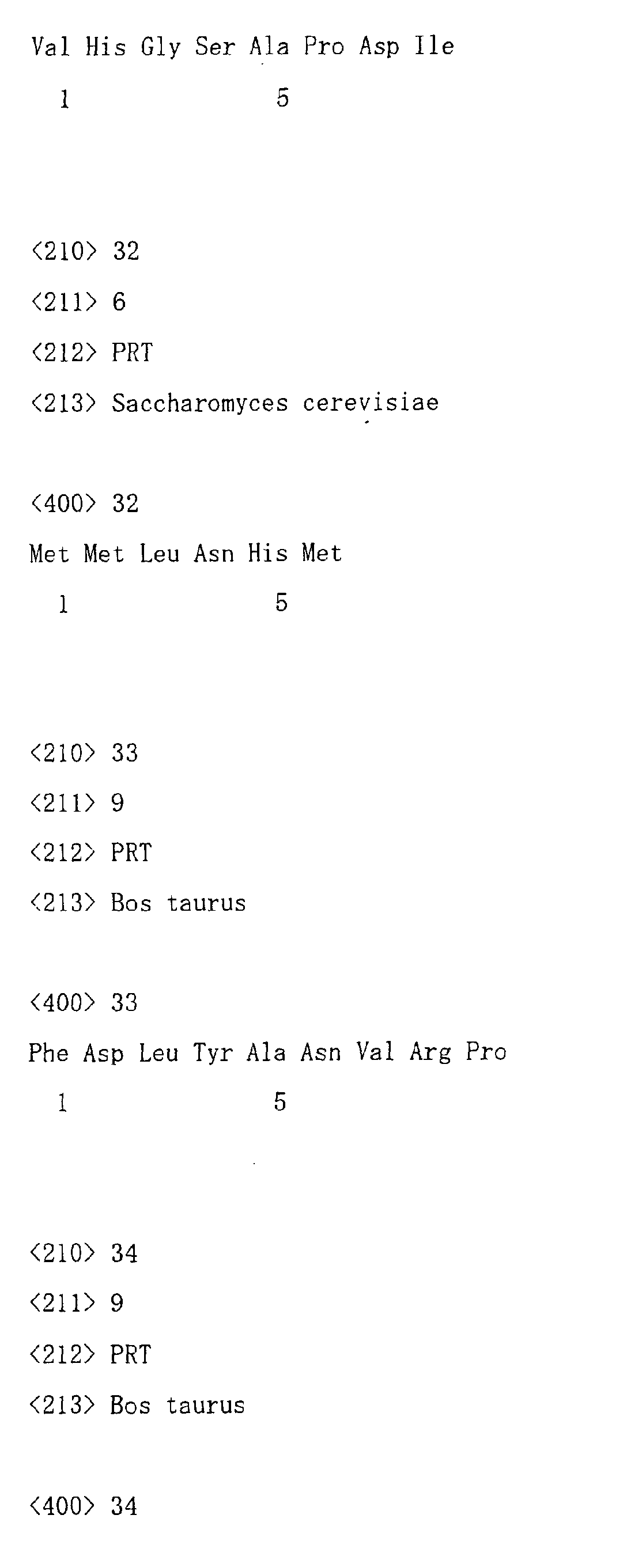


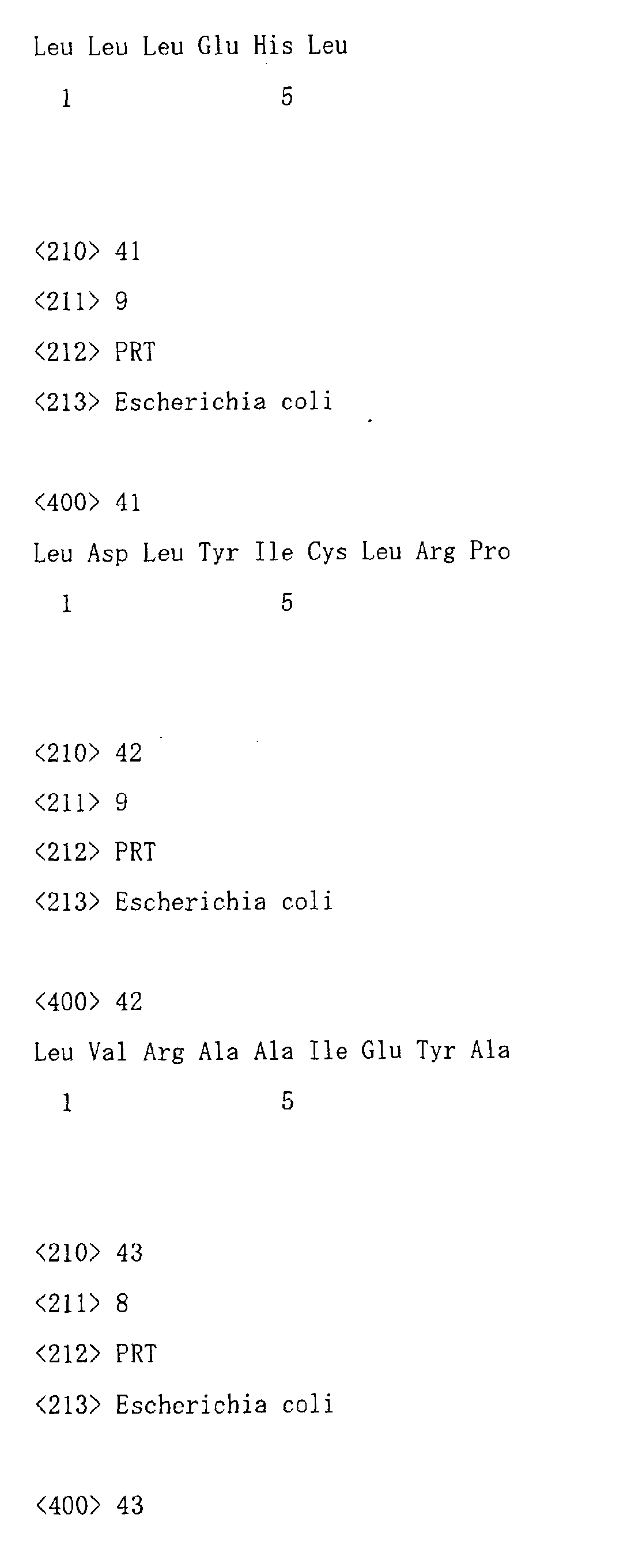


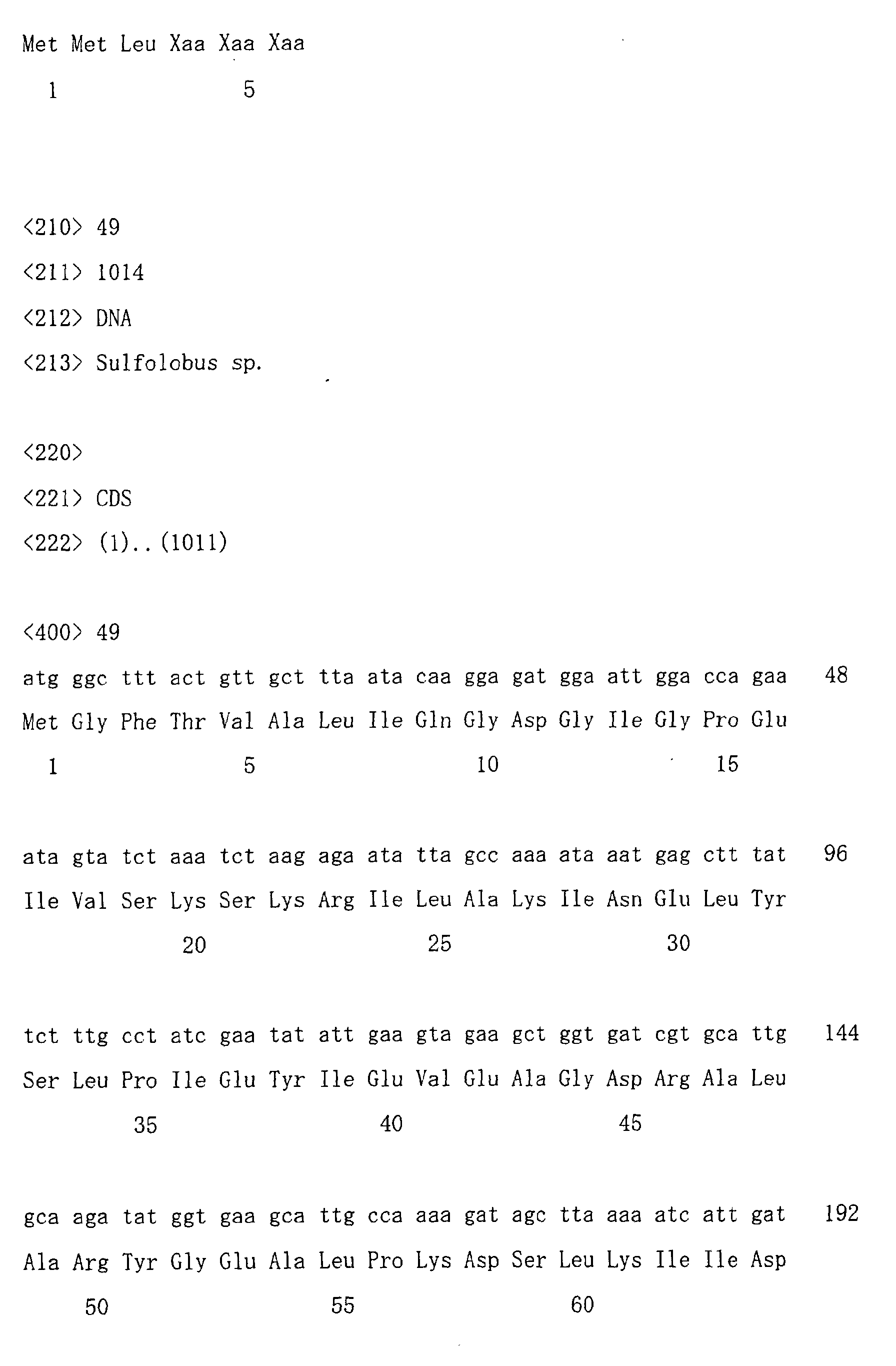

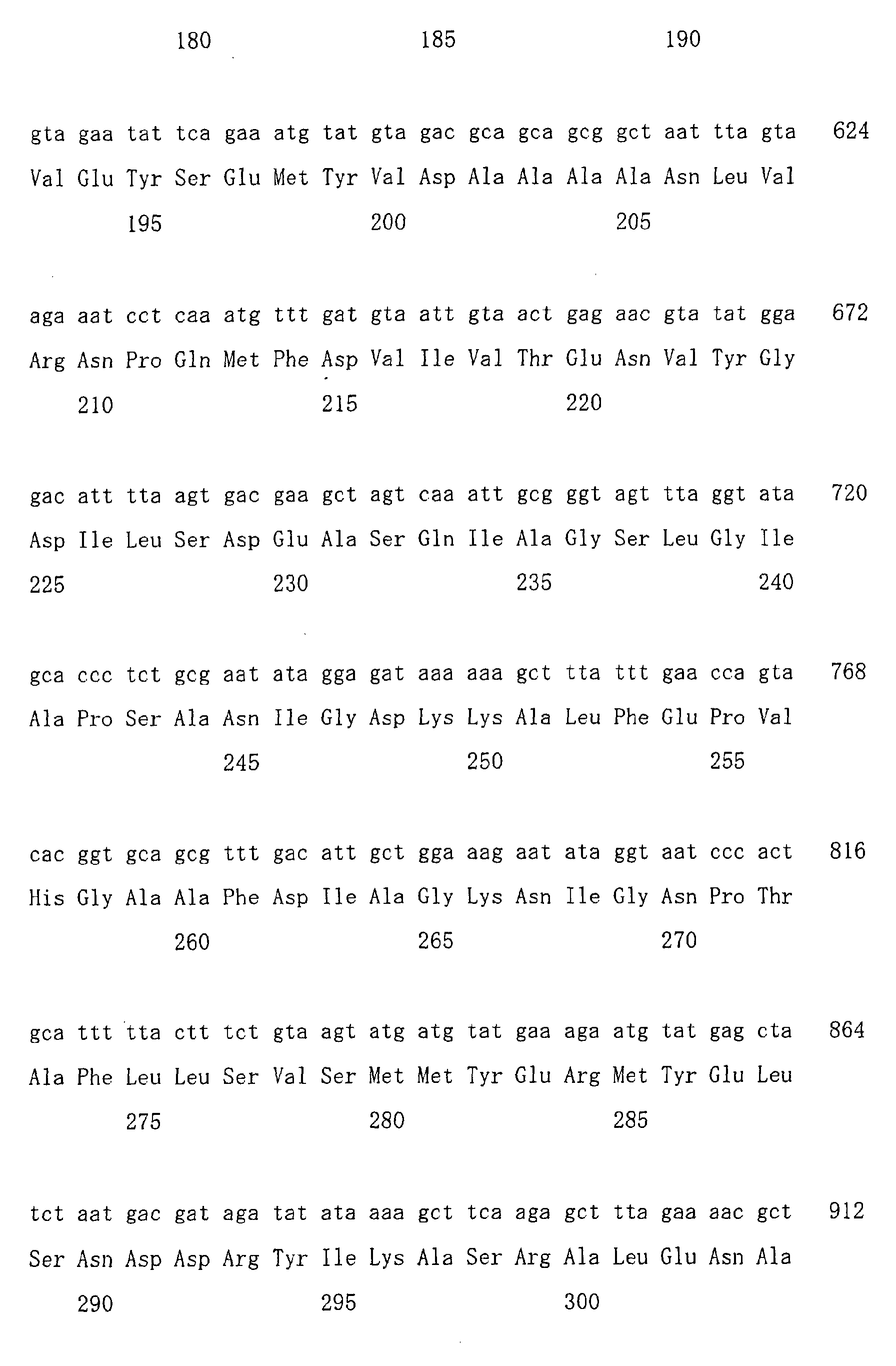
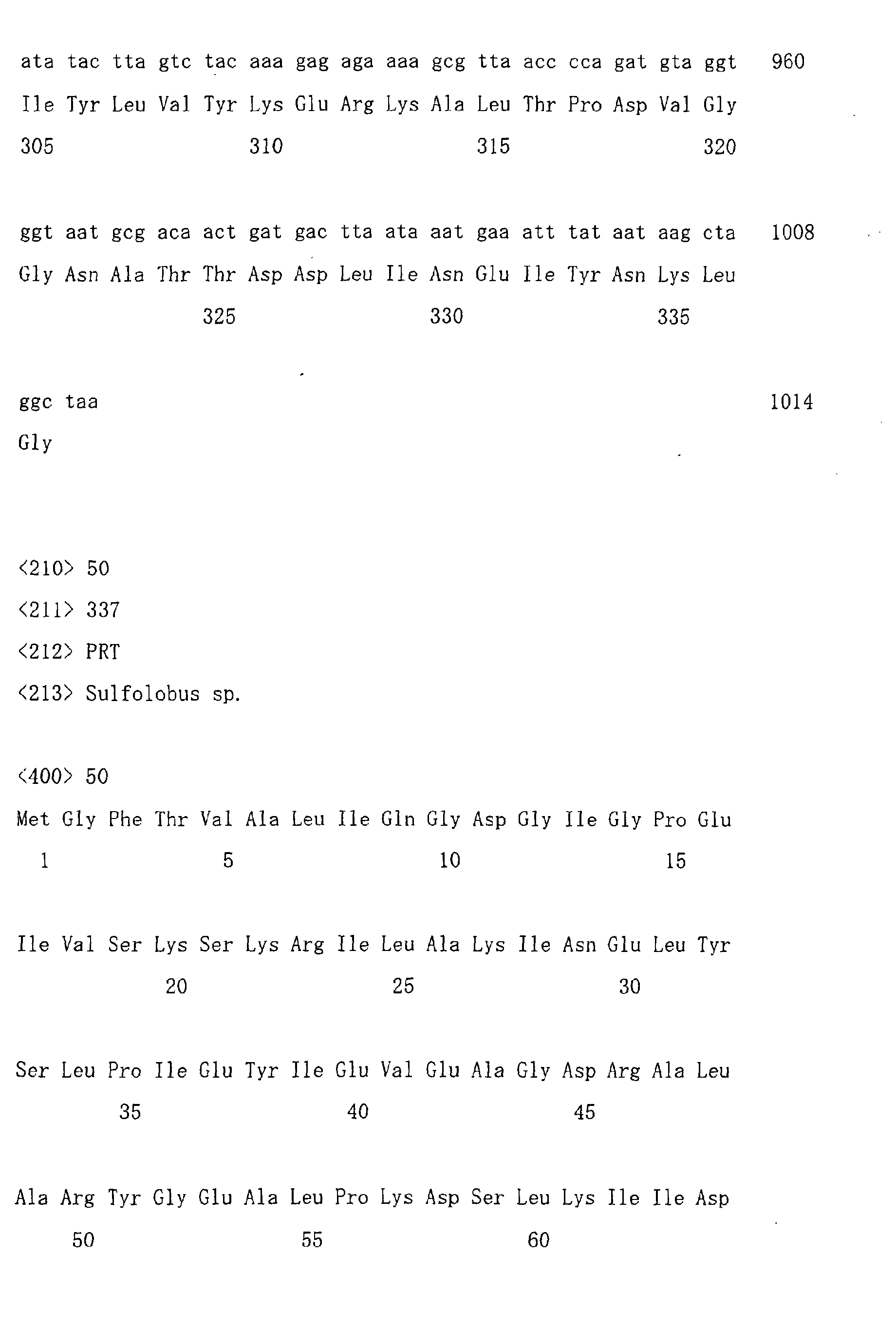
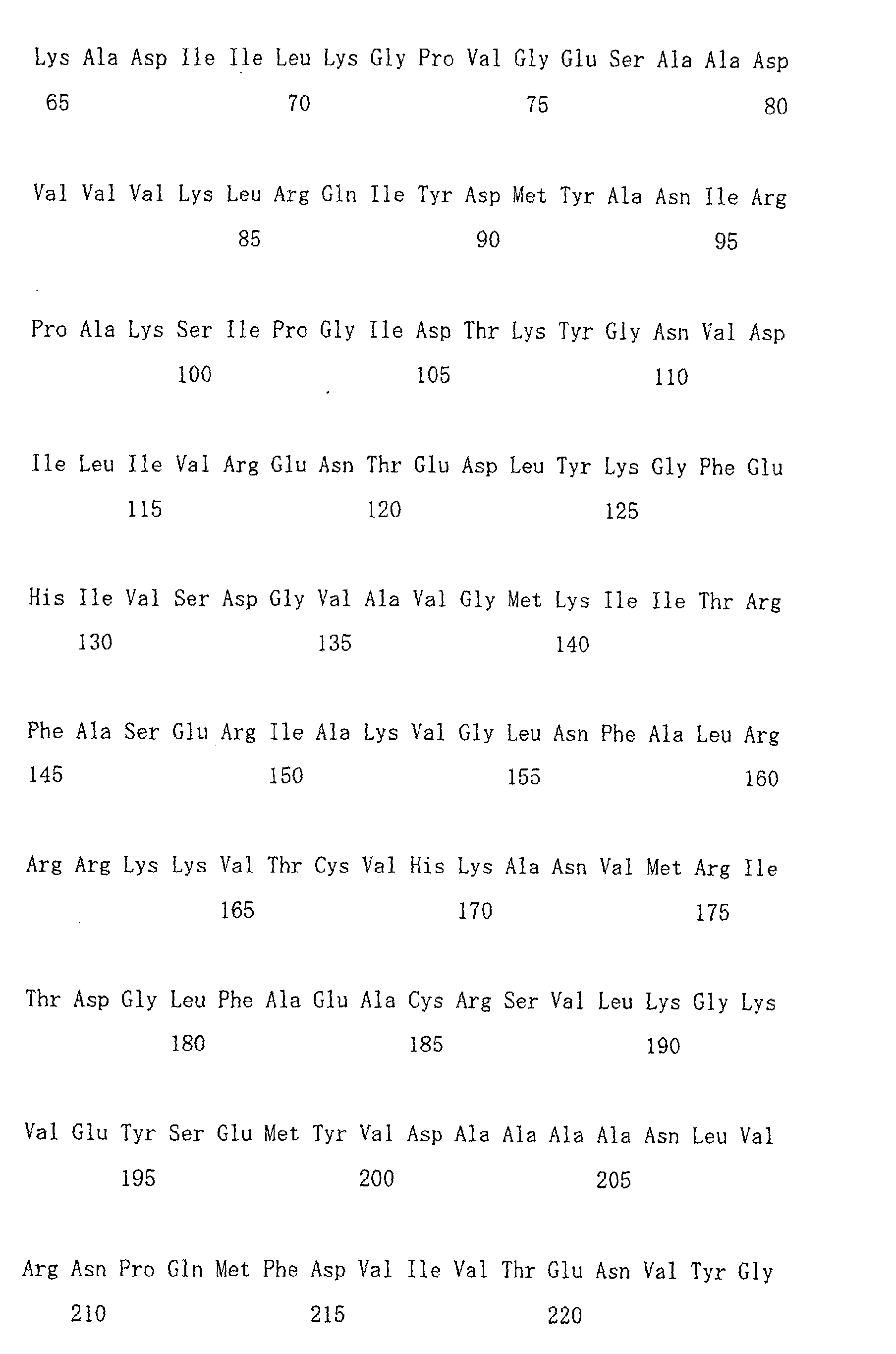
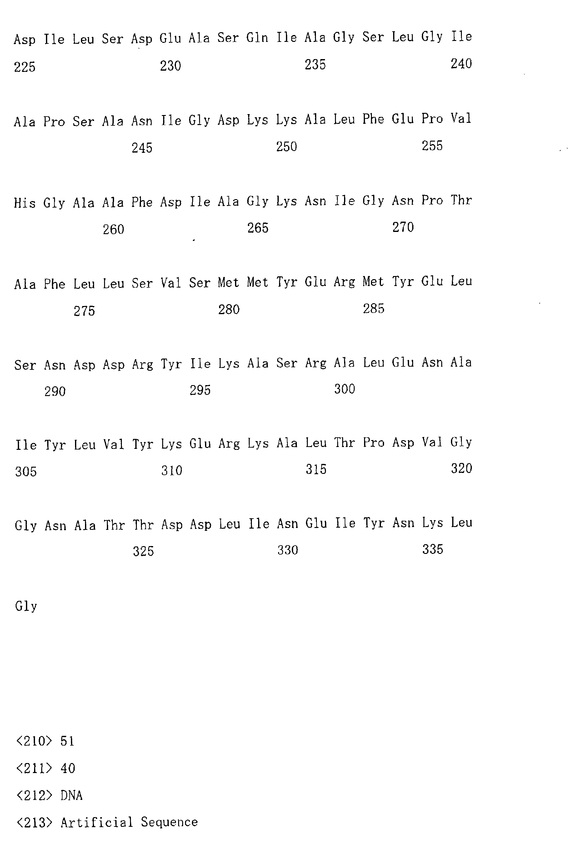
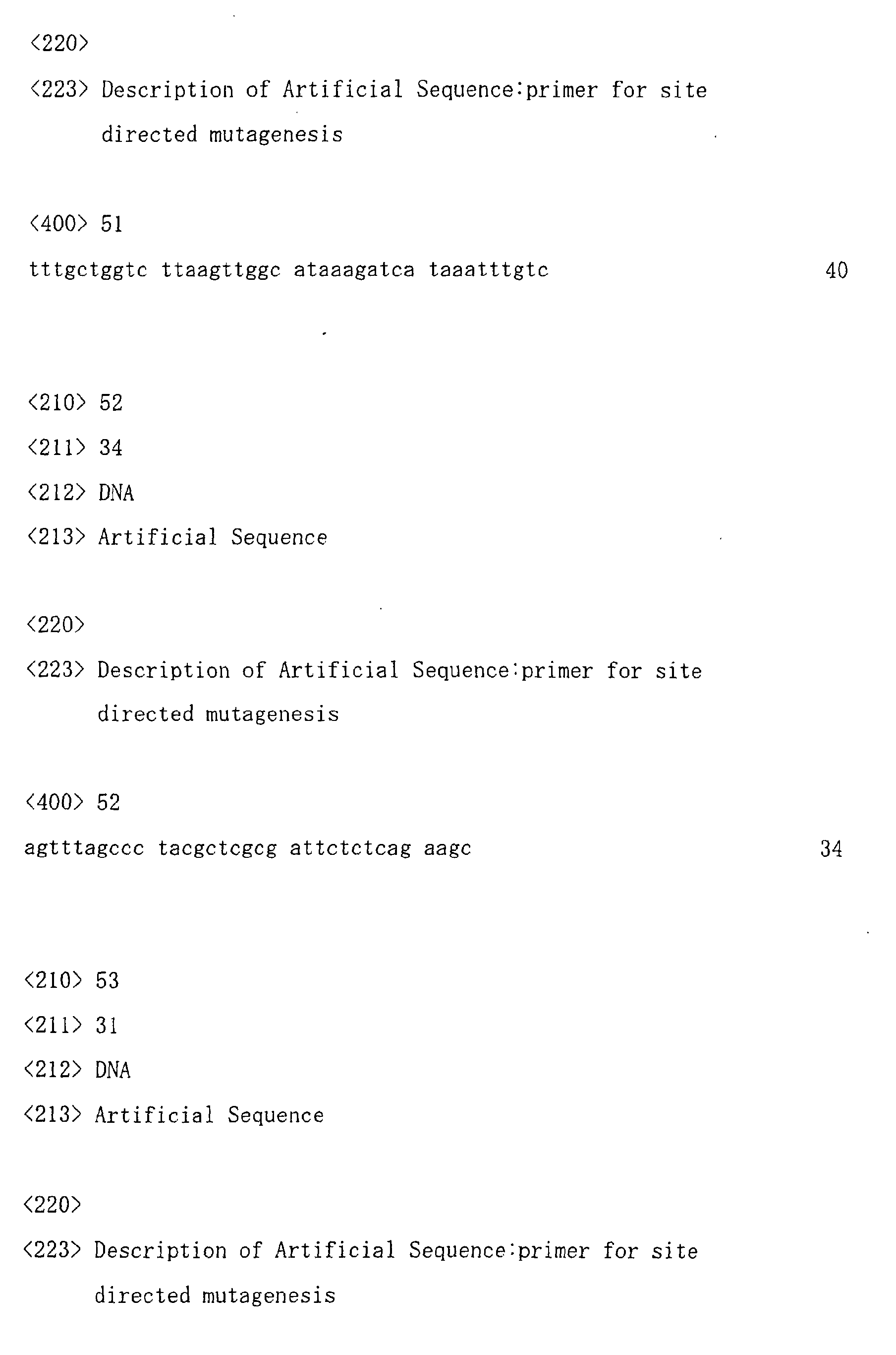
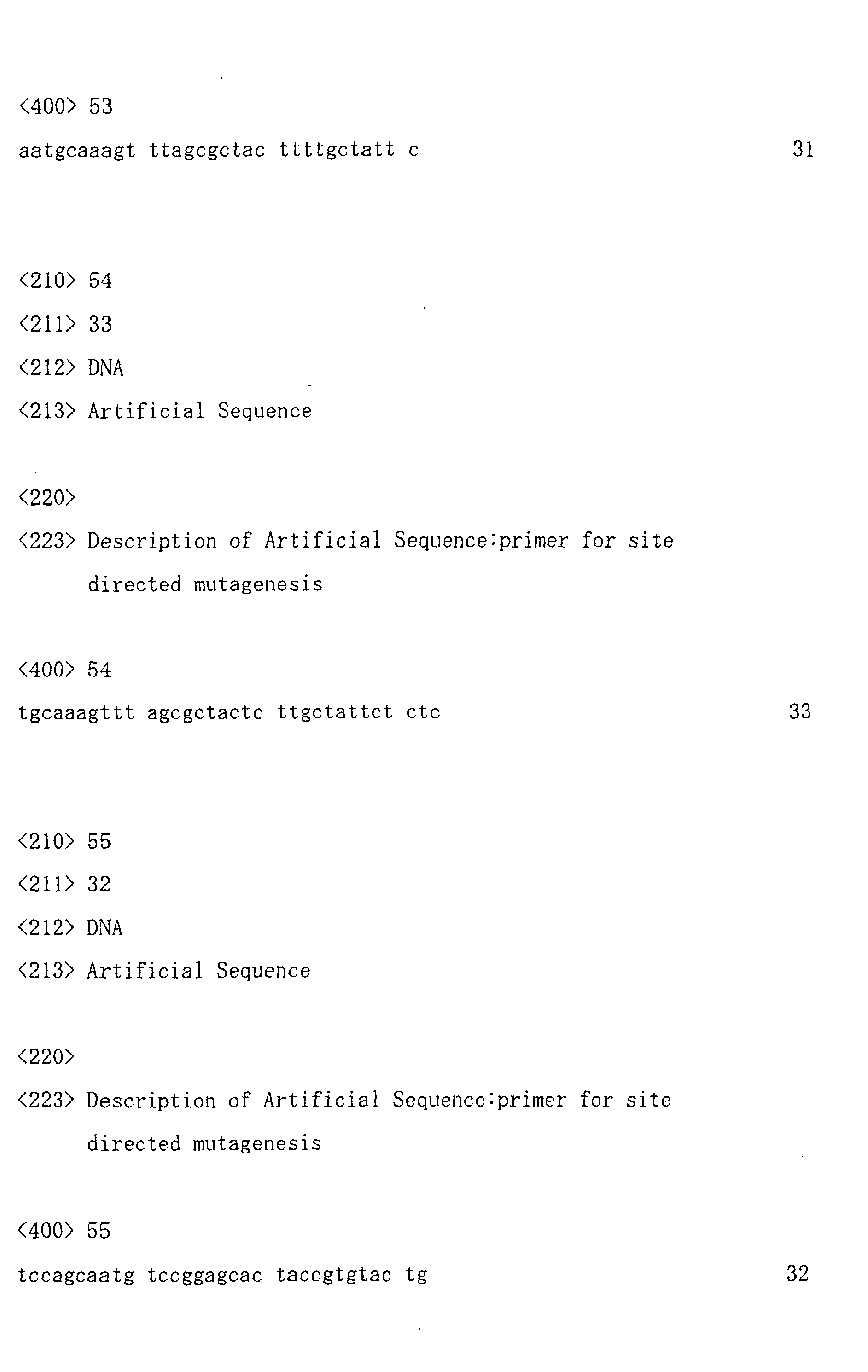


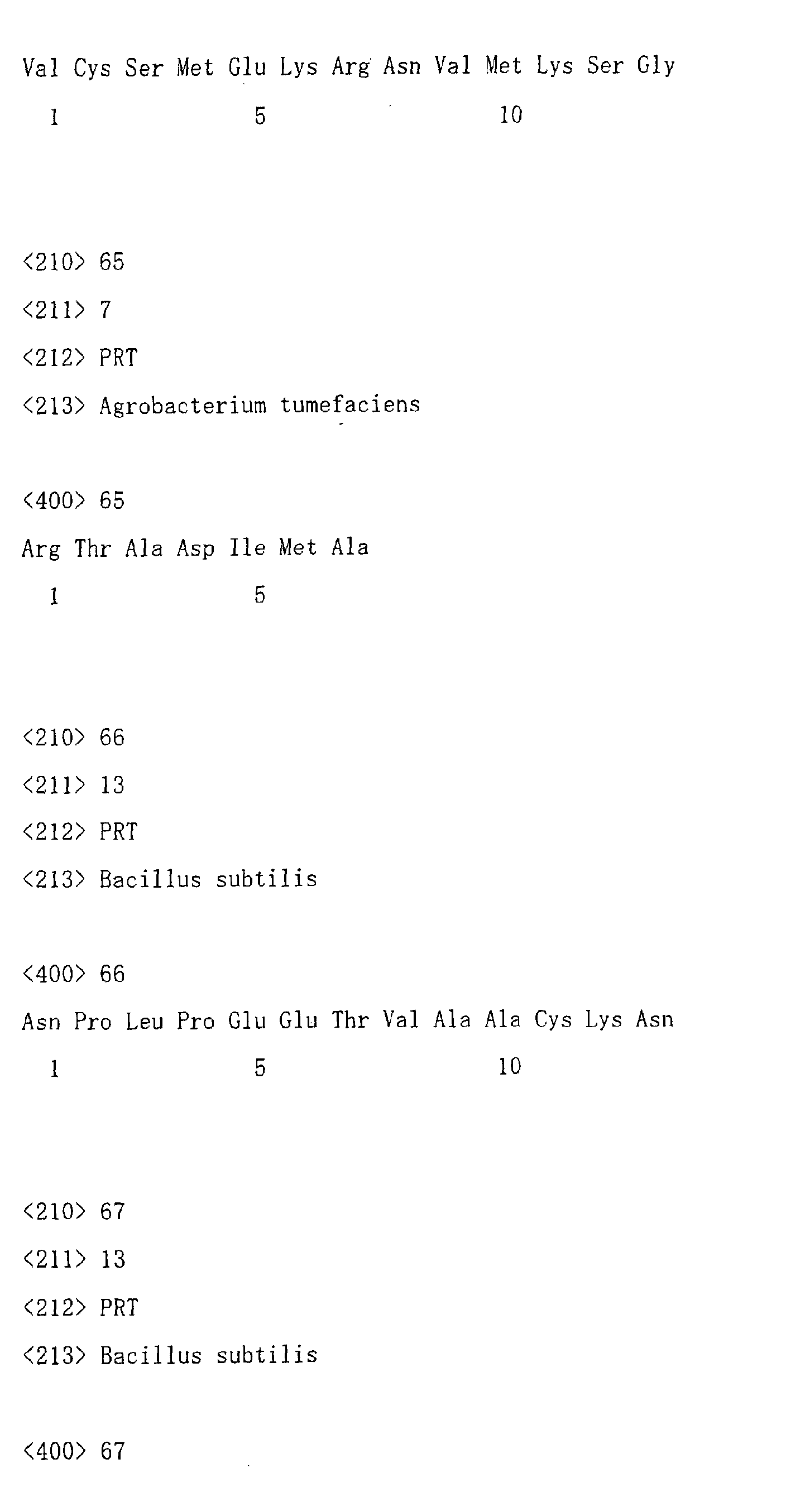

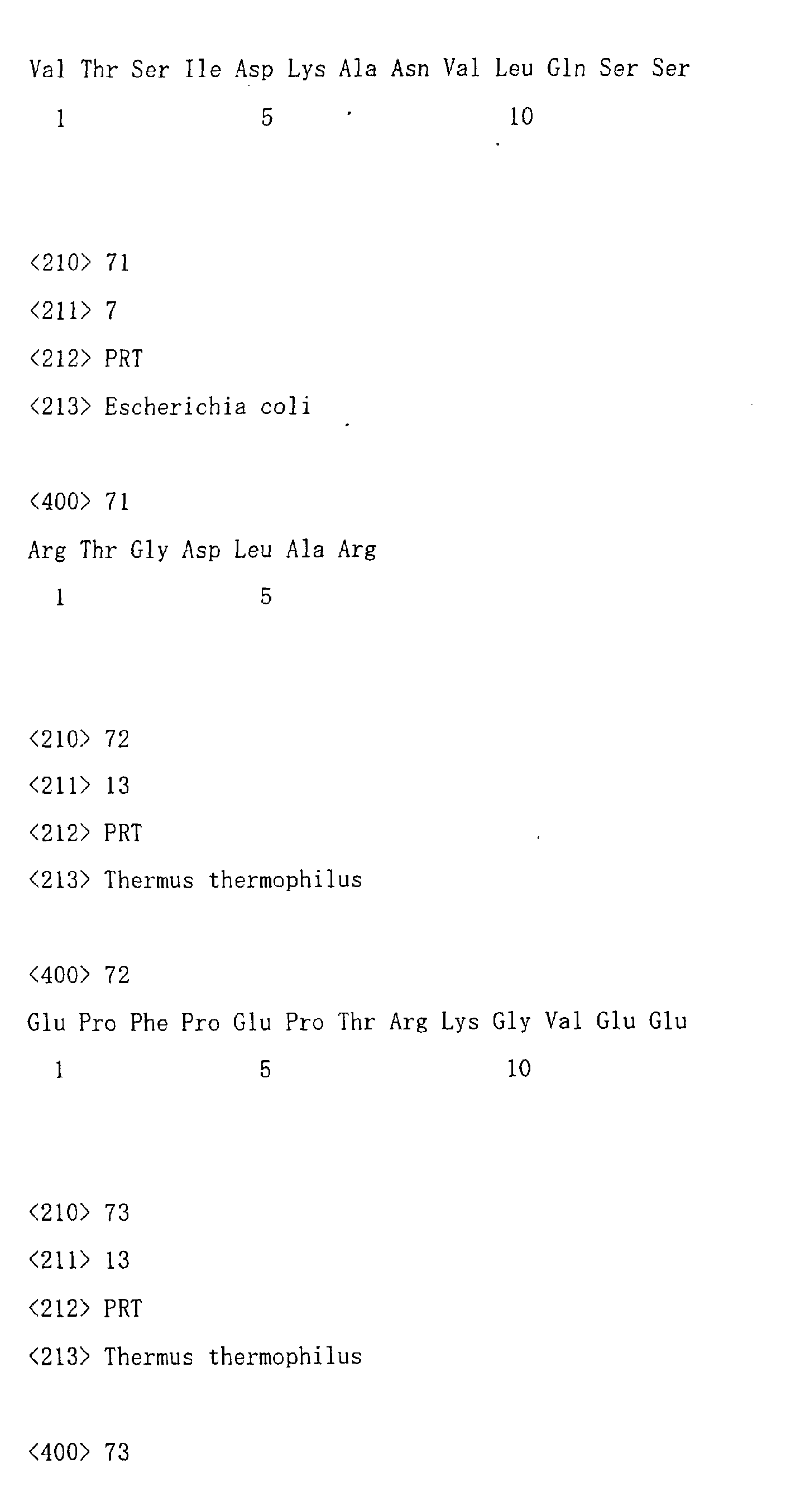
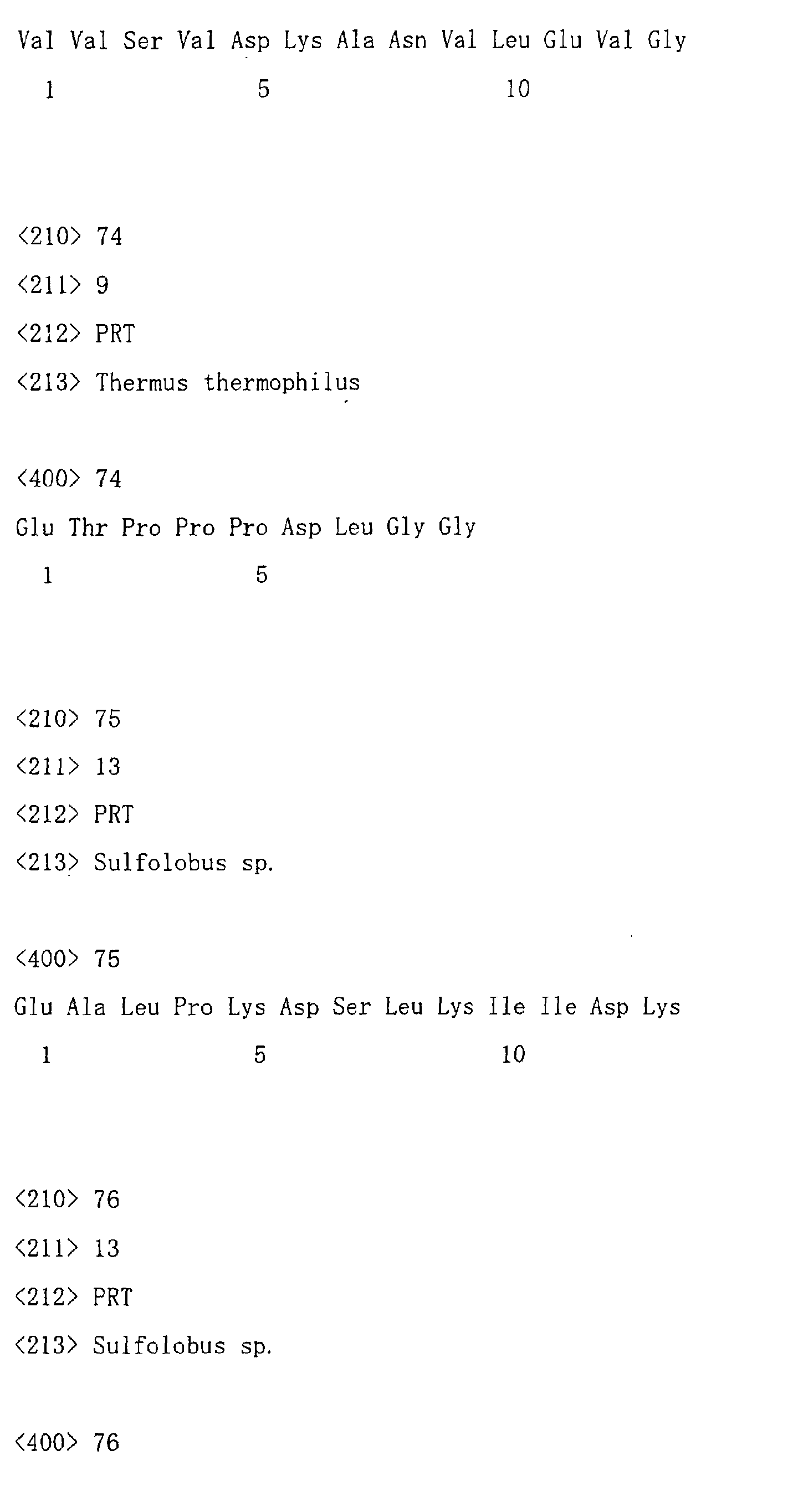
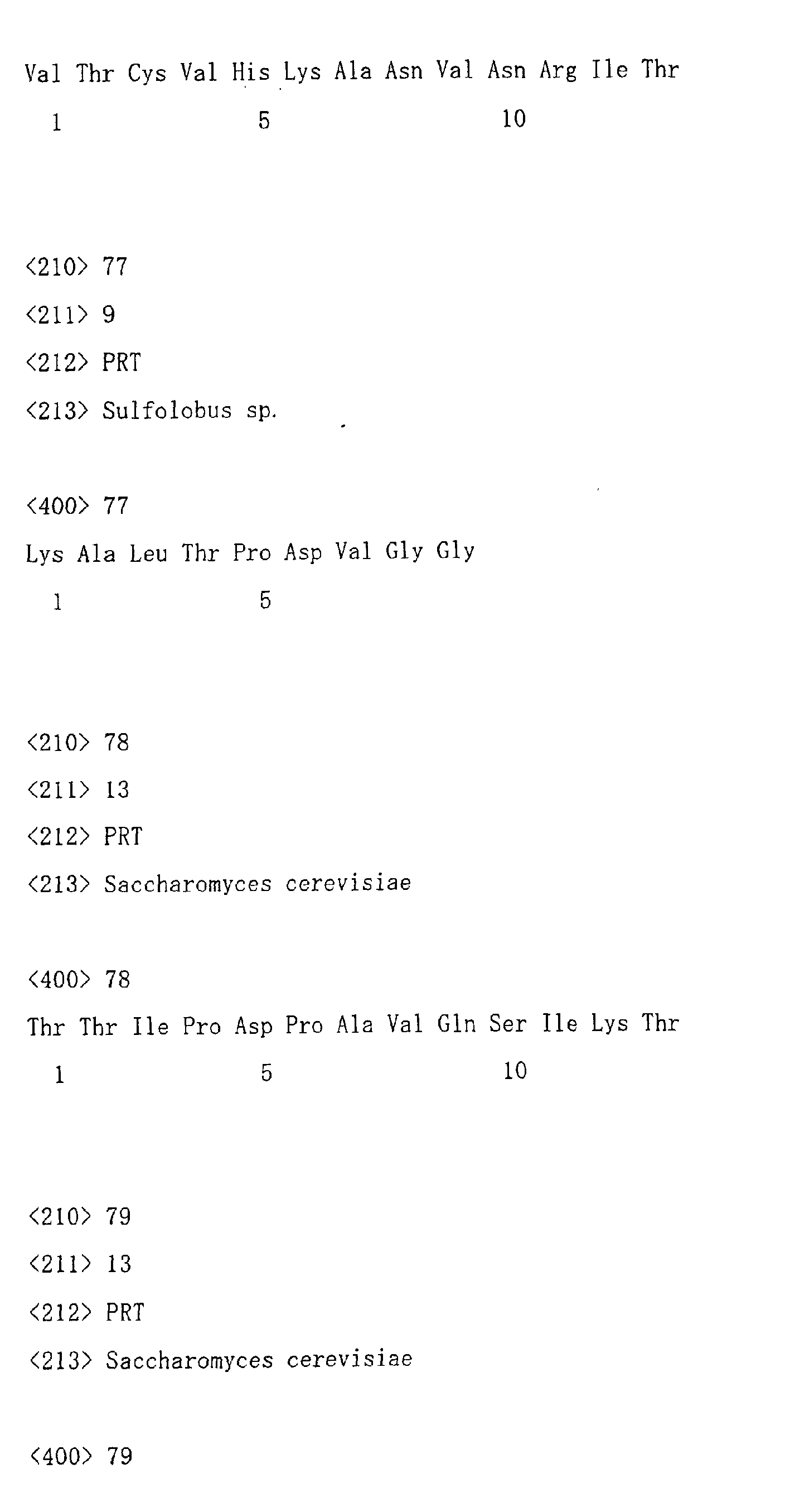


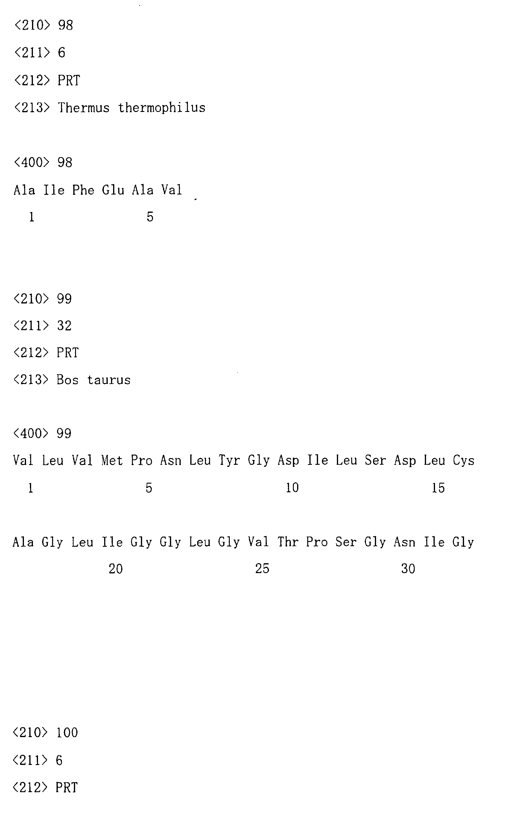
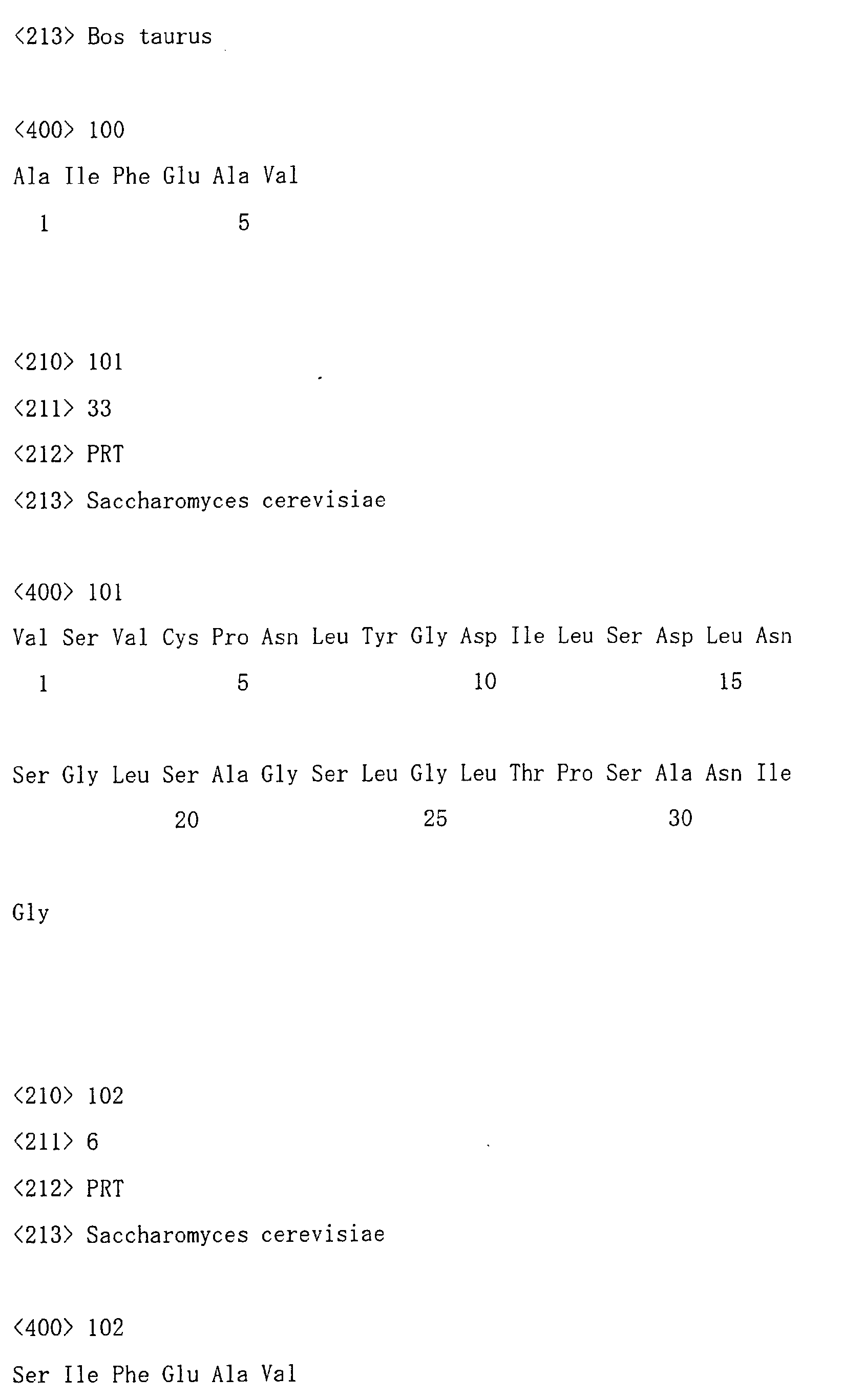
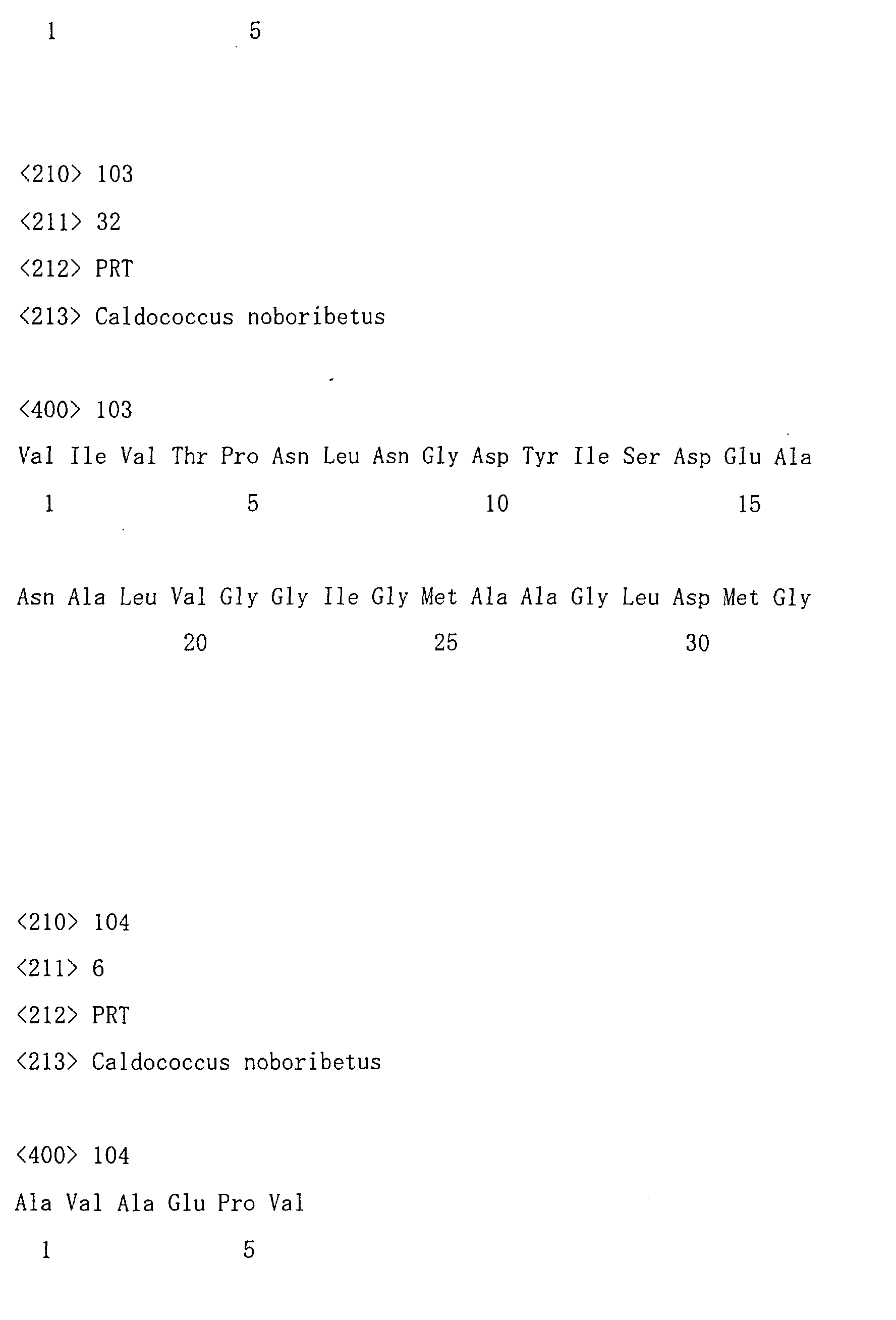
Claims (16)
- A method for improving thermostability of proteins, which comprises the steps of(i) comparing amino acid sequences of proteins from two or more species which evolutionarily correspond to each other in a phylogenetic tree;(ii) estimating an amino acid sequence of an ancestral protein corresponding to the amino acid sequences compared in step (i); and,(iii) comparing the amino acid residues in the amino acid sequence in one of the proteins compared in step (i) with amino acid residues at a corresponding position in the ancestral protein estimated in step (ii), and replacing one or more amino acid residues of the protein different from those of the ancestral protein with the same amino acid residues as those of the ancestral protein.
- The method of claim 1, further comprising the steps of(iv) testing the proteins obtained in step (iii) for thermostability; and(v) selecting a protein having improved thermostability.
- A method for improving thermostability of proteins, which comprises the steps of(i) comparing amino acid sequences of proteins from two or more species which evolutionarily correspond to each other in a phylogenetic tree by multiple alingment;(ii) estimating an amino acid sequence of an ancestral protein corresponding to the amino acid sequences compared in step (i); and,(iii) comparing the amino acid residues in the amino acid sequence in one of the proteins compared in step (i) with amino acid residues at a corresponding position in the ancestral protein estimated in step (ii), and replacing one or more amino acid residues of the protein different from those of the ancestral protein with the same amino acid residues as those of the ancestral protein.
- The method of claim 3, further comprising the steps of(iv) testing the proteins obtained in step (iii) for thermostability; and(v) selecting a protein having improved thermostability.
- The method for improving thermostability of protein according to claim 1, wherein(a) thermophilic bacteria or archaebacteria are included in the species from which the protein to be compared is derived in step (i); or(b) two or more proteins belonging to the same family are included in the proteins to be compared in (i).
- The method for improving thermostability of protein according to claim 3, wherein(a) thermophilic bacteria or archaebacteria are included in the species from which the protein to be compared is derived in step (i); or(b) two or more proteins belonging to the same family are included in the proteins to be compared in (i).
- A protein improved in thermostability by the method of claim 1.
- A Nucleic acid encoding the proteins of claim 7.
- A recombinant DNA molecule containing the nucleic acids of claim 8 in a form being functional for expression.
- A host cell having the recombinant DNA molecules of claim 9.
- The method of claim 1, wherein the protein is an 3-isopropylmalate dehydrogenase.
- The method of claim 1, wherein the protein is an isocitrate dehydrogenase.
- The method of claim 1, wherein the maximum parsimony method is used for estimating an amino acid sequence of an ancestral protein.
- The method of claim 3, wherein the maximum parsimony method is used for estimating an amino acid sequence of an ancestral protein.
- The method of claim 1, wherein the neighbor-joining method is used for estimating an amino acid sequence of an ancestral protein.
- The method of claim 3, wherein the neighbor-joining method is used for estimating an amino acid sequence of an ancestral protein.
Applications Claiming Priority (4)
| Application Number | Priority Date | Filing Date | Title |
|---|---|---|---|
| JP2000201920 | 2000-07-04 | ||
| JP2000201920 | 2000-07-04 | ||
| JP2001164332A JP2002247991A (en) | 2000-07-04 | 2001-05-31 | Method for improving heat resistance of protein, protein improved in heat resistance by the method, and nucleic acid encoding the protein |
| JP2001164332 | 2001-05-31 |
Publications (2)
| Publication Number | Publication Date |
|---|---|
| EP1182253A2 true EP1182253A2 (en) | 2002-02-27 |
| EP1182253A3 EP1182253A3 (en) | 2002-03-20 |
Family
ID=26595319
Family Applications (1)
| Application Number | Title | Priority Date | Filing Date |
|---|---|---|---|
| EP01115642A Withdrawn EP1182253A3 (en) | 2000-07-04 | 2001-07-03 | Method for improving the thermostability of proteins |
Country Status (3)
| Country | Link |
|---|---|
| US (1) | US20020137094A1 (en) |
| EP (1) | EP1182253A3 (en) |
| JP (1) | JP2002247991A (en) |
Cited By (3)
| Publication number | Priority date | Publication date | Assignee | Title |
|---|---|---|---|---|
| WO2003074697A1 (en) * | 2002-03-04 | 2003-09-12 | National Institute Of Technology And Evaluation | Method of modifying protein properties |
| WO2003095639A1 (en) * | 2002-05-08 | 2003-11-20 | Universite Libre De Bruxelles | Directed-selective screening : conception and production of active molecules that are evolutionary chaemeras |
| SE2250542A1 (en) * | 2022-05-03 | 2023-11-04 | Andrell Juni | Ancestral protein sequences and production thereof |
Families Citing this family (7)
| Publication number | Priority date | Publication date | Assignee | Title |
|---|---|---|---|---|
| US20040115621A1 (en) * | 2000-02-18 | 2004-06-17 | Allen Rodrigo | Ancestral viruses and vaccines |
| KR101221003B1 (en) | 2008-12-12 | 2013-01-10 | 한국생명공학연구원 | Methods for Modulating Thermostability and Acid Tolerance of Microbes |
| US9657328B2 (en) * | 2008-12-12 | 2017-05-23 | Korean Research Institute Of Bioscience And Biotechnology | Methods for modulating thermostability and acid tolerance of microbes |
| WO2011148938A1 (en) * | 2010-05-24 | 2011-12-01 | ユニチカ株式会社 | Protein having diaphorase activity |
| WO2012133761A1 (en) | 2011-03-30 | 2012-10-04 | ユニチカ株式会社 | Modified glucose dehydrogenase |
| WO2013067326A1 (en) * | 2011-11-04 | 2013-05-10 | University Of Georgia Research Foundation, Inc | Methods for expressing polypeptides in hyperthermophiles |
| DE102015007876A1 (en) * | 2015-06-22 | 2017-01-05 | Eblocker Gmbh | Network control device |
-
2001
- 2001-05-31 JP JP2001164332A patent/JP2002247991A/en active Pending
- 2001-07-03 EP EP01115642A patent/EP1182253A3/en not_active Withdrawn
- 2001-07-03 US US09/897,107 patent/US20020137094A1/en not_active Abandoned
Non-Patent Citations (1)
| Title |
|---|
| None * |
Cited By (5)
| Publication number | Priority date | Publication date | Assignee | Title |
|---|---|---|---|---|
| WO2003074697A1 (en) * | 2002-03-04 | 2003-09-12 | National Institute Of Technology And Evaluation | Method of modifying protein properties |
| US8510053B2 (en) | 2002-03-04 | 2013-08-13 | Ajinomoto Co., Inc. | Method for modifying a property of a protein |
| WO2003095639A1 (en) * | 2002-05-08 | 2003-11-20 | Universite Libre De Bruxelles | Directed-selective screening : conception and production of active molecules that are evolutionary chaemeras |
| SE2250542A1 (en) * | 2022-05-03 | 2023-11-04 | Andrell Juni | Ancestral protein sequences and production thereof |
| SE546520C2 (en) * | 2022-05-03 | 2024-11-19 | Syren Per Olof | Ancestral protein sequences and production thereof |
Also Published As
| Publication number | Publication date |
|---|---|
| US20020137094A1 (en) | 2002-09-26 |
| JP2002247991A (en) | 2002-09-03 |
| EP1182253A3 (en) | 2002-03-20 |
Similar Documents
| Publication | Publication Date | Title |
|---|---|---|
| JPWO2010053161A1 (en) | Modified flavin adenine dinucleotide-dependent glucose dehydrogenase | |
| Yan et al. | In vitro stabilization and in vivo solubilization of foreign proteins by the beta subunit of a chaperonin from the hyperthermophilic archaeon Pyrococcus sp. strain KOD1 | |
| Tamakoshi et al. | Screening of stable proteins in an extreme thermophile, Thermus thermophilus | |
| US20020137094A1 (en) | Method for improving thermostability of proteins, proteins having thermostability improved by the method and nucleic acids encoding the proteins | |
| US20080262201A1 (en) | Method and a system for predicting protein functional site, a method for improving protein function, and a function-modified protein | |
| AU2002327268C1 (en) | Novel glyphosate-tolerant 5-enolpyruvylshikimate-3-phosphate synthase and the gene encoding the same | |
| Nanba et al. | Purification and characterization of formate dehydrogenase from Ancylobacter aquaticus strain KNK607M, and cloning of the gene | |
| JP5289801B2 (en) | Protein with uricase activity | |
| JP4352286B2 (en) | Mutant glucose-6-phosphate dehydrogenase and method for producing the same | |
| JP4022784B2 (en) | Novel hexokinase | |
| Porcelli et al. | Expression, purification, and characterization of recombinant S-adenosylhomocysteine hydrolase from the thermophilic archaeon Sulfolobus solfataricus | |
| CN114480345A (en) | MazF mutants, recombinant vectors, recombinant engineering bacteria and their applications | |
| JP4280826B2 (en) | Novel thermostable protein having cystathionine-γ-synthase activity | |
| JP4280827B2 (en) | A novel thermostable protein with acetylglutamate kinase activity | |
| Han et al. | Cloning, analysis and expression of the gene for β-glycosidase of Thermus caldophilus GK24 and properties of the enzyme | |
| EP2695939A1 (en) | Modified glucose dehydrogenase | |
| JP4651203B2 (en) | Novel glutaminase and method for producing the same | |
| JP4292294B2 (en) | Novel thermostable protein having pyrroline-5-carboxylate reductase activity | |
| JP4250762B2 (en) | Novel thermostable protein having metabolic activity of 6-N-hydroxyaminopurine | |
| KR20250080752A (en) | Method for optimum production of hydrocarbon oxide using soluble methane monooxygenase | |
| JP2005296008A (en) | A novel thermostable protein with mercury reductase activity | |
| JP2004298186A (en) | Novel thermostable protein with DNA homologous recombination repair activity | |
| JP2005296011A (en) | A novel thermostable protein with adenylosuccinate lyase activity | |
| JP2005296012A (en) | Novel thermostable protein with phosphoenolpyruvate synthase activity | |
| JP5130479B2 (en) | Method for improving the specific activity of creatinine amide hydrolase |
Legal Events
| Date | Code | Title | Description |
|---|---|---|---|
| PUAI | Public reference made under article 153(3) epc to a published international application that has entered the european phase |
Free format text: ORIGINAL CODE: 0009012 |
|
| PUAL | Search report despatched |
Free format text: ORIGINAL CODE: 0009013 |
|
| AK | Designated contracting states |
Kind code of ref document: A2 Designated state(s): DE DK FR GB Kind code of ref document: A2 Designated state(s): AT BE CH CY DE DK ES FI FR GB GR IE IT LI LU MC NL PT SE TR |
|
| AX | Request for extension of the european patent |
Free format text: AL;LT;LV;MK;RO;SI |
|
| AK | Designated contracting states |
Kind code of ref document: A3 Designated state(s): AT BE CH CY DE DK ES FI FR GB GR IE IT LI LU MC NL PT SE TR |
|
| AX | Request for extension of the european patent |
Free format text: AL;LT;LV;MK;RO;SI |
|
| 17P | Request for examination filed |
Effective date: 20020524 |
|
| AKX | Designation fees paid |
Free format text: DE DK FR GB |
|
| 17Q | First examination report despatched |
Effective date: 20061221 |
|
| STAA | Information on the status of an ep patent application or granted ep patent |
Free format text: STATUS: THE APPLICATION IS DEEMED TO BE WITHDRAWN |
|
| 18D | Application deemed to be withdrawn |
Effective date: 20070503 |