Field of the Invention
-
The present invention relates to oligonucleotides
and method for detecting methicillin-resistant
Staphylococcus aureus (MRSA) in clinical examination.
The oligonucleotides provided in the present invention
are useful as reagents for genetic diagnosis which
involves procedures such as cleavage, amplification and
detection of RNA or DNA, and as reagents for inhibiting
reverse transcription or translation of RNA. In
particular, the sequences of the oligonucleotides
provided in the present invention are useful for reagents
or the like for quantitative determination and diagnosis
of MRSA.
Prior Art
-
MRSA is a resistant strain of Staphylococcus aureus
exhibiting resistance against β-lactamase-resistant
penicillins, including methicillin, which are stable
against (3-lactamase produced by Staphylococcus aureus.
MRSA is a major pathogen in the nosocomial infections,
and strains thereof which exhibit a slight resistance
even to vancomycin, an effective therapeutic drug against
the resistant strain, have also been detected. MRSA is
causing significant problems in medical care due to the
lack of an effective antimicrobial drug thereto. Thus,
its accurate and rapid detection in clinical examination
is an important subject in diagnosis and treatment.
-
Staphylococcus aureus generally produces four types
of cell wall composing-proteins PBPs(penicillin-binding
proteins), i.e., PBP-1 through PBP-4, however, it had
been found that MRSA also produces a new type of PBP
termed PBP-2'. This type of PBP is a specific protein
having poor affinity against β-lactam antibiotics, and is
known to play a central role in the tolerance of this
organism. The sequence of the mecA gene that codes for
PBP-2' is known (FEBS Lett., 221, 167-171, 1987, etc.).
Therefore, hybridization methods employing a gene probe
specific to mecA gene have been sought to detect and
identify MRSA.
-
As mentioned above, although attempts have been made
to detect MRSA at the gene level, since preparation of a
sample requires culturing the bacteria obtained from a
patient specimen, these methods have problems in terms of
their speed. Since detection and identification of MRSA
requires long culturing period, and it is difficult to
rapidly detect a trace amount of mecA gene in a sample,
there is a need for the development of a rapidly and
highly sensitive detection method in the field of
clinical diagnosis. Moreover, there is also a need to
develop an automated examination apparatus to simplify
examinations.
-
In order to carry out highly sensitive detection, it
is preferable to perform said detection after amplifying
a specific sequence in the gene to be detected and
identified, or in RNA derived from said gene (hereafter,
referred to as the "target nucleic acid").
-
When the target nucleic acid is DNA, the Polymerase
chain reaction (PCR) method is known as an amplification
method. This method amplifies a specific sequence by
repetition of a cycle comprising heat denaturation,
primer annealing and extension reactions, in the presence
of a pair of primers complementary and homologous to both
ends of said specific sequence in the target DNA as well
as a thermostable DNA polymerase. At this time, in order
to amplify said specific sequence by PCR,
oligonucleotides that are highly specific to said
specific sequence are required. Moreover, in order to
carry out this detection and identification with high
sensitivity, oligonucleotides that are highly specific to
the target DNA are required. Further, it is also
necessary to determine the optimum combination of those
oligonucleotides. Therefore, attempts have been made to
detect the mecA gene located on the chromosomal DNA of
Staphylococcus aureus by PCR using specific
oligonucleotides sequences. However, in order to prepare
a sample, it is necessary to culture the bacteria
obtained from a patient specimen. Therefore, as in the
hybridization method mentioned above, there is a problem
in terms of their speed. In addition, since the
detection of the mecA gene located on the chromosomal DNA
does not actually lead to an identification of the
expression of PBP-2', there are also problems in terms of
clinical significance. Further, the PCR method requires
a complicated procedure involving repetition of rapidly
increasing and decreasing the temperature, which prevents
its automation.
-
On the other hand, as amplification methods in cases
where the target nucleic acid is RNA, in addition to the
RT-PCR method, there are known the NASBA method and 3SR
method, whereby the specific sequence is amplified by the
concerted action of reverse transcriptase and RNA
polymerase. These methods involve a chain reaction,
wherein a promoter sequence-containing primer for a
specific sequence in the target RNA, reverse
transcriptase, and Ribonuclease H are used to synthesize
double-stranded DNA containing the promoter sequence, and
this double-stranded DNA is used as a template for RNA
polymerase-catalyzed synthesis of RNA containing the
specific sequence, while the RNA in turn becomes a
template for synthesis of double-stranded DNA containing
the promoter sequence. The NASBA method and 3SR method
can accomplish nucleic acid amplification at a constant
temperature, and are therefore considered to be methods
suitable for automation. In this situation, the presence
of mecA gene as well as its existing amount can be
measured, for example, by qualitative or quantitative
determination of a mRNA coding for PBP-2'. Moreover,
since this mRNA is a gene from which PBP-2' is expressed,
it presents in an amount much larger than the number of
copies of mecA gene located on the chromosomal DNA.
Thus, the mecA gene can be detected without culturing the
bacteria from a specimen, making this method useful for
rapid diagnosis. At this time, in the amplification of
the above specific sequence by the NASBA method or the
like, an oligonucleotide having high specificity to the
above specific sequence is required. Moreover, in order
to perform detection and identification with high
sensitivity, an oligonucleotide that has high specificity
to the target RNA is required. Therefore, an attempt to
detect the mecA gene located on the chromosomal DNA of
Staphylococcus aureus by the NASBA method using specific
oligonucleotide sequences has been made. However, since
the NASBA method or the like involves amplification
reaction at relatively low temperature (for example,
41°C), the target RNA forms an intramolecular structure
which inhibits binding of the primer, and may reduce the
reaction efficiency. Consequently, a procedure of heat
denaturation of the target RNA prior to the amplification
reaction was required to break down the intramolecular
structure of the target RNA, thereby to improve the
primer binding efficiency. In addition, even in the case
where detection of RNA is carried out at a low
temperature, an oligonucleotide capable of binding to RNA
which has formed the above-mentioned intramolecular
structure is required.
-
Therefore, the first object of the present invention
is to provide oligonucleotides that are useful in
specific cleavage, amplification, or the like, as well as
highly sensitive detection and identification of the mecA
gene coding for cell wall composing-protein PBP-2'
produced by MRSA or RNA derived from said gene. In
addition, the present invention provides oligonucleotide
sequences useful in a pharmaceutical composition for
inhibiting RNA reverse transcription or translation.
-
The second object of the present invention is to
provide a preferable combination of oligonucleotides
useful in specific amplification of RNA derived from the
mecA gene coding for cell wall composing-protein PBP-2'
produced by methicillin-resistant Staphylococcus aureus
at a relatively low temperature (e.g., 41°C), as well as
for the highly sensitive detection and identification
thereof.
Detailed Description of the Invention
-
The invention of claim 1, which has been
accomplished to achieve the first object, relates to an
oligonucleotide for cleavage, detection or amplification
of the mecA gene, a gene element of methicillin-resistant
Staphylococcus aureus (MRSA), or RNA derived from said
gene, which oligonucleotide is capable of binding
specifically to said mecA gene or RNA derived therefrom,
and comprises at least 10 contiguous bases of any of the
sequences listed as SEQ. ID. Nos. 1 to 17, or an
oligonucleotide complementary to said oligonucleotide.
-
The invention of claim 2, which has been
accomplished to achieve the aforementioned object,
relates to the oligonucleotide according to claim 1,
wherein said oligonucleotide is an oligonucleotide primer
for DNA elongation reaction.
-
The invention of claim 3, which has been
accomplished to achieve the aforementioned object,
relates to the oligonucleotide according to claim 1,
wherein said oligonucleotide is an oligonucleotide probe
a portion of which is modified or labeled with a
detectable marker.
-
The invention of claim 4, which has been
accomplished to achieve the aforementioned object,
relates to the oligonucleotide according to claim 3,
wherein said oligonucleotide is a synthetic
oligonucleotide in which a portion of its base(s) is(are)
modified without impairing the function of said
oligonucleotide as an oligonucleotide probe.
-
The oligonucleotides of the invention, which have
been accomplished to achieve the first object, are
oligonucleotides that complementarily bind in a specific
manner to intramolecular structure-free regions of the
target RNA in the aforementioned RNA amplification, and
they are capable of binding specifically to the target
RNA without the heat denaturation described above. In
this manner, the present invention provides
oligonucleotides that binds to intramolecular structure-free
regions of the RNA derived from the mecA gene coding
for PBP-2' at a relatively low and constant temperature
(35-50°C, and preferably 41°C), which are useful for
specific cleavage, amplification, detection or the like
of the mecA gene. More specifically, the present
invention relates to oligonucleotides which accomplish
rapidly and highly sensitive detection by their use as
oligonucleotide primers for amplifying the above target
DNA with PCR, oligonucleotide primers for amplifying the
above target RNA with NASBA or the like, and an
oligonucleotide probe for detecting the target nucleic
acid without or after these amplifications. In this
connection, recently, in order to improve genetic
detection technology, developments of new chemically
synthesized substances, which would not impair the
function of an oligonucleotide probe to recognize a
complementary sequence based on adenine, guanine,
cytosine and thymine (or uracil) bases have been carried
out. One example of such substances includes peptide
nucleic acid (PNA) in which the sugar and phosphoric acid
skeletons that provides the skeleton structure of nucleic
acid DNA have been replaced with a polyamide skeleton.
Thus, oligonucleotides that have been modified by a
substance such as PNA to an extent that would not impair
their base sequences recognizing-function are also
included in the detecting probes of the present
invention.
-
SEQ ID Nos.1 through 17 illustrate the
oligonucleotides of the present invention useful in
cleavage, amplification, detection or the like of RNA
derived from the mecA gene. In this connection, RNA
derived from the mecA gene also includes RNA that has
been produced by using these genes as templates.
Although each of the oligonucleotides of the present
invention may include entire base sequence of any of SEQ
ID Nos. 1 to 17, since an order of 10 bases is adequate
for specific binding to mecA gene, these oligonucleotides
can be oligonucleotides comprising at least 10 contiguous
bases of the described sequences, and may also be their
complementary oligonucieotides.
-
The oligonucleotides of the present invention can be
used, for example, as an oligonucleotide primer for
nucleic acid amplification. If a nucleic acid
amplification method is carried out using the
oligonucleotide of the present invention as the primer,
only the target nucleic acid, namely mecA, can be
amplified. Although examples of amplification methods
include PCR, LCR, NASBA and 3SR, nucleic acid
amplification methods that can be carried out at a
constant temperature such as LCR, NASBA and 3SR are
particularly preferable. MRSA can be detected by
detecting the amplification product by various methods.
In this case, any of the above oligonucleotides other
than the oligonucleotide used in the amplification may be
used as the probe, and the fragment of the amplified
specific sequence can be confirmed by electrophoresis or
the like.
-
The oligonucleotides of the present invention can be
used as probes by, for example, modifying portions or
labeling them with a detectable marker. When detecting
the target nucleic acid, the oligonucleotide of the
present invention labeled with the detectable marker may
be hybridized to a single-stranded target nucleic acid,
after which the hybridized probe can be detected via the
marker. The marker detection may be carried out by a
method suitable for the particular marker and, for
example, when using an intercalator fluorescent dye for
labeling the oligonucleotide, a dye with the property of
exhibiting increased fluorescent intensity by
intercalation in the double-stranded nucleic acid
comprising the target nucleic acid and the
oligonucleotide probe may be used in order to allow easy
detection of only the hybridized probe without removal of
the probe that has not hybridized to the target nucleic
acid. When using a common fluorescent dye as the marker,
the marker may be detected after removal of the probe
that has not hybridized to the target nucleic acid. For
the detection, the target nucleic acid in the sample is
preferably amplified to a detectable amount by a nucleic
acid amplification method such as PCR, NASBA or 3SR
method, among which isothermal nucleic acid amplification
methods such as the NASBA and 3SR methods are most
preferable. When incorporating the nucleotide-labeled
probe in the reaction solution during the amplification,
it is especially preferable to modify the probe by, for
example, adding glycolic acid to the 3'-end so that the
probe will not function as a nucleotide primer.
-
The invention of claim 5, which has been
accomplished to achieve the second object, relates to a
detection method employing a RNA amplification process,
which comprises the steps of: forming a cDNA with a RNA-dependent
DNA polymerase using a specific sequence of a
RNA derived from mecA gene, a gene element of MRSA,
present in a sample as a template, with a first primer
having a sequence homologous to said specific sequence
and a second primer having a sequence complementary to
said specific sequence, wherein either the first or
second primer has a sequence having the RNA polymerase
promoter sequence added at its 5'-region, thereby
producing a RNA-DNA double-strand; digesting the RNA of
said RNA-DNA double-strand with Ribonuclease H to form a
single-stranded DNA; and then forming a double-stranded
DNA that includes a promoter sequence allowing
transcription of said RNA sequence or a RNA comprising a
sequence complementary to said RNA sequence with a DNA-dependent
DNA polymerase using said single-stranded DNA
as a template, said double-stranded DNA produces a RNA
transcription product in the presence of a RNA
polymerase, and said RNA transcription product is
subsequently used as the template for the single-stranded
DNA production with said RNA-dependent DNA polymerase;
characterized in that the oligonucleotide of SEQ. ID.
No.18 is used as the first primer and the oligonucleotide
of any of SEQ. ID. Nos.19 to 21 is used as the second
primer, or the oligonucleotide of SEQ. ID. No.22 is used
as the first primer and the oligonucleotide of SEQ. ID.
Nos.23 or 24 is used as the second primer, or the
oligonucleotide of SEQ. ID. No.25 is used as the first
primer and the oligonucleotide of SEQ. ID. Nos.23 or 24
is used as the second primer.
-
The invention of claim 6, which has been
accomplished to achieve the aforementioned object,
relates to the detection method of claim 5, characterized
in that said first primer is an oligonucleotide
comprising at least 10 contiguous bases of the sequence
of SEQ. ID. Nos.18, 22 or 25.
-
The invention of claim 7, which has been
accomplished to achieve the aforementioned object,
relates to the detection method of claim 5, characterized
in that said second primer is an oligonucleotide
comprising at least 10 contiguous bases of the sequence
of SEQ. ID. Nos.19, 20, 21, 23 or 24.
-
The invention of claim 8, which has been
accomplished to achieve the aforementioned object,
relates to a detection method for a methicillin-resistant
Staphylococcus aureus (MRSA), which comprises the steps
of: conducting the RNA amplification process according to
claim 5 in the presence of an oligonucleotide probe
labeled with an intercalator fluorescent dye, wherein the
sequence of said probe is complementary to at least a
portion of said RNA transcription product, and
complementary binding of said probe to said RNA
transcription product results in a change of the
fluorescent property relative to that of a situation
where a complex formation is absent; and then measuring
the fluorescence intensity of the reaction solution.
-
The present invention provides a combination of
oligonucleotides for amplifying and detecting RNA derived
from the mecA gene of methicillin-resistant
Staphylococcus aureus at a relatively low and constant
temperature (35-50°C, and preferably 41°C). Namely, the
present invention provides a combination of an
oligonucleotide primer for amplification of RNA derived
from mecA gene and an oligonucleotide probe for detection
and thereby provides a simple, rapidly and highly
sensitive mecA gene detection method and a detection kit
for clinical examination or the like using said
combination.
-
In one mode for carrying out the present invention,
a second primer (a sequence complementary to the 3' end
region of a specific sequence of the target RNA)
complementary binds to the specific sequence of RNA
derived from mecA gene of methicillin-resistant
Staphylococcus aureus present in a sample as a template,
and cDNA is produced by an extension reaction with RNA-dependent
DNA polymerase to form a RNA-DNA double-strand,
after which the RNA of the RNA-DNA double-strand is
digested with Ribonuclease H to produce a single-stranded
DNA. Next, a first primer (a sequence homologous to the
5' end region of the target RNA, and including the RNA
polymerase promoter sequence added at the 5' end)
complementary binds to the single-stranded DNA, to
produce a double-stranded DNA having a promoter sequence
allowing transcription of RNA comprising a sequence
homologous to the target RNA sequence, using DNA-dependent
DNA polymerase. The double-stranded DNA is
then used for amplification of the RNA transcription
product comprising the sequence homologous to the target
RNA sequence in the presence of RNA polymerase. The
present invention is thus characterized by the use of an
oligonucleotide of SEQ ID Nos. 18, 22 or 25 for the first
primer and an oligonucleotide of SEQ ID Nos. 19, 20, 21,
23 or 24 for the second primer. Although the first and
second primers may be the entire length of their
respective sequences, combinations of oligonucleotides
comprising at least 10 contiguous bases within each
sequence may also be used.
-
In the above mode of the present invention, the
target RNA must be cleaved at the 5' end of the specific
sequence. The method of cleaving the target RNA is
preferably a method in which an oligonucleotide (cleaving
oligonucleotide) with a sequence complementary to a
region overlapping and adjacent to the 5'-end of the
specific sequence is added, thereby cleaving the target
RNA with Ribonuclease H or the like. The 3'-end of the
cleaving oligonucleotide is preferably treated by
amination, for example, to prevent it from functioning as
an oligonucleotide primer.
-
In the above mode of the present invention, the
amplification process is preferably carried out in the
presence of an oligonucleotide probe (detecting
oligonucleotide probe) labeled with an intercalator
fluorescent dye having a sequence complementary to at
least a portion of the RNA transcription product.
Complementary binding of the probe to the RNA
transcription product results in a change in the
fluorescent property compared to a situation where the
complex formation is absent, so that the fluorescence
intensity of the reaction solution may be measured. In
addition, when a labeled oligonucleotide probe is
incorporated during the amplification process, it is
particularly preferable to modify the probe by, for
example, addition of glycolic acid to the 3'-end, to
prevent it from functioning as a primer in the extension
reaction. Examples of oligonucleotides that can be used
for the oligonucleotide probe for detection include the
sequences described in SEQ ID Nos. 20 or 29.
-
In an another mode for carrying out the present
invention, a second primer (a sequence complementary to
the target RNA, and including the RNA polymerase promoter
sequence added at the 5' region) complementary binds to
the specific sequence of RNA derived from the mecA gene
of methicillin-resistant Staphylococcus aureus present in
a sample as a template, and cDNA is produced by extension
reaction with RNA-dependent DNA polymerase to form a RNA-DNA
double-strand, after which the RNA of the RNA-DNA
double-strand is digested with Ribonuclease H to produce
a single-stranded DNA. Next, a first primer (a sequence
homologous to the 5' end region of the target RNA)
complementary binds to the single-stranded DNA, to
produce a double-stranded DNA having a promoter allowing
the transcription of RNA comprising a sequence
complementary to the target RNA sequence, using DNA-dependent
DNA polymerase. The double-stranded DNA is
then used for amplification of the RNA transcription
product comprising the sequence complementary to the
target RNA sequence in the presence of RNA polymerase.
The present invention is thus characterized by the use of
an oligonucleotide of SEQ ID Nos.18, 22 or 25 for the
first primer and an oligonucleotide of SEQ ID Nos.19, 20,
21, 23 or 24 for the second primer. Although the first
and second primers may be the entire length of their
respective sequences, combinations of oligonucleotides
comprising at least 10 contiguous bases within each
sequence may also be used.
-
In the above mode of the present invention, the
amplification process is preferably carried out in the
presence of an oligonucleotide probe (detecting
oligonucleotide probe) labeled with an intercalator
fluorescent dye having a sequence complementary to at
least a portion of the RNA transcription product.
Complementary binding of the probe to the RNA
transcription product results in a change in the
fluorescent property compared to a situation where the
complex formation is absent, so that the fluorescence
intensity of the reaction solution may be measured. In
addition, when a labeled oligonucleotide probe is
incorporated during the amplification process, it is
particularly preferable to modify the probe by, for
example, addition of glycolic acid to the 3'-end, to
prevent it from functioning as a primer in the extension
reaction. Examples of oligonucleotides that can be used
for the oligonucleotide probe for detection include the
sequences complementary to the sequences described in SEQ
ID Nos.20 or 29.
Brief Description of the Drawings
-
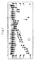
Fig. 1 is a photograph (black and white inverted)
showing the status of an oligonucleotide, which
illustrates an electrophoresis image of 7 M urea-5%
polyacrylamide gel electrophoresis for a sample after
performing a binding test to mecA-RNA at 41°C, using
oligonucleotides designed for an intramolecular
structure-free region of mecA-RNA coding for PBP-2'. In
this figure, lane M is the RNA marker, and lanes 1
through 25 are the numbers of the oligonucleotide
solutions indicated in Example 1. Lane 26 represents the
case without using an oligonucleotide.
-
Fig. 2 illustrates the results of performing an RNA
amplification reaction with various combinations of
primers in Example 2. In the figure, P represents the
case where a RNA sample of an initial RNA amount of 105
copies/30 µl is used, and N represents the case where
only diluent is used instead of RNA sample. In addition,
lane M indicates the molecular weight marker, while 1 to
15 indicate the numbers of the combinations of primer and
probe in Example 2.
-
Fig. 3 illustrates the chemical structures of the
intercalator fluorescent dye portions of the
oligonucleotides labeled with the intercalator
fluorescent dye used in Example 3. B1-B3 represent
nucleic acid bases.
-
Fig. 4 illustrates the relationship between the
reaction time and the fluorescence increasing rate. 1
and 2 represent the numbers of combinations of primers
and probes in Example 3. In the graphs, for combination
1, P represents the case where a RNA sample of an initial
RNA amount of 106 copies/30 µl is used and, for
combination 2, P represents the case where a RNA sample
of an initial RNA amount of 104 copies/30 µl is used. N
represents the case where only diluent is used instead of
RNA sample.
-
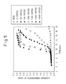
Fig. 5 illustrates a graph showing an increase in
the fluorescence increasing rate with respect to the
reaction time and the formation of RNA at initial RNA
amounts ranging from 104 copies/30 µl to 10 copies/30 µl
as measured in Example 4. Nega indicates the use of only
diluent instead of a RNA sample.
Examples
-
The present invention will now be explained in
greater detail by way of examples, with the understanding
that the invention is not limited by these examples.
Example 1
-
Specific binding of the oligonucleotides of the
invention to mecA-RNA at 41°C was examined. The mecA-RNA
is a synthesized and purified RNA obtained by in vitro
transcription using double-stranded DNA containing the
mecA-RNA base sequence as the template.
-
First, a sample of a standard RNA (2016mer)
comprising base Nos. 1 to 2013 of mecA-RNA derived from
PBP-2'(the RNA base sequence numbering is in accordance
with Matsubashi, et al. FEBS Lett., 221, 167-171 (1987))
was quantified by ultraviolet absorption at 260 nm, and
then diluted with an RNA diluent (10 mM Tris-HCl (pH
8.0), 0.1 mM EDTA, 0.5 U/µl RNase Inhibitor) to 2.0 x
10-12 mol/µl.
-
Next, the following compositions were dispensed into
0.5 ml volume PCR tubes (GeneAmp Thin-Walled Reaction™
Tubes; Perkin-Elmer Co., Ltd.).
- 0.90 µl of 1 M Tris-HCl buffer (pH 8.6)
- 0.20 µl of 1 M magnesium chloride
- 0.67 µl of 2 M potassium chloride
- 0.15 µl of 0.1 M DTT
- 0.33 µl of 119 U/µl RNase inhibitor
- 9.95 µl of distilled water
- 0.6 µl of 2 pmol/µl mecA-RNA sample
- 1.2 µl of 1.0 µM oligonucleotide solution
-
-
In this context, one of the oligonucleotide
solutions numbered below was used as the oligonucleotide
solution.
- 1. Oligonucleotide complementary to base Nos.241 to 261
of mecA-RNA (SEQ ID No. 1)
- 2. Oligonucleotide complementary to base Nos.264 to 283
of mecA-RNA (SEQ ID No. 2)
- 3. Oligonucleotide complementary to base Nos.296 to 315
of mecA-RNA (SEQ ID No. 3)
- 4. Oligonucleotide complementary to base Nos.349 to 368
of mecA-RNA (SEQ ID No. 4)
- 5. Oligonucleotide complementary to base Nos.402 to 421
of mecA-RNA
- 6. Oligonucleotide complementary to base Nos.425 to 444
of mecA-RNA
- 7. Oligonucleotide complementary to base Nos.456 to 475
of mecA-RNA (SEQ ID No. 5)
- 8. Oligonucleotide complementary to base Nos.499 to 480
of mecA-RNA
- 9. Oligonucleotide complementary to base Nos.551 to 532
of mecA-RNA (SEQ ID No. 6)
- 10. Oligonucleotide complementary to base Nos.556 to 575
of mecA-RNA (SEQ ID No. 7)
- 11. Oligonucleotide complementary to base Nos.581 to 600
of mecA-RNA (oligonucleotide of the 16th to 35th
bases from the 5' end of the sequence shown in SEQ
ID No.8)
- 12. Oligonucleotide complementary to base Nos.606 to 625
of mecA-RNA
- 13. Oligonucleotide complementary to base Nos.672 to 691
of mecA-RNA (SEQ ID No. 9)
- 14. Oligonucleotide complementary to base Nos.941 to 961
of mecA-RNA (SEQ ID No. 10)
- 15. Oligonucleotide complementary to base Nos.967 to 986
of mecA-RNA (SEQ ID No. 11)
- 16. Oligonucleotide complementary to base Nos.1134 to
1153 of mecA-RNA (SEQ ID No. 12)
- 17. Oligonucleotide complementary to base Nos.1154 to
1173 of mecA-RNA (SEQ ID No. 13)
- 18. Oligonucleotide complementary to base Nos.1221 to
1240 of mecA-RNA (SEQ ID No. 14)
- 19. Oligonucleotide complementary to base Nos.1656 to
1675 of mecA-RNA (SEQ ID No. 15)
- 20. Oligonucleotide complementary to base Nos.1701 to
1720 of mecA-RNA (SEQ ID No. 16)
- 21. Oligonucleotide complementary to base Nos.1852 to
1871 of mecA-RNA
- 22. Oligonucleotide complementary to base Nos.1906 to
1925 of mecA-RNA
- 23. Oligonucleotide complementary to base Nos.596 to 615
of mecA-RNA (oligonucleotide of the 1st to 20 the
bases from the 5' end of the sequence shown in SEQ
ID No. 8)
- 24. Oligonucleotide complementary to base Nos.577 to 615
of mecA-RNA (SEQ ID No. 8)
- 25. Oligonucleotide complementary to base Nos.1087 to
1100 of mecA-RNA (SEQ ID No. 17)
-
-
The reaction solutions were then incubated at 41°C
for 5 minutes, 1 µl of AMV-Rtase(Takara Shuzo Co., Ltd.;
AMV-RTase is an enzyme that cleaves the RNA of DNA/RNA
double strands) was added, and the PCR tube was incubated
at 41°C for 15 minutes.
-
Polyacrylamide gel (acrylamide concentration: 5%,
urea: 7 M) electrophoresis was conducted to confirm the
cleaved fragments after the reaction. Dyeing after
electrophoresis was carried out with a commercially
available dye (SYBR Green II™ (Takara Shuzo Co., Ltd.)).
Upon binding of the oligonucleotide to the specific site
of the target RNA, the RNA of the DNA/RNA double strands
is cleaved by the ribonuclease H activity of AMV-Rtase,
which allows observation of specific bands.
-
The results of electrophoresis are shown in Fig. 1.
Regarding the bands newly appearing in each lane, in the
lanes where two bands could be observed as a result of a
specific cleavage with one of the employed
oligonucleotide, bands with shorter length are indicated
with arrows. In addition, the bands exhibiting
significant non-specific cleavage are encircled. Among
the above oligonucleotides, only oligonucleotide
solutions containing SEQ ID Nos. 1 through 17 and SEQ ID
No. 8 showed characteristic cleaved bands without
significant non-specific cleavage, demonstrating that
each of these oligonucleotides strongly binds to mecA-RNA
at 41°C.
Results
-
As explained above, the oligonucleotides of the
present invention are oligonucleotides that complementary
bind to RNA derived from mecA-gene coding for PBP-2',
even under conditions of relatively low and constant
temperature (35-50°C, preferably 41°C), which tend to
produce an intramolecular structure in RNA and prevent
binding of primers and probes thereto. Specific binding
of the oligonucleotides is therefore possible without
heat denaturation of the target RNA. The
oligonucleotides of the invention are thus useful as
oligonucleotides for cleavage, amplification, detection
or the like of mecA-RNA, a gene element of MRSA, i.e. as
oligonucleotide primers or oligonucleotide probes to be
used in RNA amplification methods.
-
Furthermore, the oligonucleotides of the invention
are also clearly useful for amplification and detection
of the mecA gene. Oligonucleotides complementary to the
above-mentioned oligonucleotides are also useful for
amplification of double-stranded DNA by the PCR method,
or for detection of cDNA obtained by reverse
transcription of RNA.
-
The oligonucleotides of the invention are not
limited to the specifically listed base
sequences(20mers), and they may be oligonucleotides
comprising at least 10 or more contiguous bases within
those sequences. This is obvious from the fact that
10mer base sequences are sufficient to ensure adequate
specificity of primers or probes to target nucleic acids
under relatively low temperature condition (preferably,
at 41°C).
Example 2
-
Specific amplification of target RNA was carried out
using a combination of oligonucleotide primers according
to the present invention. The mecA-RNA is a synthesized
and purified RNA obtained by
in vitro transcription using
double-stranded DNA containing the mecA-RNA base sequence
as the template.
- (1) A sample of a standard RNA (2016mer) comprising base
Nos. 1 to 2013 of mecA-RNA derived from PBP-2'(the RNA
base sequence numbering is in accordance with Matsubashi,
et al. FEBS Lett., 221, 167-171 (1987)) was quantified by
ultraviolet absorption at 260 nm, and then diluted with
an RNA diluent (10 mM Tris-HCl (pH 8.0), 0.1 mM EDTA, 0.5
U/µl RNase Inhibitor, 5.0mM DTT) to 1.0 x 10⁴
copies/2.5µl.
In the control testing group, only the diluent was used
(Nega). - (2) 23.3 µl of a reaction liquid having the composition
indicated below was dispensed into 0.5 ml volume PCR
tubes (Gene Amp Thin-Walled Reaction Tubes, Perkin-Elmer)
followed by addition of 2.5 µl of the above RNA sample.
Composition of Reaction Liquid (concentrations refer
to the final concentrations in the reaction system
following addition of enzyme solution)
- 60.0 mM Tris-HCl buffer (pH 8.6)
- 13.0 mM magnesium chloride
- 90.0 mM potassium chloride
- 1.0 mM DTT
- 0.25 mM each of dATP, dCTP, dGTP and dTTP
- 3.0 mM each of ATP, GTP and UTP
- 2.25 mM GTP
- 3.6 mM ITP
- 1.0 µM each of the first and second primers
- 0.16 µM of a cleaving oligonucleotide probe
(oligonucleotide for cleaving the target RNA at a
position to which the first primer is capable to bind;
the 3' end thereof is aminated)
- 39 U ribonuclease inhibitor (Takara Shuzo)
- 15.0% DMSO
- Distilled water for adjusting volume
One of the combinations numbered below was used for
the combination of the first primer, the second primer
and the cleaving probe.
- 1. As for the first primer, oligonucleotide of the 4th
to 15th bases from the 5' end of the sequence shown
in SEQ ID No.18, wherein the promoter sequence of T7
polymerase shown in SEQ ID No.30 is added to its 5'
end; as for the second primer, the oligonucleotide
shown in SEQ ID No.19; and as for the cleaving
probe, the oligonucleotide shown in SEQ ID No.26.
- 2. As for the first primer, oligonucleotide of the 4th
to 15th bases from the 5' end of the sequence shown
in SEQ ID No.18, wherein the promoter sequence of T7
polymerase shown in SEQ ID No.30 is added to its 5'
end; as for the second primer, the oligonucleotide
shown in SEQ ID No.20; and as for the cleaving
probe, the oligonucleotide shown in SEQ ID No.26.
- 3. As for the first primer, oligonucleotide of the 4th
to 15th bases from the 5' end of the sequence shown
in SEQ ID No.18, wherein the promoter sequence of T7
polymerase shown in SEQ ID No.30 is added to its 5'
end; as for the second primer, the oligonucleotide
shown in SEQ ID No.21; and as for the cleaving
probe, the oiigonucieotide shown in SEQ ID No.26.
- 4. As for the first primer, oligonucleotide of the 4th
to 28th bases from the 5' end of the sequence shown
in SEQ ID No.18, wherein the promoter sequence of T7
polymerase shown in SEQ ID No.30 is added to its 5'
end; as for the second primer, the oligonucleotide
shown in SEQ ID No.20; and as for the cleaving
probe, the oligonucleotide shown in SEQ ID No.26.
- 5. As for the first primer, oligonucleotide of the 4th
to 28th bases from the 5' end of the sequence shown
in SEQ ID No.18, wherein the promoter sequence of T7
polymerase shown in SEQ ID No.30 is added to its 5'
end; as for the second primer, the oligonucleotide
shown in SEQ ID No.21; and as for the cleaving
probe, the oligonucleotide shown in SEQ ID No.26.
- 6. As for the first primer, oligonucleotide of the 1st
to 25th bases from the 5' end of the sequence shown
in SEQ ID No.18, wherein the promoter sequence of T7
polymerase shown in SEQ ID No.30 is added to its 5'
end; as for the second primer, the oligonucleotide
shown in SEQ ID No.20; and as for the cleaving
probe, the oligonucleotide shown in SEQ ID No.26.
- 7. As for the first primer, oligonucleotide of the 1st
to 25th bases from the 5' end of the sequence shown
in SEQ ID No.18, wherein the promoter sequence of T7
polymerase shown in SEQ ID No.30 is added to its 5'
end; as for the second primer, the oligonucleotide
shown in SEQ ID No.21; and as for the cleaving
probe, the oligonucleotide shown in SEQ ID No.26.
- 8. As for the first primer, oligonucleotide of the 4th
to 28th bases from the 5' end of the sequence shown
in SEQ ID No.22, wherein the promoter sequence of T7
polymerase shown in SEQ ID No.30 is added to its 5'
end; as for the second primer, the oligonucleotide
shown in SEQ ID No.23; and as for the cleaving
probe, the oligonucleotide shown in SEQ ID No.27.
- 9. As for the first primer, oligonucleotide of the 4th
to 28th bases from the 5' end of the sequence shown
in SEQ ID No.22, wherein the promoter sequence of T7
polymerase shown in SEQ ID No.30 is added to its 5'
end; as for the second primer, the oligonucleotide
shown in SEQ ID No.24; and as for the cleaving
probe, the oligonucleotide shown in SEQ ID No.27.
- 10. As for the first primer, oligonucleotide of the 1st
to 25th bases from the 5' end of the sequence shown
in SEQ ID No.22, wherein the promoter sequence of T7
polymerase shown in SEQ ID No.30 is added to its 5'
end; as for the second primer, the oligonucleotide
shown in SEQ ID No.23; and as for the cleaving
probe, the oligonucleotide shown in SEQ ID No.27.
- 11. As for the first primer, oligonucleotide of the 1st
to 25th bases from the 5' end of the sequence shown
in SEQ ID No.22, wherein the promoter sequence of T7
polymerase shown in SEQ ID No.30 is added to its 5'
end; as for the second primer, the oligonucleotide
shown in SEQ ID No.24; and as for the cleaving
probe, the oligonucleotide shown in SEQ ID No.27.
- 12. As for the first primer, oligonucleotide of the 4th
to 28th bases from the 5' end of the sequence shown
in SEQ ID No.25, wherein the promoter sequence of T7
polymerase shown in SEQ ID No.30 is added to its 5'
end; as for the second primer, the oligonucleotide
shown in SEQ ID No.23; and as for the cleaving
probe, the oligonucleotide shown in SEQ ID No.28.
- 13. As for the first primer, oligonucleotide of the 4th
to 28th bases from the 5' end of the sequence shown
in SEQ ID No.25, wherein the promoter sequence of T7
polymerase shown in SEQ ID No.30 is added to its 5'
end; as for the second primer, the oligonucleotide
shown in SEQ ID No.24; and as for the cleaving
probe, the oligonucleotide shown in SEQ ID No.28.
- 14. As for the first primer, oligonucleotide of the 1st
to 25th bases from the 5' end of the sequence shown
in SEQ ID No.25, wherein the promoter sequence of T7
polymerase shown in SEQ ID No.30 is added to its 5'
end; as for the second primer, the oligonucleotide
shown in SEQ ID No.23; and as for the cleaving
probe, the oligonucleotide shown in SEQ ID No.28.
- 15. As for the first primer, oligonucleotide of the 1st
to 25th bases from the 5' end of the sequence shown
in SEQ ID No.25, wherein the promoter sequence of T7
polymerase shown in SEQ ID No.30 is added to its 5'
end; as for the second primer, the oligonucleotide
shown in SEQ ID No.24; and as for the cleaving
probe, the oligonucleotide shown in SEQ ID No.28.
- (3) After incubating the above reaction solutions for 5
minutes at 41°C, 4.2 µl of enzyme liquid having the
following composition and pre-incubated for 2 minutes at
41°C was added.
Composition of Enzyme Liquid (final concentrations
during reaction)
- 1.7% sorbitol
- 8 units of AMV reverse transcriptase (Takara Shuzo)
- 142 units of T7 RNA polymerase (Gibco)
- 3 µg of bovine serum albumin
- Distilled water for adjusting volume
- (4) After which, the PCR tubes were incubated for 90
minutes at 41°C, and then the specific amplification
products were analyzed by electrophoresis using 4%
agarose gel.
- (5) A commercially available dye (SYBR Green II™:
Takara Shuzo) was used for staining after
electrophoresis.
-
-
The results of electrophoresis are shown in Fig. 2
(black and white inverted photograph). For all of the
combinations, specific RNA amplification products
(indicated with arrows) were obtained in the systems to
which mecA-RNA was added. On the basis of this finding,
these combinations of oligonucleotide primers were
demonstrated as being useful in the amplification and
detection of RNA derived from the mecA gene of
methicillin-resistant Staphylococcus aureus.
Example 3
-
Using the combination of oligonucleotide primers
according to the present invention, the possibility of
specific detection of mecA-RNA, the target RNA, was
confirmed.
- (1) A sample of a standard RNA (2016mer) comprising base
Nos. 1 to 2013 of mecA-RNA derived from PBP-2'(the RNA
base sequence numbering is in accordance with Matsubashi,
et al. FEBS Lett., 221, 167-171 (1987)) was quantified by
ultraviolet absorption at 260 nm, and then diluted with
an RNA diluent (10 mM Tris-HCl (pH 8.0), 0.1 mM EDTA, 0.5
U/µl RNase Inhibitor, 0.5mM DTT) to 1.0 x 10⁶
copies/2.5µl or 1.0 x 10⁴ copies/2.5µl In the control
testing group, only the diluent was used (Nega).
- (2) 23.3 µl of a reaction liquid having the composition
indicated below was dispended into 0.5 ml volume PCR
tubes (Gene Amp Thin-Walled Reaction Tubes, Perkin-Elmer)
followed by addition of 2.5 µl of the above RNA sample
(mecA-RNA).
Composition of Reaction Liquid (concentrations refer
to the final concentrations in the reaction system
following addition of enzyme solution)
- 60.0 mM Tris-HCl buffer (pH 8.6)
- 13.0 mM magnesium chloride
- 90.0 mM potassium chloride
- 1.0 mM DTT
- 0.25 mM each of dATP, dCTP, dGTP and dTTP
- 3.0 mM each of ATP, GTP and UTP
- 2.25 mM GTP
- 3.6 mM ITP
- 1.0 µM of the first oligonucleotide primer
- 1.0 µM of the second oligonucleotide primer
- 0.16 µM cleaving oligonucleotide probe
(oligonucleotide for cleaving the target RNA at a
position to which the first primer is capable to bind;
its 3' end is aminated)
- 25.0 nM of oligonucleotide probe for detection
labeled with intercalator fluorescent dye(Fig. 3) (MRSH-YO;
its 3' end modified with glycolic acid)
- 39 U ribonuclease inhibitor (Takara Shuzo)
- 15.0% DMSO
- Distilled water for adjusting volume
One of the combinations numbered below was used for
the combination of primers and probe.
- 1. As for the first primer, oligonucleotide of the 4th
to 15th bases from the 5' end of the sequence shown
in SEQ ID No.18, wherein the promoter sequence of T7
polymerase shown in SEQ ID No.30 is added to its 5'
end; as for the second primer, the oligonucleotide
shown in SEQ ID No.19; as for the cleaving probe,
the oligonucleotide shown in SEQ ID No.26, and as
for the probe for detection, the oligonucleotide
shown in SEQ ID No.20.
- 2. As for the first primer, oligonucleotide of the 1st
to 25th bases from the 5' end of the sequence shown
in SEQ ID No.25, wherein the promoter sequence of T7
polymerase shown in SEQ ID No.30 is added to its 5'
end; as for the second primer, the oligonucleotide
shown in SEQ ID No.24; as for the cleaving probe,
the oligonucleotide shown in SEQ ID No.28; and as
for the detecting probe, the oligonucleotide shown
in SEQ ID No.29.
- (3) After incubating the above reaction solution for 4
minutes at 41°C, 4.2 µl of enzyme liquid having the
following composition and pre-incubated for 2 minutes at
41°C were added.
Composition of Enzyme Liquid (final concentrations
during reaction)
- 1.7% sorbitol
- 8 units of AMV reverse transcriptase (Takara Shuzo)
- 142 units of T7 RNA polymerase (Gibco)
- 3 µg of bovine serum albumin
- Distilled water for adjusting volume
- (4) Next, using a temperature-controllable fluorescent
spectrophotometer capable of directly measuring PCR
tubes, periodic measurement of the fluorescence intensity
of the reaction solution incubated at 41°C with an
excitation wavelength of 470 nm and a fluorescent
wavelength of 510 nm was carried out. Fig. 4 shows the
periodic changes in the fluorescence intensity ratio
(fluorescence intensity at predetermined time/background
fluorescence intensity) of the sample, where enzyme was
added at 0 minutes The RNA sample concentrations were,
for combination 1, 106 copies/30 µl, and for combination
2, 104 copies/30 µl.
-
-
In the system in which mecA-RNA was added to
targeted RNA, specific fluorescent sensitization was
obtained. On the basis of this finding, the combination
of oligonucleotides of the present invention were
demonstrated as being capable to specifically amplify and
detect RNA derived from mecA gene.
Example 4
-
A combination of oligonucleotide primers according
to the invention was used for specific detection of
different initial copy numbers of the target RNA.
- (1) A sample of a standard RNA (2016mer) comprising base
Nos. 1 to 2013 of mecA-RNA derived from PBP-2'(the RNA
base sequence numbering is in accordance with Matsubashi,
et al. FEBS Lett., 221, 167-171 (1987)) was quantified by
ultraviolet absorption at 260 nm, and then diluted with
an RNA diluent (10 mM Tris-HCl (pH 8.0), 0.1 mM EDTA, 0.5
U/µl RNase Inhibitor, 5.0mM DTT) to concentrations
ranging from 1.0 x 10⁴ copies/2.5µl to 10 copies/2.5µl.
In the control testing group, only the diluent was used
(Nega).
- (2) 23.3 µl of a reaction liquid having the composition
indicated below was dispended into 0.5 ml volume PCR
tubes (Gene Amp Thin-Walled Reaction Tubes, Perkin-Elmer)
followed by addition of 2.5 µl of the above RNA sample
(mecA-RNA).
Composition of Reaction Liquid (concentrations refer
to the final concentrations in the reaction system
following addition of enzyme solution)
- 60.0 mM Tris-HCl buffer (pH 8.6)
- 13.0 mM magnesium chloride
- 90.0 mM potassium chloride
- 1.0 mM DTT
- 0.25 mM each of dATP, dCTP, dGTP and dTTP
- 3.0 mM each of ATP, CTP and UTP
- 2.25 mM GTP
- 3.6 mM ITP
- 1.0 µM of the first primer(oligonucleotide of the
4th to 28th bases from the 5' end of the sequence shown
in SEQ ID No.25, wherein the promoter sequence of T7
polymerase shown in SEQ ID No.30 is added to its 5' end
- 1.0 µM of the second oligonucleotide primer
(oligonucleotide of the 1st to 18th bases of from 5' end
of the sequence shown in SEQ ID No.23)
- 0.16 µM cleaving oligonucleotide probe (SEQ ID
No.28: oligonucleotide for cleaving the target RNA at a
position to which the first primer is able to bind,
wherein its 3' end is aminated)
- 25.0 nM of oligonucleotide probe for detection
labeled with intercalator fluorescent pigment (Fig. 3)
(MRSH-YO; its oligonucleotide sequence is shown in SEQ ID
No.29, and its 3' end is modified with glycolic acid)
- 39 U ribonuclease inhibitor (Takara Shuzo)
- 15.0% DMSO
- Distilled water for adjusting volume
- (3) After incubating the above reaction solution for 4
minutes at 41°C, 4.2 µl of enzyme liquid having the
following composition and pre-incubated for 2 minutes at
41°C were added.
Composition of Enzyme Liquid (final concentrations
during reaction)
- 1.7% sorbitol
- 8 units of AMV reverse transcriptase (Takara Shuzo)
- 142 units of T7 RNA polymerase (Gibco)
- 3 µg of bovine serum albumin
- Distilled water for adjusting volume
- (4) Next, using a temperature-controllable fluorescent
spectrophotometer capable of directly measuring PCR
tubes, periodic measurement of the fluorescence intensity
of the reaction solution incubated at 41°C with an
excitation wavelength of 470 nm and a fluorescent
wavelength of 510 nm was carried out. Fig. 5 shows the
periodic changes in the fluorescence intensity ratio
(fluorescence intensity at predetermined time/background
fluorescence intensity) of the sample, where enzyme was
added at 0 minutes. The RNA sample concentrations were
10 copies/30 µl to 104 copies/30 µl.
-
-
A fluorescence profile depending on the initial
concentration of the target RNA was obtained from Fig. 5,
indicating that it is possible to measure the amount of
RNA derived from the mecA gene present in unknown
samples.
Results
-
As has been explained above, the present invention
is useful as combinations of oligonucleotide primers and
oligonucleotide probes which specifically bind to RNA
derived from the mecA gene coding for PBP-2', and rapidly
amplify and detect the target RNA even under relatively
low and constant temperature (35-50°C and preferably
41°C) conditions in which RNA in a sample would form an
intramolecular structure which inhibits the primer and
probe binding.
-
In addition to the above, the combinations of
oligonucleotides of the present invention are not only
useful for mecA-RNA, but also as complementary sequences
of the above oligonucleotides for detecting cDNA obtained
by reverse transcription of RNA.
-
The base lengths of the oligonucleotides in the
combinations of the present invention are not limited to
the concretely described lengths, but rather include
oligonucleotides comprised of at least 10 contiguous
bases within these sequences. This is clear from the
fact that a base sequence of about 10 mer is adequate for
ensuring specificity of primer or probe to a target
nucleic acid under relatively low temperature (preferably
41°C) conditions.