EP1149899B1 - Culture medium for avian totipotential embryonic cells - Google Patents
Culture medium for avian totipotential embryonic cells Download PDFInfo
- Publication number
- EP1149899B1 EP1149899B1 EP01108584A EP01108584A EP1149899B1 EP 1149899 B1 EP1149899 B1 EP 1149899B1 EP 01108584 A EP01108584 A EP 01108584A EP 01108584 A EP01108584 A EP 01108584A EP 1149899 B1 EP1149899 B1 EP 1149899B1
- Authority
- EP
- European Patent Office
- Prior art keywords
- cells
- avian
- culture
- medium
- lif
- Prior art date
- Legal status (The legal status is an assumption and is not a legal conclusion. Google has not performed a legal analysis and makes no representation as to the accuracy of the status listed.)
- Expired - Lifetime
Links
Images


Classifications
-
- A—HUMAN NECESSITIES
- A01—AGRICULTURE; FORESTRY; ANIMAL HUSBANDRY; HUNTING; TRAPPING; FISHING
- A01K—ANIMAL HUSBANDRY; CARE OF BIRDS, FISHES, INSECTS; FISHING; REARING OR BREEDING ANIMALS, NOT OTHERWISE PROVIDED FOR; NEW BREEDS OF ANIMALS
- A01K67/00—Rearing or breeding animals, not otherwise provided for; New breeds of animals
- A01K67/027—New breeds of vertebrates
- A01K67/0275—Genetically modified vertebrates, e.g. transgenic
-
- C—CHEMISTRY; METALLURGY
- C12—BIOCHEMISTRY; BEER; SPIRITS; WINE; VINEGAR; MICROBIOLOGY; ENZYMOLOGY; MUTATION OR GENETIC ENGINEERING
- C12N—MICROORGANISMS OR ENZYMES; COMPOSITIONS THEREOF; PROPAGATING, PRESERVING, OR MAINTAINING MICROORGANISMS; MUTATION OR GENETIC ENGINEERING; CULTURE MEDIA
- C12N5/00—Undifferentiated human, animal or plant cells, e.g. cell lines; Tissues; Cultivation or maintenance thereof; Culture media therefor
- C12N5/06—Animal cells or tissues; Human cells or tissues
- C12N5/0602—Vertebrate cells
- C12N5/0603—Embryonic cells ; Embryoid bodies
- C12N5/0606—Pluripotent embryonic cells, e.g. embryonic stem cells [ES]
-
- A—HUMAN NECESSITIES
- A01—AGRICULTURE; FORESTRY; ANIMAL HUSBANDRY; HUNTING; TRAPPING; FISHING
- A01K—ANIMAL HUSBANDRY; CARE OF BIRDS, FISHES, INSECTS; FISHING; REARING OR BREEDING ANIMALS, NOT OTHERWISE PROVIDED FOR; NEW BREEDS OF ANIMALS
- A01K2217/00—Genetically modified animals
- A01K2217/05—Animals comprising random inserted nucleic acids (transgenic)
-
- C—CHEMISTRY; METALLURGY
- C12—BIOCHEMISTRY; BEER; SPIRITS; WINE; VINEGAR; MICROBIOLOGY; ENZYMOLOGY; MUTATION OR GENETIC ENGINEERING
- C12N—MICROORGANISMS OR ENZYMES; COMPOSITIONS THEREOF; PROPAGATING, PRESERVING, OR MAINTAINING MICROORGANISMS; MUTATION OR GENETIC ENGINEERING; CULTURE MEDIA
- C12N2500/00—Specific components of cell culture medium
- C12N2500/30—Organic components
- C12N2500/40—Nucleotides, nucleosides, bases
-
- C—CHEMISTRY; METALLURGY
- C12—BIOCHEMISTRY; BEER; SPIRITS; WINE; VINEGAR; MICROBIOLOGY; ENZYMOLOGY; MUTATION OR GENETIC ENGINEERING
- C12N—MICROORGANISMS OR ENZYMES; COMPOSITIONS THEREOF; PROPAGATING, PRESERVING, OR MAINTAINING MICROORGANISMS; MUTATION OR GENETIC ENGINEERING; CULTURE MEDIA
- C12N2500/00—Specific components of cell culture medium
- C12N2500/30—Organic components
- C12N2500/44—Thiols, e.g. mercaptoethanol
-
- C—CHEMISTRY; METALLURGY
- C12—BIOCHEMISTRY; BEER; SPIRITS; WINE; VINEGAR; MICROBIOLOGY; ENZYMOLOGY; MUTATION OR GENETIC ENGINEERING
- C12N—MICROORGANISMS OR ENZYMES; COMPOSITIONS THEREOF; PROPAGATING, PRESERVING, OR MAINTAINING MICROORGANISMS; MUTATION OR GENETIC ENGINEERING; CULTURE MEDIA
- C12N2501/00—Active agents used in cell culture processes, e.g. differentation
- C12N2501/10—Growth factors
- C12N2501/105—Insulin-like growth factors [IGF]
-
- C—CHEMISTRY; METALLURGY
- C12—BIOCHEMISTRY; BEER; SPIRITS; WINE; VINEGAR; MICROBIOLOGY; ENZYMOLOGY; MUTATION OR GENETIC ENGINEERING
- C12N—MICROORGANISMS OR ENZYMES; COMPOSITIONS THEREOF; PROPAGATING, PRESERVING, OR MAINTAINING MICROORGANISMS; MUTATION OR GENETIC ENGINEERING; CULTURE MEDIA
- C12N2501/00—Active agents used in cell culture processes, e.g. differentation
- C12N2501/10—Growth factors
- C12N2501/115—Basic fibroblast growth factor (bFGF, FGF-2)
-
- C—CHEMISTRY; METALLURGY
- C12—BIOCHEMISTRY; BEER; SPIRITS; WINE; VINEGAR; MICROBIOLOGY; ENZYMOLOGY; MUTATION OR GENETIC ENGINEERING
- C12N—MICROORGANISMS OR ENZYMES; COMPOSITIONS THEREOF; PROPAGATING, PRESERVING, OR MAINTAINING MICROORGANISMS; MUTATION OR GENETIC ENGINEERING; CULTURE MEDIA
- C12N2501/00—Active agents used in cell culture processes, e.g. differentation
- C12N2501/10—Growth factors
- C12N2501/125—Stem cell factor [SCF], c-kit ligand [KL]
-
- C—CHEMISTRY; METALLURGY
- C12—BIOCHEMISTRY; BEER; SPIRITS; WINE; VINEGAR; MICROBIOLOGY; ENZYMOLOGY; MUTATION OR GENETIC ENGINEERING
- C12N—MICROORGANISMS OR ENZYMES; COMPOSITIONS THEREOF; PROPAGATING, PRESERVING, OR MAINTAINING MICROORGANISMS; MUTATION OR GENETIC ENGINEERING; CULTURE MEDIA
- C12N2501/00—Active agents used in cell culture processes, e.g. differentation
- C12N2501/10—Growth factors
- C12N2501/13—Nerve growth factor [NGF]; Brain-derived neurotrophic factor [BDNF]; Cilliary neurotrophic factor [CNTF]; Glial-derived neurotrophic factor [GDNF]; Neurotrophins [NT]; Neuregulins
-
- C—CHEMISTRY; METALLURGY
- C12—BIOCHEMISTRY; BEER; SPIRITS; WINE; VINEGAR; MICROBIOLOGY; ENZYMOLOGY; MUTATION OR GENETIC ENGINEERING
- C12N—MICROORGANISMS OR ENZYMES; COMPOSITIONS THEREOF; PROPAGATING, PRESERVING, OR MAINTAINING MICROORGANISMS; MUTATION OR GENETIC ENGINEERING; CULTURE MEDIA
- C12N2501/00—Active agents used in cell culture processes, e.g. differentation
- C12N2501/20—Cytokines; Chemokines
- C12N2501/23—Interleukins [IL]
- C12N2501/2306—Interleukin-6 (IL-6)
-
- C—CHEMISTRY; METALLURGY
- C12—BIOCHEMISTRY; BEER; SPIRITS; WINE; VINEGAR; MICROBIOLOGY; ENZYMOLOGY; MUTATION OR GENETIC ENGINEERING
- C12N—MICROORGANISMS OR ENZYMES; COMPOSITIONS THEREOF; PROPAGATING, PRESERVING, OR MAINTAINING MICROORGANISMS; MUTATION OR GENETIC ENGINEERING; CULTURE MEDIA
- C12N2501/00—Active agents used in cell culture processes, e.g. differentation
- C12N2501/20—Cytokines; Chemokines
- C12N2501/23—Interleukins [IL]
- C12N2501/2311—Interleukin-11 (IL-11)
-
- C—CHEMISTRY; METALLURGY
- C12—BIOCHEMISTRY; BEER; SPIRITS; WINE; VINEGAR; MICROBIOLOGY; ENZYMOLOGY; MUTATION OR GENETIC ENGINEERING
- C12N—MICROORGANISMS OR ENZYMES; COMPOSITIONS THEREOF; PROPAGATING, PRESERVING, OR MAINTAINING MICROORGANISMS; MUTATION OR GENETIC ENGINEERING; CULTURE MEDIA
- C12N2501/00—Active agents used in cell culture processes, e.g. differentation
- C12N2501/20—Cytokines; Chemokines
- C12N2501/23—Interleukins [IL]
- C12N2501/235—Leukemia inhibitory factor [LIF]
-
- C—CHEMISTRY; METALLURGY
- C12—BIOCHEMISTRY; BEER; SPIRITS; WINE; VINEGAR; MICROBIOLOGY; ENZYMOLOGY; MUTATION OR GENETIC ENGINEERING
- C12N—MICROORGANISMS OR ENZYMES; COMPOSITIONS THEREOF; PROPAGATING, PRESERVING, OR MAINTAINING MICROORGANISMS; MUTATION OR GENETIC ENGINEERING; CULTURE MEDIA
- C12N2501/00—Active agents used in cell culture processes, e.g. differentation
- C12N2501/20—Cytokines; Chemokines
- C12N2501/237—Oncostatin M [OSM]
-
- C—CHEMISTRY; METALLURGY
- C12—BIOCHEMISTRY; BEER; SPIRITS; WINE; VINEGAR; MICROBIOLOGY; ENZYMOLOGY; MUTATION OR GENETIC ENGINEERING
- C12N—MICROORGANISMS OR ENZYMES; COMPOSITIONS THEREOF; PROPAGATING, PRESERVING, OR MAINTAINING MICROORGANISMS; MUTATION OR GENETIC ENGINEERING; CULTURE MEDIA
- C12N2501/00—Active agents used in cell culture processes, e.g. differentation
- C12N2501/30—Hormones
- C12N2501/38—Hormones with nuclear receptors
- C12N2501/385—Hormones with nuclear receptors of the family of the retinoic acid recptor, e.g. RAR, RXR; Peroxisome proliferator-activated receptor [PPAR]
-
- C—CHEMISTRY; METALLURGY
- C12—BIOCHEMISTRY; BEER; SPIRITS; WINE; VINEGAR; MICROBIOLOGY; ENZYMOLOGY; MUTATION OR GENETIC ENGINEERING
- C12N—MICROORGANISMS OR ENZYMES; COMPOSITIONS THEREOF; PROPAGATING, PRESERVING, OR MAINTAINING MICROORGANISMS; MUTATION OR GENETIC ENGINEERING; CULTURE MEDIA
- C12N2502/00—Coculture with; Conditioned medium produced by
- C12N2502/13—Coculture with; Conditioned medium produced by connective tissue cells; generic mesenchyme cells, e.g. so-called "embryonic fibroblasts"
- C12N2502/1323—Adult fibroblasts
-
- C—CHEMISTRY; METALLURGY
- C12—BIOCHEMISTRY; BEER; SPIRITS; WINE; VINEGAR; MICROBIOLOGY; ENZYMOLOGY; MUTATION OR GENETIC ENGINEERING
- C12N—MICROORGANISMS OR ENZYMES; COMPOSITIONS THEREOF; PROPAGATING, PRESERVING, OR MAINTAINING MICROORGANISMS; MUTATION OR GENETIC ENGINEERING; CULTURE MEDIA
- C12N2510/00—Genetically modified cells
Definitions
- the present invention relates to obtaining avian ES cells, in particular to a culture method and to a medium for culturing these cells.
- ES cells Embryonic Stem cells
- ES cells are totipotent embryonic cells capable of regenerating all embryonic tissues, including germinal tissue, after injection into very early embryos. These cells can therefore be considered as Trojan horses to introduce new genetic information into the genetic heritage of an animal.
- the possibility of growing these cells in vitro in the long term offers the possibility of carrying out numerous controls before they are implanted in vivo. On the other hand these cells can be kept unlimitedly in liquid nitrogen, which is a possibility of storing a gene pool.
- the present invention relates to an avian totipotent embryonic cell culture medium of the type comprising a culture medium for avian cells.
- the culture medium is substantially free of active retinoic acid.
- the retinoic acid is substantially inactivated by anti-retinoic acid (ARMA) antibodies present in the medium.
- ARMA anti-retinoic acid
- the media used often contain serum, whose endogenous quantity of retinoic acid can not be controlled.
- a monoclonal anti-retinoic acid antibody which would neutralize the action of the latter on the differentiation of the cells, the Applicant has found that the presence of this antibody increases the presence in cell cultures and colonies with alkaline phosphatase activity.
- steps 3) and 4) the addition of new medium is carried out at the 3rd day and then the medium is changed every day until the next subculture.
- Step 4) may in particular be carried out by enzymatic treatment, washing in a medium containing no growth factor and centrifugation.
- step 5 The primary cultures of cells can be collected directly, which will then be frozen, or alternatively produce successive secondary cultures from the cells of the primary culture.
- a step 5 is carried out in which the ES cells are reseeded on a carpet of feeder cells. or on gelatinized dishes, so as to obtain a secondary culture.
- Steps 4) and 5) can be repeated several times to have tertiary and successive cultures.
- the feeder cell mat may consist of different types of cells previously described, including mitomycin-irradiated or irradiated STO cells.
- Another object of the invention is an avian ES cell culture, obtainable by the culture method according to the invention defined above and as defined by claim 9.
- a modified avian totipotent embryonic cell can say obtained by integration of the gene coding for a heterologous protein into the genome of an avian ES cell in culture.
- Stem cell growth factor is preferably ⁇ -SCF (or avian Stem Cell Factor) and m-SCF (or murine Stem Cell Factor).
- One of the preferred aspects of the invention relates to a culture medium which contains, in addition to the basic nutrients necessary for cell growth, a combination of b-FGF, SCF and LIF.
- a culture medium which contains, in addition to the basic nutrients necessary for cell growth, a combination of b-FGF, SCF and LIF.
- the presence in the medium of a monoclonal antibody neutralizing the differentiation activity exerted by retinoic acid increases the number of totipotent embryogenic stem cells.
- avian ES cells promotes the growth of avian ES cells.
- Various types of cells known to those skilled in the art can be used: mention may in particular be made of cells such as STO cells, treated with miromycin or irradiated cells, GRL-3A cells, LMH cells, QT6 cells and QT6 cells altered tcllcs that QT6 cells are isolated, differentiated cells established in lineage from embryonic stem cell cultures induced to differentiate.
- STO cells are mouse embryo fibroblasts (ATCC catalog): BRL-3A cells (ATCC catalog) are liver cells of "Buffalo rat liver”.
- QT6 cells (ATCC catalog) and modified QT6 cells such as QT6 isolde cells are quail fibroblasts ( Cosset et al., 1990, J. Virol. 64, 10170-1078 ) and LMH cells come from chicken liver carcinoma ( Kawagucchi et al., 1987, Cancer Res., 47, 4460-4464 ).
- the culture medium further contains various essential nutrients and antibiotics.
- a culture medium particularly suitable for the present invention has the following composition: Ers-21 Fetal serum of cattle 10% Chicken serum 2% conalbumin 20 ng / ml Non-essential amino acids 1% Sodium pyruvate 1 mM Nucleosides stock 1% Hepes (1M) 10 mM ⁇ -mercaptoethanol -0.2 mM Penicillin 100 U / ml Streptomycin 100 ⁇ g / ml gentamicin 10 ng / ml
- Final bFGF from 1 to 20 ng / ml Has CFS from 0.5% to 296 flight SN of COS / flight IGF-1 from 5 to 50 ng / ml LIF from 1000 to 5000 U / ml of purified form, ie from approximately 0.1% to 2% vol / vol of culture supernatant of transfected COS cells IL-6 from 5 to 50 ng / ml (about 0.1% to 2% vol / vol of culture supernatant of transfected COS cells) IL-11 from 5 to 50 ng / ml (about 0.1% to 2% vol / vol of culture supernatant of transfected COS cells)
- the concentration of bFGF is greater than 5 ng / ml and the concentration of IGF-1 is greater than 10 ng / ml.
- the nucleoside stock consisting of the mixture: adenosine 80 mg guanosine 85 mg cytidine 73 mg uridine 73 mg thymidine 24 mg H 2 O 100 ml and Cos SN representing a culture supernatant of COS-7 cells transfected in transient expression with a cDNA expression vector of the factor considered, and is suitable for growing totipotent embryo bird cells.
- BHK21 medium (or MEM medium) is a culture medium which has been described in particular by Mc Pherson, I., and Stoker (1962, Virology 16, 147 ).
- Hepes is hydroxy-ethyl-piperazine-ethanesulfonate.
- the Applicant has developed a culture medium and in vitro culture conditions for maintaining in culture avian cells which have morphological, kinetic and histochemical properties reminiscent of totipotent embryonic cells. These observations were made with cells derived from both quail and chicken blastoderm discs. The growth of these cells in vitro culture is made possible by the development of an original medium specially adapted to the culture of embryonic bird cells. It is known that the presence, maintenance and propagation of totipotent cells in culture allow their injection into recipient embryos. The contribution to the morphogenesis of somatic and germinal tissues in the recipient animals by virtue of a totipotent character can lead to the production of transgenic animals.
- the blastodermic disk (3-4 mm diameter for the hen, 2-2.5 mm for the quail) is taken using a pasteur pipette in complete medium without factors.
- the cells are centrifuged at 200 g, washed twice in medium in order to eliminate the maximum of contaminating yolk, resuspended at the rate of 2 disks per 1 ml of medium and dissociated mechanically by passage in a 23 G needle. The factors are then added.
- Selection drugs are added for maintenance but removed two days before mitomycin C treatment.
- the cultures are incubated at 37 ° C. or 41 ° C. in a CO 2 controlled atmosphere (7.5%) and their evolution is monitored under a phase contrast microscope. A partial addition (50%) of new medium with the factors is carried out on the 3 rd day of culture, then the medium is changed every day. At any time, the growing cells can either be fixed for study or removed for re-seeding in secondary or higher cultures, on irradiated irradiated STO cells or on gelatinized dishes.
- the cells are washed with Tris-Glucose twice and then incubated for 10-30 min in an enzymatic solution. It is possible to use a collagenase-dispase solution (1 mg / ml or 1 U / ml final) to which a solution of hyaluronidase (1 mg / ml final or 1 U / ml) can be added: it is also possible to use a solution of pronase at 0.25 mg / ml final.
- the dissociated cells can be deposited on a multilayer gradient of Percoll and centrifuged under the same conditions. The interfaces are then removed, washed in an ESA medium and the most immature cells of the upper interfaces reseeded, or injected into recipient embryos.
- the recovered gradient cells can be frozen in a mixture consisting of 40% FBS, 50% ESA medium and 10% DMSO.
- the cells equivalent to 24 initial blastodiscs are taken up in 0.5 ml of ESA medium, resuspended and 0.4 ml of scrum is added. 0.1 ml of DMSO is then added very slowly.
- the freeze suspension is distributed in freezing tubes (0.5 ml / tube) and slowly frozen at -80 ° C. before being transferred into liquid nitrogen.
- ESA Embryonic Stem cells Avian
- the first criterion used to evaluate the effect of these factors and the modifications made to the medium was the detection by biochemical staining of the endogenous alkaline phosphatase activity which seems to be specific for a certain number of cells such as totipotent ES cells, precursor cells derived from the germ line and some differentiated cells, easily identifiable by their epithelioid morphology.
- the cells of the blastoderm disks are seeded in ESA medium in the presence of different combinations of factors. After 3 days of culture, the cells are fixed, stained and colonies positive for alkaline phosphatase activity (AP +) are counted.
- AP + alkaline phosphatase activity
- SCF Stem Cell Factor of murine origin -mSCF- or avian-aSCF-
- b-FGF basic Fibroblast Growth Factor
- LIF Leukemia Inhibitory Factor
- the cells are seeded either on untreated dishes or gelatin-treated in ESA medium complete with aFcF (avian Stem Cell Factor) growth factors, bFGF (basic Fibroblast Growth Factory, IGF-1 (Insulin-like Growth Factor- 1) and LIF (Leukemia Inhibitory factor)
- aFcF avian Stem Cell Factor
- bFGF basic Fibroblast Growth Factory
- IGF-1 Insulin-like Growth Factor- 1
- LIF Leukemia Inhibitory factor
- the addition of the retinoic acid anti-acid in the medium gives the best results as to the quality and quantity of the colonies present in the cultures
- the addition of the anti-retinoic acid antibody in the culture medium significantly increases the presence and / or maintenance of alkaline phosphatase-active colonies.
- the cells are inoculated in complete ESA medium with avian Stem Cell Factor (aSCF), bFGF (basic Fibroblast Growth Factor), IGF-1 (Insulin-like Growth Factor-I in the presence of ARMA (1 ⁇ g / ml). ) and after addition or addition of different cytokines of the same family LIF (Leukcmia Inhibitory Factor), IL-11 (Interleukin 11) and IL-6 (Interleukin 6) In order to increase the adhesion and the formation of colonies alkaline phosphatase Positive as well as their size, a second seeding takes place 2 days after the first.The fixation, staining and reading of the colonies took place 3 days after the second seeding.
- aSCF avian Stem Cell Factor
- bFGF basic Fibroblast Growth Factor
- IGF-1 Insulin-like Growth Factor-I in the presence of ARMA (1 ⁇ g / ml).
- mice In mice, the growth of certain ES cells requires the presence of a carpet of feeder cells. The effect of these cells on the cells of bird embryos was tested
- the cells are inoculated in a complete ESA medium with avian Stem Cell Facror (aSCF), bFGF (basic Fibroblast Growth Factor), IGF-1 (Insulin-like Growth Factor-1) and ARMA antibodies (1 ⁇ g / ml). ml) comparatively either on a gelatin background or on a carpet of mitomycin C-treated STO cells as indicated in the Materials and Methods section. After three days of culture, a new seeding is added to the culture.
- aSCF avian Stem Cell Facror
- bFGF basic Fibroblast Growth Factor
- IGF-1 Insulin-like Growth Factor-1
- ARMA antibodies (1 ⁇ g / ml) comparatively either on a gelatin background or on a carpet of mitomycin C-treated STO cells as indicated in the Materials and Methods section.
- cytokines CNTF Ciliary Neuro-Trophic Factor
- OSM Oncostatin M
- LIF Leukemia Inhibitory Factor
- IL-11 1 Interleukin 11
- IL-6 Interleukin 6
- the Figure 4A represents the growth of cells on gelatin in the presence of different cytokines.
- the Figure 4B represents the growth of cells on a carpet of feeder cells in the presence of the same cytokines.
- the ECMA-7, SSEA-1 and SSEA-3 antibodies specific for a totipotency state of murine ES cells, are capable of recognizing epitopes in avian cell populations maintained in cultures.
- double alkaline phosphatase and antibody markings demonstrate that all ECMA-7 recognized cells or cell pools exhibit alkaline phosphatase activity. This property has been observed with all the antibodies used to varying degrees.
- the figure 5 shows respectively the alkaline phosphatase activity and the recognition by the ECMA-7 antibody of cell colonies resulting from the culture of quail blastoderms in the presence of different cytokines.
- SSEA-1 antibodies SSEA-3. also used on murine ES cells also recognize avian cells in alkaline phosphatase positive massifs.
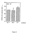
- NC-1, HNK-1 antibodies directed respectively from neural peak cell epitopes and human natural killer cells actually recognize the same epitopes and have been shown to recognize some immature cells of the chicken blastoderm disc. These two antibodies again recognize cells in alkaline phosphatase-active clusters.
- NC-1 The results with NC-1 are represented on the figure 6 .
- the cells are inoculated in a complete ESA medium with avian Stem Cell Factor (aSCF), bFGF (basic Fibroblast Growth Factor).
- IGF-1 Insulin-like Growth Factor-1
- ARMA antibody (1 ⁇ g / ml) on a mat of STO cells treated with mitomycin C as indicated in the section Materials and Methods.
- a new seeding is added to the culture.
- the cytokines LIF (Leukemia Inhibitory Factor), IL-11 (Interleukin 11) and IL-6 (Interleukin 6) are added in the medium at the concentrations indicated above. Double staining, alkaline phosphatase activity and detection of antibody epitopes is carried out according to the protocols presented above.
- the EMA-I antibody (Hahnel and Eddy, 1986) initially directed against epitopes present on the primordial cells of the murine germ line was used against these same cells in chicken.
- EMA-1 recognizes cells and colonies of cells that all have alkaline phosphatase activity. It was furthermore verified that this EMA-1 antibody only recognizes murine ES cells in their undifferentiated tolipotence state.
- the antibodies were tested either on undifferentiated cultures obtained as described in the material part and Methods as described above or on cultures which were treated with an excess of retinoic acid added to the culture (10 -6 M) for at least 48 hours.
- the table below indicates the state of recognition by the different antibodies used.
- Blastodermal discs of quail or chicken are seeded on STO feeder cell mats. After different days of culture, the cells are subcultured onto STO cell mats as described in the Materials and Methods section.
- the detection of cells and cells positive for both alkaline phosphatase activity and for the localization of ECMA-7 or NC-1 labeling suggests that the culture conditions are defined to maintain the secondary and tertiary cultures of the cells. totipotent cells.
- the transplanting process also ensures, after the first pass, homogeneity to the whole culture, both morphologically, and by the detection of the different epitopes.
- the mass of cells become very extensive and homogeneous, character increased by the large capacity of these cells to divide rapidly, unlike the differentiated cells initially present in the primary culture. So far these identification and characterization criteria can be used and detected for at least 5 weeks after seeding.
- the chicken blastoderm cells obtained in primary cultures or after successive subcultures can be injected into recipient embryos.
- the cells maintained in culture come from a pigmented strain and the recipient embryos from an unpigmented strain.
- the cells maintained in cultures are dissociated and prepared as described in the Materials and Methods part according to the same method as for a subculture.
- the cell suspension is then prepared in a proportion of 1 to 3 ⁇ 10 5 cells per ml of ESA medium.
- the freshly laid, non-incubated egg containing the reviving embryo is slightly irradiated between 5 Gy and 7 Gy. A small window of a few mm 2 is made in the recipient's shell by grinding.
- the shell membrane is cut with a scalpel and the cells are injected with a capillary stretched in the subgerminal cavity of the blastodermic disc in a volume of 1 to 5 ⁇ l, which corresponds to 100 to 1500 cells maximum.
- the average of the injected cells is 500 cells.
- the window is then covered with shell membranes and sealed.
- a piece of adhesive bandage is applied to perfect the seal and to limit the evaporation as much as possible. After 4 days of incubation under optimal conditions, the eggs are opened and the well-developed embryos are transferred to a larger shell and incubated to finish their development satisfactorily.
Abstract
Description
La présente invention se rapporte à l'obtention de cellules ES aviaires , en particulier à un procédé de culture et à un milieu permettant la culture de ces cellules.The present invention relates to obtaining avian ES cells, in particular to a culture method and to a medium for culturing these cells.
En effet, dans le cadre de la mise au point de technique de production de protéines recombinantes, le développement d'une technique de transgénèse chez les oiseaux domestiques aura des retombées économiques extrêmement importantes dans deux applications majeures:
- 1. le développement de souches aviaires présentant des caractères génétiques déterminés (résistance à certaines maladies, performances de croissance, etc)
- 2. le développement de systèmes de production de protéines recombinantes dans l'albumen de l'oeuf.
- 1. the development of avian strains with certain genetic characteristics (resistance to certain diseases, growth performance, etc.)
- 2. The development of recombinant protein production systems in the egg albumen.
L'industrie biotechnologique s'intéresse de plus en plus à la possibilité de produire des protéines d'intérêt dans des fluides biologiques ou des organismes (sang, lait, plantes, ...). La production de telles protéines dans l'oeuf d'oiseau domestique constituera certainement dans cette voie une avancée technologique majeure pour plusieurs raisons:
- de nombreuses protéines de mammifères ne peuvent être produites en système mammifère car leur surabondance dans ces organismes présente des effets délétères (exemple: l'érythropoiétine qui chez le lapin induit des hyperglobulinémies pathologiques). Beaucoup de ces protéines d'intérêt ne présentent pas d'activité croisée avec celles des oiseaux, autorisant ainsi leur surproduction dans un organisme aviaire sans effet pathologique majeur;
- il est très vraisemblable que la commercialisation de protéines recombinantes produites chez des mammifères se heurtera à des problèmes sanitaires liés à la présence chez cette espèces d'organismes latents potentiellement pathogènes pour l'Homme (lentivirus, prions,...). Ce risque est très minime, pour ne pas dire quasiment inexistant, pour des agents pathogènes des oiseaux domestiques;
- l'oeuf constitue un "tissu" très dense en un petit nombre de protéines. Par exemple la protéine majeure de l'oeuf d'oiseau, l'ovalbumine représente 54 % des protéines du blanc d'oeuf, soit un poids sec moyen par oeuf de 2 grammes de matière sèche environ. On peut raisonablement imaginer produire par oeuf au moins 10% de cette masse en protéine recombinante. La rentabilité économique apparait très grande si l'on considère qu'une poule pond en moyenne 2 oeufs tous les trois jours, et cette rentabilité apparait très supérieure à celle de grands mammifères si l'on considère les coûts d'élevage bien moindres des oiseaux domestiques.
- many mammalian proteins can not be produced in the mammalian system because their superabundance in these organisms has deleterious effects (example: erythropoietin which in the rabbit induces pathological hyperglobulinemias). Many of these proteins of interest do not show cross activity with those of birds, thus allowing their overproduction in an avian organism without major pathological effect;
- it is very likely that the commercialization of recombinant proteins produced in mammals will encounter health problems related to the presence in this species of latent organisms potentially pathogenic for humans (lentiviruses, prions, etc.). This risk is very minimal, not to say almost non-existent, for pathogens of domestic birds;
- the egg constitutes a "tissue" very dense in a small number of proteins. For example, the major protein of the egg of a bird, ovalbumin represents 54% of the proteins of the egg white, that is to say an average dry weight per egg of about 2 grams of dry matter. It is reasonable to imagine producing at least 10% of this mass of recombinant protein per egg. The economic profitability appears very large considering that a chicken lays an average of 2 eggs every three days, and this profitability appears to be much higher than that of large mammals if we consider the much lower costs of breeding birds. servants.
La réalisation d'oiseaux transgéniques est possible actuellement avec un coût extrêmement élevé à cause de sa très faible efficacité. En effet chez les oiseaux la technique de microinjection d'ADN dans l'oeuf est quasiment impossible. D'autre part l'utilisation du système des rétrovirus vecteurs, le seul sytème efficace à ce jour, reste complexe et se heurtera certainement à une réticence de la part des industriels pour des raisons sanitaires.The realization of transgenic birds is currently possible with an extremely high cost because of its very low efficiency. Indeed in birds the technique of microinjection of DNA into the egg is almost impossible. On the other hand, the use of the system of vector retroviruses, the only effective system to date, remains complex and will certainly encounter a reluctance on the part of industrialists for health reasons.
Une avancée très importante à la réalisation d'animaux transgéniques a été apportée chez la souris par le développement de la technologie des cellules ES.A very important advance in the realization of transgenic animals has been brought to the mouse by the development of ES cell technology.
Les cellules ES (pour Embryonic Stem cells) sont des cellules embryonnaires totipotentes capables de régénérer tous les tissus de l'embryon, y compris le tissu germinal, après leur injection dans des embryons très précoces. Ces cellules peuvent donc être considérées comme des chevaux de Troie pour introduire de nouvelles informations génétiques dans le patrimoine génétique d'un animal. La possibilité de cultiver ces cellules à long terme in vitro, offre la possibilité d'exercer de nombreux contrôles avant leur implantation in vivo. D'autre part ces cellules peuvent être conservées de façon illimitée dans l'azote liquide, ce qui constitue une possibilité de stockage d'un patrimoine génétique.ES cells (Embryonic Stem cells) are totipotent embryonic cells capable of regenerating all embryonic tissues, including germinal tissue, after injection into very early embryos. These cells can therefore be considered as Trojan horses to introduce new genetic information into the genetic heritage of an animal. The possibility of growing these cells in vitro in the long term offers the possibility of carrying out numerous controls before they are implanted in vivo. On the other hand these cells can be kept unlimitedly in liquid nitrogen, which is a possibility of storing a gene pool.
L'utilisation de cellules ES constitue aujourdhui chez les oiseaux domestiques la voie la plus prometteuse pour la réalisation efficace d'animaux transgéniques.The use of ES cells is nowadays the most promising avenue for domestic birds for the efficient production of transgenic animals.
La Demande
Des travaux récents d'un groupe canadien (R. Etches à la station de Guelph) ont suggéré que des cellules ES doivent exister dans l'embryon d'oiseau (Petitte et al., 1990). Ce groupe à réussi la transplantation de telles cellules dans des embryons, et par suite, la production d'animaux dont le patrimoine génétique est dérivé des cellules greffées. Cependant à ce jour la culture de ces cellules in vitro n'a pas pu être réussie ; par conséquent ces cellules n'ont pas pu être utilisées pour transférer de façon stable un transgène. C'est là un blocage majeur à l'exploitation de la technologie des cellules ES chez les oiseaux. Les cellules ES peuvent être caractérisées par trois types de critères essentiels :
- morphologie
- activité phosphatase alcaline endogène
- réaction avec des anticorps spécifiques d'un état de totipotence (ECMA-7, SSEA-1 et SSEA-3 notamment).
Recent work by a Canadian group (R. Etches at the Guelph station) has suggested that ES cells must exist in the bird embryo (Petitte et al., 1990). This group has succeeded in transplanting such cells into embryos, and hence the production of animals whose genetic heritage is derived from grafted cells. However to date the culture of these cells in vitro could not be successful; therefore these cells could not be used to stably transfer a transgene. This is a major block to the exploitation of ES cell technology in birds. ES cells can be characterized by three types of essential criteria:
- morphology
- endogenous alkaline phosphatase activity
- reaction with specific antibodies of a totipotency state (ECMA-7, SSEA-1 and SSEA-3 in particular).
Aucune culture de cellules ES identifiées par l'ensemble de ces caractéristiques n'a pu être obtenue à ce jour.No ES cell cultures identified by all of these characteristics could be obtained to date.
C'est pourquoi la présente invention a pour objet un milieu de culture de cellules embryonnaires totipotentes aviaires du type comportant un milieu de culture pour cellules aviaires .This is why the present invention relates to an avian totipotent embryonic cell culture medium of the type comprising a culture medium for avian cells.
Le cas écheant, le milieu de culture est substantiellement dépourvu d'acide rétinoïque actif.In this case, the culture medium is substantially free of active retinoic acid.
De manière avantageuse, l'acide rétinoïque est substantiellement inactivé par des anticorps anti-acide rétinoïque (ARMA) présents dans le milieu.Advantageously, the retinoic acid is substantially inactivated by anti-retinoic acid (ARMA) antibodies present in the medium.
En effet, les milieux employés contiennent souvent du sérum, dont on ne peut contrôler la quantité endogène d'acide rétinoïque. En testant l'effet de l'incorporation au milieu de culture d'un anticorps monoclonal anti-acide rétinoïque qui neutraliserait l'action de ce dernier, sur la différenciation des cellules, la Demanderesse a constaté que la présence de cet anticorps accroit la présence dans les cultures de cellules et colonies à activité phosphatase alcaline.In fact, the media used often contain serum, whose endogenous quantity of retinoic acid can not be controlled. By testing the effect of the incorporation into the culture medium of a monoclonal anti-retinoic acid antibody which would neutralize the action of the latter on the differentiation of the cells, the Applicant has found that the presence of this antibody increases the presence in cell cultures and colonies with alkaline phosphatase activity.
plus spécifiquement, la présente invention a pour objet un
- Procédé de culture de cellules embryonnaires totipotentes aviaires (ou cellules ES aviaires), caractérisé en ce que :
- 1) On met en suspension des cellules provenant de disques biastodemiques d'oeufs d'oiseau fécondés dans un milieu de culture de cellules embryonnaires totipotentes aviaires, du type comportant un milieu de culture pour cellules aviaires, comprenant :
- a) b-FGF, SCF et LIF, ou
- b) b-FGF, aSCF (avian SCF), LIF, IL-11, IL-6, et IGF-1, ou
- c) b- FGF, aSCF (avian SCF), LIF et IGF-1.
(bFGF= basic Fibroblast Growth Factor ; SCF=Stem Cell Factor ; LIF=Leukemia Inhibitory Factor ;IL 11=Interleukine 11; IL6 =Interleukin 6 ; IGF-1 = Insulin-like Growth Factor -1 ; aSCF = avian Stem Cell Factor)
- 2) On ensemence un tapis de cellules nourricières avec la suspension obtenue à l'issue de l'étape a),
- 3) On met les cellules à incuber,
- 4) Les cellules en culture sont prélevées et purifiées afin de récupérer des cellules ES aviaires.
- 1) On met en suspension des cellules provenant de disques biastodemiques d'oeufs d'oiseau fécondés dans un milieu de culture de cellules embryonnaires totipotentes aviaires, du type comportant un milieu de culture pour cellules aviaires, comprenant :
- Avantageusement, dans la procédé de culture selon l'invention entre les étapes 3) et 4), on effectue une ou plusieurs additions échelonnées dans le temps, de milieu neuf identique à celui utilisé dans l'étape 1).
- Avantageusement, dans la procédé de culture selon l'invention le milieu de culture est essentiellement dépourvu d'acide rétinoïque actif.
- Avantageusement, dans la procédé de culture selon l'invention le milieu de culture comporte un tapis de cellules nourricières.
- Avantageusement, dans la procédé de culture selon l'invention entre les étapes 3) et 4), on effectue l'addition de milieu neuf au 3ème jour, puis tous les jours.
- Avantageusement, dans la procédé de culture selon l'invention l'étape 4) est effectuée par traitement enzymatique, lavage dans un milieu ne contenant pas de facteur de croissance et centrifugation.
- Avantageusement, dans la procédé de culture selon l'invention à l'issue de l'étape 4), on effectue une étape 5) dans laquelle, les cellules ES sont réensemencées sur un tapis de cellules nourricières. en présence dudit milieu de culture, de manière à obtenir une culture secondaire.
- Avantageusement, dans la procédé de culture selon l'invention les étapes 4) et 5) sont répétées plusieurs fois.
- Avantageusement, dans la procédé de culture selon l'invention,entre les étapes 3) et 4) on effectue une ou plusieurs additions échelonnées dans le temps, le milieu neuf identique à celui utilisé dans l'étape 1).
- Avantageusement, dans la procédé de culture selon l'invention au cours de l'étape 3), on effectue un réensemencement du milieu par une suspension de cellules identique à la suspension préparée à l'étape 1).
- A method for culturing avian totipotent embryonic cells (or avian ES cells) characterized in that:
- 1) Cells from biastodemic disks of fertilized bird eggs are suspended in an avian totipotent embryonic cell culture medium, of the type comprising an avian cell culture medium, comprising:
- a) b-FGF, SCF and LIF, or
- b) FGF, aSCF (avian SCF), LIF, IL-11, IL-6, and IGF-1, or
- c) b- FGF, aSCF (avian SCF), LIF and IGF-1.
(bFGF = basic Fibroblast Growth Factor, SCF = Stem Cell Factor, LIF = Leukemia Inhibitory Factor,IL 11 =Interleukin 11, IL6 =Interleukin 6, IGF-1 = Insulin-like Growth Factor -1, aSCF = Avian Stem Cell Factor)
- 2) a carpet of feeder cells is seeded with the suspension obtained at the end of step a),
- 3) The cells are incubated,
- 4) Cells in culture are removed and purified to recover avian ES cells.
- 1) Cells from biastodemic disks of fertilized bird eggs are suspended in an avian totipotent embryonic cell culture medium, of the type comprising an avian cell culture medium, comprising:
- Advantageously, in the culture process according to the invention between steps 3) and 4), one or more additions are carried out in time, new medium identical to that used in step 1).
- Advantageously, in the culture method according to the invention the culture medium is essentially free of active retinoic acid.
- Advantageously, in the culture process according to the invention the culture medium comprises a carpet of feeder cells.
- Advantageously, in the culture process according to the invention between steps 3) and 4), is carried out the addition of fresh medium on the 3rd day, then every day.
- Advantageously, in the method of culture according to the invention step 4) is carried out by enzymatic treatment, washing in a medium containing no growth factor and centrifugation.
- Advantageously, in the culture process according to the invention at the end of step 4), a step 5) is carried out in which the ES cells are reseeded on a carpet of feeder cells. in the presence of said culture medium, so as to obtain a secondary culture.
- Advantageously, in the culture process according to the invention steps 4) and 5) are repeated several times.
- Advantageously, in the culture method according to the invention, between steps 3) and 4) one or more additions are carried out in time, the new medium identical to that used in step 1).
- Advantageously, in the culture process according to the invention during step 3), the medium is reseeded with a suspension of cells identical to the suspension prepared in step 1).
Le milieu de l'étape 1) contient de préférence les éléments suivants:
- b-FGF, a-SCF, IGF-1, LIF, IL-11, IL-6 et anticorps anti-acide rétinoïque.
- Sérum foetal de bovin.
- Sérum de poule
- Conalbumine
- Acides aminés non essentiels
- Pyruvate de sodium
- Stock de nucléosides
- Hepes (1M)
- β-mercaptoéthanol
- Penicilline
- Streptomycine
- Gentamycine
avec le stock de nucléosides.constitué du mélange :- adénosine, guanosine, cytidine, uridine et thymidine en solution aqueuse.
- b-FGF, α-SCF, IGF-1, LIF, IL-11, IL-6 and retinoic acid antibodies.
- Fetal serum of cattle.
- Hen serum
- conalbumin
- Non-essential amino acids
- Sodium pyruvate
- Stock nucleosides
- Hepes (1M)
- β-mercaptoethanol
- Penicillin
- Streptomycin
- gentamicin
with the nucleoside stock.constituted from the mixture:- adenosine, guanosine, cytidine, uridine and thymidine in aqueous solution.
De manière facultative, au cours du procédé selon l'invention, on effectue entre les étapes 3) et 4) l'addition de milieu neuf au 3ème jour puis le milieu est changé tous les jours jusqu'au prochain repiquage.Optionally, during the process according to the invention, between steps 3) and 4) the addition of new medium is carried out at the 3rd day and then the medium is changed every day until the next subculture.
L'étape 4) peut notamment être effectuée par traitement enzymatique, lavage dans un milieu ne contenant pas de facteur de croissance et centrifugation.Step 4) may in particular be carried out by enzymatic treatment, washing in a medium containing no growth factor and centrifugation.
On peut recueillir directement les cultures primaires de cellules, qui seront ensuite congelées, ou bien réaliser des cultures secondaires successives à partir des cellules de la culture primaire. Dans ce cas, à l'issue de l'étape 4), on effectue une étape 5) dans laquelle, les cellules ES sont réensemencées sur un tapis de cellules nourricières. ou sur boites gélatinées, de manière à obtenir une culture secondaire.The primary cultures of cells can be collected directly, which will then be frozen, or alternatively produce successive secondary cultures from the cells of the primary culture. In this case, at the end of step 4), a step 5) is carried out in which the ES cells are reseeded on a carpet of feeder cells. or on gelatinized dishes, so as to obtain a secondary culture.
Les étapes 4) et 5) peuvent être répétées plusieurs fois pour avoir des cultures tertiaires et successives.Steps 4) and 5) can be repeated several times to have tertiary and successive cultures.
Le tapis de cellules nourricières peut être constitue de différents types de cellules décrits précédemment, notamment de cellules STO mitomycinées ou irradiées.The feeder cell mat may consist of different types of cells previously described, including mitomycin-irradiated or irradiated STO cells.
Un autre des objets de l'invention est une culture de cellules ES aviaires, susceptibles d'être obtenues par le procédé de culture selon l'invention défini ci-dessus et comme defini par la revendication 9. Une cellule embryonnaire totipotente aviaire modifiée peut dire obtenue par intégration du gène codant pour une protéine hétérologue dans le génome d'une cellule ES aviaire en culture.Another object of the invention is an avian ES cell culture, obtainable by the culture method according to the invention defined above and as defined by
La présente invention a également pour objet une
Culture de cellules ES aviaires in vitro susceptible d'être obtenue par le procédé de culture selon l'invention ; ladite culture comprenant un milieu de culture de cellules embryonnaires totipotentes aviaires comprenant :
- a) b-FGF, SCF et LIF, ou
- b) b-FGF, aSCF (avian SCF), LIF, IL-II, IL-6, et IGF-1, ou
- c) b-FGF, aSCF (avian SCF), LIF et IGF-1.
- (bFGF= basic Fibroblast Growth Factor ; SCF=Stem Cell Factor ; LIF=Leukemia Inhibitory Factor ;
IL 11=Interleukine 11; IL6 =Interleukin 6 ; IGF-1 = Insulin-like Growth Factor -1 ; aSCF = avian Stem Cell Factor)
- Avantageusement la Culture de cellules ES aviaires selon invention présente 2 à 3 fois plus de colonies positives pour l'activité alcaline phosphatase par rapport au fond constitué en majorité de cellules faiblement positives.
- Avantageusement, dans la culture de cellules ES aviaires selon l'invention les cellules réagissent spécifiquement avec au moins un anticorps sélectionné parmi ECMA-7, SSEA-1, SSEA-3, TEC-01, EMA-1 et EMA-6.
- Avantageusement, dans la culture de cellules ES aviaires selon l'invention les cellules ne réagissent pas avec l'anticorps TROMA-1.
- Avantageusement, dans la culture de cellules ES aviaires selon l'invention les cellules sont modifiées par intégration du gène codant pour une protéine hétérologue.
In vitro avian ES cells culture obtainable by the culture method according to the invention; said culture comprising an avian totipotent embryonic cell culture medium comprising:
- a) b-FGF, SCF and LIF, or
- b) FGF, aSCF (avian SCF), LIF, IL-II, IL-6, and IGF-1, or
- c) b-FGF, aSCF (avian SCF), LIF and IGF-1.
- (bFGF = basic Fibroblast Growth Factor, SCF = Stem Cell Factor, LIF = Leukemia Inhibitory Factor,
IL 11 =Interleukin 11, IL6 =Interleukin 6, IGF-1 = Insulin-like Growth Factor -1, aSCF = Avian Stem Cell Factor)
- Advantageously, the culture of avian ES cells according to the invention has 2 to 3 times more positive colonies for the alkaline phosphatase activity than the background consisting mainly of weakly positive cells.
- Advantageously, in the culture of avian ES cells according to the invention, the cells specifically react with at least one antibody selected from ECMA-7, SSEA-1, SSEA-3, TEC-01, EMA-1 and EMA-6.
- Advantageously, in the culture of avian ES cells according to the invention, the cells do not react with the TROMA-1 antibody.
- Advantageously, in the culture of avian ES cells according to the invention the cells are modified by integration of the gene coding for a heterologous protein.
Le facteur de croissance des cellules souches (ou SCF) est de préférence l'a-SCF (ou avian Stem Cell Factor) et le m-SCF (ou murine Stem Cell Factor).Stem cell growth factor (SCF) is preferably α-SCF (or avian Stem Cell Factor) and m-SCF (or murine Stem Cell Factor).
L'un des aspects préférés de l'invention concerne un milieu de culture qui contient, outre les éléments nutritifs de base nécessaires à la croissance de cellules, une combinaison de b-FGF, SCF et LIF. En outre, la présence dans le milieu d'un anticorps monoclonal neutralisant l'activité de différenciation exercée par l'acide rétinoïque augmente le nombre de cellules souches embryogènes totipotentes.One of the preferred aspects of the invention relates to a culture medium which contains, in addition to the basic nutrients necessary for cell growth, a combination of b-FGF, SCF and LIF. In addition, the presence in the medium of a monoclonal antibody neutralizing the differentiation activity exerted by retinoic acid increases the number of totipotent embryogenic stem cells.
La présence d'un tapis de cellules nourricières favorise la croissance de cellules ES aviaires. Divers type de cellules connues de l'homme du métier peuvent-être utilisées : on peut citer en particulier des cellules telles que les cellules STO, traitées à la miromycine ou irradiées, les cellules GRL-3A, les cellules LMH, les cellules QT6 et cellules QT6 modifiées tcllcs que les cellules QT6 isolde, les cellules différenciées établies en lignée à partir des cultures de cellules souches embryonnaires induites à différencier.The presence of a carpet of feeder cells promotes the growth of avian ES cells. Various types of cells known to those skilled in the art can be used: mention may in particular be made of cells such as STO cells, treated with miromycin or irradiated cells, GRL-3A cells, LMH cells, QT6 cells and QT6 cells altered tcllcs that QT6 cells are isolated, differentiated cells established in lineage from embryonic stem cell cultures induced to differentiate.
Les cellules STO sont des fibroblastes d'embryons de souris (catalogue ATCC) : les cellules BRL-3A (catalogue ATCC) sont des cellules de foie de "Buffalo rat liver". Les cellules QT6 (catalogue ATCC) et cellules QT6 modifiées telles que les cellules QT6 isolde sont des fibroblastes de caille (
Le milieu de culture contient en outre différents éléments nutritifs essentiels et des antibiotiques.The culture medium further contains various essential nutrients and antibiotics.
Un milieu de culture particulièrement adapté à la présente invention possède la composition suivante :
De manière avantageuse la concentration en bFGF est supérieure à 5 ng/ml et la concentration en IGF-1 est supérieure à 10 ng/ml.
avec le stock de nucléosides constitué du mélange :
et convient à la culture de cellules embryonnaires totipotentes d'oiseau.Advantageously, the concentration of bFGF is greater than 5 ng / ml and the concentration of IGF-1 is greater than 10 ng / ml.
with the nucleoside stock consisting of the mixture:
and is suitable for growing totipotent embryo bird cells.
Le milieu BHK21 (ou milieu MEM) est un milieu de culture qui a été décrit notamment par
Hepes est de l'hydroxy-éthyl-pipérazine-éthane-sulfonate.Hepes is hydroxy-ethyl-piperazine-ethanesulfonate.
Enfin, un procédé de production d'une protéine recombinante, caractérisé en ce qu'on intègre le gène codant pour ladite protéine dans le génome d'une cellule embryonnaire totipotente aviaire en culture est divulgué.Finally, a method for producing a recombinant protein, characterized in that the gene coding for said protein is integrated into the genome of an avian totipotent embryonic cell in culture is disclosed.
La Demanderesse a mis au point un milieu de culture et des conditions de culture in vitro permettant de maintenir en culture des cellules aviaires qui présentent des propriétés morphologiques, cinétiques et histochimiques rappelant celles des cellules embryonnaires totipotentes. Ces observations ont été effectuées aussi bien avec des cellules dérivant de disques blastodermiques de caille que de poulet. La croissance de ces cellules en culture in vitro est rendue possible par la mise au point d'un milieu original spécialement adapté à la culture de cellules embryonnaires d'oiseau. On sait que la présence, le maintien et la propagation de cellules totipotentes en culture permettent leur injection dans des embryons receveurs. La contribution à la morphogenèse des tissus somatiques et germinaux chez les animaux receveurs grâce à un caractère totipotent, peut conduire à l'obtention d'animaux transgèniques.The Applicant has developed a culture medium and in vitro culture conditions for maintaining in culture avian cells which have morphological, kinetic and histochemical properties reminiscent of totipotent embryonic cells. These observations were made with cells derived from both quail and chicken blastoderm discs. The growth of these cells in vitro culture is made possible by the development of an original medium specially adapted to the culture of embryonic bird cells. It is known that the presence, maintenance and propagation of totipotent cells in culture allow their injection into recipient embryos. The contribution to the morphogenesis of somatic and germinal tissues in the recipient animals by virtue of a totipotent character can lead to the production of transgenic animals.
Les exemples qui suivent sont destinés à illustrer l'invention sans aucunement en limiter la portée. Dans ces exemples, on se référera aux figures suivantes :
-
Figure 1 : Effet des combinaisons de facteurs- blastodermes de caille, 0,75 bl/ml
- fond de gélatine
- culture de 3 j
-
Figure 2 : effet de l'anticorps anti-acide rétinoïque (ARMA)- blastodermes de caille, 0,75 bl/ml
- fond avec ou sans gélatine
- culture de 4 j
-
Figure 3 : comparaison de différentes cytokines- blastodermes de caille, 2 bl/ml
- fond avec gélatine
- culture de 2 + 3 j
-
Figure 4 : Comparaison d'un ensemencement sur gélatine et sur tapis de cellules traitées à la mitomycine C en présence de différentes cytokines appartenant toutes à la même famille.- 4A: - blastodermes de caille, 1+1,5 bl/ml
- fond avec gélatine
- culture de 3 + 4 j
- 4B: - blastodermes de caille, 1+1,5 bl/ml
- fond avec cellules STO
- culture de 3 +.4 j
- 4A: - blastodermes de caille, 1+1,5 bl/ml
-
Figure 5 : Activité phosphatase alcaline et reconnaissance par ECMA-7- blastodermes de caille, 1,5 bl/ml
- fond avec cellules STO
- culture de 2 + 3 j
-
Figure 6 : Activité phosphatase alcaline et reconnaissance par NC-1- blastodermes de caille, 1,5 bl/ml
- fond avec cellules STO
- culture de 2 + 3 j
-
Figure 7 : Animaux chimères obtenus par injection in ovo dans des embryons de cellules maintenues en culture. Cellules injectées après 8ou 10 jours de culture.
-
Figure 1 : Effect of combinations of factors- blastoderms of quail, 0.75 bl / ml
- gelatin background
- 3 days culture
-
Figure 2 : effect of anti-retinoic acid antibody (ARMA)- blastoderms of quail, 0.75 bl / ml
- background with or without gelatin
- 4 days culture
-
Figure 3 : comparison of different cytokines- blastoderms of quail, 2 bl / ml
- background with gelatin
- culture of 2 + 3 days
-
Figure 4 : Comparison of gelatin seeding and mitomycin C treated mats in the presence of different cytokines belonging to the same family.- 4A : - Quail blastoderms, 1 + 1.5 bl / ml
- background with gelatin
- culture of 3 + 4 days
- 4B: - quail blastoderms, 1 + 1.5 bl / ml
- background with STO cells
- culture of 3 + 4 days
- 4A : - Quail blastoderms, 1 + 1.5 bl / ml
-
Figure 5 : Alkaline phosphatase activity and recognition by ECMA-7- blastoderms of quail, 1.5 bl / ml
- background with STO cells
- culture of 2 + 3 days
-
Figure 6 : Alkaline phosphatase activity and recognition by NC-1- blastoderms of quail, 1.5 bl / ml
- background with STO cells
- culture of 2 + 3 days
-
Figure 7 : Chimeric animals obtained by in ovo injection in cell embryos maintained in culture. Cells injected after 8 or 10 days of culture.
Les oeufs de poules fraichement pondus, non incubés, correspondent au stade X de développement (Eyal Giladi and Kovak, 1976) ; les oeufs de caille " C. coturnix japonica" sont également utilisés dès la ponte et non incubés.Freshly laid eggs, not incubated, correspond to stage X of development (Eyal Giladi and Kovak, 1976); the quail eggs "C. coturnix japonica" are also used as soon as they are laid and not incubated.
Le disque blastodermique (3-4 mm de diamètre pour la poule, 2-2,5 mm pour la caille) est prelevé à l'aide d'une pipette pasteur dans du milieu complet sans facteurs. Les cellules sont centrifugées à 200 g, lavées deux fois dans du milieu afin d'éliminer le maximum de vitellus contaminant, resuspendues à raison de 2 disques pour 1 ml de milieu et dissociées mécaniquement par passage dans une aiguille de 23 G. Les facteurs sont alors ajoutés.The blastodermic disk (3-4 mm diameter for the hen, 2-2.5 mm for the quail) is taken using a pasteur pipette in complete medium without factors. The cells are centrifuged at 200 g, washed twice in medium in order to eliminate the maximum of contaminating yolk, resuspended at the rate of 2 disks per 1 ml of medium and dissociated mechanically by passage in a 23 G needle. The factors are then added.
La suspension cellulaire est déposée:
- soit sur boîtes ou puits (Costar) préalablement gélatinés (0,2 % gélatine, 1 h à t° ambiante),
- soit sur un tapis de cellules STO préalablement traitées à la mitomycine C (90 min, 37°c, 5 µg/ml) et ré-ensemencées à raison de 105 cellules / cm2,
- soit sur un tapis de cellules isolde préalablement traitées à la mitomycine C (90 min, 37°c, 5 µg/ml) et ré-ensemmencées à raison de 105 cellules / cm 2.
- either on boxes or wells (Costar) previously gelatinized (0.2% gelatin, 1 h at room temperature),
- or on a carpet of STO cells previously treated with mitomycin C (90 min, 37 ° C., 5 μg / ml) and re-seeded at a rate of 10 5 cells / cm 2 ,
- either on a carpet of isolde cells previously treated with mitomycin C (90 min, 37 ° C., 5 μg / ml) and reseeded at a rate of 10 5 cells / cm 2.
Les drogues de sélection sont ajoutées en entretien mais enlevées deux jours avant le traitement à la mitomycine C.Selection drugs are added for maintenance but removed two days before mitomycin C treatment.
Dans tous les cas, un second ensemencement est réalisé dans les mêmes conditions après deux jours de culture.In all cases, a second seeding is carried out under the same conditions after two days of culture.
Les cultures sont incubées à 37 °C ou à 41' C, dans une atmosphère contrôlée en CO2 (7,5 %) et leur évolution est suivie au microscope en contraste de phase. Une addition partielle (50 %) de milieu neuf avec les facteurs est réalisée le 3éme jour de culture, puis le milieu est changé tous les jours. A chaque moment, les cellules en croissance peuvent étre soit fixées pour étude, soit prélevées pour être ré-ensemencées en culture secondaire ou supérieure, sur tapis de cellules STO mitomycinées irradiées ou sur boîtes gélatinées.The cultures are incubated at 37 ° C. or 41 ° C. in a CO 2 controlled atmosphere (7.5%) and their evolution is monitored under a phase contrast microscope. A partial addition (50%) of new medium with the factors is carried out on the 3 rd day of culture, then the medium is changed every day. At any time, the growing cells can either be fixed for study or removed for re-seeding in secondary or higher cultures, on irradiated irradiated STO cells or on gelatinized dishes.
En cas de fixation, les cellules sont lavées en Tris-Glucose deux fois, puis fixées in situ 15 min dans une solution de paraformaldéhyde 4 % à froid (0-4 °C). Après plusieurs lavages au PBS, différentes colorations peuvent être réalisées selon l'un des protocoles suivants :
* détection de l'activité phosphatase alcaline endogène,
** détection de l'activité de β-galactosidase exogêne
*** détection par immunocytochimie de la présence d'épitopes spécifiques (réaction à 4°C)
blocage en tampon PBS - BSA (1 mg/ml)
lavage en PBS - BSA
anticorps primaire 1/10ème ou 1/50ème
anticorps secondaire fluorescent 1/ 50éme la détection est réalisée sous microscope inversé à fluorescence.In case of fixation, the cells are washed with Tris-Glucose twice and then fixed in situ for 15 min in a 4% paraformaldehyde solution in the cold (0-4 ° C.). After several washings with PBS, different colorations can be carried out according to one of the following protocols:
* detection of endogenous alkaline phosphatase activity,
** detection of exogenous β-galactosidase activity
*** immunocytochemical detection of the presence of specific epitopes (reaction at 4 ° C)
blocking in PBS-BSA buffer (1 mg / ml)
washing in PBS - BSA
fluorescent
En cas de passage en culture secondaire ou successive, les cellules sont lavées en Tris-Glucose deux fois, puis incubées 10-30 min dans une solution enzymatique. On peut utiliser une solution de collagénase-dispase (1 mg/ml soit 1 U/ml final) à laquelle une solution de hyaluronidase (1 mg/ml final soit 1 U/ml) peut être ajoutée : on peut également utiliser une solution de pronase à 0.25 mg/ml final. Les cellules ou les petits amas de cellules ainsi isolés enzymatiquement sont lavées en milieu ESA, resuspendus, déposés sur un coussin de milieu de séparation de lymphocyte de densité (d= 1,077-1,080) et centrifugés 20 min à t° ambiante à 800 g afin de débarasser les cellules non différenciées de blatodermes des cellules du tapis, des débris divers et des restes de vitellus contaminants. L'interface est alors prélevée, lavée deux fois en milieu ESA. Le culot cellulaire obtenu est resuspendu et dissocié légérement mécaniquement avant d'être ensemencé sur un nouveau tapis de cellules nourricières, comme précédemment décrit. L'équivalent de 6 disques blastodermiques initiaux est ré-ensemencé dans 2 ml. Cette étape de gradient n'est parfois pas nécessaire lors des passages successifs, en fonction de la très grande homogénéité des cultures obtenues.In case of passage in secondary or successive culture, the cells are washed with Tris-Glucose twice and then incubated for 10-30 min in an enzymatic solution. It is possible to use a collagenase-dispase solution (1 mg / ml or 1 U / ml final) to which a solution of hyaluronidase (1 mg / ml final or 1 U / ml) can be added: it is also possible to use a solution of pronase at 0.25 mg / ml final. The cells or small clusters of cells thus isolated enzymatically are washed in ESA medium, resuspended, deposited on a density cell separation medium cushion (d = 1.077-1.080) and centrifuged for 20 min at room temperature at 800 g to remove undifferentiated blatoderm cells from the cells of the carpet, various debris and remains of yolk contaminants. The interface is then removed, washed twice in ESA medium. The cell pellet obtained is resuspended and dissociated slightly mechanically before being seeded on a new carpet of feeder cells, as previously described. The equivalent of 6 initial blastoderm disks is re-seeded in 2 ml. This gradient step is sometimes not necessary during successive passages, depending on the very high homogeneity of the cultures obtained.
Les cellules dissociées peuvent être déposées sur un gradient multicouche de Percoll et centrifugées dans les mêmes conditions. Les interfaces sont alors prélevées, lavées dans un milieu ESA et les cellules les plus immatures des interfaces supérieures réensemencées, ou injectées dans des embryons receveurs.The dissociated cells can be deposited on a multilayer gradient of Percoll and centrifuged under the same conditions. The interfaces are then removed, washed in an ESA medium and the most immature cells of the upper interfaces reseeded, or injected into recipient embryos.
A l'issue de la culture primaire ou successive, les cellules récupérées de gradient peuvent être congelées dans un mélange constitué de 40 % FBS, 50 % milieu ESA et 10 % DMSO. Les cellules équivalant à 24 blastodisques initiaux sont reprises dans 0,5 ml de milieu ESA, resuspendues et 0,4 ml de scrum est ajouté. 0,1 ml de DMSO est alors ajouté très lentement. La suspension de congélation est répartie dans des tubes à congélation (0,5 ml/tube) et congelée lentement à -80 'c avant d'être transférée dans l'azote liquide.At the end of the primary or successive culture, the recovered gradient cells can be frozen in a mixture consisting of 40% FBS, 50% ESA medium and 10% DMSO. The cells equivalent to 24 initial blastodiscs are taken up in 0.5 ml of ESA medium, resuspended and 0.4 ml of scrum is added. 0.1 ml of DMSO is then added very slowly. The freeze suspension is distributed in freezing tubes (0.5 ml / tube) and slowly frozen at -80 ° C. before being transferred into liquid nitrogen.
Un milieu de base appelé milieu "ESA" pour "Embryonic Stem cells Avian" dérivant d'un milieu utilisé pour les cellules ES murines a été préparé. I1 présente la composition suivante :A base medium called "ESA" medium for "Embryonic Stem cells Avian" derived from a medium used for murine ES cells was prepared. I1 has the following composition:
A ce milieu de base "ESA", des facteurs de croissance ont été ajoutés afin de comparer leur contribution respective à la formation de colonies présentant un caractère morphologique et biochimique intéréssant. Leurs concentrations sont indiquées ci-après :To this "ESA" background medium, growth factors were added in order to compare their respective contribution to colony formation of a morphological and biochemical character of interest. Their concentrations are indicated below:
Le premier critère utilisé pour évaluer l'effet de ces facteurs et des modifications apportées au milieu a été la détection par coloration biochimique de l'activité phosphatase alcaline endogène qui semble spécifique d'un certain nombre de cellules telles que les cellules ES totipotentes, les cellules précurseurs dérivées de la lignée germinale et certaines cellules différenciées, facilement identifiables à leur morphologie épithélioïde.The first criterion used to evaluate the effect of these factors and the modifications made to the medium was the detection by biochemical staining of the endogenous alkaline phosphatase activity which seems to be specific for a certain number of cells such as totipotent ES cells, precursor cells derived from the germ line and some differentiated cells, easily identifiable by their epithelioid morphology.
Les cellules des disques blastodermiques sont ensemencées en milieu ESA en présence de différentes combinaisons de facteurs. Après 3 j de culture, les cellules sont fixées, colorées et les colonies positives pour l'activité phosphatase alcaline (AP+) sont dénombrées.The cells of the blastoderm disks are seeded in ESA medium in the presence of different combinations of factors. After 3 days of culture, the cells are fixed, stained and colonies positive for alkaline phosphatase activity (AP +) are counted.
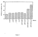
L'effet des différentes combinaisons de facteurs est représenté sur la
Parmi les facteurs testés, la combinaison du SCF (Stem Cell Factor d'origine murine -mSCF- ou aviaire -aSCF-), du b-FGF (basic Fibroblast Growth Factor) et du LIF (Leukemia Inhibitory Factor) donne le meilleur nombre de colonies positives pour l'activité phosphatase alcaline dans les cultures avec un accroissement de 2-3 fois par rapport à la présence de chaque facteur ajouté individuellement ou deux par deux et par rapport au fond, constitué en majorité de cellules faiblement: positives et présentant une morphologie épithétioïde différenciée.Among the factors tested, the combination of SCF (Stem Cell Factor of murine origin -mSCF- or avian-aSCF-), b-FGF (basic Fibroblast Growth Factor) and LIF (Leukemia Inhibitory Factor) gives the best number of colonies positive for alkaline phosphatase activity in the cultures with a 2-3 fold increase over the presence of each individually or two-fold plus background factor, predominantly consisting of weakly positive cells and differentiated epithetoid morphology.
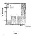
Les cellules sont ensemencées soit sur boîtes non traitées, soit traitées à la gélatine dans du milieu ESA complet avec facteurs de croissance aSCF (avian Stem Cell Factor), bFGF (basic- Fibroblast Growth factory, IGF-1 (Insulin-like Growth Factor-1) et LIF (Leukemia Inhibitory factor). L'anticorps ARMA est ajouté à raison de 1 µg/ml final. Les cellules et colonies positives pour l'activité phosphatase alcaline (AP +) sont dénombrées après.4 j de culture.The cells are seeded either on untreated dishes or gelatin-treated in ESA medium complete with aFcF (avian Stem Cell Factor) growth factors, bFGF (basic Fibroblast Growth Factory, IGF-1 (Insulin-like Growth Factor- 1) and LIF (Leukemia Inhibitory factor) The ARMA antibody is added at a rate of 1 μg / ml final The cells and colonies positive for alkaline phosphatase activity (AP +) are counted after 4 days of culture.
Les résultats sont représentés sur la
Comparativement aux différents moyens décrits comme l'utilisation de résine ou de charbon, et testés pour essayer de controler le niveau d'acide rétinoïque dans le milieu, l'addition de l'anticorps anti acide rétinoïque dans le milieu donne les meilleurs résultats quant à la qualité et la quantité des colonies présentes dans les culturesCompared to the various means described as the use of resin or charcoal, and tested to try to control the level of retinoic acid in the medium, the addition of the retinoic acid anti-acid in the medium gives the best results as to the quality and quantity of the colonies present in the cultures
L'addition de l'anticorps anti-acide rétinoïque dans le milieu de culture accroit de façon notable la présence et /ou le maintien des colonies à activité phosphatase alcaline.The addition of the anti-retinoic acid antibody in the culture medium significantly increases the presence and / or maintenance of alkaline phosphatase-active colonies.
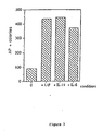
Nous avons voulu vérifier si le UF ou d'autres cytokines de la même famille pouvait induire la prolifération des cellules ES chez l'oiseau.We wanted to test whether UF or other cytokines from the same family could induce ES cell proliferation in birds.
Les cellules sont ensemencées en milieu ESA complet avec facteurs de croissance aSCF (avian Stem Cell Factor), bFGF (basic- Fibroblast Growth Factor), IGF-1 (Insulin-like Growth Factor-I en présence d'ARMA (1 µg/ml) et après addition ou non de différentes cytokines de la même famille LlF (Leukcmia Inhibitory Factor), IL-11 (Interleukine 11) et IL-6 (Interleukine 6). Afin d'accroitre l'adhésion et la formation de colonies phosphatase alcaline positives ainsi que leur taille, un second ensemencement a lieu 2 jours après le premier. La fixation, coloration et lecture des colonies a eu lieu 3 jours après le second ensemencement.The cells are inoculated in complete ESA medium with avian Stem Cell Factor (aSCF), bFGF (basic Fibroblast Growth Factor), IGF-1 (Insulin-like Growth Factor-I in the presence of ARMA (1 μg / ml). ) and after addition or addition of different cytokines of the same family LIF (Leukcmia Inhibitory Factor), IL-11 (Interleukin 11) and IL-6 (Interleukin 6) In order to increase the adhesion and the formation of colonies alkaline phosphatase Positive as well as their size, a second seeding takes place 2 days after the first.The fixation, staining and reading of the colonies took place 3 days after the second seeding.
La comparaison de l'effet des différentes cytokines est représentée sur la
Le rôle des cytokines LIF, IL-11 et IL-6 semble particulièrement prononcé et pratiquement équivalent dans l'obtention de colonies positives pour l'activité phosphatase alcaline.The role of LIF, IL-11 and IL-6 cytokines seems particularly pronounced and almost equivalent in obtaining positive colonies for alkaline phosphatase activity.
Chez la souris, la croissance de certaines cellules ES requiert la présence d'un tapis de cellules nourricières. L'effet de ces cellules sur les cellules d'embryons d'oiseau a été testéIn mice, the growth of certain ES cells requires the presence of a carpet of feeder cells. The effect of these cells on the cells of bird embryos was tested
Les cellules sont ensemencées dans un milieu ESA complet avec facteurs de croissance aSCF (avian Stem Cell Facror), bFGF (basic-Fibroblast Growth Factor), IGF-1 (Insulin-like Growth Factor-1) et anticorps ARMA (1 µg/ml) comparativement soit sur un fond de gélatine soit sur un tapis de cellules STO traitées à la mitomycine C comme indiqué dans le paragraphe Matériel et Méthodes. Après trois jours de culture, un nouvel ensemencement est ajouté à la culture. Les cytokines CNTF (Ciliary Neuro-Trophic Factor), OSM (Oncostatin M), LIF (Leukemia Inhibitory Factor), IL-11 1 (Interleukine 11) et IL-6 (Interleukin 6) sont ajoutées dans le milieu aux concentration indiquées précédemment.The cells are inoculated in a complete ESA medium with avian Stem Cell Facror (aSCF), bFGF (basic Fibroblast Growth Factor), IGF-1 (Insulin-like Growth Factor-1) and ARMA antibodies (1 μ g / ml). ml) comparatively either on a gelatin background or on a carpet of mitomycin C-treated STO cells as indicated in the Materials and Methods section. After three days of culture, a new seeding is added to the culture. The cytokines CNTF (Ciliary Neuro-Trophic Factor), OSM (Oncostatin M), LIF (Leukemia Inhibitory Factor), IL-11 1 (Interleukin 11) and IL-6 (Interleukin 6) are added in the medium at the concentrations indicated above.
La
Le nombre de colonies dérivant des cellules de blastoderme et présentant une activité phophatase alcaline est très nettemment accru en présence d'un tapis de cellules nourricières (environ 4-5 fois) avec un maintien entre les deux systèmes des même sensibilités vis à vis des cytokines ajoutées dans le milieu. Les cytokines LIF. IL-11 et II-G présentent les meilleurs résultats de stimulation de croissance. Dans des résultats préliminaires, il apparait de plus que la combinaison de ces 3 cytokines dans le milieu complet ESA avec facteurs produisent des effets cumulatifs très prometteurs quant au maintien et à la prolifération des colonies tant avec des cellules dérivées de disques blastodermiques de caille que de poulet.The number of colonies derived from blastoderm cells and exhibiting alkaline phophatase activity is very clearly increased in the presence of a carpet of feeder cells (about 4-5 fold) with maintaining the same sensitivities between the two systems with respect to cytokines added to the medium. LIF cytokines. IL-11 and II-G show the best results of growth stimulation. In preliminary results, it appears further that the combination of these 3 cytokines in ESA complete medium with factors produces very promising cumulative effects in maintaining and proliferating colonies with both quail blastoderm disc chicken.
Des études de réactivité par rapport à différents anticorps ont été réalisées. Les anticorps ECMA-7, SSEA-1 et SSEA-3, spécifiques d'un état de totipotence des cellules ES murines sont capables de reconnaitre des épitopes dans les populations de cellules aviaires, maintenues dans les cultures. Pour illustrer ces reconnaissances par les anticorps, des doubles marquages activité phosphatase alcaline et anticorps démontrent que toutes les cellules ou les massifs de cellules reconnues par ECMA-7 présentent une activité phosphatase alcaline. Cette propriété a été observée avec tous les anticorps utilisés à des degrés divers.Reactivity studies with different antibodies were performed. The ECMA-7, SSEA-1 and SSEA-3 antibodies, specific for a totipotency state of murine ES cells, are capable of recognizing epitopes in avian cell populations maintained in cultures. To illustrate these recognitions by antibodies, double alkaline phosphatase and antibody markings demonstrate that all ECMA-7 recognized cells or cell pools exhibit alkaline phosphatase activity. This property has been observed with all the antibodies used to varying degrees.
Les colonies de cellules phosphatase alcaline positives sont pour environ 20 % d'entre elles marquées par l'anticorps ECMA-7. Cette reconnaissance suggère la présence dans ces massifs et dans ces seules conditions de culture de cellules à caractère "ES". Néanmoins, une hétérogénéité dans les massifs phosphatase alcaline positifs suppose des degrés variables dans l'intensité du caractère "ES".The colonies of alkaline phosphatase positive cells are for about 20% of them labeled with the ECMA-7 antibody. This recognition suggests the presence in these masses and in these conditions of culture only cells with character "ES". Nevertheless, heterogeneity in the alkaline phosphatase positive mass assumes varying degrees in the intensity of the "ES" character.
Cette hétérogénéité de distribution a été observée sur des cultures primaires. Après repiquages, la proportion de cellules positives, notamment pour ECMA-7, mais également pour SSEA-1 et EMA-1. tend à s'accroitre de façon très importante pour obtenir des cultures très homogènes.This heterogeneity of distribution was observed on primary cultures. After subcultures, the proportion of positive cells, in particular for ECMA-7, but also for SSEA-1 and EMA-1. tends to increase very significantly to obtain very homogeneous crops.
La
Les anticorps SSEA-1. SSEA-3. également utilisés sur des cellules ES murines reconnaissent également des cellules aviaires dans les massifs phosphatase alcaline positifs.SSEA-1 antibodies. SSEA-3. also used on murine ES cells also recognize avian cells in alkaline phosphatase positive massifs.
Les anticorps NC-1, HNK-1 dirigés respectivement des épitopes de cellules de crêtes neurales et de cellules "human natural killer reconnaissent en fait les mêmes épitopes et ont été montrés comme reconnaissant certaines cellules immatures du disque blastodermique de poulet. Dans notre système, ces deux anticorps reconnaissent là encore des cellules dans des massifs à activité phosphatase alcaline.The NC-1, HNK-1 antibodies directed respectively from neural peak cell epitopes and human natural killer cells actually recognize the same epitopes and have been shown to recognize some immature cells of the chicken blastoderm disc. these two antibodies again recognize cells in alkaline phosphatase-active clusters.
Les résultats avec NC-1 sont représentés sur la
Les cellules sont ensemencées dans un milieu ESA complet avec facteurs de croissance aSCF (avian Stem Cell Factor), bFGF (basic-Fibroblast Growth Factor). IGF-1 (Insulin-like Growth Factor-1) et anticorps ARMA (1 µg/ml) sur un tapis de cellules STO traitées à la mitomycine C comme indiqué dans le paragraphe Matériel et Méthodes. Après deux jours de culture, un nouvel ensemencement est ajouté à la culture. Les cytokines LIF (Leukemia Inhibitory Factor), IL-11 (Interleukine 11) et IL-6 (Interleukine 6) sont ajoutées dans le milieu aux concentrations indiquées précédemment. La double coloration, activité phosphatase alcaline et détection des épitopes par anticorps est réalisée suivant les protocoles présentés précédemment.The cells are inoculated in a complete ESA medium with avian Stem Cell Factor (aSCF), bFGF (basic Fibroblast Growth Factor). IGF-1 (Insulin-like Growth Factor-1) and ARMA antibody (1 μg / ml) on a mat of STO cells treated with mitomycin C as indicated in the section Materials and Methods. After two days of culture, a new seeding is added to the culture. The cytokines LIF (Leukemia Inhibitory Factor), IL-11 (Interleukin 11) and IL-6 (Interleukin 6) are added in the medium at the concentrations indicated above. Double staining, alkaline phosphatase activity and detection of antibody epitopes is carried out according to the protocols presented above.
Par ailleurs, l'anticorps EMA-I (Hahnel and Eddy. 1986) initialement diriges contre des épitopes présents sur les cellules primordiales de la lignée germinale murine a été utilisé contre ces mêmes cellules chez le poulet. En testant cet anticorps dans notre système de culture, nous prouvons démontrer que EMA-1 reconnait des cellules et colonies de cellules présentant toutes une activité phosphatase alcaline. Il a été par ailleurs vérifié que cet anticorps EMA-1 ne reconnaissait les cellules ES murines que dans leur état de tolipotence non différenciée.Moreover, the EMA-I antibody (Hahnel and Eddy, 1986) initially directed against epitopes present on the primordial cells of the murine germ line was used against these same cells in chicken. By testing this antibody in our culture system, we demonstrate that EMA-1 recognizes cells and colonies of cells that all have alkaline phosphatase activity. It was furthermore verified that this EMA-1 antibody only recognizes murine ES cells in their undifferentiated tolipotence state.
Les anticorps ont été testés soit sur des cultures non diffenciées obtenues telles que décrites dans la partie matériel et Méthodes telle que décrite ci-desssus soit sur des cultures qui ont été traitées avec un excès d'acide rétinoïque ajouté à la culture (10-6 M) pendant au moins 48 heures. Le tableau ci-après indique l'état de reconnaissance par les différents anticorps utilisés.
Il apparait que l'expression de ECMA-7 (Kemler et al. (1981)) est la plus importante, suggérant une véritable nature de cellules ES, que TEC-01 (Draber et al. (1987b)) et SSEA-1 (Solter and Knowles (1978)) reconnaissent les mêmes épitopes sur des cellules non différenciées exclusivement. A l'inverse, l'augmentation d'expression de TEC-02 (Draber et al. (1987a)) peut dans ce sens indiquer un état de différenciation induit ou spontané. L'achèvement de cette perte de nature ES est caractérisé par la forte expression de TROMA-1 (Bruler et al. (1980)), présent sur les seules cellules différenciées. L'ensemble de ces anticorps permet donc d'avoir une idée sur l'état de différenciation d'une culture. Des anticorps comme TEC-03 (Draber et al. (1987a)) apparaissent comme relativement indifférents à l'état prononcé de différenciation.It appears that the expression of ECMA-7 (Kemler et al (1981)) is the most important, suggesting a true nature of ES cells, that TEC-01 (Draber et al. (1987b)) and SSEA-1 ( Solter and Knowles (1978)) recognize the same epitopes on undifferentiated cells exclusively. On the contrary, the increase in TEC-02 expression (Draber et al., (1987a)) can in this sense indicate a state of induced or spontaneous differentiation. Completion of this loss of ES nature is characterized by the strong expression of TROMA-1 (Bruler et al. (1980)), present on only differentiated cells. The set of these antibodies makes it possible to have an idea about the state of differentiation of a culture. Antibodies such as TEC-03 (Draber et al (1987a)) appear to be relatively indifferent to the pronounced state of differentiation.
Il est par ailleurs à souligner que jusqu'à présent ni ECMA-7, ni SSEA-1. SSEA-3, TEC-01, TEC-02, TEC-03, TROMA-1 n'ont fait l'objet de publication démontrant la réactivité sur des coupes, des cellules ou tout matériel d'origine aviaire.It is also noteworthy that so far neither ECMA-7 nor SSEA-1. SSEA-3, TEC-01, TEC-02, TEC-03, TROMA-1 have not been published to demonstrate reactivity on sections, cells or any material of bird origin.
Parmi les anticorps testés, certains comme ECMA-7 (Kemler et al. (1981)), SSEA-1 (Solter and Knowles (1978)), SSEA-3 (Shevinsky et al. (1982)) sont caractéristiques des cellules "ES "murines. Ces anticorps reconnaissant des cellules donc potentiellement totipotentes dans les cultures aviaires. Les mémes observations ayant été obtenues soit avec des cultures de caille soit de poule. D'autres anticorps commé EMA-1 (Hahnel and Eddy (1986)), NC-1 et HNK-1 (Obo and Balch (1981)) sont connus pour reconnaitre des épitopes aviaires (et murin pour EMA-1, Urven et al. (1988)) de cellules très indifférenciées et donc aussi susceptible de reconnaifre un profil de cellules souches aviaires.Of the antibodies tested, some such as ECMA-7 (Kemler et al. (1981)), SSEA-1 (Solter and Knowles (1978)), SSEA-3 (Shevinsky et al (1982)) are characteristic of ES cells. "murine. These antibodies recognize potentially potentially totipotent cells in avian cultures. The same observations have been obtained with either quail or hen crops. Other antibodies such as EMA-1 (Hahnel and Eddy (1986)), NC-1 and HNK-1 (Obo and Balch (1981)) are known to recognize avian (and murine epitopes for EMA-1, Urven et al. (1988)) of very undifferentiated cells and therefore also capable of recognizing an avian stem cell profile.
Les cellules de disques blastodermiques de caille ou de poulet sont ensemencées sur tapis de cellules nourricières STO. Après différents jours de culture, les cellules sont repiquées sur tapis de cellules STO comme décrit dans la partie Materiel et méthodes. La détection de cellules et massifs positifs à la fois pour l'activité de phosphatase alcaline et pour la localisation d'un marquage par ECMA-7 ou NC-1 suggère que les conditions de cultures sont définies pour maintenir dans les cultures secondaires et tertiaires des cellules à caractère totipotent. Le processus de repiquage assure d'ailleurs dès après le premier passage une homogénéité à l'ensemble de la culture, tant morphologiquement, que par la détection des différents épitopes. Les massifs de cellules deviennent très étendus et homogènes, caractère accru par la grande capacité de ces cellules à se diviser rapidement, contrairement aux cellules différenciées présentes initialement dans la culture primaire. Jusqu'à présent ces critères d'identification et de caractérisation peuvent être utilisés et détectés pendant au moins 5 semaines après l'ensemencement.Blastodermal discs of quail or chicken are seeded on STO feeder cell mats. After different days of culture, the cells are subcultured onto STO cell mats as described in the Materials and Methods section. The detection of cells and cells positive for both alkaline phosphatase activity and for the localization of ECMA-7 or NC-1 labeling suggests that the culture conditions are defined to maintain the secondary and tertiary cultures of the cells. totipotent cells. The transplanting process also ensures, after the first pass, homogeneity to the whole culture, both morphologically, and by the detection of the different epitopes. The mass of cells become very extensive and homogeneous, character increased by the large capacity of these cells to divide rapidly, unlike the differentiated cells initially present in the primary culture. So far these identification and characterization criteria can be used and detected for at least 5 weeks after seeding.
Les cellules blastodermiques de poulet obtenues en cultures primaires ou après repiquages successifs peuvent être injectées dans des embryons receveurs. Afin de visualiser rapidement une contribution phénotypique des cellules du donneur dans un embryon de poussin receveur, les cellules maintenues en culture proviennent d'une souche pigmentée et les embryons receveurs d'une souche non pigmentée. Les cellules maintenues en cultures sont dissociées et préparées comme décrit dans la partie Matériel et Méthodes selon le même procédé que pour un repiquage. La suspension cellulaire est alors préparée à raison de 1 à 3 x 10⁵ cellules par ml de milieu ESA. L'oeuf fraîchement pondu, non incubé contenant l'embryon reveceur est légérement irradié entre 5 Gy et 7 Gy. Une petite fenêtre de quelques mm2 est réalisée dans la coquille du receveur par meulage. La membrane coquillière est découpée au scalpel et les cellules sont injectées à l'aide d'un capillaire étiré dans la cavité subgerminale du disque blastodermique dans un volume de 1 à 5 µl, ce qui correspond de 100 à 1500 cellules au maximum. La moyenne des cellules injectées est de 500 cellules. La fenêtre est alors recouverte de membranes coquillières et scellée. Un morceau de pansement adhésif est appliqué pour parfaire l'étanchéité et limiter au maximum l'évaporation. Après 4 jours d'incubation dans les conditions optimales, les oeufs sont ouverts et les embryons bien développés sont transférés dans une coquille plus grande et remis en incubation pour finir leur développement de façon satisfaisante.The chicken blastoderm cells obtained in primary cultures or after successive subcultures can be injected into recipient embryos. In order to rapidly visualize a phenotypic contribution of the donor cells in a recipient chick embryo, the cells maintained in culture come from a pigmented strain and the recipient embryos from an unpigmented strain. The cells maintained in cultures are dissociated and prepared as described in the Materials and Methods part according to the same method as for a subculture. The cell suspension is then prepared in a proportion of 1 to 3 × 10 5 cells per ml of ESA medium. The freshly laid, non-incubated egg containing the reviving embryo is slightly irradiated between 5 Gy and 7 Gy. A small window of a few mm 2 is made in the recipient's shell by grinding. The shell membrane is cut with a scalpel and the cells are injected with a capillary stretched in the subgerminal cavity of the blastodermic disc in a volume of 1 to 5 μl, which corresponds to 100 to 1500 cells maximum. The average of the injected cells is 500 cells. The window is then covered with shell membranes and sealed. A piece of adhesive bandage is applied to perfect the seal and to limit the evaporation as much as possible. After 4 days of incubation under optimal conditions, the eggs are opened and the well-developed embryos are transferred to a larger shell and incubated to finish their development satisfactorily.
Un certain nombre d'animaux ont ainsi été obtenus et montrent un taux apparent de chimérisme, phénotypiquement détéctable par le marqueur de plumage utilisé et caractéristique de la souche des cellules dérivant de la souche donneur, variant de 5% à 90%. Ce chimérisme peut être obtenu jusqu'à présent indifféremment avec des cellules dérivant de cultures primaires, secondaires, tertiaires. Il est à noter que les pourcentages d'animaux chimères et les taux de chimérisme de ces animaux ne varient pas de façon importante en fonction du temps de culture des cellules injectées. Ceci contribue a souligner la capacité du milieu et au procédé décrit à maintenir des cellules avec un caractère totipotent.A certain number of animals have thus been obtained and show an apparent rate of chimerism, phenotypically detectable by the plumage marker used and characteristic of the strain of cells derived from the donor strain, ranging from 5% to 90%. This chimerism can be obtained until now indifferently with cells deriving from primary, secondary, tertiary cultures. It should be noted that the percentages of chimeric animals and the chimerism rates of these animals do not vary significantly according to the culture time of the injected cells. This helps to emphasize the ability of the medium and the method described to maintain cells with a totipotent character.
- Animal n° 1786-1787 à faible taux de chimérisme (5-10%)
- Animal n° 1782-1783 à taux moyen de chimérismc (50%)
- Animal n° 1740-1741 à fort taux de chimérisme (90%)
- Animal No. 1786-1787 with low rate of chimerism (5-10%)
- Animal n ° 1782-1783 with average rate of chimerism (50%)
- Animal # 1740-1741 with high rate of chimerism (90%)
-
Brulet et al. (1980). Froc. Natl. Acad. Sci, 77, 4113 Brulet et al. (1980). Proc. Natl. Acad. Sci 77, 4113 -
Draber et al. (1987a). Cell Differentiation 21, 119 Draber et al. (1987a). Cell Differentiation 21, 119 -
Draber et al. (1987b). Cell differenciation 21, 227 Draber et al. (1987b). Cell differentiation 21, 227 -
Eyal Giladi and Kovak (1976), Developemental Biology, 49, 321-337Eyal Giladi and Kovak (1976), Developmental Biology, 49, 321-337 -
Hahnel and Eddy (1986). Gamete Research 15. 25 Hahnel and Eddy (1986). Gamete Research 15. 25 -
Kemler et al. (1981). J. Embryo. Exp. Morph. 64, 45 Kemler et al. (nineteen eighty one). J. Embryo. Exp. Morph. 64, 45 -
Obo and Balch (1981). J. Immunology 127, 1024 Obo and Balch (1981). J. Immunology 127, 1024 -
Petine et al (1990) Development 108, 185-189Petine et al (1990) Development 108, 185-189 -
Solter and Knowles (1978). Proc. Natl. Acad. Sci. 75, 5565 Solter and Knowles (1978). Proc. Natl. Acad. Sci. 75, 5565 -
Shevinsky et al. (1982). Cell 30. 697 Shevinsky et al. (1982). Cell 30. 697 -
Tucker et al. (1984). Cell Differentiation 14, 223 Tucker et al. (1984). Cell Differentiation 14, 223 -
Urven et al. (1988). Development 103. 299 Urven et al. (1988). Development 103. 299
Claims (13)
- Avian totipotent embryonic stem cell (or avian ES cell) culture process, characterised in that:1) Cells from fertilised bird egg blastodermic disks are suspended in an avian totipotent embryonic stem cell culture medium, of the type comprising an avian cell culture medium, comprising:a) b-FGF, SCF and LIF, orb) b-FGF, aSCF (avian SCF), LIF, IL-11, IL-6, and IGF-1, orc) b-FGF, aSCF (avian SCF), LIF and IGF-1 (b-FGF = basic Fibroblast Growth Factor; SCF = Stem Cell Factor; LIF = Leukaemia Inhibitory Factor; IL-11 = Interleukin 11; IL-6 = Interleukin 6; IGF-1 = Insulin-like Growth Factor-1; aSCF = avian Stem Cell Factor),2) A feeder cell layer is inoculated with the suspension obtained following step a),3) The cells are incubated,4) The cells in culture are removed and purified so as to retrieve the avian ES cells.
- Process according to claim 1, characterised in that, between steps 3) and 4), one or a plurality of additions of fresh medium identical to that used in step 1) are made at various intervals.
- Process according to any of claims 1 to 2, characterised in that the culture medium is essentially devoid of active retinoic acid.
- Process according to any of claims 1 to 2, characterised in that the culture medium comprises a feeder cell layer.
- Process according to any of claims 1 to 4, characterised in that, between steps 3) and 4), fresh medium is added on the third day, and subsequently every day.
- Process according to any of claims 1 to 5, characterised in that step 4) is performed by means of an enzyme treatment, washing in a medium free from growth factor and centrifugation.
- Process according to any of claims 1 to 6, characterised in that, following step 4), a step 5) is performed wherein the ES cells are reinoculated on a feeder cell layer, in the presence of said culture medium, so as to obtain a secondary culture.
- Process according to claim 7, characterised in that steps 4) and 5) are repeated a plurality of times.
- In vitro avian ES cell culture liable to be obtained by means of the process according to any of claims 1 to 8, said culture comprising an avian totipotent embryonic stem cell culture medium comprising:a) b-FGF, SCF and LIF, orb) b-FGF, aSCF (avian SCF), LIF, IL-11, IL-6, and IGF-1, orc) b-FGF, aSCF (avian SCF), LIF and IGF-1 (b-FGF = basic Fibroblast Growth Factor; SCF = Stem Cell Factor; LIF = Leukaemia Inhibitory Factor; IL-11 = Interleukin 11; IL-6 = Interleukin 6; IGF-1 = Insulin-like Growth Factor-1; aSCF = avian Stem Cell Factor).
- Avian ES cell culture according to claim 9, characterised in that it exhibits 2 to 3 more alkaline phosphatase activity-positive colonies in relation to the base mainly consisting of low-positive cells.
- Avian ES cell culture according to claim 9 or 10, characterised in that the cells react specifically with at least one antibody selected from ECMA-7, SSEA-1, SSEA-3, TEC-01, EMA-1 and EMA-6.
- Avian ES cell culture according to claim 11, characterised in that the cells do not react with TROMA-1 antibody.
- Avian ES cell culture according to any of claims 9 to 12, characterised in that the cells are modified by incorporating the gene coding for a heterologous protein.
Applications Claiming Priority (3)
| Application Number | Priority Date | Filing Date | Title |
|---|---|---|---|
| FR9412598A FR2726003B1 (en) | 1994-10-21 | 1994-10-21 | CULTURE MEDIUM OF AVIAN TOTIPOTENT EMBRYONIC CELLS, METHOD FOR CULTURING THESE CELLS, AND AVIAN TOTIPOTENT EMBRYONIC CELLS |
| FR9412598 | 1994-10-21 | ||
| EP95935994A EP0787180B1 (en) | 1994-10-21 | 1995-10-20 | Active retinoic acid free culture medium for avian totipotential embryonic cells |
Related Parent Applications (1)
| Application Number | Title | Priority Date | Filing Date |
|---|---|---|---|
| EP95935994.4 Division | 1995-10-20 |
Publications (3)
| Publication Number | Publication Date |
|---|---|
| EP1149899A1 EP1149899A1 (en) | 2001-10-31 |
| EP1149899B1 true EP1149899B1 (en) | 2011-02-23 |
| EP1149899B9 EP1149899B9 (en) | 2011-09-21 |
Family
ID=9468082
Family Applications (2)
| Application Number | Title | Priority Date | Filing Date |
|---|---|---|---|
| EP95935994A Expired - Lifetime EP0787180B1 (en) | 1994-10-21 | 1995-10-20 | Active retinoic acid free culture medium for avian totipotential embryonic cells |
| EP01108584A Expired - Lifetime EP1149899B9 (en) | 1994-10-21 | 1995-10-20 | Culture medium for avian totipotential embryonic cells |
Family Applications Before (1)
| Application Number | Title | Priority Date | Filing Date |
|---|---|---|---|
| EP95935994A Expired - Lifetime EP0787180B1 (en) | 1994-10-21 | 1995-10-20 | Active retinoic acid free culture medium for avian totipotential embryonic cells |
Country Status (11)
| Country | Link |
|---|---|
| US (3) | US6114168A (en) |
| EP (2) | EP0787180B1 (en) |
| AT (2) | ATE223480T1 (en) |
| AU (1) | AU3808895A (en) |
| CA (1) | CA2203148C (en) |
| DE (2) | DE69536142D1 (en) |
| DK (1) | DK0787180T3 (en) |
| ES (2) | ES2360595T3 (en) |
| FR (1) | FR2726003B1 (en) |
| PT (1) | PT787180E (en) |
| WO (1) | WO1996012793A1 (en) |
Families Citing this family (71)
| Publication number | Priority date | Publication date | Assignee | Title |
|---|---|---|---|---|
| FR2726003B1 (en) * | 1994-10-21 | 2002-10-18 | Agronomique Inst Nat Rech | CULTURE MEDIUM OF AVIAN TOTIPOTENT EMBRYONIC CELLS, METHOD FOR CULTURING THESE CELLS, AND AVIAN TOTIPOTENT EMBRYONIC CELLS |
| EP0920490A2 (en) * | 1996-07-25 | 1999-06-09 | Genzyme Corporation | Chondrocyte media formulations and culture procedures |
| BR9808192A (en) * | 1997-03-06 | 2000-10-24 | Infigen Inc | Animal cloning process |
| US6011197A (en) | 1997-03-06 | 2000-01-04 | Infigen, Inc. | Method of cloning bovines using reprogrammed non-embryonic bovine cells |
| US5968809A (en) | 1997-04-11 | 1999-10-19 | Abbot Laboratories | Methods and compositions for synthesis of long chain poly-unsaturated fatty acids |
| US6566583B1 (en) | 1997-06-04 | 2003-05-20 | Daniel Facciotti | Schizochytrium PKS genes |
| JP2003532364A (en) * | 1997-08-04 | 2003-11-05 | ユニバシティ オブ マサチューセッツ、ア パブリック インスチチユーション オブ ハイアー エデュケイション オブ ザ コモンウエルス オブ マサチューセッツ、アズ リプリゼンテッド バイ イッツ アマースト キャンパス | Avian primordial germ cell (PGC) cell line and long-term culture method |
| US6333192B1 (en) * | 1999-08-09 | 2001-12-25 | North Carolina State University | Method of producing an undifferentiated avian cell culture using avian primordial germ cells |
| FR2808803B1 (en) * | 2000-05-11 | 2004-12-10 | Agronomique Inst Nat Rech | MODIFIED ES CELLS AND SPECIFIC GENE OF ES CELLS |
| US8273570B2 (en) * | 2000-05-16 | 2012-09-25 | Riken | Process of inducing differentiation of embryonic cell to cell expressing neural surface marker using OP9 or PA6 cells |
| FR2812004B1 (en) * | 2000-07-24 | 2002-12-27 | Ccd Lab | CULTURE MEDIA FOR IN VITRO FERTILIZATION, OR FOR THE CULTURE OF FOLLICLES, MALE GERMINAL CELLS OR EMBRYOS |
| AU8617301A (en) | 2000-08-01 | 2002-02-13 | Yissum Res Dev Co | Directed differentiation of embryonic cells |
| EP1311530A4 (en) | 2000-08-03 | 2004-10-06 | Wim-Van Schooten | Production of humanized antibodies in transgenic animals |
| US7145057B2 (en) | 2002-02-01 | 2006-12-05 | Origen Therapeutics, Inc. | Chimeric bird from embryonic stem cells |
| FR2836924B1 (en) * | 2002-03-08 | 2005-01-14 | Vivalis | AVIAN CELL LINES USEFUL FOR THE PRODUCTION OF INTEREST SUBSTANCES |
| CN1726276A (en) * | 2002-12-13 | 2006-01-25 | 艾文蒂斯·帕斯图尔公司 | Production of ALVAC on avian embryonic stem cells |
| BRPI0411125A (en) * | 2003-06-11 | 2006-07-18 | Japan Science & Tech Agency | process for the production of tissue cells from pluripotent stem cells derived from animal iris pigmented epithelial cells and tissue cells obtained by the process |
| FR2857671B1 (en) | 2003-07-18 | 2007-10-05 | Centre Nat Rech Scient | NOVEL METHOD FOR CULTURING KERATINOCYTES AND ITS APPLICATIONS |
| EP2143788A1 (en) | 2003-07-22 | 2010-01-13 | Vivalis | Production of poxviruses with duck cell lines |
| EP2811027A1 (en) | 2004-05-21 | 2014-12-10 | Novartis Vaccines and Diagnostics, Inc. | Alphavirus vectors for RSV and PIV vaccines |
| WO2005123900A1 (en) * | 2004-06-10 | 2005-12-29 | Merial Limited | Medium and methods for culturing of avian primordial germ cells |
| EP1850659B1 (en) * | 2005-02-01 | 2016-11-23 | Synageva BioPharma Corp. | Long term culture of chicken primordial germ cells |
| US20060174362A1 (en) * | 2005-02-01 | 2006-08-03 | Origen Therapeutics, Inc. | Long-term culture of avian primordial germ cells (PGCs) |
| FR2884255B1 (en) | 2005-04-11 | 2010-11-05 | Vivalis | USE OF EBX AVIATION STEM CELL LINES FOR THE PRODUCTION OF INFLUENZA VACCINE |
| FR2898909A1 (en) | 2006-03-24 | 2007-09-28 | Agronomique Inst Nat Rech | Combination of markers to characterize avian cells of StX cells, germinal cells or stem cells, comprises markers of a target gene expressed in the StX cells, stem cells or in the germinal cells |
| FR2898908A1 (en) * | 2006-03-24 | 2007-09-28 | Agronomique Inst Nat Rech | Process, useful to prepare differentiated avian cells from avian stem cells grown in culture medium, comprises induction of stem cells differentiation by inhibiting expression/activity of gene expressed in the stem cells e.g. Nanog gene |
| BRPI0714257A8 (en) * | 2006-08-09 | 2017-05-09 | Valneva | "METHODS FOR CULTURE OF AVIAN EMBRYONIC STEM CELLS (ES), FOR GENETICALLY MODIFYING EMBRYONIC STEM CELLS, FOR OBTAINING A CHIMERIC CHICK, FOR OBTAINING CHICK PROGENY, FOR ANALYSIS OF GENETIC POLYMORPHISM, CULTURE MEDIA FOR AVIAN EMBRYONIC STEM CELLS AND CULTURE MEDIA". |
| WO2008018684A1 (en) * | 2006-08-11 | 2008-02-14 | Modern Cell & Tissue Technologies Inc. | Culture medium for co-culturing of human stem cells and their feeder cells |
| EP2064325B1 (en) | 2006-09-01 | 2011-12-07 | Therapeutic Human Polyclonals, Inc. | Enhanced expression of human or humanized immunoglobulin in non-human transgenic animals |
| EP1985305A1 (en) | 2007-04-24 | 2008-10-29 | Vivalis | Duck embryonic derived stem cell lines for the production of viral vaccines |
| EP1995309A1 (en) * | 2007-05-21 | 2008-11-26 | Vivalis | Recombinant protein production in avian EBx® cells |
| CN101978047A (en) | 2008-01-18 | 2011-02-16 | 明尼苏达大学董事会 | Stem cell aggregates and methods for making and using |
| WO2010081890A1 (en) | 2009-01-19 | 2010-07-22 | Innate Pharma | Anti-kir3d antibodies |
| AR077757A1 (en) | 2009-07-15 | 2011-09-21 | Novartis Ag | COMPOSITIONS OF FUSION PROTEINS OF RESPIRATORY SINCICIAL VIRUS (RSV) AND METHODS FOR PREPARATION |
| ES2690199T3 (en) | 2009-07-21 | 2018-11-19 | Abt Holding Company | Use of stem cells to reduce leukocyte extravasation |
| US20110020293A1 (en) | 2009-07-21 | 2011-01-27 | Abt Holding Company | Use of Stem Cells to Reduce Leukocyte Extravasation |
| US8865462B2 (en) * | 2010-01-20 | 2014-10-21 | Crystal Bioscience Inc. | Sustained culture of avian gonocytes |
| EP2539439B1 (en) | 2010-02-25 | 2018-10-17 | ABT Holding Company | Modulation of angiogenesis |
| AU2011220721B2 (en) | 2010-02-25 | 2015-02-05 | Abt Holding Company | Modulation of macrophage activation |
| EP2556151A1 (en) | 2010-04-07 | 2013-02-13 | Novartis AG | Method for generating a parvovirus b19 virus-like particle |
| WO2011143415A1 (en) | 2010-05-12 | 2011-11-17 | Abt Holding Company | Modulation of splenocytes in cell therapy |
| WO2011158125A2 (en) | 2010-06-17 | 2011-12-22 | Katholieke Universiteit Leuven | Methods for differentiating cells into hepatic stellate cells and hepatic sinusoidal endothelial cells, cells produced by the methods, and methods for using the cells |
| EP3153578A1 (en) | 2010-07-06 | 2017-04-12 | Novartis Ag | Norovirus derived immunogenic compositions and methods |
| SG10201914007YA (en) | 2010-08-24 | 2020-03-30 | Univ Minnesota | Non-static suspension culture of cell aggregates |
| EP2625577B1 (en) | 2010-10-08 | 2019-06-26 | Terumo BCT, Inc. | Customizable methods and systems of growing and harvesting cells in a hollow fiber bioreactor system |
| EP4144368A1 (en) | 2011-01-26 | 2023-03-08 | GlaxoSmithKline Biologicals S.A. | Rsv immunization regimen |
| ES2782119T3 (en) | 2011-05-13 | 2020-09-10 | Glaxosmithkline Biologicals Sa | RSV prefusion F antigens |
| CA2837419C (en) | 2011-06-06 | 2021-07-13 | ReGenesys BVBA | Expansion of stem cells in hollow fiber bioreactors |
| CA2840989A1 (en) | 2011-07-06 | 2013-01-10 | Novartis Ag | Immunogenic combination compositions and uses thereof |
| CA2841047A1 (en) | 2011-07-06 | 2013-01-10 | Novartis Ag | Immunogenic compositions and uses thereof |
| EP2766385A2 (en) | 2011-10-12 | 2014-08-20 | Novartis AG | Cmv antigens and uses thereof |
| GB201119335D0 (en) | 2011-11-09 | 2011-12-21 | Univ Leuven Kath | Hepatitis virus infectable stem cells |
| JP2015522580A (en) | 2012-07-06 | 2015-08-06 | ノバルティス アーゲー | Immunological compositions and uses thereof |
| EA201590683A1 (en) | 2012-11-20 | 2015-11-30 | Глаксосмитклайн Байолоджикалс С.А. | RSV F TRIMMERS PRECEDING THE MERGER |
| SG10201710638UA (en) | 2013-04-12 | 2018-01-30 | Saverio Lafrancesca | Improving organs for transplantation |
| WO2014184666A2 (en) | 2013-04-30 | 2014-11-20 | Katholieke Universiteit Leuven | Cell therapy for myelodysplastic syndromes |
| WO2015073913A1 (en) | 2013-11-16 | 2015-05-21 | Terumo Bct, Inc. | Expanding cells in a bioreactor |
| EP2974739A1 (en) | 2014-07-15 | 2016-01-20 | Novartis AG | RSVF trimerization domains |
| EP3031822A1 (en) | 2014-12-08 | 2016-06-15 | Novartis AG | Cytomegalovirus antigens |
| EP3047856A1 (en) | 2015-01-23 | 2016-07-27 | Novartis AG | Cmv antigens and uses thereof |
| KR102505354B1 (en) | 2015-12-23 | 2023-03-02 | 화이자 인코포레이티드 | Rsv f protein mutants |
| US20170258495A1 (en) * | 2016-02-29 | 2017-09-14 | The Curators Of The University Of Missouri | Medium supplement to increase the efficiency of oocyte maturation and embryo culture in vitro |
| WO2017210537A1 (en) | 2016-06-02 | 2017-12-07 | The Cleveland Clinic Foundation | Complement inhibition for improving cell viability |
| CA3057171A1 (en) | 2017-03-30 | 2018-10-04 | The University Of Queensland | Chimeric polypeptides containing virus fusion ectodomain and complementary heptad repeats and uses thereof |
| KR20210016446A (en) | 2018-05-30 | 2021-02-15 | 인스티튜트 포 리서치 인 바이오메드슨 | Engineering of B lymphocytes using endogenous activation-induced cytidine deaminoase |
| EP3999108A1 (en) | 2019-07-21 | 2022-05-25 | GlaxoSmithKline Biologicals S.A. | Therapeutic viral vaccine |
| WO2021014385A1 (en) | 2019-07-24 | 2021-01-28 | Glaxosmithkline Biologicals Sa | Modified human cytomegalovirus proteins |
| US20220202923A1 (en) | 2020-12-23 | 2022-06-30 | Pfizer Inc. | E. coli fimh mutants and uses thereof |
| EP4032547A1 (en) | 2021-01-20 | 2022-07-27 | GlaxoSmithKline Biologicals S.A. | Hsv1 fce derived fragements for the treatment of hsv |
| WO2023144665A1 (en) | 2022-01-28 | 2023-08-03 | Glaxosmithkline Biologicals Sa | Modified human cytomegalovirus proteins |
| WO2024069420A2 (en) | 2022-09-29 | 2024-04-04 | Pfizer Inc. | Immunogenic compositions comprising an rsv f protein trimer |
Family Cites Families (7)
| Publication number | Priority date | Publication date | Assignee | Title |
|---|---|---|---|---|
| DE68928914T2 (en) * | 1988-08-04 | 1999-09-09 | Amrad Corp Ltd | (IN VITRO) PROPAGATION OF EMBRYONIC STEM CELLS USING LEUKEMIA INHIBITION FACTOR (LIF) |
| WO1993015185A1 (en) * | 1992-01-27 | 1993-08-05 | North Carolina State University | GENE TRANSFER IN POULTRY BY INTRODUCTION OF EMBRYO CELLS $i(IN OVO) |
| US5340740A (en) * | 1992-05-15 | 1994-08-23 | North Carolina State University | Method of producing an avian embryonic stem cell culture and the avian embryonic stem cell culture produced by the process |
| WO1994003585A1 (en) * | 1992-08-04 | 1994-02-17 | Commonwealth Scientific And Industrial Research Organisation | A method for maintaining embryonic stem cells and avian factor useful for same |
| US5453357A (en) * | 1992-10-08 | 1995-09-26 | Vanderbilt University | Pluripotential embryonic stem cells and methods of making same |
| US5589458A (en) * | 1992-11-13 | 1996-12-31 | Thomas Jefferson University | Compounds that inhibit T cell proliferation and methods for using the same |
| FR2726003B1 (en) * | 1994-10-21 | 2002-10-18 | Agronomique Inst Nat Rech | CULTURE MEDIUM OF AVIAN TOTIPOTENT EMBRYONIC CELLS, METHOD FOR CULTURING THESE CELLS, AND AVIAN TOTIPOTENT EMBRYONIC CELLS |
-
1994
- 1994-10-21 FR FR9412598A patent/FR2726003B1/en not_active Expired - Lifetime
-
1995
- 1995-10-20 ES ES01108584T patent/ES2360595T3/en not_active Expired - Lifetime
- 1995-10-20 US US08/817,671 patent/US6114168A/en not_active Expired - Lifetime
- 1995-10-20 EP EP95935994A patent/EP0787180B1/en not_active Expired - Lifetime
- 1995-10-20 CA CA2203148A patent/CA2203148C/en not_active Expired - Lifetime
- 1995-10-20 WO PCT/FR1995/001389 patent/WO1996012793A1/en active IP Right Grant
- 1995-10-20 EP EP01108584A patent/EP1149899B9/en not_active Expired - Lifetime
- 1995-10-20 PT PT95935994T patent/PT787180E/en unknown
- 1995-10-20 AU AU38088/95A patent/AU3808895A/en not_active Abandoned
- 1995-10-20 AT AT95935994T patent/ATE223480T1/en active
- 1995-10-20 ES ES95935994T patent/ES2180655T3/en not_active Expired - Lifetime
- 1995-10-20 AT AT01108584T patent/ATE499436T1/en not_active IP Right Cessation
- 1995-10-20 DE DE69536142T patent/DE69536142D1/en not_active Expired - Lifetime
- 1995-10-20 DK DK95935994T patent/DK0787180T3/en active
- 1995-10-20 DE DE69528071T patent/DE69528071T2/en not_active Expired - Lifetime
-
1999
- 1999-09-01 US US09/392,506 patent/US6500668B2/en not_active Expired - Lifetime
-
2002
- 2002-03-15 US US10/097,245 patent/US6998266B2/en not_active Expired - Fee Related
Non-Patent Citations (2)
| Title |
|---|
| KAWASE E. ET AL: "Tumor Necrosis Factor-alpha (TNF-alpha) Stimulates Proliferation of Mouse Primordial Germ Cells in Culture", DEVELOPMENTAL BIOLOGY, ACADEMIC PRESS, NEW YORK, NY, US LNKD- DOI:10.1006/DBIO.1994.1011, vol. 161, no. 1, 1 January 1994 (1994-01-01), pages 91 - 95, XP024779794, ISSN: 0012-1606, [retrieved on 19940101] * |
| MATSUI Y. ET AL: "Derivation of pluripotential embryonic stem cells from murine primordial germ cells in culture", CELL, CELL PRESS, CAMBRIDGE, NA, US LNKD- DOI:10.1016/0092-8674(92)90317-6, vol. 70, no. 5, 4 September 1992 (1992-09-04), pages 841 - 847, XP024245512, ISSN: 0092-8674, [retrieved on 19920904] * |
Also Published As
| Publication number | Publication date |
|---|---|
| EP0787180A1 (en) | 1997-08-06 |
| DK0787180T3 (en) | 2003-01-06 |
| AU3808895A (en) | 1996-05-15 |
| EP1149899A1 (en) | 2001-10-31 |
| ES2180655T3 (en) | 2003-02-16 |
| US6998266B2 (en) | 2006-02-14 |
| CA2203148C (en) | 2011-08-09 |
| FR2726003B1 (en) | 2002-10-18 |
| DE69528071D1 (en) | 2002-10-10 |
| ATE223480T1 (en) | 2002-09-15 |
| CA2203148A1 (en) | 1996-05-02 |
| PT787180E (en) | 2003-01-31 |
| ES2360595T3 (en) | 2011-06-07 |
| DE69528071T2 (en) | 2003-05-15 |
| EP1149899B9 (en) | 2011-09-21 |
| FR2726003A1 (en) | 1996-04-26 |
| US20020192815A1 (en) | 2002-12-19 |
| ATE499436T1 (en) | 2011-03-15 |
| US20010019840A1 (en) | 2001-09-06 |
| US6500668B2 (en) | 2002-12-31 |
| DE69536142D1 (en) | 2011-04-07 |
| US6114168A (en) | 2000-09-05 |
| WO1996012793A1 (en) | 1996-05-02 |
| EP0787180B1 (en) | 2002-09-04 |
Similar Documents
| Publication | Publication Date | Title |
|---|---|---|
| EP1149899B1 (en) | Culture medium for avian totipotential embryonic cells | |
| US5340740A (en) | Method of producing an avian embryonic stem cell culture and the avian embryonic stem cell culture produced by the process | |
| JP4841777B2 (en) | How to obtain an undifferentiated avian cell culture using avian primordial germ cells | |
| EP1280898B1 (en) | Modified es cells and es cell-specific gene | |
| US6156569A (en) | Prolonged culturing of avian primordial germ cells (PGCs) using specific growth factors, use thereof to produce chimeric avians | |
| US20140127812A1 (en) | Long-term culture of avian primordial germ cells (pgcs) | |
| MX2009001477A (en) | Method of production of transgenic avian using embryonic stem cells. | |
| JP2003532364A (en) | Avian primordial germ cell (PGC) cell line and long-term culture method | |
| JP2007501625A (en) | Avian spermatogonial stem cell culture method and avian spermatogonial stem cell obtained thereby | |
| WO2004039965A1 (en) | Composition for culturing multipotent stem cells and utilization of the same | |
| KR100502889B1 (en) | Method for Enhancing Germline Transmission Efficiency of Avian Primordial Germ Cells | |
| JP4376901B2 (en) | Method for producing avian chimera using spermatogonia and avian chimera | |
| Volkova et al. | Isolation, cultivation and characterization of quail primordial germ cells | |
| Naito et al. | Effect of mytomycin c treatment on feeder cells and the proliferation of cultured chicken primordial germ cells isolated from embryonic blood |
Legal Events
| Date | Code | Title | Description |
|---|---|---|---|
| PUAI | Public reference made under article 153(3) epc to a published international application that has entered the european phase |
Free format text: ORIGINAL CODE: 0009012 |
|
| 17P | Request for examination filed |
Effective date: 20010430 |
|
| AC | Divisional application: reference to earlier application |
Ref document number: 787180 Country of ref document: EP |
|
| AK | Designated contracting states |
Kind code of ref document: A1 Designated state(s): AT BE CH DE DK ES FR GB GR IE IT LI LU MC NL PT SE |
|
| AKX | Designation fees paid |
Free format text: AT BE CH DE DK ES FR GB GR IE IT LI LU MC NL PT SE |
|
| 17Q | First examination report despatched |
Effective date: 20070606 |
|
| RAP1 | Party data changed (applicant data changed or rights of an application transferred) |
Owner name: CENTRE NATIONAL DE LA RECHERCHE SCIENTIFIQUE (CNRS Owner name: INSTITUT NATIONAL DE LA RECHERCHE AGRONOMIQUE Owner name: ECOLE NORMALE SUPERIEURE DE LYON |
|
| GRAP | Despatch of communication of intention to grant a patent |
Free format text: ORIGINAL CODE: EPIDOSNIGR1 |
|
| RIC1 | Information provided on ipc code assigned before grant |
Ipc: C12N 5/00 20060101ALI20100528BHEP Ipc: C12N 5/0735 20100101AFI20100528BHEP |
|
| GRAS | Grant fee paid |
Free format text: ORIGINAL CODE: EPIDOSNIGR3 |
|
| RAP1 | Party data changed (applicant data changed or rights of an application transferred) |
Owner name: CENTRE NATIONAL DE LA RECHERCHE SCIENTIFIQUE (CNRS Owner name: ECOLE NORMALE SUPERIEURE DE LYON Owner name: INSTITUT NATIONAL DE LA RECHERCHE AGRONOMIQUE |
|
| GRAA | (expected) grant |
Free format text: ORIGINAL CODE: 0009210 |
|
| AC | Divisional application: reference to earlier application |
Ref document number: 0787180 Country of ref document: EP Kind code of ref document: P |
|
| AK | Designated contracting states |
Kind code of ref document: B1 Designated state(s): AT BE CH DE DK ES FR GB GR IE IT LI LU MC NL PT SE |
|
| REG | Reference to a national code |
Ref country code: GB Ref legal event code: FG4D Free format text: NOT ENGLISH |
|
| REG | Reference to a national code |
Ref country code: CH Ref legal event code: EP |
|
| REG | Reference to a national code |
Ref country code: IE Ref legal event code: FG4D Free format text: LANGUAGE OF EP DOCUMENT: FRENCH |
|
| REF | Corresponds to: |
Ref document number: 69536142 Country of ref document: DE Date of ref document: 20110407 Kind code of ref document: P |
|
| REG | Reference to a national code |
Ref country code: DE Ref legal event code: R096 Ref document number: 69536142 Country of ref document: DE Effective date: 20110407 |
|
| REG | Reference to a national code |
Ref country code: ES Ref legal event code: FG2A Ref document number: 2360595 Country of ref document: ES Kind code of ref document: T3 Effective date: 20110607 |
|
| REG | Reference to a national code |
Ref country code: NL Ref legal event code: VDEP Effective date: 20110223 |
|
| PG25 | Lapsed in a contracting state [announced via postgrant information from national office to epo] |
Ref country code: SE Free format text: LAPSE BECAUSE OF FAILURE TO SUBMIT A TRANSLATION OF THE DESCRIPTION OR TO PAY THE FEE WITHIN THE PRESCRIBED TIME-LIMIT Effective date: 20110223 Ref country code: PT Free format text: LAPSE BECAUSE OF FAILURE TO SUBMIT A TRANSLATION OF THE DESCRIPTION OR TO PAY THE FEE WITHIN THE PRESCRIBED TIME-LIMIT Effective date: 20110623 Ref country code: GR Free format text: LAPSE BECAUSE OF FAILURE TO SUBMIT A TRANSLATION OF THE DESCRIPTION OR TO PAY THE FEE WITHIN THE PRESCRIBED TIME-LIMIT Effective date: 20110524 |
|
| PG25 | Lapsed in a contracting state [announced via postgrant information from national office to epo] |
Ref country code: NL Free format text: LAPSE BECAUSE OF FAILURE TO SUBMIT A TRANSLATION OF THE DESCRIPTION OR TO PAY THE FEE WITHIN THE PRESCRIBED TIME-LIMIT Effective date: 20110223 Ref country code: AT Free format text: LAPSE BECAUSE OF FAILURE TO SUBMIT A TRANSLATION OF THE DESCRIPTION OR TO PAY THE FEE WITHIN THE PRESCRIBED TIME-LIMIT Effective date: 20110223 |
|
| REG | Reference to a national code |
Ref country code: IE Ref legal event code: FD4D |
|
| PG25 | Lapsed in a contracting state [announced via postgrant information from national office to epo] |
Ref country code: DK Free format text: LAPSE BECAUSE OF FAILURE TO SUBMIT A TRANSLATION OF THE DESCRIPTION OR TO PAY THE FEE WITHIN THE PRESCRIBED TIME-LIMIT Effective date: 20110223 Ref country code: IE Free format text: LAPSE BECAUSE OF FAILURE TO SUBMIT A TRANSLATION OF THE DESCRIPTION OR TO PAY THE FEE WITHIN THE PRESCRIBED TIME-LIMIT Effective date: 20110223 |
|
| PLBE | No opposition filed within time limit |
Free format text: ORIGINAL CODE: 0009261 |
|
| STAA | Information on the status of an ep patent application or granted ep patent |
Free format text: STATUS: NO OPPOSITION FILED WITHIN TIME LIMIT |
|
| 26N | No opposition filed |
Effective date: 20111124 |
|
| REG | Reference to a national code |
Ref country code: DE Ref legal event code: R097 Ref document number: 69536142 Country of ref document: DE Effective date: 20111124 |
|
| BERE | Be: lapsed |
Owner name: ECOLE NORMALE SUPERIEURE DE LYON Effective date: 20111031 Owner name: INSTITUT NATIONAL DE LA RECHERCHE AGRONOMIQUE Effective date: 20111031 Owner name: CENTRE NATIONAL DE LA RECHERCHE SCIENTIFIQUE (CNR Effective date: 20111031 |
|
| PG25 | Lapsed in a contracting state [announced via postgrant information from national office to epo] |
Ref country code: MC Free format text: LAPSE BECAUSE OF NON-PAYMENT OF DUE FEES Effective date: 20111031 |
|
| REG | Reference to a national code |
Ref country code: CH Ref legal event code: PL |
|
| PG25 | Lapsed in a contracting state [announced via postgrant information from national office to epo] |
Ref country code: LI Free format text: LAPSE BECAUSE OF NON-PAYMENT OF DUE FEES Effective date: 20111031 Ref country code: CH Free format text: LAPSE BECAUSE OF NON-PAYMENT OF DUE FEES Effective date: 20111031 Ref country code: BE Free format text: LAPSE BECAUSE OF NON-PAYMENT OF DUE FEES Effective date: 20111031 |
|
| PG25 | Lapsed in a contracting state [announced via postgrant information from national office to epo] |
Ref country code: LU Free format text: LAPSE BECAUSE OF NON-PAYMENT OF DUE FEES Effective date: 20111020 |
|
| PGFP | Annual fee paid to national office [announced via postgrant information from national office to epo] |
Ref country code: GB Payment date: 20141015 Year of fee payment: 20 Ref country code: ES Payment date: 20141021 Year of fee payment: 20 Ref country code: DE Payment date: 20141010 Year of fee payment: 20 Ref country code: FR Payment date: 20141015 Year of fee payment: 20 |
|
| PGFP | Annual fee paid to national office [announced via postgrant information from national office to epo] |
Ref country code: IT Payment date: 20141020 Year of fee payment: 20 |
|
| REG | Reference to a national code |
Ref country code: DE Ref legal event code: R071 Ref document number: 69536142 Country of ref document: DE |
|
| REG | Reference to a national code |
Ref country code: GB Ref legal event code: PE20 Expiry date: 20151019 |
|
| REG | Reference to a national code |
Ref country code: ES Ref legal event code: FD2A Effective date: 20160127 |
|
| PG25 | Lapsed in a contracting state [announced via postgrant information from national office to epo] |
Ref country code: GB Free format text: LAPSE BECAUSE OF EXPIRATION OF PROTECTION Effective date: 20151019 |
|
| PG25 | Lapsed in a contracting state [announced via postgrant information from national office to epo] |
Ref country code: ES Free format text: LAPSE BECAUSE OF EXPIRATION OF PROTECTION Effective date: 20151021 |