EP1114609A2 - Verfahren und Gerät zur Erfassung der A, C und V-Wellen in einer Vorhofsdruckwelle - Google Patents
Verfahren und Gerät zur Erfassung der A, C und V-Wellen in einer Vorhofsdruckwelle Download PDFInfo
- Publication number
- EP1114609A2 EP1114609A2 EP00311629A EP00311629A EP1114609A2 EP 1114609 A2 EP1114609 A2 EP 1114609A2 EP 00311629 A EP00311629 A EP 00311629A EP 00311629 A EP00311629 A EP 00311629A EP 1114609 A2 EP1114609 A2 EP 1114609A2
- Authority
- EP
- European Patent Office
- Prior art keywords
- wave
- interval
- point
- identifying
- highest peak
- Prior art date
- Legal status (The legal status is an assumption and is not a legal conclusion. Google has not performed a legal analysis and makes no representation as to the accuracy of the status listed.)
- Granted
Links
- 230000001746 atrial effect Effects 0.000 title claims abstract description 68
- 238000000034 method Methods 0.000 title claims abstract description 57
- 238000005259 measurement Methods 0.000 claims description 2
- 210000002216 heart Anatomy 0.000 description 25
- 239000008280 blood Substances 0.000 description 16
- 210000004369 blood Anatomy 0.000 description 16
- 210000005245 right atrium Anatomy 0.000 description 9
- 210000005240 left ventricle Anatomy 0.000 description 7
- 210000005241 right ventricle Anatomy 0.000 description 7
- 210000005246 left atrium Anatomy 0.000 description 5
- 210000001147 pulmonary artery Anatomy 0.000 description 5
- 230000008602 contraction Effects 0.000 description 4
- 210000002837 heart atrium Anatomy 0.000 description 4
- 210000004115 mitral valve Anatomy 0.000 description 4
- 210000000591 tricuspid valve Anatomy 0.000 description 4
- 230000000747 cardiac effect Effects 0.000 description 3
- 238000010586 diagram Methods 0.000 description 3
- 210000003492 pulmonary vein Anatomy 0.000 description 3
- 239000000523 sample Substances 0.000 description 3
- 210000001367 artery Anatomy 0.000 description 2
- 230000000694 effects Effects 0.000 description 2
- 210000004072 lung Anatomy 0.000 description 2
- 210000003462 vein Anatomy 0.000 description 2
- 210000001075 venae cavae Anatomy 0.000 description 2
- 210000000709 aorta Anatomy 0.000 description 1
- QVGXLLKOCUKJST-UHFFFAOYSA-N atomic oxygen Chemical compound [O] QVGXLLKOCUKJST-UHFFFAOYSA-N 0.000 description 1
- 210000001992 atrioventricular node Anatomy 0.000 description 1
- 238000010009 beating Methods 0.000 description 1
- 230000005540 biological transmission Effects 0.000 description 1
- 230000017531 blood circulation Effects 0.000 description 1
- 210000004375 bundle of his Anatomy 0.000 description 1
- 210000004351 coronary vessel Anatomy 0.000 description 1
- 230000003111 delayed effect Effects 0.000 description 1
- 230000001419 dependent effect Effects 0.000 description 1
- 238000002565 electrocardiography Methods 0.000 description 1
- 239000000835 fiber Substances 0.000 description 1
- 239000012530 fluid Substances 0.000 description 1
- 210000003709 heart valve Anatomy 0.000 description 1
- 239000012528 membrane Substances 0.000 description 1
- 238000012544 monitoring process Methods 0.000 description 1
- 210000004165 myocardium Anatomy 0.000 description 1
- 230000002232 neuromuscular Effects 0.000 description 1
- 229910052760 oxygen Inorganic materials 0.000 description 1
- 239000001301 oxygen Substances 0.000 description 1
- 238000005086 pumping Methods 0.000 description 1
- 238000000718 qrs complex Methods 0.000 description 1
- 230000001020 rhythmical effect Effects 0.000 description 1
- 210000001013 sinoatrial node Anatomy 0.000 description 1
- 230000002861 ventricular Effects 0.000 description 1
Images

Classifications
-
- A—HUMAN NECESSITIES
- A61—MEDICAL OR VETERINARY SCIENCE; HYGIENE
- A61B—DIAGNOSIS; SURGERY; IDENTIFICATION
- A61B5/00—Measuring for diagnostic purposes; Identification of persons
- A61B5/02—Detecting, measuring or recording for evaluating the cardiovascular system, e.g. pulse, heart rate, blood pressure or blood flow
- A61B5/021—Measuring pressure in heart or blood vessels
- A61B5/0215—Measuring pressure in heart or blood vessels by means inserted into the body
-
- A—HUMAN NECESSITIES
- A61—MEDICAL OR VETERINARY SCIENCE; HYGIENE
- A61B—DIAGNOSIS; SURGERY; IDENTIFICATION
- A61B5/00—Measuring for diagnostic purposes; Identification of persons
- A61B5/24—Detecting, measuring or recording bioelectric or biomagnetic signals of the body or parts thereof
- A61B5/316—Modalities, i.e. specific diagnostic methods
- A61B5/318—Heart-related electrical modalities, e.g. electrocardiography [ECG]
- A61B5/346—Analysis of electrocardiograms
- A61B5/349—Detecting specific parameters of the electrocardiograph cycle
- A61B5/352—Detecting R peaks, e.g. for synchronising diagnostic apparatus; Estimating R-R interval
Definitions
- the present invention relates to a method and apparatus for interpreting medical data and particularly to the identification of particular waves in an atrial pressure waveform using an ECG (electrocardiogram).
- ECG electrocardiogram
- the human heart receives blood from the veins and propels it into and through the arteries.
- the heart has two parallel and independent systems, each consisting of an atrium and a ventricle. From their anatomical positions, the right atrium and right ventricle are known as the right heart and the left atrium and left ventricle are known as the left heart. Blood from the body returns to the right atrium through two large veins, the superior and inferior venae cavae. Return of venous blood to the right atrium takes place during. the entire heart cycle of contraction and relaxation. Return of venous blood to the right ventricle occurs only during the relaxation part of the heart-pumping cycle, called diastole.
- the right atrium contracts and completes the filling of the right ventricle with blood. Contractions of the right ventricle expel the blood through the pulmonary arteries into the capillaries of the lung, where the blood receives oxygen. From the lung capillaries, the blood then empties into the pulmonary veins, and in turn into the left atrium. Return of blood from the pulmonary veins to the left atrium and left ventricle proceeds in a similar manner as the return of blood from the venae cavae to the right heart cavities. Contraction of the left ventricle propels the blood into the aorta. From there, blood is distributed to all arteries of the body, including the coronary arteries, which supply the heart muscle.
- valves at the openings of the aortic and pulmonary arteries consist of three semilunar (half-moon-shaped) flaps of membrane. The flaps are curved in the direction of blood flow and open readily in response to pressure in that direction. When the original pressure subsides, back pressure forces the edges of the flaps together.
- the tricuspid valve situated between the right atrium and right ventricle, is composed of three triangular flaps, and the bicuspid or mitral valve, between the left atrium and left ventricle, has two such flaps.
- the flaps of the mitral valve remain open until the left ventricle fills with blood. When the left ventricle begins to contract, the pressure of the pumped blood closes the mitral valve.
- the rhythmic beating of the heart is maintained by an orderly series of electrical discharges originating in the sinus node of the right atrium.
- the discharges proceed through the atrioventricular node and a bundle of neuromuscular fibers (known as the bundle of His) to the ventricles.
- a record of the electrical discharges can be obtained. This record is called an electrocardiogram, or ECG.
- Prominent parts of an ECG are the P wave, a deflection caused by the current originating in the atrium; the QRS complex, which is caused by the electrical activity of the ventricles as they contract; and the T wave, which is caused by relaxation of the ventricles. These changes in electrical activity may, in general, be sensed using probes or electrodes attached to the exterior of the body.
- the fluid pressure of blood in the atrium may be sensed by attaching a pressure sensor or pressure transducer to a probe (such as a catheter), and positioning that probe in the atrium.
- an atrial pressure waveform has several prominent parts and these parts can be associated with or related to the prominent parts of the ECG.
- the prominent parts of the atrial pressure waveform include an A wave.
- An A wave is caused by atrial systole and follows the P wave inscribed on the ECG.
- the A wave has a descent X that follows this initial positive deflection and is often interrupted by a small positive deflection called the C wave, which occurs when the tricuspid valve closes.
- the rise in atrial pressure during ventricular systole is called the V wave.
- the V wave reaches its peak prior to the opening of the tricuspid valve.
- the descent Y of the V wave occurs as the tricuspid valve opens and the right atrium empties into the right ventricle.
- Pressure in the pulmonary artery normally has a waveform similar to the waveform for the pressure in the left atrium. Although, the waveform for the pulmonary artery is delayed in transmission through the capillary vessels.
- a normal wedge pressure waveform has clear A and V waves, which may be detected by persons trained in reading ECGs. Descents X and Y are also clear, provided the pressure tracing is not overdamped. However, C waves are often not visible in the waveform.
- Detecting the occurrence of A, V, and C waves is important in determining the maximum pressure present in the atrial and pulmonary veins and to identify the presence of prolapsed or calcified heart valves. While manual techniques exist to find these waves, they are not completely satisfactory, and automated techniques for finding C waves are not generally available. Accordingly, there is a need for improved methods of determining C waves in an atrial pressure waveform.
- the present invention provides a method of determining A, C, and V waves.
- the method includes the acts of establishing a time reference based on R waves in an ECG. Using the R waves as a reference, a first interval in the atrial pressure waveform between a first point offset a predetermined amount ahead of a first R wave and a second point offset a predetermined amount behind the first R wave is determined. The highest peak within the first interval is then identified. This peak is the A wave.
- a second R wave in the ECG, subsequent in time to the first R wave is identified.
- a second interval in the atrial pressure waveform that extends from the second point to a third point is established.
- the third point is positioned at a distance ahead (subsequent in time) of the second point equal to a percentage of the interval from the second point to a predetermined amount behind the second R wave.
- the highest peak in the second interval is the V wave.
- a third interval in the atrial pressure waveform is established.
- the third interval extends from the highest peak in the first interval to a fourth point.
- the fourth point is positioned at a distance ahead of the highest peak in the first interval (the A wave) equal to a second percentage of the distance between the highest peak in the first interval and the highest peak in the second interval (the V wave).
- the C wave is the highest peak in the third interval. This same process may be repeated across the different R waves in the ECG to determine the A, C, and V waves for different "heart beats" or cardiac cycles.
- FIG. 1 illustrates an apparatus 5 for acquiring ECG and atrial pressure waveforms that embodies the invention. While this apparatus is shown as a single unit, the apparatus may be separated into discrete components that operate generally independent of the others.
- This apparatus includes an ECG acquisition circuit 6 that, in the usual application, is externally connected to the patient through leadwires and electrodes 7 attached to the patient's skin.
- the invention is equally applicable to ECG's that are acquired in other ways, e.g., through electrodes placed directly on the heart, through transesophageal monitoring, etc.
- the acquisition module also includes an atrial pressure waveform acquisition circuit 8 (APWAC).
- APWAC atrial pressure waveform acquisition circuit 8
- the APWAC is connected to a pressure sensor or transducer that is positioned in the right atrium of the patient's heart during a cardiac catheterization procedure.
- the apparatus also includes an analysis module connected to the acquisition module.
- the analysis module includes analysis software 9 that provides a means for identifying at least a portion of the APW atrial pressure waveform using the ECG waveform as a reference.
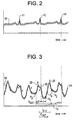
- FIG. 2 illustrates an exemplary ECG waveform 10 with three R waves 11, 12, and 14.
- FIG. 3 illustrates an exemplary atrial pressure waveform 15.
- the A, V, and C waves are determined or identified using the R waves of the ECG waveform 10 as a time reference.
- a waves are found by searching for the maximum peak pressure in an interval on the pressure waveform 15 from a point P1, located t seconds to the right of the R wave 12, to a point P2, located t seconds to the left of the R wave 12.
- the inventor has found that the value of the time-offset t should be based upon the heart rate of the patient. For heart rates less than 100 BPM (beats per minute), t should be set at about 150 ms. For heart rates between 100 and 150 BPM, t should be set at about 100 ms. For heart rates of 150 BPM or greater, t should be set at 75 ms.
- an A wave 20 occurs in the interval between P1 and P2.
- the V wave may be found in the waveform 15 by searching for the maximum peak in an interval V INT .
- the interval V INT extends from point P2 to point P3.
- Point P3 is positioned at a distance to the right of point P2 equal to 75% of an interval I v from point P2 to t seconds to the left of the R wave 14.
- a V wave 25 occurs in the interval between P2 and P3.
- the C wave may be found in the waveform 15 by searching for the highest peak following the A wave 20 in an interval C INT .
- the interval C INT extends from about 25% to about 50% of the elapsed time (represented by an interval I A-V ) from the A wave 20 to the V wave 25 depending on the particular QRS of the reference ECG. In other words, the distance the interval C INT extends is dependent on the heart rate of the patient.
- the Q of QRS refers to the ascent in the waveform before the occurrence of an R wave and the S refers to the descent in the waveform after the occurrence of an R wave.
- the percentage of elapsed time from the A wave 25 to the V wave 25 is at the low end of the 25 to 50% range.
- the percentage of elapsed time from the A wave 20 to the V wave 25 is at the high end of the 25 to 50% range.
- the highest wave in the interval C INT is a C wave 30.
- the procedure described above is implemented as software and used to analyze atrial pressure waveforms.
- the improvements related to identifying the C wave in an atrial pressure waveform may be implemented in MAC-LAB Cardiac Catheterization software sold by GE Marquette Medical Systems, Inc.
- the invention provides a method for determining or identifying A, C, and V waves in an atrial pressure waveform.
Landscapes
- Health & Medical Sciences (AREA)
- Life Sciences & Earth Sciences (AREA)
- Cardiology (AREA)
- Heart & Thoracic Surgery (AREA)
- Molecular Biology (AREA)
- Physiology (AREA)
- Biophysics (AREA)
- Pathology (AREA)
- Engineering & Computer Science (AREA)
- Biomedical Technology (AREA)
- Vascular Medicine (AREA)
- Medical Informatics (AREA)
- Physics & Mathematics (AREA)
- Surgery (AREA)
- Animal Behavior & Ethology (AREA)
- General Health & Medical Sciences (AREA)
- Public Health (AREA)
- Veterinary Medicine (AREA)
- Measuring Pulse, Heart Rate, Blood Pressure Or Blood Flow (AREA)
- Measurement And Recording Of Electrical Phenomena And Electrical Characteristics Of The Living Body (AREA)
Applications Claiming Priority (2)
| Application Number | Priority Date | Filing Date | Title |
|---|---|---|---|
| US09/474,592 US6366804B1 (en) | 1999-12-29 | 1999-12-29 | Method of and apparatus for Identifying a portion of a waveform representing a physiological event |
| US474592 | 1999-12-29 |
Publications (3)
| Publication Number | Publication Date |
|---|---|
| EP1114609A2 true EP1114609A2 (de) | 2001-07-11 |
| EP1114609A3 EP1114609A3 (de) | 2003-07-09 |
| EP1114609B1 EP1114609B1 (de) | 2007-05-09 |
Family
ID=23884204
Family Applications (1)
| Application Number | Title | Priority Date | Filing Date |
|---|---|---|---|
| EP00311629A Expired - Lifetime EP1114609B1 (de) | 1999-12-29 | 2000-12-22 | Gerät zur Erfassung der A, C und V-Wellen in einer Vorhofsdruckwelle |
Country Status (3)
| Country | Link |
|---|---|
| US (2) | US6366804B1 (de) |
| EP (1) | EP1114609B1 (de) |
| DE (1) | DE60034761T2 (de) |
Cited By (1)
| Publication number | Priority date | Publication date | Assignee | Title |
|---|---|---|---|---|
| WO2006081449A1 (en) * | 2005-01-27 | 2006-08-03 | Medtronic, Inc. | System and method for detecting cardiovascular health conditions using hemodynamic pressure waveforms |
Families Citing this family (54)
| Publication number | Priority date | Publication date | Assignee | Title |
|---|---|---|---|---|
| US7057256B2 (en) | 2001-05-25 | 2006-06-06 | President & Fellows Of Harvard College | Silicon-based visible and near-infrared optoelectric devices |
| US7442629B2 (en) | 2004-09-24 | 2008-10-28 | President & Fellows Of Harvard College | Femtosecond laser-induced formation of submicrometer spikes on a semiconductor substrate |
| US20070021680A1 (en) * | 2005-07-22 | 2007-01-25 | Transoma Medical, Inc. | Methods to reduce power to measure pressure |
| US8784336B2 (en) | 2005-08-24 | 2014-07-22 | C. R. Bard, Inc. | Stylet apparatuses and methods of manufacture |
| US8388546B2 (en) | 2006-10-23 | 2013-03-05 | Bard Access Systems, Inc. | Method of locating the tip of a central venous catheter |
| US7794407B2 (en) | 2006-10-23 | 2010-09-14 | Bard Access Systems, Inc. | Method of locating the tip of a central venous catheter |
| US8016764B1 (en) * | 2006-11-08 | 2011-09-13 | Pacesetter, Inc. | Systems and methods for evaluating ventricular dyssynchrony using atrial and ventricular pressure measurements obtained by an implantable medical device |
| US8894582B2 (en) * | 2007-01-26 | 2014-11-25 | Endotronix, Inc. | Cardiac pressure monitoring device |
| US20080281210A1 (en) * | 2007-01-26 | 2008-11-13 | Nunez Anthony I | Arterial pressure sensing device |
| US20090024042A1 (en) * | 2007-07-03 | 2009-01-22 | Endotronix, Inc. | Method and system for monitoring ventricular function of a heart |
| ES2557084T3 (es) | 2007-11-26 | 2016-01-21 | C. R. Bard, Inc. | Sistema integrado para la colocación intravascular de un catéter |
| US10524691B2 (en) | 2007-11-26 | 2020-01-07 | C. R. Bard, Inc. | Needle assembly including an aligned magnetic element |
| US9521961B2 (en) | 2007-11-26 | 2016-12-20 | C. R. Bard, Inc. | Systems and methods for guiding a medical instrument |
| US9492097B2 (en) | 2007-11-26 | 2016-11-15 | C. R. Bard, Inc. | Needle length determination and calibration for insertion guidance system |
| US8849382B2 (en) | 2007-11-26 | 2014-09-30 | C. R. Bard, Inc. | Apparatus and display methods relating to intravascular placement of a catheter |
| US9649048B2 (en) | 2007-11-26 | 2017-05-16 | C. R. Bard, Inc. | Systems and methods for breaching a sterile field for intravascular placement of a catheter |
| US8781555B2 (en) | 2007-11-26 | 2014-07-15 | C. R. Bard, Inc. | System for placement of a catheter including a signal-generating stylet |
| US10751509B2 (en) | 2007-11-26 | 2020-08-25 | C. R. Bard, Inc. | Iconic representations for guidance of an indwelling medical device |
| US10449330B2 (en) | 2007-11-26 | 2019-10-22 | C. R. Bard, Inc. | Magnetic element-equipped needle assemblies |
| US8478382B2 (en) | 2008-02-11 | 2013-07-02 | C. R. Bard, Inc. | Systems and methods for positioning a catheter |
| ES2525525T3 (es) | 2008-08-22 | 2014-12-26 | C.R. Bard, Inc. | Conjunto de catéter que incluye conjuntos de sensor de ECG y magnético |
| US8437833B2 (en) | 2008-10-07 | 2013-05-07 | Bard Access Systems, Inc. | Percutaneous magnetic gastrostomy |
| US9532724B2 (en) | 2009-06-12 | 2017-01-03 | Bard Access Systems, Inc. | Apparatus and method for catheter navigation using endovascular energy mapping |
| US9339206B2 (en) | 2009-06-12 | 2016-05-17 | Bard Access Systems, Inc. | Adaptor for endovascular electrocardiography |
| EP2464407A4 (de) | 2009-08-10 | 2014-04-02 | Bard Access Systems Inc | Vorrichtungen und verfahren für endovaskuläre elektrographie |
| CN102665541B (zh) | 2009-09-29 | 2016-01-13 | C·R·巴德股份有限公司 | 与用于导管的血管内放置的设备一起使用的探针 |
| WO2011044421A1 (en) | 2009-10-08 | 2011-04-14 | C. R. Bard, Inc. | Spacers for use with an ultrasound probe |
| WO2011097312A1 (en) * | 2010-02-02 | 2011-08-11 | C.R. Bard, Inc. | Apparatus and method for catheter navigation and tip location |
| US8692198B2 (en) | 2010-04-21 | 2014-04-08 | Sionyx, Inc. | Photosensitive imaging devices and associated methods |
| EP2912999B1 (de) | 2010-05-28 | 2022-06-29 | C. R. Bard, Inc. | Vorrichtung zur Verwendung mit einem Nadeleinsatz-Führungssystem |
| CN103037761B (zh) | 2010-05-28 | 2016-11-02 | C·R·巴德股份有限公司 | 用于针和医疗部件的插入引导系统 |
| WO2011160130A2 (en) | 2010-06-18 | 2011-12-22 | Sionyx, Inc | High speed photosensitive devices and associated methods |
| EP2603145A2 (de) | 2010-08-09 | 2013-06-19 | C.R. Bard, Inc. | Stütz- und abdeckungsstrukturen für einen ultraschallsondenkopf |
| JP5845260B2 (ja) | 2010-08-20 | 2016-01-20 | シー・アール・バード・インコーポレーテッドC R Bard Incorporated | Ecg支援カテーテル先端配置の再確認 |
| WO2012058461A1 (en) | 2010-10-29 | 2012-05-03 | C.R.Bard, Inc. | Bioimpedance-assisted placement of a medical device |
| US9496308B2 (en) | 2011-06-09 | 2016-11-15 | Sionyx, Llc | Process module for increasing the response of backside illuminated photosensitive imagers and associated methods |
| US20130016203A1 (en) | 2011-07-13 | 2013-01-17 | Saylor Stephen D | Biometric imaging devices and associated methods |
| USD724745S1 (en) | 2011-08-09 | 2015-03-17 | C. R. Bard, Inc. | Cap for an ultrasound probe |
| USD699359S1 (en) | 2011-08-09 | 2014-02-11 | C. R. Bard, Inc. | Ultrasound probe head |
| WO2013070775A1 (en) | 2011-11-07 | 2013-05-16 | C.R. Bard, Inc | Ruggedized ultrasound hydrogel insert |
| US9064764B2 (en) | 2012-03-22 | 2015-06-23 | Sionyx, Inc. | Pixel isolation elements, devices, and associated methods |
| CN104837413B (zh) | 2012-06-15 | 2018-09-11 | C·R·巴德股份有限公司 | 检测超声探测器上可移除帽的装置及方法 |
| EP3632303B1 (de) | 2012-09-14 | 2022-02-02 | Endotronix, Inc. | Drucksensor, anker, abgabesystem und verfahren |
| WO2014209421A1 (en) | 2013-06-29 | 2014-12-31 | Sionyx, Inc. | Shallow trench textured regions and associated methods |
| CN105979868B (zh) | 2014-02-06 | 2020-03-10 | C·R·巴德股份有限公司 | 用于血管内装置的导向和放置的系统和方法 |
| US10973584B2 (en) | 2015-01-19 | 2021-04-13 | Bard Access Systems, Inc. | Device and method for vascular access |
| WO2016210325A1 (en) | 2015-06-26 | 2016-12-29 | C.R. Bard, Inc. | Connector interface for ecg-based catheter positioning system |
| US11000207B2 (en) | 2016-01-29 | 2021-05-11 | C. R. Bard, Inc. | Multiple coil system for tracking a medical device |
| EP3612084A1 (de) | 2017-04-20 | 2020-02-26 | Endotronix, Inc. | Verankerungssystem für eine per katheter freigesetzte vorrichtung |
| US11622684B2 (en) | 2017-07-19 | 2023-04-11 | Endotronix, Inc. | Physiological monitoring system |
| AU2018325456B2 (en) | 2017-09-02 | 2022-09-08 | Precision Drone Services Intellectual Property, Llc | Seed distribution assembly for an aerial vehicle |
| EP3852622B1 (de) | 2018-10-16 | 2025-04-02 | Bard Access Systems, Inc. | Sicherheitsausgerüstete verbindungssysteme und verfahren dafür zur herstellung von elektrischen verbindungen |
| CN113473916A (zh) | 2019-01-30 | 2021-10-01 | 巴德阿克塞斯系统股份有限公司 | 用于跟踪医疗装置的系统和方法 |
| WO2023099250A1 (en) * | 2021-12-01 | 2023-06-08 | Koninklijke Philips N.V. | Determining a pressure-related value of a mitral valve of a heart |
Family Cites Families (7)
| Publication number | Priority date | Publication date | Assignee | Title |
|---|---|---|---|---|
| US3280817A (en) * | 1966-10-25 | Bistable memory | ||
| US4774950A (en) * | 1987-10-06 | 1988-10-04 | Leonard Bloom | Hemodynamically responsive system for and method of treating a malfunctioning heart |
| HUT64459A (en) * | 1992-03-31 | 1994-01-28 | Richter Gedeon Vegyeszet | Process and apparatus for the diagnostics of cardiovascular |
| US5497778A (en) * | 1993-06-30 | 1996-03-12 | Hon; Edward H. | Apparatus and method for noninvasive measurement of peripheral pressure pulse compliance and systolic time intervals |
| US5368040A (en) * | 1993-08-02 | 1994-11-29 | Medtronic, Inc. | Apparatus and method for determining a plurality of hemodynamic variables from a single, chroniclaly implanted absolute pressure sensor |
| US5626623A (en) * | 1996-04-30 | 1997-05-06 | Medtronic, Inc. | Method and apparatus for optimizing pacemaker AV delay |
| CA2294998C (en) * | 1997-06-12 | 2010-04-20 | Lundgren, Clas | Noninvasive monitoring of cardiac performance |
-
1999
- 1999-12-29 US US09/474,592 patent/US6366804B1/en not_active Expired - Fee Related
-
2000
- 2000-12-22 EP EP00311629A patent/EP1114609B1/de not_active Expired - Lifetime
- 2000-12-22 DE DE60034761T patent/DE60034761T2/de not_active Expired - Fee Related
-
2001
- 2001-10-10 US US09/974,228 patent/US6690968B2/en not_active Expired - Fee Related
Non-Patent Citations (1)
| Title |
|---|
| None |
Cited By (1)
| Publication number | Priority date | Publication date | Assignee | Title |
|---|---|---|---|---|
| WO2006081449A1 (en) * | 2005-01-27 | 2006-08-03 | Medtronic, Inc. | System and method for detecting cardiovascular health conditions using hemodynamic pressure waveforms |
Also Published As
| Publication number | Publication date |
|---|---|
| US6690968B2 (en) | 2004-02-10 |
| EP1114609B1 (de) | 2007-05-09 |
| US20020016549A1 (en) | 2002-02-07 |
| DE60034761D1 (de) | 2007-06-21 |
| EP1114609A3 (de) | 2003-07-09 |
| DE60034761T2 (de) | 2008-01-17 |
| US6366804B1 (en) | 2002-04-02 |
Similar Documents
| Publication | Publication Date | Title |
|---|---|---|
| EP1114609B1 (de) | Gerät zur Erfassung der A, C und V-Wellen in einer Vorhofsdruckwelle | |
| US6915162B2 (en) | Implantable medical device for measuring ventricular pressure | |
| US6360127B1 (en) | Cardiac pacing using adjustable atrio-ventricular delays | |
| AU710236B2 (en) | Cardiac electro-mechanics | |
| US8211028B2 (en) | System and method of determining arterial blood pressure and ventricular fill parameters from ventricular blood pressure waveform data | |
| EP1299151B1 (de) | Implantierbares medizinisches gerät mit mitteln zur detektierung der klappenöffnung | |
| EP1339320B1 (de) | Überwachungsgerät zur überwachung der diastolischen relaxation durch impedanzmessung | |
| US6610018B1 (en) | Non-invasive determination of left-ventricular pressure | |
| US8690787B2 (en) | Implantable coronary perfusion monitoring device | |
| EP1827573B1 (de) | Vorrichtung zur beurteilung der positionen eines implantierbaren medizinprodukts | |
| WO1999058191A1 (en) | Cardiac pacing using adjustable atrio-ventricular delays | |
| WO2010059291A1 (en) | Assessment of pulmonary vascular resistance via pulmonary artery pressure | |
| WO2006081449A1 (en) | System and method for detecting cardiovascular health conditions using hemodynamic pressure waveforms | |
| US20160317816A1 (en) | Optimization of Pacemaker Settings with Electrogram | |
| US6986741B2 (en) | Method for measurement of systolic and diastolic time intervals | |
| US20040049112A1 (en) | Apparatus and method for determining responders to cardiac resynchronization therapy using implantable accelerometers | |
| Xiong et al. | Atrial and ventricular function after cardioversion of atrial fibrillation. | |
| CN108784679B (zh) | 多路心音的心音传感器位置识别纠正方法 | |
| US20140257070A1 (en) | Processing of lap signals | |
| Kuijer et al. | Post-extrasystolic potentiation without a compensatory pause in normal and diseased hearts. | |
| LANCASTER et al. | Changes in right ventricular relaxation during acute anterior myocardial infarction in pigs | |
| CN111789590A (zh) | 一种人体心腔内刺激与电生理记录同步记录的方法和系统 | |
| RU93008420A (ru) | Способ определения скрытой коронарной и миокардиальной недостаточности | |
| Hettrick et al. | Human feasibility study of hemodynamic monitoring via continuous intrathoracic impedance monitoring | |
| Kyriakides et al. | Short‐Term Atrioventricular Sequential Pacing Does Not Adversely Affect Collateral Blood Flow: A Study During Angioplasty |
Legal Events
| Date | Code | Title | Description |
|---|---|---|---|
| PUAI | Public reference made under article 153(3) epc to a published international application that has entered the european phase |
Free format text: ORIGINAL CODE: 0009012 |
|
| AK | Designated contracting states |
Kind code of ref document: A2 Designated state(s): AT BE CH CY DE DK ES FI FR GB GR IE IT LI LU MC NL PT SE TR |
|
| AX | Request for extension of the european patent |
Free format text: AL;LT;LV;MK;RO;SI |
|
| PUAL | Search report despatched |
Free format text: ORIGINAL CODE: 0009013 |
|
| AK | Designated contracting states |
Designated state(s): AT BE CH CY DE DK ES FI FR GB GR IE IT LI LU MC NL PT SE TR |
|
| AX | Request for extension of the european patent |
Extension state: AL LT LV MK RO SI |
|
| 17P | Request for examination filed |
Effective date: 20040109 |
|
| AKX | Designation fees paid |
Designated state(s): DE FR GB |
|
| 17Q | First examination report despatched |
Effective date: 20041111 |
|
| GRAP | Despatch of communication of intention to grant a patent |
Free format text: ORIGINAL CODE: EPIDOSNIGR1 |
|
| RTI1 | Title (correction) |
Free format text: APPARATUS FOR DETECTING A, C, AND V WAVES IN AN ATRIAL PRESSURE WAVEFORM |
|
| GRAS | Grant fee paid |
Free format text: ORIGINAL CODE: EPIDOSNIGR3 |
|
| GRAA | (expected) grant |
Free format text: ORIGINAL CODE: 0009210 |
|
| AK | Designated contracting states |
Kind code of ref document: B1 Designated state(s): DE FR GB |
|
| REG | Reference to a national code |
Ref country code: GB Ref legal event code: FG4D |
|
| REF | Corresponds to: |
Ref document number: 60034761 Country of ref document: DE Date of ref document: 20070621 Kind code of ref document: P |
|
| ET | Fr: translation filed | ||
| PLBE | No opposition filed within time limit |
Free format text: ORIGINAL CODE: 0009261 |
|
| STAA | Information on the status of an ep patent application or granted ep patent |
Free format text: STATUS: NO OPPOSITION FILED WITHIN TIME LIMIT |
|
| 26N | No opposition filed |
Effective date: 20080212 |
|
| PGFP | Annual fee paid to national office [announced via postgrant information from national office to epo] |
Ref country code: DE Payment date: 20080131 Year of fee payment: 8 |
|
| PGFP | Annual fee paid to national office [announced via postgrant information from national office to epo] |
Ref country code: FR Payment date: 20071217 Year of fee payment: 8 |
|
| PGFP | Annual fee paid to national office [announced via postgrant information from national office to epo] |
Ref country code: GB Payment date: 20081229 Year of fee payment: 9 |
|
| REG | Reference to a national code |
Ref country code: FR Ref legal event code: ST Effective date: 20090831 |
|
| PG25 | Lapsed in a contracting state [announced via postgrant information from national office to epo] |
Ref country code: DE Free format text: LAPSE BECAUSE OF NON-PAYMENT OF DUE FEES Effective date: 20090701 |
|
| PG25 | Lapsed in a contracting state [announced via postgrant information from national office to epo] |
Ref country code: FR Free format text: LAPSE BECAUSE OF NON-PAYMENT OF DUE FEES Effective date: 20081231 |
|
| GBPC | Gb: european patent ceased through non-payment of renewal fee |
Effective date: 20091222 |
|
| PG25 | Lapsed in a contracting state [announced via postgrant information from national office to epo] |
Ref country code: GB Free format text: LAPSE BECAUSE OF NON-PAYMENT OF DUE FEES Effective date: 20091222 |